US4878913A - Devices for neural signal transmission - Google Patents
Devices for neural signal transmissionDownload PDFInfo
- Publication number
- US4878913A US4878913AUS07/093,371US9337187AUS4878913AUS 4878913 AUS4878913 AUS 4878913AUS 9337187 AUS9337187 AUS 9337187AUS 4878913 AUS4878913 AUS 4878913A
- Authority
- US
- United States
- Prior art keywords
- nerve
- transmission device
- channel
- stump
- proximal
- Prior art date
- Legal status (The legal status is an assumption and is not a legal conclusion. Google has not performed a legal analysis and makes no representation as to the accuracy of the status listed.)
- Expired - Fee Related
Links
Images
Classifications
- A—HUMAN NECESSITIES
- A61—MEDICAL OR VETERINARY SCIENCE; HYGIENE
- A61F—FILTERS IMPLANTABLE INTO BLOOD VESSELS; PROSTHESES; DEVICES PROVIDING PATENCY TO, OR PREVENTING COLLAPSING OF, TUBULAR STRUCTURES OF THE BODY, e.g. STENTS; ORTHOPAEDIC, NURSING OR CONTRACEPTIVE DEVICES; FOMENTATION; TREATMENT OR PROTECTION OF EYES OR EARS; BANDAGES, DRESSINGS OR ABSORBENT PADS; FIRST-AID KITS
- A61F2/00—Filters implantable into blood vessels; Prostheses, i.e. artificial substitutes or replacements for parts of the body; Appliances for connecting them with the body; Devices providing patency to, or preventing collapsing of, tubular structures of the body, e.g. stents
- A61F2/50—Prostheses not implantable in the body
- A61F2/68—Operating or control means
- A61F2/70—Operating or control means electrical
- A61F2/72—Bioelectric control, e.g. myoelectric
Definitions
- the technical field of this inventionconcerns medical devices useful for the transmission of neural information from transected nerves to prosthetic apparatus, and methods for preparing and using such devices for controlling prosthetic apparatus.
- axons or specialized nerve cell appendages in the distal (the furthest away from the spinal cord) portion of the severed nervedegenerate and eventually die, leaving only the sheath in which they were contained.
- the axons in the proximal stump which are still connected to the spinal cord or dorsal root ganglionhave the ability to regenerate provided that transection has not occurred too close to the nerve cell body.
- Factors in the degenerating distal stumpappear to exert a positive influence on regeneration, a phenomenon termed "neurotropism". The absence of a distal stump in cases of amputation seems to inhibit nerve regeneration from the proximal stump.
- An additional impediment to successful nerve-electrode connectionis the trauma produced by the manipulation of the nerve end, and the subsequent suturing typically employed to maintain alignment.
- the traumaappears to stimulate the growth and/or migration of fibroblasts and other scar-forming connective tissue cells which prevent the regenerating axons in the proximal stump from reaching, making intimate contact with, or maintaining contact with the electrode.
- nerve guidance materialscan often add further problems. For example, some of the materials identified above have lead to inflammatory reactions in the test animals and/or have failed to exclude scar tissue formation within the channels. Moreover, the total number of axons, the number of myelinated axons, the thickness of the epineurium, and the fascicular organization of nerves regenerated within guidance channels are all typically less than satisfactory and compare poorly with the original nerve structure of the test animals.
- the proximal stump of a transected nervecan regenerate through a semipermeable guidance channel in the absence of the distal stump, and can make stable contact with an electrode disposed within that channel.
- Implantable devicescomprising such guidance channels and electrodes disposed therein are useful for the transmission of neural signals from a severed nerve to a prosthesis.
- Such devicesconstructed Particularly of conductive fiber electrodes and tubular, semipermeable channels are disclosed for use in the transmission of neural signals from a transected nerve to a prosthesis.
- electrodesare employed to transmit neural signals from the stump to the prosthesis. They can be constructed of any conductive material which is also biocompatible, the preferred embodiments being formed of carbon fibers, platinum fibers or the like.
- the electrodes of the present inventioncan take various shapes but are preferably mounted as a network of parallel fibers traversing the lumen of the nerve guidance channel.
- the number of fiberscan range up to 100 individual strands or more, but preferably range from about 5 to about 25 strands.
- the diameter of each fibercan range from about 10 microns to about 50 microns, and the spacing between fibers can range from about 50 microns to about 500 microns.
- the electrodesare preferably secured to a yoke which is disposed about the outside of the nerve guidance channel and which provides a convenient location for the connection to, and insulation of, lead wires for the individual fibers.
- the yokecan be disposed about the guidance channel at any distance along the length of that channel, preferably from approximately half way (3 mm) to three quarters of the way (4.5 mm) from the proximal end. It can be connected to a prosthesis or recording device via any connective means capable of transmitting electrical signals.
- channelsor tubular nerve guides are employed to receive the stump of a transected nerve.
- the tubular channeldefines a lumen through which axons can be directed to regenerate in order to make contact with an electrode disposed therein.
- Such channelsprevent, or at least retard, the infiltration of scar-forming connective tissue.
- the channelsare formed from a porous, selectively permeable material which allows growth-promoting factors from the immediate environment to diffuse into the channel, while protecting the regenerating nerve from any inflammatory or immune responses generated due to incompatibility with the materials of which the electrode may be constructed.
- nerve guidance channelsencourage nerve regeneration by concentrating growth-Promoting factors elicited by the stump or by retaining artificially seeded factors within the channel.
- semipermeablepermselective and “selectively permeable” are used herein to describe materials which allow the exchange of nutrients and other metabolites with the regenerating nervous tissue while excluding fibroblasts and other scar-forming cells.
- semipermeable nerve guidance channelssee commonly owned U.S. patent application Ser. No. 032,489 filed Mar. 30, 1987, herein incorporated by reference.
- Channels according to the present inventioncan be formed from various semipermeable polymeric materials which allow the passage therethrough of small molecular weight molecules.
- the upper limit on permeabilitycan range from about 5,000 to about 200,000 daltons.
- the channels of the preferred embodimenthave an upper limit on permeability ranging from about 50,000 to about 150,000 daltons.
- capping the distal end of the channelresults in enhanced regeneration of the proximal stump.
- the regenerated nerve tissues in such capped, semipermeable channelshave a histological appearance which more closely resembles normal nervous tissue than does that found in devices constructed from uncapped semipermeable, or capped or uncapped impermeable channels. (This may be due to the concentration of growth factors elicited from the regenerating nerve stump within, or of similar factors which have been elicited by other cells in the general environment of the wound, and which have diffused into the device.)
- Cappingcan be performed by occluding an opening in the channel opposite the opening adapted to receive the nerve stump. Occlusion can be accomplished with any biocompatible material.
- the occluding materialcan be the same or similar copolymer of which the channel is composed, and can be applied to the channel opening as a solution.
- the nerve guidance channels of the present inventioncan also be constructed of semipermeable piezoelectric materials. Upon deformation, such piezoelectric materials can induce nerve growth within the lumen.
- piezoelectric materialsas used herein is intended to encompass natural and synthetic materials capable of generating electrical charges on their surface when subjected to mechanical strain. These transient electrical charges augment the ability of axons to regenerate.
- the piezoelectric materialsare also semipermeable or are constructed as meshes or copolymer mixtures to provide the preferred ranges of permeability across the channel walls.
- the nerve guidance channels of the present inventionare also preferably designed to retain nerve growth-promoting factors secreted at the anastomatic side or seeded therein, as well as to retain any luminal matrix material placed inside the guidance channels.
- nerve growth factorinclude nerve growth factor (NGF), macrophage-derived growth factor (MDGF), platelet-derived growth factor (PDGF), fibroblast growth factor (FGF), fibronectin, laminin, and alpha-1-acid glycoprotein.
- NGFnerve growth factor
- MDGFmacrophage-derived growth factor
- PDGFplatelet-derived growth factor
- FGFfibroblast growth factor
- fibronectinfibronectin
- lamininalpha-1-acid glycoprotein
- the guidance channelcan play an important role in optimizing nerve regrowth conditions.
- Semipermeable, tubular channelswhich have a smooth, inner surface result in significantly larger regenerated nerve cables and higher numbers of regenerated myelinated axons.
- the guidance channels of the present inventioncomprise tubular membranes featuring relatively large (i.e., on the order of about 1 to about 20 microns) pores on the outside, intercommunicating voids in the membrane, itself, and a smooth inner skin having relatively small (i.e., on the order of about 100 to about 800 angstroms) pores.
- the relatively large outside pores and the intercommunicating voidspermit capillary ingrowth into the wall of the synthetic tube which allows more optimal metabolic support, while the relatively small pores of the inner membrane prevent the invasion of scar-forming cells within the regenerating environment. Additionally, it is preferable to employ devices having membranes with longitudinally oriented trabeculae rather than radially oriented trabeculae. Studies to date reveal that longitudinally oriented trabeculae can support a larger number of capillaries and yield nerve cables with larger numbers of axons.
- the membrane wall thickness of the semipermeable nerve guidance channels of the present inventionwill range from about 0.05 to about 1.0 millimeters depending upon the particular electrode and membrane material employed, and on the application.
- the diameter of the lumencan vary from about 0.5 millimeters to about 2 centimeters, depending upon the size of nerve to be connected to the electrode network.
- the transmission device of the present inventioncan be used by locating the severed proximal nerve stump, and selecting an appropriately sized electrode and semipermeable guidance channel for interfacing with a prosthesis.
- the channel of the present devicecomprises an opening adapted to receive the severed nerve stump, and a lumen to permit regeneration of the nerve therethrough to contact the electrode disposed therein.
- the nerve stumpis then gently drawn into tube by manual manipulation or suction, placed in optimal proximity, and then secured in position without undue trauma by sutures, a biocompatible adhesive (e.g., fibrin glue), or by frictional engagement with the tube.
- a biocompatible adhesivee.g., fibrin glue
- Antibioticscan be administered to the site, and the wound is then closed.
- neuralis used herein to mean both monofascicular and polyfascicular nerves. The same general principles of regeneration are applicable to both.
- the illustrated device for transmission of neural signalscomprises a disc-shaped yoke with carbon-based fiber electrodes
- electrodes having a different shape or composed of other biocompatible materials capable o conducting electrical signalsmay be employed instead.
- Multiple networks of conductive fiberse.g., orthogonal or otherwise offset at an angle to each other
- the semipermeable nerve guidance channel in which the electrode is placedcan also have various alternative shapes, as can its lumen.
- the material from which the channel is madecan be permeable to molecules of varying molecular weights.
- sheet materialscan be employed and formed into nerve guidance tubes in situ.
- the nerve stump and electrodecan be placed on top of the sheet and secured thereto by sutures, adhesives or friction.
- the sheetcan then be wrapped around the nerve segment and the resulting tube closed by further sutures, adhesives or friction.
- the guidance channelalso can be constructed from two or more parts which are clamped together to secure the nerve stumps. It can further comprise one or any number of growth-factors.
- other materialscan also be used to fill the luminal cavity of the device besides growth-inducing factors such as physiological saline, collagen, glycosaminoglycans, or cultured Schwann cells.
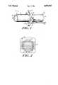
- FIG. 1is a perspective view of a device for the transmission of neural signal according to the present invention.
- FIG. 2is a cross-sectional view of the electrode yoke and fiber electrodes of the device of FIG. 1.
- an apparatus 10 for neural signal transmissionincluding a tubular nerve guidance channel 12 having an open proximal end 14 and a distal end 16 which can also be open but preferably is capped.
- the outer surface 18 of the nerve guidance channel 12is preferably partially fenestrated with relatively large (e.g., 1-20 micron) pores while the inner surface 20 of the channel 12 possesses a relatively smooth skin having relatively small (e.g., 100-800 angstrom) pores.
- the inner and outer poresare joined by interconnected passageways to provide a supporting structure for the semipermeable inner skin of the membrane.
- the nerve guidance channel 12can be formed, for example, from an acrylic copolymer.
- Tubular membranes of this typecan be obtained, for example, from the Amicon Corporation of Lexington, MA (XM50 tubing, a polyvinyl chloride acrylic copolymer, molecular weight cutoff approximately 50,000 having an internal diameter of about 1 millimeter and a wall thickness of about 100 ⁇ m).
- the distal end 16can be sealed by dipping it into an epoxy glue or a polymeric solution of the same composition as the membrane.
- the apparatus 10 of FIG. 1also includes an electrode yoke 22 disposed about the channel 12 and a plurality of electrodes 24, preferably constructed from carbon fibers and mounted onto connectors 26 on the yoke 22.
- the connectors 26provide individual connections to posts 28, which are adapted to receive lead wires 30 from electrode wire bundles 32A, 32B.
- the yoke 22is preferably constructed of silicon or other insulating biocompatible material. Lead wires 30 and bundles 32 are similarly coated with a biocompatible polymeric material, such as silicone, polytetrafluoroethylene or polyurethane.
- electrodes 24pass through the tubular membrane of channel 12 and form a network of parallel strands within the lumen of the channel.
- the diameter of the individual fibers 24can range from about 10 microns to about 50 microns and the spacing between fibers can range from about 50 microns to about 500 microns depending on the size of the lumen and the nerve.
- the proximal end of a severed nerveis drawn into the tubular channel 12 and allowed to grow by regenerative processes into the channel until it makes electrical contact with electrode network 24. Electrical signals from the nerve can then be transmitted via the electrodes 24 and lead wires 32 to a prosthetic device.
- the prosthetic device(not shown) can further include low noise amplifiers, filters and threshold trigger circuits, as known in the art of signal processing, to extract the neural motor signals necessary to control the prosthetic device.
- miceMale albino mice were anesthetized by inhalation of methoxyflurane.
- the left sciatic nervewas exposed through a skin incision along the anterior medial aspect of the thigh after retracting the gluteus maximus muscle.
- the nervewas transected about 8 mm proximal to the tibio-peroneal bifurcation and the remaining distal nerve branches were resected.
- the proximal stumpwas then secured 1 mm from the end of a 6 mm acrylic copolymer tube (Amicon Corp. Lexington, MA) with a single 10-0 nylon suture.
- micewere divided into two groups; in one group the proximal stumps were secured to polymer tubes with their distal ends left open, and in the other group the nerve stumps were secured to identical polymer tubes in which the distal ends were capped.
- a solution of the same acrylic copolymerwas applied to the distal end to occlude it.
- the polymeric solutionfused the tube's end and created a seal impervious to water.
- the channelswere primed with sterile physiological saline prior to implantation.
- micewere again deeply anesthetized and then sacrificed by transcardial perfusion with phosphate-buffered saline followed by saline solutions of paraformaldehyde and glutaraldehyde.
- the operative sitewas reopened and the guidance channel and 3 mm of native proximal nerve were removed.
- Serial transverse sections of the channels and regenerated nerveswere cut, fixed and stained for light microscopy and transmission electron microscopy.
- All capped permselective acrylic copolymer channelscontained regenerated nerve cables which extended fully to the distal end of the channel.
- the cableswere centrally located and surrounded by an acellular gel although, in one case, the cable was in direct contact with the inner wall of the guidance channel for about 2 millimeters.
- the proximal nerve stumpdisplayed the typical enlargement associated with peripheral nerve entubulation.
- the cross-sectional area of the cablesdecreased gradually as the cables extended from the original proximal nerve stump to the distal cap with a slight increase as they reached the cap end.
- the regenerated nerves in the capped channelswere sheathed by an epineurium-like tissue and contained numerous small blood vessels. They contained numerous myelinated axons up the to channel end, although their number decreased as they approached the distal cap.
- the cabledid not present the typical microfasciculation normally observed in nerves regenerated through synthetic guidance channels. There was a significant increase in the number of myelinated axons between 4 and 8 weeks at each distal interval.
- the axonswere associated with Schwann cells, which were identified from their morphological characteristics and their association with axons.
- the cables in the uncapped channelsalso contained significantly fewer myelinated axons than cables in the capped acrylic copolymer channels at both time periods. Myelinated and unmyelinated axons were located in microfascicles surrounded by perineurial-like cells.
- a comparative examplewas carried out using non-permeable silicone elastomer channels.
- capped silicone elastomer channelscontained only fine threads of connective tissue which extended for no more than 1 mm from the proximal nerve stump.
- the cableswere composed of granulation tissue with circumferentially arranged cells and did not contain any myelinated axons.
- the proximal native nerve stumpexhibited an abnormally thick epineurium.
- One of 3 uncapped silicone elastomer channel at 4 weeks and 2 of 3 at 8 weeksshowed small tissue cables extending up to the distal end.
- the cablescontained numerous blood vessels and were surrounded by a relatively thick epineurium. At both time periods these structures contained fewer than 100 myelinated axons at the midpoint of the guidance channel and no myelinated axons at their distal end. Patterns of microfasciculation were observed.
- the experimentsdemonstrate that when the proximal stump of a mouse sciatic nerve is placed into a blind-ended, 6 mm long permselective channel, a large regenerated nerve cable containing myelinated and unmyelinated axons extended to the distal end of the channel.
- blind-ended silicone channelscontained only fine threads of connective tissue extending for less than 1 mm from the proximal stump.
- permselective channelsshow substantial regeneration into a synthetic guidance channel in the absence of a distal nerve stump, refuting the dogma that the distal nerve stump is necessary for supporting more than just abortive sprouting from the proximal nerve stump. Growth and/or trophic factors other than those released by the distal nerve stump appear able to elicit the regeneration process.
- Permselective materialsappear to offer the advantages of providing a large surface area for diffusive interactions while controlling the size of the solutes which pass across the wall, and preventing any cellular invasion from the outside.
- Permselective channelsappear to support regeneration in the absence of a distal nerve stump by allowing the inward passage of nutrients and growth or trophic factors from the external wound environment while preventing the inward passage of scar forming cells.
- Cells participating in the wound healing phenomenaare known to release various peptide growth factors which are effective at monogram concentrations. Several of these factors have been purified and show molecular weights in the range of 20,000-40,000 daltons, thus they will pass through the wall of the acrylic copolymer channels used in this study.
- Activated macrophagessecrete numerous growth factors, including platelet derived growth factor (PDGF) and macrophage derived growth factor (MDGF).
- PDGFplatelet derived growth factor
- MDGFmacrophage derived growth factor
- Macrophagesare consistently observed within the trabecular structure of the acrylic copolymer guidance channels.
- These macrophagesmay be secreting growth or trophic factors into the regenerating environment.
- Growth factorsmay also be released by other cells of the wound healing process, such as endothelial cells, e.g., endothelial derived growth factor (EDGF), and fibroblasts e.g., fibroblast growth factor (FGF).
- endothelial cellse.g., endothelial derived growth factor (EDGF)
- fibroblastse.g., fibroblast growth factor (FGF).
- permselective channelmay also allow the retention within the regenerating environment of growth or trophic factors secreted by the proximal stump.
- Schwann cellshave been shown to secrete laminin, a high molecular weight glycoprotein, which exerts neurite promoting activity in vitro.
- the blood vessels located in the proximal nerve stumpmay supply high molecular weight serum molecules such as fibronectin or alpha-1-acid-glycoprotein, which have been shown to support neural survival and promote neurite elongation in vitro.
- the greater degree of regeneration observed in capped channels compared to uncapped onesmay be related to a greater retention of growth and/or trophic factors which diffused inward from the extra-channel fluid as well as the retention of growth and/or trophic factors secreted by the proximal stump.
Landscapes
- Health & Medical Sciences (AREA)
- Cardiology (AREA)
- Oral & Maxillofacial Surgery (AREA)
- Transplantation (AREA)
- Engineering & Computer Science (AREA)
- Biomedical Technology (AREA)
- Heart & Thoracic Surgery (AREA)
- Vascular Medicine (AREA)
- Life Sciences & Earth Sciences (AREA)
- Animal Behavior & Ethology (AREA)
- General Health & Medical Sciences (AREA)
- Public Health (AREA)
- Veterinary Medicine (AREA)
- Prostheses (AREA)
- Materials For Medical Uses (AREA)
- Electrotherapy Devices (AREA)
Abstract
Description
Claims (16)
Priority Applications (14)
| Application Number | Priority Date | Filing Date | Title |
|---|---|---|---|
| US07/093,371US4878913A (en) | 1987-09-04 | 1987-09-04 | Devices for neural signal transmission |
| DE8810469UDE8810469U1 (en) | 1987-09-04 | 1988-08-18 | Device for transmitting neural signals |
| EP88307737AEP0306187A3 (en) | 1987-09-04 | 1988-08-22 | Devices for neural signal transmission |
| IL87592AIL87592A0 (en) | 1987-09-04 | 1988-08-29 | Devices and method for neural signal transmission |
| ZA886506AZA886506B (en) | 1987-09-04 | 1988-09-01 | Devices and methods for neural signal transmission |
| FI884065AFI884065A7 (en) | 1987-09-04 | 1988-09-02 | Devices and methods for transmitting nerve signals. |
| NZ226031ANZ226031A (en) | 1987-09-04 | 1988-09-02 | Device for neural signal transmission: guidance channel encloses end of transected nerve |
| JP63218673AJPS6470042A (en) | 1987-09-04 | 1988-09-02 | Transmission device for nerve signal and method for manufacture of the same |
| NO88883931ANO883931L (en) | 1987-09-04 | 1988-09-02 | DEVICES AND PROCEDURES FOR NERVESIGNAL TRANSMISSION. |
| PT88408APT88408A (en) | 1987-09-04 | 1988-09-02 | DEVICES AND METHODS FOR THE TRANSMISSION OF NEURAL SINS |
| AU21842/88AAU596752B2 (en) | 1987-09-04 | 1988-09-02 | Devices and methods for neural signal transmission |
| DK489088ADK489088A (en) | 1987-09-04 | 1988-09-02 | DEVICE FOR TRANSMISSION OF NERVESIGNALS AND PROCEDURES FOR PRODUCING THEREOF |
| BR888804548ABR8804548A (en) | 1987-09-04 | 1988-09-02 | DEVICE FOR THE TRANSMISSION OF NEURAL SIGNS AND PROCESS FOR MANUFACTURING THE SAME |
| KR1019880011389AKR900008830B1 (en) | 1987-09-04 | 1988-09-03 | Device for nsural signal transmission |
Applications Claiming Priority (1)
| Application Number | Priority Date | Filing Date | Title |
|---|---|---|---|
| US07/093,371US4878913A (en) | 1987-09-04 | 1987-09-04 | Devices for neural signal transmission |
Publications (1)
| Publication Number | Publication Date |
|---|---|
| US4878913Atrue US4878913A (en) | 1989-11-07 |
Family
ID=22238554
Family Applications (1)
| Application Number | Title | Priority Date | Filing Date |
|---|---|---|---|
| US07/093,371Expired - Fee RelatedUS4878913A (en) | 1987-09-04 | 1987-09-04 | Devices for neural signal transmission |
Country Status (14)
| Country | Link |
|---|---|
| US (1) | US4878913A (en) |
| EP (1) | EP0306187A3 (en) |
| JP (1) | JPS6470042A (en) |
| KR (1) | KR900008830B1 (en) |
| AU (1) | AU596752B2 (en) |
| BR (1) | BR8804548A (en) |
| DE (1) | DE8810469U1 (en) |
| DK (1) | DK489088A (en) |
| FI (1) | FI884065A7 (en) |
| IL (1) | IL87592A0 (en) |
| NO (1) | NO883931L (en) |
| NZ (1) | NZ226031A (en) |
| PT (1) | PT88408A (en) |
| ZA (1) | ZA886506B (en) |
Cited By (79)
| Publication number | Priority date | Publication date | Assignee | Title |
|---|---|---|---|---|
| US5011486A (en)* | 1988-11-18 | 1991-04-30 | Brown University Research Foundation | Composite nerve guidance channels |
| US5030225A (en)* | 1987-03-13 | 1991-07-09 | Brown University Research Foundation | Electrically-charged nerve guidance channels |
| US5192312A (en)* | 1991-03-05 | 1993-03-09 | Colorado State University Research Foundation | Treated tissue for implantation and methods of treatment and use |
| US5222982A (en)* | 1991-02-11 | 1993-06-29 | Ommaya Ayub K | Spinal fluid driven artificial organ |
| US5314471A (en)* | 1991-07-24 | 1994-05-24 | Baxter International Inc. | Tissue inplant systems and methods for sustaining viable high cell densities within a host |
| US5344454A (en)* | 1991-07-24 | 1994-09-06 | Baxter International Inc. | Closed porous chambers for implanting tissue in a host |
| US5385582A (en)* | 1991-02-11 | 1995-01-31 | Ommaya; Ayub K. | Spinal fluid driven artificial organ |
| WO1995020359A1 (en)* | 1994-01-26 | 1995-08-03 | Institute Of Molecular Biology, Inc. | A device to promote drug-induced nerve regeneration |
| US5453278A (en)* | 1991-07-24 | 1995-09-26 | Baxter International Inc. | Laminated barriers for tissue implants |
| US5476494A (en)* | 1992-09-11 | 1995-12-19 | Massachusetts Institute Of Technology | Low pressure neural contact structure |
| US5545223A (en)* | 1990-10-31 | 1996-08-13 | Baxter International, Inc. | Ported tissue implant systems and methods of using same |
| US5569462A (en)* | 1993-09-24 | 1996-10-29 | Baxter International Inc. | Methods for enhancing vascularization of implant devices |
| US5613982A (en)* | 1994-03-14 | 1997-03-25 | Cryolife, Inc. | Method of preparing transplant tissue to reduce immunogenicity upon implantation |
| US5713888A (en)* | 1990-10-31 | 1998-02-03 | Baxter International, Inc. | Tissue implant systems |
| US5741330A (en)* | 1990-10-31 | 1998-04-21 | Baxter International, Inc. | Close vascularization implant material |
| US5925053A (en)* | 1997-09-02 | 1999-07-20 | Children's Medical Center Corporation | Multi-lumen polymeric guidance channel, method for promoting nerve regeneration, and method of manufacturing a multi-lumen nerve guidance channel |
| US6156305A (en)* | 1994-07-08 | 2000-12-05 | Baxter International Inc. | Implanted tumor cells for the prevention and treatment of cancer |
| US6500210B1 (en) | 1992-09-08 | 2002-12-31 | Seattle Systems, Inc. | System and method for providing a sense of feel in a prosthetic or sensory impaired limb |
| US6506727B1 (en)* | 1991-11-25 | 2003-01-14 | Institute Of Molecular Biology, Inc. | Nerve regeneration |
| US20030093129A1 (en)* | 2001-10-29 | 2003-05-15 | Nicolelis Miguel A.L. | Closed loop brain machine interface |
| US20030105409A1 (en)* | 2001-11-14 | 2003-06-05 | Donoghue John Philip | Neurological signal decoding |
| US6609017B1 (en)* | 1998-08-07 | 2003-08-19 | California Institute Of Technology | Processed neural signals and methods for generating and using them |
| US20030170214A1 (en)* | 2000-05-29 | 2003-09-11 | Augustinus Bader | Method for producing a bio-artificial transplant |
| US20030176905A1 (en)* | 2002-03-14 | 2003-09-18 | Nicolelis Miguel A.L. | Miniaturized high-density multichannel electrode array for long-term neuronal recordings |
| US20030204197A1 (en)* | 2002-04-26 | 2003-10-30 | Medtronic, Inc. | Sintered titanium tube for the management of spinal cord injury |
| WO2003047422A3 (en)* | 2001-12-04 | 2003-11-13 | Univ Pennsylvania | Device and method using integrated neuronal cells and an electronic device |
| US20040006264A1 (en)* | 2001-11-20 | 2004-01-08 | Mojarradi Mohammad M. | Neural prosthetic micro system |
| US20040015211A1 (en)* | 2002-06-04 | 2004-01-22 | Nurmikko Arto V. | Optically-connected implants and related systems and methods of use |
| US20040028662A1 (en)* | 2000-05-29 | 2004-02-12 | Augustinus Bader | Method for producing a recipient-specific tissue transplant or tissue implant |
| US20040111140A1 (en)* | 2001-01-11 | 2004-06-10 | Thomas Stieglitz | Sieve electrode which can be connected to a nerve stump |
| US6773458B1 (en) | 1991-07-24 | 2004-08-10 | Baxter International Inc. | Angiogenic tissue implant systems and methods |
| US20040249302A1 (en)* | 2003-06-09 | 2004-12-09 | Cyberkinetics, Inc. | Methods and systems for processing of brain signals |
| US20050113744A1 (en)* | 2003-11-21 | 2005-05-26 | Cyberkinetics, Inc. | Agent delivery systems and related methods under control of biological electrical signals |
| US20050203366A1 (en)* | 2004-03-12 | 2005-09-15 | Donoghue John P. | Neurological event monitoring and therapy systems and related methods |
| US20050251221A1 (en)* | 2004-05-08 | 2005-11-10 | Bojan Zdravkovic | Neural bridge devices and methods for restoring and modulating neural activity |
| US20050267597A1 (en)* | 2003-11-25 | 2005-12-01 | Flaherty J Christopher | Neural interface system with embedded id |
| US20060049957A1 (en)* | 2004-08-13 | 2006-03-09 | Surgenor Timothy R | Biological interface systems with controlled device selector and related methods |
| US20060167371A1 (en)* | 2005-01-10 | 2006-07-27 | Flaherty J Christopher | Biological interface system with patient training apparatus |
| US20060167564A1 (en)* | 2005-01-10 | 2006-07-27 | Flaherty J C | Limb and digit movement system |
| US20060173259A1 (en)* | 2004-10-04 | 2006-08-03 | Flaherty J C | Biological interface system |
| US20060189900A1 (en)* | 2005-01-18 | 2006-08-24 | Flaherty J C | Biological interface system with automated configuration |
| US20060217816A1 (en)* | 2004-12-16 | 2006-09-28 | California Institute Of Technology | Prosthetic devices and methods and systems related thereto |
| US20060252788A1 (en)* | 2005-04-06 | 2006-11-09 | Went Gregory T | Methods and compositions for the treatment of CNS-related conditions |
| US20070032738A1 (en)* | 2005-01-06 | 2007-02-08 | Flaherty J C | Adaptive patient training routine for biological interface system |
| US7212851B2 (en) | 2002-10-24 | 2007-05-01 | Brown University Research Foundation | Microstructured arrays for cortex interaction and related methods of manufacture and use |
| US20070106143A1 (en)* | 2005-11-08 | 2007-05-10 | Flaherty J C | Electrode arrays and related methods |
| US20070156126A1 (en)* | 2005-12-29 | 2007-07-05 | Flaherty J C | Medical device insertion system and related methods |
| US20080228240A1 (en)* | 2004-06-17 | 2008-09-18 | Edell David J | Long Term Bi-Directional Axon-Electronic Communication System |
| US20080299201A1 (en)* | 2006-07-06 | 2008-12-04 | International Business Machines Corporation | Devices, methods, and systems for accessing native neurons through artificial neural mediators (anms) |
| US20080319506A1 (en)* | 2007-06-25 | 2008-12-25 | Microtransponder, Inc. | Grooved electrode and wireless microtransponder system |
| US20090081296A1 (en)* | 2006-02-02 | 2009-03-26 | Humes H David | Extracorporeal cell-based therapeutic device and delivery system |
| US20090112286A1 (en)* | 2005-12-30 | 2009-04-30 | Southeast University | Neural channel bridge aided by a micro-electronic system |
| US20090306687A1 (en)* | 2005-10-31 | 2009-12-10 | Life Spring Biotech Co., Ltd. | Scleral buckling band and method for making the same |
| US7647097B2 (en) | 2003-12-29 | 2010-01-12 | Braingate Co., Llc | Transcutaneous implant |
| US20100023021A1 (en)* | 2005-12-27 | 2010-01-28 | Flaherty J Christopher | Biological Interface and Insertion |
| WO2010019643A3 (en)* | 2008-08-15 | 2010-06-10 | Innovative Biotherapies, Inc. | An extracorporeal cell-based therapeutic device and delivery system |
| US20100211172A1 (en)* | 2007-04-02 | 2010-08-19 | Georgia Tech Research Corporation | Implantable Device For Communicating With Biological Tissue |
| US7783360B2 (en) | 2006-10-23 | 2010-08-24 | Bojan Zdravkovic | Sensory system |
| US7783363B2 (en) | 2006-10-23 | 2010-08-24 | Artis Nanomedica, Inc. | Neural bridge gateway and calibrator |
| US7826894B2 (en) | 2004-03-22 | 2010-11-02 | California Institute Of Technology | Cognitive control signals for neural prosthetics |
| US20100286796A1 (en)* | 2009-05-05 | 2010-11-11 | Ossur Hf | Control systems and methods for prosthetic or orthotic devices |
| US20100324698A1 (en)* | 2009-06-17 | 2010-12-23 | Ossur Hf | Feedback control systems and methods for prosthetic or orthotic devices |
| US7901368B2 (en) | 2005-01-06 | 2011-03-08 | Braingate Co., Llc | Neurally controlled patient ambulation system |
| US8048419B2 (en) | 2006-02-02 | 2011-11-01 | Innovative Biotherapies, Inc. | Extracorporeal cell-based therapeutic device and delivery system |
| US8095209B2 (en) | 2005-01-06 | 2012-01-10 | Braingate Co., Llc | Biological interface system with gated control signal |
| US8122772B2 (en) | 2005-02-02 | 2012-02-28 | össur hf | Sensing systems and methods for monitoring gait dynamics |
| US8323354B2 (en) | 2003-11-18 | 2012-12-04 | Victhom Human Bionics Inc. | Instrumented prosthetic foot |
| US8617254B2 (en) | 2004-03-10 | 2013-12-31 | Ossur Hf | Control system and method for a prosthetic knee |
| US8657886B2 (en) | 2004-02-12 | 2014-02-25 | össur hf | Systems and methods for actuating a prosthetic ankle |
| US8702811B2 (en) | 2005-09-01 | 2014-04-22 | össur hf | System and method for determining terrain transitions |
| US8801802B2 (en) | 2005-02-16 | 2014-08-12 | össur hf | System and method for data communication with a mechatronic device |
| US8814949B2 (en) | 2005-04-19 | 2014-08-26 | össur hf | Combined active and passive leg prosthesis system and a method for performing a movement with such a system |
| US9029144B2 (en) | 2008-06-18 | 2015-05-12 | Innovative Bio Therapies, Inc. | Methods for enhanced propagation of cells |
| US9078774B2 (en) | 2004-12-22 | 2015-07-14 | össur hf | Systems and methods for processing limb motion |
| US9358137B2 (en) | 2002-08-22 | 2016-06-07 | Victhom Laboratory Inc. | Actuated prosthesis for amputees |
| US9526636B2 (en) | 2003-11-18 | 2016-12-27 | Victhom Laboratory Inc. | Instrumented prosthetic foot |
| US9561118B2 (en) | 2013-02-26 | 2017-02-07 | össur hf | Prosthetic foot with enhanced stability and elastic energy return |
| US9649206B2 (en) | 2002-08-22 | 2017-05-16 | Victhom Laboratory Inc. | Control device and system for controlling an actuated prosthesis |
| US10195057B2 (en) | 2004-02-12 | 2019-02-05 | össur hf. | Transfemoral prosthetic systems and methods for operating the same |
Families Citing this family (2)
| Publication number | Priority date | Publication date | Assignee | Title |
|---|---|---|---|---|
| HU0100664D0 (en)* | 2001-02-09 | 2001-04-28 | Pali Jenoe | Apparatus for simultaneous, individual measurement of numerous motor nerve-fibres, as well as for simultaneous, individual stimulation of sensory nerve fibres of the peripheral nervous system |
| DE102005054941A1 (en) | 2005-11-17 | 2007-05-31 | Gelita Ag | nerve |
Citations (16)
| Publication number | Priority date | Publication date | Assignee | Title |
|---|---|---|---|---|
| US3786817A (en)* | 1972-06-01 | 1974-01-22 | Palma J | Method and apparatus for aiding severed nerves to join |
| US3833002A (en)* | 1973-09-10 | 1974-09-03 | J Palma | Apparatus for aiding severed nerves to join |
| US3916905A (en)* | 1973-11-09 | 1975-11-04 | William E Kuhn | Method and means for the repair of severed peripheral nerves |
| US3955560A (en)* | 1974-06-10 | 1976-05-11 | Stein Richard B | Implantable neural electrode |
| US3960152A (en)* | 1974-01-21 | 1976-06-01 | American Cyanamid Company | Surgical sutures of unsymmetrically substituted 1,4-dioxane-2,5-diones |
| US3988411A (en)* | 1974-02-11 | 1976-10-26 | American Cyanamid Company | Spinning and shaping poly-(N-acetyl-D-glucosamine) |
| US4011861A (en)* | 1974-04-03 | 1977-03-15 | Case Western Reserve University | Implantable electric terminal for organic tissue |
| US4033938A (en)* | 1974-01-21 | 1977-07-05 | American Cyanamid Company | Polymers of unsymmetrically substituted 1,4-dioxane-2,5-diones |
| US4074366A (en)* | 1975-03-14 | 1978-02-21 | American Cyanamid Company | Poly(N-acetyl-D-glucosamine) products |
| US4369104A (en)* | 1979-10-22 | 1983-01-18 | Hitco | Continuous filament graphite composite electrodes |
| US4461304A (en)* | 1979-11-05 | 1984-07-24 | Massachusetts Institute Of Technology | Microelectrode and assembly for parallel recording of neurol groups |
| US4481353A (en)* | 1983-10-07 | 1984-11-06 | The Children's Medical Center Corporation | Bioresorbable polyesters and polyester composites |
| US4534349A (en)* | 1983-02-02 | 1985-08-13 | Minnesota Mining And Manufacturing Company | Absorbable sutureless nerve repair device |
| US4623355A (en)* | 1984-03-16 | 1986-11-18 | Sawruk Stephen D | Prosthetic axon |
| US4662884A (en)* | 1984-04-25 | 1987-05-05 | University Of Utah Research Foundation | Prostheses and methods for promoting nerve regeneration |
| EP0261833A2 (en)* | 1986-09-09 | 1988-03-30 | American BioInterface Corporation | Apparatus for mammalian nerve regeneration |
Family Cites Families (1)
| Publication number | Priority date | Publication date | Assignee | Title |
|---|---|---|---|---|
| US4031882A (en)* | 1975-07-14 | 1977-06-28 | Liberty Mutual Insurance Company | Apparatus for interfacing to anatomic signal sources |
- 1987
- 1987-09-04USUS07/093,371patent/US4878913A/ennot_activeExpired - Fee Related
- 1988
- 1988-08-18DEDE8810469Upatent/DE8810469U1/ennot_activeExpired
- 1988-08-22EPEP88307737Apatent/EP0306187A3/ennot_activeWithdrawn
- 1988-08-29ILIL87592Apatent/IL87592A0/enunknown
- 1988-09-01ZAZA886506Apatent/ZA886506B/enunknown
- 1988-09-02DKDK489088Apatent/DK489088A/ennot_activeApplication Discontinuation
- 1988-09-02PTPT88408Apatent/PT88408A/ennot_activeApplication Discontinuation
- 1988-09-02AUAU21842/88Apatent/AU596752B2/ennot_activeCeased
- 1988-09-02FIFI884065Apatent/FI884065A7/ennot_activeIP Right Cessation
- 1988-09-02NONO88883931Apatent/NO883931L/enunknown
- 1988-09-02NZNZ226031Apatent/NZ226031A/enunknown
- 1988-09-02JPJP63218673Apatent/JPS6470042A/enactivePending
- 1988-09-02BRBR888804548Apatent/BR8804548A/enunknown
- 1988-09-03KRKR1019880011389Apatent/KR900008830B1/ennot_activeExpired
Patent Citations (16)
| Publication number | Priority date | Publication date | Assignee | Title |
|---|---|---|---|---|
| US3786817A (en)* | 1972-06-01 | 1974-01-22 | Palma J | Method and apparatus for aiding severed nerves to join |
| US3833002A (en)* | 1973-09-10 | 1974-09-03 | J Palma | Apparatus for aiding severed nerves to join |
| US3916905A (en)* | 1973-11-09 | 1975-11-04 | William E Kuhn | Method and means for the repair of severed peripheral nerves |
| US3960152A (en)* | 1974-01-21 | 1976-06-01 | American Cyanamid Company | Surgical sutures of unsymmetrically substituted 1,4-dioxane-2,5-diones |
| US4033938A (en)* | 1974-01-21 | 1977-07-05 | American Cyanamid Company | Polymers of unsymmetrically substituted 1,4-dioxane-2,5-diones |
| US3988411A (en)* | 1974-02-11 | 1976-10-26 | American Cyanamid Company | Spinning and shaping poly-(N-acetyl-D-glucosamine) |
| US4011861A (en)* | 1974-04-03 | 1977-03-15 | Case Western Reserve University | Implantable electric terminal for organic tissue |
| US3955560A (en)* | 1974-06-10 | 1976-05-11 | Stein Richard B | Implantable neural electrode |
| US4074366A (en)* | 1975-03-14 | 1978-02-21 | American Cyanamid Company | Poly(N-acetyl-D-glucosamine) products |
| US4369104A (en)* | 1979-10-22 | 1983-01-18 | Hitco | Continuous filament graphite composite electrodes |
| US4461304A (en)* | 1979-11-05 | 1984-07-24 | Massachusetts Institute Of Technology | Microelectrode and assembly for parallel recording of neurol groups |
| US4534349A (en)* | 1983-02-02 | 1985-08-13 | Minnesota Mining And Manufacturing Company | Absorbable sutureless nerve repair device |
| US4481353A (en)* | 1983-10-07 | 1984-11-06 | The Children's Medical Center Corporation | Bioresorbable polyesters and polyester composites |
| US4623355A (en)* | 1984-03-16 | 1986-11-18 | Sawruk Stephen D | Prosthetic axon |
| US4662884A (en)* | 1984-04-25 | 1987-05-05 | University Of Utah Research Foundation | Prostheses and methods for promoting nerve regeneration |
| EP0261833A2 (en)* | 1986-09-09 | 1988-03-30 | American BioInterface Corporation | Apparatus for mammalian nerve regeneration |
Non-Patent Citations (18)
| Title |
|---|
| daSilva et al., Brain Research, vol. 342, pp. 307 315, 1985.* |
| daSilva et al., Brain Research, vol. 342, pp. 307-315, 1985. |
| Ducker et al., J. Neurosurg., vol. 28, pp. 582 587, 1967.* |
| Ducker et al., J. Neurosurg., vol. 28, pp. 582-587, 1967. |
| Edell, IEEE Trans., vol. BME 33, No. 2, (Feb. 1986).* |
| Edell, IEEE Trans., vol. BME-33, No. 2, (Feb. 1986). |
| Lundborg et al., Journal of Neuropathology and Experimental Neurology, vol. 41, No. 4, pp. 412 422, 1982.* |
| Lundborg et al., Journal of Neuropathology and Experimental Neurology, vol. 41, No. 4, pp. 412-422, 1982. |
| Midgley et al., Surgical Forum, vol. 19, pp. 519 520, 1968.* |
| Midgley et al., Surgical Forum, vol. 19, pp. 519-520, 1968. |
| Molander et al., Muscle & Nerve, vol. 5, pp. 54 57, 1982.* |
| Molander et al., Muscle & Nerve, vol. 5, pp. 54-57, 1982. |
| Nyilas et al., Trans Am Soc Artif Intern Organs, vol. XXIX, pp. 307 313, 1983.* |
| Nyilas et al., Trans Am Soc Artif Intern Organs, vol. XXIX, pp. 307-313, 1983. |
| Sickel et al., Plastic & Reconstructive Surgery, vol. 74(2), pp. 173 181, 1983.* |
| Sickel et al., Plastic & Reconstructive Surgery, vol. 74(2), pp. 173-181, 1983. |
| Uzman et al., Journal of Neuroscience Research, vol. 9, pp. 325 338, 1983.* |
| Uzman et al., Journal of Neuroscience Research, vol. 9, pp. 325-338, 1983. |
Cited By (139)
| Publication number | Priority date | Publication date | Assignee | Title |
|---|---|---|---|---|
| US5030225A (en)* | 1987-03-13 | 1991-07-09 | Brown University Research Foundation | Electrically-charged nerve guidance channels |
| US5011486A (en)* | 1988-11-18 | 1991-04-30 | Brown University Research Foundation | Composite nerve guidance channels |
| US5653756A (en)* | 1990-10-31 | 1997-08-05 | Baxter International Inc. | Closed porous chambers for implanting tissue in a host |
| US5882354A (en)* | 1990-10-31 | 1999-03-16 | Baxter International Inc. | Close vascularization implant material |
| US5593440A (en)* | 1990-10-31 | 1997-01-14 | Baxter International Inc. | Tissue implant systems and methods for sustaining viable high cell densities within a host |
| US5800529A (en)* | 1990-10-31 | 1998-09-01 | Baxter International, Inc. | Close vascularization implant material |
| US5964804A (en)* | 1990-10-31 | 1999-10-12 | Baxter International Inc. | Close vascularization implant material |
| US5741330A (en)* | 1990-10-31 | 1998-04-21 | Baxter International, Inc. | Close vascularization implant material |
| US5733336A (en)* | 1990-10-31 | 1998-03-31 | Baxter International, Inc. | Ported tissue implant systems and methods of using same |
| US5713888A (en)* | 1990-10-31 | 1998-02-03 | Baxter International, Inc. | Tissue implant systems |
| US5545223A (en)* | 1990-10-31 | 1996-08-13 | Baxter International, Inc. | Ported tissue implant systems and methods of using same |
| US5222982A (en)* | 1991-02-11 | 1993-06-29 | Ommaya Ayub K | Spinal fluid driven artificial organ |
| US5385582A (en)* | 1991-02-11 | 1995-01-31 | Ommaya; Ayub K. | Spinal fluid driven artificial organ |
| US5772695A (en)* | 1991-03-05 | 1998-06-30 | Colorado State University Research Foundation | Treated tissue for implantation and methods of treatment and use |
| US5192312A (en)* | 1991-03-05 | 1993-03-09 | Colorado State University Research Foundation | Treated tissue for implantation and methods of treatment and use |
| US5855617A (en)* | 1991-03-05 | 1999-01-05 | Colorado State University Research Foundation | Treated tissue for implantation and methods of treatment and use |
| US5863296A (en)* | 1991-03-05 | 1999-01-26 | Colorado State University Research Foundation | Treated tissue for implantation and methods of treatment and use |
| US6773458B1 (en) | 1991-07-24 | 2004-08-10 | Baxter International Inc. | Angiogenic tissue implant systems and methods |
| US5453278A (en)* | 1991-07-24 | 1995-09-26 | Baxter International Inc. | Laminated barriers for tissue implants |
| US5344454A (en)* | 1991-07-24 | 1994-09-06 | Baxter International Inc. | Closed porous chambers for implanting tissue in a host |
| US5314471A (en)* | 1991-07-24 | 1994-05-24 | Baxter International Inc. | Tissue inplant systems and methods for sustaining viable high cell densities within a host |
| US6506727B1 (en)* | 1991-11-25 | 2003-01-14 | Institute Of Molecular Biology, Inc. | Nerve regeneration |
| US6500210B1 (en) | 1992-09-08 | 2002-12-31 | Seattle Systems, Inc. | System and method for providing a sense of feel in a prosthetic or sensory impaired limb |
| US5575813A (en)* | 1992-09-11 | 1996-11-19 | Massachusetts Institute Of Technology | Low-pressure neural contact structure |
| US5476494A (en)* | 1992-09-11 | 1995-12-19 | Massachusetts Institute Of Technology | Low pressure neural contact structure |
| US5569462A (en)* | 1993-09-24 | 1996-10-29 | Baxter International Inc. | Methods for enhancing vascularization of implant devices |
| US5656605A (en)* | 1994-01-26 | 1997-08-12 | Institute Of Molecular Biology, Inc. | Device to promote drug-induced nerve regeneration |
| WO1995020359A1 (en)* | 1994-01-26 | 1995-08-03 | Institute Of Molecular Biology, Inc. | A device to promote drug-induced nerve regeneration |
| US5899936A (en)* | 1994-03-14 | 1999-05-04 | Cryolife, Inc. | Treated tissue for implantation and methods of preparation |
| US5843182A (en)* | 1994-03-14 | 1998-12-01 | Cryolife, Inc. | Treated tissue for implantation and methods of preparation |
| US5632778A (en)* | 1994-03-14 | 1997-05-27 | Cryolife, Inc. | Treated tissue for implantation and methods of preparation |
| US5613982A (en)* | 1994-03-14 | 1997-03-25 | Cryolife, Inc. | Method of preparing transplant tissue to reduce immunogenicity upon implantation |
| US6156305A (en)* | 1994-07-08 | 2000-12-05 | Baxter International Inc. | Implanted tumor cells for the prevention and treatment of cancer |
| US5925053A (en)* | 1997-09-02 | 1999-07-20 | Children's Medical Center Corporation | Multi-lumen polymeric guidance channel, method for promoting nerve regeneration, and method of manufacturing a multi-lumen nerve guidance channel |
| US6214021B1 (en) | 1997-09-02 | 2001-04-10 | Children's Medical Center Corporation | Multi-lumen polymeric guidance channel and method of manufacturing a polymeric prosthesis |
| US6609017B1 (en)* | 1998-08-07 | 2003-08-19 | California Institute Of Technology | Processed neural signals and methods for generating and using them |
| US6731964B2 (en)* | 1998-08-07 | 2004-05-04 | California Institute Of Technology | Processed neural signals and methods for generating and using them |
| US20030170214A1 (en)* | 2000-05-29 | 2003-09-11 | Augustinus Bader | Method for producing a bio-artificial transplant |
| US7915038B2 (en)* | 2000-05-29 | 2011-03-29 | Augustinus Bader | Method for producing a recipient-specific tissue transplant or tissue implant |
| US20040028662A1 (en)* | 2000-05-29 | 2004-02-12 | Augustinus Bader | Method for producing a recipient-specific tissue transplant or tissue implant |
| US6908470B2 (en)* | 2001-01-11 | 2005-06-21 | Fraunhofer-Gesellschaft Zur Foderung Der Angewandten Forschung E.V. | Sieve electrode which can be connected to a nerve stump |
| US20040111140A1 (en)* | 2001-01-11 | 2004-06-10 | Thomas Stieglitz | Sieve electrode which can be connected to a nerve stump |
| US20030093129A1 (en)* | 2001-10-29 | 2003-05-15 | Nicolelis Miguel A.L. | Closed loop brain machine interface |
| US7209788B2 (en) | 2001-10-29 | 2007-04-24 | Duke University | Closed loop brain machine interface |
| US20030105409A1 (en)* | 2001-11-14 | 2003-06-05 | Donoghue John Philip | Neurological signal decoding |
| US7392079B2 (en) | 2001-11-14 | 2008-06-24 | Brown University Research Foundation | Neurological signal decoding |
| US20040006264A1 (en)* | 2001-11-20 | 2004-01-08 | Mojarradi Mohammad M. | Neural prosthetic micro system |
| US7972367B2 (en) | 2001-12-04 | 2011-07-05 | The Trustees Of The University Of Pennsylvania | Device and method using integrated neuronal cells and an electronic device |
| WO2003047422A3 (en)* | 2001-12-04 | 2003-11-13 | Univ Pennsylvania | Device and method using integrated neuronal cells and an electronic device |
| US20080305087A1 (en)* | 2001-12-04 | 2008-12-11 | The Trustees Of The University Of Pennsylvania | Device and Method Using Integrated Neuronal Cells and an Electronic Device |
| US7429267B2 (en) | 2001-12-04 | 2008-09-30 | The Trustees Of The University Of Pennsylvania | Device and method using integrated neuronal cells and an electronic device |
| US8401635B2 (en) | 2001-12-04 | 2013-03-19 | The Trustees Of The University Of Pennsylvania | Device and method using integrated neuronal cells and an electronic device |
| US7983756B2 (en) | 2002-03-14 | 2011-07-19 | Duke University | Miniaturized high-density multichannel electrode array for long-term neuronal recordings |
| US20030176905A1 (en)* | 2002-03-14 | 2003-09-18 | Nicolelis Miguel A.L. | Miniaturized high-density multichannel electrode array for long-term neuronal recordings |
| US6993392B2 (en) | 2002-03-14 | 2006-01-31 | Duke University | Miniaturized high-density multichannel electrode array for long-term neuronal recordings |
| US20060206161A1 (en)* | 2002-03-14 | 2006-09-14 | Duke University | Miniaturized high-density multichannel electrode array for long-term neuronal recordings |
| US7147647B2 (en) | 2002-04-26 | 2006-12-12 | Medtronic, Inc. | Sintered titanium tube for the management of spinal cord injury |
| US20030204197A1 (en)* | 2002-04-26 | 2003-10-30 | Medtronic, Inc. | Sintered titanium tube for the management of spinal cord injury |
| US20040015211A1 (en)* | 2002-06-04 | 2004-01-22 | Nurmikko Arto V. | Optically-connected implants and related systems and methods of use |
| US7280870B2 (en) | 2002-06-04 | 2007-10-09 | Brown University Research Foundation | Optically-connected implants and related systems and methods of use |
| US9649206B2 (en) | 2002-08-22 | 2017-05-16 | Victhom Laboratory Inc. | Control device and system for controlling an actuated prosthesis |
| US9358137B2 (en) | 2002-08-22 | 2016-06-07 | Victhom Laboratory Inc. | Actuated prosthesis for amputees |
| US7212851B2 (en) | 2002-10-24 | 2007-05-01 | Brown University Research Foundation | Microstructured arrays for cortex interaction and related methods of manufacture and use |
| US20040249302A1 (en)* | 2003-06-09 | 2004-12-09 | Cyberkinetics, Inc. | Methods and systems for processing of brain signals |
| US8323354B2 (en) | 2003-11-18 | 2012-12-04 | Victhom Human Bionics Inc. | Instrumented prosthetic foot |
| US9526636B2 (en) | 2003-11-18 | 2016-12-27 | Victhom Laboratory Inc. | Instrumented prosthetic foot |
| US8986397B2 (en) | 2003-11-18 | 2015-03-24 | Victhom Human Bionics, Inc. | Instrumented prosthetic foot |
| US20050113744A1 (en)* | 2003-11-21 | 2005-05-26 | Cyberkinetics, Inc. | Agent delivery systems and related methods under control of biological electrical signals |
| US20050267597A1 (en)* | 2003-11-25 | 2005-12-01 | Flaherty J Christopher | Neural interface system with embedded id |
| US7751877B2 (en) | 2003-11-25 | 2010-07-06 | Braingate Co., Llc | Neural interface system with embedded id |
| US20050273890A1 (en)* | 2003-11-25 | 2005-12-08 | Flaherty J C | Neural interface system and method for neural control of multiple devices |
| US7647097B2 (en) | 2003-12-29 | 2010-01-12 | Braingate Co., Llc | Transcutaneous implant |
| US8657886B2 (en) | 2004-02-12 | 2014-02-25 | össur hf | Systems and methods for actuating a prosthetic ankle |
| US9271851B2 (en) | 2004-02-12 | 2016-03-01 | össur hf. | Systems and methods for actuating a prosthetic ankle |
| US10195057B2 (en) | 2004-02-12 | 2019-02-05 | össur hf. | Transfemoral prosthetic systems and methods for operating the same |
| US9345591B2 (en) | 2004-03-10 | 2016-05-24 | össur hf | Control system and method for a prosthetic knee |
| US8617254B2 (en) | 2004-03-10 | 2013-12-31 | Ossur Hf | Control system and method for a prosthetic knee |
| US20050203366A1 (en)* | 2004-03-12 | 2005-09-15 | Donoghue John P. | Neurological event monitoring and therapy systems and related methods |
| US7826894B2 (en) | 2004-03-22 | 2010-11-02 | California Institute Of Technology | Cognitive control signals for neural prosthetics |
| US7369900B2 (en) | 2004-05-08 | 2008-05-06 | Bojan Zdravkovic | Neural bridge devices and methods for restoring and modulating neural activity |
| US20050251221A1 (en)* | 2004-05-08 | 2005-11-10 | Bojan Zdravkovic | Neural bridge devices and methods for restoring and modulating neural activity |
| US20080228240A1 (en)* | 2004-06-17 | 2008-09-18 | Edell David J | Long Term Bi-Directional Axon-Electronic Communication System |
| US20060058627A1 (en)* | 2004-08-13 | 2006-03-16 | Flaherty J C | Biological interface systems with wireless connection and related methods |
| US20060049957A1 (en)* | 2004-08-13 | 2006-03-09 | Surgenor Timothy R | Biological interface systems with controlled device selector and related methods |
| US8560041B2 (en) | 2004-10-04 | 2013-10-15 | Braingate Co., Llc | Biological interface system |
| US20060173259A1 (en)* | 2004-10-04 | 2006-08-03 | Flaherty J C | Biological interface system |
| US7797040B2 (en) | 2004-12-16 | 2010-09-14 | California Institute Of Technology | Prosthetic devices and methods and systems related thereto |
| US8768449B2 (en) | 2004-12-16 | 2014-07-01 | California Institute Of Technology | Prosthetic devices and methods and systems related thereto |
| US20060217816A1 (en)* | 2004-12-16 | 2006-09-28 | California Institute Of Technology | Prosthetic devices and methods and systems related thereto |
| US9078774B2 (en) | 2004-12-22 | 2015-07-14 | össur hf | Systems and methods for processing limb motion |
| US7901368B2 (en) | 2005-01-06 | 2011-03-08 | Braingate Co., Llc | Neurally controlled patient ambulation system |
| US7991461B2 (en) | 2005-01-06 | 2011-08-02 | Braingate Co., Llc | Patient training routine for biological interface system |
| US20070032738A1 (en)* | 2005-01-06 | 2007-02-08 | Flaherty J C | Adaptive patient training routine for biological interface system |
| US8095209B2 (en) | 2005-01-06 | 2012-01-10 | Braingate Co., Llc | Biological interface system with gated control signal |
| US8812096B2 (en) | 2005-01-10 | 2014-08-19 | Braingate Co., Llc | Biological interface system with patient training apparatus |
| US20060167564A1 (en)* | 2005-01-10 | 2006-07-27 | Flaherty J C | Limb and digit movement system |
| US20060189901A1 (en)* | 2005-01-10 | 2006-08-24 | Flaherty J C | Biological interface system with surrogate controlled device |
| US20060189899A1 (en)* | 2005-01-10 | 2006-08-24 | Flaherty J Christopher | Joint movement apparatus |
| US20060167371A1 (en)* | 2005-01-10 | 2006-07-27 | Flaherty J Christopher | Biological interface system with patient training apparatus |
| US20060189900A1 (en)* | 2005-01-18 | 2006-08-24 | Flaherty J C | Biological interface system with automated configuration |
| US20060195042A1 (en)* | 2005-01-18 | 2006-08-31 | Flaherty J C | Biological interface system with thresholded configuration |
| US8060194B2 (en) | 2005-01-18 | 2011-11-15 | Braingate Co., Llc | Biological interface system with automated configuration |
| US7881780B2 (en) | 2005-01-18 | 2011-02-01 | Braingate Co., Llc | Biological interface system with thresholded configuration |
| US8869626B2 (en) | 2005-02-02 | 2014-10-28 | össur hf | Sensing systems and methods for monitoring gait dynamics |
| US10369025B2 (en) | 2005-02-02 | 2019-08-06 | Össur Iceland Ehf | Sensing systems and methods for monitoring gait dynamics |
| US9462966B2 (en) | 2005-02-02 | 2016-10-11 | össur hf | Sensing systems and methods for monitoring gait dynamics |
| US8122772B2 (en) | 2005-02-02 | 2012-02-28 | össur hf | Sensing systems and methods for monitoring gait dynamics |
| US8801802B2 (en) | 2005-02-16 | 2014-08-12 | össur hf | System and method for data communication with a mechatronic device |
| US20060252788A1 (en)* | 2005-04-06 | 2006-11-09 | Went Gregory T | Methods and compositions for the treatment of CNS-related conditions |
| US9717606B2 (en) | 2005-04-19 | 2017-08-01 | össur hf | Combined active and passive leg prosthesis system and a method for performing a movement with such a system |
| US9066819B2 (en) | 2005-04-19 | 2015-06-30 | össur hf | Combined active and passive leg prosthesis system and a method for performing a movement with such a system |
| US8814949B2 (en) | 2005-04-19 | 2014-08-26 | össur hf | Combined active and passive leg prosthesis system and a method for performing a movement with such a system |
| US8702811B2 (en) | 2005-09-01 | 2014-04-22 | össur hf | System and method for determining terrain transitions |
| US8852292B2 (en) | 2005-09-01 | 2014-10-07 | Ossur Hf | System and method for determining terrain transitions |
| US20090306687A1 (en)* | 2005-10-31 | 2009-12-10 | Life Spring Biotech Co., Ltd. | Scleral buckling band and method for making the same |
| US20070106143A1 (en)* | 2005-11-08 | 2007-05-10 | Flaherty J C | Electrode arrays and related methods |
| US20100023021A1 (en)* | 2005-12-27 | 2010-01-28 | Flaherty J Christopher | Biological Interface and Insertion |
| US20070156126A1 (en)* | 2005-12-29 | 2007-07-05 | Flaherty J C | Medical device insertion system and related methods |
| US20090112286A1 (en)* | 2005-12-30 | 2009-04-30 | Southeast University | Neural channel bridge aided by a micro-electronic system |
| US8000806B2 (en)* | 2005-12-30 | 2011-08-16 | Southeast University | Neural channel bridge aided by a micro-electronic system |
| US8048419B2 (en) | 2006-02-02 | 2011-11-01 | Innovative Biotherapies, Inc. | Extracorporeal cell-based therapeutic device and delivery system |
| US20090081296A1 (en)* | 2006-02-02 | 2009-03-26 | Humes H David | Extracorporeal cell-based therapeutic device and delivery system |
| US20080299201A1 (en)* | 2006-07-06 | 2008-12-04 | International Business Machines Corporation | Devices, methods, and systems for accessing native neurons through artificial neural mediators (anms) |
| US7783360B2 (en) | 2006-10-23 | 2010-08-24 | Bojan Zdravkovic | Sensory system |
| US7783363B2 (en) | 2006-10-23 | 2010-08-24 | Artis Nanomedica, Inc. | Neural bridge gateway and calibrator |
| US20100211172A1 (en)* | 2007-04-02 | 2010-08-19 | Georgia Tech Research Corporation | Implantable Device For Communicating With Biological Tissue |
| US7630771B2 (en) | 2007-06-25 | 2009-12-08 | Microtransponder, Inc. | Grooved electrode and wireless microtransponder system |
| US20080319506A1 (en)* | 2007-06-25 | 2008-12-25 | Microtransponder, Inc. | Grooved electrode and wireless microtransponder system |
| US10299943B2 (en) | 2008-03-24 | 2019-05-28 | össur hf | Transfemoral prosthetic systems and methods for operating the same |
| US9029144B2 (en) | 2008-06-18 | 2015-05-12 | Innovative Bio Therapies, Inc. | Methods for enhanced propagation of cells |
| WO2010019643A3 (en)* | 2008-08-15 | 2010-06-10 | Innovative Biotherapies, Inc. | An extracorporeal cell-based therapeutic device and delivery system |
| US20100286796A1 (en)* | 2009-05-05 | 2010-11-11 | Ossur Hf | Control systems and methods for prosthetic or orthotic devices |
| US9017418B2 (en) | 2009-05-05 | 2015-04-28 | össur hf | Control systems and methods for prosthetic or orthotic devices |
| US20100324698A1 (en)* | 2009-06-17 | 2010-12-23 | Ossur Hf | Feedback control systems and methods for prosthetic or orthotic devices |
| US9387096B2 (en) | 2009-06-17 | 2016-07-12 | Ossur Hf | Feedback control systems and methods for prosthetic or orthotic devices |
| US9561118B2 (en) | 2013-02-26 | 2017-02-07 | össur hf | Prosthetic foot with enhanced stability and elastic energy return |
| US10369019B2 (en) | 2013-02-26 | 2019-08-06 | Ossur Hf | Prosthetic foot with enhanced stability and elastic energy return |
| US11285024B2 (en) | 2013-02-26 | 2022-03-29 | Össur Iceland Ehf | Prosthetic foot with enhanced stability and elastic energy return |
| US12220330B2 (en) | 2013-02-26 | 2025-02-11 | Össur Iceland Ehf | Prosthetic foot with enhanced stability and elastic energy return |
Also Published As
| Publication number | Publication date |
|---|---|
| NO883931L (en) | 1989-03-06 |
| FI884065A0 (en) | 1988-09-02 |
| KR890004672A (en) | 1989-05-09 |
| AU2184288A (en) | 1989-03-09 |
| FI884065L (en) | 1989-03-05 |
| PT88408A (en) | 1989-07-31 |
| NZ226031A (en) | 1990-09-26 |
| IL87592A0 (en) | 1989-01-31 |
| DK489088A (en) | 1989-05-25 |
| JPS6470042A (en) | 1989-03-15 |
| ZA886506B (en) | 1990-04-25 |
| EP0306187A2 (en) | 1989-03-08 |
| DE8810469U1 (en) | 1988-10-06 |
| AU596752B2 (en) | 1990-05-10 |
| NO883931D0 (en) | 1988-09-02 |
| DK489088D0 (en) | 1988-09-02 |
| BR8804548A (en) | 1989-04-11 |
| FI884065A7 (en) | 1989-03-05 |
| KR900008830B1 (en) | 1990-11-30 |
| EP0306187A3 (en) | 1989-07-26 |
Similar Documents
| Publication | Publication Date | Title |
|---|---|---|
| US4878913A (en) | Devices for neural signal transmission | |
| KR900000845B1 (en) | Semipermeable Nerve Induction Channel | |
| CA1328710C (en) | Composite nerve guidance channels | |
| US5092871A (en) | Electrically-charged nerve guidance channels | |
| US5030225A (en) | Electrically-charged nerve guidance channels | |
| Yannas | Tissue and organ regeneration in adults | |
| US9808616B2 (en) | Regenerative peripheral nerve interface | |
| JP6733890B2 (en) | Biocompatible implants for nerve regeneration and methods of use thereof | |
| US20100211172A1 (en) | Implantable Device For Communicating With Biological Tissue | |
| CAMPBELL et al. | Microfilter sheaths in peripheral nerve surgery: A laboratory report and preliminary clinical study | |
| Edell et al. | Biocompatibility of a silicon based peripheral nerve electrode | |
| Cuadros et al. | Nerve regeneration through a synthetic microporous tube (expanded polytetrafluoroethylene): experimental study in the sciatic nerve of the rat | |
| Donzelli et al. | Role of extracellular matrix components in facial nerve regeneration: an experimental study | |
| Lewitus et al. | Designing tyrosine-derived polycarbonate polymers for biodegradable regenerative type neural interface capable of neural recording | |
| US20230086561A1 (en) | Implantable guide element and methods of fabrication and use thereof | |
| EP0307437A4 (en) | PIEZOELECTRIC CHANNELS FOR NERVE GUIDANCE. | |
| Winter et al. | Biomimetic strategies and applications in the nervous system | |
| KR20240168422A (en) | Improved fabric graft | |
| Griffiths et al. | A collagen and fibrin tube for nerve repair | |
| Ajam et al. | Handcrafted microwire regenerative peripheral nerve interfaces with wireless neural recording and stimulation capabilities | |
| Furnish et al. | Tissue engineering of the peripheral nervous system | |
| WO1990005490A1 (en) | Electrically-charged nerve guidance channels | |
| Feldman | Wound healing applications of fibrin sealants | |
| Valentini et al. | The role of materials in designing nerve guidance channels and chronic neural interfaces | |
| CN106073941A (en) | The nerve graft that a kind of bootable nerve tract precisely regenerates |
Legal Events
| Date | Code | Title | Description |
|---|---|---|---|
| AS | Assignment | Owner name:BROWN UNIVERSITY RESEARCH FOUNDATION, INC., BROWN Free format text:ASSIGNMENT OF ASSIGNORS INTEREST.;ASSIGNOR:AEBISCHER, PATRICK;REEL/FRAME:004800/0529 Effective date:19870929 Owner name:BROWN UNIVERSITY RESEARCH FOUNDATION, INC., BROWN Free format text:ASSIGNMENT OF ASSIGNORS INTEREST.;ASSIGNOR:GALLETTI, PIERRE M.;REEL/FRAME:004800/0530 Effective date:19870930 Owner name:BROWN UNIVERSITY RESEARCH FOUNDATION, INC., BROWN Free format text:ASSIGNMENT OF ASSIGNORS INTEREST.;ASSIGNOR:VALENTINI, ROBERT, F.,;REEL/FRAME:004800/0531 Effective date:19870929 Owner name:BROWN UNIVERSITY RESEARCH FOUNDATION, INC., BROWN Free format text:ASSIGNMENT OF ASSIGNORS INTEREST;ASSIGNOR:AEBISCHER, PATRICK;REEL/FRAME:004800/0529 Effective date:19870929 Owner name:BROWN UNIVERSITY RESEARCH FOUNDATION, INC., BROWN Free format text:ASSIGNMENT OF ASSIGNORS INTEREST;ASSIGNOR:GALLETTI, PIERRE M.;REEL/FRAME:004800/0530 Effective date:19870930 Owner name:BROWN UNIVERSITY RESEARCH FOUNDATION, INC., BROWN Free format text:ASSIGNMENT OF ASSIGNORS INTEREST;ASSIGNOR:VALENTINI, ROBERT, F.,;REEL/FRAME:004800/0531 Effective date:19870929 | |
| AS | Assignment | Owner name:PFIZER HOSPITAL PRODUCTS GROUP, INC., 235 EAST 42N Free format text:ASSIGNMENT OF ASSIGNORS INTEREST.;ASSIGNOR:BROWN UNIVERSITY;REEL/FRAME:004812/0983 Effective date:19871109 Owner name:BROWN UNIVERSITY, 164 ANGELL STREET, PROVIDENCE, R Free format text:ASSIGNMENT OF ASSIGNORS INTEREST.;ASSIGNOR:BROWN UNIVERSITY RESEARCH FOUNDATION INC.,;REEL/FRAME:004812/0981 Effective date:19871109 | |
| AS | Assignment | Owner name:BROWN UNIVERSITY, 164 ANGELL ST., PROVIDENCE, RI 0 Free format text:ASSIGNMENT OF ASSIGNORS INTEREST.;ASSIGNOR:PFIZER HOSPITAL PRODUCTS GROUP, INC., A CORP. OF DE;REEL/FRAME:005688/0486 Effective date:19910326 Owner name:BROWN UNIVERSITY RESEARCH FOUNDATION A RHODE ISL Free format text:ASSIGNMENT OF ASSIGNORS INTEREST.;ASSIGNOR:BROWN UNIVERSITY, A CORP. OF RI;REEL/FRAME:005689/0366 Effective date:19910417 | |
| REMI | Maintenance fee reminder mailed | ||
| LAPS | Lapse for failure to pay maintenance fees | ||
| FP | Lapsed due to failure to pay maintenance fee | Effective date:19891107 | |
| STCH | Information on status: patent discontinuation | Free format text:PATENT EXPIRED DUE TO NONPAYMENT OF MAINTENANCE FEES UNDER 37 CFR 1.362 |