US4870966A - Bioabsorbable surgical device for treating nerve defects - Google Patents
Bioabsorbable surgical device for treating nerve defectsDownload PDFInfo
- Publication number
- US4870966A US4870966AUS07/150,594US15059488AUS4870966AUS 4870966 AUS4870966 AUS 4870966AUS 15059488 AUS15059488 AUS 15059488AUS 4870966 AUS4870966 AUS 4870966A
- Authority
- US
- United States
- Prior art keywords
- tube
- nerve
- yarn
- adaptable
- bioabsorbable
- Prior art date
- Legal status (The legal status is an assumption and is not a legal conclusion. Google has not performed a legal analysis and makes no representation as to the accuracy of the status listed.)
- Expired - Lifetime
Links
- 210000005036nerveAnatomy0.000titleclaimsabstractdescription154
- 230000007547defectEffects0.000titleclaimsabstractdescription28
- 238000002788crimpingMethods0.000claimsabstractdescription9
- 230000028600axonogenesisEffects0.000claimsabstractdescription6
- 229920000954PolyglycolidePolymers0.000claimsdescription22
- 229920001577copolymerPolymers0.000claimsdescription22
- 229920005594polymer fiberPolymers0.000claimsdescription19
- 229920000642polymerPolymers0.000claimsdescription15
- 238000010276constructionMethods0.000claimsdescription9
- 239000004633polyglycolic acidSubstances0.000claimsdescription8
- 238000009940knittingMethods0.000claimsdescription7
- 229920002463poly(p-dioxanone) polymerPolymers0.000claimsdescription3
- 239000000622polydioxanoneSubstances0.000claimsdescription3
- 229920000747poly(lactic acid)Polymers0.000claimsdescription2
- 239000004626polylactic acidSubstances0.000claimsdescription2
- 239000000203mixtureSubstances0.000claims2
- 239000002253acidSubstances0.000claims1
- 229920001606poly(lactic acid-co-glycolic acid)Polymers0.000claims1
- 239000002861polymer materialSubstances0.000claims1
- 230000008439repair processEffects0.000abstractdescription16
- 238000000034methodMethods0.000description20
- 239000000835fiberSubstances0.000description18
- 239000000463materialSubstances0.000description13
- 239000003356suture materialSubstances0.000description13
- 230000008929regenerationEffects0.000description11
- 238000011069regeneration methodMethods0.000description11
- -1poly(N-acetyl-D-glucosamine)Polymers0.000description8
- 210000000578peripheral nerveAnatomy0.000description6
- 229920002379silicone rubberPolymers0.000description5
- 239000004945silicone rubberSubstances0.000description5
- 210000001519tissueAnatomy0.000description5
- RKDVKSZUMVYZHH-UHFFFAOYSA-N1,4-dioxane-2,5-dioneChemical classO=C1COC(=O)CO1RKDVKSZUMVYZHH-UHFFFAOYSA-N0.000description4
- 102000008186CollagenHuman genes0.000description4
- 108010035532CollagenProteins0.000description4
- XZMCDFZZKTWFGF-UHFFFAOYSA-NCyanamideChemical compoundNC#NXZMCDFZZKTWFGF-UHFFFAOYSA-N0.000description4
- 229920001436collagenPolymers0.000description4
- 241001465754MetazoaSpecies0.000description3
- MUBZPKHOEPUJKR-UHFFFAOYSA-NOxalic acidChemical compoundOC(=O)C(O)=OMUBZPKHOEPUJKR-UHFFFAOYSA-N0.000description3
- 241001504519Papio ursinusSpecies0.000description3
- 210000003050axonAnatomy0.000description3
- 238000005452bendingMethods0.000description3
- 230000015572biosynthetic processEffects0.000description3
- 238000002513implantationMethods0.000description3
- 210000001640nerve endingAnatomy0.000description3
- 230000008569processEffects0.000description3
- 210000001590sural nerveAnatomy0.000description3
- YFHICDDUDORKJB-UHFFFAOYSA-Ntrimethylene carbonateChemical compoundO=C1OCCCO1YFHICDDUDORKJB-UHFFFAOYSA-N0.000description3
- 238000009941weavingMethods0.000description3
- 229920004937Dexon®Polymers0.000description2
- AEMRFAOFKBGASW-UHFFFAOYSA-NGlycolic acidChemical classOCC(O)=OAEMRFAOFKBGASW-UHFFFAOYSA-N0.000description2
- 241000282412HomoSpecies0.000description2
- 208000005890NeuromaDiseases0.000description2
- 241000700159RattusSpecies0.000description2
- 229910000831SteelInorganic materials0.000description2
- 230000003376axonal effectEffects0.000description2
- 210000002808connective tissueAnatomy0.000description2
- 229920001971elastomerPolymers0.000description2
- 238000001727in vivoMethods0.000description2
- 238000003780insertionMethods0.000description2
- 230000037431insertionEffects0.000description2
- 230000001537neural effectEffects0.000description2
- 230000007514neuronal growthEffects0.000description2
- 230000000399orthopedic effectEffects0.000description2
- 229920000728polyesterPolymers0.000description2
- 230000001172regenerating effectEffects0.000description2
- 231100000241scarToxicity0.000description2
- 239000010959steelSubstances0.000description2
- 239000000126substanceSubstances0.000description2
- 230000008961swellingEffects0.000description2
- HFVMEOPYDLEHBR-UHFFFAOYSA-N(2-fluorophenyl)-phenylmethanolChemical compoundC=1C=CC=C(F)C=1C(O)C1=CC=CC=C1HFVMEOPYDLEHBR-UHFFFAOYSA-N0.000description1
- 241000282472Canis lupus familiarisSpecies0.000description1
- 229920002101ChitinPolymers0.000description1
- 235000019687LambNutrition0.000description1
- 241000283973Oryctolagus cuniculusSpecies0.000description1
- 241000282579PanSpecies0.000description1
- 241000288906PrimatesSpecies0.000description1
- KDYFGRWQOYBRFD-UHFFFAOYSA-NSuccinic acidNatural productsOC(=O)CCC(O)=OKDYFGRWQOYBRFD-UHFFFAOYSA-N0.000description1
- 208000027418Wounds and injuryDiseases0.000description1
- 229940061720alpha hydroxy acidDrugs0.000description1
- 150000001280alpha hydroxy acidsChemical class0.000description1
- 230000004075alterationEffects0.000description1
- 230000003872anastomosisEffects0.000description1
- 238000010171animal modelMethods0.000description1
- 238000013459approachMethods0.000description1
- QVGXLLKOCUKJST-UHFFFAOYSA-Natomic oxygenChemical compound[O]QVGXLLKOCUKJST-UHFFFAOYSA-N0.000description1
- 230000006472autoimmune responseEffects0.000description1
- 239000012620biological materialSubstances0.000description1
- 210000004027cellAnatomy0.000description1
- 238000002316cosmetic surgeryMethods0.000description1
- 238000001739density measurementMethods0.000description1
- 238000009792diffusion processMethods0.000description1
- 150000002009diolsChemical class0.000description1
- 238000002474experimental methodMethods0.000description1
- 239000004744fabricSubstances0.000description1
- 239000000675fabric finishingSubstances0.000description1
- 238000009962finishing (textile)Methods0.000description1
- 239000011888foilSubstances0.000description1
- 238000009998heat settingMethods0.000description1
- 230000002439hemostatic effectEffects0.000description1
- 230000028993immune responseEffects0.000description1
- 230000002163immunogenEffects0.000description1
- 208000014674injuryDiseases0.000description1
- 230000009545invasionEffects0.000description1
- 230000001788irregularEffects0.000description1
- 210000003141lower extremityAnatomy0.000description1
- 239000012528membraneSubstances0.000description1
- 230000001483mobilizing effectEffects0.000description1
- 238000012986modificationMethods0.000description1
- 230000004048modificationEffects0.000description1
- 210000003205muscleAnatomy0.000description1
- 231100000862numbnessToxicity0.000description1
- 210000000056organAnatomy0.000description1
- 235000006408oxalic acidNutrition0.000description1
- 229910052760oxygenInorganic materials0.000description1
- 239000001301oxygenSubstances0.000description1
- 210000004345peroneal nerveAnatomy0.000description1
- 229920003023plasticPolymers0.000description1
- 239000004033plasticSubstances0.000description1
- 238000011809primate modelMethods0.000description1
- 230000002062proliferating effectEffects0.000description1
- 238000011552rat modelMethods0.000description1
- 238000002278reconstructive surgeryMethods0.000description1
- 238000012552reviewMethods0.000description1
- 230000036573scar formationEffects0.000description1
- 230000001953sensory effectEffects0.000description1
- 230000001954sterilising effectEffects0.000description1
- 238000004659sterilization and disinfectionMethods0.000description1
- 239000001384succinic acidSubstances0.000description1
- 238000001356surgical procedureMethods0.000description1
- 210000002435tendonAnatomy0.000description1
- 230000008733traumaEffects0.000description1
- 210000005239tubuleAnatomy0.000description1
- 210000000658ulnar nerveAnatomy0.000description1
- 238000004804windingMethods0.000description1
- PAPBSGBWRJIAAV-UHFFFAOYSA-Nε-CaprolactoneChemical compoundO=C1CCCCCO1PAPBSGBWRJIAAV-UHFFFAOYSA-N0.000description1
Images

Classifications
- A—HUMAN NECESSITIES
- A61—MEDICAL OR VETERINARY SCIENCE; HYGIENE
- A61B—DIAGNOSIS; SURGERY; IDENTIFICATION
- A61B17/00—Surgical instruments, devices or methods
- A61B17/11—Surgical instruments, devices or methods for performing anastomosis; Buttons for anastomosis
- A61B17/1128—Surgical instruments, devices or methods for performing anastomosis; Buttons for anastomosis of nerves
- A—HUMAN NECESSITIES
- A61—MEDICAL OR VETERINARY SCIENCE; HYGIENE
- A61B—DIAGNOSIS; SURGERY; IDENTIFICATION
- A61B17/00—Surgical instruments, devices or methods
- A61B2017/00004—(bio)absorbable, (bio)resorbable or resorptive
- A—HUMAN NECESSITIES
- A61—MEDICAL OR VETERINARY SCIENCE; HYGIENE
- A61F—FILTERS IMPLANTABLE INTO BLOOD VESSELS; PROSTHESES; DEVICES PROVIDING PATENCY TO, OR PREVENTING COLLAPSING OF, TUBULAR STRUCTURES OF THE BODY, e.g. STENTS; ORTHOPAEDIC, NURSING OR CONTRACEPTIVE DEVICES; FOMENTATION; TREATMENT OR PROTECTION OF EYES OR EARS; BANDAGES, DRESSINGS OR ABSORBENT PADS; FIRST-AID KITS
- A61F2/00—Filters implantable into blood vessels; Prostheses, i.e. artificial substitutes or replacements for parts of the body; Appliances for connecting them with the body; Devices providing patency to, or preventing collapsing of, tubular structures of the body, e.g. stents
- A61F2/02—Prostheses implantable into the body
- A61F2/30—Joints
- A61F2002/30001—Additional features of subject-matter classified in A61F2/28, A61F2/30 and subgroups thereof
- A61F2002/30108—Shapes
- A61F2002/30199—Three-dimensional shapes
- A61F2002/30224—Three-dimensional shapes cylindrical
- A61F2002/30235—Three-dimensional shapes cylindrical tubular, e.g. sleeves
- A—HUMAN NECESSITIES
- A61—MEDICAL OR VETERINARY SCIENCE; HYGIENE
- A61F—FILTERS IMPLANTABLE INTO BLOOD VESSELS; PROSTHESES; DEVICES PROVIDING PATENCY TO, OR PREVENTING COLLAPSING OF, TUBULAR STRUCTURES OF THE BODY, e.g. STENTS; ORTHOPAEDIC, NURSING OR CONTRACEPTIVE DEVICES; FOMENTATION; TREATMENT OR PROTECTION OF EYES OR EARS; BANDAGES, DRESSINGS OR ABSORBENT PADS; FIRST-AID KITS
- A61F2230/00—Geometry of prostheses classified in groups A61F2/00 - A61F2/26 or A61F2/82 or A61F9/00 or A61F11/00 or subgroups thereof
- A61F2230/0063—Three-dimensional shapes
- A61F2230/0069—Three-dimensional shapes cylindrical
Definitions
- This inventionrelates generally to medical devices useful for the repair of nerve defects and, particularly, to a bioabsorbable surgical device useful for spanning a significant nerve gap where the nerve ends may not be easily pulled and sutured together.
- bioabsorbableis used herein to be synonymous with the terms “biodegradable” and “bioresorbable”, and all of the above terms signify that a material so defined is one which is absorbed in living tissue such that the material will disappear from the site of implantation and be metabolized from the body at a reasonably consistent rate and within a reasonable time period.
- a nerveWhen a nerve is lacerated or severed it may be repaired by a common surgical procedure known as nerve repair or, technically, neurorrhaphy.
- nerve repair or, technically, neurorrhaphyWith the aid of microsurgical techniques, direct nerve suture can easily be done without the use of additional devices when there is no nerve missing between the severed or lacerated nerve endings.
- a nerve gap or nerve defectexists. This situation may be overcome by mobilizing the nerve ends, bringing them together and suturing them if the gap or defect is less than 1.5 centimeters. Fairly good results have been obtained by suturing the nerve ends together in this fashion.
- problemsdo still exist. The process of direct suturing is limited because it is extremely tedious and time consuming.
- suturescan cause trauma to the nerve which stimulates the formation of intraneural and extraneural connective tissue, or scar tissue. Invasion of the repair site by connective tissue can prevent the regenerating axons in the proximal stump from entering the microscopic tubules contained in the distal stump. This situation often causes formation of painful neuromas at the suture or nerve graft site. Furthermore, it has been shown that for defects or gaps greater then 1.5 centimeters, stretching the nerve ends and directly suturing the ends together creates tension at the suture line which causes greater scar formation and, thus, providing poor results.
- the nerve graft materialis taken from another part of a person's body, genrally a nerve that goes to a sensory area of a lower extremity, such as the sural nerve.
- the sural nerveis taken from the donor site leaving an area of numbness in the lateral aspect of the patient's foot, a long scar up the patient's leg and the future potential for pain at the site at which the sural nerve graft was taken. It would be desirable to be able to provide a nerve graft material that could provide for nerve growth across a significant nerve gap or defect without using nerve graft material taken from the patient's own body.
- U.S. Pat. Nos. 4,534,349 and 4,669,474 both to T. H. Barrowsdisclose a medical device and method of use for the sutureless repair of lacerated, severed, or grafted nerves.
- the deviceis a longitudinally-openable, porous, rough-surfaced tube of a molded natural or synthetic absorbable polymer. This device was not designed for the treatment of nerve gaps. It was designed to repair a broken nerve without the use of sutures by approximating the two nerve ends together and holding them together within a rough-surfaced tube. If used in a situation involving a nerve gap, an autogenous nerve graft would be used.
- the tubular devicewould encase both the graft and the two nerve ends or two separate devices would be required, one at each end of the graft and respective nerve end. Furthermore, the Barrows molded tube comes in two parts which are then hooked together such that the tube would be fairly rigid which would not permit it to be used in situations where the repaired nerve would be required to go around a corner or be subject to bending forces.
- Sutureless tubulization techniquesare known to be successful only in the case of very small, single fascicle nerves.
- the saphenous nerve in rats(0.3-0.5 mm diameter) was transected and repaired with a preformed tube or single leaf of collagen membrane as disclosed by J. M. Rosen, E. N. Kaplan, D. L. Jewett, and J. R. Daniels, "Fascicular Sutureless and Suture Repair of the Peripheral Nerves, A Comparison Study in Laboratory Animals", Orthopedic Review 8 (4), 85 (1979).
- This method of repairavoids sutures but requires a totally tensionless situation to avoid retraction of the nerve stumps.
- U.S. Pat. No. 4,662,884 to L. J. Stensaas, et al.discloses a very similar method of nerve repair (no gap) using a nonabsorbable silicone rubber.
- the use of silicone rubber as a tube conduit for nerve repairis also not without its disadvantages. Since the rubber is non-absorbable in the human body, it will be necessary to perform a second operation to remove the rubber tube after the nerve ends have regrown together. Silicone rubber has the further disadvantage of being impermeable. See, also, R. D. Midgley, et al. "Silicone Rubber Sheathing as an Adjunct to Neural Anastomosis", Surgical Clinic of North America, 48, 1149 (1968), where they report the use of a silicone rubber tube to accomplish nerve repair (no gap) in dogs.
- U.S. Pat. No. 3,937,223 to R. W. Rothteaches a partially-compressed, heat-embossed, flexible, tissue-absorbable, compacted, surgical hemostatic felt having specific fiber and density measurements which is in the form of a thin conformable mat.
- Two related patents U.S. Pat. Nos. 4,033,938 and 3,960,152disclose bioabsorbable polymers of unsymmetrically substituted 1,4-dioxane-2,5-diones which are broadly stated in col. 9, lines 29-31 and in the bridging paragraph of cols. 9 and 10 ('938) and in col.
- Another object of the present inventionis to provide a flexible tube device manufactured from a synthetic bioabsorbable material such as those listed in Table I, below, for use as a nerve regeneration conduit.
- a further object of the present inventionis to provide a knitted or woven tube manufactured from a synthetic bioabsorbable fiber which is flexible enough to be bent through an arc of up to 180 degrees without pinching or crimping of the internal diameter of the tube device.
- Yet another object of the present inventionis to provide a bioabsorbable, flexible, knitted or woven tube having a corrugated exterior and a relatively smooth-surfaced interior so as to promote nerve axon growth within the tube device.
- Still another object of the present inventionis to provide a flexible, bioabsorbable, nerve tube device which is tissue compatible, minimizes neuroma formation, accommodates post-surgical swelling and provides an optimum environment which is nonimmunogenic for nerve regeneration across a significant nerve gap or defect.
- the present inventionprovides a device for the repair of nerve defects or gaps of one and one half centimeters or larger comprising a flexible, porous, knitted or woven mesh tube of a bioabsorbable polymer such as those listed in Table I below.
- the knitted or woven mesh structureis preferred because it provides a readily flexible structure having the right porosity to provide an excellent environment for nerve regeneration within the device and at the same time permit oxygen diffusion into the environment.
- the tube deviceis crimped along its exterior to provide a tube which can be bent through an arc of up to 180 degrees without pinching or crimping the internal circumference of the tube device.
- the internal surface of the tubeis relatively smooth due to provide an optimum environment for longitudinal nerve axon growth within the tube device. It is undesirable to provide a rough internal surface which may cause the nerve axons to regenerate in an irregular non-longitudinal fashion within the tube device.
- the knitted or woven mesh tubeis manufactured from 100 percent PGA.
- the PGA materialis a bioabsorbable polymer which maintains its tensile strength for approximately thirty days and then is hydrolyzed slowly within the body.
- the known accepted rate of neural regenerationis approximately one millimeter (1 mm) per day. Therefore a tube device manufactured from a PGA polymer would remain in place long enough to allow a nerve to regenerate across a 30 mm or 3 cm nerve gap or defect.
- the knitted or woven mesh tubeis manufactured from a copolymer of glycolide and trimethylene carbonate linkages (MAXONTM suture material). This copolymer is known to maintain its tensile strength for at least fifty-six days and is then resorbed slowly in the body.
- a tube device manufactured from MAXONTM copolymer fiberscould be used to span nerve gaps or defects of 5 centimeters or more.
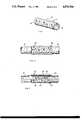
- FIG. 1is a perspective view of the crimped tubular device in accordance with the present invention
- FIG. 2is a side view of the crimped tube device shown with proximal and distal nerve ends affixed within the tube device in accordance with the present invention
- FIG. 3is a partial cross-sectional view of the tube device shown in FIG. 2 showing the corrugated exterior surface in relation to the relatively smooth interior surface and having the proximal and distal nerve ends sutured in place within the tube device;
- FIG. 4is a side view of the fixture for crimping the tube including a steel rod and chuck with an uncrimped tube in place over the rod, a line of suture material being wrapped around the tube;
- FIG. 5is a side view of the crimping fixture showing the tube being logitudinally collapsed on the rod with a collar being placed adjacent each end of the tube to insure the tube holds the desired configuration;
- FIG. 6is a view of a vacuum oven where the collapsed tube and crimping fixture are heated under a vacuuum to heat set the externally crimped surface of the tube.
- the bioabsorbable devices of the present inventionare flexible tubes made by either the knitting or weaving of bioabsorbable fibers into the shape of a tube and, then, dry heat setting of the tubes to improve the in-vivo strength of the polymer fibers and and provide a tube with corrugations along its external surface.
- the corrugated external tube surfaceallows for bending of the tube without compromising the internal passageway of the tube device.
- the tubeis used for spanning a significant nerve gap or defect such as occurs when a nerve is severed or lacerated and the nerve ends may not be easily brought back together.
- the tubeis provided with a relatively smooth interior surface to ease insertion of the nerve ends into the tube device and to provide an environment within the tube to promote longitudinal axon growth across the nerve gap or defect.
- FIG. 1shows a bioabsorbable tube device 10 made in accordance with the present invention.
- the tube 10has an exterior surface 12 and interior surface 14.
- the exterior tube surface 12is shown having a plurality of crimps or corrugations 16, thereon.
- the corrugations 16 on the exterior tube surface 12allow the tube device to be bent through an arc of 180 degrees without the pinching or crimping of the internal surface 14. This feature is extremely important to the functioning of the tube device because frequently it is necessary for the tube device to pass over joints or areas where bending of the regenerating nerve will occur. If the interior surface 14 buckles or crimps, the flow of axonal substances across the nerve gap will be blocked and the nerve will not fully regenerate across the gap.
- FIGS. 2 and 3show the tube device 10 in place spanning a nerve gap or defect between a proximal nerve end 18 and a distal nerve end 20.
- a bioabsorbable suture materialsuch as a DEXON® (American Cyanamid Company, Wayne, N.J. 07470, U.S.A.) suture or a MAXONTM (American Cyanamid Company, Wayne. N.J. 07470, U.S.A.) suture is shown at 22 and 24 connecting the proximal nerve end 18 and distal nerve end 20, to the wall of the tube device 10.
- the suture 22is threaded through the wall of the tube device at a point about 5 millimeters away from an end thereof and, then into the epineural layer of the proximal nerve end 18. The suture 22 is then pulled to bring the proximal nerve end into the end of the tube device 10. The suture 22 is tied to the wall of the tube in a manner that is known in the art. The process is then repeated with suture 24 to pull the distal nerve end 20 into the opposite end of the tube device.
- the proximal and distal nerve ends, 18 and 20are secured within the tube device 10 such that a gap exists between the nerve ends.
- the proximal nerve end 18will regenerate across the nerve gap into the distal nerve end 20.
- the corrugations 16are seen in more detail as comprising a series of ridges 26 and valleys 28 along the entire exterior tube surface 12.
- the interior surface 14is shown to have a plurality of flats 30 to provide a relatively smooth surface to ease insertion of the nerve ends into the tube device and to provide an optimum environment for axonal growth within the tube.
- FIG. 4shows an uncrimped mesh (knitted or woven) tube placed over a steel rod 32.
- the diameter of the rod 32is appropriately sized so that the tube slides snugly over the rod.
- the rod 32 and mesh tubeare mounted on a chuck 34 of a winding device such as a lathe (not shown) or other commercially available device to spin rod 32.
- a braider bobbin(not shown) is wound with a suture material 36 such as a 4/0 DEXON® (American Cyanamid Company, Wayne, N.J. 07470, U.S.A.) suture material which can be mounted on the cutting tool holder (not shown) of the lathe.
- the suture material 36is tied to one end of the tube as shown at 38 and then the lathe is rotated to wrap the suture material about the mesh tube 10.
- the suture material 36is wound around mesh tube 10 such that there are approximately twelve (12) wraps of suture material per longitudinal inch of tube.
- the suture material 36is cut and tied off around the opposite end of the mesh tube 10.
- the mesh tube 10is shown collapsed or longitudinally compressed on the rod 32 so that the tubes overall length is cut approximately in half.
- a collar 40is inserted on rod 32 to hold the mesh tube 10 in this collapsed or compressed condition.
- the compressed mesh tube 10, rod 32 and chuck 34are then placed in a vacuum oven 42 as shown in FIG. 6.
- the vacuum ovenis heated to 130° C. and a vacuum is pulled to less than or equal to 1 Torr.
- the mesh tube 10is left in the vacuum oven at ⁇ Torr and 130° C. for two hours.
- the use of a vacuum oven on the tube devicealso improves the in-vivo properties of the polymer fibers used to make up the tube device.
- the heat set processis more fully described in U.S. Pat. No. 3,422,181 to Chirgwin, Jr. and incorporated herein by reference.
- the mesh tube 10, rod 32 and chuck 34are removed from the vacuum oven 42 and cooled to room temperature in a Laminar Flow Hood (not shown).
- the suture material 36is carefully removed leaving a crimped or corrugated mesh tube.
- the mesh tube 10would then have both ends trimmed with scissors and be inserted into a thermoformed hinged tray.
- the trayis placed into a foil pouch for sterilization by known methods and sealed and sterilized a second time.
- the tube device 10 shown in FIGS. 1-6is knitted or woven from a plurality of bioabsorbable polymer fibers.
- the preferred polymers and copolymersare polyglycolic acid (U.S. Pat. No. 3,297,033), polyglycolic acid (U.S. Pat. No. 3,636,956) and poly(glycolic-co-trimethylene carbonate) (U.S. Pat. No. 4,243,775). These polymers and copolymers are preferred because they are known to be well tolerated by the body upon implantation in addition to being absorbable within the body.
- the polymer and copolymer fibersare obtainable through methods known in the art.
- the fibersare then knitted or woven into tube shape.
- the various methods of knitting or weaving such mesh tubesare further described in the examples below.
- the knitted or woven mesh tubeis manufactured totally from polymer fibers of 100 percent PGA.
- the PGA materialis a bioabsobable polymer which maintains its tensile strength for approximately thirty (30) days and is then slowly hydrolyzed within the body. Since the recognized neural growth rate is approximately one millimeter (1mm) per day, a tube device manufactured from a PGA polymer fiber would remain in place about a severed nerve long enough to allow a nerve to regenerate across a 30 mm or 3 cm nerve gap or defect.
- the knitted or woven mesh tubeis manufactured from a copolymer of glycolide and trimethylene carbonate linkages (MAXONTM suture material).
- MAXONTM suture materialThis copolymer fiber is known to maintain its tensile strength for at least fifty-six days before being slowly resorbed into the body.
- a tube device manufactured from the MAXONTM copolymer fibercould be used to span nerve gaps of five centimeters or more.
- bioabsorbableis used herein to be synonymous with the terms “biodegradable” ⁇ and “bioresorbable”. All of these terms refer to the capability of a material made from such fibers to be absorbed in living tissue such that the material will disappear from the cite of its implantation within the living tissue and be metabolized from the body at a reasonably consistent rate and within a reasonable time period. See, U.S. Pat. No. 3,297,033 which is incorporated herein by reference.
- PGA polymer fiberswere woven on a single shuttle 1 ⁇ 1 Crompton & Knowles box loom using 16 harnesses.
- the mesh tubewas woven as a double fabric with selvedge edges attached on both sides.
- the warp yarnwas 3 ply, 46 denier/21 filament (fiber) PGA yarn having 5 turns per inch of twist in the "Z" direction.
- the weft (filling) yarnwas 3 ply, 46 denier/21 filament PGA yarn having 1.5 turns per inch of twist in the "Z” direction.
- the mesh tube constructionwas a 1 ⁇ 1 plain weave having 120 ends per inch per side and 88 picks per inch.
- the total number of end in the mesh tube constructionvaried from approximately 60 to 111 to yield tube sizes of from 2 mm to 6 mm inside diameter (I.D.).
- the mesh tubewas then crimped, heat set and cut to the desired length (6 cm) as discussed above.
- This constructionyields a flexible and porous, woven mesh tube to be used in accordance with the present invention.
- MAXONTM copolymer fiberswere woven into a mesh tube on the same type of weaving loom as in Example 1.
- the warp yarnwas 5 ply, 50 denier/25 filament copolymer yarn having 5 turns per inch of twist in the "Z" direction.
- the weft (filling) yarnwas 5 ply, 50 denier/25 filament copolymer yarn having 2 turns per inch of twist in the "Z” direction.
- the mesh tube constructionwas a 1 ⁇ 1 plain weave having 62 ends per inch per side and 68 picks per inch.
- the woven mesh tubewas crimped and heat set as in Example 1 to provide a tube device in accordance with the present invention.
- PGA polymer fiberswere knit into a mesh tube on a tubular weft Lamb Knitting Machine using a single feed jersey stitch construction.
- the knitting machine cylinderhad a needle density of 25 needles per inch and the total number of needles in a given cylinder were varied to yield a mesh tube diameter of from 2 mm to 6 mm I.D. after fabric finishing.
- the yarn usedwas formed by combining 4 plies of 46 denier/21 filament PGA fibers, all plied at 2.3 turns per inch of twist in the "Z" direction.
- the knitted mesh tubeswere finished in the same manner as in Example 1 to provide a porous, flexible knitted mesh tube to be used in accordance with the present invention.
- MAXONTM copolymer fiberswere knit into a mesh tube on the same type of knitting maching and knit construction as in Example 3.
- the cylinderhad a needle density of 33 needles per inch with a total needle count of about 14 about the perimeter.
- the yarn usedwas formed by combining 3 plies of 50 denier copolymer fibers and 1 ply of 25 denier copolymer fibers, all plied at 2.3 turns per inch of twist in the "Z" direction to yield a mesh tube diameter of about 2 mm I.D. after finishing.
- the knitted mesh tubewas crimped and heat set as in Example 1, above.
Landscapes
- Health & Medical Sciences (AREA)
- Surgery (AREA)
- Life Sciences & Earth Sciences (AREA)
- Medical Informatics (AREA)
- Nuclear Medicine, Radiotherapy & Molecular Imaging (AREA)
- Engineering & Computer Science (AREA)
- Biomedical Technology (AREA)
- Heart & Thoracic Surgery (AREA)
- Neurology (AREA)
- Molecular Biology (AREA)
- Animal Behavior & Ethology (AREA)
- General Health & Medical Sciences (AREA)
- Public Health (AREA)
- Veterinary Medicine (AREA)
- Materials For Medical Uses (AREA)
- Prostheses (AREA)
Abstract
Description
TABLE I ______________________________________ (1) Poly-alpha-hydroxy acids such as polyglycolic acid (hereinafter PGA), polylactic acid, copolymers of lac- tic and glycolic acids, and said polymers copolymerized with other polyesters such as epsilon-caprolactone (i.e., U. S. Pat. No. 4,118,470). (2) Copolymers having a glycolic acid ester and trimethyl- ene carbonate linkages (U.S. Pat. No. 4,243,775), e.g. the copolymer in the MAXON™ (American Cyanamid Company, Wayne, N.J. 07470, USA) suture. (3) Polydioxanone (U.S. Pat. No. 4,052,988). (4) Polyesters formed from diols and succinic and/or oxalic acid such as U.S. Pat. Nos. 4,032,993 and 3,883,901, isomorphic copolyoxalates (U.S. Pat. No. 4,141,087), and poly(alkylene oxalates) (U.S. Pat. No. 4,140,678). (5) Polymers made from unsymmetrically-substituted 1,4- dioxane-2,5-diones (U.S. Pat. No. 3,960,152). ______________________________________
Claims (18)
Priority Applications (6)
| Application Number | Priority Date | Filing Date | Title |
|---|---|---|---|
| US07/150,594US4870966A (en) | 1988-02-01 | 1988-02-01 | Bioabsorbable surgical device for treating nerve defects |
| CA000589478ACA1335527C (en) | 1988-02-01 | 1989-01-30 | Bioabsorbable surgical device for treating nerve defects |
| EP89101634AEP0327022B1 (en) | 1988-02-01 | 1989-01-31 | Bioabsorbable surgical device for treating nerve defects and method for manufacturing it |
| DE68922319TDE68922319T2 (en) | 1988-02-01 | 1989-01-31 | Bioabsorbable surgical material for the treatment of nerve defects and method for its preparation. |
| AT89101634TATE121608T1 (en) | 1988-02-01 | 1989-01-31 | BIOABSORBABLE SURGICAL MATERIAL FOR THE TREATMENT OF NERVE DEFECTS AND METHOD FOR PRODUCING IT. |
| ES89101634TES2074446T3 (en) | 1988-02-01 | 1989-01-31 | BIOABSORBIBLE SURGICAL DEVICE FOR THE TREATMENT OF NERVE DEFECTS AND METHOD FOR ITS MANUFACTURE. |
Applications Claiming Priority (1)
| Application Number | Priority Date | Filing Date | Title |
|---|---|---|---|
| US07/150,594US4870966A (en) | 1988-02-01 | 1988-02-01 | Bioabsorbable surgical device for treating nerve defects |
Publications (1)
| Publication Number | Publication Date |
|---|---|
| US4870966Atrue US4870966A (en) | 1989-10-03 |
Family
ID=22535220
Family Applications (1)
| Application Number | Title | Priority Date | Filing Date |
|---|---|---|---|
| US07/150,594Expired - LifetimeUS4870966A (en) | 1988-02-01 | 1988-02-01 | Bioabsorbable surgical device for treating nerve defects |
Country Status (1)
| Country | Link |
|---|---|
| US (1) | US4870966A (en) |
Cited By (168)
| Publication number | Priority date | Publication date | Assignee | Title |
|---|---|---|---|---|
| US5037950A (en)* | 1990-02-09 | 1991-08-06 | Ethicon, Inc. | Bioabsorbable copolymers of polyalkylene carbonate/RHO-dioxanone for sutures and coatings |
| US5116373A (en)* | 1989-08-28 | 1992-05-26 | Sulzer Brothers Limited | Pull-through appliance |
| WO1992010218A1 (en)* | 1990-12-06 | 1992-06-25 | W.L. Gore & Associates, Inc. | Implantable bioabsorbable article |
| WO1992010975A1 (en)* | 1990-12-21 | 1992-07-09 | The University Of New Mexico | Nerve anastomosis sling and method |
| US5147385A (en)* | 1989-11-01 | 1992-09-15 | Schneider (Europe) A.G. | Stent and catheter for the introduction of the stent |
| US5147399A (en)* | 1988-02-01 | 1992-09-15 | Dellon Arnold L | Method of treating nerve defects through use of a bioabsorbable surgical device |
| US5242399A (en)* | 1990-04-25 | 1993-09-07 | Advanced Cardiovascular Systems, Inc. | Method and system for stent delivery |
| US5344426A (en)* | 1990-04-25 | 1994-09-06 | Advanced Cardiovascular Systems, Inc. | Method and system for stent delivery |
| US5354305A (en)* | 1991-09-26 | 1994-10-11 | United States Surgical Corporation | Nerve repair device |
| US5421955A (en)* | 1991-10-28 | 1995-06-06 | Advanced Cardiovascular Systems, Inc. | Expandable stents and method for making same |
| US5541304A (en)* | 1994-05-02 | 1996-07-30 | Hercules Incorporated | Crosslinked hydrogel compositions with improved mechanical performance |
| US5549676A (en)* | 1993-09-14 | 1996-08-27 | Johnson; Lanny L. | Biological replacement ligament |
| US5569295A (en)* | 1993-12-28 | 1996-10-29 | Advanced Cardiovascular Systems, Inc. | Expandable stents and method for making same |
| US5591222A (en)* | 1991-10-18 | 1997-01-07 | Susawa; Takashi | Method of manufacturing a device to dilate ducts in vivo |
| US5634946A (en)* | 1988-08-24 | 1997-06-03 | Focal, Inc. | Polymeric endoluminal paving process |
| US5653727A (en)* | 1987-10-19 | 1997-08-05 | Medtronic, Inc. | Intravascular stent |
| US5674287A (en)* | 1988-08-24 | 1997-10-07 | Endoluminal Therapeutics, Inc. | Biodegradable polymeric endoluminal sealing process, apparatus and polymeric product for use therein |
| US5935366A (en)* | 1995-09-07 | 1999-08-10 | Eli Lilly And Company | Process for environmentally safe cleaning using water-soluble polymer based packages |
| US5957975A (en)* | 1997-12-15 | 1999-09-28 | The Cleveland Clinic Foundation | Stent having a programmed pattern of in vivo degradation |
| US6019777A (en)* | 1997-04-21 | 2000-02-01 | Advanced Cardiovascular Systems, Inc. | Catheter and method for a stent delivery system |
| US6117104A (en)* | 1998-09-08 | 2000-09-12 | Advanced Cardiovascular Systems, Inc. | Stent deployment system and method of use |
| US6132360A (en)* | 1998-05-22 | 2000-10-17 | Halpern; Alan A. | Magnetic stretching of magnetized neurons for spinal cord or peripheral nerve repair and regeneration |
| US6171334B1 (en) | 1998-06-17 | 2001-01-09 | Advanced Cardiovascular Systems, Inc. | Expandable stent and method of use |
| US6221109B1 (en)* | 1999-09-15 | 2001-04-24 | Ed. Geistlich Söhne AG fur Chemische Industrie | Method of protecting spinal area |
| US6344053B1 (en) | 1993-12-22 | 2002-02-05 | Medtronic Ave, Inc. | Endovascular support device and method |
| US6355058B1 (en) | 1999-12-30 | 2002-03-12 | Advanced Cardiovascular Systems, Inc. | Stent with radiopaque coating consisting of particles in a binder |
| US6436132B1 (en) | 2000-03-30 | 2002-08-20 | Advanced Cardiovascular Systems, Inc. | Composite intraluminal prostheses |
| US6448076B2 (en) | 1998-09-15 | 2002-09-10 | The Regents Of The University Of Michigan | Method for chemically acellularizing a biological tissue sample |
| US6461380B1 (en) | 1998-07-28 | 2002-10-08 | Advanced Cardiovascular Systems, Inc. | Stent configuration |
| US6471721B1 (en) | 1999-12-30 | 2002-10-29 | Advanced Cardiovascular Systems, Inc. | Vascular stent having increased radiopacity and method for making same |
| US6537311B1 (en) | 1999-12-30 | 2003-03-25 | Advanced Cardiovascular Systems, Inc. | Stent designs for use in peripheral vessels |
| US6540774B1 (en) | 1999-08-31 | 2003-04-01 | Advanced Cardiovascular Systems, Inc. | Stent design with end rings having enhanced strength and radiopacity |
| US6652579B1 (en) | 2000-06-22 | 2003-11-25 | Advanced Cardiovascular Systems, Inc. | Radiopaque stent |
| US20040102793A1 (en)* | 2002-07-29 | 2004-05-27 | Yaszemski Michael J. | Spinal cord surgical implant |
| US20040122454A1 (en)* | 2002-12-23 | 2004-06-24 | Shu Wang | Medical guide tubes |
| US6773455B2 (en) | 1997-06-24 | 2004-08-10 | Advanced Cardiovascular Systems, Inc. | Stent with reinforced struts and bimodal deployment |
| US20040170664A1 (en)* | 2000-06-28 | 2004-09-02 | Ed. Geistlich Soehne Ag | Collagen tubes for nerve regeneration |
| US20050038471A1 (en)* | 2000-02-11 | 2005-02-17 | Barbara Chan | Photochemical tissue bonding |
| US6875229B2 (en) | 1997-08-13 | 2005-04-05 | Advanced Cardiovascular Systems, Inc. | Stent and catheter assembly and method for treating bifurcations |
| US6896697B1 (en) | 2002-12-30 | 2005-05-24 | Advanced Cardiovascular Systems, Inc. | Intravascular stent |
| US20050113910A1 (en)* | 2002-01-04 | 2005-05-26 | David Paniagua | Percutaneously implantable replacement heart valve device and method of making same |
| US6899729B1 (en) | 2002-12-18 | 2005-05-31 | Advanced Cardiovascular Systems, Inc. | Stent for treating vulnerable plaque |
| US20050119562A1 (en)* | 2003-05-23 | 2005-06-02 | Senorx, Inc. | Fibrous marker formed of synthetic polymer strands |
| US20050125049A1 (en)* | 2003-10-21 | 2005-06-09 | The Regents Of The University Of Michigan | Tissue engineered vascular construct and method for producing same |
| US20050192574A1 (en)* | 2004-02-10 | 2005-09-01 | Jason Blain | System and method for protecting neurovascular structures |
| US20050261761A1 (en)* | 1999-05-04 | 2005-11-24 | City Of Hope | Visceral anastomotic device and method of using same |
| US20060100647A1 (en)* | 2002-12-27 | 2006-05-11 | Nobutoshi Doi | Nerve regeneration-inducing tube comprising |
| US20060173280A1 (en)* | 2003-11-17 | 2006-08-03 | Inrad, Inc. | Multi Mode Imaging Marker |
| US20060178554A1 (en)* | 2005-02-04 | 2006-08-10 | Mandel Shlomo S | Nerve protection barrier |
| US20060216320A1 (en)* | 2003-03-31 | 2006-09-28 | Eiichi Kitazono | Composite of support matrix and collagen, and process for producing support substrate and composite |
| US20060241411A1 (en)* | 2005-04-20 | 2006-10-26 | Inrad, Inc. | Marking device with retracable cannula |
| US7163715B1 (en) | 2001-06-12 | 2007-01-16 | Advanced Cardiovascular Systems, Inc. | Spray processing of porous medical devices |
| US20070156158A1 (en)* | 2005-12-29 | 2007-07-05 | Uri Herzberg | Device for treating carpal tunnel syndrome |
| US7258697B1 (en) | 2003-12-22 | 2007-08-21 | Advanced Cardiovascular Systems, Inc. | Stent with anchors to prevent vulnerable plaque rupture during deployment |
| US7316710B1 (en) | 2002-12-30 | 2008-01-08 | Advanced Cardiovascular Systems, Inc. | Flexible stent |
| US20080009901A1 (en)* | 2000-02-11 | 2008-01-10 | Redmond Robert W | Photochemical tissue bonding |
| US20080039819A1 (en)* | 2006-08-04 | 2008-02-14 | Senorx, Inc. | Marker formed of starch or other suitable polysaccharide |
| US20080241209A1 (en)* | 2007-02-02 | 2008-10-02 | The Regents Of The University Of Michigan | System and Method for Forming Bone, Ligament, and Bone-Ligament Constructs |
| US20080288084A1 (en)* | 2006-08-18 | 2008-11-20 | Nobutoshi Doi | Precursor of a tissue regenerating instrument provided with a swellable rod |
| US20080299536A1 (en)* | 2001-08-13 | 2008-12-04 | University Of Florida Research Foundation, Inc. | Material and Methods for Nerve Grafting |
| US20090171198A1 (en)* | 2006-08-04 | 2009-07-02 | Jones Michael L | Powdered marker |
| US20090306770A1 (en)* | 2005-04-29 | 2009-12-10 | Kassab Ghassan S | Tissue engineering of blood vessels |
| US20100010341A1 (en)* | 2006-12-18 | 2010-01-14 | Talpade Dnyanesh A | Biopsy Marker with In Situ-Generated Imaging Properties |
| US20100030072A1 (en)* | 2006-12-12 | 2010-02-04 | Casanova R Michael | Multiple Imaging Mode Tissue Marker |
| US20100082102A1 (en)* | 2008-09-23 | 2010-04-01 | Senorx, Inc. | Porous bioabsorbable implant |
| US20100094169A1 (en)* | 2002-06-17 | 2010-04-15 | Senorx, Inc. | Plugged tip delivery tube for marker placement |
| US7753950B2 (en) | 1997-08-13 | 2010-07-13 | Advanced Cardiovascular Systems, Inc. | Stent and catheter assembly and method for treating bifurcations |
| US7763198B2 (en) | 2005-04-12 | 2010-07-27 | Abbott Cardiovascular Systems Inc. | Method for retaining a vascular stent on a catheter |
| US20100198059A1 (en)* | 1999-02-02 | 2010-08-05 | Senorx, Inc. | Remotely activated marker |
| US20100204570A1 (en)* | 2009-02-06 | 2010-08-12 | Paul Lubock | Anchor markers |
| US20100234863A1 (en)* | 2009-03-16 | 2010-09-16 | Washington, University Of | Nanofibrous conduits for nerve regeneration |
| US20100298698A1 (en)* | 2000-11-20 | 2010-11-25 | Senorx, Inc. | Tissue site markers for in vivo imaging |
| US20100298696A1 (en)* | 2003-11-17 | 2010-11-25 | Bard Peripheral Vascular, Inc. | Self-contained, self-piercing, side-expelling marking apparatus |
| US20100324416A1 (en)* | 1999-02-02 | 2010-12-23 | Senorx, Inc. | Cavity-filling biopsy site markers |
| US20100331668A1 (en)* | 2008-01-31 | 2010-12-30 | Ranpura Himanshu M | Biopsy Tissue Marker |
| US20110028836A1 (en)* | 2008-12-30 | 2011-02-03 | Himanshu Ranpura | Marker delivery device for tissue marker placement |
| US20110082547A1 (en)* | 1997-10-10 | 2011-04-07 | Senorx, Inc. | Tissue marking implant |
| US20110092815A1 (en)* | 2003-05-23 | 2011-04-21 | Senorx, Inc. | Marker or filler forming fluid |
| US7947207B2 (en) | 2005-04-12 | 2011-05-24 | Abbott Cardiovascular Systems Inc. | Method for retaining a vascular stent on a catheter |
| US20110166448A1 (en)* | 1999-02-02 | 2011-07-07 | Jones Michael L | Marker delivery device with releasable plug |
| US20110184280A1 (en)* | 1999-02-02 | 2011-07-28 | Jones Michael L | Intracorporeal marker and marker delivery device |
| US20110184449A1 (en)* | 2006-08-04 | 2011-07-28 | Senorx, Inc. | Marker delivery device with obturator |
| US8092490B2 (en) | 2000-02-11 | 2012-01-10 | The General Hospital Corporation | Photochemical tissue bonding |
| US8216209B2 (en) | 2007-05-31 | 2012-07-10 | Abbott Cardiovascular Systems Inc. | Method and apparatus for delivering an agent to a kidney |
| US8224424B2 (en) | 1999-02-02 | 2012-07-17 | Senorx, Inc. | Tissue site markers for in vivo imaging |
| WO2012133019A1 (en)* | 2011-03-30 | 2012-10-04 | テルモ株式会社 | Medical device for nerve regeneration |
| US8361144B2 (en) | 2010-03-01 | 2013-01-29 | Colibri Heart Valve Llc | Percutaneously deliverable heart valve and methods associated therewith |
| US8437834B2 (en) | 2006-10-23 | 2013-05-07 | C. R. Bard, Inc. | Breast marker |
| US8486028B2 (en) | 2005-10-07 | 2013-07-16 | Bard Peripheral Vascular, Inc. | Tissue marking apparatus having drug-eluting tissue marker |
| US8579931B2 (en) | 1999-06-17 | 2013-11-12 | Bard Peripheral Vascular, Inc. | Apparatus for the percutaneous marking of a lesion |
| US8626269B2 (en) | 2003-05-23 | 2014-01-07 | Senorx, Inc. | Fibrous marker and intracorporeal delivery thereof |
| US8668737B2 (en) | 1997-10-10 | 2014-03-11 | Senorx, Inc. | Tissue marking implant |
| US8758374B2 (en) | 2010-09-15 | 2014-06-24 | University Of Utah Research Foundation | Method for connecting nerves via a side-to-side epineurial window using artificial conduits |
| USD715442S1 (en) | 2013-09-24 | 2014-10-14 | C. R. Bard, Inc. | Tissue marker for intracorporeal site identification |
| USD715942S1 (en) | 2013-09-24 | 2014-10-21 | C. R. Bard, Inc. | Tissue marker for intracorporeal site identification |
| USD716450S1 (en) | 2013-09-24 | 2014-10-28 | C. R. Bard, Inc. | Tissue marker for intracorporeal site identification |
| USD716451S1 (en) | 2013-09-24 | 2014-10-28 | C. R. Bard, Inc. | Tissue marker for intracorporeal site identification |
| US9119738B2 (en) | 2010-06-28 | 2015-09-01 | Colibri Heart Valve Llc | Method and apparatus for the endoluminal delivery of intravascular devices |
| US9144509B2 (en) | 2007-05-31 | 2015-09-29 | Abbott Cardiovascular Systems Inc. | Method and apparatus for delivering an agent to a kidney |
| US9149341B2 (en) | 1999-02-02 | 2015-10-06 | Senorx, Inc | Deployment of polysaccharide markers for treating a site within a patient |
| US9149610B2 (en) | 2007-05-31 | 2015-10-06 | Abbott Cardiovascular Systems Inc. | Method and apparatus for improving delivery of an agent to a kidney |
| US9364586B2 (en) | 2007-05-31 | 2016-06-14 | Abbott Cardiovascular Systems Inc. | Method and apparatus for improving delivery of an agent to a kidney |
| US9737400B2 (en) | 2010-12-14 | 2017-08-22 | Colibri Heart Valve Llc | Percutaneously deliverable heart valve including folded membrane cusps with integral leaflets |
| US9820824B2 (en) | 1999-02-02 | 2017-11-21 | Senorx, Inc. | Deployment of polysaccharide markers for treating a site within a patent |
| US9931121B2 (en) | 2011-10-17 | 2018-04-03 | University Of Utah Research Foundation | Methods and devices for connecting nerves |
| US10130465B2 (en) | 2016-02-23 | 2018-11-20 | Abbott Cardiovascular Systems Inc. | Bifurcated tubular graft for treating tricuspid regurgitation |
| US10292381B2 (en) | 2012-07-20 | 2019-05-21 | The General Hospital Corporation | Vessel treatment systems, methods, and kits |
| US10549112B2 (en) | 2012-07-20 | 2020-02-04 | The General Hospital Corporation | Apparatus for tissue irradiation and methods and kits utilizing the same |
| US10575973B2 (en) | 2018-04-11 | 2020-03-03 | Abbott Cardiovascular Systems Inc. | Intravascular stent having high fatigue performance |
| US10842494B2 (en) | 2011-10-17 | 2020-11-24 | University Of Utah Research Foundation | Methods and devices for connecting nerves |
| US11065098B2 (en)* | 2016-01-18 | 2021-07-20 | Asahi Kasei Kabushiki Kaisha | Medical fabric |
| US20210259681A1 (en)* | 2010-09-30 | 2021-08-26 | Ethicon Llc | Tissue thickness compensator comprising at least one medicament |
| US20220125569A1 (en)* | 2019-02-22 | 2022-04-28 | Toray Industries, Inc. | Nerve regeneration-inducing tube |
| US11350625B2 (en) | 2013-07-18 | 2022-06-07 | The General Hospital Corporation | Vessel treatment systems, methods, and kits |
| US11395726B2 (en) | 2017-09-11 | 2022-07-26 | Incubar Llc | Conduit vascular implant sealing device for reducing endoleaks |
| WO2023034614A1 (en)* | 2021-09-02 | 2023-03-09 | The Brigham And Women's Hospital, Inc. | Systems and methods for stimulation, nerve repair and/or drug delivery |
| US11883020B2 (en) | 2006-01-31 | 2024-01-30 | Cilag Gmbh International | Surgical instrument having a feedback system |
| US11890012B2 (en) | 2004-07-28 | 2024-02-06 | Cilag Gmbh International | Staple cartridge comprising cartridge body and attached support |
| US11890029B2 (en) | 2006-01-31 | 2024-02-06 | Cilag Gmbh International | Motor-driven surgical cutting and fastening instrument |
| US11890008B2 (en) | 2006-01-31 | 2024-02-06 | Cilag Gmbh International | Surgical instrument with firing lockout |
| US11890015B2 (en) | 2015-09-30 | 2024-02-06 | Cilag Gmbh International | Compressible adjunct with crossing spacer fibers |
| US11911027B2 (en) | 2010-09-30 | 2024-02-27 | Cilag Gmbh International | Adhesive film laminate |
| US11918220B2 (en) | 2012-03-28 | 2024-03-05 | Cilag Gmbh International | Tissue thickness compensator comprising tissue ingrowth features |
| US11918210B2 (en) | 2014-10-16 | 2024-03-05 | Cilag Gmbh International | Staple cartridge comprising a cartridge body including a plurality of wells |
| US11918211B2 (en) | 2007-01-10 | 2024-03-05 | Cilag Gmbh International | Surgical stapling instrument for use with a robotic system |
| US11925354B2 (en) | 2010-09-30 | 2024-03-12 | Cilag Gmbh International | Staple cartridge comprising staples positioned within a compressible portion thereof |
| US11931028B2 (en) | 2016-04-15 | 2024-03-19 | Cilag Gmbh International | Surgical instrument with multiple program responses during a firing motion |
| US11957795B2 (en) | 2010-09-30 | 2024-04-16 | Cilag Gmbh International | Tissue thickness compensator configured to redistribute compressive forces |
| US11957345B2 (en) | 2013-03-01 | 2024-04-16 | Cilag Gmbh International | Articulatable surgical instruments with conductive pathways for signal communication |
| US11963680B2 (en) | 2017-10-31 | 2024-04-23 | Cilag Gmbh International | Cartridge body design with force reduction based on firing completion |
| US11974747B2 (en) | 2011-05-27 | 2024-05-07 | Cilag Gmbh International | Surgical stapling instruments with rotatable staple deployment arrangements |
| US11974746B2 (en) | 2014-04-16 | 2024-05-07 | Cilag Gmbh International | Anvil for use with a surgical stapling assembly |
| US11986183B2 (en) | 2008-02-14 | 2024-05-21 | Cilag Gmbh International | Surgical cutting and fastening instrument comprising a plurality of sensors to measure an electrical parameter |
| US11992208B2 (en) | 2007-06-04 | 2024-05-28 | Cilag Gmbh International | Rotary drive systems for surgical instruments |
| US11998194B2 (en) | 2008-02-15 | 2024-06-04 | Cilag Gmbh International | Surgical stapling assembly comprising an adjunct applicator |
| US11998206B2 (en) | 2008-02-14 | 2024-06-04 | Cilag Gmbh International | Detachable motor powered surgical instrument |
| US12011166B2 (en) | 2016-12-21 | 2024-06-18 | Cilag Gmbh International | Articulatable surgical stapling instruments |
| US12011165B2 (en) | 2004-07-28 | 2024-06-18 | Cilag Gmbh International | Surgical stapling instrument comprising replaceable staple cartridge |
| US12023022B2 (en) | 2014-03-26 | 2024-07-02 | Cilag Gmbh International | Systems and methods for controlling a segmented circuit |
| US12023026B2 (en) | 2021-03-22 | 2024-07-02 | Cilag Gmbh International | Staple cartridge comprising a firing lockout |
| US12029415B2 (en) | 2008-09-23 | 2024-07-09 | Cilag Gmbh International | Motor-driven surgical cutting instrument |
| US12035906B2 (en) | 2007-06-04 | 2024-07-16 | Cilag Gmbh International | Surgical instrument including a handle system for advancing a cutting member |
| US12053176B2 (en) | 2013-08-23 | 2024-08-06 | Cilag Gmbh International | End effector detention systems for surgical instruments |
| US12076018B2 (en) | 2015-02-27 | 2024-09-03 | Cilag Gmbh International | Modular stapling assembly |
| US12076017B2 (en) | 2014-09-18 | 2024-09-03 | Cilag Gmbh International | Surgical instrument including a deployable knife |
| US12076096B2 (en) | 2017-12-19 | 2024-09-03 | Cilag Gmbh International | Method for determining the position of a rotatable jaw of a surgical instrument attachment assembly |
| US12076011B2 (en) | 2017-10-30 | 2024-09-03 | Cilag Gmbh International | Surgical stapler knife motion controls |
| US12082806B2 (en) | 2007-01-10 | 2024-09-10 | Cilag Gmbh International | Surgical instrument with wireless communication between control unit and sensor transponders |
| US12121234B2 (en) | 2012-03-28 | 2024-10-22 | Cilag Gmbh International | Staple cartridge assembly comprising a compensator |
| US12137912B2 (en) | 2015-09-30 | 2024-11-12 | Cilag Gmbh International | Compressible adjunct with attachment regions |
| US12156653B2 (en) | 2015-12-30 | 2024-12-03 | Cilag Gmbh International | Surgical instruments with motor control circuits |
| US12161329B2 (en) | 2006-01-31 | 2024-12-10 | Cilag Gmbh International | Surgical systems comprising a control circuit including a timer |
| US12171508B2 (en) | 2006-03-23 | 2024-12-24 | Cilag Gmbh International | Robotically-controlled surgical instrument with selectively articulatable end effector |
| US12178429B2 (en) | 2013-04-16 | 2024-12-31 | Cilag Gmbh International | Surgical instruments having modular end effector selectively coupleable to housing assembly |
| US12178432B2 (en) | 2010-09-30 | 2024-12-31 | Cilag Gmbh International | Tissue thickness compensator comprising laterally offset layers |
| US12178434B2 (en) | 2006-10-03 | 2024-12-31 | Cilag Gmbh International | Surgical stapling system including control circuit to monitor clamping pressure |
| US12213666B2 (en) | 2010-09-30 | 2025-02-04 | Cilag Gmbh International | Tissue thickness compensator comprising layers |
| US12213671B2 (en) | 2008-02-14 | 2025-02-04 | Cilag Gmbh International | Motorized system having a plurality of power sources |
| US12239316B2 (en) | 2011-05-27 | 2025-03-04 | Cilag Gmbh International | Automated end effector component reloading system for use with a robotic system |
| US12245901B2 (en) | 2015-09-25 | 2025-03-11 | Cilag Gmbh International | Implantable layer comprising boundary indicators |
| US12256930B2 (en) | 2011-05-27 | 2025-03-25 | Cilag Gmbh International | Robotically-driven surgical instrument with E-beam driver |
| US12262888B2 (en) | 2018-08-20 | 2025-04-01 | Cilag Gmbh International | Surgical instruments with progressive jaw closure arrangements |
| US12274445B2 (en) | 2014-04-16 | 2025-04-15 | Cilag Gmbh International | Fastener cartridges including extensions having different configurations |
| US12324581B2 (en) | 2017-06-28 | 2025-06-10 | Cilag Gmbh International | Surgical instrument comprising selectively actuatable rotatable couplers |
| US12369911B2 (en) | 2012-06-28 | 2025-07-29 | Cilag Gmbh International | Firing system lockout arrangements for surgical instruments |
| US12414768B2 (en) | 2014-09-05 | 2025-09-16 | Cilag Gmbh International | Staple cartridge electrical contacts |
| US12433584B2 (en) | 2006-01-31 | 2025-10-07 | Cilag Gmbh International | Robotically-controlled end effector |
| US12440208B2 (en) | 2023-08-23 | 2025-10-14 | Cilag Gmbh International | Powered surgical instrument |
Citations (31)
| Publication number | Priority date | Publication date | Assignee | Title |
|---|---|---|---|---|
| US3044497A (en)* | 1944-05-11 | 1962-07-17 | Bodin Girin & Cie Soc | Tubular members provided with corrugated walls |
| US3108357A (en)* | 1962-06-20 | 1963-10-29 | William J Liebig | Compound absorbable prosthetic implants, fabrics and yarns therefor |
| US3297033A (en)* | 1963-10-31 | 1967-01-10 | American Cyanamid Co | Surgical sutures |
| US3316557A (en)* | 1965-02-15 | 1967-05-02 | Meadox Medicals Inc | Surgical, vascular prosthesis formed of composite yarns containing both synthetic and animal derivative strands |
| US3463158A (en)* | 1963-10-31 | 1969-08-26 | American Cyanamid Co | Polyglycolic acid prosthetic devices |
| US3588920A (en)* | 1969-09-05 | 1971-06-29 | Sigmund A Wesolowski | Surgical vascular prostheses formed of polyester fiber paper |
| US3648295A (en)* | 1970-10-12 | 1972-03-14 | James R Palma | Guide for growing blood vessels or the like |
| US3660152A (en)* | 1969-06-02 | 1972-05-02 | Ici Ltd | Coated woven materials |
| US3688317A (en)* | 1970-08-25 | 1972-09-05 | Sutures Inc | Vascular prosthetic |
| US3878565A (en)* | 1971-07-14 | 1975-04-22 | Providence Hospital | Vascular prosthesis with external pile surface |
| FR2248015A1 (en)* | 1973-10-17 | 1975-05-16 | Rhone Poulenc Ind | Artificial ureter or urethra - watertight flexible tube has helical rib in outside wall to prevent creasing |
| US3883901A (en)* | 1972-12-01 | 1975-05-20 | Rhone Poulenc Sa | Method of replacing or repairing the body with bioresorbable surgical articles |
| US3937223A (en)* | 1974-04-19 | 1976-02-10 | American Cyanamid Company | Compacted surgical hemostatic felt |
| US3960152A (en)* | 1974-01-21 | 1976-06-01 | American Cyanamid Company | Surgical sutures of unsymmetrically substituted 1,4-dioxane-2,5-diones |
| US4032993A (en)* | 1974-06-28 | 1977-07-05 | Rhone-Poulenc Industries | Bioresorbable surgical articles |
| US4033938A (en)* | 1974-01-21 | 1977-07-05 | American Cyanamid Company | Polymers of unsymmetrically substituted 1,4-dioxane-2,5-diones |
| US4074366A (en)* | 1975-03-14 | 1978-02-21 | American Cyanamid Company | Poly(N-acetyl-D-glucosamine) products |
| US4118470A (en)* | 1976-06-01 | 1978-10-03 | American Cyanamid Company | Normally-solid, bioabsorbable, hydrolyzable, polymeric reaction product |
| US4141087A (en)* | 1977-01-19 | 1979-02-27 | Ethicon, Inc. | Isomorphic copolyoxalates and sutures thereof |
| US4164045A (en)* | 1977-08-03 | 1979-08-14 | Carbomedics, Inc. | Artificial vascular and patch grafts |
| US4243775A (en)* | 1978-11-13 | 1981-01-06 | American Cyanamid Company | Synthetic polyester surgical articles |
| US4313231A (en)* | 1980-06-16 | 1982-02-02 | Kabushiki Kaisha Tatebe Seishudo | Vascular prosthesis |
| US4416028A (en)* | 1981-01-22 | 1983-11-22 | Ingvar Eriksson | Blood vessel prosthesis |
| US4517687A (en)* | 1982-09-15 | 1985-05-21 | Meadox Medicals, Inc. | Synthetic woven double-velour graft |
| US4530113A (en)* | 1983-05-20 | 1985-07-23 | Intervascular, Inc. | Vascular grafts with cross-weave patterns |
| US4534349A (en)* | 1983-02-02 | 1985-08-13 | Minnesota Mining And Manufacturing Company | Absorbable sutureless nerve repair device |
| US4652263A (en)* | 1985-06-20 | 1987-03-24 | Atrium Medical Corporation | Elasticization of microporous woven tubes |
| US4662884A (en)* | 1984-04-25 | 1987-05-05 | University Of Utah Research Foundation | Prostheses and methods for promoting nerve regeneration |
| US4669474A (en)* | 1984-01-12 | 1987-06-02 | Minnesota Mining And Manufacturing Company | Absorbable nerve repair device and method |
| US4670286A (en)* | 1983-09-20 | 1987-06-02 | Allied Corporation | Method of forming prosthetic devices |
| US4759764A (en)* | 1985-05-24 | 1988-07-26 | Clayton Foundation For Research | Peripheral nerve regeneration |
- 1988
- 1988-02-01USUS07/150,594patent/US4870966A/ennot_activeExpired - Lifetime
Patent Citations (31)
| Publication number | Priority date | Publication date | Assignee | Title |
|---|---|---|---|---|
| US3044497A (en)* | 1944-05-11 | 1962-07-17 | Bodin Girin & Cie Soc | Tubular members provided with corrugated walls |
| US3108357A (en)* | 1962-06-20 | 1963-10-29 | William J Liebig | Compound absorbable prosthetic implants, fabrics and yarns therefor |
| US3297033A (en)* | 1963-10-31 | 1967-01-10 | American Cyanamid Co | Surgical sutures |
| US3463158A (en)* | 1963-10-31 | 1969-08-26 | American Cyanamid Co | Polyglycolic acid prosthetic devices |
| US3316557A (en)* | 1965-02-15 | 1967-05-02 | Meadox Medicals Inc | Surgical, vascular prosthesis formed of composite yarns containing both synthetic and animal derivative strands |
| US3660152A (en)* | 1969-06-02 | 1972-05-02 | Ici Ltd | Coated woven materials |
| US3588920A (en)* | 1969-09-05 | 1971-06-29 | Sigmund A Wesolowski | Surgical vascular prostheses formed of polyester fiber paper |
| US3688317A (en)* | 1970-08-25 | 1972-09-05 | Sutures Inc | Vascular prosthetic |
| US3648295A (en)* | 1970-10-12 | 1972-03-14 | James R Palma | Guide for growing blood vessels or the like |
| US3878565A (en)* | 1971-07-14 | 1975-04-22 | Providence Hospital | Vascular prosthesis with external pile surface |
| US3883901A (en)* | 1972-12-01 | 1975-05-20 | Rhone Poulenc Sa | Method of replacing or repairing the body with bioresorbable surgical articles |
| FR2248015A1 (en)* | 1973-10-17 | 1975-05-16 | Rhone Poulenc Ind | Artificial ureter or urethra - watertight flexible tube has helical rib in outside wall to prevent creasing |
| US3960152A (en)* | 1974-01-21 | 1976-06-01 | American Cyanamid Company | Surgical sutures of unsymmetrically substituted 1,4-dioxane-2,5-diones |
| US4033938A (en)* | 1974-01-21 | 1977-07-05 | American Cyanamid Company | Polymers of unsymmetrically substituted 1,4-dioxane-2,5-diones |
| US3937223A (en)* | 1974-04-19 | 1976-02-10 | American Cyanamid Company | Compacted surgical hemostatic felt |
| US4032993A (en)* | 1974-06-28 | 1977-07-05 | Rhone-Poulenc Industries | Bioresorbable surgical articles |
| US4074366A (en)* | 1975-03-14 | 1978-02-21 | American Cyanamid Company | Poly(N-acetyl-D-glucosamine) products |
| US4118470A (en)* | 1976-06-01 | 1978-10-03 | American Cyanamid Company | Normally-solid, bioabsorbable, hydrolyzable, polymeric reaction product |
| US4141087A (en)* | 1977-01-19 | 1979-02-27 | Ethicon, Inc. | Isomorphic copolyoxalates and sutures thereof |
| US4164045A (en)* | 1977-08-03 | 1979-08-14 | Carbomedics, Inc. | Artificial vascular and patch grafts |
| US4243775A (en)* | 1978-11-13 | 1981-01-06 | American Cyanamid Company | Synthetic polyester surgical articles |
| US4313231A (en)* | 1980-06-16 | 1982-02-02 | Kabushiki Kaisha Tatebe Seishudo | Vascular prosthesis |
| US4416028A (en)* | 1981-01-22 | 1983-11-22 | Ingvar Eriksson | Blood vessel prosthesis |
| US4517687A (en)* | 1982-09-15 | 1985-05-21 | Meadox Medicals, Inc. | Synthetic woven double-velour graft |
| US4534349A (en)* | 1983-02-02 | 1985-08-13 | Minnesota Mining And Manufacturing Company | Absorbable sutureless nerve repair device |
| US4530113A (en)* | 1983-05-20 | 1985-07-23 | Intervascular, Inc. | Vascular grafts with cross-weave patterns |
| US4670286A (en)* | 1983-09-20 | 1987-06-02 | Allied Corporation | Method of forming prosthetic devices |
| US4669474A (en)* | 1984-01-12 | 1987-06-02 | Minnesota Mining And Manufacturing Company | Absorbable nerve repair device and method |
| US4662884A (en)* | 1984-04-25 | 1987-05-05 | University Of Utah Research Foundation | Prostheses and methods for promoting nerve regeneration |
| US4759764A (en)* | 1985-05-24 | 1988-07-26 | Clayton Foundation For Research | Peripheral nerve regeneration |
| US4652263A (en)* | 1985-06-20 | 1987-03-24 | Atrium Medical Corporation | Elasticization of microporous woven tubes |
Non-Patent Citations (19)
| Title |
|---|
| B. R. Seckel et al., "Nerve Regeneration Through Synthetic Biodegradable Nerve Guides", J. Plastic Reconstr. Surg., 74 173-181, (1984). |
| B. R. Seckel et al., Nerve Regeneration Through Synthetic Biodegradable Nerve Guides , J. Plastic Reconstr. Surg., 74 173 181, (1984).* |
| Bora, W. F. et al. Prosthetic Nerve Grafts: A Resorbable Tube as an Alternative to Autogenous Nerve Grafting , Journal of Hand Surgery 1987; 12A 2 Pt 1 : 685 692.* |
| Bora, W. F. et al.--"Prosthetic Nerve Grafts: A Resorbable Tube as an Alternative to Autogenous Nerve Grafting", Journal of Hand Surgery 1987; 12A [2 Pt 1]: 685-692. |
| D. G. Kline et al., "The Use of a Resorbable Wrapper for Peripheral Nerve Repair", J. Neurosurgery 121, 737 (1946). |
| D. G. Kline et al., The Use of a Resorbable Wrapper for Peripheral Nerve Repair , J. Neurosurgery 121, 737 (1946).* |
| Hakon Molander et al., "Regeneration of Peripheral Nerve Through a Polyglactin Tube", Muscle and Nerve, 5:54-57 (1982). |
| Hakon Molander et al., Regeneration of Peripheral Nerve Through a Polyglactin Tube , Muscle and Nerve, 5:54 57 (1982).* |
| J. M. Rosen et al., "Fascicular Sutureless and Suture Repair of the Peripheral Nerves", Orthopedic Review, 8(4) 85 (1979). |
| J. M. Rosen et al., Fascicular Sutureless and Suture Repair of the Peripheral Nerves , Orthopedic Review, 8(4) 85 (1979).* |
| J. M. Rosen, Orthopedic Transactions 6(1), 75(1982).* |
| Molander et al., "Nerve Repair Using a Polyglactin Tube and Nerve Graft", Biomaterials 4:276-280 (1983). |
| Molander et al., Nerve Repair Using a Polyglactin Tube and Nerve Graft , Biomaterials 4:276 280 (1983).* |
| R. D. Midgley et al., "Silicone Rubber Sheathing as an Adjunct to Neural Anastomosis", Surgical Clinic of North America 148, 1149 (1968). |
| R. D. Midgley et al., Silicone Rubber Sheathing as an Adjunct to Neural Anastomosis , Surgical Clinic of North America 148, 1149 (1968).* |
| R. Madison et al., "Increased Rate of Peripheral Nerve Regeneration Using Bioabsorbable Nerve Guides in a Laminin-containing Gel", Experimental Neurology, 88:767-772 (1985). |
| R. Madison et al., Increased Rate of Peripheral Nerve Regeneration Using Bioabsorbable Nerve Guides in a Laminin containing Gel , Experimental Neurology, 88:767 772 (1985).* |
| S. E. Mackinnon et al., "Nerve Regeneration Through a Pseudosynovial Sheath in a Primate Model", Plastic and Reconstr. Surg., 75 833-839 (1985). |
| S. E. Mackinnon et al., Nerve Regeneration Through a Pseudosynovial Sheath in a Primate Model , Plastic and Reconstr. Surg., 75 833 839 (1985).* |
Cited By (294)
| Publication number | Priority date | Publication date | Assignee | Title |
|---|---|---|---|---|
| US6923828B1 (en) | 1987-10-19 | 2005-08-02 | Medtronic, Inc. | Intravascular stent |
| US5653727A (en)* | 1987-10-19 | 1997-08-05 | Medtronic, Inc. | Intravascular stent |
| US5147399A (en)* | 1988-02-01 | 1992-09-15 | Dellon Arnold L | Method of treating nerve defects through use of a bioabsorbable surgical device |
| US6443941B1 (en) | 1988-08-24 | 2002-09-03 | Endoluminal Therapeutics, Inc. | Biodegradable polymeric endoluminal sealing process, apparatus and polymeric products for use therein |
| US5749922A (en)* | 1988-08-24 | 1998-05-12 | Endoluminal Therapeutics, Inc. | Biodegradable polymeric endoluminal sealing process, apparatus and polymeric products for use therein |
| US6699272B2 (en) | 1988-08-24 | 2004-03-02 | Endoluminal Therapeutics, Inc. | Biodegradable polymeric endoluminal sealing process, apparatus and polymeric products for use therein |
| US5749915A (en)* | 1988-08-24 | 1998-05-12 | Focal, Inc. | Polymeric endoluminal paving process |
| US5674287A (en)* | 1988-08-24 | 1997-10-07 | Endoluminal Therapeutics, Inc. | Biodegradable polymeric endoluminal sealing process, apparatus and polymeric product for use therein |
| US5800538A (en)* | 1988-08-24 | 1998-09-01 | Endoluminal Therapeutics, Inc. | Biodegradable polymeric endoluminal sealing process |
| US5634946A (en)* | 1988-08-24 | 1997-06-03 | Focal, Inc. | Polymeric endoluminal paving process |
| US6663661B2 (en) | 1989-08-24 | 2003-12-16 | Medtronic Ave, Inc. | Endovascular support device and method |
| US6827733B2 (en) | 1989-08-24 | 2004-12-07 | Medtronic Ave, Inc. | Endovascular support device and method |
| US5116373A (en)* | 1989-08-28 | 1992-05-26 | Sulzer Brothers Limited | Pull-through appliance |
| US5147385A (en)* | 1989-11-01 | 1992-09-15 | Schneider (Europe) A.G. | Stent and catheter for the introduction of the stent |
| US5037950A (en)* | 1990-02-09 | 1991-08-06 | Ethicon, Inc. | Bioabsorbable copolymers of polyalkylene carbonate/RHO-dioxanone for sutures and coatings |
| US5344426A (en)* | 1990-04-25 | 1994-09-06 | Advanced Cardiovascular Systems, Inc. | Method and system for stent delivery |
| US5242399A (en)* | 1990-04-25 | 1993-09-07 | Advanced Cardiovascular Systems, Inc. | Method and system for stent delivery |
| WO1992010218A1 (en)* | 1990-12-06 | 1992-06-25 | W.L. Gore & Associates, Inc. | Implantable bioabsorbable article |
| US6031148A (en)* | 1990-12-06 | 2000-02-29 | W. L. Gore & Associates, Inc. | Implantable bioabsorbable article |
| US6102921A (en)* | 1990-12-21 | 2000-08-15 | University Of New Mexico | Nerve anastomosis sling and method |
| WO1992010975A1 (en)* | 1990-12-21 | 1992-07-09 | The University Of New Mexico | Nerve anastomosis sling and method |
| US5354305A (en)* | 1991-09-26 | 1994-10-11 | United States Surgical Corporation | Nerve repair device |
| US5591222A (en)* | 1991-10-18 | 1997-01-07 | Susawa; Takashi | Method of manufacturing a device to dilate ducts in vivo |
| US5421955A (en)* | 1991-10-28 | 1995-06-06 | Advanced Cardiovascular Systems, Inc. | Expandable stents and method for making same |
| US6689159B2 (en) | 1991-10-28 | 2004-02-10 | Advanced Cardiovascular Systems, Inc. | Expandable stents and method for making same |
| US6485511B2 (en) | 1991-10-28 | 2002-11-26 | Advanced Cardiovascular Systems, Inc. | Expandable stents and method for making same |
| US5766238A (en)* | 1991-10-28 | 1998-06-16 | Advanced Cardiovascular Systems, Inc. | Expandable stents and method for making same |
| US6066167A (en)* | 1991-10-28 | 2000-05-23 | Advanced Cardiovascular Systems, Inc. | Expandable stents |
| US6066168A (en)* | 1991-10-28 | 2000-05-23 | Advanced Cardiovascular Systems, Inc. | Expandable stents and method for making same |
| US7513907B2 (en) | 1991-10-28 | 2009-04-07 | Advanced Cardiovascular Systems, Inc. | Expandable stents and method for making same |
| US6908479B2 (en) | 1991-10-28 | 2005-06-21 | Advanced Cardiovascular Systems, Inc. | Expandable stents and method for making same |
| US6596022B2 (en) | 1991-10-28 | 2003-07-22 | Advanced Cardiovascular Systems, Inc. | Expandable stents and method for making same |
| US6432133B1 (en) | 1991-10-28 | 2002-08-13 | Advanced Cardiovascular Systems, Inc. | Expandable stents and method for making same |
| US6309412B1 (en) | 1991-10-28 | 2001-10-30 | Advanced Cardiovascular Systems, Inc. | Expandable stents and method for making same |
| US5549676A (en)* | 1993-09-14 | 1996-08-27 | Johnson; Lanny L. | Biological replacement ligament |
| US6344053B1 (en) | 1993-12-22 | 2002-02-05 | Medtronic Ave, Inc. | Endovascular support device and method |
| US5569295A (en)* | 1993-12-28 | 1996-10-29 | Advanced Cardiovascular Systems, Inc. | Expandable stents and method for making same |
| US5916234A (en)* | 1993-12-28 | 1999-06-29 | Advanced Cardiovascular Systems, Inc. | Expandable stents and method for making same |
| US5649952A (en)* | 1993-12-28 | 1997-07-22 | Advanced Cardiovascular Systems, Inc. | Expandable stents and method for making same |
| US5541304A (en)* | 1994-05-02 | 1996-07-30 | Hercules Incorporated | Crosslinked hydrogel compositions with improved mechanical performance |
| US5935366A (en)* | 1995-09-07 | 1999-08-10 | Eli Lilly And Company | Process for environmentally safe cleaning using water-soluble polymer based packages |
| US6217586B1 (en) | 1997-04-21 | 2001-04-17 | Advanced Cardiovascular Systems, Inc. | Catheter and method for a stent delivery system |
| US6019777A (en)* | 1997-04-21 | 2000-02-01 | Advanced Cardiovascular Systems, Inc. | Catheter and method for a stent delivery system |
| US6773455B2 (en) | 1997-06-24 | 2004-08-10 | Advanced Cardiovascular Systems, Inc. | Stent with reinforced struts and bimodal deployment |
| US6955688B2 (en) | 1997-08-13 | 2005-10-18 | Advanced Cardiovascular Systems, Inc. | Stent and catheter assembly and method for treating bifurcations |
| US7955379B2 (en) | 1997-08-13 | 2011-06-07 | Abbott Cardiovascular Systems Inc. | Stent and catheter assembly and method for treating bifurcations |
| US6896699B2 (en) | 1997-08-13 | 2005-05-24 | Advanced Cardiovascular Systems, Inc. | Stent and catheter assembly and method for treating bifurcations |
| US6875229B2 (en) | 1997-08-13 | 2005-04-05 | Advanced Cardiovascular Systems, Inc. | Stent and catheter assembly and method for treating bifurcations |
| US7753950B2 (en) | 1997-08-13 | 2010-07-13 | Advanced Cardiovascular Systems, Inc. | Stent and catheter assembly and method for treating bifurcations |
| US20110082547A1 (en)* | 1997-10-10 | 2011-04-07 | Senorx, Inc. | Tissue marking implant |
| US9039763B2 (en) | 1997-10-10 | 2015-05-26 | Senorx, Inc. | Tissue marking implant |
| US8157862B2 (en) | 1997-10-10 | 2012-04-17 | Senorx, Inc. | Tissue marking implant |
| US8668737B2 (en) | 1997-10-10 | 2014-03-11 | Senorx, Inc. | Tissue marking implant |
| WO1999033410A3 (en)* | 1997-12-15 | 2000-11-09 | Cleveland Clinic Foundation | Stent having a programmed pattern of in vivo degradation |
| US5957975A (en)* | 1997-12-15 | 1999-09-28 | The Cleveland Clinic Foundation | Stent having a programmed pattern of in vivo degradation |
| US6132360A (en)* | 1998-05-22 | 2000-10-17 | Halpern; Alan A. | Magnetic stretching of magnetized neurons for spinal cord or peripheral nerve repair and regeneration |
| US6171334B1 (en) | 1998-06-17 | 2001-01-09 | Advanced Cardiovascular Systems, Inc. | Expandable stent and method of use |
| US6461380B1 (en) | 1998-07-28 | 2002-10-08 | Advanced Cardiovascular Systems, Inc. | Stent configuration |
| US6117104A (en)* | 1998-09-08 | 2000-09-12 | Advanced Cardiovascular Systems, Inc. | Stent deployment system and method of use |
| US6448076B2 (en) | 1998-09-15 | 2002-09-10 | The Regents Of The University Of Michigan | Method for chemically acellularizing a biological tissue sample |
| US9149341B2 (en) | 1999-02-02 | 2015-10-06 | Senorx, Inc | Deployment of polysaccharide markers for treating a site within a patient |
| US20110166448A1 (en)* | 1999-02-02 | 2011-07-07 | Jones Michael L | Marker delivery device with releasable plug |
| US8498693B2 (en) | 1999-02-02 | 2013-07-30 | Senorx, Inc. | Intracorporeal marker and marker delivery device |
| US9861294B2 (en) | 1999-02-02 | 2018-01-09 | Senorx, Inc. | Marker delivery device with releasable plug |
| US8361082B2 (en) | 1999-02-02 | 2013-01-29 | Senorx, Inc. | Marker delivery device with releasable plug |
| US8626270B2 (en) | 1999-02-02 | 2014-01-07 | Senorx, Inc. | Cavity-filling biopsy site markers |
| US8224424B2 (en) | 1999-02-02 | 2012-07-17 | Senorx, Inc. | Tissue site markers for in vivo imaging |
| US8219182B2 (en) | 1999-02-02 | 2012-07-10 | Senorx, Inc. | Cavity-filling biopsy site markers |
| US20110184280A1 (en)* | 1999-02-02 | 2011-07-28 | Jones Michael L | Intracorporeal marker and marker delivery device |
| US10172674B2 (en) | 1999-02-02 | 2019-01-08 | Senorx, Inc. | Intracorporeal marker and marker delivery device |
| US8965486B2 (en) | 1999-02-02 | 2015-02-24 | Senorx, Inc. | Cavity filling biopsy site markers |
| US9820824B2 (en) | 1999-02-02 | 2017-11-21 | Senorx, Inc. | Deployment of polysaccharide markers for treating a site within a patent |
| US20100324416A1 (en)* | 1999-02-02 | 2010-12-23 | Senorx, Inc. | Cavity-filling biopsy site markers |
| US9044162B2 (en) | 1999-02-02 | 2015-06-02 | Senorx, Inc. | Marker delivery device with releasable plug |
| US20100198059A1 (en)* | 1999-02-02 | 2010-08-05 | Senorx, Inc. | Remotely activated marker |
| US9237937B2 (en) | 1999-02-02 | 2016-01-19 | Senorx, Inc. | Cavity-filling biopsy site markers |
| US9649093B2 (en) | 1999-02-02 | 2017-05-16 | Senorx, Inc. | Cavity-filling biopsy site markers |
| US20050261761A1 (en)* | 1999-05-04 | 2005-11-24 | City Of Hope | Visceral anastomotic device and method of using same |
| US9579159B2 (en) | 1999-06-17 | 2017-02-28 | Bard Peripheral Vascular, Inc. | Apparatus for the percutaneous marking of a lesion |
| US8579931B2 (en) | 1999-06-17 | 2013-11-12 | Bard Peripheral Vascular, Inc. | Apparatus for the percutaneous marking of a lesion |
| US6540774B1 (en) | 1999-08-31 | 2003-04-01 | Advanced Cardiovascular Systems, Inc. | Stent design with end rings having enhanced strength and radiopacity |
| US6221109B1 (en)* | 1999-09-15 | 2001-04-24 | Ed. Geistlich Söhne AG fur Chemische Industrie | Method of protecting spinal area |
| US6537311B1 (en) | 1999-12-30 | 2003-03-25 | Advanced Cardiovascular Systems, Inc. | Stent designs for use in peripheral vessels |
| US6471721B1 (en) | 1999-12-30 | 2002-10-29 | Advanced Cardiovascular Systems, Inc. | Vascular stent having increased radiopacity and method for making same |
| US6814749B2 (en) | 1999-12-30 | 2004-11-09 | Advanced Cardiovascular Systems, Inc. | Stent designs for use in peripheral vessels |
| US6355058B1 (en) | 1999-12-30 | 2002-03-12 | Advanced Cardiovascular Systems, Inc. | Stent with radiopaque coating consisting of particles in a binder |
| US20050038471A1 (en)* | 2000-02-11 | 2005-02-17 | Barbara Chan | Photochemical tissue bonding |
| US20080009901A1 (en)* | 2000-02-11 | 2008-01-10 | Redmond Robert W | Photochemical tissue bonding |
| US8092490B2 (en) | 2000-02-11 | 2012-01-10 | The General Hospital Corporation | Photochemical tissue bonding |
| US8215314B2 (en)* | 2000-02-11 | 2012-07-10 | The General Hospital Corporation | Photochemical tissue bonding |
| US6436132B1 (en) | 2000-03-30 | 2002-08-20 | Advanced Cardiovascular Systems, Inc. | Composite intraluminal prostheses |
| US6652579B1 (en) | 2000-06-22 | 2003-11-25 | Advanced Cardiovascular Systems, Inc. | Radiopaque stent |
| US20040170664A1 (en)* | 2000-06-28 | 2004-09-02 | Ed. Geistlich Soehne Ag | Collagen tubes for nerve regeneration |
| US8718745B2 (en) | 2000-11-20 | 2014-05-06 | Senorx, Inc. | Tissue site markers for in vivo imaging |
| US20100298698A1 (en)* | 2000-11-20 | 2010-11-25 | Senorx, Inc. | Tissue site markers for in vivo imaging |
| US7514122B2 (en) | 2001-06-12 | 2009-04-07 | Advanced Cardiovascular Systems, Inc. | Method and apparatus for spray processing of porous medical devices |
| US7163715B1 (en) | 2001-06-12 | 2007-01-16 | Advanced Cardiovascular Systems, Inc. | Spray processing of porous medical devices |
| US7201940B1 (en) | 2001-06-12 | 2007-04-10 | Advanced Cardiovascular Systems, Inc. | Method and apparatus for thermal spray processing of medical devices |
| US8986733B2 (en)* | 2001-08-13 | 2015-03-24 | University Of Florida Research Foundation, Inc. | Materials and methods for nerve repair |
| US20110082482A1 (en)* | 2001-08-13 | 2011-04-07 | Muir David F | Materials and Methods for Nerve Repair |
| US9402868B2 (en) | 2001-08-13 | 2016-08-02 | University Of Florida Research Foundation, Inc. | Materials and methods for nerve grafting |
| US20080299536A1 (en)* | 2001-08-13 | 2008-12-04 | University Of Florida Research Foundation, Inc. | Material and Methods for Nerve Grafting |
| US10441681B2 (en) | 2001-08-13 | 2019-10-15 | University Of Florida Research Foundation, Inc. | Materials and methods for nerve grafting |
| US8109995B2 (en) | 2002-01-04 | 2012-02-07 | Colibri Heart Valve Llc | Percutaneously implantable replacement heart valve device and method of making same |
| US8900294B2 (en) | 2002-01-04 | 2014-12-02 | Colibri Heart Valve Llc | Method of controlled release of a percutaneous replacement heart valve |
| US8790398B2 (en) | 2002-01-04 | 2014-07-29 | Colibri Heart Valve Llc | Percutaneously implantable replacement heart valve device and method of making same |
| US9125739B2 (en) | 2002-01-04 | 2015-09-08 | Colibri Heart Valve Llc | Percutaneous replacement heart valve and a delivery and implantation system |
| US20050113910A1 (en)* | 2002-01-04 | 2005-05-26 | David Paniagua | Percutaneously implantable replacement heart valve device and method of making same |
| US9610158B2 (en) | 2002-01-04 | 2017-04-04 | Colibri Heart Valve Llc | Percutaneously implantable replacement heart valve device and method of making same |
| US20090030511A1 (en)* | 2002-01-04 | 2009-01-29 | David Paniagua | Percutaneously implantable replacement heart valve device and method of making same |
| US9554898B2 (en) | 2002-01-04 | 2017-01-31 | Colibri Heart Valve Llc | Percutaneous prosthetic heart valve |
| US8308797B2 (en) | 2002-01-04 | 2012-11-13 | Colibri Heart Valve, LLC | Percutaneously implantable replacement heart valve device and method of making same |
| US9186248B2 (en) | 2002-01-04 | 2015-11-17 | Colibri Heart Valve Llc | Percutaneously implantable replacement heart valve device and method of making same |
| US20100094169A1 (en)* | 2002-06-17 | 2010-04-15 | Senorx, Inc. | Plugged tip delivery tube for marker placement |
| US8784433B2 (en) | 2002-06-17 | 2014-07-22 | Senorx, Inc. | Plugged tip delivery tube for marker placement |
| US8177792B2 (en) | 2002-06-17 | 2012-05-15 | Senorx, Inc. | Plugged tip delivery tube for marker placement |
| US7163545B2 (en)* | 2002-07-29 | 2007-01-16 | Mayo Foundation For Medical Education And Research | Spinal cord surgical implant |
| US20040102793A1 (en)* | 2002-07-29 | 2004-05-27 | Yaszemski Michael J. | Spinal cord surgical implant |
| US9848956B2 (en) | 2002-11-18 | 2017-12-26 | Bard Peripheral Vascular, Inc. | Self-contained, self-piercing, side-expelling marking apparatus |
| US10813716B2 (en) | 2002-11-18 | 2020-10-27 | Bard Peripheral Vascular, Inc. | Self-contained, self-piercing, side-expelling marking apparatus |
| US6899729B1 (en) | 2002-12-18 | 2005-05-31 | Advanced Cardiovascular Systems, Inc. | Stent for treating vulnerable plaque |
| US20040122454A1 (en)* | 2002-12-23 | 2004-06-24 | Shu Wang | Medical guide tubes |
| US7135040B2 (en) | 2002-12-23 | 2006-11-14 | Agency For Science, Technology And Research | Medical guide tubes |
| US7615063B2 (en) | 2002-12-27 | 2009-11-10 | Nipro Corporation | Nerve regeneration-inducing tube comprising |
| US20060100647A1 (en)* | 2002-12-27 | 2006-05-11 | Nobutoshi Doi | Nerve regeneration-inducing tube comprising |
| US7316710B1 (en) | 2002-12-30 | 2008-01-08 | Advanced Cardiovascular Systems, Inc. | Flexible stent |
| US6896697B1 (en) | 2002-12-30 | 2005-05-24 | Advanced Cardiovascular Systems, Inc. | Intravascular stent |
| EP1618856A4 (en)* | 2003-03-31 | 2008-04-02 | Teijin Ltd | Composite of support substrate and collagen, and process for producing support substrate and composite |
| US20110140312A1 (en)* | 2003-03-31 | 2011-06-16 | Teijin Limited | Composite of support matrix and collagen, and method for production of support matrix and composite |
| US20060216320A1 (en)* | 2003-03-31 | 2006-09-28 | Eiichi Kitazono | Composite of support matrix and collagen, and process for producing support substrate and composite |
| US8263187B2 (en) | 2003-03-31 | 2012-09-11 | Teijin Limited | Composite of support matrix and collagen, and method for production of support matrix and composite |
| US8639315B2 (en) | 2003-05-23 | 2014-01-28 | Senorx, Inc. | Marker or filler forming fluid |
| US10299881B2 (en) | 2003-05-23 | 2019-05-28 | Senorx, Inc. | Marker or filler forming fluid |
| US8447386B2 (en) | 2003-05-23 | 2013-05-21 | Senorx, Inc. | Marker or filler forming fluid |
| US9801688B2 (en) | 2003-05-23 | 2017-10-31 | Senorx, Inc. | Fibrous marker and intracorporeal delivery thereof |
| US20050119562A1 (en)* | 2003-05-23 | 2005-06-02 | Senorx, Inc. | Fibrous marker formed of synthetic polymer strands |
| US20110092815A1 (en)* | 2003-05-23 | 2011-04-21 | Senorx, Inc. | Marker or filler forming fluid |
| US8626269B2 (en) | 2003-05-23 | 2014-01-07 | Senorx, Inc. | Fibrous marker and intracorporeal delivery thereof |
| US8880154B2 (en) | 2003-05-23 | 2014-11-04 | Senorx, Inc. | Fibrous marker and intracorporeal delivery thereof |
| US10045832B2 (en) | 2003-05-23 | 2018-08-14 | Senorx, Inc. | Marker or filler forming fluid |
| US20050125049A1 (en)* | 2003-10-21 | 2005-06-09 | The Regents Of The University Of Michigan | Tissue engineered vascular construct and method for producing same |
| US20060173280A1 (en)* | 2003-11-17 | 2006-08-03 | Inrad, Inc. | Multi Mode Imaging Marker |
| US20100298696A1 (en)* | 2003-11-17 | 2010-11-25 | Bard Peripheral Vascular, Inc. | Self-contained, self-piercing, side-expelling marking apparatus |
| US8634899B2 (en) | 2003-11-17 | 2014-01-21 | Bard Peripheral Vascular, Inc. | Multi mode imaging marker |
| US7258697B1 (en) | 2003-12-22 | 2007-08-21 | Advanced Cardiovascular Systems, Inc. | Stent with anchors to prevent vulnerable plaque rupture during deployment |
| US8475518B2 (en) | 2003-12-22 | 2013-07-02 | Advanced Cardiovascular Systems, Inc. | Stent with anchors to prevent vulnerable plaque rupture during deployment |
| US20100228288A1 (en)* | 2004-02-10 | 2010-09-09 | Spinal Elements, Inc. | System and method for protecting neurovascular structures |
| US8500763B2 (en) | 2004-02-10 | 2013-08-06 | Spinal Elemtents, Inc. | System and method for protecting neurovascular structures |
| US11554032B2 (en) | 2004-02-10 | 2023-01-17 | Spinal Elements, Inc. | System and method for protecting neurovascular structures |
| US20050192574A1 (en)* | 2004-02-10 | 2005-09-01 | Jason Blain | System and method for protecting neurovascular structures |
| US7744612B2 (en)* | 2004-02-10 | 2010-06-29 | Spinal Elements, Inc. | System and method for protecting neurovascular structures |
| US9072608B2 (en) | 2004-02-10 | 2015-07-07 | Spinal Elements, Inc. | System and method for protecting neurovascular structures |
| US9693870B2 (en) | 2004-02-10 | 2017-07-04 | Spinal Elements, Inc. | System and method for protecting neurovascular structures |
| US11896225B2 (en) | 2004-07-28 | 2024-02-13 | Cilag Gmbh International | Staple cartridge comprising a pan |
| US11890012B2 (en) | 2004-07-28 | 2024-02-06 | Cilag Gmbh International | Staple cartridge comprising cartridge body and attached support |
| US12011165B2 (en) | 2004-07-28 | 2024-06-18 | Cilag Gmbh International | Surgical stapling instrument comprising replaceable staple cartridge |
| US20060178554A1 (en)* | 2005-02-04 | 2006-08-10 | Mandel Shlomo S | Nerve protection barrier |
| US8221112B2 (en) | 2005-04-12 | 2012-07-17 | Abbott Cardiovascular Systems, Inc. | Method for retaining a vascular stent on a catheter |
| US7947207B2 (en) | 2005-04-12 | 2011-05-24 | Abbott Cardiovascular Systems Inc. | Method for retaining a vascular stent on a catheter |
| US7763198B2 (en) | 2005-04-12 | 2010-07-27 | Abbott Cardiovascular Systems Inc. | Method for retaining a vascular stent on a catheter |
| US20060241411A1 (en)* | 2005-04-20 | 2006-10-26 | Inrad, Inc. | Marking device with retracable cannula |
| US10342635B2 (en) | 2005-04-20 | 2019-07-09 | Bard Peripheral Vascular, Inc. | Marking device with retractable cannula |
| US10357328B2 (en) | 2005-04-20 | 2019-07-23 | Bard Peripheral Vascular, Inc. and Bard Shannon Limited | Marking device with retractable cannula |
| US11278370B2 (en) | 2005-04-20 | 2022-03-22 | Bard Peripheral Vascular, Inc. | Marking device with retractable cannula |
| US20090306770A1 (en)* | 2005-04-29 | 2009-12-10 | Kassab Ghassan S | Tissue engineering of blood vessels |
| US10500036B2 (en) | 2005-04-29 | 2019-12-10 | Dtherapeutics, Llc | Tissue engineering of blood vessels |
| US9179996B2 (en)* | 2005-04-29 | 2015-11-10 | Dtherapeutics, Llc | Tissue engineering of blood vessels |
| US8486028B2 (en) | 2005-10-07 | 2013-07-16 | Bard Peripheral Vascular, Inc. | Tissue marking apparatus having drug-eluting tissue marker |
| US7942918B2 (en) | 2005-12-29 | 2011-05-17 | Ethicon, Inc. | Device for treating carpal tunnel syndrome |
| US20070156158A1 (en)* | 2005-12-29 | 2007-07-05 | Uri Herzberg | Device for treating carpal tunnel syndrome |
| US11890008B2 (en) | 2006-01-31 | 2024-02-06 | Cilag Gmbh International | Surgical instrument with firing lockout |
| US12433584B2 (en) | 2006-01-31 | 2025-10-07 | Cilag Gmbh International | Robotically-controlled end effector |
| US11944299B2 (en) | 2006-01-31 | 2024-04-02 | Cilag Gmbh International | Surgical instrument having force feedback capabilities |
| US11890029B2 (en) | 2006-01-31 | 2024-02-06 | Cilag Gmbh International | Motor-driven surgical cutting and fastening instrument |
| US11883020B2 (en) | 2006-01-31 | 2024-01-30 | Cilag Gmbh International | Surgical instrument having a feedback system |
| US12161329B2 (en) | 2006-01-31 | 2024-12-10 | Cilag Gmbh International | Surgical systems comprising a control circuit including a timer |
| US12171508B2 (en) | 2006-03-23 | 2024-12-24 | Cilag Gmbh International | Robotically-controlled surgical instrument with selectively articulatable end effector |
| US20080039819A1 (en)* | 2006-08-04 | 2008-02-14 | Senorx, Inc. | Marker formed of starch or other suitable polysaccharide |
| US20080058640A1 (en)* | 2006-08-04 | 2008-03-06 | Senoxrx, Inc. | Marker formed of starch or other suitable polysaccharide |
| US20110184449A1 (en)* | 2006-08-04 | 2011-07-28 | Senorx, Inc. | Marker delivery device with obturator |
| US20090171198A1 (en)* | 2006-08-04 | 2009-07-02 | Jones Michael L | Powdered marker |
| US8328826B2 (en) | 2006-08-18 | 2012-12-11 | Nipro Corporation | Precursor of a tissue regenerating instrument provided with a swellable rod |
| US20080288084A1 (en)* | 2006-08-18 | 2008-11-20 | Nobutoshi Doi | Precursor of a tissue regenerating instrument provided with a swellable rod |
| US12178434B2 (en) | 2006-10-03 | 2024-12-31 | Cilag Gmbh International | Surgical stapling system including control circuit to monitor clamping pressure |
| US8437834B2 (en) | 2006-10-23 | 2013-05-07 | C. R. Bard, Inc. | Breast marker |
| US9901415B2 (en) | 2006-12-12 | 2018-02-27 | C. R. Bard, Inc. | Multiple imaging mode tissue marker |
| US11471244B2 (en) | 2006-12-12 | 2022-10-18 | C.R. Bard, Inc. | Multiple imaging mode tissue marker |
| US10682200B2 (en) | 2006-12-12 | 2020-06-16 | C. R. Bard, Inc. | Multiple imaging mode tissue marker |
| US20100030072A1 (en)* | 2006-12-12 | 2010-02-04 | Casanova R Michael | Multiple Imaging Mode Tissue Marker |
| US9579077B2 (en) | 2006-12-12 | 2017-02-28 | C.R. Bard, Inc. | Multiple imaging mode tissue marker |
| US8401622B2 (en) | 2006-12-18 | 2013-03-19 | C. R. Bard, Inc. | Biopsy marker with in situ-generated imaging properties |
| US9042965B2 (en) | 2006-12-18 | 2015-05-26 | C. R. Bard, Inc. | Biopsy marker with in situ-generated imaging properties |
| US20100010341A1 (en)* | 2006-12-18 | 2010-01-14 | Talpade Dnyanesh A | Biopsy Marker with In Situ-Generated Imaging Properties |
| US12082806B2 (en) | 2007-01-10 | 2024-09-10 | Cilag Gmbh International | Surgical instrument with wireless communication between control unit and sensor transponders |
| US12004743B2 (en) | 2007-01-10 | 2024-06-11 | Cilag Gmbh International | Staple cartridge comprising a sloped wall |
| US11918211B2 (en) | 2007-01-10 | 2024-03-05 | Cilag Gmbh International | Surgical stapling instrument for use with a robotic system |
| US20080241209A1 (en)* | 2007-02-02 | 2008-10-02 | The Regents Of The University Of Michigan | System and Method for Forming Bone, Ligament, and Bone-Ligament Constructs |
| US9259510B2 (en) | 2007-02-02 | 2016-02-16 | The Regents Of The University Of Michigan | System and method for forming bone, ligament, and bone-ligament constructs |
| US8764828B2 (en) | 2007-02-02 | 2014-07-01 | The Regents Of The University Of Michigan | System and method for forming bone, ligament, and bone-ligament constructs |
| US9149610B2 (en) | 2007-05-31 | 2015-10-06 | Abbott Cardiovascular Systems Inc. | Method and apparatus for improving delivery of an agent to a kidney |
| US9108028B2 (en) | 2007-05-31 | 2015-08-18 | Abbott Cardivascular Systems Inc. | Method and apparatus for delivering an agent to a kidney |
| US8496615B2 (en) | 2007-05-31 | 2013-07-30 | Abbott Cardiovascular Systems, Inc. | Method and apparatus for delivering an agent to a kidney |
| US8216209B2 (en) | 2007-05-31 | 2012-07-10 | Abbott Cardiovascular Systems Inc. | Method and apparatus for delivering an agent to a kidney |
| US9144509B2 (en) | 2007-05-31 | 2015-09-29 | Abbott Cardiovascular Systems Inc. | Method and apparatus for delivering an agent to a kidney |
| US9364586B2 (en) | 2007-05-31 | 2016-06-14 | Abbott Cardiovascular Systems Inc. | Method and apparatus for improving delivery of an agent to a kidney |
| US11992208B2 (en) | 2007-06-04 | 2024-05-28 | Cilag Gmbh International | Rotary drive systems for surgical instruments |
| US12023024B2 (en) | 2007-06-04 | 2024-07-02 | Cilag Gmbh International | Robotically-controlled shaft based rotary drive systems for surgical instruments |
| US12035906B2 (en) | 2007-06-04 | 2024-07-16 | Cilag Gmbh International | Surgical instrument including a handle system for advancing a cutting member |
| US8311610B2 (en) | 2008-01-31 | 2012-11-13 | C. R. Bard, Inc. | Biopsy tissue marker |
| US20100331668A1 (en)* | 2008-01-31 | 2010-12-30 | Ranpura Himanshu M | Biopsy Tissue Marker |
| US11986183B2 (en) | 2008-02-14 | 2024-05-21 | Cilag Gmbh International | Surgical cutting and fastening instrument comprising a plurality of sensors to measure an electrical parameter |
| US11998206B2 (en) | 2008-02-14 | 2024-06-04 | Cilag Gmbh International | Detachable motor powered surgical instrument |
| US12213671B2 (en) | 2008-02-14 | 2025-02-04 | Cilag Gmbh International | Motorized system having a plurality of power sources |
| US11998194B2 (en) | 2008-02-15 | 2024-06-04 | Cilag Gmbh International | Surgical stapling assembly comprising an adjunct applicator |
| US10786604B2 (en) | 2008-09-23 | 2020-09-29 | Senorx, Inc. | Porous bioabsorbable implant |
| US12029415B2 (en) | 2008-09-23 | 2024-07-09 | Cilag Gmbh International | Motor-driven surgical cutting instrument |
| US20100082102A1 (en)* | 2008-09-23 | 2010-04-01 | Senorx, Inc. | Porous bioabsorbable implant |
| US11833275B2 (en) | 2008-09-23 | 2023-12-05 | Senorx, Inc. | Porous bioabsorbable implant |
| US9327061B2 (en) | 2008-09-23 | 2016-05-03 | Senorx, Inc. | Porous bioabsorbable implant |
| US8670818B2 (en) | 2008-12-30 | 2014-03-11 | C. R. Bard, Inc. | Marker delivery device for tissue marker placement |
| US10258428B2 (en) | 2008-12-30 | 2019-04-16 | C. R. Bard, Inc. | Marker delivery device for tissue marker placement |
| US11779431B2 (en) | 2008-12-30 | 2023-10-10 | C. R. Bard, Inc. | Marker delivery device for tissue marker placement |
| US20110028836A1 (en)* | 2008-12-30 | 2011-02-03 | Himanshu Ranpura | Marker delivery device for tissue marker placement |
| US20100204570A1 (en)* | 2009-02-06 | 2010-08-12 | Paul Lubock | Anchor markers |
| US20100234863A1 (en)* | 2009-03-16 | 2010-09-16 | Washington, University Of | Nanofibrous conduits for nerve regeneration |
| US9345486B2 (en)* | 2009-03-16 | 2016-05-24 | University Of Washington | Nanofibrous conduits for nerve regeneration |
| US8361144B2 (en) | 2010-03-01 | 2013-01-29 | Colibri Heart Valve Llc | Percutaneously deliverable heart valve and methods associated therewith |
| US9119738B2 (en) | 2010-06-28 | 2015-09-01 | Colibri Heart Valve Llc | Method and apparatus for the endoluminal delivery of intravascular devices |
| US8758374B2 (en) | 2010-09-15 | 2014-06-24 | University Of Utah Research Foundation | Method for connecting nerves via a side-to-side epineurial window using artificial conduits |
| US20210259681A1 (en)* | 2010-09-30 | 2021-08-26 | Ethicon Llc | Tissue thickness compensator comprising at least one medicament |
| US12178432B2 (en) | 2010-09-30 | 2024-12-31 | Cilag Gmbh International | Tissue thickness compensator comprising laterally offset layers |
| US11957795B2 (en) | 2010-09-30 | 2024-04-16 | Cilag Gmbh International | Tissue thickness compensator configured to redistribute compressive forces |
| US11944292B2 (en) | 2010-09-30 | 2024-04-02 | Cilag Gmbh International | Anvil layer attached to a proximal end of an end effector |
| US11925354B2 (en) | 2010-09-30 | 2024-03-12 | Cilag Gmbh International | Staple cartridge comprising staples positioned within a compressible portion thereof |
| US12213666B2 (en) | 2010-09-30 | 2025-02-04 | Cilag Gmbh International | Tissue thickness compensator comprising layers |
| US11911027B2 (en) | 2010-09-30 | 2024-02-27 | Cilag Gmbh International | Adhesive film laminate |
| US10973632B2 (en) | 2010-12-14 | 2021-04-13 | Colibri Heart Valve Llc | Percutaneously deliverable heart valve including folded membrane cusps with integral leaflets |
| US9737400B2 (en) | 2010-12-14 | 2017-08-22 | Colibri Heart Valve Llc | Percutaneously deliverable heart valve including folded membrane cusps with integral leaflets |
| WO2012133019A1 (en)* | 2011-03-30 | 2012-10-04 | テルモ株式会社 | Medical device for nerve regeneration |
| US11974747B2 (en) | 2011-05-27 | 2024-05-07 | Cilag Gmbh International | Surgical stapling instruments with rotatable staple deployment arrangements |
| US12239316B2 (en) | 2011-05-27 | 2025-03-04 | Cilag Gmbh International | Automated end effector component reloading system for use with a robotic system |
| US12290261B2 (en) | 2011-05-27 | 2025-05-06 | Cilag Gmbh International | Robotically-driven surgical instrument with E-beam driver |
| US12059154B2 (en) | 2011-05-27 | 2024-08-13 | Cilag Gmbh International | Surgical instrument with detachable motor control unit |
| US12256930B2 (en) | 2011-05-27 | 2025-03-25 | Cilag Gmbh International | Robotically-driven surgical instrument with E-beam driver |
| US9931121B2 (en) | 2011-10-17 | 2018-04-03 | University Of Utah Research Foundation | Methods and devices for connecting nerves |
| US10842494B2 (en) | 2011-10-17 | 2020-11-24 | University Of Utah Research Foundation | Methods and devices for connecting nerves |
| US10772633B2 (en) | 2011-10-17 | 2020-09-15 | University Of Utah Research Foundation | Methods and devices for connecting nerves |
| US12121234B2 (en) | 2012-03-28 | 2024-10-22 | Cilag Gmbh International | Staple cartridge assembly comprising a compensator |
| US11918220B2 (en) | 2012-03-28 | 2024-03-05 | Cilag Gmbh International | Tissue thickness compensator comprising tissue ingrowth features |
| US12369911B2 (en) | 2012-06-28 | 2025-07-29 | Cilag Gmbh International | Firing system lockout arrangements for surgical instruments |
| US10549112B2 (en) | 2012-07-20 | 2020-02-04 | The General Hospital Corporation | Apparatus for tissue irradiation and methods and kits utilizing the same |
| US10292381B2 (en) | 2012-07-20 | 2019-05-21 | The General Hospital Corporation | Vessel treatment systems, methods, and kits |
| US10478284B2 (en) | 2012-07-20 | 2019-11-19 | The General Hospital Corporation | Methods for tissue passivation |
| US11957345B2 (en) | 2013-03-01 | 2024-04-16 | Cilag Gmbh International | Articulatable surgical instruments with conductive pathways for signal communication |
| US12178429B2 (en) | 2013-04-16 | 2024-12-31 | Cilag Gmbh International | Surgical instruments having modular end effector selectively coupleable to housing assembly |
| US12207648B2 (en) | 2013-07-18 | 2025-01-28 | The General Hospital Corporation | Vessel treatment systems, methods, and kits |
| US11350625B2 (en) | 2013-07-18 | 2022-06-07 | The General Hospital Corporation | Vessel treatment systems, methods, and kits |
| US12053176B2 (en) | 2013-08-23 | 2024-08-06 | Cilag Gmbh International | End effector detention systems for surgical instruments |
| USD716450S1 (en) | 2013-09-24 | 2014-10-28 | C. R. Bard, Inc. | Tissue marker for intracorporeal site identification |
| USD716451S1 (en) | 2013-09-24 | 2014-10-28 | C. R. Bard, Inc. | Tissue marker for intracorporeal site identification |
| USD715942S1 (en) | 2013-09-24 | 2014-10-21 | C. R. Bard, Inc. | Tissue marker for intracorporeal site identification |
| USD715442S1 (en) | 2013-09-24 | 2014-10-14 | C. R. Bard, Inc. | Tissue marker for intracorporeal site identification |
| US12023022B2 (en) | 2014-03-26 | 2024-07-02 | Cilag Gmbh International | Systems and methods for controlling a segmented circuit |
| US12274445B2 (en) | 2014-04-16 | 2025-04-15 | Cilag Gmbh International | Fastener cartridges including extensions having different configurations |
| US11974746B2 (en) | 2014-04-16 | 2024-05-07 | Cilag Gmbh International | Anvil for use with a surgical stapling assembly |
| US12414768B2 (en) | 2014-09-05 | 2025-09-16 | Cilag Gmbh International | Staple cartridge electrical contacts |
| US12076017B2 (en) | 2014-09-18 | 2024-09-03 | Cilag Gmbh International | Surgical instrument including a deployable knife |
| US11918210B2 (en) | 2014-10-16 | 2024-03-05 | Cilag Gmbh International | Staple cartridge comprising a cartridge body including a plurality of wells |
| US12004741B2 (en) | 2014-10-16 | 2024-06-11 | Cilag Gmbh International | Staple cartridge comprising a tissue thickness compensator |
| US12076018B2 (en) | 2015-02-27 | 2024-09-03 | Cilag Gmbh International | Modular stapling assembly |
| US12245901B2 (en) | 2015-09-25 | 2025-03-11 | Cilag Gmbh International | Implantable layer comprising boundary indicators |
| US11903586B2 (en) | 2015-09-30 | 2024-02-20 | Cilag Gmbh International | Compressible adjunct with crossing spacer fibers |
| US11944308B2 (en) | 2015-09-30 | 2024-04-02 | Cilag Gmbh International | Compressible adjunct with crossing spacer fibers |
| US12137912B2 (en) | 2015-09-30 | 2024-11-12 | Cilag Gmbh International | Compressible adjunct with attachment regions |
| US11890015B2 (en) | 2015-09-30 | 2024-02-06 | Cilag Gmbh International | Compressible adjunct with crossing spacer fibers |
| US12156653B2 (en) | 2015-12-30 | 2024-12-03 | Cilag Gmbh International | Surgical instruments with motor control circuits |
| US11065098B2 (en)* | 2016-01-18 | 2021-07-20 | Asahi Kasei Kabushiki Kaisha | Medical fabric |
| US11583399B2 (en) | 2016-02-23 | 2023-02-21 | Abbott Cardiovascular Systems Inc. | Bifurcated tubular graft for treating tricuspid regurgitation |
| US10130465B2 (en) | 2016-02-23 | 2018-11-20 | Abbott Cardiovascular Systems Inc. | Bifurcated tubular graft for treating tricuspid regurgitation |
| US11931028B2 (en) | 2016-04-15 | 2024-03-19 | Cilag Gmbh International | Surgical instrument with multiple program responses during a firing motion |
| US12185946B2 (en) | 2016-12-21 | 2025-01-07 | Cilag Gmbh International | Articulatable surgical stapling instruments |
| US12011166B2 (en) | 2016-12-21 | 2024-06-18 | Cilag Gmbh International | Articulatable surgical stapling instruments |
| US12324581B2 (en) | 2017-06-28 | 2025-06-10 | Cilag Gmbh International | Surgical instrument comprising selectively actuatable rotatable couplers |
| US11395726B2 (en) | 2017-09-11 | 2022-07-26 | Incubar Llc | Conduit vascular implant sealing device for reducing endoleaks |
| US12076011B2 (en) | 2017-10-30 | 2024-09-03 | Cilag Gmbh International | Surgical stapler knife motion controls |
| US11963680B2 (en) | 2017-10-31 | 2024-04-23 | Cilag Gmbh International | Cartridge body design with force reduction based on firing completion |
| US12076096B2 (en) | 2017-12-19 | 2024-09-03 | Cilag Gmbh International | Method for determining the position of a rotatable jaw of a surgical instrument attachment assembly |
| US10575973B2 (en) | 2018-04-11 | 2020-03-03 | Abbott Cardiovascular Systems Inc. | Intravascular stent having high fatigue performance |
| US12262888B2 (en) | 2018-08-20 | 2025-04-01 | Cilag Gmbh International | Surgical instruments with progressive jaw closure arrangements |
| US20220125569A1 (en)* | 2019-02-22 | 2022-04-28 | Toray Industries, Inc. | Nerve regeneration-inducing tube |
| US12115058B2 (en)* | 2019-02-22 | 2024-10-15 | Toray Industries, Inc. | Nerve regeneration-inducing tube |
| US12023026B2 (en) | 2021-03-22 | 2024-07-02 | Cilag Gmbh International | Staple cartridge comprising a firing lockout |
| WO2023034614A1 (en)* | 2021-09-02 | 2023-03-09 | The Brigham And Women's Hospital, Inc. | Systems and methods for stimulation, nerve repair and/or drug delivery |
| US12440208B2 (en) | 2023-08-23 | 2025-10-14 | Cilag Gmbh International | Powered surgical instrument |
Similar Documents
| Publication | Publication Date | Title |
|---|---|---|
| US4870966A (en) | Bioabsorbable surgical device for treating nerve defects | |
| US5147399A (en) | Method of treating nerve defects through use of a bioabsorbable surgical device | |
| EP0327022B1 (en) | Bioabsorbable surgical device for treating nerve defects and method for manufacturing it | |
| US6974586B2 (en) | Supported lattice for cell cultivation | |
| US5425766A (en) | Resorbable prosthesis | |
| JP5203808B2 (en) | Mesh implant with knitted structure | |
| US7901455B2 (en) | Tendon or ligament bioprostheses and methods of making same | |
| JP5116190B2 (en) | Thin flexible tissue surgical support mesh | |
| CA1317836C (en) | Pad-like implant | |
| US4469101A (en) | Suture device | |
| US6638284B1 (en) | Knitted surgical mesh | |
| KR950001375B1 (en) | Surgical repair device | |
| US6737371B1 (en) | Hernia implant, method for its manufacture and use in surgery | |
| US4979956A (en) | Device and method for tendon and ligament repair | |
| JP2763801B2 (en) | Surgical implant and method for producing the same | |
| US6773459B2 (en) | Medical, bioresorbable implant, process for its production and the use thereof | |
| US20090228021A1 (en) | Matrix material | |
| JP2008536594A (en) | Prosthetic braid for support implant | |
| JP2771230B2 (en) | Prosthetic products | |
| WO1988006872A1 (en) | A resorbable prosthesis | |
| JPH01131668A (en) | Readsorptive net for surgery | |
| CN112494174A (en) | Interface rivet weaving nail sheath device and manufacturing method thereof | |
| CN110946672B (en) | Bioresorbable knit for hernia repair and method of making same | |
| CN214259573U (en) | Interface rivet weaving nail sheath device | |
| RU2696877C2 (en) | Absorbable and biocompatible graft for prosthetic implantation after excising ipp plaque |
Legal Events
| Date | Code | Title | Description |
|---|---|---|---|
| FEPP | Fee payment procedure | Free format text:PAYOR NUMBER ASSIGNED (ORIGINAL EVENT CODE: ASPN); ENTITY STATUS OF PATENT OWNER: SMALL ENTITY | |
| FEPP | Fee payment procedure | Free format text:PAYER NUMBER DE-ASSIGNED (ORIGINAL EVENT CODE: RMPN); ENTITY STATUS OF PATENT OWNER: SMALL ENTITY Free format text:PAYOR NUMBER ASSIGNED (ORIGINAL EVENT CODE: ASPN); ENTITY STATUS OF PATENT OWNER: SMALL ENTITY | |
| FEPP | Fee payment procedure | Free format text:PAYER NUMBER DE-ASSIGNED (ORIGINAL EVENT CODE: RMPN); ENTITY STATUS OF PATENT OWNER: SMALL ENTITY Free format text:PAT HOLDER CLAIMS SMALL ENTITY STATUS - INDIV INVENTOR (ORIGINAL EVENT CODE: SM01); ENTITY STATUS OF PATENT OWNER: SMALL ENTITY Free format text:PAYOR NUMBER ASSIGNED (ORIGINAL EVENT CODE: ASPN); ENTITY STATUS OF PATENT OWNER: SMALL ENTITY | |
| FPAY | Fee payment | Year of fee payment:4 | |
| AS | Assignment | Owner name:NEURAL REGENERATION, INC., MARYLAND Free format text:ASSIGNMENT OF ASSIGNORS INTEREST;ASSIGNORS:DELLON, A. LEE;MACKINNON, SUSAN E.;REEL/FRAME:006970/0293 Effective date:19940420 | |
| AS | Assignment | Owner name:NEUROREGEN, L.L.C., MARYLAND Free format text:ASSIGNMENT OF ASSIGNORS INTEREST;ASSIGNOR:NEURAL REGENERATION, INC.;REEL/FRAME:007133/0948 Effective date:19940609 | |
| REMI | Maintenance fee reminder mailed | ||
| FEPP | Fee payment procedure | Free format text:PETITION RELATED TO MAINTENANCE FEES FILED (ORIGINAL EVENT CODE: PMFP); ENTITY STATUS OF PATENT OWNER: SMALL ENTITY | |
| FEPP | Fee payment procedure | Free format text:PETITION RELATED TO MAINTENANCE FEES GRANTED (ORIGINAL EVENT CODE: PMFG); ENTITY STATUS OF PATENT OWNER: SMALL ENTITY | |
| FP | Lapsed due to failure to pay maintenance fee | Effective date:19971008 | |
| FPAY | Fee payment | Year of fee payment:8 | |
| SULP | Surcharge for late payment | ||
| STCF | Information on status: patent grant | Free format text:PATENTED CASE | |
| PRDP | Patent reinstated due to the acceptance of a late maintenance fee | Effective date:19980424 | |
| FEPP | Fee payment procedure | Free format text:PAYOR NUMBER ASSIGNED (ORIGINAL EVENT CODE: ASPN); ENTITY STATUS OF PATENT OWNER: SMALL ENTITY | |
| FPAY | Fee payment | Year of fee payment:12 | |
| AS | Assignment | Owner name:SYNOVIS LIFE TECHNOLOGIES, INC., MINNESOTA Free format text:ASSIGNMENT OF ASSIGNORS INTEREST;ASSIGNOR:NEUROREGEN, L.L.C.;REEL/FRAME:016309/0279 Effective date:20041206 |