US4846153A - Intubating video endoscope - Google Patents
Intubating video endoscopeDownload PDFInfo
- Publication number
- US4846153A US4846153AUS07/204,740US20474088AUS4846153AUS 4846153 AUS4846153 AUS 4846153AUS 20474088 AUS20474088 AUS 20474088AUS 4846153 AUS4846153 AUS 4846153A
- Authority
- US
- United States
- Prior art keywords
- optical system
- endoscope
- sheath member
- image forming
- channel
- Prior art date
- Legal status (The legal status is an assumption and is not a legal conclusion. Google has not performed a legal analysis and makes no representation as to the accuracy of the status listed.)
- Expired - Lifetime
Links
- 230000003287optical effectEffects0.000claimsabstractdescription47
- 238000000034methodMethods0.000claimsdescription26
- 238000002627tracheal intubationMethods0.000claimsdescription20
- 210000003437tracheaAnatomy0.000claimsdescription15
- 238000003384imaging methodMethods0.000claimsdescription9
- 210000003484anatomyAnatomy0.000claimsdescription5
- 238000009792diffusion processMethods0.000claimsdescription5
- 238000005286illuminationMethods0.000claimsdescription5
- 238000006213oxygenation reactionMethods0.000claimsdescription5
- 238000012544monitoring processMethods0.000claims3
- 210000000867larynxAnatomy0.000claims1
- 210000000214mouthAnatomy0.000claims1
- 239000007789gasSubstances0.000description14
- 210000001260vocal cordAnatomy0.000description9
- 229910052760oxygenInorganic materials0.000description8
- 239000001301oxygenSubstances0.000description8
- 230000003444anaesthetic effectEffects0.000description7
- QVGXLLKOCUKJST-UHFFFAOYSA-Natomic oxygenChemical compound[O]QVGXLLKOCUKJST-UHFFFAOYSA-N0.000description7
- 210000004072lungAnatomy0.000description7
- 208000014674injuryDiseases0.000description4
- 238000012986modificationMethods0.000description4
- 230000004048modificationEffects0.000description4
- 238000009423ventilationMethods0.000description4
- 238000002555auscultationMethods0.000description3
- 239000000835fiberSubstances0.000description3
- 238000002695general anesthesiaMethods0.000description3
- 230000029058respiratory gaseous exchangeEffects0.000description3
- 230000008733traumaEffects0.000description3
- 238000012800visualizationMethods0.000description3
- CURLTUGMZLYLDI-UHFFFAOYSA-NCarbon dioxideChemical compoundO=C=OCURLTUGMZLYLDI-UHFFFAOYSA-N0.000description2
- 206010033799ParalysisDiseases0.000description2
- 238000013459approachMethods0.000description2
- 238000005452bendingMethods0.000description2
- 229910002092carbon dioxideInorganic materials0.000description2
- 210000002409epiglottisAnatomy0.000description2
- 238000001356surgical procedureMethods0.000description2
- RYGMFSIKBFXOCR-UHFFFAOYSA-NCopperChemical compound[Cu]RYGMFSIKBFXOCR-UHFFFAOYSA-N0.000description1
- 206010068319Oropharyngeal painDiseases0.000description1
- 201000007100PharyngitisDiseases0.000description1
- 206010035664PneumoniaDiseases0.000description1
- 208000005248Vocal Cord ParalysisDiseases0.000description1
- 208000027418Wounds and injuryDiseases0.000description1
- 238000010420art techniqueMethods0.000description1
- 230000005540biological transmissionEffects0.000description1
- 239000001569carbon dioxideSubstances0.000description1
- 210000000038chestAnatomy0.000description1
- 238000010276constructionMethods0.000description1
- 229910052802copperInorganic materials0.000description1
- 239000010949copperSubstances0.000description1
- 230000006378damageEffects0.000description1
- 230000006735deficitEffects0.000description1
- 230000009977dual effectEffects0.000description1
- 239000013305flexible fiberSubstances0.000description1
- 230000001939inductive effectEffects0.000description1
- 239000003983inhalation anesthetic agentSubstances0.000description1
- 238000003780insertionMethods0.000description1
- 230000037431insertionEffects0.000description1
- 150000002926oxygenChemical class0.000description1
- 230000002980postoperative effectEffects0.000description1
- 210000000115thoracic cavityAnatomy0.000description1
- 210000003813thumbAnatomy0.000description1
- 230000001052transient effectEffects0.000description1
- 230000000472traumatic effectEffects0.000description1
- 230000003144traumatizing effectEffects0.000description1
- 230000000007visual effectEffects0.000description1
Images


Classifications
- A—HUMAN NECESSITIES
- A61—MEDICAL OR VETERINARY SCIENCE; HYGIENE
- A61B—DIAGNOSIS; SURGERY; IDENTIFICATION
- A61B1/00—Instruments for performing medical examinations of the interior of cavities or tubes of the body by visual or photographical inspection, e.g. endoscopes; Illuminating arrangements therefor
- A61B1/267—Instruments for performing medical examinations of the interior of cavities or tubes of the body by visual or photographical inspection, e.g. endoscopes; Illuminating arrangements therefor for the respiratory tract, e.g. laryngoscopes, bronchoscopes
- A61B1/2676—Bronchoscopes
- A—HUMAN NECESSITIES
- A61—MEDICAL OR VETERINARY SCIENCE; HYGIENE
- A61B—DIAGNOSIS; SURGERY; IDENTIFICATION
- A61B1/00—Instruments for performing medical examinations of the interior of cavities or tubes of the body by visual or photographical inspection, e.g. endoscopes; Illuminating arrangements therefor
- A61B1/00131—Accessories for endoscopes
- A61B1/00135—Oversleeves mounted on the endoscope prior to insertion
- A—HUMAN NECESSITIES
- A61—MEDICAL OR VETERINARY SCIENCE; HYGIENE
- A61B—DIAGNOSIS; SURGERY; IDENTIFICATION
- A61B1/00—Instruments for performing medical examinations of the interior of cavities or tubes of the body by visual or photographical inspection, e.g. endoscopes; Illuminating arrangements therefor
- A61B1/005—Flexible endoscopes
- A61B1/0051—Flexible endoscopes with controlled bending of insertion part
- A—HUMAN NECESSITIES
- A61—MEDICAL OR VETERINARY SCIENCE; HYGIENE
- A61B—DIAGNOSIS; SURGERY; IDENTIFICATION
- A61B1/00—Instruments for performing medical examinations of the interior of cavities or tubes of the body by visual or photographical inspection, e.g. endoscopes; Illuminating arrangements therefor
- A61B1/12—Instruments for performing medical examinations of the interior of cavities or tubes of the body by visual or photographical inspection, e.g. endoscopes; Illuminating arrangements therefor with cooling or rinsing arrangements
- A61B1/127—Instruments for performing medical examinations of the interior of cavities or tubes of the body by visual or photographical inspection, e.g. endoscopes; Illuminating arrangements therefor with cooling or rinsing arrangements with means for preventing fogging
Definitions
- This inventionrelates in general to an endoscope, and, more particularly, to an endoscope having a sheath member with a controllably bendable distal or forward sheath portion and a generally rigid proximal or rearward sheath portion, which sheath member further includes an image transmitting system therethrough.
- the anestheticis usually administered to the patient by inhalation.
- a endotracheal tube with an inflatable cuffis introduced into the patient's trachea after the patient is sedated or "put to sleep". Once the endotracheal tube is properly positioned, the cuff is inflated to seal the trachea passageway from ambient gases, and the anesthetic is introduced to the patient for inhalation through a lumen in the endotracheal tube.
- an anesthetistis usually taught that in commencing this technique, a surgical instrument, such as a laryngoscope, is first introduced into the patient's mouth and the patient's tongue is elevated so that the patient's epiglottis and vocal cords (forming the entrance to the trachea) can be visualized.
- a surgical instrumentsuch as a laryngoscope
- the distal end of the endotracheal tubeshould be in the range of 2 to 4 cm (about 1 to 2 inches) in front of the bifurcation of the trachea in order to ventilate both of the patient's lungs equally.
- the patientmay develop a post-operative pneumonia problem.
- the anesthetistemploys techniques such as auscultation (the process of listening for sounds emanating from the patient's chest) to determine if the patient's lungs are being ventilated equally.
- auscultationthe process of listening for sounds emanating from the patient's chest
- Such a technique as auscultationrequires broad experience on the part of the practioner and some guess work, providing for a wide margin of error.
- auscultation by the anesthetist to hear both lung excursionsis not always accurate, mainly in obese patients with a large chest cavity.
- An endotracheal tube stylet instrumentwhich consists of a wire, generally copper, having a plastic covering. This endotracheal tube stylet is inserted into a central lumen of the endotracheal tube to straighten the natural curvature of the endotracheal tube so as to accommodate a patient having a narrow mouth.
- the distal or forward tip end of the endotracheal tubecan be bent or formed by using the stylet so as to enable it to follow a shorter exterior radius, thereby facilitating the intubation of the endotracheal tube in cases requiring the elevation of an "overhanging" epiglottis.
- endotracheal tube styletsare also available with a small battery operated electric bulb located at their distal tip end that provides transillumination of the trachea.
- endotracheal tube styletsappear to help, they do not avoid patient trauma, nor do they indicate the proper position and location of the patient's vocal cords.
- the completely flexible bronchoscopeprovides visualization of the area of concern, but due to the complete flexibility of the instrument, it becomes too flexible for easy use. Also, due to the extreme length of the completely flexible bronchoscope, the anesthetist had to be moved from his optimal position near the patient's head in the operating theater in order to accommodate the demands of the instrument.
- none of the instruments or techniques in use todayprovide either an instrument or a technique which can be: reconfigured to adapt to the anatomy of an individual patient; convenient for the anesthetist who has to be located near to the patient's head; provides an enlarged image of the patient's anatomical features; incorporates both a flexible and a rigid section; or, allows sufficient ventilation or oxygenation of the patient during the intubating process.
- the present inventionovercomes the above described disadvantages and problems of the prior art and provides a safer and less traumatizing instrument for the intubation of a patient.
- An object of the present inventionis to provide a novel and useful endoscope in which the above described disadvantages and problems of the prior art are avoided.
- Another object of the present inventionis to provide a novel and useful endoscope which is positioned in the lumen of an endotracheal tube.
- the distal or forward tip end portion of the endoscope and the surrounding endotracheal tubecan be precisely located and afford the practioner a clear and enlarged visualization of the patient's anatomical features.
- the endoscope constructed in accordance with the present inventionhas an elongated sheath member having distal, or forward, and proximal, or rearward, ends, which is adapted for introduction into a body cavity.
- the sheath memberhas, at its distal or forward end, a first, selectively controllable bendable section which houses an image forming optical system.
- the sheath memberhas at its proximal or rearward end, a generally rigid section which includes a control housing.
- An image transmitting optical systemextends throughout the length of the sheath member in one of the lumens thereof.
- the forward end of the image transmitting systemis operatively located behind and adjacent an image forming system in another of the lumens, while the rearward end thereof terminates in the control housing.
- This arrangementpermits the image of an object that is formed by the image forming optical system on the forward end of the image transmitting optical system and transmitted therethrough to the rearward end thereof, to be viewed in the proximal or rearward end portion of the sheath member.
- a light transmitting systemalso extends throughout the length of the sheath member in another of the lumens.
- the forward end of the light transmitting systemis operatively located adjacent the image forming optical system, and the rearward end thereof terminates in the control housing.
- the rearward endis adapted to be operatively connected to a light source for providing illumination of the area observed by the image forming optical system.
- a channel or passagewayextends throughout the length of the sheath member in another of the lumens.
- the forward end of the channelis operatively located adjacent the image forming optical system and has its rearward end terminating in the control housing.
- the channelis adapted to be operatively connected to a selectively controllable source of pressurized gas, preferably oxygen, for diffusion oxygenation of the patient undergoing intubation.
- Gas flow directing meansare operatively connected to the forward end of the channel for applying a flow of pressurized gas across the image forming optical system to keep the optical system clear during operation as well as to provide for a continuous controlled flow of oxygen into the patient's lungs.
- Controlling means in the control housingare operatively connected to a first, selectively controllable bendable section of the sheath member, for manipulating the distal or forward tip end portion of the sheath member.
- the endotracheal tubeWhen the endoscope with the surrounding endotracheal tube is successfully guided to the optimal location, just above the tracheal bifurcation, the endotracheal tube is held in place and the endoscope is withdrawn. Th cuff is then inflated and oxygen and other gases can be supplied to the patient under control of the anesthetist.
- FIG. 1is a perspective view of an endoscope constructed in accordance with the present invention
- FIG. 2is an enlarged partial view of the distal end of the endoscope shown in FIG. 1;
- FIG. 2ais an enlarged cross-section view taken on line 2a-2a of FIG. 2;
- FIG. 3is a view similar to that of FIG. 1 showing the endoscope of FIG. 1 in association with an endostracheal tube having an inflatable cuff;
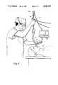
- FIG. 4is a view showing an endoscope constructed in accordance with the present invention as it would be used for insertion into a narrow passage of a body cavity such as the human trachea.
- the endoscope 10comprises an elongated sheath member 12 having distal or forward and proximal or rearward ends 14, 16 respectively.
- Sheath member 12has a cross-sectional shape, as suggested in FIGS. 2 and 2a, that is adapted for introduction of the endoscope 10 into a body cavity 18 as shown in FIG. 4.
- a first, selectively controllable bendable section 20having a length about one-third the overall length of the sheath member 12. This is an important aspect of the invention and is described further below.
- Bendable and flexible section 20houses an image forming optical system 22 at its far end 24. While an image forming system using fiber optics is described herein, new advances, such as the self-contained integrated circuit chips that act as cameras, may be substituted for the fiber optics systems described herein.
- a generally rigid section 26which includes a control housing 28. This section 26 acts to provide a directional guide for the endoscope 10.
- An image transmitting optical system 30extends through the sheath member 12, and has forward and rearward ends 32, 34 respectively.
- the forward end 32 of the image transmitting system 30is operatively located behind and adjacent the image forming system 22.
- the rearward end 34 thereofterminates in the control housing 28 and is adapted to be operatively connected to a video system 36 for viewing the image of an object on a television monitor.
- Video recording means(not shown) can be operatively connected in a preferred embodiment by attachment of the eyepiece to a small television camera to receive and record onto a recording medium such as video tape.
- a light transmitting system 38extends through the sheath member 12, and has forward and rearward ends 40, 42 respectively.
- the forward end 40 of the light transmitting systemis operatively located adjacent the image forming optical system 22.
- the rearward end 42 thereofterminates in the control housing 28 and is adapted to be connected to a light source (not shown) for providing illumination to the image forming optical system 22.
- a gas channel or passageway 44extends through the sheath member 12, and has forward and rearward ends 46, 48 respectively.
- the forward end 46 of the channel 44is operatively located adjacent the image forming optical system 22, while the rearward end 48 thereof terminates in the control housing 28, and is adapted to connect to a selectively controllable source of pressurized gas (not shown).
- Gas directing means 50at the forward end 46 of the channel 44 applies a flow of pressurized gas across the image forming optical system 22 for keeping the optical system 22 operationally clear during the intubation procedure.
- Channel 44provides a passageway for the anesthetist to provide an oxygen in-flow (generally in the range of about 4 to 6 liters per minute) to the patient undergoing the intubation procedure.
- This oxygen in-flowserves two purposes: first, it is directed across the image forming optical system 22 to defog or maintain system 22 clear; and, secondly, to provide diffuse oxygenation (that is, to inject oxygen into the patient's lungs) so as to wash out carbon dioxide (C02) and Ox diffusion of the patient during the intubation procedure.
- This later aspect of the inventionis an important safety measure during intubation, as a patient's life can be maintained by diffusion oxygenation for periods of 2 to 3 minutes without outside breathing.
- Manipulating means 54are operatively connected to the first, selectively controllable bendable section 20 of the sheath member 12 from at the control housing 28 for manipulating the distal or forward tip end portion 14 of the sheath member 12.
- the forward tip end section 14 of the sheath member 12have a dual wide radius of curvature, and a deflection in one direction (upward) of about 160 degrees, and an opposite second direction deflection of about 100 degrees.
- additional channels or lumensmay be provided throughout the sheath member for such purposes as additional gas passageways or channels for transporting instruments to selected locations within the body cavity which are visualized and located using the imaging provided in the present invention.
- the flexible portion 20 of the sheath member 12should be in the range of about 5 to 21 centimeters, while the overall length of the rigid portion 26 should be in the range of about 10 to 30 centimeters.
- the preferred length of the sheath member 12is about 35 centimeters overall, and that of the associated endotracheal tube to be about 30 centimeters overall. While these figures may vary, it is essential that the length ratios of the individual parts permits the user to adjust the flexible section 20 protruding from the endotracheal tube to accommodate the particular anatomy of the patient.
- An adjustable locking collarsecured by a screw or the like, that is selectively releasable, can be associated with the sheath member 12 of the endoscope 10, and the endotracheal tube as is shown in FIGS. 3 and 4, to hold the desired relationship once it is chosen by the user.
- a standard endotracheal tubeis placed over the sheath member of the endoscope prior to commencing the intubation process as shown in FIG. 3.
- the light transmitting system of the endoscopeis connected to a light sourceto provide illumination for the imaging system of the endoscope.
- the internal gas passage channel of the endoscopeis connected to a source of controlled pressurized gas, such as oxygen.
- a source of controlled pressurized gassuch as oxygen.
- the viewing eyepiece section of the endoscopeis connected to a video system, such as one including a small television camera, for both recording and displaying the transmitted image for the attending anesthetist.
- the display of the imagecan be done on a miniature, preferably color, television monitor that can be located in a location convenient for the anesthetist.
- the anesthetistBy displaying an enlarged transmitted image on a video monitor, two immediate benefits over prior art instruments are achieved. First, the enlarged view of the patient's anatomical details assists the anesthetist in identifying the location of the forward tip end of the endoscope. Secondly, the anesthetist is offered a binocular view of the area of interest as opposed to the miniature, monocular view afforded him by prior art instruments employing only a fiber optics imaging system.
- the anesthetistintroduces the distal end of the endoscope into the patient's mouth and manipulates the bending of the distal end portion of the sheath member of the endoscope to accommodate the encountered anatomy of the patient. Due to the controllable bending of the distal or forward tip end of the endoscope, the distal or forward tip end portion of the endoscope can be carefully maneuvered through the patient's body cavity minimizing or even eliminating patient trauma.
- the distal end of the endoscope and the endotracheal tubecan be carefully advanced through this delicate structure with ease and without inducing patient trauma. Entering the patient's trachea, the endoscope and the endotracheal tube are advanced until the bifurcation of the trachea is visualized. At this stage of the intubation process, further advancement of the endoscope is stopped and the thumb control is released on the bendable distal portion of the endoscope sheath member.
- the position of the endotracheal tubeis adjusted toward the distal end of the endoscope until it is properly located in the patient's trachea. At this point, the endoscope cab be withdrawn from the patient's trachea through the endotracheal tube.
- the endotracheal tubeis left in position and is connected to the inhalation anesthetic machnery.
- the cuff of the endotracheal tubeis then inflated to hold the tube in place and to seal the airways external to the tube.
- the entire intubation processmay be video taped or otherwise recorded on a recording medium for documentation purposes, including further study or teaching.
Landscapes
- Health & Medical Sciences (AREA)
- Life Sciences & Earth Sciences (AREA)
- Surgery (AREA)
- Biomedical Technology (AREA)
- Medical Informatics (AREA)
- Optics & Photonics (AREA)
- Pathology (AREA)
- Radiology & Medical Imaging (AREA)
- Biophysics (AREA)
- Engineering & Computer Science (AREA)
- Physics & Mathematics (AREA)
- Heart & Thoracic Surgery (AREA)
- Nuclear Medicine, Radiotherapy & Molecular Imaging (AREA)
- Molecular Biology (AREA)
- Animal Behavior & Ethology (AREA)
- General Health & Medical Sciences (AREA)
- Public Health (AREA)
- Veterinary Medicine (AREA)
- Pulmonology (AREA)
- Otolaryngology (AREA)
- Physiology (AREA)
- Endoscopes (AREA)
Abstract
Description
1. Field of the Invention
This invention relates in general to an endoscope, and, more particularly, to an endoscope having a sheath member with a controllably bendable distal or forward sheath portion and a generally rigid proximal or rearward sheath portion, which sheath member further includes an image transmitting system therethrough.
2. Description of the Related Art
When a surgical technique requires that a patient be placed under a general anesthesia, the anesthetic is usually administered to the patient by inhalation. To accomplish this, a endotracheal tube with an inflatable cuff is introduced into the patient's trachea after the patient is sedated or "put to sleep". Once the endotracheal tube is properly positioned, the cuff is inflated to seal the trachea passageway from ambient gases, and the anesthetic is introduced to the patient for inhalation through a lumen in the endotracheal tube.
When this technique is used to introduce an anesthetic, the ventilation of the patient is carefully controlled and monitored by the surgical team. The lumen in the endotracheal tube is connected to an anesthetic machine, thus forming a closed inhalation/anesthetic system between the sleeping patient and the anesthetic machine. Both the volume and cycle of the respiration of the patient during the surgical procedure is closely controlled and monitored continuously by the anesthetist.
One problem with the closed inhalation/anesthetic system technique using an endotracheal tube in association with an endoscope (which is widely used in inhalation-induced general anesthesia), is that the introduction of the endotracheal tube, that is, a plastic tube with an inflatable cuff, is not always an easy, successful or non-traumatic procedure for the patient.
Specifically, an anesthetist is usually taught that in commencing this technique, a surgical instrument, such as a laryngoscope, is first introduced into the patient's mouth and the patient's tongue is elevated so that the patient's epiglottis and vocal cords (forming the entrance to the trachea) can be visualized.
In practice, however, there are many anatomical anomalies and physical variations among patients which do not permit observation of this vital tracheal area, so that the medical practitioner attempting to perform this procedure is reduced to operating blindly, by feel, relying on his prior experience to provide him with general guidelines.
Even in the case of a "standard" intubation that proceeds without difficulties in the visualization of these vital tracheal areas, two major problems must be met and overcome by the medical practioner.
First, when the endotracheal tube approaches the patient's vocal cords, the precise location of the distal, or forward end of the endotracheal tube becomes obscured by the endotracheal tube itself. This situation requires that any further advance of the endotracheal tube through the patient's vocal cords by the medical practitioner be done semi-blindly, relying on feel.
If the endotracheal tube abrades or otherwise injures the patient's vocal cords, such inadvertent injuries can result in a number of problems ranging from a "sore throat" with transient speech impairment to a "husky voice" or even vocal cord paralysis.
Secondly, after the endotracheal tube is advanced past the vocal cords and into the patient's trachea, the distal end of the endotracheal tube should be in the range of 2 to 4 cm (about 1 to 2 inches) in front of the bifurcation of the trachea in order to ventilate both of the patient's lungs equally.
In practice, this positioning of the endotracheal tube with respect to the tracheal bifurcation is partially determined by gradation markings on the endotracheal tube itself, and by prior experience on the part of the medical practitioner. However, there are many variations in the length of the trachea of individual patients, and although such tracheal length is usually classified and determined by a patient's teeth-bifurcation distance, no absolute standard exists.
If the distal or forward tip end of the endotracheal tube is mislocated, and the patient's lungs are unequally ventilated during the general anesthesia state (half-lung ventilation), the patient may develop a post-operative pneumonia problem.
Currently, the anesthetist employs techniques such as auscultation (the process of listening for sounds emanating from the patient's chest) to determine if the patient's lungs are being ventilated equally. Such a technique as auscultation requires broad experience on the part of the practioner and some guess work, providing for a wide margin of error. Also, auscultation by the anesthetist to hear both lung excursions is not always accurate, mainly in obese patients with a large chest cavity.
The problems and complications resulting from an unsuccessful intubation attempt can be largely overcome by precise visual control of the endotracheal tube throughout the actual intubation process, that is, guiding the passage of the endotracheal tube through the patient's vocal cords and into the trachea, and, by proper positioning of the distal or forward end of the endotracheal tube in front of the patient's tracheal bifurcation so as to ventilate his lungs equally.
The following instruments and techniques of the prior art have been used to assist in the intubation process:
(1) An endotracheal tube stylet instrument which consists of a wire, generally copper, having a plastic covering. This endotracheal tube stylet is inserted into a central lumen of the endotracheal tube to straighten the natural curvature of the endotracheal tube so as to accommodate a patient having a narrow mouth.
Specifically, the distal or forward tip end of the endotracheal tube can be bent or formed by using the stylet so as to enable it to follow a shorter exterior radius, thereby facilitating the intubation of the endotracheal tube in cases requiring the elevation of an "overhanging" epiglottis.
These endotracheal tube stylets are also available with a small battery operated electric bulb located at their distal tip end that provides transillumination of the trachea.
(2) Another approach is the completely flexible fiber optic bronchoscope used in association with an endotracheal tube, siilar to that endoscope of U.S. Pat. No. 3,788,304 to Takahashi.
Completely flexible bronchoscopes were previously used for transnasal or even oral intubations. The completely flexible nature of the instrument, its extreme length, and the tiny monocular image seen in its eyepiece, all prove to be very troublesome to the anesthetist, who is not generally trained as an endoscopist.
None of the prior art techniques or instruments known to the applicant have proven to be completely successful during transnasal intubation.
While the endotracheal tube stylets appear to help, they do not avoid patient trauma, nor do they indicate the proper position and location of the patient's vocal cords.
Likewise, the completely flexible bronchoscope provides visualization of the area of concern, but due to the complete flexibility of the instrument, it becomes too flexible for easy use. Also, due to the extreme length of the completely flexible bronchoscope, the anesthetist had to be moved from his optimal position near the patient's head in the operating theater in order to accommodate the demands of the instrument.
Accordingly, none of the instruments or techniques in use today provide either an instrument or a technique which can be: reconfigured to adapt to the anatomy of an individual patient; convenient for the anesthetist who has to be located near to the patient's head; provides an enlarged image of the patient's anatomical features; incorporates both a flexible and a rigid section; or, allows sufficient ventilation or oxygenation of the patient during the intubating process.
The present invention overcomes the above described disadvantages and problems of the prior art and provides a safer and less traumatizing instrument for the intubation of a patient.
An object of the present invention is to provide a novel and useful endoscope in which the above described disadvantages and problems of the prior art are avoided.
Another object of the present invention is to provide a novel and useful endoscope which is positioned in the lumen of an endotracheal tube. The distal or forward tip end portion of the endoscope and the surrounding endotracheal tube can be precisely located and afford the practioner a clear and enlarged visualization of the patient's anatomical features.
To achieve these objects, as well as other objects described below, the endoscope constructed in accordance with the present invention has an elongated sheath member having distal, or forward, and proximal, or rearward, ends, which is adapted for introduction into a body cavity.
The sheath member has, at its distal or forward end, a first, selectively controllable bendable section which houses an image forming optical system.
Likewise, the sheath member has at its proximal or rearward end, a generally rigid section which includes a control housing. An image transmitting optical system extends throughout the length of the sheath member in one of the lumens thereof. The forward end of the image transmitting system is operatively located behind and adjacent an image forming system in another of the lumens, while the rearward end thereof terminates in the control housing.
This arrangement permits the image of an object that is formed by the image forming optical system on the forward end of the image transmitting optical system and transmitted therethrough to the rearward end thereof, to be viewed in the proximal or rearward end portion of the sheath member.
A light transmitting system also extends throughout the length of the sheath member in another of the lumens. The forward end of the light transmitting system is operatively located adjacent the image forming optical system, and the rearward end thereof terminates in the control housing. The rearward end is adapted to be operatively connected to a light source for providing illumination of the area observed by the image forming optical system.
A channel or passageway extends throughout the length of the sheath member in another of the lumens. The forward end of the channel is operatively located adjacent the image forming optical system and has its rearward end terminating in the control housing. The channel is adapted to be operatively connected to a selectively controllable source of pressurized gas, preferably oxygen, for diffusion oxygenation of the patient undergoing intubation.
Gas flow directing means are operatively connected to the forward end of the channel for applying a flow of pressurized gas across the image forming optical system to keep the optical system clear during operation as well as to provide for a continuous controlled flow of oxygen into the patient's lungs. Controlling means in the control housing are operatively connected to a first, selectively controllable bendable section of the sheath member, for manipulating the distal or forward tip end portion of the sheath member.
When the endoscope with the surrounding endotracheal tube is successfully guided to the optimal location, just above the tracheal bifurcation, the endotracheal tube is held in place and the endoscope is withdrawn. Th cuff is then inflated and oxygen and other gases can be supplied to the patient under control of the anesthetist.
The novel features of construction and operation of the invention will be more clearly apparent during the course of the following description, reference being had to the accompanying drawings wherein has been illustrated a preferred form of the device of the invention and wherein like characters of reference designate like parts throughout the drawings.
FIG. 1 is a perspective view of an endoscope constructed in accordance with the present invention;
FIG. 2 is an enlarged partial view of the distal end of the endoscope shown in FIG. 1;
FIG. 2a is an enlarged cross-section view taken on line 2a-2a of FIG. 2;
FIG. 3 is a view similar to that of FIG. 1 showing the endoscope of FIG. 1 in association with an endostracheal tube having an inflatable cuff; and
FIG. 4 is a view showing an endoscope constructed in accordance with the present invention as it would be used for insertion into a narrow passage of a body cavity such as the human trachea.
Referring to FIGS. 1 through 3 showing an endoscope constructed in accordance with the present invention, it is seen that theendoscope 10 comprises anelongated sheath member 12 having distal or forward and proximal or rearward ends 14, 16 respectively.Sheath member 12 has a cross-sectional shape, as suggested in FIGS. 2 and 2a, that is adapted for introduction of theendoscope 10 into abody cavity 18 as shown in FIG. 4.
At the distal or forward tip end 14 of thesheath member 12, is a first, selectively controllablebendable section 20, having a length about one-third the overall length of thesheath member 12. This is an important aspect of the invention and is described further below.
Bendable andflexible section 20 houses an image formingoptical system 22 at itsfar end 24. While an image forming system using fiber optics is described herein, new advances, such as the self-contained integrated circuit chips that act as cameras, may be substituted for the fiber optics systems described herein.
At the proximal or rearward end 16 of thesheath member 12, is a generallyrigid section 26 which includes acontrol housing 28. Thissection 26 acts to provide a directional guide for theendoscope 10.
An image transmittingoptical system 30 extends through thesheath member 12, and has forward and rearward ends 32, 34 respectively. Theforward end 32 of theimage transmitting system 30 is operatively located behind and adjacent theimage forming system 22. Therearward end 34 thereof terminates in thecontrol housing 28 and is adapted to be operatively connected to avideo system 36 for viewing the image of an object on a television monitor.
The image transmittingoptical system 30, as configured in this preferred embodiment, permits an image of an object that is viewed by the image formingoptical system 22 at theforward end 32 of the image transmittingoptical system 30 to be transmitted therethrough to therearward end 34 thereof, to be converted at theproximal end portion 16 of thesheath member 12 for transmission to and viewing on the television monitor. Video recording means (not shown) can be operatively connected in a preferred embodiment by attachment of the eyepiece to a small television camera to receive and record onto a recording medium such as video tape.
Alight transmitting system 38 extends through thesheath member 12, and has forward and rearward ends 40, 42 respectively. Theforward end 40 of the light transmitting system is operatively located adjacent the image formingoptical system 22. Therearward end 42 thereof terminates in thecontrol housing 28 and is adapted to be connected to a light source (not shown) for providing illumination to the image formingoptical system 22.
A gas channel orpassageway 44 extends through thesheath member 12, and has forward and rearward ends 46, 48 respectively. Theforward end 46 of thechannel 44 is operatively located adjacent the image formingoptical system 22, while therearward end 48 thereof terminates in thecontrol housing 28, and is adapted to connect to a selectively controllable source of pressurized gas (not shown).
Gas directing means 50, at theforward end 46 of thechannel 44 applies a flow of pressurized gas across the image formingoptical system 22 for keeping theoptical system 22 operationally clear during the intubation procedure.
Manipulating means 54 are operatively connected to the first, selectively controllablebendable section 20 of thesheath member 12 from at thecontrol housing 28 for manipulating the distal or forwardtip end portion 14 of thesheath member 12.
It is preferable that the forwardtip end section 14 of thesheath member 12 have a dual wide radius of curvature, and a deflection in one direction (upward) of about 160 degrees, and an opposite second direction deflection of about 100 degrees.
Various means are known and customarily used in the art, for example, U.S. Pat. No. 3,788,304 to Takahashi. All such means are applicable for use with the present invention and may, by appropriate modification, be incorporated hereinto.
In an alternate embodiment of an endoscope constructed in accordance with the present invention, additional channels or lumens may be provided throughout the sheath member for such purposes as additional gas passageways or channels for transporting instruments to selected locations within the body cavity which are visualized and located using the imaging provided in the present invention.
In general, it is seen from both the above description of a preferred embodiment of an endoscope constructed in accordance with the invention and the description of the method of operation that follows, that the proportion or ratio between the lengths of theflexible section 20 and the generallyrigid section 26, is an important aspect of the present invention.
This is because when the endotracheal tube is associated with theendoscope 10, as shown in FIGS. 3 and 4, the length of theflexible section 20 that protrudes from the endotracheal tube must be capable of variation to accommodate a patient's particular anatomy.
After a period of experimentation, it was determined that to provide an optimal instrument for general use among a wide variety of patients, theflexible portion 20 of thesheath member 12 should be in the range of about 5 to 21 centimeters, while the overall length of therigid portion 26 should be in the range of about 10 to 30 centimeters.
Also, the preferred length of thesheath member 12 is about 35 centimeters overall, and that of the associated endotracheal tube to be about 30 centimeters overall. While these figures may vary, it is essential that the length ratios of the individual parts permits the user to adjust theflexible section 20 protruding from the endotracheal tube to accommodate the particular anatomy of the patient.
An adjustable locking collar, secured by a screw or the like, that is selectively releasable, can be associated with thesheath member 12 of theendoscope 10, and the endotracheal tube as is shown in FIGS. 3 and 4, to hold the desired relationship once it is chosen by the user.
In use, a standard endotracheal tube is placed over the sheath member of the endoscope prior to commencing the intubation process as shown in FIG. 3. The light transmitting system of the endoscope is connected to a light sourceto provide illumination for the imaging system of the endoscope.
The internal gas passage channel of the endoscope is connected to a source of controlled pressurized gas, such as oxygen. Finally, the viewing eyepiece section of the endoscope is connected to a video system, such as one including a small television camera, for both recording and displaying the transmitted image for the attending anesthetist. The display of the image can be done on a miniature, preferably color, television monitor that can be located in a location convenient for the anesthetist.
By displaying an enlarged transmitted image on a video monitor, two immediate benefits over prior art instruments are achieved. First, the enlarged view of the patient's anatomical details assists the anesthetist in identifying the location of the forward tip end of the endoscope. Secondly, the anesthetist is offered a binocular view of the area of interest as opposed to the miniature, monocular view afforded him by prior art instruments employing only a fiber optics imaging system.
While observing the image displayed with the present invention, the anesthetist introduces the distal end of the endoscope into the patient's mouth and manipulates the bending of the distal end portion of the sheath member of the endoscope to accommodate the encountered anatomy of the patient. Due to the controllable bending of the distal or forward tip end of the endoscope, the distal or forward tip end portion of the endoscope can be carefully maneuvered through the patient's body cavity minimizing or even eliminating patient trauma.
When the patient's vocal cords are visualized, the distal end of the endoscope and the endotracheal tube can be carefully advanced through this delicate structure with ease and without inducing patient trauma. Entering the patient's trachea, the endoscope and the endotracheal tube are advanced until the bifurcation of the trachea is visualized. At this stage of the intubation process, further advancement of the endoscope is stopped and the thumb control is released on the bendable distal portion of the endoscope sheath member.
With the endoscope properly positioned, the position of the endotracheal tube is adjusted toward the distal end of the endoscope until it is properly located in the patient's trachea. At this point, the endoscope cab be withdrawn from the patient's trachea through the endotracheal tube. The endotracheal tube is left in position and is connected to the inhalation anesthetic machnery. The cuff of the endotracheal tube is then inflated to hold the tube in place and to seal the airways external to the tube.
While the endoscope is being advanced through the patient's vocal cords and trachea, positive pressure is supplied by a gas, such as oxygen, through the gas passageway channel of the endoscope. This continues ventilation and insufflation of the patient so as to ensure diffusion oxygenization, even in the paralyzed (respiration paralysis induced by the anesthetist) patient, as well as to prevent fogging of the imaging system of the endoscope.
Finally, the entire intubation process may be video taped or otherwise recorded on a recording medium for documentation purposes, including further study or teaching.
The invention described above is, of course, susceptible to many variations, modifications and changes, all of which are within the skill of the art. It should be understood that all such variations, modifications and changes are within the spirit and scope of the invention and of the appended claims. Similarly, it will be understood that it is intended to cover all changes, modifications and variations of the example of the invention herein disclosed for the purpose of illustration which do not constitute departures from the spirit and scope of the invention.
Claims (9)
1. An endoscope comprising:
(a) an elongated sheath member having distal and proximal ends adapted for introduction into a body cavity and having at its distal end, a first, selectively controllable bendable section housing an image-forming optical system, and, at its proximal end, a generally rigid section including a control housing, the ratio between said first, selectively controllable bendable section and said generally rigid section being approximately one-third to two-thirds;
(b) an image transmitting optical system extending through said sheath member, said image transmitting optical system having forward and rearward ends, the forward end of said image transmitting system operatively located behind and adjacent said image forming system and the rearward end thereof terminating in said control housing, thereby permitting an image of an object adjacent said distal end to be viewed in said proximal end portion;
(c) a light transmitting system extending through said sheath member, said light transmitting system having forward and rearward ends, the forward end of said light transmitting system operatively located adjacent said image forming optical system and the rearward end thereof terminating in said control housing and adapted to be operatively connected to a light source for providing illumination for said image forming optical system;
(d) a channel extending through said sheath member, said channel having forward and rearward ends, the forward end of said channel operatively located adjacent said image forming optical system and the rearward end thereof terminating in said control housing and adapted to be operatively connected to a selectively controllable source of pressurized gas;
(e) means operatively connected to the forward end of said channel for directing a flow of pressurized gas across said image forming optical system for keeping said image forming optical system operationally clear; and
(f) control means in said control housing operatively connected to said first selectively controllable bendable section of said sheath member for manipulating said distal end portion of said sheath member to permit selective manipulation of said distal end portion of said sheath member through a patient's tracheal anatomy.
2. An endoscope as recited in claim 1 wherein said rearward end of said image transmitting system is adapted to be operatively connected to a small video camera system for viewing an object image on a television monitor.
3. An endoscope as recited in claim 1 further including video recording means operatively connected to said rearward end of said image transmitting system for recording said transmitted object images onto a recording medium.
4. An endoscope as recited in claim 1 wherein said means for directing a flow of pressurized gas across said image forming optical system comprises a nozzle adapted to selectively direct the discharge of said flow of pressurized gas from said channel across said image forming optical system.
5. An endoscope as recited in claim 1 wherein the length of said selectively controllable bendable section of said sheath member is in the range of 5 to 21 centimeters and the overall length of said sheath member is about 35 centimeters.
6. An endoscope comprising:
an elongated sheath member having distal and proximal ends adapted for introduction into a body cavity and having at its distal end a first selectively controllable bendable section having a length about one-third the overall length of said sheath member, said section housing an image forming optical system, said sheath member having at its proximal end a generally rigid section including a control housing;
an image transmitting optical system extending through said sheath member, said image transmitting optical system having forward and rearward ends, said forward end being operatively located behind and adjacent said image forming system, and said rearward end terminating in said control housing, said optical system being adapted to be operatively connected to a video system for viewing a transmitted image of an object on a television monitor, thereby permitting an object image formed by said optical system on the forward end of said optical system and transmitted to the rearward end to be viewed in both said proximal end portion and on a television monitor;
video recording means operatively connected to said rearward end of said optical system for recording said the transmitted image of an object onto a recording medium;
a light transmitting system extending through said sheath member, said light transmitting system having forward and rearward ends, said forward end operatively located adjacent said image forming optical system, and said rearward end terminating in said control housing, said light transmitting system being adapted to be operatively connected to a light source for providing illumination for objects to be visualized by said image forming optical system;
a channel extending through said sheath member, said channel having forward and rearward ends, said channel forward end being operatively located adjacent said optical system, said channel rearward end terminating in said control housing and adapted to be operatively connected to a selectively controllable source of pressurized gas;
gas control means, including a directional nozzle member, operatively connected to said channel forward end for directing a flow of pressurized gas across said optical system to keep said optical system operationally clear; and
control means operatively connected to said first selectively controllable bendable section for manipulating said distal end portion of said sheath member.
7. An intubation process using an endoscope with an intubating sheath member having both flexible and and rigid sections and said endoscope further having a video imaging system connected to video monitoring and display means and a channel for applying a gas to the distal end of the endoscope, in association with an endotracheal tube having an inflatable cuff, the process comprising the steps of:
(1) placing the endoscope through the central lumen of the endotracheal tube;
(2) inserting the distal end of the endoscope into the oral cavity of a subject;
(3) using the video imaging system and the video monitoring means, positioning the distal end of the endoscope through the larynx to a selected location near the tracheal bifurcation;
(4) using the video imaging system and the video monitoring and display means, guiding the endotracheal tube over the endoscope toward the distal end of the endoscope to issure that the distal end of the endotracheal tube does not extend beyond that of the endoscope;
(5) inflating the inflatable cuff to hold the endotracheal tube in position; and
(6) withdrawing the endoscope from the trachea and the central lumen of the endotracheal tube.
8. An intubation process as recited in claim 7 further including the step of:
applying a gas to the channel and directing the gas exiting from the channel across the video imaging system to keep the video imaging system clear.
9. An intubation process as recited in claim 7 further including the step of:
applying and controlling a gas flow through the channel to ensure diffusion oxygenation of said subject during the intubation process.
Priority Applications (1)
| Application Number | Priority Date | Filing Date | Title |
|---|---|---|---|
| US07/204,740US4846153A (en) | 1988-06-10 | 1988-06-10 | Intubating video endoscope |
Applications Claiming Priority (1)
| Application Number | Priority Date | Filing Date | Title |
|---|---|---|---|
| US07/204,740US4846153A (en) | 1988-06-10 | 1988-06-10 | Intubating video endoscope |
Publications (1)
| Publication Number | Publication Date |
|---|---|
| US4846153Atrue US4846153A (en) | 1989-07-11 |
Family
ID=22759234
Family Applications (1)
| Application Number | Title | Priority Date | Filing Date |
|---|---|---|---|
| US07/204,740Expired - LifetimeUS4846153A (en) | 1988-06-10 | 1988-06-10 | Intubating video endoscope |
Country Status (1)
| Country | Link |
|---|---|
| US (1) | US4846153A (en) |
Cited By (149)
| Publication number | Priority date | Publication date | Assignee | Title |
|---|---|---|---|---|
| WO1991004703A1 (en)* | 1988-07-29 | 1991-04-18 | Karl Storz Gmbh & Co. | Video endoscopic microscope |
| US5273029A (en)* | 1992-04-30 | 1993-12-28 | Wilk Peter J | Endotracheal tube assembly and related method and obturator |
| US5285778A (en)* | 1991-04-19 | 1994-02-15 | Mackin Robert A | Endotracheal tube wih fibers optic illumination and viewing and auxiliary tube |
| US5318533A (en)* | 1992-02-21 | 1994-06-07 | Scimed Life Systems, Inc. | Balloon catheter inflation device including apparatus for monitoring and wireless transmission of inflation data, and system |
| US5327881A (en)* | 1993-02-26 | 1994-07-12 | Beth Israel Hospital Association | Fiberoptic intubating stylet |
| US5329940A (en)* | 1990-02-14 | 1994-07-19 | Adair Edwin Lloyd | Endotracheal tube intubation assist device |
| US5353783A (en)* | 1991-12-09 | 1994-10-11 | Nakao Naomi L | Endoscopic method using sheath |
| US5364357A (en)* | 1989-09-25 | 1994-11-15 | Schneider (Usa) Inc. | Small diameter dilatation catheter having wire reinforced coaxial tubular body |
| US5390661A (en)* | 1993-02-03 | 1995-02-21 | W. L. Gore & Associates, Inc. | Introducer for esophageal probes |
| US5400770A (en)* | 1992-01-15 | 1995-03-28 | Nakao; Naomi L. | Device utilizable with endoscope and related method |
| US5400771A (en)* | 1993-01-21 | 1995-03-28 | Pirak; Leon | Endotracheal intubation assembly and related method |
| US5421325A (en)* | 1992-04-30 | 1995-06-06 | Cinberg; James Z. | Endotracheal tube assembly and related method |
| US5595172A (en)* | 1994-08-12 | 1997-01-21 | Reese; John L. | Suction stylet for use with an endotracheal tube |
| US5636625A (en)* | 1994-08-30 | 1997-06-10 | Machida Endoscope Co., Ltd. | Tracheal airway apparatus |
| US5645519A (en)* | 1994-03-18 | 1997-07-08 | Jai S. Lee | Endoscopic instrument for controlled introduction of tubular members in the body and methods therefor |
| USRE35595E (en)* | 1989-05-09 | 1997-08-26 | Six; Gary | Flexible tip stylet for use with an endotracheal intubation device |
| US5676635A (en)* | 1995-08-30 | 1997-10-14 | Levin; Bruce | Instrument for insertion of an endotracheal tube |
| US5791338A (en)* | 1994-01-26 | 1998-08-11 | William T. Merchant | Endotracheal intubation apparatus |
| WO1999022636A1 (en) | 1997-10-31 | 1999-05-14 | Imagyn Medical Technologies, Inc. | Intubation device and method |
| US5921917A (en)* | 1997-10-20 | 1999-07-13 | Clarus Medical Systems, Inc. | Hand-held viewing system with removable sheath |
| WO1999035960A1 (en) | 1998-01-16 | 1999-07-22 | University Of Florida | Malleable endotracheal tube with fiberoptic scope |
| WO1999038432A1 (en)* | 1998-01-29 | 1999-08-05 | Johns Hopkins University | Copa method for fiberoptic endotracheal intubation |
| US5941816A (en)* | 1997-04-15 | 1999-08-24 | Clarus Medical Systems, Inc. | Viewing system with adapter handle for medical breathing tubes |
| DE4403757C2 (en)* | 1994-02-07 | 2000-02-10 | Schulz Stuebner Sebastian | Lifting device for the introduction of a tube |
| US6090040A (en)* | 1998-09-10 | 2000-07-18 | Metro; R. J. | Periscope and retracting laryngoscope for intubation |
| US6115523A (en)* | 1996-10-04 | 2000-09-05 | University Of Florida | Plastic optical fiber airway imaging system |
| US6142144A (en)* | 1997-12-01 | 2000-11-07 | Pacey; John A. | Intubation instrument |
| USD434147S (en)* | 1998-12-29 | 2000-11-21 | Sli, Inc. | Flexible fiberoptic scope handle |
| WO2001002042A1 (en) | 1999-07-02 | 2001-01-11 | Pulmonx | Methods, systems, and kits for lung volume reduction |
| US6196225B1 (en) | 1995-10-23 | 2001-03-06 | Dean O. Allgeyer | Endotracheal tube for use during fiberoptic assisted intubation and with other intubating stylets |
| US20020066450A1 (en)* | 2000-12-02 | 2002-06-06 | Bonutti Peter M. | Tracheal intubination |
| US20020108610A1 (en)* | 1996-02-26 | 2002-08-15 | Christopher Kent L. | Method and apparatus for endotracheal intubation using a light wand and curved guide |
| US20020195103A1 (en)* | 2001-03-05 | 2002-12-26 | Government Of The United States, As Represented By The Secretary Of The Army | Intubation device and method |
| US20030051733A1 (en)* | 2001-09-10 | 2003-03-20 | Pulmonx | Method and apparatus for endobronchial diagnosis |
| US6539942B2 (en) | 2001-04-19 | 2003-04-01 | Richard Schwartz | Endotracheal intubation device |
| US6543447B2 (en) | 1997-12-01 | 2003-04-08 | Saturn Biomedical Systems Inc | Intubation instrument |
| US6550475B1 (en)* | 1998-03-11 | 2003-04-22 | Oldfield Family Holdings Pty. Limited | Endotracheal tube for selective bronchial occlusion |
| US20030078476A1 (en)* | 2001-07-24 | 2003-04-24 | Hill Stephen D. | Apparatus for intubation |
| US20030088156A1 (en)* | 1999-10-14 | 2003-05-08 | George Berci | Video laryngoscope with detachable light and image guides |
| US6585639B1 (en) | 2000-10-27 | 2003-07-01 | Pulmonx | Sheath and method for reconfiguring lung viewing scope |
| US6655377B2 (en) | 1997-12-01 | 2003-12-02 | Saturn Biomedical Systems Inc. | Intubation instrument |
| US20040073191A1 (en)* | 2000-10-27 | 2004-04-15 | Pulmonx | Methods and devices for obstructing and aspirating lung tissue segments |
| US20040220451A1 (en)* | 1996-10-04 | 2004-11-04 | Dietrich Gravenstein | Imaging scope |
| US20050051175A1 (en)* | 2003-09-10 | 2005-03-10 | Brain Archibald I. J. | Intubating laryngeal mask airway device with fiber optic assembly |
| US20050139220A1 (en)* | 1996-02-26 | 2005-06-30 | Evergreen Medical Incorporated | Method and apparatus for ventilation / oxygenation during guided insertion of an endotracheal tube |
| US20050177024A1 (en)* | 2004-02-10 | 2005-08-11 | Mackin Robert A. | Endotracheal camera |
| US20050182297A1 (en)* | 1996-10-04 | 2005-08-18 | Dietrich Gravenstein | Imaging scope |
| US20050192481A1 (en)* | 1999-10-14 | 2005-09-01 | George Berci | Laryngoscope and camera coupling |
| US20050267331A1 (en)* | 2004-05-28 | 2005-12-01 | Secrest Dean J | Overtube assembly |
| US20060004258A1 (en)* | 2004-07-02 | 2006-01-05 | Wei-Zen Sun | Image-type intubation-aiding device |
| US20060025650A1 (en)* | 2002-10-03 | 2006-02-02 | Oren Gavriely | Tube for inspecting internal organs of a body |
| WO2006055692A2 (en) | 2004-11-16 | 2006-05-26 | Pulmonx | Pulmonary occlusal stent delivery catheter, loading system and methods of use |
| WO2006055683A2 (en) | 2004-11-16 | 2006-05-26 | Pulmonx | Occlusal stent and methods for its use |
| US20060180155A1 (en)* | 2005-01-26 | 2006-08-17 | Ezc Holding, Llc | Video-assisted laryngeal mask airway devices |
| US20070049794A1 (en)* | 2005-09-01 | 2007-03-01 | Ezc Medical Llc | Visualization stylet for medical device applications having self-contained power source |
| US20070137651A1 (en)* | 2005-12-16 | 2007-06-21 | Ezc Medical Llc | Visualization esophageal-tracheal airway apparatus and methods |
| US20070142742A1 (en)* | 2005-07-13 | 2007-06-21 | Pulmonx | Methods and systems for segmental lung diagnostics |
| US20070175482A1 (en)* | 2006-01-27 | 2007-08-02 | Ezc Medical Llc | Apparatus for introducing an airway tube into the trachea having visualization capability and methods of use |
| US20070215162A1 (en)* | 2005-12-16 | 2007-09-20 | Ezc Medical Llc | Visualization airway apparatus and methods for selective lung ventilation |
| US20080029100A1 (en)* | 2005-12-16 | 2008-02-07 | Ezc Medical Llc | Visualization laryngeal airway apparatus and methods of use |
| US20080058599A1 (en)* | 2006-09-01 | 2008-03-06 | Ai Medical Devices, Inc. | Endotracheal intubation device |
| US20080208000A1 (en)* | 2006-09-01 | 2008-08-28 | Ai Medical Devices, Inc. | Endotracheal intubation and fluid delivery device |
| US20080221703A1 (en)* | 2007-03-09 | 2008-09-11 | Pulmonx | Loading a device for a pulmonary implant |
| US20080221582A1 (en)* | 2007-03-05 | 2008-09-11 | Pulmonx | Pulmonary stent removal device |
| US20080308098A1 (en)* | 2007-06-18 | 2008-12-18 | Ai Medical Devices | Endotracheal intubation device |
| US20090032016A1 (en)* | 2007-08-04 | 2009-02-05 | John Adam Law | Airway intubation device |
| US20090192355A1 (en)* | 2008-01-28 | 2009-07-30 | Mauricio Mejia | Scope for managing difficult pathways and method to improve visibility of the same |
| US20090192350A1 (en)* | 2008-01-28 | 2009-07-30 | Mauricio Mejia | Wireless video stylet with display mounted to laryngoscope blade and method for using the same |
| US20090209826A1 (en)* | 2008-01-09 | 2009-08-20 | Ezc Medical Llc | Intubation systems and methods |
| US20090322867A1 (en)* | 2006-08-07 | 2009-12-31 | Innovative Medical Devices , Inc | System to aid in the positioning, confirmation and documentation of an endotracheal tube |
| US7658196B2 (en) | 2005-02-24 | 2010-02-09 | Ethicon Endo-Surgery, Inc. | System and method for determining implanted device orientation |
| US20100036361A1 (en)* | 2008-06-20 | 2010-02-11 | Pulmonx | System and method for delivering multiple implants into lung passageways |
| US20100094090A1 (en)* | 2008-01-28 | 2010-04-15 | Mauricio Mejia | Self-cleaning wireless video stylet with display mounted to laryngoscope blade and method for using the same |
| US20100095969A1 (en)* | 2008-10-17 | 2010-04-22 | Ai Medical Devices, Inc. | Endotracheal intubation device |
| US20100199999A1 (en)* | 2009-02-06 | 2010-08-12 | Vazales Brad E | Methods for cleaning endotracheal tubes |
| US7775215B2 (en) | 2005-02-24 | 2010-08-17 | Ethicon Endo-Surgery, Inc. | System and method for determining implanted device positioning and obtaining pressure data |
| US7775966B2 (en) | 2005-02-24 | 2010-08-17 | Ethicon Endo-Surgery, Inc. | Non-invasive pressure measurement in a fluid adjustable restrictive device |
| US7844342B2 (en) | 2008-02-07 | 2010-11-30 | Ethicon Endo-Surgery, Inc. | Powering implantable restriction systems using light |
| US20110023887A1 (en)* | 2009-02-06 | 2011-02-03 | Endoclear, Llc | Methods for tracheostomy visualization |
| US7883471B2 (en) | 2001-09-10 | 2011-02-08 | Pulmonx Corporation | Minimally invasive determination of collateral ventilation in lungs |
| WO2011018812A1 (en)* | 2009-08-10 | 2011-02-17 | Alberto Bauer | System and method to execute tracheal intubation |
| US7927270B2 (en) | 2005-02-24 | 2011-04-19 | Ethicon Endo-Surgery, Inc. | External mechanical pressure sensor for gastric band pressure measurements |
| US20110120458A1 (en)* | 2009-11-24 | 2011-05-26 | John Schwartz | Endotracheal intubation device |
| US20110152678A1 (en)* | 2005-01-20 | 2011-06-23 | Pulmonx Corporation | Methods and devices for passive residual lung volume reduction and functional lung volume expansion |
| US20110196204A1 (en)* | 2010-02-11 | 2011-08-11 | Al Medical Devices, Inc. | Shape-conforming intubation device |
| US8016744B2 (en) | 2005-02-24 | 2011-09-13 | Ethicon Endo-Surgery, Inc. | External pressure-based gastric band adjustment system and method |
| US8016745B2 (en) | 2005-02-24 | 2011-09-13 | Ethicon Endo-Surgery, Inc. | Monitoring of a food intake restriction device |
| US8034065B2 (en) | 2008-02-26 | 2011-10-11 | Ethicon Endo-Surgery, Inc. | Controlling pressure in adjustable restriction devices |
| WO2011126812A1 (en)* | 2010-03-29 | 2011-10-13 | Endoclear, Llc | Airway cleaning and visualization |
| US8057492B2 (en) | 2008-02-12 | 2011-11-15 | Ethicon Endo-Surgery, Inc. | Automatically adjusting band system with MEMS pump |
| US8066629B2 (en) | 2005-02-24 | 2011-11-29 | Ethicon Endo-Surgery, Inc. | Apparatus for adjustment and sensing of gastric band pressure |
| US20120016197A1 (en)* | 2009-05-28 | 2012-01-19 | Smiths Medical International Limited | Medico-surgical apparatus |
| US8100870B2 (en) | 2007-12-14 | 2012-01-24 | Ethicon Endo-Surgery, Inc. | Adjustable height gastric restriction devices and methods |
| US8114345B2 (en) | 2008-02-08 | 2012-02-14 | Ethicon Endo-Surgery, Inc. | System and method of sterilizing an implantable medical device |
| US8142452B2 (en) | 2007-12-27 | 2012-03-27 | Ethicon Endo-Surgery, Inc. | Controlling pressure in adjustable restriction devices |
| US8152710B2 (en) | 2006-04-06 | 2012-04-10 | Ethicon Endo-Surgery, Inc. | Physiological parameter analysis for an implantable restriction device and a data logger |
| US8187162B2 (en) | 2008-03-06 | 2012-05-29 | Ethicon Endo-Surgery, Inc. | Reorientation port |
| US8187163B2 (en) | 2007-12-10 | 2012-05-29 | Ethicon Endo-Surgery, Inc. | Methods for implanting a gastric restriction device |
| US8192350B2 (en) | 2008-01-28 | 2012-06-05 | Ethicon Endo-Surgery, Inc. | Methods and devices for measuring impedance in a gastric restriction system |
| US8221439B2 (en) | 2008-02-07 | 2012-07-17 | Ethicon Endo-Surgery, Inc. | Powering implantable restriction systems using kinetic motion |
| US8233995B2 (en) | 2008-03-06 | 2012-07-31 | Ethicon Endo-Surgery, Inc. | System and method of aligning an implantable antenna |
| EP2524713A1 (en)* | 2011-05-18 | 2012-11-21 | Centre Hospitalier Universitaire de Bordeaux | Method for positioning a disposable endotracheal tube, and corresponding system for intubation |
| WO2012156480A1 (en)* | 2011-05-18 | 2012-11-22 | Centre Hospitalier Universitaire De Bordeaux (C.H.U De Bordeaux) | Method for positioning a disposable sterile endotracheal tube, and corresponding system for intubation |
| US20120302833A1 (en)* | 2011-05-26 | 2012-11-29 | Nellcor Puritan Bennett Llc | Illumination systems and devices for tracheal tubes |
| US8337389B2 (en) | 2008-01-28 | 2012-12-25 | Ethicon Endo-Surgery, Inc. | Methods and devices for diagnosing performance of a gastric restriction system |
| US8377079B2 (en) | 2007-12-27 | 2013-02-19 | Ethicon Endo-Surgery, Inc. | Constant force mechanisms for regulating restriction devices |
| US20130178704A1 (en)* | 2009-09-01 | 2013-07-11 | Francisco Jaime | Scope Tubes, Intubation Assemblies, and Methods of Intubation |
| US8523782B2 (en) | 2005-12-07 | 2013-09-03 | Pulmonx Corporation | Minimally invasive determination of collateral ventilation in lungs |
| US8591395B2 (en) | 2008-01-28 | 2013-11-26 | Ethicon Endo-Surgery, Inc. | Gastric restriction device data handling devices and methods |
| US8591532B2 (en) | 2008-02-12 | 2013-11-26 | Ethicon Endo-Sugery, Inc. | Automatically adjusting band system |
| US8870742B2 (en) | 2006-04-06 | 2014-10-28 | Ethicon Endo-Surgery, Inc. | GUI for an implantable restriction device and a data logger |
| US8876791B2 (en) | 2005-02-25 | 2014-11-04 | Pulmonx Corporation | Collateral pathway treatment using agent entrained by aspiration flow current |
| USD716841S1 (en) | 2012-09-07 | 2014-11-04 | Covidien Lp | Display screen with annotate file icon |
| USD717340S1 (en) | 2012-09-07 | 2014-11-11 | Covidien Lp | Display screen with enteral feeding icon |
| US8887730B2 (en) | 2011-05-26 | 2014-11-18 | Covidien Lp | Dual-lumen tracheal tube with assembly portion |
| US8978657B2 (en) | 2010-07-29 | 2015-03-17 | Covidien Lp | Dual-lumen tracheal tube with shaped lumen divider |
| US8998798B2 (en) | 2010-12-29 | 2015-04-07 | Covidien Lp | Multi-lumen tracheal tube with visualization device |
| US20150126808A1 (en)* | 2011-05-18 | 2015-05-07 | Centre Hospitalier Universitaire De Bordeaux (C.H.U. De Bordeaux) | Method for positioning a disposable sterile endotracheal tube, and corresponding system for intubation |
| US9050094B2 (en) | 2007-03-12 | 2015-06-09 | Pulmonx Corporation | Methods and devices for passive residual lung volume reduction and functional lung volume expansion |
| USD735343S1 (en) | 2012-09-07 | 2015-07-28 | Covidien Lp | Console |
| US20150258296A1 (en)* | 2012-10-14 | 2015-09-17 | Evgeny Pecherer | Tool and Method for Inserting an Endotracheal Tube |
| US9155854B2 (en) | 2011-08-31 | 2015-10-13 | Covidien Lp | Tracheal tube with visualization device and integrated flushing system |
| US9179831B2 (en) | 2009-11-30 | 2015-11-10 | King Systems Corporation | Visualization instrument |
| US9198835B2 (en) | 2012-09-07 | 2015-12-01 | Covidien Lp | Catheter with imaging assembly with placement aid and related methods therefor |
| US9211060B2 (en) | 2011-04-05 | 2015-12-15 | Covidien Lp | Visualization device and holder for use with a tracheal tube |
| US20160015259A1 (en)* | 2014-07-16 | 2016-01-21 | Microcube, Llc | Minimally invasive access channels into bodily regions |
| US9433339B2 (en) | 2010-09-08 | 2016-09-06 | Covidien Lp | Catheter with imaging assembly and console with reference library and related methods therefor |
| US20160256648A1 (en)* | 2012-06-01 | 2016-09-08 | Wm & Dg Inc. | Airway device with camera |
| US9445714B2 (en) | 2010-03-29 | 2016-09-20 | Endoclear Llc | Endotracheal tube coupling adapters |
| US9517184B2 (en) | 2012-09-07 | 2016-12-13 | Covidien Lp | Feeding tube with insufflation device and related methods therefor |
| US20170086664A1 (en)* | 2011-05-18 | 2017-03-30 | Centre Hospitalier Universitaire De Bordeaux (C.H.U De Bordeaux) | Method for positioning a disposable sterile endotracheal tube, and corresponding system for intubation |
| CN107320828A (en)* | 2017-07-28 | 2017-11-07 | 李昌法 | A kind of video tracheobronchial foreign body microscopic examination general anesthesia system |
| US9840266B2 (en) | 2013-10-09 | 2017-12-12 | Glidemachines Llc | Apparatus and method for towing a load by a person |
| US9888832B2 (en) | 2010-09-24 | 2018-02-13 | Blink Device LLC | Endotracheal intubation device |
| US10004863B2 (en) | 2012-12-04 | 2018-06-26 | Endoclear Llc | Closed suction cleaning devices, systems and methods |
| US10016575B2 (en) | 2014-06-03 | 2018-07-10 | Endoclear Llc | Cleaning devices, systems and methods |
| US10149602B2 (en) | 2011-07-11 | 2018-12-11 | Ambu A/S | Endobronchial tube with integrated image sensor and a cleaning nozzle arrangement |
| US10245402B2 (en) | 2011-07-11 | 2019-04-02 | Ambu A/S | Endobronchial tube with integrated image sensor |
| US10307043B2 (en) | 2013-03-15 | 2019-06-04 | Richard Rutgers | Endotracheal intubation devices |
| US10653307B2 (en) | 2018-10-10 | 2020-05-19 | Wm & Dg, Inc. | Medical devices for airway management and methods of placement |
| US10722110B2 (en) | 2014-08-08 | 2020-07-28 | Wm & Dg, Inc. | Medical devices and methods of placement |
| US11051682B2 (en) | 2017-08-31 | 2021-07-06 | Wm & Dg, Inc. | Medical devices with camera and methods of placement |
| US11116926B2 (en) | 2016-09-27 | 2021-09-14 | Andrew Maslow | Intubating endoscopic device |
| US11147442B2 (en) | 2014-08-08 | 2021-10-19 | Wm & Dg, Inc. | Medical devices and methods of placement |
| US11202561B2 (en) | 2014-08-08 | 2021-12-21 | Wm & Dg, Inc. | Medical devices and methods of placement |
| US11497394B2 (en) | 2020-10-12 | 2022-11-15 | Wm & Dg, Inc. | Laryngoscope and intubation methods |
| USD1001278S1 (en) | 2021-06-15 | 2023-10-10 | Andrew HOUCK | Laryngoscope assembly |
| US11883029B2 (en) | 2005-01-20 | 2024-01-30 | Pulmonx Corporation | Methods and devices for passive residual lung volume reduction and functional lung volume expansion |
| US12257387B2 (en) | 2018-10-10 | 2025-03-25 | Wm & Dg, Inc. | Medical devices for airway management and methods of placement |
Citations (6)
| Publication number | Priority date | Publication date | Assignee | Title |
|---|---|---|---|---|
| US3760797A (en)* | 1971-02-03 | 1973-09-25 | V Stauffer | Method and apparatus for endotracheal intubation |
| US3788304A (en)* | 1971-06-15 | 1974-01-29 | Olympus Optical Co | Endoscope |
| US4146019A (en)* | 1976-09-30 | 1979-03-27 | University Of Southern California | Multichannel endoscope |
| US4495948A (en)* | 1981-08-10 | 1985-01-29 | Bivona Surgical Instruments, Inc. | Tracheal tubes |
| US4615332A (en)* | 1984-03-30 | 1986-10-07 | Richard Wolf Gmbh | Flexible multichannel endoscope having axial shaft portions of different flexibility |
| US4616630A (en)* | 1984-08-20 | 1986-10-14 | Fuji Photo Optical Co., Ltd. | Endoscope with an obtusely angled connecting section |
- 1988
- 1988-06-10USUS07/204,740patent/US4846153A/ennot_activeExpired - Lifetime
Patent Citations (6)
| Publication number | Priority date | Publication date | Assignee | Title |
|---|---|---|---|---|
| US3760797A (en)* | 1971-02-03 | 1973-09-25 | V Stauffer | Method and apparatus for endotracheal intubation |
| US3788304A (en)* | 1971-06-15 | 1974-01-29 | Olympus Optical Co | Endoscope |
| US4146019A (en)* | 1976-09-30 | 1979-03-27 | University Of Southern California | Multichannel endoscope |
| US4495948A (en)* | 1981-08-10 | 1985-01-29 | Bivona Surgical Instruments, Inc. | Tracheal tubes |
| US4615332A (en)* | 1984-03-30 | 1986-10-07 | Richard Wolf Gmbh | Flexible multichannel endoscope having axial shaft portions of different flexibility |
| US4616630A (en)* | 1984-08-20 | 1986-10-14 | Fuji Photo Optical Co., Ltd. | Endoscope with an obtusely angled connecting section |
Cited By (257)
| Publication number | Priority date | Publication date | Assignee | Title |
|---|---|---|---|---|
| WO1991004703A1 (en)* | 1988-07-29 | 1991-04-18 | Karl Storz Gmbh & Co. | Video endoscopic microscope |
| USRE35595E (en)* | 1989-05-09 | 1997-08-26 | Six; Gary | Flexible tip stylet for use with an endotracheal intubation device |
| US5364357A (en)* | 1989-09-25 | 1994-11-15 | Schneider (Usa) Inc. | Small diameter dilatation catheter having wire reinforced coaxial tubular body |
| US5329940A (en)* | 1990-02-14 | 1994-07-19 | Adair Edwin Lloyd | Endotracheal tube intubation assist device |
| US5285778A (en)* | 1991-04-19 | 1994-02-15 | Mackin Robert A | Endotracheal tube wih fibers optic illumination and viewing and auxiliary tube |
| US5353783A (en)* | 1991-12-09 | 1994-10-11 | Nakao Naomi L | Endoscopic method using sheath |
| US5400770A (en)* | 1992-01-15 | 1995-03-28 | Nakao; Naomi L. | Device utilizable with endoscope and related method |
| US5318533A (en)* | 1992-02-21 | 1994-06-07 | Scimed Life Systems, Inc. | Balloon catheter inflation device including apparatus for monitoring and wireless transmission of inflation data, and system |
| US5421325A (en)* | 1992-04-30 | 1995-06-06 | Cinberg; James Z. | Endotracheal tube assembly and related method |
| US5273029A (en)* | 1992-04-30 | 1993-12-28 | Wilk Peter J | Endotracheal tube assembly and related method and obturator |
| US5400771A (en)* | 1993-01-21 | 1995-03-28 | Pirak; Leon | Endotracheal intubation assembly and related method |
| US5390661A (en)* | 1993-02-03 | 1995-02-21 | W. L. Gore & Associates, Inc. | Introducer for esophageal probes |
| US5327881A (en)* | 1993-02-26 | 1994-07-12 | Beth Israel Hospital Association | Fiberoptic intubating stylet |
| US5791338A (en)* | 1994-01-26 | 1998-08-11 | William T. Merchant | Endotracheal intubation apparatus |
| DE4403757C2 (en)* | 1994-02-07 | 2000-02-10 | Schulz Stuebner Sebastian | Lifting device for the introduction of a tube |
| US5800342A (en)* | 1994-03-18 | 1998-09-01 | Lee; Jai S. | Method of endotracheal intubation |
| US5645519A (en)* | 1994-03-18 | 1997-07-08 | Jai S. Lee | Endoscopic instrument for controlled introduction of tubular members in the body and methods therefor |
| US5840013A (en)* | 1994-03-18 | 1998-11-24 | Lee; Jai S. | Method of introducing a tubular member at a site in the body |
| US5595172A (en)* | 1994-08-12 | 1997-01-21 | Reese; John L. | Suction stylet for use with an endotracheal tube |
| US5636625A (en)* | 1994-08-30 | 1997-06-10 | Machida Endoscope Co., Ltd. | Tracheal airway apparatus |
| US5676635A (en)* | 1995-08-30 | 1997-10-14 | Levin; Bruce | Instrument for insertion of an endotracheal tube |
| US6196225B1 (en) | 1995-10-23 | 2001-03-06 | Dean O. Allgeyer | Endotracheal tube for use during fiberoptic assisted intubation and with other intubating stylets |
| US6860264B2 (en)* | 1996-02-26 | 2005-03-01 | Evergreen Medical Incorporated | Method and apparatus for endotracheal intubation using a light wand and curved guide |
| US20050139220A1 (en)* | 1996-02-26 | 2005-06-30 | Evergreen Medical Incorporated | Method and apparatus for ventilation / oxygenation during guided insertion of an endotracheal tube |
| US20020108610A1 (en)* | 1996-02-26 | 2002-08-15 | Christopher Kent L. | Method and apparatus for endotracheal intubation using a light wand and curved guide |
| US6115523A (en)* | 1996-10-04 | 2000-09-05 | University Of Florida | Plastic optical fiber airway imaging system |
| US20040220451A1 (en)* | 1996-10-04 | 2004-11-04 | Dietrich Gravenstein | Imaging scope |
| US20050182297A1 (en)* | 1996-10-04 | 2005-08-18 | Dietrich Gravenstein | Imaging scope |
| US6322498B1 (en) | 1996-10-04 | 2001-11-27 | University Of Florida | Imaging scope |
| US5941816A (en)* | 1997-04-15 | 1999-08-24 | Clarus Medical Systems, Inc. | Viewing system with adapter handle for medical breathing tubes |
| US5921917A (en)* | 1997-10-20 | 1999-07-13 | Clarus Medical Systems, Inc. | Hand-held viewing system with removable sheath |
| WO1999022636A1 (en) | 1997-10-31 | 1999-05-14 | Imagyn Medical Technologies, Inc. | Intubation device and method |
| US5913816A (en)* | 1997-10-31 | 1999-06-22 | Imagyn Medical Technologies, Inc. | Intubation device and method |
| US6142144A (en)* | 1997-12-01 | 2000-11-07 | Pacey; John A. | Intubation instrument |
| US6655377B2 (en) | 1997-12-01 | 2003-12-02 | Saturn Biomedical Systems Inc. | Intubation instrument |
| US6543447B2 (en) | 1997-12-01 | 2003-04-08 | Saturn Biomedical Systems Inc | Intubation instrument |
| WO1999035960A1 (en) | 1998-01-16 | 1999-07-22 | University Of Florida | Malleable endotracheal tube with fiberoptic scope |
| WO1999038432A1 (en)* | 1998-01-29 | 1999-08-05 | Johns Hopkins University | Copa method for fiberoptic endotracheal intubation |
| US5976072A (en)* | 1998-01-29 | 1999-11-02 | Johns Hopkins University | Copa method for fiberoptic endotracheal intubation |
| US6550475B1 (en)* | 1998-03-11 | 2003-04-22 | Oldfield Family Holdings Pty. Limited | Endotracheal tube for selective bronchial occlusion |
| US6090040A (en)* | 1998-09-10 | 2000-07-18 | Metro; R. J. | Periscope and retracting laryngoscope for intubation |
| USD434147S (en)* | 1998-12-29 | 2000-11-21 | Sli, Inc. | Flexible fiberoptic scope handle |
| US6287290B1 (en) | 1999-07-02 | 2001-09-11 | Pulmonx | Methods, systems, and kits for lung volume reduction |
| WO2001002042A1 (en) | 1999-07-02 | 2001-01-11 | Pulmonx | Methods, systems, and kits for lung volume reduction |
| US8177769B2 (en) | 1999-07-02 | 2012-05-15 | Pulmonx Corporation | Methods, systems, and kits for lung volume reduction |
| US20050203483A1 (en)* | 1999-07-02 | 2005-09-15 | Pulmonx | Methods, systems, and kits for lung volume reduction |
| US7141046B2 (en) | 1999-07-02 | 2006-11-28 | Pulmonx | Methods, systems, and kits for lung volume reduction |
| US7458963B2 (en) | 1999-07-02 | 2008-12-02 | Pulmonx | Methods, systems, and kits for lung volume reduction |
| US6709401B2 (en) | 1999-07-02 | 2004-03-23 | Pulmonx | Methods, systems, and kits for lung volume reduction |
| US20020062120A1 (en)* | 1999-07-02 | 2002-05-23 | Pulmonx | Methods, systems, and kits for lung volume reduction |
| US20040158228A1 (en)* | 1999-07-02 | 2004-08-12 | Pulmonx | Methods, systems, and kits for lung volume reduction |
| US7766895B2 (en) | 1999-07-02 | 2010-08-03 | Pulmonx Corporation | Methods, systems, and kits for lung volume reduction |
| US6878141B1 (en) | 1999-07-02 | 2005-04-12 | Pulmonx | Methods systems and kits for lung volume reduction |
| US20050015106A1 (en)* | 1999-07-02 | 2005-01-20 | Pulmonx | Methods, systems, and kits for lung volume reduction |
| US20100280538A1 (en)* | 1999-07-02 | 2010-11-04 | Pulmonx Corporation | Methods, systems, and kits for lung volume reduction |
| US7186259B2 (en) | 1999-07-02 | 2007-03-06 | Pulmonx | Methods, systems, and kits for lung volume reduction |
| US6890298B2 (en) | 1999-10-14 | 2005-05-10 | Karl Storz Gmbh & Co. Kg | Video laryngoscope with detachable light and image guides |
| US20030088156A1 (en)* | 1999-10-14 | 2003-05-08 | George Berci | Video laryngoscope with detachable light and image guides |
| US7044909B2 (en) | 1999-10-14 | 2006-05-16 | Karl Storz Gmbh & Co. Kg | Video laryngoscope with detachable light and image guides |
| US20050192481A1 (en)* | 1999-10-14 | 2005-09-01 | George Berci | Laryngoscope and camera coupling |
| US20050148821A1 (en)* | 1999-10-14 | 2005-07-07 | George Berci | Video laryngoscope with detachable light and image guides |
| US6585639B1 (en) | 2000-10-27 | 2003-07-01 | Pulmonx | Sheath and method for reconfiguring lung viewing scope |
| US6997918B2 (en) | 2000-10-27 | 2006-02-14 | Pulmonx | Methods and devices for obstructing and aspirating lung tissue segments |
| US20040073191A1 (en)* | 2000-10-27 | 2004-04-15 | Pulmonx | Methods and devices for obstructing and aspirating lung tissue segments |
| US20090216066A1 (en)* | 2000-12-02 | 2009-08-27 | Bonutti Peter M | Magnetically assisted medication delivery method |
| US20020066450A1 (en)* | 2000-12-02 | 2002-06-06 | Bonutti Peter M. | Tracheal intubination |
| US20050103333A1 (en)* | 2000-12-02 | 2005-05-19 | Bonutti Peter M. | Medical device positioning system and method |
| US10071215B2 (en) | 2000-12-02 | 2018-09-11 | P Tech, Llc | Medical device system including guide rod illumination |
| US20110224539A1 (en)* | 2000-12-02 | 2011-09-15 | Bonutti Peter M | Methods for positioning an ultrasonic catheter |
| US20050103334A1 (en)* | 2000-12-02 | 2005-05-19 | Bonutti Peter M. | Medicant delivery system and method |
| US6820614B2 (en)* | 2000-12-02 | 2004-11-23 | The Bonutti 2003 Trust -A | Tracheal intubination |
| US10065009B2 (en) | 2000-12-02 | 2018-09-04 | P Tech, Llc | Methods for positioning a medical device in a respiratory system |
| US7320319B2 (en)* | 2000-12-02 | 2008-01-22 | Marctec, Llc | Medicant delivery system and method |
| US20020195103A1 (en)* | 2001-03-05 | 2002-12-26 | Government Of The United States, As Represented By The Secretary Of The Army | Intubation device and method |
| US7552729B2 (en)* | 2001-03-05 | 2009-06-30 | The United States Of America As Represented By The Secretary Of The Army | Intubation device and method |
| US20100059048A1 (en)* | 2001-03-05 | 2010-03-11 | Government Of The United States As Represented By The Secretary Of The Army | Intubation device and method |
| US6539942B2 (en) | 2001-04-19 | 2003-04-01 | Richard Schwartz | Endotracheal intubation device |
| US20030078476A1 (en)* | 2001-07-24 | 2003-04-24 | Hill Stephen D. | Apparatus for intubation |
| US6929600B2 (en)* | 2001-07-24 | 2005-08-16 | Stephen D. Hill | Apparatus for intubation |
| US10413244B2 (en) | 2001-09-10 | 2019-09-17 | Pulmonx Corporation | Method and apparatus for endobronchial diagnosis |
| US20080200797A1 (en)* | 2001-09-10 | 2008-08-21 | Pulmonx | Method and apparatus for endobronchial diagnosis |
| US20030051733A1 (en)* | 2001-09-10 | 2003-03-20 | Pulmonx | Method and apparatus for endobronchial diagnosis |
| US7883471B2 (en) | 2001-09-10 | 2011-02-08 | Pulmonx Corporation | Minimally invasive determination of collateral ventilation in lungs |
| US20110087122A1 (en)* | 2001-09-10 | 2011-04-14 | Pulmonx Corporation | Minimally invasive determination of collateral ventilation in lungs |
| US8454527B2 (en) | 2001-09-10 | 2013-06-04 | Pulmonx Corporation | Minimally invasive determination of collateral ventilation in lungs |
| US20060025650A1 (en)* | 2002-10-03 | 2006-02-02 | Oren Gavriely | Tube for inspecting internal organs of a body |
| US6875169B2 (en) | 2002-10-31 | 2005-04-05 | Karl Storz Gmbh & Co. Kg | Camera unit with a coupling for a detachable light and image guide |
| US20070119460A1 (en)* | 2003-09-10 | 2007-05-31 | Brain Archibald I | Intubating laryngeal mask airway device with fiber optic assembly |
| US7128071B2 (en)* | 2003-09-10 | 2006-10-31 | Indian Ocean Medical Inc. | Intubating laryngeal mask airway device with fiber optic assembly |
| US7896007B2 (en) | 2003-09-10 | 2011-03-01 | Indian Ocean Medical Inc. | Intubating laryngeal mask airway device with fiber optic assembly |
| US20050051175A1 (en)* | 2003-09-10 | 2005-03-10 | Brain Archibald I. J. | Intubating laryngeal mask airway device with fiber optic assembly |
| US20050177024A1 (en)* | 2004-02-10 | 2005-08-11 | Mackin Robert A. | Endotracheal camera |
| US7297105B2 (en)* | 2004-02-10 | 2007-11-20 | Mackin Robert A | Endotracheal camera |
| US7942813B2 (en) | 2004-02-10 | 2011-05-17 | Mackin Robert A | Detachable endotracheal camera |
| US20080021273A1 (en)* | 2004-02-10 | 2008-01-24 | Mackin Robert A | Detachable Endotracheal Camera |
| WO2005117684A3 (en)* | 2004-05-28 | 2009-04-02 | U S Endoscopy Group Inc | Overtube assembly |
| US9661989B2 (en)* | 2004-05-28 | 2017-05-30 | U.S. Endoscopy Group, Inc. | Overtube assembly |
| US20050267331A1 (en)* | 2004-05-28 | 2005-12-01 | Secrest Dean J | Overtube assembly |
| JP2008511341A (en)* | 2004-05-28 | 2008-04-17 | ユー.エス. エンドスコピー グループ, インコーポレイテッド | Overtube assembly |
| US20060004258A1 (en)* | 2004-07-02 | 2006-01-05 | Wei-Zen Sun | Image-type intubation-aiding device |
| US9131942B2 (en) | 2004-11-16 | 2015-09-15 | Pulmonx Corporation | Pulmonary occlusal stent delivery catheter, loading system and methods of use |
| WO2006055683A2 (en) | 2004-11-16 | 2006-05-26 | Pulmonx | Occlusal stent and methods for its use |
| EP3295990A2 (en) | 2004-11-16 | 2018-03-21 | Pulmonx | Pulmonary occlusal stent delivery catheter and loading system |
| WO2006055692A2 (en) | 2004-11-16 | 2006-05-26 | Pulmonx | Pulmonary occlusal stent delivery catheter, loading system and methods of use |
| US20100175693A1 (en)* | 2004-11-16 | 2010-07-15 | Pulmonx Corporation | Pulmonary occlusal stent delivery catheter, loading system and methods of use |
| US20060162731A1 (en)* | 2004-11-16 | 2006-07-27 | Pulmonx | Pulmonary occlusal stent delivery catheter, loading system and methods of use |
| US8409168B2 (en) | 2004-11-16 | 2013-04-02 | Pulmonx Corporation | Pulmonary occlusal stent delivery catheter, loading system and methods of use |
| US20110152678A1 (en)* | 2005-01-20 | 2011-06-23 | Pulmonx Corporation | Methods and devices for passive residual lung volume reduction and functional lung volume expansion |
| US8496006B2 (en) | 2005-01-20 | 2013-07-30 | Pulmonx Corporation | Methods and devices for passive residual lung volume reduction and functional lung volume expansion |
| US9533116B2 (en) | 2005-01-20 | 2017-01-03 | Pulmonx Corporation | Methods and devices for passive residual lung volume reduction and functional lung volume expansion |
| US10758239B2 (en) | 2005-01-20 | 2020-09-01 | Pulmonx Corporation | Methods and devices for passive residual lung volume reduction and functional lung volume expansion |
| US11883029B2 (en) | 2005-01-20 | 2024-01-30 | Pulmonx Corporation | Methods and devices for passive residual lung volume reduction and functional lung volume expansion |
| US11413045B2 (en) | 2005-01-20 | 2022-08-16 | Pulmonx Corporation | Methods and devices for passive residual lung volume reduction and functional lung volume expansion |
| US20060180155A1 (en)* | 2005-01-26 | 2006-08-17 | Ezc Holding, Llc | Video-assisted laryngeal mask airway devices |
| US8016744B2 (en) | 2005-02-24 | 2011-09-13 | Ethicon Endo-Surgery, Inc. | External pressure-based gastric band adjustment system and method |
| US8016745B2 (en) | 2005-02-24 | 2011-09-13 | Ethicon Endo-Surgery, Inc. | Monitoring of a food intake restriction device |
| US7775966B2 (en) | 2005-02-24 | 2010-08-17 | Ethicon Endo-Surgery, Inc. | Non-invasive pressure measurement in a fluid adjustable restrictive device |
| US8066629B2 (en) | 2005-02-24 | 2011-11-29 | Ethicon Endo-Surgery, Inc. | Apparatus for adjustment and sensing of gastric band pressure |
| US7775215B2 (en) | 2005-02-24 | 2010-08-17 | Ethicon Endo-Surgery, Inc. | System and method for determining implanted device positioning and obtaining pressure data |
| US7658196B2 (en) | 2005-02-24 | 2010-02-09 | Ethicon Endo-Surgery, Inc. | System and method for determining implanted device orientation |
| US7927270B2 (en) | 2005-02-24 | 2011-04-19 | Ethicon Endo-Surgery, Inc. | External mechanical pressure sensor for gastric band pressure measurements |
| US8876791B2 (en) | 2005-02-25 | 2014-11-04 | Pulmonx Corporation | Collateral pathway treatment using agent entrained by aspiration flow current |
| US20070142742A1 (en)* | 2005-07-13 | 2007-06-21 | Pulmonx | Methods and systems for segmental lung diagnostics |
| US20070049794A1 (en)* | 2005-09-01 | 2007-03-01 | Ezc Medical Llc | Visualization stylet for medical device applications having self-contained power source |
| US8523782B2 (en) | 2005-12-07 | 2013-09-03 | Pulmonx Corporation | Minimally invasive determination of collateral ventilation in lungs |
| US20070137651A1 (en)* | 2005-12-16 | 2007-06-21 | Ezc Medical Llc | Visualization esophageal-tracheal airway apparatus and methods |
| US20070215162A1 (en)* | 2005-12-16 | 2007-09-20 | Ezc Medical Llc | Visualization airway apparatus and methods for selective lung ventilation |
| US20080029100A1 (en)* | 2005-12-16 | 2008-02-07 | Ezc Medical Llc | Visualization laryngeal airway apparatus and methods of use |
| US20070175482A1 (en)* | 2006-01-27 | 2007-08-02 | Ezc Medical Llc | Apparatus for introducing an airway tube into the trachea having visualization capability and methods of use |
| US8152710B2 (en) | 2006-04-06 | 2012-04-10 | Ethicon Endo-Surgery, Inc. | Physiological parameter analysis for an implantable restriction device and a data logger |
| US8870742B2 (en) | 2006-04-06 | 2014-10-28 | Ethicon Endo-Surgery, Inc. | GUI for an implantable restriction device and a data logger |
| US8416291B2 (en) | 2006-08-07 | 2013-04-09 | Innovative Medical Devices, Inc. | System to aid in the positioning, confirmation and documentation of an endotracheal tube |
| US20090322867A1 (en)* | 2006-08-07 | 2009-12-31 | Innovative Medical Devices , Inc | System to aid in the positioning, confirmation and documentation of an endotracheal tube |
| US20080208000A1 (en)* | 2006-09-01 | 2008-08-28 | Ai Medical Devices, Inc. | Endotracheal intubation and fluid delivery device |
| US8042545B2 (en) | 2006-09-01 | 2011-10-25 | Al Medical Devices, Inc. | Endotracheal intubation and fluid delivery device |
| US20080058599A1 (en)* | 2006-09-01 | 2008-03-06 | Ai Medical Devices, Inc. | Endotracheal intubation device |
| US7458375B2 (en) | 2006-09-01 | 2008-12-02 | Ai Medical Devices, Inc. | Endotracheal intubation device |
| US20080221582A1 (en)* | 2007-03-05 | 2008-09-11 | Pulmonx | Pulmonary stent removal device |
| US8100959B2 (en) | 2007-03-09 | 2012-01-24 | Pulmonx Corporation | Loading device for a pulmonary implant |
| US20080221703A1 (en)* | 2007-03-09 | 2008-09-11 | Pulmonx | Loading a device for a pulmonary implant |
| US11298489B2 (en) | 2007-03-12 | 2022-04-12 | Pulmonx Corporation | Methods and devices for passive residual lung volume reduction and functional lung volume expansion |
| US9050094B2 (en) | 2007-03-12 | 2015-06-09 | Pulmonx Corporation | Methods and devices for passive residual lung volume reduction and functional lung volume expansion |
| US10314992B2 (en) | 2007-03-12 | 2019-06-11 | Pulmonx Corporation | Methods and devices for passive residual lung volume reduction and functional lung volume expansion |
| US12350428B2 (en) | 2007-03-12 | 2025-07-08 | Pulmonx Corporation | Methods and devices for passive residual lung volume reduction and functional lung volume expansion |
| US20080308098A1 (en)* | 2007-06-18 | 2008-12-18 | Ai Medical Devices | Endotracheal intubation device |
| US8495999B2 (en)* | 2007-08-04 | 2013-07-30 | John Adam Law | Airway intubation device |
| US20090032016A1 (en)* | 2007-08-04 | 2009-02-05 | John Adam Law | Airway intubation device |
| US9820642B2 (en) | 2007-08-04 | 2017-11-21 | King Systems Corporation | Airway intubation device |
| US8187163B2 (en) | 2007-12-10 | 2012-05-29 | Ethicon Endo-Surgery, Inc. | Methods for implanting a gastric restriction device |
| US8100870B2 (en) | 2007-12-14 | 2012-01-24 | Ethicon Endo-Surgery, Inc. | Adjustable height gastric restriction devices and methods |
| US8377079B2 (en) | 2007-12-27 | 2013-02-19 | Ethicon Endo-Surgery, Inc. | Constant force mechanisms for regulating restriction devices |
| US8142452B2 (en) | 2007-12-27 | 2012-03-27 | Ethicon Endo-Surgery, Inc. | Controlling pressure in adjustable restriction devices |
| US20090209826A1 (en)* | 2008-01-09 | 2009-08-20 | Ezc Medical Llc | Intubation systems and methods |
| US8591395B2 (en) | 2008-01-28 | 2013-11-26 | Ethicon Endo-Surgery, Inc. | Gastric restriction device data handling devices and methods |
| US8192350B2 (en) | 2008-01-28 | 2012-06-05 | Ethicon Endo-Surgery, Inc. | Methods and devices for measuring impedance in a gastric restriction system |
| US8888683B2 (en) | 2008-01-28 | 2014-11-18 | Mauricio Mejia | Modifications in endoscope apparatus, using fluid and gas dynamics, and methods for improving visibility during endoscopy |
| US8337389B2 (en) | 2008-01-28 | 2012-12-25 | Ethicon Endo-Surgery, Inc. | Methods and devices for diagnosing performance of a gastric restriction system |
| US20100094090A1 (en)* | 2008-01-28 | 2010-04-15 | Mauricio Mejia | Self-cleaning wireless video stylet with display mounted to laryngoscope blade and method for using the same |
| US20090192350A1 (en)* | 2008-01-28 | 2009-07-30 | Mauricio Mejia | Wireless video stylet with display mounted to laryngoscope blade and method for using the same |
| US20090192355A1 (en)* | 2008-01-28 | 2009-07-30 | Mauricio Mejia | Scope for managing difficult pathways and method to improve visibility of the same |
| US7844342B2 (en) | 2008-02-07 | 2010-11-30 | Ethicon Endo-Surgery, Inc. | Powering implantable restriction systems using light |
| US8221439B2 (en) | 2008-02-07 | 2012-07-17 | Ethicon Endo-Surgery, Inc. | Powering implantable restriction systems using kinetic motion |
| US8114345B2 (en) | 2008-02-08 | 2012-02-14 | Ethicon Endo-Surgery, Inc. | System and method of sterilizing an implantable medical device |
| US8057492B2 (en) | 2008-02-12 | 2011-11-15 | Ethicon Endo-Surgery, Inc. | Automatically adjusting band system with MEMS pump |
| US8591532B2 (en) | 2008-02-12 | 2013-11-26 | Ethicon Endo-Sugery, Inc. | Automatically adjusting band system |
| US8034065B2 (en) | 2008-02-26 | 2011-10-11 | Ethicon Endo-Surgery, Inc. | Controlling pressure in adjustable restriction devices |
| US8233995B2 (en) | 2008-03-06 | 2012-07-31 | Ethicon Endo-Surgery, Inc. | System and method of aligning an implantable antenna |
| US8187162B2 (en) | 2008-03-06 | 2012-05-29 | Ethicon Endo-Surgery, Inc. | Reorientation port |
| US20100036361A1 (en)* | 2008-06-20 | 2010-02-11 | Pulmonx | System and method for delivering multiple implants into lung passageways |
| US9750913B2 (en) | 2008-10-17 | 2017-09-05 | Centurion Medical Products Corporation | Endotracheal intubation device |
| US20100095969A1 (en)* | 2008-10-17 | 2010-04-22 | Ai Medical Devices, Inc. | Endotracheal intubation device |
| US8764638B2 (en) | 2008-10-17 | 2014-07-01 | Al Medical Devices, Inc. | Endotracheal intubation device |
| US8468637B2 (en) | 2009-02-06 | 2013-06-25 | Endoclear Llc | Mechanically-actuated endotracheal tube cleaning device |
| US10441380B2 (en) | 2009-02-06 | 2019-10-15 | Endoclear Llc | Body-inserted tube cleaning |
| US9855111B2 (en) | 2009-02-06 | 2018-01-02 | Endoclear Llc | Methods of removing biofilm from endotracheal tubes |
| US8601633B2 (en) | 2009-02-06 | 2013-12-10 | Endoclear Llc | Cleaning of body-inserted medical tubes |
| US8534287B2 (en)* | 2009-02-06 | 2013-09-17 | Endoclear, Llc | Methods for tracheostomy visualization |
| US9962233B2 (en) | 2009-02-06 | 2018-05-08 | Endoclear Llc | Body-inserted tube cleaning |
| US20100199999A1 (en)* | 2009-02-06 | 2010-08-12 | Vazales Brad E | Methods for cleaning endotracheal tubes |
| US9907624B2 (en) | 2009-02-06 | 2018-03-06 | Endoclear Llc | Body-inserted tube cleaning with suction |
| US20110023887A1 (en)* | 2009-02-06 | 2011-02-03 | Endoclear, Llc | Methods for tracheostomy visualization |
| US9579012B2 (en) | 2009-02-06 | 2017-02-28 | Endoclear Llc | Visualized endotracheal tube placement systems |
| US10682203B2 (en) | 2009-02-06 | 2020-06-16 | Endoclear Llc | Methods of cleaning endotracheal tubes including light treatment |
| US9095286B2 (en) | 2009-02-06 | 2015-08-04 | Endoclear Llc | Body-inserted tube cleaning |
| US8381345B2 (en) | 2009-02-06 | 2013-02-26 | Endoclear, Llc | Devices for cleaning endotracheal tubes |
| US8382908B2 (en) | 2009-02-06 | 2013-02-26 | Endoclear, Llc | Methods for cleaning endotracheal tubes |
| US9398837B2 (en) | 2009-02-06 | 2016-07-26 | Endoclear Llc | Methods for confirming placement of endotracheal tubes |
| US9386907B2 (en) | 2009-02-06 | 2016-07-12 | Endoclear Llc | Visualization systems and methods |
| US8458844B2 (en) | 2009-02-06 | 2013-06-11 | Endoclear, Llc | Medical tube cleaning apparatus |
| US9332891B2 (en) | 2009-02-06 | 2016-05-10 | Endoclear Llc | Tracheostomy visualization |
| US8157919B2 (en) | 2009-02-06 | 2012-04-17 | Endoclear, Llc | Methods for removing debris from medical tubes |
| US9801535B2 (en)* | 2009-05-28 | 2017-10-31 | Smiths Medical International Limited | Medico-surgical apparatus |
| US20120016197A1 (en)* | 2009-05-28 | 2012-01-19 | Smiths Medical International Limited | Medico-surgical apparatus |
| WO2011018812A1 (en)* | 2009-08-10 | 2011-02-17 | Alberto Bauer | System and method to execute tracheal intubation |
| US20130178704A1 (en)* | 2009-09-01 | 2013-07-11 | Francisco Jaime | Scope Tubes, Intubation Assemblies, and Methods of Intubation |
| US20110120458A1 (en)* | 2009-11-24 | 2011-05-26 | John Schwartz | Endotracheal intubation device |
| US8336541B2 (en) | 2009-11-24 | 2012-12-25 | Ai Medical Devices, Inc. | Endotracheal intubation device |
| US9179831B2 (en) | 2009-11-30 | 2015-11-10 | King Systems Corporation | Visualization instrument |
| US9854962B2 (en) | 2009-11-30 | 2018-01-02 | King Systems Corporation | Visualization instrument |
| US20110196204A1 (en)* | 2010-02-11 | 2011-08-11 | Al Medical Devices, Inc. | Shape-conforming intubation device |
| US10722322B2 (en) | 2010-03-29 | 2020-07-28 | Endoclear Llc | Distal airway cleaning devices |
| US9445714B2 (en) | 2010-03-29 | 2016-09-20 | Endoclear Llc | Endotracheal tube coupling adapters |
| WO2011126812A1 (en)* | 2010-03-29 | 2011-10-13 | Endoclear, Llc | Airway cleaning and visualization |
| US8978657B2 (en) | 2010-07-29 | 2015-03-17 | Covidien Lp | Dual-lumen tracheal tube with shaped lumen divider |
| US9585813B2 (en) | 2010-09-08 | 2017-03-07 | Covidien Lp | Feeding tube system with imaging assembly and console |
| US9538908B2 (en) | 2010-09-08 | 2017-01-10 | Covidien Lp | Catheter with imaging assembly |
| US9433339B2 (en) | 2010-09-08 | 2016-09-06 | Covidien Lp | Catheter with imaging assembly and console with reference library and related methods therefor |
| US10272016B2 (en) | 2010-09-08 | 2019-04-30 | Kpr U.S., Llc | Catheter with imaging assembly |
| US9888832B2 (en) | 2010-09-24 | 2018-02-13 | Blink Device LLC | Endotracheal intubation device |
| US8998798B2 (en) | 2010-12-29 | 2015-04-07 | Covidien Lp | Multi-lumen tracheal tube with visualization device |
| US9211060B2 (en) | 2011-04-05 | 2015-12-15 | Covidien Lp | Visualization device and holder for use with a tracheal tube |
| EP2524713A1 (en)* | 2011-05-18 | 2012-11-21 | Centre Hospitalier Universitaire de Bordeaux | Method for positioning a disposable endotracheal tube, and corresponding system for intubation |
| US20170086664A1 (en)* | 2011-05-18 | 2017-03-30 | Centre Hospitalier Universitaire De Bordeaux (C.H.U De Bordeaux) | Method for positioning a disposable sterile endotracheal tube, and corresponding system for intubation |
| US20150126808A1 (en)* | 2011-05-18 | 2015-05-07 | Centre Hospitalier Universitaire De Bordeaux (C.H.U. De Bordeaux) | Method for positioning a disposable sterile endotracheal tube, and corresponding system for intubation |
| WO2012156480A1 (en)* | 2011-05-18 | 2012-11-22 | Centre Hospitalier Universitaire De Bordeaux (C.H.U De Bordeaux) | Method for positioning a disposable sterile endotracheal tube, and corresponding system for intubation |
| US10806371B2 (en)* | 2011-05-26 | 2020-10-20 | Covidien Lp | Illumination systems and devices for tracheal tubes |
| US9788755B2 (en)* | 2011-05-26 | 2017-10-17 | Covidien Lp | Illumination systems and devices for tracheal tubes |
| US8887730B2 (en) | 2011-05-26 | 2014-11-18 | Covidien Lp | Dual-lumen tracheal tube with assembly portion |
| US20120302833A1 (en)* | 2011-05-26 | 2012-11-29 | Nellcor Puritan Bennett Llc | Illumination systems and devices for tracheal tubes |
| US20180035912A1 (en)* | 2011-05-26 | 2018-02-08 | Covidien Lp | Illumination systems and devices for tracheal tubes |
| US10888679B2 (en) | 2011-07-11 | 2021-01-12 | Ambu A/S | Endobronchial tube with integrated image sensor |
| US10406309B2 (en) | 2011-07-11 | 2019-09-10 | Ambu A/S | Endobronchial tube with integrated image sensor and a cleaning nozzle arrangement |
| US10149602B2 (en) | 2011-07-11 | 2018-12-11 | Ambu A/S | Endobronchial tube with integrated image sensor and a cleaning nozzle arrangement |
| US10245402B2 (en) | 2011-07-11 | 2019-04-02 | Ambu A/S | Endobronchial tube with integrated image sensor |
| US9155854B2 (en) | 2011-08-31 | 2015-10-13 | Covidien Lp | Tracheal tube with visualization device and integrated flushing system |
| US20160256648A1 (en)* | 2012-06-01 | 2016-09-08 | Wm & Dg Inc. | Airway device with camera |
| US10342944B2 (en)* | 2012-06-01 | 2019-07-09 | Wm & Dg, Inc. | Airway device with camera |
| US10279136B2 (en) | 2012-06-01 | 2019-05-07 | Wm & Dg Inc. | Method of opening an airway of a patient by a medical professional in a medical procedure using an airway device |
| USD717340S1 (en) | 2012-09-07 | 2014-11-11 | Covidien Lp | Display screen with enteral feeding icon |
| US9198835B2 (en) | 2012-09-07 | 2015-12-01 | Covidien Lp | Catheter with imaging assembly with placement aid and related methods therefor |
| USD716841S1 (en) | 2012-09-07 | 2014-11-04 | Covidien Lp | Display screen with annotate file icon |
| US9517184B2 (en) | 2012-09-07 | 2016-12-13 | Covidien Lp | Feeding tube with insufflation device and related methods therefor |
| USD735343S1 (en) | 2012-09-07 | 2015-07-28 | Covidien Lp | Console |
| US20150258296A1 (en)* | 2012-10-14 | 2015-09-17 | Evgeny Pecherer | Tool and Method for Inserting an Endotracheal Tube |
| US10166353B2 (en)* | 2012-10-14 | 2019-01-01 | Evgeny Pecherer | Tool and method for inserting an endotracheal tube |
| US10821249B2 (en) | 2012-12-04 | 2020-11-03 | Endoclear Llc | Closed suction cleaning devices, systems and methods |
| US10004863B2 (en) | 2012-12-04 | 2018-06-26 | Endoclear Llc | Closed suction cleaning devices, systems and methods |
| US11173266B2 (en) | 2012-12-04 | 2021-11-16 | Endoclear Llc | Closed suction cleaning devices, systems and methods |
| US10307043B2 (en) | 2013-03-15 | 2019-06-04 | Richard Rutgers | Endotracheal intubation devices |
| US9840266B2 (en) | 2013-10-09 | 2017-12-12 | Glidemachines Llc | Apparatus and method for towing a load by a person |
| US10016575B2 (en) | 2014-06-03 | 2018-07-10 | Endoclear Llc | Cleaning devices, systems and methods |
| US10850062B2 (en) | 2014-06-03 | 2020-12-01 | Endoclear Llc | Cleaning devices, systems and methods |
| US12245807B2 (en) | 2014-07-16 | 2025-03-11 | Microcube, Llc | Minimally invasive access channels into bodily regions |
| US11540875B2 (en)* | 2014-07-16 | 2023-01-03 | Microcube, Llc | Minimally invasive access channels into bodily regions |
| US20160015259A1 (en)* | 2014-07-16 | 2016-01-21 | Microcube, Llc | Minimally invasive access channels into bodily regions |
| US10722110B2 (en) | 2014-08-08 | 2020-07-28 | Wm & Dg, Inc. | Medical devices and methods of placement |
| US11202561B2 (en) | 2014-08-08 | 2021-12-21 | Wm & Dg, Inc. | Medical devices and methods of placement |
| US11147442B2 (en) | 2014-08-08 | 2021-10-19 | Wm & Dg, Inc. | Medical devices and methods of placement |
| US11633093B2 (en) | 2014-08-08 | 2023-04-25 | Wm & Dg, Inc. | Medical devices and methods of placement |
| US11116926B2 (en) | 2016-09-27 | 2021-09-14 | Andrew Maslow | Intubating endoscopic device |
| CN107320828A (en)* | 2017-07-28 | 2017-11-07 | 李昌法 | A kind of video tracheobronchial foreign body microscopic examination general anesthesia system |
| US11051682B2 (en) | 2017-08-31 | 2021-07-06 | Wm & Dg, Inc. | Medical devices with camera and methods of placement |
| US11628036B2 (en) | 2018-10-10 | 2023-04-18 | Wm & Dg, Inc. | Medical devices for airway management and methods of placement |
| US10653307B2 (en) | 2018-10-10 | 2020-05-19 | Wm & Dg, Inc. | Medical devices for airway management and methods of placement |
| US12257387B2 (en) | 2018-10-10 | 2025-03-25 | Wm & Dg, Inc. | Medical devices for airway management and methods of placement |
| US11497394B2 (en) | 2020-10-12 | 2022-11-15 | Wm & Dg, Inc. | Laryngoscope and intubation methods |
| USD1001278S1 (en) | 2021-06-15 | 2023-10-10 | Andrew HOUCK | Laryngoscope assembly |
Similar Documents
| Publication | Publication Date | Title |
|---|---|---|
| US4846153A (en) | Intubating video endoscope | |
| US6146402A (en) | Endotracheal tube guide introducer and method of intubation | |
| US20230173209A1 (en) | Articulating stylet | |
| US5431152A (en) | Oral fiberoptic intubating apparatus and method | |
| US5607386A (en) | Malleable fiberoptic intubating stylet and method | |
| US5941816A (en) | Viewing system with adapter handle for medical breathing tubes | |
| US5665052A (en) | Multifunctional intubating guide stylet and laryngoscope | |
| US5842973A (en) | Nasal intubation apparatus | |
| US5329940A (en) | Endotracheal tube intubation assist device | |
| US3776222A (en) | Fiber optic entubator and method of entubation of the trachea through the nasopharynx | |
| US5287848A (en) | Easy intubator | |
| EP0768903B1 (en) | A fibreoptic intubating laryngeal mask airway | |
| US11678794B2 (en) | Side loading articulating laryngeal access system | |
| US6257236B1 (en) | Intubation device | |
| US6539942B2 (en) | Endotracheal intubation device | |
| US20140275772A1 (en) | Intubation device with video and anatomic stylet steering | |
| US20100010307A1 (en) | Intubation tube | |
| US9283342B1 (en) | Endotracheal tube insertion device | |
| JP2016527024A (en) | Medical device and method of using the medical device | |
| US20200171256A1 (en) | Intubation Systems and Methods | |
| EP3727541B1 (en) | Tracheal intubation facilitator with superior ventilating capability, with a system to accurately place endobronchial tubes in the desired bronchus | |
| US8425409B2 (en) | Laryngoscope | |
| US20240075232A1 (en) | Bite block device | |
| JPH06217933A (en) | Endoscope | |
| Klock et al. | Tracheal intubation using the flexible optical bronchoscope |
Legal Events
| Date | Code | Title | Description |
|---|---|---|---|
| STCF | Information on status: patent grant | Free format text:PATENTED CASE | |
| FEPP | Fee payment procedure | Free format text:PAYOR NUMBER ASSIGNED (ORIGINAL EVENT CODE: ASPN); ENTITY STATUS OF PATENT OWNER: SMALL ENTITY | |
| FPAY | Fee payment | Year of fee payment:4 | |
| FPAY | Fee payment | Year of fee payment:8 | |
| FPAY | Fee payment | Year of fee payment:12 |