US4840793A - Method of reducing tissue damage at an inflammatory site using a monoclonal antibody - Google Patents
Method of reducing tissue damage at an inflammatory site using a monoclonal antibodyDownload PDFInfo
- Publication number
- US4840793A US4840793AUS07/061,336US6133687AUS4840793AUS 4840793 AUS4840793 AUS 4840793AUS 6133687 AUS6133687 AUS 6133687AUS 4840793 AUS4840793 AUS 4840793A
- Authority
- US
- United States
- Prior art keywords
- monoclonal antibody
- inflammatory
- mol
- neutrophils
- cells
- Prior art date
- Legal status (The legal status is an assumption and is not a legal conclusion. Google has not performed a legal analysis and makes no representation as to the accuracy of the status listed.)
- Expired - Lifetime
Links
Images

Classifications
- C—CHEMISTRY; METALLURGY
- C07—ORGANIC CHEMISTRY
- C07K—PEPTIDES
- C07K16/00—Immunoglobulins [IGs], e.g. monoclonal or polyclonal antibodies
- C07K16/18—Immunoglobulins [IGs], e.g. monoclonal or polyclonal antibodies against material from animals or humans
- C07K16/28—Immunoglobulins [IGs], e.g. monoclonal or polyclonal antibodies against material from animals or humans against receptors, cell surface antigens or cell surface determinants
- C07K16/2839—Immunoglobulins [IGs], e.g. monoclonal or polyclonal antibodies against material from animals or humans against receptors, cell surface antigens or cell surface determinants against the integrin superfamily
- C07K16/2845—Immunoglobulins [IGs], e.g. monoclonal or polyclonal antibodies against material from animals or humans against receptors, cell surface antigens or cell surface determinants against the integrin superfamily against integrin beta2-subunit-containing molecules, e.g. CD11, CD18
- A—HUMAN NECESSITIES
- A61—MEDICAL OR VETERINARY SCIENCE; HYGIENE
- A61K—PREPARATIONS FOR MEDICAL, DENTAL OR TOILETRY PURPOSES
- A61K38/00—Medicinal preparations containing peptides
- Y—GENERAL TAGGING OF NEW TECHNOLOGICAL DEVELOPMENTS; GENERAL TAGGING OF CROSS-SECTIONAL TECHNOLOGIES SPANNING OVER SEVERAL SECTIONS OF THE IPC; TECHNICAL SUBJECTS COVERED BY FORMER USPC CROSS-REFERENCE ART COLLECTIONS [XRACs] AND DIGESTS
- Y10—TECHNICAL SUBJECTS COVERED BY FORMER USPC
- Y10S—TECHNICAL SUBJECTS COVERED BY FORMER USPC CROSS-REFERENCE ART COLLECTIONS [XRACs] AND DIGESTS
- Y10S530/00—Chemistry: natural resins or derivatives; peptides or proteins; lignins or reaction products thereof
- Y10S530/806—Antigenic peptides or proteins
- Y—GENERAL TAGGING OF NEW TECHNOLOGICAL DEVELOPMENTS; GENERAL TAGGING OF CROSS-SECTIONAL TECHNOLOGIES SPANNING OVER SEVERAL SECTIONS OF THE IPC; TECHNICAL SUBJECTS COVERED BY FORMER USPC CROSS-REFERENCE ART COLLECTIONS [XRACs] AND DIGESTS
- Y10—TECHNICAL SUBJECTS COVERED BY FORMER USPC
- Y10S—TECHNICAL SUBJECTS COVERED BY FORMER USPC CROSS-REFERENCE ART COLLECTIONS [XRACs] AND DIGESTS
- Y10S530/00—Chemistry: natural resins or derivatives; peptides or proteins; lignins or reaction products thereof
- Y10S530/868—Chemistry: natural resins or derivatives; peptides or proteins; lignins or reaction products thereof involving autoimmunity, allergy, immediate hypersensitivity, delayed hypersensitivity, immunosuppression, or immunotolerance
Definitions
- This inventionrelates to a novel method of reducing tissue damage mediated by inflammatory leukocyte activation by administering in vivo a monoclonal antibody specific for leukocyte adhesion-promoting molecules. More particularly, substantial reduction in the inflammatory response of leukocytes to inflammatory signals resulting in damage to tissue or other parts of a human body is achieved by administering in vivo a select monoclonal antibody that binds to a surface determinant expressed by granulocytes and mononuclear phagocytes of human or animal origin whereby to specifically inhibit certain adhesion-dependent leukocyte functions which contribute to tissue injury especially, but not limited to, in a myocardial infarct experience
- Peripheral blood in the circulatory system of a human or animalis comprised principally of red blood cells, i.e. erythrocytes, and white blood cells, i.e. leukocytes.
- red blood cellsi.e. erythrocytes
- white blood cellsi.e. leukocytes.
- the variety of functions of leukocytes and their clinical relevancehas generated great interest in the scientific community.
- the family of white blood cellsis comprised of neutrophils, monocytes, eosinophils, basophils and lymphocytes.
- Lymphocytesare of T-lymphocyte and B-lymphocyte types which have numerous subsets.
- Neutrophils, eosinophils and basophilsare known as "granulocytes" because of their content of cytoplasmic granules.
- Neutrophils, monocytes, eosinophils and basophilsare known as phagocytes because their primary function in the human immune system is to phagocytize or ingest bacteria, microorganisms and other types of foreign materials. These cells are produced from common progenitor cells in the bone marrow of a human or animal and are known to circulate in peripheral blood and finally, enter tissues as necessary for control of infection or to participate in any type of inflammatory reaction. However, each of these phagocytes has different functions and behaves as a related but separate system.
- the neutrophilis the most common leukocyte in human and animal peripheral blood.
- One microliter of normal human whole bloodincludes, on average, 5 ⁇ 10 3 leukocytes of which 3,075 are neutrophils, 150 are eosinophils, 25 are basophils, 250 are monocytes, and 1,500 are lymphocytes.
- granulocytes or mononuclear phagocytesIn the response of granulocytes or mononuclear phagocytes to any type of infection or inflammation, these cells are activated first to migrate to the appropriate area in response to chemoattractant factors, such as, certain bacterial products, complement component, and other factors. This attraction process is termed "chemotaxis". Once in an area of inflammation or infection, granulocytes and mononuclear phagocytes must establish a firm attachment to their targets. For this purpose, these cells possess a number of specific cell surface receptor glycoproteins that promote this interaction, such as complement, Fc, and fibronectin receptors.
- LFA-1leukocyte cell adhesion molecular
- Molcell adhesion molecular weight
- P150,94CD 11c/CD18
- the Mol glycoproteinhas been of particular interest as it has been shown that this particular structure has the capacity to bind a component of complement termed iC3b, a fragment of the third component of complement.
- iC3ba component of complement
- Molis a cell surface glycoprotein present on granulocytes, mononuclear phagocytes and null cells. Todd, R.F.III, Nadler, L.M. and Schlossman, S.F., Antigens on Human Monocytes, Journal of Immunology, 126: 1435-1442 (1981). In humans, this molecule consists of two non-covalently linked proteins of 155,000 and 94,000 daltons. Todd, R.F. III, van Agthoven, A., Schlossman, S.F., and Terhorst, C. Structural analysis of differentiation antigens. Mol and Mo2 on human monocytes, Hybridoma 1:329-337 (1982).
- This complexhas been shown to mediate cell adhesion to a variety of surfaces including other granulocytes, endothelium, and inert substrates. Genetic deficiencies in these molecules result in recurrent bacterial infections due to the inability of granulocytes to mediate an anti-microbial inflammatory response. Patients who are deficient in these molecules are characterized by an elevated leukocyte count (called “leukocytosis” and functional defects in phagocyte activity as measured in vitro by reduced or absent aggregation adhesion to substrates, chemotaxis, and phagocytosis of opsonized particles. Activation of granulocytes and monocytes by soluble inflammatory mediators increases expression of these molecules. Todd, R.F.
- Monoclonal antibodies directed against the Mol glycoproteineffectively prevent neutrophil aggregation in vitro as well as prevent phagocytosis.
- ARDSacute respiratory distress syndrome
- anti-Mol monoclonal antibodysignificantly inhibited plumonary endothelial damage produced by activated human neutrophils. Ismail, G., Morganroth, H.L., Todd, R.F. III, and Boxer, L.A. Prevention of pulmonary injury in isolated perfused rat lungs by activated human neutrophils preincubated with anti-Mol monoclonal antibody, Blood 69:1167-1174, (1987).
- MY904monoclonal antibody which evidences the capability of inhibiting adhesion-dependent functions but does not affect binding of iC3b.
- the binding of the monoclonal antibody MY904 to neutrophilscould specifically inhibit migration of neutrophils to an area of inflammation or infection.
- such specific binding of MY904 antibodycould inhibit the adhesion and spreading of activated neutrophils reaching such an area and then block the deleterious effects of toxic substances released by the granulocyte.
- the method embodying the inventionutilizes the specific advantages of the MY904 monoclonal antibody for reducing injury in vivo.
- the MY904 monoclonal antibodyis administered in vivo in the setting of an acute inflammatory response mediated by inflammatory leukocytes, for example, in an acute coronary thrombosis experience just prior to the restoration of myocardial blood flow to ischemic myocardium.
- This infusion of MY904 antibody to impact on the phagocyte population in peripheral blood or tissuemay inhibit or diminish the ability of these inflammatory cells to mirgrate to the inflammatory site within the affected tissue; and further, may inhibit adhesion of neutrophils, for instance, in such area so as to inhibit or minimize the potential deleterious effects of toxic substances released by adherent cells.
- this procedurewas determined to materially reduce tissue damage in the area of myocardial infarction after myocardial blood flow is returned.
- the method of materially reducing tissue injury mediated by inflammatory phagocytic leukocytessuch as, but not limited to, myocardial infarct size.
- the monoclonal antibody MY904is administered in vivo in anticipation of or early in the course of a potentially injurious inflammatory response mediated by activated phagocytic leukocytes, such as in a myocardial infarct experience, prior to the restoration of myocardial blood flow (interrupted due to an acute coronary thrombosis) by the action of a thrombolytic agent or surgery.
- the MY904 monoclonal antibodyserves to inhibit certain functions of granulocytes and mononuclear phagocytes which ordinarily induce damage of tissue at an inflammatory site, such as in the area of myocardial ischemia with attendant tissue damage after reperfusion.
- the use of the MY904 monoclonal antibody in the setting of reperfusion myocardial injuryis shown to be effective in decreasing the size of anticipated infarction by a significant percentage when administered in vivo prior to reperfusion of ischemic mycocardium.
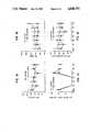
- FIG. 1Ais a graphic illustration of mean arterial blood pressure (MAP) data accumulated in practicing the method embodying the invention
- FIG. 1Bis a graphic illustration of the heart rate (HR) data accumulated in practicing the said method
- FIG. 1Cis a graphic illustration of the left circumflex blood flow (LCX) data accumulated in practicing said method
- FIG. 1Dis a graphic illustration of the rate pressure product (RPP) data accumulated in practicing said method
- FIG. 2is a graphic illustration of data accumulated to show effect of the invention on resulting myocardial infarct size
- FIG. 3is a graphic illustration of data accumulated to express infarct size as a function of the extent of the ST segment of an electrocardiogram
- FIG. 4is a graphic illustration of data accumulated to show circulating neutrophil counts during the myocardial infarction process.
- the monoclonal antibody employed in the method of the inventionis identified by the designation MY904. It was developed from the fusion of murine spleen cells immunized with human chronic granulocytic leukemia (CGL) cells by standard procedure described by Kohler and Millstein, Nature 256: 495-497 (1975). The granulocytic leukemia cells used in the immunization procedure were obtained from newly diagnosed patients with CGL as part of diagnostic evaluation. Blood was obtained by venipuncture and mononuclear cells separated from granulocytes and red blood cells by Ficoll-Hypaque density gradient sedimentation, 1.077 g/cc. The mononuclear cell fraction was composed of immature granulocytes and blast cells.
- CGLchronic granulocytic leukemia
- micewere cryopreserved, and mice immunized at weekly intervals for 4 weeks with 10 ⁇ 10 6 thawed mononuclear cells injected intraperitoneally.
- 10 ⁇ 10 6 similarly treated CGL mononuclear cellswere injected intravenously into the tail vein of the mouse.
- the spleenwas removed and a single cell suspension made of splenocytes.
- the spleen cellswere then mixed with the NS-1 plasmacytoma cell line at a ratio of 8 spleen cells l to 1 NS-1 cell in serum free media.
- the cellswere centrifuged to a pellet, suspended in 0.5 ml. of 30% polyethylene glycol for 8 minutes at 25° C., followed by washing of the cells one time in serum free media and dilution in HAT media prior to distribution of the cells in microtiter plates.
- Monoclonal antibody producing hybridoma clones reactive with the immunizing cell populationwere selected by immunofluorescence screening 14 days after the infusion.
- Monoclonal antibody MY904was identified as an antibody which reacted with CGL cells as well as with normal human granulocytes, monocytes, and a fraction of large granular lymphocytes. The monoclonal antibody reacted with more than 90% of granulocytes of 10 of 10 patients tested. The monoclonal antibody MY904 does not react with T lymphocytes or B lymphocytes.
- the monoclonal antibodyimmunoprecipitates a glycoprotein composed of 2 subunits, 155,000 daltons and 94,000 daltons from surface labelled normal human granulocytes.
- monoclonal antibody MY904does not inhibit iC3b binding but is a potent inhibitor of the adhesion-dependent processes, granulocyte spreading on plastic and chemotaxis (Dana et al., Two Functional Domains In The Phagocyte Membrane Glycoprotein Mol Identified With Monoclonal Antibodies, Journal of Immunology 137:3259-3263, 1986).
- antibody MY904was unique in that it inhibited only adhesion-dependent functions but not binding of iC3b.
- Other antibodies testedinclude monoclonal antibodies 44, 903, 94, 17, OKM10, and Leu-15. Dana et al., ibid.
- monoclonal antibody MY904identifed the Mol granulo-cyte-monocyte cell surface glycoprotein, and further binds specifically to an epitope on that glycoprotein which is involved in adhesion-dependent processes of granulocyte/monocyte activities.
- a sample of the hyrbid cell line capable of producing the MY904 monoclonal antibodyis on deposit with the American Type Culture Collection (A.T.C.C.) and is assigned A.T.C.C. No. HB 9510.
- a group of 23 adult dogswas first prepared.
- the animalswere anesthetized and their hearts were exposed through a left thoracotomy in the fifth intercostal space and suspended in a pericardial sling.
- the left circumflex coronary arterywas instrumented with a calibrated electromagnetic flow probe for the continuous recording of blood flow.
- Catheterswere placed into the aorta for blood pressure recording and into the left jugular vein for monoclonal antibody infusion and blood sampling.
- the standard limb lead 11 electrocardiogramwere recorded continuously.
- the heartswere then cut into 5 or 6 centimeter thick transverse sections and infarct size was determined planimetrically. This accepted method of measuring infarct size accurately demarcates viable from non-viable myocardial tissue as determined by histochemical reaction between TPT and myocardial dehydrogenase enzymes.
- the second group of animalsreceived the pharmaceutic grade monoclonal antibody MY904 in amounts of one (1) mg/kg infused intravenously over a ten (10) minute period (45 minutes) after regional myocardial ischemia was induced (45 minutes prior to reinitiation of coronary blood flow). Detection of binding of the MY904 antibody to the Mol antigen of dog leukocytes was monitored.
- the monoclonal antibody MY904was supplied by Coulter Immunology Division, Coulter Corporation, in Hialeah, Florida.
- the cellswere then washed and incubated for an additional 30 minutes at 4° C. in buffer containing a saturating concentration of fluoresceinconjugated goat anti-mouse immunoglobulin.
- Antibody bindingeither as a result of in vivo administration of anti-Mol or after in vitro exposure to additional anti-Mol antibody, to dog neutrophils and monocytes was assessed by flow cytometry after selective gating on these myeloid cells, as determined by log forward angle versus log right angle light scatter, using an EPICS® C flow cytometer available from Coulter Corporation of Hialeah, Florida. The fluorescence intensity of 5000 cells per determination was used as a quantitative measure of monoclonal antibody binding.
- neutrophilswere isolated from venous blood of untreated dogs by Ficoll-hypaque gradient techniques. Red blood cells were lysed with buffered ammonium chloride and resuspended to a concentration of 10 7 per ml in Hank's balanced salt solution. Aggregation was assessed in a platelet aggregation profiler, Model PAP-3, BIO/DATA Corporation, Horsham, PA. Samples of neutrophils were preincubated with MY904 and negative control antibodies and then activated with 125 ng/ml PMA.
- MAPmean arterial blood pressure
- HRheart rate
- LCXleft circumflex
- RPPrate pressure product
- Infarct sizewas reduced by 46% by the administration of the anti-Mol antibody as compared to control, as seen from the data of FIG. 2. Similarly, infarct size was significantly smaller with monoclonal antibody treatment when infarct size was expressed as a percentage of the total left ventricle. The percentage of the left ventricle that was rendered ischemic, i.e., the area at risk/left ventricle, was similar between treatment groups. When infarct size is expressed as a function of the extent of ST segment elevation on the electrocardiogram, there exists a good correlation with each treatment group describing a different regression line as seen from the data of FIG. 3.
- the neutrophils of all 20 experimental subjectsexpressed the antigenic epitope detectable by the anti-Mol MY904 monoclonal antibody.
- the serum or plasmacontained residual anti-Mol antibody as assayed by immunofluorescence staining of serum or plasma-treated Mol positive test cells. This indicated that an infusion of 1 mg/kg was sufficient to produce antibody excess in vivo.
- Detectable subsaturating amounts of bound antibodywere found on the leukocytes of only 6 out of 9 dogs which may reflect antibody dissociation from the membrane prior to assay by incubation with the fluorescein-conjugated goat anti-mouse immunoglobulin reagent.
- FIG. 4 datashows that neutrophil counts normally increase as seen in the control group during the process of myocardial infarction.
- treatment with anti-Mol antibodysuppressed this early rise in circulating neutrophil counts when the two groups are compared.
- the data developed by the method of the inventionindicate that administration of the anti-Mol monoclonal antibody MY904 can reduce myocardial infarct size, but not by lessening the severity of the ischemia. Since the duration of ischemia was ninety (90) minutes in all instances, the mechanism of protection by the MY904 antibody is a likely consequence of its effects on neutrophil adhesion with a reduction neutrophil-mediated injury of ischemic myocardium. Heretofore, it was considered that the two major determinants of myocardial infarct size were the severity and duration of the ischemia period.
- medical experienceindicates that a patient who experiences pain or discomfort in his chest would be brought to a hospital usually within thirty to sixty minutes thereafter on an emergency basis. He would be examined promptly to document the occurence of acute myocardial ischemia and the location of a coronary artery occlusion, and treatment would be prescribed.
- Such treatmentmay be that of infusing a thrombolytic agent (e.g., streptokinase or tissue plasminogen activator [TPA]) which might dissolve the blood clot which caused the acute coronary occlusion.
- a thrombolytic agente.g., streptokinase or tissue plasminogen activator [TPA]
- Another treatmentis surgical in nature where a balloon catheter is inserted into the circulatory system and directed to the blood clot area for eliminating the blockage.
- a quantity of MY904 monoclonal antibodyis injected intravenously into the patient.
- a single intravenous dose or multiple dosesmay be administered to optimally attenuate the inflammatory response.
- the MY904 monoclonal antibodythereafter functions in the manner described herein to reduce tissue damage (infarct size) independently of the severity of the ischemic event.
- the MY904 monoclonal antibody infusionfunctions to reduce myocardial reperfusion injury by inhibiting undesired neutrophil functions.
Landscapes
- Health & Medical Sciences (AREA)
- Chemical & Material Sciences (AREA)
- Immunology (AREA)
- Organic Chemistry (AREA)
- General Health & Medical Sciences (AREA)
- Biophysics (AREA)
- Biochemistry (AREA)
- Genetics & Genomics (AREA)
- Medicinal Chemistry (AREA)
- Molecular Biology (AREA)
- Proteomics, Peptides & Aminoacids (AREA)
- Life Sciences & Earth Sciences (AREA)
- Medicines Containing Antibodies Or Antigens For Use As Internal Diagnostic Agents (AREA)
- Preparation Of Compounds By Using Micro-Organisms (AREA)
- Peptides Or Proteins (AREA)
Abstract
Description
Claims (11)
Priority Applications (14)
| Application Number | Priority Date | Filing Date | Title |
|---|---|---|---|
| US07/061,336US4840793A (en) | 1987-06-11 | 1987-06-11 | Method of reducing tissue damage at an inflammatory site using a monoclonal antibody |
| US07/165,025US4935234A (en) | 1987-06-11 | 1988-03-07 | Method of reducing tissue damage at an inflammatory site using a monoclonal antibody |
| AU19463/88AAU621304B2 (en) | 1987-06-11 | 1988-06-03 | Method of reducing tissue damage at an inflammatory site using a monoclonal antibody |
| PCT/US1988/001928WO1988009672A1 (en) | 1987-06-11 | 1988-06-03 | Method of reducing tissue damage at an inflammatory site using a monoclonal antibody |
| BR888807559ABR8807559A (en) | 1987-06-11 | 1988-06-03 | PROCESS TO REDUCE TISSUE DAMAGE THAT OCCURS IN AN INFLAMMATORY PLACE IN ANY PART OF THE HUMAN BODY AND TO REDUCE DAMAGE FROM MYOCARDIAL INFLAMMATION IN A HUMAN PATIENT |
| AT88905555TATE126703T1 (en) | 1987-06-11 | 1988-06-03 | METHOD OF REDUCING TISSUE DAMAGE TO AN INFLAMMED SITES BY USING MONOCLONAL ANTIBODIES. |
| DE3854351TDE3854351T2 (en) | 1987-06-11 | 1988-06-03 | METHOD FOR REDUCING TISSUE DAMAGE IN AN INFLAMMED SITE BY USING MONOCLONAL ANTIBODIES. |
| EP88905555AEP0371036B1 (en) | 1987-06-11 | 1988-06-03 | Method of reducing tissue damage at an inflammatory site using a monoclonal antibody |
| JP63505211AJPH02503796A (en) | 1987-06-11 | 1988-06-03 | A method to reduce tissue injury at sites of inflammation using monoclonal antibodies |
| CN88103476ACN1034055C (en) | 1987-06-11 | 1988-06-10 | Method for decrease histic damage of imflammation region by monoclone antibody |
| ZA884166AZA884166B (en) | 1987-06-11 | 1988-06-10 | A monoclonal antibody |
| CA000569247ACA1317880C (en) | 1987-06-11 | 1988-06-10 | Method of reducing tissue damage at an inflammatory site using a monoclonal antibody |
| NO89894946ANO894946L (en) | 1987-06-11 | 1989-12-08 | PROCEDURE FOR REDUCING TISSUE DAMAGE IN A BOTTLE OF APPLICATION USING A MONOCLONAL ANTIBODY. |
| DK620089ADK620089A (en) | 1987-06-11 | 1989-12-08 | MONOCLONAL ANTIBODY TO REDUCE TABLE DAMAGE IN A SITUATION PLACE |
Applications Claiming Priority (1)
| Application Number | Priority Date | Filing Date | Title |
|---|---|---|---|
| US07/061,336US4840793A (en) | 1987-06-11 | 1987-06-11 | Method of reducing tissue damage at an inflammatory site using a monoclonal antibody |
Related Child Applications (1)
| Application Number | Title | Priority Date | Filing Date |
|---|---|---|---|
| US07/165,025Continuation-In-PartUS4935234A (en) | 1987-06-11 | 1988-03-07 | Method of reducing tissue damage at an inflammatory site using a monoclonal antibody |
Publications (1)
| Publication Number | Publication Date |
|---|---|
| US4840793Atrue US4840793A (en) | 1989-06-20 |
Family
ID=22035128
Family Applications (1)
| Application Number | Title | Priority Date | Filing Date |
|---|---|---|---|
| US07/061,336Expired - LifetimeUS4840793A (en) | 1987-06-11 | 1987-06-11 | Method of reducing tissue damage at an inflammatory site using a monoclonal antibody |
Country Status (2)
| Country | Link |
|---|---|
| US (1) | US4840793A (en) |
| ZA (1) | ZA884166B (en) |
Cited By (63)
| Publication number | Priority date | Publication date | Assignee | Title |
|---|---|---|---|---|
| WO1990001338A1 (en)* | 1988-08-15 | 1990-02-22 | Brigham And Women's Hospital | Leukocyte adhesion inhibitor |
| US4935234A (en)* | 1987-06-11 | 1990-06-19 | Dana-Farber Cancer Institute | Method of reducing tissue damage at an inflammatory site using a monoclonal antibody |
| US4986979A (en)* | 1989-03-14 | 1991-01-22 | Neorx Corporation | Imaging tissue sites of inflammation |
| US5019648A (en)* | 1987-07-06 | 1991-05-28 | Dana-Farber Cancer Institute | Monoclonal antibody specific for the adhesion function domain of a phagocyte cell surface protein |
| WO1991008483A1 (en)* | 1989-11-29 | 1991-06-13 | Brigham And Women's Hospital | Endothelial-derived il-8 |
| WO1992006697A1 (en)* | 1990-10-23 | 1992-04-30 | Repligen Corporation | Anti-inflammatory composition |
| US5147637A (en)* | 1988-06-07 | 1992-09-15 | The Rockefeller University | Method of inhibiting the influx of leukocytes into organs during sepsis or other trauma |
| WO1993007888A1 (en)* | 1991-10-16 | 1993-04-29 | Brigham And Women's Hospital | Inhib, a factor which inhibits cytokine-activated leukocytes |
| WO1993007889A1 (en)* | 1991-10-23 | 1993-04-29 | Thomas Jefferson University | Synthesis of human procollagens and collagens in recombinant dna systems |
| US5219997A (en)* | 1987-07-06 | 1993-06-15 | Dana-Farber Cancer Institute | Monoclonal antibody which inhibits the adhesion functions of the β integrin, CR3 |
| WO1993019782A1 (en)* | 1992-04-02 | 1993-10-14 | Dana-Farber Cancer Institute, Inc. | Specific detection of cell surface receptor leukocyte adhesion molecule-1 |
| US5322699A (en)* | 1991-02-04 | 1994-06-21 | The Rockefeller University | Leukocyte-derived CR3 modulator, integrin modulating factor-1 (IMF-1) |
| US5403713A (en)* | 1988-11-14 | 1995-04-04 | Brigham & Women's Hospital | Antibodies specific for ELAM-1 and the use thereof |
| US5424399A (en)* | 1988-06-28 | 1995-06-13 | The Children's Medical Center Corporation | Human CR3α/β heterodimers |
| US5595737A (en)* | 1989-02-21 | 1997-01-21 | Dana-Farber Cancer Institute, Inc. | Methods for using monoclonal antibodies specific for cell-surface bound LAM-1 |
| US5632991A (en)* | 1988-11-14 | 1997-05-27 | Brigham & Women's Hospital | Antibodies specific for E-selectin and the uses thereof |
| US5708141A (en)* | 1992-05-11 | 1998-01-13 | Corvas International, Inc. | Neutrophil inhibitors |
| US5723583A (en)* | 1990-11-23 | 1998-03-03 | The General Hospital Corporation | Antibody containing sialyl lewis X determinants |
| US5797870A (en)* | 1995-06-07 | 1998-08-25 | Indiana University Foundation | Pericardial delivery of therapeutic and diagnostic agents |
| US5823742A (en)* | 1995-12-15 | 1998-10-20 | Dresser-Rand Company | Variable and bidirectional steam flow apparatus and method |
| US5877275A (en)* | 1988-06-28 | 1999-03-02 | The General Hospital Corporation | Controlling cellular immune/inflammatory responses with β2 integrins |
| US5914112A (en)* | 1996-01-23 | 1999-06-22 | Genentech, Inc. | Anti-CD18 antibodies in stroke |
| US5985279A (en)* | 1991-07-16 | 1999-11-16 | Waldmann; Herman | Humanized antibody against CD18 |
| US6001356A (en)* | 1995-09-29 | 1999-12-14 | Rush-Presbyterian-St. Luke's Medical Center | Method of inhibiting tissue destruction in autoimmune disease using anti-CD44 antibodies |
| US6033665A (en)* | 1989-09-27 | 2000-03-07 | Elan Pharmaceuticals, Inc. | Compositions and methods for modulating leukocyte adhesion to brain endothelial cells |
| US6663863B2 (en) | 2000-03-17 | 2003-12-16 | Millennium Pharmaceuticals, Inc. | Method of inhibiting stenosis and restenosis |
| US20040057951A1 (en)* | 1996-01-23 | 2004-03-25 | Genentech, Inc. | Co-administration of a thrombolytic and an anti-CD18 antibody in stroke |
| US20070264193A1 (en)* | 2006-03-29 | 2007-11-15 | Genentech, Inc. | Diagnostics and treatments for tumors |
| US20080140138A1 (en)* | 2002-02-26 | 2008-06-12 | Ivanova Svetlana M | Inhibition of inflammatory cytokine production by stimulation of brain muscarinic receptors |
| US20090062874A1 (en)* | 2007-08-27 | 2009-03-05 | Tracey Kevin J | Devices and methods for inhibiting granulocyte activation by neural stimulation |
| US20090247934A1 (en)* | 2008-03-31 | 2009-10-01 | Tracey Kevin J | Methods and systems for reducing inflammation by neuromodulation of t-cell activity |
| WO2010017083A1 (en)* | 2008-08-04 | 2010-02-11 | Wayne State University | Methods of treating cancer with cd11b antibodies |
| US20110054569A1 (en)* | 2009-09-01 | 2011-03-03 | Zitnik Ralph J | Prescription pad for treatment of inflammatory disorders |
| WO2013025944A1 (en) | 2011-08-17 | 2013-02-21 | Genentech, Inc. | Inhibition of angiogenesis in refractory tumors |
| US8412338B2 (en) | 2008-11-18 | 2013-04-02 | Setpoint Medical Corporation | Devices and methods for optimizing electrode placement for anti-inflamatory stimulation |
| US8612002B2 (en) | 2009-12-23 | 2013-12-17 | Setpoint Medical Corporation | Neural stimulation devices and systems for treatment of chronic inflammation |
| US8729129B2 (en) | 2004-03-25 | 2014-05-20 | The Feinstein Institute For Medical Research | Neural tourniquet |
| US8788034B2 (en) | 2011-05-09 | 2014-07-22 | Setpoint Medical Corporation | Single-pulse activation of the cholinergic anti-inflammatory pathway to treat chronic inflammation |
| US8886339B2 (en) | 2009-06-09 | 2014-11-11 | Setpoint Medical Corporation | Nerve cuff with pocket for leadless stimulator |
| US8914114B2 (en) | 2000-05-23 | 2014-12-16 | The Feinstein Institute For Medical Research | Inhibition of inflammatory cytokine production by cholinergic agonists and vagus nerve stimulation |
| US8996116B2 (en) | 2009-10-30 | 2015-03-31 | Setpoint Medical Corporation | Modulation of the cholinergic anti-inflammatory pathway to treat pain or addiction |
| US20150337039A1 (en)* | 2014-05-22 | 2015-11-26 | Case Western Reserve University | Mac-1 antibodies and uses thereof |
| US9211410B2 (en) | 2009-05-01 | 2015-12-15 | Setpoint Medical Corporation | Extremely low duty-cycle activation of the cholinergic anti-inflammatory pathway to treat chronic inflammation |
| US9572983B2 (en) | 2012-03-26 | 2017-02-21 | Setpoint Medical Corporation | Devices and methods for modulation of bone erosion |
| US9662490B2 (en) | 2008-03-31 | 2017-05-30 | The Feinstein Institute For Medical Research | Methods and systems for reducing inflammation by neuromodulation and administration of an anti-inflammatory drug |
| US9833621B2 (en) | 2011-09-23 | 2017-12-05 | Setpoint Medical Corporation | Modulation of sirtuins by vagus nerve stimulation |
| US10314501B2 (en) | 2016-01-20 | 2019-06-11 | Setpoint Medical Corporation | Implantable microstimulators and inductive charging systems |
| US10583304B2 (en) | 2016-01-25 | 2020-03-10 | Setpoint Medical Corporation | Implantable neurostimulator having power control and thermal regulation and methods of use |
| US10596367B2 (en) | 2016-01-13 | 2020-03-24 | Setpoint Medical Corporation | Systems and methods for establishing a nerve block |
| US10695569B2 (en) | 2016-01-20 | 2020-06-30 | Setpoint Medical Corporation | Control of vagal stimulation |
| US10912712B2 (en) | 2004-03-25 | 2021-02-09 | The Feinstein Institutes For Medical Research | Treatment of bleeding by non-invasive stimulation |
| US11051744B2 (en) | 2009-11-17 | 2021-07-06 | Setpoint Medical Corporation | Closed-loop vagus nerve stimulation |
| US11173307B2 (en) | 2017-08-14 | 2021-11-16 | Setpoint Medical Corporation | Vagus nerve stimulation pre-screening test |
| US11207518B2 (en) | 2004-12-27 | 2021-12-28 | The Feinstein Institutes For Medical Research | Treating inflammatory disorders by stimulation of the cholinergic anti-inflammatory pathway |
| US11260229B2 (en) | 2018-09-25 | 2022-03-01 | The Feinstein Institutes For Medical Research | Methods and apparatuses for reducing bleeding via coordinated trigeminal and vagal nerve stimulation |
| US11311725B2 (en) | 2014-10-24 | 2022-04-26 | Setpoint Medical Corporation | Systems and methods for stimulating and/or monitoring loci in the brain to treat inflammation and to enhance vagus nerve stimulation |
| US11344724B2 (en) | 2004-12-27 | 2022-05-31 | The Feinstein Institutes For Medical Research | Treating inflammatory disorders by electrical vagus nerve stimulation |
| US11406833B2 (en) | 2015-02-03 | 2022-08-09 | Setpoint Medical Corporation | Apparatus and method for reminding, prompting, or alerting a patient with an implanted stimulator |
| US11471681B2 (en) | 2016-01-20 | 2022-10-18 | Setpoint Medical Corporation | Batteryless implantable microstimulators |
| US11938324B2 (en) | 2020-05-21 | 2024-03-26 | The Feinstein Institutes For Medical Research | Systems and methods for vagus nerve stimulation |
| US12172017B2 (en) | 2011-05-09 | 2024-12-24 | Setpoint Medical Corporation | Vagus nerve stimulation to treat neurodegenerative disorders |
| US12343535B2 (en) | 2019-04-12 | 2025-07-01 | Setpoint Medical Corporation | Vagus nerve stimulation to treat neurodegenerative disorders |
| US12444497B2 (en) | 2022-05-17 | 2025-10-14 | Setpoint Medical Corporation | Neurostimulation parameter authentication and expiration system for neurostimulation |
- 1987
- 1987-06-11USUS07/061,336patent/US4840793A/ennot_activeExpired - Lifetime
- 1988
- 1988-06-10ZAZA884166Apatent/ZA884166B/enunknown
Non-Patent Citations (11)
| Title |
|---|
| Arnaout et al., Federation Proc., 44(10), 2664 69, 1985.* |
| Arnaout et al., Federation Proc., 44(10), 2664-69, 1985. |
| Arnaout et al., J. Clin. Invest., 72: 171 9, 1983.* |
| Arnaout et al., J. Clin. Invest., 72: 171-9, 1983. |
| Te Velde et al., Chem. Abs., 104: 184712d, 1986.* |
| Todd et al., Hybridoma, 1(3), 329 37, 1982.* |
| Todd et al., Hybridoma, 1(3), 329-37, 1982. |
| Todd et al., J. Clin. Invest., 74, 1280 90, 1984.* |
| Todd et al., J. Clin. Invest., 74, 1280-90, 1984. |
| Todd et al., J. Immunol., 126(4), 1435 42, 1981.* |
| Todd et al., J. Immunol., 126(4), 1435-42, 1981. |
Cited By (109)
| Publication number | Priority date | Publication date | Assignee | Title |
|---|---|---|---|---|
| US4935234A (en)* | 1987-06-11 | 1990-06-19 | Dana-Farber Cancer Institute | Method of reducing tissue damage at an inflammatory site using a monoclonal antibody |
| US5219997A (en)* | 1987-07-06 | 1993-06-15 | Dana-Farber Cancer Institute | Monoclonal antibody which inhibits the adhesion functions of the β integrin, CR3 |
| US5019648A (en)* | 1987-07-06 | 1991-05-28 | Dana-Farber Cancer Institute | Monoclonal antibody specific for the adhesion function domain of a phagocyte cell surface protein |
| US5147637A (en)* | 1988-06-07 | 1992-09-15 | The Rockefeller University | Method of inhibiting the influx of leukocytes into organs during sepsis or other trauma |
| US5424399A (en)* | 1988-06-28 | 1995-06-13 | The Children's Medical Center Corporation | Human CR3α/β heterodimers |
| US5877275A (en)* | 1988-06-28 | 1999-03-02 | The General Hospital Corporation | Controlling cellular immune/inflammatory responses with β2 integrins |
| WO1990001338A1 (en)* | 1988-08-15 | 1990-02-22 | Brigham And Women's Hospital | Leukocyte adhesion inhibitor |
| US5302384A (en)* | 1988-08-15 | 1994-04-12 | Brigham And Women's Hospital | Endothelial-derived II-8 adhesion inhibitor |
| US5403713A (en)* | 1988-11-14 | 1995-04-04 | Brigham & Women's Hospital | Antibodies specific for ELAM-1 and the use thereof |
| US5632991A (en)* | 1988-11-14 | 1997-05-27 | Brigham & Women's Hospital | Antibodies specific for E-selectin and the uses thereof |
| US5322838A (en)* | 1988-11-16 | 1994-06-21 | Brigham & Women's Hospital | Use of INHIB (the C3 β-chain) in the detection and inhibition of inflammation |
| US5595737A (en)* | 1989-02-21 | 1997-01-21 | Dana-Farber Cancer Institute, Inc. | Methods for using monoclonal antibodies specific for cell-surface bound LAM-1 |
| US5389520A (en)* | 1989-02-21 | 1995-02-14 | Dana-Farber Cancer Institute, Inc. | Specific detection of cell surface receptor leukocyte adhesion molecule-1 |
| US4986979A (en)* | 1989-03-14 | 1991-01-22 | Neorx Corporation | Imaging tissue sites of inflammation |
| US6033665A (en)* | 1989-09-27 | 2000-03-07 | Elan Pharmaceuticals, Inc. | Compositions and methods for modulating leukocyte adhesion to brain endothelial cells |
| WO1991008483A1 (en)* | 1989-11-29 | 1991-06-13 | Brigham And Women's Hospital | Endothelial-derived il-8 |
| WO1992006697A1 (en)* | 1990-10-23 | 1992-04-30 | Repligen Corporation | Anti-inflammatory composition |
| US5723583A (en)* | 1990-11-23 | 1998-03-03 | The General Hospital Corporation | Antibody containing sialyl lewis X determinants |
| US6613746B1 (en) | 1990-11-23 | 2003-09-02 | The General Hospital Corporation | AGP-antibody fusion proteins and related molecules and methods |
| US6156881A (en)* | 1990-11-23 | 2000-12-05 | The General Hospital Corporation | Inhibition of cell adhesion protein-carbohydrate interactions |
| US5801044A (en)* | 1990-11-23 | 1998-09-01 | The General Hospital Corporation | Nucleic acid encoding an antibody that inhibits cell adhesion protein-carbohydrate interactions |
| US5858983A (en)* | 1990-11-23 | 1999-01-12 | The General Hospital Corporation | Inhibition of cell adhesion protein-carbohydrate interactions |
| US5322699A (en)* | 1991-02-04 | 1994-06-21 | The Rockefeller University | Leukocyte-derived CR3 modulator, integrin modulating factor-1 (IMF-1) |
| US6689869B2 (en) | 1991-07-16 | 2004-02-10 | Cambridge University Technical Services Limited | Labeled humanized anti-CD18 antibodies and fragments and kits comprising same |
| US5985279A (en)* | 1991-07-16 | 1999-11-16 | Waldmann; Herman | Humanized antibody against CD18 |
| US5997867A (en)* | 1991-07-16 | 1999-12-07 | Waldmann; Herman | Method of using humanized antibody against CD18 |
| WO1993007888A1 (en)* | 1991-10-16 | 1993-04-29 | Brigham And Women's Hospital | Inhib, a factor which inhibits cytokine-activated leukocytes |
| WO1993007889A1 (en)* | 1991-10-23 | 1993-04-29 | Thomas Jefferson University | Synthesis of human procollagens and collagens in recombinant dna systems |
| US6274347B1 (en) | 1992-04-02 | 2001-08-14 | Dana-Farber Cancer Institute, Inc. | Method of producing a cell surface bound lam-1 specific antibody |
| WO1993019782A1 (en)* | 1992-04-02 | 1993-10-14 | Dana-Farber Cancer Institute, Inc. | Specific detection of cell surface receptor leukocyte adhesion molecule-1 |
| AU686998B2 (en)* | 1992-04-02 | 1998-02-19 | Dana-Farber Cancer Institute, Inc. | Specific detection of cell surface receptor leukocyte adhesion molecule-1 |
| US5708141A (en)* | 1992-05-11 | 1998-01-13 | Corvas International, Inc. | Neutrophil inhibitors |
| US5797870A (en)* | 1995-06-07 | 1998-08-25 | Indiana University Foundation | Pericardial delivery of therapeutic and diagnostic agents |
| US6001356A (en)* | 1995-09-29 | 1999-12-14 | Rush-Presbyterian-St. Luke's Medical Center | Method of inhibiting tissue destruction in autoimmune disease using anti-CD44 antibodies |
| US5823742A (en)* | 1995-12-15 | 1998-10-20 | Dresser-Rand Company | Variable and bidirectional steam flow apparatus and method |
| US7655230B2 (en) | 1996-01-23 | 2010-02-02 | Genentech, Inc. | Co-administration of a tissue plasminogen activator, anti-CD11b antibody and anti-CD18 antibody in stroke |
| US20040057951A1 (en)* | 1996-01-23 | 2004-03-25 | Genentech, Inc. | Co-administration of a thrombolytic and an anti-CD18 antibody in stroke |
| US20050255108A1 (en)* | 1996-01-23 | 2005-11-17 | Bednar Martin M | Co-administration of a thrombolytic and an-anti-CD18 antibody in stroke |
| US5914112A (en)* | 1996-01-23 | 1999-06-22 | Genentech, Inc. | Anti-CD18 antibodies in stroke |
| US7361344B2 (en) | 1996-01-23 | 2008-04-22 | Genentech, Inc. | Co-administration of a thrombolytic and an-anti-CD18 antibody in stroke |
| US6663863B2 (en) | 2000-03-17 | 2003-12-16 | Millennium Pharmaceuticals, Inc. | Method of inhibiting stenosis and restenosis |
| US10166395B2 (en) | 2000-05-23 | 2019-01-01 | The Feinstein Institute For Medical Research | Inhibition of inflammatory cytokine production by cholinergic agonists and vagus nerve stimulation |
| US9987492B2 (en) | 2000-05-23 | 2018-06-05 | The Feinstein Institute For Medical Research | Inhibition of inflammatory cytokine production by cholinergic agonists and vagus nerve stimulation |
| US10561846B2 (en) | 2000-05-23 | 2020-02-18 | The Feinstein Institutes For Medical Research | Inhibition of inflammatory cytokine production by cholinergic agonists and vagus nerve stimulation |
| US8914114B2 (en) | 2000-05-23 | 2014-12-16 | The Feinstein Institute For Medical Research | Inhibition of inflammatory cytokine production by cholinergic agonists and vagus nerve stimulation |
| US20080140138A1 (en)* | 2002-02-26 | 2008-06-12 | Ivanova Svetlana M | Inhibition of inflammatory cytokine production by stimulation of brain muscarinic receptors |
| US8729129B2 (en) | 2004-03-25 | 2014-05-20 | The Feinstein Institute For Medical Research | Neural tourniquet |
| US10912712B2 (en) | 2004-03-25 | 2021-02-09 | The Feinstein Institutes For Medical Research | Treatment of bleeding by non-invasive stimulation |
| US11207518B2 (en) | 2004-12-27 | 2021-12-28 | The Feinstein Institutes For Medical Research | Treating inflammatory disorders by stimulation of the cholinergic anti-inflammatory pathway |
| US11344724B2 (en) | 2004-12-27 | 2022-05-31 | The Feinstein Institutes For Medical Research | Treating inflammatory disorders by electrical vagus nerve stimulation |
| US20100239568A1 (en)* | 2006-03-29 | 2010-09-23 | Genentech, Inc. | Diagnostics and treatments for tumors |
| US20070264193A1 (en)* | 2006-03-29 | 2007-11-15 | Genentech, Inc. | Diagnostics and treatments for tumors |
| US8391970B2 (en) | 2007-08-27 | 2013-03-05 | The Feinstein Institute For Medical Research | Devices and methods for inhibiting granulocyte activation by neural stimulation |
| US20090062874A1 (en)* | 2007-08-27 | 2009-03-05 | Tracey Kevin J | Devices and methods for inhibiting granulocyte activation by neural stimulation |
| US9211409B2 (en) | 2008-03-31 | 2015-12-15 | The Feinstein Institute For Medical Research | Methods and systems for reducing inflammation by neuromodulation of T-cell activity |
| US9662490B2 (en) | 2008-03-31 | 2017-05-30 | The Feinstein Institute For Medical Research | Methods and systems for reducing inflammation by neuromodulation and administration of an anti-inflammatory drug |
| US20090247934A1 (en)* | 2008-03-31 | 2009-10-01 | Tracey Kevin J | Methods and systems for reducing inflammation by neuromodulation of t-cell activity |
| WO2010017083A1 (en)* | 2008-08-04 | 2010-02-11 | Wayne State University | Methods of treating cancer with cd11b antibodies |
| US8412338B2 (en) | 2008-11-18 | 2013-04-02 | Setpoint Medical Corporation | Devices and methods for optimizing electrode placement for anti-inflamatory stimulation |
| US9849286B2 (en) | 2009-05-01 | 2017-12-26 | Setpoint Medical Corporation | Extremely low duty-cycle activation of the cholinergic anti-inflammatory pathway to treat chronic inflammation |
| US9211410B2 (en) | 2009-05-01 | 2015-12-15 | Setpoint Medical Corporation | Extremely low duty-cycle activation of the cholinergic anti-inflammatory pathway to treat chronic inflammation |
| US9700716B2 (en) | 2009-06-09 | 2017-07-11 | Setpoint Medical Corporation | Nerve cuff with pocket for leadless stimulator |
| US10716936B2 (en) | 2009-06-09 | 2020-07-21 | Setpoint Medical Corporation | Nerve cuff with pocket for leadless stimulator |
| US9174041B2 (en) | 2009-06-09 | 2015-11-03 | Setpoint Medical Corporation | Nerve cuff with pocket for leadless stimulator |
| US10220203B2 (en) | 2009-06-09 | 2019-03-05 | Setpoint Medical Corporation | Nerve cuff with pocket for leadless stimulator |
| US8886339B2 (en) | 2009-06-09 | 2014-11-11 | Setpoint Medical Corporation | Nerve cuff with pocket for leadless stimulator |
| US12251558B2 (en) | 2009-06-09 | 2025-03-18 | Setpoint Medical Corporation | Nerve cuff with pocket for leadless stimulator |
| US20110054569A1 (en)* | 2009-09-01 | 2011-03-03 | Zitnik Ralph J | Prescription pad for treatment of inflammatory disorders |
| US8996116B2 (en) | 2009-10-30 | 2015-03-31 | Setpoint Medical Corporation | Modulation of the cholinergic anti-inflammatory pathway to treat pain or addiction |
| US11051744B2 (en) | 2009-11-17 | 2021-07-06 | Setpoint Medical Corporation | Closed-loop vagus nerve stimulation |
| US12290695B2 (en) | 2009-12-23 | 2025-05-06 | Setpoint Medical Corporation | Neural stimulation devices and systems for treatment of chronic inflammation |
| US9162064B2 (en) | 2009-12-23 | 2015-10-20 | Setpoint Medical Corporation | Neural stimulation devices and systems for treatment of chronic inflammation |
| US8612002B2 (en) | 2009-12-23 | 2013-12-17 | Setpoint Medical Corporation | Neural stimulation devices and systems for treatment of chronic inflammation |
| US10384068B2 (en) | 2009-12-23 | 2019-08-20 | Setpoint Medical Corporation | Neural stimulation devices and systems for treatment of chronic inflammation |
| US8855767B2 (en) | 2009-12-23 | 2014-10-07 | Setpoint Medical Corporation | Neural stimulation devices and systems for treatment of chronic inflammation |
| US9993651B2 (en) | 2009-12-23 | 2018-06-12 | Setpoint Medical Corporation | Neural stimulation devices and systems for treatment of chronic inflammation |
| US11110287B2 (en) | 2009-12-23 | 2021-09-07 | Setpoint Medical Corporation | Neural stimulation devices and systems for treatment of chronic inflammation |
| US12172017B2 (en) | 2011-05-09 | 2024-12-24 | Setpoint Medical Corporation | Vagus nerve stimulation to treat neurodegenerative disorders |
| US8788034B2 (en) | 2011-05-09 | 2014-07-22 | Setpoint Medical Corporation | Single-pulse activation of the cholinergic anti-inflammatory pathway to treat chronic inflammation |
| US12357826B2 (en) | 2011-05-09 | 2025-07-15 | Setpoint Medical Corporation | Extremely low duty-cycle activation of the cholinergic anti-inflammatory pathway to treat chronic inflammation |
| WO2013025944A1 (en) | 2011-08-17 | 2013-02-21 | Genentech, Inc. | Inhibition of angiogenesis in refractory tumors |
| US9833621B2 (en) | 2011-09-23 | 2017-12-05 | Setpoint Medical Corporation | Modulation of sirtuins by vagus nerve stimulation |
| US9572983B2 (en) | 2012-03-26 | 2017-02-21 | Setpoint Medical Corporation | Devices and methods for modulation of bone erosion |
| US10449358B2 (en) | 2012-03-26 | 2019-10-22 | Setpoint Medical Corporation | Devices and methods for modulation of bone erosion |
| US11969253B2 (en) | 2013-04-10 | 2024-04-30 | Setpoint Medical Corporation | Closed-loop vagus nerve stimulation |
| US12402826B2 (en) | 2013-04-10 | 2025-09-02 | Setpoint Medical Corporation | Closed-loop vagus nerve stimulation |
| US20150337039A1 (en)* | 2014-05-22 | 2015-11-26 | Case Western Reserve University | Mac-1 antibodies and uses thereof |
| US11311725B2 (en) | 2014-10-24 | 2022-04-26 | Setpoint Medical Corporation | Systems and methods for stimulating and/or monitoring loci in the brain to treat inflammation and to enhance vagus nerve stimulation |
| US12383741B2 (en) | 2014-10-24 | 2025-08-12 | Setpoint Medical Corporation | Systems and methods for stimulating and/or monitoring loci in the brain to treat inflammation and to enhance vagus nerve stimulation |
| US11406833B2 (en) | 2015-02-03 | 2022-08-09 | Setpoint Medical Corporation | Apparatus and method for reminding, prompting, or alerting a patient with an implanted stimulator |
| US11278718B2 (en) | 2016-01-13 | 2022-03-22 | Setpoint Medical Corporation | Systems and methods for establishing a nerve block |
| US10596367B2 (en) | 2016-01-13 | 2020-03-24 | Setpoint Medical Corporation | Systems and methods for establishing a nerve block |
| US11471681B2 (en) | 2016-01-20 | 2022-10-18 | Setpoint Medical Corporation | Batteryless implantable microstimulators |
| US10314501B2 (en) | 2016-01-20 | 2019-06-11 | Setpoint Medical Corporation | Implantable microstimulators and inductive charging systems |
| US12296169B2 (en) | 2016-01-20 | 2025-05-13 | Setpoint Medical Corporation | Batteryless implantable microstimulators |
| US10695569B2 (en) | 2016-01-20 | 2020-06-30 | Setpoint Medical Corporation | Control of vagal stimulation |
| US11964150B2 (en) | 2016-01-20 | 2024-04-23 | Setpoint Medical Corporation | Batteryless implantable microstimulators |
| US11547852B2 (en) | 2016-01-20 | 2023-01-10 | Setpoint Medical Corporation | Control of vagal stimulation |
| US12121726B2 (en) | 2016-01-20 | 2024-10-22 | Setpoint Medical Corporation | Control of vagal stimulation |
| US10583304B2 (en) | 2016-01-25 | 2020-03-10 | Setpoint Medical Corporation | Implantable neurostimulator having power control and thermal regulation and methods of use |
| US11383091B2 (en) | 2016-01-25 | 2022-07-12 | Setpoint Medical Corporation | Implantable neurostimulator having power control and thermal regulation and methods of use |
| US11890471B2 (en) | 2017-08-14 | 2024-02-06 | Setpoint Medical Corporation | Vagus nerve stimulation pre-screening test |
| US11173307B2 (en) | 2017-08-14 | 2021-11-16 | Setpoint Medical Corporation | Vagus nerve stimulation pre-screening test |
| US12220579B2 (en) | 2018-09-25 | 2025-02-11 | The Feinstein Institutes For Medical Research | Methods and apparatuses for reducing bleeding via coordinated trigeminal and vagal nerve stimulation |
| US11260229B2 (en) | 2018-09-25 | 2022-03-01 | The Feinstein Institutes For Medical Research | Methods and apparatuses for reducing bleeding via coordinated trigeminal and vagal nerve stimulation |
| US11857788B2 (en) | 2018-09-25 | 2024-01-02 | The Feinstein Institutes For Medical Research | Methods and apparatuses for reducing bleeding via coordinated trigeminal and vagal nerve stimulation |
| US12343535B2 (en) | 2019-04-12 | 2025-07-01 | Setpoint Medical Corporation | Vagus nerve stimulation to treat neurodegenerative disorders |
| US11938324B2 (en) | 2020-05-21 | 2024-03-26 | The Feinstein Institutes For Medical Research | Systems and methods for vagus nerve stimulation |
| US12444497B2 (en) | 2022-05-17 | 2025-10-14 | Setpoint Medical Corporation | Neurostimulation parameter authentication and expiration system for neurostimulation |
Also Published As
| Publication number | Publication date |
|---|---|
| ZA884166B (en) | 1990-02-28 |
Similar Documents
| Publication | Publication Date | Title |
|---|---|---|
| US4840793A (en) | Method of reducing tissue damage at an inflammatory site using a monoclonal antibody | |
| US4935234A (en) | Method of reducing tissue damage at an inflammatory site using a monoclonal antibody | |
| Simpson et al. | Reduction of experimental canine myocardial reperfusion injury by a monoclonal antibody (anti-Mo1, anti-CD11b) that inhibits leukocyte adhesion. | |
| US5019648A (en) | Monoclonal antibody specific for the adhesion function domain of a phagocyte cell surface protein | |
| Haught et al. | Alterations in circulating intercellular adhesion molecule-1 and L-selectin: further evidence for chronic inflammation in ischemic heart disease | |
| EP0289949B1 (en) | Intercellular adhesion molecules, and their binding ligands | |
| Simpson et al. | Sustained limitation of myocardial reperfusion injury by a monoclonal antibody that alters leukocyte function. | |
| Giger et al. | Deficiency of leukocyte surface glycoproteins Mo1, LFA-1, and Leu M5 in a dog with recurrent bacterial infections: an animal model | |
| EP0625912B1 (en) | Treatment for inflammatory bowel disease | |
| Perico et al. | Colchicine interferes with L-selectin and leukocyte function-associated antigen-1 expression on human T lymphocytes and inhibits T cell activation. | |
| Stocks et al. | CD66-dependent neutrophil activation: a possible mechanism for vascular selectin-mediated regulation of neutrophil adhesion | |
| US5219997A (en) | Monoclonal antibody which inhibits the adhesion functions of the β integrin, CR3 | |
| EP0606518B1 (en) | Intercellular adhesion molecules and their binding ligands | |
| HK1007683B (en) | Treatment for inflammatory bowel disease | |
| HK1003105B (en) | Intercellular adhesion molecules, and their binding ligands | |
| Schott et al. | F (ab') 2 fragments of anti-Mo1 (904) monoclonal antibodies do not prevent myocardial stunning. | |
| Ota | The critical role of intercellular adhesion molecule-1 in Masugi nephritis in rats | |
| EP0726942B1 (en) | Monoclonal antibodies to foam cells and their pharmaceutical and diagnostic use | |
| Jung et al. | from zyxwvutsrqponml | |
| HK1003117B (en) | Intercellular adhesion molecules, and their binding ligands |
Legal Events
| Date | Code | Title | Description |
|---|---|---|---|
| AS | Assignment | Owner name:DANA-FARBER CANCER INSTITUTE, 44 BINNEY STREET, BO Free format text:ASSIGNMENT OF ASSIGNORS INTEREST.;ASSIGNORS:GRIFFIN, JAMES D.;SCHLOSSMAN, STUART F.;REEL/FRAME:004739/0350 Effective date:19870727 | |
| AS | Assignment | Owner name:UNIVERSITY OF MICHIGAN, THE, 550 EAST UNIVERSITY A Free format text:ASSIGNMENT OF ASSIGNORS INTEREST.;ASSIGNORS:TODD, ROBERT F. III;LUCCHESI, BENEDICT R.;SIMPSON, PAUL J.;REEL/FRAME:004746/0955 Effective date:19870810 Owner name:UNIVERSITY OF MICHIGAN, THE, MICHIGAN Free format text:ASSIGNMENT OF ASSIGNORS INTEREST;ASSIGNORS:TODD, ROBERT F. III;LUCCHESI, BENEDICT R.;SIMPSON, PAUL J.;REEL/FRAME:004746/0955 Effective date:19870810 | |
| STCF | Information on status: patent grant | Free format text:PATENTED CASE | |
| CC | Certificate of correction | ||
| CC | Certificate of correction | ||
| FPAY | Fee payment | Year of fee payment:4 | |
| REFU | Refund | Free format text:REFUND PROCESSED. MAINTENANCE FEE HAS ALREADY BEEN PAID (ORIGINAL EVENT CODE: R160); ENTITY STATUS OF PATENT OWNER: LARGE ENTITY | |
| FEPP | Fee payment procedure | Free format text:PAYOR NUMBER ASSIGNED (ORIGINAL EVENT CODE: ASPN); ENTITY STATUS OF PATENT OWNER: LARGE ENTITY | |
| FPAY | Fee payment | Year of fee payment:8 | |
| FPAY | Fee payment | Year of fee payment:12 |