US4828716A - Apparatus and method for separating phases of blood - Google Patents
Apparatus and method for separating phases of bloodDownload PDFInfo
- Publication number
- US4828716A US4828716AUS07/033,769US3376987AUS4828716AUS 4828716 AUS4828716 AUS 4828716AUS 3376987 AUS3376987 AUS 3376987AUS 4828716 AUS4828716 AUS 4828716A
- Authority
- US
- United States
- Prior art keywords
- sample
- chamber
- blood
- phase
- phases
- Prior art date
- Legal status (The legal status is an assumption and is not a legal conclusion. Google has not performed a legal analysis and makes no representation as to the accuracy of the status listed.)
- Expired - Fee Related
Links
- 238000000034methodMethods0.000titleclaimsabstractdescription33
- 210000004369bloodAnatomy0.000titleabstractdescription145
- 239000008280bloodSubstances0.000titleabstractdescription145
- 238000000926separation methodMethods0.000claimsabstractdescription48
- 239000007788liquidSubstances0.000claimsabstractdescription12
- 230000004044responseEffects0.000claimsabstractdescription10
- 238000000638solvent extractionMethods0.000claimsabstractdescription8
- 230000036961partial effectEffects0.000claimsdescription4
- 238000005191phase separationMethods0.000claims2
- 239000000523sampleSubstances0.000description138
- 210000002966serumAnatomy0.000description61
- 230000001413cellular effectEffects0.000description42
- 239000000306componentSubstances0.000description41
- 210000002381plasmaAnatomy0.000description29
- 210000004027cellAnatomy0.000description13
- 239000000463materialSubstances0.000description13
- 238000004458analytical methodMethods0.000description11
- 239000012530fluidSubstances0.000description11
- 238000012360testing methodMethods0.000description11
- 238000005119centrifugationMethods0.000description10
- 230000003287optical effectEffects0.000description10
- 238000006073displacement reactionMethods0.000description9
- 206010018910HaemolysisDiseases0.000description7
- 230000004888barrier functionEffects0.000description7
- 230000006835compressionEffects0.000description7
- 238000007906compressionMethods0.000description7
- 230000008588hemolysisEffects0.000description7
- 230000000452restraining effectEffects0.000description7
- 102000009123FibrinHuman genes0.000description6
- 108010073385FibrinProteins0.000description6
- BWGVNKXGVNDBDI-UHFFFAOYSA-NFibrin monomerChemical compoundCNC(=O)CNC(=O)CNBWGVNKXGVNDBDI-UHFFFAOYSA-N0.000description6
- 210000000601blood cellAnatomy0.000description6
- 229950003499fibrinDrugs0.000description6
- 230000008569processEffects0.000description6
- 238000009987spinningMethods0.000description6
- 239000003146anticoagulant agentSubstances0.000description5
- 229940127219anticoagulant drugDrugs0.000description5
- 238000002955isolationMethods0.000description5
- 239000004033plasticSubstances0.000description5
- 239000000126substanceSubstances0.000description5
- 238000001914filtrationMethods0.000description4
- 239000011521glassSubstances0.000description4
- 238000012545processingMethods0.000description4
- 238000007789sealingMethods0.000description4
- 238000012546transferMethods0.000description4
- 230000008901benefitEffects0.000description3
- 210000004204blood vesselAnatomy0.000description3
- 238000001514detection methodMethods0.000description3
- 238000005194fractionationMethods0.000description3
- 208000015181infectious diseaseDiseases0.000description3
- 230000003993interactionEffects0.000description3
- 238000011068loading methodMethods0.000description3
- 238000002360preparation methodMethods0.000description3
- BPYKTIZUTYGOLE-IFADSCNNSA-NBilirubinChemical compoundN1C(=O)C(C)=C(C=C)\C1=C\C1=C(C)C(CCC(O)=O)=C(CC2=C(C(C)=C(\C=C/3C(=C(C=C)C(=O)N\3)C)N2)CCC(O)=O)N1BPYKTIZUTYGOLE-IFADSCNNSA-N0.000description2
- 230000001133accelerationEffects0.000description2
- 230000004913activationEffects0.000description2
- 239000012491analyteSubstances0.000description2
- 238000004159blood analysisMethods0.000description2
- 229920005549butyl rubberPolymers0.000description2
- 230000035602clottingEffects0.000description2
- 239000000470constituentSubstances0.000description2
- 238000010276constructionMethods0.000description2
- 238000011109contaminationMethods0.000description2
- 230000003247decreasing effectEffects0.000description2
- 229920001971elastomerPolymers0.000description2
- 230000006870functionEffects0.000description2
- 230000005484gravityEffects0.000description2
- 231100001261hazardousToxicity0.000description2
- 239000012678infectious agentSubstances0.000description2
- 230000002458infectious effectEffects0.000description2
- 150000002632lipidsChemical class0.000description2
- 238000004519manufacturing processMethods0.000description2
- 239000000203mixtureSubstances0.000description2
- 238000012544monitoring processMethods0.000description2
- 239000002245particleSubstances0.000description2
- 238000003825pressingMethods0.000description2
- 230000002035prolonged effectEffects0.000description2
- 238000005199ultracentrifugationMethods0.000description2
- 208000030507AIDSDiseases0.000description1
- 206010053567CoagulopathiesDiseases0.000description1
- 229920004943Delrin®Polymers0.000description1
- 241001662043IcterusSpecies0.000description1
- 206010023126JaundiceDiseases0.000description1
- 229910000831SteelInorganic materials0.000description1
- 208000007536ThrombosisDiseases0.000description1
- 230000005856abnormalityEffects0.000description1
- 230000009471actionEffects0.000description1
- 239000012190activatorSubstances0.000description1
- 230000006978adaptationEffects0.000description1
- 230000003044adaptive effectEffects0.000description1
- 239000000654additiveSubstances0.000description1
- 239000000443aerosolSubstances0.000description1
- 239000000538analytical sampleSubstances0.000description1
- 230000000712assemblyEffects0.000description1
- 238000000429assemblyMethods0.000description1
- 238000012742biochemical analysisMethods0.000description1
- 230000015572biosynthetic processEffects0.000description1
- 239000012503blood componentSubstances0.000description1
- 239000000872bufferSubstances0.000description1
- 239000003054catalystSubstances0.000description1
- 230000008859changeEffects0.000description1
- 238000012790confirmationMethods0.000description1
- 230000006378damageEffects0.000description1
- 238000013461designMethods0.000description1
- 238000010586diagramMethods0.000description1
- 238000009792diffusion processMethods0.000description1
- 229940079593drugDrugs0.000description1
- 239000003814drugSubstances0.000description1
- 230000009977dual effectEffects0.000description1
- 230000000694effectsEffects0.000description1
- 238000005538encapsulationMethods0.000description1
- 238000005516engineering processMethods0.000description1
- 230000007613environmental effectEffects0.000description1
- 210000003743erythrocyteAnatomy0.000description1
- 238000003306harvestingMethods0.000description1
- 238000010438heat treatmentMethods0.000description1
- 208000006454hepatitisDiseases0.000description1
- 231100000283hepatitisToxicity0.000description1
- 230000002209hydrophobic effectEffects0.000description1
- 238000003780insertionMethods0.000description1
- 230000037431insertionEffects0.000description1
- 230000010354integrationEffects0.000description1
- 150000002500ionsChemical class0.000description1
- 230000000670limiting effectEffects0.000description1
- 230000001000lipidemic effectEffects0.000description1
- 238000012423maintenanceMethods0.000description1
- 230000007257malfunctionEffects0.000description1
- 238000005259measurementMethods0.000description1
- 229910052751metalInorganic materials0.000description1
- 239000002184metalSubstances0.000description1
- 150000002739metalsChemical class0.000description1
- 238000012986modificationMethods0.000description1
- 230000004048modificationEffects0.000description1
- 230000007935neutral effectEffects0.000description1
- 238000004806packaging method and processMethods0.000description1
- 238000005192partitionMethods0.000description1
- 230000009257reactivityEffects0.000description1
- 238000011084recoveryMethods0.000description1
- 230000002829reductive effectEffects0.000description1
- 230000002441reversible effectEffects0.000description1
- 238000005070samplingMethods0.000description1
- 239000010959steelSubstances0.000description1
- 238000003860storageMethods0.000description1
- 208000011580syndromic diseaseDiseases0.000description1
- 230000001225therapeutic effectEffects0.000description1
- 238000013519translationMethods0.000description1
- 229960005486vaccineDrugs0.000description1
- 210000003462veinAnatomy0.000description1
Images


Classifications
- B—PERFORMING OPERATIONS; TRANSPORTING
- B01—PHYSICAL OR CHEMICAL PROCESSES OR APPARATUS IN GENERAL
- B01L—CHEMICAL OR PHYSICAL LABORATORY APPARATUS FOR GENERAL USE
- B01L3/00—Containers or dishes for laboratory use, e.g. laboratory glassware; Droppers
- B01L3/50—Containers for the purpose of retaining a material to be analysed, e.g. test tubes
- B01L3/502—Containers for the purpose of retaining a material to be analysed, e.g. test tubes with fluid transport, e.g. in multi-compartment structures
- B01L3/5021—Test tubes specially adapted for centrifugation purposes
- B01L3/50215—Test tubes specially adapted for centrifugation purposes using a float to separate phases
- B—PERFORMING OPERATIONS; TRANSPORTING
- B01—PHYSICAL OR CHEMICAL PROCESSES OR APPARATUS IN GENERAL
- B01L—CHEMICAL OR PHYSICAL LABORATORY APPARATUS FOR GENERAL USE
- B01L9/00—Supporting devices; Holding devices
- B01L9/06—Test-tube stands; Test-tube holders
- G—PHYSICS
- G01—MEASURING; TESTING
- G01N—INVESTIGATING OR ANALYSING MATERIALS BY DETERMINING THEIR CHEMICAL OR PHYSICAL PROPERTIES
- G01N33/00—Investigating or analysing materials by specific methods not covered by groups G01N1/00 - G01N31/00
- G01N33/48—Biological material, e.g. blood, urine; Haemocytometers
- G01N33/483—Physical analysis of biological material
- G01N33/487—Physical analysis of biological material of liquid biological material
- G01N33/49—Blood
- G01N33/491—Blood by separating the blood components
- Y—GENERAL TAGGING OF NEW TECHNOLOGICAL DEVELOPMENTS; GENERAL TAGGING OF CROSS-SECTIONAL TECHNOLOGIES SPANNING OVER SEVERAL SECTIONS OF THE IPC; TECHNICAL SUBJECTS COVERED BY FORMER USPC CROSS-REFERENCE ART COLLECTIONS [XRACs] AND DIGESTS
- Y10—TECHNICAL SUBJECTS COVERED BY FORMER USPC
- Y10T—TECHNICAL SUBJECTS COVERED BY FORMER US CLASSIFICATION
- Y10T436/00—Chemistry: analytical and immunological testing
- Y10T436/25—Chemistry: analytical and immunological testing including sample preparation
- Y10T436/25375—Liberation or purification of sample or separation of material from a sample [e.g., filtering, centrifuging, etc.]
Definitions
- the present inventionrefers to a method and apparatus for separating a pre-selected phase of a sample of liquid such as blood contained in a chamber, and pertains to means for ordering the phases of a sample of liquid contained in a chamber by rotating the chamber about its longitudinal axis.
- the inventionpertains to apparatus for collecting a blood sample in a test tube or syringe-like device, separating the phases of the blood sample by rotating the test tube or syringe like device about a longitudinal axis, and receiving from the device the separated phases in order of phase.
- Blood to be analyzed for diagnostic and monitoring purposesis customarily collected by venipuncture through a special cannula or needle attached to a syringe or evacuated collection tube.
- Such collection techniques and devicesmust offer ease and flexibility of use because of the large number of blood specimens that are processed and because of requirements for additives, variable volumes and adaptation to individual medical conditions.
- the blood and its fractionshave characteristics of volume, color and turbidity, of which it is important the analyst take note, since these may affect subsequent analyses.
- the bloodmay contain infectious agents and should be kept isolated, preferably in a closed system to reduce exposure to laboratory personnel. Blood specimens may be processed in small numbers in physicians' offices where compactness and simplicity of use are required or in large numbers in clinics and hospitals where efficiency and assured identification are essential and automation is desirable.
- a blood collection and separation device or systemwhich combines the features of: ability to separate the blood phases under conditions which limit personnel exposure; maintenance of these phases separated and unchanged; monitoring of gross characteristics of the phases; ready adaptability to varying blood collection requirements; and flexibility for stand-alone use or integration into automated systems.
- Serum and plasmaare commonly used analytical samples. If serum is desired the specimen must be permitted to clot or coagulate before further separation is attempted. Activation of this clot formation may result as a consequence of contact with the glass collection tube in which the blood was collected and can be enhanced by the addition of various clot-activating materials as described in U.S. Pat. No. 4,189,382 by Zine. If plasma is desired the specimen must have an anticoagulant mixed with it immediately after collection. For this purpose such anticoagulant materials are commonly placed in blood collection devices at the time of manufacture.
- the most commonly used blood collection devicesare syringes and evacuated tubes. Each is characterized by advantages and disadvantages in certain situations.
- This designprovides a closed, test tube-like container and is sold under the tradename "Monovette” by W. Sarstedt, Inc. (Princeton, N.J.). However, after centrifugal separation or clotting has taken place the plasma or serum fraction remains in contact with the cellular component resulting in analyte changes if it is not promptly removed to another container.
- the more popular pre-evacuated blood collection tube(such as described by Kleiner U.S. Pat. No. 2,460,641) has the following advantages: once sterilized, its interior remains sterile without additional packaging; simplicity of structure and use, in that its basic form consists of only a glass tube permanently closed at one end with a rubber stopper in the open end; and it is self-healing when blood drawing is complete and the cannula which was used to puncture the rubber stopper has been removed.
- a problem with the evacuated collection tubeis that the uncontrolled rush of blood into the tube may result in hemolysis or collapsed veins, especially when used for collection from infants or people with fragile cells or blood vessels.
- Another problemis the need for a wide range of tube sizes to accommodate the need for different specimen volumes. It is usually desirable to have the tube completely filled with blood, and hence the vacuum relieved, since some gaseous or volatile constituents of the blood may otherwise diffuse into the empty volume of the tube, distorting the true concentration in the sample.
- the blood plasma or serum phaseis readily separated from the blood cells or clot phase by centrifugation since the specific gravities of these two phases are different.
- the recommended and usual practiceis to centrifuge the specimen at a relative centripetal acceleration of 1000 to 1200 gravities for about 10 minutes.
- Various materials and deviceshave been described to physically separate the serum or plasma from the cellular phase, which are either activated during centrifugation or applied after separation is complete. These include gel-like compositions with densities intermediate to the phases as described, for example, in U.S. Pat. No. 4,350,593 by Kessler and U.S. Pat. Nos. 3,852,194 and 4,083,784 by Zine.
- Such substancesare sealed in the evacuated blood collection tube at the time of manufacture and will migrate to, and form a barrier at, the interface between the blood phases under the influence of the correct centrifugal force.
- a problem with such materialsis that although they are made from substances with low chemical reactivity they nevertheless contain substances which will contaminate the serum or plasma (such as low levels of some metals used as catalysts for the formation of those compositions).
- Some substances which are determined by blood analysiscan be significantly adsorbed or absorbed out of the sample by such gel-like materials, resulting in incorrect analyses.
- Other separatorsconsisting of a variety of plug-like objects have been used as described, for example, in U.S. Pat. Nos.
- a problem with most such barriersis that they may not completely isolate the serum or plasma from the cellular phase or, in the case of the gel-like separator barriers, they may be disrupted if subjected to severe jarring such as will be encountered in shipping or mailing to testing laboratories. If this should occur, interaction of the separated phases will cause inaccurate analytical results. Moreover, prolonged contact of the blood phases with a gel-like barrier separator will increase the degree of analytical error caused by interaction between the blood and the barrier. Therefore, with most such devices it is necessary to separate the phases soon after the blood is collected and then transfer the separated plasma or serum to another container for prolonged storage or transport. Problems which then arise are that the transferred sample can become incorrectly identified and that the process of transfer exposes the user to potentially hazardous or infectious blood.
- a portion of the serum or plasmamay be completely isolated after centrifugation by a device which is inserted into the open end of the collection tube and permits the one-way flow of serum from the collection tube into a separate sampling container through a filter which prevents any of the fibrin from passing into the serum or plasma sample.
- a devicewhich is inserted into the open end of the collection tube and permits the one-way flow of serum from the collection tube into a separate sampling container through a filter which prevents any of the fibrin from passing into the serum or plasma sample.
- Such devicesare described, for example, in U.S. Pat. Nos. 4,464,254 by Dojki, 3,929,646 by Adler, 4,602,995 by Cassaday and are manufactured and distributed under the name of "serum/plasma filter" by W. Sarstedt, Inc. It is possible to isolate the phases of blood with such a device so as to prevent diffusion of ions or other interaction between the phases.
- Serum separator devices and methods similar to those described abovehave been further described in U.S. Pat. Nos. 4,046,699 by Zine and 4,326,959 by Ferrara; these devices permit ready access to both phases of the collected blood.
- a problem with any of these devicesis that they require additional manipulation by the user and consequent greater exposure of the user to the blood specimen and risk of infection.
- Zonal rotorsmay be loaded and unloaded through a rotating seal while spinning (dynamically). A majority cannot be loaded or unloaded statically, while a few cannot be loaded or unloaded dynamically. In either case they are usually used for ultracentrifugation at rotational speeds of 20,000-60,000 rpm.
- Fluidsare loaded by a pump and unloaded from them by displacement with air or a denser fluid pumped in during rotation.
- a single chamber, axially spun, centrifuge rotor with a variable volume which can be used for separation of plasma from bloodwas described by Brown in U.S. Pat. No. 4,530,691. This is intended for preparation of blood fractions for therapeutic use and relies upon the fractionation by centrifugation and isolation of those fractions by release of pressure exerted by a spring-loaded movable mandrel upon a flexible chamber. In this way the higher density cellular components can be taken off from the outer radius and the plasma through the center through fluid conduits while the rotor is in motion.
- Procedures for blood separation and analysisexpose laboratory personnel to infectious agents that may be passed through contact with blood; e.g. hepatitis or acquired immune deficiency syndrome.
- conventional batch processing of blood specimen separationis labor-intensive and has not generally been automated whereas other processes in clinical laboratories have. Automation of blood separation can effectively isolate laboratory personnel from the dangers of blood processing while theoretically increasing the speed of the overall analytical procedure.
- the present inventionprovides a method and apparatus for partitioning a pre-selected phase of a sample of liquid such as blood in a chamber.
- the method according to the inventioncomprises the steps of containing the sample in a tubular chamber having a pre-determined length and constant cross sectional shape, ordering the phases of the sample by rotating the chamber about its longitudinal axis, reducing the volume of the chamber in response to a separation control signal while the phases are ordered, receiving from the chamber the separated phases in order of phase, deriving information about the portion of the sample being received from the chamber, and using this information to modify the separation control signal.
- Additional aspects of the methodinclude producing a spin control signal to control the speed and duration of the rotation by modifying the spin control signal in response to information derived from the sample, and initially establishing a partial vacuum in the chamber to facilitate introducing the sample into the chamber.
- the inventionalso provides apparatus for partitioning a pre-selected phase of a liquid such as blood according to the method, comprising: a chamber having a pre-determined initial length and a cross sectional shape which is substantially constant along its length, in which a sample of liquid is contained; a sample ordering means in which the chamber is rotated about its longitudinal axis to order the phases of the sample; a sample partitioning means for determining the portion of the sample to be partitioned; a chamber volume control means for reducing the chamber volume; and a valve means for receiving an ordered portion of the sample.
- Other objects of the present inventioninclude: providing a sample sensing means for sensing a parameter of the sample and producing a signal representative thereof; providing a means whereby the chamber volume control means is responsive to a control signal produced by the sample partitioning means; providing a means by which the sample ordering means is responsive to a signal produced by a second sample sensing means; aiding introduction of the sample into the chamber by providing apparatus in which the chamber initially contains a partial vacuum; and providing apparatus wherein the chamber includes markings which can be remotely sensed while the chamber is rotating in order to produce a chamber identification signal.
- An apparatuscomprises an evacuated tubular chamber or syringe-like blood collection device which can be used in a conventional manner with available blood collection cannulae to collect a blood sample, and contains clot activation materials or anticoagulants as required, a separating means consisting of a device to rotate the evacuated tubular chamber or syringe-like device at a rotational speed and for a duration that can be adjusted with a control signal, and a means for displacing a partition within either the test tube or syringe-like blood collection device such that the separated phases of the blood sample are displaced into a containment sub-volume within the blood collection device in order of increasing density.
- the inventionprovides a physical barrier between the phases of the sample, such that the separation of the phases can be maintained over a long time period and both the evacuated tubular chamber and syringe-like devices can be used in a conventional centrifuge. Additional advantages of the syringe-like blood collection device are that one size of the device can be used to collect blood samples of varying volumes, and hemolysis of the sample is reduced through control of the partial vacuum present in the blood collection device.
- FIG. 1shows the preferred blood collection and separating assembly of this application being used to separate blood.
- FIG. 1aillustrates the preferred blood collection and separating assembly.
- FIG. 1bshows the preferred collection and separating assembly being loaded with a blood sample.
- FIG. 1cshows the preferred collection and separating assembly loaded with a blood sample.
- FIG. 1dshows the preferred collection and separating assembly being spun around its longitudinal axis so as to cause separation of the loaded blood sample into its denser cellular component and its less dense non-cellular component.
- FIG. 1eshows the isolation of the cellular and non-cellular components of the seprated blood sample in the preferred collection and separating assembly.
- FIG. 1fshows the preferred collection and separating assembly with the cellular and non-cellular components fully isolated.
- FIG. 1gshows the separated non-cellular component of the blood sample being drawn from the preferred collection and separating assembly.
- FIG. 2shows the preferred collection and separating assembly separating blood sample after conventional centrifugation.
- FIG. 2ashows the preferred collection and separating assembly after its loaded blood sample has been separated by conventional centrifugation and the separating segment of the preferred assembly has moved towards the serum or plasma to cell interface.
- FIG. 2bshows the preferred collection and separating assembly after its loaded blood sample has been separated by conventional centrifugation and the separating segment of the preferred assembly has remained attached to the cap segment.
- FIG. 2cshows the separating segment of the preferred collection and separating assembly being pushed through the separated blood sample.
- FIG. 2dshows the separating segment of the preferred collection and separating assembly after the separation of a portion of the non-cellular component of the blood sample.
- FIG. 2eshows the preferred collection and separating assembly with the cap removed to provide access to the separated non-cellular component.
- FIG. 3shows the alternate blood sample collection and separating assembly of this application being used to separate blood.
- FIG. 3aillustrates the alternate blood sample collection and separating assembly of this application.
- FIG. 3bshows the alternate assembly in its expanded position.
- FIG. 3cshows blood being drawn into a first chamber of the alternate collection and separating assembly.
- FIG. 3dshows the alternate collection and separating assembly as it is being spun around its central axis causing radial separation of the blood sample.
- FIG. 3eshows the alternate collection and separating assembly being collapsed during spinning such that the non-cellular component passes from the blood collection chamber to the serum collection chamber by way of an intermediate chamber where the presence of cells can be detected.
- FIG. 3fshows cells entering the intermediate chamber of the alternate collection and separating assembly.
- FIG. 3gshows the alternate collection and separating assembly stationary with blood separation complete.
- FIG. 4is an illustration of an apparatus that accomplishes separation of a blood sample in the alternate collection and separating assembly.
- FIG. 5is a block diagram of the sensors and control hardware of the machine of FIG. 4.
- FIG. 6illustrates the manual loading of a blood sample into the machine of FIG. 4.
- FIG. 7is an illustration of an isolated blood separating workcell incorporating several machines as illustrated in FIG. 4.
- FIGS. 1 and 2illustrate the preferred embodiment of a sample collection and separating assembly of the present invention.
- the preferred sample collection and separating assembly 10 shown in FIGS. 1 and 2consists of a tubular chamber 12 and a formed cap assembly 14.
- Tubular chamber 12is preferably constructed of glass, plastic or some other transparent or translucent and chemically inert material, has predetermined length and constant cross-sectional shape, and has a closed end and an open end shaped to receive and be adequately sealed by cap assembly 14.
- Tubular chamber 12may also include non-removable machine readable markings, such as a bar code, located around the perimeter of said tubular chamber 12. Said markings allow a specific sample collection and separating assembly 10 to be uniquely identified while it is being rotated around its longitudinal axis.
- Said cap assembly 14comprises pierceable closure segment 16 and separating segment 18 attached at separable joint 20.
- Cap assembly 14is so constructed to allow detachment of separating segment 18 from closure segment 16 at separable joint 20 when axial probe 22 is forced through said closure segment 16.
- Cap assembly 14also forms a seal with the inside wall of tubular chamber 12 adequate to allow said chamber 12 to be pre-evacuated to aid in blood sample collection.
- separating segment 18forms a second seal adequate to prevent blood cells from passing from blood sample collection chamber 24 around separating segment 18 into serum collection chamber 26 when said separating segment 18 is moved.
- Said cap 14is preferably constructed of a self-healing medical grade butyl rubber.
- Cast into separating segment 18is annular intermediate chamber 28 fed by first passage 30 from axially located port 32.
- Said port 32has an orifice substantially smaller in diameter than that of tubular chamber 12 and is shaped so that port 32 forms a one-way valve is achieved.
- pressurebuilds up in blood sample collection chamber 24.
- Port 32will then open allowing fluid located near the longitudinal axis of tubular chamber 12 to enter intermediate chamber 28 through first passage 30.
- a second passage 34then allows the fluid to pass from intermediate chamber 28 to the gap between separating segment 18 and closure segment 16 at separable joint 20. Construction of second passage 34 allows said passage 34 to function as a second one-way valve in a manner similar to that of port 32 when pressure builds up in intermediate chamber 28.
- blood sample 38will be drawn into blood sample collection chamber 24 by said pre-established vacuum.
- FIG. 1dshows sample collection and separating assembly 10 being spun around its longitudinal axis so that concentric ordering of blood into cellular component 40 and non-cellular component 42 occurs.
- FIG. 1eillustrates the separation process where axial probe 22 is inserted through pierceable closure segment 16.
- Said closure 16forms a seal around said axial probe 22 so that sample collection and separating assembly 10 remains hermetically sealed.
- Axial probe 22rotates with sample collection and separating assembly 10, and acts as an intermediate link to transmit an axial force from non-rotating rod 44 to separating segment 18. This force causes separating segment 18 to detach from closure segment 16 along separable joint 20 and be displaced along the length of tubular chamber 12 thereby decreasing the volume of blood collection chamber 24.
- FIG. 1fillustrates collection and separating assembly 10 at the moment of this detection when displacement of said separating segment 18 is being stopped. Stopping said separating segment 18 allows port 32 and second passage 34 to close and effectively isolates the fluid in blood sample collection chamber 24 and intermediate chamber 28 from the fluid in serum collection chamber 26.
- an optical methodis used to detect when the interface between blood cells and serum is reached, it is clear there are a number of other criteria (i.e. differences in viscosity, density or magnetic properties) that could be used instead. As such, the use of an optical means is not meant to be a limitation of this patent.
- the small diameter of the orifice at the terminus of axial port 32provides an effective method of filtering fibrin from separated serum passing into intermediate chamber 28. Fibrin in blood serum can cause blood analysis machines to clog, therefore, many clinical chemistry laboratories filter all serum as a precaution. This filtration normally involves a separate manual operation involving a disposable filtering device and is accomplished after the primary separation of serum from cells. Assembly 10 allows both serum from cell and fibrin from serum separation to be accomplished with one operation.
- FIG. 1gshows non-cellular component 42 being drawn from serum collection chamber 26 by cannula 48.
- Preferred sample collection and separating assembly 10can be used to separate blood in conjunction with a conventional non-axial centrifuge. This may be accomplished in either of two ways.
- the firstis to construct separating segment 18 of a material having a density such that when preferred assembly 10 and its sample contents are spun in a conventional non-axial centrifuge at speeds sufficient to cause separation of cellular component 50 and non-cellular component 52 of said sample, said separating segment 18 moves under centrifugal force toward the interface of said cellular and non-cellular components 50 and 52. Said separating segment 18 will come to rest before reaching said interface as shown in FIG. 2a. This method requires that said separating segment 18 be detached from said closure segment 16 prior or concurrently to the separation of said sample by conventional centrifugation.
- FIG. 2bshows preferred assembly 10 and said sample contents after conventional centrifuging.
- the centrifuginghas caused the denser cellular component 50 to separate from the lighter serum or plasma component 52 in a longitudinal density gradient increasing away from the rotational axis of the centrifuge.
- separating segment 18is broken away from closure segment 16 and pushed along the length of tubular chamber 12, possibly by a device like axial probe 22, as shown in FIG. 2c.
- Closure segment 16can then be removed and serum or plasma component 52 decanted from said preferred assembly 10 as illustrated in FIG. 2e.
- an invasive deviceaxial probe 22 or equivalent
- a further limitation of preferred assembly 10is the possibility of increased hemolysis of the sample due to the blood rushing into the pre-evacuated compartment when said sample is drawn.
- An alternate sample collection and separating assembly 54(pictured in FIG. 3) allows blood to be drawn either through pre-evacuation of its blood sample collection chamber 56 or through syringe-like evacuation of said chamber 56.
- actuation of the separating segment of this alternate assembly 54is accomplished non-invasively.
- FIGS. 3a through 3gThe device depicted in FIGS. 3a through 3g is, more specifically, a blood sample collection and separating assembly 54 consisting of lower assembly 58; blood drawing port 60; upper assembly 62; and closure 64; all of which will now be described in more detail.
- Lower assembly 58which is preferably made of glass, plastic or some other transparent or translucent and chemically inert material, has predetermined length and is tubular in cross-section with one end open to receive upper assembly 62 and the other end suitably shaped to restrain blood drawing port 60 from moving in response to either positive or negative chamber pressures.
- Blood drawing port 60is preferably cast in lower assembly 58 from a self-healing medical grade butyl rubber or other suitable material.
- the volume between the upper and lower assemblies 62 and 58is defined as blood sample collection chamber 56.
- Upper assembly 62which is preferably made from a transparent or translucent and chemically inert plastic, is constructed to include two pressure preferential one-way valves 66 and 68 defining an intermediate chamber 70 between them. Said upper assembly 62 also includes sealing lip 72 at the end adjacent said intermediate chamber 70. The opposite end of upper assembly 62 is constructed to receive pierceable closure 64 in such a way that closure 64 may withstand positive chamber pressures without its seal being broken, yet is still easily removed by hand. The volume between one-way valve 68 and said closure 64 is now defined as serum collection chamber 74. Upper assembly 62 may also include restraining groove 76, which is used with restraining ring 78, to lock sample collection and separating assembly 54 in an extended position.
- One-way valves 66 and 68are constructed so as to seal fluid from moving through them unless a sufficient positive pressure difference exists between blood collecting chamber 56 and serum collection chamber 74.
- the positioning of one-way valves 66 and 68defines chamber 70 intermediate to blood collecting chamber 56 and serum collection chamber 74 whose volume is small by comparison to chambers 74 and 56 but whose cylindrical surface is sufficient to cause blood cells collected there to interrupt light shining through said intermediate chamber 70.
- said one-way valves 66 and 68In their open position, said one-way valves 66 and 68 have orifices substantially smaller then the diameter of said lower assembly 58 and, because of this, function as coarse filters able to filter fibrin from the non-cellular component (serum or plasma) of the sample passing through said intermediate chamber 70. Assembly 54 therefore allows for both serum from cell and fibrin from serum separation to be accomplished with one operation.
- Sealing lip 72is constructed so as to provide a seal between blood collecting chamber 56 and the exterior of the tube sufficient to allow a vacuum to be created and maintained in blood sample collection chamber 56 and also to wipe blood from the sides of said collection chamber 56 when sample collection and separating assembly 54 is compressed axially. Also, said lip 72 prevents upper assembly 62 from accidentally sliding out of lower assembly 58 during normal handling of the tube but permits the removal of upper assembly 62 when access to blood sample collection chamber 56 is desired.
- sample collection and separating assembly 54is in a neutral position with all chambers at a pressure equivalent to or slightly lower than atmospheric.
- FIG. 3bshows the same sample collection and separating assembly 54 as described by FIG. 3a except that upper assembly 62 has been moved relative to lower assembly 58 so as to create a vacuum in blood sample collection chamber 56.
- one-way valves 66 and 68are sealing so as to prevent a vacuum from occurring in either intermediate chamber 70 or serum collection chamber 74.
- a restraining means(which could consist of restraining ring 78 inserted in groove 76) can be used to hold the vacuum in said blood sample collection chamber 56.
- FIG. 3cshows blood sample 80 being introduced into blood sample collection chamber 56 with the use of tube holder 82.
- Affixed through the center of holder 82is double-ended needle 84, one end of which is inserted in a patient's blood vessel 86 while the other end pierces sample collection and separating assembly 54 through blood drawing port 60.
- the blood drawing procedurethus operates in a manner quite similar to a conventional blood sample collection employing an evacuated "Vacutainer TM" style test tube and either a double-ended or a "butterfly” style blood collection needle.
- restraining ring 78is removed from restraining groove 76 to allow upper assembly 62 to move relative to lower assembly 58.
- FIG. 3dshows blood sample collection and separating assembly 54 as it would appear after it has been spun around its axis for sufficient time at adequate speed to cause blood sample 80 to separate concentrically into denser cellular component 88 and lighter non-cellular component (serum or plasma) 90.
- Optical emitter 92 and detector 94are positioned so that light is shone from emitter 92 through intermediate chamber 70 and into detector 94. It should be noted that, although this implementation uses an optical method of detecting the serum to cell interface 96, other differences between the two phases (examples given previously) could be a suitable basis for determining when this interface has been reached. As such, the use of an optical method should not be considered a limitation of this patent.
- FIG. 3eshows assembly 54 while axial separation of non-cellular component 90 from cellular component 88 is being accomplished. While sample collection and separating assembly 54 continues spinning to achieve the desired radial separation, upper assembly 62 and lower assembly 58 are moved relative to each other so that upper assembly 62 passes into lower assembly 58 and blood sample collection chamber 56 is decreased in volume. This action will cause a positive pressure difference between blood sample collection chamber 56 and serum collection chamber 74 and one-way valves 66 and 68 will open. This, in turn, allows the least dense non-cellular component of the collected blood 90 to pass first into intermediate chamber 70 and then into serum collection chamber 74. Also, during the longitudinal motion between upper assembly 62 and lower assembly 58, sealing lip 72 has allowed the said pressure difference to increase and has wiped blood cells from the inside walls of lower assembly 58.
- FIG. 3fshows the instant at which all non-cellular component 90 has been pushed from the center of blood sample collection chamber 56 and cellular component 88 has begun to flow into intermediate chamber 70.
- the presence of cells 98 in intermediate chamber 70is sensed (accomplished in our implementation through the use of optical emitter 92 and detector 94, although this is not meant to be a limitation on the patent) and compression of assembly 54 is halted. Stopping the compression causes one-way valves 66 and 68 to close thereby physically isolating non-cellular component 90 in serum collection chamber 74 and cellular component 88 in blood sample collection chamber 56 and intermediate chamber 70.
- FIG. 3gshows assembly 54 after it has expanded to allow the pressures inside to equalize.
- the non-cellular component of blood 90may now either be decanted from assembly 54 by removing closure 64 or drawn from the assembly by piercing said closure 64 with some type of drawing cannula.
- insertion of a cannula into blood collection chamber 56 through port 60 or removal of upper assembly 62allows cellular component 88 to be conveniently drawn or decanted should said component 88 be required for analysis.
- sample collection and separating assembly 54can be processed in a conventional centrifuge.
- restraining ring 78is not removed prior to processing.
- Said ring 78restrains upper assembly 62 from moving with respect to lower assembly 58 during centrifuging and is removed after the sample has been separated (longitudinally in blood sample collection chamber 56) into its cellular and non-cellular components.
- Upper assembly 62is then pushed manually into lower assembly 58 until cells appear in intermediate chamber 70.
- Said upper assemblyis then released to allow one-way valves 66 and 68 to seal serum collection chamber 74.
- serum or plasmamay then be removed from said serum collection chamber 74 by withdrawing or piercing closure 64 and cellular component 88 may be extracted by removing upper assembly 62.
- FIG. 4illustrates a device constructed to cause axial blood separation in sample collection and separating assembly 54. This device is hitherto referred to as an axial centrifuge 100.
- assembly 54is clamped to rotor 102 of axial centrifuge 100 by clamp 104 engaging pins 106a and 106b and spun at high speed by rotational belt drive 108.
- Bearings 110support rotor 102 and allow said rotor 102 to spin with a minimum of frictional resistance.
- the small mass and low frictional resistance of rotor 102 and assembly 54allow high rotational accelerations and speeds to be achieved which can dramatically reduce the time that the sample collected in assembly 54 must be spun.
- a dish-shaped live center 112which is preferably made of "Delrin TM" or some other low friction plastic, spins in an axial hole located at the tip of lead screw 114, and supports said assembly 54 at its rounded end.
- a small diameter steel ball 116provides point contact between said lead screw 114 and siid live center 112 to decrease wear and frictional heating in both components.
- Assembly 54is restrained from radial motion or vibration by a loose fitting sleeve 118.
- Engaging displacement belt drive 119causes pulley 120, which has an axial hole threaded to accept the thread of lead screw 114, to rotate on said lead screw 114.
- pulley 120As lead screw 114 is constrained against rotation by guide 122 and bracket 124, rotation of said pulley 120 relative to said lead screw 114 moves said live center 112 axially and compression of assembly 54 occurs in the process.
- Thrust bearings 126 and 128 and thrust plate 130transmit the force generated by compression of air in the serum collection chamber 74 of assembly 54 to base 132.
- FIG. 5is a schematic of the control and sensing circuit of axial centrifuge 100. Control of the speed of rotational motor 134 is accomplished by control computer 136, interface board 138, and rotational speed control circuit 140. Control computer 136 and interface board 138 produce a signal proportional to a set rotational speed of the tube. Rotational speed control circuit 140 causes rotational motor 134 to rotate at a speed proportional to this signal.
- Control of the linear velocity of the linear actuation motor 142is accomplished by control computer 136, interface board 138, and linear velocity control circuit 144.
- Control computer 136 and interface board 138produce a signal proportional to a set linear velocity of the tube.
- Linear velocity control circuit 144causes linear actuation motor 142 to advance or retract at a speed proportional to this signal.
- Optical emitter and detector pair 146a and 146bare used to sense the presence of cells in intermediate chamber 148 which remains fixed axially as the lower part of assembly 54 slides over it.
- Three emitter and detector pairs 150, 152 and 154use chromatic filters 156 to return a signal indicative of the color and degree of turbidity of the separated fluid as would be required to sense the presence of, for example, hemolysis, icterus, and lipemia of serum or plasma retrieved from the sample.
- control computer 136uses the signals produced by optical sensor pairs 146a and 146b, 150, 152, and 154 to determine when optimal separation of the blood sample has occurred. By pulsing the compression of assembly 54 both forwards and backwards until it is determined that all serum or plasma has been separated from the blood sample, an adaptive separation method is achieved where any given sample is spun for only as long a time as it needs independent of prior results or the processing of other samples.
- Linear position sensor 158provides an accurate measurement of the amount of compression of assembly 54. When used in conjunction with emitter 146a and detector 146b, linear sensor 158 can help determine the volume of serum or plasma so far recovered by measuring the compression of assembly 54 from the time serum or plasma first begins to enter intermediate chamber 70.
- the identity of said sampleis determined by a bar code reader 160 reading a bar code 162 embedded or attached to the side of the tube as it spins. Bar code reader interface 164 transfers the read information to control computer 136. Given the tube identity input, control computer 136 could then access a general laboratory data-base to determine the test to be performed (including the volume of serum required) or to update the data-base for a patient if lipemia or excessive hemolysis are detected in the serum.
- Reflective optical sensor 166 and phase lock loop circuit 168use bar code 162 to produce a signal proportional to the speed of rotation of the tube.
- the control computer 136can use this signal to calculate the centrifugal force produced inside the tube and thus, for a given speed of rotation, determine the minimum spin time required for adequate separation of the blood sample.
- Accelerometer 170 with maximum amplitude detector 172produce a signal proportional to the maximum instantaneous vibration of the axial centrifuge structure to detect gross abnormalities in operation.
- Temperature sensor 174is used to monitor the temperature of centrifuge 100 and allows for observation of any frictoonal heat build-up.
- Start button 176manually initiates the separation sequence, while stop button 178 is used to manually interrupt said sequence.
- Latch 180is used to hold the start line of interface board 138 at its high value and is cleared by stop button 178 resetting the start line to its low value.
- Limit switch 182is used to determine full retraction of lead screw 114.
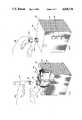
- FIGS. 6a and 6billustrate how a single encapsulated axial centrifuge unit 184 is loaded by hand.
- Centrifuge unit 184includes rotor 186 and frame 188.
- Rotor 186is driven by a high speed motor 190 through a first pulley and belt drive 192.
- the axial lead screw 114is driven by a second motor 194 through a second pulley and belt drive (not shown).
- Centrifuge 184, drive unit and the independent control electronicsare encapsulated by cover 196 so all that appears to the centrifuge user are rotor 186 and controls needed to initiate and modify the blood separation process (in the simplest form only power button 198 and process start button 176 would be required).
- Blood sample 200is loaded by a technician (or qualified other) 202 into rotor 186. Clamping cap 204 is then affixed to rotor 186 by the pressing of said cap 204 down onto said rotor 186 and, with a twist, the securing of "bayonet" style notches in said cap 204 around protruding pins 206. Blood sample 200 is inherently balanced when spinning, so no additional operations are required to balance the centrifuge module (e.g. the addition of balancing tubes).
- the userinitiates the blood separating process by pressing start button 176. Once spinning has begun no further user involvement is required during the separation process. Any discrepancies from the expected vibration or temperature level of the sample are detected by the module which then interrupts driving power to the machine. Problems involving human error, such as failure to load sample 200, failure to clamp cap 204 on or the loading of a leaking sample are not detected by the module and must be corrected by hand after the power is turned off.
- the sampleis spun for a preset (or adjustable) length of time at a high rotational speed in order to cause separation of the sample radially. Displacement of the lower density component from the center of the sample is then initiated and continues until the denser cellular component of the sample is detected. The displacement procedure is then stopped and high speed motor 190 is turned off.
- the separation processmaybe initiated from the keyboard of the controlling computer 136 or by a program running on said controlling computer.
- the identification number of the test tubeis read from bar code 162 on the tube and can be displayed on the computer terminal for confirmation or stored in computer memory.
- this tube identification numbercan be used by control computer 136 to reference data about the sample in a main laboratory data base (i.e. patient's name, tests to be performed on the sample, etc.)
- the displacement procedureis then initiated as in the simplest mode and sensors on the centrifuge module send to the computer information on the quality (presence and extent of hemolysis, lipemia or bilirubin in the serum) and volume of the serum that has been extracted from the sample.
- Termination of the displacement procedurecan be initiated by any one of five means: by manual interruption from the keyboard; by detection of blood cells in the tube' s intermediate chamber; by a serum quality reading that falls below some preset guideline; by recovery of a sufficient volume of serum to complete the required tests; or by detection of an operational malfunction. As in the simplest case termination involves the interruption of the displacement procedure and the stopping of the rotational drive.
- Clamping cap 204is then removed in the reverse manner to which it was applied. Grasping the top of sample 200, technician 202 removes said sample 200 from the module. Serum or plasma may then be extracted from said tube either by removing the closure located at the top of the tube and decanting or by piercing said closure with a cannula and drawing out serum or plasma. The remaining portion of the tube is then either discarded or stored for further tests.
- FIG. 7illustrates several features of the axial separation process and the centrifuge of this application as they relate to automated handling of blood samples.
- Isolated blood separating workcell 208incorporates six axial centrifuges 210, robotic arm 212, buffers 214 for delivery of the preferred embodiment sample tubes into said workcell 208, and computer 216 for controlling said workcell 208 and accessing data on the samples.
- Axial centrifuges 210are truly serial processors constituting a random access machine where any one module can be accessed independent of any other module. This condition aids in optimizing the per unit service time of any sample in the workcell.
- each centrifuge modulefacilitates the arrangement of these modules in the limited work envelope of a robotic workcell and, as the position of a sample tube does not change in space, said sample tube is easily located by said robotic arm 212 for loading and unloading.
- Isolation gloves 218are used for manual intervention in said workcell 208 thus providing the flexibility of manual manipulation while retaining the isolation of potentially hazardous blood samples or aerosols afforded by the encapsulation.
Landscapes
- Health & Medical Sciences (AREA)
- Chemical & Material Sciences (AREA)
- Life Sciences & Earth Sciences (AREA)
- Engineering & Computer Science (AREA)
- Hematology (AREA)
- Biomedical Technology (AREA)
- Physics & Mathematics (AREA)
- Analytical Chemistry (AREA)
- General Health & Medical Sciences (AREA)
- Clinical Laboratory Science (AREA)
- Chemical Kinetics & Catalysis (AREA)
- Biophysics (AREA)
- General Physics & Mathematics (AREA)
- Molecular Biology (AREA)
- Urology & Nephrology (AREA)
- Food Science & Technology (AREA)
- Medicinal Chemistry (AREA)
- Biochemistry (AREA)
- Ecology (AREA)
- Immunology (AREA)
- Pathology (AREA)
- Investigating Or Analysing Biological Materials (AREA)
- External Artificial Organs (AREA)
- Medicines Containing Material From Animals Or Micro-Organisms (AREA)
- Centrifugal Separators (AREA)
Abstract
Description
Claims (9)
Priority Applications (11)
| Application Number | Priority Date | Filing Date | Title |
|---|---|---|---|
| US07/033,769US4828716A (en) | 1987-04-03 | 1987-04-03 | Apparatus and method for separating phases of blood |
| CA000561554ACA1296693C (en) | 1987-04-03 | 1988-03-16 | Apparatus and method for separating phases of blood |
| DE3850287TDE3850287T2 (en) | 1987-04-03 | 1988-03-28 | Device and method for separating blood phases. |
| EP88105002AEP0285076B1 (en) | 1987-04-03 | 1988-03-28 | Apparatus and method for separating phases of blood |
| ES88105002TES2055716T3 (en) | 1987-04-03 | 1988-03-28 | APPARATUS AND METHOD FOR SEPARATING BLOOD PHASES. |
| AT88105002TATE107774T1 (en) | 1987-04-03 | 1988-03-28 | DEVICE AND METHOD FOR SEPARATION OF BLOOD PHASE. |
| AU14041/88AAU593988B2 (en) | 1987-04-03 | 1988-03-31 | Apparatus and method for separating phases of blood |
| JP63082132AJP2596963B2 (en) | 1987-04-03 | 1988-04-02 | Method and apparatus for separating liquid sample |
| US07/346,063US5030341A (en) | 1987-04-03 | 1989-05-02 | Apparatus for separating phases of blood |
| US07/346,065US5019243A (en) | 1987-04-03 | 1989-05-02 | Apparatus for collecting blood |
| US07/999,533US5308506A (en) | 1987-04-03 | 1992-12-31 | Apparatus and method for separating a sample of blood |
Applications Claiming Priority (1)
| Application Number | Priority Date | Filing Date | Title |
|---|---|---|---|
| US07/033,769US4828716A (en) | 1987-04-03 | 1987-04-03 | Apparatus and method for separating phases of blood |
Related Child Applications (1)
| Application Number | Title | Priority Date | Filing Date |
|---|---|---|---|
| US19284788AContinuation | 1987-04-03 | 1988-05-11 |
Publications (1)
| Publication Number | Publication Date |
|---|---|
| US4828716Atrue US4828716A (en) | 1989-05-09 |
Family
ID=21872338
Family Applications (2)
| Application Number | Title | Priority Date | Filing Date |
|---|---|---|---|
| US07/033,769Expired - Fee RelatedUS4828716A (en) | 1987-04-03 | 1987-04-03 | Apparatus and method for separating phases of blood |
| US07/999,533Expired - Fee RelatedUS5308506A (en) | 1987-04-03 | 1992-12-31 | Apparatus and method for separating a sample of blood |
Family Applications After (1)
| Application Number | Title | Priority Date | Filing Date |
|---|---|---|---|
| US07/999,533Expired - Fee RelatedUS5308506A (en) | 1987-04-03 | 1992-12-31 | Apparatus and method for separating a sample of blood |
Country Status (8)
| Country | Link |
|---|---|
| US (2) | US4828716A (en) |
| EP (1) | EP0285076B1 (en) |
| JP (1) | JP2596963B2 (en) |
| AT (1) | ATE107774T1 (en) |
| AU (1) | AU593988B2 (en) |
| CA (1) | CA1296693C (en) |
| DE (1) | DE3850287T2 (en) |
| ES (1) | ES2055716T3 (en) |
Cited By (92)
| Publication number | Priority date | Publication date | Assignee | Title |
|---|---|---|---|---|
| WO1990003834A1 (en)* | 1988-10-06 | 1990-04-19 | Medical Automation Specialties, Inc. | Method and apparatus for automatic processing and analyzing of blood serum |
| US5032288A (en)* | 1989-11-29 | 1991-07-16 | Eastman Kodak Company | Blood collection method |
| US5036861A (en)* | 1990-01-11 | 1991-08-06 | Sembrowich Walter L | Method and apparatus for non-invasively monitoring plasma glucose levels |
| US5039401A (en)* | 1990-05-16 | 1991-08-13 | Eastman Kodak Company | Blood collection and centrifugal separation device including a valve |
| US5104802A (en)* | 1989-07-28 | 1992-04-14 | The United States Of America As Represented By The Administrator Of The National Aeronautics And Space Administration | Hollow fiber clinostat for simulating microgravity in cell culture |
| DE4212325A1 (en)* | 1992-04-13 | 1993-10-14 | Hoechst Ag | Concrete moldings with improved acid resistance |
| US5271852A (en)* | 1992-05-01 | 1993-12-21 | E. I. Du Pont De Nemours And Company | Centrifugal methods using a phase-separation tube |
| US5282981A (en)* | 1992-05-01 | 1994-02-01 | E. I. Du Pont De Nemours And Company | Flow restrictor-separation device |
| US5308506A (en)* | 1987-04-03 | 1994-05-03 | Mcewen James A | Apparatus and method for separating a sample of blood |
| US5316667A (en)* | 1989-05-26 | 1994-05-31 | Baxter International Inc. | Time based interface detection systems for blood processing apparatus |
| US5316666A (en)* | 1987-01-30 | 1994-05-31 | Baxter International Inc. | Blood processing systems with improved data transfer between stationary and rotating elements |
| US5354483A (en)* | 1992-10-01 | 1994-10-11 | Andronic Technologies, Inc. | Double-ended tube for separating phases of blood |
| USD363353S (en) | 1993-10-27 | 1995-10-17 | Du Pont Canada Inc. | Axial spin centrifuge |
| US5462716A (en)* | 1991-11-11 | 1995-10-31 | Holm; Niels E. | Container for receiving and separating a fluid, preferably blood plasma, into its ingredients |
| US5474687A (en)* | 1994-08-31 | 1995-12-12 | Activated Cell Therapy, Inc. | Methods for enriching CD34+ human hematopoietic progenitor cells |
| US5478479A (en)* | 1994-05-20 | 1995-12-26 | Haemonetics Corporation | Two-stage cell wash process controlled by optical sensor |
| US5480378A (en)* | 1990-05-14 | 1996-01-02 | Weis-Fogh; Ulla | Apparatus for preparing a concentrate of coagulation factors from a blood sample |
| US5547108A (en)* | 1994-08-02 | 1996-08-20 | Pall Corporation | Expressor |
| US5550060A (en)* | 1992-11-03 | 1996-08-27 | Chronomed, Inc. | Method and procedure for preparing red blood fractions |
| US5577513A (en)* | 1994-08-31 | 1996-11-26 | Activated Cell Therapy, Inc. | Centrifugation syringe, system and method |
| WO1996041148A1 (en)* | 1995-06-07 | 1996-12-19 | Alpha Scientific Instruments, Inc. | Automatic blood film preparation device |
| US5603845A (en)* | 1993-11-19 | 1997-02-18 | E. R. Squibb & Sons, Inc. | Liquid separation apparatus and method |
| US5646004A (en)* | 1994-08-31 | 1997-07-08 | Activated Cell Therapy, Inc. | Methods for enriching fetal cells from maternal body fluids |
| US5648223A (en)* | 1994-08-31 | 1997-07-15 | Activated Cell Therapy, Inc. | Methods for enriching breast tumor cells |
| US5663051A (en)* | 1994-08-31 | 1997-09-02 | Activated Cell Therapy, Inc. | Separation apparatus and method |
| US5690815A (en)* | 1992-07-13 | 1997-11-25 | Pall Corporation | Automated system for processing biological fluid |
| US5733446A (en)* | 1994-12-02 | 1998-03-31 | Bristol-Myers Squibb Company | Centrifuge with annular filter |
| US5738784A (en)* | 1994-12-02 | 1998-04-14 | E.R. Squibb & Sons, Inc. | Device for separating a blood component from blood or plasma |
| US5811061A (en)* | 1992-02-10 | 1998-09-22 | Baxter International Inc. | Method and device for testing blood units for viral contamination |
| US5830352A (en)* | 1994-12-02 | 1998-11-03 | Bristol-Myers Squibb Company | Centrifuge reagent delivery system |
| US5840502A (en)* | 1994-08-31 | 1998-11-24 | Activated Cell Therapy, Inc. | Methods for enriching specific cell-types by density gradient centrifugation |
| WO1999003557A1 (en)* | 1997-07-18 | 1999-01-28 | Baxter International Inc. | Blood processing systems and methods which optically derive the volume of platelets contained in a plasma constituent |
| WO1999004837A1 (en)* | 1997-07-25 | 1999-02-04 | Bristol-Myers Squibb Company | Blood product delivery system |
| US5968018A (en)* | 1996-10-30 | 1999-10-19 | Cohesion Corporation | Cell separation device and in-line orifice mixer system |
| US5980757A (en)* | 1997-04-03 | 1999-11-09 | Baxter International Inc. | Interface detection and control systems and method |
| US5993370A (en)* | 1987-01-30 | 1999-11-30 | Baxter International Inc. | Enhanced yield collection systems and methods for obtaining concentrated platelets from platelet-rich plasma |
| US5997811A (en)* | 1997-07-02 | 1999-12-07 | Cohesion Technologies, Inc. | Method for sterile syringe packaging and handling |
| US6039868A (en)* | 1995-02-09 | 2000-03-21 | First Medical, Inc. | Blood separator system |
| US6060022A (en)* | 1996-07-05 | 2000-05-09 | Beckman Coulter, Inc. | Automated sample processing system including automatic centrifuge device |
| US6077442A (en)* | 1996-11-19 | 2000-06-20 | Sarstedt Ag & Co. | Automatically pressing a filter into a blood-collection tube |
| US6132353A (en)* | 1996-10-21 | 2000-10-17 | Winkelman; James W. | Apparatus and method for separating plasma or serum from the red cells of a blood sample |
| WO2001028652A1 (en)* | 1999-10-16 | 2001-04-26 | Baxter International Inc. | Blood processing systems which monitor optical densities |
| US6245570B1 (en)* | 1991-05-08 | 2001-06-12 | Baxter International Inc. | Container for irradiation of blood products |
| US6302836B1 (en) | 1998-10-01 | 2001-10-16 | Howard L. North, Jr. | Method for partitioning blood and delivering clean serum |
| US20020023884A1 (en)* | 2000-07-28 | 2002-02-28 | Anderson Norman G. | Method and apparatus for unloading gradients |
| US20020160443A1 (en)* | 1998-05-09 | 2002-10-31 | Ikonisys, Inc. | Method and apparatus for computer controlled rare cell, including fetal cell, based diagnosis |
| WO2003028844A1 (en)* | 2001-10-04 | 2003-04-10 | Ikonisys, Inc. | System and method for fractionation of a centrifuged sample |
| US6565809B1 (en)* | 1999-10-29 | 2003-05-20 | Teruaki Itoh | Specimen processing system |
| US20040033170A1 (en)* | 2000-03-22 | 2004-02-19 | Dewalch Binz | Method and apparatus for processing substances in a single container |
| US20040167004A1 (en)* | 1999-07-08 | 2004-08-26 | Jorgensen Glen E. | Platelet concentration syringe kit |
| US6846298B1 (en) | 1997-07-25 | 2005-01-25 | Bristol-Myers Squibb Company | Blood product delivery system |
| US6946079B1 (en)* | 1994-12-02 | 2005-09-20 | Bristol-Myers Squibb Company | Centrifugal filtration method for separating fibrin monomer from blood |
| US20060196885A1 (en)* | 2005-02-21 | 2006-09-07 | Biomet Manufacturing Corp. | Method and apparatus for application of a fluid |
| US20060223176A1 (en)* | 2005-03-31 | 2006-10-05 | Evgenia Mandrusov | Integrated system for on-site cell acquisition, processing, and delivery |
| US20060251622A1 (en)* | 2003-05-21 | 2006-11-09 | Koji Suzuki | Container for serum production and method of regenerative medicine using the same |
| US20070293385A1 (en)* | 2002-06-14 | 2007-12-20 | Dolecek Victor D | Centrifuge system utilizing disposable components and automated processing of blood to collect platelet rich plasma |
| US20080017577A1 (en)* | 2006-07-21 | 2008-01-24 | Becton, Dickinson And Company | Membrane-based Double-layer Tube for Sample Collections |
| US20080206774A1 (en)* | 1998-05-09 | 2008-08-28 | Ikonisys, Inc. | Automated cancer diagnostic methods using fish |
| US20080221929A1 (en)* | 2007-03-09 | 2008-09-11 | Cerner Innovation, Inc. | System and method for associating a patient specimen identifier with a radiology image for the patient |
| US20080219523A1 (en)* | 2007-03-09 | 2008-09-11 | Cerner Innovation, Inc. | System and method for associating electronic images in the healthcare environment |
| US20080219524A1 (en)* | 2007-03-09 | 2008-09-11 | Cerner Innovation, Inc. | Graphical user interface for displaying a radiology image for a patient and an associated laboratory report summary |
| US20080241848A1 (en)* | 1998-05-09 | 2008-10-02 | Ikonisys, Inc. | Methods for prenatal diagnosis of aneuploidy |
| US20080260593A1 (en)* | 2000-03-22 | 2008-10-23 | Dewalch Norman Binz | Method and apparatus for processing substances in a single container |
| US20090111101A1 (en)* | 1998-05-09 | 2009-04-30 | Ikonisys, Inc. | Automated Cancer Diagnostic Methods Using FISH |
| US20090130646A1 (en)* | 2006-02-08 | 2009-05-21 | Becton, Dickinson And Company | Blood collection device, method, and system for using the same |
| US20090148957A1 (en)* | 2007-12-11 | 2009-06-11 | Ajay Gupta | Platelet-free analyte assay method |
| US20090250413A1 (en)* | 2008-04-04 | 2009-10-08 | Biomet Biologics, Llc | Sterile Blood Separating System |
| US20100093551A1 (en)* | 2008-10-09 | 2010-04-15 | Decision Biomarkers, Inc. | Liquid Transfer and Filter System |
| US20100155343A1 (en)* | 2008-07-21 | 2010-06-24 | Becton, Dickinson And Company | Density Phase Separation Device |
| US20100155319A1 (en)* | 2008-07-21 | 2010-06-24 | Becton, Dickinson And Company | Density Phase Separation Device |
| US8182769B2 (en) | 2008-04-04 | 2012-05-22 | Biomet Biologics, Llc | Clean transportation system |
| US20120203167A1 (en)* | 2005-09-12 | 2012-08-09 | Lanny Johnson | Use of autologous sediment from fluid aspirates as vehicles for drug delivery |
| US8317672B2 (en) | 2010-11-19 | 2012-11-27 | Kensey Nash Corporation | Centrifuge method and apparatus |
| US8394006B2 (en) | 2010-11-19 | 2013-03-12 | Kensey Nash Corporation | Centrifuge |
| US8469871B2 (en) | 2010-11-19 | 2013-06-25 | Kensey Nash Corporation | Centrifuge |
| US8556794B2 (en) | 2010-11-19 | 2013-10-15 | Kensey Nash Corporation | Centrifuge |
| WO2014025490A1 (en) | 2012-08-10 | 2014-02-13 | Montagu Jean I | Filtering blood |
| US8747781B2 (en) | 2008-07-21 | 2014-06-10 | Becton, Dickinson And Company | Density phase separation device |
| US8794452B2 (en) | 2009-05-15 | 2014-08-05 | Becton, Dickinson And Company | Density phase separation device |
| US8870733B2 (en) | 2010-11-19 | 2014-10-28 | Kensey Nash Corporation | Centrifuge |
| US8973293B2 (en) | 2010-11-19 | 2015-03-10 | Becton, Dickinson And Company | Specimen container label for automated clinical laboratory processing systems |
| US9555171B2 (en) | 2010-09-30 | 2017-01-31 | Depuy Mitek, Llc | Methods and devices for collecting separate components of whole blood |
| ITUB20152909A1 (en)* | 2015-08-05 | 2017-02-05 | Eltek Spa | MEDICAL AND RELATIVE DEVICE EQUIPMENT, IN PARTICULAR FOR THE SEPARATION OF A FLUID |
| US9682373B2 (en) | 1999-12-03 | 2017-06-20 | Becton, Dickinson And Company | Device for separating components of a fluid sample |
| US9694359B2 (en) | 2014-11-13 | 2017-07-04 | Becton, Dickinson And Company | Mechanical separator for a biological fluid |
| US9927450B2 (en)* | 2013-03-08 | 2018-03-27 | Siemens Healthcare Diagnostics Inc. | Tube characterization station |
| US10125345B2 (en) | 2014-01-31 | 2018-11-13 | Dsm Ip Assets, B.V. | Adipose tissue centrifuge and method of use |
| EP3470142A1 (en) | 2017-10-11 | 2019-04-17 | Orthogen AG | Device comprising a first chamber for receiving a body fluid |
| US10668207B2 (en) | 2015-07-13 | 2020-06-02 | Haemonetics Corporation | System and method for removing fat from salvaged blood |
| US11090646B2 (en) | 2017-07-27 | 2021-08-17 | Biomerieux, Inc. | Isolation tube |
| US11541161B2 (en) | 2016-06-24 | 2023-01-03 | Haemonetics Corporation | System and method for continuous flow red blood cell washing |
| US12337097B2 (en) | 2010-11-19 | 2025-06-24 | Dsm Ip Assets B.V. | Centrifuge |
Families Citing this family (49)
| Publication number | Priority date | Publication date | Assignee | Title |
|---|---|---|---|---|
| DE9013914U1 (en)* | 1990-10-05 | 1991-02-14 | Walter Sarstedt Geräte und Verbrauchsmaterial für Medizin und Wissenschaft, 5223 Nümbrecht | Separation device for liquid, especially blood fractions |
| US5211310A (en)* | 1991-04-30 | 1993-05-18 | Andronic Devices Ltd. | Apparatus and method for dispensing phases of blood |
| US5555920A (en)* | 1991-04-30 | 1996-09-17 | Automed Corporation | Method and apparatus for aliquotting blood serum or blood plasma |
| US5322192A (en)* | 1992-07-28 | 1994-06-21 | Automed Corporation | Pipetting apparatus |
| EP0609986B1 (en)* | 1993-01-29 | 1999-12-15 | Becton, Dickinson and Company | Compact blood culture apparatus |
| US5389265A (en)* | 1993-06-02 | 1995-02-14 | E. I. Du Pont De Nemours And Company | Phase-separation tube |
| US5946220A (en)* | 1993-08-25 | 1999-08-31 | Lemelson; Jerome H. | Computer operated material processing systems and method |
| US5525240A (en)* | 1993-12-27 | 1996-06-11 | Lemelson; Jerome H. | Adaptively controlled centrifugation method |
| AU708820B2 (en)* | 1993-11-19 | 1999-08-12 | Vivolution A/S | Annular assembly for centrifuge device |
| US5741423A (en)* | 1994-08-23 | 1998-04-21 | Bates; John | Liquid-liquid extraction |
| EP0753741A4 (en)* | 1995-01-30 | 1998-04-29 | Niigata Engineering Co Ltd | Component separation member and component separator equipped with said member |
| JP3132342B2 (en)* | 1995-07-07 | 2001-02-05 | 松下電器産業株式会社 | Specimen test method and device |
| US5584790A (en)* | 1995-09-08 | 1996-12-17 | Beckman Instruments, Inc. | Variable inclination centrifugation assembly for rapid separation of blood |
| AU700988B2 (en)* | 1995-10-03 | 1999-01-14 | Beckman Instruments, Inc. | Axial spin blood separation system and method |
| JP2859845B2 (en)* | 1996-05-09 | 1999-02-24 | 照明 伊藤 | Serum collection aid |
| JP3493910B2 (en)* | 1996-08-23 | 2004-02-03 | 株式会社日立製作所 | Automated processing system |
| US5786898A (en)* | 1996-08-23 | 1998-07-28 | Fitzpatrick; Stephen T. | Structure and method for centrifugal sedimentation particle size analysis of particles of lower density than their suspension medium |
| US5785925A (en)* | 1996-08-29 | 1998-07-28 | Saigene Corporation | Centrifuge tube phase separation plug |
| WO1998030331A1 (en)* | 1997-01-08 | 1998-07-16 | Bristol-Myers Squibb Company | Centrifuge apparatus for separating blood |
| DE19703921C1 (en)* | 1997-02-03 | 1999-03-18 | Sarstedt Walter Geraete | Blood collection device |
| US5935051A (en)* | 1997-08-29 | 1999-08-10 | Beckman Instruments, Inc. | Blood separation device |
| EP1027145A4 (en)* | 1997-09-17 | 2004-08-25 | Gentra Systems Inc | Apparatuses and methods for isolating nucleic acid |
| DE19836559A1 (en)* | 1998-08-12 | 2000-03-23 | Antigen Gmbh | Blood collection vessel |
| US6406671B1 (en)* | 1998-12-05 | 2002-06-18 | Becton, Dickinson And Company | Device and method for separating components of a fluid sample |
| US6793892B1 (en)* | 1999-12-06 | 2004-09-21 | Volker Niermann | Device and method for separating components of a fluid sample |
| WO2002072265A1 (en) | 2001-03-09 | 2002-09-19 | Gen-Probe Incorporated | Penetrable cap |
| US6869792B2 (en) | 2001-03-16 | 2005-03-22 | Irm, Llc | Method and apparatus for performing multiple processing steps on a sample in a single vessel |
| EP1466160B1 (en)* | 2002-01-19 | 2010-12-22 | PVT Probenverteiltechnik GmbH | Method and device for the analysis of body fluids |
| US20030154639A1 (en)* | 2002-02-12 | 2003-08-21 | Bowers Scott D. | Apparatus, system and method for displaying and providing customizable and changeable signs |
| WO2004096046A1 (en)* | 2003-04-25 | 2004-11-11 | Sekisui Chemical Co., Ltd. | Method and tool for filtrating specimen using specimen sampling container, and specimen sampling container |
| CA2458497A1 (en)* | 2004-02-24 | 2005-08-24 | Cme Telemetrix Inc. | Spectrophotometric analysis of plasma or serum in a sealed tube |
| GB0422358D0 (en)* | 2004-10-08 | 2004-11-10 | Rts Thurnall Plc | Determination of the boundaries between fractions and extraction of selected fractions in a fractional sample |
| EP2278383A1 (en)* | 2004-11-24 | 2011-01-26 | Battelle Memorial Institute | A test tube handling apparatus |
| JP2007098260A (en)* | 2005-10-04 | 2007-04-19 | Hitachi Koki Co Ltd | centrifuge |
| JP2007152157A (en)* | 2005-11-30 | 2007-06-21 | Hitachi Koki Co Ltd | Centrifuge |
| EP1949962A1 (en)* | 2007-01-25 | 2008-07-30 | F.Hoffmann-La Roche Ag | Device and method for separating a liquid component of a blood sample, and analyzer apparatus comprising such a device |
| JP5479319B2 (en)* | 2007-04-12 | 2014-04-23 | バイオメット・バイオロジックス・リミテッド・ライアビリティ・カンパニー | Buoy suspension fractionation system |
| US8387810B2 (en)* | 2007-04-16 | 2013-03-05 | Becton, Dickinson And Company | Pierceable cap having piercing extensions for a sample container |
| US8387811B2 (en)* | 2007-04-16 | 2013-03-05 | Bd Diagnostics | Pierceable cap having piercing extensions |
| JP5433139B2 (en)* | 2007-06-29 | 2014-03-05 | 株式会社東芝 | Microchemical analyzer, measuring method thereof, and microcassette |
| US8092361B2 (en)* | 2008-05-02 | 2012-01-10 | Ortho-Clinical Diagnostics, Inc. | Split spin centrifugation of test elements |
| US9272083B2 (en) | 2009-05-29 | 2016-03-01 | Endocellutions, Inc. | Apparatus and methods for aspirating and separating components of different densities from a physiological fluid containing cells |
| KR102100665B1 (en)* | 2012-02-15 | 2020-04-14 | 마이크로에어 서지컬 인스투르먼츠 엘엘씨 | Apparatus for centrifugation and methods therefore |
| US10603665B2 (en) | 2013-01-29 | 2020-03-31 | Endocellutions, Inc. | Cell concentration devices and methods that include an insert defining a lumen and a cannula assembly |
| DE102013220282A1 (en)* | 2013-10-08 | 2015-04-09 | Siemens Aktiengesellschaft | Device with at least one channel and at least two liquids |
| CN110785649A (en) | 2017-06-26 | 2020-02-11 | 埃斯特万·门多萨 | Sample filter device |
| KR102051207B1 (en) | 2018-01-30 | 2019-12-03 | 이준석 | Piston for centriguation |
| USD920534S1 (en) | 2018-12-21 | 2021-05-25 | CanaryQ, Inc. | Blood sample filtration device |
| CN111940003A (en)* | 2020-08-13 | 2020-11-17 | 陈龙刚 | Transfer device for stem cell sampling |
Citations (39)
| Publication number | Priority date | Publication date | Assignee | Title |
|---|---|---|---|---|
| US2460641A (en)* | 1945-08-14 | 1949-02-01 | Joseph J Kleiner | Blood collecting apparatus |
| US3468474A (en)* | 1966-07-07 | 1969-09-23 | Arvid W Shoblom | Centrifuge accessory |
| US3508653A (en)* | 1967-11-17 | 1970-04-28 | Charles M Coleman | Method and apparatus for fluid handling and separation |
| US3741400A (en)* | 1970-06-15 | 1973-06-26 | J Dick | Blood sample container |
| US3813031A (en)* | 1972-08-02 | 1974-05-28 | Atomic Energy Commission | Rotor having sample holding means |
| US3849072A (en)* | 1972-04-25 | 1974-11-19 | Becton Dickinson Co | Plasma separator |
| US3852194A (en)* | 1972-12-11 | 1974-12-03 | Corning Glass Works | Apparatus and method for fluid collection and partitioning |
| US3929646A (en)* | 1974-07-22 | 1975-12-30 | Technicon Instr | Serum separator and fibrin filter |
| US3957654A (en)* | 1974-02-27 | 1976-05-18 | Becton, Dickinson And Company | Plasma separator with barrier to eject sealant |
| US3957197A (en)* | 1975-04-25 | 1976-05-18 | The United States Of America As Represented By The United States Energy Research And Development Administration | Centrifuge apparatus |
| US4021352A (en)* | 1974-03-30 | 1977-05-03 | Walter Sarstedt Kunststoff-Spritzgusswerk | Filter device for separating blood fractions |
| US4046699A (en)* | 1976-11-01 | 1977-09-06 | Corning Glass Works | Access device for centrifugal separation assemblies |
| US4083784A (en)* | 1974-12-16 | 1978-04-11 | Corning Glass Works | Stabilized blood separating composition |
| US4152270A (en)* | 1976-05-06 | 1979-05-01 | Sherwood Medical Industries Inc. | Phase separation device |
| US4189382A (en)* | 1974-11-07 | 1980-02-19 | Sherwood Medical Industries Inc. | Blood coagulation and separation |
| US4202769A (en)* | 1977-06-16 | 1980-05-13 | Greenspan Donald J | Method for separating serum or plasma from the formed elements of blood |
| US4278202A (en)* | 1978-07-25 | 1981-07-14 | Separek Teknik Ab | Centrifuge rotor and collapsible separation container for use therewith |
| US4279863A (en)* | 1979-09-12 | 1981-07-21 | Sherwood Medical Industries, Inc. | Reagent separator for a blood collection tube |
| US4283276A (en)* | 1980-02-29 | 1981-08-11 | E. I. Du Pont De Nemours And Company | Rotor for sedimentation field flow fractionation |
| US4284602A (en)* | 1979-12-10 | 1981-08-18 | Immutron, Inc. | Integrated fluid manipulator |
| US4285810A (en)* | 1980-02-29 | 1981-08-25 | E. I. Du Pont De Nemours And Company | Method and apparatus for field flow fractionation |
| US4322298A (en)* | 1981-06-01 | 1982-03-30 | Advanced Blood Component Technology, Inc. | Centrifugal cell separator, and method of use thereof |
| US4326959A (en)* | 1981-02-04 | 1982-04-27 | Ferrara Louis T | Blood separator and dispenser |
| US4350593A (en)* | 1977-12-19 | 1982-09-21 | Becton, Dickinson And Company | Assembly, compositions and method for separating blood |
| US4369117A (en)* | 1980-05-12 | 1983-01-18 | American Hospital Supply Corporation | Serum separating method and apparatus |
| US4417981A (en)* | 1981-05-04 | 1983-11-29 | Becton, Dickinson And Company | Blood phase separator device |
| US4425235A (en)* | 1982-03-22 | 1984-01-10 | Sherwood Medical Company | Blood collection device with phase partitioning means |
| US4443345A (en)* | 1982-06-28 | 1984-04-17 | Wells John R | Serum preparator |
| US4446106A (en)* | 1982-01-15 | 1984-05-01 | Instrumentation Laboratory Inc. | Analysis system |
| US4447220A (en)* | 1979-09-22 | 1984-05-08 | Eberle Guenter | Method and apparatus for separating blood components |
| US4451250A (en)* | 1982-09-27 | 1984-05-29 | E. I. Du Pont De Nemours And Company | Inside adapter for a sample container |
| US4464254A (en)* | 1982-06-03 | 1984-08-07 | Porex Technologies, Corp. | Device for separating serum from blood sample |
| US4492634A (en)* | 1982-09-28 | 1985-01-08 | Emde Medical Research | Pre-evacuated blood collection tube with anti-hemolysis baffle system and centrifugation propelled filtration disc and efficient serum-from cells separator |
| US4522713A (en)* | 1982-11-26 | 1985-06-11 | Sartorius Gmbh | Apparatus for static membrane filtration |
| US4530691A (en)* | 1983-12-13 | 1985-07-23 | Baxter Travenol Laboratories, Inc. | Centrifuge with movable mandrel |
| US4534465A (en)* | 1983-10-13 | 1985-08-13 | Coulter Electronics, Inc. | Cassette for supporting test tubes of different diameters and/or lengths |
| US4550084A (en)* | 1982-01-15 | 1985-10-29 | Allied Corporation | Analysis system |
| US4602995A (en)* | 1985-05-20 | 1986-07-29 | Technicon Instruments Corporation | Liquid level adjusting and filtering device |
| US4639316A (en)* | 1984-12-14 | 1987-01-27 | Becton, Dickinson And Company | Automatic liquid component separator |
Family Cites Families (16)
| Publication number | Priority date | Publication date | Assignee | Title |
|---|---|---|---|---|
| US3341073A (en)* | 1965-04-14 | 1967-09-12 | Milton J Arps | Metering and dispensing apparatus |
| FR1568072A (en)* | 1968-01-24 | 1969-05-23 | ||
| DE2115032C3 (en)* | 1971-03-29 | 1975-05-07 | Kernforschungsanlage Juelich Gmbh, 5170 Juelich | Device for separating two phases in liquid-liquid extraction in a centrifuge |
| US3782548A (en)* | 1972-12-07 | 1974-01-01 | J Bowen | Serum skimmer |
| US4001122A (en)* | 1973-08-22 | 1977-01-04 | Telan Corporation | Method and device for separating blood components |
| US3941699A (en)* | 1974-02-27 | 1976-03-02 | Becton, Dickinson And Company | Plasma separator with centrifugal valve |
| US3887466A (en)* | 1974-02-27 | 1975-06-03 | Becton Dickinson Co | Serum/plasma separator cannula fluid by-pass type centrifugal valve cannula seal |
| US4142668A (en)* | 1976-10-01 | 1979-03-06 | Lee Jae Y | Serum-plasma separator and transfer apparatus |
| US4156570A (en)* | 1977-04-18 | 1979-05-29 | Robert A. Levine | Apparatus and method for measuring white blood cell and platelet concentrations in blood |
| US4169060A (en)* | 1977-10-25 | 1979-09-25 | Eastman Kodak Company | Blood-collecting and serum-dispensing device |
| SE416617B (en)* | 1979-03-28 | 1981-01-26 | Johansson A S | DEVICE FOR BLOOD SEPARATION BY CLIPPING BLOOD COMPONENTS FROM A BLOOD PHASE |
| DE3301113C2 (en)* | 1983-01-14 | 1985-01-10 | Fresenius AG, 6380 Bad Homburg | Method and device for separating media |
| US5030341A (en)* | 1987-04-03 | 1991-07-09 | Andronic Technologies, Inc. | Apparatus for separating phases of blood |
| US4828716A (en)* | 1987-04-03 | 1989-05-09 | Andronic Devices, Ltd. | Apparatus and method for separating phases of blood |
| FR2624757B1 (en)* | 1987-12-17 | 1990-05-25 | Roussel Uclaf | DEVICE FOR THE EXTRACTION OF A SEPARATE PHASE |
| US4927545A (en)* | 1988-10-06 | 1990-05-22 | Medical Automation Specialties, Inc. | Method and apparatus for automatic processing and analyzing of blood serum |
- 1987
- 1987-04-03USUS07/033,769patent/US4828716A/ennot_activeExpired - Fee Related
- 1988
- 1988-03-16CACA000561554Apatent/CA1296693C/ennot_activeExpired - Lifetime
- 1988-03-28ESES88105002Tpatent/ES2055716T3/ennot_activeExpired - Lifetime
- 1988-03-28EPEP88105002Apatent/EP0285076B1/ennot_activeExpired - Lifetime
- 1988-03-28ATAT88105002Tpatent/ATE107774T1/ennot_activeIP Right Cessation
- 1988-03-28DEDE3850287Tpatent/DE3850287T2/ennot_activeExpired - Fee Related
- 1988-03-31AUAU14041/88Apatent/AU593988B2/ennot_activeCeased
- 1988-04-02JPJP63082132Apatent/JP2596963B2/ennot_activeExpired - Lifetime
- 1992
- 1992-12-31USUS07/999,533patent/US5308506A/ennot_activeExpired - Fee Related
Patent Citations (39)
| Publication number | Priority date | Publication date | Assignee | Title |
|---|---|---|---|---|
| US2460641A (en)* | 1945-08-14 | 1949-02-01 | Joseph J Kleiner | Blood collecting apparatus |
| US3468474A (en)* | 1966-07-07 | 1969-09-23 | Arvid W Shoblom | Centrifuge accessory |
| US3508653A (en)* | 1967-11-17 | 1970-04-28 | Charles M Coleman | Method and apparatus for fluid handling and separation |
| US3741400A (en)* | 1970-06-15 | 1973-06-26 | J Dick | Blood sample container |
| US3849072A (en)* | 1972-04-25 | 1974-11-19 | Becton Dickinson Co | Plasma separator |
| US3813031A (en)* | 1972-08-02 | 1974-05-28 | Atomic Energy Commission | Rotor having sample holding means |
| US3852194A (en)* | 1972-12-11 | 1974-12-03 | Corning Glass Works | Apparatus and method for fluid collection and partitioning |
| US3957654A (en)* | 1974-02-27 | 1976-05-18 | Becton, Dickinson And Company | Plasma separator with barrier to eject sealant |
| US4021352A (en)* | 1974-03-30 | 1977-05-03 | Walter Sarstedt Kunststoff-Spritzgusswerk | Filter device for separating blood fractions |
| US3929646A (en)* | 1974-07-22 | 1975-12-30 | Technicon Instr | Serum separator and fibrin filter |
| US4189382A (en)* | 1974-11-07 | 1980-02-19 | Sherwood Medical Industries Inc. | Blood coagulation and separation |
| US4083784A (en)* | 1974-12-16 | 1978-04-11 | Corning Glass Works | Stabilized blood separating composition |
| US3957197A (en)* | 1975-04-25 | 1976-05-18 | The United States Of America As Represented By The United States Energy Research And Development Administration | Centrifuge apparatus |
| US4152270A (en)* | 1976-05-06 | 1979-05-01 | Sherwood Medical Industries Inc. | Phase separation device |
| US4046699A (en)* | 1976-11-01 | 1977-09-06 | Corning Glass Works | Access device for centrifugal separation assemblies |
| US4202769A (en)* | 1977-06-16 | 1980-05-13 | Greenspan Donald J | Method for separating serum or plasma from the formed elements of blood |
| US4350593A (en)* | 1977-12-19 | 1982-09-21 | Becton, Dickinson And Company | Assembly, compositions and method for separating blood |
| US4278202A (en)* | 1978-07-25 | 1981-07-14 | Separek Teknik Ab | Centrifuge rotor and collapsible separation container for use therewith |
| US4279863A (en)* | 1979-09-12 | 1981-07-21 | Sherwood Medical Industries, Inc. | Reagent separator for a blood collection tube |
| US4447220A (en)* | 1979-09-22 | 1984-05-08 | Eberle Guenter | Method and apparatus for separating blood components |
| US4284602A (en)* | 1979-12-10 | 1981-08-18 | Immutron, Inc. | Integrated fluid manipulator |
| US4283276A (en)* | 1980-02-29 | 1981-08-11 | E. I. Du Pont De Nemours And Company | Rotor for sedimentation field flow fractionation |
| US4285810A (en)* | 1980-02-29 | 1981-08-25 | E. I. Du Pont De Nemours And Company | Method and apparatus for field flow fractionation |
| US4369117A (en)* | 1980-05-12 | 1983-01-18 | American Hospital Supply Corporation | Serum separating method and apparatus |
| US4326959A (en)* | 1981-02-04 | 1982-04-27 | Ferrara Louis T | Blood separator and dispenser |
| US4417981A (en)* | 1981-05-04 | 1983-11-29 | Becton, Dickinson And Company | Blood phase separator device |
| US4322298A (en)* | 1981-06-01 | 1982-03-30 | Advanced Blood Component Technology, Inc. | Centrifugal cell separator, and method of use thereof |
| US4550084A (en)* | 1982-01-15 | 1985-10-29 | Allied Corporation | Analysis system |
| US4446106A (en)* | 1982-01-15 | 1984-05-01 | Instrumentation Laboratory Inc. | Analysis system |
| US4425235A (en)* | 1982-03-22 | 1984-01-10 | Sherwood Medical Company | Blood collection device with phase partitioning means |
| US4464254A (en)* | 1982-06-03 | 1984-08-07 | Porex Technologies, Corp. | Device for separating serum from blood sample |
| US4443345A (en)* | 1982-06-28 | 1984-04-17 | Wells John R | Serum preparator |
| US4451250A (en)* | 1982-09-27 | 1984-05-29 | E. I. Du Pont De Nemours And Company | Inside adapter for a sample container |
| US4492634A (en)* | 1982-09-28 | 1985-01-08 | Emde Medical Research | Pre-evacuated blood collection tube with anti-hemolysis baffle system and centrifugation propelled filtration disc and efficient serum-from cells separator |
| US4522713A (en)* | 1982-11-26 | 1985-06-11 | Sartorius Gmbh | Apparatus for static membrane filtration |
| US4534465A (en)* | 1983-10-13 | 1985-08-13 | Coulter Electronics, Inc. | Cassette for supporting test tubes of different diameters and/or lengths |
| US4530691A (en)* | 1983-12-13 | 1985-07-23 | Baxter Travenol Laboratories, Inc. | Centrifuge with movable mandrel |
| US4639316A (en)* | 1984-12-14 | 1987-01-27 | Becton, Dickinson And Company | Automatic liquid component separator |
| US4602995A (en)* | 1985-05-20 | 1986-07-29 | Technicon Instruments Corporation | Liquid level adjusting and filtering device |
Non-Patent Citations (8)
| Title |
|---|
| 1 page brochure introducing Stat Spin Technology.* |
| 1 page brochure introducing Stat-Spin Technology. |
| 10 page instruction manual on the Beckman Airfuge Ultracentrifuge AF IM 5.* |
| 10 page instruction manual on the Beckman Airfuge Ultracentrifuge AF-IM-5. |
| 21 page article on Preparative Zonal Centrifugation in Methods of Biochemical Analysis, vol. XV.* |
| 21-page article on Preparative Zonal Centrifugation in Methods of Biochemical Analysis, vol. XV. |
| 3 page brochure of Sarstedt showing Monovette and Safety Monovette Beckman 3 page brochure of Jun. 1981.* |
| 3 page brochure of Sarstedt showing Monovette and Safety Monovette Beckman 3-page brochure of Jun. 1981. |
Cited By (203)
| Publication number | Priority date | Publication date | Assignee | Title |
|---|---|---|---|---|
| US5993370A (en)* | 1987-01-30 | 1999-11-30 | Baxter International Inc. | Enhanced yield collection systems and methods for obtaining concentrated platelets from platelet-rich plasma |
| US5316666A (en)* | 1987-01-30 | 1994-05-31 | Baxter International Inc. | Blood processing systems with improved data transfer between stationary and rotating elements |
| US5308506A (en)* | 1987-04-03 | 1994-05-03 | Mcewen James A | Apparatus and method for separating a sample of blood |
| US4927545A (en)* | 1988-10-06 | 1990-05-22 | Medical Automation Specialties, Inc. | Method and apparatus for automatic processing and analyzing of blood serum |
| WO1990003834A1 (en)* | 1988-10-06 | 1990-04-19 | Medical Automation Specialties, Inc. | Method and apparatus for automatic processing and analyzing of blood serum |
| US5316667A (en)* | 1989-05-26 | 1994-05-31 | Baxter International Inc. | Time based interface detection systems for blood processing apparatus |
| US5104802A (en)* | 1989-07-28 | 1992-04-14 | The United States Of America As Represented By The Administrator Of The National Aeronautics And Space Administration | Hollow fiber clinostat for simulating microgravity in cell culture |
| US5032288A (en)* | 1989-11-29 | 1991-07-16 | Eastman Kodak Company | Blood collection method |
| US5036861A (en)* | 1990-01-11 | 1991-08-06 | Sembrowich Walter L | Method and apparatus for non-invasively monitoring plasma glucose levels |
| US5480378A (en)* | 1990-05-14 | 1996-01-02 | Weis-Fogh; Ulla | Apparatus for preparing a concentrate of coagulation factors from a blood sample |
| US5039401A (en)* | 1990-05-16 | 1991-08-13 | Eastman Kodak Company | Blood collection and centrifugal separation device including a valve |
| US6245570B1 (en)* | 1991-05-08 | 2001-06-12 | Baxter International Inc. | Container for irradiation of blood products |
| US6696023B2 (en) | 1991-05-08 | 2004-02-24 | Baxter International Inc. | Apparatus for irradiation of blood products |
| US20040172007A1 (en)* | 1991-05-08 | 2004-09-02 | Grimm Daniel J. | Container for irradiation of blood products |
| US5658533A (en)* | 1991-11-11 | 1997-08-19 | E.R. Squibb & Sons, Inc. | Container for receiving and separating a fluid into its ingredients |
| US5462716A (en)* | 1991-11-11 | 1995-10-31 | Holm; Niels E. | Container for receiving and separating a fluid, preferably blood plasma, into its ingredients |
| US5674458A (en)* | 1991-11-11 | 1997-10-07 | E. R. Squibb & Sons, Inc. | Container for receiving and separating a fluid into its ingredients |
| US5746979A (en)* | 1991-11-11 | 1998-05-05 | F. R, Squibb & Sons, Inc. | Method for receiving and separating a fluid into its ingredients |
| US5811061A (en)* | 1992-02-10 | 1998-09-22 | Baxter International Inc. | Method and device for testing blood units for viral contamination |
| DE4212325A1 (en)* | 1992-04-13 | 1993-10-14 | Hoechst Ag | Concrete moldings with improved acid resistance |
| US5271852A (en)* | 1992-05-01 | 1993-12-21 | E. I. Du Pont De Nemours And Company | Centrifugal methods using a phase-separation tube |
| US5419835A (en)* | 1992-05-01 | 1995-05-30 | E. I. Du Pont De Nemours And Company | Flow restrictor-separation device |
| US5282981A (en)* | 1992-05-01 | 1994-02-01 | E. I. Du Pont De Nemours And Company | Flow restrictor-separation device |
| US6322709B1 (en) | 1992-07-13 | 2001-11-27 | Pall Corporation | Automated method for processing biological fluid |
| US6106727A (en)* | 1992-07-13 | 2000-08-22 | Pall Corporation | Automated system and method for processing biological fluid |
| US5690815A (en)* | 1992-07-13 | 1997-11-25 | Pall Corporation | Automated system for processing biological fluid |
| US5354483A (en)* | 1992-10-01 | 1994-10-11 | Andronic Technologies, Inc. | Double-ended tube for separating phases of blood |
| US5550060A (en)* | 1992-11-03 | 1996-08-27 | Chronomed, Inc. | Method and procedure for preparing red blood fractions |
| USD363353S (en) | 1993-10-27 | 1995-10-17 | Du Pont Canada Inc. | Axial spin centrifuge |
| US5858253A (en)* | 1993-11-19 | 1999-01-12 | Bristol-Myers Squibb Company | Blood separation process |
| US5603845A (en)* | 1993-11-19 | 1997-02-18 | E. R. Squibb & Sons, Inc. | Liquid separation apparatus and method |
| US6027655A (en)* | 1993-11-19 | 2000-02-22 | Bristol-Myers Squibb Company | Apparatus and method for centrifugally separating blood and then forming a fibrin monomer |
| US5741428A (en)* | 1993-11-19 | 1998-04-21 | E.R. Squibb & Sons, Inc. | Rapid centrifugal process for preparing fibrin monomer solution |
| US5776336A (en)* | 1993-11-19 | 1998-07-07 | Bristol-Myers Squibb Company | Annular filter assembly |
| US5792344A (en)* | 1993-11-19 | 1998-08-11 | Bristol-Myers Squibb Company | Liquid separation container for a centrifugal separator |
| RU2133468C1 (en)* | 1993-11-19 | 1999-07-20 | Бристоль-Мейерз Сквибб Компани | Device for separation of sample of liquid, method of separation of sample of liquid, method of separation of liquid into two or more components; rings; ring unit; method of separation of blood component from sample of liquid; method of obtaining fibrin-monomer from blood |
| US5478479A (en)* | 1994-05-20 | 1995-12-26 | Haemonetics Corporation | Two-stage cell wash process controlled by optical sensor |
| US5547108A (en)* | 1994-08-02 | 1996-08-20 | Pall Corporation | Expressor |
| US5646004A (en)* | 1994-08-31 | 1997-07-08 | Activated Cell Therapy, Inc. | Methods for enriching fetal cells from maternal body fluids |
| US5663051A (en)* | 1994-08-31 | 1997-09-02 | Activated Cell Therapy, Inc. | Separation apparatus and method |
| US5474687A (en)* | 1994-08-31 | 1995-12-12 | Activated Cell Therapy, Inc. | Methods for enriching CD34+ human hematopoietic progenitor cells |
| US5840502A (en)* | 1994-08-31 | 1998-11-24 | Activated Cell Therapy, Inc. | Methods for enriching specific cell-types by density gradient centrifugation |
| US5577513A (en)* | 1994-08-31 | 1996-11-26 | Activated Cell Therapy, Inc. | Centrifugation syringe, system and method |
| US5648223A (en)* | 1994-08-31 | 1997-07-15 | Activated Cell Therapy, Inc. | Methods for enriching breast tumor cells |
| US5733446A (en)* | 1994-12-02 | 1998-03-31 | Bristol-Myers Squibb Company | Centrifuge with annular filter |
| US5795489A (en)* | 1994-12-02 | 1998-08-18 | Bristol-Myers Squibb Company | Centrifugal filtration method |
| US5935432A (en)* | 1994-12-02 | 1999-08-10 | Bristol-Myers Squibb Company | Centrifuge reagent delivery system |
| US6946079B1 (en)* | 1994-12-02 | 2005-09-20 | Bristol-Myers Squibb Company | Centrifugal filtration method for separating fibrin monomer from blood |
| US5958253A (en)* | 1994-12-02 | 1999-09-28 | Bristol-Myers Squibb Company | Centrifuge reagent delivery method |
| US5830352A (en)* | 1994-12-02 | 1998-11-03 | Bristol-Myers Squibb Company | Centrifuge reagent delivery system |
| US5824230A (en)* | 1994-12-02 | 1998-10-20 | E.R. Squibb & Sons, Inc. | Method and device for separating a component such as fibrin I from blood plasma |
| US5738784A (en)* | 1994-12-02 | 1998-04-14 | E.R. Squibb & Sons, Inc. | Device for separating a blood component from blood or plasma |
| US6039868A (en)* | 1995-02-09 | 2000-03-21 | First Medical, Inc. | Blood separator system |
| US5958250A (en)* | 1995-06-07 | 1999-09-28 | Baxter International Inc. | Blood processing systems and methods which optically derive the volume of platelets contained in a plasma constituent |
| US5676910A (en)* | 1995-06-07 | 1997-10-14 | Alpha Scientific Corporation | Automatic blood film preparation device |
| US6312607B1 (en)* | 1995-06-07 | 2001-11-06 | Baxter International Inc. | Blood processing systems and methods which optically monitor incremental platelet volumes in a plasma constituent |
| WO1996041148A1 (en)* | 1995-06-07 | 1996-12-19 | Alpha Scientific Instruments, Inc. | Automatic blood film preparation device |
| US6060022A (en)* | 1996-07-05 | 2000-05-09 | Beckman Coulter, Inc. | Automated sample processing system including automatic centrifuge device |
| US20090047179A1 (en)* | 1996-07-05 | 2009-02-19 | Ping Wing S | Automated sample processing system |
| US8038942B2 (en) | 1996-07-05 | 2011-10-18 | Beckman Coulter, Inc. | Automated sample processing system |
| US6398705B1 (en)* | 1996-10-21 | 2002-06-04 | Manfred Grumberg | Apparatus for separating plasma or serum from the red cells with a blood sample |
| US6132353A (en)* | 1996-10-21 | 2000-10-17 | Winkelman; James W. | Apparatus and method for separating plasma or serum from the red cells of a blood sample |
| US5968018A (en)* | 1996-10-30 | 1999-10-19 | Cohesion Corporation | Cell separation device and in-line orifice mixer system |
| US6077442A (en)* | 1996-11-19 | 2000-06-20 | Sarstedt Ag & Co. | Automatically pressing a filter into a blood-collection tube |
| US5980757A (en)* | 1997-04-03 | 1999-11-09 | Baxter International Inc. | Interface detection and control systems and method |
| US5997811A (en)* | 1997-07-02 | 1999-12-07 | Cohesion Technologies, Inc. | Method for sterile syringe packaging and handling |
| US6183651B1 (en) | 1997-07-18 | 2001-02-06 | Baxter International Inc. | Blood processing systems and methods which optically monitor plasma opacity |
| WO1999003557A1 (en)* | 1997-07-18 | 1999-01-28 | Baxter International Inc. | Blood processing systems and methods which optically derive the volume of platelets contained in a plasma constituent |
| AU745761B2 (en)* | 1997-07-25 | 2002-03-28 | Vivolution A/S | Blood product delivery system |
| US6846298B1 (en) | 1997-07-25 | 2005-01-25 | Bristol-Myers Squibb Company | Blood product delivery system |
| WO1999004837A1 (en)* | 1997-07-25 | 1999-02-04 | Bristol-Myers Squibb Company | Blood product delivery system |
| US20060068377A1 (en)* | 1998-05-09 | 2006-03-30 | Ikonisys, Inc. | Method and apparatus for computer controlled rare cell, including fetal cell, based diagnosis |
| US20060068376A1 (en)* | 1998-05-09 | 2006-03-30 | Ikonisys, Inc. | Method and apparatus for computer controlled rare cell, including fetal cell, based diagnosis |
| US20080206774A1 (en)* | 1998-05-09 | 2008-08-28 | Ikonisys, Inc. | Automated cancer diagnostic methods using fish |
| US7901887B2 (en) | 1998-05-09 | 2011-03-08 | Ikonisys, Inc. | Automated cancer diagnostic methods using fish |
| US20080241848A1 (en)* | 1998-05-09 | 2008-10-02 | Ikonisys, Inc. | Methods for prenatal diagnosis of aneuploidy |
| US7835869B2 (en) | 1998-05-09 | 2010-11-16 | Ikonisys, Inc. | Method and apparatus for computer controlled rare cell, including fetal cell, based diagnosis |
| US20090111101A1 (en)* | 1998-05-09 | 2009-04-30 | Ikonisys, Inc. | Automated Cancer Diagnostic Methods Using FISH |
| US7945391B2 (en) | 1998-05-09 | 2011-05-17 | Iknonisys, Inc. | Method and apparatus for computer controlled rare cell, including fetal cell, based diagnosis |
| US20020160443A1 (en)* | 1998-05-09 | 2002-10-31 | Ikonisys, Inc. | Method and apparatus for computer controlled rare cell, including fetal cell, based diagnosis |
| US7640112B2 (en) | 1998-05-09 | 2009-12-29 | Ikenisys, Inc. | Method and apparatus for computer controlled rare cell, including fetal cell, based diagnosis |
| US20060068375A1 (en)* | 1998-05-09 | 2006-03-30 | Ikonisys, Inc. | Method and apparatus for computer controlled rare cell, including fetal cell, based diagnosis |
| US20060078877A1 (en)* | 1998-05-09 | 2006-04-13 | Ikonisys, Inc. | Method and apparatus for computer controlled rare cell, including fetal cell, based diagnosis |
| US6387277B1 (en)* | 1998-10-01 | 2002-05-14 | Howard L. North, Jr. | Method for removing contaminants from a fluid stream |
| US6302836B1 (en) | 1998-10-01 | 2001-10-16 | Howard L. North, Jr. | Method for partitioning blood and delivering clean serum |
| US7452344B2 (en)* | 1999-07-08 | 2008-11-18 | Biomet 3I, Llc | Platelet concentration syringe kit |
| US20040167004A1 (en)* | 1999-07-08 | 2004-08-26 | Jorgensen Glen E. | Platelet concentration syringe kit |
| WO2001028652A1 (en)* | 1999-10-16 | 2001-04-26 | Baxter International Inc. | Blood processing systems which monitor optical densities |
| US6565809B1 (en)* | 1999-10-29 | 2003-05-20 | Teruaki Itoh | Specimen processing system |
| US9682373B2 (en) | 1999-12-03 | 2017-06-20 | Becton, Dickinson And Company | Device for separating components of a fluid sample |
| US20040033170A1 (en)* | 2000-03-22 | 2004-02-19 | Dewalch Binz | Method and apparatus for processing substances in a single container |
| US20080260593A1 (en)* | 2000-03-22 | 2008-10-23 | Dewalch Norman Binz | Method and apparatus for processing substances in a single container |
| US6758804B2 (en)* | 2000-07-28 | 2004-07-06 | Large Scale Proteomics | Method and apparatus for unloading gradients |
| US20020023884A1 (en)* | 2000-07-28 | 2002-02-28 | Anderson Norman G. | Method and apparatus for unloading gradients |
| AU2002362437B2 (en)* | 2001-10-04 | 2007-11-22 | Ikonisys, Inc. | System and method for fractionation of a centrifuged sample |
| WO2003028844A1 (en)* | 2001-10-04 | 2003-04-10 | Ikonisys, Inc. | System and method for fractionation of a centrifuged sample |
| US7867159B2 (en)* | 2002-06-14 | 2011-01-11 | Arteriocyte Medical Systems, Inc. | Centrifuge system utilizing disposable components and automated processing of blood to collect platelet rich plasma |
| US20070293385A1 (en)* | 2002-06-14 | 2007-12-20 | Dolecek Victor D | Centrifuge system utilizing disposable components and automated processing of blood to collect platelet rich plasma |
| US8993321B2 (en) | 2003-05-21 | 2015-03-31 | Jms Co., Ltd. | Container for preparing serum and regenerative medical process using the same |
| US20060251622A1 (en)* | 2003-05-21 | 2006-11-09 | Koji Suzuki | Container for serum production and method of regenerative medicine using the same |
| US8796017B2 (en)* | 2003-05-21 | 2014-08-05 | Jms Co., Ltd. | Container for preparing serum and regenerative medical process using the same |
| US7766900B2 (en) | 2005-02-21 | 2010-08-03 | Biomet Manufacturing Corp. | Method and apparatus for application of a fluid |
| US8444620B2 (en) | 2005-02-21 | 2013-05-21 | Biomet Biologics, Llc | Method and apparatus for application of a fluid |
| US20060196885A1 (en)* | 2005-02-21 | 2006-09-07 | Biomet Manufacturing Corp. | Method and apparatus for application of a fluid |
| US9028457B2 (en) | 2005-02-21 | 2015-05-12 | Biomet Biologics, Llc | Method and apparatus for application of a fluid |
| US20100274206A1 (en)* | 2005-02-21 | 2010-10-28 | Biomet Manufacturing Corp. | Method and Apparatus for Application of a Fluid |
| US20080145345A1 (en)* | 2005-03-31 | 2008-06-19 | Evgenla Mandrusov | Integrated system for on-site cell acquisition, processing and delivery |
| US20060223176A1 (en)* | 2005-03-31 | 2006-10-05 | Evgenia Mandrusov | Integrated system for on-site cell acquisition, processing, and delivery |
| US7364657B2 (en)* | 2005-03-31 | 2008-04-29 | Abbott Cardiovascular Systems Inc. | Integrated system for on-site cell acquisition, processing, and delivery |
| US8968991B2 (en) | 2005-03-31 | 2015-03-03 | Abbott Cardiovascular Systems Inc. | Integrated system for on-site mononuclear cell acquisition, processing and delivery |
| US8518349B2 (en)* | 2005-09-12 | 2013-08-27 | Lanny Johnson | Use of autologous sediment from fluid aspirates as vehicles for drug delivery |
| US20120203167A1 (en)* | 2005-09-12 | 2012-08-09 | Lanny Johnson | Use of autologous sediment from fluid aspirates as vehicles for drug delivery |
| US20090130646A1 (en)* | 2006-02-08 | 2009-05-21 | Becton, Dickinson And Company | Blood collection device, method, and system for using the same |
| US20100067024A1 (en)* | 2006-02-08 | 2010-03-18 | Becton, Dickinson And Company | Label processor and method relating thereto |
| US9724690B2 (en) | 2006-02-08 | 2017-08-08 | Becton, Dickinson And Company | Blood collection device, method, and system for using the same |
| US8630016B2 (en) | 2006-02-08 | 2014-01-14 | Becton, Dickinson And Company | Label processor and method relating thereto |
| US20100279397A1 (en)* | 2006-02-08 | 2010-11-04 | Becton, Dickinson And Company | Biological specimen collection and storage devices |
| US20080017577A1 (en)* | 2006-07-21 | 2008-01-24 | Becton, Dickinson And Company | Membrane-based Double-layer Tube for Sample Collections |
| US8031920B2 (en) | 2007-03-09 | 2011-10-04 | Cerner Innovation, Inc. | System and method for associating electronic images in the healthcare environment |
| US20080221929A1 (en)* | 2007-03-09 | 2008-09-11 | Cerner Innovation, Inc. | System and method for associating a patient specimen identifier with a radiology image for the patient |
| US20080219523A1 (en)* | 2007-03-09 | 2008-09-11 | Cerner Innovation, Inc. | System and method for associating electronic images in the healthcare environment |
| US7936908B2 (en) | 2007-03-09 | 2011-05-03 | Cerner Innovation, Inc. | Graphical user interface for displaying a radiology image for a patient and an associated laboratory report summary |
| US20080219524A1 (en)* | 2007-03-09 | 2008-09-11 | Cerner Innovation, Inc. | Graphical user interface for displaying a radiology image for a patient and an associated laboratory report summary |
| US20090148957A1 (en)* | 2007-12-11 | 2009-06-11 | Ajay Gupta | Platelet-free analyte assay method |
| US8182769B2 (en) | 2008-04-04 | 2012-05-22 | Biomet Biologics, Llc | Clean transportation system |
| US9211487B2 (en) | 2008-04-04 | 2015-12-15 | Biomet Biologics, Llc | Sterile blood separating system |
| US20090250413A1 (en)* | 2008-04-04 | 2009-10-08 | Biomet Biologics, Llc | Sterile Blood Separating System |
| US8518272B2 (en) | 2008-04-04 | 2013-08-27 | Biomet Biologics, Llc | Sterile blood separating system |
| US9700886B2 (en) | 2008-07-21 | 2017-07-11 | Becton, Dickinson And Company | Density phase separation device |
| US10350591B2 (en) | 2008-07-21 | 2019-07-16 | Becton, Dickinson And Company | Density phase separation device |
| US20100155343A1 (en)* | 2008-07-21 | 2010-06-24 | Becton, Dickinson And Company | Density Phase Separation Device |
| US20100155319A1 (en)* | 2008-07-21 | 2010-06-24 | Becton, Dickinson And Company | Density Phase Separation Device |
| US9452427B2 (en) | 2008-07-21 | 2016-09-27 | Becton, Dickinson And Company | Density phase separation device |
| US8747781B2 (en) | 2008-07-21 | 2014-06-10 | Becton, Dickinson And Company | Density phase separation device |
| US9339741B2 (en) | 2008-07-21 | 2016-05-17 | Becton, Dickinson And Company | Density phase separation device |
| US9333445B2 (en) | 2008-07-21 | 2016-05-10 | Becton, Dickinson And Company | Density phase separation device |
| US9933344B2 (en) | 2008-07-21 | 2018-04-03 | Becton, Dickinson And Company | Density phase separation device |
| US9714890B2 (en) | 2008-07-21 | 2017-07-25 | Becton, Dickinson And Company | Density phase separation device |
| US8394342B2 (en) | 2008-07-21 | 2013-03-12 | Becton, Dickinson And Company | Density phase separation device |
| US20100093551A1 (en)* | 2008-10-09 | 2010-04-15 | Decision Biomarkers, Inc. | Liquid Transfer and Filter System |
| US8998000B2 (en) | 2009-05-15 | 2015-04-07 | Becton, Dickinson And Company | Density phase separation device |
| US10343157B2 (en) | 2009-05-15 | 2019-07-09 | Becton, Dickinson And Company | Density phase separation device |
| US10376879B2 (en) | 2009-05-15 | 2019-08-13 | Becton, Dickinson And Company | Density phase separation device |
| US10413898B2 (en) | 2009-05-15 | 2019-09-17 | Becton, Dickinson And Company | Density phase separation device |
| US8794452B2 (en) | 2009-05-15 | 2014-08-05 | Becton, Dickinson And Company | Density phase separation device |
| US9079123B2 (en) | 2009-05-15 | 2015-07-14 | Becton, Dickinson And Company | Density phase separation device |
| US9919309B2 (en) | 2009-05-15 | 2018-03-20 | Becton, Dickinson And Company | Density phase separation device |
| US9919308B2 (en) | 2009-05-15 | 2018-03-20 | Becton, Dickinson And Company | Density phase separation device |
| US10456782B2 (en) | 2009-05-15 | 2019-10-29 | Becton, Dickinson And Company | Density phase separation device |
| US11786895B2 (en) | 2009-05-15 | 2023-10-17 | Becton, Dickinson And Company | Density phase separation device |
| US9364828B2 (en) | 2009-05-15 | 2016-06-14 | Becton, Dickinson And Company | Density phase separation device |
| US9919307B2 (en) | 2009-05-15 | 2018-03-20 | Becton, Dickinson And Company | Density phase separation device |
| US10807088B2 (en) | 2009-05-15 | 2020-10-20 | Becton, Dickinson And Company | Density phase separation device |
| US12090476B2 (en) | 2009-05-15 | 2024-09-17 | Becton, Dickinson And Company | Density phase separation device |
| US9802189B2 (en) | 2009-05-15 | 2017-10-31 | Becton, Dickinson And Company | Density phase separation device |
| US9731290B2 (en) | 2009-05-15 | 2017-08-15 | Becton, Dickinson And Company | Density phase separation device |
| US11351535B2 (en) | 2009-05-15 | 2022-06-07 | Becton, Dickinson And Company | Density phase separation device |
| US10518275B2 (en) | 2010-09-30 | 2019-12-31 | DePuy Synthes Products, Inc. | Methods and devices for collecting separate components of whole blood |
| US9555171B2 (en) | 2010-09-30 | 2017-01-31 | Depuy Mitek, Llc | Methods and devices for collecting separate components of whole blood |
| US9114408B2 (en) | 2010-11-19 | 2015-08-25 | Kensey Nash Corporation | Centrifuge |
| US8469871B2 (en) | 2010-11-19 | 2013-06-25 | Kensey Nash Corporation | Centrifuge |
| US9604217B2 (en) | 2010-11-19 | 2017-03-28 | Becton, Dickinson And Company | Specimen container label for automated clinical laboratory processing systems |
| US8556794B2 (en) | 2010-11-19 | 2013-10-15 | Kensey Nash Corporation | Centrifuge |
| US11167292B2 (en) | 2010-11-19 | 2021-11-09 | Dsm Ip Assets B.V. | Centrifuge |
| US8317672B2 (en) | 2010-11-19 | 2012-11-27 | Kensey Nash Corporation | Centrifuge method and apparatus |
| US8562501B2 (en) | 2010-11-19 | 2013-10-22 | Kensey Nash Corporation | Methods for separating constituents of biologic liquid mixtures |
| US8747291B2 (en) | 2010-11-19 | 2014-06-10 | Kensey Nash Corporation | Methods for separating constituents of biologic liquid mixtures |
| US8394006B2 (en) | 2010-11-19 | 2013-03-12 | Kensey Nash Corporation | Centrifuge |
| US9987638B2 (en) | 2010-11-19 | 2018-06-05 | Dsm Ip Assets, B.V. | Centrifuge |
| US10646884B2 (en) | 2010-11-19 | 2020-05-12 | Dsm Ip Assets B.V. | Centrifuge |
| US12337097B2 (en) | 2010-11-19 | 2025-06-24 | Dsm Ip Assets B.V. | Centrifuge |
| US8617042B2 (en) | 2010-11-19 | 2013-12-31 | Kensey Nash Corporation | Methods for separating constituents of biologic liquid mixtures |
| US8973293B2 (en) | 2010-11-19 | 2015-03-10 | Becton, Dickinson And Company | Specimen container label for automated clinical laboratory processing systems |
| US8974362B2 (en) | 2010-11-19 | 2015-03-10 | Kensey Nash Corporation | Centrifuge |
| US8758211B2 (en) | 2010-11-19 | 2014-06-24 | Kensey Nash Corporation | Centrifuge |
| US8870733B2 (en) | 2010-11-19 | 2014-10-28 | Kensey Nash Corporation | Centrifuge |
| US8485958B2 (en) | 2010-11-19 | 2013-07-16 | Kensey Nash Corporation | Systems and methods for separating constituents of biologic liquid mixtures |
| US10531822B2 (en) | 2012-08-10 | 2020-01-14 | Jean I. Montagu | Filtering in pre-evacuated containers |
| US9427707B2 (en) | 2012-08-10 | 2016-08-30 | Jean I. Montagu | Filtering blood |
| WO2014025490A1 (en) | 2012-08-10 | 2014-02-13 | Montagu Jean I | Filtering blood |
| US10705103B2 (en) | 2013-03-08 | 2020-07-07 | Siemens Healthcare Diagnostics Inc. | Tube characterization station |
| US9927450B2 (en)* | 2013-03-08 | 2018-03-27 | Siemens Healthcare Diagnostics Inc. | Tube characterization station |
| US11549094B2 (en) | 2014-01-31 | 2023-01-10 | Dsm Ip Assets B.V. | Adipose tissue centrifuge and method of use |
| US10125345B2 (en) | 2014-01-31 | 2018-11-13 | Dsm Ip Assets, B.V. | Adipose tissue centrifuge and method of use |
| US10711239B2 (en) | 2014-01-31 | 2020-07-14 | Dsm Ip Assets B.V. | Adipose tissue centrifuge and method of use |
| US12018244B2 (en) | 2014-01-31 | 2024-06-25 | Dsm Ip Assets B.V. | Adipose tissue centrifuge and method of use |
| US9694359B2 (en) | 2014-11-13 | 2017-07-04 | Becton, Dickinson And Company | Mechanical separator for a biological fluid |
| US10668207B2 (en) | 2015-07-13 | 2020-06-02 | Haemonetics Corporation | System and method for removing fat from salvaged blood |
| ITUB20152909A1 (en)* | 2015-08-05 | 2017-02-05 | Eltek Spa | MEDICAL AND RELATIVE DEVICE EQUIPMENT, IN PARTICULAR FOR THE SEPARATION OF A FLUID |
| US11541161B2 (en) | 2016-06-24 | 2023-01-03 | Haemonetics Corporation | System and method for continuous flow red blood cell washing |
| US11440000B2 (en)* | 2017-07-27 | 2022-09-13 | Biomerieux, Inc. | Isolation tube with an endcap |
| US11383231B2 (en) | 2017-07-27 | 2022-07-12 | Biomerieux, Inc. | Isolation tube |
| US11325117B2 (en)* | 2017-07-27 | 2022-05-10 | Biomerieux, Inc. | Centrifugally separating samples in a container having a seal and containing a plunger for opening the seal |
| US11305273B2 (en) | 2017-07-27 | 2022-04-19 | Biomerieux, Inc. | Isolation tube with a rheological control member and a plunger |
| US11850584B2 (en) | 2017-07-27 | 2023-12-26 | Biomerieux, Inc. | Isolation tube |
| US11883818B2 (en) | 2017-07-27 | 2024-01-30 | Biomerieux, Inc. | Isolation tube |
| US11918998B2 (en) | 2017-07-27 | 2024-03-05 | BIOMéRIEUX, INC. | Assembly comprising a sample collection vessel and a separation container having seal, plunger with seal-piercing point, retainer, and flexible sealing member |
| US11090646B2 (en) | 2017-07-27 | 2021-08-17 | Biomerieux, Inc. | Isolation tube |
| US12070745B2 (en) | 2017-07-27 | 2024-08-27 | Biomerieux, Inc. | Isolation tube woth and endcap |
| US12220695B2 (en) | 2017-07-27 | 2025-02-11 | Biomerieux, Inc. | Isolation tube |
| US11896972B2 (en) | 2017-10-11 | 2024-02-13 | Orthogen Ag | Device having a first chamber for receiving a body fluid |
| WO2019072903A1 (en) | 2017-10-11 | 2019-04-18 | Orthogen Ag | DEVICE WITH A FIRST CHAMBER FOR RECORDING A BODY FLUID |
| EP3470142A1 (en) | 2017-10-11 | 2019-04-17 | Orthogen AG | Device comprising a first chamber for receiving a body fluid |
Also Published As
| Publication number | Publication date |
|---|---|
| ATE107774T1 (en) | 1994-07-15 |
| JP2596963B2 (en) | 1997-04-02 |
| DE3850287D1 (en) | 1994-07-28 |
| US5308506A (en) | 1994-05-03 |
| EP0285076A2 (en) | 1988-10-05 |
| EP0285076A3 (en) | 1990-05-16 |
| EP0285076B1 (en) | 1994-06-22 |
| DE3850287T2 (en) | 1994-09-29 |
| ES2055716T3 (en) | 1994-09-01 |
| AU593988B2 (en) | 1990-02-22 |
| JPS63315161A (en) | 1988-12-22 |
| AU1404188A (en) | 1988-10-06 |
| CA1296693C (en) | 1992-03-03 |
Similar Documents
| Publication | Publication Date | Title |
|---|---|---|
| US4828716A (en) | Apparatus and method for separating phases of blood | |
| US5030341A (en) | Apparatus for separating phases of blood | |
| EP0341586B1 (en) | Apparatus for separating phases of blood | |
| US5354483A (en) | Double-ended tube for separating phases of blood | |
| US3786985A (en) | Blood collection container | |
| US5271852A (en) | Centrifugal methods using a phase-separation tube | |
| US5163582A (en) | Apparatus and method for aliquotting blood serum or blood plasma | |
| US4962044A (en) | Test tube filter/dispenser apparatus and method | |
| US3654925A (en) | Plasma separator system | |
| US5275731A (en) | Apparatus for rapidly separating blood into filtered fractions | |
| US5555920A (en) | Method and apparatus for aliquotting blood serum or blood plasma | |
| US5413246A (en) | Apparatus and method for aliquotting phases of blood | |
| EP0627261B1 (en) | Phase-separation tube | |
| US4187861A (en) | Blood sample handling apparatus and method | |
| US20100093551A1 (en) | Liquid Transfer and Filter System | |
| US6132353A (en) | Apparatus and method for separating plasma or serum from the red cells of a blood sample | |
| EP0493838A1 (en) | Constituent layer harvesting from a centrifuged sample in a tube | |
| CN1969184A (en) | Sample Collection, Processing and Analysis Components | |
| JPH0416747B2 (en) | ||
| US3937213A (en) | Body fluid collection device | |
| WO1994000169A1 (en) | Device and system for blood separation | |
| US5322192A (en) | Pipetting apparatus | |
| GB2461076A (en) | Devices and methods for the separation of blood serum from a blood sample | |
| US3782197A (en) | Biological fluid sampling and testing apparatus | |
| CA1338468C (en) | Apparatus for separating phases of blood |
Legal Events
| Date | Code | Title | Description |
|---|---|---|---|
| AS | Assignment | Owner name:ANDRONIC DEVICES LTD., P.O. BOX 58382, VANCOUVER, Free format text:ASSIGNMENT OF ASSIGNORS INTEREST.;ASSIGNORS:MC EWEN, JAMES A.;GODOLPHIN, WILLIAM J.;BOHL, RAINER M.;AND OTHERS;REEL/FRAME:004685/0899 Effective date:19870212 Owner name:ANDRONIC DEVICES LTD.,CANADA Free format text:ASSIGNMENT OF ASSIGNORS INTEREST;ASSIGNORS:MC EWEN, JAMES A.;GODOLPHIN, WILLIAM J.;BOHL, RAINER M.;AND OTHERS;REEL/FRAME:004685/0899 Effective date:19870212 | |
| AS | Assignment | Owner name:ANDRONIC TECHNOLOGIES, INC., A CORP. OF BRITISH CO Free format text:ASSIGNMENT OF ASSIGNORS INTEREST.;ASSIGNOR:ANDRONIC DEVICES, LTD.;REEL/FRAME:004984/0876 Effective date:19881202 Owner name:ANDRONIC TECHNOLOGIES, INC., A CORP. OF BRITISH CO Free format text:ASSIGNMENT OF ASSIGNORS INTEREST;ASSIGNOR:ANDRONIC DEVICES, LTD.;REEL/FRAME:004984/0876 Effective date:19881202 | |
| FEPP | Fee payment procedure | Free format text:PAYOR NUMBER ASSIGNED (ORIGINAL EVENT CODE: ASPN); ENTITY STATUS OF PATENT OWNER: LARGE ENTITY | |
| FPAY | Fee payment | Year of fee payment:4 | |
| AS | Assignment | Owner name:ANDRONIC TECHNOLOGIES, INC., CANADA Free format text:CHANGE OF ADDRESS OF ASSIGNEE;ASSIGNOR:ANDRONIC DEVICES, LTD.;REEL/FRAME:006990/0047 Effective date:19940506 | |
| AS | Assignment | Owner name:DUPONT CANADA INC., CANADA Free format text:ASSIGNMENT OF ASSIGNORS INTEREST;ASSIGNOR:ANDRONIC TECHNOLOGIES INC.;REEL/FRAME:007097/0446 Effective date:19940503 | |
| FPAY | Fee payment | Year of fee payment:8 | |
| REMI | Maintenance fee reminder mailed | ||
| LAPS | Lapse for failure to pay maintenance fees | ||
| FP | Lapsed due to failure to pay maintenance fee | Effective date:20010509 | |
| STCH | Information on status: patent discontinuation | Free format text:PATENT EXPIRED DUE TO NONPAYMENT OF MAINTENANCE FEES UNDER 37 CFR 1.362 |