US4819752A - Blood constituent measuring device and method - Google Patents
Blood constituent measuring device and methodDownload PDFInfo
- Publication number
- US4819752A US4819752AUS07/103,713US10371387AUS4819752AUS 4819752 AUS4819752 AUS 4819752AUS 10371387 AUS10371387 AUS 10371387AUS 4819752 AUS4819752 AUS 4819752A
- Authority
- US
- United States
- Prior art keywords
- pulsatile component
- voltage
- pulse
- signals
- pulsatile
- Prior art date
- Legal status (The legal status is an assumption and is not a legal conclusion. Google has not performed a legal analysis and makes no representation as to the accuracy of the status listed.)
- Expired - Lifetime
Links
- 210000004369bloodAnatomy0.000titleclaimsabstractdescription73
- 239000008280bloodSubstances0.000titleclaimsabstractdescription73
- 238000000034methodMethods0.000titleclaimsabstractdescription49
- 239000000470constituentSubstances0.000titleclaimsabstractdescription27
- 230000000541pulsatile effectEffects0.000claimsabstractdescription178
- 229910052760oxygenInorganic materials0.000claimsabstractdescription36
- QVGXLLKOCUKJST-UHFFFAOYSA-Natomic oxygenChemical compound[O]QVGXLLKOCUKJST-UHFFFAOYSA-N0.000claimsabstractdescription35
- 239000001301oxygenSubstances0.000claimsabstractdescription35
- 238000012360testing methodMethods0.000claimsabstractdescription11
- 230000005855radiationEffects0.000claimsabstractdescription4
- 238000012545processingMethods0.000claimsdescription54
- 230000008859changeEffects0.000claimsdescription18
- 238000012544monitoring processMethods0.000claimsdescription16
- 230000004044responseEffects0.000claimsdescription12
- 108010054147HemoglobinsProteins0.000claimsdescription11
- 102000001554HemoglobinsHuman genes0.000claimsdescription11
- 238000001914filtrationMethods0.000claimsdescription6
- 210000000624ear auricleAnatomy0.000claimsdescription5
- 230000035945sensitivityEffects0.000claimsdescription4
- 230000002452interceptive effectEffects0.000claimsdescription3
- 230000005670electromagnetic radiationEffects0.000claims4
- 238000005259measurementMethods0.000abstractdescription14
- 230000035790physiological processes and functionsEffects0.000abstractdescription5
- 230000036772blood pressureEffects0.000abstractdescription3
- 230000006870functionEffects0.000description17
- 230000008569processEffects0.000description17
- 230000009102absorptionEffects0.000description14
- 238000010521absorption reactionMethods0.000description14
- 230000014509gene expressionEffects0.000description14
- 210000001519tissueAnatomy0.000description8
- 108010064719OxyhemoglobinsProteins0.000description7
- 238000013459approachMethods0.000description7
- 230000005540biological transmissionEffects0.000description7
- 230000002829reductive effectEffects0.000description7
- 239000000523sampleSubstances0.000description7
- 230000008901benefitEffects0.000description6
- 230000036961partial effectEffects0.000description6
- 238000004364calculation methodMethods0.000description5
- 238000004458analytical methodMethods0.000description4
- 235000013405beerNutrition0.000description4
- 239000002131composite materialSubstances0.000description4
- 210000003743erythrocyteAnatomy0.000description4
- 238000002496oximetryMethods0.000description4
- 210000004204blood vesselAnatomy0.000description3
- 230000002596correlated effectEffects0.000description3
- 230000000875corresponding effectEffects0.000description3
- 230000000694effectsEffects0.000description3
- 239000000203mixtureSubstances0.000description3
- 238000002798spectrophotometry methodMethods0.000description3
- 239000000126substanceSubstances0.000description3
- 230000000287tissue oxygenationEffects0.000description3
- 238000012935AveragingMethods0.000description2
- 230000003321amplificationEffects0.000description2
- 210000000988bone and boneAnatomy0.000description2
- 238000010586diagramMethods0.000description2
- 230000003205diastolic effectEffects0.000description2
- 238000005286illuminationMethods0.000description2
- 238000012623in vivo measurementMethods0.000description2
- 238000003199nucleic acid amplification methodMethods0.000description2
- 230000000630rising effectEffects0.000description2
- 238000005070samplingMethods0.000description2
- 206010002091AnaesthesiaDiseases0.000description1
- 230000037005anaesthesiaEffects0.000description1
- 230000000747cardiac effectEffects0.000description1
- 230000001276controlling effectEffects0.000description1
- 238000012937correctionMethods0.000description1
- 230000003247decreasing effectEffects0.000description1
- 230000001419dependent effectEffects0.000description1
- 238000009792diffusion processMethods0.000description1
- 210000005069earsAnatomy0.000description1
- 238000005516engineering processMethods0.000description1
- 238000002474experimental methodMethods0.000description1
- 238000004880explosionMethods0.000description1
- 239000000835fiberSubstances0.000description1
- 238000011990functional testingMethods0.000description1
- 238000010438heat treatmentMethods0.000description1
- 238000005534hematocritMethods0.000description1
- 230000001771impaired effectEffects0.000description1
- 230000006872improvementEffects0.000description1
- 238000000338in vitroMethods0.000description1
- 238000001727in vivoMethods0.000description1
- 230000031700light absorptionEffects0.000description1
- 210000004072lungAnatomy0.000description1
- 238000013178mathematical modelMethods0.000description1
- 230000003287optical effectEffects0.000description1
- 238000006213oxygenation reactionMethods0.000description1
- 230000036513peripheral conductanceEffects0.000description1
- 230000004962physiological conditionEffects0.000description1
- 238000002106pulse oximetryMethods0.000description1
- 230000014624response to red lightEffects0.000description1
- 230000000717retained effectEffects0.000description1
- 210000003491skinAnatomy0.000description1
- 238000001228spectrumMethods0.000description1
- 238000001356surgical procedureMethods0.000description1
- 230000001360synchronised effectEffects0.000description1
- 230000002792vascularEffects0.000description1
- 239000002023woodSubstances0.000description1
Images


Classifications
- A—HUMAN NECESSITIES
- A61—MEDICAL OR VETERINARY SCIENCE; HYGIENE
- A61B—DIAGNOSIS; SURGERY; IDENTIFICATION
- A61B5/00—Measuring for diagnostic purposes; Identification of persons
- A61B5/145—Measuring characteristics of blood in vivo, e.g. gas concentration or pH-value ; Measuring characteristics of body fluids or tissues, e.g. interstitial fluid or cerebral tissue
- A61B5/1455—Measuring characteristics of blood in vivo, e.g. gas concentration or pH-value ; Measuring characteristics of body fluids or tissues, e.g. interstitial fluid or cerebral tissue using optical sensors, e.g. spectral photometrical oximeters
- A61B5/14551—Measuring characteristics of blood in vivo, e.g. gas concentration or pH-value ; Measuring characteristics of body fluids or tissues, e.g. interstitial fluid or cerebral tissue using optical sensors, e.g. spectral photometrical oximeters for measuring blood gases
Definitions
- the inventionrelates to a blood constituent measuring device and method, and more particularly relates to a non-invasive device and method for determining the concentration of oxygen in the blood.
- the well-known explosion in electronics technology over the past few decadeshas found many diverse areas of application. On such area is the monitoring of physiological functions.
- the present inventionrelates to such monitoring, and specifically, to the measurement of tissue oxygenation.
- Monitoring oxygenation levelsis desirable in the more critical areas of the hospital, espcially when a patient is being ventilated by machine. There is potential for mishap, both physiological and mechanical. Foremost examples are patients under anesthesia in the operating room, and patients in intensive/critical care units.
- Transcutaneous monitoringseeks to measure directly the partial pressure of oxygen in the tissues by measuring the oxygen which diffuses through a locally heated area of the skin.
- An implicit assumption of transcutaneous monitoringis good correlation between the partial pressure of diffused oxygen and the partial pressure of oxygen in the tissues. Thick and fatty skin is the Achilles' heel of this approach.
- Oximetryseeks to determine the percentage of available hemoglobin in the red blood cells carrying oxygen to the tissues from the lungs. This percentage is related to the partial pressure of oxygen in the blood by the well established oxygen-disassociation curve. The higher the partial pressure, the greater is the diffusion of oxygen from the capillaries to the tissues. Thus, although oxygen saturation is not a direct measurement of the degree of tissue oxygenation, unless the cardiac output (rate at which the heart pumps blood to the body) is impaired, the two measurements will be strongly correlated.
- the oximetry measurementis optical--it essentially measures how red the blood is. As most are aware from common experience, oxyhemoglobin (hemoglobin bound with oxygen) is "redder" than hemoglobin.
- Spectrophotometrycan determine the relative concentrations of N substances in a mixture by measuring the absorption by the mixture of N wavelengths of light, if the absorptions by the individual substances are sufficiently different.
- the approachamounts to solving N equations in N variables.
- hemoglobin and oxyhemoglobinare the primary substances which absorb light in the red and near-infrared region of the spectrum.
- two wavelengths of lighttypically one red and one near-infrared are employed for maximum discrimination
- the percentage saturationis required to measure the percentage saturation (oxyhemoglobin as a percentage of total hemoglobin and oxyhemoglobin).
- Newer deviceswhich are gaining widespread acceptance, are of a type called "pulse oximeters".
- the principle upon which they are basedis simple.
- the light transmitted through the monitoring sitetypically the finger, ear or toe
- spectrophotometric analysisis based upon a model wiich includes pure collimated light, the intensity of which is reduced only by aborption by the mixture to be analyzed. The intensity is reduced by an exponential process known as "Beer's Law". Calculations used in in vivo measurement assume this exponential process.
- non-invasive pulsatile oximetrythe light is diffused by the tissues being analyzed and the pulsatile signal received is due to scattering by the red blood cells as well as absorption by the hemoglobin and oxyhemoglobin molecules in the arterial vessels.
- ESUelectrosurgical unit
- any non-invasive oximetersuch as those to which U.S. Pat. No. 3,998,550, 4,266,554, 4,407,290 and 4,621,643 are directed
- the present inventioncomprises means for sensing electromagnetic energy of at least two wavelengths as it passes through a portion of a patient's body, processes the signals so produced so as to separate out a pulsatile portion of each signal which is related to the physiological pulse, and then determines the percent saturation as a function of the relative sizes of the pulsatile and non-pulsatile components.
- a number of discrete gainsare used to compensate for variations in the total amount of electromagnetic energy received due to variation in the strength of the emitting source, the thickness of the portion of the body through which the electromagnetic energy is being sent, and placement of the detector of energy with respect to the emitters.
- a digital-to-analog converteris provided to allow variable amounts of voltage to be subtracted off these signals, and another series of discrete gains are applied to the residual signal, which is primarily composed of the pulsatile signal, to allow variable pulse strengths (i.e., weak or strong) to be digitized for analysis by a microprocessor subsystem.
- This structureenables the unit to respond to changes in signal sizes essentially instantaneously--that is, without having to skip the processing of any pulses.
- the processing and computing meanspreferably comprises means for recognizing this component of the received signal, and calculating the value of the pulsatile component in a manner which compensates for it.
- the processing of the signals from the multiple wavelengthsis multiplexed onto a single channel to the extent possible, to reduce the number of components.
- the only part of the procesing performed in parallelis the filtering which is performed to reject large interfering noise sources such as produced by an ESU unit.
- the basic structure disclosedcan be used to measure the relative concentration of any number of constituents of any pulsatile cavity, provided the electromagnetic absorption characteristics of such constituents are suitably related to one another.
- the present inventiondiffers from the previous systems, inter alia, in the way in which it processes the signals, both in relation to separating out the pulsatile component, determining the size of the pulsatile component and determining the size of the non-pulsatile component.
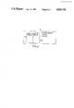
- FIG. 1is a block diagram of the oximeter of the present invention
- FIG. 1ais a schematic illustration showing a detail of the microprocessor control unit shown in FIG. 1;
- FIG. 2aillustrates the voltage waveform output from the detector preamplifier, as the latter responds to red and infrared LED pulses;
- FIG. 2billustrates the voltage waveform from one of the red or infrared filters, in which the physiological pulsatile and non-pulsatile components are shown;
- FIG. 2cis a schematic view of the operation of the remultiplexer control, the digital-to-analog converter, and the sample-and-hold circuit;
- FIG. 3ais a graphical representation of the red and infrared waveforms after these waveforms have been filtered and amplified;
- FIG. 3bis a graphical representation of the red and infrared waveforms at a time later than that illustrated in FIG. 3a, after a portion of the non-pulsatile component of these waveforms has been subtracted;
- FIG. 3cis a graphical representation of the red and infrared waveforms at a time later than that illustrated in FIG. 3b, after the red and infrared waveforms, absent a large portion of the non-pulsatile component, have been amplified;
- FIG. 4is a schematic illustration of the drift compensation of the present invention.
- FIG. 5is a schematic illustration of the method of compensating for electronic noise generated by the electrical components comprising the oximeter of the present invention.
- the detailed description of the inventionis divided into three sections.
- the firstdescribes the structure of the preferred embodiment of the device, with reference to the block diagram of FIG. 1.
- the seconddescribes how the illustrated structure enables the oximeter of the invention to operate on a greater variety of patients, with a wider range of pulse strengths, than is possible with conventional devices, and how, therefore, the present invention is a more practical device.
- the thirddescribes how the processing of the signals increases the accuracy of the measurement of percent saturation, as compared with that obtained with previous devices.
- FIG. 1illustrates an oximeter generally referred to by reference numeral 10. It should be noted that, although the embodiment of the present invention described herein is a device for measuring the oxygen content of the blood, it is within the scope of the present invention to measure the concentration of other blood constituents.
- Oximeter 10comprises a light emitting section 12, a light sensing section comprising a photodetector 14, a signal converting section including a current-to-voltage preamp 16, a demultiplexing and filtering section 18, a remultiplexing section 20, a gain and subtraction section 22, a digitizing section 24, and a processing, computing, and display section comprising a microprocessor subsystem control system 26, which also assists digitizing section 24.
- Light emitting section 12comprises a digital Sequencer control device 30 for controlling LED drivers 32 which alternately drive two red LED's 34 and an infrared LED 36 at a 2.5 kHz rate to produce alternating red and infrared pulses having well-defined pulse widths.
- Sequencer 30is actuated by microprocessor subsystem 26.
- Sequencer 30alternately pulses the LED's so that the circuitry detecting and processing the light transmitted through a portion of the patient's body can discriminate the photodetector's response to red light from its response to infrared light.
- a fingeris illustrated in FIG. 1 as the portion of the body through which light is transmitted and measured, and the following description is in terms of a finger, it is to be understood that the preferred embodiment can be used to calculate the oxygen saturation of the blood by transmitting light through other portions of the body, such as an ear lobe, a toe, etc.
- a finite-state machine(implemented in the preferred embodiment by digital sequencer 30), rather than microprocessor subsystem 26, provides the immediate control for the LED drive 32. This is because timing signals from a microprocessor subsystem cannot be generated as precisely as with a digital sequencer. Output signals from digital sequencers are externally latched to the devices to be controlled (such as the LED's) at precise intervals defined by a single clock signal. Sharp, well defined red and infrared pulses are generated by the red and infrared LED's under control of the digital sequencer 30. If controlled directly by the microprocessor subsystem 26, the pulse widths and times would exhibit much greater amounts of jitter noise.
- the digital sequencer 30also controls a demultiplexer 38, as is dicsussed below.
- LED's 34 and 36are positioned on one side of the patient s finger.
- a single photodetector 14is positioned on the other side of the finger to detect the light, both red and infrared, transmitted through the finger and to produce an electrical signal proportional to the amount of light received. Because LED's 34 and 36 are driven by sequencer 30 in such a manner as to produce alternating pulses of red and infrared light, odd-numbered pulses produced by photodetector 14 carry information about the finger's absorption of red light, and the even-numbered pulses carry information about the finger's absorption of infrared light (see FIG. 2a).
- Use of a single photodetector to sense both the red and the infrared lightfurther guarantees that the detected light of both wavelengths has passed light through the same portion of the finger. This ensures that the two signals produced by processing the response to the red and the infrared light are correlated, in the sense that variations between them are related to the different characteristic attenuations of red and infrared light by a single group of tissues and are not due to the two wavelengths of light passing through two different groups of tissues.
- two red LED's 34instead of only one, can be used.
- the two red LED'sare on either side of the single infrared LED 36 and are operated in unison. Thus, there is a plane of symmetry in the illumination pattern.
- the photodetector 14produces an electrical signal in the form of a current.
- a current-to-voltage preamp 16converts the current signal to a voltage signal.
- the amount of light impinging upon the photodetector, and therefore the magnitude of the current signalis a function of the width of the part of the body chosen for examination. Since these can vary widely, it is another function of the preamp 16 to compensate coarsely for the intensity of light reaching the photodetector 14. This is accomplished by designing the preamp 16 to have several, e.g., three, different transimpedance (current-to-voltage) gains. These gains are controlled by the finite-state machine implemented by the digital sequencer 30 and can preferably be chosen independently for the red and the infrared pulses. In addition, a gain of zero can be chosen, to permit calibration of the system as described below at the end of the section entitled "Structure of the Preferred Embodiment". The appropriate gains are transmitted to the digital sequencer from the microprocessor subsystem 26.
- the signals output from preamp 16are multiplexed signals in which signals representing the transmission of red light through the finger alternate in time with signals representing the transmission of infrared light through the finger.
- This multiplexed signalis now demultiplexed to filter the red and the infrared signals separately. This is done because, in order for the filters adequately to reduce high frequency noise components, the time constant of the filters must be substantially longer than the multiplexing rate. If sent through a single filter with this longer time constant, the information inherent in the red signal would be mixed with that from the infrared signal, thus corrupting the measurement.
- the output from preamp 16is demultiplexed by a demultiplexer 38.
- Demultiplexer 38produces two separate signals, one representing red light transmitted through the finger and one representing the infrared light so transmitted.
- the two signalsare transmitted through two parallel, frequency-matched low pass filters 40 and 42. Frequency matching ensures that the shape of the physiological signals passing through the two filters is processed in substantially the same manner.
- These signalsare generated by the demultiplexer in the following way.
- the signalis switched into the channel with the low pass filter 40, and during the period of time that the multiplexed signal from photodetector 14 represents transmission of infrared light, the signal is switched into the channel with the low pass filter 42,
- a portion of the signal being switched into the low pass filters 40 and 42is an offset signal not associated with transmission of light fom LED's 34 and 36 through the finger. This includes ambient light falling on the photodetector 14 and electronic offset voltags generated by the photodetector 14, preamp 16 and demultiplexer 38 (see FIG. 2a).
- the demultiplexercauses this portion of the signal to be subtracted, by creating a signal which is the negative of the multiplexed signal (i.e., by amplifying by -1) from photodetector 14 and switching it into both the filters 40 and 42 during the portions of time that none of the LED's 34 and 36 are transmitting light (i.e., in between the red and infrared pulses). During these portions of time this signal is the exact negative of the offset signal. Thus, if the negative signal is switched into the two filters 40 and 42 for a period of time equal in duration to the period of time when the signal from photodetector 14 is switched into the filters 40 and 42, the averaging effect of the low pass filters will cause the offset signal to be exactly cancelled out.
- Demultiplexer 38is controlled by digital sequencer 30 so that the process of demultiplexing is synchronous with the alternating pulses of red and infrared light, their generation being controlled by the digital sequencer 30 as well.
- the sequence of events controlled by the finite-state machinecan be altered to allow the function of the entire instrument to be tested in actual operation (as opposed to a "bench test" of the electronics, in the factory, for example).
- a control signal from the microprocessor subsystem 26alerts the finite-state machine implemented by the digital sequencer 30 to pulse only the infrared LED, both during the time when the red LED or LED's would be pulsed and when the infrared LED would normally be pulsed.
- the two signals demultiplexed into the filters 40 and 42are identical.
- This aspect of the inventioncan be used in any pulsatile oximeter in which radiation of two or more wavelengths emitted seriatim through the portion of the body being monitored and the level of the transmission of which through the body is monitored by a single detector element (which currently includes all pulsatile oximeters known to the present inventor).
- a single detector elementwhich currently includes all pulsatile oximeters known to the present inventor. The only requirement for this is that the signals which control the LED's be distinct from those which control the demultiplexing of the signals into the parallel processing channels.
- a grounded translucent conductive window (43)is preferably placed between photodetector 14 and the finger to prevent interfering current from passing through the electrosurgical unit, through the finger to photodetector 14 and into the current-voltage preamp 16.
- FIG. 2bAn illustration of one of the red and infrared waveforms after the demultiplexed LED pulse signal is transmitted through the low pass filters is shown in FIG. 2b.
- This waveformcalled a plethysmographic waveform, has a pulsatile nature which should be noted. These pulses represent the rising and falling in the intensity of the light transmitted through the finger.
- the intensity of light transmitted through the bodyis a function of the amount of blood the light encounters as it passes through.
- the amount of bloodvaries with the heartbeat of the patient. Each time the heart pulses arterial blood through the blood vessels of the finger, the amount of blood increases, reducing the amount of transmitted light. As the blood vessels relax between heartbeats, the amount of blood is reduced and the amount of light transmitted through the finger returns to its previous level.
- the electronic instrumentation discussed belowprocesses this varying portion of the photodetector's output, which is therefore sensitive only to the attenuation properties of arterial blood and not to other portions of the finger, such as skin, bones,
- the pulses in the plethysmographic waveform in the red channelwill be shaped identically to those in the infrared channel. Only their size and the slowly-varying voltage on which they sit will be different.
- Remultiplexer 20preferably operates at a 240 Hz rate so as to sample the two channels and the two plethyscographic waveforms at a 120 Hz rate. This rate ensures adequate resolution of the high-frequency protions of the waveform.
- the signalsAfter being remultiplexed, the signals are transmitted to gain and subtraction section 22 where the signals are amplified by a first programmable microprocessor subsystem controlled gain 44. A portion of the signals is then subtracted by a subtraction circuit 46 under the control of microprocessor subsystem 26, and the remaining portion of each signal is then amplified by a second programmable microprocessor subsystem controlled gain 48.
- the signalsare shown in FIG. 3a as a series of dots to indicate that they are multiplexed into a single channel. If that figure were drawn on as expanded time scale, it would be apparent that the signal alternates between the two plethysmographic waveforms at the 240 Hz multiplexing rate. It should be understood that all subsequent processing of the remultiplexed signal is actually a hybrid of two independent signal processing procedures. All gains and voltage subtractions to be described are chosen independently for the red and the infrared signals and as such are changed back and forth at the 240 Hz multiplex rate.
- Gain 44independently amplifies the red and infrared portions of the remultiplexed signal by one of several gains, in the preferred embodiment: 1, 2, or 4.
- the ability of gain 44 to apply one of three discrete gains to the remultiplexed signalallows the red and infrared signals to be maintained within a range that enables them to be digitized with a resolution of at least eight bits.
- preamp 16also amplifies the signal by one of several (again, preferably three) discrete gains, nine (if the number of values assumable by gain 44 is three for each channel) possible discrete gains can be applied independently to the red and the infrared signals. This permits oximeter 10 to measure the oxygen content of patients having a wide range of finger thicknesses. A more complete description of this aspect of the system is presented below in the section entitled "Advantages of Structure of the Preferred Embodiment”.
- DACdigital-to-analog converter
- the manner in which this code is chosenis also described in the section entitled "Advantages of Structure".
- the value of the codeis stored by the microprocessor subsystem for use in later calculation. As shown in FIG. 3b, the amount subtracted off is selected so as to leave a small component of non pulsatile signal. This is to ensure that the complete pulsatile component is retained for further processing.
- Gain 48amplifies the pulsatile component of the signal by one of several distinct gains, preferably 5, 22.4, or 100. Because a majority of the non-pulsatile component has been subtracted from the signal, gain 48 is able to amplify the pulsatile component sufficiently so that even very weak pulses yield information on the percentage of oxygen saturation of the blood, while still keeping the amplified signal within the sensitivity range of the digitizing circuitry which follows.
- digitization section 24digitizes the signal, which is transmitted to the microprocessor subsystem for processing.
- Digitization section 24comprises a sample-and-hold circuit 52, a comparator 54, and DAC 50. Digitization is performed under microprocessor subsystem control of DAC 50 in conjunction with sample and hold circuit 52 and comparator 50.
- Microprocessor subsystem 26actuates DAC 50 to send an analog signal to comparator 54.
- Comparator 54compares this signal from DAC 50 with the subtracted and amplified pulsatile red or infrared signal outputted from sample and hold circuit 52.
- oximeter 10can digitize and discriminate changes in the voltage of the pulsatile component lower than 0.1% of the total signal.
- DAC 50therefore, performs two functions: it subtracts a portion of the non-pulsatile component and it assists in digitizing the remaining pulsatile component. Using the DAC to perform these two functions reduces the amount of hardware needed to process the signals.
- Remultiplexer 20, DAC 50, and sample-and-hold circuit 52function together so that DAC 50 can both subtract and assist in digitizing the electrical signals as illustrated in FIG. 2c. More specifically, every 1/240th of a second the control functions of device 10 are changed to process the red or the infrared portion of the signal. For example, every 1/240th of a second remultiplexer 20 switches from sampling the red portion of the signals to sampling the infrared portions of the signals, or vice versa, as illustrated at the top of FIG. 2c.
- the amount of amplification provided to the signal by preamp 16, gain 44, and gain 48 and the digital input code transmitted to DAC 50 for use in the subtraction circuit 46is changed from the values associated with the red waveform to the values associated with the infrared waveform, or vice-versa, under the control of the microprocessor subsystem so that the red and infrared portions of the signals are independently amplified as needed.
- DAC 50participates in digitizing the infrared waveform that immediately preceded the red waveform now being sampled. This is accomplished relatively quickly so that during the vast majority of the 1/240th of a second during which remultiplexer 20 samples the red waveform, DAC 50 subtracts a large portion of the non-pulsatile component of the red waveform, leaving the pulsatile component of the red waveform and a small non-pulsatile buffer.
- the subtracted signalsare digitized they are stored in the memory of microprocessor subsystem 26, which computes the oxygen saturation of the blood as a function of the digitized, subtracted, and amplified pulsatile component of the signals and as a function of the stored subtracted portion of the non pulsatile component of the signals, as is described below, in the section entitled "Processing of the Signals by the Microprocessor Subsystem".
- the preamp 16can be made to have a gain of zero to allow offsets in the circuitry to be calibrated out. This calibration process is now described.
- the calibrationoccurs when the unit is first turned on before any signals are processed. Offsets determined by the calibration are stored in the microprocessor subsystem's memory for access by the microprocessor subsystem when processing the signals during normal operation.
- Offsetsare introduced by the standard signal processing components which are used to implement the functional blocks described above.
- the offsetswill be a function of the gain settings 44 and 48.
- the offsetswill be different for the processing of the red signal and the processing of the infrared signal since the red signal is processed by filter 40 and the infrared signal is processed by filter 42.
- the microprocessor subsystemmust determine and store offsets for all 18 combinations of gain 44, gain 48 and channel (red vs. infrared).
- V offset-subtractis used to modify the value of V subtract in calculations by the microprocessor subsystem
- V offset-digitizeis used to modify the value of V digitize in these calculations.
- the digitizing section 24should sense zero voltage when DAC 50 is set by code generated by the microprocessor subsystem 26 to subtract off zero volts after amplifier gain 44.
- the offset pair V offset-subtract and V offset-digitizerepresent the deviation from this ideal situation.
- V offset-digitizerepresents the voltage digitized when V offset-subtract is subtracted from the signal at the output of gain 44 by DAC 50.
- the offsetsare measured via a successive approximation approach similar to the approach previously described used to perform digitization.
- the microprocessor subsystem 26alternatively increments and decrements the code it sends to the DAC 50 for subtraction off of the voltage output by gain 44, until it reaches a code which results in a suitably small voltage signal being input to the digitizer 24 (less than 1 volt). Since the DAC 50 can only generate discrete voltages (1 of 4096 values for a 12-bit DAC), a code which could generate a voltage at the input to the digitizer much closer to zero volts may not exist.
- the code at the input to the DAC which generates the subtraction voltageis then stored as V offset-subtract and the resulting digitized code is stored as V offset-digitize .
- One of the objects of the inventionis to allow the oximeter to be used on a wide variety of patients under a great variety of conditions. How this is achieved can be better understood by referring to the plethysmographic signal in FIG. 2b.
- the height of the total signal levelcorresponds to the amount of light which is transmitte through the finger (or other body part) and is thus a function of the thickness of the finger.
- the amount of light coming throughcan vary by a factor of more than 100.
- the percentage modulation(ratio of the size of the plethysmographic pulse to the total signal level) is a function of the strength of the patient's pulse: the stronger the pulse, the more extra blood will be pumped into the blood vessels during the heart beat, and therefore the greater will be the percentage modulation. This can also vary greatly among patients.
- the full height of the pulsatile componentshould usually be at least approximately fifty times as large as the resolution of the digitizing system. (At higher saturation levels, where the accuracy of the computation of %SaO2 is less. sensitive to errors in measuring the plethysmagraphic signal, the height can be as small as about twenty-five times the resolution of the digitizing system).
- the gains 16, 44 and 48are preferably chosen to maintain the height of the pulsatile component one-hundred times as large as the resolution of the digitizing subsystem 24.
- the pulse heightshould be no more than one third to one-fourth the full scale range of the digitizing subsystem. This is because drifts in the total signal level (discussed in more detail in the next section, "Processing of Signals by the Microprocessor Subsystem") tend to push the pulsatile signal out of the digitizing range.
- the oximeterhas more time to react in order to maintain the signal within the range of the digitization subsystem 24 by changing the voltage substracted off by the DAC 50.
- the deviceFor an eight-volt full scale range of a twelve-bit digitizing subsystem, corresponding approximately to a two-millivolt resolution, the device should preferably maintain the pulsa-height within a range of from 200 millivolts to between 2 and 2.5 volts.
- Tables 1 and 2indicate how the gains 16 and 44 maintain the output of gain 44 between 2 volts and 5 volts for detector current signal levels between 0.25 microamp (very thick finger and dim LED) and 40 microamps (very thin finger and bright LED).
- gain 48can be chosen to maintain a pulsatile height between 200 mullivolts and 2.5 volts (see Table 3).
- the oximetercan operate on patients who produce signals outside these preferable ranges, because as discussed above, the operating ranges described are not absolute physical limits. Also, the range limits described in Tables 1, 2 and 3 are for worst case combinations of total signal current level and modulation levels. For example, a pulse much smaller than 0.1% modulation can easily be measured if the total current signal produced by the detector is much greater than 0.25 microamp.
- Tables 1, 2 and 3are for worst case combinations of total signal current level and modulation levels. For example, a pulse much smaller than 0.1% modulation can easily be measured if the total current signal produced by the detector is much greater than 0.25 microamp.
- the tablesindicate the advantage of using multiple gain settings for greatly expanding the operating range of a pulsatile oximeter, which is important in a practical device.
- gain-changing circuitryis a significant improvement over the prior art.
- the devices shown in previous patentssuch as the four referred to above maintain a single fixed gain for either the plethysmographic portion of the signal, or both that portion and the entire signal. This limits their usefulness to patients with relatively strong pulses.
- the oximetercan readjust itself automatically by altering the gains 44 and 48 without losing any digital samples of the plethysmographic waveforms, and thus without interrupting the flow of information to the user.
- Part of the readjustmentis changing the amount of signal subtracted off by the DAC 50, that is, changing the digital input data. In general, this is necessary when gain 16, 44 or 48 is changed, but it is also required due to changes in the amount of light transmitted (see the description below of the processing of the digitized signal).
- the digital input codeis altered to maintain the plethysmographic pulse within the range of the digitizer in the face of these slow changes.
- the first expressionrelates the digitized signal ("V* digitize ”) in terms of the voltage of the transmitted signal before gain 44 ("V* signal "), the two gains 44 and 48 and the voltage subtracted off of the signal by the DAC 50 ("V* subtract ”):
- V* subtract and V* digitizedenote that these are values modified by one of eighteen pairs of values V offset-digitizer and V offset-subtract described in the previous section.
- V subtractis the actual value subtracted by the DAC 50 and V digitize is the actual value produced by the digitization section 24.
- So equation (2)is used to reconstruct the size of the total signal at the input to gain 44 from its component parts. If we need to chanqe a gain, and wish to maintain the plethysmographic pulse in the same relative location within the digitization range, we use the third of the above equations to determine the new digital code for the DAC 50 to use to generate a signal to be subtracted from the outfit of gain 44.
- Equation (3)To change a gain while maintaining the plethysmographic pulse in the same relative location within the digitization range, we use equation (3) to determine the new digital code for the DAC 50 to use to generate a signal to be subtracted from the output of gain 44.
- the value of V signal determined from equation (2)is used in evaluating equation (3), and the most recent digitized value is inserted as the value V digitize in the latter equation. Since successive samples are taken at a 120 Hz rate for each of the two channels (the red and the infrared), the physiological signal can be assumed to be constant over the period between the two successive samples.
- Equation (3)is also used to determine a new digital code if we want to shift the plethysmographic pulse signal within the digitization range when slow changes in the transmitted level threaten to push the pulse outside of the range. In this case the amount by which we wish to shift the pulse is added to the most recent digitized value and inserted into V digitize .
- Gain 16being before the low passed filters 40 and 42, cannot be altered without affecting continuous operation of the unit.
- the filtersrequire about 25 to 30 ms to settle fully to the new voltage level, and thus a number of 240-Hz samples are lost.
- the unitrejects the next few pulses while it determines from scratch the correct amount to be subtracted off by the DAC 50.
- the approach to finding the correct digital code for the DAC 50is also used to lock onto the pulse initially, and involves a successive approximation routine similar to that used to perform the digitization period.
- gains 16 and 44are designed such that the combination of them exhibits hysteresis.
- the electronic gainsare held constant before the filters 40, 42 so that the filters respond only to the true plethysmographic signal.
- V R (t) and V I (t)are the voltages generated by the detector and the signal processing circuitry for the red and infrared wavelengths respectively, and
- d(t)represents the average path length through the arterial vascular bed that the incident light must travel to reach the detector; this path length increases when the volume of arterial blood in the finger increases;
- nthe concentration of hemoglobin within that bed
- c Orepresents the fraction of oxyhemoglobin (%Sa0 2 );
- ⁇ Orepresents a characteristic absorption coefficient of oxyhemoglobin (one for red light, one for infrared);
- ⁇ Hrepresents a characteristic absorption coefficient of hemoglobin (one for red light, one for infrared).
- Each signalhas three components, as follows.
- Very Slowly Changing Component (A)--Treating this as a constant componenthas an insignificant effect on the accuracy of the measurement.
- This componentis a function of the illumination level, assorption by all the components of the finger except the blood, the detector sensitivity, and the gain of the signal processing circuitry.
- Pulsatile Component[exp(-d(t)nc O ⁇ O +(1-c O ) ⁇ H ]--This is the component which is sensitive only to the characteristics of the arterial blood. The signal is caused by the changes in volume induced by the physiological pulse. Its size is dependent upon the %SaO 2 .
- K(t)The modeling of the slowly changing component, K(t), is one of the features which distinguish the present invention from conventional oximeters.
- the complicated exponentialcan be reduced to its first order approximation.
- Equation (12)is based on the first order linear approximation for exp (-x).
- the size of the physiological pulsebecomes greater than 1% of the total signal (it can be as large as 10%) then it becomes necessary to calculate the second order term from the Taylor series approximation:
- the absorption characteristicsmay depend upon the body portion used.
- several look-up tablesare provided, one table 27b for when the measurements are made using the patient's finger, one table 27b for when they are made using the earlobe, etc.
- Z in equation (13)is a quotient of two individual quotients.
- the quotient in the numeratoris associated with the red waveform and the quotient in the denominator is associated with the infrared waveform.
- Each of these individual quotientsrelates the change in the plethysmographic signal due to the physiological pulse (and the slowly varying component K(t)) to the average level of the two components.
- the two times t 1 and t 2are chosen to correspond to the beginning of the systolic phase, when the voltage of the waveform is at a maximum, and the end of the systolic phase, when the voltage of the pulse is at a minimum, then the individual quotients would correspond directly to the percentage modulation.
- the shapes of the pulsatile components of the red and infrared signalsboth reflect the change in the volume of blood in the monitored area during the physiological pulse, and are therefore identical, we can choose any combination of voltage differences from any number of pairs of points as a measure of size, as long as we choose the identical combinations from corresponding pairs from the two waveforms. That is, we can use any arbitrary measure of the size of the pulse.
- each piece of red data used in the calculations of sizeis actually the average of two consecutive values.
- interpolationestimates what the red value would have been if digitized simultaneously with the corresponding infrared velue, and points are taken from identical portions of the identically shaped waveforms.
- the measure of size we choose to employis preferably one which minimizes the electronic noise inherent in the various components of the oximeter 10.
- the preferred measureinvolves the use of multiple pairs of data points V(t 1 ), V(t 2 ) so that noise can be reduced by averaging contributions from these multiple pairs.
- FIG. 5shows an amplified version of how the voltage of the pulsatile component of the red waveform changes over time, with various voltages at different times identified by points on the waveform.
- Various points on the systolic portion of the waveformare paired up only with other points on the systolic portion of the waveform to produce pairs of points whose difference is greater than three-fourths of the full pulse size.
- the pairs satisfying this criterionare labeled data point pairs 1, 2, and 3. These pairs are used to compute the value of the pulsatile component of the red and infrared waveforms.
- the full pulse sizerepresents the difference between the minimum and maximum values of the voltage of the red and infrared waveforms.
- the points in these pairsare seguentially digitized points.
- the time difference associated with each pairis equal.
- the average of the voltage differences from the pairs from the red and infrared waveformsis used as the measure of the pulse size--that is, they are used for the two individual numerators in the expression for Z, from equation (13). In the same way, the average of the average values is used for the two denominators in the computation of Z.
- Pairs of pointsare only used from the systolic portion of the waveform because the rate of change of the signal is greater in that portion of the waveforms and thus the influence of slowly varying component K(t) described below is reduced.
- K(t)which represents the average amount of blood in that portion of the body illuminated by LED's 34 and 36, as noted above, is not constant, but changes slowly. If K(t) is treated as a constant, a significant source of error is introduced into the computation of the oxygen saturation of the blood. This source of error occurs because a component of the computed size of the pulsatile signal will actually be due to the apparent drift of the pulse caused by the variation of K(t), as illustrated in FIG. 4. In order to compensate for this slowly changing component, a compensation algorithm is used by microprocessor subsystem 26 which assumes that the drift is linear from one pulse to the next.
- the algorithmpermits microprocessor subsystem 26 to determine the approximate drift rate for each pair of pulses by subtracting the average of the maximum and minimum values of the voltages of the latter pulse in the pair of pulses (called the (n+1)st pulse) from the average of the maximum and minimum values for the voltages of the prior pulse in that same pair of pulses (called the nth pulse), and dividing this difference by the period of time between the minimum values of the (n+1)st pulse and the nth pulse.
- microprocessor subsystem 26corrects the computed difference of each pair of voltages in the systolic portion of the (n+1)st pulse (that is, the pair of points on the waveform used earlier to compute the size of each pulse) by subtracting off from each of these differences, the product of the drift rate times the period of time elapsed between the points in each pair. However, because each pair has the same elapsed time between the two points in the pair, the product can be subtracted directly from the average value of the size computed above.
- FIG. 4illustrates two pulses of either the red or infrared waveform, in which the voltage of the waveforms (the vertical axis) varies with time (the horizontal axis).
- the systolic or left hand portion of each pulseis of shorter duration than the diastolic portion of each pulse.
- the voltage of the first or nth pulse, i.e. the pulse on the left,has an average level called the "last level” and the voltage of the second or (n+1)st pulse, i.e., the pulse on the right, has an average level called "level".
- Points on the upper half of the systolic portion of the second or (n +1)st pulseare labeled x 1 , x 2 , x 3 , x 4 , and x 5
- points on the lower half of the systolic portion of the second or (n+1)st pulseare labeled y 1 , y 2 , y 3 , y 4 , and y 5 .
- points on the upper half and the lower half of the systolic portion of the second pulseare paired together so that the period of time elapsing between points in each pair are the same, i.e., ⁇ T is the same for each pair of points.
- the difference between pairs of pointsare added and the resulting sum is divided by the number of pairs, i.e.: ##EQU8##
- the drift experienced by the second pulse over interval ⁇ Tis approximately: ##EQU9## where "last level” and “level” are the values of expression (13) for the nth and (n+1)st pulse, respectively, and where “last pulse up+pulse down” represents the elapsed time between the minimum value of the voltage of the first or nth pulse and the minimum value for the voltage of the second or (n+1)st pulse.
- Microprocessor subsystem 26compensates for the drift K(t) over time ⁇ T in the non pulsatile component by using the following modified version of expression (15) to determine the size of the pulse: ##EQU10##
- the quotient of Zis determined.
- Zis a quotient of two quotients, each of which is indicative of a percentage modulation of either a red or an infrared signal.
- any scale factors applied to the numerator of each quotientis also applied to the denominator of each quotient. If we use the expression (17) to evaluate the numerators of the two individual quotients, we recognize that each data point in that expression has been multiplied by gains 16, 44 and 48. And if we use eguation (2) to evaluate the individual denominators, we recognize that the result reflects a signal which has been multiplied by the value of gain 16 only.
- Equation (14)which expresses the desired concentration, c O , in terms of the signals detected and the constants ⁇ HR , ⁇ HI , ⁇ OR and Z.
- the scattering of light from red blood cell interfacesperturbs the effective coefficients.
- the best way to correlate the red/infrared ratio to the %SaO 2is by empirical experiment on a large number of subjects whose %SaO 2 is determined by other methods.
Landscapes
- Health & Medical Sciences (AREA)
- Physics & Mathematics (AREA)
- Life Sciences & Earth Sciences (AREA)
- Biomedical Technology (AREA)
- Medical Informatics (AREA)
- Biophysics (AREA)
- Pathology (AREA)
- Engineering & Computer Science (AREA)
- Spectroscopy & Molecular Physics (AREA)
- Heart & Thoracic Surgery (AREA)
- Optics & Photonics (AREA)
- Molecular Biology (AREA)
- Surgery (AREA)
- Animal Behavior & Ethology (AREA)
- General Health & Medical Sciences (AREA)
- Public Health (AREA)
- Veterinary Medicine (AREA)
- Measurement Of The Respiration, Hearing Ability, Form, And Blood Characteristics Of Living Organisms (AREA)
Abstract
Description
V*.sub.digitize =[(V.sub.signal ×gain 44)-V*.sub.subtract ]×gain 48 (Equation 1)
V.sub.signal =[V*.sub.subtract +(V*.sub.digitize /gain 48)]/gain 44 (Equation 2)
V*.sub.subtract =(V.sub.signal ×gain 44)-(V*.sub.digitize)/gain 48 (Equation 3)
V*.sub.subtract =V.sub.subtract -V.sub.offset-subtract (Equation 4)
V*.sub.digitize =V*.sub.digitize -V.sub.offset-digitize (Equation 5)
V.sub.R (t)=A.sub.R K.sub.R (t)exp[-d(t)n(c.sub.O α.sub.OR +(1-c.sub.O)α.sub.HR)] (Equation 6a)
V.sub.I (t)=A.sub.I K.sub.I (t)exp[-d(t)n(c.sub.O α.sub.OI +(1-c.sub.O)α.sub.HR)] (Equation 6b)
exp(-x)=1-x(for x<<1).
V.sub.R (t)=A.sub.R K.sub.R (t)[1-d(t)n(c.sub.O α.sub.OR +(1-c.sub.O)α.sub.HR)] (Equation 7a)
V.sub.I (t)=A.sub.I K.sub.I (t)[1-d(t)n(c.sub.O α.sub.OI +(1-c.sub.O)α.sub.HI)] (Equation 7b)
exp(-x)=1-x+x.sup.2 /2
TABLE 1 ______________________________________GAIN 16GAIN 44 Gain # Transimpedance Gain Gain # Voltage Gain ______________________________________ (0) 125 kΩ (0) × 1 (1) 500 kΩ (1) × 2 (2) 2 MΩ (2) × 4 ______________________________________
TABLE 2 ______________________________________ Composite Transimpedance Gain (Gain 16) × (Gain 44) (For Preferable 2V-5V Output of Gain Stage 44) Composite (Gain (Gain Transimped-Gain Photocurrent 16#) 44#) ance Gain Change ______________________________________ 16 μA->40 μA (0) (0) 125 kΩ 8μA-20 μA (0) (1) 250 kΩ 4 μA-10 μA (0) (2) 500 kΩ 4 μA-10 μA (1) (0) 500 kΩ 2 μA-5 μA (1) (1) 1MΩ 1 μA-25 μA (1) (2) 2MΩ 1 μA-2.5 μA (2) (0) 2 MΩ 0.50 μA-1.25 μA (2) (1) 4 MΩ <0.25 μA-0.65 μA (2) (2) 8 MΩ ______________________________________
TABLE 3 ______________________________________ BEFORE USE GAIN AFTER ______________________________________ <2 mV (0) × 100 <200 mV Preferable 2 mV-20 mV (0) × 100 200 mV-2 V Range 8.9 mV-89 mV (1) × 22.4 200 mV-2V 40 mV-500 mV (2) × 5 200 mV-2.5 V >500 mV (2) × 5 >2.5 V ______________________________________ Notes: *Signal out of gain stage (44) is between 2V and 5V *(0.1%)(2V) = 2 mV can be < plethysmographic signal before gain (48), which can be < (10%)(5V) = 500 mV *200 mV should preferably be < plethysmographic signal after gain (48), which should preferably be < 2-2.5 V
Claims (60)
[(1/2)(Max+Min).sub.1 -(1/2)(Max+Min).sub.2 ]/ΔT
[(1/2)(Max+Min).sub.n -(1/2)(Max+Min).sub.n+1 ]/ ΔT
[(1/2)(Max+Min).sub.1 (1/2)(Max+Min).sub.2 ]/ΔT
[(1/2)(Max+Min).sub.n -(1/2)(Max+Min).sub.n+1 ]/ΔT
[(1/2)(Max+Min)-(1/2)(Max+Min).sub.2 ]/ΔT
[(1/2)(Max+Min).sub.n -(1/2)(Max+Min).sub.n+1 ]/ΔT
[(1/2)(Max+Min).sub.1 -(1/2)(Max+Min).sub.2 ]/ΔT
[(1/2)(Max+Min).sub.n -(1/2)(Max+Min).sub.n+1 ]/ΔT
Priority Applications (1)
| Application Number | Priority Date | Filing Date | Title |
|---|---|---|---|
| US07/103,713US4819752A (en) | 1987-10-02 | 1987-10-02 | Blood constituent measuring device and method |
Applications Claiming Priority (1)
| Application Number | Priority Date | Filing Date | Title |
|---|---|---|---|
| US07/103,713US4819752A (en) | 1987-10-02 | 1987-10-02 | Blood constituent measuring device and method |
Publications (1)
| Publication Number | Publication Date |
|---|---|
| US4819752Atrue US4819752A (en) | 1989-04-11 |
Family
ID=22296665
Family Applications (1)
| Application Number | Title | Priority Date | Filing Date |
|---|---|---|---|
| US07/103,713Expired - LifetimeUS4819752A (en) | 1987-10-02 | 1987-10-02 | Blood constituent measuring device and method |
Country Status (1)
| Country | Link |
|---|---|
| US (1) | US4819752A (en) |
Cited By (208)
| Publication number | Priority date | Publication date | Assignee | Title |
|---|---|---|---|---|
| WO1990009003A1 (en)* | 1989-02-06 | 1990-08-09 | Nim, Incorporated | Phase modulated spectrophotometry |
| US4948248A (en)* | 1988-07-22 | 1990-08-14 | Invivo Research Inc. | Blood constituent measuring device and method |
| US5014713A (en)* | 1989-12-05 | 1991-05-14 | Tarris Enterprises, Inc. | Method and apparatus for measuring thickness of fat using infrared light |
| US5057695A (en)* | 1988-12-19 | 1991-10-15 | Otsuka Electronics Co., Ltd. | Method of and apparatus for measuring the inside information of substance with the use of light scattering |
| WO1991015991A1 (en)* | 1990-04-19 | 1991-10-31 | Worcester Polytechnic Institute | Method and apparatus for monitoring blood analytes noninvasively by pulsatile photoplethysmography |
| US5078136A (en)* | 1988-03-30 | 1992-01-07 | Nellcor Incorporated | Method and apparatus for calculating arterial oxygen saturation based plethysmographs including transients |
| US5101271A (en)* | 1990-03-30 | 1992-03-31 | Hughes Aircraft Company | Image restoration and faulty sensor detection and compensation system and process |
| US5144951A (en)* | 1989-03-09 | 1992-09-08 | Macttor Co., Ltd. | Apparatus for measuring biopermeability |
| WO1992021280A1 (en)* | 1991-06-06 | 1992-12-10 | Somanetics Corporation | Patient headpiece for optical cerebral oximeter |
| US5193544A (en)* | 1991-01-31 | 1993-03-16 | Board Of Trustees Of The Leland Stanford Junior University | System for conveying gases from and to a subject's trachea and for measuring physiological parameters in vivo |
| WO1993013395A3 (en)* | 1991-12-24 | 1993-08-05 | Insite Technologies Inc | Path constrained spectrophotometer |
| US5239185A (en)* | 1990-06-22 | 1993-08-24 | Hitachi, Ltd. | Method and equipment for measuring absorptance of light scattering materials using plural wavelengths of light |
| US5263244A (en)* | 1992-04-17 | 1993-11-23 | Gould Inc. | Method of making a flexible printed circuit sensor assembly for detecting optical pulses |
| US5285783A (en)* | 1990-02-15 | 1994-02-15 | Hewlett-Packard Company | Sensor, apparatus and method for non-invasive measurement of oxygen saturation |
| WO1994003102A1 (en)* | 1992-08-01 | 1994-02-17 | University College Of Swansea | Optical monitor (oximeter, etc.) with motion artefact suppression |
| WO1994004070A1 (en)* | 1992-08-14 | 1994-03-03 | Angiomedics Ii, Incorporated | Non-invasive blood analysis by near infrared absorption measurements using two closely spaced wavelengths |
| US5309912A (en)* | 1991-11-08 | 1994-05-10 | The United States Of America As Represented By The Secretary Of The Department Of Health And Human Services | Multidimensional imaging using a single point detector for a phase encoded modulated optical carrier |
| WO1994009698A1 (en)* | 1992-10-23 | 1994-05-11 | Nellcor Incorporated | Method and apparatus for reducing ambient noise effects in electronic monitoring instruments |
| US5313941A (en)* | 1993-01-28 | 1994-05-24 | Braig James R | Noninvasive pulsed infrared spectrophotometer |
| US5331958A (en)* | 1992-03-31 | 1994-07-26 | University Of Manitoba | Spectrophotometric blood analysis |
| EP0611549A1 (en)* | 1993-01-25 | 1994-08-24 | Spacelabs Medical, Inc. | Automatic blood pressure monitor having reduced data loss sensitivity to cuff pressure changes |
| WO1994022362A1 (en)* | 1993-03-31 | 1994-10-13 | Nellcor Incorporated | Electronic processor for pulse oximeters |
| US5379774A (en)* | 1990-10-23 | 1995-01-10 | Sankyo Company Limited | Measurement of arterial elasticity and the frequency characteristic of the compliance of an artery |
| US5385143A (en)* | 1992-02-06 | 1995-01-31 | Nihon Kohden Corporation | Apparatus for measuring predetermined data of living tissue |
| US5408998A (en)* | 1994-03-10 | 1995-04-25 | Ethicon Endo-Surgery | Video based tissue oximetry |
| US5458128A (en)* | 1994-06-17 | 1995-10-17 | Polanyi; Michael | Method and apparatus for noninvasively measuring concentration of a dye in arterial blood |
| US5465714A (en)* | 1993-05-20 | 1995-11-14 | Somanetics Corporation | Electro-optical sensor for spectrophotometric medical devices |
| US5477327A (en)* | 1993-12-15 | 1995-12-19 | Bergman Research Group, Inc. | High resolution low noise optical polarimeter |
| US5482034A (en)* | 1993-05-28 | 1996-01-09 | Somanetics Corporation | Method and apparatus for spectrophotometric cerebral oximetry and the like |
| EP0524083B1 (en)* | 1991-07-17 | 1996-03-06 | EFFETS BIOLOGIQUES DE L'EXERCICE dit GIP EXERCICE, (Groupement d'interêt publique) | Non-invasive method for determining, in vivo, the rate of oxygen saturation of arterial blood and device for carrying out this method |
| US5503148A (en)* | 1994-11-01 | 1996-04-02 | Ohmeda Inc. | System for pulse oximetry SPO2 determination |
| US5515847A (en)* | 1993-01-28 | 1996-05-14 | Optiscan, Inc. | Self-emission noninvasive infrared spectrophotometer |
| US5553615A (en)* | 1994-01-31 | 1996-09-10 | Minnesota Mining And Manufacturing Company | Method and apparatus for noninvasive prediction of hematocrit |
| WO1996039935A1 (en)* | 1995-06-07 | 1996-12-19 | Grable Richard J | Diagnostic tomographic laser imaging apparatus |
| US5588425A (en)* | 1993-05-21 | 1996-12-31 | Nims, Incorporated | Method and apparatus for discriminating between valid and artifactual pulse waveforms in pulse oximetry |
| US5601080A (en)* | 1994-12-28 | 1997-02-11 | Coretech Medical Technologies Corporation | Spectrophotometric blood analysis |
| US5622182A (en)* | 1994-06-27 | 1997-04-22 | Jaffe; Richard A. | System for measuring core body temperature in vivo |
| US5657754A (en)* | 1995-07-10 | 1997-08-19 | Rosencwaig; Allan | Apparatus for non-invasive analyses of biological compounds |
| US5676141A (en)* | 1993-03-31 | 1997-10-14 | Nellcor Puritan Bennett Incorporated | Electronic processor for pulse oximeters |
| US5697367A (en)* | 1994-10-14 | 1997-12-16 | Somanetics Corporation | Specially grounded sensor for clinical spectrophotometric procedures |
| EP0784448A4 (en)* | 1991-03-07 | 1998-01-07 | Masimo Corp | Signal processing apparatus |
| US5800349A (en)* | 1996-10-15 | 1998-09-01 | Nonin Medical, Inc. | Offset pulse oximeter sensor |
| US5803908A (en)* | 1990-10-06 | 1998-09-08 | In-Line Diagnostics Corporation | System for noninvasive hematocrit monitoring |
| US5891024A (en)* | 1997-04-09 | 1999-04-06 | Ohmeda Inc. | Two stage calibration and analyte measurement scheme for spectrophotomeric analysis |
| US5894844A (en)* | 1996-11-07 | 1999-04-20 | Rohrberg; Roderick G. | Three-dimensional floatation-enhanced body examination system |
| US5934277A (en)* | 1991-09-03 | 1999-08-10 | Datex-Ohmeda, Inc. | System for pulse oximetry SpO2 determination |
| US5983120A (en)* | 1995-10-23 | 1999-11-09 | Cytometrics, Inc. | Method and apparatus for reflected imaging analysis |
| WO1999058973A1 (en)* | 1998-05-13 | 1999-11-18 | Cygnus, Inc. | Method and device for predicting physiological values |
| US6018674A (en)* | 1997-08-11 | 2000-01-25 | Datex-Ohmeda, Inc. | Fast-turnoff photodiodes with switched-gain preamplifiers in photoplethysmographic measurement instruments |
| US6026312A (en)* | 1995-07-21 | 2000-02-15 | Respironics, Inc. | Method and apparatus for diode laser pulse oximetry using fiber optical cables |
| US6025597A (en)* | 1995-10-17 | 2000-02-15 | Optiscan Biomedical Corporation | Non-invasive infrared absorption spectrometer for measuring glucose or other constituents in a human or other body |
| US6041246A (en)* | 1997-10-14 | 2000-03-21 | Transonic Systems, Inc. | Single light sensor optical probe for monitoring blood parameters and cardiovascular measurements |
| US6064898A (en)* | 1998-09-21 | 2000-05-16 | Essential Medical Devices | Non-invasive blood component analyzer |
| US6072180A (en)* | 1995-10-17 | 2000-06-06 | Optiscan Biomedical Corporation | Non-invasive infrared absorption spectrometer for the generation and capture of thermal gradient spectra from living tissue |
| WO2000032099A1 (en)* | 1998-12-01 | 2000-06-08 | Criticare Systems, Inc. | Direct to digital oximeter and method for calculating oxygenation levels |
| US6095974A (en)* | 1995-07-21 | 2000-08-01 | Respironics, Inc. | Disposable fiber optic probe |
| US6104938A (en)* | 1996-06-12 | 2000-08-15 | Instrumentarium Oy | Procedure, apparatus and detector for the determination of fractional oxygen saturation |
| US6115621A (en)* | 1997-07-30 | 2000-09-05 | Nellcor Puritan Bennett Incorporated | Oximetry sensor with offset emitters and detector |
| US6130743A (en)* | 1999-06-28 | 2000-10-10 | The United States Of America As Represented By The Secretary Of The Navy | Colorimetric red blood cell sensor |
| US6195580B1 (en) | 1995-07-10 | 2001-02-27 | Richard J. Grable | Diagnostic tomographic laser imaging apparatus |
| WO2001043632A1 (en)* | 1999-12-17 | 2001-06-21 | Datex-Ohmeda, Inc. | Improved photoplethysmographic instrument |
| US6263222B1 (en) | 1991-03-07 | 2001-07-17 | Masimo Corporation | Signal processing apparatus |
| US6266546B1 (en) | 1990-10-06 | 2001-07-24 | In-Line Diagnostics Corporation | System for noninvasive hematocrit monitoring |
| US6343223B1 (en)* | 1997-07-30 | 2002-01-29 | Mallinckrodt Inc. | Oximeter sensor with offset emitters and detector and heating device |
| US6397092B1 (en) | 1999-12-17 | 2002-05-28 | Datex-Ohmeda, Inc. | Oversampling pulse oximeter |
| US6505133B1 (en) | 2000-11-15 | 2003-01-07 | Datex-Ohmeda, Inc. | Simultaneous signal attenuation measurements utilizing code division multiplexing |
| US6577884B1 (en) | 2000-06-19 | 2003-06-10 | The General Hospital Corporation | Detection of stroke events using diffuse optical tomagraphy |
| US20030130570A1 (en)* | 1997-10-14 | 2003-07-10 | Transonic Systems, Inc. | Sensor calibration and blood volume determination |
| US6594518B1 (en) | 1993-02-26 | 2003-07-15 | David A. Benaron | Device and method for classification of tissue |
| US20030143116A1 (en)* | 2001-11-21 | 2003-07-31 | Peng Zheng | Method for adjusting a blood analyte measurement |
| US20030178569A1 (en)* | 2001-12-14 | 2003-09-25 | Sterling Bernhard B. | Pathlength-independent methods for optically determining material composition |
| US6650917B2 (en) | 1991-03-07 | 2003-11-18 | Masimo Corporation | Signal processing apparatus |
| US6658277B2 (en) | 2001-09-13 | 2003-12-02 | Imagyn Medical Technologies, Inc. | Signal processing method and device for signal-to-noise improvement |
| US6662042B1 (en) | 2000-08-22 | 2003-12-09 | Richard J. Grable | Diagnostic tomographic laser imaging apparatus |
| US6681128B2 (en) | 1990-10-06 | 2004-01-20 | Hema Metrics, Inc. | System for noninvasive hematocrit monitoring |
| US20040019431A1 (en)* | 2001-12-14 | 2004-01-29 | Sterling Bernhard B. | Method of determining an analyte concentration in a sample from an absorption spectrum |
| US6687519B2 (en) | 1990-10-06 | 2004-02-03 | Hema Metrics, Inc. | System and method for measuring blood urea nitrogen, blood osmolarity, plasma free hemoglobin and tissue water content |
| AU770773B2 (en)* | 1997-07-17 | 2004-03-04 | Criticare Systems, Inc. | Direct to digital oximeter and method for calculating oxygenation levels |
| US20040059206A1 (en)* | 2001-11-09 | 2004-03-25 | Braig James R. | Method for transforming phase spectra to absorption spectra |
| US6714803B1 (en) | 1991-09-03 | 2004-03-30 | Datex-Ohmeda, Inc. | Pulse oximetry SpO2 determination |
| US6725072B2 (en) | 1990-10-06 | 2004-04-20 | Hema Metrics, Inc. | Sensor for transcutaneous measurement of vascular access blood flow |
| US20040097913A1 (en)* | 2002-11-19 | 2004-05-20 | Tim Refior | Electrosurgical generator and method for cross-checking mode functionality |
| US20040097915A1 (en)* | 2002-11-19 | 2004-05-20 | Tim Refior | Electrosurgical generator and method for cross-checking output power |
| US6746407B2 (en) | 2000-12-29 | 2004-06-08 | Hema Metrics, Inc. | Method of measuring transcutaneous access blood flow |
| US20040158135A1 (en)* | 1995-08-07 | 2004-08-12 | Nellcor Incorporated, A Delaware Corporation | Pulse oximeter sensor off detector |
| US6801799B2 (en) | 2000-10-05 | 2004-10-05 | Cybro Medical, Ltd. | Pulse oximeter and method of operation |
| US6804543B2 (en) | 1998-02-05 | 2004-10-12 | Hema Metrics, Inc. | Sensor for transcutaneous measurement of vascular access blood flow |
| US20040204638A1 (en)* | 1991-03-07 | 2004-10-14 | Diab Mohamed Kheir | Signal processing apparatus and method |
| US20040215095A1 (en)* | 2003-04-25 | 2004-10-28 | Jong-Youn Lee | Apparatus and method for diagnosing sleep apnea |
| US20050036147A1 (en)* | 2003-04-15 | 2005-02-17 | Sterling Bernhard B. | Method of determining analyte concentration in a sample using infrared transmission data |
| US20050187451A1 (en)* | 2004-02-25 | 2005-08-25 | Norris Mark A. | Simultaneous signal attenuation measurements utilizing frequency orthogonal random codes |
| US6987994B1 (en) | 1991-09-03 | 2006-01-17 | Datex-Ohmeda, Inc. | Pulse oximetry SpO2 determination |
| US6997879B1 (en) | 2002-07-09 | 2006-02-14 | Pacesetter, Inc. | Methods and devices for reduction of motion-induced noise in optical vascular plethysmography |
| US20060200016A1 (en)* | 1997-04-14 | 2006-09-07 | Diab Mohamed K | Signal processing apparatus and method |
| US20060258914A1 (en)* | 2005-04-20 | 2006-11-16 | Derchak P A | Systems and methods for non-invasive physiological monitoring of non-human animals |
| US20070032707A1 (en)* | 2005-08-08 | 2007-02-08 | Joseph Coakley | Medical sensor and technique for using the same |
| US20070050715A1 (en)* | 2005-07-26 | 2007-03-01 | Vivometrics, Inc. | Computer interfaces including physiologically guided avatars |
| US7194293B2 (en) | 2004-03-08 | 2007-03-20 | Nellcor Puritan Bennett Incorporated | Selection of ensemble averaging weights for a pulse oximeter based on signal quality metrics |
| US20070177770A1 (en)* | 2006-01-30 | 2007-08-02 | Derchak P A | System and method for identity confirmation using physiologic biometrics to determine a physiologic fingerprint |
| US20070208238A1 (en)* | 2002-04-26 | 2007-09-06 | Hannu Harjunmaa | Three diode optical bridge system |
| US20070260130A1 (en)* | 2006-05-02 | 2007-11-08 | Chin Rodney P | Medical sensor and technique for using the same |
| WO2007002378A3 (en)* | 2005-06-22 | 2007-11-15 | Reactive Nanotechnologies Inc | Method and apparatus for electromagnetic emission by reactive composite materials |
| US20070270671A1 (en)* | 2006-04-10 | 2007-11-22 | Vivometrics, Inc. | Physiological signal processing devices and associated processing methods |
| US20080004513A1 (en)* | 2006-06-30 | 2008-01-03 | Walker Stephen D | VCSEL Tissue Spectrometer |
| US20080015454A1 (en)* | 2005-09-21 | 2008-01-17 | Yoav Gal | Band-like garment for physiological monitoring |
| US20080045815A1 (en)* | 2006-06-20 | 2008-02-21 | Derchak P A | Automatic and ambulatory monitoring of congestive heart failure patients |
| EP1905352A1 (en)* | 1994-10-07 | 2008-04-02 | Masimo Corporation | Signal processing apparatus |
| US20080082018A1 (en)* | 2003-04-10 | 2008-04-03 | Sackner Marvin A | Systems and methods for respiratory event detection |
| US20080081966A1 (en)* | 2006-09-29 | 2008-04-03 | Nellcor Puritan Bennett Incorporated | Symmetric LED array for pulse oximetry |
| US7376453B1 (en) | 1993-10-06 | 2008-05-20 | Masimo Corporation | Signal processing apparatus |
| WO2008042147A3 (en)* | 2006-09-29 | 2008-05-22 | Nellcor Puritan Bennett Llc | Symmetric led array for pulse oximetry |
| US20080161663A1 (en)* | 2006-12-29 | 2008-07-03 | Tatung Company | Digital logic module of oximeter sensor probe |
| US20080177189A1 (en)* | 2007-01-19 | 2008-07-24 | Samsung Electronics Co., Ltd. | Photoplethysmography sensor |
| US20080183056A1 (en)* | 2007-01-25 | 2008-07-31 | Hirokazu Atsumori | Optical measurement instrument for living body |
| US20080208020A1 (en)* | 2007-02-28 | 2008-08-28 | Can Cinbis | Implantable tissue perfusion sensing system and method |
| US20080221401A1 (en)* | 2006-10-27 | 2008-09-11 | Derchak P Alexander | Identification of emotional states using physiological responses |
| US7477924B2 (en) | 2006-05-02 | 2009-01-13 | Nellcor Puritan Bennett Llc | Medical sensor and technique for using the same |
| US7483731B2 (en) | 2005-09-30 | 2009-01-27 | Nellcor Puritan Bennett Llc | Medical sensor and technique for using the same |
| US7486979B2 (en) | 2005-09-30 | 2009-02-03 | Nellcor Puritan Bennett Llc | Optically aligned pulse oximetry sensor and technique for using the same |
| US7499740B2 (en) | 2004-02-25 | 2009-03-03 | Nellcor Puritan Bennett Llc | Techniques for detecting heart pulses and reducing power consumption in sensors |
| US7555327B2 (en) | 2005-09-30 | 2009-06-30 | Nellcor Puritan Bennett Llc | Folding medical sensor and technique for using the same |
| US20090171171A1 (en)* | 2007-12-31 | 2009-07-02 | Nellcor Puritan Bennett Llc | Oximetry sensor overmolding location features |
| US20090171176A1 (en)* | 2007-12-28 | 2009-07-02 | Nellcor Puritan Bennett Llc | Snapshot Sensor |
| US20090171170A1 (en)* | 2007-12-28 | 2009-07-02 | Nellcor Puritan Bennett Llc | Medical Monitoring With Portable Electronic Device System And Method |
| US20090182211A1 (en)* | 1994-10-07 | 2009-07-16 | Masimo Corporation | Signal processing apparatus |
| US7569018B1 (en)* | 2003-02-18 | 2009-08-04 | Purdue Research Foundation | Apparatus and method for noninvasively detecting the quality of cardiac pumping |
| US7574244B2 (en) | 2005-08-08 | 2009-08-11 | Nellcor Puritan Bennett Llc | Compliant diaphragm medical sensor and technique for using the same |
| US7574245B2 (en) | 2006-09-27 | 2009-08-11 | Nellcor Puritan Bennett Llc | Flexible medical sensor enclosure |
| US7590439B2 (en) | 2005-08-08 | 2009-09-15 | Nellcor Puritan Bennett Llc | Bi-stable medical sensor and technique for using the same |
| US20090326347A1 (en)* | 2008-06-30 | 2009-12-31 | Bennett Scharf | Synchronous Light Detection Utilizing CMOS/CCD Sensors For Oximetry Sensing |
| US20090325408A1 (en)* | 2008-06-30 | 2009-12-31 | Marvin Wong | Single Use Connector For Pulse Oximetry Sensors |
| US7650177B2 (en) | 2005-09-29 | 2010-01-19 | Nellcor Puritan Bennett Llc | Medical sensor for reducing motion artifacts and technique for using the same |
| US7658652B2 (en) | 2006-09-29 | 2010-02-09 | Nellcor Puritan Bennett Llc | Device and method for reducing crosstalk |
| US7676253B2 (en) | 2005-09-29 | 2010-03-09 | Nellcor Puritan Bennett Llc | Medical sensor and technique for using the same |
| US7680522B2 (en) | 2006-09-29 | 2010-03-16 | Nellcor Puritan Bennett Llc | Method and apparatus for detecting misapplied sensors |
| US7684842B2 (en) | 2006-09-29 | 2010-03-23 | Nellcor Puritan Bennett Llc | System and method for preventing sensor misuse |
| US20100076282A1 (en)* | 2008-09-25 | 2010-03-25 | Nellcor Puritan Bennett Llc | Medical Sensor And Technique For Using The Same |
| US20100076276A1 (en)* | 2008-09-25 | 2010-03-25 | Nellcor Puritan Bennett Llc | Medical Sensor, Display, and Technique For Using The Same |
| US7689259B2 (en) | 2000-04-17 | 2010-03-30 | Nellcor Puritan Bennett Llc | Pulse oximeter sensor with piece-wise function |
| US20100081902A1 (en)* | 2008-09-30 | 2010-04-01 | Nellcor Puritan Bennett Llc | Medical Sensor and Technique for Using the Same |
| US7738935B1 (en) | 2002-07-09 | 2010-06-15 | Pacesetter, Inc. | Methods and devices for reduction of motion-induced noise in pulse oximetry |
| US7796403B2 (en) | 2006-09-28 | 2010-09-14 | Nellcor Puritan Bennett Llc | Means for mechanical registration and mechanical-electrical coupling of a faraday shield to a photodetector and an electrical circuit |
| US20100261986A1 (en)* | 1996-10-10 | 2010-10-14 | Nellcor Puritan Bennett Llc | Motion compatible sensor for non-invasive optical blood analysis |
| US20100274100A1 (en)* | 2004-06-18 | 2010-10-28 | Andrew Behar | Systems and methods for monitoring subjects in potential physiological distress |
| US20100280342A1 (en)* | 2009-04-30 | 2010-11-04 | The General Electric Company | Multiple wavelength physiological measuring apparatus, sensor and interface unit for determination of blood parameters |
| US20100331640A1 (en)* | 2009-06-26 | 2010-12-30 | Nellcor Puritan Bennett Llc | Use of photodetector array to improve efficiency and accuracy of an optical medical sensor |
| US7869849B2 (en) | 2006-09-26 | 2011-01-11 | Nellcor Puritan Bennett Llc | Opaque, electrically nonconductive region on a medical sensor |
| US7881762B2 (en) | 2005-09-30 | 2011-02-01 | Nellcor Puritan Bennett Llc | Clip-style medical sensor and technique for using the same |
| US7880884B2 (en) | 2008-06-30 | 2011-02-01 | Nellcor Puritan Bennett Llc | System and method for coating and shielding electronic sensor components |
| US7887492B1 (en)* | 2004-09-28 | 2011-02-15 | Impact Sports Technologies, Inc. | Monitoring device, method and system |
| US7890153B2 (en) | 2006-09-28 | 2011-02-15 | Nellcor Puritan Bennett Llc | System and method for mitigating interference in pulse oximetry |
| US7894869B2 (en) | 2007-03-09 | 2011-02-22 | Nellcor Puritan Bennett Llc | Multiple configuration medical sensor and technique for using the same |
| US7899510B2 (en) | 2005-09-29 | 2011-03-01 | Nellcor Puritan Bennett Llc | Medical sensor and technique for using the same |
| US20110087115A1 (en)* | 1997-03-17 | 2011-04-14 | Adidas Ag | Systems and Methods For Ambulatory Monitoring of Physiological Signs |
| US8062221B2 (en) | 2005-09-30 | 2011-11-22 | Nellcor Puritan Bennett Llc | Sensor for tissue gas detection and technique for using the same |
| US8068891B2 (en) | 2006-09-29 | 2011-11-29 | Nellcor Puritan Bennett Llc | Symmetric LED array for pulse oximetry |
| US8070508B2 (en) | 2007-12-31 | 2011-12-06 | Nellcor Puritan Bennett Llc | Method and apparatus for aligning and securing a cable strain relief |
| US8073518B2 (en) | 2006-05-02 | 2011-12-06 | Nellcor Puritan Bennett Llc | Clip-style medical sensor and technique for using the same |
| US8071935B2 (en) | 2008-06-30 | 2011-12-06 | Nellcor Puritan Bennett Llc | Optical detector with an overmolded faraday shield |
| US8092379B2 (en) | 2005-09-29 | 2012-01-10 | Nellcor Puritan Bennett Llc | Method and system for determining when to reposition a physiological sensor |
| US8092993B2 (en) | 2007-12-31 | 2012-01-10 | Nellcor Puritan Bennett Llc | Hydrogel thin film for use as a biosensor |
| US20120029330A1 (en)* | 2010-07-29 | 2012-02-02 | Nellcor Puritan Bennett Llc | Cable cross talk suppression |
| US8112375B2 (en) | 2008-03-31 | 2012-02-07 | Nellcor Puritan Bennett Llc | Wavelength selection and outlier detection in reduced rank linear models |
| US8133176B2 (en) | 1999-04-14 | 2012-03-13 | Tyco Healthcare Group Lp | Method and circuit for indicating quality and accuracy of physiological measurements |
| US8145288B2 (en) | 2006-08-22 | 2012-03-27 | Nellcor Puritan Bennett Llc | Medical sensor for reducing signal artifacts and technique for using the same |
| US8175671B2 (en) | 2006-09-22 | 2012-05-08 | Nellcor Puritan Bennett Llc | Medical sensor for reducing signal artifacts and technique for using the same |
| US8190224B2 (en) | 2006-09-22 | 2012-05-29 | Nellcor Puritan Bennett Llc | Medical sensor for reducing signal artifacts and technique for using the same |
| US8199007B2 (en) | 2007-12-31 | 2012-06-12 | Nellcor Puritan Bennett Llc | Flex circuit snap track for a biometric sensor |
| US8219170B2 (en) | 2006-09-20 | 2012-07-10 | Nellcor Puritan Bennett Llc | System and method for practicing spectrophotometry using light emitting nanostructure devices |
| US8224412B2 (en) | 2000-04-17 | 2012-07-17 | Nellcor Puritan Bennett Llc | Pulse oximeter sensor with piece-wise function |
| US8221319B2 (en) | 2009-03-25 | 2012-07-17 | Nellcor Puritan Bennett Llc | Medical device for assessing intravascular blood volume and technique for using the same |
| US8233954B2 (en) | 2005-09-30 | 2012-07-31 | Nellcor Puritan Bennett Llc | Mucosal sensor for the assessment of tissue and blood constituents and technique for using the same |
| US8260391B2 (en) | 2005-09-12 | 2012-09-04 | Nellcor Puritan Bennett Llc | Medical sensor for reducing motion artifacts and technique for using the same |
| US8265724B2 (en) | 2007-03-09 | 2012-09-11 | Nellcor Puritan Bennett Llc | Cancellation of light shunting |
| US8280469B2 (en) | 2007-03-09 | 2012-10-02 | Nellcor Puritan Bennett Llc | Method for detection of aberrant tissue spectra |
| US8311601B2 (en) | 2009-06-30 | 2012-11-13 | Nellcor Puritan Bennett Llc | Reflectance and/or transmissive pulse oximeter |
| US8346328B2 (en) | 2007-12-21 | 2013-01-01 | Covidien Lp | Medical sensor and technique for using the same |
| US8352004B2 (en) | 2007-12-21 | 2013-01-08 | Covidien Lp | Medical sensor and technique for using the same |
| US8366613B2 (en) | 2007-12-26 | 2013-02-05 | Covidien Lp | LED drive circuit for pulse oximetry and method for using same |
| US8391941B2 (en) | 2009-07-17 | 2013-03-05 | Covidien Lp | System and method for memory switching for multiple configuration medical sensor |
| US8396527B2 (en) | 2006-09-22 | 2013-03-12 | Covidien Lp | Medical sensor for reducing signal artifacts and technique for using the same |
| US8417309B2 (en) | 2008-09-30 | 2013-04-09 | Covidien Lp | Medical sensor |
| US8417310B2 (en) | 2009-08-10 | 2013-04-09 | Covidien Lp | Digital switching in multi-site sensor |
| US8428675B2 (en) | 2009-08-19 | 2013-04-23 | Covidien Lp | Nanofiber adhesives used in medical devices |
| WO2012109116A3 (en)* | 2011-02-07 | 2013-04-25 | Quantum Catch Corporation | Noninvasive eye-property monitoring, including aqueous-humor glucose monitoring as an indication of blood glucose level |
| US8433383B2 (en) | 2001-10-12 | 2013-04-30 | Covidien Lp | Stacked adhesive optical sensor |
| US8437822B2 (en) | 2008-03-28 | 2013-05-07 | Covidien Lp | System and method for estimating blood analyte concentration |
| US8442608B2 (en) | 2007-12-28 | 2013-05-14 | Covidien Lp | System and method for estimating physiological parameters by deconvolving artifacts |
| US8452366B2 (en) | 2009-03-16 | 2013-05-28 | Covidien Lp | Medical monitoring device with flexible circuitry |
| US8452364B2 (en) | 2007-12-28 | 2013-05-28 | Covidien LLP | System and method for attaching a sensor to a patient's skin |
| US8483790B2 (en) | 2002-10-18 | 2013-07-09 | Covidien Lp | Non-adhesive oximeter sensor for sensitive skin |
| US8509869B2 (en) | 2009-05-15 | 2013-08-13 | Covidien Lp | Method and apparatus for detecting and analyzing variations in a physiologic parameter |
| US8505821B2 (en) | 2009-06-30 | 2013-08-13 | Covidien Lp | System and method for providing sensor quality assurance |
| US8577434B2 (en) | 2007-12-27 | 2013-11-05 | Covidien Lp | Coaxial LED light sources |
| US8628480B2 (en) | 2005-05-20 | 2014-01-14 | Adidas Ag | Methods and systems for monitoring respiratory data |
| US8634891B2 (en) | 2009-05-20 | 2014-01-21 | Covidien Lp | Method and system for self regulation of sensor component contact pressure |
| US8897850B2 (en) | 2007-12-31 | 2014-11-25 | Covidien Lp | Sensor with integrated living hinge and spring |
| US8914088B2 (en) | 2008-09-30 | 2014-12-16 | Covidien Lp | Medical sensor and technique for using the same |
| US9010634B2 (en) | 2009-06-30 | 2015-04-21 | Covidien Lp | System and method for linking patient data to a patient and providing sensor quality assurance |
| US9247876B2 (en) | 2011-03-02 | 2016-02-02 | Brien Holden Vision Diagnostics, Inc. | Ocular fundus camera system and methodology |
| US20160235369A1 (en)* | 2013-09-27 | 2016-08-18 | Pioneer Corporation | Measuring instrument |
| US20160256088A1 (en)* | 2014-02-28 | 2016-09-08 | Tech4Life Enterprises Canada, Inc. | Device and mechanism for facilitating non-invasive, non-piercing monitoring of blood hemoglobin |
| US9770210B2 (en) | 2011-09-23 | 2017-09-26 | Nellcor Puritan Bennett Ireland | Systems and methods for analyzing a physiological sensor signal |
| CN109805888A (en)* | 2017-11-22 | 2019-05-28 | 三星电子株式会社 | Spectrometer, method for controlling output gain of spectrometer, and device and method for measuring biological information |
| CN110418606A (en)* | 2017-03-30 | 2019-11-05 | 阿尔卑斯阿尔派株式会社 | Biological information measuring device |
| US10475529B2 (en) | 2011-07-19 | 2019-11-12 | Optiscan Biomedical Corporation | Method and apparatus for analyte measurements using calibration sets |
| US10765375B2 (en) | 2015-11-20 | 2020-09-08 | Covidien Lp | Physiological monitoring methods and systems utilizing filter scaling |
| US11006865B2 (en) | 2015-12-08 | 2021-05-18 | Anthony Filice | Determining viability for resuscitation |
Citations (6)
| Publication number | Priority date | Publication date | Assignee | Title |
|---|---|---|---|---|
| US3998550A (en)* | 1974-10-14 | 1976-12-21 | Minolta Camera Corporation | Photoelectric oximeter |
| US4167331A (en)* | 1976-12-20 | 1979-09-11 | Hewlett-Packard Company | Multi-wavelength incremental absorbence oximeter |
| US4266554A (en)* | 1978-06-22 | 1981-05-12 | Minolta Camera Kabushiki Kaisha | Digital oximeter |
| US4407290A (en)* | 1981-04-01 | 1983-10-04 | Biox Technology, Inc. | Blood constituent measuring device and method |
| US4523279A (en)* | 1980-11-24 | 1985-06-11 | Oximetrix, Inc. | Apparatus for determining oxygen saturation levels in blood |
| US4621643A (en)* | 1982-09-02 | 1986-11-11 | Nellcor Incorporated | Calibrated optical oximeter probe |
- 1987
- 1987-10-02USUS07/103,713patent/US4819752A/ennot_activeExpired - Lifetime
Patent Citations (7)
| Publication number | Priority date | Publication date | Assignee | Title |
|---|---|---|---|---|
| US3998550A (en)* | 1974-10-14 | 1976-12-21 | Minolta Camera Corporation | Photoelectric oximeter |
| US4167331A (en)* | 1976-12-20 | 1979-09-11 | Hewlett-Packard Company | Multi-wavelength incremental absorbence oximeter |
| US4266554A (en)* | 1978-06-22 | 1981-05-12 | Minolta Camera Kabushiki Kaisha | Digital oximeter |
| US4523279A (en)* | 1980-11-24 | 1985-06-11 | Oximetrix, Inc. | Apparatus for determining oxygen saturation levels in blood |
| US4407290A (en)* | 1981-04-01 | 1983-10-04 | Biox Technology, Inc. | Blood constituent measuring device and method |
| US4407290B1 (en)* | 1981-04-01 | 1986-10-14 | ||
| US4621643A (en)* | 1982-09-02 | 1986-11-11 | Nellcor Incorporated | Calibrated optical oximeter probe |
Cited By (382)
| Publication number | Priority date | Publication date | Assignee | Title |
|---|---|---|---|---|
| US5217013A (en)* | 1983-10-14 | 1993-06-08 | Somanetics Corporation | Patient sensor for optical cerebral oximeter and the like |
| US5078136A (en)* | 1988-03-30 | 1992-01-07 | Nellcor Incorporated | Method and apparatus for calculating arterial oxygen saturation based plethysmographs including transients |
| US4948248A (en)* | 1988-07-22 | 1990-08-14 | Invivo Research Inc. | Blood constituent measuring device and method |
| US5057695A (en)* | 1988-12-19 | 1991-10-15 | Otsuka Electronics Co., Ltd. | Method of and apparatus for measuring the inside information of substance with the use of light scattering |
| US4972331A (en)* | 1989-02-06 | 1990-11-20 | Nim, Inc. | Phase modulated spectrophotometry |
| WO1990009003A1 (en)* | 1989-02-06 | 1990-08-09 | Nim, Incorporated | Phase modulated spectrophotometry |
| US5144951A (en)* | 1989-03-09 | 1992-09-08 | Macttor Co., Ltd. | Apparatus for measuring biopermeability |
| US5014713A (en)* | 1989-12-05 | 1991-05-14 | Tarris Enterprises, Inc. | Method and apparatus for measuring thickness of fat using infrared light |
| US5285783A (en)* | 1990-02-15 | 1994-02-15 | Hewlett-Packard Company | Sensor, apparatus and method for non-invasive measurement of oxygen saturation |
| US5101271A (en)* | 1990-03-30 | 1992-03-31 | Hughes Aircraft Company | Image restoration and faulty sensor detection and compensation system and process |
| WO1991015991A1 (en)* | 1990-04-19 | 1991-10-31 | Worcester Polytechnic Institute | Method and apparatus for monitoring blood analytes noninvasively by pulsatile photoplethysmography |
| US5239185A (en)* | 1990-06-22 | 1993-08-24 | Hitachi, Ltd. | Method and equipment for measuring absorptance of light scattering materials using plural wavelengths of light |
| US6681128B2 (en) | 1990-10-06 | 2004-01-20 | Hema Metrics, Inc. | System for noninvasive hematocrit monitoring |
| US6266546B1 (en) | 1990-10-06 | 2001-07-24 | In-Line Diagnostics Corporation | System for noninvasive hematocrit monitoring |
| US6687519B2 (en) | 1990-10-06 | 2004-02-03 | Hema Metrics, Inc. | System and method for measuring blood urea nitrogen, blood osmolarity, plasma free hemoglobin and tissue water content |
| US5803908A (en)* | 1990-10-06 | 1998-09-08 | In-Line Diagnostics Corporation | System for noninvasive hematocrit monitoring |
| US6725072B2 (en) | 1990-10-06 | 2004-04-20 | Hema Metrics, Inc. | Sensor for transcutaneous measurement of vascular access blood flow |
| US5379774A (en)* | 1990-10-23 | 1995-01-10 | Sankyo Company Limited | Measurement of arterial elasticity and the frequency characteristic of the compliance of an artery |
| US5193544A (en)* | 1991-01-31 | 1993-03-16 | Board Of Trustees Of The Leland Stanford Junior University | System for conveying gases from and to a subject's trachea and for measuring physiological parameters in vivo |
| US8948834B2 (en) | 1991-03-07 | 2015-02-03 | Masimo Corporation | Signal processing apparatus |
| USRE38476E1 (en)* | 1991-03-07 | 2004-03-30 | Masimo Corporation | Signal processing apparatus |
| US6263222B1 (en) | 1991-03-07 | 2001-07-17 | Masimo Corporation | Signal processing apparatus |
| US20080004514A1 (en)* | 1991-03-07 | 2008-01-03 | Diab Mohamed K | Signal processing apparatus |
| US6236872B1 (en) | 1991-03-07 | 2001-05-22 | Masimo Corporation | Signal processing apparatus |
| US20070291832A1 (en)* | 1991-03-07 | 2007-12-20 | Diab Mohamed K | Signal processing apparatus |
| US6157850A (en)* | 1991-03-07 | 2000-12-05 | Masimo Corporation | Signal processing apparatus |
| US7509154B2 (en) | 1991-03-07 | 2009-03-24 | Masimo Corporation | Signal processing apparatus |
| US20090099430A1 (en)* | 1991-03-07 | 2009-04-16 | Masimo Corporation | Signal processing apparatus |
| US8364226B2 (en) | 1991-03-07 | 2013-01-29 | Masimo Corporation | Signal processing apparatus |
| US20040236196A1 (en)* | 1991-03-07 | 2004-11-25 | Diab Mohamed K. | Signal processing apparatus |
| US20040210146A1 (en)* | 1991-03-07 | 2004-10-21 | Diab Mohamed K. | Signal processing apparatus |
| US20040204638A1 (en)* | 1991-03-07 | 2004-10-14 | Diab Mohamed Kheir | Signal processing apparatus and method |
| US20040204636A1 (en)* | 1991-03-07 | 2004-10-14 | Diab Mohamed K. | Signal processing apparatus |
| US7530955B2 (en) | 1991-03-07 | 2009-05-12 | Masimo Corporation | Signal processing apparatus |
| US8046042B2 (en) | 1991-03-07 | 2011-10-25 | Masimo Corporation | Signal processing apparatus |
| US8128572B2 (en) | 1991-03-07 | 2012-03-06 | Masimo Corporation | Signal processing apparatus |
| US8046041B2 (en) | 1991-03-07 | 2011-10-25 | Masimo Corporation | Signal processing apparatus |
| US7254433B2 (en) | 1991-03-07 | 2007-08-07 | Masimo Corporation | Signal processing apparatus |
| US8036728B2 (en) | 1991-03-07 | 2011-10-11 | Masimo Corporation | Signal processing apparatus |
| US20060217609A1 (en)* | 1991-03-07 | 2006-09-28 | Diab Mohamed K | Signal processing apparatus |
| US6745060B2 (en) | 1991-03-07 | 2004-06-01 | Masimo Corporation | Signal processing apparatus |
| US7215986B2 (en) | 1991-03-07 | 2007-05-08 | Masimo Corporation | Signal processing apparatus |
| US20040068164A1 (en)* | 1991-03-07 | 2004-04-08 | Diab Mohamed K. | Signal processing apparatus |
| US6081735A (en)* | 1991-03-07 | 2000-06-27 | Masimo Corporation | Signal processing apparatus |
| EP2341446A1 (en)* | 1991-03-07 | 2011-07-06 | Masimo Corporation | Signal processing apparatus |
| US20040064020A1 (en)* | 1991-03-07 | 2004-04-01 | Diab Mohamed K. | Signal processing apparatus |
| US7496393B2 (en) | 1991-03-07 | 2009-02-24 | Masimo Corporation | Signal processing apparatus |
| EP0784448A4 (en)* | 1991-03-07 | 1998-01-07 | Masimo Corp | Signal processing apparatus |
| US7215984B2 (en) | 1991-03-07 | 2007-05-08 | Masimo Corporation | Signal processing apparatus |
| US20050256385A1 (en)* | 1991-03-07 | 2005-11-17 | Diab Mohamed K | Signal processing apparatus |
| US8942777B2 (en) | 1991-03-07 | 2015-01-27 | Masimo Corporation | Signal processing apparatus |
| US7962190B1 (en) | 1991-03-07 | 2011-06-14 | Masimo Corporation | Signal processing apparatus |
| US7454240B2 (en) | 1991-03-07 | 2008-11-18 | Masimo Corporation | Signal processing apparatus |
| US7383070B2 (en) | 1991-03-07 | 2008-06-03 | Masimo Corporation | Signal processing apparatus |
| US6650917B2 (en) | 1991-03-07 | 2003-11-18 | Masimo Corporation | Signal processing apparatus |
| US7937130B2 (en) | 1991-03-07 | 2011-05-03 | Masimo Corporation | Signal processing apparatus |
| US7469157B2 (en) | 1991-03-07 | 2008-12-23 | Masimo Corporation | Signal processing apparatus |
| WO1992021280A1 (en)* | 1991-06-06 | 1992-12-10 | Somanetics Corporation | Patient headpiece for optical cerebral oximeter |
| EP0524083B1 (en)* | 1991-07-17 | 1996-03-06 | EFFETS BIOLOGIQUES DE L'EXERCICE dit GIP EXERCICE, (Groupement d'interêt publique) | Non-invasive method for determining, in vivo, the rate of oxygen saturation of arterial blood and device for carrying out this method |
| US6714803B1 (en) | 1991-09-03 | 2004-03-30 | Datex-Ohmeda, Inc. | Pulse oximetry SpO2 determination |
| US5934277A (en)* | 1991-09-03 | 1999-08-10 | Datex-Ohmeda, Inc. | System for pulse oximetry SpO2 determination |
| US6385471B1 (en) | 1991-09-03 | 2002-05-07 | Datex-Ohmeda, Inc. | System for pulse oximetry SpO2 determination |
| US6987994B1 (en) | 1991-09-03 | 2006-01-17 | Datex-Ohmeda, Inc. | Pulse oximetry SpO2 determination |
| US5309912A (en)* | 1991-11-08 | 1994-05-10 | The United States Of America As Represented By The Secretary Of The Department Of Health And Human Services | Multidimensional imaging using a single point detector for a phase encoded modulated optical carrier |
| WO1993013395A3 (en)* | 1991-12-24 | 1993-08-05 | Insite Technologies Inc | Path constrained spectrophotometer |
| US5385143A (en)* | 1992-02-06 | 1995-01-31 | Nihon Kohden Corporation | Apparatus for measuring predetermined data of living tissue |
| US5331958A (en)* | 1992-03-31 | 1994-07-26 | University Of Manitoba | Spectrophotometric blood analysis |
| US5263244A (en)* | 1992-04-17 | 1993-11-23 | Gould Inc. | Method of making a flexible printed circuit sensor assembly for detecting optical pulses |
| US5390670A (en)* | 1992-04-17 | 1995-02-21 | Gould Electronics Inc. | Flexible printed circuit sensor assembly for detecting optical pulses |
| WO1994003102A1 (en)* | 1992-08-01 | 1994-02-17 | University College Of Swansea | Optical monitor (oximeter, etc.) with motion artefact suppression |
| WO1994004070A1 (en)* | 1992-08-14 | 1994-03-03 | Angiomedics Ii, Incorporated | Non-invasive blood analysis by near infrared absorption measurements using two closely spaced wavelengths |
| WO1994009698A1 (en)* | 1992-10-23 | 1994-05-11 | Nellcor Incorporated | Method and apparatus for reducing ambient noise effects in electronic monitoring instruments |
| US5555882A (en)* | 1992-10-23 | 1996-09-17 | Nellcor Incorporated | Method and apparatus for reducing ambient noise effects in electronic monitoring instruments |
| US5368224A (en)* | 1992-10-23 | 1994-11-29 | Nellcor Incorporated | Method for reducing ambient noise effects in electronic monitoring instruments |
| AU681357B2 (en)* | 1992-10-23 | 1997-08-28 | Nellcor Puritan Bennett Incorporated | Method and apparatus for reducing ambient noise effects in electronic monitoring instruments |
| US5713355A (en)* | 1992-10-23 | 1998-02-03 | Nellcor Puritan Bennett Incorporated | Method and apparatus for reducing ambient noise effects in electronic monitoring instruments |
| EP0611549A1 (en)* | 1993-01-25 | 1994-08-24 | Spacelabs Medical, Inc. | Automatic blood pressure monitor having reduced data loss sensitivity to cuff pressure changes |
| US5515847A (en)* | 1993-01-28 | 1996-05-14 | Optiscan, Inc. | Self-emission noninvasive infrared spectrophotometer |
| US5313941A (en)* | 1993-01-28 | 1994-05-24 | Braig James R | Noninvasive pulsed infrared spectrophotometer |
| US6594518B1 (en) | 1993-02-26 | 2003-07-15 | David A. Benaron | Device and method for classification of tissue |
| WO1994022362A1 (en)* | 1993-03-31 | 1994-10-13 | Nellcor Incorporated | Electronic processor for pulse oximeters |
| US5676141A (en)* | 1993-03-31 | 1997-10-14 | Nellcor Puritan Bennett Incorporated | Electronic processor for pulse oximeters |
| US5465714A (en)* | 1993-05-20 | 1995-11-14 | Somanetics Corporation | Electro-optical sensor for spectrophotometric medical devices |
| US5588425A (en)* | 1993-05-21 | 1996-12-31 | Nims, Incorporated | Method and apparatus for discriminating between valid and artifactual pulse waveforms in pulse oximetry |
| US5482034A (en)* | 1993-05-28 | 1996-01-09 | Somanetics Corporation | Method and apparatus for spectrophotometric cerebral oximetry and the like |
| US8560034B1 (en) | 1993-10-06 | 2013-10-15 | Masimo Corporation | Signal processing apparatus |
| US7376453B1 (en) | 1993-10-06 | 2008-05-20 | Masimo Corporation | Signal processing apparatus |
| US7328053B1 (en) | 1993-10-06 | 2008-02-05 | Masimo Corporation | Signal processing apparatus |
| US5477327A (en)* | 1993-12-15 | 1995-12-19 | Bergman Research Group, Inc. | High resolution low noise optical polarimeter |
| US5755226A (en)* | 1994-01-31 | 1998-05-26 | Minnesota Mining And Manufacturing Company | Method and apparatus for noninvasive prediction of hematocrit |
| US5553615A (en)* | 1994-01-31 | 1996-09-10 | Minnesota Mining And Manufacturing Company | Method and apparatus for noninvasive prediction of hematocrit |
| US5408998A (en)* | 1994-03-10 | 1995-04-25 | Ethicon Endo-Surgery | Video based tissue oximetry |
| US5458128A (en)* | 1994-06-17 | 1995-10-17 | Polanyi; Michael | Method and apparatus for noninvasively measuring concentration of a dye in arterial blood |
| US5622182A (en)* | 1994-06-27 | 1997-04-22 | Jaffe; Richard A. | System for measuring core body temperature in vivo |
| US8126528B2 (en) | 1994-10-07 | 2012-02-28 | Masimo Corporation | Signal processing apparatus |
| US20090182211A1 (en)* | 1994-10-07 | 2009-07-16 | Masimo Corporation | Signal processing apparatus |
| US8755856B2 (en) | 1994-10-07 | 2014-06-17 | Masimo Corporation | Signal processing apparatus |
| US8463349B2 (en) | 1994-10-07 | 2013-06-11 | Masimo Corporation | Signal processing apparatus |
| US8019400B2 (en) | 1994-10-07 | 2011-09-13 | Masimo Corporation | Signal processing apparatus |
| EP1905352A1 (en)* | 1994-10-07 | 2008-04-02 | Masimo Corporation | Signal processing apparatus |
| US8359080B2 (en) | 1994-10-07 | 2013-01-22 | Masimo Corporation | Signal processing apparatus |
| US5795292A (en)* | 1994-10-14 | 1998-08-18 | Somanetics Corporation | Method for improving signal-to-noise in clinical spectrometric procedures |
| US5697367A (en)* | 1994-10-14 | 1997-12-16 | Somanetics Corporation | Specially grounded sensor for clinical spectrophotometric procedures |
| US5503148A (en)* | 1994-11-01 | 1996-04-02 | Ohmeda Inc. | System for pulse oximetry SPO2 determination |
| US5601080A (en)* | 1994-12-28 | 1997-02-11 | Coretech Medical Technologies Corporation | Spectrophotometric blood analysis |
| US5692511A (en)* | 1995-06-07 | 1997-12-02 | Grable; Richard J. | Diagnostic tomographic laser imaging apparatus |
| EP1389441B1 (en)* | 1995-06-07 | 2006-05-10 | Richard J. Grable | Diagnostic tomographic laser imaging apparatus |
| WO1996039935A1 (en)* | 1995-06-07 | 1996-12-19 | Grable Richard J | Diagnostic tomographic laser imaging apparatus |
| US5657754A (en)* | 1995-07-10 | 1997-08-19 | Rosencwaig; Allan | Apparatus for non-invasive analyses of biological compounds |
| US6195580B1 (en) | 1995-07-10 | 2001-02-27 | Richard J. Grable | Diagnostic tomographic laser imaging apparatus |
| US6026312A (en)* | 1995-07-21 | 2000-02-15 | Respironics, Inc. | Method and apparatus for diode laser pulse oximetry using fiber optical cables |
| US6095974A (en)* | 1995-07-21 | 2000-08-01 | Respironics, Inc. | Disposable fiber optic probe |
| US20110071375A1 (en)* | 1995-08-07 | 2011-03-24 | Nellcor Incorporated, A Delaware Corporation | Method and apparatus for estimating physiological parameters using model-based adaptive filtering |
| US7931599B2 (en) | 1995-08-07 | 2011-04-26 | Nellcor Puritan Bennett Llc | Method and apparatus for estimating a physiological parameter |
| US20050143634A1 (en)* | 1995-08-07 | 2005-06-30 | Nellcor Incorporated, A Delaware Corporation | Method and apparatus for estimating a physiological parameter |
| US20050124871A1 (en)* | 1995-08-07 | 2005-06-09 | Nellcor Puritan Bennett Incorporated | Pulse oximeter with parallel saturation calculation modules |
| US20060183988A1 (en)* | 1995-08-07 | 2006-08-17 | Baker Clark R Jr | Pulse oximeter with parallel saturation calculation modules |
| US7315753B2 (en) | 1995-08-07 | 2008-01-01 | Nellcor Puritan Bennett Llc | Pulse oximeter with parallel saturation calculation modules |
| US20040158135A1 (en)* | 1995-08-07 | 2004-08-12 | Nellcor Incorporated, A Delaware Corporation | Pulse oximeter sensor off detector |
| US6836679B2 (en) | 1995-08-07 | 2004-12-28 | Nellcor Puritan Bennett Incorporated | Method and apparatus for estimating physiological parameters using model-based adaptive filtering |
| US20040181134A1 (en)* | 1995-08-07 | 2004-09-16 | Nellcor Puritan Bennett Incorporated | Pulse oximeter with parallel saturation calculation modules |
| US20050085735A1 (en)* | 1995-08-07 | 2005-04-21 | Nellcor Incorporated, A Delaware Corporation | Method and apparatus for estimating a physiological parameter |
| US7336983B2 (en) | 1995-08-07 | 2008-02-26 | Nellcor Puritan Bennett Llc | Pulse oximeter with parallel saturation calculation modules |
| US7130671B2 (en) | 1995-08-07 | 2006-10-31 | Nellcor Puritan Bennett Incorporated | Pulse oximeter sensor off detector |
| US7865224B2 (en) | 1995-08-07 | 2011-01-04 | Nellcor Puritan Bennett Llc | Method and apparatus for estimating a physiological parameter |
| US7302284B2 (en) | 1995-08-07 | 2007-11-27 | Nellcor Puritan Bennett Llc | Pulse oximeter with parallel saturation calculation modules |
| US6072180A (en)* | 1995-10-17 | 2000-06-06 | Optiscan Biomedical Corporation | Non-invasive infrared absorption spectrometer for the generation and capture of thermal gradient spectra from living tissue |
| US6025597A (en)* | 1995-10-17 | 2000-02-15 | Optiscan Biomedical Corporation | Non-invasive infrared absorption spectrometer for measuring glucose or other constituents in a human or other body |
| US6104939A (en)* | 1995-10-23 | 2000-08-15 | Cytometrics, Inc. | Method and apparatus for reflected imaging analysis |
| US5983120A (en)* | 1995-10-23 | 1999-11-09 | Cytometrics, Inc. | Method and apparatus for reflected imaging analysis |
| US6104938A (en)* | 1996-06-12 | 2000-08-15 | Instrumentarium Oy | Procedure, apparatus and detector for the determination of fractional oxygen saturation |
| EP1346683A3 (en)* | 1996-06-12 | 2004-04-14 | Instrumentarium Oy | Procedure, apparatus and sensor for the determination of fractional oxygen saturation |
| US6163715A (en)* | 1996-07-17 | 2000-12-19 | Criticare Systems, Inc. | Direct to digital oximeter and method for calculating oxygenation levels |
| US20100261986A1 (en)* | 1996-10-10 | 2010-10-14 | Nellcor Puritan Bennett Llc | Motion compatible sensor for non-invasive optical blood analysis |
| US8649839B2 (en) | 1996-10-10 | 2014-02-11 | Covidien Lp | Motion compatible sensor for non-invasive optical blood analysis |
| US5800349A (en)* | 1996-10-15 | 1998-09-01 | Nonin Medical, Inc. | Offset pulse oximeter sensor |
| US5894844A (en)* | 1996-11-07 | 1999-04-20 | Rohrberg; Roderick G. | Three-dimensional floatation-enhanced body examination system |
| US9462975B2 (en) | 1997-03-17 | 2016-10-11 | Adidas Ag | Systems and methods for ambulatory monitoring of physiological signs |
| US20110087115A1 (en)* | 1997-03-17 | 2011-04-14 | Adidas Ag | Systems and Methods For Ambulatory Monitoring of Physiological Signs |
| US5891024A (en)* | 1997-04-09 | 1999-04-06 | Ohmeda Inc. | Two stage calibration and analyte measurement scheme for spectrophotomeric analysis |
| US7499741B2 (en) | 1997-04-14 | 2009-03-03 | Masimo Corporation | Signal processing apparatus and method |
| US8888708B2 (en) | 1997-04-14 | 2014-11-18 | Masimo Corporation | Signal processing apparatus and method |
| US7471971B2 (en) | 1997-04-14 | 2008-12-30 | Masimo Corporation | Signal processing apparatus and method |
| US7489958B2 (en) | 1997-04-14 | 2009-02-10 | Masimo Corporation | Signal processing apparatus and method |
| US8190227B2 (en) | 1997-04-14 | 2012-05-29 | Masimo Corporation | Signal processing apparatus and method |
| US9289167B2 (en) | 1997-04-14 | 2016-03-22 | Masimo Corporation | Signal processing apparatus and method |
| US8180420B2 (en) | 1997-04-14 | 2012-05-15 | Masimo Corporation | Signal processing apparatus and method |
| US20090209835A1 (en)* | 1997-04-14 | 2009-08-20 | Masimo Corporation | Signal processing apparatus and method |
| US20060200016A1 (en)* | 1997-04-14 | 2006-09-07 | Diab Mohamed K | Signal processing apparatus and method |
| AU770773B2 (en)* | 1997-07-17 | 2004-03-04 | Criticare Systems, Inc. | Direct to digital oximeter and method for calculating oxygenation levels |
| US6115621A (en)* | 1997-07-30 | 2000-09-05 | Nellcor Puritan Bennett Incorporated | Oximetry sensor with offset emitters and detector |
| US6343223B1 (en)* | 1997-07-30 | 2002-01-29 | Mallinckrodt Inc. | Oximeter sensor with offset emitters and detector and heating device |
| US6018674A (en)* | 1997-08-11 | 2000-01-25 | Datex-Ohmeda, Inc. | Fast-turnoff photodiodes with switched-gain preamplifiers in photoplethysmographic measurement instruments |
| US6041246A (en)* | 1997-10-14 | 2000-03-21 | Transonic Systems, Inc. | Single light sensor optical probe for monitoring blood parameters and cardiovascular measurements |
| US7734322B2 (en) | 1997-10-14 | 2010-06-08 | Transonic Systems, Inc. | Blood volume determination and sensor calibration |
| US6718190B2 (en) | 1997-10-14 | 2004-04-06 | Transonic Systems, Inc. | Sensor calibration and blood volume determination |
| US6493567B1 (en) | 1997-10-14 | 2002-12-10 | Transonic Systems, Inc. | Single light sensor optical probe for monitoring blood parameters and cardiovascular measurements |
| US20030130570A1 (en)* | 1997-10-14 | 2003-07-10 | Transonic Systems, Inc. | Sensor calibration and blood volume determination |
| US6804543B2 (en) | 1998-02-05 | 2004-10-12 | Hema Metrics, Inc. | Sensor for transcutaneous measurement of vascular access blood flow |
| US6272364B1 (en) | 1998-05-13 | 2001-08-07 | Cygnus, Inc. | Method and device for predicting physiological values |
| WO1999058973A1 (en)* | 1998-05-13 | 1999-11-18 | Cygnus, Inc. | Method and device for predicting physiological values |
| US6546269B1 (en) | 1998-05-13 | 2003-04-08 | Cygnus, Inc. | Method and device for predicting physiological values |
| US6615064B1 (en) | 1998-09-21 | 2003-09-02 | Essential Medical Devices, Inc. | Non-invasive blood component analyzer |
| US6064898A (en)* | 1998-09-21 | 2000-05-16 | Essential Medical Devices | Non-invasive blood component analyzer |
| WO2000032099A1 (en)* | 1998-12-01 | 2000-06-08 | Criticare Systems, Inc. | Direct to digital oximeter and method for calculating oxygenation levels |
| US8133176B2 (en) | 1999-04-14 | 2012-03-13 | Tyco Healthcare Group Lp | Method and circuit for indicating quality and accuracy of physiological measurements |
| US6130743A (en)* | 1999-06-28 | 2000-10-10 | The United States Of America As Represented By The Secretary Of The Navy | Colorimetric red blood cell sensor |
| US6360113B1 (en)* | 1999-12-17 | 2002-03-19 | Datex-Ohmeda, Inc. | Photoplethysmographic instrument |
| US6397092B1 (en) | 1999-12-17 | 2002-05-28 | Datex-Ohmeda, Inc. | Oversampling pulse oximeter |
| US6748253B2 (en) | 1999-12-17 | 2004-06-08 | Datex-Ohmeda, Inc. | Oversampling pulse oximeter |
| WO2001043632A1 (en)* | 1999-12-17 | 2001-06-21 | Datex-Ohmeda, Inc. | Improved photoplethysmographic instrument |
| US7689259B2 (en) | 2000-04-17 | 2010-03-30 | Nellcor Puritan Bennett Llc | Pulse oximeter sensor with piece-wise function |
| US9750429B1 (en) | 2000-04-17 | 2017-09-05 | Adidas Ag | Systems and methods for ambulatory monitoring of physiological signs |
| US8224412B2 (en) | 2000-04-17 | 2012-07-17 | Nellcor Puritan Bennett Llc | Pulse oximeter sensor with piece-wise function |
| US8078246B2 (en) | 2000-04-17 | 2011-12-13 | Nellcor Puritan Bennett Llc | Pulse oximeter sensor with piece-wise function |
| US6577884B1 (en) | 2000-06-19 | 2003-06-10 | The General Hospital Corporation | Detection of stroke events using diffuse optical tomagraphy |
| US6662042B1 (en) | 2000-08-22 | 2003-12-09 | Richard J. Grable | Diagnostic tomographic laser imaging apparatus |
| US6801799B2 (en) | 2000-10-05 | 2004-10-05 | Cybro Medical, Ltd. | Pulse oximeter and method of operation |
| US20030028357A1 (en)* | 2000-11-15 | 2003-02-06 | Norris Mark A. | Reduced cross talk pulse oximeter |
| US6778923B2 (en) | 2000-11-15 | 2004-08-17 | DatexθOhmeda, Inc. | Reduced cross talk pulse oximeter |
| US6505133B1 (en) | 2000-11-15 | 2003-01-07 | Datex-Ohmeda, Inc. | Simultaneous signal attenuation measurements utilizing code division multiplexing |
| US6746407B2 (en) | 2000-12-29 | 2004-06-08 | Hema Metrics, Inc. | Method of measuring transcutaneous access blood flow |
| US20040220484A1 (en)* | 2000-12-29 | 2004-11-04 | Steuer Robert R | Method of measuring transcutaneous access blood flow |
| US6987993B2 (en) | 2000-12-29 | 2006-01-17 | Hema Metrics, Inc. | Sensor for transcutaneous measurement of vascular access blood flow |
| US20040116790A1 (en)* | 2000-12-29 | 2004-06-17 | Steuer Robert R. | Sensor for transcutaneous measurement of vascular access blood flow |
| US6937882B2 (en) | 2000-12-29 | 2005-08-30 | Hema Metrics, Inc. | Sensor for transcutaneous measurement of vascular access blood flow |
| US20040122330A1 (en)* | 2000-12-29 | 2004-06-24 | Steuer Robert R. | Sensor for transcutaneous measurement of vascular access blood flow |
| US6658277B2 (en) | 2001-09-13 | 2003-12-02 | Imagyn Medical Technologies, Inc. | Signal processing method and device for signal-to-noise improvement |
| US7060035B2 (en) | 2001-09-13 | 2006-06-13 | Conmed Corporation | Signal processing method and device for signal-to-noise improvement |
| US20040087846A1 (en)* | 2001-09-13 | 2004-05-06 | Yoram Wasserman | Signal processing method and device for signal-to-noise improvement |
| US20040010188A1 (en)* | 2001-09-13 | 2004-01-15 | Yoram Wasserman | Signal processing method and device for signal-to-noise improvement |
| US7027850B2 (en) | 2001-09-13 | 2006-04-11 | Conmed Corporation | Signal processing method and device for signal-to-noise improvement |
| US8433383B2 (en) | 2001-10-12 | 2013-04-30 | Covidien Lp | Stacked adhesive optical sensor |
| US6731961B2 (en) | 2001-11-09 | 2004-05-04 | Optiscan Biomedical Corp. | Method for transforming phase spectra to absorption spectra |
| US20040059206A1 (en)* | 2001-11-09 | 2004-03-25 | Braig James R. | Method for transforming phase spectra to absorption spectra |
| US6825044B2 (en) | 2001-11-21 | 2004-11-30 | Optiscan Biomedical Corporation | Method for adjusting a blood analyte measurement |
| US20030143116A1 (en)* | 2001-11-21 | 2003-07-31 | Peng Zheng | Method for adjusting a blood analyte measurement |
| US20050197790A1 (en)* | 2001-12-14 | 2005-09-08 | Sterling Bernhard B. | Method of determining an analyte concentration in a sample from an absorption spectrum |
| US6862534B2 (en) | 2001-12-14 | 2005-03-01 | Optiscan Biomedical Corporation | Method of determining an analyte concentration in a sample from an absorption spectrum |
| US20040019431A1 (en)* | 2001-12-14 | 2004-01-29 | Sterling Bernhard B. | Method of determining an analyte concentration in a sample from an absorption spectrum |
| US20030178569A1 (en)* | 2001-12-14 | 2003-09-25 | Sterling Bernhard B. | Pathlength-independent methods for optically determining material composition |
| US7009180B2 (en) | 2001-12-14 | 2006-03-07 | Optiscan Biomedical Corp. | Pathlength-independent methods for optically determining material composition |
| US7096124B2 (en) | 2001-12-14 | 2006-08-22 | Optiscan Biomedical Corporation | Method of determining an analyte concentration in a sample from an absorption spectrum |
| US8175666B2 (en)* | 2002-04-26 | 2012-05-08 | Grove Instruments, Inc. | Three diode optical bridge system |
| US20070208238A1 (en)* | 2002-04-26 | 2007-09-06 | Hannu Harjunmaa | Three diode optical bridge system |
| US7738935B1 (en) | 2002-07-09 | 2010-06-15 | Pacesetter, Inc. | Methods and devices for reduction of motion-induced noise in pulse oximetry |
| US6997879B1 (en) | 2002-07-09 | 2006-02-14 | Pacesetter, Inc. | Methods and devices for reduction of motion-induced noise in optical vascular plethysmography |
| US8483790B2 (en) | 2002-10-18 | 2013-07-09 | Covidien Lp | Non-adhesive oximeter sensor for sensitive skin |
| US20060020261A1 (en)* | 2002-11-19 | 2006-01-26 | Tim Refior | Electrosurgical generator and method for cross-checking output power |
| US6875210B2 (en) | 2002-11-19 | 2005-04-05 | Conmed Corporation | Electrosurgical generator and method for cross-checking mode functionality |
| US7972329B2 (en) | 2002-11-19 | 2011-07-05 | Conmed Corporation | Electrosurgical generator and method for cross-checking output power |
| US6948503B2 (en) | 2002-11-19 | 2005-09-27 | Conmed Corporation | Electrosurgical generator and method for cross-checking output power |
| US20040097913A1 (en)* | 2002-11-19 | 2004-05-20 | Tim Refior | Electrosurgical generator and method for cross-checking mode functionality |
| US20040097915A1 (en)* | 2002-11-19 | 2004-05-20 | Tim Refior | Electrosurgical generator and method for cross-checking output power |
| US7569018B1 (en)* | 2003-02-18 | 2009-08-04 | Purdue Research Foundation | Apparatus and method for noninvasively detecting the quality of cardiac pumping |
| US20080082018A1 (en)* | 2003-04-10 | 2008-04-03 | Sackner Marvin A | Systems and methods for respiratory event detection |
| US7593108B2 (en) | 2003-04-15 | 2009-09-22 | Optiscan Biomedical Corporation | Method of determining analyte concentration in a sample using infrared transmission data |
| US20050036147A1 (en)* | 2003-04-15 | 2005-02-17 | Sterling Bernhard B. | Method of determining analyte concentration in a sample using infrared transmission data |
| US7271912B2 (en) | 2003-04-15 | 2007-09-18 | Optiscan Biomedical Corporation | Method of determining analyte concentration in a sample using infrared transmission data |
| US20080212071A1 (en)* | 2003-04-15 | 2008-09-04 | Optiscan Biomedical Corporation | Method of determining analyte concentration in a sample using infrared transmission data |
| US7169110B2 (en)* | 2003-04-25 | 2007-01-30 | Samsung Electronics Co., Ltd. | Apparatus and method for diagnosing sleep apnea |
| US20040215095A1 (en)* | 2003-04-25 | 2004-10-28 | Jong-Youn Lee | Apparatus and method for diagnosing sleep apnea |
| US20050187451A1 (en)* | 2004-02-25 | 2005-08-25 | Norris Mark A. | Simultaneous signal attenuation measurements utilizing frequency orthogonal random codes |
| US7194292B2 (en) | 2004-02-25 | 2007-03-20 | General Electric Company | Simultaneous signal attenuation measurements utilizing frequency orthogonal random codes |
| US7499740B2 (en) | 2004-02-25 | 2009-03-03 | Nellcor Puritan Bennett Llc | Techniques for detecting heart pulses and reducing power consumption in sensors |
| US20090082651A1 (en)* | 2004-03-08 | 2009-03-26 | Nellcor Puritan Bennett Llc | Selection of ensemble averaging weights for a pulse oximeter based on signal quality metrics |
| US7474907B2 (en) | 2004-03-08 | 2009-01-06 | Nellcor Puritan Bennett Inc. | Selection of ensemble averaging weights for a pulse oximeter based on signal quality metrics |
| US7194293B2 (en) | 2004-03-08 | 2007-03-20 | Nellcor Puritan Bennett Incorporated | Selection of ensemble averaging weights for a pulse oximeter based on signal quality metrics |
| US7890154B2 (en) | 2004-03-08 | 2011-02-15 | Nellcor Puritan Bennett Llc | Selection of ensemble averaging weights for a pulse oximeter based on signal quality metrics |
| US20070208242A1 (en)* | 2004-03-08 | 2007-09-06 | Nellcor Puritan Bennett Inc. | Selection of ensemble averaging weights for a pulse oximeter based on signal quality metrics |
| US20110092785A1 (en)* | 2004-03-08 | 2011-04-21 | Nellcor Puritan Bennett Llc | Selection of Ensemble Averaging Weights for a Pulse Oximeter Based on Signal Quality Metrics |
| US8560036B2 (en) | 2004-03-08 | 2013-10-15 | Covidien Lp | Selection of ensemble averaging weights for a pulse oximeter based on signal quality metrics |
| US9492084B2 (en) | 2004-06-18 | 2016-11-15 | Adidas Ag | Systems and methods for monitoring subjects in potential physiological distress |
| US20100274100A1 (en)* | 2004-06-18 | 2010-10-28 | Andrew Behar | Systems and methods for monitoring subjects in potential physiological distress |
| US10478065B2 (en) | 2004-06-18 | 2019-11-19 | Adidas Ag | Systems and methods for monitoring subjects in potential physiological distress |
| US7887492B1 (en)* | 2004-09-28 | 2011-02-15 | Impact Sports Technologies, Inc. | Monitoring device, method and system |
| US20060258914A1 (en)* | 2005-04-20 | 2006-11-16 | Derchak P A | Systems and methods for non-invasive physiological monitoring of non-human animals |
| US8628480B2 (en) | 2005-05-20 | 2014-01-14 | Adidas Ag | Methods and systems for monitoring respiratory data |
| WO2007002378A3 (en)* | 2005-06-22 | 2007-11-15 | Reactive Nanotechnologies Inc | Method and apparatus for electromagnetic emission by reactive composite materials |
| US8790255B2 (en) | 2005-07-26 | 2014-07-29 | Adidas Ag | Computer interfaces including physiologically guided avatars |
| US20070050715A1 (en)* | 2005-07-26 | 2007-03-01 | Vivometrics, Inc. | Computer interfaces including physiologically guided avatars |
| US8033996B2 (en) | 2005-07-26 | 2011-10-11 | Adidas Ag | Computer interfaces including physiologically guided avatars |
| US20070032707A1 (en)* | 2005-08-08 | 2007-02-08 | Joseph Coakley | Medical sensor and technique for using the same |
| US7657295B2 (en) | 2005-08-08 | 2010-02-02 | Nellcor Puritan Bennett Llc | Medical sensor and technique for using the same |
| US7738937B2 (en) | 2005-08-08 | 2010-06-15 | Nellcor Puritan Bennett Llc | Medical sensor and technique for using the same |
| US7693559B2 (en) | 2005-08-08 | 2010-04-06 | Nellcor Puritan Bennett Llc | Medical sensor having a deformable region and technique for using the same |
| US7657296B2 (en) | 2005-08-08 | 2010-02-02 | Nellcor Puritan Bennett Llc | Unitary medical sensor assembly and technique for using the same |
| US8311602B2 (en) | 2005-08-08 | 2012-11-13 | Nellcor Puritan Bennett Llc | Compliant diaphragm medical sensor and technique for using the same |
| US7574244B2 (en) | 2005-08-08 | 2009-08-11 | Nellcor Puritan Bennett Llc | Compliant diaphragm medical sensor and technique for using the same |
| US7590439B2 (en) | 2005-08-08 | 2009-09-15 | Nellcor Puritan Bennett Llc | Bi-stable medical sensor and technique for using the same |
| US7657294B2 (en) | 2005-08-08 | 2010-02-02 | Nellcor Puritan Bennett Llc | Compliant diaphragm medical sensor and technique for using the same |
| US20070032709A1 (en)* | 2005-08-08 | 2007-02-08 | Joseph Coakley | Medical sensor and technique for using the same |
| US8528185B2 (en) | 2005-08-08 | 2013-09-10 | Covidien Lp | Bi-stable medical sensor and technique for using the same |
| US7684843B2 (en) | 2005-08-08 | 2010-03-23 | Nellcor Puritan Bennett Llc | Medical sensor and technique for using the same |
| US7647084B2 (en) | 2005-08-08 | 2010-01-12 | Nellcor Puritan Bennett Llc | Medical sensor and technique for using the same |
| US8260391B2 (en) | 2005-09-12 | 2012-09-04 | Nellcor Puritan Bennett Llc | Medical sensor for reducing motion artifacts and technique for using the same |
| US9504410B2 (en) | 2005-09-21 | 2016-11-29 | Adidas Ag | Band-like garment for physiological monitoring |
| US20080015454A1 (en)* | 2005-09-21 | 2008-01-17 | Yoav Gal | Band-like garment for physiological monitoring |
| US7904130B2 (en) | 2005-09-29 | 2011-03-08 | Nellcor Puritan Bennett Llc | Medical sensor and technique for using the same |
| US8600469B2 (en) | 2005-09-29 | 2013-12-03 | Covidien Lp | Medical sensor and technique for using the same |
| US7899510B2 (en) | 2005-09-29 | 2011-03-01 | Nellcor Puritan Bennett Llc | Medical sensor and technique for using the same |
| US7676253B2 (en) | 2005-09-29 | 2010-03-09 | Nellcor Puritan Bennett Llc | Medical sensor and technique for using the same |
| US8092379B2 (en) | 2005-09-29 | 2012-01-10 | Nellcor Puritan Bennett Llc | Method and system for determining when to reposition a physiological sensor |
| US8060171B2 (en) | 2005-09-29 | 2011-11-15 | Nellcor Puritan Bennett Llc | Medical sensor for reducing motion artifacts and technique for using the same |
| US7869850B2 (en) | 2005-09-29 | 2011-01-11 | Nellcor Puritan Bennett Llc | Medical sensor for reducing motion artifacts and technique for using the same |
| US7650177B2 (en) | 2005-09-29 | 2010-01-19 | Nellcor Puritan Bennett Llc | Medical sensor for reducing motion artifacts and technique for using the same |
| US8965473B2 (en) | 2005-09-29 | 2015-02-24 | Covidien Lp | Medical sensor for reducing motion artifacts and technique for using the same |
| US7729736B2 (en) | 2005-09-29 | 2010-06-01 | Nellcor Puritan Bennett Llc | Medical sensor and technique for using the same |
| US7486979B2 (en) | 2005-09-30 | 2009-02-03 | Nellcor Puritan Bennett Llc | Optically aligned pulse oximetry sensor and technique for using the same |
| US8386002B2 (en) | 2005-09-30 | 2013-02-26 | Covidien Lp | Optically aligned pulse oximetry sensor and technique for using the same |
| US8233954B2 (en) | 2005-09-30 | 2012-07-31 | Nellcor Puritan Bennett Llc | Mucosal sensor for the assessment of tissue and blood constituents and technique for using the same |
| US8352009B2 (en) | 2005-09-30 | 2013-01-08 | Covidien Lp | Medical sensor and technique for using the same |
| US7881762B2 (en) | 2005-09-30 | 2011-02-01 | Nellcor Puritan Bennett Llc | Clip-style medical sensor and technique for using the same |
| US8062221B2 (en) | 2005-09-30 | 2011-11-22 | Nellcor Puritan Bennett Llc | Sensor for tissue gas detection and technique for using the same |
| US8352010B2 (en) | 2005-09-30 | 2013-01-08 | Covidien Lp | Folding medical sensor and technique for using the same |
| US7555327B2 (en) | 2005-09-30 | 2009-06-30 | Nellcor Puritan Bennett Llc | Folding medical sensor and technique for using the same |
| US7483731B2 (en) | 2005-09-30 | 2009-01-27 | Nellcor Puritan Bennett Llc | Medical sensor and technique for using the same |
| US20070177770A1 (en)* | 2006-01-30 | 2007-08-02 | Derchak P A | System and method for identity confirmation using physiologic biometrics to determine a physiologic fingerprint |
| US8762733B2 (en) | 2006-01-30 | 2014-06-24 | Adidas Ag | System and method for identity confirmation using physiologic biometrics to determine a physiologic fingerprint |
| US20070270671A1 (en)* | 2006-04-10 | 2007-11-22 | Vivometrics, Inc. | Physiological signal processing devices and associated processing methods |
| US8073518B2 (en) | 2006-05-02 | 2011-12-06 | Nellcor Puritan Bennett Llc | Clip-style medical sensor and technique for using the same |
| US20070260130A1 (en)* | 2006-05-02 | 2007-11-08 | Chin Rodney P | Medical sensor and technique for using the same |
| US8437826B2 (en) | 2006-05-02 | 2013-05-07 | Covidien Lp | Clip-style medical sensor and technique for using the same |
| US7477924B2 (en) | 2006-05-02 | 2009-01-13 | Nellcor Puritan Bennett Llc | Medical sensor and technique for using the same |
| US7522948B2 (en) | 2006-05-02 | 2009-04-21 | Nellcor Puritan Bennett Llc | Medical sensor and technique for using the same |
| US8475387B2 (en) | 2006-06-20 | 2013-07-02 | Adidas Ag | Automatic and ambulatory monitoring of congestive heart failure patients |
| US20080045815A1 (en)* | 2006-06-20 | 2008-02-21 | Derchak P A | Automatic and ambulatory monitoring of congestive heart failure patients |
| US20080004513A1 (en)* | 2006-06-30 | 2008-01-03 | Walker Stephen D | VCSEL Tissue Spectrometer |
| US8145288B2 (en) | 2006-08-22 | 2012-03-27 | Nellcor Puritan Bennett Llc | Medical sensor for reducing signal artifacts and technique for using the same |
| US8577436B2 (en) | 2006-08-22 | 2013-11-05 | Covidien Lp | Medical sensor for reducing signal artifacts and technique for using the same |
| US8219170B2 (en) | 2006-09-20 | 2012-07-10 | Nellcor Puritan Bennett Llc | System and method for practicing spectrophotometry using light emitting nanostructure devices |
| US8195264B2 (en) | 2006-09-22 | 2012-06-05 | Nellcor Puritan Bennett Llc | Medical sensor for reducing signal artifacts and technique for using the same |
| US8396527B2 (en) | 2006-09-22 | 2013-03-12 | Covidien Lp | Medical sensor for reducing signal artifacts and technique for using the same |
| US8175671B2 (en) | 2006-09-22 | 2012-05-08 | Nellcor Puritan Bennett Llc | Medical sensor for reducing signal artifacts and technique for using the same |
| US8190224B2 (en) | 2006-09-22 | 2012-05-29 | Nellcor Puritan Bennett Llc | Medical sensor for reducing signal artifacts and technique for using the same |
| US8190225B2 (en) | 2006-09-22 | 2012-05-29 | Nellcor Puritan Bennett Llc | Medical sensor for reducing signal artifacts and technique for using the same |
| US7869849B2 (en) | 2006-09-26 | 2011-01-11 | Nellcor Puritan Bennett Llc | Opaque, electrically nonconductive region on a medical sensor |
| US8315685B2 (en) | 2006-09-27 | 2012-11-20 | Nellcor Puritan Bennett Llc | Flexible medical sensor enclosure |
| US7574245B2 (en) | 2006-09-27 | 2009-08-11 | Nellcor Puritan Bennett Llc | Flexible medical sensor enclosure |
| US8660626B2 (en) | 2006-09-28 | 2014-02-25 | Covidien Lp | System and method for mitigating interference in pulse oximetry |
| US7890153B2 (en) | 2006-09-28 | 2011-02-15 | Nellcor Puritan Bennett Llc | System and method for mitigating interference in pulse oximetry |
| US7796403B2 (en) | 2006-09-28 | 2010-09-14 | Nellcor Puritan Bennett Llc | Means for mechanical registration and mechanical-electrical coupling of a faraday shield to a photodetector and an electrical circuit |
| US20080081966A1 (en)* | 2006-09-29 | 2008-04-03 | Nellcor Puritan Bennett Incorporated | Symmetric LED array for pulse oximetry |
| US7794266B2 (en) | 2006-09-29 | 2010-09-14 | Nellcor Puritan Bennett Llc | Device and method for reducing crosstalk |
| WO2008042147A3 (en)* | 2006-09-29 | 2008-05-22 | Nellcor Puritan Bennett Llc | Symmetric led array for pulse oximetry |
| US7684842B2 (en) | 2006-09-29 | 2010-03-23 | Nellcor Puritan Bennett Llc | System and method for preventing sensor misuse |
| US7680522B2 (en) | 2006-09-29 | 2010-03-16 | Nellcor Puritan Bennett Llc | Method and apparatus for detecting misapplied sensors |
| US7658652B2 (en) | 2006-09-29 | 2010-02-09 | Nellcor Puritan Bennett Llc | Device and method for reducing crosstalk |
| US8175667B2 (en) | 2006-09-29 | 2012-05-08 | Nellcor Puritan Bennett Llc | Symmetric LED array for pulse oximetry |
| US8068891B2 (en) | 2006-09-29 | 2011-11-29 | Nellcor Puritan Bennett Llc | Symmetric LED array for pulse oximetry |
| US20080221401A1 (en)* | 2006-10-27 | 2008-09-11 | Derchak P Alexander | Identification of emotional states using physiological responses |
| US9833184B2 (en) | 2006-10-27 | 2017-12-05 | Adidas Ag | Identification of emotional states using physiological responses |
| US20080161663A1 (en)* | 2006-12-29 | 2008-07-03 | Tatung Company | Digital logic module of oximeter sensor probe |
| US8078248B2 (en)* | 2006-12-29 | 2011-12-13 | Tatung Company | Digital logic module of oximeter sensor probe |
| US20080177189A1 (en)* | 2007-01-19 | 2008-07-24 | Samsung Electronics Co., Ltd. | Photoplethysmography sensor |
| US20080183056A1 (en)* | 2007-01-25 | 2008-07-31 | Hirokazu Atsumori | Optical measurement instrument for living body |
| US8244324B2 (en)* | 2007-01-25 | 2012-08-14 | Hitachi, Ltd. | Optical measurement instrument for living body |
| US8886465B2 (en) | 2007-02-28 | 2014-11-11 | Medtronic, Inc. | Implantable tissue perfusion sensing system and method |
| US20080208066A1 (en)* | 2007-02-28 | 2008-08-28 | Can Cinbis | Implantable tissue perfusion sensing system and method |
| US8170650B2 (en)* | 2007-02-28 | 2012-05-01 | Medtronic, Inc. | Implantable tissue perfusion sensing system and method |
| US20080208020A1 (en)* | 2007-02-28 | 2008-08-28 | Can Cinbis | Implantable tissue perfusion sensing system and method |
| US9636059B2 (en) | 2007-02-28 | 2017-05-02 | Medtronic, Inc. | Implantable tissue perfusion sensing system and method |
| US8280469B2 (en) | 2007-03-09 | 2012-10-02 | Nellcor Puritan Bennett Llc | Method for detection of aberrant tissue spectra |
| US7894869B2 (en) | 2007-03-09 | 2011-02-22 | Nellcor Puritan Bennett Llc | Multiple configuration medical sensor and technique for using the same |
| US8265724B2 (en) | 2007-03-09 | 2012-09-11 | Nellcor Puritan Bennett Llc | Cancellation of light shunting |
| US8352004B2 (en) | 2007-12-21 | 2013-01-08 | Covidien Lp | Medical sensor and technique for using the same |
| US8346328B2 (en) | 2007-12-21 | 2013-01-01 | Covidien Lp | Medical sensor and technique for using the same |
| US8366613B2 (en) | 2007-12-26 | 2013-02-05 | Covidien Lp | LED drive circuit for pulse oximetry and method for using same |
| US8577434B2 (en) | 2007-12-27 | 2013-11-05 | Covidien Lp | Coaxial LED light sources |
| US8452364B2 (en) | 2007-12-28 | 2013-05-28 | Covidien LLP | System and method for attaching a sensor to a patient's skin |
| US20090171176A1 (en)* | 2007-12-28 | 2009-07-02 | Nellcor Puritan Bennett Llc | Snapshot Sensor |
| US20090171170A1 (en)* | 2007-12-28 | 2009-07-02 | Nellcor Puritan Bennett Llc | Medical Monitoring With Portable Electronic Device System And Method |
| US8442608B2 (en) | 2007-12-28 | 2013-05-14 | Covidien Lp | System and method for estimating physiological parameters by deconvolving artifacts |
| US8199007B2 (en) | 2007-12-31 | 2012-06-12 | Nellcor Puritan Bennett Llc | Flex circuit snap track for a biometric sensor |
| US20090171171A1 (en)* | 2007-12-31 | 2009-07-02 | Nellcor Puritan Bennett Llc | Oximetry sensor overmolding location features |
| US8897850B2 (en) | 2007-12-31 | 2014-11-25 | Covidien Lp | Sensor with integrated living hinge and spring |
| US8070508B2 (en) | 2007-12-31 | 2011-12-06 | Nellcor Puritan Bennett Llc | Method and apparatus for aligning and securing a cable strain relief |
| US8092993B2 (en) | 2007-12-31 | 2012-01-10 | Nellcor Puritan Bennett Llc | Hydrogel thin film for use as a biosensor |
| US8437822B2 (en) | 2008-03-28 | 2013-05-07 | Covidien Lp | System and method for estimating blood analyte concentration |
| US8112375B2 (en) | 2008-03-31 | 2012-02-07 | Nellcor Puritan Bennett Llc | Wavelength selection and outlier detection in reduced rank linear models |
| US20090326347A1 (en)* | 2008-06-30 | 2009-12-31 | Bennett Scharf | Synchronous Light Detection Utilizing CMOS/CCD Sensors For Oximetry Sensing |
| US8071935B2 (en) | 2008-06-30 | 2011-12-06 | Nellcor Puritan Bennett Llc | Optical detector with an overmolded faraday shield |
| US20090325408A1 (en)* | 2008-06-30 | 2009-12-31 | Marvin Wong | Single Use Connector For Pulse Oximetry Sensors |
| US7880884B2 (en) | 2008-06-30 | 2011-02-01 | Nellcor Puritan Bennett Llc | System and method for coating and shielding electronic sensor components |
| US7887345B2 (en) | 2008-06-30 | 2011-02-15 | Nellcor Puritan Bennett Llc | Single use connector for pulse oximetry sensors |
| US8364220B2 (en) | 2008-09-25 | 2013-01-29 | Covidien Lp | Medical sensor and technique for using the same |
| US20100076282A1 (en)* | 2008-09-25 | 2010-03-25 | Nellcor Puritan Bennett Llc | Medical Sensor And Technique For Using The Same |
| US20100076276A1 (en)* | 2008-09-25 | 2010-03-25 | Nellcor Puritan Bennett Llc | Medical Sensor, Display, and Technique For Using The Same |
| US20100081902A1 (en)* | 2008-09-30 | 2010-04-01 | Nellcor Puritan Bennett Llc | Medical Sensor and Technique for Using the Same |
| US8417309B2 (en) | 2008-09-30 | 2013-04-09 | Covidien Lp | Medical sensor |
| US8423112B2 (en) | 2008-09-30 | 2013-04-16 | Covidien Lp | Medical sensor and technique for using the same |
| US8914088B2 (en) | 2008-09-30 | 2014-12-16 | Covidien Lp | Medical sensor and technique for using the same |
| US8452366B2 (en) | 2009-03-16 | 2013-05-28 | Covidien Lp | Medical monitoring device with flexible circuitry |
| US8221319B2 (en) | 2009-03-25 | 2012-07-17 | Nellcor Puritan Bennett Llc | Medical device for assessing intravascular blood volume and technique for using the same |
| US20100280342A1 (en)* | 2009-04-30 | 2010-11-04 | The General Electric Company | Multiple wavelength physiological measuring apparatus, sensor and interface unit for determination of blood parameters |
| US8401605B2 (en)* | 2009-04-30 | 2013-03-19 | General Electric Company | Multiple wavelength physiological measuring apparatus, sensor and interface unit for determination of blood parameters |
| US8509869B2 (en) | 2009-05-15 | 2013-08-13 | Covidien Lp | Method and apparatus for detecting and analyzing variations in a physiologic parameter |
| US8634891B2 (en) | 2009-05-20 | 2014-01-21 | Covidien Lp | Method and system for self regulation of sensor component contact pressure |
| US20100331640A1 (en)* | 2009-06-26 | 2010-12-30 | Nellcor Puritan Bennett Llc | Use of photodetector array to improve efficiency and accuracy of an optical medical sensor |
| US9010634B2 (en) | 2009-06-30 | 2015-04-21 | Covidien Lp | System and method for linking patient data to a patient and providing sensor quality assurance |
| US8505821B2 (en) | 2009-06-30 | 2013-08-13 | Covidien Lp | System and method for providing sensor quality assurance |
| US8311601B2 (en) | 2009-06-30 | 2012-11-13 | Nellcor Puritan Bennett Llc | Reflectance and/or transmissive pulse oximeter |
| US8391941B2 (en) | 2009-07-17 | 2013-03-05 | Covidien Lp | System and method for memory switching for multiple configuration medical sensor |
| US8417310B2 (en) | 2009-08-10 | 2013-04-09 | Covidien Lp | Digital switching in multi-site sensor |
| US8428675B2 (en) | 2009-08-19 | 2013-04-23 | Covidien Lp | Nanofiber adhesives used in medical devices |
| US8521246B2 (en)* | 2010-07-29 | 2013-08-27 | Covidien Lp | Cable cross talk suppression |
| US20120029330A1 (en)* | 2010-07-29 | 2012-02-02 | Nellcor Puritan Bennett Llc | Cable cross talk suppression |
| US8639306B2 (en) | 2011-02-07 | 2014-01-28 | Brien Holden Vision Diagnostics, Inc. | Noninvasive eye-property monitoring, including aqueous-humor glucose monitoring as an indication of blood glucose level |
| WO2012109116A3 (en)* | 2011-02-07 | 2013-04-25 | Quantum Catch Corporation | Noninvasive eye-property monitoring, including aqueous-humor glucose monitoring as an indication of blood glucose level |
| US9247876B2 (en) | 2011-03-02 | 2016-02-02 | Brien Holden Vision Diagnostics, Inc. | Ocular fundus camera system and methodology |
| US9579018B2 (en) | 2011-03-02 | 2017-02-28 | Brien Holden Vision Diagnostics, Inc. | Ocular fundus camera system and methodology |
| US9936869B2 (en) | 2011-03-02 | 2018-04-10 | Brien Holden Vision Institute | Ocular fundus camera system and methodology |
| US10420466B2 (en) | 2011-03-02 | 2019-09-24 | Brien Holden Vision Insititute Limited | Ocular fundus camera system and methodology |
| US10475529B2 (en) | 2011-07-19 | 2019-11-12 | Optiscan Biomedical Corporation | Method and apparatus for analyte measurements using calibration sets |
| US9770210B2 (en) | 2011-09-23 | 2017-09-26 | Nellcor Puritan Bennett Ireland | Systems and methods for analyzing a physiological sensor signal |
| US20160235369A1 (en)* | 2013-09-27 | 2016-08-18 | Pioneer Corporation | Measuring instrument |
| US20160256088A1 (en)* | 2014-02-28 | 2016-09-08 | Tech4Life Enterprises Canada, Inc. | Device and mechanism for facilitating non-invasive, non-piercing monitoring of blood hemoglobin |
| US10765375B2 (en) | 2015-11-20 | 2020-09-08 | Covidien Lp | Physiological monitoring methods and systems utilizing filter scaling |
| US11006865B2 (en) | 2015-12-08 | 2021-05-18 | Anthony Filice | Determining viability for resuscitation |
| CN110418606A (en)* | 2017-03-30 | 2019-11-05 | 阿尔卑斯阿尔派株式会社 | Biological information measuring device |
| CN109805888A (en)* | 2017-11-22 | 2019-05-28 | 三星电子株式会社 | Spectrometer, method for controlling output gain of spectrometer, and device and method for measuring biological information |
| CN109805888B (en)* | 2017-11-22 | 2023-12-01 | 三星电子株式会社 | Spectrometer, method for controlling the output gain of the spectrometer, and device and method for measuring biological information |
Similar Documents
| Publication | Publication Date | Title |
|---|---|---|
| US4819752A (en) | Blood constituent measuring device and method | |
| US4869253A (en) | Method and apparatus for indicating perfusion and oxygen saturation trends in oximetry | |
| US5111817A (en) | Noninvasive system and method for enhanced arterial oxygen saturation determination and arterial blood pressure monitoring | |
| US8515514B2 (en) | Compensation of human variability in pulse oximetry | |
| US4653498A (en) | Pulse oximeter monitor | |
| US6501974B2 (en) | Compensation of human variability in pulse oximetry | |
| JP3590408B2 (en) | Self-luminous non-invasive infrared spectrophotometer | |
| US6760609B2 (en) | Adaptive calibration pulsed oximetry method and device | |
| USRE44875E1 (en) | Active pulse blood constituent monitoring | |
| US6400972B1 (en) | Non-invasive method and system of optical measurements for determining the concentration of a substance in blood | |
| US7239905B2 (en) | Active pulse blood constituent monitoring | |
| EP0261789B1 (en) | Feedback-controlled method and apparatus for processing signals | |
| US4892101A (en) | Method and apparatus for offsetting baseline portion of oximeter signal | |
| US5860919A (en) | Active pulse blood constituent monitoring method | |
| EP0850013B1 (en) | Procedure for the determination of fractional oxygen saturation | |
| US5193543A (en) | Method and apparatus for measuring arterial blood constituents | |
| DE69430366T2 (en) | NON-INVASIVE INFRARED SPECTROMETER WITH PULSE OPERATION | |
| EP0104771B1 (en) | Pulse oximeter monitor | |
| WO1991011137A1 (en) | Enhanced arterial oxygen saturation determination and arterial blood pressure monitoring | |
| WO2001003577A9 (en) | Adaptive calibration pulsed oximetry method and device | |
| EP0555553A2 (en) | Improved arterial blood monitoring system | |
| JP3815119B2 (en) | Biological signal detector | |
| CA2074956A1 (en) | Enhanced arterial oxygen saturation determination and arterial blood pressure monitoring | |
| HK1021126B (en) | Implantable sensor and system for in vivo measurement and control of fluid constituent levels |
Legal Events
| Date | Code | Title | Description |
|---|---|---|---|
| AS | Assignment | Owner name:DATASCOPE CORP., 580 WINTERS AVENUE, PARAMUS, NJ 0 Free format text:ASSIGNMENT OF ASSIGNORS INTEREST.;ASSIGNOR:ZELIN, MICHAEL P.;REEL/FRAME:004786/0138 Effective date:19871001 Owner name:DATASCOPE CORP., 580 WINTERS AVENUE, PARAMUS, 0765 Free format text:ASSIGNMENT OF ASSIGNORS INTEREST;ASSIGNOR:ZELIN, MICHAEL P.;REEL/FRAME:004786/0138 Effective date:19871001 | |
| STCF | Information on status: patent grant | Free format text:PATENTED CASE | |
| AS | Assignment | Owner name:DATASCOPE INVESTMENT CORP. A CORP. OF NEW JERSE Free format text:ASSIGNMENT OF ASSIGNORS INTEREST.;ASSIGNOR:DATASCOPE CORP., A DE CORP.;REEL/FRAME:005773/0690 Effective date:19910627 | |
| FPAY | Fee payment | Year of fee payment:4 | |
| FPAY | Fee payment | Year of fee payment:8 | |
| FPAY | Fee payment | Year of fee payment:12 | |
| SULP | Surcharge for late payment | Year of fee payment:11 | |
| AS | Assignment | Owner name:ACACIA PATENT ACQUISITION CORPORATION, CALIFORNIA Free format text:ASSIGNMENT OF ASSIGNORS INTEREST;ASSIGNOR:DATASCOPE INVESTMENT CORPORATION;REEL/FRAME:020518/0099 Effective date:20080109 | |
| AS | Assignment | Owner name:CARDIO ACCESS LLC, TEXAS Free format text:ASSIGNMENT OF ASSIGNORS INTEREST;ASSIGNOR:ACACIA PATENT ACQUISITION LLC;REEL/FRAME:021006/0845 Effective date:20080508 | |
| AS | Assignment | Owner name:CARDIO ACCESS LLC, TEXAS Free format text:ASSIGNMENT OF ASSIGNORS INTEREST;ASSIGNORS:DATASCOPE INVESTMENT CORPORATION;DATASCOPE CORPORATION;REEL/FRAME:021339/0894 Effective date:20080630 |