US4805016A - Endoscopic system for converting primary color images into hue, saturation and intensity images - Google Patents
Endoscopic system for converting primary color images into hue, saturation and intensity imagesDownload PDFInfo
- Publication number
- US4805016A US4805016AUS07/087,787US8778787AUS4805016AUS 4805016 AUS4805016 AUS 4805016AUS 8778787 AUS8778787 AUS 8778787AUS 4805016 AUS4805016 AUS 4805016A
- Authority
- US
- United States
- Prior art keywords
- image
- saturation
- histogram
- hue
- images
- Prior art date
- Legal status (The legal status is an assumption and is not a legal conclusion. Google has not performed a legal analysis and makes no representation as to the accuracy of the status listed.)
- Expired - Lifetime
Links
- 238000000034methodMethods0.000claimsabstractdescription44
- 230000008569processEffects0.000claimsabstractdescription41
- 230000015654memoryEffects0.000claimsdescription42
- 230000007246mechanismEffects0.000claimsdescription4
- 238000006243chemical reactionMethods0.000description20
- 206010028980NeoplasmDiseases0.000description17
- 201000011510cancerDiseases0.000description17
- 239000003086colorantSubstances0.000description8
- 230000006870functionEffects0.000description8
- 238000004364calculation methodMethods0.000description5
- 238000010586diagramMethods0.000description5
- 230000008859changeEffects0.000description3
- 238000010606normalizationMethods0.000description3
- 210000002784stomachAnatomy0.000description2
- 210000001835visceraAnatomy0.000description2
- 230000000694effectsEffects0.000description1
- 230000002708enhancing effectEffects0.000description1
- 239000000835fiberSubstances0.000description1
- 230000006872improvementEffects0.000description1
- 238000012886linear functionMethods0.000description1
- 230000004044responseEffects0.000description1
- 238000007619statistical methodMethods0.000description1
Images


Classifications
- H—ELECTRICITY
- H04—ELECTRIC COMMUNICATION TECHNIQUE
- H04N—PICTORIAL COMMUNICATION, e.g. TELEVISION
- H04N9/00—Details of colour television systems
- H04N9/64—Circuits for processing colour signals
- H04N9/643—Hue control means, e.g. flesh tone control
- H—ELECTRICITY
- H04—ELECTRIC COMMUNICATION TECHNIQUE
- H04N—PICTORIAL COMMUNICATION, e.g. TELEVISION
- H04N9/00—Details of colour television systems
- H04N9/64—Circuits for processing colour signals
- H04N9/68—Circuits for processing colour signals for controlling the amplitude of colour signals, e.g. automatic chroma control circuits
- H—ELECTRICITY
- H04—ELECTRIC COMMUNICATION TECHNIQUE
- H04N—PICTORIAL COMMUNICATION, e.g. TELEVISION
- H04N23/00—Cameras or camera modules comprising electronic image sensors; Control thereof
- H04N23/80—Camera processing pipelines; Components thereof
- H04N23/84—Camera processing pipelines; Components thereof for processing colour signals
- H04N23/85—Camera processing pipelines; Components thereof for processing colour signals for matrixing
Definitions
- the present inventionrelates to an endoscopic system which provides a color picture by combining, for instance, the three primary color images, i.e., a red (R) image, a green (G) image, and a blue (B) image, and particularly to an improvement of a mechanism for adjusting the color and contrast of a color picture.
- Endoscopic systemsusually use a plane sequential method in which the color of an illuminating light is sequentially changed among R, G, and B colors, or a point sequential method in which an image is photographed with the three primary colors at the same time, or a simultaneous method.
- the color and contrast of a color pictureare adjusted by stretching histograms of, for instance, R image, G image, and B image of the color picture. According to such a stretching adjustment, the contrast of the picture is enhanced but the color thereof which is an important factor in the color picture, tends to differ from the actual color.
- the histograms of the R, G, and B imagesare subjected to the stretching adjustment to enhance the contrast of the color picture, but it is difficult to always maintain the actual color of the picture.
- the prior art endoscopic systemsare not provided with functions and mechanisms for executing a histogram equalization process of a hue image and a saturation image.
- an object of the present inventionis to provide an endoscopic system which can correctly and quickly enhance the color and contrast of a color picture.
- the present inventionprovides an endoscopic system comprising an image converting portion for converting the three primary color images into a hue image, a saturation image, and an intensity image, and for reproducing the three primary color images from the converted hue, saturation, and intensity images; and a histogram processing portion for obtaining histograms of the respective hue, saturation, and intensity images prepared by the image converting portion, stretching the histograms of the hue and intensity images, and shifting the histogram of the saturation image.
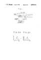
- FIG. 1is a functional block diagram showing a schematic constitution of the present invention
- FIGS. 2A and Bare explanatory views showing a process for obtaining the histogram of a hue image
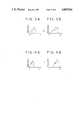
- FIGS. 3A and Bare explanatory views showing a process for obtaining the histogram of an intensity image
- FIGS. 4A and Bare explanatory views showing a process for obtaining the histogram of a saturation image
- FIG. 5is a block diagram showing an essential part of a first embodiment according to the present invention.
- FIG. 6is a schematic view showing an endoscopic system according to a second embodiment of the present invention.
- FIG. 7is a graph showing the relationship between hue and frequency according to an HSI conversion
- FIG. 8is a graph showing the relationship between hue and frequency according to an HSV conversion
- FIGS. 9 and 10are graphs showing the relationship between hue and frequency after carrying out a histogram equalization process with respect to the histograms shown in FIGS. 7 and 8;
- FIG. 11is a block diagram showing a processor for the histogram equalization
- FIG. 12Ais a graph showing a histogram stored in a histogram memory shown in FIG. 11;
- FIGS. 12B and 12Care graphs showing histograms stored in an integrated histogram table memory shown in FIG. 11 before and after a normalization;
- FIG. 12Dis a graph showing the contents of a reference table converted from FIG. 12C in order to perform a converting process by a value converting circuit shown in FIG. 11;
- FIG. 13is a graph showing the relationship between saturation and frequency according to the HSV conversion
- FIGS. 14 and 15are graphs showing the results of a histogram equalization process carried out for saturation values between 0 and 1, and between 0.3 and 0.8 respectively;
- FIG. 16is a schematic view showing a first embodiment of the stretching process according to the present invention.
- FIGS. 17 and 18are graphs showing a linear stretching an arbitrary function stretching respectively.
- FIGS. 19 to 21are graphs for explaining a process according to the combination of the saturation histogram equalization and the conversion from saturation to hue.
- a color picture provided by an endoscopic system according to the present inventioncomprises, for instance, the three primary colors of red (R), green (G), and blue (B).
- an image converting portion 1converts a color picture comprising, for instance, the primary color images, i.e., R, G, and B images into color spaces of hue, saturation, and intensity images which are more comprehensible to human eyes.
- a histogram processing portion 2executes a histogram preparing process for the respective hue, saturation, and intensity images to obtain histograms of the hue, intensity, and saturation images, as typically shown in FIGS. 2A, 3A, and 4A respectively.
- the histogram processing portion 2carries out the so-called histogram stretching process to stretch the range (from 0° to 360°) of color change such that a state shown in FIG. 2B is obtained from a state shown in FIG. 2A. At this time, to prevent the color of the whole picture plane on a display from changing, the histogram is stretched around the most frequently appearing hue value of the original picture.
- the histogram of the intensity imageis stretched from a state shown in FIG. 3A to a state shown in FIG. 3B.
- the kind of the conversionfor instance if an HSI conversion is used, and if the intensity is excessively strong, the color of the picture is whitish when the original picture with R, G, and B colors is reproduced based on the stretched data. Therefore, it is preferable not to stretch the intensity image up to its maximum extent.
- an effect corresponding to a contrast enhancing effect in a black-and-white picturecan be obtained.
- the histogram of the saturation imageis shifted to its higher side to obtain a state shown in FIG. 4B from a state shown in FIG. 4A.
- the image converting portion 1After performing proper processes in the histogram processing portion 2 as mentioned in the above, the image converting portion 1 reproduces color images from the hue, saturation, and intensity images. Then, a picture which has been enhanced in color and contrast as required is displayed on a display.
- the hue, saturation, and intensity imagescan be displayed as a black-and-white picture on a color display via a black-and-white displaying device included in the image converting portion, or can be displayed as a black-and-white image on a black-and-white display (not shown).
- FIG. 5is a block diagram showing an essential constitution of an endoscopic system of an embodiment according to the present invention.
- a numeral 11represents a controller disposed as a controlling center. Driven in response to instructions from and connected to the controller 11 are a controlling console 12, a camera controller 15 for controlling a CCD camera 13 fitted to an endoscope 14, a switching device 16, a red (R) image memory 17, a green (G) image memory 18, a blue (B) image memory 19, an RGB/(hue-saturation-intensity) converter 20, a hue image memory 21, a saturation image memory 22, an intensity image memory 23, and a histogram processor 24.
- a numeral 25represents a decoder, 26 a color display, and 27 a black-and-white display interface.
- Video signals provided by the CCD camera 11are transferred as NTSC signals as they are, to the color display 26.
- the video signalsare also transmitted through the decoder 25 and stored in a frame memory comprising the R, G, and B image memories 17, 18, and 19.
- a selecting instruction from the controlling console 12 via the controller 11decides whether the contents of the NTSC signals or the contents of the data stored in the frame memory are displayed.
- the image data stored in the R, G, and B image memories 17, 18, and 19are processed in the converter 20, and converted into hue, saturation, and intensity images which are in turn stored in the hue, saturation, and intensity image memories 21, 22, and 23.
- the histogram processor 24is actuated to stretch the histogram of the hue image around an average hue value or around the most frequently appearing hue value (FIGS. 2A and 2B).
- the stretching size of the hue imagemay be determined based on parameters previously obtained by a statistical method, or based on different parameters obtained for respective images.
- a stretching process similar to that carried out for the histogram of the hue imageis carried out for the histogram of the intensity image (FIGS. 3A and 3B).
- the stretching processneed not be especially carried out around the most frequently appearing value but may be used when necessary.
- the controller 11actuates the converter 20 which converts the data processed by the histogram processor 24 into R, G, and B images.
- the converted dataare stored in the R, G, and B image memories 17, 18 and 19.
- the controller 11After that, the controller 11 generates instructions to transfer the converted data stored in the R, G, and B image memories 17, 18, and 19 to the color display 26.
- the switching device 16When the switching device 16 is switched according to the above-mentioned instructions, a picture corresponding to the converted data stored in the R, G, and B image memories 17, 18, and 19 is displayed on the color display 26.
- the histogram processor 24When it is desired to change the whole color picture plane on the color display 26 into the one having a high saturation (namely a picture with vivid colors for the whole plane), it is preferable to provide the histogram processor 24 with a process for changing all of the saturation image data stored in the saturation image memory 21 to have the maximum saturation values. In this case, the hue and intensity images are still subjected to the above-mentioned histogram processing. In connection with the hue, saturation, and intensity images, images before the histogram processing and images after the histogram processing are displayed on the display as black-and-white images according to instructions from the black-and-white display interface and the controller.
- a color changecan be enhanced by processing the histogram of a hue image, and the brightness can be enhanced by processing the histogram of intensity image. Further, the vividness of the color of the whole picture plane can be increased by processing the histogram of saturation image.
- FIG. 6is a view showing a second embodiment of the endoscopic system according to the present invention.
- a color picture photographed by a CCD camerais, on the one hand, guided as NTSC signals to a display, and, on the other hand, transferred through a decoder 25 to R, G, and B frame memories 17, 18, and 19.
- a controlling console 12it is decided which image is displayed on a display 26.
- Such instructionsmay be given by an operator through a switch (not shown).
- the image data stored in the R, G, and B image memories 17, 18, and 19are processed by an RGB/(hue-saturation-intensity) converter 20 and converted into hue, saturation, and intensity images, which are in turn stored in hue, saturation, and intensity image memories 21, 22, and 23.
- a histogram processor 24is actuated to carry out a histogram equalization process for the histogram of the hue image.
- This histogram equalizationmay be done in the range of 0° to 360°, or the original hue histogram may be converted to have values between -180° and 180°, thereafter performing the histogram equalization between -180° and 180°.
- the valuesare returned to values between 0° and 360°.
- the reason why the values are returned to those between 0° and 360°is because the conversion used is the HSV conversion or the HSI conversion.
- the histogram equalizationmay be done within this range, and is efficient when the input hue band area is relatively narrow.
- the entire hue image obtained by the histogram equalizationcan be turned by a predetermined times according to, or not according to, statistical data or parameters. Such a turn is made for "x" times (x is a real number) with one turn being 0° to 360°, in a manner of turning the dial plate of a clock while fixing the hands thereof, to obtain a required color system for the entire image.
- the controller 11automatically actuates the converter 20 to convert the hue, saturation, and intensity images into R, G, and B images. After that, the controller 11 sends instructions to display a picture on the display 26 according to the images subjected to the histogram processing.
- R, G, and B color imagescan be obtained.
- the imagesare converted into hue, saturation, and intensity spaces.
- hue image, saturation image, and intensity imagecan be respectively obtained.
- frequency in hue imageis increased around 120° when the HSI conversion is used, and, when the HSV conversion is used, around 0° and 360° as shown in FIG. 8, i.e., around a red color. This is because an image of an internal organ (for instance a stomach wall) of a human body is entirely reddish.
- the data of the hue imageare subjected to a process corresponding to the histogram equalization process. Namely, the data are converted such that the average frequencies of respective colors are the same entirely. In this case, the data are converted into values between 0° and 360°. Thus, the histograms shown in FIGS. 7 and 8 are changed to those shown in FIGS. 9 and 10.
- the data of the hue image subjected to the above-mentioned process and the data of the original saturation and intensity imagesare reconverted into R, G, and B images. Then, the reddish image is changed to an image having various colors. Namely, delicate differences in color are changed to large changes of color so that a diseased portion such as a cancer can easily be detected.
- FIG. 6according to instructions generated by the controlling console 12 or by pushing the switch (not shown), the histogram processor 24 is operated to perform the histogram equalization process for saturation image.
- FIG. 11is a block diagram showing the histogram processor 24 according to the third embodiment of the present invention in which the histogram equalization process is carried out for the saturation image.
- the histogram of the saturation image stored in the saturation image memory 22 shown in FIG. 6is calculated by a histogram calculating circuit 51.
- the results of the calculationare stored in a histogram memory 52.
- the range (from A1 to A2) of the histogram calculationis supplied from a parameter register 50.
- An example of the histogramis shown in FIG. 12A for clearly understanding the calculation.
- An integrating circuit 53sequentially integrates the histogram, and the results are written into an integrated histogram table memory 54. At this time, the memory 54 stores values such as those shown in FIG.
- a normalization circuit 55rewrites the values in the integrated histogram table memory 54 such that the maximum value in FIG. 12B is set to "1". At this moment, the integrated histogram table memory 54 has values shown in FIG. 12C.
- a value converting circuit 56converts the values in the integrated histogram table memory 54 into values of the range (from B1 to B2) stored in the parameter register 50, i.e., into values ranging from 0 to 1. Namely, the histogram equalization is carried out for the respective data stored in the saturation image memory by referring to the data in the integrated histogram table memory 54. If the values are in the range of A1 to A2, the histogram equalization is carried out in the range of B1 to B2.
- the values A1, A2, B1, and B2may be predetermined or obtained by statistical calculation of the original images.
- FIG. 12shows an example of the conversion carried out by the value converting circuit 56.
- the controller 11automatically activates the RGB/HSV converter 20 to convert the respective data into R, G, and B images. After that, instructions are given from the controller 11 to the color display 26 to display a picture as a whole based on the images subjected to the histogram processing.
- average pixel numbers for the respective saturation valuesare constant, and the difference in saturation between the cancer portion and the normal portion is enhanced.
- the endoscopic systemprovides R, G, and B color images.
- the color imagesare converted into hue, saturation, and intensity spaces.
- the HSV conversionfor instance, is used.
- only a hue image, only a saturation image, and only an intensity imageare respectively obtained.
- the saturation imagemay have the histogram shown in FIG. 13 which has two peaks, one for the cancer portion and the other for the normal portion. But the peaks are located closely to each other so that it is very difficult to distinguish one from the other.
- the saturation dataare subjected to the histogram equalization process. Namely, the data are converted such that values within a designated range have the same average frequency. Accordingly, the histogram shown in FIG. 13 is changed to the one shown in FIG. 14 after the histogram equalization is carried out for values between 0 and 1, and to the one shown in FIG. 1 after the histogram equalization is carried out for example values between 0.3 and 0.8.
- the saturation data subjected to the histogram equalization process and the data of the original hue and intensity imagesare processed by, for instance, a reverse HSV conversion to reproduce R, G, and B images. Then, a picture in which the vividness of the color of the cancer portion is remarkably different from that of the normal portion is displayed. Namely, a picture in which the cancer portion is easily distinguished from the normal portion can be obtained.
- the original picture and the processed picturemay always be displayed together on the display at real time.
- a delicate difference in saturation between the cancer portion and the normal portionis enhanced by the histogram equalization process of the saturation image, improving the resolution of the color picture as a whole. Since the difference in color between the cancer portion and the normal portion is enhanced, the image diagnostic capacity of a physician can be improved.
- FIG. 16is a view showing an embodiment of the stretching process according to the present invention. This process is carried out in the following sequences:
- a function designating circuit 63decides whether a function is linear or arbitrary.
- a table in an arbitrary function memory 64(a function table and data are in advance set from an external equipment, a CPU, a console, a disc, or a memory, which are not shown) follows as shown in, for instance, FIG. 18.
- input valuesrange from A1 to A2
- the stretchingis carried out so as to have output values ranging from B1 to B2.
- An input selecting circuit 61inputs input data into the stretching circuit 62 only when the input data are in the range of A1 to A2.
- the cancer portionis not clear in the saturation image, the preferable results can be obtained by a saturation to hue conversion in the HSV color space.
- a saturation to hue conversion in the HSV color spaceis carried out in the following sequences:
- FIG. 19is a view showing the histogram of an original saturation image
- FIG. 20a view showing a histogram after the histogram equalization process of the saturation image
- FIG. 21a view showing a histogram after the above-mentioned conversion from the saturation into the hue.
- the projected portion on the right sideindicates a histogram of the normal portion
- the projected portion on the left sideindicates the cancer portion.
- the difference between the normal portion and the cancer portionis enhanced by the saturation to hue conversion.
- the present inventionis also applicable to a system in which a television camera for a CCD is connected to an eyepiece portion of a conventional fiber scope.
Landscapes
- Engineering & Computer Science (AREA)
- Multimedia (AREA)
- Signal Processing (AREA)
- Endoscopes (AREA)
- Closed-Circuit Television Systems (AREA)
Abstract
Description
1. Field of the Invention
The present invention relates to an endoscopic system which provides a color picture by combining, for instance, the three primary color images, i.e., a red (R) image, a green (G) image, and a blue (B) image, and particularly to an improvement of a mechanism for adjusting the color and contrast of a color picture.
2. Description of the Prior Art
Endoscopic systems usually use a plane sequential method in which the color of an illuminating light is sequentially changed among R, G, and B colors, or a point sequential method in which an image is photographed with the three primary colors at the same time, or a simultaneous method.
In such prior art endoscopic systems, the color and contrast of a color picture are adjusted by stretching histograms of, for instance, R image, G image, and B image of the color picture. According to such a stretching adjustment, the contrast of the picture is enhanced but the color thereof which is an important factor in the color picture, tends to differ from the actual color.
As described in the above, according to the prior art, the histograms of the R, G, and B images are subjected to the stretching adjustment to enhance the contrast of the color picture, but it is difficult to always maintain the actual color of the picture.
Therefore, when the stomach of a patient is monitored by a prior art endoscopic system to see whether or not a cancer exists, it is frequently difficult to distinguish a delicate color difference between the cancer and a normal part. In addition, the prior art endoscopic systems are not provided with functions and mechanisms for executing a histogram equalization process of a hue image and a saturation image.
In view of the above problems, an object of the present invention is to provide an endoscopic system which can correctly and quickly enhance the color and contrast of a color picture.
In order to accomplish the object mentioned in the above, the present invention provides an endoscopic system comprising an image converting portion for converting the three primary color images into a hue image, a saturation image, and an intensity image, and for reproducing the three primary color images from the converted hue, saturation, and intensity images; and a histogram processing portion for obtaining histograms of the respective hue, saturation, and intensity images prepared by the image converting portion, stretching the histograms of the hue and intensity images, and shifting the histogram of the saturation image.
The other objects, features and advantages of the present invention will become more apparent from the following descriptions of the preferred embodiments taken in conjunction with the accompanying drawings in which:
FIG. 1 is a functional block diagram showing a schematic constitution of the present invention;
FIGS. 2A and B are explanatory views showing a process for obtaining the histogram of a hue image;
FIGS. 3A and B are explanatory views showing a process for obtaining the histogram of an intensity image;
FIGS. 4A and B are explanatory views showing a process for obtaining the histogram of a saturation image;
FIG. 5 is a block diagram showing an essential part of a first embodiment according to the present invention;
FIG. 6 is a schematic view showing an endoscopic system according to a second embodiment of the present invention;
FIG. 7 is a graph showing the relationship between hue and frequency according to an HSI conversion;
FIG. 8 is a graph showing the relationship between hue and frequency according to an HSV conversion;
FIGS. 9 and 10 are graphs showing the relationship between hue and frequency after carrying out a histogram equalization process with respect to the histograms shown in FIGS. 7 and 8;
FIG. 11 is a block diagram showing a processor for the histogram equalization;
FIG. 12A is a graph showing a histogram stored in a histogram memory shown in FIG. 11;
FIGS. 12B and 12C are graphs showing histograms stored in an integrated histogram table memory shown in FIG. 11 before and after a normalization;
FIG. 12D is a graph showing the contents of a reference table converted from FIG. 12C in order to perform a converting process by a value converting circuit shown in FIG. 11;
FIG. 13 is a graph showing the relationship between saturation and frequency according to the HSV conversion;
FIGS. 14 and 15 are graphs showing the results of a histogram equalization process carried out for saturation values between 0 and 1, and between 0.3 and 0.8 respectively;
FIG. 16 is a schematic view showing a first embodiment of the stretching process according to the present invention;
FIGS. 17 and 18 are graphs showing a linear stretching an arbitrary function stretching respectively; and
FIGS. 19 to 21 are graphs for explaining a process according to the combination of the saturation histogram equalization and the conversion from saturation to hue.
The preferred embodiments of the present invention will now be described with reference to the drawings.
A color picture provided by an endoscopic system according to the present invention comprises, for instance, the three primary colors of red (R), green (G), and blue (B).
To adjust the color and contrast of the color picture, the present invention uses an arrangement with a functional block shown in FIG. 1. In this figure, animage converting portion 1 converts a color picture comprising, for instance, the primary color images, i.e., R, G, and B images into color spaces of hue, saturation, and intensity images which are more comprehensible to human eyes.
After the conversion, ahistogram processing portion 2 executes a histogram preparing process for the respective hue, saturation, and intensity images to obtain histograms of the hue, intensity, and saturation images, as typically shown in FIGS. 2A, 3A, and 4A respectively.
For the hue image, thehistogram processing portion 2 carries out the so-called histogram stretching process to stretch the range (from 0° to 360°) of color change such that a state shown in FIG. 2B is obtained from a state shown in FIG. 2A. At this time, to prevent the color of the whole picture plane on a display from changing, the histogram is stretched around the most frequently appearing hue value of the original picture.
The histogram of the intensity image is stretched from a state shown in FIG. 3A to a state shown in FIG. 3B. Depending on the kind of the conversion, for instance if an HSI conversion is used, and if the intensity is excessively strong, the color of the picture is whitish when the original picture with R, G, and B colors is reproduced based on the stretched data. Therefore, it is preferable not to stretch the intensity image up to its maximum extent. However, by stretching the intensity image, an effect corresponding to a contrast enhancing effect in a black-and-white picture can be obtained.
The histogram of the saturation image is shifted to its higher side to obtain a state shown in FIG. 4B from a state shown in FIG. 4A.
After performing proper processes in thehistogram processing portion 2 as mentioned in the above, theimage converting portion 1 reproduces color images from the hue, saturation, and intensity images. Then, a picture which has been enhanced in color and contrast as required is displayed on a display.
The hue, saturation, and intensity images can be displayed as a black-and-white picture on a color display via a black-and-white displaying device included in the image converting portion, or can be displayed as a black-and-white image on a black-and-white display (not shown).
The present invention will be described in more detail according to the embodiments.
FIG. 5 is a block diagram showing an essential constitution of an endoscopic system of an embodiment according to the present invention.
In this figure, a numeral 11 represents a controller disposed as a controlling center. Driven in response to instructions from and connected to thecontroller 11 are a controllingconsole 12, acamera controller 15 for controlling aCCD camera 13 fitted to anendoscope 14, aswitching device 16, a red (R)image memory 17, a green (G)image memory 18, a blue (B)image memory 19, an RGB/(hue-saturation-intensity)converter 20, ahue image memory 21, asaturation image memory 22, anintensity image memory 23, and ahistogram processor 24. A numeral 25 represents a decoder, 26 a color display, and 27 a black-and-white display interface.
Video signals provided by theCCD camera 11 are transferred as NTSC signals as they are, to thecolor display 26. The video signals are also transmitted through thedecoder 25 and stored in a frame memory comprising the R, G, andB image memories console 12 via thecontroller 11 decides whether the contents of the NTSC signals or the contents of the data stored in the frame memory are displayed.
The image data stored in the R, G, andB image memories converter 20, and converted into hue, saturation, and intensity images which are in turn stored in the hue, saturation, andintensity image memories
Under this state, if the controllingconsole 12 is operated, or if a switch (not shown) is pressed to transmit instructions from thecontroller 11 to thehistogram processor 24, thehistogram processor 24 is actuated to stretch the histogram of the hue image around an average hue value or around the most frequently appearing hue value (FIGS. 2A and 2B).
The stretching size of the hue image may be determined based on parameters previously obtained by a statistical method, or based on different parameters obtained for respective images.
A stretching process similar to that carried out for the histogram of the hue image is carried out for the histogram of the intensity image (FIGS. 3A and 3B). In this case, not like the one for the hue image, the stretching process need not be especially carried out around the most frequently appearing value but may be used when necessary.
In connection with the saturation image, the same stretching process is carried out, or the histogram thereof as a whole is shifted to its higher value side, while maintaining the shape of the histogram (FIGS. 4A and 4B). It is also possible to obtain a good result by carrying out the same histogram stretching process as that executed for the intensity image.
When the histograms of the hue, saturation, and intensity images are completely processed, thecontroller 11 actuates theconverter 20 which converts the data processed by thehistogram processor 24 into R, G, and B images. The converted data are stored in the R, G, andB image memories
After that, thecontroller 11 generates instructions to transfer the converted data stored in the R, G, andB image memories color display 26. When theswitching device 16 is switched according to the above-mentioned instructions, a picture corresponding to the converted data stored in the R, G, andB image memories color display 26.
When it is desired to change the whole color picture plane on thecolor display 26 into the one having a high saturation (namely a picture with vivid colors for the whole plane), it is preferable to provide thehistogram processor 24 with a process for changing all of the saturation image data stored in thesaturation image memory 21 to have the maximum saturation values. In this case, the hue and intensity images are still subjected to the above-mentioned histogram processing. In connection with the hue, saturation, and intensity images, images before the histogram processing and images after the histogram processing are displayed on the display as black-and-white images according to instructions from the black-and-white display interface and the controller.
As described in the above, according to the endoscopic system of the present invention, a color change can be enhanced by processing the histogram of a hue image, and the brightness can be enhanced by processing the histogram of intensity image. Further, the vividness of the color of the whole picture plane can be increased by processing the histogram of saturation image. By reproducing the three primary color images after the above-mentioned processes, the density resolution of the entire color picture can remarkably be improved.
As a result, a delicate color difference between an diseased part and a normal part of an internal organ can correctly be displayed on a color display. Therefore, the image diagnostic capacity of a physician can be improved.
Since the components of colors are enhanced, an S/N ratio can remarkably be improved.
FIG. 6 is a view showing a second embodiment of the endoscopic system according to the present invention. In this embodiment, a color picture photographed by a CCD camera is, on the one hand, guided as NTSC signals to a display, and, on the other hand, transferred through adecoder 25 to R, G, andB frame memories console 12, it is decided which image is displayed on adisplay 26. Such instructions may be given by an operator through a switch (not shown). The image data stored in the R, G, andB image memories converter 20 and converted into hue, saturation, and intensity images, which are in turn stored in hue, saturation, andintensity image memories console 12 or by pushing the switch (not shown), ahistogram processor 24 is actuated to carry out a histogram equalization process for the histogram of the hue image.
This histogram equalization may be done in the range of 0° to 360°, or the original hue histogram may be converted to have values between -180° and 180°, thereafter performing the histogram equalization between -180° and 180°. In this case, after the histogram equalization, the values are returned to values between 0° and 360°. The reason why the values are returned to those between 0° and 360° is because the conversion used is the HSV conversion or the HSI conversion. In another conversion, for instance, of the range from -60° to 300°, the histogram equalization may be done within this range, and is efficient when the input hue band area is relatively narrow.
The entire hue image obtained by the histogram equalization can be turned by a predetermined times according to, or not according to, statistical data or parameters. Such a turn is made for "x" times (x is a real number) with one turn being 0° to 360°, in a manner of turning the dial plate of a clock while fixing the hands thereof, to obtain a required color system for the entire image. When the histogram processing is completed, thecontroller 11 automatically actuates theconverter 20 to convert the hue, saturation, and intensity images into R, G, and B images. After that, thecontroller 11 sends instructions to display a picture on thedisplay 26 according to the images subjected to the histogram processing.
According to the endoscopic system of this embodiment, R, G, and B color images can be obtained. The images are converted into hue, saturation, and intensity spaces. Thus, only a hue image, only a saturation image, and only an intensity image can be respectively obtained.
As shown in FIG. 7, frequency in hue image is increased around 120° when the HSI conversion is used, and, when the HSV conversion is used, around 0° and 360° as shown in FIG. 8, i.e., around a red color. This is because an image of an internal organ (for instance a stomach wall) of a human body is entirely reddish.
The data of the hue image are subjected to a process corresponding to the histogram equalization process. Namely, the data are converted such that the average frequencies of respective colors are the same entirely. In this case, the data are converted into values between 0° and 360°. Thus, the histograms shown in FIGS. 7 and 8 are changed to those shown in FIGS. 9 and 10.
The data of the hue image subjected to the above-mentioned process and the data of the original saturation and intensity images are reconverted into R, G, and B images. Then, the reddish image is changed to an image having various colors. Namely, delicate differences in color are changed to large changes of color so that a diseased portion such as a cancer can easily be detected.
In this embodiment, the operational sequences are as follows:
(1) observing a picture, (2) displaying the picture of an object (such as a cancer) on a display, (3) pushing a freeze button (not shown), (4) storing the picture as R, G, and B images in R, G, and B memories, (5) inputting histogram process instructions from a console, (6) converting the R, G, and B images into hue, saturation, and intensity images and storing them in the respective memories, (7) carrying out a histogram processing, (8) taking the data of the hue, saturation, and intensity images out of the respective memories, converting them into R, G, and B images, and storing them in the respective memories, and (9) displaying a picture based on the stored R, G, and B images on the color display.
Therefore, a delicate color difference between the cancer and the normal part is enhanced by the histogram equalization process of the hue image, and the resolution of the color image as a whole can be improved. Since the color difference between the cancer portion and the normal part is enhanced, the image diagnostic capacity of a physician can be improved.
A third embodiment of the present invention will be described. In FIG. 6, according to instructions generated by the controllingconsole 12 or by pushing the switch (not shown), thehistogram processor 24 is operated to perform the histogram equalization process for saturation image.
FIG. 11 is a block diagram showing thehistogram processor 24 according to the third embodiment of the present invention in which the histogram equalization process is carried out for the saturation image. The histogram of the saturation image stored in thesaturation image memory 22 shown in FIG. 6 is calculated by ahistogram calculating circuit 51. The results of the calculation are stored in ahistogram memory 52. At this time, the range (from A1 to A2) of the histogram calculation is supplied from aparameter register 50. An example of the histogram is shown in FIG. 12A for clearly understanding the calculation. An integratingcircuit 53 sequentially integrates the histogram, and the results are written into an integratedhistogram table memory 54. At this time, thememory 54 stores values such as those shown in FIG. 12B derived by integrating the values shown in FIG. 12A. Then, anormalization circuit 55 rewrites the values in the integratedhistogram table memory 54 such that the maximum value in FIG. 12B is set to "1". At this moment, the integratedhistogram table memory 54 has values shown in FIG. 12C.
Avalue converting circuit 56, with reference to the integratedhistogram table memory 54, converts the values in the integratedhistogram table memory 54 into values of the range (from B1 to B2) stored in theparameter register 50, i.e., into values ranging from 0 to 1. Namely, the histogram equalization is carried out for the respective data stored in the saturation image memory by referring to the data in the integratedhistogram table memory 54. If the values are in the range of A1 to A2, the histogram equalization is carried out in the range of B1 to B2. The values A1, A2, B1, and B2 may be predetermined or obtained by statistical calculation of the original images. FIG. 12 shows an example of the conversion carried out by thevalue converting circuit 56.
When the histogram processing is completed, thecontroller 11 automatically activates the RGB/HSV converter 20 to convert the respective data into R, G, and B images. After that, instructions are given from thecontroller 11 to thecolor display 26 to display a picture as a whole based on the images subjected to the histogram processing.
According to the normalization of the histogram, as shown in FIG. 12C, average pixel numbers for the respective saturation values are constant, and the difference in saturation between the cancer portion and the normal portion is enhanced. For parameters A1, A2, B1, and B2, the preferable results are generally obtained with A1=0, A2=1, B1=0, and B2=1.
The endoscopic system according to the third embodiment of the present invention provides R, G, and B color images. The color images are converted into hue, saturation, and intensity spaces. At this time, the HSV conversion, for instance, is used. As a result, only a hue image, only a saturation image, and only an intensity image are respectively obtained.
The saturation image may have the histogram shown in FIG. 13 which has two peaks, one for the cancer portion and the other for the normal portion. But the peaks are located closely to each other so that it is very difficult to distinguish one from the other.
To cope with this, the saturation data are subjected to the histogram equalization process. Namely, the data are converted such that values within a designated range have the same average frequency. Accordingly, the histogram shown in FIG. 13 is changed to the one shown in FIG. 14 after the histogram equalization is carried out for values between 0 and 1, and to the one shown in FIG. 1 after the histogram equalization is carried out for example values between 0.3 and 0.8.
The saturation data subjected to the histogram equalization process and the data of the original hue and intensity images are processed by, for instance, a reverse HSV conversion to reproduce R, G, and B images. Then, a picture in which the vividness of the color of the cancer portion is remarkably different from that of the normal portion is displayed. Namely, a picture in which the cancer portion is easily distinguished from the normal portion can be obtained.
According to this embodiment, the operational sequence are as follows:
(1) observing a picture, (2) displaying the picture of an object (such as a cancer) on a display, (3) pushing a freeze button (not shown), (4) storing the picture as R, G, and B images in R, G, and B memories, (5) inputting histogram process instructions from a console, (6) converting the R, G, and B images into hue, saturation, and intensity images and storing them in the respective memories, (7) carrying out a histogram processing, (8) taking the data of the hue, saturation, and intensity images out of the respective memories, converting them into R, G, and B images, and storing them in the respective memories, and (9) displaying a picture based on the stored R, G, and B images on the color display.
Instead of pushing the freeze button, the original picture and the processed picture may always be displayed together on the display at real time.
According to the embodiment mentioned in the above, a delicate difference in saturation between the cancer portion and the normal portion is enhanced by the histogram equalization process of the saturation image, improving the resolution of the color picture as a whole. Since the difference in color between the cancer portion and the normal portion is enhanced, the image diagnostic capacity of a physician can be improved.
FIG. 16 is a view showing an embodiment of the stretching process according to the present invention. This process is carried out in the following sequences:
(1) Afunction designating circuit 63 decides whether a function is linear or arbitrary.
(2) When the stretching is carried out with the linear function, the range of A1 to A2 of an input (saturation, hue, or intensity) is stretched to the range of B1 to B2. Supposing the input is "x" and an output "y", thefunction designating circuit 63 decides parameters "a" and "b" in the formula of y=ax+b as shown in FIG. 17, and a stretchingcircuit 62 performs the calculation.
(3) When the stretching is carried out with the arbitrary function, a table in an arbitrary function memory 64 (a function table and data are in advance set from an external equipment, a CPU, a console, a disc, or a memory, which are not shown) follows as shown in, for instance, FIG. 18. When input values range from A1 to A2, the stretching is carried out so as to have output values ranging from B1 to B2.
(4) Aninput selecting circuit 61 inputs input data into the stretchingcircuit 62 only when the input data are in the range of A1 to A2.
If the cancer portion is not clear in the saturation image, the preferable results can be obtained by a saturation to hue conversion in the HSV color space. Such a conversion is carried out in the following sequences:
(1) finding a histogram of the saturation image;
(2) finding a median mode or an average value of each of the cancer portion and the normal portion; and
(3) converting the saturation data into the hue data with the above-mentioned parameters.
FIG. 19 is a view showing the histogram of an original saturation image, FIG. 20 a view showing a histogram after the histogram equalization process of the saturation image, and FIG. 21 a view showing a histogram after the above-mentioned conversion from the saturation into the hue. As a typical example, in FIGS. 19 to 21, the projected portion on the right side indicates a histogram of the normal portion, and the projected portion on the left side indicates the cancer portion. As is apparent from FIG. 21, the difference between the normal portion and the cancer portion is enhanced by the saturation to hue conversion. The present invention is also applicable to a system in which a television camera for a CCD is connected to an eyepiece portion of a conventional fiber scope.
Claims (10)
1. An endoscopic system for obtaining a color picture by combining three primary color images with each other, said system comprising:
image converting means for converting said three primary color images into hue, saturation, and intensity images;
means for reproducing said three primary color images from said converted hue, saturation, and intensity images;
histogram processing means for obtaining a histogram based on at least one of said hue, saturation, and intensity images obtained by said image converting means; and then performing at least one of a
means for stretching or shifting said histogram, or both.
2. An endoscopic system as claimed in claim 1, wherein said histogram is stretched around a most frequently appearing hue value of an original picture so that the overall color of the picture displayed on a color display is not changed.
3. An endoscopic system as claimed in claim 1, wherein, when the histogram of said intensity image is stretched, said intensity image is not stretched so far as to reach the maximum value thereof.
4. An endoscopic system as claimed in claim 1, wherein the histogram of said saturation image as a whole is shifted on the side of a higher saturation value.
5. An endoscopic system as claimed in claim 1, wherein said image converting means is provided with memories for storing said three primary color images respectively.
6. An endoscopic system as claimed in claim 1, wherein said histogram processing means is provided with memories for storing data from said image converting means.
7. An endoscopic system comprising:
a first device for converting red, green and blue images of a color picture into a hue image, a saturation image, and an intensity image, and reconverting said hue, saturation, and intensity images into red, green and blue images; and
a mechanism for processing a histogram of at least one of said hue and saturation images by a histogram equalization process such that average frequencies at respective values of said at least one of said hue and saturation images are constant.
8. An endoscopic system as claimed in claim 7, wherein said mechanism converts the data of said hue image into values between 0° and 360°.
9. An endoscopic system as claimed in claim 7, wherein, after said histogram equalization process of said saturation image, said processed saturation image is converted into a hue image.
10. An endoscopic system as claimed in claim 7, wherein said histogram equalization process is carried out in a predetermined area of the saturation values between 0 and 1.
Applications Claiming Priority (4)
| Application Number | Priority Date | Filing Date | Title |
|---|---|---|---|
| JP61-197293 | 1986-08-25 | ||
| JP61197293AJPS6354144A (en) | 1986-08-25 | 1986-08-25 | Electronic endoscope apparatus |
| JP62-100154 | 1987-04-24 | ||
| JP62100154AJPH0669438B2 (en) | 1987-04-24 | 1987-04-24 | Endoscope device |
Publications (1)
| Publication Number | Publication Date |
|---|---|
| US4805016Atrue US4805016A (en) | 1989-02-14 |
Family
ID=26441238
Family Applications (1)
| Application Number | Title | Priority Date | Filing Date |
|---|---|---|---|
| US07/087,787Expired - LifetimeUS4805016A (en) | 1986-08-25 | 1987-08-21 | Endoscopic system for converting primary color images into hue, saturation and intensity images |
Country Status (1)
| Country | Link |
|---|---|
| US (1) | US4805016A (en) |
Cited By (50)
| Publication number | Priority date | Publication date | Assignee | Title |
|---|---|---|---|---|
| US4916531A (en)* | 1988-03-23 | 1990-04-10 | Data Translation, Inc. | Color video processing circuitry |
| US5034888A (en)* | 1988-02-26 | 1991-07-23 | Olympus Optical Co., Ltd. | Electronic endoscope apparatus having different image processing characteristics for a moving image and a still image |
| EP0386983A3 (en)* | 1989-03-07 | 1991-10-09 | Sony Corporation | Television image display apparatus |
| US5111281A (en)* | 1987-09-28 | 1992-05-05 | Kabushiki Kaisha Toshiba | Color correction device for an endoscope |
| FR2673799A1 (en)* | 1991-02-28 | 1992-09-11 | Broadcast Television Syst | METHOD FOR CORRECTING THE COLORS OF A VIDEO SIGNAL AND ARRANGEMENT FOR IMPLEMENTING THE METHOD. |
| US5204948A (en)* | 1989-09-08 | 1993-04-20 | Advantest Corporation | Method and apparatus for evaluating color image signals |
| US5212546A (en)* | 1990-07-03 | 1993-05-18 | Electronics For Imaging, Inc. | Color correction system employing reference pictures |
| US5220620A (en)* | 1989-10-09 | 1993-06-15 | Fujitsu Limited | Color image data processing apparatus |
| WO1993013878A1 (en)* | 1992-01-08 | 1993-07-22 | Connolly Joseph W | Color detection and separation method |
| US5309228A (en)* | 1991-05-23 | 1994-05-03 | Fuji Photo Film Co., Ltd. | Method of extracting feature image data and method of extracting person's face data |
| EP0561228A3 (en)* | 1992-03-17 | 1994-08-17 | Massen Robert | Method and apparatus for colour image endoscopy with colour transformation |
| US5434683A (en)* | 1991-05-14 | 1995-07-18 | Fuji Xerox Co., Ltd. | Color image editing apparatus |
| US5450502A (en)* | 1993-10-07 | 1995-09-12 | Xerox Corporation | Image-dependent luminance enhancement |
| US5465104A (en)* | 1989-06-20 | 1995-11-07 | Digital Equipment Corporation | Color information storage and processing system |
| US5544258A (en)* | 1991-03-14 | 1996-08-06 | Levien; Raphael L. | Automatic tone correction of images using non-linear histogram processing |
| EP0684729A3 (en)* | 1994-05-23 | 1996-12-11 | Xerox Corp | Image-dependent color saturation correction in a natural scene pictorial image. |
| US5772832A (en)* | 1991-06-27 | 1998-06-30 | Applied Materials, Inc | Process for etching oxides in an electromagnetically coupled planar plasma apparatus |
| US5973801A (en)* | 1995-08-21 | 1999-10-26 | Scitex Corp., Ltd. | Method for matching colors of an object to printing colors |
| US6028646A (en)* | 1996-03-25 | 2000-02-22 | Lg Electronics, Inc. | Color image enhancement device for video display appliance |
| US6080104A (en)* | 1995-05-16 | 2000-06-27 | Asahi Kogaku Kogyo Kabushiki Kaisha | Electronic endoscope system |
| US6392713B1 (en) | 2000-03-06 | 2002-05-21 | Media 100 Inc. | Digital processing amplifier |
| US6462834B1 (en)* | 1993-10-29 | 2002-10-08 | Canon Kabushiki Kaisha | Image processing system which forms two-color images |
| DE19701527C2 (en)* | 1996-01-18 | 2002-10-10 | Ricoh Kk | Device for improving the picture quality |
| US6515764B1 (en)* | 1998-12-18 | 2003-02-04 | Xerox Corporation | Method and apparatus for detecting photocopier tracking signatures |
| US20030194116A1 (en)* | 2002-04-10 | 2003-10-16 | Wong Pak Chung | Visualization of information with an established order |
| US20040013298A1 (en)* | 2002-07-20 | 2004-01-22 | Samsung Electronics Co., Ltd. | Method and apparatus for adaptively enhancing colors in color images |
| WO2004008778A1 (en)* | 2002-07-17 | 2004-01-22 | Koninklijke Philips Electronics N.V. | Non-linear picture processing |
| US20040141659A1 (en)* | 2003-01-17 | 2004-07-22 | Yun Zhang | System and method for image fusion |
| US20050068427A1 (en)* | 1999-02-04 | 2005-03-31 | Olympus Optical Co., Ltd. | Endoscope image sensing method and apparatus |
| US20060058684A1 (en)* | 2001-05-07 | 2006-03-16 | Fuji Photo Film Co., Ltd. | Fluorescence image display apparatus |
| US20060110032A1 (en)* | 2004-11-24 | 2006-05-25 | Shunsaku Toshihiro | Method and an apparatus for detecting the base concentration of a photographic film |
| US20060109527A1 (en)* | 2004-11-24 | 2006-05-25 | Shunsaku Toshihiro | Photograph image-processing method and device thereof |
| US20060110033A1 (en)* | 2004-11-24 | 2006-05-25 | Shunsaku Toshihiro | Photograph image-processing method and device thereof |
| US20060247514A1 (en)* | 2004-11-29 | 2006-11-02 | Panasyuk Svetlana V | Medical hyperspectral imaging for evaluation of tissue and tumor |
| US20070016079A1 (en)* | 2005-04-04 | 2007-01-18 | Freeman Jenny E | Hyperspectral imaging in diabetes and peripheral vascular disease |
| GB2418316B (en)* | 2004-09-21 | 2007-01-31 | Hitachi Ltd | Image display apparatus |
| US20070038042A1 (en)* | 2005-04-04 | 2007-02-15 | Freeman Jenny E | Hyperspectral technology for assessing and treating diabetic foot and tissue disease |
| US20070232930A1 (en)* | 2005-04-04 | 2007-10-04 | Jenny Freeman | Hyperspectral Imaging in Diabetes and Peripheral Vascular Disease |
| US20070249913A1 (en)* | 2004-11-29 | 2007-10-25 | Jenny Freeman | Hyperspectral Imaging of Angiogenesis |
| EP1862109A2 (en) | 2006-06-01 | 2007-12-05 | FUJIFILM Corporation | Capsule endoscopic system and image processing apparatus |
| US20080232686A1 (en)* | 2007-03-19 | 2008-09-25 | Yosuke Ohashi | Representative color extracting method and apparatus |
| US20080284845A1 (en)* | 2007-05-14 | 2008-11-20 | Olympus Winter & Ibe Gmbh | Method and apparatus to process endoscope images |
| DE102008027905A1 (en) | 2008-06-12 | 2009-12-17 | Olympus Winter & Ibe Gmbh | Method and endoscope for improving endoscope images |
| US20140015850A1 (en)* | 2012-07-11 | 2014-01-16 | Korea University Research And Business Foundation | Color transformation method and apparatus for person with color vision defect |
| WO2017063223A1 (en)* | 2015-10-13 | 2017-04-20 | 深圳市华星光电技术有限公司 | Method and system for improving contrast of oled display panel |
| US20170273543A1 (en)* | 2015-08-13 | 2017-09-28 | Hoya Corporation | Evaluation value calculation device and electronic endoscope system |
| US20170280971A1 (en)* | 2015-08-13 | 2017-10-05 | Hoya Corporation | Evaluation value calculation device and electronic endoscope system |
| US10339410B1 (en)* | 2016-01-13 | 2019-07-02 | Snap Inc. | Color extraction of a video stream |
| WO2022187691A1 (en)* | 2021-03-05 | 2022-09-09 | Lab4U, Inc. | Methods and electronic devices for quantitatively determining changes in color of a sample over time |
| CN119014791A (en)* | 2023-05-26 | 2024-11-26 | 青岛海信医疗设备股份有限公司 | Medical endoscope device and image processing method |
Citations (4)
| Publication number | Priority date | Publication date | Assignee | Title |
|---|---|---|---|---|
| US4500919A (en)* | 1982-05-04 | 1985-02-19 | Massachusetts Institute Of Technology | Color reproduction system |
| US4623973A (en)* | 1983-12-14 | 1986-11-18 | Dr. Ing. Rudolf Hell Gmbh | Method and apparatus for the recognition of hues and colors |
| US4633303A (en)* | 1984-08-31 | 1986-12-30 | Olympus Optical Co., Ltd. | Two-dimensional bandwidth compensating circuit for an endoscope using a solid state image pick-up device |
| US4654720A (en)* | 1984-04-27 | 1987-03-31 | International Business Machines Corporation | Color image display system |
- 1987
- 1987-08-21USUS07/087,787patent/US4805016A/ennot_activeExpired - Lifetime
Patent Citations (4)
| Publication number | Priority date | Publication date | Assignee | Title |
|---|---|---|---|---|
| US4500919A (en)* | 1982-05-04 | 1985-02-19 | Massachusetts Institute Of Technology | Color reproduction system |
| US4623973A (en)* | 1983-12-14 | 1986-11-18 | Dr. Ing. Rudolf Hell Gmbh | Method and apparatus for the recognition of hues and colors |
| US4654720A (en)* | 1984-04-27 | 1987-03-31 | International Business Machines Corporation | Color image display system |
| US4633303A (en)* | 1984-08-31 | 1986-12-30 | Olympus Optical Co., Ltd. | Two-dimensional bandwidth compensating circuit for an endoscope using a solid state image pick-up device |
Non-Patent Citations (4)
| Title |
|---|
| "Digital Image Processing" by K. R. Castleman, Prentice-Hall, 1979; pp. 87-92. |
| Digital Image Processing by K. R. Castleman, Prentice Hall, 1979; pp. 87 92.* |
| IBM J. Res. Develop., vol. 27, No. 4, Jul. 1983; Edward J. Farrell; pp. 356 366.* |
| IBM J. Res. Develop., vol. 27, No. 4, Jul. 1983; Edward J. Farrell; pp. 356-366. |
Cited By (99)
| Publication number | Priority date | Publication date | Assignee | Title |
|---|---|---|---|---|
| US5111281A (en)* | 1987-09-28 | 1992-05-05 | Kabushiki Kaisha Toshiba | Color correction device for an endoscope |
| US5034888A (en)* | 1988-02-26 | 1991-07-23 | Olympus Optical Co., Ltd. | Electronic endoscope apparatus having different image processing characteristics for a moving image and a still image |
| US4916531A (en)* | 1988-03-23 | 1990-04-10 | Data Translation, Inc. | Color video processing circuitry |
| EP0386983A3 (en)* | 1989-03-07 | 1991-10-09 | Sony Corporation | Television image display apparatus |
| US5465104A (en)* | 1989-06-20 | 1995-11-07 | Digital Equipment Corporation | Color information storage and processing system |
| US5204948A (en)* | 1989-09-08 | 1993-04-20 | Advantest Corporation | Method and apparatus for evaluating color image signals |
| US5220620A (en)* | 1989-10-09 | 1993-06-15 | Fujitsu Limited | Color image data processing apparatus |
| US5212546A (en)* | 1990-07-03 | 1993-05-18 | Electronics For Imaging, Inc. | Color correction system employing reference pictures |
| FR2673799A1 (en)* | 1991-02-28 | 1992-09-11 | Broadcast Television Syst | METHOD FOR CORRECTING THE COLORS OF A VIDEO SIGNAL AND ARRANGEMENT FOR IMPLEMENTING THE METHOD. |
| US5355225A (en)* | 1991-02-28 | 1994-10-11 | Bts Broadcast Television Systems Gmbh | Video signal color correction with dual function memories and color window |
| US5436673A (en)* | 1991-02-28 | 1995-07-25 | Bts Broadcast Television Systems Gmbh | Video signal color correction based on color hue |
| US5544258A (en)* | 1991-03-14 | 1996-08-06 | Levien; Raphael L. | Automatic tone correction of images using non-linear histogram processing |
| US5434683A (en)* | 1991-05-14 | 1995-07-18 | Fuji Xerox Co., Ltd. | Color image editing apparatus |
| US5309228A (en)* | 1991-05-23 | 1994-05-03 | Fuji Photo Film Co., Ltd. | Method of extracting feature image data and method of extracting person's face data |
| EP0514933B1 (en)* | 1991-05-23 | 1997-07-30 | Fuji Photo Film Co., Ltd. | Method of extracting feature image data |
| US5772832A (en)* | 1991-06-27 | 1998-06-30 | Applied Materials, Inc | Process for etching oxides in an electromagnetically coupled planar plasma apparatus |
| US5432545A (en)* | 1992-01-08 | 1995-07-11 | Connolly; Joseph W. | Color detection and separation method |
| WO1993013878A1 (en)* | 1992-01-08 | 1993-07-22 | Connolly Joseph W | Color detection and separation method |
| EP0561228A3 (en)* | 1992-03-17 | 1994-08-17 | Massen Robert | Method and apparatus for colour image endoscopy with colour transformation |
| US5450502A (en)* | 1993-10-07 | 1995-09-12 | Xerox Corporation | Image-dependent luminance enhancement |
| US6462834B1 (en)* | 1993-10-29 | 2002-10-08 | Canon Kabushiki Kaisha | Image processing system which forms two-color images |
| EP0684729A3 (en)* | 1994-05-23 | 1996-12-11 | Xerox Corp | Image-dependent color saturation correction in a natural scene pictorial image. |
| US6080104A (en)* | 1995-05-16 | 2000-06-27 | Asahi Kogaku Kogyo Kabushiki Kaisha | Electronic endoscope system |
| DE19619734C2 (en)* | 1995-05-16 | 2001-02-01 | Asahi Optical Co Ltd | Electronic endoscope system |
| US5973801A (en)* | 1995-08-21 | 1999-10-26 | Scitex Corp., Ltd. | Method for matching colors of an object to printing colors |
| DE19701527C2 (en)* | 1996-01-18 | 2002-10-10 | Ricoh Kk | Device for improving the picture quality |
| US6028646A (en)* | 1996-03-25 | 2000-02-22 | Lg Electronics, Inc. | Color image enhancement device for video display appliance |
| DE19712482B4 (en)* | 1996-03-25 | 2005-12-15 | Lg Electronics Inc. | Color image enhancement apparatus for a viewing device |
| US6515764B1 (en)* | 1998-12-18 | 2003-02-04 | Xerox Corporation | Method and apparatus for detecting photocopier tracking signatures |
| US7221388B2 (en) | 1999-02-04 | 2007-05-22 | Olympus Optical Co., Ltd. | Endoscope image sensing method and apparatus |
| US20050068427A1 (en)* | 1999-02-04 | 2005-03-31 | Olympus Optical Co., Ltd. | Endoscope image sensing method and apparatus |
| US6392713B1 (en) | 2000-03-06 | 2002-05-21 | Media 100 Inc. | Digital processing amplifier |
| US20060058684A1 (en)* | 2001-05-07 | 2006-03-16 | Fuji Photo Film Co., Ltd. | Fluorescence image display apparatus |
| US7583993B2 (en)* | 2001-05-07 | 2009-09-01 | Fujifilm Corporation | Fluorescence image display apparatus |
| US20030194116A1 (en)* | 2002-04-10 | 2003-10-16 | Wong Pak Chung | Visualization of information with an established order |
| US7177452B2 (en)* | 2002-04-10 | 2007-02-13 | Battelle Memorial Institute | Visualization of information with an established order |
| WO2004008778A1 (en)* | 2002-07-17 | 2004-01-22 | Koninklijke Philips Electronics N.V. | Non-linear picture processing |
| CN100450190C (en)* | 2002-07-17 | 2009-01-07 | 皇家飞利浦电子股份有限公司 | Nonlinear image processing method and device for realizing the method |
| EP1383341A3 (en)* | 2002-07-20 | 2004-12-08 | Samsung Electronics Co., Ltd. | Method and apparatus for adaptively enhancing colors in color images |
| US7433104B2 (en)* | 2002-07-20 | 2008-10-07 | Samsung Electronics Co., Ltd. | Method and apparatus for adaptively enhancing colors in color images |
| CN1297941C (en)* | 2002-07-20 | 2007-01-31 | 三星电子株式会社 | Self-adaptive enhancing image colour method and equipment |
| US20040013298A1 (en)* | 2002-07-20 | 2004-01-22 | Samsung Electronics Co., Ltd. | Method and apparatus for adaptively enhancing colors in color images |
| US7340099B2 (en)* | 2003-01-17 | 2008-03-04 | University Of New Brunswick | System and method for image fusion |
| US20040141659A1 (en)* | 2003-01-17 | 2004-07-22 | Yun Zhang | System and method for image fusion |
| GB2429598A (en)* | 2004-09-21 | 2007-02-28 | Hitachi Ltd | Colour correction of an image signal using histograms of hue and saturation |
| GB2418316B (en)* | 2004-09-21 | 2007-01-31 | Hitachi Ltd | Image display apparatus |
| CN100576880C (en)* | 2004-09-21 | 2009-12-30 | 株式会社日立制作所 | Image display device |
| GB2429598B (en)* | 2004-09-21 | 2007-08-29 | Hitachi Ltd | Image display apparatus |
| CN100373914C (en)* | 2004-11-24 | 2008-03-05 | 诺日士钢机株式会社 | Photo image processing method and device thereof |
| US7616808B2 (en) | 2004-11-24 | 2009-11-10 | Noritsu Koki Co., Ltd. | Photograph image-processing method and device thereof |
| EP1662315A3 (en)* | 2004-11-24 | 2006-06-07 | Noritsu Koki Co., Ltd. | Photograph image-processing method and device thereof |
| US7613339B2 (en)* | 2004-11-24 | 2009-11-03 | Noritsu Koki Co., Ltd. | Method and an apparatus for detecting the base concentration of a photographic film |
| US20060110032A1 (en)* | 2004-11-24 | 2006-05-25 | Shunsaku Toshihiro | Method and an apparatus for detecting the base concentration of a photographic film |
| US20060109527A1 (en)* | 2004-11-24 | 2006-05-25 | Shunsaku Toshihiro | Photograph image-processing method and device thereof |
| US20060110033A1 (en)* | 2004-11-24 | 2006-05-25 | Shunsaku Toshihiro | Photograph image-processing method and device thereof |
| EP1662314A1 (en)* | 2004-11-24 | 2006-05-31 | Noritsu Koki Co., Ltd. | Photograph image-processing method and device thereof |
| US20070249913A1 (en)* | 2004-11-29 | 2007-10-25 | Jenny Freeman | Hyperspectral Imaging of Angiogenesis |
| US20060247514A1 (en)* | 2004-11-29 | 2006-11-02 | Panasyuk Svetlana V | Medical hyperspectral imaging for evaluation of tissue and tumor |
| US8320996B2 (en)* | 2004-11-29 | 2012-11-27 | Hypermed Imaging, Inc. | Medical hyperspectral imaging for evaluation of tissue and tumor |
| US9345428B2 (en) | 2004-11-29 | 2016-05-24 | Hypermed Imaging, Inc. | Hyperspectral imaging of angiogenesis |
| US10321869B2 (en) | 2004-11-29 | 2019-06-18 | Hypermed Imaging, Inc. | Systems and methods for combining hyperspectral images with color images |
| US10117582B2 (en) | 2004-11-29 | 2018-11-06 | Hypermed Imaging, Inc. | Medical hyperspectral imaging for evaluation of tissue and tumor |
| US9204805B2 (en) | 2004-11-29 | 2015-12-08 | Hypermed Imaging, Inc. | Medical hyperspectral imaging for evaluation of tissue and tumor |
| US9795303B2 (en) | 2004-11-29 | 2017-10-24 | Hypermed Imaging, Inc. | Medical hyperspectral imaging for evaluation of tissue and tumor |
| US8548570B2 (en) | 2004-11-29 | 2013-10-01 | Hypermed Imaging, Inc. | Hyperspectral imaging of angiogenesis |
| US20070232930A1 (en)* | 2005-04-04 | 2007-10-04 | Jenny Freeman | Hyperspectral Imaging in Diabetes and Peripheral Vascular Disease |
| US10028676B2 (en) | 2005-04-04 | 2018-07-24 | Hypermed Imaging, Inc. | Hyperspectral technology for assessing and treating diabetic foot and tissue disease |
| US20070038042A1 (en)* | 2005-04-04 | 2007-02-15 | Freeman Jenny E | Hyperspectral technology for assessing and treating diabetic foot and tissue disease |
| US8374682B2 (en)* | 2005-04-04 | 2013-02-12 | Hypermed Imaging, Inc. | Hyperspectral imaging in diabetes and peripheral vascular disease |
| US8655433B2 (en) | 2005-04-04 | 2014-02-18 | Hypermed Imaging, Inc. | Hyperspectral imaging in diabetes and peripheral vascular disease |
| US8971984B2 (en) | 2005-04-04 | 2015-03-03 | Hypermed Imaging, Inc. | Hyperspectral technology for assessing and treating diabetic foot and tissue disease |
| US20070016079A1 (en)* | 2005-04-04 | 2007-01-18 | Freeman Jenny E | Hyperspectral imaging in diabetes and peripheral vascular disease |
| US8224425B2 (en)* | 2005-04-04 | 2012-07-17 | Hypermed Imaging, Inc. | Hyperspectral imaging in diabetes and peripheral vascular disease |
| US10779773B2 (en) | 2005-04-04 | 2020-09-22 | Hypermed Imaging, Inc. | Hyperspectral imaging in diabetes and peripheral vascular disease |
| EP1862109A2 (en) | 2006-06-01 | 2007-12-05 | FUJIFILM Corporation | Capsule endoscopic system and image processing apparatus |
| CN101081162B (en)* | 2006-06-01 | 2011-06-01 | 富士胶片株式会社 | Capsule endoscopic system and image processing apparatus |
| EP1862109A3 (en)* | 2006-06-01 | 2008-09-24 | FUJIFILM Corporation | Capsule endoscopic system and image processing apparatus |
| US20070282169A1 (en)* | 2006-06-01 | 2007-12-06 | Fujifilm Corporation | Capsule endoscopic system and image processing apparatus |
| US8094935B2 (en)* | 2007-03-19 | 2012-01-10 | Fujifilm Corporation | Representative color extracting method and apparatus based on human color sense and data histogram distributions |
| US20080232686A1 (en)* | 2007-03-19 | 2008-09-25 | Yosuke Ohashi | Representative color extracting method and apparatus |
| US20080284845A1 (en)* | 2007-05-14 | 2008-11-20 | Olympus Winter & Ibe Gmbh | Method and apparatus to process endoscope images |
| DE102007022888B3 (en)* | 2007-05-14 | 2008-11-27 | Olympus Winter & Ibe Gmbh | Method and device for image processing of endoscope images |
| DE102008027905A1 (en) | 2008-06-12 | 2009-12-17 | Olympus Winter & Ibe Gmbh | Method and endoscope for improving endoscope images |
| CN102057681B (en)* | 2008-06-12 | 2013-09-25 | 奥林匹斯冬季和Ibe有限公司 | Method for improving endoscopic images and endoscope |
| US20110164127A1 (en)* | 2008-06-12 | 2011-07-07 | Olympus Winter & Ibe Gmbh | Method and endoscope for improving endoscope images |
| CN102057681A (en)* | 2008-06-12 | 2011-05-11 | 奥林匹斯冬季和Ibe有限公司 | Method for improving endoscopic images and endoscope |
| US20140015850A1 (en)* | 2012-07-11 | 2014-01-16 | Korea University Research And Business Foundation | Color transformation method and apparatus for person with color vision defect |
| US20170280971A1 (en)* | 2015-08-13 | 2017-10-05 | Hoya Corporation | Evaluation value calculation device and electronic endoscope system |
| US20170273543A1 (en)* | 2015-08-13 | 2017-09-28 | Hoya Corporation | Evaluation value calculation device and electronic endoscope system |
| US11559186B2 (en)* | 2015-08-13 | 2023-01-24 | Hoya Corporation | Evaluation value calculation device and electronic endoscope system |
| US11571108B2 (en)* | 2015-08-13 | 2023-02-07 | Hoya Corporation | Evaluation value calculation device and electronic endoscope system |
| GB2556855A (en)* | 2015-10-13 | 2018-06-06 | Shenzhen China Star Optoelect | Method and system for improving contrast of OLED display panel |
| WO2017063223A1 (en)* | 2015-10-13 | 2017-04-20 | 深圳市华星光电技术有限公司 | Method and system for improving contrast of oled display panel |
| GB2556855B (en)* | 2015-10-13 | 2021-09-01 | Shenzhen China Star Optoelect | Method of raising contrast of OLED display panel and system |
| US10339410B1 (en)* | 2016-01-13 | 2019-07-02 | Snap Inc. | Color extraction of a video stream |
| US10803342B1 (en)* | 2016-01-13 | 2020-10-13 | Snap Inc. | Color extraction of a video stream |
| US11354884B2 (en) | 2016-01-13 | 2022-06-07 | Snap Inc. | Color extraction of a video stream |
| WO2022187691A1 (en)* | 2021-03-05 | 2022-09-09 | Lab4U, Inc. | Methods and electronic devices for quantitatively determining changes in color of a sample over time |
| CN119014791A (en)* | 2023-05-26 | 2024-11-26 | 青岛海信医疗设备股份有限公司 | Medical endoscope device and image processing method |
Similar Documents
| Publication | Publication Date | Title |
|---|---|---|
| US4805016A (en) | Endoscopic system for converting primary color images into hue, saturation and intensity images | |
| US4885634A (en) | Endoscope apparatus capable of monochrome display with respect to specific wavelength regions in the visible region | |
| US4914512A (en) | Electronic endoscope apparatus capable of displaying hemoglobin concentration on color image | |
| US20200169686A1 (en) | Simultaneous Display of Two or More Different Sequentially Processed Images | |
| JPH02213817A (en) | electronic endoscope device | |
| JPS62199190A (en) | Electronic endoscope device | |
| JPS6338430A (en) | Electronic endoscope | |
| JPS6379632A (en) | electronic endoscope device | |
| JPH01113018A (en) | Endoscopic color enhancement apparatus | |
| JPH01138876A (en) | Color image processing device | |
| JPS62266028A (en) | endoscope equipment | |
| JP2680534B2 (en) | Endoscope device | |
| JPH06269409A (en) | Endoscope apparatus | |
| JPH02114931A (en) | Electronic endoscope image processing device | |
| JPS63240824A (en) | endoscope equipment | |
| JPS6379631A (en) | Endoscope imaging apparatus | |
| JPS6354144A (en) | Electronic endoscope apparatus | |
| JPS62266041A (en) | Endoscope apparatus | |
| WO2021182048A1 (en) | Endoscope system and operation method for endoscope system | |
| JPS62266030A (en) | Endoscope apparatus | |
| JPS6340528A (en) | endoscope equipment | |
| JPS63267328A (en) | endoscope equipment | |
| JP3842995B2 (en) | Electronic endoscope device | |
| JP2003010113A (en) | Electronic endoscope device | |
| JP2003126030A (en) | Electronic endoscope device |
Legal Events
| Date | Code | Title | Description |
|---|---|---|---|
| AS | Assignment | Owner name:KABUSHIKI KAISHA TOSHIBA, KAWASAKI-SHI, KANAGAWA-K Free format text:ASSIGNMENT OF ASSIGNORS INTEREST.;ASSIGNOR:KATO, HARUO;REEL/FRAME:004774/0565 Effective date:19870804 Owner name:KABUSHIKI KAISHA TOSHIBA,JAPAN Free format text:ASSIGNMENT OF ASSIGNORS INTEREST;ASSIGNOR:KATO, HARUO;REEL/FRAME:004774/0565 Effective date:19870804 | |
| STCF | Information on status: patent grant | Free format text:PATENTED CASE | |
| FEPP | Fee payment procedure | Free format text:PAYOR NUMBER ASSIGNED (ORIGINAL EVENT CODE: ASPN); ENTITY STATUS OF PATENT OWNER: LARGE ENTITY | |
| FPAY | Fee payment | Year of fee payment:4 | |
| FPAY | Fee payment | Year of fee payment:8 | |
| FPAY | Fee payment | Year of fee payment:12 |