US4773086A - Operator console for X-ray tomographs - Google Patents
Operator console for X-ray tomographsDownload PDFInfo
- Publication number
- US4773086A US4773086AUS07/117,363US11736387AUS4773086AUS 4773086 AUS4773086 AUS 4773086AUS 11736387 AUS11736387 AUS 11736387AUS 4773086 AUS4773086 AUS 4773086A
- Authority
- US
- United States
- Prior art keywords
- switches
- scanning
- operated
- light
- blinking
- Prior art date
- Legal status (The legal status is an assumption and is not a legal conclusion. Google has not performed a legal analysis and makes no representation as to the accuracy of the status listed.)
- Expired - Lifetime
Links
- 230000004397blinkingEffects0.000claimsdescription28
- 238000003384imaging methodMethods0.000claims4
- 238000011017operating methodMethods0.000abstractdescription2
- 230000000994depressogenic effectEffects0.000description14
- 238000000034methodMethods0.000description9
- 210000003484anatomyAnatomy0.000description7
- 238000010586diagramMethods0.000description2
- 230000002452interceptive effectEffects0.000description2
- 238000012986modificationMethods0.000description2
- 230000004048modificationEffects0.000description2
- 238000004891communicationMethods0.000description1
- 238000010276constructionMethods0.000description1
- 230000007812deficiencyEffects0.000description1
- 230000000881depressing effectEffects0.000description1
- 238000002405diagnostic procedureMethods0.000description1
- 238000012545processingMethods0.000description1
Images

Classifications
- H—ELECTRICITY
- H05—ELECTRIC TECHNIQUES NOT OTHERWISE PROVIDED FOR
- H05G—X-RAY TECHNIQUE
- H05G1/00—X-ray apparatus involving X-ray tubes; Circuits therefor
- H05G1/08—Electrical details
- H05G1/26—Measuring, controlling or protecting
- H05G1/30—Controlling
Definitions
- This inventionrelates to an X-ray computerized tomograph (hereinafter called "X-ray CT”), and more particularly to an operator console for inputting scanning conditions of the X-ray CT.
- X-ray CTX-ray computerized tomograph
- Computerized tomographsare known in the art, which emit an X-ray beam at a number of angles in a plane across a subject or a patient, and determine a distribution of X-ray absorptivities of various body sections, and display the absorptivity distribution on a screen of a display unit.
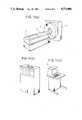
- FIGS. 1 1(A) (B), and 1(C)depict the main components of a general X-ray CT.
- FIG. 1(A)illustrates a scanning gantry 1 and a patient table 2.
- Scanning gantry 1has therein an X-ray tube and an X-ray detecting device (not shown) which are disposed in confronting relation to each other across an opening.
- Table 2includes a cradle 4 axially slidable forward into opening 3, or backward out of opening 3, while carrying a subject or a patient (not shown) thereon, for exposure to the X-ray in gantry 1.
- FIG. 1(B)shows a computer for controlling the overall X-ray CT and effecting computations for image reconstruction, for example.
- FIG. 1(C)depicts an operator console having a variety of switches disposed on a panel thereof and selectively actuable for scanning operation of the X-ray CT, and a cathode ray tube 5 (hereinafter called "CRT").
- CRTcathode ray tube 5
- Pieces of informationare inputted normally through scanning operation on the operator console.
- Methods of entering such informationinclude (1) a conversation or interactive process using a keyboard on the operator console while watching messages displayed on the CRT of the console; and (2) process of selectively actuating the switches on the panel of the console.
- Method (1)is , however, disadvantageous, in that the operator must become experienced in keyboarding. It takes time to master operation of the CT. Numerous erroneous control operations tend to occur, since the operator is required to do the keyboarding while looking at the messages on the CRT.
- Method (2)has a problem, in that, that the switches cannot be properly actuated unless the operator knows the correct switching operating sequence.
- the scanning of the X-ray CTcomprise such conditions as field of view (FOV), scanning time, current to flow through the X-ray tube for generating an X-ray, slicing interval, thickness to be sliced, and other parameters.
- FOVfield of view
- scan parametersare set while watching messages displayed on the CRT in a conversation or interactive process, or (b) scan parameters are selected and set by various selecting and setting switches on the panel each time the patients are changed or scan parameters are changed.
- a predetermined combination of scanning conditions or parameterswhich are tailored to a particular hospital, is usually available for most of the scans thereat.
- scan parametershave to be established in each scanning operation, even when scanning is to be effected under routine conditions.
- prior parameter settinghas been tedious and time consuming.
- an object of the inventionis the improve the prior art, and to overcome the aforementioned and other deficiencies and disadvantages of the prior art.
- Another objectis to provide an operator console for X-ray computerized tomographs which allows various pieces of information to be entered easily without error, even when operated by a novice operator.
- an operator console for an X-ray Tomographcomprising a scan parameter display for displaying scan parameters for scanning operation of the X-ray tomograph, means for presetting the scan parameters, means for storing the preset scan parameters, switch means for manually changing the preset scan parameters, a plurality of scanning control switches for indicating the scanning operation, display output circuits for displaying controllable conditions and selected conditions of the scanning control switches, to allow the scanning control switches to be individually operated on, and means for controlling the display output circuits to display a controllable condition of a switch to be selected next according to a scanning control procedure and to display a selected condition of a selected switch and for controlling the presetting means to preset the scan parameters for each anatomy section to be scanned.
- FIGS. 1 (A), 1(B) and 1(C)are perspective view of major components of a general X-ray CT.
- FIG. 2is a block diagram of a circuit arrangement of an illustrative embodiment of the invention.
- FIG. 3is a front elevational view of the console of the embodiment.
- FIG. 4is an enlarged front elevational view of the embodiment of FIG. 3.
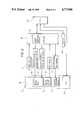
- FIG. 2is a block diagram depicting a circuit arrangement used in the operator console of the invention.
- the circuit arrangementcomprises a scanning control panel 10, a controller 30 for receiving scanning information from scanning control panel 10 and feeding data and control signal to scanning control panel 10, a keyboard 40, a cathode ray tube (CRT) monitor 50, for displaying images and characters, and a central processing unit 60 (called "CPU"), comprising a computer for issuing necessary data and control signals to the components of the operator console.
- CPUcentral processing unit 60
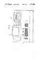
- FIG. 3depicts the exterior construction of the operator console with keyboard 40 in a front position, CRT 50 in a left upper position on a slanted surface behind keyboard 40, and scanning control panel 10 on the right of CRT 50.
- FIG. 4illustrates in greater detail scanning control panel 10, comprising a scanning control switch group 11, a scan parameter display 12 for displaying scan parameters (numbers "88" and "888" are shown) and manual change switches 13.
- Scanning control switch group 11comprises, for example, a switch 111 for registering a patient's data, a switch 112 for selecting a patient's orientation, a switch 113 for selecting an anatomy section to be scanned, a plurality of switches 114 for selecting scout and axial views and related switches, and power related switches 115 including a power supply switch and other switches.
- These switchesmay comprise pushbutton switches with light emitting means disposed in their caps.
- the light emitting meansmay comprise lamps, light emitting diodes, plasma display devices, etc. When such light emitting means is energized in the cap of a pushbutton switch, attention of the operator can be drawn to that particular switch which is energized.
- the light emitting meansneed not necessarily be disposed in tne switch caps, but may be located outside of and in the immediate vicinity of the caps.
- the scanning control switches just describedindicate, for example, a controllable condition when the lamps blink and a selected condition when the lamps are kept continuously energized.
- a controllable condition display output circuit 22(see FIG. 2) is responsive to information delivered from controller 30 for blinking the switches.
- a selected condition display output cirucit 23is responsive to information delivered from controller 30 for continuously energizing the switches.
- Controller 30supervises and controls an operation sequence and issues outputs indicative of blinking, continued energization, and de-energization, to the lamps in the switches according to a predetermined sequence.
- the scan parametersinclude a scanning time, a thickness to be sliced, a FOV, a tube current, a slicing interval, and other parameters; and are delivered from controller 30 to a scan parameter register 25 for enabling scan parameter display 12 to indicate the parameters in the numerals in the display areas. Since combinations of the scan parameters vary with anatomy sections to be scanned and scanning modes (transmissive image or sectional image), the scan parameters are displayed at first after an anatomy section to be scanned and a scanning mode, have been selected. Prior to such selection, the display areas are left blank. The values to be displayed thereat in the display areas have been preset.
- the scan parameterscan be preset by entering parameter values through keyboard 40 for each combination of an anatomy section to be scanned and scanning mode.
- the parameter presetting operationis controlled and praccessed by CPU 60. The preset scan parameters will remain unchanged most of the time once they are tailored according to the diagnostic procedure of the particular user (such as hospital or the like) when the tomograph is installed.
- controller 30When it is necessary to change any scan parameter from its preset value, manual change switches 13 are depressed. Input signals from the depressed manual change switches are delivered through input circuit 24 to controller 30, which then changes the corresponding scan parameter register 25. Since the content of a preset scan parameter data memory 31 in controller 30 is not rewritten, the original preset values are displayed again when the scanning mode is changed or a next patient scan starts. Controller 30 communicates with CPU 60, as required for the exchange of scanning conditions and sequence information. The preset scan parameters down loaded from CPU 60 into controller 30 when the system starts operating or the preset values are to be changed.
- the "new patient" switch 111(See FIG. 4) is blinking.
- switch 111is depressed, it is then continuously energized, and a list of patients appears on the CRT monitor 50 (see also FIG. 2).
- Keyboard 40is operated to enter necessary patient data.
- one of the patient orientation selector switches, which are now blinking, or "Head First" switch 112a or “Feet First” switch 112bis selected according to the orientation of the patient (i.e. whether the gantry 1 side is closer to the head or the feet). The selected switch is continuously energized while the other switch is de-energized.
- anatomy section selector switches 113start to blink.
- the "Head” switch 113ais depressed.
- the "Head” switch 113ais now continuously energized, and the other switches in the group of switches 113, are de-energized.
- the "Scout scan” switch 114a and the “Axial Scan” switch 114bthen blink. These switches serve to select a scanning mode indicating a transmissive image or a sectional image.
- the operatorgoes to the next step without changing the preset values.
- the item selector switch 13a in the manual change switches 13is depressed, to select an item to be changed, and a command from the item selector switch 13a is given through the input circuit 24 to the controller 30.
- the item selector switch 13ais depressed once, only the display unit on the left end is kept lit to indicate selection of the left end item while the other display units are turned darker.
- the "Scan Set” switchstarts blinking again when the system is ready for a next scanning operation. If the next scanning operation is to be made under the same condition, then the "Scan set” and “Start” switches are depressed as they blink. When it is necessary to change any scan parameter at this time, the manual change switch should be depressed while the "Scan Set” switch is blinking.
- each switchis provided with an indicator means for indicating a controllable condition to give the operator a switch operating procedure, and also with an indicator means for indicating a selected condition to allow the operator to know a current setting at a glance.
- the number of operating steps for a routine scanning operationcan be reduced by providing a capability for presetting scanning parameters for respective body areas to be scanned.
- the controllable conditionsmay be represented by dark lighting of the lamps in the switches, or blinking or dark lighting of indicators associated with respective switches.
- the selected conditionsmay be represented by continuous energization of indicators associated with respective switches.
- the preset scan parameter memoryis contained in controller 30 to reduce the number of occurences of communication between CPU 60 and controller 30, the preset scan parameter memory may be contained in CPU 60.
- the means for manually changing the scan parametersmay comprise an increment switch and a decrement switch for each parameter item. Moroever, with the present arrangement of the invention, the operator console for an X-ray CT, can easily be operated without any errors, even by a novice operator, in entering information through the operator console of the invention.
Landscapes
- Health & Medical Sciences (AREA)
- General Health & Medical Sciences (AREA)
- Toxicology (AREA)
- Apparatus For Radiation Diagnosis (AREA)
Abstract
Description
This is a continuation of U.S. Ser. No. 06/871179 filed 6-4-86 which is a continuation of Ser. No. 06/598976, filed 4-11-84 both abandoned.
1. Field of Invention
This invention relates to an X-ray computerized tomograph (hereinafter called "X-ray CT"), and more particularly to an operator console for inputting scanning conditions of the X-ray CT.
2. Description of Prior Art
Computerized tomographs are known in the art, which emit an X-ray beam at a number of angles in a plane across a subject or a patient, and determine a distribution of X-ray absorptivities of various body sections, and display the absorptivity distribution on a screen of a display unit.
FIGS. 1 1(A) (B), and 1(C) depict the main components of a general X-ray CT. FIG. 1(A) illustrates a scanning gantry 1 and a patient table 2. Scanning gantry 1 has therein an X-ray tube and an X-ray detecting device (not shown) which are disposed in confronting relation to each other across an opening. Table 2 includes a cradle 4 axially slidable forward into opening 3, or backward out of opening 3, while carrying a subject or a patient (not shown) thereon, for exposure to the X-ray in gantry 1.
FIG. 1(B) shows a computer for controlling the overall X-ray CT and effecting computations for image reconstruction, for example.
FIG. 1(C) depicts an operator console having a variety of switches disposed on a panel thereof and selectively actuable for scanning operation of the X-ray CT, and a cathode ray tube 5 (hereinafter called "CRT").
In operating the X-ray CT of the above configuration, pieces of information, such as scanning conditions, are inputted normally through scanning operation on the operator console. Methods of entering such information include (1) a conversation or interactive process using a keyboard on the operator console while watching messages displayed on the CRT of the console; and (2) process of selectively actuating the switches on the panel of the console.
Method (1) is , however, disadvantageous, in that the operator must become experienced in keyboarding. It takes time to master operation of the CT. Numerous erroneous control operations tend to occur, since the operator is required to do the keyboarding while looking at the messages on the CRT.
Method (2) has a problem, in that, that the switches cannot be properly actuated unless the operator knows the correct switching operating sequence.
The scanning of the X-ray CT comprise such conditions as field of view (FOV), scanning time, current to flow through the X-ray tube for generating an X-ray, slicing interval, thickness to be sliced, and other parameters. In known CTs, (a) scan parameters are set while watching messages displayed on the CRT in a conversation or interactive process, or (b) scan parameters are selected and set by various selecting and setting switches on the panel each time the patients are changed or scan parameters are changed. Once an anatomy section to be scanned has been determined, a predetermined combination of scanning conditions or parameters, which are tailored to a particular hospital, is usually available for most of the scans thereat. However, with the above processes (a) and (b), scan parameters have to be established in each scanning operation, even when scanning is to be effected under routine conditions. Thus, prior parameter setting has been tedious and time consuming.
Accordingly, an object of the invention is the improve the prior art, and to overcome the aforementioned and other deficiencies and disadvantages of the prior art.
Another object is to provide an operator console for X-ray computerized tomographs which allows various pieces of information to be entered easily without error, even when operated by a novice operator.
The foregoing and other objects are attained in the invention which encompasses an operator console for an X-ray Tomograph, comprising a scan parameter display for displaying scan parameters for scanning operation of the X-ray tomograph, means for presetting the scan parameters, means for storing the preset scan parameters, switch means for manually changing the preset scan parameters, a plurality of scanning control switches for indicating the scanning operation, display output circuits for displaying controllable conditions and selected conditions of the scanning control switches, to allow the scanning control switches to be individually operated on, and means for controlling the display output circuits to display a controllable condition of a switch to be selected next according to a scanning control procedure and to display a selected condition of a selected switch and for controlling the presetting means to preset the scan parameters for each anatomy section to be scanned.
FIGS. 1 (A), 1(B) and 1(C) are perspective view of major components of a general X-ray CT.
FIG. 2 is a block diagram of a circuit arrangement of an illustrative embodiment of the invention.
FIG. 3 is a front elevational view of the console of the embodiment.
FIG. 4 is an enlarged front elevational view of the embodiment of FIG. 3.
FIG. 2 is a block diagram depicting a circuit arrangement used in the operator console of the invention. The circuit arrangement comprises ascanning control panel 10, acontroller 30 for receiving scanning information fromscanning control panel 10 and feeding data and control signal to scanningcontrol panel 10, akeyboard 40, a cathode ray tube (CRT)monitor 50, for displaying images and characters, and a central processing unit 60 (called "CPU"), comprising a computer for issuing necessary data and control signals to the components of the operator console.
FIG. 3 depicts the exterior construction of the operator console withkeyboard 40 in a front position, CRT 50 in a left upper position on a slanted surface behindkeyboard 40, and scanningcontrol panel 10 on the right of CRT 50.
FIG. 4 illustrates in greater detailscanning control panel 10, comprising a scanningcontrol switch group 11, ascan parameter display 12 for displaying scan parameters (numbers "88" and "888" are shown) andmanual change switches 13.
Scanningcontrol switch group 11 comprises, for example, aswitch 111 for registering a patient's data, aswitch 112 for selecting a patient's orientation, aswitch 113 for selecting an anatomy section to be scanned, a plurality ofswitches 114 for selecting scout and axial views and related switches, and powerrelated switches 115 including a power supply switch and other switches.
These switches may comprise pushbutton switches with light emitting means disposed in their caps. The light emitting means may comprise lamps, light emitting diodes, plasma display devices, etc. When such light emitting means is energized in the cap of a pushbutton switch, attention of the operator can be drawn to that particular switch which is energized. The light emitting means need not necessarily be disposed in tne switch caps, but may be located outside of and in the immediate vicinity of the caps.
Although the pushbutton switches are better in controllability, switches based on other principles may be used.
The scanning control switches just described, indicate, for example, a controllable condition when the lamps blink and a selected condition when the lamps are kept continuously energized. A controllable condition display output circuit 22 (see FIG. 2) is responsive to information delivered fromcontroller 30 for blinking the switches. A selected conditiondisplay output cirucit 23 is responsive to information delivered fromcontroller 30 for continuously energizing the switches. When a desired switch is depressed, its output is read through aninput circuit 21 bycontroller 30. Inputs from switches other than the blinking switches, are determined as being ineffective by thecontroller 30.Controller 30 supervises and controls an operation sequence and issues outputs indicative of blinking, continued energization, and de-energization, to the lamps in the switches according to a predetermined sequence.
The scan parameters include a scanning time, a thickness to be sliced, a FOV, a tube current, a slicing interval, and other parameters; and are delivered fromcontroller 30 to ascan parameter register 25 for enablingscan parameter display 12 to indicate the parameters in the numerals in the display areas. Since combinations of the scan parameters vary with anatomy sections to be scanned and scanning modes (transmissive image or sectional image), the scan parameters are displayed at first after an anatomy section to be scanned and a scanning mode, have been selected. Prior to such selection, the display areas are left blank. The values to be displayed thereat in the display areas have been preset. The scan parameters can be preset by entering parameter values throughkeyboard 40 for each combination of an anatomy section to be scanned and scanning mode. The parameter presetting operation is controlled and prccessed byCPU 60. The preset scan parameters will remain unchanged most of the time once they are tailored according to the diagnostic procedure of the particular user (such as hospital or the like) when the tomograph is installed.
When it is necessary to change any scan parameter from its preset value,manual change switches 13 are depressed. Input signals from the depressed manual change switches are delivered throughinput circuit 24 tocontroller 30, which then changes the correspondingscan parameter register 25. Since the content of a preset scanparameter data memory 31 incontroller 30 is not rewritten, the original preset values are displayed again when the scanning mode is changed or a next patient scan starts.Controller 30 communicates withCPU 60, as required for the exchange of scanning conditions and sequence information. The preset scan parameters down loaded fromCPU 60 intocontroller 30 when the system starts operating or the preset values are to be changed.
Operation of the operator console will now be described. When the X-ray CT is ready for a next patient, the "new patient" switch 111 (See FIG. 4) is blinking. Whenswitch 111 is depressed, it is then continuously energized, and a list of patients appears on the CRT monitor 50 (see also FIG. 2).Keyboard 40 is operated to enter necessary patient data. Then, one of the patient orientation selector switches, which are now blinking, or "Head First"switch 112a or "Feet First"switch 112b is selected according to the orientation of the patient (i.e. whether the gantry 1 side is closer to the head or the feet). The selected switch is continuously energized while the other switch is de-energized. Thereafter, anatomy section selector switches 113 start to blink. When the head is to be scanned, the "Head"switch 113a is depressed. The "Head"switch 113a is now continuously energized, and the other switches in the group ofswitches 113, are de-energized. The "Scout scan" switch 114a and the "Axial Scan"switch 114b then blink. These switches serve to select a scanning mode indicating a transmissive image or a sectional image.
The following description is based on the selection of the axial scan. When the "Axial Scan"switch 114b is depressed, this switch is energized, and the "scout Scan" switch 114a is de-energized. Simultaneously, the "Single", "Multi-1" and "Multi-2" switches inswitch group 114d related to the "Axial Scan"switch 114b start being energized. At this time, preset values for the axial scan of the head are indicated on thescan parameter display 12.
In most cases, the operator goes to the next step without changing the preset values. When any preset value is to be changed, theitem selector switch 13a in the manual change switches 13, is depressed, to select an item to be changed, and a command from theitem selector switch 13a is given through theinput circuit 24 to thecontroller 30. When theitem selector switch 13a is depressed once, only the display unit on the left end is kept lit to indicate selection of the left end item while the other display units are turned darker.
Whenitem selector switch 13a is depressed once more, the second display unit from the left end is turned on to indicate selection of the second item, with the left hand end display unit turned darker. After an item to be changed has been selected in this manner, the up scrollingswitch 13b or thedown scrolling switch 13a, is depressed to increment or decrement the value in the selected item. The up scrolling or down scrolling switch is released when the desired value is reached. Upon elapse of a few seconds, the darker display units are restored, and the manual change procedure is brought to an end.
When the "Single" switch in the blinkingswitch group 114d is depressed, the other "Multi-1" and "Multi-2" switches are de-energized. Then, the "Scan Set" switch ingroup 114e blinks. After confirming that the settings selected up to now are correct, the "Scan Set" switch is depressed. The "Scan set" is now continuously energized, and the system is brought into a stage to prepare for a scanning operation. When the system is ready for scanning operation, a "Start" switch starts blinking. The scanning operation is started by depressing the "Start" switch, which will remain energized until the scanning operation is completed.
When the scanning operation is over, a reconstructed tomographic image is displayed on CRT monitor 50 after a short time interval The "Scan Set" switch starts blinking again when the system is ready for a next scanning operation. If the next scanning operation is to be made under the same condition, then the "Scan set" and "Start" switches are depressed as they blink. When it is necessary to change any scan parameter at this time, the manual change switch should be depressed while the "Scan Set" switch is blinking.
As described above, each switch is provided with an indicator means for indicating a controllable condition to give the operator a switch operating procedure, and also with an indicator means for indicating a selected condition to allow the operator to know a current setting at a glance. The number of operating steps for a routine scanning operation can be reduced by providing a capability for presetting scanning parameters for respective body areas to be scanned. The controllable conditions may be represented by dark lighting of the lamps in the switches, or blinking or dark lighting of indicators associated with respective switches. The selected conditions may be represented by continuous energization of indicators associated with respective switches.
While in the illustrative embodiment, the preset scan parameter memory is contained incontroller 30 to reduce the number of occurences of communication betweenCPU 60 andcontroller 30, the preset scan parameter memory may be contained inCPU 60. Also, the means for manually changing the scan parameters may comprise an increment switch and a decrement switch for each parameter item. Moroever, with the present arrangement of the invention, the operator console for an X-ray CT, can easily be operated without any errors, even by a novice operator, in entering information through the operator console of the invention.
The foregoing description is illustrative of the principles of the invention. Numerous modifications and extensions thereof would be apparent to the worker skilled in the art. All such modifications and extensions are to be considered to be within the spirit and scope of the invention.
Claims (5)
1. An operator console for a tomograph apparatus comprising an imaging source, an imaging detector and a source scanning means, said console comprising
a plurality of scanning control switches, each switch being equipped with a light emitter having a blinking light state, a continuous light state and an unlit state, said blinking light indicating a scanning step to be selected, said continuous light indicating a selected scanning step which has been initiated, wherein said plurality of scanning control switches are divided into a plurality of groups with each group comprising a plurality of scanning control switches, and wherein for each successive group one or more of said plurality of switches having the scanning steps to be selected displayed on the light emitters as blinking lights are successively operated by an operator to produce signals for causing the tomograph to perform the respective scanning steps in the successive groups, whereupon the light emitters on the just operated switches change from a a blinking light to a continuous light to indicate that the scanning step just selected has been initiated;
control means responsive to operation of a scanning control switch for concurrently generating a selected display signal and a plurality of to be selected display signals;
a selected display output means, responsive to said selected display signal applied from said control means, for causing the light emitter asociated with the just operated scanning control switch to change from a blinking light to a continuous light to indicate that the respective control switch has been just operated and the selected scanning step has been initiated; and
a to be selected dispaly output means, responsive to said plurality of to be selected display signals applied from said control means, for concurrently causing the light emitters associated with a group of said plurality of scanning control switches next to be operated to change from an unlit state to blinking lights to indicate that one or more of the respective control switches are to be next operated in that group, such that said operator operates in sequence at least one switch in successive groups of said plurality of to be operated switches having the associated light emitter having blinking lights to cause desired scanning steps, whereupon the just operated one or more of the plurality of switches in each successive group have their associated light emitters causes to be changed from blinking lights to continuous lights, and concurrently another successive group of said plurality of to be operated switches have their associated light emitters caused to be changed from unlit states to blinking lights to indicate to the operator the next successive group of said plurality of switches to be operated.
2. The console of claim 1, wherein said switches are push button switches.
3. An operator console for a tomograph apparatus comprising an imaging source, an imaging detector and a source scanning means, said console comprising
a plurality of scanning control switchs, each switch being equipped with a light emitter having a blinking light state, a continous light state and an unlit state, said blinking state indicating a scanning step to be selected, said continuous light indicating a selected scanning step which has been initiated, wherein said plurality of scanning control switches are divided into a plurality of groups with each group comprising a plurality of scanning control switches, and wherein for each successive group one or more of said plurality of switches having the scanning steps to be selected displayed on the light emitters as blinking lights are successively operated by an operator to produce signals for causing the tomograph to perform the respective scanning steps in the successive groups, whereupon the light emitters on the just operated switches change from a blinking light to a continuous light to indicate that the scanning step just selected has been initiated, the scanning control switches incorporating switches for designating the selection of an object to be imaged;
a storage means for storing scan parameters preset for each section of the object to be imaged;
a scan parameter display means for displaying the scan parameters;
control means responsive to operation of a scanning control switch for concurrently generating a selected display signal and a plurality of to be selected dispaly signals, and said control means responsive to operation of a scanning control switch designating the section of the object to be imaged for generating a scan parameter display signal;
means responsive to said scan parameter display signal for displaying corresponding parameters of said preset scan parameters stored in said storage means;
a selected display output means, responsive to said selected display signal applied from said control means, for causing the light emitter associated with the just operated scanning control switch to change from a blinking light to a continuous light to indicate that the respective control switch has just been operated and the indicated scanning step has been initiated; and
a to be selected display output means, responsive to said plurality of to be selected display signals applied from said control means, for concurrently causing the light emitters associated with a group of said plurality of scanning control switches next to be operated to change form an unlit state to blinking lights to indicate that one or more of the respective control switches are to be next operated in that group, such that said operator operates in sequence at least one switch in successive groups of said plurality of to be operated switches having the associated light emitter having blinking lights to cause desired scanning steps, whereupon the just operated one or more of the plurality of switches in each successive group have their associated light emitters caused to be changed from blinking lights to continuous lights, and concurrently another successive groups of said plurality of to be operated switches have their associated light emitters caused to be changed from unlit states to blinking lights to indicate to the operator the next successive group of said plurality of switches to be operated.
4. The console of claim 3, wherein said switches are push button switches.
5. The console of claim 3, wherein said scan parameter display means includes a register which temporarily holds scan parameters that are used for displaying purposes, the contents of the register being alterable by said operator.
Applications Claiming Priority (2)
| Application Number | Priority Date | Filing Date | Title |
|---|---|---|---|
| JP58237386AJPS60129034A (en) | 1983-12-16 | 1983-12-16 | Operation table of x-ray tomographic apparatus |
| JP58-237386 | 1983-12-16 |
Related Parent Applications (1)
| Application Number | Title | Priority Date | Filing Date |
|---|---|---|---|
| US06871179Continuation | 1986-06-04 |
Publications (1)
| Publication Number | Publication Date |
|---|---|
| US4773086Atrue US4773086A (en) | 1988-09-20 |
Family
ID=17014617
Family Applications (1)
| Application Number | Title | Priority Date | Filing Date |
|---|---|---|---|
| US07/117,363Expired - LifetimeUS4773086A (en) | 1983-12-16 | 1987-10-28 | Operator console for X-ray tomographs |
Country Status (4)
| Country | Link |
|---|---|
| US (1) | US4773086A (en) |
| JP (1) | JPS60129034A (en) |
| DE (1) | DE3414518A1 (en) |
| GB (1) | GB2151890B (en) |
Cited By (68)
| Publication number | Priority date | Publication date | Assignee | Title |
|---|---|---|---|---|
| US5018178A (en)* | 1988-03-08 | 1991-05-21 | Kabushiki Kaisha Toshiba | Medical apparatus with a control console |
| US5231651A (en)* | 1990-03-30 | 1993-07-27 | Kabushiki Kaisha Toshiba | X-ray computerized tomographic method and apparatus with simple programmable anatomical selecting operation |
| USD355717S (en) | 1993-04-01 | 1995-02-21 | Kabushiki Kaisha Toshiba | Combined x-ray control board and support structure for medical treatment |
| USD356792S (en) | 1993-04-01 | 1995-03-28 | Kabushiki Kaisha Toshiba | X-ray control board for medical treatment |
| US5608772A (en)* | 1993-11-26 | 1997-03-04 | Kabushiki Kaisha Toshiba | Computerized tomography apparatus |
| WO2000019783A1 (en)* | 1998-09-25 | 2000-04-06 | Fluoroscan Imaging Systems, Inc. | Miniature c-arm apparatus with multiple x-ray indicators |
| WO2000031522A1 (en)* | 1998-11-20 | 2000-06-02 | Direct Radiography Corp | Interactive digital radiographic system |
| DE10201321A1 (en)* | 2002-01-15 | 2003-07-31 | Siemens Ag | Computer tomography instrument has an improved control interface and filter electronics to allow improved extraction of image information from image data thus enabling a reduction in the radiation dose received by a patient |
| US20040081341A1 (en)* | 2002-07-18 | 2004-04-29 | Dieter Cherek | Method and arrangement for positioning a patient in a medical diagnosis or therapy device |
| US20040101095A1 (en)* | 2002-11-27 | 2004-05-27 | Hologic Inc. | Full field mammography with tissue exposure control, tomosynthesis, and dynamic field of view processing |
| US20040162484A1 (en)* | 2003-02-18 | 2004-08-19 | Nemoto Kyorindo Co., Ltd. | Liquid injector with appropriate operating conditions set by selecting displayed image |
| US6801594B1 (en)* | 1997-11-26 | 2004-10-05 | General Electric Company | Computed tomography fluoroscopy system |
| US20050100201A1 (en)* | 2003-10-24 | 2005-05-12 | Robert Mayer | Device for monitoring an operating parameter of a medical device |
| US20070211844A1 (en)* | 2005-09-15 | 2007-09-13 | Yilun Shi | Radiographing plan assisting method and x-ray ct system |
| US20090003519A1 (en)* | 2004-11-26 | 2009-01-01 | Kenneth Defreitas | Integrated Multi-Mode Mammography/Tomosynthesis X-Ray System And Method |
| CN100457042C (en)* | 2005-09-15 | 2009-02-04 | 上海西门子医疗器械有限公司 | Scanning position parameter regulating method for medical imaging system |
| US20090087069A1 (en)* | 2007-09-27 | 2009-04-02 | Fujifilm Corporation | Apparatus and method for processing radiation image |
| US20090262896A1 (en)* | 2008-04-22 | 2009-10-22 | Siemens Aktiengesellschaft | User interface of an x-ray system and method for manufacturing such an user interface |
| US20090296882A1 (en)* | 2002-11-27 | 2009-12-03 | Hologic, Inc. | Image Handling And Display In X-Ray Mammography And Tomosynthess |
| US20090323892A1 (en)* | 2008-06-24 | 2009-12-31 | Georgia Hitzke | Breast Tomosynthesis System With Shifting Face Shield |
| CN100581472C (en)* | 2005-01-31 | 2010-01-20 | 西门子公司 | Method and apparatus for controlling image mode |
| US7991106B2 (en) | 2008-08-29 | 2011-08-02 | Hologic, Inc. | Multi-mode tomosynthesis/mammography gain calibration and image correction using gain map information from selected projection angles |
| US8131049B2 (en) | 2007-09-20 | 2012-03-06 | Hologic, Inc. | Breast tomosynthesis with display of highlighted suspected calcifications |
| US8155421B2 (en) | 2004-11-15 | 2012-04-10 | Hologic, Inc. | Matching geometry generation and display of mammograms and tomosynthesis images |
| US20130208868A1 (en)* | 2012-02-15 | 2013-08-15 | Samsung Electronics Co., Ltd. | X-ray device and method for controlling the same |
| US8787522B2 (en) | 2010-10-05 | 2014-07-22 | Hologic, Inc | Upright x-ray breast imaging with a CT mode, multiple tomosynthesis modes, and a mammography mode |
| US8897535B2 (en) | 2002-11-27 | 2014-11-25 | Hologic, Inc. | System and method for generating a 2D image from a tomosynthesis data set |
| US9095306B2 (en) | 2002-11-27 | 2015-08-04 | Hologic, Inc. | Image handling and display in X-ray mammography and tomosynthesis |
| US9180312B2 (en) | 2005-11-18 | 2015-11-10 | Hologic, Inc. | Brachytherapy device for asymmetrical irradiation of a body cavity |
| US9248311B2 (en) | 2009-02-11 | 2016-02-02 | Hologic, Inc. | System and method for modifying a flexibility of a brachythereapy catheter |
| US9498175B2 (en) | 2002-11-27 | 2016-11-22 | Hologic, Inc. | System and method for low dose tomosynthesis |
| US9579524B2 (en) | 2009-02-11 | 2017-02-28 | Hologic, Inc. | Flexible multi-lumen brachytherapy device |
| US9623260B2 (en) | 2004-11-05 | 2017-04-18 | Theragenics Corporation | Expandable brachytherapy device |
| US9805507B2 (en) | 2012-02-13 | 2017-10-31 | Hologic, Inc | System and method for navigating a tomosynthesis stack using synthesized image data |
| US10008184B2 (en) | 2005-11-10 | 2018-06-26 | Hologic, Inc. | System and method for generating a 2D image using mammography and/or tomosynthesis image data |
| US10022557B2 (en) | 2010-09-30 | 2018-07-17 | Hologic, Inc. | Using a guided member to facilitate brachytherapy device swap |
| US10207126B2 (en) | 2009-05-11 | 2019-02-19 | Cytyc Corporation | Lumen visualization and identification system for multi-lumen balloon catheter |
| US10342992B2 (en) | 2011-01-06 | 2019-07-09 | Hologic, Inc. | Orienting a brachytherapy applicator |
| US10573276B2 (en) | 2011-11-27 | 2020-02-25 | Hologic, Inc. | System and method for generating a 2D image using mammography and/or tomosynthesis image data |
| US10638994B2 (en) | 2002-11-27 | 2020-05-05 | Hologic, Inc. | X-ray mammography with tomosynthesis |
| US10792003B2 (en) | 2010-10-05 | 2020-10-06 | Hologic, Inc. | X-ray breast tomosynthesis enhancing spatial resolution including in the thickness direction of a flattened breast |
| US10881359B2 (en) | 2017-08-22 | 2021-01-05 | Hologic, Inc. | Computed tomography system for imaging multiple anatomical targets |
| US11076820B2 (en) | 2016-04-22 | 2021-08-03 | Hologic, Inc. | Tomosynthesis with shifting focal spot x-ray system using an addressable array |
| US11090017B2 (en) | 2018-09-13 | 2021-08-17 | Hologic, Inc. | Generating synthesized projection images for 3D breast tomosynthesis or multi-mode x-ray breast imaging |
| US11403483B2 (en) | 2017-06-20 | 2022-08-02 | Hologic, Inc. | Dynamic self-learning medical image method and system |
| US11406332B2 (en) | 2011-03-08 | 2022-08-09 | Hologic, Inc. | System and method for dual energy and/or contrast enhanced breast imaging for screening, diagnosis and biopsy |
| US11419569B2 (en) | 2017-08-16 | 2022-08-23 | Hologic, Inc. | Image quality compliance tool |
| US11419565B2 (en) | 2014-02-28 | 2022-08-23 | IIologic, Inc. | System and method for generating and displaying tomosynthesis image slabs |
| US11445993B2 (en) | 2017-03-30 | 2022-09-20 | Hologic, Inc. | System and method for targeted object enhancement to generate synthetic breast tissue images |
| US11455754B2 (en) | 2017-03-30 | 2022-09-27 | Hologic, Inc. | System and method for synthesizing low-dimensional image data from high-dimensional image data using an object grid enhancement |
| US11452486B2 (en) | 2006-02-15 | 2022-09-27 | Hologic, Inc. | Breast biopsy and needle localization using tomosynthesis systems |
| US11471118B2 (en) | 2020-03-27 | 2022-10-18 | Hologic, Inc. | System and method for tracking x-ray tube focal spot position |
| US11510306B2 (en) | 2019-12-05 | 2022-11-22 | Hologic, Inc. | Systems and methods for improved x-ray tube life |
| US11589944B2 (en) | 2013-03-15 | 2023-02-28 | Hologic, Inc. | Tomosynthesis-guided biopsy apparatus and method |
| US11701199B2 (en) | 2009-10-08 | 2023-07-18 | Hologic, Inc. | Needle breast biopsy system and method of use |
| US11775156B2 (en) | 2010-11-26 | 2023-10-03 | Hologic, Inc. | User interface for medical image review workstation |
| US11786191B2 (en) | 2021-05-17 | 2023-10-17 | Hologic, Inc. | Contrast-enhanced tomosynthesis with a copper filter |
| US11957497B2 (en) | 2017-03-30 | 2024-04-16 | Hologic, Inc | System and method for hierarchical multi-level feature image synthesis and representation |
| US12029602B2 (en) | 2013-10-24 | 2024-07-09 | Hologic, Inc. | System and method for navigating x-ray guided breast biopsy |
| US12170140B2 (en) | 2018-11-25 | 2024-12-17 | Hologic, Inc. | Customizable multimodality image hanging protocols |
| US12191027B2 (en) | 2019-03-29 | 2025-01-07 | Hologic, Inc. | Snip-triggered digital image report generation |
| US12186119B2 (en) | 2021-10-05 | 2025-01-07 | Hologic, Inc. | Interactive model interface for image selection in medical imaging systems |
| US12211608B2 (en) | 2013-03-15 | 2025-01-28 | Hologic, Inc. | System and method for navigating a tomosynthesis stack including automatic focusing |
| US12207963B2 (en) | 2018-09-28 | 2025-01-28 | Hologic, Inc. | Image generation by high density element suppression |
| US12236597B2 (en) | 2021-11-29 | 2025-02-25 | Hologic, Inc. | Systems and methods for correlating objects of interest |
| US12236582B2 (en) | 2018-09-24 | 2025-02-25 | Hologic, Inc. | Breast mapping and abnormality localization |
| US12254586B2 (en) | 2021-10-25 | 2025-03-18 | Hologic, Inc. | Auto-focus tool for multimodality image review |
| US12414217B2 (en) | 2022-02-07 | 2025-09-09 | Hologic, Inc. | Systems and methods for adaptively controlling filament current in an X-ray tube |
Families Citing this family (8)
| Publication number | Priority date | Publication date | Assignee | Title |
|---|---|---|---|---|
| DE8514524U1 (en)* | 1985-05-14 | 1986-09-25 | Fritz Hofmann GmbH für Elektrotechnik, 8520 Erlangen | X-ray generator with operator guidance |
| JPH0628661B2 (en)* | 1985-09-10 | 1994-04-20 | 株式会社東芝 | X-ray CT system |
| DE3624901A1 (en)* | 1986-07-23 | 1988-01-28 | Picker Int Gmbh | X-RAY SYSTEM FOR X-RAY DIAGNOSTICS AND / OR X-RAY TREATMENT |
| JP2612116B2 (en)* | 1991-09-27 | 1997-05-21 | 株式会社東芝 | CT device |
| US5394871A (en)* | 1992-06-25 | 1995-03-07 | Siemens Aktiengesellschaft | Medical diagnostics installation |
| EP1378869B1 (en) | 1997-10-06 | 2014-08-06 | Hitachi-Omron Terminal Solutions, Corp. | Leaflets handling apparatus |
| JP4965934B2 (en)* | 2006-08-18 | 2012-07-04 | 株式会社東芝 | X-ray diagnostic imaging equipment |
| JP7567357B2 (en)* | 2020-10-23 | 2024-10-16 | 株式会社島津製作所 | Radiography equipment |
Citations (10)
| Publication number | Priority date | Publication date | Assignee | Title |
|---|---|---|---|---|
| US3187321A (en)* | 1961-05-11 | 1965-06-01 | Bunker Ramo | Operator-computer communication console |
| US3916192A (en)* | 1973-10-05 | 1975-10-28 | Siemens Ag | X-ray diagnostic apparatus including a control panel having operating keys for the organwise-programmed setting of exposure data |
| DE2655661A1 (en)* | 1976-12-08 | 1978-06-15 | Siemens Ag | Radiation diagnosis instrument for transverse plane images - uses X=ray appts. with image amplifier and patient positioned on rolling table |
| US4137571A (en)* | 1976-10-22 | 1979-01-30 | Siemens Aktiengesellschaft | Program control apparatus for the photographic operating sequence of an x-ray photographic installation |
| US4160906A (en)* | 1977-06-23 | 1979-07-10 | General Electric Company | Anatomically coordinated user dominated programmer for diagnostic x-ray apparatus |
| US4234928A (en)* | 1978-02-27 | 1980-11-18 | Siemens Aktiengesellschaft | X-ray diagnostic installation for x-ray photographs |
| US4247777A (en)* | 1977-06-20 | 1981-01-27 | Siemens Aktiengesellschaft | Operating console for an x-ray diagnostic installation |
| US4250386A (en)* | 1977-10-24 | 1981-02-10 | Siemens Aktiengesellschaft | X-ray diagnostic installation for X-ray photographs |
| US4251729A (en)* | 1978-03-03 | 1981-02-17 | Siemens Aktiengesellschaft | X-ray diagnostic installation for X-ray photographs |
| US4255662A (en)* | 1977-12-14 | 1981-03-10 | Siemens Aktiengesellschaft | X-ray diagnostic apparatus having operating keys for the organwise programmed setting of exposure data |
Family Cites Families (13)
| Publication number | Priority date | Publication date | Assignee | Title |
|---|---|---|---|---|
| GB1097094A (en)* | 1965-04-16 | 1967-12-29 | London Electricity Board | Improvements relating to an electrical switching system |
| GB1172222A (en)* | 1965-08-05 | 1969-11-26 | Mini Of Technology | Touch Displays |
| GB1479584A (en)* | 1973-07-14 | 1977-07-13 | Solartron Electronic Group | Data terminals and data processing apparatus incorporating such terminals |
| JPS5262385A (en)* | 1975-11-19 | 1977-05-23 | Mitsui Toatsu Chem Inc | Production of resin laminated structures |
| JPS5516066A (en)* | 1978-07-24 | 1980-02-04 | Sony Corp | Grease |
| US4177744A (en)* | 1978-07-28 | 1979-12-11 | The Singer Company | Digital override control of bight and feed in a sewing machine |
| JPS5546289A (en)* | 1978-09-28 | 1980-03-31 | Shimadzu Corp | X-ray apparatus of photographing condition program establishing type |
| JPS5588745A (en)* | 1978-12-26 | 1980-07-04 | Tokyo Shibaura Electric Co | Method of controlling computerrtomography device |
| JPS5654796A (en)* | 1979-10-12 | 1981-05-14 | Toshiba Corp | X-ray diagnosing device |
| DE3012480C2 (en)* | 1980-03-31 | 1984-10-11 | Siemens AG, 1000 Berlin und 8000 München | Control panel keyboard |
| GB2074346B (en)* | 1980-04-17 | 1984-09-26 | Kenwood Mfg Co Ltd | Control device |
| DE3117153A1 (en)* | 1981-04-30 | 1982-11-18 | Philips Patentverwaltung Gmbh, 2000 Hamburg | X-RAY GENERATOR FOR CARRYING OUT RECORDING METHODS CONTAINING A SEQUENCE OF RECORDING STEPS |
| JPS5819938A (en)* | 1981-07-30 | 1983-02-05 | 大日本スクリ−ン製造株式会社 | Display method and device for menu part of digitizer |
- 1983
- 1983-12-16JPJP58237386Apatent/JPS60129034A/enactivePending
- 1984
- 1984-04-09GBGB08409147Apatent/GB2151890B/ennot_activeExpired
- 1984-04-17DEDE19843414518patent/DE3414518A1/enactiveGranted
- 1987
- 1987-10-28USUS07/117,363patent/US4773086A/ennot_activeExpired - Lifetime
Patent Citations (10)
| Publication number | Priority date | Publication date | Assignee | Title |
|---|---|---|---|---|
| US3187321A (en)* | 1961-05-11 | 1965-06-01 | Bunker Ramo | Operator-computer communication console |
| US3916192A (en)* | 1973-10-05 | 1975-10-28 | Siemens Ag | X-ray diagnostic apparatus including a control panel having operating keys for the organwise-programmed setting of exposure data |
| US4137571A (en)* | 1976-10-22 | 1979-01-30 | Siemens Aktiengesellschaft | Program control apparatus for the photographic operating sequence of an x-ray photographic installation |
| DE2655661A1 (en)* | 1976-12-08 | 1978-06-15 | Siemens Ag | Radiation diagnosis instrument for transverse plane images - uses X=ray appts. with image amplifier and patient positioned on rolling table |
| US4247777A (en)* | 1977-06-20 | 1981-01-27 | Siemens Aktiengesellschaft | Operating console for an x-ray diagnostic installation |
| US4160906A (en)* | 1977-06-23 | 1979-07-10 | General Electric Company | Anatomically coordinated user dominated programmer for diagnostic x-ray apparatus |
| US4250386A (en)* | 1977-10-24 | 1981-02-10 | Siemens Aktiengesellschaft | X-ray diagnostic installation for X-ray photographs |
| US4255662A (en)* | 1977-12-14 | 1981-03-10 | Siemens Aktiengesellschaft | X-ray diagnostic apparatus having operating keys for the organwise programmed setting of exposure data |
| US4234928A (en)* | 1978-02-27 | 1980-11-18 | Siemens Aktiengesellschaft | X-ray diagnostic installation for x-ray photographs |
| US4251729A (en)* | 1978-03-03 | 1981-02-17 | Siemens Aktiengesellschaft | X-ray diagnostic installation for X-ray photographs |
Cited By (157)
| Publication number | Priority date | Publication date | Assignee | Title |
|---|---|---|---|---|
| US5018178A (en)* | 1988-03-08 | 1991-05-21 | Kabushiki Kaisha Toshiba | Medical apparatus with a control console |
| US5231651A (en)* | 1990-03-30 | 1993-07-27 | Kabushiki Kaisha Toshiba | X-ray computerized tomographic method and apparatus with simple programmable anatomical selecting operation |
| USD355717S (en) | 1993-04-01 | 1995-02-21 | Kabushiki Kaisha Toshiba | Combined x-ray control board and support structure for medical treatment |
| USD356792S (en) | 1993-04-01 | 1995-03-28 | Kabushiki Kaisha Toshiba | X-ray control board for medical treatment |
| US5608772A (en)* | 1993-11-26 | 1997-03-04 | Kabushiki Kaisha Toshiba | Computerized tomography apparatus |
| US6801594B1 (en)* | 1997-11-26 | 2004-10-05 | General Electric Company | Computed tomography fluoroscopy system |
| US7006592B2 (en) | 1997-11-26 | 2006-02-28 | General Electric Company | Computed tomography fluoroscopy system |
| US20040247070A1 (en)* | 1997-11-26 | 2004-12-09 | Fazle Ali | Computed tomography fluoroscopy system |
| WO2000019783A1 (en)* | 1998-09-25 | 2000-04-06 | Fluoroscan Imaging Systems, Inc. | Miniature c-arm apparatus with multiple x-ray indicators |
| US6236712B1 (en)* | 1998-09-25 | 2001-05-22 | Fluoroscan Imaging Systems, Inc. | Miniature C-arm apparatus with multiple x-ray indicators |
| WO2000031522A1 (en)* | 1998-11-20 | 2000-06-02 | Direct Radiography Corp | Interactive digital radiographic system |
| US6614873B1 (en) | 1998-11-20 | 2003-09-02 | Direct Radiography Corp. | Interactive ditigal radiographic system |
| DE10201321A1 (en)* | 2002-01-15 | 2003-07-31 | Siemens Ag | Computer tomography instrument has an improved control interface and filter electronics to allow improved extraction of image information from image data thus enabling a reduction in the radiation dose received by a patient |
| DE10201321B4 (en)* | 2002-01-15 | 2011-02-24 | Siemens Ag | Computed tomography device and method with active adaptation of the measuring electronics |
| US8005183B2 (en) | 2002-01-15 | 2011-08-23 | Siemens Aktiengesellschaft | Computed tomography device with active adaptation of the measuring electronics |
| US20100111247A1 (en)* | 2002-01-15 | 2010-05-06 | Bjoern Heismann | Computed tomography device with active adaptation of the measuring electronics |
| US20040081341A1 (en)* | 2002-07-18 | 2004-04-29 | Dieter Cherek | Method and arrangement for positioning a patient in a medical diagnosis or therapy device |
| US7433503B2 (en)* | 2002-07-18 | 2008-10-07 | Siemens Aktiengesellschaft | Method and arrangement for positioning a patient in a medical diagnosis or therapy device |
| US11372534B2 (en) | 2002-11-27 | 2022-06-28 | Hologic, Inc. | Image handling and display in x-ray mammography and tomosynthesis |
| US10959694B2 (en) | 2002-11-27 | 2021-03-30 | Hologic, Inc. | Full field mammography with tissue exposure control, tomosynthesis, and dynamic field of view processing |
| US7430272B2 (en) | 2002-11-27 | 2008-09-30 | Hologic, Inc. | Full field mammography with tissue exposure control, tomosynthesis, and dynamic field of view processing |
| US20070030949A1 (en)* | 2002-11-27 | 2007-02-08 | Zhenxue Jing | Full field mammography with tissue exposure control, tomosynthesis, and dynamic field of view processing |
| US10638994B2 (en) | 2002-11-27 | 2020-05-05 | Hologic, Inc. | X-ray mammography with tomosynthesis |
| US20090010384A1 (en)* | 2002-11-27 | 2009-01-08 | Hologic, Inc. | Full field mammography with tissue exposure control, tomosynthesis, and dynamic field of view processing |
| US10452252B2 (en) | 2002-11-27 | 2019-10-22 | Hologic, Inc. | Image handling and display in X-ray mammography and tomosynthesis |
| US10413263B2 (en) | 2002-11-27 | 2019-09-17 | Hologic, Inc. | System and method for generating a 2D image from a tomosynthesis data set |
| US10296199B2 (en) | 2002-11-27 | 2019-05-21 | Hologic, Inc. | Image handling and display in X-Ray mammography and tomosynthesis |
| US10108329B2 (en) | 2002-11-27 | 2018-10-23 | Hologic, Inc. | Image handling and display in x-ray mammography and tomosynthesis |
| US20090296882A1 (en)* | 2002-11-27 | 2009-12-03 | Hologic, Inc. | Image Handling And Display In X-Ray Mammography And Tomosynthess |
| US10010302B2 (en) | 2002-11-27 | 2018-07-03 | Hologic, Inc. | System and method for generating a 2D image from a tomosynthesis data set |
| US9851888B2 (en) | 2002-11-27 | 2017-12-26 | Hologic, Inc. | Image handling and display in X-ray mammography and tomosynthesis |
| US7123684B2 (en)* | 2002-11-27 | 2006-10-17 | Hologic, Inc. | Full field mammography with tissue exposure control, tomosynthesis, and dynamic field of view processing |
| US7760853B2 (en) | 2002-11-27 | 2010-07-20 | Hologic, Inc. | Full field mammography with tissue exposure control, tomosynthesis, and dynamic field of view processing |
| US10719223B2 (en) | 2002-11-27 | 2020-07-21 | Hologic, Inc. | Image handling and display in X-ray mammography and tomosynthesis |
| US9808215B2 (en) | 2002-11-27 | 2017-11-07 | Hologic, Inc. | System and method for generating a 2D image from a tomosynthesis data set |
| US9498175B2 (en) | 2002-11-27 | 2016-11-22 | Hologic, Inc. | System and method for low dose tomosynthesis |
| US8416915B2 (en) | 2002-11-27 | 2013-04-09 | Hologic, Inc. | Full field mammography with tissue exposure control, tomosynthesis, and dynamic field of view processing |
| US7916915B2 (en) | 2002-11-27 | 2011-03-29 | Hologic, Inc | Image handling and display in x-ray mammography and tomosynthesis |
| US7949091B2 (en) | 2002-11-27 | 2011-05-24 | Hologic, Inc. | Full field mammography with tissue exposure control, tomosynthesis, and dynamic field of view processing |
| US20110135185A1 (en)* | 2002-11-27 | 2011-06-09 | Hologic, Inc. | Image handling and display in x-ray mammography and tomosynthesis |
| US9456797B2 (en) | 2002-11-27 | 2016-10-04 | Hologic, Inc. | System and method for generating a 2D image from a tomosynthesis data set |
| US20040101095A1 (en)* | 2002-11-27 | 2004-05-27 | Hologic Inc. | Full field mammography with tissue exposure control, tomosynthesis, and dynamic field of view processing |
| US9460508B2 (en) | 2002-11-27 | 2016-10-04 | Hologic, Inc. | Image handling and display in X-ray mammography and tomosynthesis |
| US9095306B2 (en) | 2002-11-27 | 2015-08-04 | Hologic, Inc. | Image handling and display in X-ray mammography and tomosynthesis |
| US9042612B2 (en) | 2002-11-27 | 2015-05-26 | Hologic, Inc. | Image handling and display in X-ray mammography and tomosynthesis |
| US8897535B2 (en) | 2002-11-27 | 2014-11-25 | Hologic, Inc. | System and method for generating a 2D image from a tomosynthesis data set |
| US8285020B2 (en) | 2002-11-27 | 2012-10-09 | Hologic, Inc. | Image handling and display in x-ray mammography and tomosynthesis |
| US8831171B2 (en) | 2002-11-27 | 2014-09-09 | Hologic, Inc. | Full field mammography with tissue exposure control, tomosynthesis, and dynamic field of view processing |
| US9827368B2 (en) | 2003-02-18 | 2017-11-28 | Nemoto Kyorindo Co., Ltd. | Liquid injector with appropriate operating conditions set by selecting displayed image |
| US8706199B2 (en) | 2003-02-18 | 2014-04-22 | Nemoto Kyorindo Co., Ltd. | Liquid injector with appropriate operating conditions set by selecting displayed image |
| US20040162484A1 (en)* | 2003-02-18 | 2004-08-19 | Nemoto Kyorindo Co., Ltd. | Liquid injector with appropriate operating conditions set by selecting displayed image |
| US8359087B2 (en)* | 2003-02-18 | 2013-01-22 | Nemoto Kyorindo Co., Ltd. | Liquid injector with appropriate operating conditions set by selecting displayed image |
| US20050100201A1 (en)* | 2003-10-24 | 2005-05-12 | Robert Mayer | Device for monitoring an operating parameter of a medical device |
| US11096644B2 (en) | 2003-11-26 | 2021-08-24 | Hologic, Inc. | X-ray mammography with tomosynthesis |
| US10413255B2 (en) | 2003-11-26 | 2019-09-17 | Hologic, Inc. | System and method for low dose tomosynthesis |
| US9623260B2 (en) | 2004-11-05 | 2017-04-18 | Theragenics Corporation | Expandable brachytherapy device |
| US8155421B2 (en) | 2004-11-15 | 2012-04-10 | Hologic, Inc. | Matching geometry generation and display of mammograms and tomosynthesis images |
| US9084579B2 (en) | 2004-11-15 | 2015-07-21 | Hologic, Inc. | Matching geometry generation and display of mammograms and tomosynthesis |
| US9811758B2 (en) | 2004-11-15 | 2017-11-07 | Hologic, Inc. | Matching geometry generation and display of mammograms and tomosynthesis |
| US10679095B2 (en) | 2004-11-15 | 2020-06-09 | Hologic, Inc. | Matching geometry generation and display of mammograms and tomosynthesis images |
| US10248882B2 (en) | 2004-11-15 | 2019-04-02 | Hologic, Inc. | Matching geometry generation and display of mammograms and tomosynthesis images |
| US8712127B2 (en) | 2004-11-15 | 2014-04-29 | Hologic, Inc. | Matching geometry generation and display of mammograms and tomosynthesis images |
| US9549709B2 (en) | 2004-11-26 | 2017-01-24 | Hologic, Inc. | Integrated multi-mode mammography/tomosynthesis X-ray system and method |
| US10194875B2 (en) | 2004-11-26 | 2019-02-05 | Hologic, Inc. | Integrated multi-mode mammography/tomosynthesis X-ray system and method |
| US20090003519A1 (en)* | 2004-11-26 | 2009-01-01 | Kenneth Defreitas | Integrated Multi-Mode Mammography/Tomosynthesis X-Ray System And Method |
| US11617548B2 (en) | 2004-11-26 | 2023-04-04 | Hologic, Inc. | Integrated multi-mode mammography/tomosynthesis x-ray system and method |
| US8565374B2 (en) | 2004-11-26 | 2013-10-22 | Hologic, Inc. | Integrated multi-mode mammography/tomosynthesis x-ray system and method |
| US10905385B2 (en) | 2004-11-26 | 2021-02-02 | Hologic, Inc. | Integrated multi-mode mammography/tomosynthesis x-ray system and method |
| US8175219B2 (en) | 2004-11-26 | 2012-05-08 | Hologic, Inc. | Integrated multi-mode mammography/tomosynthesis X-ray system and method |
| US9066706B2 (en) | 2004-11-26 | 2015-06-30 | Hologic, Inc. | Integrated multi-mode mammography/tomosynthesis x-ray system and method |
| US7869563B2 (en) | 2004-11-26 | 2011-01-11 | Hologic, Inc. | Integrated multi-mode mammography/tomosynthesis x-ray system and method |
| CN100581472C (en)* | 2005-01-31 | 2010-01-20 | 西门子公司 | Method and apparatus for controlling image mode |
| US7502445B2 (en)* | 2005-09-15 | 2009-03-10 | Ge Medical Systems Global Technology Company, Llc | Radiographing plan assisting method and X-ray CT system |
| CN100457042C (en)* | 2005-09-15 | 2009-02-04 | 上海西门子医疗器械有限公司 | Scanning position parameter regulating method for medical imaging system |
| US20070211844A1 (en)* | 2005-09-15 | 2007-09-13 | Yilun Shi | Radiographing plan assisting method and x-ray ct system |
| US10008184B2 (en) | 2005-11-10 | 2018-06-26 | Hologic, Inc. | System and method for generating a 2D image using mammography and/or tomosynthesis image data |
| US10413750B2 (en) | 2005-11-18 | 2019-09-17 | Hologic, Inc. | Brachytherapy device for facilitating asymmetrical irradiation of a body cavity |
| US9415239B2 (en) | 2005-11-18 | 2016-08-16 | Hologic, Inc. | Brachytherapy device for facilitating asymmetrical irradiation of a body cavity |
| US9180312B2 (en) | 2005-11-18 | 2015-11-10 | Hologic, Inc. | Brachytherapy device for asymmetrical irradiation of a body cavity |
| US11452486B2 (en) | 2006-02-15 | 2022-09-27 | Hologic, Inc. | Breast biopsy and needle localization using tomosynthesis systems |
| US11918389B2 (en) | 2006-02-15 | 2024-03-05 | Hologic, Inc. | Breast biopsy and needle localization using tomosynthesis systems |
| US12193853B2 (en) | 2006-02-15 | 2025-01-14 | Hologic, Inc. | Breast biopsy and needle localization using tomosynthesis systems |
| US8873824B2 (en) | 2007-09-20 | 2014-10-28 | Hologic, Inc. | Breast tomosynthesis with display of highlighted suspected calcifications |
| US9202275B2 (en) | 2007-09-20 | 2015-12-01 | Hologic, Inc. | Breast tomosynthesis with display of highlighted suspected calcifications |
| US8131049B2 (en) | 2007-09-20 | 2012-03-06 | Hologic, Inc. | Breast tomosynthesis with display of highlighted suspected calcifications |
| US8571292B2 (en) | 2007-09-20 | 2013-10-29 | Hologic Inc | Breast tomosynthesis with display of highlighted suspected calcifications |
| US8300913B2 (en)* | 2007-09-27 | 2012-10-30 | Fujifilm Corporation | Apparatus and method for processing radiation image |
| US20090087069A1 (en)* | 2007-09-27 | 2009-04-02 | Fujifilm Corporation | Apparatus and method for processing radiation image |
| US20090262896A1 (en)* | 2008-04-22 | 2009-10-22 | Siemens Aktiengesellschaft | User interface of an x-ray system and method for manufacturing such an user interface |
| US7835496B2 (en)* | 2008-04-22 | 2010-11-16 | Siemens Aktiengesellschaft | User interface of an X-ray system and method for manufacturing such an user interface |
| US7792245B2 (en) | 2008-06-24 | 2010-09-07 | Hologic, Inc. | Breast tomosynthesis system with shifting face shield |
| US20090323892A1 (en)* | 2008-06-24 | 2009-12-31 | Georgia Hitzke | Breast Tomosynthesis System With Shifting Face Shield |
| US7991106B2 (en) | 2008-08-29 | 2011-08-02 | Hologic, Inc. | Multi-mode tomosynthesis/mammography gain calibration and image correction using gain map information from selected projection angles |
| US9119593B2 (en) | 2008-08-29 | 2015-09-01 | Hologic, Inc. | Multi-mode tomosynthesis/mammography gain calibration and image correction using gain map information from selected projection angles |
| US8275090B2 (en) | 2008-08-29 | 2012-09-25 | Hologic, Inc. | Multi-mode tomosynthesis/mammography gain calibration and image correction using gain map information from selected projection angles |
| US9579524B2 (en) | 2009-02-11 | 2017-02-28 | Hologic, Inc. | Flexible multi-lumen brachytherapy device |
| US9248311B2 (en) | 2009-02-11 | 2016-02-02 | Hologic, Inc. | System and method for modifying a flexibility of a brachythereapy catheter |
| US10207126B2 (en) | 2009-05-11 | 2019-02-19 | Cytyc Corporation | Lumen visualization and identification system for multi-lumen balloon catheter |
| US11701199B2 (en) | 2009-10-08 | 2023-07-18 | Hologic, Inc. | Needle breast biopsy system and method of use |
| US12193886B2 (en) | 2009-10-08 | 2025-01-14 | Hologic, Inc. | Needle breast biopsy system and method of use |
| US10022557B2 (en) | 2010-09-30 | 2018-07-17 | Hologic, Inc. | Using a guided member to facilitate brachytherapy device swap |
| US9808214B2 (en) | 2010-10-05 | 2017-11-07 | Hologic, Inc. | Upright X-ray breast imaging with a CT mode, multiple tomosynthesis modes, and a mammography mode |
| US10792003B2 (en) | 2010-10-05 | 2020-10-06 | Hologic, Inc. | X-ray breast tomosynthesis enhancing spatial resolution including in the thickness direction of a flattened breast |
| US11478206B2 (en) | 2010-10-05 | 2022-10-25 | Hologic, Inc. | X-ray breast tomosynthesis enhancing spatial resolution including in the thickness direction of a flattened breast |
| US12144668B2 (en) | 2010-10-05 | 2024-11-19 | Hologic, Inc. | Upright x-ray breast imaging with a CT mode, multiple tomosynthesis modes, and a mammography mode |
| US8787522B2 (en) | 2010-10-05 | 2014-07-22 | Hologic, Inc | Upright x-ray breast imaging with a CT mode, multiple tomosynthesis modes, and a mammography mode |
| US11191502B2 (en) | 2010-10-05 | 2021-12-07 | Hologic, Inc. | Upright x-ray breast imaging with a CT mode, multiple tomosynthesis modes, and a mammography mode |
| US11775156B2 (en) | 2010-11-26 | 2023-10-03 | Hologic, Inc. | User interface for medical image review workstation |
| US10342992B2 (en) | 2011-01-06 | 2019-07-09 | Hologic, Inc. | Orienting a brachytherapy applicator |
| US12239471B2 (en) | 2011-03-08 | 2025-03-04 | Hologic, Inc. | System and method for dual energy and/or contrast enhanced breast imaging for screening, diagnosis and biopsy |
| US11406332B2 (en) | 2011-03-08 | 2022-08-09 | Hologic, Inc. | System and method for dual energy and/or contrast enhanced breast imaging for screening, diagnosis and biopsy |
| US11837197B2 (en) | 2011-11-27 | 2023-12-05 | Hologic, Inc. | System and method for generating a 2D image using mammography and/or tomosynthesis image data |
| US10573276B2 (en) | 2011-11-27 | 2020-02-25 | Hologic, Inc. | System and method for generating a 2D image using mammography and/or tomosynthesis image data |
| US11508340B2 (en) | 2011-11-27 | 2022-11-22 | Hologic, Inc. | System and method for generating a 2D image using mammography and/or tomosynthesis image data |
| US12183309B2 (en) | 2011-11-27 | 2024-12-31 | Hologic, Inc. | System and method for generating a 2D image using mammography and/or tomosynthesis image data |
| US10978026B2 (en) | 2011-11-27 | 2021-04-13 | Hologic, Inc. | System and method for generating a 2D image using mammography and/or tomosynthesis image data |
| US10410417B2 (en) | 2012-02-13 | 2019-09-10 | Hologic, Inc. | System and method for navigating a tomosynthesis stack using synthesized image data |
| US9805507B2 (en) | 2012-02-13 | 2017-10-31 | Hologic, Inc | System and method for navigating a tomosynthesis stack using synthesized image data |
| US12307604B2 (en) | 2012-02-13 | 2025-05-20 | Hologic, Inc. | System and method for navigating a tomosynthesis stack using synthesized image data |
| US10977863B2 (en) | 2012-02-13 | 2021-04-13 | Hologic, Inc. | System and method for navigating a tomosynthesis stack using synthesized image data |
| US11663780B2 (en) | 2012-02-13 | 2023-05-30 | Hologic Inc. | System and method for navigating a tomosynthesis stack using synthesized image data |
| US20130208868A1 (en)* | 2012-02-15 | 2013-08-15 | Samsung Electronics Co., Ltd. | X-ray device and method for controlling the same |
| US10085700B2 (en) | 2012-02-15 | 2018-10-02 | Samsung Electronics Co., Ltd. | X-ray device and method for controlling the same |
| US8781073B2 (en)* | 2012-02-15 | 2014-07-15 | Samsung Electronics Co., Ltd. | X-ray device and method for controlling the same |
| US11589944B2 (en) | 2013-03-15 | 2023-02-28 | Hologic, Inc. | Tomosynthesis-guided biopsy apparatus and method |
| US12064291B2 (en) | 2013-03-15 | 2024-08-20 | Hologic, Inc. | Tomosynthesis-guided biopsy in prone |
| US12211608B2 (en) | 2013-03-15 | 2025-01-28 | Hologic, Inc. | System and method for navigating a tomosynthesis stack including automatic focusing |
| US12324707B2 (en) | 2013-03-15 | 2025-06-10 | Hologic, Inc. | Tomosynthesis-guided biopsy in prone |
| US12029602B2 (en) | 2013-10-24 | 2024-07-09 | Hologic, Inc. | System and method for navigating x-ray guided breast biopsy |
| US11801025B2 (en) | 2014-02-28 | 2023-10-31 | Hologic, Inc. | System and method for generating and displaying tomosynthesis image slabs |
| US11419565B2 (en) | 2014-02-28 | 2022-08-23 | IIologic, Inc. | System and method for generating and displaying tomosynthesis image slabs |
| US11076820B2 (en) | 2016-04-22 | 2021-08-03 | Hologic, Inc. | Tomosynthesis with shifting focal spot x-ray system using an addressable array |
| US12211124B2 (en) | 2017-03-30 | 2025-01-28 | Hologic, Inc. | System and method for synthesizing low-dimensional image data from high-dimensional image data using an object grid enhancement |
| US11455754B2 (en) | 2017-03-30 | 2022-09-27 | Hologic, Inc. | System and method for synthesizing low-dimensional image data from high-dimensional image data using an object grid enhancement |
| US11983799B2 (en) | 2017-03-30 | 2024-05-14 | Hologic, Inc. | System and method for synthesizing low-dimensional image data from high-dimensional image data using an object grid enhancement |
| US11957497B2 (en) | 2017-03-30 | 2024-04-16 | Hologic, Inc | System and method for hierarchical multi-level feature image synthesis and representation |
| US11445993B2 (en) | 2017-03-30 | 2022-09-20 | Hologic, Inc. | System and method for targeted object enhancement to generate synthetic breast tissue images |
| US12070349B2 (en) | 2017-03-30 | 2024-08-27 | Hologic, Inc. | System and method for targeted object enhancement to generate synthetic breast tissue images |
| US11850021B2 (en) | 2017-06-20 | 2023-12-26 | Hologic, Inc. | Dynamic self-learning medical image method and system |
| US11403483B2 (en) | 2017-06-20 | 2022-08-02 | Hologic, Inc. | Dynamic self-learning medical image method and system |
| US12053319B2 (en) | 2017-08-16 | 2024-08-06 | Hologic, Inc. | Image quality compliance tool |
| US11672500B2 (en) | 2017-08-16 | 2023-06-13 | Hologic, Inc. | Image quality compliance tool |
| US11419569B2 (en) | 2017-08-16 | 2022-08-23 | Hologic, Inc. | Image quality compliance tool |
| US12011310B2 (en) | 2017-08-16 | 2024-06-18 | Hologic, Inc. | Image quality compliance tool |
| US10881359B2 (en) | 2017-08-22 | 2021-01-05 | Hologic, Inc. | Computed tomography system for imaging multiple anatomical targets |
| US11090017B2 (en) | 2018-09-13 | 2021-08-17 | Hologic, Inc. | Generating synthesized projection images for 3D breast tomosynthesis or multi-mode x-ray breast imaging |
| US12236582B2 (en) | 2018-09-24 | 2025-02-25 | Hologic, Inc. | Breast mapping and abnormality localization |
| US12207963B2 (en) | 2018-09-28 | 2025-01-28 | Hologic, Inc. | Image generation by high density element suppression |
| US12170140B2 (en) | 2018-11-25 | 2024-12-17 | Hologic, Inc. | Customizable multimodality image hanging protocols |
| US12191027B2 (en) | 2019-03-29 | 2025-01-07 | Hologic, Inc. | Snip-triggered digital image report generation |
| US11510306B2 (en) | 2019-12-05 | 2022-11-22 | Hologic, Inc. | Systems and methods for improved x-ray tube life |
| US11471118B2 (en) | 2020-03-27 | 2022-10-18 | Hologic, Inc. | System and method for tracking x-ray tube focal spot position |
| US11786191B2 (en) | 2021-05-17 | 2023-10-17 | Hologic, Inc. | Contrast-enhanced tomosynthesis with a copper filter |
| US12186119B2 (en) | 2021-10-05 | 2025-01-07 | Hologic, Inc. | Interactive model interface for image selection in medical imaging systems |
| US12254586B2 (en) | 2021-10-25 | 2025-03-18 | Hologic, Inc. | Auto-focus tool for multimodality image review |
| US12236597B2 (en) | 2021-11-29 | 2025-02-25 | Hologic, Inc. | Systems and methods for correlating objects of interest |
| US12414217B2 (en) | 2022-02-07 | 2025-09-09 | Hologic, Inc. | Systems and methods for adaptively controlling filament current in an X-ray tube |
Also Published As
| Publication number | Publication date |
|---|---|
| GB8409147D0 (en) | 1984-05-16 |
| GB2151890A (en) | 1985-07-24 |
| DE3414518A1 (en) | 1985-07-04 |
| DE3414518C2 (en) | 1987-10-29 |
| JPS60129034A (en) | 1985-07-10 |
| GB2151890B (en) | 1987-03-18 |
Similar Documents
| Publication | Publication Date | Title |
|---|---|---|
| US4773086A (en) | Operator console for X-ray tomographs | |
| KR100799515B1 (en) | Medical tomography display | |
| US5400792A (en) | Medical diagnostics installation controllable from a central work station | |
| US4642621A (en) | Image display system for computerized tomographs | |
| EP0080717A2 (en) | An X-ray computerized tomographic apparatus | |
| US5231651A (en) | X-ray computerized tomographic method and apparatus with simple programmable anatomical selecting operation | |
| JPH06180A (en) | Image display system | |
| EP0019276A2 (en) | Heating apparatus with programmable timer | |
| US20060104412A1 (en) | X-ray CT apparatus | |
| US4825365A (en) | Multi-imaging apparatus | |
| US6289075B1 (en) | X-ray CT apparatus | |
| JP3499248B2 (en) | CT device | |
| US5452721A (en) | Scanning gamma camera including a system for controlling the spatial limits of the scan | |
| US20030004409A1 (en) | Magnetic resonance imaging apparatus with remotely controlled patient support | |
| JP3335948B2 (en) | Video display device and input switching method for video display device | |
| JPS58169438A (en) | X-ray CT device | |
| JPH05232917A (en) | Image display device | |
| JP2004209018A (en) | X-ray image diagnostic apparatus and X-ray imaging method | |
| JPH06292085A (en) | Radiation image processing method and apparatus thereof | |
| JPH11126697A (en) | X-ray controller | |
| JP2593157Y2 (en) | X-ray CT system | |
| HU195383B (en) | Device for adjusting parameters of exposure of an x-ray equipment | |
| JPS63272328A (en) | Power source control system of medical imaging apparatus | |
| JPS5836530A (en) | X-ray photographing apparatus | |
| JP3499420B2 (en) | CT device |
Legal Events
| Date | Code | Title | Description |
|---|---|---|---|
| FEPP | Fee payment procedure | Free format text:PAYOR NUMBER ASSIGNED (ORIGINAL EVENT CODE: ASPN); ENTITY STATUS OF PATENT OWNER: LARGE ENTITY | |
| STCF | Information on status: patent grant | Free format text:PATENTED CASE | |
| FPAY | Fee payment | Year of fee payment:4 | |
| FPAY | Fee payment | Year of fee payment:8 | |
| FPAY | Fee payment | Year of fee payment:12 |