US4712542A - System for establishing ligament graft orientation and isometry - Google Patents
System for establishing ligament graft orientation and isometryDownload PDFInfo
- Publication number
- US4712542A US4712542AUS06/880,055US88005586AUS4712542AUS 4712542 AUS4712542 AUS 4712542AUS 88005586 AUS88005586 AUS 88005586AUS 4712542 AUS4712542 AUS 4712542A
- Authority
- US
- United States
- Prior art keywords
- sled
- graft
- frame
- fixation
- relative
- Prior art date
- Legal status (The legal status is an assumption and is not a legal conclusion. Google has not performed a legal analysis and makes no representation as to the accuracy of the status listed.)
- Expired - Lifetime
Links
- 210000003041ligamentAnatomy0.000titleclaimsabstractdescription22
- 238000006073displacement reactionMethods0.000claimsabstractdescription30
- 210000003127kneeAnatomy0.000claimsabstractdescription30
- 238000000034methodMethods0.000claimsabstractdescription16
- 210000002303tibiaAnatomy0.000claimsdescription20
- 210000000988bone and boneAnatomy0.000claimsdescription13
- 210000000689upper legAnatomy0.000claimsdescription12
- 230000001054cortical effectEffects0.000claimsdescription7
- 238000005553drillingMethods0.000claimsdescription7
- 230000000694effectsEffects0.000claimsdescription5
- 230000006835compressionEffects0.000claimsdescription4
- 238000007906compressionMethods0.000claimsdescription4
- 230000003247decreasing effectEffects0.000claimsdescription3
- 210000003813thumbAnatomy0.000abstractdescription11
- 238000001356surgical procedureMethods0.000abstractdescription5
- 208000005137Joint instabilityDiseases0.000abstractdescription4
- 206010070874Joint laxityDiseases0.000abstractdescription4
- 210000002967posterior cruciate ligamentAnatomy0.000abstractdescription2
- 238000011156evaluationMethods0.000abstract1
- 210000001264anterior cruciate ligamentAnatomy0.000description7
- 238000012360testing methodMethods0.000description7
- 210000003414extremityAnatomy0.000description6
- 239000002874hemostatic agentSubstances0.000description4
- 210000001699lower legAnatomy0.000description3
- 238000000926separation methodMethods0.000description3
- 206010042618Surgical procedure repeatedDiseases0.000description2
- 238000012937correctionMethods0.000description2
- 230000001419dependent effectEffects0.000description2
- 238000002224dissectionMethods0.000description2
- 210000002414legAnatomy0.000description2
- 238000005259measurementMethods0.000description2
- 241000669069Chrysomphalus aonidumSpecies0.000description1
- 208000035965Postoperative ComplicationsDiseases0.000description1
- 238000004873anchoringMethods0.000description1
- 238000012986modificationMethods0.000description1
- 230000004048modificationEffects0.000description1
- 230000001575pathological effectEffects0.000description1
- 230000036316preloadEffects0.000description1
- 238000002278reconstructive surgeryMethods0.000description1
- 238000013112stability testMethods0.000description1
- 238000012546transferMethods0.000description1
- 238000013519translationMethods0.000description1
- 238000012795verificationMethods0.000description1
Images

Classifications
- A—HUMAN NECESSITIES
- A61—MEDICAL OR VETERINARY SCIENCE; HYGIENE
- A61F—FILTERS IMPLANTABLE INTO BLOOD VESSELS; PROSTHESES; DEVICES PROVIDING PATENCY TO, OR PREVENTING COLLAPSING OF, TUBULAR STRUCTURES OF THE BODY, e.g. STENTS; ORTHOPAEDIC, NURSING OR CONTRACEPTIVE DEVICES; FOMENTATION; TREATMENT OR PROTECTION OF EYES OR EARS; BANDAGES, DRESSINGS OR ABSORBENT PADS; FIRST-AID KITS
- A61F2/00—Filters implantable into blood vessels; Prostheses, i.e. artificial substitutes or replacements for parts of the body; Appliances for connecting them with the body; Devices providing patency to, or preventing collapsing of, tubular structures of the body, e.g. stents
- A61F2/02—Prostheses implantable into the body
- A61F2/08—Muscles; Tendons; Ligaments
- A61F2/0805—Implements for inserting tendons or ligaments
Definitions
- the present inventionrelates to a skeletal referenced method and instrument for establishing ligament graft tension and isometry, particularly during knee surgery involving the cruciate ligaments.
- the present method and instrumenthave general application to proper orientation of a ligament graft between fixation sites on adjacent articulated bone structures, they have particular application to reconstructive surgery of the anterior and posterior cruciate ligaments of the knee, and especially isometric positioning of autogenous or prosthetic anterior cruciate ligament grafts.
- anterior cruciate ligament graftIn addition to proper graft selection, tensioning and fixation, correct isometric positioning of the anterior cruciate ligament graft is important to minimize postoperative complications such as stiffness and residual pathologic laxity.
- graftsmaintain a constant length and tension throughout passive knee motion or flexion.
- Graft isometryis dependent on proper graft orientation. Malpositioning of the cruciate ligaments can result in limitation of knee motion or more likely, elongation of the graft as it experiences different tensions at different knee positions.
- Various graft orientationsare commonly used clinically in the prior art and there is a need for an interoperative instrument to aid in locating the path for isometric graft placement by precisely establishing expected graft tension and displacement changes prior to graft fixation.
- the method and instrument of the present inventionare uniquely suited to establishment of proper skeletal referenced ligament graft tension and isometry, particularly during knee surgery involving the cruciate ligaments.
- the surgeonselects the locations of the graft fixation sites based upon prior art studies of optimum graft fixation sites.
- postero-superior positioned femoral fixation sites and associated femoral drill holes, and anteriorally positioned tibial fixation sites and associated drill holes, together with a socalled "over-the-top" orientation with deep cancellous bone trough in the lateral femoral condyle,has been found to best reproduce the normal anterior cruciate ligament isometry by allowing minimal graft length or tension changes with knee motion.
- the size of the femoral bone troughis dependent on the placement of the tibial fixation site or drill hole.
- the present methodinvolves the drilling of suture openings in the femur and tibia, and location and skeletal fixation of the distal extremity of the frame of the instrument of invention adjacent one of the fixation sites, for example the tibial fixation site.
- a wire, drill guide or sutureis disposed through the openings, it is fixed at one end to the femoral site and at its opposite end is fixed to a movable portion or sled of the instrument.
- the instrument sledincludes means adapted to mount various attachments for temporary fixation.
- the instrument frameincludes a distal nose means adapted to mount various tools or attachments to effect skeletal mounting of the frame adjacent rhe tibial site.
- the instrument sledincludes a central cavity bounded by distal and proximal end walls, which houses a compression spring.
- a threaded thumb nut on the instrument frameis operative to advance a lead screw having an abutment element which is located adjacent the distal end wall of the sled cavity.
- Indicating meansare on the frame and the sled indicate the longitudinal position of the sled relative to the frame, and further indicate the bias developed upon the sled, and consequently the level of tension in the graft.
- the methodinvolves enlarging the suture openings to provide graft holes in the femur and tibia extending between the established graft fixation sites.
- the graftis passed through the enlarged holes and fastened at the femoral fixation site by mechanical means to the femur.
- the opposite endis temporarily fastened to the distal end of the sled using the appropriate attachment.
- By rotation of the thumb nut tensionis applied to the graft.
- By rotation of a locking thumb screw at the side of the framethe sled is prevented from movement simulating fixation. Knee flexion is repeated several times followed by laxity and knee rotating stability tests.
- the thumb screwis released and tension adjustment or final fixation at the tibia is performed.
- the present instrumentcomes in either a lineal or a dial indicia version, either of which is able to show the position of the sled relative to the frame, and consequently the amount of graft displacement.
- Various attachmentsare provided to facilitate skeletal mounting of the instrument frame, including an attachment adapted for receipt in the tibial graft opening to achieve such skeletal mounting.
- the instrument which develops and measures the tension in the ligament graftis skeletally fixed during ligament tensioning.
- the graft tension which is developed by the instrumentis developed with the instrument in abutment or skeletal fixation with the tibia. This preloads the connective bone structure such that after the graft is fixed at the tibia site, the tension developed and measured prior to the tibia site fixation will be the tension existing in the finally fixated ligament graft.
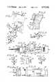
- FIG. 1is a perspective view of the present instrument as it would be used in testing for isometricity of the anterior cruciate ligament graft path in reconstructive knee surgery;
- FIG. 2is an enlarged sagittal section of the tibia with the instrument fitted with an attachment operatively positioned to accept a suture;
- FIG. 3is a top plan view, partially in section, illustrating the instrument of FIG. 1;
- FIG. 4is a detail perspective view of an instrument attachment for separably receiving a graft and skeletally mounting the instrument frame in operative position;
- FIG. 5is a detail perspective view of a bracket and cortical screw arrangement to effect skeletal mounting of the instrument frame
- FIG. 6is a detail side elevational view of an alternate form of graft retaining attachment to be mounted to the instrument sled;
- FIG. 7is a detail perspective view of an alternate form of bracket and cortical screw means for skeletally fixing a modified form of instrument frame
- FIG. 8is a partial side elevational view of the modified form of instrument frame adapted to accept the device of FIG. 7, and illustrating a modified form of suture nose for mounting on the modified instrument frame;
- FIG. 9is a partial top plan view of an instrument wherein the position of the sled relative to the instrument frame is indicated by linearly oriented indicia;
- FIG. 10is a top plan view, partially in longitudinal cross section, illustrating the sled bias means in a lightly loaded or compressed state
- FIG. 11is a view similar to FIG. 10, but illustrating the sled bias means in a more heavily loaded or compressed state;
- FIGS. 12 and 13are schematic views illustrating the relationship of the instrument to the femur and tibia through the range of knee flexion during graft testing;
- FIG. 14is a schematic view similar to FIGS. 12 and 13, but illustrating permanent fixation of the graft at the femoral fixation site;
- FIG. 15is a schematic view similar to FIG. 14, but illustrating the positions of the femur and tibia at the point of permanent fixation of the graft at the tibial fixation site and before separation of the instrument from the graft;
- FIG. 16is a schematic view similar to FIG. 15, but illustrating the appearance of the graft after instrument separation.
- FIG. 17is an enlarged detail view of the device of FIG. 5 skeletally temporarily fixing the graft prior to final graft fixation to the tibial fixation site.
- an instrument 10is illustrated in association with a femur 12 and tibia 14 characterized by proposed femoral and tibial graft fixation sites 16 and 18, respectively.
- drill guide or wire or suture openingssuch as the opening 20 seen in FIG. 2 are drilled in the femur and tibia between the sites 16 and 18.
- a tension elementsuch as a wire cable or suture 22 is passed through the suture openings and the end of the suture 22 protruding through the lateral wall of the femur 12 is temporarily clamped or fixed against movement by a hemostat 24.
- the other end of the suture 22is temporarily clamped or fixed by a hemostat 26 to a movable portion or sled 28 which is slidably carried by an elongated instrument body or frame 30.
- the frame 30is skeletally fixed or mounted at its distal extremity adjacent the tibial fixation site 18, which is important to maintain the instrument frame 30 in a relatively fixed position so that movement of the sled 28 relative to the frame 30 can establish the amount of graft displacement and tensioning.
- the instrument frame 30is generally rectangular and includes longitudinally extending, spaced apart side walls 32, an end wall 34 and a relatively thick end wall 36.
- the wallsare spaced apart to define a rectangular internal space or sled opening 38.
- Side walls 32include integral rails 40 which longitudinally slidably support elongated side margins 42 of the sled 28.
- the sled 28is longitudinally movable along the rails 40 by means of a thumb nut and lead screw arrangement.
- An adjusting thumb nut 44is rotatably carried within a transverse opening provided in the instrument frame end wall 36, and an elongated lead screw 46 extends through a suitable longitudinal opening in the end wall 36.
- the screw 46is in threadable engagement with the thumbwheel 44 and, as seen in FIG. 10, extends into the sled opening 38, through an opening in an end wall 48 of the sled 28, and into a central sled cavity 54.
- the sled 28is longitudinally movable relative to the screw 46.
- the free or sled end of the screw 46fixedly mounts an abutment element or pointer 52 which is located adjacent an end wall 50 of the sled.
- the pointer 52is thus located within the sled cavity 54 between the sled end walls 48 and 50.
- a bias means in the form of an elongated compression spring 56is disposed about the lead screw 46 within the sled cavity 54, and its opposite ends are engaged upon the pointer 52 and the sled end wall 48.
- the sled 28Once the sled 28 is attached to the suture 22, for example, it will be constrained against longitudinal movement relative to the frame 30. Rotation of the thumbwheel 44 in one direction will develop an increasing bias upon the sled 28 as the pointer 52 moves from the position of FIG. 10 to that of FIG. 11, tending to move the sled 28 proximally to increase the tension on the suture 22. Rotation of the thumbwheel 44 in the opposite direction develops a decreasing bias on the sled, tending to decrease the tension in the suture 22.
- Pointer 52is visible in FIG. 3 through an elongated slot 58 provided in the sled 28.
- Force indicia 60is linearly disposed along a margin of the slot 58 and, in association with the pointer 52, yield an indication of the bias force developed upon the sled 28.
- the present inventionprovides two means for indicating the longitudinal position of the sled 28 relative to the frame 30, one involving use of a linear scale, while the other involves an expanded circular scale.
- the linear versionis best seen in FIG. 9 and comprises linearly arranged displacement indicia 62 engraved upon one of the frame side walls 32, and cooperative with an indicia or index 64 engraved upon the sled 28.
- the circular expanded displacement scaleis seen in FIGS. 3, 10 and 11.
- the instrument frameis modified to include an arcuate portion 64 having an engraved indicia or index 66 and an arcuate recessed ledge (not shown).
- This ledgerotatably seats and supports the edge margin of a circular dial face 68 which carries circularly arranged displacement indicia 70.
- dial face 68is integral with a transverse shaft (not shown) which is rotatably carried by the instrument frame and integrally associated with a pinion gear 72.
- the sledis modified to include an elongated recess which receives a rack 74 whose teeth are meshed with the teeth of the pinion gear 72.
- a gear ratiois preferably selected such that a predetermined amount of longitudinal movement of the sled relative to the instrument frame produces a greater change in the relative displacement of the indicia 66 and 70 as compared to the relative displacement of the indicia of the linear arrangement of FIG. 9. This makes the amount of sled displacement easier to read.
- a means for fixing the sled 28 against longitudinal movement relative to the frame 30can be accomplished by rotation of a thumb screw 76 which is rotatably carried by a side wall 32 of the instrument frame, and which includes a support shaft inner end (not shown) that can be brought into binding engagement with the sled 28 upon rotation of the thumb screw 76.
- the sled cavity 54is covered by a sled plate attached to the sled by suitable screws (not shown) to retain in position the components contained within the cavity 54.
- the instrument 10is adapted to accept or include a variety of attachments for locating or verifying the graft isometric path and for providing skeletal mounting and graft tensioning. As will be seen, some of these attachments are used in determining the proper or isometric path for the suture 22, while others are adapted for use in testing of the graft.
- the end wall 34 of the frame 30includes an opening to separably accept the tubular shank of a nose piece 78.
- the nose piece 78includes a head or circular flange which engages the inner face of the end wall 34 to maintain the nose piece 78 in position.
- the diameter of the opening in the nose piece 78is sufficiently large to slidably and removably accept the proximal extremity of a tubular suture nose 80 having an annular flange which seats against the end of the nose piece 78.
- the suture nose 80includes a tubular extension 82 which, as seen in FIG. 2, is adapted to fit within the tunnel or suture opening 20 in the tibia 14 to skeletally mount or fix the frame 30 against movement.
- An alternate version of the nose piece 78, indicated at 78a in FIG. 8,includes an integral transverse bracket 84 having a central opening aligned with the opening in the frame end wall 34.
- the extremities of the bracket 84include laterally open slots adapted to removably fit within the annular grooves of a pair of posts 86 attached in spaced relation to the instrument frame.
- This post and bracket arrangementis simply a different way of mounting a suture nose piece.
- Other equivalent arrangementswill suggest themselves to those skilled in the art.
- the skeletal mounting or fixation provided by the nose pieces 78 and 78ais preferred but, if desired, fixation may be provided by a skeletal fixation angle or plate 88, as seen in FIGS. 5 and 17.
- fixationmay be provided by a skeletal fixation angle or plate 88, as seen in FIGS. 5 and 17.
- the nose piece 78is removably fitted within an opening 90 in one leg of the plate 88, and the instrument frame is fixed against movement by attachment to the tibia of the other leg of the bracket 84 by a cortical screw 92.
- This arrangementthus requires dissection and provision of a tapped hole in the tibia.
- FIG. 7Yet another form of skeletal fixation plate is illustrated in FIG. 7, the plate comprising a transverse element 94 which integrally mounts a cortical screw 96.
- the element 94is characterized by laterally open slots 98 adapted to removably fit over the posts 86 of the post mount version of the instrument frame.
- nose piece 78is readily removable, various sizes of nose pieces may be fitted to the frame 30 to fit within different sizes of suture or graft openings.
- FIG. 3Two versions of suture or graft retaining means can be mounted to the sled end wall 50.
- One versionis illustrated in FIG. 3 and comprises a retainer 100 having a longitudinal shank, and a transverse plate 102 which removably fits within a T-shaped slot provided in the sled end wall 50.
- the extremity of the retainer 100includes lateral hook portions 104 about which a suture or graft can be looped or otherwise fitted for fixation in position by the hemostat 26.
- the other or alternate form of suture or graft reretaineris illustrated at 100a in FIGS. 6, 10 and 11.
- the retainer 100aincludes a shank and a plate 102 adapted to fit within the T-shaped slot of the sled distal wall 50.
- retainer 100ais characterized by side plates 106 which support a transverse shaft 108 which eccentrically and rotatably mounts a roller 110. If a graft is disposed beneath the roller 110 and toward the sled 28, and thence upwardly and reversely away from the sled 28, tensioning the graft tends to rotate the roller 110 in a clockwise direction, as viewed in FIG. 6. This traps the graft between the roller 110 and a lateral lower wall 112 of the retainer 100a thereby preventing separation of the graft from the retainer 100a.
- a preferred means for temporarily skeletally mounting or fixing the instrument framecomprises a graft nose 114 which includes a centrally apertured transverse portion 116 adapted to be removably fitted over the protruding end of the nose piece 78.
- the graft nose 14includes inwardly convergent sides 118 which terminate in a semicylindrical, laterally open extension 120 adapted to fit within a graft hole, such as is seen at 122 in FIG. 17.
- the extension 120is preferably nearly exactly semicircular in transverse cross section so that its maximum external diameter is only slightly less then the maximum internal diameter of the graft hole 122. There is then virtually no opportunity for lateral movement of the extension 120 within the graft hole 122, insuring exact centering of the graft within the graft hole 22.
- a suture 22is first fixed at the femoral fixation site 16. It is then passed through the suture opening 20 to the tibia fixation site 18.
- the instrument frame 30must be temporarily skeletally fixed or mounted relative to the tibia 14. This can be done by cortical screw attachment to the tibia by using the plate 88 of FIG. 5 or the element 94 of FIG. 7. One of these is fitted over the nose piece 78, in one version of the instrument frame, or upon the posts 86 in the other version of the instrument frame.
- temporary skeletal mountingis preferably done by anchoring the frame 30 within the suture opening 20, which does not require dissection and provision of a tapped hole. With this arrangement the suture 22 is passed through the tubular extension 82 of the suture nose 80, and the nose 80 is then mounted to the nose piece 78 on the frame 30. The free end of the suture 22 is then disposed about the hook portions 104 of the retainer 100 and fixed by the hemostat 26.
- Thumbwheel 44is next operated to develop tension in the suture 22 and thereby hold the instrument in position. With the knee in 90 degrees of flexion, as illustrated in FIG. 12, thumbwheel 44 is operated to develop a test tension in the suture 22 of six pounds, for example, as indicated by the pointer 52. At this time the position of the sled 28 relative to the frame 30, that is, the graft displacement, is noted from the dial face of FIG. 3 or, in the version of FIG. 9, from the linear scale. The knee is then placed in full extension, as seen in FIG. 13, and the thumbwheel 44 operated until a force scale reading of the same six pounds is achieved. The displacement scale is again noted to determine if any change occurred in the position of the sled relative to the instrument frame. If no substantial change occurred, or if it is within acceptable limits, such as approximately 3 millimeters, the fixation sites selected are confirmed as satisfactory. Otherwise, different fixation sites must be selected to compensate for the undesired displacement and the procedure repeated.
- the suture opening 20is enlarged sufficiently by drilling or the like to receive a graft 124.
- the graft 124is disposed through graft hole 122, is fixed at its femoral extremity by means of a cancellous bone screw or staple, as seen in FIG. 14, and is extended out of the tibial fixation site 18 and through the preferred graft nose 114.
- the nose 114is then fitted onto the frame 30 and seated within the graft hole.
- the free end of the graftis then fixed to the sled 28 by means of sutures sewn into the graft, and graft displacement and tension is determined in the manner described in connection with the suture 22.
- the instrument 10can also be used to evaluate graft tension change with range of knee motion. This is done by placing the knee in 90 degree flexion, as seen in FIG. 12. The thumbwheel 44 is operated to develop a six pound load on the graft and the reading on the displacement scale is noted. The knee is then extended to the position of FIG. 13 and the thumbwheel 44 operated until the displacement scale reading is the same as when the knee was in 90 degrees of flexion. The force scale at this time indicates the ligament load
- the instrument 10can also be used to tension the ligament graft and assess the resulting joint laxity. To accomplish this, the knee is placed in the desired degree of flexion and the desired ligament load is developed by rotation of the thumbwheel 44. Thumb screw 76 is tightened to lock the sled 28 to the frame 30. Joint laxity can now be assessed either through manual tests or by use of an arthrometer.
- the tibial end of the graftis reached through the central open portion of the graft nose 114 and a cortical screw assembly 126, such as that seen in FIG. 17, is employed to fix the graft at the tibial fixation site, as seen in FIG. 15.
- the graftis then severed and the instrument 10 removed, as seen in FIG. 16.
Landscapes
- Health & Medical Sciences (AREA)
- Orthopedic Medicine & Surgery (AREA)
- Rehabilitation Therapy (AREA)
- Rheumatology (AREA)
- Cardiology (AREA)
- Oral & Maxillofacial Surgery (AREA)
- Transplantation (AREA)
- Engineering & Computer Science (AREA)
- Biomedical Technology (AREA)
- Heart & Thoracic Surgery (AREA)
- Vascular Medicine (AREA)
- Life Sciences & Earth Sciences (AREA)
- Animal Behavior & Ethology (AREA)
- General Health & Medical Sciences (AREA)
- Public Health (AREA)
- Veterinary Medicine (AREA)
- Surgical Instruments (AREA)
Abstract
Description
Claims (15)
Priority Applications (1)
| Application Number | Priority Date | Filing Date | Title |
|---|---|---|---|
| US06/880,055US4712542A (en) | 1986-06-30 | 1986-06-30 | System for establishing ligament graft orientation and isometry |
Applications Claiming Priority (1)
| Application Number | Priority Date | Filing Date | Title |
|---|---|---|---|
| US06/880,055US4712542A (en) | 1986-06-30 | 1986-06-30 | System for establishing ligament graft orientation and isometry |
Publications (1)
| Publication Number | Publication Date |
|---|---|
| US4712542Atrue US4712542A (en) | 1987-12-15 |
Family
ID=25375429
Family Applications (1)
| Application Number | Title | Priority Date | Filing Date |
|---|---|---|---|
| US06/880,055Expired - LifetimeUS4712542A (en) | 1986-06-30 | 1986-06-30 | System for establishing ligament graft orientation and isometry |
Country Status (1)
| Country | Link |
|---|---|
| US (1) | US4712542A (en) |
Cited By (115)
| Publication number | Priority date | Publication date | Assignee | Title |
|---|---|---|---|---|
| FR2626763A1 (en)* | 1986-10-03 | 1989-08-11 | Temple University Commonwealth | DEVICE AND METHOD FOR IDENTIFYING OPTIMAL LOCATIONS OF TUNNELS TO BE PRACTICAL IN BONES FOR THE REPLACEMENT OF LIGAMENTS OF AN ARTICULATION |
| EP0361756A1 (en)* | 1988-09-19 | 1990-04-04 | E. Marlowe Goble | Apparatus and procedure for verifying isometric ligament positioning |
| US4932972A (en)* | 1986-03-14 | 1990-06-12 | Richards Medical Company | Prosthetic ligament |
| EP0379789A1 (en)* | 1989-01-23 | 1990-08-01 | SMITH & NEPHEW RICHARDS, INC. | Apparatus and method for determining the tension on a ligament graft |
| US4946462A (en)* | 1988-12-12 | 1990-08-07 | Watanabe Robert S | Arthroscopic guide and method |
| US4955910A (en)* | 1989-07-17 | 1990-09-11 | Boehringer Mannheim Corporation | Fixation system for an elongated prosthesis |
| US4961416A (en)* | 1989-06-12 | 1990-10-09 | Orthopedic Systems, Inc. | Knee brace |
| US4964862A (en)* | 1989-08-31 | 1990-10-23 | Micro Strain Company | Method of and means for measuring ligament tension |
| WO1990008516A3 (en)* | 1989-02-06 | 1990-11-01 | Univ Minnesota | Ligament graft apparatus and method |
| US5002574A (en)* | 1989-08-18 | 1991-03-26 | Minnesota Mining And Manufacturing Co. | Tensioning means for prosthetic devices |
| EP0440991A1 (en)* | 1990-01-31 | 1991-08-14 | Acufex Microsurgical Inc. | Method and instruments for the reconstruction of the anterior cruciate ligament |
| DE4016476A1 (en)* | 1990-05-22 | 1991-11-28 | Aesculap Ag | ISOMETER |
| US5082003A (en)* | 1990-01-05 | 1992-01-21 | Orthopedic Systems, Inc. | Apparatus for determining interskeletal distances |
| US5108433A (en)* | 1989-08-18 | 1992-04-28 | Minnesota Mining And Manufacturing Company | Tensioning means for prosthetic devices |
| DE4112205A1 (en)* | 1991-04-13 | 1992-10-15 | Univ Halle Wittenberg | Method of transplanting crucial ligaments of knee joint - involves first stretching ligaments to prevent subsequent relaxation |
| US5197488A (en)* | 1991-04-05 | 1993-03-30 | N. K. Biotechnical Engineering Co. | Knee joint load measuring instrument and joint prosthesis |
| USRE34293E (en)* | 1987-02-17 | 1993-06-22 | Globe Marlowe E | Ligament attachment method and apparatus |
| US5253655A (en)* | 1992-12-17 | 1993-10-19 | Stone Kevin R | Apparatus and method for measuring range of motion of an articulated joint |
| USD352111S (en) | 1993-03-08 | 1994-11-01 | Medmetric Corporation | Interoperative knee ligament arthrometer for orthopedic surgery |
| US5391170A (en)* | 1991-12-13 | 1995-02-21 | David A. McGuire | Angled surgical screw driver and methods of arthroscopic ligament reconstruction |
| US5394888A (en)* | 1992-12-17 | 1995-03-07 | Stone; Kevin R. | Arthrometer with gravity switches and adjustable limit signaling |
| USRE34871E (en)* | 1989-05-15 | 1995-03-07 | Mcguire; David A. | Process of endosteal fixation of a ligament |
| US5407420A (en)* | 1992-11-12 | 1995-04-18 | Smith & Nephew Donjoy, Inc. | Fully adjustable shoulder brace |
| US5425733A (en)* | 1992-02-19 | 1995-06-20 | Arthrex, Inc. | Interference screw with rounded back end and cannulated sheath for endosteal fixation of ligaments |
| US5464407A (en)* | 1991-02-19 | 1995-11-07 | Mcguire; David A. | Flexible surgical screwdriver and methods of arthroscopic ligament reconstruction |
| US5470354A (en)* | 1991-11-12 | 1995-11-28 | Biomet Inc. | Force sensing apparatus and method for orthopaedic joint reconstruction |
| FR2720921A1 (en)* | 1994-06-09 | 1995-12-15 | Vergnolle Jean Pierre | Instrument for determining isometric point of knee joint |
| US5507812A (en)* | 1992-12-28 | 1996-04-16 | Moore; David E. | Modular prosthetic ligament |
| US5514143A (en)* | 1991-11-27 | 1996-05-07 | Apogee Medical Products, Inc. | Apparatus and method for use during surgery |
| US5520693A (en)* | 1992-02-19 | 1996-05-28 | Mcguire; David A. | Femoral guide and methods of precisely forming bone tunnels in cruciate ligament reconstruction of the knee |
| US5527342A (en)* | 1993-12-14 | 1996-06-18 | Pietrzak; William S. | Method and apparatus for securing soft tissues, tendons and ligaments to bone |
| US5564437A (en)* | 1992-12-15 | 1996-10-15 | Universite Joseph Fourier | Method and system for determining the fixation point on the femur of a crossed ligament of the knee |
| US5570706A (en)* | 1990-07-16 | 1996-11-05 | Howell; Stephen M. | Method for ACL reconstruction |
| US5713897A (en)* | 1997-03-06 | 1998-02-03 | Goble; E. Marlowe | Anterior cruciate ligament tensioning device and method for its use |
| US5944724A (en)* | 1997-10-30 | 1999-08-31 | Mitek Surgical Products, Inc. | Suture anchor insertion system |
| US6001106A (en)* | 1997-09-03 | 1999-12-14 | M & R Medical, Inc. | System for tensioning ligament grafts |
| US6019767A (en)* | 1990-07-16 | 2000-02-01 | Arthrotek | Tibial guide |
| US6036694A (en)* | 1998-08-03 | 2000-03-14 | Innovasive Devices, Inc. | Self-tensioning soft tissue fixation device and method |
| US6132442A (en)* | 1999-03-25 | 2000-10-17 | Smith & Nephew | Graft clamp |
| US6171310B1 (en)* | 1998-04-01 | 2001-01-09 | Aesculap Ag & Co. Kg | Device and method for handling an implant covering a bone tunnel |
| US6254604B1 (en) | 1990-07-16 | 2001-07-03 | Arthrotek, Inc. | Tibial guide |
| FR2814359A1 (en)* | 2000-09-28 | 2002-03-29 | Wolf Gmbh Richard | DEVICE FOR APPEARING A LIGAMENT TO BE IMPLANTED |
| US6478753B2 (en)* | 2000-05-31 | 2002-11-12 | Atlantech House | Tension measuring device |
| US6520980B1 (en) | 2000-11-02 | 2003-02-18 | Opus Medical, Inc. | Method and apparatus for attaching connective tissues to bone using a self-locking knotless suture anchoring device |
| US6524317B1 (en) | 1999-12-30 | 2003-02-25 | Opus Medical, Inc. | Method and apparatus for attaching connective tissues to bone using a knotless suture anchoring device |
| US6582453B1 (en) | 2000-07-14 | 2003-06-24 | Opus Medical, Inc. | Method and apparatus for attaching connective tissues to bone using a suture anchoring device |
| US6585730B1 (en) | 2000-08-30 | 2003-07-01 | Opus Medical, Inc. | Method and apparatus for attaching connective tissues to bone using a knotless suture anchoring device |
| US6599289B1 (en) | 2000-03-10 | 2003-07-29 | Smith & Nephew, Inc. | Graft anchor |
| US20030208210A1 (en)* | 2002-05-01 | 2003-11-06 | Dreyfuss Peter J. | Suture tensioning device |
| US6652561B1 (en) | 2000-10-13 | 2003-11-25 | Opus Medical, Inc | Method and apparatus for attaching connective tissues to bone using a perforated suture anchoring device |
| US6660008B1 (en) | 2001-06-07 | 2003-12-09 | Opus Medical, Inc. | Method and apparatus for attaching connective tissues to bone using a suture anchoring device |
| US6679889B1 (en) | 2000-11-13 | 2004-01-20 | Hs West Investments, Llc | Apparatus and methods for independently conditioning and pretensioning a plurality of ligament grafts during joint repair surgery |
| US20040039396A1 (en)* | 2002-08-23 | 2004-02-26 | Orthosoft Inc. | Universal positioning block |
| US20040097943A1 (en)* | 1996-11-21 | 2004-05-20 | Hart Rickey D. | Apparatus for anchoring autologous or artificial tendon grafts in bone |
| US20040193063A1 (en)* | 2003-02-28 | 2004-09-30 | Teiyuu Kimura | Method and apparatus for measuring biological condition |
| US20050049598A1 (en)* | 2003-08-29 | 2005-03-03 | West Hugh S. | Suture pulley for use with graft tensioning device |
| US20050049597A1 (en)* | 2003-08-29 | 2005-03-03 | West Hugh S. | Suture separation and organization devices for use with graft tensioning device |
| US20050203528A1 (en)* | 2002-08-23 | 2005-09-15 | Pierre Couture | Surgical universal positioning block and tool guide |
| US20050267577A1 (en)* | 2004-05-26 | 2005-12-01 | Trieu Hai H | Methods for treating the spine |
| US20070073299A1 (en)* | 1999-02-02 | 2007-03-29 | Arthrex, Inc. | Suture anchor with insert-molded eyelet shield |
| US20070123902A1 (en)* | 2005-04-22 | 2007-05-31 | Sascha Berberich | Device for forming a drill hole in bone |
| US20080033549A1 (en)* | 2006-06-26 | 2008-02-07 | Peter Marshall | Graft ligament strand tensioner |
| US20080195148A1 (en)* | 2007-01-19 | 2008-08-14 | Arthrex, Inc. | Method and suture-button construct for stabilization of cranial cruciate ligament deficient stifle |
| US20080306413A1 (en)* | 2004-06-15 | 2008-12-11 | Denis Crottet | Device for Measuring Tibio-Femoral Force Amplitudes and Force Locations in Total Knee Arthroplasty |
| US7556640B2 (en) | 2001-02-12 | 2009-07-07 | Arthrocare Corporation | Bone anchor device having toggle member for attaching connective tissues to bone |
| US20090228017A1 (en)* | 2008-03-06 | 2009-09-10 | Collins Evan D | Ligament reconstruction system |
| US7591850B2 (en) | 2005-04-01 | 2009-09-22 | Arthrocare Corporation | Surgical methods for anchoring and implanting tissues |
| US7615061B2 (en) | 2006-02-28 | 2009-11-10 | Arthrocare Corporation | Bone anchor suture-loading system, method and apparatus |
| US20090278484A1 (en)* | 2008-05-09 | 2009-11-12 | Degree Controls, Inc. | Fan conducted noise reduction |
| US7637926B2 (en) | 2002-02-04 | 2009-12-29 | Arthrocare Corporation | Method and apparatus for attaching connective tissues to bone using a knotless suture anchoring device |
| US20100049319A1 (en)* | 2008-08-19 | 2010-02-25 | Dougherty Christopher P | Single-tunnel double bundle anterior cruciate ligament reconstruction |
| US7674274B2 (en) | 2001-06-06 | 2010-03-09 | Arthrocare Corporation | Method and apparatus for attaching connective tissues to bone using a cortical bone anchoring device |
| US7682374B2 (en) | 2003-10-21 | 2010-03-23 | Arthrocare Corporation | Knotless suture lock and bone anchor implant method |
| US7686838B2 (en) | 2006-11-09 | 2010-03-30 | Arthrocare Corporation | External bullet anchor apparatus and method for use in surgical repair of ligament or tendon |
| US7713293B2 (en) | 2002-04-16 | 2010-05-11 | Arthrocare Corporation | Transverse suspension device |
| US7842042B2 (en) | 2005-05-16 | 2010-11-30 | Arthrocare Corporation | Convergent tunnel guide apparatus and method |
| US7901404B2 (en) | 2004-01-16 | 2011-03-08 | Arthrocare Corporation | Bone harvesting device and method |
| US7905924B2 (en) | 2003-09-03 | 2011-03-15 | Ralph Richard White | Extracapsular surgical procedure |
| US20110112537A1 (en)* | 2009-10-09 | 2011-05-12 | Acute Innovations, Llc | System for tensioning a surgical wire |
| US7963972B2 (en) | 2007-09-12 | 2011-06-21 | Arthrocare Corporation | Implant and delivery system for soft tissue repair |
| US8105343B2 (en) | 2008-06-30 | 2012-01-31 | Arthrocare Corporation | Independent suture tensioning and snaring apparatus |
| US8133258B2 (en) | 2006-08-03 | 2012-03-13 | Arthrocare Corporation | Method and apparatus for attaching connective tissues to bone using a knotless suture anchoring device |
| US8137381B2 (en) | 2007-04-25 | 2012-03-20 | Arthrocare Corporation | Knotless suture anchor having discrete polymer components and related methods |
| US20120253361A1 (en)* | 2009-10-26 | 2012-10-04 | Ortotip, Ltd. | Microdrive for Use in Stereotactic Surgery |
| US8449612B2 (en) | 2009-11-16 | 2013-05-28 | Arthrocare Corporation | Graft pulley and methods of use |
| US8496705B2 (en) | 1996-11-21 | 2013-07-30 | DePuy Mitek, LLCR | Method of anchoring autologous or artificial tendon grafts in bone |
| US8657854B2 (en) | 2001-02-12 | 2014-02-25 | Arthrocare Corporation | Knotless suture anchoring device having deforming section to accommodate sutures of various diameters |
| US8747439B2 (en) | 2000-03-13 | 2014-06-10 | P Tech, Llc | Method of using ultrasonic vibration to secure body tissue with fastening element |
| US8784495B2 (en) | 2000-01-14 | 2014-07-22 | Bonutti Skeletal Innovations Llc | Segmental knee arthroplasty |
| US8801755B2 (en) | 2004-04-06 | 2014-08-12 | Arthrex, Inc. | Suture anchor |
| US8808329B2 (en) | 1998-02-06 | 2014-08-19 | Bonutti Skeletal Innovations Llc | Apparatus and method for securing a portion of a body |
| US8814902B2 (en) | 2000-05-03 | 2014-08-26 | Bonutti Skeletal Innovations Llc | Method of securing body tissue |
| US8834490B2 (en) | 2001-08-28 | 2014-09-16 | Bonutti Skeletal Innovations Llc | Method for robotic arthroplasty using navigation |
| US8845699B2 (en) | 1999-08-09 | 2014-09-30 | Bonutti Skeletal Innovations Llc | Method of securing tissue |
| US8845687B2 (en) | 1996-08-19 | 2014-09-30 | Bonutti Skeletal Innovations Llc | Anchor for securing a suture |
| US20150088192A1 (en)* | 2009-11-10 | 2015-03-26 | Wake Forest University Health Sciences | Tissue tensioning devices and related methods |
| US9023083B2 (en) | 2012-01-27 | 2015-05-05 | Arthrocare Corporation | Method for soft tissue repair with free floating suture locking member |
| US9034014B2 (en) | 2012-01-27 | 2015-05-19 | Arthrocare Corporation | Free floating wedge suture anchor for soft tissue repair |
| US9179907B2 (en) | 2000-06-22 | 2015-11-10 | Arthrex, Inc. | Knotless graft fixation assembly |
| US9186133B2 (en) | 2001-12-06 | 2015-11-17 | Arthrocare Corporation | Bone anchor insertion device |
| US9198649B2 (en) | 2012-01-27 | 2015-12-01 | Arthrocare Corporation | Rotating locking member suture anchor and method for soft tissue repair |
| US9226742B2 (en) | 2012-01-27 | 2016-01-05 | Arthrocare Corporation | Restricted wedge suture anchor and method for soft tissue repair |
| US9364210B2 (en) | 2012-01-27 | 2016-06-14 | Arthrocare Corporation | Biased wedge suture anchor and method for soft tissue repair |
| US9521999B2 (en) | 2005-09-13 | 2016-12-20 | Arthrex, Inc. | Fully-threaded bioabsorbable suture anchor |
| US9636101B2 (en) | 2011-09-01 | 2017-05-02 | Arthrocare Corporation | Bone anchor having an integrated stress isolator |
| US9770238B2 (en) | 2001-12-03 | 2017-09-26 | P Tech, Llc | Magnetic positioning apparatus |
| US9770272B2 (en) | 2012-12-12 | 2017-09-26 | Wright Medical Technology, Inc. | Orthopedic compression/distraction device |
| US9855028B2 (en) | 2012-04-06 | 2018-01-02 | Arthrocare Corporation | Multi-suture knotless anchor for attaching tissue to bone and related method |
| IT201600076352A1 (en)* | 2016-07-20 | 2018-01-20 | Leonardo Osti | TENDON TENSIONER DEVICE |
| US10265157B2 (en) | 2016-06-30 | 2019-04-23 | Medos International Sarl | Methods and devices for tensioning grafts |
| US10307189B2 (en) | 2013-03-28 | 2019-06-04 | Robert A. Kaufmann | Prosthesis for partial and total joint replacement |
| US10426459B2 (en) | 2016-07-05 | 2019-10-01 | Mortise Medical, LLC | Extra joint stabilization construct |
| US10828147B1 (en) | 2018-01-21 | 2020-11-10 | Robert A. Kaufmann | Ligament retention device |
| WO2021216243A1 (en)* | 2020-04-22 | 2021-10-28 | Edwards Lifesciences Corporation | Controlled suture tensioning |
| CN116509470A (en)* | 2022-01-24 | 2023-08-01 | 北京和华瑞博医疗科技有限公司 | Joint Space Measuring Device |
Citations (19)
| Publication number | Priority date | Publication date | Assignee | Title |
|---|---|---|---|---|
| US1347579A (en)* | 1918-09-21 | 1920-07-27 | Henrikson Bernhardt | Hose-clamp tool |
| US1822352A (en)* | 1930-05-12 | 1931-09-08 | Marius N Laursen | Planter wire scale |
| US2337629A (en)* | 1941-12-27 | 1943-12-28 | Millers Falls Co | Tension gauge for hack saw blades |
| US2516079A (en)* | 1946-05-18 | 1950-07-18 | Millers Falls Co | Tensioning device for hacksaw blades |
| US2590498A (en)* | 1949-08-03 | 1952-03-25 | Harry H Bomberger | Check wire tension measuring apparatus |
| US3016741A (en)* | 1958-09-19 | 1962-01-16 | Harry W Kulp | Accessory for use by fishermen |
| US3432930A (en)* | 1965-02-25 | 1969-03-18 | Ture Anders Ljungberg | Device for the accurate measurement of large distances with an unsupported flexible measuring tape |
| US3696667A (en)* | 1970-04-22 | 1972-10-10 | Inductosyn Corp | Tensioning device for position measuring transformer |
| US3896500A (en)* | 1973-01-17 | 1975-07-29 | Andre Rambert | Method of and prosthesis for restoring ligaments of a joint |
| US3953896A (en)* | 1974-09-06 | 1976-05-04 | Richards Manufacturing Company, Inc. | Prosthetic ligament |
| US3973277A (en)* | 1974-01-30 | 1976-08-10 | James Campbell Semple | Attaching fibrous connective tissue to bone |
| US3976060A (en)* | 1974-04-09 | 1976-08-24 | Messerschmitt-Bolkow-Blohm Gmbh | Extension apparatus, especially for osteotomic surgery |
| US4024860A (en)* | 1976-01-05 | 1977-05-24 | Gennady Ivanovich Chelnokov | Apparatus for drawing together and fixing the pelvic halves in pubic symphysis ruptures |
| US4050464A (en)* | 1975-04-28 | 1977-09-27 | Downs Surgical Limited | Surgical cable tensioning instrument |
| US4220146A (en)* | 1979-01-18 | 1980-09-02 | Cloutier Jean Marie | Biplanar joint distractor |
| US4246660A (en)* | 1978-12-26 | 1981-01-27 | Queen's University At Kingston | Artificial ligament |
| US4317377A (en)* | 1980-09-26 | 1982-03-02 | Wrinkle Donald R | Drag adjustment device for fishing reels |
| US4364389A (en)* | 1980-03-19 | 1982-12-21 | Waldemar Link Gmbh & Co. | Instrument for holding and inserting the tibia plate for an endo-knee prosthesis having sliding surfaces |
| US4501266A (en)* | 1983-03-04 | 1985-02-26 | Biomet, Inc. | Knee distraction device |
- 1986
- 1986-06-30USUS06/880,055patent/US4712542A/ennot_activeExpired - Lifetime
Patent Citations (19)
| Publication number | Priority date | Publication date | Assignee | Title |
|---|---|---|---|---|
| US1347579A (en)* | 1918-09-21 | 1920-07-27 | Henrikson Bernhardt | Hose-clamp tool |
| US1822352A (en)* | 1930-05-12 | 1931-09-08 | Marius N Laursen | Planter wire scale |
| US2337629A (en)* | 1941-12-27 | 1943-12-28 | Millers Falls Co | Tension gauge for hack saw blades |
| US2516079A (en)* | 1946-05-18 | 1950-07-18 | Millers Falls Co | Tensioning device for hacksaw blades |
| US2590498A (en)* | 1949-08-03 | 1952-03-25 | Harry H Bomberger | Check wire tension measuring apparatus |
| US3016741A (en)* | 1958-09-19 | 1962-01-16 | Harry W Kulp | Accessory for use by fishermen |
| US3432930A (en)* | 1965-02-25 | 1969-03-18 | Ture Anders Ljungberg | Device for the accurate measurement of large distances with an unsupported flexible measuring tape |
| US3696667A (en)* | 1970-04-22 | 1972-10-10 | Inductosyn Corp | Tensioning device for position measuring transformer |
| US3896500A (en)* | 1973-01-17 | 1975-07-29 | Andre Rambert | Method of and prosthesis for restoring ligaments of a joint |
| US3973277A (en)* | 1974-01-30 | 1976-08-10 | James Campbell Semple | Attaching fibrous connective tissue to bone |
| US3976060A (en)* | 1974-04-09 | 1976-08-24 | Messerschmitt-Bolkow-Blohm Gmbh | Extension apparatus, especially for osteotomic surgery |
| US3953896A (en)* | 1974-09-06 | 1976-05-04 | Richards Manufacturing Company, Inc. | Prosthetic ligament |
| US4050464A (en)* | 1975-04-28 | 1977-09-27 | Downs Surgical Limited | Surgical cable tensioning instrument |
| US4024860A (en)* | 1976-01-05 | 1977-05-24 | Gennady Ivanovich Chelnokov | Apparatus for drawing together and fixing the pelvic halves in pubic symphysis ruptures |
| US4246660A (en)* | 1978-12-26 | 1981-01-27 | Queen's University At Kingston | Artificial ligament |
| US4220146A (en)* | 1979-01-18 | 1980-09-02 | Cloutier Jean Marie | Biplanar joint distractor |
| US4364389A (en)* | 1980-03-19 | 1982-12-21 | Waldemar Link Gmbh & Co. | Instrument for holding and inserting the tibia plate for an endo-knee prosthesis having sliding surfaces |
| US4317377A (en)* | 1980-09-26 | 1982-03-02 | Wrinkle Donald R | Drag adjustment device for fishing reels |
| US4501266A (en)* | 1983-03-04 | 1985-02-26 | Biomet, Inc. | Knee distraction device |
Cited By (205)
| Publication number | Priority date | Publication date | Assignee | Title |
|---|---|---|---|---|
| US4932972A (en)* | 1986-03-14 | 1990-06-12 | Richards Medical Company | Prosthetic ligament |
| DE3803208A1 (en)* | 1986-10-03 | 1989-10-19 | Univ Temple | DEVICE AND METHOD FOR RESTORATION SURGERY |
| FR2626763A1 (en)* | 1986-10-03 | 1989-08-11 | Temple University Commonwealth | DEVICE AND METHOD FOR IDENTIFYING OPTIMAL LOCATIONS OF TUNNELS TO BE PRACTICAL IN BONES FOR THE REPLACEMENT OF LIGAMENTS OF AN ARTICULATION |
| USRE34293E (en)* | 1987-02-17 | 1993-06-22 | Globe Marlowe E | Ligament attachment method and apparatus |
| US5037426A (en)* | 1988-09-19 | 1991-08-06 | Marlowe Goble E | Procedure for verifying isometric ligament positioning |
| EP0361756A1 (en)* | 1988-09-19 | 1990-04-04 | E. Marlowe Goble | Apparatus and procedure for verifying isometric ligament positioning |
| USRE34762E (en)* | 1988-09-19 | 1994-10-18 | Goble; E. Marlowe | Procedure for verifying isometric ligament positioning |
| US4946462A (en)* | 1988-12-12 | 1990-08-07 | Watanabe Robert S | Arthroscopic guide and method |
| EP0379789A1 (en)* | 1989-01-23 | 1990-08-01 | SMITH & NEPHEW RICHARDS, INC. | Apparatus and method for determining the tension on a ligament graft |
| US4969895A (en)* | 1989-01-23 | 1990-11-13 | Richards Medical Company | Apparatus and method for determining the tension on a ligament graft |
| AU628430B2 (en)* | 1989-01-23 | 1992-09-17 | Smith & Nephew Richards Inc. | Apparatus and method for determining the tension on a ligament graft |
| WO1990008516A3 (en)* | 1989-02-06 | 1990-11-01 | Univ Minnesota | Ligament graft apparatus and method |
| USRE34871E (en)* | 1989-05-15 | 1995-03-07 | Mcguire; David A. | Process of endosteal fixation of a ligament |
| US4961416A (en)* | 1989-06-12 | 1990-10-09 | Orthopedic Systems, Inc. | Knee brace |
| US4955910A (en)* | 1989-07-17 | 1990-09-11 | Boehringer Mannheim Corporation | Fixation system for an elongated prosthesis |
| US5002574A (en)* | 1989-08-18 | 1991-03-26 | Minnesota Mining And Manufacturing Co. | Tensioning means for prosthetic devices |
| US5108433A (en)* | 1989-08-18 | 1992-04-28 | Minnesota Mining And Manufacturing Company | Tensioning means for prosthetic devices |
| US4964862A (en)* | 1989-08-31 | 1990-10-23 | Micro Strain Company | Method of and means for measuring ligament tension |
| US5082003A (en)* | 1990-01-05 | 1992-01-21 | Orthopedic Systems, Inc. | Apparatus for determining interskeletal distances |
| US5139520A (en)* | 1990-01-31 | 1992-08-18 | American Cyanamid Company | Method for acl reconstruction |
| EP0440991A1 (en)* | 1990-01-31 | 1991-08-14 | Acufex Microsurgical Inc. | Method and instruments for the reconstruction of the anterior cruciate ligament |
| DE4016476A1 (en)* | 1990-05-22 | 1991-11-28 | Aesculap Ag | ISOMETER |
| US6254604B1 (en) | 1990-07-16 | 2001-07-03 | Arthrotek, Inc. | Tibial guide |
| US6254605B1 (en) | 1990-07-16 | 2001-07-03 | Stephen M. Howell | Tibial guide |
| US6019767A (en)* | 1990-07-16 | 2000-02-01 | Arthrotek | Tibial guide |
| US5570706A (en)* | 1990-07-16 | 1996-11-05 | Howell; Stephen M. | Method for ACL reconstruction |
| US5464407A (en)* | 1991-02-19 | 1995-11-07 | Mcguire; David A. | Flexible surgical screwdriver and methods of arthroscopic ligament reconstruction |
| US5197488A (en)* | 1991-04-05 | 1993-03-30 | N. K. Biotechnical Engineering Co. | Knee joint load measuring instrument and joint prosthesis |
| US5360016A (en)* | 1991-04-05 | 1994-11-01 | N. K. Biotechnical Engineering Company | Force transducer for a joint prosthesis |
| DE4112205A1 (en)* | 1991-04-13 | 1992-10-15 | Univ Halle Wittenberg | Method of transplanting crucial ligaments of knee joint - involves first stretching ligaments to prevent subsequent relaxation |
| US5470354A (en)* | 1991-11-12 | 1995-11-28 | Biomet Inc. | Force sensing apparatus and method for orthopaedic joint reconstruction |
| US5514143A (en)* | 1991-11-27 | 1996-05-07 | Apogee Medical Products, Inc. | Apparatus and method for use during surgery |
| US5391170A (en)* | 1991-12-13 | 1995-02-21 | David A. McGuire | Angled surgical screw driver and methods of arthroscopic ligament reconstruction |
| US5520693A (en)* | 1992-02-19 | 1996-05-28 | Mcguire; David A. | Femoral guide and methods of precisely forming bone tunnels in cruciate ligament reconstruction of the knee |
| US5425733A (en)* | 1992-02-19 | 1995-06-20 | Arthrex, Inc. | Interference screw with rounded back end and cannulated sheath for endosteal fixation of ligaments |
| US7025770B2 (en) | 1992-02-19 | 2006-04-11 | Mcguire David A | Femoral guide and methods of precisely forming bone tunnels in cruciate ligament reconstruction of the knee |
| US20030009173A1 (en)* | 1992-02-19 | 2003-01-09 | Mcguire David A. | Femoral guide and methods of precisely forming bone tunnels in cruciate ligament reconstruction of the knee |
| US6352538B2 (en) | 1992-02-19 | 2002-03-05 | Mcguire David A. | Femoral guide and methods of precisely forming bone tunnels in cruciate ligament reconstruction of the knee |
| US5407420A (en)* | 1992-11-12 | 1995-04-18 | Smith & Nephew Donjoy, Inc. | Fully adjustable shoulder brace |
| US5564437A (en)* | 1992-12-15 | 1996-10-15 | Universite Joseph Fourier | Method and system for determining the fixation point on the femur of a crossed ligament of the knee |
| US5253655A (en)* | 1992-12-17 | 1993-10-19 | Stone Kevin R | Apparatus and method for measuring range of motion of an articulated joint |
| US5394888A (en)* | 1992-12-17 | 1995-03-07 | Stone; Kevin R. | Arthrometer with gravity switches and adjustable limit signaling |
| US5507812A (en)* | 1992-12-28 | 1996-04-16 | Moore; David E. | Modular prosthetic ligament |
| USD352111S (en) | 1993-03-08 | 1994-11-01 | Medmetric Corporation | Interoperative knee ligament arthrometer for orthopedic surgery |
| US5527342A (en)* | 1993-12-14 | 1996-06-18 | Pietrzak; William S. | Method and apparatus for securing soft tissues, tendons and ligaments to bone |
| FR2720921A1 (en)* | 1994-06-09 | 1995-12-15 | Vergnolle Jean Pierre | Instrument for determining isometric point of knee joint |
| US8845687B2 (en) | 1996-08-19 | 2014-09-30 | Bonutti Skeletal Innovations Llc | Anchor for securing a suture |
| US7637949B2 (en)* | 1996-11-21 | 2009-12-29 | Innovasive Devices, Inc. | Method for anchoring autologous or artificial tendon grafts in bone |
| US8100969B2 (en) | 1996-11-21 | 2012-01-24 | Depuy Mitek, Inc. | Methods for anchoring autologous or artificial tendon grafts using first and second bone anchors |
| US20040097943A1 (en)* | 1996-11-21 | 2004-05-20 | Hart Rickey D. | Apparatus for anchoring autologous or artificial tendon grafts in bone |
| US8496705B2 (en) | 1996-11-21 | 2013-07-30 | DePuy Mitek, LLCR | Method of anchoring autologous or artificial tendon grafts in bone |
| WO1998038937A1 (en)* | 1997-03-06 | 1998-09-11 | Marlowe Goble E | Anterior cruciate ligament tensioning device and method for its use |
| US5713897A (en)* | 1997-03-06 | 1998-02-03 | Goble; E. Marlowe | Anterior cruciate ligament tensioning device and method for its use |
| US6001106A (en)* | 1997-09-03 | 1999-12-14 | M & R Medical, Inc. | System for tensioning ligament grafts |
| US5944724A (en)* | 1997-10-30 | 1999-08-31 | Mitek Surgical Products, Inc. | Suture anchor insertion system |
| US8808329B2 (en) | 1998-02-06 | 2014-08-19 | Bonutti Skeletal Innovations Llc | Apparatus and method for securing a portion of a body |
| US6171310B1 (en)* | 1998-04-01 | 2001-01-09 | Aesculap Ag & Co. Kg | Device and method for handling an implant covering a bone tunnel |
| US6036694A (en)* | 1998-08-03 | 2000-03-14 | Innovasive Devices, Inc. | Self-tensioning soft tissue fixation device and method |
| US9549726B2 (en) | 1999-02-02 | 2017-01-24 | Arthrex, Inc. | Suture anchor with insert-molded rigid member |
| US20070073299A1 (en)* | 1999-02-02 | 2007-03-29 | Arthrex, Inc. | Suture anchor with insert-molded eyelet shield |
| US8821541B2 (en) | 1999-02-02 | 2014-09-02 | Arthrex, Inc. | Suture anchor with insert-molded rigid member |
| US9526493B2 (en) | 1999-02-02 | 2016-12-27 | Arthrex, Inc. | Suture anchor with insert-molded rigid member |
| US6132442A (en)* | 1999-03-25 | 2000-10-17 | Smith & Nephew | Graft clamp |
| US8845699B2 (en) | 1999-08-09 | 2014-09-30 | Bonutti Skeletal Innovations Llc | Method of securing tissue |
| US6524317B1 (en) | 1999-12-30 | 2003-02-25 | Opus Medical, Inc. | Method and apparatus for attaching connective tissues to bone using a knotless suture anchoring device |
| US7247164B1 (en) | 1999-12-30 | 2007-07-24 | Arthrocare Corporation | Methods for attaching connective tissues to bone using a multi-component bone anchor |
| US8109966B2 (en) | 1999-12-30 | 2012-02-07 | Arthrocare Corporation | Methods for attaching connective tissues to bone using a multi-component anchor |
| US8784495B2 (en) | 2000-01-14 | 2014-07-22 | Bonutti Skeletal Innovations Llc | Segmental knee arthroplasty |
| US9192459B2 (en) | 2000-01-14 | 2015-11-24 | Bonutti Skeletal Innovations Llc | Method of performing total knee arthroplasty |
| US9101443B2 (en) | 2000-01-14 | 2015-08-11 | Bonutti Skeletal Innovations Llc | Methods for robotic arthroplasty |
| US9795394B2 (en) | 2000-01-14 | 2017-10-24 | Bonutti Skeletal Innovations Llc | Method for placing implant using robotic system |
| US6599289B1 (en) | 2000-03-10 | 2003-07-29 | Smith & Nephew, Inc. | Graft anchor |
| US8747439B2 (en) | 2000-03-13 | 2014-06-10 | P Tech, Llc | Method of using ultrasonic vibration to secure body tissue with fastening element |
| US8814902B2 (en) | 2000-05-03 | 2014-08-26 | Bonutti Skeletal Innovations Llc | Method of securing body tissue |
| US6478753B2 (en)* | 2000-05-31 | 2002-11-12 | Atlantech House | Tension measuring device |
| US9706986B2 (en) | 2000-06-22 | 2017-07-18 | Arthrex, Inc. | Knotless suture and tissue securing method |
| US9179907B2 (en) | 2000-06-22 | 2015-11-10 | Arthrex, Inc. | Knotless graft fixation assembly |
| US10052091B2 (en) | 2000-06-22 | 2018-08-21 | Arthrex, Inc. | Knotless suture or tissue fixation using an implant having a pointed tip |
| US10709436B2 (en) | 2000-06-22 | 2020-07-14 | Arthrex, Inc. | Graft fixation using a plug against suture |
| US10716556B2 (en) | 2000-06-22 | 2020-07-21 | Arthtrex, Inc. | Knotless tissue fixation assembly |
| US9775599B2 (en) | 2000-06-22 | 2017-10-03 | Arthrex, Inc. | Knotless tissue fixation assembly |
| US6582453B1 (en) | 2000-07-14 | 2003-06-24 | Opus Medical, Inc. | Method and apparatus for attaching connective tissues to bone using a suture anchoring device |
| US20030195564A1 (en)* | 2000-07-14 | 2003-10-16 | Opus Medical, Inc. | Method and apparatus for attaching connective tissues to bone using a suture anchoring device |
| US6585730B1 (en) | 2000-08-30 | 2003-07-01 | Opus Medical, Inc. | Method and apparatus for attaching connective tissues to bone using a knotless suture anchoring device |
| DE10049270A1 (en)* | 2000-09-28 | 2002-05-02 | Wolf Gmbh Richard | Device for conditioning a band to be implanted |
| US6723125B2 (en) | 2000-09-28 | 2004-04-20 | Richard Wolf Gmbh | Device for conditioning a ligament to be implanted |
| DE10049270B4 (en)* | 2000-09-28 | 2005-09-15 | Richard Wolf Gmbh | Apparatus for conditioning a band to be implanted |
| FR2814359A1 (en)* | 2000-09-28 | 2002-03-29 | Wolf Gmbh Richard | DEVICE FOR APPEARING A LIGAMENT TO BE IMPLANTED |
| US6652561B1 (en) | 2000-10-13 | 2003-11-25 | Opus Medical, Inc | Method and apparatus for attaching connective tissues to bone using a perforated suture anchoring device |
| US6520980B1 (en) | 2000-11-02 | 2003-02-18 | Opus Medical, Inc. | Method and apparatus for attaching connective tissues to bone using a self-locking knotless suture anchoring device |
| US20040039389A1 (en)* | 2000-11-13 | 2004-02-26 | West Hugh S. | Apparatus and methods for independently conditioning and pre-tensioning a plurality of ligament grafts during joint repair surgery |
| US7118578B2 (en) | 2000-11-13 | 2006-10-10 | Hs West Investments, Llc | Apparatus and methods for independently conditioning and pre-tensioning a plurality of ligament grafts during joint repair surgery |
| US6679889B1 (en) | 2000-11-13 | 2004-01-20 | Hs West Investments, Llc | Apparatus and methods for independently conditioning and pretensioning a plurality of ligament grafts during joint repair surgery |
| EP1339334A4 (en)* | 2000-11-13 | 2004-09-08 | Hugh S West Jr | Apparatus and methods for independently conditioning and pre-tensioning a plurality of ligament grafts during joint repair surgery |
| US7695494B2 (en) | 2001-02-12 | 2010-04-13 | Arthrocare Corporation | Method and apparatus for attaching connective tissues to bone using a knotless suture anchoring device |
| US8657854B2 (en) | 2001-02-12 | 2014-02-25 | Arthrocare Corporation | Knotless suture anchoring device having deforming section to accommodate sutures of various diameters |
| US8685060B2 (en) | 2001-02-12 | 2014-04-01 | Arthrocare Corporation | Methods and devices for attaching connective tissues to bone using a knotless suture anchoring device |
| US8444672B2 (en) | 2001-02-12 | 2013-05-21 | Arthrocare Corporation | Methods and devices for attaching connective tissues to bone using a knotless suture anchoring device |
| US7556640B2 (en) | 2001-02-12 | 2009-07-07 | Arthrocare Corporation | Bone anchor device having toggle member for attaching connective tissues to bone |
| US7674274B2 (en) | 2001-06-06 | 2010-03-09 | Arthrocare Corporation | Method and apparatus for attaching connective tissues to bone using a cortical bone anchoring device |
| US6660008B1 (en) | 2001-06-07 | 2003-12-09 | Opus Medical, Inc. | Method and apparatus for attaching connective tissues to bone using a suture anchoring device |
| US20040236336A1 (en)* | 2001-06-07 | 2004-11-25 | Opus Medical, Inc. | Method and apparatus for attaching connective tissues to bone using a suture anchoring device |
| US8840629B2 (en) | 2001-08-28 | 2014-09-23 | Bonutti Skeletal Innovations Llc | Robotic arthroplasty system including navigation |
| US10470780B2 (en) | 2001-08-28 | 2019-11-12 | Bonutti Skeletal Innovations Llc | Systems and methods for ligament balancing in robotic surgery |
| US8834490B2 (en) | 2001-08-28 | 2014-09-16 | Bonutti Skeletal Innovations Llc | Method for robotic arthroplasty using navigation |
| US9763683B2 (en) | 2001-08-28 | 2017-09-19 | Bonutti Skeletal Innovations Llc | Method for performing surgical procedures using optical cutting guides |
| US10231739B1 (en) | 2001-08-28 | 2019-03-19 | Bonutti Skeletal Innovations Llc | System and method for robotic surgery |
| US10321918B2 (en) | 2001-08-28 | 2019-06-18 | Bonutti Skeletal Innovations Llc | Methods for robotic surgery using a cannula |
| US8858557B2 (en) | 2001-08-28 | 2014-10-14 | Bonutti Skeletal Innovations Llc | Method of preparing a femur and tibia in knee arthroplasty |
| US9060797B2 (en) | 2001-08-28 | 2015-06-23 | Bonutti Skeletal Innovations Llc | Method of preparing a femur and tibia in knee arthroplasty |
| US9770238B2 (en) | 2001-12-03 | 2017-09-26 | P Tech, Llc | Magnetic positioning apparatus |
| US9186133B2 (en) | 2001-12-06 | 2015-11-17 | Arthrocare Corporation | Bone anchor insertion device |
| US7637926B2 (en) | 2002-02-04 | 2009-12-29 | Arthrocare Corporation | Method and apparatus for attaching connective tissues to bone using a knotless suture anchoring device |
| US7713293B2 (en) | 2002-04-16 | 2010-05-11 | Arthrocare Corporation | Transverse suspension device |
| US20030208210A1 (en)* | 2002-05-01 | 2003-11-06 | Dreyfuss Peter J. | Suture tensioning device |
| US7326222B2 (en)* | 2002-05-01 | 2008-02-05 | Arthrex, Inc. | Suture tensioning device |
| US20100211077A1 (en)* | 2002-08-23 | 2010-08-19 | Pierre Couture | Surgical universal positioning block and tool guide |
| US7736368B2 (en) | 2002-08-23 | 2010-06-15 | Orthosoft Inc. | Surgical universal positioning block and tool guide |
| US20080065085A1 (en)* | 2002-08-23 | 2008-03-13 | Orthosoft Inc. | Surgical universal positioning block and tool guide |
| US20040039396A1 (en)* | 2002-08-23 | 2004-02-26 | Orthosoft Inc. | Universal positioning block |
| US20050203528A1 (en)* | 2002-08-23 | 2005-09-15 | Pierre Couture | Surgical universal positioning block and tool guide |
| US7857821B2 (en) | 2002-08-23 | 2010-12-28 | Orthosoft Inc. | Surgical universal positioning block and tool guide |
| US8147496B2 (en) | 2002-08-23 | 2012-04-03 | Orthosoft Inc. | Surgical universal positioning block and tool guide |
| US7896882B2 (en) | 2002-08-23 | 2011-03-01 | Orthosoft Inc. | Surgical universal positioning block and tool guide |
| US20080065084A1 (en)* | 2002-08-23 | 2008-03-13 | Orthosoft Inc. | Surgical universal positioning block and tool guide |
| US20040193063A1 (en)* | 2003-02-28 | 2004-09-30 | Teiyuu Kimura | Method and apparatus for measuring biological condition |
| US7686810B2 (en)* | 2003-08-29 | 2010-03-30 | Hs West Investments, Llc | Suture separation and organization devices for use with graft tensioning device |
| US20050049598A1 (en)* | 2003-08-29 | 2005-03-03 | West Hugh S. | Suture pulley for use with graft tensioning device |
| US20050049597A1 (en)* | 2003-08-29 | 2005-03-03 | West Hugh S. | Suture separation and organization devices for use with graft tensioning device |
| US7905924B2 (en) | 2003-09-03 | 2011-03-15 | Ralph Richard White | Extracapsular surgical procedure |
| US8523949B2 (en) | 2003-09-03 | 2013-09-03 | Ralph Richard White | Extracapsular surgical procedure and surgical referencing instrument therefor |
| US20110125161A1 (en)* | 2003-09-03 | 2011-05-26 | Ralph Richard White | Extracapsular Surgical Procedure and Surgical Referencing Instrument Therefor |
| US7682374B2 (en) | 2003-10-21 | 2010-03-23 | Arthrocare Corporation | Knotless suture lock and bone anchor implant method |
| US7901404B2 (en) | 2004-01-16 | 2011-03-08 | Arthrocare Corporation | Bone harvesting device and method |
| US10537319B2 (en) | 2004-04-06 | 2020-01-21 | Arthrex, Inc. | Suture anchor |
| US9622739B2 (en) | 2004-04-06 | 2017-04-18 | Arthrex, Inc. | Suture anchor |
| US8801755B2 (en) | 2004-04-06 | 2014-08-12 | Arthrex, Inc. | Suture anchor |
| US20050267577A1 (en)* | 2004-05-26 | 2005-12-01 | Trieu Hai H | Methods for treating the spine |
| US20080306413A1 (en)* | 2004-06-15 | 2008-12-11 | Denis Crottet | Device for Measuring Tibio-Femoral Force Amplitudes and Force Locations in Total Knee Arthroplasty |
| US7587945B2 (en)* | 2004-06-15 | 2009-09-15 | Synthes Usa, Llc | Device for measuring tibio-femoral force amplitudes and force locations in total knee arthroplasty |
| US7591850B2 (en) | 2005-04-01 | 2009-09-22 | Arthrocare Corporation | Surgical methods for anchoring and implanting tissues |
| US20070123902A1 (en)* | 2005-04-22 | 2007-05-31 | Sascha Berberich | Device for forming a drill hole in bone |
| US7972341B2 (en)* | 2005-04-22 | 2011-07-05 | Karl Storz Gmbh & Co. Kg | Device for forming a drill hole in bone |
| US7842042B2 (en) | 2005-05-16 | 2010-11-30 | Arthrocare Corporation | Convergent tunnel guide apparatus and method |
| US11324493B2 (en) | 2005-09-13 | 2022-05-10 | Arthrex, Inc. | Fully-threaded bioabsorbable suture anchor |
| US9521999B2 (en) | 2005-09-13 | 2016-12-20 | Arthrex, Inc. | Fully-threaded bioabsorbable suture anchor |
| US12064104B2 (en) | 2005-09-13 | 2024-08-20 | Arthrex, Inc. | Fully-threaded bioabsorbable suture anchor |
| US10595847B2 (en) | 2005-09-13 | 2020-03-24 | Arthrex, Inc. | Fully-threaded bioabsorbable suture anchor |
| US7615061B2 (en) | 2006-02-28 | 2009-11-10 | Arthrocare Corporation | Bone anchor suture-loading system, method and apparatus |
| US8197485B2 (en) | 2006-06-26 | 2012-06-12 | Tyco Healthcare Group Lp | Graft ligament strand tensioner |
| US20080033549A1 (en)* | 2006-06-26 | 2008-02-07 | Peter Marshall | Graft ligament strand tensioner |
| WO2008002550A3 (en)* | 2006-06-26 | 2008-08-21 | Scandius Biomedical Inc | Graft ligament strand tensioner |
| US8317829B2 (en) | 2006-08-03 | 2012-11-27 | Arthrocare Corporation | Method and apparatus for attaching connective tissues to bone using a knotless suture anchoring device |
| US8133258B2 (en) | 2006-08-03 | 2012-03-13 | Arthrocare Corporation | Method and apparatus for attaching connective tissues to bone using a knotless suture anchoring device |
| US7686838B2 (en) | 2006-11-09 | 2010-03-30 | Arthrocare Corporation | External bullet anchor apparatus and method for use in surgical repair of ligament or tendon |
| US7875057B2 (en)* | 2007-01-19 | 2011-01-25 | Arthrex, Inc. | Method and suture-button construct for stabilization of cranial cruciate ligament deficient stifle |
| US20110118781A1 (en)* | 2007-01-19 | 2011-05-19 | Cook James L | Method and suture-button construct for stabilization of cranial cruciate ligament deficient stifle |
| US8202298B2 (en) | 2007-01-19 | 2012-06-19 | Arthrex, Inc. | Method and suture-button construct for stabilization of cranial cruciate ligament deficient stifle |
| US20080195148A1 (en)* | 2007-01-19 | 2008-08-14 | Arthrex, Inc. | Method and suture-button construct for stabilization of cranial cruciate ligament deficient stifle |
| US8137381B2 (en) | 2007-04-25 | 2012-03-20 | Arthrocare Corporation | Knotless suture anchor having discrete polymer components and related methods |
| US8425536B2 (en) | 2007-09-12 | 2013-04-23 | Arthrocare Corporation | Implant and delivery system for soft tissue repair |
| US7963972B2 (en) | 2007-09-12 | 2011-06-21 | Arthrocare Corporation | Implant and delivery system for soft tissue repair |
| US20090228017A1 (en)* | 2008-03-06 | 2009-09-10 | Collins Evan D | Ligament reconstruction system |
| US8110000B2 (en) | 2008-03-06 | 2012-02-07 | Collins Evan D | Ligament reconstruction system |
| US20090278484A1 (en)* | 2008-05-09 | 2009-11-12 | Degree Controls, Inc. | Fan conducted noise reduction |
| US8834495B2 (en) | 2008-06-30 | 2014-09-16 | Arthrocare Corporation | Independent suture tensioning and snaring apparatus |
| US8828029B2 (en) | 2008-06-30 | 2014-09-09 | Arthrocare Corporation | Independent suture tensioning and snaring apparatus |
| US8105343B2 (en) | 2008-06-30 | 2012-01-31 | Arthrocare Corporation | Independent suture tensioning and snaring apparatus |
| US8617186B2 (en) | 2008-06-30 | 2013-12-31 | Arthrocare Corporation | Independent suture tensioning and snaring apparatus |
| US20100049319A1 (en)* | 2008-08-19 | 2010-02-25 | Dougherty Christopher P | Single-tunnel double bundle anterior cruciate ligament reconstruction |
| US8333802B2 (en)* | 2008-08-19 | 2012-12-18 | Dougherty Christopher P | Single tunnel double bundle anterior cruciate ligament reconstruction |
| US20110112537A1 (en)* | 2009-10-09 | 2011-05-12 | Acute Innovations, Llc | System for tensioning a surgical wire |
| US8679122B2 (en) | 2009-10-09 | 2014-03-25 | Acute Innovations Llc | System for tensioning a surgical wire |
| US20120253361A1 (en)* | 2009-10-26 | 2012-10-04 | Ortotip, Ltd. | Microdrive for Use in Stereotactic Surgery |
| US20150088192A1 (en)* | 2009-11-10 | 2015-03-26 | Wake Forest University Health Sciences | Tissue tensioning devices and related methods |
| US8449612B2 (en) | 2009-11-16 | 2013-05-28 | Arthrocare Corporation | Graft pulley and methods of use |
| US9636101B2 (en) | 2011-09-01 | 2017-05-02 | Arthrocare Corporation | Bone anchor having an integrated stress isolator |
| US9034014B2 (en) | 2012-01-27 | 2015-05-19 | Arthrocare Corporation | Free floating wedge suture anchor for soft tissue repair |
| US9023083B2 (en) | 2012-01-27 | 2015-05-05 | Arthrocare Corporation | Method for soft tissue repair with free floating suture locking member |
| US9198649B2 (en) | 2012-01-27 | 2015-12-01 | Arthrocare Corporation | Rotating locking member suture anchor and method for soft tissue repair |
| US9226742B2 (en) | 2012-01-27 | 2016-01-05 | Arthrocare Corporation | Restricted wedge suture anchor and method for soft tissue repair |
| US9364210B2 (en) | 2012-01-27 | 2016-06-14 | Arthrocare Corporation | Biased wedge suture anchor and method for soft tissue repair |
| US9855028B2 (en) | 2012-04-06 | 2018-01-02 | Arthrocare Corporation | Multi-suture knotless anchor for attaching tissue to bone and related method |
| US10631900B2 (en) | 2012-12-12 | 2020-04-28 | Wright Medical Technology, Inc. | Orthopedic compression/distraction device |
| US9770272B2 (en) | 2012-12-12 | 2017-09-26 | Wright Medical Technology, Inc. | Orthopedic compression/distraction device |
| US10307189B2 (en) | 2013-03-28 | 2019-06-04 | Robert A. Kaufmann | Prosthesis for partial and total joint replacement |
| US11253349B2 (en) | 2016-06-30 | 2022-02-22 | Medos International Sarl | Methods and devices for tensioning grafts |
| US10265157B2 (en) | 2016-06-30 | 2019-04-23 | Medos International Sarl | Methods and devices for tensioning grafts |
| US12178697B2 (en) | 2016-06-30 | 2024-12-31 | Medos International Sarl | Methods and devices for tensioning grafts |
| US11937801B2 (en) | 2016-07-05 | 2024-03-26 | Crossroads Extremity Systems, Llc | Intra joint stabilization construct |
| US10426459B2 (en) | 2016-07-05 | 2019-10-01 | Mortise Medical, LLC | Extra joint stabilization construct |
| US10842480B2 (en) | 2016-07-05 | 2020-11-24 | Crossroads Extremity Systems, Llc | Multiple suture threader and methods of use |
| US12357295B2 (en) | 2016-07-05 | 2025-07-15 | Crossroads Extremity Systems, Llc | Compression and tension instruments and methods of use to reinforce ligaments |
| US11234688B2 (en) | 2016-07-05 | 2022-02-01 | Crossroads Extremity Systems, Llc | Compression and tension instruments and methods of use to reinforce ligaments |
| US11241225B2 (en) | 2016-07-05 | 2022-02-08 | Crossroads Extremity Systems, Llc | Extra joint stabilization construct |
| US12343002B2 (en) | 2016-07-05 | 2025-07-01 | Crossroads Extremity Systems, Llc | Extra joint stabilization construct |
| US10426460B2 (en) | 2016-07-05 | 2019-10-01 | Mortise Medical, LLC | Compression and tension instruments and methods of use to reinforce ligaments |
| US12042138B2 (en) | 2016-07-05 | 2024-07-23 | Crossroads Extremity Systems, Llc | Multiple suture threader and methods of use |
| US10682131B2 (en) | 2016-07-05 | 2020-06-16 | Crossroads Extremity Systems, Llc | Intra joint stabilization construct |
| US11589910B2 (en)* | 2016-07-20 | 2023-02-28 | Leonardo Osti | Tensioning device for tendons |
| WO2018015905A1 (en)* | 2016-07-20 | 2018-01-25 | Osti Leonardo | Tensioning device for tendons |
| IT201600076352A1 (en)* | 2016-07-20 | 2018-01-20 | Leonardo Osti | TENDON TENSIONER DEVICE |
| US10828147B1 (en) | 2018-01-21 | 2020-11-10 | Robert A. Kaufmann | Ligament retention device |
| WO2021216243A1 (en)* | 2020-04-22 | 2021-10-28 | Edwards Lifesciences Corporation | Controlled suture tensioning |
| CN116509470A (en)* | 2022-01-24 | 2023-08-01 | 北京和华瑞博医疗科技有限公司 | Joint Space Measuring Device |
Similar Documents
| Publication | Publication Date | Title |
|---|---|---|
| US4712542A (en) | System for establishing ligament graft orientation and isometry | |
| EP0379789B1 (en) | Apparatus and method for determining the tension on a ligament graft | |
| AU676888B2 (en) | Drill guide apparatus and method | |
| EP0746282B1 (en) | Drill guide apparatus | |
| US4938762A (en) | Reference system for implantation of condylar total knee prostheses | |
| EP1070482B1 (en) | Apparatus for tibial fixation of soft tissue | |
| US6001106A (en) | System for tensioning ligament grafts | |
| US4567886A (en) | Flexion spacer guide for fitting a knee prosthesis | |
| US4211228A (en) | Multipurpose tibial template | |
| US6796977B2 (en) | Variable graft tensioner | |
| AU2004257445B2 (en) | Device for assisting in total knee prosthesis implantation | |
| US5669914A (en) | Rotation alignment instrument | |
| US4950271A (en) | Ligament graft apparatus and method | |
| US5860980A (en) | Surgical apparatus for use in total knee arthroplasty and surgical methods for using said apparatus | |
| US5071420A (en) | Isometry testing device | |
| KR101413407B1 (en) | Assembly for guiding cuts to the femur and tibia during knee joint plastic surgery | |
| US20110046685A1 (en) | Surgical instrument for fitting a knee prosthesis | |
| US20060052795A1 (en) | Extracapsular surgical procedure and surgical referencing instrument therefor | |
| US20050080428A1 (en) | Extracapsular surgical procedure for repair of anterior cruciate ligament rupture and surgical referencing instrument therefor | |
| AU2023203840A1 (en) | Systems and methods for providing a tibial baseplate | |
| US5507750A (en) | Method and apparatus for tensioning grafts or ligaments | |
| US20070239166A1 (en) | Surgical Device for Anterolateral and Posterolateral Reconstruction | |
| Stewart et al. | Maintenance of set force in anterior cruciate ligament grafts | |
| US20050261701A1 (en) | Surgical device for a anterolateral reconstruction |
Legal Events
| Date | Code | Title | Description |
|---|---|---|---|
| AS | Assignment | Owner name:MEDMETRIC CORPORATION, 4901 MORENA BOULEVARD, SAN Free format text:ASSIGNMENT OF ASSIGNORS INTEREST.;ASSIGNORS:DANIEL, DALE M.;WATKINS, KAY R.;REEL/FRAME:004581/0136 Effective date:19860627 Owner name:MEDMETRIC CORPORATION,CALIFORNIA Free format text:ASSIGNMENT OF ASSIGNORS INTEREST;ASSIGNORS:DANIEL, DALE M.;WATKINS, KAY R.;REEL/FRAME:004581/0136 Effective date:19860627 | |
| STCF | Information on status: patent grant | Free format text:PATENTED CASE | |
| FEPP | Fee payment procedure | Free format text:PAYOR NUMBER ASSIGNED (ORIGINAL EVENT CODE: ASPN); ENTITY STATUS OF PATENT OWNER: SMALL ENTITY | |
| FEPP | Fee payment procedure | Free format text:PAYER NUMBER DE-ASSIGNED (ORIGINAL EVENT CODE: RMPN); ENTITY STATUS OF PATENT OWNER: SMALL ENTITY Free format text:PAYOR NUMBER ASSIGNED (ORIGINAL EVENT CODE: ASPN); ENTITY STATUS OF PATENT OWNER: SMALL ENTITY | |
| FPAY | Fee payment | Year of fee payment:4 | |
| FPAY | Fee payment | Year of fee payment:8 | |
| FPAY | Fee payment | Year of fee payment:12 |