US4704130A - Biocompatible microporous polymeric materials and methods of making same - Google Patents
Biocompatible microporous polymeric materials and methods of making sameDownload PDFInfo
- Publication number
- US4704130A US4704130AUS06/788,850US78885085AUS4704130AUS 4704130 AUS4704130 AUS 4704130AUS 78885085 AUS78885085 AUS 78885085AUS 4704130 AUS4704130 AUS 4704130A
- Authority
- US
- United States
- Prior art keywords
- solution
- article
- bath
- solvent
- process according
- Prior art date
- Legal status (The legal status is an assumption and is not a legal conclusion. Google has not performed a legal analysis and makes no representation as to the accuracy of the status listed.)
- Expired - Lifetime
Links
- 239000000463materialSubstances0.000titleclaimsabstractdescription57
- 238000000034methodMethods0.000titleclaimsdescription45
- 239000012528membraneSubstances0.000claimsabstractdescription58
- 239000002904solventSubstances0.000claimsabstractdescription52
- 239000011148porous materialSubstances0.000claimsabstractdescription41
- 238000001556precipitationMethods0.000claimsabstractdescription30
- 239000007787solidSubstances0.000claimsabstractdescription14
- 239000004721Polyphenylene oxideSubstances0.000claimsabstractdescription7
- 229920000570polyetherPolymers0.000claimsabstractdescription7
- 239000000243solutionSubstances0.000claimsdescription93
- 230000008569processEffects0.000claimsdescription35
- XLYOFNOQVPJJNP-UHFFFAOYSA-NwaterSubstancesOXLYOFNOQVPJJNP-UHFFFAOYSA-N0.000claimsdescription25
- 238000000605extractionMethods0.000claimsdescription24
- FXHOOIRPVKKKFG-UHFFFAOYSA-NN,N-DimethylacetamideChemical compoundCN(C)C(C)=OFXHOOIRPVKKKFG-UHFFFAOYSA-N0.000claimsdescription20
- 229940113088dimethylacetamideDrugs0.000claimsdescription20
- 229920002635polyurethanePolymers0.000claimsdescription20
- 239000004814polyurethaneSubstances0.000claimsdescription20
- 238000001035dryingMethods0.000claimsdescription16
- 239000000758substrateSubstances0.000claimsdescription14
- 239000007788liquidSubstances0.000claimsdescription11
- 239000000203mixtureSubstances0.000claimsdescription10
- 238000007654immersionMethods0.000claimsdescription7
- 239000012982microporous membraneSubstances0.000claimsdescription5
- 229920003226polyurethane ureaPolymers0.000claimsdescription5
- 238000005086pumpingMethods0.000claimsdescription4
- 150000001298alcoholsChemical class0.000claimsdescription3
- 239000007864aqueous solutionSubstances0.000claimsdescription2
- MHABMANUFPZXEB-UHFFFAOYSA-NO-demethyl-aloesaponarin INatural productsO=C1C2=CC=CC(O)=C2C(=O)C2=C1C=C(O)C(C(O)=O)=C2CMHABMANUFPZXEB-UHFFFAOYSA-N0.000claims3
- 229920001807Urea-formaldehydePolymers0.000claims3
- 238000005406washingMethods0.000claims2
- 229920005989resinPolymers0.000claims1
- 239000011347resinSubstances0.000claims1
- OYQYHJRSHHYEIG-UHFFFAOYSA-Nethyl carbamate;ureaChemical compoundNC(N)=O.CCOC(N)=OOYQYHJRSHHYEIG-UHFFFAOYSA-N0.000abstractdescription4
- 238000000137annealingMethods0.000abstractdescription3
- 239000011800void materialSubstances0.000abstractdescription3
- 239000000560biocompatible materialSubstances0.000abstractdescription2
- 229920000642polymerPolymers0.000description44
- IAZDPXIOMUYVGZ-UHFFFAOYSA-NDimethylsulphoxideChemical compoundCS(C)=OIAZDPXIOMUYVGZ-UHFFFAOYSA-N0.000description21
- 206010052428WoundDiseases0.000description21
- 208000027418Wounds and injuryDiseases0.000description21
- 230000002792vascularEffects0.000description14
- FAPWRFPIFSIZLT-UHFFFAOYSA-MSodium chlorideChemical compound[Na+].[Cl-]FAPWRFPIFSIZLT-UHFFFAOYSA-M0.000description13
- 238000004519manufacturing processMethods0.000description13
- 230000015572biosynthetic processEffects0.000description11
- OKKJLVBELUTLKV-UHFFFAOYSA-NMethanolChemical compoundOCOKKJLVBELUTLKV-UHFFFAOYSA-N0.000description9
- ZMXDDKWLCZADIW-UHFFFAOYSA-NN,N-DimethylformamideChemical compoundCN(C)C=OZMXDDKWLCZADIW-UHFFFAOYSA-N0.000description9
- 239000011521glassSubstances0.000description9
- 210000001367arteryAnatomy0.000description7
- 238000002360preparation methodMethods0.000description6
- 239000011780sodium chlorideSubstances0.000description5
- 229920002334SpandexPolymers0.000description4
- 239000008280bloodSubstances0.000description4
- 210000004369bloodAnatomy0.000description4
- 230000002308calcificationEffects0.000description4
- 238000005266castingMethods0.000description4
- 210000000416exudates and transudateAnatomy0.000description4
- 239000000835fiberSubstances0.000description4
- -1polytetrafluoroethylenePolymers0.000description4
- 229920001343polytetrafluoroethylenePolymers0.000description4
- 239000004810polytetrafluoroethyleneSubstances0.000description4
- 239000013557residual solventSubstances0.000description4
- 239000004759spandexSubstances0.000description4
- 101100165186Caenorhabditis elegans bath-34 geneProteins0.000description3
- 229920004934Dacron®Polymers0.000description3
- 230000000747cardiac effectEffects0.000description3
- 230000001276controlling effectEffects0.000description3
- 238000010586diagramMethods0.000description3
- 238000001125extrusionMethods0.000description3
- 238000010438heat treatmentMethods0.000description3
- CSCPPACGZOOCGX-UHFFFAOYSA-NAcetoneChemical compoundCC(C)=OCSCPPACGZOOCGX-UHFFFAOYSA-N0.000description2
- 235000014653Carica parvifloraNutrition0.000description2
- 241000243321CnidariaSpecies0.000description2
- LFQSCWFLJHTTHZ-UHFFFAOYSA-NEthanolChemical compoundCCOLFQSCWFLJHTTHZ-UHFFFAOYSA-N0.000description2
- WYURNTSHIVDZCO-UHFFFAOYSA-NTetrahydrofuranChemical compoundC1CCOC1WYURNTSHIVDZCO-UHFFFAOYSA-N0.000description2
- 239000000853adhesiveSubstances0.000description2
- 230000001070adhesive effectEffects0.000description2
- 239000012790adhesive layerSubstances0.000description2
- 230000003872anastomosisEffects0.000description2
- 230000000694effectsEffects0.000description2
- 239000010408filmSubstances0.000description2
- 239000001257hydrogenSubstances0.000description2
- 229910052739hydrogenInorganic materials0.000description2
- 230000003993interactionEffects0.000description2
- 150000002500ionsChemical class0.000description2
- 239000010410layerSubstances0.000description2
- 230000007774longtermEffects0.000description2
- 230000000873masking effectEffects0.000description2
- 230000017074necrotic cell deathEffects0.000description2
- 229920006254polymer filmPolymers0.000description2
- 238000004626scanning electron microscopyMethods0.000description2
- 239000000126substanceSubstances0.000description2
- 239000002344surface layerSubstances0.000description2
- 230000004083survival effectEffects0.000description2
- 239000010409thin filmSubstances0.000description2
- 239000004970Chain extenderSubstances0.000description1
- GUTLYIVDDKVIGB-OUBTZVSYSA-NCobalt-60Chemical compound[60Co]GUTLYIVDDKVIGB-OUBTZVSYSA-N0.000description1
- RYECOJGRJDOGPP-UHFFFAOYSA-NEthylureaChemical compoundCCNC(N)=ORYECOJGRJDOGPP-UHFFFAOYSA-N0.000description1
- 229920005372Plexiglas®Polymers0.000description1
- 239000004698PolyethyleneSubstances0.000description1
- 239000004743PolypropyleneSubstances0.000description1
- 229910000831SteelInorganic materials0.000description1
- 229920006362Teflon®Polymers0.000description1
- XSQUKJJJFZCRTK-UHFFFAOYSA-NUreaChemical compoundNC(N)=OXSQUKJJJFZCRTK-UHFFFAOYSA-N0.000description1
- 238000010521absorption reactionMethods0.000description1
- 239000002253acidSubstances0.000description1
- 238000013019agitationMethods0.000description1
- 230000001580bacterial effectEffects0.000description1
- 230000023555blood coagulationEffects0.000description1
- 230000036760body temperatureEffects0.000description1
- 239000004202carbamideSubstances0.000description1
- 235000013877carbamideNutrition0.000description1
- 230000008859changeEffects0.000description1
- 239000007795chemical reaction productSubstances0.000description1
- 230000015271coagulationEffects0.000description1
- 238000005345coagulationMethods0.000description1
- 230000000052comparative effectEffects0.000description1
- 230000008602contractionEffects0.000description1
- 239000002178crystalline materialSubstances0.000description1
- 230000001419dependent effectEffects0.000description1
- MXCPYJZDGPQDRA-UHFFFAOYSA-Ndialuminum;2-acetyloxybenzoic acid;oxygen(2-)Chemical compound[O-2].[O-2].[O-2].[Al+3].[Al+3].CC(=O)OC1=CC=CC=C1C(O)=OMXCPYJZDGPQDRA-UHFFFAOYSA-N0.000description1
- 238000004090dissolutionMethods0.000description1
- 239000013536elastomeric materialSubstances0.000description1
- 230000005684electric fieldEffects0.000description1
- 238000010041electrostatic spinningMethods0.000description1
- 238000011049fillingMethods0.000description1
- 230000009477glass transitionEffects0.000description1
- 230000005484gravityEffects0.000description1
- 230000035876healingEffects0.000description1
- 230000002209hydrophobic effectEffects0.000description1
- 239000007943implantSubstances0.000description1
- 238000002513implantationMethods0.000description1
- 238000009413insulationMethods0.000description1
- 239000013627low molecular weight specieSubstances0.000description1
- 210000003141lower extremityAnatomy0.000description1
- 230000004048modificationEffects0.000description1
- 238000012986modificationMethods0.000description1
- 239000012457nonaqueous mediaSubstances0.000description1
- 230000001453nonthrombogenic effectEffects0.000description1
- 210000000056organAnatomy0.000description1
- 238000004806packaging method and processMethods0.000description1
- 239000002245particleSubstances0.000description1
- 230000035515penetrationEffects0.000description1
- CWEFIMQKSZFZNY-UHFFFAOYSA-Npentyl 2-[4-[[4-[4-[[4-[[4-(pentoxycarbonylamino)phenyl]methyl]phenyl]carbamoyloxy]butoxycarbonylamino]phenyl]methyl]phenyl]acetateChemical compoundC1=CC(CC(=O)OCCCCC)=CC=C1CC(C=C1)=CC=C1NC(=O)OCCCCOC(=O)NC(C=C1)=CC=C1CC1=CC=C(NC(=O)OCCCCC)C=C1CWEFIMQKSZFZNY-UHFFFAOYSA-N0.000description1
- 230000000704physical effectEffects0.000description1
- 239000004417polycarbonateSubstances0.000description1
- 229920000515polycarbonatePolymers0.000description1
- 229920000573polyethylenePolymers0.000description1
- 239000004926polymethyl methacrylateSubstances0.000description1
- 229920001155polypropylenePolymers0.000description1
- 230000001376precipitating effectEffects0.000description1
- RCOUWKSZRXJXLA-UHFFFAOYSA-NpropylbarbitalChemical compoundCCCC1(CCC)C(=O)NC(=O)NC1=ORCOUWKSZRXJXLA-UHFFFAOYSA-N0.000description1
- 108090000623proteins and genesProteins0.000description1
- 102000004169proteins and genesHuman genes0.000description1
- 230000001105regulatory effectEffects0.000description1
- 238000005245sinteringMethods0.000description1
- 150000003384small moleculesChemical class0.000description1
- 239000006104solid solutionSubstances0.000description1
- 241000894007speciesSpecies0.000description1
- 229910001220stainless steelInorganic materials0.000description1
- 239000010935stainless steelSubstances0.000description1
- 239000010959steelSubstances0.000description1
- 229920001059synthetic polymerPolymers0.000description1
- 210000000626ureterAnatomy0.000description1
Images

Classifications
- C—CHEMISTRY; METALLURGY
- C08—ORGANIC MACROMOLECULAR COMPOUNDS; THEIR PREPARATION OR CHEMICAL WORKING-UP; COMPOSITIONS BASED THEREON
- C08J—WORKING-UP; GENERAL PROCESSES OF COMPOUNDING; AFTER-TREATMENT NOT COVERED BY SUBCLASSES C08B, C08C, C08F, C08G or C08H
- C08J9/00—Working-up of macromolecular substances to porous or cellular articles or materials; After-treatment thereof
- C08J9/28—Working-up of macromolecular substances to porous or cellular articles or materials; After-treatment thereof by elimination of a liquid phase from a macromolecular composition or article, e.g. drying of coagulum
- A—HUMAN NECESSITIES
- A61—MEDICAL OR VETERINARY SCIENCE; HYGIENE
- A61F—FILTERS IMPLANTABLE INTO BLOOD VESSELS; PROSTHESES; DEVICES PROVIDING PATENCY TO, OR PREVENTING COLLAPSING OF, TUBULAR STRUCTURES OF THE BODY, e.g. STENTS; ORTHOPAEDIC, NURSING OR CONTRACEPTIVE DEVICES; FOMENTATION; TREATMENT OR PROTECTION OF EYES OR EARS; BANDAGES, DRESSINGS OR ABSORBENT PADS; FIRST-AID KITS
- A61F13/00—Bandages or dressings; Absorbent pads
- A61F13/00987—Apparatus or processes for manufacturing non-adhesive dressings or bandages
- A61F13/00991—Apparatus or processes for manufacturing non-adhesive dressings or bandages for treating webs, e.g. for moisturising, coating, impregnating or applying powder
- A61F13/00995—Apparatus or processes for manufacturing non-adhesive dressings or bandages for treating webs, e.g. for moisturising, coating, impregnating or applying powder for mechanical treatments
- A—HUMAN NECESSITIES
- A61—MEDICAL OR VETERINARY SCIENCE; HYGIENE
- A61F—FILTERS IMPLANTABLE INTO BLOOD VESSELS; PROSTHESES; DEVICES PROVIDING PATENCY TO, OR PREVENTING COLLAPSING OF, TUBULAR STRUCTURES OF THE BODY, e.g. STENTS; ORTHOPAEDIC, NURSING OR CONTRACEPTIVE DEVICES; FOMENTATION; TREATMENT OR PROTECTION OF EYES OR EARS; BANDAGES, DRESSINGS OR ABSORBENT PADS; FIRST-AID KITS
- A61F13/00—Bandages or dressings; Absorbent pads
- A61F13/02—Adhesive bandages or dressings
- A61F13/0276—Apparatus or processes for manufacturing adhesive dressings or bandages
- A61F13/0289—Apparatus or processes for manufacturing adhesive dressings or bandages manufacturing of adhesive dressings
- A—HUMAN NECESSITIES
- A61—MEDICAL OR VETERINARY SCIENCE; HYGIENE
- A61F—FILTERS IMPLANTABLE INTO BLOOD VESSELS; PROSTHESES; DEVICES PROVIDING PATENCY TO, OR PREVENTING COLLAPSING OF, TUBULAR STRUCTURES OF THE BODY, e.g. STENTS; ORTHOPAEDIC, NURSING OR CONTRACEPTIVE DEVICES; FOMENTATION; TREATMENT OR PROTECTION OF EYES OR EARS; BANDAGES, DRESSINGS OR ABSORBENT PADS; FIRST-AID KITS
- A61F2/00—Filters implantable into blood vessels; Prostheses, i.e. artificial substitutes or replacements for parts of the body; Appliances for connecting them with the body; Devices providing patency to, or preventing collapsing of, tubular structures of the body, e.g. stents
- A61F2/02—Prostheses implantable into the body
- A61F2/04—Hollow or tubular parts of organs, e.g. bladders, tracheae, bronchi or bile ducts
- A61F2/06—Blood vessels
- A61F2/062—Apparatus for the production of blood vessels made from natural tissue or with layers of living cells
- A—HUMAN NECESSITIES
- A61—MEDICAL OR VETERINARY SCIENCE; HYGIENE
- A61L—METHODS OR APPARATUS FOR STERILISING MATERIALS OR OBJECTS IN GENERAL; DISINFECTION, STERILISATION OR DEODORISATION OF AIR; CHEMICAL ASPECTS OF BANDAGES, DRESSINGS, ABSORBENT PADS OR SURGICAL ARTICLES; MATERIALS FOR BANDAGES, DRESSINGS, ABSORBENT PADS OR SURGICAL ARTICLES
- A61L15/00—Chemical aspects of, or use of materials for, bandages, dressings or absorbent pads
- A61L15/16—Bandages, dressings or absorbent pads for physiological fluids such as urine or blood, e.g. sanitary towels, tampons
- A61L15/22—Bandages, dressings or absorbent pads for physiological fluids such as urine or blood, e.g. sanitary towels, tampons containing macromolecular materials
- A61L15/26—Macromolecular compounds obtained otherwise than by reactions only involving carbon-to-carbon unsaturated bonds; Derivatives thereof
- A—HUMAN NECESSITIES
- A61—MEDICAL OR VETERINARY SCIENCE; HYGIENE
- A61L—METHODS OR APPARATUS FOR STERILISING MATERIALS OR OBJECTS IN GENERAL; DISINFECTION, STERILISATION OR DEODORISATION OF AIR; CHEMICAL ASPECTS OF BANDAGES, DRESSINGS, ABSORBENT PADS OR SURGICAL ARTICLES; MATERIALS FOR BANDAGES, DRESSINGS, ABSORBENT PADS OR SURGICAL ARTICLES
- A61L15/00—Chemical aspects of, or use of materials for, bandages, dressings or absorbent pads
- A61L15/16—Bandages, dressings or absorbent pads for physiological fluids such as urine or blood, e.g. sanitary towels, tampons
- A61L15/42—Use of materials characterised by their function or physical properties
- A61L15/425—Porous materials, e.g. foams or sponges
- A—HUMAN NECESSITIES
- A61—MEDICAL OR VETERINARY SCIENCE; HYGIENE
- A61L—METHODS OR APPARATUS FOR STERILISING MATERIALS OR OBJECTS IN GENERAL; DISINFECTION, STERILISATION OR DEODORISATION OF AIR; CHEMICAL ASPECTS OF BANDAGES, DRESSINGS, ABSORBENT PADS OR SURGICAL ARTICLES; MATERIALS FOR BANDAGES, DRESSINGS, ABSORBENT PADS OR SURGICAL ARTICLES
- A61L27/00—Materials for grafts or prostheses or for coating grafts or prostheses
- A61L27/14—Macromolecular materials
- A61L27/18—Macromolecular materials obtained otherwise than by reactions only involving carbon-to-carbon unsaturated bonds
- B—PERFORMING OPERATIONS; TRANSPORTING
- B29—WORKING OF PLASTICS; WORKING OF SUBSTANCES IN A PLASTIC STATE IN GENERAL
- B29C—SHAPING OR JOINING OF PLASTICS; SHAPING OF MATERIAL IN A PLASTIC STATE, NOT OTHERWISE PROVIDED FOR; AFTER-TREATMENT OF THE SHAPED PRODUCTS, e.g. REPAIRING
- B29C48/00—Extrusion moulding, i.e. expressing the moulding material through a die or nozzle which imparts the desired form; Apparatus therefor
- B29C48/03—Extrusion moulding, i.e. expressing the moulding material through a die or nozzle which imparts the desired form; Apparatus therefor characterised by the shape of the extruded material at extrusion
- B29C48/07—Flat, e.g. panels
- B29C48/08—Flat, e.g. panels flexible, e.g. films
- B—PERFORMING OPERATIONS; TRANSPORTING
- B29—WORKING OF PLASTICS; WORKING OF SUBSTANCES IN A PLASTIC STATE IN GENERAL
- B29C—SHAPING OR JOINING OF PLASTICS; SHAPING OF MATERIAL IN A PLASTIC STATE, NOT OTHERWISE PROVIDED FOR; AFTER-TREATMENT OF THE SHAPED PRODUCTS, e.g. REPAIRING
- B29C48/00—Extrusion moulding, i.e. expressing the moulding material through a die or nozzle which imparts the desired form; Apparatus therefor
- B29C48/25—Component parts, details or accessories; Auxiliary operations
- B29C48/88—Thermal treatment of the stream of extruded material, e.g. cooling
- B29C48/919—Thermal treatment of the stream of extruded material, e.g. cooling using a bath, e.g. extruding into an open bath to coagulate or cool the material
- A—HUMAN NECESSITIES
- A61—MEDICAL OR VETERINARY SCIENCE; HYGIENE
- A61F—FILTERS IMPLANTABLE INTO BLOOD VESSELS; PROSTHESES; DEVICES PROVIDING PATENCY TO, OR PREVENTING COLLAPSING OF, TUBULAR STRUCTURES OF THE BODY, e.g. STENTS; ORTHOPAEDIC, NURSING OR CONTRACEPTIVE DEVICES; FOMENTATION; TREATMENT OR PROTECTION OF EYES OR EARS; BANDAGES, DRESSINGS OR ABSORBENT PADS; FIRST-AID KITS
- A61F13/00—Bandages or dressings; Absorbent pads
- A61F2013/00089—Wound bandages
- A61F2013/00093—Wound bandages tubular
- A—HUMAN NECESSITIES
- A61—MEDICAL OR VETERINARY SCIENCE; HYGIENE
- A61F—FILTERS IMPLANTABLE INTO BLOOD VESSELS; PROSTHESES; DEVICES PROVIDING PATENCY TO, OR PREVENTING COLLAPSING OF, TUBULAR STRUCTURES OF THE BODY, e.g. STENTS; ORTHOPAEDIC, NURSING OR CONTRACEPTIVE DEVICES; FOMENTATION; TREATMENT OR PROTECTION OF EYES OR EARS; BANDAGES, DRESSINGS OR ABSORBENT PADS; FIRST-AID KITS
- A61F13/00—Bandages or dressings; Absorbent pads
- A61F2013/00089—Wound bandages
- A61F2013/00119—Wound bandages elastic
- A—HUMAN NECESSITIES
- A61—MEDICAL OR VETERINARY SCIENCE; HYGIENE
- A61F—FILTERS IMPLANTABLE INTO BLOOD VESSELS; PROSTHESES; DEVICES PROVIDING PATENCY TO, OR PREVENTING COLLAPSING OF, TUBULAR STRUCTURES OF THE BODY, e.g. STENTS; ORTHOPAEDIC, NURSING OR CONTRACEPTIVE DEVICES; FOMENTATION; TREATMENT OR PROTECTION OF EYES OR EARS; BANDAGES, DRESSINGS OR ABSORBENT PADS; FIRST-AID KITS
- A61F13/00—Bandages or dressings; Absorbent pads
- A61F2013/00089—Wound bandages
- A61F2013/00217—Wound bandages not adhering to the wound
- A—HUMAN NECESSITIES
- A61—MEDICAL OR VETERINARY SCIENCE; HYGIENE
- A61F—FILTERS IMPLANTABLE INTO BLOOD VESSELS; PROSTHESES; DEVICES PROVIDING PATENCY TO, OR PREVENTING COLLAPSING OF, TUBULAR STRUCTURES OF THE BODY, e.g. STENTS; ORTHOPAEDIC, NURSING OR CONTRACEPTIVE DEVICES; FOMENTATION; TREATMENT OR PROTECTION OF EYES OR EARS; BANDAGES, DRESSINGS OR ABSORBENT PADS; FIRST-AID KITS
- A61F13/00—Bandages or dressings; Absorbent pads
- A61F13/15—Absorbent pads, e.g. sanitary towels, swabs or tampons for external or internal application to the body; Supporting or fastening means therefor; Tampon applicators
- A61F2013/15008—Absorbent pads, e.g. sanitary towels, swabs or tampons for external or internal application to the body; Supporting or fastening means therefor; Tampon applicators characterized by the use
- A61F2013/15048—Absorbent pads, e.g. sanitary towels, swabs or tampons for external or internal application to the body; Supporting or fastening means therefor; Tampon applicators characterized by the use for protection against contamination, or protection in using body disinfecting wipes
- A—HUMAN NECESSITIES
- A61—MEDICAL OR VETERINARY SCIENCE; HYGIENE
- A61F—FILTERS IMPLANTABLE INTO BLOOD VESSELS; PROSTHESES; DEVICES PROVIDING PATENCY TO, OR PREVENTING COLLAPSING OF, TUBULAR STRUCTURES OF THE BODY, e.g. STENTS; ORTHOPAEDIC, NURSING OR CONTRACEPTIVE DEVICES; FOMENTATION; TREATMENT OR PROTECTION OF EYES OR EARS; BANDAGES, DRESSINGS OR ABSORBENT PADS; FIRST-AID KITS
- A61F13/00—Bandages or dressings; Absorbent pads
- A61F13/15—Absorbent pads, e.g. sanitary towels, swabs or tampons for external or internal application to the body; Supporting or fastening means therefor; Tampon applicators
- A61F13/53—Absorbent pads, e.g. sanitary towels, swabs or tampons for external or internal application to the body; Supporting or fastening means therefor; Tampon applicators characterised by the absorbing medium
- A61F2013/530481—Absorbent pads, e.g. sanitary towels, swabs or tampons for external or internal application to the body; Supporting or fastening means therefor; Tampon applicators characterised by the absorbing medium having superabsorbent materials, i.e. highly absorbent polymer gel materials
- A—HUMAN NECESSITIES
- A61—MEDICAL OR VETERINARY SCIENCE; HYGIENE
- A61F—FILTERS IMPLANTABLE INTO BLOOD VESSELS; PROSTHESES; DEVICES PROVIDING PATENCY TO, OR PREVENTING COLLAPSING OF, TUBULAR STRUCTURES OF THE BODY, e.g. STENTS; ORTHOPAEDIC, NURSING OR CONTRACEPTIVE DEVICES; FOMENTATION; TREATMENT OR PROTECTION OF EYES OR EARS; BANDAGES, DRESSINGS OR ABSORBENT PADS; FIRST-AID KITS
- A61F2250/00—Special features of prostheses classified in groups A61F2/00 - A61F2/26 or A61F2/82 or A61F9/00 or A61F11/00 or subgroups thereof
- A61F2250/0014—Special features of prostheses classified in groups A61F2/00 - A61F2/26 or A61F2/82 or A61F9/00 or A61F11/00 or subgroups thereof having different values of a given property or geometrical feature, e.g. mechanical property or material property, at different locations within the same prosthesis
- A61F2250/0023—Special features of prostheses classified in groups A61F2/00 - A61F2/26 or A61F2/82 or A61F9/00 or A61F11/00 or subgroups thereof having different values of a given property or geometrical feature, e.g. mechanical property or material property, at different locations within the same prosthesis differing in porosity
- B—PERFORMING OPERATIONS; TRANSPORTING
- B29—WORKING OF PLASTICS; WORKING OF SUBSTANCES IN A PLASTIC STATE IN GENERAL
- B29C—SHAPING OR JOINING OF PLASTICS; SHAPING OF MATERIAL IN A PLASTIC STATE, NOT OTHERWISE PROVIDED FOR; AFTER-TREATMENT OF THE SHAPED PRODUCTS, e.g. REPAIRING
- B29C48/00—Extrusion moulding, i.e. expressing the moulding material through a die or nozzle which imparts the desired form; Apparatus therefor
- B29C48/03—Extrusion moulding, i.e. expressing the moulding material through a die or nozzle which imparts the desired form; Apparatus therefor characterised by the shape of the extruded material at extrusion
- B29C48/09—Articles with cross-sections having partially or fully enclosed cavities, e.g. pipes or channels
- C—CHEMISTRY; METALLURGY
- C08—ORGANIC MACROMOLECULAR COMPOUNDS; THEIR PREPARATION OR CHEMICAL WORKING-UP; COMPOSITIONS BASED THEREON
- C08J—WORKING-UP; GENERAL PROCESSES OF COMPOUNDING; AFTER-TREATMENT NOT COVERED BY SUBCLASSES C08B, C08C, C08F, C08G or C08H
- C08J2201/00—Foams characterised by the foaming process
- C08J2201/04—Foams characterised by the foaming process characterised by the elimination of a liquid or solid component, e.g. precipitation, leaching out, evaporation
- C08J2201/054—Precipitating the polymer by adding a non-solvent or a different solvent
- C08J2201/0542—Precipitating the polymer by adding a non-solvent or a different solvent from an organic solvent-based polymer composition
- C08J2201/0544—Precipitating the polymer by adding a non-solvent or a different solvent from an organic solvent-based polymer composition the non-solvent being aqueous
- Y—GENERAL TAGGING OF NEW TECHNOLOGICAL DEVELOPMENTS; GENERAL TAGGING OF CROSS-SECTIONAL TECHNOLOGIES SPANNING OVER SEVERAL SECTIONS OF THE IPC; TECHNICAL SUBJECTS COVERED BY FORMER USPC CROSS-REFERENCE ART COLLECTIONS [XRACs] AND DIGESTS
- Y10—TECHNICAL SUBJECTS COVERED BY FORMER USPC
- Y10T—TECHNICAL SUBJECTS COVERED BY FORMER US CLASSIFICATION
- Y10T428/00—Stock material or miscellaneous articles
- Y10T428/249921—Web or sheet containing structurally defined element or component
- Y10T428/249953—Composite having voids in a component [e.g., porous, cellular, etc.]
Definitions
- the present inventionrelates to microporous polymeric materials and to novel and improved methods of forming same for implantation or use in physiological environments.
- biocompatiblemeans a material that is relatively non-thrombogenic when used in direct contact with blood and is compatible with tissue.
- tissue and blood compatibility of polymeric materials and deviceshave been advanced over recent years and have resulted in certain controls to the end of making such materials safely implantable into living organisms.
- Such controlsgenerally can be divided into two categories: (1) materials parameters and (2) structure parameters.
- the category of structural parametersprincipally has to do with the mechanical properties, porosity and fiber size of the material.
- complianceis directed to matching the mechanical properties of the host vessel and prosthetic material; whereas, the level of porosity and fiber size selected is concerned more with that which will permit the tissue to ingrow enough to anchor the prosthesis and to promote longterm survival.
- arterial prosthesesare customarily knits or weaves of DACRON® or fibrous polytetrafluoroethylene (PTFE).
- PTFEpolytetrafluoroethylene
- the porosity of DACRON® prosthesesis on a scale which is visible to the naked eye and results in a preclotting requirement when used surgically for blood conduits.
- PTFE prosthesesare generally made porous by sintering and stretching the PTFE in particle form. Although the porosity of these materials is substantially less than that found in DACRON® prostheses, it is such that host tissue tends to grow completely through the material and to render it hard, rigid and prone to calcification.
- 4,203,847discloses a process of forming a filter having pores of uniform size and in the range of 250 Angstroms up to 14 micrometers wherein a crystalline polymer solution is applied as a thin film on a traveling belt which is immersed into a precipitation bath that includes a non-solvent for the polymer but which is miscible in the liquid vehicle of the polymer solution.
- the solutionis immersed in the bath until the film has been converted to a porous membrane, after which it is removed from the bath and separated from the belt, any residual solvent being extracted from the membrane and the membrane then dried.
- the molecular filtersin accordance with U.S. Pat. No.
- polyurethanes and polyurethane ureasin particular are notorious for being difficult to control and reproduce, particulariy those utilizing aliphatic diamine chain extenders.
- certain materials selected from the segmented polyetherurethane urea family of polymers, or socalled "spandex" polymers whose chains consist of alternating hard and soft blocksare suited for use as biocompatible membrane structures when the materials are carefully prepared in solution form with a proper solvent and caused to undergo closely-controlled precipitation, extraction and heat treatment.
- the resultant prostheseshave predictably uniform characteristics within close tolerances with respect to tensile strength, elongation and gradation in pore size.
- the ability to achieve the desired uniformity in characteristics and properties of the prosthesis formedlies in the recognition of those material and structural parameters essential to the formation of a biocompatible structure having the desired characteristics.
- Another object of the present inventionis to provide for a novel and improved method for the controlled precipitation of selected polymer solutions in the formation of biocompatible membranes in sheet or tubular form which closely simulate organs in the human body; and further wherein the porosity can be controlled to a level such that the tissue ingrows to a sufficient extent to anchor the marerial but not enough to prevent its longterm survival.
- a further object of the present inventionis to provide for a novel and improved process for the controlled precipitation of polyurethane solutions into biocompatible membranes in such a way as to closely regulate the shrinkage and variation in pore size throughout the thickness of the membranes.
- a microporous biocompatible materialis formed in accordance with the present invention by preparing a segmented polyether urethane urea solution containing 25% ⁇ 1.5% solids dissolved in a solvent and which solution has a viscosity at 22° C.-25° C. between 12,000 and 30,000 cps.
- the solutionhas sufficient viscosity that it can be preshaped and formed into the desired thickness of the finished article then immediately immersed into a precipitation bath in which lhe solvent present in the solution is miscible and for a time interval sufficient to cause the solution to set up into an opaque elastomeric article.
- the articleis immediately removed from the bath and excess solvent extracted, after which the article is dried at a temperature on the order of 35° C. to 70° C.
- the articleis then heat treated by annealing for a time period on the order of sixty minutes at a temperature at 100° C. to 130° C.
- An important feature of the present inventionresides in the ability to control the void volume of the membrane structure to within the 50% to 80% range, the pore size from ca ⁇ 0.1 microns to several mms., as well as the shape of the pores and solid structures between them.
- the ability to control porosity of the material along its outer or skin surfacesallows the performance of the material to be optimized according to its application.
- implant materialit is possible to control interaction of material with particular components of tissue in the body; and, when employed as a surgical or wound dressing, enables close control over the characteristics and structure of the material by selective control of the variation in porosity not only between opposite skin surfaces but of the intermediate bulk or thickness of the material as well.
- the present inventionresides in a unique method and means not only for controlling the structure and porosity of the material but to impose close controls over variations in structural characteristics and porosity across the thickness of the material according to its intended application and use.
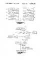
- FIG. 1is a flow diagram of the preferred process of the present invention employed in the manufacture of membranes
- FIG. 2Ais a schematic view of a holding frame for application of the solution to a substrate
- FIG. 2Bis a side elevation of a precipitant tank
- FIG. 2Cis a top plan view of a holding tray
- FIG. 2Dis a top plan view of a drying frame
- FIG. 3is a cross-sectional view of a membrane formed in accordance with the process described in FIGS. 1 and 2;
- FIG. 4is a flow diagram of a preferred process employed in the manufacture of vascular grafts and other tubular prostheses
- FIG. 5is an enlarged view of the solution feed block and nozzle employed in forming a vascular graft
- FIG. 6is a cross-sectional view of one form of precipitant bath and extraction tank employed in the formation of a vascular graft.
- FIG. 1there is schematically illustrated the process for forming microporous, elastomeric membranes with a maximum pore size on the order of 100 microns and a thickness on the order of 0.025".
- the segmented polyurethane urea solution represented at 10is first cast onto a series of substrates in the form of glass plates 12. Each plate should be clean and dry and have a surface area for application of the solution corresponding to that of the size of the finished article.
- glassis the preferred substrate, other materials may be used, such as, TEFLON®, polyethylene or stainless steel.
- the preferred or optimum range of thickness for the solutionis on the order of 0.035" to 0.045".
- a precipitant bath as at 14is provided by filling a tub large enough to accommodate several plates with a precipitant which is miscible with the solvent present in the polyurethane solution but not with the solution itself.
- the temperature of the precipitant bathis 5° C. to 25° C.
- the composition of the precipitantmay vary depending upon the porous structure desired and, in the case of polyurethane urea solutions containing a solvent in the form of dimethyl acetamide (DMAC) or dimethyl formamide (DMF), the bath composition may be an aqueous solution of alcohols, such as, methanol, ethanol, proponal or isoproponal, or acqueous mixtures of solvents for the polymer being used. In certain cases, non-aqueous solutions may be utilized either alone or in combination at various concentrations which are miscible with the solvent.
- DMACdimethyl acetamide
- DMFdimethyl formamide
- non-aqueous solutionsmay be utilized either alone or in combination at various concentrations which are miscible with the
- the polyurethane solution 10is poured onto a glass plate or substrate 12 and a casting bar 22 is then slowly advanced by sliding across the glass plate 12 so as to uniformly spread the solution across each plate 12. Any excess solution is removed from the casting bar as the solution is applied to each plate.
- Each plate 12is immediately immersed into the precipitant tub 14 and left to stand for a period on the order of ten minutes to an hour, or long enough to precipitate the casting solution onto the plates 12 and form a membrane-like layer.
- the membraneis peeled from each plate while still in the precipitant bath and then removed from the bath and placed into an extraction tank 26 with the shiny side of the membrane facing up.
- an extraction tank 26bis filled with filtered water via inlet 27 to a level opposite a drain port 28.
- a rectangular extraction frame 30 illustrated in FIG. 2Cis placed over the membrane and forced underwater to retain the membrane at the bottom of the extraction tank 26. All membranes are similarly peeled off their substrates 12 and placed into the extraction tank as described above so as to be stacked on top of one another and separated by the extraction frames 30. Preferably, the membranes are left in the extraction tank 26 for a minimum period of fifteen hours but no longer than forty hours.
- wateris constantly run through the extraction tank 26 at the rate of four to eight liters per minute to completely flush or remove any of the solvent and precipitant solution from the membranes.
- each membraneis centered on a drying frame 32 and attached to the frame by suitable means, such as, masking tape 33 applied along the edges of the membrane.
- suitable meanssuch as, masking tape 33 applied along the edges of the membrane.
- the drying frames or racks with attached membranesare advanced through a clean room and then placed in an oven where they are dried for a minimum of four hours and a maximum of seven hours at a temperature of 50° C. ⁇ 5° C., all as represented at 20 in FIG. 1.
- the membranes or patchesare inspected, cut loose from the drying frame along the edges just inside the masking tape and are individually placed in an autoclave bag.
- the bags with enclosed membranesare then evenly distributed over a rack in an oven for the purpose of heat treating the membranes. Care should be taken to maintain the autoclave bags and membranes perfectly flat, and the oven temperature is set to 120° C.
- the membranesare heated for a period of one to one and a half hours at the desired temperature level after which the oven is turned off and the membranes permitted to cool in the oven.
- the autoclave bags and enclosed membranesare then removed from the oven and placed under a laminar flow hood for further processing and packaging as represented at 25.
- the patchesare sterilized as at 26 by irradiation, such as, cobalt 60 gamma irradiation in the range of 0.5-4.0 megarads.
- the process as hereinbefore describedis carried out using one of the segmented polyether urethane urea family of polymers, or "spandex" polymers whose chains consists of alternating hard and soft blocks.
- the soft blockshave glass transition temperatures (T g s) below the use temperature or 0° C.
- the hard blockshave T g s above the use temperature, or 100° C.
- T g sglass transition temperatures
- MITRATHANETMmanufactured and sold by Mitral Medical International, Inc. of Denver, Colo. which is produced as a 25% w/v solution in dimethyl acetamide (DMAC) of 12,000-30,000 centipoise viscosity.
- the hard blocksare extremely short; however, the interchain interaction is enhanced by a hydrogen bonding system which is produced by four hydrogen bonds acting in concert within each hard segment.
- This molecular structureproduces the necessary properties in solution which will result in a variety of microporous structures.
- the resultant membraneis a microporous structure having on the order of 50% void volume with a difference in pore size, for instance, of ⁇ 0.10 microns at the exposed surface to 100 microns at the surface contacting the substrate.
- the structure and porosity of the MITRATHANETM microporous structurescan be altered by adjusting any or all of the following variables:
- a tub or container 36is substantially filled with a precipitant solution 34 and filled to a level as designated at 37.
- a nozzle block 39contains spaced inner and outer concentric tubes 40 and 41 which are suspended above the container 36 for downward vertical extension centrally of the upper end of the container 36 with their lower extremities terminating directly opposite to the upper edge of the container 36, and the inner tube 40 having its lower edge terminating just above that of the outer tube 41.
- the lower end of the nozzlehas an annular flange which supports a retaining tube 38 for downward extension into the solution 34 in the container 36.
- the tubular structure of the graftis formed by extruding a polyurethane solution S through the concentric or annular space between the inner and outer tubes 40 and 41 downwardly through the retaining tube 38 and into the precipitant bath 34.
- Selective control over the porosity of the materialis achieved both internally and externally by pumping an internal precipitant solution P through the inner tube 40 at a comparative rate to the polyurethane solution.
- the precipitant solution Pcontacts the solution S beneath the inner tube 40 it will establish a gradient or rate of precipitation to alleviate forces otherwise tending to cause the outer wall to collapse into the inner wall as the solution S begins to precipitate.
- the resultant tubular prosthesis S'advances into the precipitant bath where it remains immersed in the bath for a period on the order of at least ten minutes and a maximum of sixty minutes.
- the polyurethanecoagulates into a tubular prosthesis S'
- itis advanced through the precipitant bath as illustrated then drawn over a rotating drum member 50 through conduit 52 into an extraction tank 44' where it is flushed with an extraction solution, such as, water or an isotonic saline to remove residual solvents or precipitants.
- the resident time of the prosthesis S' in the precipitant bath 34is regulated by the speed of rotation of the drum 50 in relation to the pumping rate of the solutions into the nozzle 39.
- one side of the nozzleis capable of forming different sized tubular prostheses by control of the pumping rate, the size of the microporous tube ranging from 1 to 10 millimeters ID.
- the variation in pore size between the outer wall and the inner wallcorresponds very much to that experienced in the cardiac patch or membrane manufacture.
- the pore sizedid not change along the inner wall but nevertheless the saline solution was operative to prevent a collapse of the wall and was of sufficient density to cause the tubular prosthesis to descend through the precipitant bath.
- the extraction step as describedrequires from fifteen to forty hours for complete removal of any excess solvent.
- the prosthesesare heat treated, packaged and sterilized for the time periods and temperatures described with respect to the membrane formation of FIG. 1.
- the surfaces of the tubular prothesismay be further modified by passing the extruded member through a modifying liquid 42 placed in the retaining tube 38 above the precipitant bath 34 so that the external surface is brought into contact with the modifying liquid preliminary to immersion in the bath to facilitate finer control over the outer surface porosity.
- a MITRATHANETM polymer solutionwas extruded to form an internal diameter in the range of 2 mm to 10 mm and a wall thickness of 0.20 mm to 2 mm.
- the polyurethaneis injected through the nozzle at a flow rate on the order of 0.5 ml/min. to 20 ml/min. and the internal solution P flowing at a rate of 0.5 ml/min.
- the polymer solution with a DMAC solventwas precipitated in a bath containing 0.9% sodium chloride solution in water at 25° C. with a capillary precipitant of 30% DMSO in water.
- the DMSO/water solutionis of a greater density than the bath solution and will therefore remain trapped in the tubular member and retard the rate of precipitation along the inner wall as well as to encourage it to descend into the bath by gravity.
- a modifying liquid 42 of DMACwas used between the nozzle and the precipitant bath to retard the rate of precipitation and control the porosity to a degree dependent upon the depth of liquid in the outer tube 38.
- vascular grafthaving a porosity which would allow the transport of water, ions and low molecular weight species of less than 2,000, but will not permit ingrowth or adhesions on the external surface or internal, lumenal surface.
- tubular polyurethane prothesesmay be prepared as described in the above from most, if not all, segmented polyurethane urea compositions at concentrations varying from 10% to 30% solids and formed with a number of different solvents, such as, DMAC, DMSO, DMF, THF and combinations thereof.
- the bath and capillary precipitantsmay consist of any liquid or combination of liquids and dissolved solids that fulfill the criteria of being a non solvent for the chosen polyurethane yet are miscible with the solvent for that polyurethane.
- the modifying liquid 42may be a solvent or "near" solvent for the polyurethane solution, such as, dimethyl acetamide which is utilized as described to control pore size; also, it will serve to prevent collapse of the tubular member in advancing downwardly through the bath.
- a MITRATHANETM polymer solutionwas used conforming to the following specifications: 25% ⁇ 1.5% solids dissolved in dimethyl acetamide (DMAC). Viscosity at 22° C.-25° C. between 12,000 and 30,000 cps.
- the polymer solutionwas spread onto a glass plate (approximately 12" ⁇ 10") to a thickness of between 0.030"-0.045".
- the glass plate with cast polymer filmwas quickly immersed in a precipitation bath.
- the composition of this bathwill vary depending upon the type of porous structure required for the end product. Typically, it will contain mixtures of water, alcohols and water/solvent solutions. For the manufacture of the cardiac patch and wound dressings, this bath is water.
- the solutionwas passed through an extrusion nozzle as shown in FIG. 4.
- the polymer solutionwas pumped such that it was extruded in a cylindrical form from the space between the two cylinders.
- a non-solventwas extruded through the central orifice and which acts as a non-solvent or precipitant for the polymer.
- the overall extrudatewas allowed to pass through a bath of non-solvent.
- the effectwas to extrude a tube of polymer solution which precipitated both from the inside and the outside simultaneously so that the wall structure had a controlled thickness and microporosity.
- the physical propertiesmay be altered at will by the size of the respective nozzle and the composition of the capillary solution and precipitation bath. Fine control over the porosity of the outer surface of the graft may be exerted by passing the graft through a "modifying" solution before ultimate precipitation/coagulation in the precipitation bath.
- the respective nozzle insertis placed in the nozzle block.
- the polymer flow and capillary floware set to the desired flow rates (polymer flow 0.5 mls-20.0 mls, capillary flow 0.5 mls-50 mls).
- the nozzleis placed directly in contact with the precipitant bath, such as, isotonic saline, or the surface of the modifying solution above the bath such that no air gap exists between the nozzle and the surface of the bath.
- the precipitant bathsuch as, isotonic saline, or the surface of the modifying solution above the bath such that no air gap exists between the nozzle and the surface of the bath.
- Microporous arterieswill be extruded and precipitated. After a minimum of ten minutes in the precipitant bath, the artery was washed internally and externally with sterile filtered saline for a minimum of forty hours to remove traces of residual solvent.
- the washed arterywas cut to the desired lengths.
- the artery, immersed in saline,can be heat treated to optimize mechanical properties by autoclaving at 121° C. for sixty minutes.
- the arterieswere then packaged in polycarbonate tubes still in isotonic saline and were then sterilized by gamma irradiation at 0.5-4.0 megarads.
- Example IIThe steps outlined in Example II were followed in precipitating a 25% polyether urethane urea in a DMAC solution, using an internal precipitant of 30% DMSO in water and a bath precipitant of 0.9% sodium chloride solution. A modifying solution of acetone was used to form an external surface with two to five micron pores.
- the resultant prosthesishad a smooth inner surface impermeable to molecules of greater than 2,000 molecular weight and a microporous outer wall which would permit ingrowth sufficient to immobilize the prosthesis in the tissue bed but not permit severe loss of compliance or calcification.
- Example IIThe method of Example II was followed to prepare a tubular elastomeric structure with sufficient lumenal porosity to anchor any lumenal ingrowth that may occur from the anastomoses while allowing the exterior wall to be freely sliding within the tissue bed.
- the processwas modified by preparing the graft by precipitation of a 17.5% solid solution of MITRATHANETM in a solvent composed of 62% DMAC and 38% DMSO.
- the internal precipitantwas prepared from 56% DMSO, 24% methanol and 20% water. Again, the precipitant bath was 0.9% sodium chloride solution.
- the resultant porosity of the internal surfacewas 3 microns to 10 microns and the external porosity on the order of 20 Angstroms.
- a spandex polymer solution sold under the trademark BIOMER®was diluted with dimethyl acetamide (DMAC) to form a solution containing 15% by weight of solids. This solution was cast onto a glass plate to a wet thickness of 0.024". The temperature of the solution was 21° C. ⁇ 2° C. The plate plus cast solution was immersed in a water bath maintained at 15° C. The polymer was precipitated out of the solution while in the water bath. The total time elapsed in the water bath was eighteen hours. The membrane thus formed was dried while being constrained at 50° C. ⁇ 5° C. for two hours.
- DMACdimethyl acetamide
- the resulting porous structurewas examined by scanning electron microscopy to reveal a structure consisting of a surface layer with pores in the range of 0.1 microns to 1 micron under which lies a substructure with "finger-like" voids of approximately 100 microns ⁇ 200 microns.
- the membrane mechanical propertieswere quantified using an Instron tensile tester. The membrane having an ultimate tensile strength of 0.26 kg/mm 2 and an elongation at break of 480%.
- the structureconsisted of a surface layer with pores approximately 2 microns to 15 microns and a substructure with "finger-like" pores of approximately 200 ⁇ 500 microns.
- the membrane's mechanical propertieswere quantified using an Instron tensile tester, the membrane having an ultimate tensile strength of 0.12 kg/mm 2 and an elongation at break of 350%.
- a wound dressingwas prepared according to the steps outlined in Example I but where the polymer solution was spread to a thickness of between 0.005" to 0.045". After immersion in the precipitation bath for ten minutes the sheet was removed and extracted under running water for a period of fifteen hours. Thereafter, the sheets were affixed to drying frames and dried for a period of two to six hours at a temperature ranging from 35° C. to 70° C.
- the resultant membranehad a porosity of 1 micron to 3 microns on one side and less than 0.2 microns at the opposite side or surface, the opposing surfaces being separated by an intermediate layer composed of relatively large intersticial voids.
- the pore size along the surface to be placed in contact with the skinwas increased by using methanol in the precipitation bath and increasing the immersion time in the bath to 20 minutes.
- the resultant wound dressinghad a porosity of 3 microns to 7 microns on the skin contacting surface and a porosity of less than 0.2 microns on the opposite or external surface.
- the concentration of the polymer in solutionwas reduced from that described in Examples VII and VIII.
- a polymer solutionwas used having 10% ⁇ 1.5% solids dissolved in DMAC with a viscosity of 23° C. to 25° C. between 1,000 and 15,000 cps.
- the solutionwas spread to a thickness of between 0.030" and 0.045" and immersed for a period of 20 minutes. Extraction and drying were accomplished as previously described.
- the resultant wound dressinghad a porosity of 28 microns on the skin contacting surface and a porosity of less than 0.2 microns on the external surface.
- an adhesivemay be applied to one surface for fixation to the wound site.
- a typical biocompatible adhesivemay be formed in a solvent and spread to a thickness of between 0.001" and 0.01" onto a siliconized release paper. The solvent is then evaporated in the forced hot air oven leaving a solvent-free adhesive layer on the release paper. The wound dressing is then laminated with the adhesive layer, cut to size, packaged and sterilized.
- the skin-contacting surface 44is given a porosity which will permit absorption of liquid exudate from a wound and which optimally is in the range of 1 micron to 10 microns but may be increased to as much as 50 microns depending upon the amount of exudate to be removed from the wound.
- the opposite or external surface 48is made porous to the extent of preventing bacterial penetration; i.e., less than 0.2 microns but preferably is porous only at a molecular level so as to permit transport of water vapor.
- the intermediate or intersticial thickness 46 between the opposite skin surfacesis characterized by being occupied by rather large voids which are separated laterally by less porous material.
- voidsprovide a degree of insulation to the wound which is of importance as the rate of healing is maximized when the wound is kept as close to normal body temperature as possible. Further, it has been observed that insulated wounds are less painful to the patient than those which are not insulated. Another important factor in controlling porosity of the wound dressing is to regulate the amount of moisture vapor transport which is the function both of the polymer type and porosity of the structure. Selective variation in the pore sizes of opposite surfaces of the wound dressing enables close control over the moisture vapor transport rate.
- selective control of the pore size to less than 45 micronswill afford the necessary control over ingrowth to yield a viable fibrohystiocytic tissue and capillaries.
- Shallow ingrowth sufficient to achieve adhesion between tissue and prosthesis but not leading to necrosis and calcificationmay be achieved with porosities from about 3 microns to 20 microns.
- Where desired to prevent any ingrowth while permitting free transport of ions and soluble organic speciesmay be achieved by forming the skin surfaces with porosities of less than 1 micron.
- Vascular grafts having outer surfaces with porosities in the range of less than 1 micronhave demonstrated a similar freedom in tissue to the natural artery.
Landscapes
- Health & Medical Sciences (AREA)
- Engineering & Computer Science (AREA)
- Chemical & Material Sciences (AREA)
- Animal Behavior & Ethology (AREA)
- Veterinary Medicine (AREA)
- Public Health (AREA)
- General Health & Medical Sciences (AREA)
- Life Sciences & Earth Sciences (AREA)
- Biomedical Technology (AREA)
- Epidemiology (AREA)
- Materials Engineering (AREA)
- Chemical Kinetics & Catalysis (AREA)
- Vascular Medicine (AREA)
- Heart & Thoracic Surgery (AREA)
- Hematology (AREA)
- Medicinal Chemistry (AREA)
- Manufacturing & Machinery (AREA)
- Mechanical Engineering (AREA)
- Transplantation (AREA)
- Oral & Maxillofacial Surgery (AREA)
- Gastroenterology & Hepatology (AREA)
- Cardiology (AREA)
- Pulmonology (AREA)
- Thermal Sciences (AREA)
- Dispersion Chemistry (AREA)
- Physics & Mathematics (AREA)
- Polymers & Plastics (AREA)
- Organic Chemistry (AREA)
- Dermatology (AREA)
- Materials For Medical Uses (AREA)
- Manufacture Of Porous Articles, And Recovery And Treatment Of Waste Products (AREA)
- Prostheses (AREA)
Abstract
Description
Claims (18)
Priority Applications (8)
| Application Number | Priority Date | Filing Date | Title |
|---|---|---|---|
| US06/788,850US4704130A (en) | 1985-10-18 | 1985-10-18 | Biocompatible microporous polymeric materials and methods of making same |
| EP19860308052EP0223415B1 (en) | 1985-10-18 | 1986-10-16 | Biocompatible microporous polymeric materials and methods of making same |
| DE86308052TDE3688873T2 (en) | 1985-10-18 | 1986-10-16 | Biocompatible microporous polymer materials and processes for their manufacture. |
| AT86308052TATE92740T1 (en) | 1985-10-18 | 1986-10-16 | BIOFRIENDLY MICROPOROUS POLYMER MATERIALS AND THEIR PRODUCTION PROCESSES. |
| AU64149/86AAU600681B2 (en) | 1985-10-18 | 1986-10-17 | Biocompatible microporous polymeric materials and methods of making same |
| CA 520830CA1322834C (en) | 1985-10-18 | 1986-10-17 | Biocompatible microporous polymeric materials and methods of making same |
| JP24726886AJPH0798057B2 (en) | 1985-10-18 | 1986-10-17 | Biocompatible elastomeric article and method of making the same |
| US07/092,643US4813966A (en) | 1985-10-18 | 1987-09-03 | Biocompatible microporous polymeric materials and methods of making same |
Applications Claiming Priority (1)
| Application Number | Priority Date | Filing Date | Title |
|---|---|---|---|
| US06/788,850US4704130A (en) | 1985-10-18 | 1985-10-18 | Biocompatible microporous polymeric materials and methods of making same |
Related Child Applications (2)
| Application Number | Title | Priority Date | Filing Date |
|---|---|---|---|
| US06/914,591Continuation-In-PartUS5041581A (en) | 1985-10-18 | 1986-10-07 | Hydrophobic cis-platinum complexes efficiently incorporated into liposomes |
| US07/092,643DivisionUS4813966A (en) | 1985-10-18 | 1987-09-03 | Biocompatible microporous polymeric materials and methods of making same |
Publications (1)
| Publication Number | Publication Date |
|---|---|
| US4704130Atrue US4704130A (en) | 1987-11-03 |
Family
ID=25145767
Family Applications (2)
| Application Number | Title | Priority Date | Filing Date |
|---|---|---|---|
| US06/788,850Expired - LifetimeUS4704130A (en) | 1985-10-18 | 1985-10-18 | Biocompatible microporous polymeric materials and methods of making same |
| US07/092,643Expired - LifetimeUS4813966A (en) | 1985-10-18 | 1987-09-03 | Biocompatible microporous polymeric materials and methods of making same |
Family Applications After (1)
| Application Number | Title | Priority Date | Filing Date |
|---|---|---|---|
| US07/092,643Expired - LifetimeUS4813966A (en) | 1985-10-18 | 1987-09-03 | Biocompatible microporous polymeric materials and methods of making same |
Country Status (7)
| Country | Link |
|---|---|
| US (2) | US4704130A (en) |
| EP (1) | EP0223415B1 (en) |
| JP (1) | JPH0798057B2 (en) |
| AT (1) | ATE92740T1 (en) |
| AU (1) | AU600681B2 (en) |
| CA (1) | CA1322834C (en) |
| DE (1) | DE3688873T2 (en) |
Cited By (40)
| Publication number | Priority date | Publication date | Assignee | Title |
|---|---|---|---|---|
| US4828773A (en)* | 1987-10-14 | 1989-05-09 | Exxon Research And Engineering Company | Highly aromatic anisotropic polyurea/urethane membranes and their use for the separation of aromatics from non-aromatics |
| US4879044A (en)* | 1987-10-14 | 1989-11-07 | Exxon Research And Engineering Company | Highly aromatic anisotropic polyurea/urethane membranes and their use for the separation of aromatics from non aromatics |
| US4941870A (en)* | 1986-11-10 | 1990-07-17 | Ube-Nitto Kasei Co., Ltd. | Method for manufacturing a synthetic vascular prosthesis |
| US4957508A (en)* | 1986-10-31 | 1990-09-18 | Ube Industries, Ltd. | Medical tubes |
| US5098500A (en)* | 1988-02-01 | 1992-03-24 | Polymedica Industries, Inc. | Adhesive-faced porous absorbent sheet and method of making same |
| US5264281A (en)* | 1988-10-18 | 1993-11-23 | Nitto Denko Corporation | Adhesive tapes for medical or sanitary use |
| US5265558A (en)* | 1992-05-19 | 1993-11-30 | Schoenrock Thomas | Foam bed for domestic animals having integrally formed, liquid impermeable outer skin |
| US5273742A (en)* | 1991-12-30 | 1993-12-28 | Tyndale Plains-Hunter Ltd. | Biomedical water soluble hydrophilic polyurethane polymers and method of use thereof |
| US5308695A (en)* | 1988-10-18 | 1994-05-03 | Nitto Denko Corporation | Adhesive tapes for medical or sanitary use |
| US5385670A (en)* | 1992-06-23 | 1995-01-31 | Gambro Dialysatoren Gmbh & Co. Kg | Method for drying porous ultrafiltration membranes |
| US5462704A (en)* | 1994-04-26 | 1995-10-31 | Industrial Technology Research Institute | Method for preparing a porous polyurethane vascular graft prosthesis |
| US5508036A (en)* | 1992-04-24 | 1996-04-16 | Osteotech, Inc. | Devices for preventing tissue adhesion |
| US5549924A (en)* | 1987-07-17 | 1996-08-27 | Robin Renee Thill Shlenker | Method of forming a membrane, especially a latex or polymer membrane, including a deactivating barrier and indicating layer |
| DE19539449A1 (en)* | 1995-10-24 | 1997-04-30 | Biotronik Mess & Therapieg | Process for the production of intraluminal stents from bioresorbable polymer material |
| US5634901A (en)* | 1992-11-02 | 1997-06-03 | Localmed, Inc. | Method of using a catheter sleeve |
| US5653699A (en)* | 1994-09-13 | 1997-08-05 | Polymedica Industries, Inc. | Spyrosorbent wound dressings for exudate management |
| US5679399A (en)* | 1987-07-17 | 1997-10-21 | Bio Barrier, Inc. | Method of forming a membrane, especially a latex or polymer membrane, including multiple discrete layers |
| WO1999047077A1 (en)* | 1998-03-18 | 1999-09-23 | Meadox Medicals, Inc. | Improved ptfe vascular prosthesis and method of manufacture |
| EP1148896A1 (en)* | 1999-02-02 | 2001-10-31 | Artimplant AB | A film for medical use, consisting of linear block polymers of polyurethane and a method for the production of such a film |
| US20040193242A1 (en)* | 1996-01-22 | 2004-09-30 | Scimed Life Systems, Inc. | Self-sealing PTFE vascular graft and manufacturing methods |
| US20050055075A1 (en)* | 2003-09-08 | 2005-03-10 | Leonard Pinchuk | Methods for the manufacture of porous prostheses |
| US20050114613A1 (en)* | 2003-10-30 | 2005-05-26 | Takayuki Otani | Multi-chip package type memory system |
| US20060085023A1 (en)* | 2004-10-15 | 2006-04-20 | Davies William F Jr | Medical balloon having strengthening rods |
| US20060085024A1 (en)* | 2004-10-15 | 2006-04-20 | Pepper Lanny R | Non-compliant medical balloon having an integral non-woven fabric layer |
| US20060085022A1 (en)* | 2004-10-15 | 2006-04-20 | Kelli Hayes | Non-compliant medical balloon having an integral woven fabric layer |
| US20070244539A1 (en)* | 1996-01-22 | 2007-10-18 | Boston Scientific Scimed, Inc. | Self-sealing PTFE vascular graft and manufacturing methods |
| US20080183132A1 (en)* | 2004-10-15 | 2008-07-31 | Futurematrix Interventional, Inc. | Non-compliant medical balloon having braided or knitted reinforcement |
| US20090043254A1 (en)* | 2007-08-06 | 2009-02-12 | Pepper Lanny R | Non-compliant medical balloon |
| US7544201B2 (en) | 2005-07-05 | 2009-06-09 | Futurematrix Interventional, Inc. | Rapid exchange balloon dilation catheter having reinforced multi-lumen distal portion |
| US20090171277A1 (en)* | 2005-06-22 | 2009-07-02 | Futurematrix Interventional, Inc. | Balloon dilation catheter having transition from coaxial lumens to non-coaxial multiple lumens |
| US20090248172A1 (en)* | 2006-06-02 | 2009-10-01 | Eidgenossische Technische Hochschule Zurich | Porous membrane comprising a biocompatible block-copolymer |
| US20100185145A1 (en)* | 2009-01-16 | 2010-07-22 | Futurematrix Interventional, Inc | Balloon dilation catheter shaft having end transition |
| US20100217189A1 (en)* | 2009-02-23 | 2010-08-26 | Pepper Lanny R | Balloon catheter pressure relief valve |
| US20100243135A1 (en)* | 2007-08-06 | 2010-09-30 | Pepper Lanny R | Non-compliant medical balloon |
| US20100318029A1 (en)* | 2009-06-12 | 2010-12-16 | Pepper Lanny R | Semi-compliant medical balloon |
| US20110082489A1 (en)* | 2009-09-24 | 2011-04-07 | Davies Jr William F | Balloon with variable pitch reinforcing fibers |
| US8597240B2 (en) | 2011-02-02 | 2013-12-03 | Futurematrix Interventional, Inc. | Coaxial catheter shaft having balloon attachment feature with axial fluid path |
| US9259559B2 (en) | 2009-02-23 | 2016-02-16 | Futurematrix Interventional, Inc. | Balloon catheter pressure relief valve |
| US9895497B2 (en)* | 2008-05-20 | 2018-02-20 | Hexal Ag | Method for reducing leachables and extractables in syringes |
| US10188838B2 (en) | 2009-08-24 | 2019-01-29 | Cook Medical Technologies Llc | Textile-reinforced high-pressure balloon |
Families Citing this family (13)
| Publication number | Priority date | Publication date | Assignee | Title |
|---|---|---|---|---|
| US4986832A (en)* | 1987-09-04 | 1991-01-22 | Ube Industries, Ltd. | Artificial blood vessel and process for preparing it |
| CH673388A5 (en)* | 1987-12-07 | 1990-03-15 | Sulzer Ag | |
| EP0323144A3 (en)* | 1987-12-28 | 1990-05-16 | Vyzkumny Ustav Potravinarskeho Prumyslu | Method of manufacturing at least single-layer tubular blood vessel endoprosthesis, especially of a small internal diameter, and extruding nozzle for carrying out this method |
| NL8801741A (en)* | 1988-07-08 | 1990-02-01 | Utermoehlen Nv | ART SKIN. |
| NL9000113A (en)* | 1990-01-17 | 1991-08-16 | X Flow Bv | METHOD FOR MANUFACTURING A FLAT POROUS PRODUCT AND ITS APPLICATION IN THE FIELD OF PHARMACY, MEDICINE, DENTISTRY OR COSMETICS. |
| US5549664A (en)* | 1990-07-31 | 1996-08-27 | Ube Industries, Ltd. | Artificial blood vessel |
| US5104389A (en)* | 1991-06-27 | 1992-04-14 | Cordis Corporation | Medical instrument valve with foam partition member having vapor permeable skin |
| US5501682A (en)* | 1993-06-10 | 1996-03-26 | Edwards-Cofie; Sophia | Multi purpose vibrating foot stool |
| ES2129185T3 (en)* | 1993-11-04 | 1999-06-01 | Smith & Nephew | Bandages. |
| US20060205301A1 (en)* | 2005-03-11 | 2006-09-14 | Bha Technologies, Inc. | Composite membrane having hydrophilic properties and method of manufacture |
| US7665615B2 (en)* | 2005-09-30 | 2010-02-23 | General Electric Company | Composite article having hydrophilic properties and method of manufacture |
| US7584860B2 (en)* | 2006-07-17 | 2009-09-08 | General Electric Company | Hydrophilic body and method of manufacture |
| US20090025724A1 (en)* | 2007-07-16 | 2009-01-29 | Herron Jr Roy Howard | System for removal of water from a hose and the hygroscopic hose utilized |
Citations (11)
| Publication number | Priority date | Publication date | Assignee | Title |
|---|---|---|---|---|
| US3301257A (en)* | 1963-07-15 | 1967-01-31 | Johnson & Johnson | Absorbent surgical dressing |
| US3645835A (en)* | 1968-07-09 | 1972-02-29 | Smith & Nephew | Moisture-vapor-permeable pressure-sensitive adhesive materials |
| US3665918A (en)* | 1970-01-12 | 1972-05-30 | Johnson & Johnson | Conformable adhesive sheet |
| US3949742A (en)* | 1974-09-20 | 1976-04-13 | Frigitronics, Inc. | Medical dressing |
| US3978855A (en)* | 1975-01-17 | 1976-09-07 | Ionics Lyo Products Company | Polyurethane foam surgical dressing |
| US4173689A (en)* | 1976-02-03 | 1979-11-06 | University Of Utah | Synthetic polymer prosthesis material |
| US4203847A (en)* | 1977-05-25 | 1980-05-20 | Millipore Corporation | Making porous membranes and the membrane products |
| US4341207A (en)* | 1979-09-07 | 1982-07-27 | Kingsdown Medical Consultants Limited | Wound dressing |
| US4452845A (en)* | 1980-08-13 | 1984-06-05 | Smith And Nephew Associated Companies Limited | Moisture vapor transmitting film of polyurethane blended with an incompatible polymer |
| US4460369A (en)* | 1978-11-17 | 1984-07-17 | Smith & Nephew Research Ltd. | Adhesive-coated sheet material incorporating anti-bacterial substances |
| EP0128501A2 (en)* | 1983-06-06 | 1984-12-19 | Kanegafuchi Kagaku Kogyo Kabushiki Kaisha | Artificial vessel and process for preparing the same |
Family Cites Families (6)
| Publication number | Priority date | Publication date | Assignee | Title |
|---|---|---|---|---|
| GB1562244A (en)* | 1976-11-11 | 1980-03-05 | Lock P M | Wound dressing materials |
| US4427737A (en)* | 1981-04-23 | 1984-01-24 | E. R. Squibb & Sons, Inc. | Microporous adhesive tape |
| EP0130401B1 (en)* | 1983-06-06 | 1989-05-17 | Kanegafuchi Kagaku Kogyo Kabushiki Kaisha | Artificial vessel and process for preparing the same |
| SE452404B (en)* | 1984-02-03 | 1987-11-30 | Medinvent Sa | MULTILAYER PROTEST MATERIAL AND PROCEDURE FOR ITS MANUFACTURING |
| SE452110B (en)* | 1984-11-08 | 1987-11-16 | Medinvent Sa | MULTILAYER PROTEST MATERIAL AND PROCEDURE FOR ITS MANUFACTURING |
| DE3525731A1 (en)* | 1985-07-16 | 1987-01-22 | Wilfried Dr Ing Lemm | Process for the production of a non-toxic plastic foam article |
- 1985
- 1985-10-18USUS06/788,850patent/US4704130A/ennot_activeExpired - Lifetime
- 1986
- 1986-10-16ATAT86308052Tpatent/ATE92740T1/ennot_activeIP Right Cessation
- 1986-10-16EPEP19860308052patent/EP0223415B1/ennot_activeExpired - Lifetime
- 1986-10-16DEDE86308052Tpatent/DE3688873T2/ennot_activeExpired - Lifetime
- 1986-10-17JPJP24726886Apatent/JPH0798057B2/ennot_activeExpired - Fee Related
- 1986-10-17AUAU64149/86Apatent/AU600681B2/ennot_activeExpired
- 1986-10-17CACA 520830patent/CA1322834C/ennot_activeExpired - Lifetime
- 1987
- 1987-09-03USUS07/092,643patent/US4813966A/ennot_activeExpired - Lifetime
Patent Citations (11)
| Publication number | Priority date | Publication date | Assignee | Title |
|---|---|---|---|---|
| US3301257A (en)* | 1963-07-15 | 1967-01-31 | Johnson & Johnson | Absorbent surgical dressing |
| US3645835A (en)* | 1968-07-09 | 1972-02-29 | Smith & Nephew | Moisture-vapor-permeable pressure-sensitive adhesive materials |
| US3665918A (en)* | 1970-01-12 | 1972-05-30 | Johnson & Johnson | Conformable adhesive sheet |
| US3949742A (en)* | 1974-09-20 | 1976-04-13 | Frigitronics, Inc. | Medical dressing |
| US3978855A (en)* | 1975-01-17 | 1976-09-07 | Ionics Lyo Products Company | Polyurethane foam surgical dressing |
| US4173689A (en)* | 1976-02-03 | 1979-11-06 | University Of Utah | Synthetic polymer prosthesis material |
| US4203847A (en)* | 1977-05-25 | 1980-05-20 | Millipore Corporation | Making porous membranes and the membrane products |
| US4460369A (en)* | 1978-11-17 | 1984-07-17 | Smith & Nephew Research Ltd. | Adhesive-coated sheet material incorporating anti-bacterial substances |
| US4341207A (en)* | 1979-09-07 | 1982-07-27 | Kingsdown Medical Consultants Limited | Wound dressing |
| US4452845A (en)* | 1980-08-13 | 1984-06-05 | Smith And Nephew Associated Companies Limited | Moisture vapor transmitting film of polyurethane blended with an incompatible polymer |
| EP0128501A2 (en)* | 1983-06-06 | 1984-12-19 | Kanegafuchi Kagaku Kogyo Kabushiki Kaisha | Artificial vessel and process for preparing the same |
Cited By (70)
| Publication number | Priority date | Publication date | Assignee | Title |
|---|---|---|---|---|
| US4957508A (en)* | 1986-10-31 | 1990-09-18 | Ube Industries, Ltd. | Medical tubes |
| US4941870A (en)* | 1986-11-10 | 1990-07-17 | Ube-Nitto Kasei Co., Ltd. | Method for manufacturing a synthetic vascular prosthesis |
| US5549924A (en)* | 1987-07-17 | 1996-08-27 | Robin Renee Thill Shlenker | Method of forming a membrane, especially a latex or polymer membrane, including a deactivating barrier and indicating layer |
| US5965276A (en)* | 1987-07-17 | 1999-10-12 | Bio Barrier, Inc. | Method of forming a membrane especially a latex or polymer membrane including multiple discrete layers |
| US5679399A (en)* | 1987-07-17 | 1997-10-21 | Bio Barrier, Inc. | Method of forming a membrane, especially a latex or polymer membrane, including multiple discrete layers |
| US4828773A (en)* | 1987-10-14 | 1989-05-09 | Exxon Research And Engineering Company | Highly aromatic anisotropic polyurea/urethane membranes and their use for the separation of aromatics from non-aromatics |
| US4879044A (en)* | 1987-10-14 | 1989-11-07 | Exxon Research And Engineering Company | Highly aromatic anisotropic polyurea/urethane membranes and their use for the separation of aromatics from non aromatics |
| US5098500A (en)* | 1988-02-01 | 1992-03-24 | Polymedica Industries, Inc. | Adhesive-faced porous absorbent sheet and method of making same |
| US5264281A (en)* | 1988-10-18 | 1993-11-23 | Nitto Denko Corporation | Adhesive tapes for medical or sanitary use |
| US5308695A (en)* | 1988-10-18 | 1994-05-03 | Nitto Denko Corporation | Adhesive tapes for medical or sanitary use |
| US5273742A (en)* | 1991-12-30 | 1993-12-28 | Tyndale Plains-Hunter Ltd. | Biomedical water soluble hydrophilic polyurethane polymers and method of use thereof |
| KR100274481B1 (en)* | 1992-04-24 | 2001-11-22 | 웬디 딕슨 | Device to prevent tissue adhesion |
| US5508036A (en)* | 1992-04-24 | 1996-04-16 | Osteotech, Inc. | Devices for preventing tissue adhesion |
| US5265558A (en)* | 1992-05-19 | 1993-11-30 | Schoenrock Thomas | Foam bed for domestic animals having integrally formed, liquid impermeable outer skin |
| US5385670A (en)* | 1992-06-23 | 1995-01-31 | Gambro Dialysatoren Gmbh & Co. Kg | Method for drying porous ultrafiltration membranes |
| US5634901A (en)* | 1992-11-02 | 1997-06-03 | Localmed, Inc. | Method of using a catheter sleeve |
| US5876374A (en)* | 1992-11-02 | 1999-03-02 | Localmed, Inc. | Catheter sleeve for use with a balloon catheter |
| US5462704A (en)* | 1994-04-26 | 1995-10-31 | Industrial Technology Research Institute | Method for preparing a porous polyurethane vascular graft prosthesis |
| US5653699A (en)* | 1994-09-13 | 1997-08-05 | Polymedica Industries, Inc. | Spyrosorbent wound dressings for exudate management |
| DE19539449A1 (en)* | 1995-10-24 | 1997-04-30 | Biotronik Mess & Therapieg | Process for the production of intraluminal stents from bioresorbable polymer material |
| US7244271B2 (en) | 1996-01-22 | 2007-07-17 | Boston Scientific Scimed, Inc. | Self-sealing PTFE vascular graft and manufacturing methods |
| US20070244539A1 (en)* | 1996-01-22 | 2007-10-18 | Boston Scientific Scimed, Inc. | Self-sealing PTFE vascular graft and manufacturing methods |
| US20040193242A1 (en)* | 1996-01-22 | 2004-09-30 | Scimed Life Systems, Inc. | Self-sealing PTFE vascular graft and manufacturing methods |
| WO1999047077A1 (en)* | 1998-03-18 | 1999-09-23 | Meadox Medicals, Inc. | Improved ptfe vascular prosthesis and method of manufacture |
| EP1148896A1 (en)* | 1999-02-02 | 2001-10-31 | Artimplant AB | A film for medical use, consisting of linear block polymers of polyurethane and a method for the production of such a film |
| US6627258B1 (en)* | 1999-02-02 | 2003-09-30 | Artimplant Ab | Film for medical use, consisting of linear block polymers of polyurethane and a method for the production of such a film |
| US20050055075A1 (en)* | 2003-09-08 | 2005-03-10 | Leonard Pinchuk | Methods for the manufacture of porous prostheses |
| US20050114613A1 (en)* | 2003-10-30 | 2005-05-26 | Takayuki Otani | Multi-chip package type memory system |
| US7354419B2 (en) | 2004-10-15 | 2008-04-08 | Futuremed Interventional, Inc. | Medical balloon having strengthening rods |
| US8221351B2 (en) | 2004-10-15 | 2012-07-17 | Bard Peripheral Vascular, Inc. | Non-compliant medical balloon having an integral non-woven fabric layer |
| US20070213760A1 (en)* | 2004-10-15 | 2007-09-13 | Futuremed Interventional, Inc. | Non-compliant medical balloon having an integral woven fabric layer |
| US20070219490A1 (en)* | 2004-10-15 | 2007-09-20 | Futuremed Interventional, Inc. | Non-compliant medical balloon having an integral non-woven fabric layer |
| US20060085024A1 (en)* | 2004-10-15 | 2006-04-20 | Pepper Lanny R | Non-compliant medical balloon having an integral non-woven fabric layer |
| US7309324B2 (en) | 2004-10-15 | 2007-12-18 | Futuremed Interventional, Inc. | Non-compliant medical balloon having an integral woven fabric layer |
| US20060085023A1 (en)* | 2004-10-15 | 2006-04-20 | Davies William F Jr | Medical balloon having strengthening rods |
| US20080183132A1 (en)* | 2004-10-15 | 2008-07-31 | Futurematrix Interventional, Inc. | Non-compliant medical balloon having braided or knitted reinforcement |
| US20080188805A1 (en)* | 2004-10-15 | 2008-08-07 | Futurematrix Interventional, Inc. | Medical balloon having strengthening rods |
| US8353868B2 (en) | 2004-10-15 | 2013-01-15 | Bard Peripheral Vascular, Inc. | Medical balloon having strengthening rods |
| US7914487B2 (en) | 2004-10-15 | 2011-03-29 | Futurematrix Interventional, Inc. | Non-compliant medical balloon having braided or knitted reinforcement |
| US20110054513A1 (en)* | 2004-10-15 | 2011-03-03 | Futurematrix Interventional, Inc. | Non-compliant medical balloon having an integral non-woven fabric layer |
| US8002741B2 (en) | 2004-10-15 | 2011-08-23 | Bard Peripheral Vascular, Inc. | Non-compliant medical balloon having an integral woven fabric layer |
| US20100234802A1 (en)* | 2004-10-15 | 2010-09-16 | Futurematrix Interventional, Inc. | Non-compliant medical balloon having an integral non-woven fabric layer |
| US20060085022A1 (en)* | 2004-10-15 | 2006-04-20 | Kelli Hayes | Non-compliant medical balloon having an integral woven fabric layer |
| US7682335B2 (en) | 2004-10-15 | 2010-03-23 | Futurematrix Interventional, Inc. | Non-compliant medical balloon having an integral non-woven fabric layer |
| US8105275B2 (en) | 2004-10-15 | 2012-01-31 | Bard Peripheral Vascular, Inc. | Non-compliant medical balloon having an integral non-woven fabric layer |
| US7780629B2 (en) | 2004-10-15 | 2010-08-24 | Futurematrix Interventional, Inc. | Non-compliant medical balloon having an integral non-woven fabric layer |
| US7985235B2 (en) | 2005-06-22 | 2011-07-26 | Bard Peripheral Vascular, Inc. | Balloon dilation catheter having transition from coaxial lumens to non-coaxial multiple lumens |
| US20090171277A1 (en)* | 2005-06-22 | 2009-07-02 | Futurematrix Interventional, Inc. | Balloon dilation catheter having transition from coaxial lumens to non-coaxial multiple lumens |
| US20090247947A1 (en)* | 2005-07-05 | 2009-10-01 | Futurematrix Interventional, Inc. | Rapid exchange balloon dilation catheter having reinforced multi-lumen distal portion |
| US7985236B2 (en) | 2005-07-05 | 2011-07-26 | Bard Peripheral Vascular, Inc. | Rapid exchange balloon dilation catheter having reinforced multi-lumen distal portion |
| US7544201B2 (en) | 2005-07-05 | 2009-06-09 | Futurematrix Interventional, Inc. | Rapid exchange balloon dilation catheter having reinforced multi-lumen distal portion |
| US20090248172A1 (en)* | 2006-06-02 | 2009-10-01 | Eidgenossische Technische Hochschule Zurich | Porous membrane comprising a biocompatible block-copolymer |
| US8236221B2 (en) | 2007-08-06 | 2012-08-07 | Bard Peripheral Vascular, Inc. | Non-compliant medical balloon |
| US20090043254A1 (en)* | 2007-08-06 | 2009-02-12 | Pepper Lanny R | Non-compliant medical balloon |
| US8002744B2 (en) | 2007-08-06 | 2011-08-23 | Bard Peripheral Vascular, Inc | Non-compliant medical balloon |
| US20100243135A1 (en)* | 2007-08-06 | 2010-09-30 | Pepper Lanny R | Non-compliant medical balloon |
| US20090294031A1 (en)* | 2007-08-06 | 2009-12-03 | Futurematrix Interventional, Inc. | Non-compliant medical balloon |
| US8313601B2 (en) | 2007-08-06 | 2012-11-20 | Bard Peripheral Vascular, Inc. | Non-compliant medical balloon |
| US9895497B2 (en)* | 2008-05-20 | 2018-02-20 | Hexal Ag | Method for reducing leachables and extractables in syringes |
| US8728110B2 (en) | 2009-01-16 | 2014-05-20 | Bard Peripheral Vascular, Inc. | Balloon dilation catheter shaft having end transition |
| US20100185145A1 (en)* | 2009-01-16 | 2010-07-22 | Futurematrix Interventional, Inc | Balloon dilation catheter shaft having end transition |
| US20100217189A1 (en)* | 2009-02-23 | 2010-08-26 | Pepper Lanny R | Balloon catheter pressure relief valve |
| US8814899B2 (en) | 2009-02-23 | 2014-08-26 | Futurematrix Interventional, Inc. | Balloon catheter pressure relief valve |
| US9259559B2 (en) | 2009-02-23 | 2016-02-16 | Futurematrix Interventional, Inc. | Balloon catheter pressure relief valve |
| US8900215B2 (en) | 2009-06-12 | 2014-12-02 | Bard Peripheral Vascular, Inc. | Semi-compliant medical balloon |
| US20100318029A1 (en)* | 2009-06-12 | 2010-12-16 | Pepper Lanny R | Semi-compliant medical balloon |
| US10188838B2 (en) | 2009-08-24 | 2019-01-29 | Cook Medical Technologies Llc | Textile-reinforced high-pressure balloon |
| US20110082489A1 (en)* | 2009-09-24 | 2011-04-07 | Davies Jr William F | Balloon with variable pitch reinforcing fibers |
| US9211391B2 (en) | 2009-09-24 | 2015-12-15 | Bard Peripheral Vascular, Inc. | Balloon with variable pitch reinforcing fibers |
| US8597240B2 (en) | 2011-02-02 | 2013-12-03 | Futurematrix Interventional, Inc. | Coaxial catheter shaft having balloon attachment feature with axial fluid path |
Also Published As
| Publication number | Publication date |
|---|---|
| JPH0798057B2 (en) | 1995-10-25 |
| AU600681B2 (en) | 1990-08-23 |
| JPS62161375A (en) | 1987-07-17 |
| EP0223415B1 (en) | 1993-08-11 |
| ATE92740T1 (en) | 1993-08-15 |
| EP0223415A3 (en) | 1988-07-13 |
| US4813966A (en) | 1989-03-21 |
| DE3688873D1 (en) | 1993-09-16 |
| CA1322834C (en) | 1993-10-12 |
| EP0223415A2 (en) | 1987-05-27 |
| DE3688873T2 (en) | 1994-03-10 |
| AU6414986A (en) | 1987-04-30 |
Similar Documents
| Publication | Publication Date | Title |
|---|---|---|
| US4704130A (en) | Biocompatible microporous polymeric materials and methods of making same | |
| EP0118458B1 (en) | Biocompatible, antithrombogenic materials suitable for reconstructive surgery | |
| DE60214477T2 (en) | Implant for the regeneration of cartilage tissue | |
| JP3506718B2 (en) | Poly (vinyl alcohol) cryogel | |
| US7175852B2 (en) | Implant and process for producing it | |
| JP3423330B2 (en) | Non-porous collagen sheet for therapy and method and apparatus for preparing the same | |
| JP3221690B2 (en) | Collagen structure | |
| EP0228379A1 (en) | A method of producing a mono- or multilayered prosthesis material and the material hereby obtained | |
| HU215534B (en) | Biocompatible, perforated membranes, method for their preparation, use for promoting in vitro growth of epithelial cells and artificial skin thus obtained | |
| Gogolewski et al. | Resorbable materials of poly (L-lactide) III. Porous materials for medical application | |
| CN111317867A (en) | Nerve conduit and preparation method thereof | |
| US5380589A (en) | Biotextured surfaces | |
| CN111068113B (en) | Nanofiber coating artificial blood vessel, and preparation method and preparation device of coating | |
| CN119119412A (en) | A three-dimensional scaffold with bioenergetic activity and a preparation method thereof | |
| RU2135214C1 (en) | Method of pre-implantation treatment of textile articles for cardiovascular surgery | |
| JPH06339520A (en) | Preparation of porous protease | |
| US7976860B2 (en) | Implant and process for its production | |
| US20050049331A1 (en) | Microporous latex membranes, related articles and methods | |
| JP2007037764A (en) | Artificial valve | |
| JP2004097687A (en) | Resin base material for living body, and production method therefor | |
| SU764742A1 (en) | Coating method | |
| JPH0622590B2 (en) | Artificial blood vessel and method for producing the same | |
| JP2004097268A (en) | Bioimplant coating material | |
| RU2630061C1 (en) | Method for manufacture of three-layer frame for bile duct prosthetics | |
| White et al. | Microporous flow surface variation and short term thrombogenicity in dogs |
Legal Events
| Date | Code | Title | Description |
|---|---|---|---|
| AS | Assignment | Owner name:MITRAL MEDICAL INTERNATIONAL, INC., 4070 YOUNGFIEL Free format text:ASSIGNMENT OF ASSIGNORS INTEREST.;ASSIGNORS:GILDING, D. KEITH;REED, ANDREW M.;ASKILL, IAN N.;AND OTHERS;REEL/FRAME:004471/0328 Effective date:19851014 | |
| STCF | Information on status: patent grant | Free format text:PATENTED CASE | |
| AS | Assignment | Owner name:MATRIX MEDICA, INC., Free format text:CHANGE OF NAME;ASSIGNOR:MITRAL MEDICAL INTERNATIONAL, INC.,;REEL/FRAME:004863/0940 Effective date:19871113 Owner name:MATRIX MEDICA, INC.,COLORADO Free format text:CHANGE OF NAME;ASSIGNOR:MITRAL MEDICAL INTERNATIONAL, INC.;REEL/FRAME:004863/0940 Effective date:19871113 | |
| AS | Assignment | Owner name:TECHNOLOGY FUNDING PARTNERS III, CALIFORNIA Free format text:SECURITY INTEREST;ASSIGNOR:MATRIX MEDICA, INC., A CORP. OF CO;REEL/FRAME:005298/0884 Effective date:19890503 | |
| AS | Assignment | Owner name:EMERGING SCIENCES, INC., A MA CORP., MASSACHUSETTS Free format text:ASSIGNMENT OF ASSIGNORS INTEREST.;ASSIGNOR:TECHNOLOGY FUNDING INC.;REEL/FRAME:005357/0314 Effective date:19900615 | |
| AS | Assignment | Owner name:EMERGING SCIENCES, INC., A CORP. OF MA, MASSACHUSE Free format text:ASSIGNMENT OF ASSIGNORS INTEREST.;ASSIGNOR:TECHNOLOGY FUNDING PARTNERS, III;REEL/FRAME:005363/0353 Effective date:19900615 | |
| FEPP | Fee payment procedure | Free format text:PAYOR NUMBER ASSIGNED (ORIGINAL EVENT CODE: ASPN); ENTITY STATUS OF PATENT OWNER: SMALL ENTITY Free format text:PAT HLDR NO LONGER CLAIMS SMALL ENT STAT AS SMALL BUSINESS (ORIGINAL EVENT CODE: LSM2); ENTITY STATUS OF PATENT OWNER: SMALL ENTITY | |
| AS | Assignment | Owner name:TECHNOLOGY FUNDING PARTNERS, III, 2000 ALMEDA DE L Free format text:RELEASED BY SECURED PARTY;ASSIGNOR:MATRIX MEMBRANES, INC.;REEL/FRAME:005678/0273 Effective date:19900615 Owner name:MATRIX MEMBRANES, INC. Free format text:CHANGE OF NAME;ASSIGNOR:MATRIX MEDICA, INC.,;REEL/FRAME:005678/0279 Effective date:19900608 | |
| FPAY | Fee payment | Year of fee payment:4 | |
| AS | Assignment | Owner name:POLYMEDICA INDUSTRIES, INC. A CORP. OF MASSACHUS Free format text:CHANGE OF NAME;ASSIGNOR:EMERGING SCIENCES, INC. A CORP. OF MASSACHUSETTS;REEL/FRAME:005758/0233 Effective date:19900709 | |
| AS | Assignment | Owner name:POLYMEDICA INDUSTRIES, INC. A MASSACHUSETTS CORP Free format text:RELEASED BY SECURED PARTY;ASSIGNOR:FURMAN SELZ HOLDING CORPORATION;REEL/FRAME:006396/0812 Effective date:19930126 | |
| FEPP | Fee payment procedure | Free format text:PAYOR NUMBER ASSIGNED (ORIGINAL EVENT CODE: ASPN); ENTITY STATUS OF PATENT OWNER: SMALL ENTITY Free format text:PAT HOLDER CLAIMS SMALL ENTITY STATUS - SMALL BUSINESS (ORIGINAL EVENT CODE: SM02); ENTITY STATUS OF PATENT OWNER: SMALL ENTITY Free format text:PAYER NUMBER DE-ASSIGNED (ORIGINAL EVENT CODE: RMPN); ENTITY STATUS OF PATENT OWNER: SMALL ENTITY | |
| FPAY | Fee payment | Year of fee payment:8 | |
| FEPP | Fee payment procedure | Free format text:PAT HLDR NO LONGER CLAIMS SMALL ENT STAT AS SMALL BUSINESS (ORIGINAL EVENT CODE: LSM2); ENTITY STATUS OF PATENT OWNER: SMALL ENTITY | |
| REFU | Refund | Free format text:REFUND - PAYMENT OF MAINTENANCE FEE, 12TH YR, SMALL ENTITY (ORIGINAL EVENT CODE: R285); ENTITY STATUS OF PATENT OWNER: SMALL ENTITY | |
| FPAY | Fee payment | Year of fee payment:12 | |
| FEPP | Fee payment procedure | Free format text:PAT HOLDER CLAIMS SMALL ENTITY STATUS, ENTITY STATUS SET TO SMALL (ORIGINAL EVENT CODE: LTOS); ENTITY STATUS OF PATENT OWNER: SMALL ENTITY |