US4681588A - Biomaterial - Google Patents
BiomaterialDownload PDFInfo
- Publication number
- US4681588A US4681588AUS06/760,745US76074585AUS4681588AUS 4681588 AUS4681588 AUS 4681588AUS 76074585 AUS76074585 AUS 76074585AUS 4681588 AUS4681588 AUS 4681588A
- Authority
- US
- United States
- Prior art keywords
- pleura
- biomaterial
- glutaraldehyde
- tanning
- sheet
- Prior art date
- Legal status (The legal status is an assumption and is not a legal conclusion. Google has not performed a legal analysis and makes no representation as to the accuracy of the status listed.)
- Expired - Lifetime
Links
- 239000012620biological materialSubstances0.000titleclaimsdescription12
- 210000004224pleuraAnatomy0.000claimsabstractdescription50
- 239000000463materialSubstances0.000claimsabstractdescription28
- 241000283690Bos taurusSpecies0.000claimsabstractdescription16
- SXRSQZLOMIGNAQ-UHFFFAOYSA-NGlutaraldehydeChemical compoundO=CCCCC=OSXRSQZLOMIGNAQ-UHFFFAOYSA-N0.000claimsabstractdescription16
- 230000009278visceral effectEffects0.000claimsabstractdescription12
- 210000004369bloodAnatomy0.000claimsabstractdescription10
- 239000008280bloodSubstances0.000claimsabstractdescription10
- 241001465754MetazoaSpecies0.000claimsabstractdescription9
- 210000001519tissueAnatomy0.000claimsabstractdescription7
- 210000004379membraneAnatomy0.000claimsabstractdescription3
- 239000012528membraneSubstances0.000claimsabstractdescription3
- 238000000034methodMethods0.000claimsdescription17
- 230000003511endothelial effectEffects0.000claimsdescription3
- 241000282472Canis lupus familiarisSpecies0.000description22
- 230000002685pulmonary effectEffects0.000description10
- 238000011282treatmentMethods0.000description10
- 230000001746atrial effectEffects0.000description9
- 230000007547defectEffects0.000description9
- 238000002513implantationMethods0.000description8
- 241000700159RattusSpecies0.000description7
- 206010052428WoundDiseases0.000description6
- 208000027418Wounds and injuryDiseases0.000description6
- 238000002474experimental methodMethods0.000description4
- 230000008439repair processEffects0.000description4
- 102000008186CollagenHuman genes0.000description3
- 108010035532CollagenProteins0.000description3
- 241001494479PecoraSpecies0.000description3
- 210000004027cellAnatomy0.000description3
- 230000008859changeEffects0.000description3
- 229920001436collagenPolymers0.000description3
- 238000011156evaluationMethods0.000description3
- 239000007943implantSubstances0.000description3
- 210000004072lungAnatomy0.000description3
- 230000001936parietal effectEffects0.000description3
- 210000003516pericardiumAnatomy0.000description3
- 239000011505plasterSubstances0.000description3
- 210000001147pulmonary arteryAnatomy0.000description3
- 239000000243solutionSubstances0.000description3
- HEDRZPFGACZZDS-UHFFFAOYSA-NChloroformChemical compoundClC(Cl)ClHEDRZPFGACZZDS-UHFFFAOYSA-N0.000description2
- RTZKZFJDLAIYFH-UHFFFAOYSA-NDiethyl etherChemical compoundCCOCCRTZKZFJDLAIYFH-UHFFFAOYSA-N0.000description2
- LFQSCWFLJHTTHZ-UHFFFAOYSA-NEthanolChemical compoundCCOLFQSCWFLJHTTHZ-UHFFFAOYSA-N0.000description2
- GQPLMRYTRLFLPF-UHFFFAOYSA-NNitrous OxideChemical compound[O-][N+]#NGQPLMRYTRLFLPF-UHFFFAOYSA-N0.000description2
- FAPWRFPIFSIZLT-UHFFFAOYSA-MSodium chlorideChemical compound[Na+].[Cl-]FAPWRFPIFSIZLT-UHFFFAOYSA-M0.000description2
- 230000003444anaesthetic effectEffects0.000description2
- 210000000038chestAnatomy0.000description2
- 210000002808connective tissueAnatomy0.000description2
- 210000002837heart atriumAnatomy0.000description2
- 210000005246left atriumAnatomy0.000description2
- 210000005033mesothelial cellAnatomy0.000description2
- 210000004165myocardiumAnatomy0.000description2
- 239000002356single layerSubstances0.000description2
- 239000011780sodium chlorideSubstances0.000description2
- 238000012360testing methodMethods0.000description2
- 210000000115thoracic cavityAnatomy0.000description2
- 102000004190EnzymesHuman genes0.000description1
- 108090000790EnzymesProteins0.000description1
- VGGSQFUCUMXWEO-UHFFFAOYSA-NEtheneChemical compoundC=CVGGSQFUCUMXWEO-UHFFFAOYSA-N0.000description1
- 239000005977EthyleneSubstances0.000description1
- 239000004792ProleneSubstances0.000description1
- 241000700157Rattus norvegicusSpecies0.000description1
- 241000282887SuidaeSpecies0.000description1
- 102000004142TrypsinHuman genes0.000description1
- 108090000631TrypsinProteins0.000description1
- JPKKQJKQTPNWTR-KQAYXBCTSA-N[(1r,5s)-8-methyl-8-azabicyclo[3.2.1]octan-3-yl] (2r)-3-hydroxy-2-phenylpropanoate;sulfuric acid;hydrateChemical compoundO.OS(O)(=O)=O.C1([C@H](CO)C(=O)OC2C[C@H]3CC[C@@H](C2)N3C)=CC=CC=C1.C1([C@H](CO)C(=O)OC2C[C@H]3CC[C@@H](C2)N3C)=CC=CC=C1JPKKQJKQTPNWTR-KQAYXBCTSA-N0.000description1
- NOSIYYJFMPDDSA-UHFFFAOYSA-NacepromazineChemical compoundC1=C(C(C)=O)C=C2N(CCCN(C)C)C3=CC=CC=C3SC2=C1NOSIYYJFMPDDSA-UHFFFAOYSA-N0.000description1
- 229960005054acepromazineDrugs0.000description1
- 230000001464adherent effectEffects0.000description1
- 210000001789adipocyteAnatomy0.000description1
- 238000002266amputationMethods0.000description1
- 230000000890antigenic effectEffects0.000description1
- QVGXLLKOCUKJST-UHFFFAOYSA-Natomic oxygenChemical compound[O]QVGXLLKOCUKJST-UHFFFAOYSA-N0.000description1
- 239000000560biocompatible materialSubstances0.000description1
- 210000004204blood vesselAnatomy0.000description1
- 244000309466calfSpecies0.000description1
- 230000008602contractionEffects0.000description1
- 239000007799corkSubstances0.000description1
- 238000000502dialysisMethods0.000description1
- 230000003292diminished effectEffects0.000description1
- LOKCTEFSRHRXRJ-UHFFFAOYSA-Idipotassium trisodium dihydrogen phosphate hydrogen phosphate dichlorideChemical compoundP(=O)(O)(O)[O-].[K+].P(=O)(O)([O-])[O-].[Na+].[Na+].[Cl-].[K+].[Cl-].[Na+]LOKCTEFSRHRXRJ-UHFFFAOYSA-I0.000description1
- 239000002270dispersing agentSubstances0.000description1
- 238000010894electron beam technologyMethods0.000description1
- 238000005530etchingMethods0.000description1
- 238000001914filtrationMethods0.000description1
- BCQZXOMGPXTTIC-UHFFFAOYSA-NhalothaneChemical compoundFC(F)(F)C(Cl)BrBCQZXOMGPXTTIC-UHFFFAOYSA-N0.000description1
- 229960003132halothaneDrugs0.000description1
- 230000035876healingEffects0.000description1
- 208000025339heart septal defectDiseases0.000description1
- 210000003709heart valveAnatomy0.000description1
- 208000015181infectious diseaseDiseases0.000description1
- 230000003601intercostal effectEffects0.000description1
- 239000010410layerSubstances0.000description1
- 210000005248left atrial appendageAnatomy0.000description1
- 210000002540macrophageAnatomy0.000description1
- 238000005259measurementMethods0.000description1
- 229960001620methohexital sodiumDrugs0.000description1
- KDXZREBVGAGZHS-UHFFFAOYSA-Mmethohexital sodiumChemical compound[Na+].CCC#CC(C)C1(CC=C)C(=O)N=C([O-])N(C)C1=OKDXZREBVGAGZHS-UHFFFAOYSA-M0.000description1
- 238000001000micrographMethods0.000description1
- 239000000203mixtureSubstances0.000description1
- 210000005036nerveAnatomy0.000description1
- 239000001272nitrous oxideSubstances0.000description1
- 239000001301oxygenSubstances0.000description1
- 229910052760oxygenInorganic materials0.000description1
- 239000012188paraffin waxSubstances0.000description1
- 230000035699permeabilityEffects0.000description1
- 239000002953phosphate buffered salineSubstances0.000description1
- 239000002504physiological saline solutionSubstances0.000description1
- 230000008569processEffects0.000description1
- 210000005247right atrial appendageAnatomy0.000description1
- 230000036573scar formationEffects0.000description1
- 239000002904solventSubstances0.000description1
- 238000001356surgical procedureMethods0.000description1
- 210000002435tendonAnatomy0.000description1
- AWLILQARPMWUHA-UHFFFAOYSA-Mthiopental sodiumChemical compound[Na+].CCCC(C)C1(CC)C(=O)NC([S-])=NC1=OAWLILQARPMWUHA-UHFFFAOYSA-M0.000description1
- 210000000779thoracic wallAnatomy0.000description1
- 230000007704transitionEffects0.000description1
- 239000012588trypsinSubstances0.000description1
- 230000002792vascularEffects0.000description1
- 230000002861ventricularEffects0.000description1
- 238000005303weighingMethods0.000description1
Images


Classifications
- A—HUMAN NECESSITIES
- A61—MEDICAL OR VETERINARY SCIENCE; HYGIENE
- A61L—METHODS OR APPARATUS FOR STERILISING MATERIALS OR OBJECTS IN GENERAL; DISINFECTION, STERILISATION OR DEODORISATION OF AIR; CHEMICAL ASPECTS OF BANDAGES, DRESSINGS, ABSORBENT PADS OR SURGICAL ARTICLES; MATERIALS FOR BANDAGES, DRESSINGS, ABSORBENT PADS OR SURGICAL ARTICLES
- A61L27/00—Materials for grafts or prostheses or for coating grafts or prostheses
- A61L27/36—Materials for grafts or prostheses or for coating grafts or prostheses containing ingredients of undetermined constitution or reaction products thereof, e.g. transplant tissue, natural bone, extracellular matrix
- A61L27/3683—Materials for grafts or prostheses or for coating grafts or prostheses containing ingredients of undetermined constitution or reaction products thereof, e.g. transplant tissue, natural bone, extracellular matrix subjected to a specific treatment prior to implantation, e.g. decellularising, demineralising, grinding, cellular disruption/non-collagenous protein removal, anti-calcification, crosslinking, supercritical fluid extraction, enzyme treatment
- A61L27/3687—Materials for grafts or prostheses or for coating grafts or prostheses containing ingredients of undetermined constitution or reaction products thereof, e.g. transplant tissue, natural bone, extracellular matrix subjected to a specific treatment prior to implantation, e.g. decellularising, demineralising, grinding, cellular disruption/non-collagenous protein removal, anti-calcification, crosslinking, supercritical fluid extraction, enzyme treatment characterised by the use of chemical agents in the treatment, e.g. specific enzymes, detergents, capping agents, crosslinkers, anticalcification agents
- A—HUMAN NECESSITIES
- A61—MEDICAL OR VETERINARY SCIENCE; HYGIENE
- A61K—PREPARATIONS FOR MEDICAL, DENTAL OR TOILETRY PURPOSES
- A61K35/00—Medicinal preparations containing materials or reaction products thereof with undetermined constitution
- A61K35/12—Materials from mammals; Compositions comprising non-specified tissues or cells; Compositions comprising non-embryonic stem cells; Genetically modified cells
- A61K35/42—Respiratory system, e.g. lungs, bronchi or lung cells
- A—HUMAN NECESSITIES
- A61—MEDICAL OR VETERINARY SCIENCE; HYGIENE
- A61L—METHODS OR APPARATUS FOR STERILISING MATERIALS OR OBJECTS IN GENERAL; DISINFECTION, STERILISATION OR DEODORISATION OF AIR; CHEMICAL ASPECTS OF BANDAGES, DRESSINGS, ABSORBENT PADS OR SURGICAL ARTICLES; MATERIALS FOR BANDAGES, DRESSINGS, ABSORBENT PADS OR SURGICAL ARTICLES
- A61L15/00—Chemical aspects of, or use of materials for, bandages, dressings or absorbent pads
- A61L15/16—Bandages, dressings or absorbent pads for physiological fluids such as urine or blood, e.g. sanitary towels, tampons
- A61L15/40—Bandages, dressings or absorbent pads for physiological fluids such as urine or blood, e.g. sanitary towels, tampons containing ingredients of undetermined constitution or reaction products thereof, e.g. plant or animal extracts
- A—HUMAN NECESSITIES
- A61—MEDICAL OR VETERINARY SCIENCE; HYGIENE
- A61L—METHODS OR APPARATUS FOR STERILISING MATERIALS OR OBJECTS IN GENERAL; DISINFECTION, STERILISATION OR DEODORISATION OF AIR; CHEMICAL ASPECTS OF BANDAGES, DRESSINGS, ABSORBENT PADS OR SURGICAL ARTICLES; MATERIALS FOR BANDAGES, DRESSINGS, ABSORBENT PADS OR SURGICAL ARTICLES
- A61L27/00—Materials for grafts or prostheses or for coating grafts or prostheses
- A61L27/36—Materials for grafts or prostheses or for coating grafts or prostheses containing ingredients of undetermined constitution or reaction products thereof, e.g. transplant tissue, natural bone, extracellular matrix
- A61L27/3604—Materials for grafts or prostheses or for coating grafts or prostheses containing ingredients of undetermined constitution or reaction products thereof, e.g. transplant tissue, natural bone, extracellular matrix characterised by the human or animal origin of the biological material, e.g. hair, fascia, fish scales, silk, shellac, pericardium, pleura, renal tissue, amniotic membrane, parenchymal tissue, fetal tissue, muscle tissue, fat tissue, enamel
- A—HUMAN NECESSITIES
- A61—MEDICAL OR VETERINARY SCIENCE; HYGIENE
- A61L—METHODS OR APPARATUS FOR STERILISING MATERIALS OR OBJECTS IN GENERAL; DISINFECTION, STERILISATION OR DEODORISATION OF AIR; CHEMICAL ASPECTS OF BANDAGES, DRESSINGS, ABSORBENT PADS OR SURGICAL ARTICLES; MATERIALS FOR BANDAGES, DRESSINGS, ABSORBENT PADS OR SURGICAL ARTICLES
- A61L27/00—Materials for grafts or prostheses or for coating grafts or prostheses
- A61L27/50—Materials characterised by their function or physical properties, e.g. injectable or lubricating compositions, shape-memory materials, surface modified materials
- A61L27/60—Materials for use in artificial skin
- A—HUMAN NECESSITIES
- A61—MEDICAL OR VETERINARY SCIENCE; HYGIENE
- A61L—METHODS OR APPARATUS FOR STERILISING MATERIALS OR OBJECTS IN GENERAL; DISINFECTION, STERILISATION OR DEODORISATION OF AIR; CHEMICAL ASPECTS OF BANDAGES, DRESSINGS, ABSORBENT PADS OR SURGICAL ARTICLES; MATERIALS FOR BANDAGES, DRESSINGS, ABSORBENT PADS OR SURGICAL ARTICLES
- A61L2430/00—Materials or treatment for tissue regeneration
- A61L2430/40—Preparation and treatment of biological tissue for implantation, e.g. decellularisation, cross-linking
Definitions
- This inventionprovides a novel kind of material for use in a biological environment. More particularly this material may be used in treatments of the human body for example as a surgical dressing or for the repair of herniae, heart valves, holes in the heart or other defects. In some cases the material produced in accordance with the present invention may be used for dialysis and other filtering procedures.
- the material of the present inventioncomprises a sheet of an animal parietal pleura which has been subjected to glutaraldehyde tanning.
- the inventionalso extends to the use of such material as a surgical graft or as a dressing applied to a living human patient.
- the inventionalso provides a method of producing a biocompatible material comprising subjecting a sheet of animal parietal pleura to glutaraldehyde tanning.
- the materialmay be subjected to irradiation before or after the glutaraldehyde tanning treatment. It may for example be subjected to 0.2 to 5.0 megarads of irradiation.
- a biomaterialshould be:
- Pleurais a thin membranous lining of the thoracic cavity and lungs.
- the pleura covering the thoracic cavityis termed parietal pleura and that covering the lungs is termed visceral.
- the inventionmakes use of the parietal pleura which consists of loose connective tissue covered with a single layer of mesothelial cells.
- the other components that are found in the connective tissueare macrophages, blood vessels, lymphatics, nerve fibres and fat cells.
- human pleuramay be available but more generally parietal pleura will be obtained from freshly slaughtered carcasses of animals such as cattle, sheep and pigs. In this way it has been possible to obtain sheets of pleura that measure as much as 60 cm ⁇ 60 cm square. For special uses pleura can also be obtained from foetal calves.
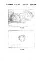
- FIG. 1shows the parietal side of a sheet of tanned bovine pleura produced in accordance with the invention
- FIG. 2shows the visceral side of the same sheet of material
- FIG. 3is a reproduction of a photomicrograph showing a section through a sheet of tanned bovine pleura
- FIG. 4shows a pericardial patch recovered after six months implantation in a first dog
- FIG. 5illustrates a pericardial patch recovered after three months implantation in another dog
- FIGS. 6 and 7show a left atrial patch and a pulmonary outflow patch recovered after three months implantation in another dog;
- FIG. 8illustrates a pulmonary outflow patch recovered after three months implantation in a further dog
- FIG. 9is a reproduction of a photomicrograph through the junction of the pulmonary artery and the pulmonary outflow patch of FIG. 7;
- FIG. 10is a reproduction of a photomicrograph showing a section through the junction of the left atrium and the atrial patch of Figure.
- a sheet of parietal pleura obtained fresh from an abattoiris laid out flat with a single layer of gauze covering its smooth (visceral) surface.
- the pleurais transported in saline to a laboratory where it is stretched and cleansed of all excessive tissue to result in a uniform membrane 50-150 micron in thickness. This may be achieved by stretching the pleura on a board of cork or other suitable material under clean conditions, pinning it at the edges, and removing excess tissue with surgical instruments and subsequent treatment with ether, chloroform or other fat solvents or dispersants.
- the stretched and cleaned pleura, still pinned to the supporting board,is then washed in saline and immersed in a bath of tanning solution.
- the tanning solutionmay typically be buffered glutaraldehyde between 2-8 pH, having a concentration of 0.5% to 5% by weight and the pleura may be immersed for periods varying from 15 minutes to 72 hours depending on the final configuration and integrity required. This treatment renders the product non-antigenic and sterile. It also imparts additional strength.
- permeability, strength and biodegradabilitycan be greatly varied by controlling tanning (concentration, pH and duration) and by subsequent treatment.
- Weak biodegradable materialscan be obtained by appropriate tanning followed or preceded by treatment with enzymes such as trypsin, by irradiation, mechanical perforation with spikes, perforation by laser beam or electron beam etching.
- enzymessuch as trypsin
- the materialwould be subjected to a full glutaraldehyde treatment in cases where maximum strength and non-biodegradability was required, for example for patches in the vascular system, hernial defect repair or for tendon repair.
- the materialcan be rendered sterile by the glutaraldehyde treatment by irradiation or in some instances by treatment with ethylene dioxide.
- the tanned and sterilised materialmay be stored in a 50% alcohol solution. Alternatively it may be stored in dried form and be reconstituted with physiological saline prior to use. As another alternative it could be stored in a phosphate buffered saline so as to be in a similar state as physiological plasma.
- FIGS. 1 to 3 of the accompanying drawingsshow the parietal side and FIG. 2 the visceral side after tanning and FIG. 3 is a photo-micrograph showing a mesothelial cell covering on the visceral surface of the material overlaying an inner structure of collagen fibres.
- the anaesthetised dogswere placed in the right lateral position, prepared and draped, and a left transcostal thoractomy via the fourth left intercostal space was performed.
- a pericardial defect and left atrial appendage amputationwas carried out.
- the defectswere repaired with a patch of the tanned pleura.
- the pleurawas sewn with 6/0 prolene to the defects with the visceral surface contacting blood and myocardium.
- Two of the dogsalso had their right atrial appendage amputated and the defect repaired with a pleural patch.
- the pulmonary outflow tractwas incised longitudinally and a gusset of pleura was sewn in with the visceral surface contacting blood. At the end of the procedure the wound was closed in layers and the chest was drained.
- Atrial patchesexcept one left atrial patch, were found shrivelled with the surrounding atrial material closing over to heal the wounds.
- One atrial patch that did not shrivelhad a smooth shiny blood contact surface.
- the external surfacewas relatively adhesion free. Histologically, the collagen architecture was preserved.
- the blood contact surfaceshowed "endothelial-like" cells.
- the pulmonary outflow tract gussetswere all identified. They did not exhibit any dilatation or contraction that was obvious.
- the blood contact surfacewas again smooth and shiny. Histologically the collagen architecture was preserved with "endothelial-like" cells in the flow surface.
- FIG. 4illustrates the pericardial patch after six months implantation in dog no. 1.
- FIG. 5shows the pericardial patch reflected off the left ventricular epicardium of dog no. 3 after three months implantation.
- FIG. 6shows the left atrial patch after three months implantation in dog no. 4 and
- FIG. 7illustrates the pulmonary outflow patch from the same dog.
- FIG. 8shows the pulmonary outflow patch of dog no. 5 at three months.
- FIG. 9shows the junction of the pulmonary artery (Pa) and the pulmonary outflow patch (Pl) from dog no. 4.
- Endothelial-like cellscan be seen to extend from the pulmonary artery over the pleura patch.
- FIG. 1shows the appearance at the junction of the left atrium (At) with the pleural patch (Pl) in dog no. 4 showing a smooth transition from left atrial tissue to the tissue of the implant.
- tanned bovine pleuracan be both a successful biological patching material and a successful blood contact material.
- material produced in accordance with the present inventionmay have wide application in surgical treatments for the repair of defects in the body, including use as implants or grafts which must provide a blood contact surface.
Landscapes
- Health & Medical Sciences (AREA)
- Life Sciences & Earth Sciences (AREA)
- Chemical & Material Sciences (AREA)
- Epidemiology (AREA)
- Veterinary Medicine (AREA)
- Public Health (AREA)
- Engineering & Computer Science (AREA)
- General Health & Medical Sciences (AREA)
- Animal Behavior & Ethology (AREA)
- Chemical Kinetics & Catalysis (AREA)
- Biomedical Technology (AREA)
- Medicinal Chemistry (AREA)
- Botany (AREA)
- Oral & Maxillofacial Surgery (AREA)
- Dermatology (AREA)
- Transplantation (AREA)
- Zoology (AREA)
- Molecular Biology (AREA)
- Cell Biology (AREA)
- Physiology (AREA)
- Developmental Biology & Embryology (AREA)
- Materials Engineering (AREA)
- Urology & Nephrology (AREA)
- General Chemical & Material Sciences (AREA)
- Pulmonology (AREA)
- Biotechnology (AREA)
- Hematology (AREA)
- Immunology (AREA)
- Virology (AREA)
- Pharmacology & Pharmacy (AREA)
- Materials For Medical Uses (AREA)
- Prostheses (AREA)
- Saccharide Compounds (AREA)
- Polysaccharides And Polysaccharide Derivatives (AREA)
Abstract
Description
______________________________________ 1. Shelf storable. 2. Available in a wide range of sizes and shapes. 3. Sterile. 4. Non-antigenic. 5. Available in biodegradable or non- biodegradable form as the situation demands. 6. Able to support epithelial or endothelial growth as the situation demands. ______________________________________
__________________________________________________________________________ PULMONARY DOG L. ATRIUM R. ATRIUM OUTFLOW PERICARDIUM DURATION __________________________________________________________________________1 2 cm × 2 cm 2 cm × 2cm 5 cm × 10 cm 6 months 2 1.5 cm × 1.5 cm 1.5 cm × 1.5 cm 3 cm oval 3 months 3 0.75 cm × 0.75 cm 1.5 cm × 1.75 cm 3 cm × 4 cm 3months 4 2 cm × 2 cm 3 cm × 3 cm 6 cm × 4 cm 3months 5 3 cm × 3 cm 2.5 cm × 2.5cm 4 cm × 4 cm 3 months __________________________________________________________________________
______________________________________ DIMENSIONS RAT DAY (mm) OBSERVATIONS ______________________________________ 1 4 20 × 20 No change 2 7 20 × 20 No change 3 11 22 × 20 Nochange 4 14 15 × 13 Diminished 5 15 Not Known Cast fell off and dressing lost ______________________________________
Claims (12)
Applications Claiming Priority (2)
| Application Number | Priority Date | Filing Date | Title |
|---|---|---|---|
| AUPG195583 | 1983-10-20 | ||
| AUPG1955 | 1983-10-20 |
Publications (1)
| Publication Number | Publication Date |
|---|---|
| US4681588Atrue US4681588A (en) | 1987-07-21 |
Family
ID=3770377
Family Applications (1)
| Application Number | Title | Priority Date | Filing Date |
|---|---|---|---|
| US06/760,745Expired - LifetimeUS4681588A (en) | 1983-10-20 | 1984-10-17 | Biomaterial |
Country Status (7)
| Country | Link |
|---|---|
| US (1) | US4681588A (en) |
| EP (1) | EP0160025B1 (en) |
| JP (1) | JPS61500302A (en) |
| AU (1) | AU559373B2 (en) |
| CA (1) | CA1234653A (en) |
| DE (1) | DE3480693D1 (en) |
| WO (1) | WO1985001651A1 (en) |
Cited By (66)
| Publication number | Priority date | Publication date | Assignee | Title |
|---|---|---|---|---|
| US5503638A (en)* | 1994-02-10 | 1996-04-02 | Bio-Vascular, Inc. | Soft tissue stapling buttress |
| US5752965A (en)* | 1996-10-21 | 1998-05-19 | Bio-Vascular, Inc. | Apparatus and method for producing a reinforced surgical fastener suture line |
| US5759190A (en)* | 1996-08-30 | 1998-06-02 | Vts Holdings Limited | Method and kit for autologous transplantation |
| US5769892A (en)* | 1996-10-22 | 1998-06-23 | Mitroflow International Inc. | Surgical stapler sleeve for reinforcing staple lines |
| US5782914A (en)* | 1996-11-29 | 1998-07-21 | Bio-Vascular, Inc. | Method for preparing heterogeneous tissue grafts |
| US5805551A (en)* | 1994-04-18 | 1998-09-08 | Matsushita Electric Industrial Co., Ltd. | Method and apparatus for preventing illegal copy or illegal installation of information of optical recording medium |
| US5881038A (en)* | 1994-04-18 | 1999-03-09 | Matsushita Electric Industrial Co., Ltd. | Method and apparatus for preventing illegal copy or illegal installation of information of optical recording medium |
| US5902228A (en)* | 1996-10-11 | 1999-05-11 | Cornell Research Foundation, Inc. | Method and apparatus for support and tubularization of surgical grafts |
| US5989269A (en)* | 1996-08-30 | 1999-11-23 | Vts Holdings L.L.C. | Method, instruments and kit for autologous transplantation |
| US6203755B1 (en) | 1994-03-04 | 2001-03-20 | St. Jude Medical, Inc. | Electron beam sterilization of biological tissues |
| US6273897B1 (en) | 2000-02-29 | 2001-08-14 | Ethicon, Inc. | Surgical bettress and surgical stapling apparatus |
| US6325810B1 (en) | 1999-06-30 | 2001-12-04 | Ethicon, Inc. | Foam buttress for stapling apparatus |
| US20020045940A1 (en)* | 1998-08-14 | 2002-04-18 | Bruno Giannetti | Methods, instruments and materials for chondrocyte cell transplantation |
| US20020116063A1 (en)* | 1999-08-02 | 2002-08-22 | Bruno Giannetti | Kit for chondrocyte cell transplantation |
| US20020173806A1 (en)* | 1996-08-30 | 2002-11-21 | Verigen Transplantation Service International (Vtsi) Ag | Method for autologous transplantation |
| US6569172B2 (en) | 1996-08-30 | 2003-05-27 | Verigen Transplantation Service International (Vtsi) | Method, instruments, and kit for autologous transplantation |
| US20030199923A1 (en)* | 1998-11-06 | 2003-10-23 | Ev3 Sunnyvale, Inc., A California Corporation | Adjustable left atrial appendage implant deployment system |
| US6666892B2 (en) | 1996-08-23 | 2003-12-23 | Cook Biotech Incorporated | Multi-formed collagenous biomaterial medical device |
| US20040030407A1 (en)* | 2000-12-20 | 2004-02-12 | Vettivetpillai Ketharanathan | Method of creating biological and biosynthetic material for implantation |
| US20040034366A1 (en)* | 1999-11-08 | 2004-02-19 | Ev3 Sunnyvale, Inc., A California Corporation | Device for containing embolic material in the LAA having a plurality of tissue retention structures |
| US20040044361A1 (en)* | 1998-11-06 | 2004-03-04 | Frazier Andrew G.C. | Detachable atrial appendage occlusion balloon |
| US20040078076A1 (en)* | 1996-08-23 | 2004-04-22 | Badylak Stephen F. | Purified submucosa graft material |
| US20040098031A1 (en)* | 1998-11-06 | 2004-05-20 | Van Der Burg Erik J. | Method and device for left atrial appendage occlusion |
| US20040136968A1 (en)* | 2002-09-27 | 2004-07-15 | Verigen Ag | Autologous cells on a support matrix for tissue repair |
| US20040158185A1 (en)* | 1998-12-01 | 2004-08-12 | Moran Christopher J. | Embolization device |
| US20040215230A1 (en)* | 2003-04-28 | 2004-10-28 | Frazier Andrew G. C. | Left atrial appendage occlusion device with active expansion |
| US20040220610A1 (en)* | 1999-11-08 | 2004-11-04 | Kreidler Marc S. | Thin film composite lamination |
| US20040236411A1 (en)* | 2001-07-19 | 2004-11-25 | The Cleveland Clinic Foundation | Prosthetic cardiac valve and method for making same |
| US20060025786A1 (en)* | 1996-08-30 | 2006-02-02 | Verigen Transplantation Service International (Vtsi) Ag | Method for autologous transplantation |
| US20080009830A1 (en)* | 2006-06-27 | 2008-01-10 | Fujimoto Kazuro L | Biodegradable elastomeric patch for treating cardiac or cardiovascular conditions |
| US20080145397A1 (en)* | 1998-12-01 | 2008-06-19 | Hiles Michael C | Multi-formed collagenous biomaterial medical device |
| US20080213335A1 (en)* | 1996-08-23 | 2008-09-04 | Cook William A | Graft prosthesis, materials and methods |
| US7575586B2 (en) | 1998-01-30 | 2009-08-18 | St. Jude Medical Atg, Inc. | Medical graft connector or plug structures, and methods of making and installing same |
| US20100059570A1 (en)* | 2007-03-06 | 2010-03-11 | Roland Ostapoff | Wound Closure Material |
| US20100069954A1 (en)* | 2003-04-11 | 2010-03-18 | St. Jude Medical Cardiovascular Division | Closure devices, related delivery methods and related methods of use |
| US20100076489A1 (en)* | 2007-03-06 | 2010-03-25 | Joshua Stopek | Wound closure material |
| US7691128B2 (en) | 2002-05-06 | 2010-04-06 | St. Jude Medical, Cardiology Division, Inc. | PFO closure devices and related methods of use |
| US20100087840A1 (en)* | 2007-03-06 | 2010-04-08 | Garrett Ebersole | Wound closure material |
| US7717937B2 (en) | 2001-06-01 | 2010-05-18 | St. Jude Medical, Cardiology Division, Inc. | Closure devices, related delivery methods and tools, and related methods of use |
| US7735493B2 (en) | 2003-08-15 | 2010-06-15 | Atritech, Inc. | System and method for delivering a left atrial appendage containment device |
| US7972359B2 (en) | 2005-09-16 | 2011-07-05 | Atritech, Inc. | Intracardiac cage and method of delivering same |
| US8372112B2 (en) | 2003-04-11 | 2013-02-12 | St. Jude Medical, Cardiology Division, Inc. | Closure devices, related delivery methods, and related methods of use |
| US8801746B1 (en) | 2004-05-04 | 2014-08-12 | Covidien Lp | System and method for delivering a left atrial appendage containment device |
| US20150064140A1 (en)* | 2012-02-10 | 2015-03-05 | Cvdevices, Llc | Methods and uses of biological tissues for various stent and other medical applications |
| US9364199B2 (en) | 2013-03-14 | 2016-06-14 | Covidien Lp | Medical devices |
| US9408687B2 (en) | 2011-09-27 | 2016-08-09 | Boston Scientific Scimed, Inc. | Tissue modification |
| US10076335B2 (en) | 2005-12-01 | 2018-09-18 | Atritech, Inc. | Apparatus for delivering an implant without bias to a left atrial appendage |
| US10080820B2 (en) | 2015-09-03 | 2018-09-25 | Boston Scientific Scimed, Inc. | Tissue modification devices, systems, and methods |
| US10952741B2 (en) | 2017-12-18 | 2021-03-23 | Boston Scientific Scimed, Inc. | Occlusive device with expandable member |
| US11123079B2 (en) | 2018-06-08 | 2021-09-21 | Boston Scientific Scimed, Inc. | Occlusive device with actuatable fixation members |
| US11241239B2 (en) | 2018-05-15 | 2022-02-08 | Boston Scientific Scimed, Inc. | Occlusive medical device with charged polymer coating |
| US11331104B2 (en) | 2018-05-02 | 2022-05-17 | Boston Scientific Scimed, Inc. | Occlusive sealing sensor system |
| US11382635B2 (en) | 2018-07-06 | 2022-07-12 | Boston Scientific Scimed, Inc. | Occlusive medical device |
| US11413048B2 (en) | 2018-01-19 | 2022-08-16 | Boston Scientific Scimed, Inc. | Occlusive medical device with delivery system |
| US11432809B2 (en) | 2017-04-27 | 2022-09-06 | Boston Scientific Scimed, Inc. | Occlusive medical device with fabric retention barb |
| US11540838B2 (en) | 2019-08-30 | 2023-01-03 | Boston Scientific Scimed, Inc. | Left atrial appendage implant with sealing disk |
| US11596533B2 (en) | 2018-08-21 | 2023-03-07 | Boston Scientific Scimed, Inc. | Projecting member with barb for cardiovascular devices |
| US11672541B2 (en) | 2018-06-08 | 2023-06-13 | Boston Scientific Scimed, Inc. | Medical device with occlusive member |
| US11903589B2 (en) | 2020-03-24 | 2024-02-20 | Boston Scientific Scimed, Inc. | Medical system for treating a left atrial appendage |
| US11944314B2 (en) | 2019-07-17 | 2024-04-02 | Boston Scientific Scimed, Inc. | Left atrial appendage implant with continuous covering |
| US12023036B2 (en) | 2020-12-18 | 2024-07-02 | Boston Scientific Scimed, Inc. | Occlusive medical device having sensing capabilities |
| US12318092B2 (en) | 2021-06-22 | 2025-06-03 | Boston Scientific Scimed, Inc. | Left atrial appendage implant |
| US12329500B2 (en) | 2020-11-30 | 2025-06-17 | Boston Scientific Scimed, Inc. | Implantable passive mean pressure sensor |
| US12349918B2 (en) | 2021-09-08 | 2025-07-08 | Boston Scientific Scimed, Inc. | Multi-sharpness split top soft tissue anchors |
| US12383278B2 (en) | 2021-07-08 | 2025-08-12 | Boston Scientific Scimed, Inc. | Left atrial appendage closure device |
| US12383201B2 (en) | 2021-02-03 | 2025-08-12 | Boston Scientific Scimed, Inc. | Medical system for treating a left atrial appendage |
Families Citing this family (2)
| Publication number | Priority date | Publication date | Assignee | Title |
|---|---|---|---|---|
| EP1064031B1 (en) | 1998-03-23 | 2002-02-06 | Bio-Vascular, Inc. | Implants and method of making |
| CA2900862C (en) | 2013-02-11 | 2017-10-03 | Cook Medical Technologies Llc | Expandable support frame and medical device |
Citations (9)
| Publication number | Priority date | Publication date | Assignee | Title |
|---|---|---|---|---|
| US3093439A (en)* | 1961-07-05 | 1963-06-11 | Johnson & Johnson | Process for preparing tanned collagenous material with dialdehyde starch |
| US3974526A (en)* | 1973-07-06 | 1976-08-17 | Dardik Irving I | Vascular prostheses and process for producing the same |
| US3988782A (en)* | 1973-07-06 | 1976-11-02 | Dardik Irving I | Non-antigenic, non-thrombogenic infection-resistant grafts from umbilical cord vessels and process for preparing and using same |
| US4120649A (en)* | 1975-04-10 | 1978-10-17 | Israel Schechter | Transplants |
| GB2063675A (en)* | 1979-11-26 | 1981-06-10 | Matsuda Ika Kogyo | Medical reinforcing material |
| US4275469A (en)* | 1979-12-13 | 1981-06-30 | Shelhigh Inc. | Prosthetic heart valve |
| WO1982000091A1 (en)* | 1980-07-01 | 1982-01-21 | V Ketharanathan | Vascular prostheses |
| EP0052288A2 (en)* | 1980-11-13 | 1982-05-26 | Heyl Chemisch-pharmazeutische Fabrik GmbH & Co. KG | Collagen preparations, process for their preparation and their use in human and veterinary medicine |
| US4477930A (en)* | 1982-09-28 | 1984-10-23 | Mitral Medical International, Inc. | Natural tissue heat valve and method of making same |
Family Cites Families (1)
| Publication number | Priority date | Publication date | Assignee | Title |
|---|---|---|---|---|
| GB1493459A (en)* | 1975-01-23 | 1977-11-30 | Dardik I | Non-antigenic non-thrombogenic infection resistant grafts from umbilical cord vessels and process for preparing sam |
- 1984
- 1984-10-17JPJP59503807Apatent/JPS61500302A/enactivePending
- 1984-10-17DEDE8484903800Tpatent/DE3480693D1/ennot_activeExpired - Lifetime
- 1984-10-17EPEP84903800Apatent/EP0160025B1/ennot_activeExpired
- 1984-10-17WOPCT/AU1984/000206patent/WO1985001651A1/enactiveIP Right Grant
- 1984-10-17AUAU35532/84Apatent/AU559373B2/ennot_activeExpired
- 1984-10-17USUS06/760,745patent/US4681588A/ennot_activeExpired - Lifetime
- 1984-10-18CACA000465811Apatent/CA1234653A/ennot_activeExpired
Patent Citations (10)
| Publication number | Priority date | Publication date | Assignee | Title |
|---|---|---|---|---|
| US3093439A (en)* | 1961-07-05 | 1963-06-11 | Johnson & Johnson | Process for preparing tanned collagenous material with dialdehyde starch |
| US3974526A (en)* | 1973-07-06 | 1976-08-17 | Dardik Irving I | Vascular prostheses and process for producing the same |
| US3988782A (en)* | 1973-07-06 | 1976-11-02 | Dardik Irving I | Non-antigenic, non-thrombogenic infection-resistant grafts from umbilical cord vessels and process for preparing and using same |
| US4120649A (en)* | 1975-04-10 | 1978-10-17 | Israel Schechter | Transplants |
| GB2063675A (en)* | 1979-11-26 | 1981-06-10 | Matsuda Ika Kogyo | Medical reinforcing material |
| US4275469A (en)* | 1979-12-13 | 1981-06-30 | Shelhigh Inc. | Prosthetic heart valve |
| WO1982000091A1 (en)* | 1980-07-01 | 1982-01-21 | V Ketharanathan | Vascular prostheses |
| US4466139A (en)* | 1980-07-01 | 1984-08-21 | Vettivetpillai Ketharanathan | Vascular prostheses |
| EP0052288A2 (en)* | 1980-11-13 | 1982-05-26 | Heyl Chemisch-pharmazeutische Fabrik GmbH & Co. KG | Collagen preparations, process for their preparation and their use in human and veterinary medicine |
| US4477930A (en)* | 1982-09-28 | 1984-10-23 | Mitral Medical International, Inc. | Natural tissue heat valve and method of making same |
Non-Patent Citations (2)
| Title |
|---|
| Stedman s Medical Dictionary , 24th ed., 1982, Williams & Wilkins Co. Baltimore, p. 1053.* |
| Stedman's Medical Dictionary, 24th ed., 1982, Williams & Wilkins Co. Baltimore, p. 1053. |
Cited By (154)
| Publication number | Priority date | Publication date | Assignee | Title |
|---|---|---|---|---|
| US5549628A (en)* | 1994-02-10 | 1996-08-27 | Bio-Vascular, Inc. | Soft tissue stapling buttress |
| US5575803A (en)* | 1994-02-10 | 1996-11-19 | Bio-Vascular, Inc. | Soft tissue stapling buttress |
| US5503638A (en)* | 1994-02-10 | 1996-04-02 | Bio-Vascular, Inc. | Soft tissue stapling buttress |
| US6203755B1 (en) | 1994-03-04 | 2001-03-20 | St. Jude Medical, Inc. | Electron beam sterilization of biological tissues |
| US5805551A (en)* | 1994-04-18 | 1998-09-08 | Matsushita Electric Industrial Co., Ltd. | Method and apparatus for preventing illegal copy or illegal installation of information of optical recording medium |
| US5881038A (en)* | 1994-04-18 | 1999-03-09 | Matsushita Electric Industrial Co., Ltd. | Method and apparatus for preventing illegal copy or illegal installation of information of optical recording medium |
| US7652077B2 (en) | 1996-08-23 | 2010-01-26 | Cook Incorporated | Graft prosthesis, materials and methods |
| US20080171092A1 (en)* | 1996-08-23 | 2008-07-17 | Cook William A | Graft prosthesis, materials and methods |
| US20100104658A2 (en)* | 1996-08-23 | 2010-04-29 | Cook Biotech Incorporated | Graft prosthesis, materials and methods |
| US20100106256A2 (en)* | 1996-08-23 | 2010-04-29 | Cook Biotech Incorporated | Graft prosthesis, materials and methods |
| US20100104617A2 (en)* | 1996-08-23 | 2010-04-29 | Cook Biotech Incorporated | Graft prosthesis, materials and methods |
| US8128708B2 (en) | 1996-08-23 | 2012-03-06 | Cook Biotech Incorporated | Multi-formed collagenous biomaterial medical device for use in wound care |
| US7699895B2 (en) | 1996-08-23 | 2010-04-20 | Cook Biotech Incorporated | Multi-formed collagenous biomaterial medical device |
| US8716227B2 (en) | 1996-08-23 | 2014-05-06 | Cook Biotech Incorporated | Graft prosthesis, materials and methods |
| US20040078076A1 (en)* | 1996-08-23 | 2004-04-22 | Badylak Stephen F. | Purified submucosa graft material |
| US8808392B2 (en) | 1996-08-23 | 2014-08-19 | Cook Biotech Incorporated | Graft prosthesis, materials and methods |
| US20080213335A1 (en)* | 1996-08-23 | 2008-09-04 | Cook William A | Graft prosthesis, materials and methods |
| US20100106257A2 (en)* | 1996-08-23 | 2010-04-29 | Cook Biotech Incorporated | Graft prosthesis, materials and methods |
| US20080167728A1 (en)* | 1996-08-23 | 2008-07-10 | Cook William A | Graft prosthesis, materials and methods |
| US20080167727A1 (en)* | 1996-08-23 | 2008-07-10 | Cook William A | Graft prosthesis, materials and methods |
| US20080145395A1 (en)* | 1996-08-23 | 2008-06-19 | Hiles Michael C | Multi-formed collagenous biomaterial medical device |
| US20080063680A1 (en)* | 1996-08-23 | 2008-03-13 | Hiles Michael C | Dried collagenous biomaterial medical device prepared from a urinary tissue source |
| US8007542B2 (en) | 1996-08-23 | 2011-08-30 | Cook Biotech Incorporated | Freeze-dried collagenous biomaterial medical sponge device |
| US8920516B2 (en) | 1996-08-23 | 2014-12-30 | Cook Biotech Incorporated | Graft prosthesis, material and methods |
| US8920515B2 (en) | 1996-08-23 | 2014-12-30 | Cook Biotech Incorporated | Graft prosthesis, materials and methods |
| US20110076329A1 (en)* | 1996-08-23 | 2011-03-31 | Cook William A | Graft prosthesis, material and methods |
| US9138444B2 (en) | 1996-08-23 | 2015-09-22 | Cook Biotech Incorporated | Dried collagenous biomaterial medical device |
| US6666892B2 (en) | 1996-08-23 | 2003-12-23 | Cook Biotech Incorporated | Multi-formed collagenous biomaterial medical device |
| US20040180042A1 (en)* | 1996-08-23 | 2004-09-16 | Cook William A. | Graft prosthesis, materials and methods |
| US20040137042A1 (en)* | 1996-08-23 | 2004-07-15 | Hiles Michael C | Multi-formed collagenous biomaterial medical device |
| US6599300B2 (en) | 1996-08-30 | 2003-07-29 | Verigen Transplantation Service International (Vtsi) | Method, instruments, and kit for autologous transplantation |
| US6592599B2 (en) | 1996-08-30 | 2003-07-15 | Verigen Transplantation Service International (Vtsi) | Method, instruments, and kit for autologous transplantation |
| US6379367B1 (en) | 1996-08-30 | 2002-04-30 | Verigen Transplantation Service International (Vtsi) Ag | Method instruments and kit for autologous transplantation |
| US20020173806A1 (en)* | 1996-08-30 | 2002-11-21 | Verigen Transplantation Service International (Vtsi) Ag | Method for autologous transplantation |
| US6569172B2 (en) | 1996-08-30 | 2003-05-27 | Verigen Transplantation Service International (Vtsi) | Method, instruments, and kit for autologous transplantation |
| US6283980B1 (en) | 1996-08-30 | 2001-09-04 | Verigen Transplantation Services Internt'l | Method, instruments, and kit for autologous transplantation |
| US5759190A (en)* | 1996-08-30 | 1998-06-02 | Vts Holdings Limited | Method and kit for autologous transplantation |
| US6592598B2 (en) | 1996-08-30 | 2003-07-15 | Verigen Transplantation Service International (Vtsi) | Method, instruments, and kit for autologous transplantation |
| US6599301B2 (en) | 1996-08-30 | 2003-07-29 | Verrgen Transplantation Service International (Vtsi) | Method, instruments, and kit for autologous transplantation |
| US20030195532A1 (en)* | 1996-08-30 | 2003-10-16 | Verigen Transplantation Service International (Vtsi) Ag | Method, instruments, and kit for autologous transplantation |
| US5989269A (en)* | 1996-08-30 | 1999-11-23 | Vts Holdings L.L.C. | Method, instruments and kit for autologous transplantation |
| US7137989B2 (en) | 1996-08-30 | 2006-11-21 | Verigen Ag | Method, instruments, and kit for autologous transplantation |
| US20060195122A1 (en)* | 1996-08-30 | 2006-08-31 | Verigen Ag | Method, instruments, and kit for autologous transplantation |
| US20060025786A1 (en)* | 1996-08-30 | 2006-02-02 | Verigen Transplantation Service International (Vtsi) Ag | Method for autologous transplantation |
| US20060167483A1 (en)* | 1996-08-30 | 2006-07-27 | Verigen Ag | Method, instruments, and kit for autologous transplantation |
| US7048750B2 (en) | 1996-08-30 | 2006-05-23 | Verigen Ag | Method, instruments, and kits for autologous transplantation |
| US5902228A (en)* | 1996-10-11 | 1999-05-11 | Cornell Research Foundation, Inc. | Method and apparatus for support and tubularization of surgical grafts |
| US5752965A (en)* | 1996-10-21 | 1998-05-19 | Bio-Vascular, Inc. | Apparatus and method for producing a reinforced surgical fastener suture line |
| US5769892A (en)* | 1996-10-22 | 1998-06-23 | Mitroflow International Inc. | Surgical stapler sleeve for reinforcing staple lines |
| US5782914A (en)* | 1996-11-29 | 1998-07-21 | Bio-Vascular, Inc. | Method for preparing heterogeneous tissue grafts |
| US7575586B2 (en) | 1998-01-30 | 2009-08-18 | St. Jude Medical Atg, Inc. | Medical graft connector or plug structures, and methods of making and installing same |
| US20050129668A1 (en)* | 1998-08-14 | 2005-06-16 | Verigen Ag | Methods, instruments and materials for chondrocyte cell transplantation |
| US20020045940A1 (en)* | 1998-08-14 | 2002-04-18 | Bruno Giannetti | Methods, instruments and materials for chondrocyte cell transplantation |
| US6866668B2 (en) | 1998-08-14 | 2005-03-15 | Verigen Transplantation Service International (“VTSL”) AG | Methods, instruments and materials for chondrocyte cell transplantation |
| US9168043B2 (en) | 1998-11-06 | 2015-10-27 | Atritech, Inc. | Method for left atrial appendage occlusion |
| US7722641B2 (en) | 1998-11-06 | 2010-05-25 | Atritech, Inc. | Filter mesh for preventing passage of embolic material form an atrial appendage |
| US7713282B2 (en) | 1998-11-06 | 2010-05-11 | Atritech, Inc. | Detachable atrial appendage occlusion balloon |
| US20040044361A1 (en)* | 1998-11-06 | 2004-03-04 | Frazier Andrew G.C. | Detachable atrial appendage occlusion balloon |
| US8080032B2 (en) | 1998-11-06 | 2011-12-20 | Atritech, Inc. | Method and device for left atrial appendage occlusion |
| US8523897B2 (en) | 1998-11-06 | 2013-09-03 | Atritech, Inc. | Device for left atrial appendage occlusion |
| US20030199923A1 (en)* | 1998-11-06 | 2003-10-23 | Ev3 Sunnyvale, Inc., A California Corporation | Adjustable left atrial appendage implant deployment system |
| US20110218566A1 (en)* | 1998-11-06 | 2011-09-08 | Atritech, Inc. | Method for left atrial appendage occlusion |
| US8535343B2 (en) | 1998-11-06 | 2013-09-17 | Atritech, Inc. | Method for left atrial appendage occlusion |
| US20040098031A1 (en)* | 1998-11-06 | 2004-05-20 | Van Der Burg Erik J. | Method and device for left atrial appendage occlusion |
| US8834519B2 (en) | 1998-11-06 | 2014-09-16 | Artritech, Inc. | Method and device for left atrial appendage occlusion |
| US7152605B2 (en) | 1998-11-06 | 2006-12-26 | Ev3 Endovascular, Inc. | Adjustable left atrial appendage implant deployment system |
| US7128073B1 (en) | 1998-11-06 | 2006-10-31 | Ev3 Endovascular, Inc. | Method and device for left atrial appendage occlusion |
| US8439942B2 (en) | 1998-12-01 | 2013-05-14 | Cook Bioteck Incorporated | Embolization device |
| US7857825B2 (en) | 1998-12-01 | 2010-12-28 | Cook Biotech Incorporated | Embolization device |
| US20080145397A1 (en)* | 1998-12-01 | 2008-06-19 | Hiles Michael C | Multi-formed collagenous biomaterial medical device |
| US8882850B2 (en) | 1998-12-01 | 2014-11-11 | Cook Biotech Incorporated | Multi-formed collagenous biomaterial medical device |
| US20040158185A1 (en)* | 1998-12-01 | 2004-08-12 | Moran Christopher J. | Embolization device |
| US6325810B1 (en) | 1999-06-30 | 2001-12-04 | Ethicon, Inc. | Foam buttress for stapling apparatus |
| US20020116063A1 (en)* | 1999-08-02 | 2002-08-22 | Bruno Giannetti | Kit for chondrocyte cell transplantation |
| US20030212432A1 (en)* | 1999-11-08 | 2003-11-13 | Ev3 Sunnyvale, Inc., A California Corporation | Method of removing an implanted device |
| US8287563B2 (en) | 1999-11-08 | 2012-10-16 | Atritech, Inc. | Implant retrieval system |
| US9943299B2 (en) | 1999-11-08 | 2018-04-17 | Atritech, Inc. | Method of implanting an adjustable occlusion device |
| US6994092B2 (en) | 1999-11-08 | 2006-02-07 | Ev3 Sunnyvale, Inc. | Device for containing embolic material in the LAA having a plurality of tissue retention structures |
| US7192439B2 (en) | 1999-11-08 | 2007-03-20 | Ev3 Endovascular, Inc. | Method of removing an implanted device |
| US8323309B2 (en) | 1999-11-08 | 2012-12-04 | Atritech, Inc. | Adjustable left atrial appendage implant |
| US20040220610A1 (en)* | 1999-11-08 | 2004-11-04 | Kreidler Marc S. | Thin film composite lamination |
| US7044134B2 (en) | 1999-11-08 | 2006-05-16 | Ev3 Sunnyvale, Inc | Method of implanting a device in the left atrial appendage |
| US8043329B2 (en) | 1999-11-08 | 2011-10-25 | Atritech, Inc. | Method of implanting an adjustable occlusion device |
| US20040034366A1 (en)* | 1999-11-08 | 2004-02-19 | Ev3 Sunnyvale, Inc., A California Corporation | Device for containing embolic material in the LAA having a plurality of tissue retention structures |
| US8663273B2 (en) | 1999-11-08 | 2014-03-04 | Atritech, Inc. | Method of implanting an adjustable occlusion device |
| US6273897B1 (en) | 2000-02-29 | 2001-08-14 | Ethicon, Inc. | Surgical bettress and surgical stapling apparatus |
| US20040030407A1 (en)* | 2000-12-20 | 2004-02-12 | Vettivetpillai Ketharanathan | Method of creating biological and biosynthetic material for implantation |
| US7022348B2 (en) | 2000-12-20 | 2006-04-04 | Vettivetpillai Ketharanathan | Method of creating biological and biosynthetic material for implantation |
| US9078630B2 (en) | 2001-06-01 | 2015-07-14 | St. Jude Medical, Cardiology Division, Inc. | Closure devices, related delivery methods and tools, and related methods of use |
| US8777985B2 (en) | 2001-06-01 | 2014-07-15 | St. Jude Medical, Cardiology Division, Inc. | Closure devices, related delivery methods and tools, and related methods of use |
| US20100234882A1 (en)* | 2001-06-01 | 2010-09-16 | St. Jude Medical, Cardiology Division, Inc. | Closure devices, related delivery methods and tools, and related methods of use |
| US7717937B2 (en) | 2001-06-01 | 2010-05-18 | St. Jude Medical, Cardiology Division, Inc. | Closure devices, related delivery methods and tools, and related methods of use |
| US20040236411A1 (en)* | 2001-07-19 | 2004-11-25 | The Cleveland Clinic Foundation | Prosthetic cardiac valve and method for making same |
| US7377938B2 (en) | 2001-07-19 | 2008-05-27 | The Cleveland Clinic Foundation | Prosthetic cardiac value and method for making same |
| US7547322B2 (en) | 2001-07-19 | 2009-06-16 | The Cleveland Clinic Foundation | Prosthetic valve and method for making same |
| US20040260390A1 (en)* | 2001-07-19 | 2004-12-23 | The Cleveland Clinic Foundation | Prosthetic valve and method for making same |
| US7691128B2 (en) | 2002-05-06 | 2010-04-06 | St. Jude Medical, Cardiology Division, Inc. | PFO closure devices and related methods of use |
| US20100234881A1 (en)* | 2002-05-06 | 2010-09-16 | St. Jude Medical, Cardiology Division, Inc. | Pfo closure devices and related methods of use |
| US7976564B2 (en) | 2002-05-06 | 2011-07-12 | St. Jude Medical, Cardiology Division, Inc. | PFO closure devices and related methods of use |
| US20040136968A1 (en)* | 2002-09-27 | 2004-07-15 | Verigen Ag | Autologous cells on a support matrix for tissue repair |
| US20070212396A1 (en)* | 2002-09-27 | 2007-09-13 | Verigen Ag | Autologous cells on a support matrix for tissue repair |
| US8382796B2 (en) | 2003-04-11 | 2013-02-26 | St. Jude Medical, Cardiology Division, Inc. | Closure devices, related delivery methods and related methods of use |
| US8372112B2 (en) | 2003-04-11 | 2013-02-12 | St. Jude Medical, Cardiology Division, Inc. | Closure devices, related delivery methods, and related methods of use |
| US8574264B2 (en) | 2003-04-11 | 2013-11-05 | St. Jude Medical, Cardiology Division, Inc. | Method for retrieving a closure device |
| US20100069954A1 (en)* | 2003-04-11 | 2010-03-18 | St. Jude Medical Cardiovascular Division | Closure devices, related delivery methods and related methods of use |
| US20040215230A1 (en)* | 2003-04-28 | 2004-10-28 | Frazier Andrew G. C. | Left atrial appendage occlusion device with active expansion |
| US7597704B2 (en) | 2003-04-28 | 2009-10-06 | Atritech, Inc. | Left atrial appendage occlusion device with active expansion |
| US7735493B2 (en) | 2003-08-15 | 2010-06-15 | Atritech, Inc. | System and method for delivering a left atrial appendage containment device |
| US8801746B1 (en) | 2004-05-04 | 2014-08-12 | Covidien Lp | System and method for delivering a left atrial appendage containment device |
| US9314249B2 (en) | 2004-05-04 | 2016-04-19 | Covidien Lp | System and method for delivering a left atrial appendage containment device |
| US9445895B2 (en) | 2005-09-16 | 2016-09-20 | Atritech, Inc. | Intracardiac cage and method of delivering same |
| US7972359B2 (en) | 2005-09-16 | 2011-07-05 | Atritech, Inc. | Intracardiac cage and method of delivering same |
| US10143458B2 (en) | 2005-09-16 | 2018-12-04 | Atritech, Inc. | Intracardiac cage and method of delivering same |
| US10898198B2 (en) | 2005-12-01 | 2021-01-26 | Atritech, Inc. | Apparatus for delivering an implant without bias to a left atrial appendage |
| US10076335B2 (en) | 2005-12-01 | 2018-09-18 | Atritech, Inc. | Apparatus for delivering an implant without bias to a left atrial appendage |
| US20080009830A1 (en)* | 2006-06-27 | 2008-01-10 | Fujimoto Kazuro L | Biodegradable elastomeric patch for treating cardiac or cardiovascular conditions |
| US8974542B2 (en)* | 2006-06-27 | 2015-03-10 | University of Pittsburgh—of the Commonwealth System of Higher Education | Biodegradable elastomeric patch for treating cardiac or cardiovascular conditions |
| US9968714B2 (en) | 2006-06-27 | 2018-05-15 | University of Pittsburgh—of the Commonwealth System of Higher Education | Biodegradable elastomeric patch for treating cardiac or cardiovascular conditions |
| US9888924B2 (en) | 2007-03-06 | 2018-02-13 | Covidien Lp | Wound closure material |
| US20100076489A1 (en)* | 2007-03-06 | 2010-03-25 | Joshua Stopek | Wound closure material |
| US20100087840A1 (en)* | 2007-03-06 | 2010-04-08 | Garrett Ebersole | Wound closure material |
| US8529819B2 (en) | 2007-03-06 | 2013-09-10 | Covidien Lp | Wound closure material |
| US20100059570A1 (en)* | 2007-03-06 | 2010-03-11 | Roland Ostapoff | Wound Closure Material |
| EP2305135A1 (en)* | 2009-10-02 | 2011-04-06 | Tyco Healthcare Group LP | Wound closure material |
| US9408687B2 (en) | 2011-09-27 | 2016-08-09 | Boston Scientific Scimed, Inc. | Tissue modification |
| US20150064140A1 (en)* | 2012-02-10 | 2015-03-05 | Cvdevices, Llc | Methods and uses of biological tissues for various stent and other medical applications |
| US10940167B2 (en)* | 2012-02-10 | 2021-03-09 | Cvdevices, Llc | Methods and uses of biological tissues for various stent and other medical applications |
| US9364199B2 (en) | 2013-03-14 | 2016-06-14 | Covidien Lp | Medical devices |
| US10016535B2 (en) | 2013-03-14 | 2018-07-10 | Covidien Lp | Medical devices |
| US10443107B2 (en) | 2015-09-03 | 2019-10-15 | Boston Scientific Scimed, Inc. | Tissue modification devices, systems, and methods |
| US10080820B2 (en) | 2015-09-03 | 2018-09-25 | Boston Scientific Scimed, Inc. | Tissue modification devices, systems, and methods |
| US11432809B2 (en) | 2017-04-27 | 2022-09-06 | Boston Scientific Scimed, Inc. | Occlusive medical device with fabric retention barb |
| US12082797B2 (en) | 2017-04-27 | 2024-09-10 | Boston Scientific Scimed, Inc. | Occlusive medical device with fabric retention barb |
| US10952741B2 (en) | 2017-12-18 | 2021-03-23 | Boston Scientific Scimed, Inc. | Occlusive device with expandable member |
| US11925356B2 (en) | 2017-12-18 | 2024-03-12 | Boston Scientific Scimed, Inc. | Occlusive device with expandable member |
| US11413048B2 (en) | 2018-01-19 | 2022-08-16 | Boston Scientific Scimed, Inc. | Occlusive medical device with delivery system |
| US11331104B2 (en) | 2018-05-02 | 2022-05-17 | Boston Scientific Scimed, Inc. | Occlusive sealing sensor system |
| US11241239B2 (en) | 2018-05-15 | 2022-02-08 | Boston Scientific Scimed, Inc. | Occlusive medical device with charged polymer coating |
| US11672541B2 (en) | 2018-06-08 | 2023-06-13 | Boston Scientific Scimed, Inc. | Medical device with occlusive member |
| US11890018B2 (en) | 2018-06-08 | 2024-02-06 | Boston Scientific Scimed, Inc. | Occlusive device with actuatable fixation members |
| US11123079B2 (en) | 2018-06-08 | 2021-09-21 | Boston Scientific Scimed, Inc. | Occlusive device with actuatable fixation members |
| US11382635B2 (en) | 2018-07-06 | 2022-07-12 | Boston Scientific Scimed, Inc. | Occlusive medical device |
| US12232736B2 (en) | 2018-07-06 | 2025-02-25 | Boston Scientific Scimed, Inc | Occlusive medical device |
| US11596533B2 (en) | 2018-08-21 | 2023-03-07 | Boston Scientific Scimed, Inc. | Projecting member with barb for cardiovascular devices |
| US11944314B2 (en) | 2019-07-17 | 2024-04-02 | Boston Scientific Scimed, Inc. | Left atrial appendage implant with continuous covering |
| US11540838B2 (en) | 2019-08-30 | 2023-01-03 | Boston Scientific Scimed, Inc. | Left atrial appendage implant with sealing disk |
| US11903589B2 (en) | 2020-03-24 | 2024-02-20 | Boston Scientific Scimed, Inc. | Medical system for treating a left atrial appendage |
| US12329500B2 (en) | 2020-11-30 | 2025-06-17 | Boston Scientific Scimed, Inc. | Implantable passive mean pressure sensor |
| US12023036B2 (en) | 2020-12-18 | 2024-07-02 | Boston Scientific Scimed, Inc. | Occlusive medical device having sensing capabilities |
| US12383201B2 (en) | 2021-02-03 | 2025-08-12 | Boston Scientific Scimed, Inc. | Medical system for treating a left atrial appendage |
| US12318092B2 (en) | 2021-06-22 | 2025-06-03 | Boston Scientific Scimed, Inc. | Left atrial appendage implant |
| US12336715B2 (en) | 2021-06-22 | 2025-06-24 | Boston Scientific Scimed, Inc. | Left atrial appendage implant |
| US12383278B2 (en) | 2021-07-08 | 2025-08-12 | Boston Scientific Scimed, Inc. | Left atrial appendage closure device |
| US12349918B2 (en) | 2021-09-08 | 2025-07-08 | Boston Scientific Scimed, Inc. | Multi-sharpness split top soft tissue anchors |
Also Published As
| Publication number | Publication date |
|---|---|
| EP0160025A1 (en) | 1985-11-06 |
| EP0160025B1 (en) | 1989-12-13 |
| WO1985001651A1 (en) | 1985-04-25 |
| DE3480693D1 (en) | 1990-01-18 |
| EP0160025A4 (en) | 1987-01-20 |
| AU559373B2 (en) | 1987-03-05 |
| AU3553284A (en) | 1985-05-07 |
| CA1234653A (en) | 1988-04-05 |
| JPS61500302A (en) | 1986-02-27 |
Similar Documents
| Publication | Publication Date | Title |
|---|---|---|
| US4681588A (en) | Biomaterial | |
| US5350583A (en) | Cell-penetrable medical material and artificial skin | |
| EP0403650B1 (en) | Medical material permitting cells to enter thereinto and artificial skin | |
| DE60010287T2 (en) | RESORBABLE IMPLANT MATERIALS | |
| US7354702B2 (en) | Processing tissue to produce a biopolymer scaffold for tissue engineering | |
| CA2717619C (en) | Graft materials and methods for staged delivery of bioactive components | |
| JP5208752B2 (en) | Antibacterial collagen construct | |
| JP3542170B2 (en) | Medical material and method for producing the same | |
| US5916266A (en) | Raw membranous material for medical materials and manufacturing methods thereof | |
| US9474791B2 (en) | Sterile autologous, allogenic or xenogenic implant and the method of its production | |
| JPH09512463A (en) | Improved blood contact surface utilizing natural subendothelial matrix and method of making and using same | |
| JPH11503051A (en) | Peracetic acid cross-linked non-antigenic ICL graft | |
| JP3726280B2 (en) | Medical collagen membrane | |
| US5413798A (en) | Process for preparing bovine pericard materials and use thereof | |
| CA1333050C (en) | Biologic absorbable implant material for filling and closing soft-tissue cavities and method of its preparation | |
| US4695281A (en) | Medical material | |
| EP0411124B1 (en) | Medical material permitting cells to enter thereinto and artificial skin | |
| Noishiki et al. | Healing pattern of collagen-impregnated and preclotted vascular grafts in dogs | |
| Miyata et al. | A biodegradable antiadhesion collagen membrane with slow release heparin | |
| JPH05184662A (en) | Artificial skin and its manufacture | |
| US12059509B1 (en) | Method and matrix for tissue regeneration | |
| JP2000262610A (en) | Artificial corium for private extraction hair foliculus transplantation designed for epidermis regeneration | |
| KR102673609B1 (en) | Inhibition of fibrosis in tissues using biocompatible polymers | |
| Planche et al. | Long‐term evaluation of five biomaterials for angioplastic enlargement of the pulmonary artery in a young dog model | |
| AU2024205144B1 (en) | Crosslinked amniotic membrane material, preparation method therefore, and applications thereof |
Legal Events
| Date | Code | Title | Description |
|---|---|---|---|
| STCF | Information on status: patent grant | Free format text:PATENTED CASE | |
| FEPP | Fee payment procedure | Free format text:PAYER NUMBER DE-ASSIGNED (ORIGINAL EVENT CODE: RMPN); ENTITY STATUS OF PATENT OWNER: SMALL ENTITY Free format text:PAYOR NUMBER ASSIGNED (ORIGINAL EVENT CODE: ASPN); ENTITY STATUS OF PATENT OWNER: SMALL ENTITY | |
| AS | Assignment | Owner name:BIO NOVA NEO TECHNICS PTY. LTD., 36 MUNSTER TERRAC Free format text:ASSIGNMENT OF ASSIGNORS INTEREST.;ASSIGNOR:VETTIVETPILLAI KETHARANATHAN;REEL/FRAME:005113/0543 Effective date:19871118 Owner name:BIO NOVA NEO TECHNICS PTY. LTD., OF VICTORIA,COLOR Free format text:ASSIGNMENT OF ASSIGNORS INTEREST;ASSIGNOR:VETTIVETPILLAI KETHARANATHAN;REEL/FRAME:005113/0543 Effective date:19871118 | |
| FPAY | Fee payment | Year of fee payment:4 | |
| FPAY | Fee payment | Year of fee payment:8 | |
| AS | Assignment | Owner name:WILKRACHT PTY. LTD, AUSTRALIA Free format text:ASSIGNMENT OF ASSIGNORS INTEREST;ASSIGNOR:BIO NOVA NEO TECHNICS PTY, LTD;REEL/FRAME:007803/0159 Effective date:19900806 | |
| FEPP | Fee payment procedure | Free format text:PAT HOLDER CLAIMS SMALL ENTITY STATUS - SMALL BUSINESS (ORIGINAL EVENT CODE: SM02); ENTITY STATUS OF PATENT OWNER: SMALL ENTITY | |
| REFU | Refund | Free format text:REFUND - PAYMENT OF MAINTENANCE FEE, 12TH YEAR, LARGE ENTITY (ORIGINAL EVENT CODE: R185); ENTITY STATUS OF PATENT OWNER: SMALL ENTITY | |
| FPAY | Fee payment | Year of fee payment:12 |