US4665925A - Doppler catheter - Google Patents
Doppler catheterDownload PDFInfo
- Publication number
- US4665925A US4665925AUS06/775,857US77585785AUS4665925AUS 4665925 AUS4665925 AUS 4665925AUS 77585785 AUS77585785 AUS 77585785AUS 4665925 AUS4665925 AUS 4665925A
- Authority
- US
- United States
- Prior art keywords
- catheter
- passageway
- vessel
- blood
- doppler
- Prior art date
- Legal status (The legal status is an assumption and is not a legal conclusion. Google has not performed a legal analysis and makes no representation as to the accuracy of the status listed.)
- Expired - Lifetime
Links
Images
Classifications
- A—HUMAN NECESSITIES
- A61—MEDICAL OR VETERINARY SCIENCE; HYGIENE
- A61B—DIAGNOSIS; SURGERY; IDENTIFICATION
- A61B8/00—Diagnosis using ultrasonic, sonic or infrasonic waves
- A61B8/06—Measuring blood flow
- A—HUMAN NECESSITIES
- A61—MEDICAL OR VETERINARY SCIENCE; HYGIENE
- A61B—DIAGNOSIS; SURGERY; IDENTIFICATION
- A61B8/00—Diagnosis using ultrasonic, sonic or infrasonic waves
- A61B8/12—Diagnosis using ultrasonic, sonic or infrasonic waves in body cavities or body tracts, e.g. by using catheters
- A—HUMAN NECESSITIES
- A61—MEDICAL OR VETERINARY SCIENCE; HYGIENE
- A61B—DIAGNOSIS; SURGERY; IDENTIFICATION
- A61B8/00—Diagnosis using ultrasonic, sonic or infrasonic waves
- A61B8/44—Constructional features of the ultrasonic, sonic or infrasonic diagnostic device
- A61B8/4444—Constructional features of the ultrasonic, sonic or infrasonic diagnostic device related to the probe
- A61B8/445—Details of catheter construction
- A—HUMAN NECESSITIES
- A61—MEDICAL OR VETERINARY SCIENCE; HYGIENE
- A61M—DEVICES FOR INTRODUCING MEDIA INTO, OR ONTO, THE BODY; DEVICES FOR TRANSDUCING BODY MEDIA OR FOR TAKING MEDIA FROM THE BODY; DEVICES FOR PRODUCING OR ENDING SLEEP OR STUPOR
- A61M25/00—Catheters; Hollow probes
- A61M25/10—Balloon catheters
Definitions
- This inventionbroadly relates to a catheter-like device and method for measuring in vivo the velocity of a biological fluid, such as blood.
- a velocity measuring, Doppler crystal, steerable catheterwhich incorporates an angioplasty, expandable balloon for identifying and treating arterial stenoses.
- Coronary artery diseaseis quite common and is usually manifested in a constriction or stenosis in the arterial tree.
- An inability to adequately increase flow through stenosed coronary arteriesis symptomatic of coronary artery disease.
- Coronary vasodilator reserve or maximum coronary blood flowis a key indicator of the adequacy of the arterial tree. That is, the coronary vasodilator reserve correlates to the ability of the arterial tree to respond to an increase in myocardial oxygen demand.
- Hyperemic response or increased blood flow caused by vessel dilationhas long been used as a measure of coronary vasodilator reserve.
- Progressive coronary artery diseasecan lead to increased vessel stenosis, selective lesions and gradual diminution of the reactive hyperemic response.
- a coronary arteriogramor angiogram
- angiodyea radiopaque material
- angiodyea radiopaque material
- the angiodyeinduces a certain degree of hyperemic response with other pharmacological agents (e.g. dipyridamole, meglumine diatrizoate, etc.) often used to increase the degree of hyperemia.
- arteriograph analysisinvolves a longitudinal cross-sectional view of the vessel in question and usually compares the region in question with the immediately adjacent vessel region. This protocol assumes that the region of the vessel adjacent the lesioned section is normal. Of course, this assumption is often incorrect in that the adjacent region of the vessel may have moderate to severe stenosis which would be readily apparent on a histological or cross-sectional view of the vessel.
- the arteriogramis only reliable in identifying a stenosis with greater than about 80% constriction of the vessel.
- marked impairment of the coronary vasodilator reservecan also occur in the 30-80% constriction range, but such stenoses are often not identified by the arteriogram.
- the coronary arteriogramis useful in giving anatomic definition to coronary occlusions, it often provides little information concerning the hemodynamic consequences until near total occlusion of the vessel occurs.
- radionuclide techniquesmeasure regional blood flow, but do not permit continuous assessment of coronary blood flow and are only accurate at low blood flow rates.
- Doppler techniques for measuring floware advantageous because rapid and dynamic changes in flow can be detected, real-time recordings can be obtained and such techniques are adaptable for miniaturization.
- past studies with Doppler cathetersappear to have validated the accuracy of such Doppler measurements as an indication of flow. These studies contend that the obstruction of blood flow caused by the catheter is insignificant and that the velocity measurements obtained are linearly related to the actual flow rates. Further, these velocity measurements purportedly accurately track actual flow rates throughout hyperemic response.
- Doppler cathetersare largely impractical for clinical applications and are beset with technical difficulties.
- such past Doppler cathetershave been of such a size that stenoses located in most parts of the arterial tree cannot be effectively evaluated.
- such past Doppler-based cathetershave not been effectively steerable and thus cannot be accurately placed within the arterial tree.
- signal instability and errorhas been often encountered due to catheter placement and orientation relative to the vessel walls and flow axis.
- of primary consideration in a Doppler-based catheter designis the safety to the patient by the electrical isolation of the Doppler crystal.
- the problems outlined aboveare largely solved by the steerable, catheter-like, velocity-measuring device and method of the present invention.
- the device hereofis achieved on a very small scale--approximately number three French (0.039 inch diameter)--for placement in many locations previously unattainable in the arterial tree.
- the deviceis designed to accept a conventional wire guide, which enables the steerable placement of the device at a desired location.
- the deviceincorporates an inflatable angio-balloon which enables treatment of stenosis and analysis of the blood velocity through this stenosis without withdrawal of the cathether-like device.
- the catheter-like deviceincludes an elongated, flexible body having first and second passageways longitudinally oriented within the body with a Doppler mechanism attached adjacent the distal end of the body for determining the velocity of a biological fluid.
- the Doppler mechanismincludes electrical leads operably disposed within one of the passageways, while the other passageway is adapted for exposure to the biological fluid.
- the bodyis tubular with an inner channel structure operably received within the body to define and separate the two passageways.
- the Doppler mechanismpreferably incorporates a Doppler crystal attached adjacent the distal end of the body.
- the inner channelis tubular and coaxially aligned within the body to define a central lumen within the channel and an annular lumen between the channel and body.
- the Doppler crystalis a generally flat, annular, doughnut-shaped piezoelectric ceramic having a central aperture through which the channel extends.
- the Doppler crystalis sealingly attached adjacent the distal end of the body.
- the electrical leadsare received in the annular lumen in isolation from the biological fluid.
- the central lumenis not only adapted for receiving a steerable wire guide, but can equally be used to inject pharmacological agents or angiodye into the arterial tree.
- an expansion meansis operably coupled to the body and is operable for selectably exerting outward pressure against the vessel.
- a pneumatic passagewayextends longitudinally through the body.
- the expansion meanspreferably includes a flexible, balloon-like sleeve coupled to the pneumatic passageway which is selectably outwardly expandable to occlude or distend a portion of the blood vessel.
- the device of the present inventionlends itself to a method of identifying and treating stenoses in the arterial tree, particularly when utilizing the embodiment of the device which incorporates an angio-balloon proximate to the distal end of the device.
- the catheter-like deviceWith a wire guide received in the device, the catheter-like device is easily inserted and steerable in the arterial tree.
- the wire guideis manipulated to position the catheter in the region of a stenosis.
- the Doppler crystalis utilized to measure the velocity of the blood in the region of the stenosis.
- the Doppler crystalcan be operated to determine the velocity of the blood both before and after the angio-balloon is expanded in the region of the stenosis.
- angio-balloonto distend the arterial vessel in the region of the stenosis, in a manner similar to conventional angioplasty techniques.
- Another optionis to expand the angio-balloon only to the extent of occluding the vessel for a short period of time to induce a hyperemic response.
- the Doppler mechanismcontinuously provides velocity measurements throughout a hyperemic response.
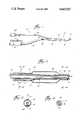
- FIG. 1is a fragmentary view with some parts broken away for clarity, of the preferred embodiment of the device of the present invention
- FIG. 2is an enlarged, fragmentary, longitudinal view in partial section of the distal end of a device in accordance with the present invention
- FIG. 3is a sectional view taken along line 3--3 of FIG. 2;
- FIG. 4is a sectional view taken along line 4--4 of the device illustrated in FIG. 2.
- the device 10includes an elongated, flexible, tubular body 12, Doppler mechanism 14, and expansion means 16.
- the device 10is dimensioned and adapted for insertion in vivo into the arterial tree of a patient.
- the body 12includes a distal section 20 extending between the Doppler mechanism 14 and a branch connector 22. Dividing from the branch connector 22 towards the proximal end is a pneumatic section 24 and a section 26 (lower left portion of FIG. 1). The pneumatic section 24 terminates in a Luer fitting 28 which is adapted for connection to a selectable pneumatic source. Section 26 terminates in a Doppler electrical connector 30 joined to the section 26 and a multi-purpose Luer fitting 32.
- a flexible, elongated, inner, tubular channel 40extends from the Luer fitting 32 (FIG. 1) through the distal section 20 (see FIG. 2).
- the tubular, infusion channel 40defines a central, infusion lumen 42 (see FIGS. 3, 4).
- the distal section 20includes an outer flexible tube 44 which, towards the distal end, converges inwardly into closely spaced relation to the inner channel 40.
- the annular region between the inner walls of the tube 44 and outer walls of the channel 40defines annular lumen 46 (see FIGS. 2-3).
- the expansion means 16includes an expandable flexible balloon-like sleeve 50 sealingly fitted circumferentially to the outer tube 44 as shown in FIG. 2. As illustrated in FIGS. 2-3, and a chord structure 52 longitudinally extends through the distal section 20 between the sleeve 50 and branch connector 22. The chord structure 52 and a portion of the inner wall of the tube 44 (see FIG. 3) defines a pneumatic passageway 54 which is operably connected between the sleeve 50 and pneumatic section 24. An aperture 56 through the tube 44 (FIG. 2) connects the passageway 54 with the interior of the sleeve 50. A pair of radiopaque ring markers 58 are operably disposed about the tube 44 inside of the sleeve 50.
- the Doppler mechanism 14incorporates a pair of electrical leads 60 operably connected to the electrical connector 30 and passing through the section 26 (see FIG. 1) and into the distal section 20 (see FIG. 2). As shown in FIG. 2, the electrical leads 60 are disposed in the annular lumen 46 defined by the region between the channel 40 and tube 44 (see FIGS. 2-3).
- the Doppler leads 60terminate at the washer-like, flat, doughnut-shaped Doppler crystal 62 which is sealingly disposed about the channel 40.
- the Doppler crystal 62 in the preferred embodimentis a piezoelectric ceramic crystal comprising a lead-zirconate-titanate material which is about 0.003 inch in thickness and 0.035 inch in outside diameter.
- the Doppler crystal 62is designed to resonate at 20 megaHertz with a voltage applied to generate a 20 megaHertz signal (acoustic tone).
- the crystal 62is a single crystal to operate as a pulsed Doppler, transducer acting alternately as a transmitter and receiver.
- An encapsulant 64is sealingly disposed about the Doppler crystal 62. As shown in FIG. 2, the tip of the channel 40 extends slightly past the marginal periphery of the tube 44 and past the crystal 62, which in combination with the encapsulant 64 provides effective isolation of the Doppler crystal 62 and leads 60 from the biological fluid when in use in the arterial tree.
- the Doppler crystal 62is a single piezoelectric ceramic crystal having a master oscillator frequency of 20 megaHertz and is pulsed at a repetition frequency (prf) of 62.5 kiloHertz. Each pulse is approximately one-half microsecond in duration.
- each acoustic tone burstis transmitted through the blood and reflected by various structures, for example red blood cells, vessel wall, plaque, etc.
- various structuresfor example red blood cells, vessel wall, plaque, etc.
- the returning acoustic signals received by the Doppler crystal 62are separated in time.
- An adjustable receiver gateis incorporated to select signals reflected from the structures at a specified distance from the crystal 62.
- the reflected signals received by the Doppler crystal 62are amplified and compared in phase and frequency to the master oscillator signal of 20 megaHertz.
- the Doppler shift ( ⁇ f)is defined by the Dopper equation: ##EQU1## where F is the transmitted frequency; V is the velocity of the fluid; c is velocity of sound in the fluid; and ⁇ is the angle between the fluid flow axis and the acoustic axis. Since transmitted frequency (F), velocity of sound in the fluid (c), and the angle ( ⁇ ) are constant, the Doppler shift ( ⁇ f) is linearly related to the velocity of the fluid (V). This assumes that the device 10 is in a stable position in the arterial tree and that the angle ( ⁇ ) remains constant.
- the term "velocity" or "velocity of the fluid”means either the absolute velocity or a number linearly related to the absolute velocity.
- the device 10need not incorporate the expansion means 16 to be useful in the diagnosis of coronary artery disease.
- prototypeshave been constructed using a USCI (United States Catheters and Instruments) Rentrop reperfusion catheter with the Doppler mechanism 14 attached to the tip. These prototypes incorporated a 4 French outermost tubular main body (1.3 millimeters) which tapered to a tip 3 French in size (1 millimeter).
- the catheterwas 110 millimeters long having an internal diameter of 0.020 inches running the length thereof.
- An internal channel tubingwas fitted within this body and had a 0.018 inch outside diameter to define an annular lumen between the channel and body.
- the inner diameter of the inner channelwas 0.0155 inches and defined an infusion or central lumen.
- a 0.014 inch wire guidewas receivable in the central lumen.
- the electrical leadsran in the annular lumen with a Doppler crystal mounted at the tip of the catheter.
- the Doppler crystal in the prototypecomprised a thin disc about 0.003 inches thickness with an outside diameter of 0.035 inches.
- An aperture of about 0.022 incheswas centrally located to give the Doppler crystal a doughnut-shaped configuration.
- the Doppler crystalwas interfitted over the channel and insulated and attached by epoxy between the channel and body. The channel terminated just beyond the Doppler crystal at the distal tip of the catheter.
- the Doppler crystal 62is attached at the distal tip of the device 10 and axially oriented. This installation and orientation has been found preferable in giving good signal stability and accuracy.
- the Doppler crystal 62can be installed in a more radial or oblique orientation.
- a radial orientationmight provide inherent inaccuracies due to placement of the device in the arterial vessel with the crystal adjoining the vessel wall. Measurement of the fluid velocity would, of course, be impossible under these circumstances.
- the acoustic axisgenerally linearly aligned with the fluid flow axis as compared with the oblique angle produced if the Doppler crystal 62 is radially oriented relative to the body 12. Doppler shift is usually more accurately determined with the axes aligned.
- the device 10 of the preferred embodimentenvisions a catheter-like device similar to a conventional angioplastic catheter which incorporates the Doppler mechanism.
- the device 10 of the present inventionmay be incorporated into a mechanism resembling a steerable wire guide having the Doppler mechanism at the tip, which might be inserted through a catheter when it is desired to take the velocity measurements.
- the inner and outer dimensions of the channel 40 and tube 44are not critical. That is, the outer surface of the channel 40 and inner surface of the tube 44 may in fact be adjoining with the electrical leads 60 compressed therebetween. Alternatively, the electrical leads 60 may be incorporated integral with either the channel 40 or tube 44.
- the electrical leads 60can be routed through other passages out of possible contact with the biological fluid.
- the electrical leads 60could be routed through the pneumatic passageway 54 with or without the channel 40 or sleeve 50 incorporated in the device.
- the device 10is inserted into the arterial tree through a Judkins-type guiding catheter.
- the device 10is preferably preloaded with a teflon-coated steerable guidewire (e.g. 0.014 inch O.D. USCI-type) through the central lumen 42.
- a teflon-coated steerable guidewiree.g. 0.014 inch O.D. USCI-type
- the guidewireis used to selectively place the distal end of the device 10 proximate to the coronary stenosis under study.
- the guidewirecan be withdrawn from the device 10.
- a hyperemic responsewill typically be induced to effectively analyze the coronary vasodilator reserve capacity.
- the sleeve 50is expanded in the area of the stenosis until occlusion of the vessel is obtained.
- the Doppler mechanism 14operates to verify the occlusion. The occlusion is held for approximately 20 seconds then released; this technique has been found to induce a hyperemic response. Other techniques are of course available.
- a pharmacological agentsuch as dipyridamole or meglumine diatrizoate may be injected to induce the hyperemic response.
- a pharmacological agentsuch as dipyridamole or meglumine diatrizoate may be injected to induce the hyperemic response.
- contrast media or angiodye injectionwill in most cases induce a sufficient hyperemic response for proper analysis.
- coronary angioplastycan be performed by expanding the sleeve 50 to dilate or distend the vessel.
- conventional angiographic assessment of the efficacy of the dilationcan be utilized or alternatively, the dilation can be performed at progressively increasing pressures and the effectiveness analyzed by the velocity measurements.
- the Doppler mechanism 14can be operated to determine the blood flow velocity before and after the dilation and hence the efficacy of the dilation. Subsequent dilations can be made as needed.
- the device 10 of the present inventionis a significant advance over previous in vivo coronary catheters and allows for a significant advance in the diagnosis and treatment of coronary arterial disease.
- the criticisms of the use of conventional coronary arteriograms as a method of measuring the extent of coronary stenosisis, to some extent, valid.
- some studiessuggest that conventional arteriograms are particularly inaccurate where the stenosis is less than approximately an 80% occlusion.
- an apparatus which more directly takes flow measurementsis ideal in analyzing coronary vasodilator reserve capacity.
- the device 10 of the present inventionprovides real time analysis by providing continuous velocity measurements which are linearly related to blood flow. This is in sharp contrast to conventional angiograms which only accurately identify regions of possible stenoses, but not the physiological importance of such stenoses.
- the velocity measurementsare very accurate in evaluating the flow. That is, various studies have shown that maximal coronary reactive hyperemic response is relatively constant whether or not a catheter the size of the device 10 is present or absent in the artery being investigated. This implies that the obstruction of blood flow by the device 10 is minimal during the hyperemic response. Further, vessel diameter expansion during hyperemic response has been found to minimally affect the accuracy of the velocity measurements as an indication of flow. Additionally, the range gating of the pulsed Doppler crystal of the device 10 of the present invention allows for selective analysis of a target region a sufficient distance (approximately 2-10 mm) away from the catheter tip to avoid tip induced turbulent flow which would affect the accuracy of the velocity measurements.
- the device 10 of the present inventionUtilizing the device 10 of the present invention, coronary artery disease is more effectively identified and treated than by use of conventional methods and devices.
- the device and method of the present inventionpresent a significant advance in the art.
Landscapes
- Health & Medical Sciences (AREA)
- Life Sciences & Earth Sciences (AREA)
- Heart & Thoracic Surgery (AREA)
- Animal Behavior & Ethology (AREA)
- Biophysics (AREA)
- Veterinary Medicine (AREA)
- Public Health (AREA)
- General Health & Medical Sciences (AREA)
- Engineering & Computer Science (AREA)
- Biomedical Technology (AREA)
- Surgery (AREA)
- Molecular Biology (AREA)
- Physics & Mathematics (AREA)
- Medical Informatics (AREA)
- Radiology & Medical Imaging (AREA)
- Pathology (AREA)
- Nuclear Medicine, Radiotherapy & Molecular Imaging (AREA)
- Hematology (AREA)
- Pulmonology (AREA)
- Child & Adolescent Psychology (AREA)
- Anesthesiology (AREA)
- Measuring Pulse, Heart Rate, Blood Pressure Or Blood Flow (AREA)
- Ultra Sonic Daignosis Equipment (AREA)
- Surgical Instruments (AREA)
- Media Introduction/Drainage Providing Device (AREA)
- Measurement Of The Respiration, Hearing Ability, Form, And Blood Characteristics Of Living Organisms (AREA)
- External Artificial Organs (AREA)
Abstract
Description
Claims (27)
Priority Applications (12)
| Application Number | Priority Date | Filing Date | Title |
|---|---|---|---|
| US06/775,857US4665925A (en) | 1985-09-13 | 1985-09-13 | Doppler catheter |
| CA000517964ACA1286368C (en) | 1985-09-13 | 1986-09-11 | Doppler catheter |
| DE8686307048TDE3674986D1 (en) | 1985-09-13 | 1986-09-12 | DOPPLER CATHETER. |
| DK437486ADK437486A (en) | 1985-09-13 | 1986-09-12 | Doppler Catheters for Measurement of the Bioavailability of a biological fluid in a vessel, e.g. BLOOD FLOW SPEED IN A BLOOD CART |
| AT86307048TATE57463T1 (en) | 1985-09-13 | 1986-09-12 | DOPPLER CATHETER. |
| BR8604395ABR8604395A (en) | 1985-09-13 | 1986-09-12 | ORIENTABLE CATHETER TO MEASURE THE SPEED OF A BIOLOGICAL FLUID |
| ES8601895AES2001683A6 (en) | 1985-09-13 | 1986-09-12 | Doppler catheter. |
| EP86307048AEP0224997B1 (en) | 1985-09-13 | 1986-09-12 | Doppler catheter |
| AU62697/86AAU569824B2 (en) | 1985-09-13 | 1986-09-15 | Steerable catheter with doppler means |
| ZA866995AZA866995B (en) | 1985-09-13 | 1986-09-15 | Doppler catheter |
| US06/943,446US4889128A (en) | 1985-09-13 | 1986-12-16 | Doppler catheter |
| US07/248,777US4957111A (en) | 1985-09-13 | 1988-09-23 | Method of using a doppler catheter |
Applications Claiming Priority (1)
| Application Number | Priority Date | Filing Date | Title |
|---|---|---|---|
| US06/775,857US4665925A (en) | 1985-09-13 | 1985-09-13 | Doppler catheter |
Related Child Applications (1)
| Application Number | Title | Priority Date | Filing Date |
|---|---|---|---|
| US06/943,446ContinuationUS4889128A (en) | 1985-09-13 | 1986-12-16 | Doppler catheter |
Publications (1)
| Publication Number | Publication Date |
|---|---|
| US4665925Atrue US4665925A (en) | 1987-05-19 |
Family
ID=25105738
Family Applications (1)
| Application Number | Title | Priority Date | Filing Date |
|---|---|---|---|
| US06/775,857Expired - LifetimeUS4665925A (en) | 1985-09-13 | 1985-09-13 | Doppler catheter |
Country Status (10)
| Country | Link |
|---|---|
| US (1) | US4665925A (en) |
| EP (1) | EP0224997B1 (en) |
| AT (1) | ATE57463T1 (en) |
| AU (1) | AU569824B2 (en) |
| BR (1) | BR8604395A (en) |
| CA (1) | CA1286368C (en) |
| DE (1) | DE3674986D1 (en) |
| DK (1) | DK437486A (en) |
| ES (1) | ES2001683A6 (en) |
| ZA (1) | ZA866995B (en) |
Cited By (117)
| Publication number | Priority date | Publication date | Assignee | Title |
|---|---|---|---|---|
| US4758221A (en)* | 1986-02-18 | 1988-07-19 | St. Louis University | Catheter with a tip manipulation feature |
| US4757822A (en)* | 1985-02-07 | 1988-07-19 | Biotronix S.R.L. | Instrument to detect and represent the cross-sectional variations of a blood vessel |
| WO1988009150A1 (en)* | 1987-05-26 | 1988-12-01 | Inter Therapy, Inc. | Ultrasonic imaging array and balloon catheter assembly |
| US4793359A (en)* | 1987-04-24 | 1988-12-27 | Gv Medical, Inc. | Centering balloon structure for transluminal angioplasty catheter |
| US4819751A (en)* | 1987-10-16 | 1989-04-11 | Baxter Travenol Laboratories, Inc. | Valvuloplasty catheter and method |
| US4844080A (en)* | 1987-02-19 | 1989-07-04 | Michael Frass | Ultrasound contact medium dispenser |
| US4869263A (en)* | 1988-02-04 | 1989-09-26 | Cardiometrics, Inc. | Device and method for measuring volumetric blood flow in a vessel |
| US4887606A (en)* | 1986-09-18 | 1989-12-19 | Yock Paul G | Apparatus for use in cannulation of blood vessels |
| US4917097A (en)* | 1987-10-27 | 1990-04-17 | Endosonics Corporation | Apparatus and method for imaging small cavities |
| US4936826A (en)* | 1988-10-24 | 1990-06-26 | Amarasinghe Disamodha C | Vena cava window |
| US4938220A (en)* | 1986-08-01 | 1990-07-03 | Advanced Cardiovascular Systems, Inc. | Catheter with split tip marker and method of manufacture |
| US4991588A (en)* | 1986-07-21 | 1991-02-12 | Pfizer Hospital Products Group, Inc. | Doppler guide wire |
| US5022399A (en)* | 1989-05-10 | 1991-06-11 | Biegeleisen Ken P | Venoscope |
| WO1991008014A1 (en)* | 1989-11-28 | 1991-06-13 | Leocor, Inc. | Low profile catheter |
| US5035705A (en)* | 1989-01-13 | 1991-07-30 | Scimed Life Systems, Inc. | Method of purging a balloon catheter |
| US5038789A (en)* | 1989-09-28 | 1991-08-13 | Frazin Leon J | Method and device for doppler-guided retrograde catheterization |
| US5046503A (en)* | 1989-04-26 | 1991-09-10 | Advanced Cardiovascular Systems, Inc. | Angioplasty autoperfusion catheter flow measurement method and apparatus |
| EP0286359A3 (en)* | 1987-04-10 | 1991-12-11 | Cardiometrics, Inc. | Apparatus, system and method for measuring volumetric flow of blood in a vessel |
| US5076278A (en)* | 1990-10-15 | 1991-12-31 | Catheter Technology Co. | Annular ultrasonic transducers employing curved surfaces useful in catheter localization |
| US5085636A (en)* | 1989-01-13 | 1992-02-04 | Scimed Life Systems, Inc. | Balloon catheter with inflation-deflation valve |
| US5100385A (en)* | 1989-01-27 | 1992-03-31 | C. R. Bard, Inc. | Fast purge balloon dilatation catheter |
| US5105818A (en)* | 1987-04-10 | 1992-04-21 | Cardiometric, Inc. | Apparatus, system and method for measuring spatial average velocity and/or volumetric flow of blood in a vessel and screw joint for use therewith |
| US5109861A (en)* | 1989-04-28 | 1992-05-05 | Thomas Jefferson University | Intravascular, ultrasonic imaging catheters and methods for making same |
| WO1992011809A1 (en)* | 1991-01-07 | 1992-07-23 | Endosonics Corporation | Dilating and imaging apparatus |
| US5135001A (en)* | 1990-12-05 | 1992-08-04 | C. R. Bard, Inc. | Ultrasound sheath for medical diagnostic instruments |
| US5156595A (en)* | 1989-12-28 | 1992-10-20 | Scimed Life Systems, Inc. | Dilatation balloon catheter and method of manufacturing |
| US5190045A (en)* | 1989-09-28 | 1993-03-02 | Frazin Leon J | Method and device for doppler-guided and imaged retrograde catheterization |
| US5220924A (en)* | 1989-09-28 | 1993-06-22 | Frazin Leon J | Doppler-guided retrograde catheterization using transducer equipped guide wire |
| US5240437A (en)* | 1988-11-02 | 1993-08-31 | Cardiometrics, Inc. | Torqueable guide wire assembly with electrical functions, male and female connectors for use therewith and system and apparatus for utilizing the same |
| WO1993017623A1 (en)* | 1992-03-06 | 1993-09-16 | Cardiometrics, Inc. | Doppler member having inflatable balloon |
| US5254112A (en)* | 1990-10-29 | 1993-10-19 | C. R. Bard, Inc. | Device for use in laser angioplasty |
| US5259385A (en)* | 1991-12-23 | 1993-11-09 | Advanced Cardiovascular Systems, Inc. | Apparatus for the cannulation of blood vessels |
| US5271406A (en)* | 1992-05-22 | 1993-12-21 | Diagnostic Devices Group, Limited | Low-profile ultrasonic transducer incorporating static beam steering |
| US5316001A (en)* | 1990-09-11 | 1994-05-31 | Ferek Petric Bozidar | Cardiac measurement system for measuring blood flow velocity by use of a sensor implanted inside the heart |
| US5345938A (en)* | 1991-09-30 | 1994-09-13 | Kabushiki Kaisha Toshiba | Diagnostic apparatus for circulatory systems |
| US5368037A (en)* | 1993-02-01 | 1994-11-29 | Endosonics Corporation | Ultrasound catheter |
| US5372138A (en)* | 1988-03-21 | 1994-12-13 | Boston Scientific Corporation | Acousting imaging catheters and the like |
| US5403339A (en)* | 1991-06-21 | 1995-04-04 | Terumo Kabushiki Kaisha | Blood vessel dilator |
| US5431628A (en)* | 1992-09-29 | 1995-07-11 | Millar Instruments, Inc. | Pressure-sensing diagnostic catheter |
| US5454789A (en)* | 1989-01-13 | 1995-10-03 | Scimed Life Systems, Inc. | Innerless dilatation balloon catheter |
| US5501227A (en)* | 1986-04-15 | 1996-03-26 | Yock; Paul G. | Angioplasty apparatus facilitating rapid exchange and method |
| US5603327A (en)* | 1993-02-01 | 1997-02-18 | Endosonics Corporation | Ultrasound catheter probe |
| US5690115A (en)* | 1995-09-21 | 1997-11-25 | Feldman; Charles L. | Detecting vascular stenosis in chronic hemodialysis patients |
| US5759191A (en)* | 1989-06-27 | 1998-06-02 | C. R. Bard, Inc. | Coaxial PTCA catheter with anchor joint |
| US5846246A (en)* | 1994-10-21 | 1998-12-08 | Cordis Corporation | Dual-balloon rapid-exchange stent delivery catheter with guidewire channel |
| US5857974A (en)* | 1997-01-08 | 1999-01-12 | Endosonics Corporation | High resolution intravascular ultrasound transducer assembly having a flexible substrate |
| US5899203A (en)* | 1992-12-24 | 1999-05-04 | Defares; Peter Bernard | Interactive respiratory regulator |
| US5902248A (en)* | 1996-11-06 | 1999-05-11 | Millar Instruments, Inc. | Reduced size catheter tip measurement device |
| US6066157A (en)* | 1998-09-16 | 2000-05-23 | Medtronics Ave, Inc. | Anchor joint for coaxial balloon dilatation catheter |
| US6179856B1 (en)* | 1989-07-05 | 2001-01-30 | Medtronic Ave, Inc. | Coaxial PTCA catheter with anchor joint |
| US6394986B1 (en) | 1999-11-06 | 2002-05-28 | Millar Instruments, Inc. | Pressure sensing module for a catheter pressure transducer |
| US20020091362A1 (en)* | 1998-01-06 | 2002-07-11 | Maginot Thomas J. | Medical procedure using catheter system having removability feature |
| US6475207B1 (en) | 1999-01-15 | 2002-11-05 | Maginot Catheter Technologies, Inc. | Retractable catheter systems and associated methods |
| US6480740B2 (en) | 2000-12-26 | 2002-11-12 | Cardiac Pacemakers, Inc. | Safety pacing in multi-site CRM devices |
| US6493586B1 (en) | 2000-08-30 | 2002-12-10 | Cardiac Pacemakers, Inc. | Site reversion in cardiac rhythm management |
| US20030045920A1 (en)* | 2000-10-17 | 2003-03-06 | Medtronic, Inc. | Radiopaque marking of lead electrode zone in a continuous conductor construction |
| US6584362B1 (en) | 2000-08-30 | 2003-06-24 | Cardiac Pacemakers, Inc. | Leads for pacing and/or sensing the heart from within the coronary veins |
| US6585657B2 (en) | 1986-04-15 | 2003-07-01 | Scimed Life Systems, Inc. | Angioplasty apparatus facilitating rapid exchanges |
| US6585705B1 (en) | 1999-01-15 | 2003-07-01 | Maginot Catheter Technologies, Inc. | Retractable catheter systems |
| WO2003086520A1 (en)* | 2002-04-05 | 2003-10-23 | Cardiac Pacemakers, Inc. | Catheter comprising a doppler sensor for measurement of turbulence of the blood |
| US20040044286A1 (en)* | 2002-08-29 | 2004-03-04 | Hossack Norman Hugh | Ultrasonic imaging devices and methods of fabrication |
| US6702750B2 (en) | 1986-04-15 | 2004-03-09 | Cardiovascular Imaging Systems, Inc. | Angioplasty apparatus facilitating rapid exchanges and methods |
| US20040054287A1 (en)* | 2002-08-29 | 2004-03-18 | Stephens Douglas Neil | Ultrasonic imaging devices and methods of fabrication |
| US6743218B2 (en) | 1999-01-15 | 2004-06-01 | Cathlogic, Inc. | Retractable catheter systems and associated methods |
| US6805669B2 (en) | 2001-01-25 | 2004-10-19 | Rebecca L. Swanbom | Method and device for marking skin during an ultrasound examination |
| US20050059925A1 (en)* | 1999-01-15 | 2005-03-17 | Maginot Thomas J. | Catheter systems and associated methods |
| US20050079666A1 (en)* | 2002-04-05 | 2005-04-14 | French Roger Harquail | Method for providing nano-structures of uniform length |
| US20050096609A1 (en)* | 1999-01-15 | 2005-05-05 | Maginot Thomas J. | Methods of performing medical procedures with catheter systems having movable member |
| US20050171591A1 (en)* | 2004-01-29 | 2005-08-04 | Scimed Life Systems, Inc. | Catherter tip |
| US20050209674A1 (en)* | 2003-09-05 | 2005-09-22 | Kutscher Tuvia D | Balloon assembly (V) |
| US6960188B2 (en) | 2001-11-30 | 2005-11-01 | Abbott Laboratories Vascular Entities Limited | Catheter having enhanced distal pushability |
| US7008412B2 (en) | 1998-01-06 | 2006-03-07 | Cathlogic, Inc. | Subcutaneous port catheter system and associated method |
| US20070005092A1 (en)* | 2005-06-09 | 2007-01-04 | Dominick Godin | Balloon catheters with increased column strength |
| US20070016071A1 (en)* | 1993-02-01 | 2007-01-18 | Volcano Corporation | Ultrasound transducer assembly |
| US20070049846A1 (en)* | 2005-08-24 | 2007-03-01 | C.R.Bard, Inc. | Stylet Apparatuses and Methods of Manufacture |
| US7223238B2 (en) | 2001-01-25 | 2007-05-29 | Swanbom Rebecca L | Method and device for marking skin during an ultrasound examination |
| US7245959B1 (en)* | 2001-03-02 | 2007-07-17 | Scimed Life Systems, Inc. | Imaging catheter for use inside a guiding catheter |
| US20070225605A1 (en)* | 2001-01-25 | 2007-09-27 | Swanbom Rebecca L | Method and Device for Marking Skin During an Ultrasound Examination |
| US20090088827A1 (en)* | 2007-10-02 | 2009-04-02 | Cardiac Pacemakers, Inc | Lead assembly providing sensing or stimulation of spaced-apart myocardial contact areas |
| US20100036227A1 (en)* | 2007-11-26 | 2010-02-11 | C. R. Bard, Inc. | Apparatus and display methods relating to intravascular placement of a catheter |
| US20100094116A1 (en)* | 2008-10-07 | 2010-04-15 | Lucent Medical Systems, Inc. | Percutaneous magnetic gastrostomy |
| US20100204569A1 (en)* | 2007-11-26 | 2010-08-12 | C. R. Bard, Inc. | System for placement of a catheter including a signal-generating stylet |
| US20100317981A1 (en)* | 2009-06-12 | 2010-12-16 | Romedex International Srl | Catheter Tip Positioning Method |
| US20100318026A1 (en)* | 2009-06-12 | 2010-12-16 | Romedex International Srl | Devices and Methods for Endovascular Electrography |
| US20100331712A1 (en)* | 2006-10-23 | 2010-12-30 | Bard Access Systems, Inc. | Method of locating the tip of a central venous catheter |
| US20110015533A1 (en)* | 2007-11-26 | 2011-01-20 | C.R. Bard, Inc. | Stylets for use with apparatus for intravascular placement of a catheter |
| US20110196248A1 (en)* | 2009-06-12 | 2011-08-11 | Bard Access Systems, Inc. | Apparatus and method for catheter navigation and tip location |
| US8388546B2 (en) | 2006-10-23 | 2013-03-05 | Bard Access Systems, Inc. | Method of locating the tip of a central venous catheter |
| US8388541B2 (en) | 2007-11-26 | 2013-03-05 | C. R. Bard, Inc. | Integrated system for intravascular placement of a catheter |
| US8478382B2 (en) | 2008-02-11 | 2013-07-02 | C. R. Bard, Inc. | Systems and methods for positioning a catheter |
| USD699359S1 (en) | 2011-08-09 | 2014-02-11 | C. R. Bard, Inc. | Ultrasound probe head |
| US8801693B2 (en) | 2010-10-29 | 2014-08-12 | C. R. Bard, Inc. | Bioimpedance-assisted placement of a medical device |
| USD724745S1 (en) | 2011-08-09 | 2015-03-17 | C. R. Bard, Inc. | Cap for an ultrasound probe |
| US9211107B2 (en) | 2011-11-07 | 2015-12-15 | C. R. Bard, Inc. | Ruggedized ultrasound hydrogel insert |
| WO2016007288A1 (en)* | 2014-07-08 | 2016-01-14 | Nadarasa Visveshwara | System and method for measuring fluidics in arteries |
| US9320493B2 (en) | 2014-07-08 | 2016-04-26 | Nadarasa Visveshwara | System and method for measuring fluidics in arteries |
| US9456766B2 (en) | 2007-11-26 | 2016-10-04 | C. R. Bard, Inc. | Apparatus for use with needle insertion guidance system |
| US9492097B2 (en) | 2007-11-26 | 2016-11-15 | C. R. Bard, Inc. | Needle length determination and calibration for insertion guidance system |
| US9521961B2 (en) | 2007-11-26 | 2016-12-20 | C. R. Bard, Inc. | Systems and methods for guiding a medical instrument |
| US9532724B2 (en) | 2009-06-12 | 2017-01-03 | Bard Access Systems, Inc. | Apparatus and method for catheter navigation using endovascular energy mapping |
| US9554716B2 (en) | 2007-11-26 | 2017-01-31 | C. R. Bard, Inc. | Insertion guidance system for needles and medical components |
| US9649048B2 (en) | 2007-11-26 | 2017-05-16 | C. R. Bard, Inc. | Systems and methods for breaching a sterile field for intravascular placement of a catheter |
| US9839372B2 (en) | 2014-02-06 | 2017-12-12 | C. R. Bard, Inc. | Systems and methods for guidance and placement of an intravascular device |
| US9901714B2 (en) | 2008-08-22 | 2018-02-27 | C. R. Bard, Inc. | Catheter assembly including ECG sensor and magnetic assemblies |
| US10046139B2 (en) | 2010-08-20 | 2018-08-14 | C. R. Bard, Inc. | Reconfirmation of ECG-assisted catheter tip placement |
| US10349890B2 (en) | 2015-06-26 | 2019-07-16 | C. R. Bard, Inc. | Connector interface for ECG-based catheter positioning system |
| US10449330B2 (en) | 2007-11-26 | 2019-10-22 | C. R. Bard, Inc. | Magnetic element-equipped needle assemblies |
| US10456581B2 (en) | 2015-11-20 | 2019-10-29 | Cardiac Pacemakers, Inc | Single pass coronary venous lead for multiple chamber sense and pace |
| US10524691B2 (en) | 2007-11-26 | 2020-01-07 | C. R. Bard, Inc. | Needle assembly including an aligned magnetic element |
| US10639008B2 (en) | 2009-10-08 | 2020-05-05 | C. R. Bard, Inc. | Support and cover structures for an ultrasound probe head |
| US10751509B2 (en) | 2007-11-26 | 2020-08-25 | C. R. Bard, Inc. | Iconic representations for guidance of an indwelling medical device |
| US10820885B2 (en) | 2012-06-15 | 2020-11-03 | C. R. Bard, Inc. | Apparatus and methods for detection of a removable cap on an ultrasound probe |
| WO2020261591A1 (en)* | 2019-06-28 | 2020-12-30 | 朝日インテック株式会社 | Balloon catheter |
| US10973584B2 (en) | 2015-01-19 | 2021-04-13 | Bard Access Systems, Inc. | Device and method for vascular access |
| US10992079B2 (en) | 2018-10-16 | 2021-04-27 | Bard Access Systems, Inc. | Safety-equipped connection systems and methods thereof for establishing electrical connections |
| US11000207B2 (en) | 2016-01-29 | 2021-05-11 | C. R. Bard, Inc. | Multiple coil system for tracking a medical device |
| US11103213B2 (en) | 2009-10-08 | 2021-08-31 | C. R. Bard, Inc. | Spacers for use with an ultrasound probe |
Families Citing this family (3)
| Publication number | Priority date | Publication date | Assignee | Title |
|---|---|---|---|---|
| JP2653792B2 (en)* | 1986-07-18 | 1997-09-17 | ハウメディカ・インコーポレーテッド | Blood velocity measurement wire guide |
| US4899757A (en)* | 1988-02-22 | 1990-02-13 | Intertherapy, Inc. | Ultrasound imaging probe with zero dead space |
| DE69534748T2 (en) | 1994-09-02 | 2006-11-02 | Volcano Corp. (n.d, Ges.d.Staates Delaware), Rancho Cordova | ULTRAMINIATUR PRESSURE SENSOR AND GUIDE WIRE THEREFORE |
Citations (9)
| Publication number | Priority date | Publication date | Assignee | Title |
|---|---|---|---|---|
| US3817089A (en)* | 1971-06-30 | 1974-06-18 | Interscience Res Inst | Rotating probe high data acquistion rate apparatus |
| US3827115A (en)* | 1972-02-22 | 1974-08-06 | Univ Erasmus | Method of manufacturing a catheter |
| US3938502A (en)* | 1972-02-22 | 1976-02-17 | Nicolaas Bom | Apparatus with a catheter for examining hollow organs or bodies with the ultrasonic waves |
| US3995623A (en)* | 1974-12-23 | 1976-12-07 | American Hospital Supply Corporation | Multipurpose flow-directed catheter |
| US4112773A (en)* | 1977-05-02 | 1978-09-12 | Rhode Island Hospital | Ultrasonic particulate sensing |
| US4319580A (en)* | 1979-08-28 | 1982-03-16 | The Board Of Regents Of The University Of Washington | Method for detecting air emboli in the blood in an intracorporeal blood vessel |
| US4545390A (en)* | 1982-09-22 | 1985-10-08 | C. R. Bard, Inc. | Steerable guide wire for balloon dilatation procedure |
| US4576177A (en)* | 1983-02-18 | 1986-03-18 | Webster Wilton W Jr | Catheter for removing arteriosclerotic plaque |
| US4589419A (en)* | 1984-11-01 | 1986-05-20 | University Of Iowa Research Foundation | Catheter for treating arterial occlusion |
Family Cites Families (8)
| Publication number | Priority date | Publication date | Assignee | Title |
|---|---|---|---|---|
| US4024873A (en)* | 1976-05-24 | 1977-05-24 | Becton, Dickinson And Company | Balloon catheter assembly |
| US4299226A (en)* | 1979-08-08 | 1981-11-10 | Banka Vidya S | Coronary dilation method |
| US4354502A (en)* | 1979-08-28 | 1982-10-19 | The Board Of Regents Of The University Of Washington | Intravascular catheter including untrasonic transducer for use in detection and aspiration of air emboli |
| SE8103309L (en)* | 1981-05-26 | 1982-11-27 | Karolinska Inst | OVERVAKNINGSSOND |
| DE3235974A1 (en)* | 1981-11-24 | 1983-06-01 | Volkmar Dipl.-Ing. Merkel (FH), 8520 Erlangen | DEVICE FOR REMOVAL OR FOR THE EXPANSION OF CONSTRAINTS IN BODY LIQUID LEADING VESSELS |
| NZ202965A (en)* | 1982-01-07 | 1986-05-09 | Technicare Corp | Exploratory needle with ultrasonic conducting coaxial stylet |
| EP0132344A3 (en)* | 1983-07-20 | 1986-01-22 | Purdue Research Foundation | Improved catheter based cardiac output sensor |
| US4637401A (en)* | 1984-11-01 | 1987-01-20 | Johnston G Gilbert | Volumetric flow rate determination in conduits not directly accessible |
- 1985
- 1985-09-13USUS06/775,857patent/US4665925A/ennot_activeExpired - Lifetime
- 1986
- 1986-09-11CACA000517964Apatent/CA1286368C/ennot_activeExpired - Fee Related
- 1986-09-12BRBR8604395Apatent/BR8604395A/ennot_activeIP Right Cessation
- 1986-09-12ESES8601895Apatent/ES2001683A6/ennot_activeExpired
- 1986-09-12ATAT86307048Tpatent/ATE57463T1/ennot_activeIP Right Cessation
- 1986-09-12DKDK437486Apatent/DK437486A/ennot_activeApplication Discontinuation
- 1986-09-12EPEP86307048Apatent/EP0224997B1/ennot_activeExpired - Lifetime
- 1986-09-12DEDE8686307048Tpatent/DE3674986D1/ennot_activeExpired - Lifetime
- 1986-09-15AUAU62697/86Apatent/AU569824B2/ennot_activeCeased
- 1986-09-15ZAZA866995Apatent/ZA866995B/enunknown
Patent Citations (9)
| Publication number | Priority date | Publication date | Assignee | Title |
|---|---|---|---|---|
| US3817089A (en)* | 1971-06-30 | 1974-06-18 | Interscience Res Inst | Rotating probe high data acquistion rate apparatus |
| US3827115A (en)* | 1972-02-22 | 1974-08-06 | Univ Erasmus | Method of manufacturing a catheter |
| US3938502A (en)* | 1972-02-22 | 1976-02-17 | Nicolaas Bom | Apparatus with a catheter for examining hollow organs or bodies with the ultrasonic waves |
| US3995623A (en)* | 1974-12-23 | 1976-12-07 | American Hospital Supply Corporation | Multipurpose flow-directed catheter |
| US4112773A (en)* | 1977-05-02 | 1978-09-12 | Rhode Island Hospital | Ultrasonic particulate sensing |
| US4319580A (en)* | 1979-08-28 | 1982-03-16 | The Board Of Regents Of The University Of Washington | Method for detecting air emboli in the blood in an intracorporeal blood vessel |
| US4545390A (en)* | 1982-09-22 | 1985-10-08 | C. R. Bard, Inc. | Steerable guide wire for balloon dilatation procedure |
| US4576177A (en)* | 1983-02-18 | 1986-03-18 | Webster Wilton W Jr | Catheter for removing arteriosclerotic plaque |
| US4589419A (en)* | 1984-11-01 | 1986-05-20 | University Of Iowa Research Foundation | Catheter for treating arterial occlusion |
Non-Patent Citations (24)
| Title |
|---|
| Benchimol, A., et al., "Aortic Flow Velocity in Man During Cardiac Arrhythmias Measured with the Doppler Catheter-Flowmeter System", 78, Amer. Heart, pp. 649-659 (Nov. 1969). |
| Benchimol, A., et al., Aortic Flow Velocity in Man During Cardiac Arrhythmias Measured with the Doppler Catheter Flowmeter System , 78, Amer. Heart, pp. 649 659 (Nov. 1969).* |
| Cole, J. S. and Hartley, C. J., "The Pulsed Doppler Coronary Artery Catheter", 56, Circulation, 18-25 (Jul. 1977). |
| Cole, J. S. and Hartley, C. J., The Pulsed Doppler Coronary Artery Catheter , 56, Circulation, 18 25 (Jul. 1977).* |
| Coppess, M. A., Young, D. F., White, C. W. and Laughlin, D. E., "An Ultrasonic Pulsed Doppler Balloon Catheter for Use in Cardiovascular Diagnosis", 19, Bio. Med. Sci. Instru., 9-16 (1983). |
| Coppess, M. A., Young, D. F., White, C. W. and Laughlin, D. E., An Ultrasonic Pulsed Doppler Balloon Catheter for Use in Cardiovascular Diagnosis , 19, Bio. Med. Sci. Instru., 9 16 (1983).* |
| Hartley, C. J. and Cole, J. S., "A Single-Crystal Ultrasonic Catheter-Tip Velocity Probe", 8, Medical Instrumentation, 241-243 (1974). |
| Hartley, C. J. and Cole, J. S., "An Ultrasonic Pulsed Doppler System for Measuring Blood Flow in Small Vessels", 37, J. App. Physiology, pp. 626-629. |
| Hartley, C. J. and Cole, J. S., A Single Crystal Ultrasonic Catheter Tip Velocity Probe , 8, Medical Instrumentation, 241 243 (1974).* |
| Hartley, C. J. and Cole, J. S., An Ultrasonic Pulsed Doppler System for Measuring Blood Flow in Small Vessels , 37, J. App. Physiology, pp. 626 629.* |
| Laenger, C. J., et al., "Development of Special Purpose Catheter Tip Transducers", (Aug. 1967), (unpublished manuscript). |
| Laenger, C. J., et al., Development of Special Purpose Catheter Tip Transducers , (Aug. 1967), (unpublished manuscript).* |
| Martin, et al., "Ultrasonic Catheter Tip Instrument for Measurement of Vessel Cross-Sectional Area", unpublished paper presented at the 27th Annual Conference on Engineering in Medicine and Biology (Oct. 1974, pp. 15-16). |
| Martin, et al., Ultrasonic Catheter Tip Instrument for Measurement of Vessel Cross Sectional Area , unpublished paper presented at the 27th Annual Conference on Engineering in Medicine and Biology (Oct. 1974, pp. 15 16).* |
| Martin, R. W., et al, "An Ultrasonic Catheter for Intravascular Measurement of Blood Flow: Technical Details", Transactions on Sonics & Ultrasonics, vol. SU-27, No. 6, Nov. 1980. |
| Martin, R. W., et al, An Ultrasonic Catheter for Intravascular Measurement of Blood Flow: Technical Details , Transactions on Sonics & Ultrasonics, vol. SU 27, No. 6, Nov. 1980.* |
| Naeleigh, R. C. & Miller, C. W., "A Venous Pulse Doppler Catheter-Tip Flowmeter for Measuring Arterial Blood Velocity, Flow and Diameter in Deep Arteries", 12th Annula R.M. Bioengineering Symposium (Apr. 28-30, 1975). |
| Naeleigh, R. C. & Miller, C. W., A Venous Pulse Doppler Catheter Tip Flowmeter for Measuring Arterial Blood Velocity, Flow and Diameter in Deep Arteries , 12th Annula R.M. Bioengineering Symposium (Apr. 28 30, 1975).* |
| Nealeigh, R. C. and Miller, C. W., "A Venous Pulse Doppler Catheter-Tip Flowmeter for Measuring Arterial Blood Velocity, Flow, and Diameter in Deep Arteries", 15, ISA Trans., 84-87 (1976). |
| Nealeigh, R. C. and Miller, C. W., A Venous Pulse Doppler Catheter Tip Flowmeter for Measuring Arterial Blood Velocity, Flow, and Diameter in Deep Arteries , 15, ISA Trans., 84 87 (1976).* |
| White, C. W., et al., "Does Visual Interpretation of the Coronary Arteriogram Predict the Physiologic Importance of a Coronary Stenosis?", 310, N. Eng. J. Med., 819-824 (Mar. 1984). |
| White, C. W., et al., Does Visual Interpretation of the Coronary Arteriogram Predict the Physiologic Importance of a Coronary Stenosis , 310, N. Eng. J. Med., 819 824 (Mar. 1984).* |
| Wilson, R. F., et al., "Transluminal Subselective Measurement of Coronary Artery Blood Flow Velocity & Vasodilator Reserve in Man", 72, Circulation--82-92 (Jul. 1985). |
| Wilson, R. F., et al., Transluminal Subselective Measurement of Coronary Artery Blood Flow Velocity & Vasodilator Reserve in Man , 72, Circulation 82 92 (Jul. 1985).* |
Cited By (218)
| Publication number | Priority date | Publication date | Assignee | Title |
|---|---|---|---|---|
| US4757822A (en)* | 1985-02-07 | 1988-07-19 | Biotronix S.R.L. | Instrument to detect and represent the cross-sectional variations of a blood vessel |
| US4758221A (en)* | 1986-02-18 | 1988-07-19 | St. Louis University | Catheter with a tip manipulation feature |
| US6702750B2 (en) | 1986-04-15 | 2004-03-09 | Cardiovascular Imaging Systems, Inc. | Angioplasty apparatus facilitating rapid exchanges and methods |
| US5501227A (en)* | 1986-04-15 | 1996-03-26 | Yock; Paul G. | Angioplasty apparatus facilitating rapid exchange and method |
| US6575993B1 (en)* | 1986-04-15 | 2003-06-10 | Paul G. Yock | Angioplasty apparatus facilitating rapid exchanges |
| US6921411B2 (en)* | 1986-04-15 | 2005-07-26 | Advanced Cardiovascular Systems Inc. | Angioplasty apparatus facilitating rapid exchanges and method |
| US5685312A (en)* | 1986-04-15 | 1997-11-11 | Yock; Paul G. | Angioplasty apparatus facilitating rapid exchanges and method |
| US20030120301A1 (en)* | 1986-04-15 | 2003-06-26 | Yock Paul G. | Angioplasty apparatus facilitating rapid exchanges and method |
| US6585657B2 (en) | 1986-04-15 | 2003-07-01 | Scimed Life Systems, Inc. | Angioplasty apparatus facilitating rapid exchanges |
| US4991588A (en)* | 1986-07-21 | 1991-02-12 | Pfizer Hospital Products Group, Inc. | Doppler guide wire |
| US4938220A (en)* | 1986-08-01 | 1990-07-03 | Advanced Cardiovascular Systems, Inc. | Catheter with split tip marker and method of manufacture |
| US4887606A (en)* | 1986-09-18 | 1989-12-19 | Yock Paul G | Apparatus for use in cannulation of blood vessels |
| US4844080A (en)* | 1987-02-19 | 1989-07-04 | Michael Frass | Ultrasound contact medium dispenser |
| EP0286359A3 (en)* | 1987-04-10 | 1991-12-11 | Cardiometrics, Inc. | Apparatus, system and method for measuring volumetric flow of blood in a vessel |
| US5105818A (en)* | 1987-04-10 | 1992-04-21 | Cardiometric, Inc. | Apparatus, system and method for measuring spatial average velocity and/or volumetric flow of blood in a vessel and screw joint for use therewith |
| US4793359A (en)* | 1987-04-24 | 1988-12-27 | Gv Medical, Inc. | Centering balloon structure for transluminal angioplasty catheter |
| AU599818B2 (en)* | 1987-05-26 | 1990-07-26 | Inter Therapy, Inc. | Ultrasonic imaging array and balloon catheter assembly |
| WO1988009150A1 (en)* | 1987-05-26 | 1988-12-01 | Inter Therapy, Inc. | Ultrasonic imaging array and balloon catheter assembly |
| US4841977A (en)* | 1987-05-26 | 1989-06-27 | Inter Therapy, Inc. | Ultra-thin acoustic transducer and balloon catheter using same in imaging array subassembly |
| US4819751A (en)* | 1987-10-16 | 1989-04-11 | Baxter Travenol Laboratories, Inc. | Valvuloplasty catheter and method |
| US4917097A (en)* | 1987-10-27 | 1990-04-17 | Endosonics Corporation | Apparatus and method for imaging small cavities |
| US4869263A (en)* | 1988-02-04 | 1989-09-26 | Cardiometrics, Inc. | Device and method for measuring volumetric blood flow in a vessel |
| US5372138A (en)* | 1988-03-21 | 1994-12-13 | Boston Scientific Corporation | Acousting imaging catheters and the like |
| US4936826A (en)* | 1988-10-24 | 1990-06-26 | Amarasinghe Disamodha C | Vena cava window |
| US5240437A (en)* | 1988-11-02 | 1993-08-31 | Cardiometrics, Inc. | Torqueable guide wire assembly with electrical functions, male and female connectors for use therewith and system and apparatus for utilizing the same |
| US5085636A (en)* | 1989-01-13 | 1992-02-04 | Scimed Life Systems, Inc. | Balloon catheter with inflation-deflation valve |
| US5454789A (en)* | 1989-01-13 | 1995-10-03 | Scimed Life Systems, Inc. | Innerless dilatation balloon catheter |
| US5531689A (en)* | 1989-01-13 | 1996-07-02 | Scimed Life Systems, Inc. | Innerless dilatation balloon catheter |
| US5919162A (en)* | 1989-01-13 | 1999-07-06 | Scimed Technology, Inc. | Balloon catheter with inflation/deflation valve |
| US5035705A (en)* | 1989-01-13 | 1991-07-30 | Scimed Life Systems, Inc. | Method of purging a balloon catheter |
| US5100385A (en)* | 1989-01-27 | 1992-03-31 | C. R. Bard, Inc. | Fast purge balloon dilatation catheter |
| US5046503A (en)* | 1989-04-26 | 1991-09-10 | Advanced Cardiovascular Systems, Inc. | Angioplasty autoperfusion catheter flow measurement method and apparatus |
| US5109861A (en)* | 1989-04-28 | 1992-05-05 | Thomas Jefferson University | Intravascular, ultrasonic imaging catheters and methods for making same |
| US5022399A (en)* | 1989-05-10 | 1991-06-11 | Biegeleisen Ken P | Venoscope |
| US5759191A (en)* | 1989-06-27 | 1998-06-02 | C. R. Bard, Inc. | Coaxial PTCA catheter with anchor joint |
| US6179856B1 (en)* | 1989-07-05 | 2001-01-30 | Medtronic Ave, Inc. | Coaxial PTCA catheter with anchor joint |
| US5220924A (en)* | 1989-09-28 | 1993-06-22 | Frazin Leon J | Doppler-guided retrograde catheterization using transducer equipped guide wire |
| US5038789A (en)* | 1989-09-28 | 1991-08-13 | Frazin Leon J | Method and device for doppler-guided retrograde catheterization |
| US5190045A (en)* | 1989-09-28 | 1993-03-02 | Frazin Leon J | Method and device for doppler-guided and imaged retrograde catheterization |
| WO1991008014A1 (en)* | 1989-11-28 | 1991-06-13 | Leocor, Inc. | Low profile catheter |
| US5156595A (en)* | 1989-12-28 | 1992-10-20 | Scimed Life Systems, Inc. | Dilatation balloon catheter and method of manufacturing |
| US5316001A (en)* | 1990-09-11 | 1994-05-31 | Ferek Petric Bozidar | Cardiac measurement system for measuring blood flow velocity by use of a sensor implanted inside the heart |
| WO1992006637A1 (en)* | 1990-10-15 | 1992-04-30 | Echocath, Ltd. | Annular ultrasonic transducers in a catheter |
| US5076278A (en)* | 1990-10-15 | 1991-12-31 | Catheter Technology Co. | Annular ultrasonic transducers employing curved surfaces useful in catheter localization |
| US5254112A (en)* | 1990-10-29 | 1993-10-19 | C. R. Bard, Inc. | Device for use in laser angioplasty |
| US5135001A (en)* | 1990-12-05 | 1992-08-04 | C. R. Bard, Inc. | Ultrasound sheath for medical diagnostic instruments |
| US5167233A (en)* | 1991-01-07 | 1992-12-01 | Endosonics Corporation | Dilating and imaging apparatus |
| WO1992011809A1 (en)* | 1991-01-07 | 1992-07-23 | Endosonics Corporation | Dilating and imaging apparatus |
| US5403339A (en)* | 1991-06-21 | 1995-04-04 | Terumo Kabushiki Kaisha | Blood vessel dilator |
| US5345938A (en)* | 1991-09-30 | 1994-09-13 | Kabushiki Kaisha Toshiba | Diagnostic apparatus for circulatory systems |
| US5259385A (en)* | 1991-12-23 | 1993-11-09 | Advanced Cardiovascular Systems, Inc. | Apparatus for the cannulation of blood vessels |
| WO1993017623A1 (en)* | 1992-03-06 | 1993-09-16 | Cardiometrics, Inc. | Doppler member having inflatable balloon |
| US5271406A (en)* | 1992-05-22 | 1993-12-21 | Diagnostic Devices Group, Limited | Low-profile ultrasonic transducer incorporating static beam steering |
| US5431628A (en)* | 1992-09-29 | 1995-07-11 | Millar Instruments, Inc. | Pressure-sensing diagnostic catheter |
| US5899203A (en)* | 1992-12-24 | 1999-05-04 | Defares; Peter Bernard | Interactive respiratory regulator |
| US6283920B1 (en) | 1993-02-01 | 2001-09-04 | Endosonics Corporation | Ultrasound transducer assembly |
| US6962567B2 (en) | 1993-02-01 | 2005-11-08 | Volcano Therapeutics, Inc. | Ultrasound transducer assembly |
| US5779644A (en)* | 1993-02-01 | 1998-07-14 | Endosonics Coporation | Ultrasound catheter probe |
| US6123673A (en)* | 1993-02-01 | 2000-09-26 | Endosonics Corporation | Method of making an ultrasound transducer assembly |
| US5603327A (en)* | 1993-02-01 | 1997-02-18 | Endosonics Corporation | Ultrasound catheter probe |
| US20060058681A1 (en)* | 1993-02-01 | 2006-03-16 | Volcano Corporation | Ultrasound transducer assembly |
| US5368037A (en)* | 1993-02-01 | 1994-11-29 | Endosonics Corporation | Ultrasound catheter |
| US5938615A (en)* | 1993-02-01 | 1999-08-17 | Endosonics Corporation | Ultrasound catheter probe |
| US20070016071A1 (en)* | 1993-02-01 | 2007-01-18 | Volcano Corporation | Ultrasound transducer assembly |
| US5846246A (en)* | 1994-10-21 | 1998-12-08 | Cordis Corporation | Dual-balloon rapid-exchange stent delivery catheter with guidewire channel |
| US5690115A (en)* | 1995-09-21 | 1997-11-25 | Feldman; Charles L. | Detecting vascular stenosis in chronic hemodialysis patients |
| US5902248A (en)* | 1996-11-06 | 1999-05-11 | Millar Instruments, Inc. | Reduced size catheter tip measurement device |
| US20050197574A1 (en)* | 1997-01-08 | 2005-09-08 | Volcano Corporation | Ultrasound transducer array having a flexible substrate |
| US5857974A (en)* | 1997-01-08 | 1999-01-12 | Endosonics Corporation | High resolution intravascular ultrasound transducer assembly having a flexible substrate |
| US6899682B2 (en) | 1997-01-08 | 2005-05-31 | Volcano Therapeutics, Inc. | Intravascular ultrasound transducer assembly having a flexible substrate and method for manufacturing such assembly |
| US6049958A (en)* | 1997-01-08 | 2000-04-18 | Endosonics Corporation | High resolution intravascular ultrasound transducer assembly having a flexible substrate and method for manufacture thereof |
| US6618916B1 (en) | 1997-01-08 | 2003-09-16 | Jomed Inc. | Method for manufacturing a high resolution intravascular ultrasound transducer assembly having a flexible substrate |
| US20020091362A1 (en)* | 1998-01-06 | 2002-07-11 | Maginot Thomas J. | Medical procedure using catheter system having removability feature |
| US7008412B2 (en) | 1998-01-06 | 2006-03-07 | Cathlogic, Inc. | Subcutaneous port catheter system and associated method |
| US6066157A (en)* | 1998-09-16 | 2000-05-23 | Medtronics Ave, Inc. | Anchor joint for coaxial balloon dilatation catheter |
| US6585705B1 (en) | 1999-01-15 | 2003-07-01 | Maginot Catheter Technologies, Inc. | Retractable catheter systems |
| US20050059925A1 (en)* | 1999-01-15 | 2005-03-17 | Maginot Thomas J. | Catheter systems and associated methods |
| US6723084B1 (en) | 1999-01-15 | 2004-04-20 | Maginot Catheter Technologies, Inc. | Catheter systems having multilumen guide catheter and retractable working catheter positioned in at least one lumen thereof |
| US6743218B2 (en) | 1999-01-15 | 2004-06-01 | Cathlogic, Inc. | Retractable catheter systems and associated methods |
| US6475207B1 (en) | 1999-01-15 | 2002-11-05 | Maginot Catheter Technologies, Inc. | Retractable catheter systems and associated methods |
| US20050096609A1 (en)* | 1999-01-15 | 2005-05-05 | Maginot Thomas J. | Methods of performing medical procedures with catheter systems having movable member |
| US6994695B1 (en) | 1999-11-06 | 2006-02-07 | Millar Instruments, Inc. | Pressure sensing module for a catheter pressure transducer |
| US6974422B1 (en) | 1999-11-06 | 2005-12-13 | Millar Instruments, Inc. | Catheter pressure transducer with pressure sensing module |
| US7731664B1 (en) | 1999-11-06 | 2010-06-08 | Millar Instruments, Inc. | Pressure sensing module for a catheter pressure transducer |
| US8025623B1 (en) | 1999-11-06 | 2011-09-27 | Millar Instruments, Inc. | Pressure sensing module for a catheter pressure transducer |
| US6394986B1 (en) | 1999-11-06 | 2002-05-28 | Millar Instruments, Inc. | Pressure sensing module for a catheter pressure transducer |
| US7139614B2 (en) | 2000-08-30 | 2006-11-21 | Cardiac Pacemakers, Inc. | Leads for pacing and/or sensing the heart from within the coronary veins |
| US8050775B2 (en) | 2000-08-30 | 2011-11-01 | Cardiac Pacemakers, Inc. | Coronary vein lead having pre-formed biased portions for fixation |
| US20070067008A1 (en)* | 2000-08-30 | 2007-03-22 | Cardiac Pacemakers, Inc. | Leads for pacing and/or sensing the heart from within the coronary veins |
| US20100049288A1 (en)* | 2000-08-30 | 2010-02-25 | Randy Westlund | Coronary vein lead having pre-formed biased portions for fixation |
| US8498721B2 (en) | 2000-08-30 | 2013-07-30 | Cardiac Pacemakers, Inc. | Coronary vein leads having pre-formed biased portions for fixation |
| US6922589B2 (en) | 2000-08-30 | 2005-07-26 | Cardiac Pacemakers, Inc. | Site reversion in cardiac rhythm management |
| US20050256547A1 (en)* | 2000-08-30 | 2005-11-17 | Cardiac Pacemakers, Inc. | Site reversion in cardiac rhythm management |
| US6493586B1 (en) | 2000-08-30 | 2002-12-10 | Cardiac Pacemakers, Inc. | Site reversion in cardiac rhythm management |
| US20030176894A1 (en)* | 2000-08-30 | 2003-09-18 | Cardiac Pacemakers, Inc. | Site reversion in cardiac rhythm management |
| US6584362B1 (en) | 2000-08-30 | 2003-06-24 | Cardiac Pacemakers, Inc. | Leads for pacing and/or sensing the heart from within the coronary veins |
| US7277762B2 (en)* | 2000-10-17 | 2007-10-02 | Belden Elisabeth L | Radiopague marking of lead electrode zone in a continuous conductor construction |
| US20110160573A1 (en)* | 2000-10-17 | 2011-06-30 | Medtronic Inc. | Radiopaque Marking of Lead Electrode Zone in a Continuous Conductor Construction |
| US20030045920A1 (en)* | 2000-10-17 | 2003-03-06 | Medtronic, Inc. | Radiopaque marking of lead electrode zone in a continuous conductor construction |
| US20070293924A1 (en)* | 2000-10-17 | 2007-12-20 | Belden Elisabeth L | Radiopaque marking of lead electrode zone in a continuous conductor construction |
| US7925358B2 (en) | 2000-10-17 | 2011-04-12 | Medtronic, Inc. | Radiopaque marking of lead electrode zone in a continuous conductor construction |
| US6480740B2 (en) | 2000-12-26 | 2002-11-12 | Cardiac Pacemakers, Inc. | Safety pacing in multi-site CRM devices |
| US7630765B2 (en) | 2000-12-26 | 2009-12-08 | Cardiac Pacemakers, Inc. | Safety pacing in multi-site CRM devices |
| US6963774B2 (en) | 2000-12-26 | 2005-11-08 | Cardiac Pacemakers, Inc. | Safety pacing in multi-site CRM devices |
| US20050004606A1 (en)* | 2000-12-26 | 2005-01-06 | Cardiac Pacemakers, Inc. | Safety pacing in multi-site CRM devices |
| US7058449B2 (en) | 2000-12-26 | 2006-06-06 | Cardiac Pacemakers, Inc. | Safety pacing in multi-site CRM devices |
| US20060206156A1 (en)* | 2000-12-26 | 2006-09-14 | Cardiac Pacemakers, Inc. | Safety pacing in multi-site crm devices |
| US8260417B2 (en) | 2000-12-26 | 2012-09-04 | Cardiac Pacemakers, Inc. | Safety pacing in multi-site CRM devices |
| US6805669B2 (en) | 2001-01-25 | 2004-10-19 | Rebecca L. Swanbom | Method and device for marking skin during an ultrasound examination |
| US7223238B2 (en) | 2001-01-25 | 2007-05-29 | Swanbom Rebecca L | Method and device for marking skin during an ultrasound examination |
| US20070225605A1 (en)* | 2001-01-25 | 2007-09-27 | Swanbom Rebecca L | Method and Device for Marking Skin During an Ultrasound Examination |
| US7245959B1 (en)* | 2001-03-02 | 2007-07-17 | Scimed Life Systems, Inc. | Imaging catheter for use inside a guiding catheter |
| US20050273052A1 (en)* | 2001-11-30 | 2005-12-08 | Abbott Laboratories Vascular Entities Limited | Catheter having enhanced distal pushability |
| US6960188B2 (en) | 2001-11-30 | 2005-11-01 | Abbott Laboratories Vascular Entities Limited | Catheter having enhanced distal pushability |
| US7022106B2 (en) | 2001-11-30 | 2006-04-04 | Abbott Laboratories Vascular Entities Limited | Catheter having enhanced distal pushability |
| US6704590B2 (en) | 2002-04-05 | 2004-03-09 | Cardiac Pacemakers, Inc. | Doppler guiding catheter using sensed blood turbulence levels |
| WO2003086520A1 (en)* | 2002-04-05 | 2003-10-23 | Cardiac Pacemakers, Inc. | Catheter comprising a doppler sensor for measurement of turbulence of the blood |
| US20040176688A1 (en)* | 2002-04-05 | 2004-09-09 | Cardiac Pacemakers, Inc. | Doppler guiding catheter using sensed blood turbulence levels |
| US7850614B2 (en) | 2002-04-05 | 2010-12-14 | Cardiac Pacemakers, Inc. | Doppler guiding catheter using sensed blood turbulence levels |
| US20050079666A1 (en)* | 2002-04-05 | 2005-04-14 | French Roger Harquail | Method for providing nano-structures of uniform length |
| US6712767B2 (en)* | 2002-08-29 | 2004-03-30 | Volcano Therapeutics, Inc. | Ultrasonic imaging devices and methods of fabrication |
| US20040044286A1 (en)* | 2002-08-29 | 2004-03-04 | Hossack Norman Hugh | Ultrasonic imaging devices and methods of fabrication |
| US20040054287A1 (en)* | 2002-08-29 | 2004-03-18 | Stephens Douglas Neil | Ultrasonic imaging devices and methods of fabrication |
| US20050209674A1 (en)* | 2003-09-05 | 2005-09-22 | Kutscher Tuvia D | Balloon assembly (V) |
| US20050171591A1 (en)* | 2004-01-29 | 2005-08-04 | Scimed Life Systems, Inc. | Catherter tip |
| US7972350B2 (en)* | 2004-01-29 | 2011-07-05 | Boston Scientific Scimed, Inc. | Catheter tip |
| US20070005092A1 (en)* | 2005-06-09 | 2007-01-04 | Dominick Godin | Balloon catheters with increased column strength |
| US10426936B2 (en) | 2005-06-09 | 2019-10-01 | Boston Scientific Scimed, Inc. | Balloon catheters with increased column strength |
| US9352133B2 (en) | 2005-06-09 | 2016-05-31 | Boston Scientific Scimed, Inc. | Balloon catheters with increased column strength |
| US11207496B2 (en) | 2005-08-24 | 2021-12-28 | C. R. Bard, Inc. | Stylet apparatuses and methods of manufacture |
| US10004875B2 (en) | 2005-08-24 | 2018-06-26 | C. R. Bard, Inc. | Stylet apparatuses and methods of manufacture |
| US8784336B2 (en) | 2005-08-24 | 2014-07-22 | C. R. Bard, Inc. | Stylet apparatuses and methods of manufacture |
| US20070049846A1 (en)* | 2005-08-24 | 2007-03-01 | C.R.Bard, Inc. | Stylet Apparatuses and Methods of Manufacture |
| US9833169B2 (en) | 2006-10-23 | 2017-12-05 | Bard Access Systems, Inc. | Method of locating the tip of a central venous catheter |
| US9345422B2 (en) | 2006-10-23 | 2016-05-24 | Bard Acess Systems, Inc. | Method of locating the tip of a central venous catheter |
| US8774907B2 (en) | 2006-10-23 | 2014-07-08 | Bard Access Systems, Inc. | Method of locating the tip of a central venous catheter |
| US8388546B2 (en) | 2006-10-23 | 2013-03-05 | Bard Access Systems, Inc. | Method of locating the tip of a central venous catheter |
| US9265443B2 (en) | 2006-10-23 | 2016-02-23 | Bard Access Systems, Inc. | Method of locating the tip of a central venous catheter |
| US8858455B2 (en) | 2006-10-23 | 2014-10-14 | Bard Access Systems, Inc. | Method of locating the tip of a central venous catheter |
| US20100331712A1 (en)* | 2006-10-23 | 2010-12-30 | Bard Access Systems, Inc. | Method of locating the tip of a central venous catheter |
| US8512256B2 (en) | 2006-10-23 | 2013-08-20 | Bard Access Systems, Inc. | Method of locating the tip of a central venous catheter |
| US20090088827A1 (en)* | 2007-10-02 | 2009-04-02 | Cardiac Pacemakers, Inc | Lead assembly providing sensing or stimulation of spaced-apart myocardial contact areas |
| US10231753B2 (en) | 2007-11-26 | 2019-03-19 | C. R. Bard, Inc. | Insertion guidance system for needles and medical components |
| US10602958B2 (en) | 2007-11-26 | 2020-03-31 | C. R. Bard, Inc. | Systems and methods for guiding a medical instrument |
| US8781555B2 (en) | 2007-11-26 | 2014-07-15 | C. R. Bard, Inc. | System for placement of a catheter including a signal-generating stylet |
| US10165962B2 (en) | 2007-11-26 | 2019-01-01 | C. R. Bard, Inc. | Integrated systems for intravascular placement of a catheter |
| US11779240B2 (en) | 2007-11-26 | 2023-10-10 | C. R. Bard, Inc. | Systems and methods for breaching a sterile field for intravascular placement of a catheter |
| US8849382B2 (en) | 2007-11-26 | 2014-09-30 | C. R. Bard, Inc. | Apparatus and display methods relating to intravascular placement of a catheter |
| US20100204569A1 (en)* | 2007-11-26 | 2010-08-12 | C. R. Bard, Inc. | System for placement of a catheter including a signal-generating stylet |
| US9999371B2 (en) | 2007-11-26 | 2018-06-19 | C. R. Bard, Inc. | Integrated system for intravascular placement of a catheter |
| US11707205B2 (en) | 2007-11-26 | 2023-07-25 | C. R. Bard, Inc. | Integrated system for intravascular placement of a catheter |
| US11529070B2 (en) | 2007-11-26 | 2022-12-20 | C. R. Bard, Inc. | System and methods for guiding a medical instrument |
| US20100036227A1 (en)* | 2007-11-26 | 2010-02-11 | C. R. Bard, Inc. | Apparatus and display methods relating to intravascular placement of a catheter |
| US11134915B2 (en) | 2007-11-26 | 2021-10-05 | C. R. Bard, Inc. | System for placement of a catheter including a signal-generating stylet |
| US8388541B2 (en) | 2007-11-26 | 2013-03-05 | C. R. Bard, Inc. | Integrated system for intravascular placement of a catheter |
| US11123099B2 (en) | 2007-11-26 | 2021-09-21 | C. R. Bard, Inc. | Apparatus for use with needle insertion guidance system |
| US10238418B2 (en) | 2007-11-26 | 2019-03-26 | C. R. Bard, Inc. | Apparatus for use with needle insertion guidance system |
| US10966630B2 (en) | 2007-11-26 | 2021-04-06 | C. R. Bard, Inc. | Integrated system for intravascular placement of a catheter |
| US10849695B2 (en) | 2007-11-26 | 2020-12-01 | C. R. Bard, Inc. | Systems and methods for breaching a sterile field for intravascular placement of a catheter |
| US20110015533A1 (en)* | 2007-11-26 | 2011-01-20 | C.R. Bard, Inc. | Stylets for use with apparatus for intravascular placement of a catheter |
| US10751509B2 (en) | 2007-11-26 | 2020-08-25 | C. R. Bard, Inc. | Iconic representations for guidance of an indwelling medical device |
| US10105121B2 (en) | 2007-11-26 | 2018-10-23 | C. R. Bard, Inc. | System for placement of a catheter including a signal-generating stylet |
| US9456766B2 (en) | 2007-11-26 | 2016-10-04 | C. R. Bard, Inc. | Apparatus for use with needle insertion guidance system |
| US9492097B2 (en) | 2007-11-26 | 2016-11-15 | C. R. Bard, Inc. | Needle length determination and calibration for insertion guidance system |
| US10524691B2 (en) | 2007-11-26 | 2020-01-07 | C. R. Bard, Inc. | Needle assembly including an aligned magnetic element |
| US9521961B2 (en) | 2007-11-26 | 2016-12-20 | C. R. Bard, Inc. | Systems and methods for guiding a medical instrument |
| US9526440B2 (en) | 2007-11-26 | 2016-12-27 | C.R. Bard, Inc. | System for placement of a catheter including a signal-generating stylet |
| US10449330B2 (en) | 2007-11-26 | 2019-10-22 | C. R. Bard, Inc. | Magnetic element-equipped needle assemblies |
| US9549685B2 (en) | 2007-11-26 | 2017-01-24 | C. R. Bard, Inc. | Apparatus and display methods relating to intravascular placement of a catheter |
| US9554716B2 (en) | 2007-11-26 | 2017-01-31 | C. R. Bard, Inc. | Insertion guidance system for needles and medical components |
| US9636031B2 (en) | 2007-11-26 | 2017-05-02 | C.R. Bard, Inc. | Stylets for use with apparatus for intravascular placement of a catheter |
| US9649048B2 (en) | 2007-11-26 | 2017-05-16 | C. R. Bard, Inc. | Systems and methods for breaching a sterile field for intravascular placement of a catheter |
| US9681823B2 (en) | 2007-11-26 | 2017-06-20 | C. R. Bard, Inc. | Integrated system for intravascular placement of a catheter |
| US10342575B2 (en) | 2007-11-26 | 2019-07-09 | C. R. Bard, Inc. | Apparatus for use with needle insertion guidance system |
| US8971994B2 (en) | 2008-02-11 | 2015-03-03 | C. R. Bard, Inc. | Systems and methods for positioning a catheter |
| US8478382B2 (en) | 2008-02-11 | 2013-07-02 | C. R. Bard, Inc. | Systems and methods for positioning a catheter |
| US11027101B2 (en) | 2008-08-22 | 2021-06-08 | C. R. Bard, Inc. | Catheter assembly including ECG sensor and magnetic assemblies |
| US9901714B2 (en) | 2008-08-22 | 2018-02-27 | C. R. Bard, Inc. | Catheter assembly including ECG sensor and magnetic assemblies |
| US20100094116A1 (en)* | 2008-10-07 | 2010-04-15 | Lucent Medical Systems, Inc. | Percutaneous magnetic gastrostomy |
| US9907513B2 (en) | 2008-10-07 | 2018-03-06 | Bard Access Systems, Inc. | Percutaneous magnetic gastrostomy |
| US8437833B2 (en) | 2008-10-07 | 2013-05-07 | Bard Access Systems, Inc. | Percutaneous magnetic gastrostomy |
| US20100317981A1 (en)* | 2009-06-12 | 2010-12-16 | Romedex International Srl | Catheter Tip Positioning Method |
| US20110196248A1 (en)* | 2009-06-12 | 2011-08-11 | Bard Access Systems, Inc. | Apparatus and method for catheter navigation and tip location |
| US10231643B2 (en) | 2009-06-12 | 2019-03-19 | Bard Access Systems, Inc. | Apparatus and method for catheter navigation and tip location |
| US9339206B2 (en) | 2009-06-12 | 2016-05-17 | Bard Access Systems, Inc. | Adaptor for endovascular electrocardiography |
| US10912488B2 (en) | 2009-06-12 | 2021-02-09 | Bard Access Systems, Inc. | Apparatus and method for catheter navigation and tip location |
| US10271762B2 (en) | 2009-06-12 | 2019-04-30 | Bard Access Systems, Inc. | Apparatus and method for catheter navigation using endovascular energy mapping |
| US20100318026A1 (en)* | 2009-06-12 | 2010-12-16 | Romedex International Srl | Devices and Methods for Endovascular Electrography |
| US9125578B2 (en) | 2009-06-12 | 2015-09-08 | Bard Access Systems, Inc. | Apparatus and method for catheter navigation and tip location |
| US11419517B2 (en) | 2009-06-12 | 2022-08-23 | Bard Access Systems, Inc. | Apparatus and method for catheter navigation using endovascular energy mapping |
| US9532724B2 (en) | 2009-06-12 | 2017-01-03 | Bard Access Systems, Inc. | Apparatus and method for catheter navigation using endovascular energy mapping |
| US9445734B2 (en) | 2009-06-12 | 2016-09-20 | Bard Access Systems, Inc. | Devices and methods for endovascular electrography |
| US11103213B2 (en) | 2009-10-08 | 2021-08-31 | C. R. Bard, Inc. | Spacers for use with an ultrasound probe |
| US10639008B2 (en) | 2009-10-08 | 2020-05-05 | C. R. Bard, Inc. | Support and cover structures for an ultrasound probe head |
| US11998386B2 (en) | 2009-10-08 | 2024-06-04 | C. R. Bard, Inc. | Support and cover structures for an ultrasound probe head |
| US10046139B2 (en) | 2010-08-20 | 2018-08-14 | C. R. Bard, Inc. | Reconfirmation of ECG-assisted catheter tip placement |
| US9415188B2 (en) | 2010-10-29 | 2016-08-16 | C. R. Bard, Inc. | Bioimpedance-assisted placement of a medical device |
| US8801693B2 (en) | 2010-10-29 | 2014-08-12 | C. R. Bard, Inc. | Bioimpedance-assisted placement of a medical device |
| USD754357S1 (en) | 2011-08-09 | 2016-04-19 | C. R. Bard, Inc. | Ultrasound probe head |
| USD724745S1 (en) | 2011-08-09 | 2015-03-17 | C. R. Bard, Inc. | Cap for an ultrasound probe |
| USD699359S1 (en) | 2011-08-09 | 2014-02-11 | C. R. Bard, Inc. | Ultrasound probe head |
| US9211107B2 (en) | 2011-11-07 | 2015-12-15 | C. R. Bard, Inc. | Ruggedized ultrasound hydrogel insert |
| US10820885B2 (en) | 2012-06-15 | 2020-11-03 | C. R. Bard, Inc. | Apparatus and methods for detection of a removable cap on an ultrasound probe |
| US10863920B2 (en) | 2014-02-06 | 2020-12-15 | C. R. Bard, Inc. | Systems and methods for guidance and placement of an intravascular device |
| US9839372B2 (en) | 2014-02-06 | 2017-12-12 | C. R. Bard, Inc. | Systems and methods for guidance and placement of an intravascular device |
| US9795359B2 (en) | 2014-07-08 | 2017-10-24 | Nadarasa Visveshwara | System and method for measuring fluidics in arteries |
| US9320493B2 (en) | 2014-07-08 | 2016-04-26 | Nadarasa Visveshwara | System and method for measuring fluidics in arteries |
| US9504443B2 (en) | 2014-07-08 | 2016-11-29 | Nadarasa Visveshwara | System and method for measuring fluidics in arteries |
| WO2016007288A1 (en)* | 2014-07-08 | 2016-01-14 | Nadarasa Visveshwara | System and method for measuring fluidics in arteries |
| US10973584B2 (en) | 2015-01-19 | 2021-04-13 | Bard Access Systems, Inc. | Device and method for vascular access |
| US11026630B2 (en) | 2015-06-26 | 2021-06-08 | C. R. Bard, Inc. | Connector interface for ECG-based catheter positioning system |
| US10349890B2 (en) | 2015-06-26 | 2019-07-16 | C. R. Bard, Inc. | Connector interface for ECG-based catheter positioning system |
| US10456581B2 (en) | 2015-11-20 | 2019-10-29 | Cardiac Pacemakers, Inc | Single pass coronary venous lead for multiple chamber sense and pace |
| US11000207B2 (en) | 2016-01-29 | 2021-05-11 | C. R. Bard, Inc. | Multiple coil system for tracking a medical device |
| US11621518B2 (en) | 2018-10-16 | 2023-04-04 | Bard Access Systems, Inc. | Safety-equipped connection systems and methods thereof for establishing electrical connections |
| US10992079B2 (en) | 2018-10-16 | 2021-04-27 | Bard Access Systems, Inc. | Safety-equipped connection systems and methods thereof for establishing electrical connections |
| JP2021006123A (en)* | 2019-06-28 | 2021-01-21 | 朝日インテック株式会社 | Balloon catheter |
| WO2020261591A1 (en)* | 2019-06-28 | 2020-12-30 | 朝日インテック株式会社 | Balloon catheter |
Also Published As
| Publication number | Publication date |
|---|---|
| ES2001683A6 (en) | 1988-06-01 |
| EP0224997B1 (en) | 1990-10-17 |
| AU569824B2 (en) | 1988-02-18 |
| ATE57463T1 (en) | 1990-11-15 |
| AU6269786A (en) | 1987-03-19 |
| BR8604395A (en) | 1987-05-12 |
| DE3674986D1 (en) | 1990-11-22 |
| DK437486D0 (en) | 1986-09-12 |
| DK437486A (en) | 1987-03-14 |
| ZA866995B (en) | 1988-04-27 |
| EP0224997A1 (en) | 1987-06-10 |
| CA1286368C (en) | 1991-07-16 |
Similar Documents
| Publication | Publication Date | Title |
|---|---|---|
| US4665925A (en) | Doppler catheter | |
| US4889128A (en) | Doppler catheter | |
| US4957111A (en) | Method of using a doppler catheter | |
| US5333614A (en) | Measurement of absolute vascular flow | |
| US6986744B1 (en) | Method and apparatus for determining blood flow during a vascular corrective procedure | |
| US4856529A (en) | Ultrasonic pulmonary artery catheter and method | |
| US4024873A (en) | Balloon catheter assembly | |
| US5351693A (en) | Ultrasound probe for use with transport catheter and method of making same | |
| US6471656B1 (en) | Method and system for pressure based measurements of CFR and additional clinical hemodynamic parameters | |
| US4105022A (en) | Method of determining cardiac output by thermodilution principles and utilization of a catheter assembly | |
| Newell et al. | Comparison of flow and velocity during dynamic autoregulation testing in humans. | |
| Sibley et al. | Subselective measurement of coronary blood flow velocity using a steerable Doppler catheter | |
| JPH0332640A (en) | Pressure-monitoring catheter used together with monorail system | |
| US4637401A (en) | Volumetric flow rate determination in conduits not directly accessible | |
| US4733669A (en) | Blood flow measurement catheter | |
| US4901731A (en) | Single sensor pressure differential device | |
| US4589419A (en) | Catheter for treating arterial occlusion | |
| US4869263A (en) | Device and method for measuring volumetric blood flow in a vessel | |
| JPH0431262B2 (en) | ||
| EP0568637A1 (en) | Device and method for measurement of blood flow | |
| US20080312543A1 (en) | Measurement of pulmonary hypertension from within the airways | |
| JPH0363059A (en) | Blood flow measurement using self-irrigation catheter for blood vessel formation and its device | |
| EP0511499A2 (en) | Method and catheter for controlled intravascular drug delivery | |
| JPS6384521A (en) | Doppler catheter | |
| Lundell et al. | Intracranial Doppler flow velocimetry compared with extracranial carotid blood flow measurements |
Legal Events
| Date | Code | Title | Description |
|---|---|---|---|
| AS | Assignment | Owner name:MILLAR INSTRUMENTS, INC. 6001 GULF FREEWAY, HOUSTO Free format text:ASSIGNMENT OF ASSIGNORS INTEREST.;ASSIGNOR:MILLAR, HUNTLY D.;REEL/FRAME:004535/0314 Effective date:19851001 | |
| AS | Assignment | Owner name:PFIZER HOSPITAL PRODUCTS GROUP, INC., 235 EAST 42N Free format text:ASSIGNMENT OF ASSIGNORS INTEREST.;ASSIGNOR:MILLAR INSTRUMENTS, INC.;REEL/FRAME:004626/0587 Effective date:19861003 | |
| STCF | Information on status: patent grant | Free format text:PATENTED CASE | |
| FEPP | Fee payment procedure | Free format text:PAYOR NUMBER ASSIGNED (ORIGINAL EVENT CODE: ASPN); ENTITY STATUS OF PATENT OWNER: LARGE ENTITY | |
| FPAY | Fee payment | Year of fee payment:4 | |
| AS | Assignment | Owner name:HOWMEDICA INC. Free format text:CHANGE OF NAME;ASSIGNOR:PFIZER HOSPITAL PRODUCTS GROUP, INC.;REEL/FRAME:006314/0610 Effective date:19920709 | |
| FPAY | Fee payment | Year of fee payment:8 | |
| AS | Assignment | Owner name:SCHNEIDER (USA) INC., MINNESOTA Free format text:ASSIGNMENT OF ASSIGNORS INTEREST;ASSIGNOR:HOWMEDICA INC.;REEL/FRAME:009479/0732 Effective date:19980910 | |
| FPAY | Fee payment | Year of fee payment:12 |