US4657019A - Anastomosis devices and kits - Google Patents
Anastomosis devices and kitsDownload PDFInfo
- Publication number
- US4657019A US4657019AUS06/598,900US59890084AUS4657019AUS 4657019 AUS4657019 AUS 4657019AUS 59890084 AUS59890084 AUS 59890084AUS 4657019 AUS4657019 AUS 4657019A
- Authority
- US
- United States
- Prior art keywords
- clip
- opposed
- anastomosis
- members
- tissue
- Prior art date
- Legal status (The legal status is an assumption and is not a legal conclusion. Google has not performed a legal analysis and makes no representation as to the accuracy of the status listed.)
- Expired - Fee Related
Links
- 230000003872anastomosisEffects0.000titleclaimsabstractdescription93
- 238000000926separation methodMethods0.000claimsabstractdescription15
- 239000000463materialSubstances0.000claimsabstractdescription12
- 239000000560biocompatible materialSubstances0.000claimsdescription4
- 239000000853adhesiveSubstances0.000claims1
- 230000001070adhesive effectEffects0.000claims1
- 230000000087stabilizing effectEffects0.000claims1
- 238000000034methodMethods0.000abstractdescription32
- 230000008901benefitEffects0.000abstractdescription3
- 238000007796conventional methodMethods0.000abstractdescription2
- 210000001367arteryAnatomy0.000description54
- 210000003462veinAnatomy0.000description50
- 210000002683footAnatomy0.000description15
- 241000282472Canis lupus familiarisSpecies0.000description8
- 238000003780insertionMethods0.000description7
- 230000037431insertionEffects0.000description7
- 239000003381stabilizerSubstances0.000description7
- 239000008280bloodSubstances0.000description6
- 210000004369bloodAnatomy0.000description6
- 241001465754MetazoaSpecies0.000description5
- 238000002583angiographyMethods0.000description5
- 210000004204blood vesselAnatomy0.000description4
- 206010002329AneurysmDiseases0.000description3
- 241000283973Oryctolagus cuniculusSpecies0.000description3
- 230000000747cardiac effectEffects0.000description3
- 238000001727in vivoMethods0.000description3
- 230000002980postoperative effectEffects0.000description3
- ATUOYWHBWRKTHZ-UHFFFAOYSA-NPropaneChemical compoundCCCATUOYWHBWRKTHZ-UHFFFAOYSA-N0.000description2
- 230000009471actionEffects0.000description2
- 229910000701elgiloys (Co-Cr-Ni Alloy)Inorganic materials0.000description2
- 239000010935stainless steelSubstances0.000description2
- 229910001220stainless steelInorganic materials0.000description2
- 238000001356surgical procedureMethods0.000description2
- 230000002792vascularEffects0.000description2
- 238000012404In vitro experimentMethods0.000description1
- 229910045601alloyInorganic materials0.000description1
- 239000000956alloySubstances0.000description1
- 210000001099axillaAnatomy0.000description1
- 230000004888barrier functionEffects0.000description1
- 230000015572biosynthetic processEffects0.000description1
- 230000000740bleeding effectEffects0.000description1
- 210000004556brainAnatomy0.000description1
- 210000001715carotid arteryAnatomy0.000description1
- 230000035602clottingEffects0.000description1
- 229910017052cobaltInorganic materials0.000description1
- 239000010941cobaltSubstances0.000description1
- GUTLYIVDDKVIGB-UHFFFAOYSA-Ncobalt atomChemical compound[Co]GUTLYIVDDKVIGB-UHFFFAOYSA-N0.000description1
- 230000001419dependent effectEffects0.000description1
- 238000013461designMethods0.000description1
- 238000011161developmentMethods0.000description1
- 238000006073displacement reactionMethods0.000description1
- 238000002474experimental methodMethods0.000description1
- 210000003414extremityAnatomy0.000description1
- 238000007689inspectionMethods0.000description1
- 229910052751metalInorganic materials0.000description1
- 239000002184metalSubstances0.000description1
- 229910001092metal group alloyInorganic materials0.000description1
- 238000002406microsurgeryMethods0.000description1
- 239000000203mixtureSubstances0.000description1
- 230000002093peripheral effectEffects0.000description1
- 239000001294propaneSubstances0.000description1
- 239000010902strawSubstances0.000description1
- 239000003356suture materialSubstances0.000description1
- 210000003371toeAnatomy0.000description1
- 238000012549trainingMethods0.000description1
Images


Classifications
- A—HUMAN NECESSITIES
- A61—MEDICAL OR VETERINARY SCIENCE; HYGIENE
- A61B—DIAGNOSIS; SURGERY; IDENTIFICATION
- A61B17/00—Surgical instruments, devices or methods
- A61B17/30—Surgical pincettes, i.e. surgical tweezers without pivotal connections
- A—HUMAN NECESSITIES
- A61—MEDICAL OR VETERINARY SCIENCE; HYGIENE
- A61B—DIAGNOSIS; SURGERY; IDENTIFICATION
- A61B17/00—Surgical instruments, devices or methods
- A61B17/11—Surgical instruments, devices or methods for performing anastomosis; Buttons for anastomosis
- A—HUMAN NECESSITIES
- A61—MEDICAL OR VETERINARY SCIENCE; HYGIENE
- A61B—DIAGNOSIS; SURGERY; IDENTIFICATION
- A61B17/00—Surgical instruments, devices or methods
- A61B2017/00681—Aspects not otherwise provided for
- A61B2017/00738—Aspects not otherwise provided for part of the tool being offset with respect to a main axis, e.g. for better view for the surgeon
- A—HUMAN NECESSITIES
- A61—MEDICAL OR VETERINARY SCIENCE; HYGIENE
- A61B—DIAGNOSIS; SURGERY; IDENTIFICATION
- A61B17/00—Surgical instruments, devices or methods
- A61B17/11—Surgical instruments, devices or methods for performing anastomosis; Buttons for anastomosis
- A61B2017/1121—Surgical instruments, devices or methods for performing anastomosis; Buttons for anastomosis adapted for performing tissue or graft eversion
- A—HUMAN NECESSITIES
- A61—MEDICAL OR VETERINARY SCIENCE; HYGIENE
- A61B—DIAGNOSIS; SURGERY; IDENTIFICATION
- A61B17/00—Surgical instruments, devices or methods
- A61B17/11—Surgical instruments, devices or methods for performing anastomosis; Buttons for anastomosis
- A61B2017/1135—End-to-side connections, e.g. T- or Y-connections
Definitions
- This inventionrelates to the connection of tubular tissue members, more especially blood vessels.
- End-to-end connection of interrupted blood vessels in surgeryis generally carried out by stitching with suture material.
- Stitched connectionsare time consuming to complete, typically taking twenty minutes for each connection. While many surgeons have become adept in forming stitched connections, success is very much dependent on the skill of the individual surgeon.
- the stitching of a connection between small diameter vessels in microsurgerypresents special problems.
- the stitching operationis conducted under a microscope, often in a confined area.
- Stitchingalso has the disadvantage that a foreign material, namely the suture is exposed at the interior blood contacting surface of the connection, and this presents a nidus for clot formation which in small vessel anastomosis is particularly likely to lend to occlusion.
- Proposalshave been made for end-to-end stitchless connections, for example, in U.S. Pat. Nos. 3,155,095; 3,254,650; 3,254,651; 3,774,615 and 3,974,835. None of these prior proposals has proved to be practical and the prior devices have not been used in clinical applications.
- Stitchless connectionsprovide the possibility of completing anastomosis in a much shorter time, more simply, while at the same time avoiding the presence of foreign material at the internal blood contacting surface of the connection.
- It is a further object of this invention to provide an anastomosis kitcomprising anastomosis devices for non-suture connections, and instruments for the handling and application of the devices.
- an anastomosis devicefor non-suture end-to-end connection of tubular tissue members to be anastomosed comprising: a tubular connection member of sterilizable, biocompatible material having an inner cylindrical surface and an outer cylindrical surface; first and second, spaced apart clip-retaining means on said outer surface, first and second clip members, each clip member having a ring-shaped body part and opposed ends separable under spring tension, each body part defining a substantially circular opening, the body parts of said first and second clip members being adapted to circumferentially surround said outer cylindrical surface; said opposed ends having opposed handling elements to facilitate handling of said clip members and separation of said opposed ends for application of said clip members about said tubular connection; said clip-retaining means being effective to prevent axial dislodgement of the clip members, mounted on said connection member, at said first and second ends.
- anastomosis devicefor non-suture end-to-side connection of tubular tissue members to be anastomosed comprising: a tubular connection member of sterilizable, biocompatible material having a smooth inner cylindrical surface, an outer cylindrical surface, and a clip retaining means on said outer cylindrical surface adjacent a first end of said connection member; clip means of sterilizable, biocompatible spring material having a ring-shaped body part defining a substantially circular opening, and opposed ends separable under spring pressure to enlarge said opening, said ring-shaped body part being adapted to circumferentially surround said outer cylindrical surface; a plurality of spaced apart tissue piercing and retaining members on said ring-shaped body part; said clip-retaining means being effective to prevent axial dislodgement of the clip member mounted on said tubular member, at said first end.
- anastomosis kit for non-suture connection of tubular tissue memberswhich comprises a plurality of anastomosis devices of the invention of different sizes, a clip applicator comprising a pair of opposed legs connected at one end and having support means remote from said one end to supportingly engage the opposed handling elements of said clip members, said legs being operable under spring tension to separate said support means and the engaged opposed handling elements, to enlarge said substantially circular opening; and a holder for the connection members comprising a pair of opposed legs connected at one end and having opposed feet remote from from said one end, adapted to engage the inner cylindrical surface of a connection member of said devices, said holder legs being operable under spring tension to forcefully urge said feet in opposite directions against opposed sides of the inner cylindrical surface of said connection member.
- a method of non-suture end-to-end anastomosis of tubular tissue memberswhich comprises: feeding a free end of a first tubular tissue member through a tubular connection member from a first end thereof; everting said free end over said connection member, from a second end thereof; holding the everted free end on said connection member, against anastomatic separation; applying a second tubular tissue member over the everted free end from said second end of said connection member; and holding said second member on said connection member, with said everted free end therebetween, against anastomatic separation.
- a method of non-suture anastomosis of tubular tissue memberswhich comprises: feeding a free end of a first tubular tissue member through a tubular connection member from a first end thereof; everting said free end over said connection member from a second end thereof, forming an expandible tissue opening in a side of a second tubular tissue member; expanding said tissue opening to receive said second end with the everted free end of said first tissue member; inserting said second end in said opening; and retracting said tissue opening into engagement with said everted free end.
- anastomosis devicefor non-suture end-to-end connection of tubular tissue members to be anastomosed comprising: a first support member having a first orifice therethrough for passage of an end of a first tubular tissue member, a second support member having a second orifice therethrough for passage of an end of a second tubular tissue member, said first support member having means to secure said end of said first tubular tissue member thereto, said second support member having means to secure said end of said second tubular tissue member thereto, and means adapted to hold said first and second support members in a first position in which the support means are in a substantially contacting relationship with said first and second orifices in alignment, and a second position in which said first and second support members are in spaced apart relationship.
- a method of non-suture end-to-end anastomosis of tubular tissue memberswhich comprises: disposing a free end of a first tubular tissue member through an orifice in a first support member, everting said free end over said first support member, disposing a free end of a second tubular tissue member through an orifice in a second support member, everting said free end over said second support member, and holding the everted ends of said tubular tissue members in contact against anastomatic separation.
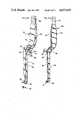
- FIG. 1illustrates an anastomosis device of the invention for end-to-end anastomosis
- FIG. 2shows a connection cylinder, a component of the device of FIG. 1;
- FIG. 3shows a spring clip, a component of the device of FIG. 1;
- FIGS. 4A, 4B, 4C and 4Dare different views of a cylinder holder for use with the connection cylinder of FIG. 2;
- FIGS. 5A, 5B, 5C and 5Dare different views of a clip applicator for use with the clip of FIG. 3;
- FIGS. 6A and 6Billustrate an obturator for use in the invention
- FIGS. 7A and 7Billustrate an anastomosis device of the invention for end-to-end anastomosis, in a different embodiment
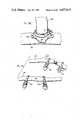
- FIGS. 8 to 12show sequential steps in an anastomosis procedure of the invention.
- FIG. 8illustrates the step of measuring the internal diameter of the separated ends of an artery which are to be connected by a vein
- FIGS. 9A and 9Bshow application of parts of the device of FIG. 1 to the vein
- FIG. 10shows the vein having connection cylinders secured at both ends, ready to be anastomised at one end to the artery;
- FIG. 11shows the anastomization of one end of the artery to the vein.
- FIG. 12shows the completed anastomosis between the vein and the separated ends of the artery
- FIG. 13shows an anastomosis device for end-to-side anastomosis
- FIGS. 14A, 14B and 14Cshow a bayonet clip holder for use in applying a clip of the device of FIG. 13;
- FIGS. 15A and 15Bshow a bayonet cylinder holder for use with the cylinder of FIG. 13;
- FIGS. 16A and 16Bshow an alternative bayonet clip holder
- FIGS. 17A and 17Bshow a perimeter cylinder holder for use with the device of FIG. 13;
- FIGS. 18A to 18Eillustrate schematically the technique of end-to-side anastomosis in accordance with the invention
- FIGS. 19 to 22show sequential steps in an anastomosis procedure of the invention, in particular:
- FIG. 19shows the formation of openings in arteries to be connected
- FIG. 20shows the mounting of a clip in a first artery
- FIG. 21shows the completion of the connection between the bridging vein and the second artery
- FIG. 22shows the completed connection
- FIGS. 23A and 23Bshow another device for end-to-end anastomosis
- FIG. 24shows the final stage of an end-to-end anastomosis employing the device of FIGS. 23A and 23B.
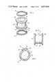
- a device 10 for end-to-end anastomosiscomprises a tubular connection member in the form of a connection cylinder 12 and spring clips 14 and 16 which suitably are coded, for example, by colour coding, to identify either the cardiac or peripheral ends of a vein.
- connection cylinder 12comprises a smooth inner cylindrical surface 18 and a smooth outer cylindrical surface 20.
- An annular channel 22 in outer surface 20extends between a first annular flange 26 and a second annular flange 28; the annular flanges 26 and 28 extend generally radially of the outer surface 20.
- spring clip 14comprises a ring-shaped body 30 defining a generally circular opening 31, and opposed clip ends 32 and 34.
- the clip ends 32 and 34include eyelets 36 and 38, respectively.
- Spring clip 16is generally identical to spring clip 14 and suitably may be slight larger.
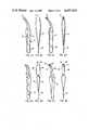
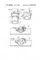
- FIG. 4Ashows a cylinder holder 40 having legs 42 terminating in holder feet 44, each holder foot 44 having an outer cylindrical surface 46. Legs 42 are joined under spring tension at head 48.
- FIGS. 4C and 4Dshow the cylinder holder 40 supporting the connection cylinder 12 of FIG. 2, with the cylindrical surfaces 46 of feet 44 engaging inner surface 18 of connection cylinder 12 under spring tension.
- FIGS. 5A to 5Dshow a clip applicator 50.
- applicator 50comprises legs 52 and 54 terminating in crossed arms 56 and 58, respectively.
- Legs 52 and 54are connected, under spring tension at applicator head 64, and spikes 60 and 62 extend from arms 56 and 58, respectively.
- FIGS. 5C and 5Dshow clip applicator 50 supporting a clip 14 of FIG. 2, with the spikes 60 and 62 extending through eyelets 36 and 38, respectively.
- handle element 66comprises an elongated handle 72 and a tubular end 74.
- FIG. 6Bshows an obturator 68 having a generally conical end portion 70 comprising annular segments 76 of different defined diameters, and a truncated cone portion 82 comprising annular segments 78 of different defined diameters.
- a spigot 80extends from obturator 68.
- the annular segments 76 and 78have diameters ranging from 0.5 to 4.5 mm, the diameters of adjacent segments 76 and 78 increasing in increments of 0.5 mm with increase in distance from the spigot 80.
- a device 200 for end-to-end anastomosiscomprises a tubular connection member in the form of a connection cylinder 212 and spring clips 214 and 216 which are essentially the same as clips 14 and 16 described with reference to FIGS. 1 to 3.
- connection cylinder 212has a smooth inner cylindrical surface 218 and a smooth outer cylindrical surface 220.
- Generally parallel annular grooves 222 and 224are formed in outer surface 220 and define first and second ends 226 and 228 in surface 220.
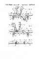
- FIGS. 8 to 12illustrate a method for forming an anastomosis between ends 84 and 86 of an artery 85; the anastomosis being carried out with a vein 92 which is to form a bridge between ends 84 and 86.
- the anastomosis methodis described by reference to the device 100 of FIGS. 1 to 3 but the device 200 of FIGS. 7A and 7B could be employed in a similar manner.
- an appropriate vein 92 for the anastomosisis selected, is tagged to identify the cardiac end and is then removed from the body.
- the internal diameter of the vein 92 and its radial stretchabilityis measured using an obturator of the type illustrated in FIGS. 6A and 6B.
- the artery 85 to be usedis exposed and the free separated ends 84 and 86 are supported between clamps 88 and bridge support 90.
- connection cylinder 12In order to select a connection cylinder 12 of appropriate size a comparison is made between the stretch diameter of the vein 92 and the internal diameter of the artery 85.
- FIG. 8particularly illustrates the use of an obturator 68 of FIG. 6B to measure the internal diameter of artery 85 at end 84.
- Obturator 68is secured to handle element 66 by inserting spigot 80 in tubular end 74.
- the obturator 68is inserted into the open end 84 of artery 85.
- the annular segments 76 and 78are of different specified diameters. In this way an unstretched and stretched diameter of the artery 85 can be determined.
- the unstretched and stretched diameters of the vein 92are determined in a similar manner (not illustrated).
- connection cylinder 12having an appropriate diameter is then selected.
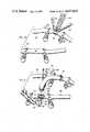
- FIGS. 9A and 9Bshow schematically the application of connection cylinder 12 to one end of vein 92.
- vein end 94 of vein 92is passed through a first end adjacent flange 26 and emerges from a second end adjacent flange 28 of connection cylinder 12.
- Vein end 94is then everted over connection cylinder 12 in the manner indicated by the arrow in FIG. 9A.
- the everted portion 98extends over annular channel 22 in the direction of flange 26.
- a spring clip 14is then seated in annular channel 22 adjacent flange 26 with the everted portion 98 of vein 92 thereunder.
- the circular opening 31 of spring clip 14has a diameter selected having regard to the size of connection cylinder 12.
- the spring tension in spring clip 14 in conjunction with the diameter of circular opening 31 and the depth of annular groove 32are such that ring-shaped body part 30 is firmly seated in annular channel 22 and the everted portion 98 is securely held on connection cylinder 12 against anastomatic separation.
- the everted portion 98is firmly held on cylinder 12 under spring pressure, and will not become separated from the cylinder such as to permit leakage of blood at the connection.
- Vein end 96is mounted on a second connection cylinder 12 in the same manner, but employing a spring clip 14' over everted portion 98a (see FIG. 10).
- Spring clips 14 and 14'are coded differently for use with different ends of the vein 92, so that rapid identification of the cardiac and distal ends of the vein may be achieved, for correct orientation of vein 92.
- the vein 92has cylinders 12 (not visible) mounted at its ends 94 and 96 (see FIGS. 9A and 9B), and secured by coded spring clips 14 and 14', respectively.
- One cylinder 12is held by a curved cylinder holder 40 (see FIGS. 4A to 4D) which engages the interior thereof, and the end 84 of artery 85 is held open by forceps 100 having hook ends 102 which pierce the walls of artery end 84.
- the cylinder 12 with everted portion 98is inserted into the artery 85 at end 84.
- spikes 60 and 62 of applicator 50are inserted through eyelets 36 and 38 of spring clip 16.
- legs 52 and 54 of applicator 50are urged together arms 56 and 58 separate thereby separating clip ends 32 and 34, under spring tension, with enlargement of circular opening 31.
- the clip 16With clip ends 32 and 34 separated, and opening 31 enlarged, the clip 16 is applied over artery end 84 and is seated over everted portion 98 in the annular groove 22 adjacent flange 28. Release of the inward pressure on legs 52 and 54 urges clip ends 32 and 34 towards each other under the spring tension of ring-shaped body 30.
- the size of circular opening 31, the spring tension of clip 16 and the depth of annular channel 22 in conjunction with the thickness of everted portion 98are selected such that spring clip 16 is firmly seated in annular channel 22 and holds artery end 84 on connection cylinder 12 with everted portion 98 therebetween against anastomatic separation.
- the flanges 26 and 28prevent axial dislodgement of the clips 14 and 16 from cylinder 12, and the pressure of the clips 14 and 16 on the underlying vein and artery walls holds the vein and artery walls in contact on the connection cylinder 12 against anastomatic separation.
- the curved cylinder holder 40(FIGS. 4A to 4D) comprises legs 42 and feet 44 adapted to engage the inner surface of cylinder 12, with vein 92 thereon. Legs 42 are connected under spring tension. In use the feet 44 are located on opposed inner sides of cylinder 12 and are brought into contact therewith by spring tension on the inner cylinder walls urging legs 42 away from each other.
- the clips 214 and 216are firmly seated in annular grooves 222 and 224 and hold the tissue material of the artery and vein in connection member 212 and in contact with each other against anastomatic separation.
- the depth and width of grooves 222 and 224may be selected such that clips 214 and 216 may be securely seated in grooves 222 and 224 with the underlying tissue material, whereby axial dislodgement of the clips 214 and 216, and anastomatic separation is avoided.
- the device 10 of FIGS. 1 to 3is appropriate for the anastomosis of tubular vessels having a diameter above about 2.5 mm; the device 200 of FIGS. 7A and 7B is especially appropriate for microanastomosis, i.e. anastomosis of vessels having a diameter of 1 to 2 mm.
- the thickness of the tubular vessel wallincreases with vessel diameter, and greater care is needed with vessels having thinner walls in ensuring that the vessel wall is not damaged as a result of stretching, during the eversion of the vessel end over the connection cylinder.
- the diameters of the inner and outer cylindrical surfaces 218 and 220are selected having regard to the diameter of the vessel to be everted thereon, the diameter of surface 220 being only slightly greater than that of surface 218, and the diameter of the ends 226 and 228 is the same as that of the outer cylindrical surface 220.
- the relatively shallow grooves 222 and 224 in conjunction with the clips 214 and 216are adequate to prevent axial or radial movement of the anastomosed vessels.
- the thicker vessel walls of the larger size vesselsare less susceptible to damage when being stretched over cylinder 12.
- the annular channel 22is relatively deep or otherwise stated the diameter of flanges 26 and 28 is significantly larger than the diameter of outer surface 20; the diameter of outer surface 20 being only slightly larger than the diameter of inner surface 18.
- the depth of channel 22 forming relatively large diameter flanges 26 and 28compensates for the absence of discrete annular grooves similar to grooves 222 and 224 in the device 200, to accommodate clips 14 and 16.
- the flanges 26 and 28 defined by the deep channel 22serve to prevent axial dislodgement of clips 14 and 16 from cylinder 12; and in addition their relative diameter is such that even if some radial opening or displacement of the clips 14 and 16 occurs, the partially opened clips 14 and 16 will not pass over flanges 26 and 28.
- the relatively deep channel 22also serves to accommodate any bunching or folds of thicker walled vessels, and ensures that such folds are held on the cylinder 12 between flanges 26 and 28.
- the device 200can also be employed for anastomosis of larger vessels, however, the device 10 is found to be less suitable for microanastomosis.
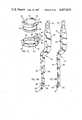
- a device 110 for end-to-side anastomosiscomprises a tubular connection member in the form of a connection cylinder 112 and spring clip 114.
- Connection cylinder 112has a smooth inner cylindrical surface 116, a smooth outer cylindrical surface 118 and annular flanges 120 and 122 at opposed ends.
- Annular flange 120has opposed flat walls 124 and a spike 125 extending from each wall 124.
- Spring clip 114includes a ring-shaped body 126 defining a substantially circular opening 127, and clip ends 128 and 130 having eyelets 132 and 134, respectively.
- Tines 136extend substantially radially outwardly from ring-shaped body 126.
- the cylinder 112is suitably made of stainless steel.
- the flange 120prevents the donor vessel and cylinder 112 from slipping into the lumen of the recipient vessel.
- Outer cylindrical surface 118is suitably of relatively short length so that clip 114 may fit snugly between flanges 120 and 122 and prevent protrusion of the graft donor vessel into the lumen of the recipient vessel.
- a bayonet clip applicator 146comprises legs 154 and 156 joined under spring tension at head 152.
- Crossing arms 158 and 160extend from legs 154 and 156, respectively and terminate in spikes 162.
- a spring loaded stabilizer arm 163 having a spring 167extends between legs 154 and 156, and arms 158 and 160, in a casing 166, and terminates in a stabilizer foot 164.
- Stabilizer arm 163includes a locking arm 165 which engages a recess 169 in leg 156 to firmly locate arm 166.
- Stabilizer arm 163is urged downwardly by spring 167 but is restrained against downward movement, as shown in FIG. 14A, by locking arm 165 which engages recess 169.
- locking arm 165When locking arm 165 is released from recess 169 the stabilizer arm 166 moves downwardly under the action of spring 167 and stabilizer foot 164 lightly engages clip ends 128 and 130 of clip 114 and stabilizes the location of clip 114 during the enlargement of opening 127 and insertion of the cylinder 112.
- the opening 127is enlarged for the insertion of cylinder 112 by pressing legs 154 and 156 towards each other.
- a bayonet cylinder holder 148comprises upper legs 170 and 172 and lower legs 174 and 176 terminating in holder feet 175. Legs 174 and 176 are off-set relative to legs 170 and 172.
- the holder feet 175have toes 179 with outer cylindrical surfaces for engaging the inner surface 116 of cylinder 112.
- Upper legs 170 and 172are connected at head 177 under spring pressure.
- legs 174 and 176The off-setting of legs 174 and 176 relative to head 177 and legs 170 and 172 avoids obstruction in the line of vision of the surgeon.
- Applicator 178has legs 180 and 182 and crossing arms 184 and 186 terminating in feet 185 having spikes 187. Legs 180 and 182 are connected under spring tension at head 189.
- Applicator 178functions in the same manner as applicator 50 in FIGS. 5A, 5B, 5C and 5D, however, the off-setting of arms 184 and 186 relative to legs 180 and 182 avoids obstruction in the line of vision of the surgeon.
- a perimeter cylinder holder 190comprises upper legs 191 and 192 and lower legs 193 and 194 terminating in feet 195. Legs 193 and 194 are off-set relative to legs 191 and 192.
- Legs 191 and 192are connected at head 196 under spring pressure.
- a spring leaf locking arm 197includes an upper arm 198 mounted on leg 191 and a lower arm 199 having a projecting stop 201.
- a recess 203 having a stop 204is defined in leg 192.
- Feet 195are urged apart by the spring pressure as shown in FIG. 17A. Legs 191 and 192 are urged towards each other by finger pressure until feet 195 engage opposed outer sides of cylinder 112, and are locked in position under the spring action of arm 197 urging lower arm 199 into recess 203, where it is held by engagement of stops 201 and 204.
- FIGS. 18A and 18Bshow the mounting of a vein 500 on connection cylinder 112 of FIG. 13, the everted portion 502 being held by the spikes 125.
- FIGS. 18C and 18Dshow the mounting of a clip 114 around an opening in an artery 510 to form a circular opening corresponding to opening 127 of clip 114, and bounded by tissue annulus 550.
- FIG. 18Eshows the completed anastomosis in which flange 122 of cylinder 112 is disposed below clip 114 and prevents axial dislodgement of clip 114.
- the end-to-side anastomosis techniqueis illustrated in a stepwise fashion in FIGS. 19 to 22.
- Flow of bloodis interrupted in arteries 510 to 512 to be connected, by means of clamps 544; a vein 500 is to be anastomosed between the arteries 510 and 512.
- an openingis formed in each of arteries 510 and 512 suitably by applying two incisions at right angles forming a cross in the tissue material (see artery 510 in FIG. 19).
- the artery tissue adjacent the incisionsis folded back (see artery 512 in FIG. 19).
- the folded back tissue of artery 512is inserted upwardly through opening 127 in clip 114 and is secured over ring shaped body 126 by the tines 136 as illustrated schematically in FIGS. 18C and 18D.
- Forceps 515are employed to draw the folded back tissue over the tines 136.
- the clip 114is supported by the clip applicator 146 of FIGS. 14A to 14C.
- vein 500An end of vein 500 is passed through connection cylinder 112 and everted thereover in the general manner illustrated in FIGS. 18A and 18B, the vein 500 being everted over the flanges 120 and 122 and being pierced by spikes 125 to form the everted portion 502.
- FIG. 21A final stage of the anastomosis is illustrated in FIG. 21, wherein the connection of vein 500 to artery 512 is already completed and a similar connection between vein 500 and artery 510 is about to be completed.
- eyelets 132 and 134 of clip 114 mounted in artery 510are engaged by the spikes 162 of bayonet clip applicator 146.
- Cylinder 112 with vein 500 mounted thereonis held by bayonet perimeter cylinder holder 190 (see FIGS. 17A and 17B) and is then inserted into the enlarged opening 127 so that flange 122 of cylinder 112 is disposed on the interior side of ring-shaped body 126 (see FIG. 18E), within artery 510 and ring-shaped body 126 is disposed about outer surface 118.
- this insertion stabilizer arm 166applies pressure on clip ends 128 and 130 to firmly position spring clip 114 for insertion of cylinder 112 and its mounted vein 500.
- legs 154 and 156Thereafter pressure on legs 154 and 156 is released so that they move apart under spring pressure and arms 158 and 160 move towards each other to their normal position and clip ends 128 and 130 likewise are urged towards each other. In this way clip 114 is firmly located under spring tension over outer surface 118 with the everted portion 502 of vein 500 therebetween.
- the flanges 122 and 120 and spikes 125prevent axial separation of clip 114 from cylinder 112 and the pressure of clip 114 holds tissue annulus 550 in contact with the everted portion 502 of vein 500 whereby anastomatic separation is avoided.
- the spring tension in body 126 and the dimensions of flange 122are such that opening 127 can be enlarged to a diameter greater than that of flange 122 for insertion of cylinder 112 with vein 500 mounted thereon into tissue annulus 550.
- anastomosis clip 600having a pair of legs 602 and 604 and cross-over arms 606 and 608. Arms 606 and 608 terminate in aligned rings 610 and 612. Rings 610 and 612 have circular orifices or openings 614 and 616 which form a continuous orifice 615.
- Ring 610has a plurality of spaced apart, outwardly extending teeth 618 and ring 612 has a similar plurality of teeth 620.
- Legs 602 and 604are connected at head 622 under spring pressure which tends to urge legs 602 and 604 away from each other, thereby urging rings 610 and 612 into contact with each other as shown in FIG. 23A. Pressure on legs 602 and 604 forces them together thereby forcing rings 610 and 612 apart, as shown in FIG. 23B.
- a small gap 624is defined in ring 612.
- the anastomosis clip 600is particularly suitable for anastomosis of vessels having a diameter greater than 2 mm.
- FIG. 24The anastomosis technique employing clips 600 is illustrated in FIG. 24 in which the separated ends 684 and 686 of an artery 685 are connected with a vein 692.
- the two clipsare identical but for convenience the parts of one clip are additionally designated by the letter ⁇ a ⁇ .
- Clips 600 and 600a having orifices 615 and 615aare selected according to the dimensions of artery 685 and a vein 692 of appropriate dimensions is selected.
- the clips 600 and 600aare connected to the ends of vein 692, the artery end 684 is connected to clip 600 and finally the artery end 686 is connected to clip 600a.
- vein 692is fed through orifice 614a of ring 610a and is everted over teeth 618a thereby forming an annulus of vein tissue 650a.
- end 686 of artery 685is feed into orifice 616a, and is everted over teeth 620a thereby forming an annulus of artery tissue 652a.
- the anastomosis techniquehas been particularly described, with reference to the drawings, for the joining of completely separated vessel ends. It is, however, possible to employ the same devices and techniques to complete a connection between a partly separated wall of a tubular vessel.
- the artery wallbulges and creates a weak spot in the wall which may burst under the pressure of the blood.
- the arterymay be partially severed around the aneurysm and the separated edges of the artery wall may be joined using the devices of the invention to by-pass the aneurysm.
- gap 624 in ring 612 of clip 600may serve for introduction of the unsevered portion of the artery into clip 600.
- the surgeoncan readily hold and support the devices during the anastomosis. It will be understood that the surgeon is working in a very small area under a microscope, and so in developing a satisfactory and practical technique it is important that one person, namely the surgeon, be able to hold and apply the principal components of the anastomosis devices during the anastomosis.
- connection cylinderIn end-to-end anastomosis, it is necessary that the connection cylinder be held still during application of the spring clip.
- the surgeonis able to hold the connection cylinder with the everted vein with one hand for insertion of the cylinder into the artery held open with the other hand.
- the surgeoncan hold the spring clip and open and apply it with the other hand.
- the person holding the cylinderalso applies the spring clips so that his hands remain axial and the cylinder is mounted on the true axis of the artery to be anastomosed.
- the anastomosis procedure of the inventionhas also been made applicable to microanastomosis and to long vein intercranial bypass procedures.
- cylinder holders and clip applicatorshave been developed suitable for use in fine deeply situated regions at the base of the brain.
- instrumentshave been developed of increased length and incorporating a bayonet design to allow a more direct, unobstructed, view and facilitate anastomosis.
- the bayonet cylinder holder described abovehas its cylinder engaging end offset relative to the handle, thus the handle does not obstruct the surgeon's view of the anastomosis site.
- the bayonet cylinder holderis thus especially suitable in microanastomosis of 1 mm vessels in a confined space.
- connection cylinders 12 and 112typically have diameters of 3.5 mm, 3 mm, 2.5 mm, 2 mm, 1.5 mm, 1.0 mm and 0.75 mm, with a wall thickness of 5 thousandths of an inch.
- the length of the connection cylinderis suitably of the same order as the diameter.
- the cylindersare made of stainless steel.
- Spring clips 14 and 16are suitably of metal or metal alloy wire, for example, Elgiloy wire, typically having a wire diameter of 7 to 18 thousandths of an inch. Typically these clips may have diameters of 3.5 mm, 3 mm, 2.5 mm, 2 mm, 1.5 mm, 1.0 mm and 0.75 mm.
- Elgiloyis a cobalt base alloy of the following composition in weight %:
- the clips fabricated from the wireas suitably heat-treated to provide spring tension characteristics.
- the clipsare heated with a propane torch to a light straw colour at an elevated temperature.
- the surgeoncan complete a connection much more rapidly than with conventional techniques and has the significant advantage of intima to intima contact at the site of anastomosis with no foreign material exposed to the lumen of the vessels being joined.
- the surgeoncan complete a connection regardless of vessel size, including a microvascular anastomosis of 1 mm diameter vessels typically in a time of less than three minutes.
- Such microvascular anastomosis employing conventional suture techniques, even with use of a microscope,requires a significantly longer time, typically 20 to 40 minutes.
- Table Ishows the results of in vivo end-to-end anastomosis in dogs employing the procedure and devices of the invention. Seventeen vascular anastomoses are carried out in five dogs. Carotid, femoral and axillary vessels were used with diameters ranging from 2 to 3.5 millimeters. Control angiograms were taken, following anastomosis, for selected animals. The patency rate in the anastomoses that have had post-operative angiography has been 91%. There has been only one anastomosis that has not been visualized on post-operative angiography and it is uncertain whether this is related to occlusion of the anastomosis or to technical factors in performing the angiogram.
- Table 2shows the results of in vivo end-to-end anastomosis in other dogs. Thirty three vascular anastomoses have been carried out in 11 dogs. Carotid, femoral and axillary vessels were used with diameters ranging from 1 to 3.5 mm. These animals have been followed for up to eight months following anastomosis (Table 2). The patency rates in the anastomoses that have had post-operative angiography have been 88%. Indeed, three of the four occlusions have been in brachial vessels which have been found unsuitable for anastomosis because of the excessive twisting of the graft in the axilla on movement of the limb of the dog when ambulating. If the brachial vessels are eliminated the patency rate is over 95%.
- the rabbit's carotid arteryhas a diameter that ranges between 1.5 mm to 2 mm. Appropriate size clips were developed.
- To assess for graft patencyboth angiography and direct gross inspection of the anastomotic site has been carried out. A 100% patency rate has been achieved with the end-to-side anastomotic devices of the invention in those animals having had post-op assessment. This includes anastomoses in vessels ranging in size from 2 mm to 3.5 mm. Although these animals have to date been followed over only a few months, longer follow up studies will be carried out.
Landscapes
- Health & Medical Sciences (AREA)
- Life Sciences & Earth Sciences (AREA)
- Surgery (AREA)
- Heart & Thoracic Surgery (AREA)
- Engineering & Computer Science (AREA)
- Biomedical Technology (AREA)
- Nuclear Medicine, Radiotherapy & Molecular Imaging (AREA)
- Medical Informatics (AREA)
- Molecular Biology (AREA)
- Animal Behavior & Ethology (AREA)
- General Health & Medical Sciences (AREA)
- Public Health (AREA)
- Veterinary Medicine (AREA)
- Surgical Instruments (AREA)
Abstract
Description
______________________________________ Co 40% Cr 20% Ni 15% Mo 7% Mn 2% Be 0.4% C 0.15% Fe 15.81% ______________________________________
TABLE 1 ______________________________________ DIAMETER PATENCY DOG NO. VESSEL (mm) (angio) ______________________________________ 666 Right Carotid 3.0 P Left Carotid 2.5 P Left Carotid 2.5 -- Right Femoral 3.2 P 668 Right Carotid 2.5 P Left Carotid 2.5 P Right Femoral 2.5 P Left Femoral 2.0 -- Left Axillary 2.0 -- 669 Right Carotid 3.5 P Left Carotid 3.0 P Right Femoral 3.5 -- 702 Right Carotid 4.0 O Left Carotid 3.0 P Right Femoral 3.0 P 716 Right Carotid 3.5 -- Left Carotid 3.5 -- No angio ______________________________________ -- Angiogram not carried out. O Anastomosis not visualized on angiography.
TABLE 2 ______________________________________ DIA- DOG VESSEL METER FOLLOW UP PATENCY NO. (artery) (mm) (months) (angio) ______________________________________ 741 Right carotid 3.0 3.0 P Left carotid 3.5 1.5 P Right femoral 3.0 2.0 P 740 Right carotid 3.5 3.0 P Left carotid 3.0 2.5 P Right femoral 1.0 3.0 O 739 Right carotid 3.0 1.5 P Left carotid 3.0 2.0 P Right femoral 2.5 2.0 P 726 Right carotid 3.5 6.0 P Left carotid 3.5 4.0 P Right brachial 2.5 6.0 O Left brachial 2.5 6.0 P Right femoral 3.5 6.0 P Renal 3.0 4.0 P 718 Right carotid 3.5 1 week P Left carotid 3.5 1 week P 717 Right carotid 3.0 3.0 P Left carotid 3.0 3.0 P Right Brachial 2.5 2.0 P Left Brachial 2.5 2.5 P Right femoral 3.0 3.0 P 716 Left Brachial 1.0 2.0 P Left femoral 2.5 2.5 P 702 Left brachial 1.0 1.0 P Right brachial 1.0 1.0 P 669 Right brachial 2.0 2.0 P 668 Right brachial 2.0 1.0 P Left brachial 2.0 8.0 P Left femoral 2.0 7.5 P 666 Right brachial 2.0 1.0 P Left brachial 2.5 1.0 O Left carotid 2.5 7.0 P ______________________________________ P: Patent O: Occluded
TABLE 3 ______________________________________ VESSEL DIAMETER PATENCY (artery) (mm) (angio) ______________________________________ A. End-to-side anastomosis DOG No. 723 Right brachial 1.5 P 738 Right brachial 2.0 NA Left to right carotid 3.0, 2.5 NA 739 Right to left carotid 3.0, 3.0 P Left brachial 2.0 P 741 Right to left carotid 2.5, 3.0 P Right brachial 2.0 P 740 Right brachial 1.5 P Left to right carotid 3.5, 3.5 P 760 Right to left carotid 3.0, 3.0 P Left to right carotid 3.0, 3.0 P Right brachial 2.5 P Left brachial 2.5 P RABBIT No. 339 Right to left carotid 2.0 P B. End-to-end anastomosis RAT No. 295 Carotid 1.0 P 278 Carotid 1.0 P 247 Carotid 1.0 P 248 Carotid 1.0 P ______________________________________ P: Patent O: Occluded NA: Angio not done to date.
Claims (5)
Priority Applications (9)
| Application Number | Priority Date | Filing Date | Title |
|---|---|---|---|
| US06/598,900US4657019A (en) | 1984-04-10 | 1984-04-10 | Anastomosis devices and kits |
| CA000478072ACA1261706A (en) | 1984-04-10 | 1985-04-01 | Anastomosis devices, kits and method |
| EP88106511AEP0303767A1 (en) | 1984-04-10 | 1985-04-09 | Anastomosis devices, kits and method |
| EP85104271AEP0158316A3 (en) | 1984-04-10 | 1985-04-09 | Anastomosis devices and kit |
| US06/923,209US4771775A (en) | 1984-04-10 | 1986-10-27 | Anastomosis devices, kits and method |
| US06/923,490US4787386A (en) | 1984-04-10 | 1986-10-27 | Anastomosis devices, and kits |
| US07/132,599US4873975A (en) | 1984-04-10 | 1987-12-14 | Anastomosis device and method |
| US07/238,730US4917087A (en) | 1984-04-10 | 1988-08-30 | Anastomosis devices, kits and method |
| CA000609124ACA1277197C (en) | 1984-04-10 | 1989-08-23 | Anastomosis devices, kits and method |
Applications Claiming Priority (1)
| Application Number | Priority Date | Filing Date | Title |
|---|---|---|---|
| US06/598,900US4657019A (en) | 1984-04-10 | 1984-04-10 | Anastomosis devices and kits |
Related Child Applications (2)
| Application Number | Title | Priority Date | Filing Date |
|---|---|---|---|
| US06/923,209DivisionUS4771775A (en) | 1984-04-10 | 1986-10-27 | Anastomosis devices, kits and method |
| US06/923,490ContinuationUS4787386A (en) | 1984-04-10 | 1986-10-27 | Anastomosis devices, and kits |
Publications (1)
| Publication Number | Publication Date |
|---|---|
| US4657019Atrue US4657019A (en) | 1987-04-14 |
Family
ID=24397395
Family Applications (1)
| Application Number | Title | Priority Date | Filing Date |
|---|---|---|---|
| US06/598,900Expired - Fee RelatedUS4657019A (en) | 1984-04-10 | 1984-04-10 | Anastomosis devices and kits |
Country Status (3)
| Country | Link |
|---|---|
| US (1) | US4657019A (en) |
| EP (2) | EP0158316A3 (en) |
| CA (2) | CA1261706A (en) |
Cited By (156)
| Publication number | Priority date | Publication date | Assignee | Title |
|---|---|---|---|---|
| US4931057A (en)* | 1988-03-29 | 1990-06-05 | Pfizer Hospital Products Group, Inc. | Compression anastomosis coupling assembly |
| US5520704A (en)* | 1992-10-09 | 1996-05-28 | United States Surgical Corporation | Everting forceps with locking mechanism |
| WO1996029938A1 (en)* | 1995-03-24 | 1996-10-03 | Organ, Inc. | Anastomosis cuff manipulator tool |
| US5695504A (en)* | 1995-02-24 | 1997-12-09 | Heartport, Inc. | Devices and methods for performing a vascular anastomosis |
| US5702048A (en)* | 1993-03-18 | 1997-12-30 | Eberlin; Rene | Device for microanatomosis of blood vessels |
| US5732872A (en)* | 1994-06-17 | 1998-03-31 | Heartport, Inc. | Surgical stapling instrument |
| US5741296A (en)* | 1993-11-02 | 1998-04-21 | Karl Storz Gmbh & Co. | Instrument for the application of prostheses inside the body |
| US5868763A (en)* | 1996-09-16 | 1999-02-09 | Guidant Corporation | Means and methods for performing an anastomosis |
| US5881943A (en)* | 1994-06-17 | 1999-03-16 | Heartport, Inc. | Surgical anastomosis apparatus and method thereof |
| US5904697A (en)* | 1995-02-24 | 1999-05-18 | Heartport, Inc. | Devices and methods for performing a vascular anastomosis |
| EP0931512A1 (en) | 1998-01-23 | 1999-07-28 | Ethicon Endo-Surgery, Inc. | Surgical stapling instrument |
| US5951576A (en)* | 1998-03-02 | 1999-09-14 | Wakabayashi; Akio | End-to-side vascular anastomosing stapling device |
| US6015416A (en)* | 1998-02-26 | 2000-01-18 | Ethicon Endo-Surgery, Inc. | Surgical anastomosis instrument |
| US6017352A (en)* | 1997-09-04 | 2000-01-25 | Kensey Nash Corporation | Systems for intravascular procedures and methods of use |
| US6019788A (en)* | 1996-11-08 | 2000-02-01 | Gore Enterprise Holdings, Inc. | Vascular shunt graft and junction for same |
| US6030395A (en)* | 1997-05-22 | 2000-02-29 | Kensey Nash Corporation | Anastomosis connection system |
| US6063114A (en)* | 1997-09-04 | 2000-05-16 | Kensey Nash Corporation | Connector system for vessels, ducts, lumens or hollow organs and methods of use |
| US6066144A (en)* | 1997-10-07 | 2000-05-23 | Ethicon Endo-Surgery, Inc. | Surgical anastomosis method |
| US6110187A (en)* | 1995-02-24 | 2000-08-29 | Heartport, Inc. | Device and method for minimizing heart displacements during a beating heart surgical procedure |
| US6110188A (en)* | 1998-03-09 | 2000-08-29 | Corvascular, Inc. | Anastomosis method |
| WO2000056228A1 (en)* | 1999-03-19 | 2000-09-28 | By-Pass, Inc. | Low profile anastomosis connector |
| US6146393A (en)* | 1998-12-18 | 2000-11-14 | Wakabayashi; Akio | External tubular stapling device for anastomosing a vascular graft to an anastomosing sheath |
| US6179849B1 (en) | 1999-06-10 | 2001-01-30 | Vascular Innovations, Inc. | Sutureless closure for connecting a bypass graft to a target vessel |
| US6206913B1 (en) | 1998-08-12 | 2001-03-27 | Vascular Innovations, Inc. | Method and system for attaching a graft to a blood vessel |
| US6217585B1 (en) | 1996-08-16 | 2001-04-17 | Converge Medical, Inc. | Mechanical stent and graft delivery system |
| US20010001826A1 (en)* | 1998-01-23 | 2001-05-24 | Heartport, Inc. | System for performing vascular anastomoses |
| US6241741B1 (en) | 1998-03-09 | 2001-06-05 | Corvascular Surgical Systems, Inc. | Anastomosis device and method |
| US6248117B1 (en) | 1999-04-16 | 2001-06-19 | Vital Access Corp | Anastomosis apparatus for use in intraluminally directed vascular anastomosis |
| US6254615B1 (en) | 1995-02-24 | 2001-07-03 | Heartport, Inc. | Surgical clips and methods for tissue approximation |
| US6280460B1 (en) | 1998-02-13 | 2001-08-28 | Heartport, Inc. | Devices and methods for performing vascular anastomosis |
| US20010023354A1 (en)* | 1999-04-16 | 2001-09-20 | Blatter Duane D. | Locking compression plate apparatus |
| US6293955B1 (en) | 1996-09-20 | 2001-09-25 | Converge Medical, Inc. | Percutaneous bypass graft and securing system |
| US20010037139A1 (en)* | 1998-08-12 | 2001-11-01 | Yencho Stephen A. | Method and system for attaching a graft to a blood vessel |
| US6361559B1 (en) | 1998-06-10 | 2002-03-26 | Converge Medical, Inc. | Thermal securing anastomosis systems |
| US6371964B1 (en) | 1999-05-18 | 2002-04-16 | Vascular Innovations, Inc. | Trocar for use in deploying an anastomosis device and method of performing anastomosis |
| US6391038B2 (en) | 1999-07-28 | 2002-05-21 | Cardica, Inc. | Anastomosis system and method for controlling a tissue site |
| US20020077637A1 (en)* | 1999-05-18 | 2002-06-20 | Jaime Vargas | Trocar for use in deploying an asastomosis device and method of performing anastomosis |
| US20020155025A1 (en)* | 2001-02-22 | 2002-10-24 | Wolfgang Daum | Material for nuclear spin tomography magnetic resonance imaging (MRI) |
| US6471713B1 (en) | 2000-11-13 | 2002-10-29 | Cardica, Inc. | System for deploying an anastomosis device and method of performing anastomosis |
| US6475222B1 (en) | 1998-11-06 | 2002-11-05 | St. Jude Medical Atg, Inc. | Minimally invasive revascularization apparatus and methods |
| US20020173809A1 (en)* | 1999-09-01 | 2002-11-21 | Fleischman Sidney D. | Sutureless anastomosis system deployment concepts |
| US20020176797A1 (en)* | 1997-01-24 | 2002-11-28 | Roberts Craig P. | Methods and devices for maintaining cardiopulmonary bypass and arresting a patient's heart |
| US6488692B1 (en) | 1996-09-16 | 2002-12-03 | Origin Medsystems, Inc. | Access and cannulation device and method for rapidly placing same and for rapidly closing same in minimally invasive surgery |
| US6494889B1 (en) | 1999-09-01 | 2002-12-17 | Converge Medical, Inc. | Additional sutureless anastomosis embodiments |
| US20030014063A1 (en)* | 1996-11-08 | 2003-01-16 | Houser Russell A. | Percutaneous bypass graft and securing system |
| US20030014064A1 (en)* | 1999-04-16 | 2003-01-16 | Blatter Duane D. | Anvil apparatus for anastomosis and related methods and systems |
| US20030023252A1 (en)* | 2001-07-05 | 2003-01-30 | Whayne James G. | Distal anastomosis system |
| US20030023253A1 (en)* | 2001-04-27 | 2003-01-30 | Cardica, Inc. | Anastomosis system |
| US6514263B1 (en) | 2000-08-30 | 2003-02-04 | Ethicon Endo-Surgery, Inc. | Helical needle and suture combination having a strain relief element |
| US6520973B1 (en) | 2000-08-30 | 2003-02-18 | Ethicon Endo-Surgery, Inc. | Anastomosis device having an improved needle driver |
| US6530932B1 (en) | 2000-08-30 | 2003-03-11 | Ethicon Endo-Surgery, Inc. | Anastomosis device having improved tissue presentation |
| US20030055441A1 (en)* | 1997-10-24 | 2003-03-20 | Suyker Wilhelmus Joseph Leonardus | Mechanical anastomosis system for hollow structures |
| US6537288B2 (en) | 1999-05-18 | 2003-03-25 | Cardica, Inc. | Implantable medical device such as an anastomosis device |
| US20030065341A1 (en)* | 2001-09-28 | 2003-04-03 | Schulze Dale R. | Arrangement and method for vascular anastomosis |
| US20030065343A1 (en)* | 2001-09-28 | 2003-04-03 | Yencho Stephen A. | Access port system for anastomosis |
| US6551334B2 (en) | 1999-04-16 | 2003-04-22 | Integrated Vascular Interventional Technologies, Lc | Externally directed anastomosis systems and externally positioned anastomosis fenestra cutting apparatus |
| US6554764B1 (en) | 2000-11-13 | 2003-04-29 | Cardica, Inc. | Graft vessel preparation device and methods for using the same |
| US20030093095A1 (en)* | 2001-07-05 | 2003-05-15 | Whayne James G. | Distal anastomosis system |
| US6565581B1 (en) | 1996-09-16 | 2003-05-20 | Origin Medsystems, Inc. | Apparatus and method for performing an anastomosis |
| US6569173B1 (en) | 1999-12-14 | 2003-05-27 | Integrated Vascular Interventional Technologies, L.C. | Compression plate anastomosis apparatus |
| US20030109893A1 (en)* | 2001-12-06 | 2003-06-12 | Cardica,Inc. | Implantable medical device such as an anastomosis device |
| US20030130671A1 (en)* | 1999-11-23 | 2003-07-10 | Duhaylongsod Francis G. | Anastomosis device and method |
| US6613058B1 (en) | 2000-08-30 | 2003-09-02 | Ethicon Endo-Surgery, Inc. | Anastomosis device having needle receiver for capturing the needle |
| US20030167064A1 (en)* | 1999-09-01 | 2003-09-04 | Whayne James G. | Advanced anastomosis systems (II) |
| US20030191482A1 (en)* | 1998-10-22 | 2003-10-09 | Suyker Wilhelmus Joseph Leonardus | Mechanical anastomosis system for hollow structures |
| US20030208214A1 (en)* | 2000-03-20 | 2003-11-06 | Amir Loshakove | Anastomotic connector and graft expander for mounting a graft |
| US20030212418A1 (en)* | 2000-10-12 | 2003-11-13 | Cardica, Inc. | Implantable superelastic anastomosis device |
| US6652542B2 (en) | 1999-04-16 | 2003-11-25 | Integrated Vascular Interventional Technologies, L.C. (Ivit, Lc) | External anastomosis operators and related systems for anastomosis |
| US20030229365A1 (en)* | 2002-06-10 | 2003-12-11 | Whayne James G. | Angled vascular anastomosis system |
| US20040002721A1 (en)* | 1999-09-01 | 2004-01-01 | Podmore Jonathan L. | Method and apparatus for performing end-to-end and end-to-side anastomosis with eversion of tissue edges |
| US20040049221A1 (en)* | 1998-05-29 | 2004-03-11 | By-Pass, Inc. | Method and apparatus for forming apertures in blood vessels |
| US20040068278A1 (en)* | 1999-12-06 | 2004-04-08 | Converge Medical Inc. | Anastomosis systems |
| US6719769B2 (en) | 1999-11-15 | 2004-04-13 | Cardica, Inc. | Integrated anastomosis tool with graft vessel attachment device and cutting device |
| US20040073247A1 (en)* | 1998-05-29 | 2004-04-15 | By-Pass, Inc. | Method and apparatus for forming apertures in blood vessels |
| US20040073248A1 (en)* | 1999-05-18 | 2004-04-15 | Cardica, Inc. | Tissue punch |
| US6726704B1 (en) | 1998-05-29 | 2004-04-27 | By-Pass, Inc. | Advanced closure device |
| US6726694B2 (en) | 1999-04-16 | 2004-04-27 | Integrated Vascular Interventional Technologies, L.C. (Ivit, Lc) | Intraluminally directed anvil apparatus and related methods and systems |
| US20040087985A1 (en)* | 1999-03-19 | 2004-05-06 | Amir Loshakove | Graft and connector delivery |
| US20040092972A1 (en)* | 2000-11-09 | 2004-05-13 | Leonardus Suyker Wilhelmus Joseph | Connector, applicator and method for mechanically connecting hollow structures, in particular small blood vessels, as well a auxiliary devices |
| US6736825B2 (en) | 1999-12-14 | 2004-05-18 | Integrated Vascular Interventional Technologies, L C (Ivit Lc) | Paired expandable anastomosis devices and related methods |
| US20040097973A1 (en)* | 2000-03-20 | 2004-05-20 | Amir Loshakove | Transvascular bybass method and system |
| US6743244B2 (en) | 1999-04-16 | 2004-06-01 | Integrated Vascular Interventional Technologies, L.C. | Soft anvil apparatus for cutting anastomosis fenestra |
| US6746459B2 (en) | 2000-10-19 | 2004-06-08 | Terumo Kabushiki Kaisha | End-to-side blood vessel anastomosis method and instruments therefor |
| US20040116945A1 (en)* | 2000-04-29 | 2004-06-17 | Ventrica, Inc., A Delaware Corporation | Components, systems and methods for forming anastomoses using magnetism or other coupling means |
| US20040153104A1 (en)* | 2002-11-05 | 2004-08-05 | Damage Control Surgical Technologies, Inc. | Method and device for temporary emergency vessel anastomoses |
| US20040215214A1 (en)* | 2000-12-13 | 2004-10-28 | Samuel Crews | Methods, devices and systems for forming magnetic anastomoses |
| US6811555B1 (en) | 1996-09-16 | 2004-11-02 | Origin Medsystems, Inc. | Method and apparatus for performing anastomosis with eversion of tissue edges and joining of exposed intima of the everted tissue |
| US20040236178A1 (en)* | 2003-02-14 | 2004-11-25 | Cardica, Inc. | Method for preparing a graft vessel for anastomosis |
| US20040249415A1 (en)* | 2001-01-16 | 2004-12-09 | Cardica, Inc. | Method for tensioning an incision during an anastomosis procedure |
| US20050021059A1 (en)* | 2000-04-29 | 2005-01-27 | Cole David H. | Magnetic components for use in forming anastomoses, creating ports in vessels and closing openings in tissue |
| US20050021060A1 (en)* | 2001-02-27 | 2005-01-27 | Davis John W. | Device, tools and methods for performing anastomosis |
| US20050033329A1 (en)* | 1999-07-28 | 2005-02-10 | Cardica, Inc. | System for performing anastomosis |
| US20050038457A1 (en)* | 2002-01-22 | 2005-02-17 | Cardica, Inc. | Tool for deploying an anastomosis device |
| US6858035B2 (en) | 2001-07-05 | 2005-02-22 | Converge Medical, Inc. | Distal anastomosis system |
| US20050057180A1 (en)* | 2003-09-17 | 2005-03-17 | Changaris David G. | Method and circuit for repetitively firing a flash lamp or the like |
| US20050075657A1 (en)* | 1999-07-28 | 2005-04-07 | Cardica, Inc. | Method of performing anastomosis |
| US20050080439A1 (en)* | 2000-04-29 | 2005-04-14 | Carson Dean F. | Devices and methods for forming magnetic anastomoses and ports in vessels |
| US20050101983A1 (en)* | 1998-05-29 | 2005-05-12 | By-Pass,Inc. | Method and apparatus for forming apertures in blood vessels |
| US6905504B1 (en) | 2002-02-26 | 2005-06-14 | Cardica, Inc. | Tool for performing end-to-end anastomosis |
| US20050143758A1 (en)* | 2003-12-24 | 2005-06-30 | Ryan Abbott | Anastomosis device, tools and methods of using |
| US20050149071A1 (en)* | 2003-12-24 | 2005-07-07 | Ryan Abbott | Anastomosis device, tools and method of using |
| US20050149073A1 (en)* | 2003-12-17 | 2005-07-07 | Arani Djavad T. | Mechanisms and methods used in the anastomosis of biological conduits |
| US20050154406A1 (en)* | 1999-07-28 | 2005-07-14 | Cardica, Inc. | Method for anastomosing vessels |
| US20050192603A1 (en)* | 2000-12-13 | 2005-09-01 | Medtronic Avecor Cardiovascular, Inc. A Minnesota Corporation | Extravascular anastomotic components and methods for forming magnetic anastomoses |
| US20050197664A1 (en)* | 1998-04-28 | 2005-09-08 | Blomme Adri M. | Suturing means for connecting a tubular vascular prosthesis to a blood vessel in the body in addition to branch means, a vascular prosthesis, a device for inserting and suturing a vascular prosthesis in the body, and a vascular prosthesis system |
| US20050234483A1 (en)* | 2000-11-06 | 2005-10-20 | Cardica, Inc. | Unitary anastomosis device |
| US20050251163A1 (en)* | 2001-07-05 | 2005-11-10 | Converge Medical, Inc. | Vascular anastomosis systems |
| US20050262673A1 (en)* | 2003-10-09 | 2005-12-01 | Strahm Textile Systems Ag | Device for removing needles from a fabric web |
| US20050283173A1 (en)* | 2004-06-17 | 2005-12-22 | Abbott Ryan C | Angled anastomosis device, tools and method of using |
| US6979338B1 (en) | 1998-05-29 | 2005-12-27 | By-Pass Inc. | Low profile anastomosis connector |
| US20060030869A1 (en)* | 2002-11-14 | 2006-02-09 | By-Pass, Inc. | Adhesive anastomosis connection system |
| US7014644B1 (en) | 1999-07-28 | 2006-03-21 | Cardica, Inc. | Tissue bonding system and method for controlling a tissue site during anastomosis |
| US7022131B1 (en) | 1998-05-29 | 2006-04-04 | By-Pass Inc. | Methods and devices for vascular surgery |
| US7063711B1 (en) | 1998-05-29 | 2006-06-20 | By-Pass, Inc. | Vascular surgery |
| US20060142791A1 (en)* | 1998-03-09 | 2006-06-29 | Chapman Troy J | Anastomosis device and method |
| US20070010834A1 (en)* | 2000-04-29 | 2007-01-11 | Sharkawy A A | Components, systems and methods for forming anastomoses using magnetism or other coupling means |
| US7223274B2 (en) | 2002-01-23 | 2007-05-29 | Cardica, Inc. | Method of performing anastomosis |
| US7285131B1 (en) | 1999-07-28 | 2007-10-23 | Cardica, Inc. | System for performing anastomosis |
| US7300444B1 (en) | 1999-07-28 | 2007-11-27 | Cardica, Inc. | Surgical system and method for connecting hollow tissue structures |
| US20080045985A1 (en)* | 2006-06-21 | 2008-02-21 | The Board Of Trustees Of The Leland Stanford Junior University | Compositions and methods for joining non-conjoined lumens |
| US7335216B2 (en) | 2002-01-22 | 2008-02-26 | Cardica, Inc. | Tool for creating an opening in tissue |
| US20080051811A1 (en)* | 1999-04-16 | 2008-02-28 | Integrated Vascular Interventional Technologies, L.C. | Systems for anastomosing an everted vessel with another vessel |
| US7371243B1 (en) | 1999-07-28 | 2008-05-13 | Cardica, Inc. | Surgical apparatus and method for anastomosis |
| US20080147114A1 (en)* | 1998-05-29 | 2008-06-19 | Bypass, Inc. | Vascular port device |
| US20080262519A1 (en)* | 2006-06-21 | 2008-10-23 | The Board Of Trustees Of The Leland Stanford Junior University | Compositions and methods for joining non-conjoined lumens |
| US20080269784A1 (en)* | 2003-12-24 | 2008-10-30 | Ryan Abbott | Anastomosis device, tools and methods of using |
| US20090138030A1 (en)* | 2006-04-21 | 2009-05-28 | Anders Gronberg | Device and a method for anastomosis |
| US20090162438A1 (en)* | 2007-12-20 | 2009-06-25 | Synvascular, Inc. | Compositions and methods for joining non-conjoined lumens |
| US20090187199A1 (en)* | 2006-06-21 | 2009-07-23 | The Board Of Trustees Of The Leland Stanford Junior University | Compositions and methods for joining non-conjoined lumens |
| US20090198297A1 (en)* | 2004-08-18 | 2009-08-06 | Yongxing Zhang | Transeptal lead |
| US20100004678A1 (en)* | 2006-08-18 | 2010-01-07 | Valeria Querol Garica | Ambidextrous grasping system for a medical instrument |
| US20100049223A1 (en)* | 2006-06-06 | 2010-02-25 | Luiz Gonzaga Granja Filho | Prosthesis for anastomosis |
| US20100069934A1 (en)* | 1999-07-28 | 2010-03-18 | Cardica, Inc. | Anastomosis Method Utilizing Tool with Fluid-Driven Actuator |
| US20100204718A1 (en)* | 2009-02-06 | 2010-08-12 | Synvascular, Inc. | Compositions and methods for joining non-conjoined lumens |
| US7794471B1 (en) | 2003-06-26 | 2010-09-14 | Cardica, Inc. | Compliant anastomosis system |
| US20100230464A1 (en)* | 2007-09-06 | 2010-09-16 | Cardica, Inc. | Driverless Surgical Stapler |
| US8012164B1 (en) | 2002-01-22 | 2011-09-06 | Cardica, Inc. | Method and apparatus for creating an opening in the wall of a tubular vessel |
| US8167898B1 (en) | 2009-05-05 | 2012-05-01 | Cardica, Inc. | Flexible cutter for surgical stapler |
| USD660960S1 (en)* | 2007-07-13 | 2012-05-29 | Carponovum Ab | Rigid parts of an anastomosis device |
| WO2012080390A1 (en)* | 2010-12-15 | 2012-06-21 | Meteso Ag | Medical device |
| USD670807S1 (en)* | 2011-02-22 | 2012-11-13 | Oren Fuerst | Male circumcision tool set |
| US20120303041A1 (en)* | 2011-05-27 | 2012-11-29 | Tyco Healthcare Group Lp | Clamp for male circumcision and related method of use |
| US8361092B1 (en) | 2007-06-18 | 2013-01-29 | Wilson T. Asfora | Vascular anastomosis device and method |
| US8394114B2 (en) | 2003-09-26 | 2013-03-12 | Medtronic, Inc. | Surgical connection apparatus and methods |
| US8518062B2 (en) | 2000-04-29 | 2013-08-27 | Medtronic, Inc. | Devices and methods for forming magnetic anastomoses between vessels |
| US8574246B1 (en) | 2004-06-25 | 2013-11-05 | Cardica, Inc. | Compliant anastomosis system utilizing suture |
| US20130325026A1 (en)* | 2010-12-06 | 2013-12-05 | Circ Medtech Ltd. | Support element for circumcision and system comprising the same |
| US9168039B1 (en) | 2007-09-06 | 2015-10-27 | Cardica, Inc. | Surgical stapler with staples of different sizes |
| US20160095599A1 (en)* | 2013-04-24 | 2016-04-07 | Trustees Of Tufts College | Bioresorbable biopolymer anastomosis devices |
| US9345478B2 (en) | 2007-09-06 | 2016-05-24 | Cardica, Inc. | Method for surgical stapling |
| US10004507B2 (en) | 2007-06-18 | 2018-06-26 | Asfora Ip, Llc | Vascular anastomosis device and method |
| US10206681B2 (en) | 2007-06-18 | 2019-02-19 | Asfora Ip, Llc | Vascular anastomosis device and method |
| US20220323195A1 (en)* | 2019-04-04 | 2022-10-13 | Korea University Research And Business Foundation | Anti-reflux valve for preventing gastroesophageal reflux disease |
| US11751876B2 (en) | 2019-05-07 | 2023-09-12 | Easyflomicro Inc. | Apparatuses for anastomosis of tubular vessels and related methods |
| CN117481727A (en)* | 2023-12-29 | 2024-02-02 | 泓欣科创生物科技(北京)有限公司 | Vascular anastomosis assembly |
| US12279763B2 (en)* | 2022-07-20 | 2025-04-22 | Samothrace Medical Innovations, Inc. | Anatomical tissue anchor and related methods |
Families Citing this family (20)
| Publication number | Priority date | Publication date | Assignee | Title |
|---|---|---|---|---|
| US4665917A (en)* | 1985-01-28 | 1987-05-19 | Ethicon, Inc. | Tissue gripper for use with intraluminal stapling device |
| IT1226649B (en)* | 1988-07-18 | 1991-01-31 | Ippolito Giuseppe Donini | FORCEPS FOR POSITIONING AND IMPLEMENTATION OF ANULAR INSTRUMENTS FOR PRESSURE INTESTINAL SUTURE |
| IT1227229B (en)* | 1988-09-28 | 1991-03-27 | Carlo Rebuffat | COMPRESSION DEVICE FOR CABLE ORGAN ANASTOMOSIS |
| JP3127378B2 (en)* | 1989-05-31 | 2001-01-22 | バクスター インターナショナル インコーポレーテッド | Biological valve prosthesis |
| US5609626A (en)* | 1989-05-31 | 1997-03-11 | Baxter International Inc. | Stent devices and support/restrictor assemblies for use in conjunction with prosthetic vascular grafts |
| EP0637224B1 (en)* | 1992-04-21 | 1998-12-30 | Baxter International Inc. | Vascular implant system |
| US5312420A (en)* | 1992-10-09 | 1994-05-17 | United States Surgical Corporation | Surgical apparatus for removing fasteners |
| WO1994027506A2 (en)* | 1993-05-21 | 1994-12-08 | Biovision Gmbh Entwicklung, Herstellung Und Vertrieb Von Biomaterialien | Anastomosis device |
| WO2000033745A1 (en)* | 1998-12-07 | 2000-06-15 | Guidant Corporation | Means and method for performing an anastomosis |
| RU2284770C1 (en)* | 2005-10-04 | 2006-10-10 | Общество с ограниченной ответственностью "БЕРУ" | Clamp |
| EP2352443A2 (en)* | 2008-09-05 | 2011-08-10 | Papworth Hospital NHS Foundation Trust | Sutureless connector |
| CN101904758B (en)* | 2010-07-01 | 2012-04-25 | 西安交通大学 | Hepaticojejunostomic and intestinal end-to-side anastomotic magnetic device |
| US8805519B2 (en) | 2010-09-30 | 2014-08-12 | Nevro Corporation | Systems and methods for detecting intrathecal penetration |
| US8965482B2 (en) | 2010-09-30 | 2015-02-24 | Nevro Corporation | Systems and methods for positioning implanted devices in a patient |
| US9308022B2 (en) | 2012-12-10 | 2016-04-12 | Nevro Corporation | Lead insertion devices and associated systems and methods |
| US9265935B2 (en)* | 2013-06-28 | 2016-02-23 | Nevro Corporation | Neurological stimulation lead anchors and associated systems and methods |
| US9789321B2 (en) | 2015-04-03 | 2017-10-17 | Nevro Corp. | Couplings for implanted leads and external stimulators, and associated systems and methods |
| US10980999B2 (en) | 2017-03-09 | 2021-04-20 | Nevro Corp. | Paddle leads and delivery tools, and associated systems and methods |
| WO2019191423A1 (en) | 2018-03-29 | 2019-10-03 | Nevro Corp. | Leads having sidewall openings, and associated systems and methods |
| IT202300009384A1 (en)* | 2023-05-11 | 2023-08-11 | Phoenix S R L | Aorta connection connector |
Citations (6)
| Publication number | Priority date | Publication date | Assignee | Title |
|---|---|---|---|---|
| US2453056A (en)* | 1947-03-12 | 1948-11-02 | Zack William Edwin | Surgical anastomosis apparatus and method |
| US3032039A (en)* | 1959-05-26 | 1962-05-01 | Jack O Beaty | Arterial and veinous clamp and clamp applicator |
| US3155095A (en)* | 1961-02-07 | 1964-11-03 | Adolph M Brown | Anastomosis method and means |
| GB1181563A (en)* | 1966-03-08 | 1970-02-18 | Pfau Wanfried G M B H H | A Device for Anastomosing Canalicular Organs. |
| US3683926A (en)* | 1970-07-09 | 1972-08-15 | Dainippon Pharmaceutical Co | Tube for connecting blood vessels |
| US4366819A (en)* | 1980-11-17 | 1983-01-04 | Kaster Robert L | Anastomotic fitting |
Family Cites Families (11)
| Publication number | Priority date | Publication date | Assignee | Title |
|---|---|---|---|---|
| GB632812A (en)* | 1947-03-14 | 1949-12-05 | Walter Joseph Tindall | Device for implanting or inserting hormone products, medicaments or the like in the body |
| US3221746A (en)* | 1963-01-25 | 1965-12-07 | Noble John William | Surgical connecting device |
| US3316914A (en)* | 1963-02-14 | 1967-05-02 | Michael B Collito | Surgical methods and devices for anastomosis |
| US3620218A (en)* | 1963-10-31 | 1971-11-16 | American Cyanamid Co | Cylindrical prosthetic devices of polyglycolic acid |
| FR1518083A (en)* | 1967-03-03 | 1968-03-22 | Pfau Wanfried G M B H H | Device for anastomosis of blood vessels |
| US3774615A (en)* | 1971-02-08 | 1973-11-27 | Ceskoslovenska Akademie Ved | Device for connecting or joining the ends of interrupted tubular organs in surgical operations without stitching |
| US3916910A (en)* | 1974-06-10 | 1975-11-04 | Philip Seeling | Surgical instrument for plastic surgery |
| DE2713093C3 (en)* | 1977-03-24 | 1981-07-16 | Aesculap-Werke Ag Vormals Jetter & Scheerer, 7200 Tuttlingen | Approximator for anastomotic surgery |
| US4168708A (en)* | 1977-04-20 | 1979-09-25 | Medical Engineering Corp. | Blood vessel occlusion means suitable for use in anastomosis |
| US4368736A (en)* | 1980-11-17 | 1983-01-18 | Kaster Robert L | Anastomotic fitting |
| SE431609B (en)* | 1982-06-24 | 1984-02-20 | Unilink Ab | SURGICAL INSTRUMENT FOR THE ASTAD COMMAND OF ANASTOMOS AND ITS PARTS |
- 1984
- 1984-04-10USUS06/598,900patent/US4657019A/ennot_activeExpired - Fee Related
- 1985
- 1985-04-01CACA000478072Apatent/CA1261706A/ennot_activeExpired
- 1985-04-09EPEP85104271Apatent/EP0158316A3/ennot_activeCeased
- 1985-04-09EPEP88106511Apatent/EP0303767A1/ennot_activeWithdrawn
- 1989
- 1989-08-23CACA000609124Apatent/CA1277197C/ennot_activeExpired - Lifetime
Patent Citations (6)
| Publication number | Priority date | Publication date | Assignee | Title |
|---|---|---|---|---|
| US2453056A (en)* | 1947-03-12 | 1948-11-02 | Zack William Edwin | Surgical anastomosis apparatus and method |
| US3032039A (en)* | 1959-05-26 | 1962-05-01 | Jack O Beaty | Arterial and veinous clamp and clamp applicator |
| US3155095A (en)* | 1961-02-07 | 1964-11-03 | Adolph M Brown | Anastomosis method and means |
| GB1181563A (en)* | 1966-03-08 | 1970-02-18 | Pfau Wanfried G M B H H | A Device for Anastomosing Canalicular Organs. |
| US3683926A (en)* | 1970-07-09 | 1972-08-15 | Dainippon Pharmaceutical Co | Tube for connecting blood vessels |
| US4366819A (en)* | 1980-11-17 | 1983-01-04 | Kaster Robert L | Anastomotic fitting |
Cited By (370)
| Publication number | Priority date | Publication date | Assignee | Title |
|---|---|---|---|---|
| US4931057A (en)* | 1988-03-29 | 1990-06-05 | Pfizer Hospital Products Group, Inc. | Compression anastomosis coupling assembly |
| US5520704A (en)* | 1992-10-09 | 1996-05-28 | United States Surgical Corporation | Everting forceps with locking mechanism |
| US5702048A (en)* | 1993-03-18 | 1997-12-30 | Eberlin; Rene | Device for microanatomosis of blood vessels |
| US5741296A (en)* | 1993-11-02 | 1998-04-21 | Karl Storz Gmbh & Co. | Instrument for the application of prostheses inside the body |
| US20040200876A1 (en)* | 1994-06-17 | 2004-10-14 | Bolduc Lee R. | Surgical stapling instrument and method thereof |
| US6763993B2 (en) | 1994-06-17 | 2004-07-20 | Bolduc Lee R | Surgical stapling instrument and method thereof |
| US5732872A (en)* | 1994-06-17 | 1998-03-31 | Heartport, Inc. | Surgical stapling instrument |
| US6450390B2 (en) | 1994-06-17 | 2002-09-17 | Hearport, Inc. | Surgical anastomosis apparatus and method thereof |
| US6588643B2 (en) | 1994-06-17 | 2003-07-08 | Hearport, Inc. | Surgical stapling instrument and method thereof |
| US7122044B2 (en) | 1994-06-17 | 2006-10-17 | Heartport, Inc. | Surgical stapling instrument and method thereof |
| US5881943A (en)* | 1994-06-17 | 1999-03-16 | Heartport, Inc. | Surgical anastomosis apparatus and method thereof |
| US6176413B1 (en) | 1994-06-17 | 2001-01-23 | Heartport, Inc. | Surgical anastomosis apparatus and method thereof |
| US6659327B2 (en) | 1994-06-17 | 2003-12-09 | Heartport, Inc. | Surgical anastomosis apparatus and method thereof |
| US6631837B1 (en) | 1994-06-17 | 2003-10-14 | Heartport, Inc. | Surgical stapling instrument and method thereof |
| US5695504A (en)* | 1995-02-24 | 1997-12-09 | Heartport, Inc. | Devices and methods for performing a vascular anastomosis |
| US20050149077A1 (en)* | 1995-02-24 | 2005-07-07 | Gifford Hanson S.Iii | Devices and methods for performing a vascular anastomosis |
| US20040172050A1 (en)* | 1995-02-24 | 2004-09-02 | Bolduc Lee R. | Surgical clips and methods for tissue approximation |
| US7763041B2 (en) | 1995-02-24 | 2010-07-27 | Heartport, Inc. | Surgical clips and methods for tissue approximation |
| US20050096676A1 (en)* | 1995-02-24 | 2005-05-05 | Gifford Hanson S.Iii | Devices and methods for performing a vascular anastomosis |
| US20040073240A1 (en)* | 1995-02-24 | 2004-04-15 | Bolduc Lee R. | Surgical clips and methods for tissue approximation |
| US20050096675A1 (en)* | 1995-02-24 | 2005-05-05 | Gifford Hanson S.Iii | Devices and methods for performing avascular anastomosis |
| US6676678B2 (en) | 1995-02-24 | 2004-01-13 | Heartport, Inc. | Devices and methods for performing a vascular anastomosis |
| US6899718B2 (en) | 1995-02-24 | 2005-05-31 | Heartport, Inc. | Devices and methods for performing avascular anastomosis |
| US6110187A (en)* | 1995-02-24 | 2000-08-29 | Heartport, Inc. | Device and method for minimizing heart displacements during a beating heart surgical procedure |
| US7935129B2 (en) | 1995-02-24 | 2011-05-03 | Heartport, Inc. | Device for engaging tissue having a preexisting opening |
| US6371965B2 (en) | 1995-02-24 | 2002-04-16 | Gifford, Iii Hanson S. | Devices and methods for performing a vascular anastomosis |
| US20040199188A1 (en)* | 1995-02-24 | 2004-10-07 | Gifford Hanson S. | Devices and methods for performing avascular anastomosis |
| US6171321B1 (en) | 1995-02-24 | 2001-01-09 | Heartport, Inc. | Devices and methods for performing a vascular anastomosis |
| US5904697A (en)* | 1995-02-24 | 1999-05-18 | Heartport, Inc. | Devices and methods for performing a vascular anastomosis |
| US20040186490A1 (en)* | 1995-02-24 | 2004-09-23 | Gifford Hanson S. | Devices and methods for performing avascular anastomosis |
| US6183486B1 (en) | 1995-02-24 | 2001-02-06 | Heartport, Inc. | Device and method for minimizing heart displacements during a beating heart surgical procedure |
| US20040167551A1 (en)* | 1995-02-24 | 2004-08-26 | Gifford Hanson S. | Devices and methods for performing a vascular anastomosis |
| US5817113A (en)* | 1995-02-24 | 1998-10-06 | Heartport, Inc. | Devices and methods for performing a vascular anastomosis |
| US20050251164A1 (en)* | 1995-02-24 | 2005-11-10 | Gifford Hanson S Iii | Devices and methods for performing avascular anastomosis |
| US7112211B2 (en) | 1995-02-24 | 2006-09-26 | Heartport, Inc. | Devices and methods for performing avascular anastomosis |
| US20030114867A1 (en)* | 1995-02-24 | 2003-06-19 | Bolduc Lee R. | Surgical clips and methods for tissue approximation |
| US7087066B2 (en) | 1995-02-24 | 2006-08-08 | Bolduc Lee R | Surgical clips and methods for tissue approximation |
| US6984238B2 (en) | 1995-02-24 | 2006-01-10 | Gifford Iii Hanson S | Devices and methods for performing avascular anastomosis |
| US6491704B2 (en)* | 1995-02-24 | 2002-12-10 | Heartport, Inc. | Devices and methods for performing a vascular anastomosis |
| US6491705B2 (en)* | 1995-02-24 | 2002-12-10 | Heartport, Inc. | Devices and methods for performing a vascular anastomosis |
| US6254615B1 (en) | 1995-02-24 | 2001-07-03 | Heartport, Inc. | Surgical clips and methods for tissue approximation |
| US8617190B2 (en) | 1995-02-24 | 2013-12-31 | Heartport, Inc. | Device for engaging tissue having a preexisting opening |
| US6461365B2 (en) | 1995-02-24 | 2002-10-08 | Heartport, Inc. | Surgical clips and methods for tissue approximation |
| US6443965B1 (en) | 1995-02-24 | 2002-09-03 | Heartport, Inc. | Devices and methods for performing a vascular anastomosis |
| US6451034B1 (en) | 1995-02-24 | 2002-09-17 | Gifford, Iii Hanson S. | Devices and methods for performing a vascular anastomosis |
| US5591203A (en)* | 1995-03-24 | 1997-01-07 | Organ, Inc. | Anastomosis cuff manipulator tool |
| WO1996029938A1 (en)* | 1995-03-24 | 1996-10-03 | Organ, Inc. | Anastomosis cuff manipulator tool |
| US6217585B1 (en) | 1996-08-16 | 2001-04-17 | Converge Medical, Inc. | Mechanical stent and graft delivery system |
| US6652543B2 (en) | 1996-09-16 | 2003-11-25 | Origin Medsystems, Inc. | Means and method for performing an anastomosis |
| US6565581B1 (en) | 1996-09-16 | 2003-05-20 | Origin Medsystems, Inc. | Apparatus and method for performing an anastomosis |
| US5868763A (en)* | 1996-09-16 | 1999-02-09 | Guidant Corporation | Means and methods for performing an anastomosis |
| US6811555B1 (en) | 1996-09-16 | 2004-11-02 | Origin Medsystems, Inc. | Method and apparatus for performing anastomosis with eversion of tissue edges and joining of exposed intima of the everted tissue |
| US20040220597A1 (en)* | 1996-09-16 | 2004-11-04 | Willis Geoffrey H. | Method and apparatus for performing anastomosis with eversion of tissue edges and joining of exposed intima of the everted tissue |
| US6488692B1 (en) | 1996-09-16 | 2002-12-03 | Origin Medsystems, Inc. | Access and cannulation device and method for rapidly placing same and for rapidly closing same in minimally invasive surgery |
| US6254617B1 (en) | 1996-09-16 | 2001-07-03 | Origin Medsystems, Inc. | Means and method for performing an anastomosis |
| US6884251B2 (en) | 1996-09-16 | 2005-04-26 | Origin Medsystems, Inc. | Apparatus and method for performing an anastomosis |
| US7497865B2 (en) | 1996-09-16 | 2009-03-03 | Maquet Cardiovascular, Llc | Method and apparatus for performing anastomosis with eversion of tissue edges and joining of exposed intima of the everted tissue |
| US6241742B1 (en) | 1996-09-16 | 2001-06-05 | Origin Medsystems, Inc. | Means and method for performing an anastomosis |
| US6190397B1 (en) | 1996-09-16 | 2001-02-20 | Origin Medsystems, Inc. | Means and method for performing an anastomosis |
| US6293955B1 (en) | 1996-09-20 | 2001-09-25 | Converge Medical, Inc. | Percutaneous bypass graft and securing system |
| US6019788A (en)* | 1996-11-08 | 2000-02-01 | Gore Enterprise Holdings, Inc. | Vascular shunt graft and junction for same |
| US6652544B2 (en) | 1996-11-08 | 2003-11-25 | Converge Medical, Inc. | Percutaneous bypass graft and securing system |
| US20030014061A1 (en)* | 1996-11-08 | 2003-01-16 | Houser Russell A. | Percutaneous bypass graft and securing system |
| US20030014062A1 (en)* | 1996-11-08 | 2003-01-16 | Houser Russell A. | Percutaneous bypass graft and securing system |
| US20030014063A1 (en)* | 1996-11-08 | 2003-01-16 | Houser Russell A. | Percutaneous bypass graft and securing system |
| US7083631B2 (en) | 1996-11-08 | 2006-08-01 | Converge Medical, Inc. | Percutaneous bypass graft and securing system |
| US6974434B2 (en) | 1997-01-24 | 2005-12-13 | Heartport, Inc. | Methods and devices for maintaining cardiopulmonary bypass and arresting a patient's heart |
| US20020176797A1 (en)* | 1997-01-24 | 2002-11-28 | Roberts Craig P. | Methods and devices for maintaining cardiopulmonary bypass and arresting a patient's heart |
| US6036705A (en)* | 1997-05-22 | 2000-03-14 | Kensey Nash Corporation | Anastomosis connection system and method of use |
| US6056762A (en)* | 1997-05-22 | 2000-05-02 | Kensey Nash Corporation | Anastomosis system and method of use |
| US6402767B1 (en) | 1997-05-22 | 2002-06-11 | Kensey Nash Corporation | Anastomosis connection system and method of use |
| US6030395A (en)* | 1997-05-22 | 2000-02-29 | Kensey Nash Corporation | Anastomosis connection system |
| US6063114A (en)* | 1997-09-04 | 2000-05-16 | Kensey Nash Corporation | Connector system for vessels, ducts, lumens or hollow organs and methods of use |
| US6017352A (en)* | 1997-09-04 | 2000-01-25 | Kensey Nash Corporation | Systems for intravascular procedures and methods of use |
| US6350280B1 (en) | 1997-09-04 | 2002-02-26 | Kensey Nash Corporation | Surgical connector systems and methods of use |
| US6066144A (en)* | 1997-10-07 | 2000-05-23 | Ethicon Endo-Surgery, Inc. | Surgical anastomosis method |
| US20030055441A1 (en)* | 1997-10-24 | 2003-03-20 | Suyker Wilhelmus Joseph Leonardus | Mechanical anastomosis system for hollow structures |
| US8182498B2 (en) | 1997-10-24 | 2012-05-22 | Innovative International Technologies B.V. | Mechanical anastomosis system for hollow structures |
| US7022127B2 (en) | 1997-10-24 | 2006-04-04 | Innovative Interventional Technologies Bv | Mechanical anastomosis system for hollow structures |
| US20040215221A1 (en)* | 1997-10-24 | 2004-10-28 | Suyker Wilhelmus Joseph Leonar | Mechanical anastomosis system for hollow structures |
| US20030195534A1 (en)* | 1997-10-24 | 2003-10-16 | Suyker Wilhelmus Joseph Leonardus | Mechanical anastomosis system for hollow structures |
| US7666198B2 (en) | 1997-10-24 | 2010-02-23 | Innovative Interventional Technologies B.V. | Mechanical anastomosis system for hollow structures |
| US5993464A (en)* | 1998-01-23 | 1999-11-30 | Ethicon Endo-Surgery, Inc. | Surgical stapling instrument |
| EP0931512A1 (en) | 1998-01-23 | 1999-07-28 | Ethicon Endo-Surgery, Inc. | Surgical stapling instrument |
| US20010001826A1 (en)* | 1998-01-23 | 2001-05-24 | Heartport, Inc. | System for performing vascular anastomoses |
| US7651510B2 (en) | 1998-01-23 | 2010-01-26 | Heartport, Inc. | System for performing vascular anastomoses |
| US6962596B2 (en) | 1998-01-23 | 2005-11-08 | Bolduc Lee R | System for performing vascular anastomoses |
| US6280460B1 (en) | 1998-02-13 | 2001-08-28 | Heartport, Inc. | Devices and methods for performing vascular anastomosis |
| US6709441B2 (en) | 1998-02-13 | 2004-03-23 | Heartport, Inc. | Devices and methods for performing vascular anastomosis |
| US20030176878A1 (en)* | 1998-02-13 | 2003-09-18 | Bolduc Lee R. | Devices and methods for performing vascular anastomosis |
| US6187019B1 (en) | 1998-02-26 | 2001-02-13 | Ethicon Endo-Surgery, Inc. | Surgical anastomosis instrument |
| US6015416A (en)* | 1998-02-26 | 2000-01-18 | Ethicon Endo-Surgery, Inc. | Surgical anastomosis instrument |
| US5951576A (en)* | 1998-03-02 | 1999-09-14 | Wakabayashi; Akio | End-to-side vascular anastomosing stapling device |
| US6241741B1 (en) | 1998-03-09 | 2001-06-05 | Corvascular Surgical Systems, Inc. | Anastomosis device and method |
| US7105002B2 (en) | 1998-03-09 | 2006-09-12 | Ethicon, Inc. | Anastomosis device and method |
| US20060142791A1 (en)* | 1998-03-09 | 2006-06-29 | Chapman Troy J | Anastomosis device and method |
| US6110188A (en)* | 1998-03-09 | 2000-08-29 | Corvascular, Inc. | Anastomosis method |
| US20050197664A1 (en)* | 1998-04-28 | 2005-09-08 | Blomme Adri M. | Suturing means for connecting a tubular vascular prosthesis to a blood vessel in the body in addition to branch means, a vascular prosthesis, a device for inserting and suturing a vascular prosthesis in the body, and a vascular prosthesis system |
| US8118860B2 (en)* | 1998-04-28 | 2012-02-21 | Adri Marinus Blomme | Device for connecting a tubular vascular prosthesis to a blood vessel and a vascular prosthesis |
| US20040073247A1 (en)* | 1998-05-29 | 2004-04-15 | By-Pass, Inc. | Method and apparatus for forming apertures in blood vessels |
| US20080147114A1 (en)* | 1998-05-29 | 2008-06-19 | Bypass, Inc. | Vascular port device |
| US7022131B1 (en) | 1998-05-29 | 2006-04-04 | By-Pass Inc. | Methods and devices for vascular surgery |
| US6726704B1 (en) | 1998-05-29 | 2004-04-27 | By-Pass, Inc. | Advanced closure device |
| US20050101983A1 (en)* | 1998-05-29 | 2005-05-12 | By-Pass,Inc. | Method and apparatus for forming apertures in blood vessels |
| US6979338B1 (en) | 1998-05-29 | 2005-12-27 | By-Pass Inc. | Low profile anastomosis connector |
| US7063711B1 (en) | 1998-05-29 | 2006-06-20 | By-Pass, Inc. | Vascular surgery |
| US20040049221A1 (en)* | 1998-05-29 | 2004-03-11 | By-Pass, Inc. | Method and apparatus for forming apertures in blood vessels |
| US6599302B2 (en) | 1998-06-10 | 2003-07-29 | Converge Medical, Inc. | Aortic aneurysm treatment systems |
| US6740101B2 (en) | 1998-06-10 | 2004-05-25 | Converge Medical, Inc. | Sutureless anastomosis systems |
| US20020099394A1 (en)* | 1998-06-10 | 2002-07-25 | Houser Russell A. | Sutureless anastomosis systems |
| US6361559B1 (en) | 1998-06-10 | 2002-03-26 | Converge Medical, Inc. | Thermal securing anastomosis systems |
| US6648900B2 (en) | 1998-06-10 | 2003-11-18 | Converge Medical, Inc. | Anastomosis systems |
| US6648901B2 (en) | 1998-06-10 | 2003-11-18 | Converge Medical, Inc. | Anastomosis systems |
| US6887249B1 (en) | 1998-06-10 | 2005-05-03 | Converge Medical Inc. | Positioning systems for sutureless anastomosis systems |
| US6843795B1 (en) | 1998-06-10 | 2005-01-18 | Converge Medical, Inc. | Anastomotic connector for sutureless anastomosis systems |
| US6497710B2 (en) | 1998-08-12 | 2002-12-24 | Cardica, Inc. | Method and system for attaching a graft to a blood vessel |
| US6805708B1 (en) | 1998-08-12 | 2004-10-19 | Cardica, Inc. | Method and system for attaching a graft to a blood vessel |
| US7018388B2 (en) | 1998-08-12 | 2006-03-28 | Cardica, Inc. | Method and system for attaching a graft to a blood vessel |
| US20040102795A1 (en)* | 1998-08-12 | 2004-05-27 | Cardica, Inc. | Method and system for attaching a graft to a blood vessel |
| US6206913B1 (en) | 1998-08-12 | 2001-03-27 | Vascular Innovations, Inc. | Method and system for attaching a graft to a blood vessel |
| US20040243155A1 (en)* | 1998-08-12 | 2004-12-02 | Cardica, Inc. | Anastomosis device having at least one frangible member |
| US7108702B2 (en) | 1998-08-12 | 2006-09-19 | Cardica, Inc. | Anastomosis device having at least one frangible member |
| US20040015180A1 (en)* | 1998-08-12 | 2004-01-22 | Cardica, Inc. | Method and system for attaching a graft to a blood vessel |
| US7004949B2 (en) | 1998-08-12 | 2006-02-28 | Cardica, Inc. | Method and system for attaching a graft to a blood vessel |
| US20020026137A1 (en)* | 1998-08-12 | 2002-02-28 | Yencho Stephen A. | Method and system for attaching a graft to a blood vessel |
| US20010037139A1 (en)* | 1998-08-12 | 2001-11-01 | Yencho Stephen A. | Method and system for attaching a graft to a blood vessel |
| US7041110B2 (en) | 1998-08-12 | 2006-05-09 | Cardica, Inc. | Method and system for attaching a graft to a blood vessel |
| US6461320B1 (en) | 1998-08-12 | 2002-10-08 | Cardica, Inc. | Method and system for attaching a graft to a blood vessel |
| US7018387B2 (en) | 1998-10-22 | 2006-03-28 | Innovative Interventional Technologies B.V. | Mechanical anastomosis system for hollow structures |
| US20030191482A1 (en)* | 1998-10-22 | 2003-10-09 | Suyker Wilhelmus Joseph Leonardus | Mechanical anastomosis system for hollow structures |
| US20020188302A1 (en)* | 1998-11-06 | 2002-12-12 | St. Jude Medical Atg, Inc. | Minimally invasive revascularization apparatus and methods |
| US6475222B1 (en) | 1998-11-06 | 2002-11-05 | St. Jude Medical Atg, Inc. | Minimally invasive revascularization apparatus and methods |
| US20030028200A1 (en)* | 1998-11-06 | 2003-02-06 | St. Jude Medical Atg, Inc. | Minimally invasive revascularization apparatus and methods |
| US6146393A (en)* | 1998-12-18 | 2000-11-14 | Wakabayashi; Akio | External tubular stapling device for anastomosing a vascular graft to an anastomosing sheath |
| US20040092975A1 (en)* | 1999-03-19 | 2004-05-13 | Amir Loshakove | Anastomotic connection system |
| WO2000056228A1 (en)* | 1999-03-19 | 2000-09-28 | By-Pass, Inc. | Low profile anastomosis connector |
| US20040087985A1 (en)* | 1999-03-19 | 2004-05-06 | Amir Loshakove | Graft and connector delivery |
| US20080287972A1 (en)* | 1999-04-16 | 2008-11-20 | Integrated Vascular Interventional Technologies, L.C. | Systems for anastomosing an everted vessel with another vessel |
| US6623494B1 (en) | 1999-04-16 | 2003-09-23 | Integrated Vascular Interventional Technologies, L.C. (Ivit, Lc) | Methods and systems for intraluminally directed vascular anastomosis |
| US20040097994A1 (en)* | 1999-04-16 | 2004-05-20 | Blatter Duane D. | Staple and anvil anastomosis system |
| US6726694B2 (en) | 1999-04-16 | 2004-04-27 | Integrated Vascular Interventional Technologies, L.C. (Ivit, Lc) | Intraluminally directed anvil apparatus and related methods and systems |
| US20060167485A1 (en)* | 1999-04-16 | 2006-07-27 | Blatter Duane D | Staple and anvil anastomosis system |
| US6652542B2 (en) | 1999-04-16 | 2003-11-25 | Integrated Vascular Interventional Technologies, L.C. (Ivit, Lc) | External anastomosis operators and related systems for anastomosis |
| US6743244B2 (en) | 1999-04-16 | 2004-06-01 | Integrated Vascular Interventional Technologies, L.C. | Soft anvil apparatus for cutting anastomosis fenestra |
| US7901417B2 (en) | 1999-04-16 | 2011-03-08 | Vital Access Corporation | Systems for forming an anastomosis with an anvil and an apparatus having at least one guide |
| US7160311B2 (en) | 1999-04-16 | 2007-01-09 | Integrated Vascular Interventional Technologies, L.C. (Ivit Lc) | Locking compression plate anastomosis apparatus |
| US7981126B2 (en) | 1999-04-16 | 2011-07-19 | Vital Access Corporation | Locking compression plate anastomosis apparatus |
| US20080051811A1 (en)* | 1999-04-16 | 2008-02-28 | Integrated Vascular Interventional Technologies, L.C. | Systems for anastomosing an everted vessel with another vessel |
| US7922734B2 (en) | 1999-04-16 | 2011-04-12 | Vital Access Corporation | Methods for forming an anastomosis with a vessel having everted tissue |
| US8034064B2 (en) | 1999-04-16 | 2011-10-11 | Vital Access Corporation | Methods for forming an anastomosis opening in a side of a blood vessel |
| US20010023354A1 (en)* | 1999-04-16 | 2001-09-20 | Blatter Duane D. | Locking compression plate apparatus |
| US7220268B2 (en) | 1999-04-16 | 2007-05-22 | Integrated Vascular Interventional Technologies, L.C. (Ivit Lc) | Methods for anastomosis of a graft vessel to a side of a receiving vessel |
| US6551334B2 (en) | 1999-04-16 | 2003-04-22 | Integrated Vascular Interventional Technologies, Lc | Externally directed anastomosis systems and externally positioned anastomosis fenestra cutting apparatus |
| US8109949B2 (en) | 1999-04-16 | 2012-02-07 | Vital Access Corporation | Systems for forming an anastomosis |
| US20030014064A1 (en)* | 1999-04-16 | 2003-01-16 | Blatter Duane D. | Anvil apparatus for anastomosis and related methods and systems |
| US6248117B1 (en) | 1999-04-16 | 2001-06-19 | Vital Access Corp | Anastomosis apparatus for use in intraluminally directed vascular anastomosis |
| US6673088B1 (en) | 1999-05-18 | 2004-01-06 | Cardica, Inc. | Tissue punch |
| US7175637B2 (en) | 1999-05-18 | 2007-02-13 | Cardica, Inc. | Sutureless closure and deployment system for connecting blood vessels |
| US6786914B1 (en) | 1999-05-18 | 2004-09-07 | Cardica, Inc. | Sutureless closure and deployment system for connecting blood vessels |
| US20040210244A1 (en)* | 1999-05-18 | 2004-10-21 | Cardica, Inc. | Sutureless closure and deployment system for connecting blood vessels |
| US20040167550A1 (en)* | 1999-05-18 | 2004-08-26 | Cardica, Inc. | Integrated anastomosis tool with graft vessel attachment device and cutting device |
| US6893449B2 (en) | 1999-05-18 | 2005-05-17 | Cardica, Inc. | Device for cutting and anastomosing tissue |
| US6371964B1 (en) | 1999-05-18 | 2002-04-16 | Vascular Innovations, Inc. | Trocar for use in deploying an anastomosis device and method of performing anastomosis |
| US20020077637A1 (en)* | 1999-05-18 | 2002-06-20 | Jaime Vargas | Trocar for use in deploying an asastomosis device and method of performing anastomosis |
| US6419681B1 (en) | 1999-05-18 | 2002-07-16 | Cardica, Inc. | Implantable medical device such as an anastomosis device |
| US6428550B1 (en) | 1999-05-18 | 2002-08-06 | Cardica, Inc. | Sutureless closure and deployment system for connecting blood vessels |
| US20040098011A1 (en)* | 1999-05-18 | 2004-05-20 | Cardica, Inc. | Method for cutting tissue |
| US20040249400A1 (en)* | 1999-05-18 | 2004-12-09 | Cardica, Inc. | Anastomosis device |
| US7144405B2 (en) | 1999-05-18 | 2006-12-05 | Cardica, Inc. | Tissue punch |
| US7172608B2 (en) | 1999-05-18 | 2007-02-06 | Cardica, Inc. | Sutureless closure and deployment system for connecting blood vessels |
| US20040097991A1 (en)* | 1999-05-18 | 2004-05-20 | Cardica, Inc. | Method for sutureless connection of vessels |
| US7128749B1 (en) | 1999-05-18 | 2006-10-31 | Cardica, Inc. | Sutureless closure and deployment system for connecting blood vessels |
| US20070043387A1 (en)* | 1999-05-18 | 2007-02-22 | Cardica, Inc. | Surgical method for treating a vessel wall |
| US6537288B2 (en) | 1999-05-18 | 2003-03-25 | Cardica, Inc. | Implantable medical device such as an anastomosis device |
| US20040073248A1 (en)* | 1999-05-18 | 2004-04-15 | Cardica, Inc. | Tissue punch |
| US7611523B2 (en) | 1999-05-18 | 2009-11-03 | Cardica, Inc. | Method for sutureless connection of vessels |
| US20040092977A1 (en)* | 1999-05-18 | 2004-05-13 | Cardica, Inc. | Sutureless closure and deployment system for connecting blood vessels |
| US7309343B2 (en) | 1999-05-18 | 2007-12-18 | Cardica, Inc. | Method for cutting tissue |
| US7357807B2 (en) | 1999-05-18 | 2008-04-15 | Cardica, Inc. | Integrated anastomosis tool with graft vessel attachment device and cutting device |
| US20050149078A1 (en)* | 1999-05-18 | 2005-07-07 | Cardica, Inc. | Trocar for use in deploying an anastomosis device and method of performing anastomosis |
| US7468066B2 (en) | 1999-05-18 | 2008-12-23 | Cardica, Inc. | Trocar for use in deploying an anastomosis device and method of performing anastomosis |
| US6652541B1 (en) | 1999-05-18 | 2003-11-25 | Cardica, Inc | Method of sutureless closure for connecting blood vessels |
| US20030120293A1 (en)* | 1999-06-10 | 2003-06-26 | Cardica, Inc. | Sutureless closure for connecting a bypass graft to a target vessel |
| US6966920B2 (en) | 1999-06-10 | 2005-11-22 | Cardica, Inc. | Sutureless closure for connecting a bypass graft to a target vessel |
| US6537287B1 (en) | 1999-06-10 | 2003-03-25 | Cardica, Inc. | Sutureless closure for connecting a bypass graft to a target vessel |
| US6179849B1 (en) | 1999-06-10 | 2001-01-30 | Vascular Innovations, Inc. | Sutureless closure for connecting a bypass graft to a target vessel |
| US20100069934A1 (en)* | 1999-07-28 | 2010-03-18 | Cardica, Inc. | Anastomosis Method Utilizing Tool with Fluid-Driven Actuator |
| US7699859B2 (en) | 1999-07-28 | 2010-04-20 | Cardica, Inc. | Method of performing anastomosis |
| US6391038B2 (en) | 1999-07-28 | 2002-05-21 | Cardica, Inc. | Anastomosis system and method for controlling a tissue site |
| US6398797B2 (en) | 1999-07-28 | 2002-06-04 | Cardica, Inc. | Tissue bonding system and method for controlling a tissue site during anastomosis |
| US9622748B2 (en) | 1999-07-28 | 2017-04-18 | Dextera Surgical Inc. | Anastomosis system with flexible shaft |
| US6478804B2 (en) | 1999-07-28 | 2002-11-12 | Cardica, Inc. | Anastomosis system and method for controlling a tissue site |
| US20050075657A1 (en)* | 1999-07-28 | 2005-04-07 | Cardica, Inc. | Method of performing anastomosis |
| US7850703B2 (en) | 1999-07-28 | 2010-12-14 | Cardica, Inc. | System for performing anastomosis |
| US20040260342A1 (en)* | 1999-07-28 | 2004-12-23 | Cardica, Inc. | Apparatus for performing anastomosis |
| US8475474B2 (en) | 1999-07-28 | 2013-07-02 | Cardica, Inc. | Anastomosis method utilizing tool with fluid-driven actuator |
| US7014644B1 (en) | 1999-07-28 | 2006-03-21 | Cardica, Inc. | Tissue bonding system and method for controlling a tissue site during anastomosis |
| US7766924B1 (en) | 1999-07-28 | 2010-08-03 | Cardica, Inc. | System for performing anastomosis |
| US7371243B1 (en) | 1999-07-28 | 2008-05-13 | Cardica, Inc. | Surgical apparatus and method for anastomosis |
| US20050154406A1 (en)* | 1999-07-28 | 2005-07-14 | Cardica, Inc. | Method for anastomosing vessels |
| US7217285B2 (en) | 1999-07-28 | 2007-05-15 | Cardica, Inc. | Apparatus for performing anastomosis |
| US20050033329A1 (en)* | 1999-07-28 | 2005-02-10 | Cardica, Inc. | System for performing anastomosis |
| US7682368B1 (en) | 1999-07-28 | 2010-03-23 | Cardica, Inc. | Anastomosis tool actuated with stored energy |
| US20070119902A1 (en)* | 1999-07-28 | 2007-05-31 | Cardica, Inc. | Anastomosis Stapler |
| US7303570B2 (en) | 1999-07-28 | 2007-12-04 | Cardica, Inc. | Anastomosis tool having a connector holder |
| US20100155453A1 (en)* | 1999-07-28 | 2010-06-24 | Cardica, Inc. | Surgical Staples Frangibly Connected to Staple Holding Strip |
| US7285131B1 (en) | 1999-07-28 | 2007-10-23 | Cardica, Inc. | System for performing anastomosis |
| US7300444B1 (en) | 1999-07-28 | 2007-11-27 | Cardica, Inc. | Surgical system and method for connecting hollow tissue structures |
| US6494889B1 (en) | 1999-09-01 | 2002-12-17 | Converge Medical, Inc. | Additional sutureless anastomosis embodiments |
| US20020173809A1 (en)* | 1999-09-01 | 2002-11-21 | Fleischman Sidney D. | Sutureless anastomosis system deployment concepts |
| US20040002721A1 (en)* | 1999-09-01 | 2004-01-01 | Podmore Jonathan L. | Method and apparatus for performing end-to-end and end-to-side anastomosis with eversion of tissue edges |
| US20030167064A1 (en)* | 1999-09-01 | 2003-09-04 | Whayne James G. | Advanced anastomosis systems (II) |
| US6719769B2 (en) | 1999-11-15 | 2004-04-13 | Cardica, Inc. | Integrated anastomosis tool with graft vessel attachment device and cutting device |
| US20030130671A1 (en)* | 1999-11-23 | 2003-07-10 | Duhaylongsod Francis G. | Anastomosis device and method |
| US20040068278A1 (en)* | 1999-12-06 | 2004-04-08 | Converge Medical Inc. | Anastomosis systems |
| US6736825B2 (en) | 1999-12-14 | 2004-05-18 | Integrated Vascular Interventional Technologies, L C (Ivit Lc) | Paired expandable anastomosis devices and related methods |
| US6569173B1 (en) | 1999-12-14 | 2003-05-27 | Integrated Vascular Interventional Technologies, L.C. | Compression plate anastomosis apparatus |
| US20040225306A1 (en)* | 1999-12-14 | 2004-11-11 | Blatter Duane D. | Paired expandable anastomosis devices |
| US20030208214A1 (en)* | 2000-03-20 | 2003-11-06 | Amir Loshakove | Anastomotic connector and graft expander for mounting a graft |
| US20040097973A1 (en)* | 2000-03-20 | 2004-05-20 | Amir Loshakove | Transvascular bybass method and system |
| US20040116945A1 (en)* | 2000-04-29 | 2004-06-17 | Ventrica, Inc., A Delaware Corporation | Components, systems and methods for forming anastomoses using magnetism or other coupling means |
| US20110184505A1 (en)* | 2000-04-29 | 2011-07-28 | Medtronic, Inc. | Components, Systems and Methods for Forming Anastomoses Using Magnetism or Other Coupling Means |
| US20070010834A1 (en)* | 2000-04-29 | 2007-01-11 | Sharkawy A A | Components, systems and methods for forming anastomoses using magnetism or other coupling means |
| US20050080439A1 (en)* | 2000-04-29 | 2005-04-14 | Carson Dean F. | Devices and methods for forming magnetic anastomoses and ports in vessels |
| US20050021059A1 (en)* | 2000-04-29 | 2005-01-27 | Cole David H. | Magnetic components for use in forming anastomoses, creating ports in vessels and closing openings in tissue |
| US7431727B2 (en) | 2000-04-29 | 2008-10-07 | Medtronic, Inc. | Magnetic components for use in forming anastomoses, creating ports in vessels and closing openings in tissue |
| US7241300B2 (en)* | 2000-04-29 | 2007-07-10 | Medtronic, Inc, | Components, systems and methods for forming anastomoses using magnetism or other coupling means |
| US7938841B2 (en) | 2000-04-29 | 2011-05-10 | Medtronic, Inc. | Components, systems and methods for forming anastomoses using magnetism or other coupling means |
| US8518062B2 (en) | 2000-04-29 | 2013-08-27 | Medtronic, Inc. | Devices and methods for forming magnetic anastomoses between vessels |
| US6520973B1 (en) | 2000-08-30 | 2003-02-18 | Ethicon Endo-Surgery, Inc. | Anastomosis device having an improved needle driver |
| US6613058B1 (en) | 2000-08-30 | 2003-09-02 | Ethicon Endo-Surgery, Inc. | Anastomosis device having needle receiver for capturing the needle |
| US6530932B1 (en) | 2000-08-30 | 2003-03-11 | Ethicon Endo-Surgery, Inc. | Anastomosis device having improved tissue presentation |
| US6514263B1 (en) | 2000-08-30 | 2003-02-04 | Ethicon Endo-Surgery, Inc. | Helical needle and suture combination having a strain relief element |
| US20040181245A1 (en)* | 2000-10-12 | 2004-09-16 | Cardica, Inc. | Superelastic anastomosis device |
| US20030212418A1 (en)* | 2000-10-12 | 2003-11-13 | Cardica, Inc. | Implantable superelastic anastomosis device |
| US6776785B1 (en) | 2000-10-12 | 2004-08-17 | Cardica, Inc. | Implantable superelastic anastomosis device |
| US7303569B2 (en) | 2000-10-12 | 2007-12-04 | Cardica, Inc. | Implantable superelastic anastomosis device |
| US6746459B2 (en) | 2000-10-19 | 2004-06-08 | Terumo Kabushiki Kaisha | End-to-side blood vessel anastomosis method and instruments therefor |
| US20050234483A1 (en)* | 2000-11-06 | 2005-10-20 | Cardica, Inc. | Unitary anastomosis device |
| US8066723B2 (en) | 2000-11-09 | 2011-11-29 | De Vries & Metman | Connector, applicator and method for mechanically connecting hollow structures, in particular small blood vessels, as well as auxiliary devices |
| US6966917B1 (en) | 2000-11-09 | 2005-11-22 | Innovation Interventional Technologies B.V. | Deformable connector for mechanically connecting hollow structures |
| US20040092972A1 (en)* | 2000-11-09 | 2004-05-13 | Leonardus Suyker Wilhelmus Joseph | Connector, applicator and method for mechanically connecting hollow structures, in particular small blood vessels, as well a auxiliary devices |
| US20050288693A1 (en)* | 2000-11-09 | 2005-12-29 | Suyker Wilhemus J L | Connector, applicator and method for mechanically connecting hollow structures, in particular small blood vessels |
| US6554764B1 (en) | 2000-11-13 | 2003-04-29 | Cardica, Inc. | Graft vessel preparation device and methods for using the same |
| US6471713B1 (en) | 2000-11-13 | 2002-10-29 | Cardica, Inc. | System for deploying an anastomosis device and method of performing anastomosis |
| US6786862B2 (en) | 2000-11-13 | 2004-09-07 | Cardica, Inc. | Graft vessel preparation device and methods for using the same |
| US20050192603A1 (en)* | 2000-12-13 | 2005-09-01 | Medtronic Avecor Cardiovascular, Inc. A Minnesota Corporation | Extravascular anastomotic components and methods for forming magnetic anastomoses |
| US7909837B2 (en) | 2000-12-13 | 2011-03-22 | Medtronic, Inc. | Methods, devices and systems for forming magnetic anastomoses |
| US20040215214A1 (en)* | 2000-12-13 | 2004-10-28 | Samuel Crews | Methods, devices and systems for forming magnetic anastomoses |
| US20040249415A1 (en)* | 2001-01-16 | 2004-12-09 | Cardica, Inc. | Method for tensioning an incision during an anastomosis procedure |
| US7041112B2 (en) | 2001-01-16 | 2006-05-09 | Cardica, Inc. | Method for tensioning an incision during an anastomosis procedure |
| US6908468B2 (en)* | 2001-02-22 | 2005-06-21 | Mri Devices Daum Gmbh | Devices for nuclear spin tomography magnetic resonance imaging (MRI) |
| US20020155025A1 (en)* | 2001-02-22 | 2002-10-24 | Wolfgang Daum | Material for nuclear spin tomography magnetic resonance imaging (MRI) |
| US6890338B1 (en) | 2001-02-27 | 2005-05-10 | Origin Medsystems, Inc. | Method and apparatus for performing anastomosis using ring having tines with weak sections |
| US20050027308A1 (en)* | 2001-02-27 | 2005-02-03 | Davis John W. | Methods for performing anastomosis |
| US20050021060A1 (en)* | 2001-02-27 | 2005-01-27 | Davis John W. | Device, tools and methods for performing anastomosis |
| US20030023253A1 (en)* | 2001-04-27 | 2003-01-30 | Cardica, Inc. | Anastomosis system |
| US7063712B2 (en) | 2001-04-27 | 2006-06-20 | Cardica, Inc. | Anastomosis method |
| US6994714B2 (en) | 2001-04-27 | 2006-02-07 | Cardica, Inc. | Anastomosis system |
| US20030028205A1 (en)* | 2001-04-27 | 2003-02-06 | Cardica, Inc. | Anastomosis method |
| US20060064119A9 (en)* | 2001-07-05 | 2006-03-23 | Converge Medical, Inc. | Vascular anastomosis systems |
| US20030023252A1 (en)* | 2001-07-05 | 2003-01-30 | Whayne James G. | Distal anastomosis system |
| US20030093095A1 (en)* | 2001-07-05 | 2003-05-15 | Whayne James G. | Distal anastomosis system |
| US6972023B2 (en) | 2001-07-05 | 2005-12-06 | Converge Medical, Inc. | Distal anastomosis system |
| US6858035B2 (en) | 2001-07-05 | 2005-02-22 | Converge Medical, Inc. | Distal anastomosis system |
| US20050251163A1 (en)* | 2001-07-05 | 2005-11-10 | Converge Medical, Inc. | Vascular anastomosis systems |
| US6626920B2 (en) | 2001-07-05 | 2003-09-30 | Converge Medical, Inc. | Distal anastomosis system |
| US20090043298A1 (en)* | 2001-09-28 | 2009-02-12 | Ethicon, Inc. | Arrangement and method for vascular anastomosis |
| US8690901B2 (en) | 2001-09-28 | 2014-04-08 | Ethicon, Inc. | Arrangement and method for vascular anastomosis |
| US20050149079A1 (en)* | 2001-09-28 | 2005-07-07 | Schulze Dale R. | Arrangement and method for vascular anastomosis |
| US6860891B2 (en) | 2001-09-28 | 2005-03-01 | Ethicen, Inc. | Arrangement and method for vascular anastomosis |
| US20030065343A1 (en)* | 2001-09-28 | 2003-04-03 | Yencho Stephen A. | Access port system for anastomosis |
| US20050171565A1 (en)* | 2001-09-28 | 2005-08-04 | Cardica, Inc. | Anastomosis system utilizing an access port |
| US6913609B2 (en)* | 2001-09-28 | 2005-07-05 | Cardica, Inc. | Access port system for anastomosis |
| US20030065341A1 (en)* | 2001-09-28 | 2003-04-03 | Schulze Dale R. | Arrangement and method for vascular anastomosis |
| US20030109893A1 (en)* | 2001-12-06 | 2003-06-12 | Cardica,Inc. | Implantable medical device such as an anastomosis device |
| US7048751B2 (en) | 2001-12-06 | 2006-05-23 | Cardica, Inc. | Implantable medical device such as an anastomosis device |
| US7029482B1 (en) | 2002-01-22 | 2006-04-18 | Cardica, Inc. | Integrated anastomosis system |
| US7455677B2 (en) | 2002-01-22 | 2008-11-25 | Cardica, Inc. | Anastomosis device having a deployable section |
| US7335216B2 (en) | 2002-01-22 | 2008-02-26 | Cardica, Inc. | Tool for creating an opening in tissue |
| US20050038456A1 (en)* | 2002-01-22 | 2005-02-17 | Cardica, Inc. | Anastomosis device having a deployable section |
| US6962595B1 (en) | 2002-01-22 | 2005-11-08 | Cardica, Inc. | Integrated anastomosis system |
| US20050038457A1 (en)* | 2002-01-22 | 2005-02-17 | Cardica, Inc. | Tool for deploying an anastomosis device |
| US8012164B1 (en) | 2002-01-22 | 2011-09-06 | Cardica, Inc. | Method and apparatus for creating an opening in the wall of a tubular vessel |
| US7223274B2 (en) | 2002-01-23 | 2007-05-29 | Cardica, Inc. | Method of performing anastomosis |
| US20050251179A1 (en)* | 2002-02-26 | 2005-11-10 | Cardica, Inc. | Method for end-to-end anastomosis |
| US6905504B1 (en) | 2002-02-26 | 2005-06-14 | Cardica, Inc. | Tool for performing end-to-end anastomosis |
| US7347864B2 (en) | 2002-02-26 | 2008-03-25 | Cardica, Inc. | Method for end-to-end anastomosis |
| US6942675B1 (en) | 2002-02-26 | 2005-09-13 | Cardica, Inc. | Tool for performing end-to-end anastomosis |
| US20030229365A1 (en)* | 2002-06-10 | 2003-12-11 | Whayne James G. | Angled vascular anastomosis system |
| US20080294084A1 (en)* | 2002-11-05 | 2008-11-27 | Buckman Robert F | Method and device for temporary emergency vessel anastomoses |
| US20040153104A1 (en)* | 2002-11-05 | 2004-08-05 | Damage Control Surgical Technologies, Inc. | Method and device for temporary emergency vessel anastomoses |
| US8480693B2 (en) | 2002-11-05 | 2013-07-09 | Damage Control Surgical Technologies, Inc. | Method and device for temporary emergency vessel anastomoses |
| US7335215B2 (en) | 2002-11-05 | 2008-02-26 | Damage Control Surgical Technologies, Inc. | Method and device for temporary emergency vessel anastomoses |
| WO2004041074A3 (en)* | 2002-11-05 | 2005-08-11 | Damage Control Surgical Techno | Method and device for temporary emergency vessel anastomoses |
| US20060030869A1 (en)* | 2002-11-14 | 2006-02-09 | By-Pass, Inc. | Adhesive anastomosis connection system |
| US20040236178A1 (en)* | 2003-02-14 | 2004-11-25 | Cardica, Inc. | Method for preparing a graft vessel for anastomosis |
| US7794471B1 (en) | 2003-06-26 | 2010-09-14 | Cardica, Inc. | Compliant anastomosis system |
| US20050057180A1 (en)* | 2003-09-17 | 2005-03-17 | Changaris David G. | Method and circuit for repetitively firing a flash lamp or the like |
| US8394114B2 (en) | 2003-09-26 | 2013-03-12 | Medtronic, Inc. | Surgical connection apparatus and methods |
| US20050262673A1 (en)* | 2003-10-09 | 2005-12-01 | Strahm Textile Systems Ag | Device for removing needles from a fabric web |
| US20080027472A1 (en)* | 2003-11-24 | 2008-01-31 | Cardica, Inc. | Anastomosis System with Anvil Entry Hole Sealer |
| US8915934B2 (en) | 2003-11-24 | 2014-12-23 | Cardica, Inc. | Anastomosis system with anvil entry hole sealer |
| US20050149073A1 (en)* | 2003-12-17 | 2005-07-07 | Arani Djavad T. | Mechanisms and methods used in the anastomosis of biological conduits |
| US20050143758A1 (en)* | 2003-12-24 | 2005-06-30 | Ryan Abbott | Anastomosis device, tools and methods of using |
| US20080269784A1 (en)* | 2003-12-24 | 2008-10-30 | Ryan Abbott | Anastomosis device, tools and methods of using |
| US7585306B2 (en) | 2003-12-24 | 2009-09-08 | Maquet Cardiovascular Llc | Anastomosis device, tools and methods of using |
| US20050149071A1 (en)* | 2003-12-24 | 2005-07-07 | Ryan Abbott | Anastomosis device, tools and method of using |
| US8162963B2 (en) | 2004-06-17 | 2012-04-24 | Maquet Cardiovascular Llc | Angled anastomosis device, tools and method of using |
| US20050283173A1 (en)* | 2004-06-17 | 2005-12-22 | Abbott Ryan C | Angled anastomosis device, tools and method of using |
| US8574246B1 (en) | 2004-06-25 | 2013-11-05 | Cardica, Inc. | Compliant anastomosis system utilizing suture |
| US20090198297A1 (en)* | 2004-08-18 | 2009-08-06 | Yongxing Zhang | Transeptal lead |
| US8781604B2 (en) | 2004-08-18 | 2014-07-15 | Cardiac Pacemakers, Inc. | Method of implanting stimulation lead with biased curved section through the interatrial septum |
| US10709451B2 (en) | 2006-04-21 | 2020-07-14 | Carponovum Ab | Device and method for anastomosis |
| US8512361B2 (en)* | 2006-04-21 | 2013-08-20 | Carponovum Ab | Device and a method for anastomosis |
| US9724099B2 (en) | 2006-04-21 | 2017-08-08 | Carponovum Ab | Device and method for anastomosis |
| US20090138030A1 (en)* | 2006-04-21 | 2009-05-28 | Anders Gronberg | Device and a method for anastomosis |
| AU2007242728B2 (en)* | 2006-04-21 | 2013-11-21 | Carponovum Ab | A device and a method for anastomosis |
| US20100049223A1 (en)* | 2006-06-06 | 2010-02-25 | Luiz Gonzaga Granja Filho | Prosthesis for anastomosis |
| US20090187199A1 (en)* | 2006-06-21 | 2009-07-23 | The Board Of Trustees Of The Leland Stanford Junior University | Compositions and methods for joining non-conjoined lumens |
| US8216259B2 (en) | 2006-06-21 | 2012-07-10 | The Board Of Trustees Of The Leland Stanford Jr. University | Compositions and methods for joining non-conjoined lumens |
| WO2007149999A3 (en)* | 2006-06-21 | 2008-10-16 | Univ Leland Stanford Junior | Compositions and methods for joining non-conjoined lumens |
| US8608760B2 (en) | 2006-06-21 | 2013-12-17 | The Board Of Trustees Of The Leland Stanford Junior University | Compositions and methods for joining non-conjoined lumens |
| US8197499B2 (en) | 2006-06-21 | 2012-06-12 | The Board Of Trustees Of The Leland Stanford Junior University | Compositions and methods for joining non-conjoined lumens |
| US20080045985A1 (en)* | 2006-06-21 | 2008-02-21 | The Board Of Trustees Of The Leland Stanford Junior University | Compositions and methods for joining non-conjoined lumens |
| US20080262519A1 (en)* | 2006-06-21 | 2008-10-23 | The Board Of Trustees Of The Leland Stanford Junior University | Compositions and methods for joining non-conjoined lumens |
| US20100004678A1 (en)* | 2006-08-18 | 2010-01-07 | Valeria Querol Garica | Ambidextrous grasping system for a medical instrument |
| US10004507B2 (en) | 2007-06-18 | 2018-06-26 | Asfora Ip, Llc | Vascular anastomosis device and method |
| US10881406B2 (en) | 2007-06-18 | 2021-01-05 | Asfora Ip, Llc | Vascular anastomosis device and method |
| US8361092B1 (en) | 2007-06-18 | 2013-01-29 | Wilson T. Asfora | Vascular anastomosis device and method |
| US10206681B2 (en) | 2007-06-18 | 2019-02-19 | Asfora Ip, Llc | Vascular anastomosis device and method |
| USD687144S1 (en) | 2007-07-13 | 2013-07-30 | Carpnovum AB | Female part of an anastomosis device |
| USD660960S1 (en)* | 2007-07-13 | 2012-05-29 | Carponovum Ab | Rigid parts of an anastomosis device |
| USD687145S1 (en) | 2007-07-13 | 2013-07-30 | Carponovum Ab | Male part of an anastomosis device |
| US8679155B2 (en) | 2007-09-06 | 2014-03-25 | Cardica, Inc. | Surgical method utilizing a true multiple-fire surgical stapler |
| US8789738B2 (en) | 2007-09-06 | 2014-07-29 | Cardica, Inc. | Surgical method for stapling tissue |
| US20100230464A1 (en)* | 2007-09-06 | 2010-09-16 | Cardica, Inc. | Driverless Surgical Stapler |
| US10405856B2 (en) | 2007-09-06 | 2019-09-10 | Aesculap Ag | Method for surgical stapling |
| US9168039B1 (en) | 2007-09-06 | 2015-10-27 | Cardica, Inc. | Surgical stapler with staples of different sizes |
| US8272551B2 (en) | 2007-09-06 | 2012-09-25 | Cardica, Inc. | Method of utilizing a driverless surgical stapler |
| US9345478B2 (en) | 2007-09-06 | 2016-05-24 | Cardica, Inc. | Method for surgical stapling |
| US7963432B2 (en) | 2007-09-06 | 2011-06-21 | Cardica, Inc. | Driverless surgical stapler |
| US8439245B2 (en) | 2007-09-06 | 2013-05-14 | Cardica, Inc. | True multi-fire endocutter |
| US9144427B2 (en) | 2007-09-06 | 2015-09-29 | Cardica, Inc. | Surgical method utilizing a true multiple-fire surgical stapler |
| US20110210157A1 (en)* | 2007-09-06 | 2011-09-01 | Cardica, Inc. | Surgical Method for Stapling Tissue |
| US9655618B2 (en) | 2007-09-06 | 2017-05-23 | Dextera Surgical Inc. | Surgical method utilizing a true multiple-fire surgical stapler |
| US8172861B2 (en) | 2007-12-20 | 2012-05-08 | Tautona Group, L.P. | Compositions and methods for joining non-conjoined lumens |
| US20090162438A1 (en)* | 2007-12-20 | 2009-06-25 | Synvascular, Inc. | Compositions and methods for joining non-conjoined lumens |
| US20100204718A1 (en)* | 2009-02-06 | 2010-08-12 | Synvascular, Inc. | Compositions and methods for joining non-conjoined lumens |
| US8563037B2 (en) | 2009-02-06 | 2013-10-22 | Tautona Group, L.P. | Compositions and methods for joining non-conjoined lumens |
| US8167898B1 (en) | 2009-05-05 | 2012-05-01 | Cardica, Inc. | Flexible cutter for surgical stapler |
| AU2011340038B2 (en)* | 2010-12-06 | 2015-12-24 | Circ Medtech Ltd | A support element for circumcision and system comprising the same |
| US20140309654A1 (en)* | 2010-12-06 | 2014-10-16 | Circ Medtech Ltd. | Support element for circumcision and system comprising the same |
| US20130325026A1 (en)* | 2010-12-06 | 2013-12-05 | Circ Medtech Ltd. | Support element for circumcision and system comprising the same |
| AP4044A (en)* | 2010-12-06 | 2017-02-28 | Circ Medtech Ltd | A support element for circumcision and system comprising the same |
| US9155556B2 (en)* | 2010-12-06 | 2015-10-13 | Circ Med Tech Ltd. | Support element for circumcision and system comprising the same |
| US20130296890A1 (en)* | 2010-12-15 | 2013-11-07 | Rossella Sgarzani | Medical device |
| US9364237B2 (en)* | 2010-12-15 | 2016-06-14 | Meteso Ag | Medical device |
| TWI581812B (en)* | 2010-12-15 | 2017-05-11 | 美德索股份有限公司 | Intima-to-intima anastomosis kit,clip/clip applicator used in the same, and intima-to-intima anastomosis method |
| WO2012080390A1 (en)* | 2010-12-15 | 2012-06-21 | Meteso Ag | Medical device |
| USD670807S1 (en)* | 2011-02-22 | 2012-11-13 | Oren Fuerst | Male circumcision tool set |
| US20120303041A1 (en)* | 2011-05-27 | 2012-11-29 | Tyco Healthcare Group Lp | Clamp for male circumcision and related method of use |
| US9144435B2 (en)* | 2011-05-27 | 2015-09-29 | Covidien Lp | Clamp for male circumcision and related method of use |
| US10285702B2 (en)* | 2013-04-24 | 2019-05-14 | Trustees Of Tufts College | Bioresorbable biopolymer anastomosis devices |
| US20160095599A1 (en)* | 2013-04-24 | 2016-04-07 | Trustees Of Tufts College | Bioresorbable biopolymer anastomosis devices |
| US20220323195A1 (en)* | 2019-04-04 | 2022-10-13 | Korea University Research And Business Foundation | Anti-reflux valve for preventing gastroesophageal reflux disease |
| US11751876B2 (en) | 2019-05-07 | 2023-09-12 | Easyflomicro Inc. | Apparatuses for anastomosis of tubular vessels and related methods |
| US12279763B2 (en)* | 2022-07-20 | 2025-04-22 | Samothrace Medical Innovations, Inc. | Anatomical tissue anchor and related methods |
| CN117481727A (en)* | 2023-12-29 | 2024-02-02 | 泓欣科创生物科技(北京)有限公司 | Vascular anastomosis assembly |
| CN117481727B (en)* | 2023-12-29 | 2024-04-02 | 泓欣科创生物科技(北京)有限公司 | Vascular anastomosis components |
Also Published As
| Publication number | Publication date |
|---|---|
| EP0158316A2 (en) | 1985-10-16 |
| CA1261706A (en) | 1989-09-26 |
| EP0303767A1 (en) | 1989-02-22 |
| CA1277197C (en) | 1990-12-04 |
| EP0158316A3 (en) | 1986-02-26 |
Similar Documents
| Publication | Publication Date | Title |
|---|---|---|
| US4657019A (en) | Anastomosis devices and kits | |
| US4917087A (en) | Anastomosis devices, kits and method | |
| US4787386A (en) | Anastomosis devices, and kits | |
| US4873975A (en) | Anastomosis device and method | |
| US5037428A (en) | Vessel approximation and alignment device | |
| US11259809B2 (en) | Blood vessel connectors and methods for blood vessel connection | |
| US4586503A (en) | Surgical microclip | |
| US7763040B2 (en) | Tissue connector apparatus and methods | |
| US7547313B2 (en) | Tissue connector apparatus and methods | |
| US5053041A (en) | Vessel holder | |
| AU593581B2 (en) | Surgical clip, applier, and method | |
| US4929240A (en) | Surgical clip and applier | |
| US6743243B1 (en) | Support device for endoscopic suturless anastomosis | |
| JP4166947B2 (en) | Tissue connector device and method | |
| US6945980B2 (en) | Multiple loop tissue connector apparatus and methods | |
| EP1083832B1 (en) | Tissue connector apparatus | |
| US4771775A (en) | Anastomosis devices, kits and method | |
| US20020165561A1 (en) | Self-closing surgical clip for tissue | |
| US20060293701A1 (en) | Self-closing surgical clip for tissue | |
| US20030065347A1 (en) | Devices and methods for performing a vascular anastomosis | |
| EP0593600A1 (en) | Tubular surgical implant | |
| EP0103005A1 (en) | Method and apparatus for anastomosing blood vessels | |
| USRE37107E1 (en) | Device and method for the surgical anastomasis of tubular structures | |
| US20070167964A1 (en) | Tubular anastomosis ring having alternating flexible and rigid sections | |
| JP2006006648A (en) | Blood vessel connecting tool and blood vessel connecting device |
Legal Events
| Date | Code | Title | Description |
|---|---|---|---|
| AS | Assignment | Owner name:IMASCO-CDC RESEARCH FOUNDATION, STE 200, 444 YONGE Free format text:ASSIGNMENT OF ASSIGNORS INTEREST.;ASSIGNORS:GENTILI, FRED;WALSH, DAVID J.;LOUGHEED, WILLIAM M.;AND OTHERS;REEL/FRAME:004260/0393 Effective date:19840405 Owner name:IMASCO-CDC RESEARCH FOUNDATION, ONTARIO Free format text:ASSIGNMENT OF ASSIGNORS INTEREST;ASSIGNORS:GENTILI, FRED;WALSH, DAVID J.;LOUGHEED, WILLIAM M.;AND OTHERS;REEL/FRAME:004260/0393 Effective date:19840405 | |
| AS | Assignment | Owner name:LOUGHEED, WILLIAM MACMURRAY ROOM 219, 7TH FLOOR EA Free format text:ASSIGNMENT OF ASSIGNORS INTEREST.;ASSIGNOR:IMASCO-CDC RESEARCH FOUNDATION;REEL/FRAME:004532/0627 Effective date:19851030 Owner name:GENTILI, FRED ROOM 224, 7TH FLOOR EATON WING, TORO Free format text:ASSIGNMENT OF ASSIGNORS INTEREST.;ASSIGNOR:IMASCO-CDC RESEARCH FOUNDATION;REEL/FRAME:004532/0627 Effective date:19851030 Owner name:FAZL, MAHMOOD 2075 BAYVIEW AVENUE, TORONTO, ONTARI Free format text:ASSIGNMENT OF ASSIGNORS INTEREST.;ASSIGNOR:IMASCO-CDC RESEARCH FOUNDATION;REEL/FRAME:004532/0627 Effective date:19851030 Owner name:WALSH, DAVID, J., 2512 MISSISSAUGA RD., MISSISSAUG Free format text:ASSIGNMENT OF ASSIGNORS INTEREST.;ASSIGNOR:IMASCO-CDC RESEARCH FOUNDATION;REEL/FRAME:004532/0627 Effective date:19851030 | |
| AS | Assignment | Owner name:WALSH MANUFACTURING (MISSISSAUGA) LIMITED, 1209 NO Free format text:ASSIGNMENT OF ASSIGNORS INTEREST.;ASSIGNOR:IDEA RESEARCH INVESTMENT FUND, INC.,;REEL/FRAME:004983/0488 Effective date:19880712 Owner name:WALSH MANUFACTURING (MISSISSAUGA) LIMITED, A CORP Free format text:ASSIGNMENT OF ASSIGNORS INTEREST;ASSIGNOR:IDEA RESEARCH INVESTMENT FUND, INC.,;REEL/FRAME:004983/0488 Effective date:19880712 | |
| FPAY | Fee payment | Year of fee payment:4 | |
| FPAY | Fee payment | Year of fee payment:8 | |
| REMI | Maintenance fee reminder mailed | ||
| LAPS | Lapse for failure to pay maintenance fees | ||
| FP | Lapsed due to failure to pay maintenance fee | Effective date:19990414 | |
| STCH | Information on status: patent discontinuation | Free format text:PATENT EXPIRED DUE TO NONPAYMENT OF MAINTENANCE FEES UNDER 37 CFR 1.362 |