US4643187A - High-frequency incising and excising instrument - Google Patents
High-frequency incising and excising instrumentDownload PDFInfo
- Publication number
- US4643187A US4643187AUS06/615,419US61541984AUS4643187AUS 4643187 AUS4643187 AUS 4643187AUS 61541984 AUS61541984 AUS 61541984AUS 4643187 AUS4643187 AUS 4643187A
- Authority
- US
- United States
- Prior art keywords
- slider
- wire
- sheath
- loop
- instrument
- Prior art date
- Legal status (The legal status is an assumption and is not a legal conclusion. Google has not performed a legal analysis and makes no representation as to the accuracy of the status listed.)
- Expired - Fee Related
Links
Images


Classifications
- A—HUMAN NECESSITIES
- A61—MEDICAL OR VETERINARY SCIENCE; HYGIENE
- A61B—DIAGNOSIS; SURGERY; IDENTIFICATION
- A61B18/00—Surgical instruments, devices or methods for transferring non-mechanical forms of energy to or from the body
- A61B18/04—Surgical instruments, devices or methods for transferring non-mechanical forms of energy to or from the body by heating
- A61B18/12—Surgical instruments, devices or methods for transferring non-mechanical forms of energy to or from the body by heating by passing a current through the tissue to be heated, e.g. high-frequency current
- A61B18/14—Probes or electrodes therefor
- A61B2018/1405—Electrodes having a specific shape
- A61B2018/1407—Loop
- A61B2018/141—Snare
Definitions
- This inventionrelates to an endoscope having a high-frequency incising and excising instrument by which polyps, tumors, growths, or the like, in and along internal passages of a human or animal body and accessible through a body opening can be incised and removed by passing a high-frequency current through a wire loop.
- Endoscopesin recent years, have become and continue to be more widely and extensively used in medical and industrial fields where areas not otherwise exposed or accessible can be viewed and procedures performed thereon.
- Two basic types of endoscopesare used, one having a hard or rigid distal end for insertion into the area to be viewed or where a procedure is to be performed and the other having a soft or flexible distal end. Where the distal end is soft or flexible, the soft or flexible end can be bent or deflected as it is inserted into the opening and passed throught the passage.
- the endoscopesare designed so that the desired area or areas along the internal passage can be observed and dignosis made at the distal end from the remote or proximal end of the endoscope and so that, where required, treatment can be effected to the area at the distal end by means of instruments inserted through a channel in the endoscope and manipulated from the proximal end.
- endoscopesas described above, are commonly used is in the location, diagnosis and removal of phyma or polyps from the walls of the stomach.
- the stomach wallis examined from the distal end of the endoscope, the phyma or polyps are located and diagnosed and can then be excised by a high frequency incising and excising instrument inserted through the channel of a treatment instrument while being observed through the endoscope.
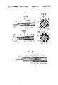
- FIG. 1(a) hereofa ring 2 inside of a sheath at the distal end of a flexible tube 1.
- a freely advancing and retracting slider 3is positioned inside of flexible tube 1, behind ring 2, and is fixed to the bent end portion 4 of wire loop 5.
- the opposite end of wire loop 5extends through passage 6 in slider 3 and axially through tube to the proximal end, not shown, where the wire is fastened to a handle, loop, or other device by which the wire and wire loop 5 can be manipulated.
- loop 5By manipulating wire loop 5 from the opposite, or proximal end of flexible tube 1, loop 5 can be advanced from the sheath end of tube 1, the bent portion 4 of wire loop 5 can be hooked around the polyp to be incised and tightened, in the body cavity, around such polyp.
- the polypBy applying high frequency current to the wire loop, the polyp is burnt out.
- carbidesform on the wire loop, stick to the wire and the loop is deformed.
- high mechanical resistance to the advance and retraction of wire loop 5 through passage 6 in slider 3is encountered.
- slider 3is pulled toward the proximal or handle end of tube 1 before loop 5 is completely closed, resulting in the loop remaining open as shown in FIG. 1(b).
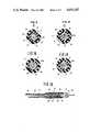
- a flexible C ring 7, FIG. 2(a)is mounted inside flexible sheath 1, a part of the sheath 1, as shown in FIG. 2(b), is formed to project inwardly as a convex portion, or, as shown in FIG. 2(c), a convex portion 10 is formed on lever 9 by extending the rear end side of the engagement ring 2.
- slider 5A, FIG. 1is pushed forward through C ring 7, however, the C ring is pushed forward before slider 3 goes through C ring 7. This, of course, is not desirable.
- the instant inventionprovides a high-frequency incising and excising instrument capable of reliably opening and closing the wire loop when operation of the loop to opening and closing is otherwise increased by the sticking of carbides.
- the instant inventionfurther provides a high-frequency incising and excising instrument capable of incision and excision with safety and certainty.
- the instant inventionprovides such an incision and excising instrument of simple construction.
- FIG. 1(a)is a longitudinal section view of a prior art instrument.
- FIG. 1(b)is a side view of the instrument of FIG. 1(a), showing the wire loop in a first position in retraction.
- FIG. 1(c)is a side view, similar to FIG. 1(b) but showing the wire loop in a further position in retraction.
- FIGS. 2(a), 2(b) and 2(c)are longitudinal section views of other prior art instruments.
- FIG. 3is a perspective view of one embodiment of the incising and excising instrument of the instant invention.
- FIG. 4is a longitudinal section view of the structure of the insertion portion, or distal end of the instrument of FIG. 3.
- FIG. 5is a longitudinal partial sectional view of the remote, or proximal end of the instrument of FIG. 3.
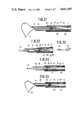
- FIG. 6(a)is a longitudinal sectional view of the insertion or distal portion of the instrument of FIGS. 3 and 4 with wire projected.
- FIG. 6(b)is an enlarged sectional view taken at A--A', FIG. 6(a).
- FIG. 7(a)is a longitudinal sectional view similar to FIG. 6(a) with the wire further projected.
- FIG. 7(b)is an enlarged sectional view taken a A--A' FIG. 7(a).
- FIG. 8(a)is a view similar to FIG. 6(a) but of second embodiment of the invention.
- FIG. 8(b)is an enlarged sectional view taken at A--A' FIG. 8(a).
- FIG. 9(a)is a view similar to FIG. 8(a) with the wire further projected.
- FIG. 9(b)is an enlarged sectional view taken at A--A' FIG. 9(a).
- FIG. 10(a)is a view similar to FIGS. 6(a) and 8(a) but showing a third embodiment of the invention.
- FIG. 10(b)is an enlarged sectional view taken at A--A' FIG. 10(a).
- FIG. 11(a)is a view similar to FIG. 10(a) with the wire further projected.
- FIG. 11(b)is an enlarged sectional view taken at A--A' FIG. 11(a).
- FIG. 12is a view similar to FIGS. 6(a), 8(a) and 10(a) but showing a fourth embodiment of the invention.
- FIG. 13(a)is an enlarged sectional view taken at A--A' FIG. 12.
- FIG. 13(b)is an enlarged sectional view taken at B--B' FIG. 12.
- FIG. 14is an enlarged sectional view of the stopper of the fifth embodiment of the invention.
- FIG. 15is an enlarged sectional view of the stopper of the sixth embodiment of the invention.
- FIG. 16is a view similar to FIG. 4 but showing the insertion portion of distal end of the seventh embodiment of the invention.
- FIG. 17is a view similar to FIGS. 6(A), 8(a), 10(A) and 12 but showing the eighth embodiment of the invention.
- FIG. 18is a front enlarged view of the reverse stop member of FIG. 17.
- FIG. 19is a view similar to FIG. 17 but showing the wire projected with the wire loop formed.
- FIG. 20is a view similar to FIGS. 6(a), 8(a), 10(a), 12 and 17 but showing the ninth embodiment of the invention.
- FIG. 21is a view similar to FIG. 20 with the wire projected and forming a loop.
- FIG. 22is a view similar to FIGS. 6(a), 8(a), 10(a), 12, 17 and 20 but showing the tenth embodiment of the invention.
- FIG. 23is a perspective view showing the reverse stop member of the embodiment of FIG. 22 enlarged.
- FIG. 24is a view similar to FIGS. 6(a), 8(a), 10(a), 12, 17, 20 and 22 but showing the eleventh embodiment of the invention.
- FIG. 25is a view similar to FIG. 24 with the wire projected and forming a loop.
- the high-frequency incising and excising instrument of the first embodiment of the instant inventionincludes a slender and flexible insertion, or distal end, portion, generally designated 12, for insertion through the channel of an endoscope, an operating portion, or proximal end, generally designated 13, to which the distal end portion 12 can be freely attached and detached, and a notched portion 14 to be projected from the forward, or distal end of the insertion portion 12.
- Flexible, tubular sheath 15, FIG. 4,is positioned in insertion portion 12.
- the front or forward end of sheath 15is obliquely notched and open.
- Wire loop 16, for excisionis positioned in sheath 15 for projection from and retraction into sheath 15.
- sheath 15is fitted and attached to one end of cap 17 and extends forwardly from cap 17 forming an outer circular sleeve spaced outwardly from sheath 15 to form a protective tube 18 around sheath 15 for preventing breakage and buckling of the sheath.
- Notched portion 14includes a wire loop 16, having a bending portion 19, for retraction into and projection out of sheath 15. At one of its ends, wire loop 16 is secured to slider 21 which is freely extendable and retractable in sheath 15 for advancing and retracting wire loop 16. Annular stopper 22 is secured to the other end of wire loop 16 beyond bending portion 19. At the rear side of stopper 22, guide groove 23, as best shown in FIG. 6(b), is formed on slider 21. Operating pipe 24 is fixed to the end of wire loop. A fixing tool 25 is formed at the rear end of operating pipe 24.
- Slider 21has an outer diameter smaller than the inner diameter of sheath 15 and, thus, can be advanced and retracted freely in sheath 15.
- the outer diameter of slider 21is greater than the inner diameter of the front engaging ring 26, secured to the inner wall of sheath 15.
- Slider 21is not movable forward from the fixed position of engaging ring 26 and the front engaging ring 26 forms a first engagement portion for controlling the forward movement of slider 21.
- the outer diameter of slider 21is smaller than the inner diameter of the rear engagement ring 27 secured to the inner wall of sheath 15.
- Operating portion 13, FIG. 5,includes an operation body 29, fixed to the rear end portion of the hand side of sheath 15 by a fixing member 28, and an operation slider 31 slidably fixed to the operation body 29 and forms a fixing core securing portion 30, making the rear end of loop wire 16 freely detachable and attachable with fixing core 25.
- the fixing tool securing portion 30, FIG. 5,includes button 32 whose end portion projects upward from a hole formed in the center of operation slider 31, a frame 33 for containing the base side of button 32, and spring 34 which is contained in frame 33 and fuctions to project button 32 upwards. At the front side of frame 33 there is formed a hole 35 containing fixing core 25. On the base side of button 32, hole 36 communicates with hole 35 of frame 33.
- Loop wire 16 to be connected to operating portion 13is with high-frequency electric current from a high-frequency generating device via a cable, not shown, through fixing tool securing portion 30 and fixing core 25.
- Ring-shaped finger rest 37is formed at the rear or proximal end of operating body 29. Ring-shaped finger rests 38, 38, FIG. 3, are provided on operation slider 31. Operation slider 31 is moved back and forth, relative to operation body 29, by manipulating the finger tips on the finger rests 37, 38 and 38.
- Operation slider 31is pressed forward to project loop wire 16 from the end of sheath 15.
- Slider 21passes through rear engagement ring 27 and contacts front engagement ring 26.
- Operating pipe 24, in contact with the side of guide groove 23 of slider 21,is pressed forward and loop wire 16, projecting from the front of sheath 15, on its side connected to operating pipe 24, is bent at the bending portion 19, FIG. 7(a), and forms a crescent shaped loop of a size sufficient for excision.
- Slider 21is displaced longitudinally to engage front engaging ring 26 and is then displaced laterally by operating pipe 24 to be pressed against the inner wall of sheath 15.
- button 32 of the fixing tool securing portion 30, formed at operation slider 31, FIG. 5,is pressed to insert fixing core 25 at the rear end of operating pipe 24.
- Fixing core 25is secured by then releasing button 32.
- Cap 17, at the rear of sheath 15, FIG. 4is secured to the front end of operation body 29 by fixing member 28 and the instrument of the first embodiment is, thus, assembled.
- the distal end of sheath 15is inserted into the body cavity through the channel of the endoscope.
- the finger tips of the operatorare placed in the finger rests 37, 38 and 38 of the operation body 29 and the operation slider 31.
- Slider 21 and loop wire 16are advance in sheath 15.
- bending portion 19 of loop wire 16is projected from the distal end of sheath 15.
- the front end of slider 21contacts the rear side of front engagement ring 26 and slider 21 is stopped.
- the cresent shaped loop in loop wire 16is formed and enlarges at the bending portion 19, FIG. 7(a).
- Operation pipe 24passes through guide groove 23 and slider 21 is pressed toward the inner wall of sheath 15 between front engagement ring 26 and rear engagement ring 27.
- the crescent shaped loop of projecting loop wire 16is then hooked around the polyp, or the like, to be excised.
- slider 31With loop wire 16 around the polyp, slider 31 is receded, or pulled back, by operating of finger rests 38, 38 of operation slider 31 toward finger rest 37 of operation body 29. Operating pipe 24 slides back through guide groove 23 and is pulled to the rear of sheath 15. Wire loop 16, at its bent portion 19 of greater bend, is decreased in loop size and is drawn back into sheath 15. Even if mechanical operation resistance is encountered because of carbides deposited on loop wire 16 during prior exercising, slider 21 is pressed to the inner wall surface of sheath 15 until operation pipe 24 is withdrawn through guide groove 23. Thus, the connection of slider 21 at rear engagement ring 27 is maintained until operation pipe 24 clears guide groove 23 and loop wire 16 tightly binds the neck of the polyp, or the like, in the loop of loop wire 16.
- guide groove 23as a guide portion in the slider 21, which freely advances and retracts in the sheath 15 and forms an engagement means for preventing the rearward movement when pressed by the operation pipe 24 when the operation pipe 24 is in insertion contact with the guide groove 23 and, with the rear engagement ring 27, prevents the free end of the loop wire 16 from retracting until the loop in loop wire 16 is contracted.
- FIGS. 8(a), 8(b), 9(a) and 9(b)the guide groove 23 of the first embodiment, FIGS. 7(a) and 7(b), is replaced with a guide hole 41.
- guide hole 41is formed in an eccentric manner in front engagement ring 26.
- FIGS. 10(a), 10(b), 11(a) and 11(b)a short crescent shaped slider 21 is employed.
- a cylinder having an outer diameter slightly smaller than the inner diameter of rear ring 27is notched in a longitudinal direction to make the ring largely half-cylindrical.
- the half-cylinder 21has a maximum length greater than the front engagement ring 26 to prevent half-cylinder 21 from moving forward past engagement ring 26.
- the operating pipe 24is made to be inserted through the notched portion 42 as a guide portion.
- the half-cylindrical slider 21is displaced in a longitudinal direction and pressed to the inner wall of sheath 15, thus giving a configuration wherein the slider 21 is pressed and displaced in a longitudinal direction relative to rear engagement ring 27, as shown in FIG. 11.
- the remainder of the instrument and the operation thereofare the same as in the first and second embodiments.
- the front engagement ring 26 and the rear engagement ring 27are formed inside the protective tube 18. It is to be understood, however, that the invention is not limited to such particular location. Such front and rear engagement rings may be positioned near the end of sheath 15 or formed further toward the rear end of the sheath.
- the projection for engagement by the front engagement ring 26 or rear engagement ring 27is not limited to being fixed inside sheath 15.
- a projection having substantially the same functioncan be formed by attaching a ring, or the like, around the outer circle of sheath 15 and inwardly projecting the inner circle of sheath 15 of the ring attached. Such configuration would be simpler than that of fixing the front engagement ring 26 or rear engagement ring 27 to the inside of sheath 15.
- slider 21may be formed of an elastic material, such as rubber, the guide groove 23 or guide hole 41 may be made of a smaller shape than the outer diameter of operating pipe 24 and the slider 21, upon insertion, can be displaced so as to be expanded in a longitudinal direction to be engaged with the projection of the rear engagement ring 27.
- the present inventioncan be applied not only to excision but, also, to incision.
- the use of the inventionis not limited to be inserted through the channel of a soft of a hard endoscope and sheath 15 is not limited to be formed as a flexible tube but also may be applied to a hard tube.
- stopper 52differs from stopper 22 in the third embodiment.
- stopper 52is mounted in sheath 15 on the wire of wire loop 16 in front of operation pipe 24 and is fixed to the wire with an adhesive. Loop wire 16 bent, at bending portion 19, passes freely through guide port 52a in stopper 52. Slider 21 is fixed to the otherwise free end of loop wire 16 behind stopper 52. Stopper 52 has an outer diameter smaller than the inner diameter of front engagement ring 26 and, thus, passes easily therethrough. The stopper 52 also forms a guide port 52A through which the side of loop wire 16 fixed to the slider 21 can easily pass. The bending portion 19 of loop wire 16 retreats rearwardly and inwardly after excision and operating pipe 24 is retracted from the end of sheath 15 without failure.
- slider 21is pressed against the inner wall surface of the sheath 15 by operating pipe 24 and is in engagement with the rear engagement ring 27.
- loop wire 16 on the slider 21 side inserted through guide port 52A of stopper 52is maintained at a prescribed interval with respect to the other side of loop wire 16 fixed to stopper 52.
- the guide port 52Afunctions to return loop wire 16, secured to the slider 21, to the central side and the engagement state with the rear engagement ring 27 is assuredly released after excision.
- slider 21, together with loop wire 16moves rearward to cause the bending portion 19 to withdraw into the sheath 15.
- FIG. 14there is formed, as the wire inserting guide portion of the stopper 52, an upper portion 52B, for receiving and passing the side of loop wire 16 secured to slider 21.
- the working effects of the fifth embodimentare the same as those described with respect to the fourth embodiment.
- stopper 52has a notched portion 52C which acts as a guide for the side of loop wire 16 secured to slider 21.
- the constructionis similar to that of the first embodiment shown in FIG. 4.
- the front engagement ring 26 and the rear engagement ring 27are formed integrally with cap 17.
- the engagement ringscan be used for a prolonged period of time without change of positions despite repeated use.
- this embodimenthas the same construction and operates in the same manner as the first embodiment.
- the crescent loop, projected from sheath 15 in the eighth embodiment of the invention,can be hooked on a phyma like polyp, of the like, when slider 31 is retracted, the operating pipe 24 is withdrawn to the rear of sheath 15 with its side portion is in contact with reverse stop portion 57.
- the side of loop wire 16, greatly bent at the bending portion 19has a low bending degree to close the loop. Even if operation resistance, due to carbides produced in prior excision have increased and the wire loop is sticky, the slider 21 is maintained in an engagement state until the operation pipe 24 moves more rearward than the rear end of reverse stop portion 57 and portion 57 is released from pressing the inner wall of sheath 15, since the rearward movement of reverse stop portion 57 is stopped by engagement portion 59.
- slider 21retracts in an engagement state of contacting stopper 22.
- the bending portion 19 of loop wire 16is withdrawn into sheath 15 in a state where the end projected from sheath 15 of loop wire 16 is the bending portion 19, completing the incision of the polyp in the body cavity.
- the reverse stop portion 57 in slider 21freely advances and retracts inside sheath 15.
- An engagement meansis formed by which the reverse stop portion 57 is connected to engagement portion 59 by displacing the reverse stop portion in a longitudinal direction when the operating pipe 24 is inserted while contacting the reverse stop member 57.
- binding body 61is secured to loop wire 16 adjacent to port 62 for allowing reverse stop portion 57 to pass therethrough.
- the reverse stop portion 57is maintained integrally with loop wire 16 by the binding body 61. Thus, the deformation of reverse stop portion 57 is prevented.
- the rear end of reverse stop memberis not in the bent shape, as shown in the embodiment of FIG. 18, but is a tip 63 in the shape shown in FIG. 23 and is securely hooked by the engagement portion 59 at the front end of cap 17.
- engagement ring 64is secured to the inner wall of sheath 15. Reverse stop portion 57, when deflected downward by contact with operating pipe 24 abuts ring 64, as best shown in FIG. 25. Except for such contact, the operation of this embodiment is substantially the same as in the eighth embodiment described above.
Landscapes
- Health & Medical Sciences (AREA)
- Surgery (AREA)
- Engineering & Computer Science (AREA)
- Life Sciences & Earth Sciences (AREA)
- Biomedical Technology (AREA)
- Otolaryngology (AREA)
- Nuclear Medicine, Radiotherapy & Molecular Imaging (AREA)
- Plasma & Fusion (AREA)
- Physics & Mathematics (AREA)
- Heart & Thoracic Surgery (AREA)
- Medical Informatics (AREA)
- Molecular Biology (AREA)
- Animal Behavior & Ethology (AREA)
- General Health & Medical Sciences (AREA)
- Public Health (AREA)
- Veterinary Medicine (AREA)
- Surgical Instruments (AREA)
Abstract
Description
Claims (7)
Applications Claiming Priority (5)
| Application Number | Priority Date | Filing Date | Title |
|---|---|---|---|
| JP58095530AJPS59222146A (en) | 1983-05-30 | 1983-05-30 | High frequency incision and cutting tool |
| JP58-95530 | 1983-05-30 | ||
| JP58-108851[U]JPX | 1983-07-12 | ||
| JP10885183UJPS6015307U (en) | 1983-07-12 | 1983-07-12 | High frequency cutting tool |
| JP58133074AJPS6024834A (en) | 1983-07-20 | 1983-07-20 | High frequency incision and cutting tool |
Publications (1)
| Publication Number | Publication Date |
|---|---|
| US4643187Atrue US4643187A (en) | 1987-02-17 |
Family
ID=27307835
Family Applications (1)
| Application Number | Title | Priority Date | Filing Date |
|---|---|---|---|
| US06/615,419Expired - Fee RelatedUS4643187A (en) | 1983-05-30 | 1984-05-30 | High-frequency incising and excising instrument |
Country Status (2)
| Country | Link |
|---|---|
| US (1) | US4643187A (en) |
| DE (1) | DE3419962A1 (en) |
Cited By (54)
| Publication number | Priority date | Publication date | Assignee | Title |
|---|---|---|---|---|
| US4776336A (en)* | 1986-10-30 | 1988-10-11 | Olympus Optical Co., Ltd. | Resectoscope |
| US4869716A (en)* | 1988-10-24 | 1989-09-26 | Smirmaul Heinz J | Surgical instrument and method for cutting the lens of an eye |
| US4869248A (en)* | 1987-04-17 | 1989-09-26 | Narula Onkar S | Method and apparatus for localized thermal ablation |
| US4920978A (en)* | 1988-08-31 | 1990-05-01 | Triangle Research And Development Corporation | Method and apparatus for the endoscopic treatment of deep tumors using RF hyperthermia |
| US5190554A (en)* | 1992-04-08 | 1993-03-02 | Eastern Virginia Medical School | Appendix extractor |
| US5201740A (en)* | 1991-11-05 | 1993-04-13 | Nakao Naomi L | Surgical retrieval assembly and related method |
| US5336227A (en)* | 1991-11-05 | 1994-08-09 | Wilk & Nakao Medical Technology Incorporated | Surgical cauterization snare with polyp capturing web net |
| US5374273A (en)* | 1992-10-05 | 1994-12-20 | Nakao; Naomi L. | Method for retrieval of retained common bile duct stones |
| US5486182A (en)* | 1991-11-05 | 1996-01-23 | Wilk & Nakao Medical Technology Inc. | Polyp retrieval assembly with separable web member |
| US5535759A (en)* | 1994-11-02 | 1996-07-16 | Wilk; Peter J. | Endoscopic method of cleaning and operating on a site within a patient |
| US5738683A (en)* | 1994-07-16 | 1998-04-14 | Osypka; Peter | Mapping and ablation catheter |
| US5759187A (en)* | 1991-11-05 | 1998-06-02 | Wilk & Nakao Medical Technology, Incorporated | Surgical retrieval assembly and associated method |
| US5810807A (en)* | 1996-05-22 | 1998-09-22 | Ganz; Robert A. | Sphincterotome with deflectable cutting plane and method of using the same |
| US6093185A (en)* | 1998-03-05 | 2000-07-25 | Scimed Life Systems, Inc. | Expandable PMR device and method |
| US6136014A (en)* | 1998-09-01 | 2000-10-24 | Vivant Medical, Inc. | Percutaneous tissue removal device |
| US6224611B1 (en) | 1998-09-14 | 2001-05-01 | Asahi Kogaku Kogyo Kabushiki Kaisha | Snare for endoscope |
| US6245078B1 (en) | 1999-04-26 | 2001-06-12 | Asahi Kogaku Kogyo Kabushiki Kaisha | Snare for endoscope |
| US20020049467A1 (en)* | 1997-11-07 | 2002-04-25 | Paul Gilson | Embolic protection system |
| US20020107541A1 (en)* | 1999-05-07 | 2002-08-08 | Salviac Limited. | Filter element for embolic protection device |
| US6471709B1 (en) | 1998-10-30 | 2002-10-29 | Vivant Medical, Inc. | Expandable ring percutaneous tissue removal device |
| US20030009189A1 (en)* | 1997-11-07 | 2003-01-09 | Salviac Limited | Embolic protection device |
| US20030032977A1 (en)* | 1997-11-07 | 2003-02-13 | Salviac Limited | Filter element with retractable guidewire tip |
| US20030130684A1 (en)* | 2001-12-21 | 2003-07-10 | Eamon Brady | Support frame for an embolic protection device |
| US20030144688A1 (en)* | 1999-05-07 | 2003-07-31 | Salviac Limited | Support frame for an embolic protection device |
| US20030144687A1 (en)* | 1999-05-07 | 2003-07-31 | Salviac Limited | Support frame for an embolic protection device |
| US6638234B2 (en) | 1998-03-03 | 2003-10-28 | Senorx, Inc. | Sentinel node location and biopsy |
| US20030212429A1 (en)* | 2002-03-05 | 2003-11-13 | Martin Keegan | Embolic protection system |
| US6679851B2 (en) | 1998-09-01 | 2004-01-20 | Senorx, Inc. | Tissue accessing and anchoring device and method |
| US20040215208A1 (en)* | 2002-12-02 | 2004-10-28 | Foushee Jason D. | Loop tip wire guide |
| US20050065453A1 (en)* | 2003-02-24 | 2005-03-24 | Senorx, Inc. | Biopsy device with selectable tissue receiving aperture orientation and site illumination |
| US20050159677A1 (en)* | 2003-12-23 | 2005-07-21 | Shabaz Martin V. | Biopsy device with aperture orientation and improved tip |
| US6955676B2 (en) | 1999-06-22 | 2005-10-18 | Senorx, Inc. | Shaped scalpel |
| US20070038146A1 (en)* | 2005-08-05 | 2007-02-15 | Quick Richard L | Biopsy device with fluid delivery to tissue specimens |
| US20070282328A1 (en)* | 2006-06-05 | 2007-12-06 | Naohisa Yahagi | High-frequency treatment instrument |
| US20080058672A1 (en)* | 2004-12-16 | 2008-03-06 | Senorx, Inc. | Biopsy device with aperture orientation and improved tip |
| US20080167677A1 (en)* | 1999-05-07 | 2008-07-10 | Salviac Limited | Filter element for embolic protection device |
| US20080281323A1 (en)* | 1999-01-27 | 2008-11-13 | Burbank Fred H | Tissue specimen isolating and damaging device and method |
| US20080319468A1 (en)* | 2003-02-24 | 2008-12-25 | Senorx, Inc. | Biopsy device with selectable tissue receiving aperature orientation and site illumination |
| US20090043317A1 (en)* | 2007-08-08 | 2009-02-12 | Cavanaugh Brian J | Method and apparatus for delivery of a ligating suture |
| US20090112118A1 (en)* | 2005-08-05 | 2009-04-30 | Senorx, Inc. | Biopsy device with fluid delivery to tissue specimens |
| US20090204021A1 (en)* | 2004-12-16 | 2009-08-13 | Senorx, Inc. | Apparatus and method for accessing a body site |
| US20120239073A1 (en)* | 2007-12-20 | 2012-09-20 | Ureca B.V. | Catheter assembly |
| US8641640B2 (en) | 2005-05-23 | 2014-02-04 | Senorx, Inc. | Tissue cutting member for a biopsy device |
| US20140243822A1 (en)* | 2011-11-03 | 2014-08-28 | Gunter Farin | RF Surgical Resection Instrument Having a Resection Loop for Removal of Pathological Tissue |
| US9216012B2 (en) | 1998-09-01 | 2015-12-22 | Senorx, Inc | Methods and apparatus for securing medical instruments to desired locations in a patient's body |
| US20160175042A1 (en)* | 2014-12-23 | 2016-06-23 | Cook Medical Technologies Llc | Variable thickness electrosurgical snare |
| US20170215911A1 (en)* | 2016-02-03 | 2017-08-03 | Osaka University | Endoscopic Snare |
| EP3804776A1 (en)* | 2016-01-30 | 2021-04-14 | Carl Zeiss Meditec Cataract Technology Inc. | Devices and methods for ocular surgery |
| US11278450B2 (en) | 2017-05-04 | 2022-03-22 | Carl Zeiss Meditec Cataract Technology Inc. | Devices and methods for ocular surgery |
| US11369407B2 (en) | 2017-07-05 | 2022-06-28 | Olympus Corporation | Method of operating a treatment tool |
| US11638660B2 (en) | 2018-06-05 | 2023-05-02 | Carl Zeiss Meditec Cataract Technology Inc. | Ophthalmic microsurgical tools, systems, and methods of use |
| US11730625B2 (en) | 2019-05-17 | 2023-08-22 | Carl Zeiss Meditec Cataract Technology Inc. | Ophthalmic cutting instruments having integrated aspiration pump |
| US11801163B2 (en) | 2019-06-07 | 2023-10-31 | Carl Zeiss Meditec Cataract Technology Inc. | Multi-stage trigger for ophthalmology cutting tool |
| US12285361B2 (en) | 2019-02-01 | 2025-04-29 | Carl Zeiss Meditec Cataract Technology Inc. | Ophthalmic cutting instruments having integrated aspiration pump |
Families Citing this family (4)
| Publication number | Priority date | Publication date | Assignee | Title |
|---|---|---|---|---|
| US4718419A (en)* | 1985-08-05 | 1988-01-12 | Olympus Optical Co., Ltd. | Snare assembly for endoscope |
| JPS62211060A (en)* | 1986-03-12 | 1987-09-17 | オリンパス光学工業株式会社 | High frequency treatment tool |
| DE4036570A1 (en)* | 1990-11-16 | 1992-05-21 | Osypka Peter | CATHETER FOR REDUCING OR REMOVING CONSTRUCTIONS IN VESSELS |
| DE102012015834A1 (en)* | 2012-08-12 | 2014-02-13 | medwork GmbH | Device for endoscopic resection in the upper or lower gastrointestinal tract |
Citations (3)
| Publication number | Priority date | Publication date | Assignee | Title |
|---|---|---|---|---|
| JPS5150377A (en)* | 1974-10-29 | 1976-05-01 | Kanegafuchi Chemical Ind | |
| US4181131A (en)* | 1977-02-28 | 1980-01-01 | Olympus Optical Co., Ltd. | High frequency electrosurgical instrument for cutting human body cavity structures |
| JPS56104506A (en)* | 1980-01-25 | 1981-08-20 | Pioneer Electronic Corp | Amplifier |
Family Cites Families (1)
| Publication number | Priority date | Publication date | Assignee | Title |
|---|---|---|---|---|
| JPH05150377A (en)* | 1991-11-29 | 1993-06-18 | Canon Inc | Lighting equipment |
- 1984
- 1984-05-29DEDE19843419962patent/DE3419962A1/enactiveGranted
- 1984-05-30USUS06/615,419patent/US4643187A/ennot_activeExpired - Fee Related
Patent Citations (3)
| Publication number | Priority date | Publication date | Assignee | Title |
|---|---|---|---|---|
| JPS5150377A (en)* | 1974-10-29 | 1976-05-01 | Kanegafuchi Chemical Ind | |
| US4181131A (en)* | 1977-02-28 | 1980-01-01 | Olympus Optical Co., Ltd. | High frequency electrosurgical instrument for cutting human body cavity structures |
| JPS56104506A (en)* | 1980-01-25 | 1981-08-20 | Pioneer Electronic Corp | Amplifier |
Cited By (145)
| Publication number | Priority date | Publication date | Assignee | Title |
|---|---|---|---|---|
| US4776336A (en)* | 1986-10-30 | 1988-10-11 | Olympus Optical Co., Ltd. | Resectoscope |
| US4869248A (en)* | 1987-04-17 | 1989-09-26 | Narula Onkar S | Method and apparatus for localized thermal ablation |
| US4920978A (en)* | 1988-08-31 | 1990-05-01 | Triangle Research And Development Corporation | Method and apparatus for the endoscopic treatment of deep tumors using RF hyperthermia |
| US4869716A (en)* | 1988-10-24 | 1989-09-26 | Smirmaul Heinz J | Surgical instrument and method for cutting the lens of an eye |
| US5336227A (en)* | 1991-11-05 | 1994-08-09 | Wilk & Nakao Medical Technology Incorporated | Surgical cauterization snare with polyp capturing web net |
| US5201740A (en)* | 1991-11-05 | 1993-04-13 | Nakao Naomi L | Surgical retrieval assembly and related method |
| US5486182A (en)* | 1991-11-05 | 1996-01-23 | Wilk & Nakao Medical Technology Inc. | Polyp retrieval assembly with separable web member |
| US5759187A (en)* | 1991-11-05 | 1998-06-02 | Wilk & Nakao Medical Technology, Incorporated | Surgical retrieval assembly and associated method |
| US5190554A (en)* | 1992-04-08 | 1993-03-02 | Eastern Virginia Medical School | Appendix extractor |
| US5374273A (en)* | 1992-10-05 | 1994-12-20 | Nakao; Naomi L. | Method for retrieval of retained common bile duct stones |
| US5738683A (en)* | 1994-07-16 | 1998-04-14 | Osypka; Peter | Mapping and ablation catheter |
| US5535759A (en)* | 1994-11-02 | 1996-07-16 | Wilk; Peter J. | Endoscopic method of cleaning and operating on a site within a patient |
| US5810807A (en)* | 1996-05-22 | 1998-09-22 | Ganz; Robert A. | Sphincterotome with deflectable cutting plane and method of using the same |
| US20050283184A1 (en)* | 1997-11-07 | 2005-12-22 | Salviac Limited | Embolic protection device |
| US20060293704A1 (en)* | 1997-11-07 | 2006-12-28 | Salviac Limited | Embolic protection device |
| US7780697B2 (en) | 1997-11-07 | 2010-08-24 | Salviac Limited | Embolic protection system |
| US7833242B2 (en) | 1997-11-07 | 2010-11-16 | Salviac Limited | Embolic protection device |
| US20020049467A1 (en)* | 1997-11-07 | 2002-04-25 | Paul Gilson | Embolic protection system |
| US7846176B2 (en) | 1997-11-07 | 2010-12-07 | Salviac Limited | Embolic protection system |
| US20090143814A1 (en)* | 1997-11-07 | 2009-06-04 | Salviac Limited | Embolic protection device |
| US20110054516A1 (en)* | 1997-11-07 | 2011-03-03 | Salviac Limited | Embolic protection method |
| US20030009189A1 (en)* | 1997-11-07 | 2003-01-09 | Salviac Limited | Embolic protection device |
| US20030032977A1 (en)* | 1997-11-07 | 2003-02-13 | Salviac Limited | Filter element with retractable guidewire tip |
| US20090099593A1 (en)* | 1997-11-07 | 2009-04-16 | Salviac Limited | Embolic protection device |
| US7510565B2 (en) | 1997-11-07 | 2009-03-31 | Salviac Limited | Embolic protection device |
| US7901426B2 (en) | 1997-11-07 | 2011-03-08 | Salviac Limited | Embolic protection device |
| US20030187474A1 (en)* | 1997-11-07 | 2003-10-02 | Martin Keegan | Embolic protection system |
| US20110125182A1 (en)* | 1997-11-07 | 2011-05-26 | Salviac Limited | Filter element with retractable guidewire tip |
| US20080188884A1 (en)* | 1997-11-07 | 2008-08-07 | Salviac Limited | Embolic protection device |
| US8123776B2 (en) | 1997-11-07 | 2012-02-28 | Salviac Limited | Embolic protection system |
| US20040034385A1 (en)* | 1997-11-07 | 2004-02-19 | Paul Gilson | Embolic protection device |
| US20040039411A1 (en)* | 1997-11-07 | 2004-02-26 | Paul Gilson | Embolic protection device |
| US20070282369A1 (en)* | 1997-11-07 | 2007-12-06 | Salviac Limited | Embolic protection device |
| US20040073198A1 (en)* | 1997-11-07 | 2004-04-15 | Salviac Limited | Embolic protection device |
| US20040127934A1 (en)* | 1997-11-07 | 2004-07-01 | Salviac Limited | Embolic protection system |
| US20070250107A1 (en)* | 1997-11-07 | 2007-10-25 | Salviac Limited | Embolic protection system |
| US20070244505A1 (en)* | 1997-11-07 | 2007-10-18 | Abbott Laboratories | Embolic protection device |
| US20070239200A1 (en)* | 1997-11-07 | 2007-10-11 | Abbott Laboratories | Embolic protection device |
| US20070233181A1 (en)* | 1997-11-07 | 2007-10-04 | Abbott Laboratories | Embolic protection device |
| US20050228437A1 (en)* | 1997-11-07 | 2005-10-13 | Salviac Limited | Embolic protection system |
| US20070173884A1 (en)* | 1997-11-07 | 2007-07-26 | Salviac Limited | Embolic protection device |
| US20050234502A1 (en)* | 1997-11-07 | 2005-10-20 | Paul Gilson | Embolic protection system |
| US20070173883A1 (en)* | 1997-11-07 | 2007-07-26 | Martin Keegan | Embolic protection system |
| US20060004403A1 (en)* | 1997-11-07 | 2006-01-05 | Salviac Limited | Embolic protection system |
| US20060074446A1 (en)* | 1997-11-07 | 2006-04-06 | Paul Gilson | Embolic protection system |
| US20060089663A1 (en)* | 1997-11-07 | 2006-04-27 | Salviac Limited | Embolic protection device |
| US20070162070A1 (en)* | 1997-11-07 | 2007-07-12 | Salviac Limited | Embolic protection device |
| US20070162069A1 (en)* | 1997-11-07 | 2007-07-12 | Salviac Limited | Embolic protection device |
| US20060129182A1 (en)* | 1997-11-07 | 2006-06-15 | Salviac Limited | Embolic protection device |
| US20060259069A1 (en)* | 1997-11-07 | 2006-11-16 | Salviac Limited | Embolic protection device |
| US20070123931A1 (en)* | 1997-11-07 | 2007-05-31 | Salviac Limited | Embolic protection system |
| US20070106322A1 (en)* | 1997-11-07 | 2007-05-10 | Salviac Limited | Embolic protection device |
| US20070005096A1 (en)* | 1997-11-07 | 2007-01-04 | Salviac Limited | Embolic protection system |
| US6638234B2 (en) | 1998-03-03 | 2003-10-28 | Senorx, Inc. | Sentinel node location and biopsy |
| US6716179B2 (en) | 1998-03-03 | 2004-04-06 | Senorx, Inc. | Sentinel node location and biopsy |
| US6093185A (en)* | 1998-03-05 | 2000-07-25 | Scimed Life Systems, Inc. | Expandable PMR device and method |
| US6402740B1 (en) | 1998-03-05 | 2002-06-11 | Scimed Systems, Inc. | Expandable PMR device and method |
| US6679851B2 (en) | 1998-09-01 | 2004-01-20 | Senorx, Inc. | Tissue accessing and anchoring device and method |
| US6136014A (en)* | 1998-09-01 | 2000-10-24 | Vivant Medical, Inc. | Percutaneous tissue removal device |
| US20050197594A1 (en)* | 1998-09-01 | 2005-09-08 | Senorx, Inc. | Tissue accessing and anchoring device and method |
| US9216012B2 (en) | 1998-09-01 | 2015-12-22 | Senorx, Inc | Methods and apparatus for securing medical instruments to desired locations in a patient's body |
| US7282034B2 (en) | 1998-09-01 | 2007-10-16 | Senorx, Inc. | Tissue accessing and anchoring device and method |
| US6224611B1 (en) | 1998-09-14 | 2001-05-01 | Asahi Kogaku Kogyo Kabushiki Kaisha | Snare for endoscope |
| US6471709B1 (en) | 1998-10-30 | 2002-10-29 | Vivant Medical, Inc. | Expandable ring percutaneous tissue removal device |
| US20080281323A1 (en)* | 1999-01-27 | 2008-11-13 | Burbank Fred H | Tissue specimen isolating and damaging device and method |
| US9510809B2 (en) | 1999-01-27 | 2016-12-06 | Senorx, Inc. | Tissue specimen isolating and damaging device and method |
| US8636734B2 (en) | 1999-01-27 | 2014-01-28 | Senorx, Inc. | Tissue specimen isolating and damaging device and method |
| US6245078B1 (en) | 1999-04-26 | 2001-06-12 | Asahi Kogaku Kogyo Kabushiki Kaisha | Snare for endoscope |
| US20020107541A1 (en)* | 1999-05-07 | 2002-08-08 | Salviac Limited. | Filter element for embolic protection device |
| US8002790B2 (en) | 1999-05-07 | 2011-08-23 | Salviac Limited | Support frame for an embolic protection device |
| US20080167677A1 (en)* | 1999-05-07 | 2008-07-10 | Salviac Limited | Filter element for embolic protection device |
| US20060122645A1 (en)* | 1999-05-07 | 2006-06-08 | Salviac Limited | Support frame for an embolic protection device |
| US20060122644A1 (en)* | 1999-05-07 | 2006-06-08 | Salviac Limited | Support frame for an embolic protection device |
| US20030144687A1 (en)* | 1999-05-07 | 2003-07-31 | Salviac Limited | Support frame for an embolic protection device |
| US7491215B2 (en) | 1999-05-07 | 2009-02-17 | Salviac Limited | Filter element for embolic protection device |
| US20030144688A1 (en)* | 1999-05-07 | 2003-07-31 | Salviac Limited | Support frame for an embolic protection device |
| US20090149881A1 (en)* | 1999-05-07 | 2009-06-11 | Salviac Limited | Filter element for embolic protection device |
| US7799051B2 (en) | 1999-05-07 | 2010-09-21 | Salviac Limited | Support frame for an embolic protection device |
| US7572256B2 (en) | 1999-06-22 | 2009-08-11 | Senorx, Inc. | Shaped scalpel |
| US6955676B2 (en) | 1999-06-22 | 2005-10-18 | Senorx, Inc. | Shaped scalpel |
| US20030130684A1 (en)* | 2001-12-21 | 2003-07-10 | Eamon Brady | Support frame for an embolic protection device |
| US20030212429A1 (en)* | 2002-03-05 | 2003-11-13 | Martin Keegan | Embolic protection system |
| US20070060946A1 (en)* | 2002-03-05 | 2007-03-15 | Salviac Limited | Embolic protection system |
| US7144408B2 (en) | 2002-03-05 | 2006-12-05 | Salviac Limited | Embolic protection system |
| US20070244504A1 (en)* | 2002-03-05 | 2007-10-18 | Salviac Limited | Embolic protection system |
| US7520881B2 (en)* | 2002-12-02 | 2009-04-21 | Wilson-Cook Medical Incorporated | Loop tip wire guide |
| US20040215208A1 (en)* | 2002-12-02 | 2004-10-28 | Foushee Jason D. | Loop tip wire guide |
| US20050065453A1 (en)* | 2003-02-24 | 2005-03-24 | Senorx, Inc. | Biopsy device with selectable tissue receiving aperture orientation and site illumination |
| US20080319468A1 (en)* | 2003-02-24 | 2008-12-25 | Senorx, Inc. | Biopsy device with selectable tissue receiving aperature orientation and site illumination |
| US7819819B2 (en) | 2003-02-24 | 2010-10-26 | Senorx, Inc. | Biopsy device with inner cutting member |
| US11589849B2 (en) | 2003-02-24 | 2023-02-28 | Senorx, Inc. | Biopsy device with selectable tissue receiving aperature orientation and site illumination |
| US11534147B2 (en) | 2003-02-24 | 2022-12-27 | Senorx, Inc. | Biopsy device with a removable sample recieving cartridge |
| US10335127B2 (en) | 2003-02-24 | 2019-07-02 | Senorx, Inc. | Biopsy device with selectable tissue receiving aperature orientation and site illumination |
| US10231715B2 (en) | 2003-02-24 | 2019-03-19 | Senorx, Inc. | Biopsy device with inner cutting member |
| US8460204B2 (en) | 2003-02-24 | 2013-06-11 | Senorx, Inc. | Biopsy device with inner cutting member |
| US10172595B2 (en) | 2003-02-24 | 2019-01-08 | Senorx, Inc. | Biopsy device with selectable tissue receiving aperture orientation and site illumination |
| US20100268117A1 (en)* | 2003-02-24 | 2010-10-21 | Senorx, Inc. | Biopsy device with selectable tissue receiving aperture orientation and site illumination |
| US9044215B2 (en) | 2003-02-24 | 2015-06-02 | Senorx, Inc. | Biopsy device with selectable tissue receiving aperature orientation and site illumination |
| US9204866B2 (en) | 2003-02-24 | 2015-12-08 | Senorx, Inc. | Biopsy device with selectable tissue receiving aperture orientation and site illumination |
| US8282573B2 (en) | 2003-02-24 | 2012-10-09 | Senorx, Inc. | Biopsy device with selectable tissue receiving aperture orientation and site illumination |
| US9408592B2 (en) | 2003-12-23 | 2016-08-09 | Senorx, Inc. | Biopsy device with aperture orientation and improved tip |
| US20050159677A1 (en)* | 2003-12-23 | 2005-07-21 | Shabaz Martin V. | Biopsy device with aperture orientation and improved tip |
| US8343071B2 (en) | 2004-12-16 | 2013-01-01 | Senorx, Inc. | Biopsy device with aperture orientation and improved tip |
| US20080058672A1 (en)* | 2004-12-16 | 2008-03-06 | Senorx, Inc. | Biopsy device with aperture orientation and improved tip |
| US11246574B2 (en) | 2004-12-16 | 2022-02-15 | Senorx, Inc. | Biopsy device with aperture orientation and improved tip |
| US10105125B2 (en) | 2004-12-16 | 2018-10-23 | Senorx, Inc. | Biopsy device with aperture orientation and improved tip |
| US20090204021A1 (en)* | 2004-12-16 | 2009-08-13 | Senorx, Inc. | Apparatus and method for accessing a body site |
| US20080058675A1 (en)* | 2004-12-16 | 2008-03-06 | Senorx, Inc. | Biopsy device with aperture orientation and improved tip |
| US8360990B2 (en) | 2004-12-16 | 2013-01-29 | Senorx, Inc. | Biopsy device with aperture orientation and improved tip |
| US10478161B2 (en) | 2005-05-23 | 2019-11-19 | Senorx, Inc. | Tissue cutting member for a biopsy device |
| US8641640B2 (en) | 2005-05-23 | 2014-02-04 | Senorx, Inc. | Tissue cutting member for a biopsy device |
| US9750487B2 (en) | 2005-05-23 | 2017-09-05 | Senorx, Inc. | Tissue cutting member for a biopsy device |
| US11426149B2 (en) | 2005-05-23 | 2022-08-30 | SenoRx., Inc. | Tissue cutting member for a biopsy device |
| US9095325B2 (en) | 2005-05-23 | 2015-08-04 | Senorx, Inc. | Tissue cutting member for a biopsy device |
| US8915864B2 (en) | 2005-08-05 | 2014-12-23 | Senorx, Inc. | Biopsy device with fluid delivery to tissue specimens |
| US7572236B2 (en) | 2005-08-05 | 2009-08-11 | Senorx, Inc. | Biopsy device with fluid delivery to tissue specimens |
| US20070038146A1 (en)* | 2005-08-05 | 2007-02-15 | Quick Richard L | Biopsy device with fluid delivery to tissue specimens |
| US8317725B2 (en) | 2005-08-05 | 2012-11-27 | Senorx, Inc. | Biopsy device with fluid delivery to tissue specimens |
| US10874381B2 (en) | 2005-08-05 | 2020-12-29 | Senorx, Inc. | Biopsy device with fluid delivery to tissue specimens |
| US10064609B2 (en) | 2005-08-05 | 2018-09-04 | Senorx, Inc. | Method of collecting one or more tissue specimens |
| US7981051B2 (en) | 2005-08-05 | 2011-07-19 | Senorx, Inc. | Biopsy device with fluid delivery to tissue specimens |
| US20090112118A1 (en)* | 2005-08-05 | 2009-04-30 | Senorx, Inc. | Biopsy device with fluid delivery to tissue specimens |
| US9033978B2 (en)* | 2006-06-05 | 2015-05-19 | Olympus Medical Systems Corp. | High-frequency treatment instrument |
| EP2756816A1 (en)* | 2006-06-05 | 2014-07-23 | Olympus Medical Systems Corp. | High-frequency treatment instrument |
| US20070282328A1 (en)* | 2006-06-05 | 2007-12-06 | Naohisa Yahagi | High-frequency treatment instrument |
| US20090043317A1 (en)* | 2007-08-08 | 2009-02-12 | Cavanaugh Brian J | Method and apparatus for delivery of a ligating suture |
| US8540738B2 (en) | 2007-08-08 | 2013-09-24 | Cavanaugh Medical Devices, Llc | Method and apparatus for delivery of a ligating suture |
| US20120239073A1 (en)* | 2007-12-20 | 2012-09-20 | Ureca B.V. | Catheter assembly |
| US10814096B2 (en)* | 2007-12-20 | 2020-10-27 | Ureca B.V. | Catheter assembly |
| US20140243822A1 (en)* | 2011-11-03 | 2014-08-28 | Gunter Farin | RF Surgical Resection Instrument Having a Resection Loop for Removal of Pathological Tissue |
| US10299858B2 (en)* | 2014-12-23 | 2019-05-28 | Cook Medical Technologies Llc | Variable thickness electrosurgical snare |
| US20160175042A1 (en)* | 2014-12-23 | 2016-06-23 | Cook Medical Technologies Llc | Variable thickness electrosurgical snare |
| EP3804776A1 (en)* | 2016-01-30 | 2021-04-14 | Carl Zeiss Meditec Cataract Technology Inc. | Devices and methods for ocular surgery |
| US11723802B2 (en) | 2016-01-30 | 2023-08-15 | Carl Zeiss Meditec Cataract Technology Inc. | Devices and methods for ocular surgery |
| US11241249B2 (en)* | 2016-02-03 | 2022-02-08 | Hakko Co., Ltd. | Endoscopic snare |
| US20170215911A1 (en)* | 2016-02-03 | 2017-08-03 | Osaka University | Endoscopic Snare |
| US11278450B2 (en) | 2017-05-04 | 2022-03-22 | Carl Zeiss Meditec Cataract Technology Inc. | Devices and methods for ocular surgery |
| US11607338B2 (en) | 2017-05-04 | 2023-03-21 | Carl Zeiss Meditec Cataract Technology Inc. | Devices and methods for ocular surgery |
| US11622888B2 (en) | 2017-05-04 | 2023-04-11 | Carl Zeiss Meditec Cataract Technology Inc. | Devices and methods for ocular surgery |
| US11622887B2 (en) | 2017-05-04 | 2023-04-11 | Carl Zeiss Meditec Cataract Technology Inc. | Devices and methods for ocular surgery |
| US11369407B2 (en) | 2017-07-05 | 2022-06-28 | Olympus Corporation | Method of operating a treatment tool |
| US11638660B2 (en) | 2018-06-05 | 2023-05-02 | Carl Zeiss Meditec Cataract Technology Inc. | Ophthalmic microsurgical tools, systems, and methods of use |
| US12285361B2 (en) | 2019-02-01 | 2025-04-29 | Carl Zeiss Meditec Cataract Technology Inc. | Ophthalmic cutting instruments having integrated aspiration pump |
| US11730625B2 (en) | 2019-05-17 | 2023-08-22 | Carl Zeiss Meditec Cataract Technology Inc. | Ophthalmic cutting instruments having integrated aspiration pump |
| US11801163B2 (en) | 2019-06-07 | 2023-10-31 | Carl Zeiss Meditec Cataract Technology Inc. | Multi-stage trigger for ophthalmology cutting tool |
Also Published As
| Publication number | Publication date |
|---|---|
| DE3419962A1 (en) | 1984-12-06 |
Similar Documents
| Publication | Publication Date | Title |
|---|---|---|
| US4643187A (en) | High-frequency incising and excising instrument | |
| US6071233A (en) | Endoscope | |
| JP4266743B2 (en) | Endoscopic hood and endoscopic mucosal resection tool | |
| US6123665A (en) | Endoscope apparatus and surgical instrument therefor | |
| US4222380A (en) | Celiac injector | |
| US6843792B2 (en) | Device for controlled endoscopic penetration of injection needle | |
| US4682599A (en) | Basket forceps assembly for endoscope | |
| KR101433683B1 (en) | Transbuccal plate holding cannula | |
| US6123678A (en) | Endoscopic bioptome with a hard stop to control biting force | |
| JP5371610B2 (en) | Suture device | |
| CN105377151B (en) | Suture threading-in device and its operating method | |
| EP1155708A2 (en) | Injector instrument for an endoscope | |
| JP3634644B2 (en) | Endoscope operation part | |
| JP2010264001A (en) | Repetitive clip treatment tool | |
| JP4450873B2 (en) | Endoscopic puncture needle operation assist device | |
| US6086565A (en) | Syringe for endoscope | |
| JPH10286224A (en) | Endoscope for ligating | |
| JPH04329944A (en) | High-frequency incision apparatus | |
| JP2006515996A5 (en) | ||
| JP5065184B2 (en) | Repetitive clip treatment tool | |
| JP2001170063A (en) | Operating device for treatment tools for endoscopes | |
| JPH05212045A (en) | Treatment means for endoscope | |
| US20180296208A1 (en) | Devices and methods for suture placement | |
| JP4334906B2 (en) | Grasping forceps | |
| JP2005021346A (en) | Forceps for endoscope |
Legal Events
| Date | Code | Title | Description |
|---|---|---|---|
| AS | Assignment | Owner name:OLYMPUS OPTICAL CO., LTD., NO. 43-2, HATAGAYA 2-CH Free format text:ASSIGNMENT OF ASSIGNORS INTEREST.;ASSIGNOR:OKADA, TSUTOMU;REEL/FRAME:004266/0865 Effective date:19840409 Owner name:OLYMPUS OPTICAL CO., LTD.,JAPAN Free format text:ASSIGNMENT OF ASSIGNORS INTEREST;ASSIGNOR:OKADA, TSUTOMU;REEL/FRAME:004266/0865 Effective date:19840409 | |
| FEPP | Fee payment procedure | Free format text:PAYOR NUMBER ASSIGNED (ORIGINAL EVENT CODE: ASPN); ENTITY STATUS OF PATENT OWNER: LARGE ENTITY | |
| FPAY | Fee payment | Year of fee payment:4 | |
| FEPP | Fee payment procedure | Free format text:PAYER NUMBER DE-ASSIGNED (ORIGINAL EVENT CODE: RMPN); ENTITY STATUS OF PATENT OWNER: LARGE ENTITY Free format text:PAYOR NUMBER ASSIGNED (ORIGINAL EVENT CODE: ASPN); ENTITY STATUS OF PATENT OWNER: LARGE ENTITY | |
| FPAY | Fee payment | Year of fee payment:8 | |
| REMI | Maintenance fee reminder mailed | ||
| LAPS | Lapse for failure to pay maintenance fees | ||
| FP | Lapsed due to failure to pay maintenance fee | Effective date:19990217 | |
| STCH | Information on status: patent discontinuation | Free format text:PATENT EXPIRED DUE TO NONPAYMENT OF MAINTENANCE FEES UNDER 37 CFR 1.362 |