US4593699A - Sterile cover for intraoperative ultrasonic diagnostic devices and method and kit for providing same - Google Patents
Sterile cover for intraoperative ultrasonic diagnostic devices and method and kit for providing sameDownload PDFInfo
- Publication number
- US4593699A US4593699AUS06/503,838US50383883AUS4593699AUS 4593699 AUS4593699 AUS 4593699AUS 50383883 AUS50383883 AUS 50383883AUS 4593699 AUS4593699 AUS 4593699A
- Authority
- US
- United States
- Prior art keywords
- sheath
- probe
- sleeve
- distal end
- cable
- Prior art date
- Legal status (The legal status is an assumption and is not a legal conclusion. Google has not performed a legal analysis and makes no representation as to the accuracy of the status listed.)
- Expired - Fee Related
Links
- 238000000034methodMethods0.000titleclaimsabstractdescription25
- 239000000523sampleSubstances0.000claimsabstractdescription98
- 238000003384imaging methodMethods0.000claimsabstractdescription14
- 235000015110jelliesNutrition0.000claimsdescription16
- 239000008274jellySubstances0.000claimsdescription16
- 238000005096rolling processMethods0.000claimsdescription3
- 238000000465mouldingMethods0.000claimsdescription2
- 239000000463materialSubstances0.000description5
- 239000004816latexSubstances0.000description4
- 229920000126latexPolymers0.000description4
- 230000008030eliminationEffects0.000description3
- 238000003379elimination reactionMethods0.000description3
- 230000036512infertilityEffects0.000description3
- 238000004519manufacturing processMethods0.000description3
- 230000001954sterilising effectEffects0.000description3
- 238000004659sterilization and disinfectionMethods0.000description3
- 210000001519tissueAnatomy0.000description3
- 238000003780insertionMethods0.000description2
- 230000037431insertionEffects0.000description2
- 238000012986modificationMethods0.000description2
- 230000004048modificationEffects0.000description2
- 210000000056organAnatomy0.000description2
- 239000004033plasticSubstances0.000description2
- 229920003023plasticPolymers0.000description2
- 239000000843powderSubstances0.000description2
- 238000012414sterilization procedureMethods0.000description2
- 206010018691GranulomaDiseases0.000description1
- 239000004698PolyethyleneSubstances0.000description1
- 208000002847Surgical WoundDiseases0.000description1
- 230000015572biosynthetic processEffects0.000description1
- 210000004556brainAnatomy0.000description1
- 239000003795chemical substances by applicationSubstances0.000description1
- 238000011109contaminationMethods0.000description1
- 238000005520cutting processMethods0.000description1
- 229920001971elastomerPolymers0.000description1
- 239000000806elastomerSubstances0.000description1
- 239000004744fabricSubstances0.000description1
- 229920002457flexible plasticPolymers0.000description1
- 238000012423maintenanceMethods0.000description1
- 230000013011matingEffects0.000description1
- 238000005259measurementMethods0.000description1
- 239000002184metalSubstances0.000description1
- 239000002991molded plasticSubstances0.000description1
- 210000002445nippleAnatomy0.000description1
- ISWSIDIOOBJBQZ-UHFFFAOYSA-Nphenol groupChemical groupC1(=CC=CC=C1)OISWSIDIOOBJBQZ-UHFFFAOYSA-N0.000description1
- -1polyethylenePolymers0.000description1
- 229920000573polyethylenePolymers0.000description1
- 210000003625skullAnatomy0.000description1
- 238000003860storageMethods0.000description1
- 239000000126substanceSubstances0.000description1
- 230000000007visual effectEffects0.000description1
Images

Classifications
- A—HUMAN NECESSITIES
- A61—MEDICAL OR VETERINARY SCIENCE; HYGIENE
- A61B—DIAGNOSIS; SURGERY; IDENTIFICATION
- A61B8/00—Diagnosis using ultrasonic, sonic or infrasonic waves
- A61B8/42—Details of probe positioning or probe attachment to the patient
- A61B8/4272—Details of probe positioning or probe attachment to the patient involving the acoustic interface between the transducer and the tissue
- A61B8/4281—Details of probe positioning or probe attachment to the patient involving the acoustic interface between the transducer and the tissue characterised by sound-transmitting media or devices for coupling the transducer to the tissue
- A—HUMAN NECESSITIES
- A61—MEDICAL OR VETERINARY SCIENCE; HYGIENE
- A61B—DIAGNOSIS; SURGERY; IDENTIFICATION
- A61B1/00—Instruments for performing medical examinations of the interior of cavities or tubes of the body by visual or photographical inspection, e.g. endoscopes; Illuminating arrangements therefor
- A61B1/00142—Instruments for performing medical examinations of the interior of cavities or tubes of the body by visual or photographical inspection, e.g. endoscopes; Illuminating arrangements therefor with means for preventing contamination, e.g. by using a sanitary sheath
- A—HUMAN NECESSITIES
- A61—MEDICAL OR VETERINARY SCIENCE; HYGIENE
- A61B—DIAGNOSIS; SURGERY; IDENTIFICATION
- A61B50/00—Containers, covers, furniture or holders specially adapted for surgical or diagnostic appliances or instruments, e.g. sterile covers
- A61B50/30—Containers specially adapted for packaging, protecting, dispensing, collecting or disposing of surgical or diagnostic appliances or instruments
- A—HUMAN NECESSITIES
- A61—MEDICAL OR VETERINARY SCIENCE; HYGIENE
- A61B—DIAGNOSIS; SURGERY; IDENTIFICATION
- A61B8/00—Diagnosis using ultrasonic, sonic or infrasonic waves
- A61B8/12—Diagnosis using ultrasonic, sonic or infrasonic waves in body cavities or body tracts, e.g. by using catheters
- A—HUMAN NECESSITIES
- A61—MEDICAL OR VETERINARY SCIENCE; HYGIENE
- A61B—DIAGNOSIS; SURGERY; IDENTIFICATION
- A61B8/00—Diagnosis using ultrasonic, sonic or infrasonic waves
- A61B8/44—Constructional features of the ultrasonic, sonic or infrasonic diagnostic device
- A61B8/4422—Constructional features of the ultrasonic, sonic or infrasonic diagnostic device related to hygiene or sterilisation
- A—HUMAN NECESSITIES
- A61—MEDICAL OR VETERINARY SCIENCE; HYGIENE
- A61B—DIAGNOSIS; SURGERY; IDENTIFICATION
- A61B46/00—Surgical drapes
- A61B46/10—Surgical drapes specially adapted for instruments, e.g. microscopes
- A—HUMAN NECESSITIES
- A61—MEDICAL OR VETERINARY SCIENCE; HYGIENE
- A61B—DIAGNOSIS; SURGERY; IDENTIFICATION
- A61B8/00—Diagnosis using ultrasonic, sonic or infrasonic waves
- A61B8/44—Constructional features of the ultrasonic, sonic or infrasonic diagnostic device
Definitions
- This inventionrelates to a sterile cover for an ultrasonic probe inserted through the outer integument of the body, as well as a method and a kit for providing the cover.
- ultrasonic diagnostic equipmentfor providing images of tissue in organs within the body.
- this equipmentoperates by transmitting high frequency pulses of sound into the body, detecting the pattern of reflection of this sonic energy, and translating this information into a visual image.
- the advantage of this techniqueis that the images of the tissue or organs inside the body can be obtained without disrupting the integrity of the body.
- Ultrasonic diagnostic probeshave been designed for insertion through the skin of the patient and, in the case when an image of the brain is desired, even through the skull.
- it is necessary that the procedure be carried out under asceptic conditions and that the sterility of the probe, the connecting cable, and all persons and other devices coming into actual or approximate contact with the probemust be maintained.
- One way that sterility has been maintainedis to sterilize the probe and the cable after each use, but this technique is not practical.
- the sterilization procedureincluding the necessary controls to ensure the effectiveness of the sterilization procedure, requires one to eight days.
- a sterile coverfor a sonic probe and connecting cable of a sonic imaging instrument, as well as a method and kit for providing the cover.
- the sterile coverincludes a flexible sleeve which can cover at least a portion of a connecting cable and all but a distal end of a transducer probe, and a flexible sheath that fits over a portion of the sleeve and covers at least the distal end of the probe.
- the method for providing the sterile coverincludes the step of inserting the probe through an untapered end of the sleeve until all but the distal end of the probe and at least a portion of the cable is covered by the sleeve, followed by the step of applying the sheath to cover at least the distal end of the probe.
- a kitis also disclosed which includes a sterile package which contains the sleeve and the sheath.
- FIG. 1is a perspective view of the sterile cover as applied to the sonic probe and connecting cable of a sonic imaging instrument.
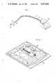
- FIG. 2is a perspective view of a kit usable to provide the sterile cover depicted in FIG. 1.
- FIG. 3is a perspective view of a sonic probe and connecting cable before being inserted into the sleeve.
- FIG. 4is a perspective view depicting the manner in which the sleeve covers a probe and cable.
- FIG. 5(a)is a perspective view depicting a sheath before being applied to the distal end of the probe.
- FIG. 5(b)is a perspective view depicting the manner in which the sheath covers the sleeve and the probe.
- FIG. 5(c)is a perspective view of an applied sterile cover in accordance with the invention.
- the sterile cover as applied to a probe and cableis depicted in FIG. 1.
- the sterile covercomprises a sleeve 10 which covers the major portion of cable 12, and, as best shown in FIG. 4, all but the distal end 14 of a probe 16.
- Sheath 18fits over sleeve 10 and covers at least the distal end 14 of probe 16.
- the sheathcovers all of the probe and a portion of the cable 12, and an elastic band 20 is applied to the portion of the sheath that covers the cable.
- a second elastic band 22can be applied at the end of the sleeve nearest to the sonic imaging instrument 24.
- the sleeve 10is flexible and can be made of any suitable sterilizable plastic, such as polyethylene. As depicted in FIGS. 1 and 3, it has openings at both ends and is elongated to cover at least a portion of cable 12.
- the circumference of the sleeveshould be slightly larger than that of the probe, and one end of the sleeve is preferably tapered to correspond with the tapered shape 40 of the probe.
- the opening 26 at the tapered endpreferably has a circumference just large enough to allow the passage of the distal end 14 of the probe.
- the sleevecan be seamed or unseamed, since no ultrasonic sound waves will need to be passed through it. At least the exterior surface of the sleeve is sterile.
- the sheath 18is designed to fit snugly around the probe and preferably to extend a short distance behind the probe, to thus cover a portion of the cable, as best depicted in FIG. 5(b). It has a closed end and an open end, and the closed end is shaped to mate with the tapered shape 40 of the probe.
- the tip 30 of the sheathis nipple shaped in order to receive the distal end 14 of the probe.
- the sheathshould be flexible and sterilizable, and is preferably composed of an elastomer to provide the snug fit with the probe.
- the part of the probe that is inserted through the integument of the body and into the body properis the distal end 14.
- this distal portionalthough covered, be wrapped by a completely homogenous, air-free, regular and non-seamed surface.
- the sheathis shaped to mirror the 3-dimensional configuration of the probe in an exact fashion, the elimination of air between the probe and the sheath is facilitated.
- the elimination of airis also facilitated by first rolling sheath 18, as depicted in FIG. 5a, and applying the sheath by unrolling it onto the probe.
- the production of an air-free contact between the probe and sheathis further facilitated by the application of sound-conducting jelly at the contacting surfaces between the distal end 14 and sheath 18.
- An economic advantage of the two-part sterile cover system of the present inventionis that, while the distal end 14 of the probe requires a non-seamed cover, the body of the probe and cable can be covered with materials that do not meet this requirement.
- the inventioncombines the economy of what may be a seamed, flexible plastic sleeve 10 with the high quality sheath 18 for the distal end 14 of the probe.
- it would be possible to produce a molded latex cover of high qualitywhich would cover both the distal end of the probe and the connecting cable 12, but such a cover would be impractical, since the molds and processes necessary to produce such a cover would be extremely expensive, and the resulting product would be unwieldy.
- the homogeneity of the sheath 18is important.
- latex productsare normally molded, they are released from their mold with the aid of a particulate powdiferous substance. If such a product were used to cover the distal end 14, the powder would decrease the accuracy of the sonic imaging equipment. Moreover, such powder is harmful to body tissues, and granuloma and adhesion formation may occur in response to particulate contamination of a surgical wound.
- it has been found that these problemscan be overcome by increasing the dwell time of the molding process used to produce sheath 18 so that the thickness of the cover is increased. This thicker cover may be stripped from its mold without a powdiferous releasing agent, and thus a latex sheath 18 with the proper requirements can be obtained.
- a semi-rigid O-ringcan be attached to or incorporated in the open end of sheath 18.
- the O-ringmay be made of a phenolic material, a metal, or any other suitable semi-rigid material.
- the diameter of the O-ringis slightly larger than that of the probe body and the width of the wall of the O-ring is less than 1/4 inch and, preferably, this width is extremely small.
- the O-ringcan be attached to or inserted in the sheath 18 in any suitable manner. One way is to place the O-ring onto the sheath 18 and entrap the O-ring in the sheath in the manufacturing process of rolling the sheath. By incorporating or attaching the O-ring, the mouth of sheath 18 will be held open to facilitate the insertion of the probe, and the unrolling of the sheath along the probe body will be facilitated.
- sheath 18will preferably extend beyond the body of the probe to thus cover a portion of the cable 12.
- the extended portioncan be drawn in towards the cable by using band 20 as depicted in FIG. 5(c).
- band 20By drawing the extended portion in in this manner, the possibility that the extended portion will interfere with the manipulation of the probe body is eliminated, and the sterile field is better defined and becomes more difficult to accidentally contaminate.
- the use of the O-ring in sheath 18can help to maintain band 20 in place.
- the method for applying the sterile coveris as follows. Prior to the application of the sterile cover, the probe is preferably disinfected and placed in a sterile basin until the imaging process takes place.
- the sleeve 10which can originally be in a rolled storage position as depicted in FIG. 2, is extended and probe 16 is inserted through the untapered end 28 as depicted in FIG. 3.
- the probeWhen fully inserted in the sleeve, the probe will stop as its tapered end contacts the tapered end of the sleeve. As depicted in FIG. 4, the distal end 14 of the probe will then protrude through the small terminal slit at the end of the sleeve.
- the sleeve 10is banded with band 22 to secure the sleeve in position and to ensure maintenance of sterility.
- the distal end of the probeshould be covered by an air-free surface.
- sound conducting jellyis applied to the distal end of the probe.
- a suitable sound conducting jellyis Ultra/Phonic, which is distributed by Pharmaceutical Innovations, Inc. of Newark, N.J.
- Application of the jelly to this regioncan be effected either by placing the jelly on the distal end 14 or by placing the jelly into the nipple-shaped tip 30 of the sheath.
- the application of the jellyhelps to eliminate the possibility of air being trapped between the sheath and distal end of the probe.
- sheath 18which is advantageously provided in a rolled fashion as depicted in FIG. 5(a), is first positioned on the distal end 14 of the probe and then unrolled along the body of the probe until the probe is completely covered. It can be seen that the tapering of sleeve 10 near opening 26 ensures that the sleeve will not be pushed off the body of the probe during the unrolling step of the sheath.
- sheath 18is long enough to extend beyond probe 16, and the extended portion can be banded with band 20.
- an outer sheathis applied in the same manner as the first, after sound conducting jelly is applied to the exterior of tip 30 of the first sheath or to the interior of a corresponding nipple-shaped portion of the outer sheath.
- the circulating nursewho is not sterile, to apply the sleeve 10 to the probe and cable by grasping the sleeve at only the untapered end 28 and then inserting the probe 16 into this end of the sleeve. Then, while still grasping the sleeve at only the untapered end, the circulating nurse can draw the sleeve over the probe and over the connecting cable until the tapered end of the sleeve engages the mating conical end of the probe. Since the circulating nurse touches only the untapered end of the sleeve, this is the only portion of the sleeve which is rendered unsterile by the procedure performed by the circulating nurse. The sterile nurse, wearing sterile surgical gloves, can then apply the sheath 18 to the probe over the sleeve.
- the sheath and sleevecan be removed and disposed of.
- the probeis cleaned by removing the sound conducting jelly with a damp cloth or sponge.
- the probe and instrumentare immediately ready for reuse after the application of a new sterile sleeve and sheath to the probe and cable.
- the component parts of the sterile coverare conveniently enclosed in a sterile kit, as depicted in FIG. 2.
- the kitincludes a molded plastic tray 32 which is provided with recessions to hold the component parts of the sterile cover.
- recessionsare provided for sleeve 10, sheaths 18, bands 20, 22, and a package of sound conducting jelly 34. Bands 20, 22 are maintained in place by drawing them around protruding clips 36. All of the component parts as well as the tray 32 are sterilized, and these parts are enclosed in a sterile container, such as plastic bag 38.
- a lower part of the recession for sheaths 18is designed to suspend the sheath to enable the tips 30 of the sheaths to be inverted and sufficiently open so that sound conducting jelly can be inserted into the tips 30 while the sheaths are still in the tray.
- Tray 32can be made of any suitable, substantially rigid and sterilizable material.
- Bag 38is preferably clear and flexible and can be provided with a means to facilitate opening without the need of a cutting instrument.
Landscapes
- Health & Medical Sciences (AREA)
- Life Sciences & Earth Sciences (AREA)
- Surgery (AREA)
- General Health & Medical Sciences (AREA)
- Veterinary Medicine (AREA)
- Biomedical Technology (AREA)
- Heart & Thoracic Surgery (AREA)
- Medical Informatics (AREA)
- Molecular Biology (AREA)
- Animal Behavior & Ethology (AREA)
- Nuclear Medicine, Radiotherapy & Molecular Imaging (AREA)
- Public Health (AREA)
- Engineering & Computer Science (AREA)
- Physics & Mathematics (AREA)
- Biophysics (AREA)
- Pathology (AREA)
- Radiology & Medical Imaging (AREA)
- Acoustics & Sound (AREA)
- Optics & Photonics (AREA)
- Ultra Sonic Daignosis Equipment (AREA)
Abstract
Description
This invention relates to a sterile cover for an ultrasonic probe inserted through the outer integument of the body, as well as a method and a kit for providing the cover.
There is now in existence ultrasonic diagnostic equipment for providing images of tissue in organs within the body. Basically, this equipment operates by transmitting high frequency pulses of sound into the body, detecting the pattern of reflection of this sonic energy, and translating this information into a visual image. The advantage of this technique is that the images of the tissue or organs inside the body can be obtained without disrupting the integrity of the body.
More recently, it has been found that better images can be obtained if the sonic probe or transducer of the equipment is inserted through the outer integument of the body. Ultrasonic diagnostic probes have been designed for insertion through the skin of the patient and, in the case when an image of the brain is desired, even through the skull. When such a probe is used, it is necessary that the procedure be carried out under asceptic conditions and that the sterility of the probe, the connecting cable, and all persons and other devices coming into actual or approximate contact with the probe must be maintained. One way that sterility has been maintained is to sterilize the probe and the cable after each use, but this technique is not practical. The sterilization procedure, including the necessary controls to ensure the effectiveness of the sterilization procedure, requires one to eight days. Of course, during this sterilization period, no further diagnoses can be made with the probe. This problem can be avoided by the use of multiple substitute probes, but the probes themselves cost over $10,000.00 each and most hospitals cannot afford to purchase a large number. Another inadequate solution to the problem of sterilization has been to cover the probe and cable in whatever sterile materials are available, such as surgical gloves. Thus, there is a need in the art for an effective, convenient, and inexpensive way to provide for and maintain the sterilization of sonic probes and connecting cables.
In accordance with the present invention, a sterile cover is provided for a sonic probe and connecting cable of a sonic imaging instrument, as well as a method and kit for providing the cover. The sterile cover includes a flexible sleeve which can cover at least a portion of a connecting cable and all but a distal end of a transducer probe, and a flexible sheath that fits over a portion of the sleeve and covers at least the distal end of the probe. The method for providing the sterile cover includes the step of inserting the probe through an untapered end of the sleeve until all but the distal end of the probe and at least a portion of the cable is covered by the sleeve, followed by the step of applying the sheath to cover at least the distal end of the probe. A kit is also disclosed which includes a sterile package which contains the sleeve and the sheath.
FIG. 1 is a perspective view of the sterile cover as applied to the sonic probe and connecting cable of a sonic imaging instrument.
FIG. 2 is a perspective view of a kit usable to provide the sterile cover depicted in FIG. 1.
FIG. 3 is a perspective view of a sonic probe and connecting cable before being inserted into the sleeve.
FIG. 4 is a perspective view depicting the manner in which the sleeve covers a probe and cable.
FIG. 5(a) is a perspective view depicting a sheath before being applied to the distal end of the probe.
FIG. 5(b) is a perspective view depicting the the manner in which the sheath covers the sleeve and the probe.
FIG. 5(c) is a perspective view of an applied sterile cover in accordance with the invention.
The sterile cover as applied to a probe and cable is depicted in FIG. 1. The sterile cover comprises asleeve 10 which covers the major portion ofcable 12, and, as best shown in FIG. 4, all but thedistal end 14 of aprobe 16. Sheath 18 fits oversleeve 10 and covers at least thedistal end 14 ofprobe 16. In the preferred embodiment depicted in FIG. 1, the sheath covers all of the probe and a portion of thecable 12, and anelastic band 20 is applied to the portion of the sheath that covers the cable. A secondelastic band 22 can be applied at the end of the sleeve nearest to thesonic imaging instrument 24.
Thesleeve 10 is flexible and can be made of any suitable sterilizable plastic, such as polyethylene. As depicted in FIGS. 1 and 3, it has openings at both ends and is elongated to cover at least a portion ofcable 12. The circumference of the sleeve should be slightly larger than that of the probe, and one end of the sleeve is preferably tapered to correspond with thetapered shape 40 of the probe. As depicted in FIG. 3, the opening 26 at the tapered end preferably has a circumference just large enough to allow the passage of thedistal end 14 of the probe. The sleeve can be seamed or unseamed, since no ultrasonic sound waves will need to be passed through it. At least the exterior surface of the sleeve is sterile.
Thesheath 18 is designed to fit snugly around the probe and preferably to extend a short distance behind the probe, to thus cover a portion of the cable, as best depicted in FIG. 5(b). It has a closed end and an open end, and the closed end is shaped to mate with thetapered shape 40 of the probe. Thetip 30 of the sheath is nipple shaped in order to receive thedistal end 14 of the probe. The sheath should be flexible and sterilizable, and is preferably composed of an elastomer to provide the snug fit with the probe.
The part of the probe that is inserted through the integument of the body and into the body proper is thedistal end 14. In order for a clear image to be generated, it is important that this distal portion, although covered, be wrapped by a completely homogenous, air-free, regular and non-seamed surface. By providing that the sheath is shaped to mirror the 3-dimensional configuration of the probe in an exact fashion, the elimination of air between the probe and the sheath is facilitated. The elimination of air is also facilitated by firstrolling sheath 18, as depicted in FIG. 5a, and applying the sheath by unrolling it onto the probe. The production of an air-free contact between the probe and sheath is further facilitated by the application of sound-conducting jelly at the contacting surfaces between thedistal end 14 andsheath 18.
An economic advantage of the two-part sterile cover system of the present invention is that, while thedistal end 14 of the probe requires a non-seamed cover, the body of the probe and cable can be covered with materials that do not meet this requirement. Thus, the invention combines the economy of what may be a seamed, flexibleplastic sleeve 10 with thehigh quality sheath 18 for thedistal end 14 of the probe. Theoretically, it would be possible to produce a molded latex cover of high quality which would cover both the distal end of the probe and the connectingcable 12, but such a cover would be impractical, since the molds and processes necessary to produce such a cover would be extremely expensive, and the resulting product would be unwieldy. Further, such an inordinately long latex cover could not be rolled up, so that some of the advantages of the present invention, i.e., the elimination of air arounddistal end 14 and the facilitation of placing thesheath 18 on the probe, would be difficult to achieve.
In order to achieve the most accurate measurements from the sonic imaging equipment, the homogeneity of thesheath 18 is important. When latex products are normally molded, they are released from their mold with the aid of a particulate powdiferous substance. If such a product were used to cover thedistal end 14, the powder would decrease the accuracy of the sonic imaging equipment. Moreover, such powder is harmful to body tissues, and granuloma and adhesion formation may occur in response to particulate contamination of a surgical wound. In carrying out the present invention, it has been found that these problems can be overcome by increasing the dwell time of the molding process used to producesheath 18 so that the thickness of the cover is increased. This thicker cover may be stripped from its mold without a powdiferous releasing agent, and thus alatex sheath 18 with the proper requirements can be obtained.
In a preferred embodiment, a semi-rigid O-ring can be attached to or incorporated in the open end ofsheath 18. The O-ring may be made of a phenolic material, a metal, or any other suitable semi-rigid material. Preferably the diameter of the O-ring is slightly larger than that of the probe body and the width of the wall of the O-ring is less than 1/4 inch and, preferably, this width is extremely small. The O-ring can be attached to or inserted in thesheath 18 in any suitable manner. One way is to place the O-ring onto thesheath 18 and entrap the O-ring in the sheath in the manufacturing process of rolling the sheath. By incorporating or attaching the O-ring, the mouth ofsheath 18 will be held open to facilitate the insertion of the probe, and the unrolling of the sheath along the probe body will be facilitated.
As depicted in FIG. 5(b),sheath 18 will preferably extend beyond the body of the probe to thus cover a portion of thecable 12. When the sheath is so extended, the extended portion can be drawn in towards the cable by usingband 20 as depicted in FIG. 5(c). By drawing the extended portion in in this manner, the possibility that the extended portion will interfere with the manipulation of the probe body is eliminated, and the sterile field is better defined and becomes more difficult to accidentally contaminate. The use of the O-ring insheath 18 can help to maintainband 20 in place.
The method for applying the sterile cover is as follows. Prior to the application of the sterile cover, the probe is preferably disinfected and placed in a sterile basin until the imaging process takes place. When the imaging process is to commence, thesleeve 10, which can originally be in a rolled storage position as depicted in FIG. 2, is extended andprobe 16 is inserted through theuntapered end 28 as depicted in FIG. 3. When fully inserted in the sleeve, the probe will stop as its tapered end contacts the tapered end of the sleeve. As depicted in FIG. 4, thedistal end 14 of the probe will then protrude through the small terminal slit at the end of the sleeve. Preferably, thesleeve 10 is banded withband 22 to secure the sleeve in position and to ensure maintenance of sterility.
It has been noted that, in order for the sonic imaging equipment to generate a clear image, the distal end of the probe should be covered by an air-free surface. In order to facilitate the production of an air-free environment between thedistal end 14 and thesheath 18, sound conducting jelly is applied to the distal end of the probe. A suitable sound conducting jelly is Ultra/Phonic, which is distributed by Pharmaceutical Innovations, Inc. of Newark, N.J. Application of the jelly to this region can be effected either by placing the jelly on thedistal end 14 or by placing the jelly into the nipple-shapedtip 30 of the sheath. The application of the jelly helps to eliminate the possibility of air being trapped between the sheath and distal end of the probe.
Next,sheath 18, which is advantageously provided in a rolled fashion as depicted in FIG. 5(a), is first positioned on thedistal end 14 of the probe and then unrolled along the body of the probe until the probe is completely covered. It can be seen that the tapering ofsleeve 10 near opening 26 ensures that the sleeve will not be pushed off the body of the probe during the unrolling step of the sheath. In a preferred embodiment,sheath 18 is long enough to extend beyondprobe 16, and the extended portion can be banded withband 20.
In another embodiment, an outer sheath is applied in the same manner as the first, after sound conducting jelly is applied to the exterior oftip 30 of the first sheath or to the interior of a corresponding nipple-shaped portion of the outer sheath.
In a hospital environment, it will be convenient for the circulating nurse, who is not sterile, to apply thesleeve 10 to the probe and cable by grasping the sleeve at only theuntapered end 28 and then inserting theprobe 16 into this end of the sleeve. Then, while still grasping the sleeve at only the untapered end, the circulating nurse can draw the sleeve over the probe and over the connecting cable until the tapered end of the sleeve engages the mating conical end of the probe. Since the circulating nurse touches only the untapered end of the sleeve, this is the only portion of the sleeve which is rendered unsterile by the procedure performed by the circulating nurse. The sterile nurse, wearing sterile surgical gloves, can then apply thesheath 18 to the probe over the sleeve.
Upon completion of the ultrasonic scan and imaging process, the sheath and sleeve can be removed and disposed of. Preferably, after removal of the used sheath and sleeve, the probe is cleaned by removing the sound conducting jelly with a damp cloth or sponge. The probe and instrument are immediately ready for reuse after the application of a new sterile sleeve and sheath to the probe and cable.
In accordance with the present invention, the component parts of the sterile cover are conveniently enclosed in a sterile kit, as depicted in FIG. 2. As depicted in the drawing, the kit includes a moldedplastic tray 32 which is provided with recessions to hold the component parts of the sterile cover. Thus, recessions are provided forsleeve 10,sheaths 18,bands sound conducting jelly 34.Bands tray 32 are sterilized, and these parts are enclosed in a sterile container, such asplastic bag 38. In a preferred embodiment, a lower part of the recession forsheaths 18 is designed to suspend the sheath to enable thetips 30 of the sheaths to be inverted and sufficiently open so that sound conducting jelly can be inserted into thetips 30 while the sheaths are still in the tray.Tray 32 can be made of any suitable, substantially rigid and sterilizable material.Bag 38 is preferably clear and flexible and can be provided with a means to facilitate opening without the need of a cutting instrument.
While there have been described what are considered to be preferred embodiments of this invention, it will be obvious to those skilled in the art that various changes and modifications may be made therein without departing from the invention. It is intended that the present description cover all such changes and modifications as may be within the spirit and scope of the invention.
Claims (18)
1. A method of providing a sterile cover over a sonic probe and flexible connecting cable of a sonic imaging instrument, said cable extending between said probe and a sonic imaging instrument, comprising placing a flexible sleeve, open at both ends and having a sterile exterior surface, over said cable and said probe with a distal end of said probe protruding from one end of said sleeve, and placing a flexible sheath, open at one end and closed at the other end and having a sterile exterior surface, over at least the distal end of said probe and over said one end of said sleeve.
2. The method according to claim 1, wherein said one end of said sleeve is tapered to define the opening at said one end of said sleeve to have a circumference shorter than the circumference of the remainder of said sleeve, and wherein said placing step includes placing said sleeve onto said probe and said cable by inserting said probe into the end of said sleeve opposite from said one end and drawing said sleeve over said probe and over said cable until the tapered portion of said sleeve at said one end engages a tapered shape of said probe.
3. The method according to claim 1, further comprising the steps of rolling up said sheath prior to placing it on said probe, and placing said sheath on said probe by positioning a tip of said sheath on said distal end of said probe and unrolling said sheath over said probe and over said one end of said sleeve.
4. The method according to claim 1, further comprising the step of applying sound conducting jelly to said distal end of said probe or to the interior of a tip of said sheath prior to said placing said sheath over said distal end.
5. The method according to claim 1, further comprising the step of banding an end of said sleeve that is opposite from said one end of said sleeve.
6. The method according to claim 1, wherein said sheath covers all of said probe and a portion of said connecting cable, further comprising the step of banding said sheath at a portion of said sheath which covers said cable.
7. The method according to claim 1, further comprising the steps of applying an outer sheath over said sheath after first applying sound conducting jelly to the exterior of a tip of said sheath or the interior of a tip of said outer sheath.
8. The method according to claim 1, further comprising the step of removing and discarding said sheath and said sleeve after a sonic imaging procedure has been completed.
9. A kit for providing a sterile cover over a sonic probe having a distal end with a predetermined shape and the cable connecting said probe to a sonic imaging instrument, said kit comprising:
a sleeve tapered at one end to define an opening at said one end substantially smaller than the passage through the remainder of said sleeve, said taper being shaped to correspond with the shape of the distal end of said probe, said sleeve being sufficiently flexible to bend to a substantial degree of curvature without damage, being open at both ends and having a sterile exterior surface;
a sheath at one end and closed at another end and having a sterile exterior surface; and
a package having a sterile interior, said package enclosing said sleeve and said sheath so that the sterile condition of said sheath and sleeve is maintained,
wherein said sleeve is adapted to cover at least a portion of said cable and all but said distal end of said probe and said sheath is adapted to fit over a portion of said sleeve and cover at least said distal end of said probe, the closed end of said sheath being shaped to fit with the distal end of said probe and correspond to the shape of the tapered end of said sleeve so that the tapered end of said sleeve and the closed end of said sheath fit together when mounted on said probe.
10. The kit according to claim 9, wherein said sleeve is tapered at one end adapted to be adjacent to said distal end of said probe, such that the tapered portion of said sleeve mates with a tapered shape of said probe.
11. The kit according to claim 9, wherein said sheath is elastomeric and produced in a molding process without use of a powdiferous releasing agent.
12. The kit according to claim 9, further comprising an amount of sound conducting jelly enclosed in said package, said jelly adapted to be applied between said distal end and at least a tip of said sheath.
13. The kit according to claim 9, wherein said sheath is adapted to cover all of said probe and at least a portion of said cable, and wherein said kit further comprises a first band adapted to constrict a portion of said sheath that covers said cable.
14. The kit according to claim 13, further comprising a second band adapted to constrict an end of said sleeve that is opposite to said one end.
15. The kit according to claim 9 wherein said open end of said sheath comprises an O-ring.
16. The kit according to claim 9, further comprising an outer sheath adapted to cover at least a tip of said sheath.
17. The kit according to claim 9 wherein a tray is provided with recesses which correspond to component parts of said kit to hold said parts.
18. The kit according to claim 17 wherein a recess in said tray for said sheath maintains a tip of said sheath in an inverted, open position so that sound conducting jelly can be dispensed into said tip while the sheath is disposed in said tray.
Priority Applications (1)
| Application Number | Priority Date | Filing Date | Title |
|---|---|---|---|
| US06/503,838US4593699A (en) | 1983-06-13 | 1983-06-13 | Sterile cover for intraoperative ultrasonic diagnostic devices and method and kit for providing same |
Applications Claiming Priority (1)
| Application Number | Priority Date | Filing Date | Title |
|---|---|---|---|
| US06/503,838US4593699A (en) | 1983-06-13 | 1983-06-13 | Sterile cover for intraoperative ultrasonic diagnostic devices and method and kit for providing same |
Publications (1)
| Publication Number | Publication Date |
|---|---|
| US4593699Atrue US4593699A (en) | 1986-06-10 |
Family
ID=24003734
Family Applications (1)
| Application Number | Title | Priority Date | Filing Date |
|---|---|---|---|
| US06/503,838Expired - Fee RelatedUS4593699A (en) | 1983-06-13 | 1983-06-13 | Sterile cover for intraoperative ultrasonic diagnostic devices and method and kit for providing same |
Country Status (1)
| Country | Link |
|---|---|
| US (1) | US4593699A (en) |
Cited By (81)
| Publication number | Priority date | Publication date | Assignee | Title |
|---|---|---|---|---|
| WO1989000832A1 (en)* | 1987-07-27 | 1989-02-09 | B.V. Optische Industrie "De Oude Delft" | Ultrasonic endoscope provided with protective sheath |
| US4815470A (en)* | 1987-11-13 | 1989-03-28 | Advanced Diagnostic Medical Systems, Inc. | Inflatable sheath for ultrasound probe |
| US4817592A (en)* | 1987-01-23 | 1989-04-04 | Andronic Devices, Ltd. | Toroidal surgical shield |
| EP0310515A1 (en)* | 1987-10-01 | 1989-04-05 | Fuji Optical Systems, Inc. | Means and structure for prevention of cross contamination during use of dental camera |
| DE3929612A1 (en)* | 1988-11-22 | 1990-05-31 | Siemens Ag | Intracavitary ultrasonic probe coupler to investigation object - has inner and outer casings with liquid-tight seal forming inner liquid-filled chamber, protective tube |
| WO1990015569A1 (en)* | 1989-06-22 | 1990-12-27 | Norbert Lemke | Television camera system for use in sterile conditions |
| US5010900A (en)* | 1989-02-27 | 1991-04-30 | Auchinleck Geoffrey F | Lower limb positioning apparatus and surgical drape |
| US5016098A (en)* | 1987-03-05 | 1991-05-14 | Fuji Optical Systems, Incorporated | Electronic video dental camera |
| US5025778A (en)* | 1990-03-26 | 1991-06-25 | Opielab, Inc. | Endoscope with potential channels and method of using the same |
| US5051823A (en)* | 1988-01-28 | 1991-09-24 | Fuji Optical Systems, Inc. | Dental instrument including laser device and electronic video dental camera |
| GB2218636B (en)* | 1988-05-04 | 1992-01-02 | Rhys Ap Delwyn Phillips | Hygienic protection devices |
| US5115307A (en)* | 1987-03-05 | 1992-05-19 | Fuji Optical Systems | Electronic video dental camera |
| US5135001A (en)* | 1990-12-05 | 1992-08-04 | C. R. Bard, Inc. | Ultrasound sheath for medical diagnostic instruments |
| US5168863A (en)* | 1990-08-27 | 1992-12-08 | Medical Concepts, Inc. | Sterile endoscopic system |
| US5222485A (en)* | 1990-09-17 | 1993-06-29 | Ravinder Jerath | Ultrasound labor monitoring method and apparatus |
| US5251025A (en)* | 1987-03-05 | 1993-10-05 | Fuji Optical Systems, Inc. | Electronic video dental camera |
| US5259383A (en)* | 1990-08-30 | 1993-11-09 | Johnson & Johnson Medical, Inc. | Sterile ultrasound cover tube |
| WO1994001037A1 (en)* | 1992-07-01 | 1994-01-20 | British Technology Group Ltd. | Medical devices |
| ES2052421A1 (en)* | 1991-12-10 | 1994-07-01 | Dominguez Jose Fernando Losa | Prophylactic protector for echographic probes. |
| US5335663A (en)* | 1992-12-11 | 1994-08-09 | Tetrad Corporation | Laparoscopic probes and probe sheaths useful in ultrasonic imaging applications |
| US5390678A (en)* | 1993-10-12 | 1995-02-21 | Baxter International Inc. | Method and device for measuring ultrasonic activity in an ultrasound delivery system |
| US5400785A (en)* | 1994-02-03 | 1995-03-28 | Boston Scientific Corp. | Acoustic window and septum for imaging catheters |
| US5419310A (en)* | 1992-11-03 | 1995-05-30 | Vision Sciences, Inc. | Partially inflated protective endoscope sheath |
| GB2287195A (en)* | 1994-02-25 | 1995-09-13 | Vermon | Steerable ultrasonic endoscope |
| US5595565A (en)* | 1994-06-30 | 1997-01-21 | The Trustees Of Columbia University In The City Of New York | Self-propelled endoscope using pressure driven linear actuators |
| US5619992A (en)* | 1995-04-06 | 1997-04-15 | Guthrie; Robert B. | Methods and apparatus for inhibiting contamination of reusable pulse oximetry sensors |
| US5671747A (en)* | 1996-01-24 | 1997-09-30 | Hewlett-Packard Company | Ultrasound probe having interchangeable accessories |
| US5695491A (en)* | 1994-11-22 | 1997-12-09 | Washington Research Foundation | Endoscopic accessory and containment system |
| WO1998012968A1 (en)* | 1996-09-27 | 1998-04-02 | Boston Scientific Corporation | Device for controlled longitudinal movement of an operative element within a catheter sheath and method |
| US5814736A (en)* | 1994-04-19 | 1998-09-29 | Siemens Aktiengesellschaft | Holder for ultrasonic transducers |
| US5910113A (en)* | 1998-03-24 | 1999-06-08 | Pruter; Rick L. | Sheath for ultrasound probe |
| US5957941A (en)* | 1996-09-27 | 1999-09-28 | Boston Scientific Corporation | Catheter system and drive assembly thereof |
| US5997481A (en)* | 1998-02-17 | 1999-12-07 | Ultra Sound Probe Covers, Llc | Probe cover with deformable membrane gel reservoir |
| US6039694A (en)* | 1998-06-25 | 2000-03-21 | Sonotech, Inc. | Coupling sheath for ultrasound transducers |
| US6210336B1 (en)* | 1998-12-30 | 2001-04-03 | G.E. Vingmed Ultrasound A/S | Damping cushion for ultrasound probes |
| US6302848B1 (en) | 1999-07-01 | 2001-10-16 | Sonotech, Inc. | In vivo biocompatible acoustic coupling media |
| US20030028178A1 (en)* | 2001-08-03 | 2003-02-06 | Scimed Life Systems, Inc. | Protective sleeve for an endoscopic instrument and related method of use |
| US6589164B1 (en)* | 2000-02-15 | 2003-07-08 | Transvascular, Inc. | Sterility barriers for insertion of non-sterile apparatus into catheters or other medical devices |
| US20040087976A1 (en)* | 2002-08-29 | 2004-05-06 | Devries Robert B. | Devices and methods for fastening tissue layers |
| US6755789B2 (en) | 2002-02-05 | 2004-06-29 | Inceptio Medical Technologies, Llc | Ultrasonic vascular imaging system and method of blood vessel cannulation |
| US6805669B2 (en) | 2001-01-25 | 2004-10-19 | Rebecca L. Swanbom | Method and device for marking skin during an ultrasound examination |
| US20040217675A1 (en)* | 2003-03-31 | 2004-11-04 | Liposonix, Inc. | Vortex transducer |
| US20050079666A1 (en)* | 2002-04-05 | 2005-04-14 | French Roger Harquail | Method for providing nano-structures of uniform length |
| DE10343590A1 (en)* | 2003-09-18 | 2005-04-21 | Paul Junk | Device for creation of tattoo or permanent make-up, comprising needle module with permanently attached latex cover for accommodation of complete unit |
| US20050143798A1 (en)* | 2002-10-18 | 2005-06-30 | Radiant Medical, Inc. | Valved connector assembly and sterility barriers for heat exchange catheters and other closed loop catheters |
| US20050154313A1 (en)* | 2003-12-30 | 2005-07-14 | Liposonix, Inc. | Disposable transducer seal |
| US20050154431A1 (en)* | 2003-12-30 | 2005-07-14 | Liposonix, Inc. | Systems and methods for the destruction of adipose tissue |
| US20050154309A1 (en)* | 2003-12-30 | 2005-07-14 | Liposonix, Inc. | Medical device inline degasser |
| US20050154295A1 (en)* | 2003-12-30 | 2005-07-14 | Liposonix, Inc. | Articulating arm for medical procedures |
| US20050187495A1 (en)* | 2003-12-30 | 2005-08-25 | Liposonix, Inc. | Ultrasound therapy head with movement control |
| US20050193451A1 (en)* | 2003-12-30 | 2005-09-01 | Liposonix, Inc. | Articulating arm for medical procedures |
| US20060020210A1 (en)* | 1997-08-19 | 2006-01-26 | Mendlein John D | Ultrasonic transmission films and devices for hygienic transducer surfaces |
| US20070016112A1 (en)* | 2005-06-09 | 2007-01-18 | Reiner Schultheiss | Shock Wave Treatment Device and Method of Use |
| US20070016030A1 (en)* | 2002-02-05 | 2007-01-18 | Stringer Bradley J | Multiplanar ultrasonic vascular sensor assembly and apparatus for movably affixing a sensor assembly to a body |
| US20070055156A1 (en)* | 2003-12-30 | 2007-03-08 | Liposonix, Inc. | Apparatus and methods for the destruction of adipose tissue |
| US7223238B2 (en) | 2001-01-25 | 2007-05-29 | Swanbom Rebecca L | Method and device for marking skin during an ultrasound examination |
| US20070130889A1 (en)* | 2004-02-20 | 2007-06-14 | Healthcare Media Technologies, Inc. | Disposable cover for a pillow speaker or the like |
| US20070225605A1 (en)* | 2001-01-25 | 2007-09-27 | Swanbom Rebecca L | Method and Device for Marking Skin During an Ultrasound Examination |
| CN100370957C (en)* | 2006-01-02 | 2008-02-27 | 赵建中 | Supersonic anti-cross infection device in operation |
| US20080243003A1 (en)* | 2007-03-26 | 2008-10-02 | Liposonix, Inc. | Slip ring space and method for its use |
| US20090240146A1 (en)* | 2007-10-26 | 2009-09-24 | Liposonix, Inc. | Mechanical arm |
| US20100063359A1 (en)* | 2007-04-11 | 2010-03-11 | Tyco Healthcare Group Lp | Endoscopic/laparoscopic introducer sleeve |
| US20100234733A1 (en)* | 2009-03-13 | 2010-09-16 | Paul Wahlheim | Sterile Ultrasound Probe Cover and Method of Releasing Coupling Agent from a Sealed Compartment |
| EP1988833A4 (en)* | 2006-02-17 | 2010-11-17 | Esi Inc | Immersion bag system for use with an ultrasound probe |
| US20100312121A1 (en)* | 2009-06-09 | 2010-12-09 | Zhonghui Guan | Apparatus for a needle director for an ultrasound transducer probe |
| US20110178443A1 (en)* | 2004-11-24 | 2011-07-21 | Medicis Technologies Corporation | System and methods for destroying adipose tissue |
| US8926533B2 (en) | 2003-12-30 | 2015-01-06 | Liposonix, Inc. | Therapy head for use with an ultrasound system |
| WO2014174251A3 (en)* | 2013-04-23 | 2015-03-19 | P3 Medical Limited | Cover for medical probe |
| USD754357S1 (en) | 2011-08-09 | 2016-04-19 | C. R. Bard, Inc. | Ultrasound probe head |
| US20160192903A1 (en)* | 2014-12-29 | 2016-07-07 | Civco Medical Instruments Co., Inc. | Sterile covers for ultrasound probe |
| US20160228188A1 (en)* | 2015-02-05 | 2016-08-11 | Spinal Generations, Llc | Individual packaging arrangement for orthopedic tools |
| EP3072438A1 (en)* | 2015-03-27 | 2016-09-28 | Parker Laboratories, Inc. | Protective cover set for a medical probe |
| CN106955163A (en)* | 2017-05-25 | 2017-07-18 | 郑州大学第附属医院 | Disposable closed ultrasonic probe bush |
| US10064599B2 (en) | 2015-11-09 | 2018-09-04 | HealthCare Evolution LLC | Ultrashield devices and methods for use in ultrasonic procedures |
| US10085716B2 (en) | 2013-03-15 | 2018-10-02 | J. Jordan Romano | System and method for sterile sheathing of a medical probe |
| CN108836381A (en)* | 2018-03-24 | 2018-11-20 | 余军辉 | Ultrasonic-B probe wears covering device automatically |
| US10639008B2 (en) | 2009-10-08 | 2020-05-05 | C. R. Bard, Inc. | Support and cover structures for an ultrasound probe head |
| US10820885B2 (en) | 2012-06-15 | 2020-11-03 | C. R. Bard, Inc. | Apparatus and methods for detection of a removable cap on an ultrasound probe |
| US20210030261A1 (en)* | 2018-04-27 | 2021-02-04 | Jürgen Kress | Device and method for performing an endoscopic examination free from contamination |
| US11103213B2 (en) | 2009-10-08 | 2021-08-31 | C. R. Bard, Inc. | Spacers for use with an ultrasound probe |
| US12343200B2 (en) | 2015-10-09 | 2025-07-01 | Boston Scientific Scimed, Inc. | Intravascular ultrasound systems, catheters, and methods with a manual pullback arrangement |
Citations (6)
| Publication number | Priority date | Publication date | Assignee | Title |
|---|---|---|---|---|
| US2677965A (en)* | 1947-12-19 | 1954-05-11 | Jacob A Saffir | Heat conducting sheath for clinical thermometers |
| US3017705A (en)* | 1960-04-08 | 1962-01-23 | Peters John | Foot and leg apparel article |
| US3779234A (en)* | 1971-06-30 | 1973-12-18 | Intersc Res Inst | Ultrasonic catheter with rotating transducers |
| US4069913A (en)* | 1975-08-11 | 1978-01-24 | Harrigan Roy Major | Surgical glove package and fixture |
| US4224936A (en)* | 1978-05-31 | 1980-09-30 | Vickers Limited | Transit isolator |
| US4349033A (en)* | 1980-11-06 | 1982-09-14 | Eden Robert D | Intrauterine catheter |
- 1983
- 1983-06-13USUS06/503,838patent/US4593699A/ennot_activeExpired - Fee Related
Patent Citations (6)
| Publication number | Priority date | Publication date | Assignee | Title |
|---|---|---|---|---|
| US2677965A (en)* | 1947-12-19 | 1954-05-11 | Jacob A Saffir | Heat conducting sheath for clinical thermometers |
| US3017705A (en)* | 1960-04-08 | 1962-01-23 | Peters John | Foot and leg apparel article |
| US3779234A (en)* | 1971-06-30 | 1973-12-18 | Intersc Res Inst | Ultrasonic catheter with rotating transducers |
| US4069913A (en)* | 1975-08-11 | 1978-01-24 | Harrigan Roy Major | Surgical glove package and fixture |
| US4224936A (en)* | 1978-05-31 | 1980-09-30 | Vickers Limited | Transit isolator |
| US4349033A (en)* | 1980-11-06 | 1982-09-14 | Eden Robert D | Intrauterine catheter |
Non-Patent Citations (2)
| Title |
|---|
| Taylor et al., "A High Resolution Transrectal Ultrasonographic System" Ultra Med. Bio., vol. 5, No. 2, 1979, pp. 128-138. |
| Taylor et al., A High Resolution Transrectal Ultrasonographic System Ultra Med. Bio., vol. 5, No. 2, 1979, pp. 128 138.* |
Cited By (137)
| Publication number | Priority date | Publication date | Assignee | Title |
|---|---|---|---|---|
| US4817592A (en)* | 1987-01-23 | 1989-04-04 | Andronic Devices, Ltd. | Toroidal surgical shield |
| US5429502A (en)* | 1987-03-05 | 1995-07-04 | Fuji Optical Systems, Inc. | Electronic video dental camera |
| US5115307A (en)* | 1987-03-05 | 1992-05-19 | Fuji Optical Systems | Electronic video dental camera |
| US5016098A (en)* | 1987-03-05 | 1991-05-14 | Fuji Optical Systems, Incorporated | Electronic video dental camera |
| US5290168A (en)* | 1987-03-05 | 1994-03-01 | Optical Systems, Inc. | Electronic video dental camera |
| US5251025A (en)* | 1987-03-05 | 1993-10-05 | Fuji Optical Systems, Inc. | Electronic video dental camera |
| US5088178A (en)* | 1987-07-27 | 1992-02-18 | Bv Optische Industrie | Ultrasonic endoscope provided with protective sheath |
| WO1989000832A1 (en)* | 1987-07-27 | 1989-02-09 | B.V. Optische Industrie "De Oude Delft" | Ultrasonic endoscope provided with protective sheath |
| EP0310515A1 (en)* | 1987-10-01 | 1989-04-05 | Fuji Optical Systems, Inc. | Means and structure for prevention of cross contamination during use of dental camera |
| US4815470A (en)* | 1987-11-13 | 1989-03-28 | Advanced Diagnostic Medical Systems, Inc. | Inflatable sheath for ultrasound probe |
| US5051823A (en)* | 1988-01-28 | 1991-09-24 | Fuji Optical Systems, Inc. | Dental instrument including laser device and electronic video dental camera |
| GB2218636B (en)* | 1988-05-04 | 1992-01-02 | Rhys Ap Delwyn Phillips | Hygienic protection devices |
| DE3929612A1 (en)* | 1988-11-22 | 1990-05-31 | Siemens Ag | Intracavitary ultrasonic probe coupler to investigation object - has inner and outer casings with liquid-tight seal forming inner liquid-filled chamber, protective tube |
| US5010900A (en)* | 1989-02-27 | 1991-04-30 | Auchinleck Geoffrey F | Lower limb positioning apparatus and surgical drape |
| WO1990015569A1 (en)* | 1989-06-22 | 1990-12-27 | Norbert Lemke | Television camera system for use in sterile conditions |
| US5025778A (en)* | 1990-03-26 | 1991-06-25 | Opielab, Inc. | Endoscope with potential channels and method of using the same |
| US5168863A (en)* | 1990-08-27 | 1992-12-08 | Medical Concepts, Inc. | Sterile endoscopic system |
| US5259383A (en)* | 1990-08-30 | 1993-11-09 | Johnson & Johnson Medical, Inc. | Sterile ultrasound cover tube |
| US5222485A (en)* | 1990-09-17 | 1993-06-29 | Ravinder Jerath | Ultrasound labor monitoring method and apparatus |
| US5135001A (en)* | 1990-12-05 | 1992-08-04 | C. R. Bard, Inc. | Ultrasound sheath for medical diagnostic instruments |
| ES2052421A1 (en)* | 1991-12-10 | 1994-07-01 | Dominguez Jose Fernando Losa | Prophylactic protector for echographic probes. |
| US5355886A (en)* | 1991-12-10 | 1994-10-18 | Losa Dominguez Jose F | Prophylactic protector for echographic probes |
| WO1994001037A1 (en)* | 1992-07-01 | 1994-01-20 | British Technology Group Ltd. | Medical devices |
| US5507295A (en)* | 1992-07-01 | 1996-04-16 | British Technology Group Limited | Medical devices |
| US5419310A (en)* | 1992-11-03 | 1995-05-30 | Vision Sciences, Inc. | Partially inflated protective endoscope sheath |
| US5437283A (en)* | 1992-12-11 | 1995-08-01 | Tetrad Corporation | Endosurgical ultrasonic probe with integrated biopsy actuator |
| US5335663A (en)* | 1992-12-11 | 1994-08-09 | Tetrad Corporation | Laparoscopic probes and probe sheaths useful in ultrasonic imaging applications |
| US5390678A (en)* | 1993-10-12 | 1995-02-21 | Baxter International Inc. | Method and device for measuring ultrasonic activity in an ultrasound delivery system |
| US5400785A (en)* | 1994-02-03 | 1995-03-28 | Boston Scientific Corp. | Acoustic window and septum for imaging catheters |
| WO1995020913A1 (en)* | 1994-02-03 | 1995-08-10 | Boston Scientific Corporation | Acoustic window and septum for imaging catheters |
| GB2287195B (en)* | 1994-02-25 | 1998-05-27 | Vermon | An endoscope for ultrasonic echography |
| GB2287195A (en)* | 1994-02-25 | 1995-09-13 | Vermon | Steerable ultrasonic endoscope |
| US5814736A (en)* | 1994-04-19 | 1998-09-29 | Siemens Aktiengesellschaft | Holder for ultrasonic transducers |
| US5595565A (en)* | 1994-06-30 | 1997-01-21 | The Trustees Of Columbia University In The City Of New York | Self-propelled endoscope using pressure driven linear actuators |
| US5695491A (en)* | 1994-11-22 | 1997-12-09 | Washington Research Foundation | Endoscopic accessory and containment system |
| US5931833A (en)* | 1994-11-22 | 1999-08-03 | Silverstein; Fred E. | Endoscopic accessory and containment system |
| US5619992A (en)* | 1995-04-06 | 1997-04-15 | Guthrie; Robert B. | Methods and apparatus for inhibiting contamination of reusable pulse oximetry sensors |
| US5671747A (en)* | 1996-01-24 | 1997-09-30 | Hewlett-Packard Company | Ultrasound probe having interchangeable accessories |
| WO1998012968A1 (en)* | 1996-09-27 | 1998-04-02 | Boston Scientific Corporation | Device for controlled longitudinal movement of an operative element within a catheter sheath and method |
| US5827313A (en)* | 1996-09-27 | 1998-10-27 | Boston Scientific Corporation | Device for controlled longitudinal movement of an operative element within a catheter sheath and method |
| EP1397996A3 (en)* | 1996-09-27 | 2004-05-26 | Boston Scientific Corporation | Device for controlled longitudinal movement of an operative element within a catheter sheath and method |
| US5957941A (en)* | 1996-09-27 | 1999-09-28 | Boston Scientific Corporation | Catheter system and drive assembly thereof |
| US20060030778A1 (en)* | 1997-08-19 | 2006-02-09 | Mendlein John D | Ultrasonic transmission films and devices for hygienic transducer surfaces |
| US20060020210A1 (en)* | 1997-08-19 | 2006-01-26 | Mendlein John D | Ultrasonic transmission films and devices for hygienic transducer surfaces |
| US5997481A (en)* | 1998-02-17 | 1999-12-07 | Ultra Sound Probe Covers, Llc | Probe cover with deformable membrane gel reservoir |
| US5910113A (en)* | 1998-03-24 | 1999-06-08 | Pruter; Rick L. | Sheath for ultrasound probe |
| US6039694A (en)* | 1998-06-25 | 2000-03-21 | Sonotech, Inc. | Coupling sheath for ultrasound transducers |
| US6866630B2 (en) | 1998-10-09 | 2005-03-15 | Sonotech, Inc. | Vivo biocompatible acoustic coupling media |
| US6210336B1 (en)* | 1998-12-30 | 2001-04-03 | G.E. Vingmed Ultrasound A/S | Damping cushion for ultrasound probes |
| US6776757B2 (en) | 1999-07-01 | 2004-08-17 | Sonotech, Inc. | In vivo biocompatible acoustic coupling media |
| US6302848B1 (en) | 1999-07-01 | 2001-10-16 | Sonotech, Inc. | In vivo biocompatible acoustic coupling media |
| US6589164B1 (en)* | 2000-02-15 | 2003-07-08 | Transvascular, Inc. | Sterility barriers for insertion of non-sterile apparatus into catheters or other medical devices |
| US7223238B2 (en) | 2001-01-25 | 2007-05-29 | Swanbom Rebecca L | Method and device for marking skin during an ultrasound examination |
| US20070225605A1 (en)* | 2001-01-25 | 2007-09-27 | Swanbom Rebecca L | Method and Device for Marking Skin During an Ultrasound Examination |
| US6805669B2 (en) | 2001-01-25 | 2004-10-19 | Rebecca L. Swanbom | Method and device for marking skin during an ultrasound examination |
| US6749601B2 (en)* | 2001-08-03 | 2004-06-15 | Scimed Life Systems, Inc. | Protective sleeve for an endoscopic instrument and related method of use |
| US20030028178A1 (en)* | 2001-08-03 | 2003-02-06 | Scimed Life Systems, Inc. | Protective sleeve for an endoscopic instrument and related method of use |
| US7819810B2 (en) | 2002-02-05 | 2010-10-26 | Inceptio Medical Technologies, Lc | Multiplanar ultrasonic vascular sensor assembly, system and methods employing same, apparatus for movably affixing a sensor assembly to a body and associated methods |
| US20050020919A1 (en)* | 2002-02-05 | 2005-01-27 | Stringer Bradley J. | Multiplanar ultrasonic vascular sensor assembly, system and methods employing same, apparatus for movably affixing a sensor assembly to a body and associated methods |
| US7806828B2 (en) | 2002-02-05 | 2010-10-05 | Inceptio Medical Technologies, Lc | Multiplanar ultrasonic vascular sensor assembly and apparatus for movably affixing a sensor assembly to a body |
| US6755789B2 (en) | 2002-02-05 | 2004-06-29 | Inceptio Medical Technologies, Llc | Ultrasonic vascular imaging system and method of blood vessel cannulation |
| US20070016030A1 (en)* | 2002-02-05 | 2007-01-18 | Stringer Bradley J | Multiplanar ultrasonic vascular sensor assembly and apparatus for movably affixing a sensor assembly to a body |
| US7214191B2 (en) | 2002-02-05 | 2007-05-08 | Inceptio Medical Technologies, L.C. | Multiplanar ultrasonic vascular imaging device, system incorporating same, method of use and protective sheath |
| US20040236224A1 (en)* | 2002-02-05 | 2004-11-25 | Stringer Bradley J. | Multiplanar ultrasonic vascular imaging device, system incorporating same, method of use and protective sheath |
| US20050079666A1 (en)* | 2002-04-05 | 2005-04-14 | French Roger Harquail | Method for providing nano-structures of uniform length |
| US9675352B2 (en) | 2002-08-29 | 2017-06-13 | Boston Scientific Scimed, Inc. | Devices and methods for fastening tissue layers |
| US7837698B2 (en) | 2002-08-29 | 2010-11-23 | Boston Scientific Scimed, Inc. | Devices and methods for fastening tissue layers |
| US20110124963A1 (en)* | 2002-08-29 | 2011-05-26 | Boston Scientific Scimed, Inc. (formerly, Scimed Life Systems, Inc.) | Devices and methods for fastening tissue layers |
| US8728104B2 (en) | 2002-08-29 | 2014-05-20 | Boston Scientific Scimed, Inc. | Devices and methods for fastening tissue layers |
| US7083630B2 (en) | 2002-08-29 | 2006-08-01 | Scimed Life Systems, Inc. | Devices and methods for fastening tissue layers |
| US20040087976A1 (en)* | 2002-08-29 | 2004-05-06 | Devries Robert B. | Devices and methods for fastening tissue layers |
| US8262716B2 (en) | 2002-10-18 | 2012-09-11 | Zoll Circulation, Inc. | Valved connector assembly and sterility barriers for heat exchange catheters and other closed loop catheters |
| US7510568B2 (en) | 2002-10-18 | 2009-03-31 | Zoll Circulation, Inc. | Valved connector assembly and sterility barriers for heat exchange catheters and other closed looped catheters |
| US20050143798A1 (en)* | 2002-10-18 | 2005-06-30 | Radiant Medical, Inc. | Valved connector assembly and sterility barriers for heat exchange catheters and other closed loop catheters |
| US20090247963A1 (en)* | 2002-10-18 | 2009-10-01 | Zoll Circulation, Inc. | Valved connector assembly and sterility barriers for heat exchange catheters and other closed loop catheters |
| US20070035201A1 (en)* | 2003-03-31 | 2007-02-15 | Liposonix, Inc. | Medical ultrasound transducer having non-ideal focal region |
| US20040217675A1 (en)* | 2003-03-31 | 2004-11-04 | Liposonix, Inc. | Vortex transducer |
| US7273459B2 (en) | 2003-03-31 | 2007-09-25 | Liposonix, Inc. | Vortex transducer |
| US7766848B2 (en) | 2003-03-31 | 2010-08-03 | Medicis Technologies Corporation | Medical ultrasound transducer having non-ideal focal region |
| DE10343590B4 (en)* | 2003-09-18 | 2010-08-05 | Junk, Paul, Dipl.-Ing. | Attachment device for tattoo and permanent make-up devices with protective cover |
| DE10343590A1 (en)* | 2003-09-18 | 2005-04-21 | Paul Junk | Device for creation of tattoo or permanent make-up, comprising needle module with permanently attached latex cover for accommodation of complete unit |
| US20050187495A1 (en)* | 2003-12-30 | 2005-08-25 | Liposonix, Inc. | Ultrasound therapy head with movement control |
| US8926533B2 (en) | 2003-12-30 | 2015-01-06 | Liposonix, Inc. | Therapy head for use with an ultrasound system |
| US20050154313A1 (en)* | 2003-12-30 | 2005-07-14 | Liposonix, Inc. | Disposable transducer seal |
| US20050154431A1 (en)* | 2003-12-30 | 2005-07-14 | Liposonix, Inc. | Systems and methods for the destruction of adipose tissue |
| US8337407B2 (en) | 2003-12-30 | 2012-12-25 | Liposonix, Inc. | Articulating arm for medical procedures |
| US7695437B2 (en) | 2003-12-30 | 2010-04-13 | Medicis Technologies Corporation | Ultrasound therapy head with movement control |
| US7311679B2 (en) | 2003-12-30 | 2007-12-25 | Liposonix, Inc. | Disposable transducer seal |
| US20050154309A1 (en)* | 2003-12-30 | 2005-07-14 | Liposonix, Inc. | Medical device inline degasser |
| US7993289B2 (en) | 2003-12-30 | 2011-08-09 | Medicis Technologies Corporation | Systems and methods for the destruction of adipose tissue |
| US20070055156A1 (en)* | 2003-12-30 | 2007-03-08 | Liposonix, Inc. | Apparatus and methods for the destruction of adipose tissue |
| US20050154295A1 (en)* | 2003-12-30 | 2005-07-14 | Liposonix, Inc. | Articulating arm for medical procedures |
| US20110077559A1 (en)* | 2003-12-30 | 2011-03-31 | Medicis Technologies Corporation | Ultrasound therapy head with movement control |
| US20050193451A1 (en)* | 2003-12-30 | 2005-09-01 | Liposonix, Inc. | Articulating arm for medical procedures |
| US20110066084A1 (en)* | 2003-12-30 | 2011-03-17 | Medicis Technologies Corporation | Apparatus and methods for the destruction of adipose tissue |
| US7857773B2 (en) | 2003-12-30 | 2010-12-28 | Medicis Technologies Corporation | Apparatus and methods for the destruction of adipose tissue |
| US7905844B2 (en) | 2003-12-30 | 2011-03-15 | Medicis Technologies Corporation | Disposable transducer seal |
| US20070130889A1 (en)* | 2004-02-20 | 2007-06-14 | Healthcare Media Technologies, Inc. | Disposable cover for a pillow speaker or the like |
| US20110178443A1 (en)* | 2004-11-24 | 2011-07-21 | Medicis Technologies Corporation | System and methods for destroying adipose tissue |
| US20070016112A1 (en)* | 2005-06-09 | 2007-01-18 | Reiner Schultheiss | Shock Wave Treatment Device and Method of Use |
| US8162859B2 (en)* | 2005-06-09 | 2012-04-24 | General Patent , LLC | Shock wave treatment device and method of use |
| CN100370957C (en)* | 2006-01-02 | 2008-02-27 | 赵建中 | Supersonic anti-cross infection device in operation |
| EP1988833A4 (en)* | 2006-02-17 | 2010-11-17 | Esi Inc | Immersion bag system for use with an ultrasound probe |
| US20080243003A1 (en)* | 2007-03-26 | 2008-10-02 | Liposonix, Inc. | Slip ring space and method for its use |
| US8142200B2 (en) | 2007-03-26 | 2012-03-27 | Liposonix, Inc. | Slip ring spacer and method for its use |
| US20100063359A1 (en)* | 2007-04-11 | 2010-03-11 | Tyco Healthcare Group Lp | Endoscopic/laparoscopic introducer sleeve |
| US8353819B2 (en) | 2007-04-11 | 2013-01-15 | Covidien Lp | Endoscopic/laparoscopic introducer sleeve |
| US20090240146A1 (en)* | 2007-10-26 | 2009-09-24 | Liposonix, Inc. | Mechanical arm |
| US20100234733A1 (en)* | 2009-03-13 | 2010-09-16 | Paul Wahlheim | Sterile Ultrasound Probe Cover and Method of Releasing Coupling Agent from a Sealed Compartment |
| US20100312121A1 (en)* | 2009-06-09 | 2010-12-09 | Zhonghui Guan | Apparatus for a needle director for an ultrasound transducer probe |
| US11998386B2 (en) | 2009-10-08 | 2024-06-04 | C. R. Bard, Inc. | Support and cover structures for an ultrasound probe head |
| US11103213B2 (en) | 2009-10-08 | 2021-08-31 | C. R. Bard, Inc. | Spacers for use with an ultrasound probe |
| US10639008B2 (en) | 2009-10-08 | 2020-05-05 | C. R. Bard, Inc. | Support and cover structures for an ultrasound probe head |
| USD754357S1 (en) | 2011-08-09 | 2016-04-19 | C. R. Bard, Inc. | Ultrasound probe head |
| US10820885B2 (en) | 2012-06-15 | 2020-11-03 | C. R. Bard, Inc. | Apparatus and methods for detection of a removable cap on an ultrasound probe |
| US10085716B2 (en) | 2013-03-15 | 2018-10-02 | J. Jordan Romano | System and method for sterile sheathing of a medical probe |
| US11013493B2 (en) | 2013-03-15 | 2021-05-25 | Massachusetts Institute Of Technology | System and method for sterile sheathing of a medical probe |
| WO2014174251A3 (en)* | 2013-04-23 | 2015-03-19 | P3 Medical Limited | Cover for medical probe |
| US10799211B2 (en) | 2014-12-29 | 2020-10-13 | Civco Medical Instruments Co., Inc. | Sterile covers for ultrasound probe |
| US20160192903A1 (en)* | 2014-12-29 | 2016-07-07 | Civco Medical Instruments Co., Inc. | Sterile covers for ultrasound probe |
| WO2016109520A1 (en) | 2014-12-29 | 2016-07-07 | Civco Medical Instruments Co., Inc. | Sterile covers for ultrasound probe |
| US11707267B2 (en) | 2015-02-05 | 2023-07-25 | Spinal Generations, Llc | Individual packaging arrangement for orthopedic tools |
| US10485528B2 (en)* | 2015-02-05 | 2019-11-26 | Spinal Generations, Llc | Individual packaging arrangement for orthopedic tools |
| US20160228188A1 (en)* | 2015-02-05 | 2016-08-11 | Spinal Generations, Llc | Individual packaging arrangement for orthopedic tools |
| US10154828B2 (en) | 2015-03-27 | 2018-12-18 | Parker Laboratories, Inc. | Protective cover set for a medical probe |
| EP3711651A1 (en)* | 2015-03-27 | 2020-09-23 | Parker Laboratories, Inc. | Protective cover set for a medical probe |
| EP3072438A1 (en)* | 2015-03-27 | 2016-09-28 | Parker Laboratories, Inc. | Protective cover set for a medical probe |
| US12343200B2 (en) | 2015-10-09 | 2025-07-01 | Boston Scientific Scimed, Inc. | Intravascular ultrasound systems, catheters, and methods with a manual pullback arrangement |
| US10206653B2 (en) | 2015-11-09 | 2019-02-19 | HealthCare Evolution LLC | Ultrashield devices and methods for use in ultrasonic procedures |
| US11213274B2 (en) | 2015-11-09 | 2022-01-04 | Cal Tenn Innovation, Inc. | Ultrashield devices and methods for use in ultrasonic procedures |
| US10064599B2 (en) | 2015-11-09 | 2018-09-04 | HealthCare Evolution LLC | Ultrashield devices and methods for use in ultrasonic procedures |
| US11744548B2 (en) | 2015-11-09 | 2023-09-05 | Cal Tenn Innovation, Inc. | Ultrashteld devices and methods for use in ultrasonic procedures |
| CN106955163B (en)* | 2017-05-25 | 2020-05-26 | 郑州大学第一附属医院 | Disposable closed ultrasonic probe sleeve |
| CN106955163A (en)* | 2017-05-25 | 2017-07-18 | 郑州大学第附属医院 | Disposable closed ultrasonic probe bush |
| CN108836381A (en)* | 2018-03-24 | 2018-11-20 | 余军辉 | Ultrasonic-B probe wears covering device automatically |
| US20210030261A1 (en)* | 2018-04-27 | 2021-02-04 | Jürgen Kress | Device and method for performing an endoscopic examination free from contamination |
| US12329357B2 (en)* | 2018-04-27 | 2025-06-17 | Jürgen Kress | Device and method for contamination-free performance of an endoscopic examination |
Similar Documents
| Publication | Publication Date | Title |
|---|---|---|
| US4593699A (en) | Sterile cover for intraoperative ultrasonic diagnostic devices and method and kit for providing same | |
| US5507295A (en) | Medical devices | |
| US5941889A (en) | Multiple angle disposable needle guide system | |
| US6361499B1 (en) | Multiple angle needle guide | |
| US4289139A (en) | Ultrasonic transducer probe | |
| US4538612A (en) | Skin preparation method and product | |
| US7524302B2 (en) | Prenatal balloon catheter | |
| US6039694A (en) | Coupling sheath for ultrasound transducers | |
| JP2674601B2 (en) | Means and structures for preventing cross-contamination during use of dental cameras | |
| US20080139944A1 (en) | Devices for covering ultrasound probes of ultrasound machines | |
| US4408611A (en) | Probe for ultrasonic imaging apparatus | |
| EP1406229A2 (en) | Medical ultrasound devices practice dummy | |
| JPS5844033A (en) | Adaptor type treating tool introducing apparatus for endoscope | |
| EP1226081B1 (en) | Protector tube for flexible probe cover | |
| US6277066B1 (en) | Endocavity imaging sensor positioning apparatus and method | |
| WO1999059471A1 (en) | Stethoscope sheathing system | |
| US6267726B1 (en) | Cover for ultrasound probe | |
| US6402695B1 (en) | Cover for ultrasound probe | |
| JPS59131336A (en) | Ultrasonic probe | |
| CN217244814U (en) | Disposable sterilization medical protective sleeve | |
| GB2336540A (en) | Flexible endoscope cover | |
| KR102127976B1 (en) | Improving sharpness for cover assembly of probe with ultrasonic diagnosis device | |
| JPH0213439A (en) | Piercing needle guide instrument for ultrasonic thickness detector | |
| EP1989991A1 (en) | Protective device for endoscopic apparatus | |
| CN215078835U (en) | Intracavity ultrasonic probe protective sleeve mounting device and intracavity ultrasonic inspection device |
Legal Events
| Date | Code | Title | Description |
|---|---|---|---|
| FEPP | Fee payment procedure | Free format text:PAYOR NUMBER ASSIGNED (ORIGINAL EVENT CODE: ASPN); ENTITY STATUS OF PATENT OWNER: SMALL ENTITY | |
| FPAY | Fee payment | Year of fee payment:4 | |
| REMI | Maintenance fee reminder mailed | ||
| LAPS | Lapse for failure to pay maintenance fees | ||
| FP | Lapsed due to failure to pay maintenance fee | Effective date:19940615 | |
| STCH | Information on status: patent discontinuation | Free format text:PATENT EXPIRED DUE TO NONPAYMENT OF MAINTENANCE FEES UNDER 37 CFR 1.362 |