US4445226A - Multiple-energy X-ray subtraction imaging system - Google Patents
Multiple-energy X-ray subtraction imaging systemDownload PDFInfo
- Publication number
- US4445226A US4445226AUS06/260,694US26069481AUS4445226AUS 4445226 AUS4445226 AUS 4445226AUS 26069481 AUS26069481 AUS 26069481AUS 4445226 AUS4445226 AUS 4445226A
- Authority
- US
- United States
- Prior art keywords
- ray
- energy
- region
- contrast
- measuring
- Prior art date
- Legal status (The legal status is an assumption and is not a legal conclusion. Google has not performed a legal analysis and makes no representation as to the accuracy of the status listed.)
- Expired - Lifetime
Links
- 238000003384imaging methodMethods0.000titledescription7
- 238000005259measurementMethods0.000claimsabstractdescription40
- 238000001228spectrumMethods0.000claimsabstractdescription36
- 239000002872contrast mediaSubstances0.000claimsabstractdescription30
- 239000000463materialSubstances0.000claimsdescription22
- 238000000034methodMethods0.000claimsdescription12
- 238000012545processingMethods0.000claimsdescription6
- 238000002083X-ray spectrumMethods0.000claimsdescription2
- 230000005540biological transmissionEffects0.000claims11
- 230000033001locomotionEffects0.000abstractdescription17
- 210000004872soft tissueAnatomy0.000abstractdescription16
- ZCYVEMRRCGMTRW-UHFFFAOYSA-N7553-56-2Chemical compound[I]ZCYVEMRRCGMTRW-UHFFFAOYSA-N0.000description9
- 210000000988bone and boneAnatomy0.000description9
- 229910052740iodineInorganic materials0.000description9
- 239000011630iodineSubstances0.000description9
- 230000003595spectral effectEffects0.000description5
- 210000004204blood vesselAnatomy0.000description3
- 238000001990intravenous administrationMethods0.000description3
- 230000005855radiationEffects0.000description3
- FVAUCKIRQBBSSJ-UHFFFAOYSA-Msodium iodideChemical compound[Na+].[I-]FVAUCKIRQBBSSJ-UHFFFAOYSA-M0.000description3
- 238000011161developmentMethods0.000description2
- 230000018109developmental processEffects0.000description2
- 238000010586diagramMethods0.000description2
- 230000000694effectsEffects0.000description2
- 230000036039immunityEffects0.000description2
- 230000008855peristalsisEffects0.000description2
- 230000009747swallowingEffects0.000description2
- 210000001519tissueAnatomy0.000description2
- 229910052724xenonInorganic materials0.000description2
- FHNFHKCVQCLJFQ-UHFFFAOYSA-Nxenon atomChemical compound[Xe]FHNFHKCVQCLJFQ-UHFFFAOYSA-N0.000description2
- RYGMFSIKBFXOCR-UHFFFAOYSA-NCopperChemical compound[Cu]RYGMFSIKBFXOCR-UHFFFAOYSA-N0.000description1
- 229910052688GadoliniumInorganic materials0.000description1
- 239000012190activatorSubstances0.000description1
- XAGFODPZIPBFFR-UHFFFAOYSA-NaluminiumChemical compound[Al]XAGFODPZIPBFFR-UHFFFAOYSA-N0.000description1
- 229910052782aluminiumInorganic materials0.000description1
- 238000002583angiographyMethods0.000description1
- 210000000709aortaAnatomy0.000description1
- 238000013459approachMethods0.000description1
- 210000001367arteryAnatomy0.000description1
- 229910052788bariumInorganic materials0.000description1
- DSAJWYNOEDNPEQ-UHFFFAOYSA-Nbarium atomChemical compound[Ba]DSAJWYNOEDNPEQ-UHFFFAOYSA-N0.000description1
- 230000000747cardiac effectEffects0.000description1
- 210000001715carotid arteryAnatomy0.000description1
- 229910052802copperInorganic materials0.000description1
- 239000010949copperSubstances0.000description1
- 238000001514detection methodMethods0.000description1
- 238000002059diagnostic imagingMethods0.000description1
- 230000009977dual effectEffects0.000description1
- 238000001914filtrationMethods0.000description1
- UIWYJDYFSGRHKR-UHFFFAOYSA-Ngadolinium atomChemical compound[Gd]UIWYJDYFSGRHKR-UHFFFAOYSA-N0.000description1
- 238000010191image analysisMethods0.000description1
- 238000010253intravenous injectionMethods0.000description1
- 238000002601radiographyMethods0.000description1
- 230000000241respiratory effectEffects0.000description1
- 230000029058respiratory gaseous exchangeEffects0.000description1
- 238000000926separation methodMethods0.000description1
- 235000009518sodium iodideNutrition0.000description1
- 238000001356surgical procedureMethods0.000description1
- 229910052715tantalumInorganic materials0.000description1
- GUVRBAGPIYLISA-UHFFFAOYSA-Ntantalum atomChemical compound[Ta]GUVRBAGPIYLISA-UHFFFAOYSA-N0.000description1
- 230000002123temporal effectEffects0.000description1
Images

Classifications
- A—HUMAN NECESSITIES
- A61—MEDICAL OR VETERINARY SCIENCE; HYGIENE
- A61B—DIAGNOSIS; SURGERY; IDENTIFICATION
- A61B6/00—Apparatus or devices for radiation diagnosis; Apparatus or devices for radiation diagnosis combined with radiation therapy equipment
- A61B6/48—Diagnostic techniques
- A61B6/481—Diagnostic techniques involving the use of contrast agents
- A—HUMAN NECESSITIES
- A61—MEDICAL OR VETERINARY SCIENCE; HYGIENE
- A61B—DIAGNOSIS; SURGERY; IDENTIFICATION
- A61B6/00—Apparatus or devices for radiation diagnosis; Apparatus or devices for radiation diagnosis combined with radiation therapy equipment
- A61B6/42—Arrangements for detecting radiation specially adapted for radiation diagnosis
- A61B6/4208—Arrangements for detecting radiation specially adapted for radiation diagnosis characterised by using a particular type of detector
- A61B6/4241—Arrangements for detecting radiation specially adapted for radiation diagnosis characterised by using a particular type of detector using energy resolving detectors, e.g. photon counting
- A—HUMAN NECESSITIES
- A61—MEDICAL OR VETERINARY SCIENCE; HYGIENE
- A61B—DIAGNOSIS; SURGERY; IDENTIFICATION
- A61B6/00—Apparatus or devices for radiation diagnosis; Apparatus or devices for radiation diagnosis combined with radiation therapy equipment
- A61B6/48—Diagnostic techniques
- A61B6/482—Diagnostic techniques involving multiple energy imaging
- A—HUMAN NECESSITIES
- A61—MEDICAL OR VETERINARY SCIENCE; HYGIENE
- A61B—DIAGNOSIS; SURGERY; IDENTIFICATION
- A61B6/00—Apparatus or devices for radiation diagnosis; Apparatus or devices for radiation diagnosis combined with radiation therapy equipment
- A61B6/50—Apparatus or devices for radiation diagnosis; Apparatus or devices for radiation diagnosis combined with radiation therapy equipment specially adapted for specific body parts; specially adapted for specific clinical applications
- A61B6/504—Apparatus or devices for radiation diagnosis; Apparatus or devices for radiation diagnosis combined with radiation therapy equipment specially adapted for specific body parts; specially adapted for specific clinical applications for diagnosis of blood vessels, e.g. by angiography
- H—ELECTRICITY
- H04—ELECTRIC COMMUNICATION TECHNIQUE
- H04N—PICTORIAL COMMUNICATION, e.g. TELEVISION
- H04N5/00—Details of television systems
- H04N5/30—Transforming light or analogous information into electric information
- H04N5/32—Transforming X-rays
- H04N5/3205—Transforming X-rays using subtraction imaging techniques
Definitions
- This inventionrelates to x-ray imaging systems.
- the inventionrelates to obtaining isolated images of an administered contrast agent.
- the x-ray tubeexhibits very low efficiency. It has been found convenient, therefore, to use this selective material imaging system with fewer spectral measurements. If two spectral measurements are made, a more limited set of materials can be separated. For example, two measurements at the lower and upper regions of the diagnostic x-ray spectrum can be processed as described in U.S. Pat. No. 3,843,130, to provide an image of iodine and bone components, with the soft tissue cancelled. Similarly, these same two measurements can be combined to obtain an image of iodine and soft tissue, with the bone cancelled. A system of this type is described in a paper by R. E.
- a preferred processing systemis described in U.S. Pat. No. 4,029,963, issued to R. E. Alvarez and A. Macovski.
- the two spectral measurementsare subjected to a nonlinear processing system to provide two energy-independent components; the Compton scattering component and the photoelectric component. These represent primarily the density and atomic number of each material respectively.
- any materialcan be cancelled by using a linear weighted sum of the two components. This process is described in the previously referenced paper by R. E. Alvarez.
- projection measurementsare made at different x-ray energy spectra. These measurements are processed to obtain image data where the soft tissue components have been cancelled. Image data of this type is obtained both before and after the adminstration of the contrast agent. The two sets of image data are subtracted to provide an isolated contrast image immune to soft tissue motions.
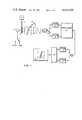
- FIG. 2is a block diagram of an embodiment of the invention using an energy-selective detection system.
- FIG. 1 of the drawingsAn understanding of the broad aspects of the invention may best be had by reference to FIG. 1 of the drawings. It is desired to make an image of blood vessel 11 in a region of the body 10. In the prior art, images were taken before and after the intravenous administration of an iodinated contrast agent 26 using syringe 2 and subtracted. These images are often distorted by motion artifacts due to involuntary soft tissue motions occurring between the two images.
- the stored measurements at each energy spectracan be processed to provide selective material imaging.
- a limited class of materialscan be selected.
- the processor 19provides substantial cancellation of the soft tissue components so that the processed data set contains information primarily of bone.
- Cancellation of soft tissueis provided since it represents the source of involuntary motions such as swallowing, breathing, heartbeat, pulsating vessels, peristalsis, etc. If a patient is asked to hold still during an intravenous injection he can usually reliably hold his bone structure steady. Therefore the bone motion is not a problem.
- This first processed data set from processor 19is stored, for example, in digital store 20 using switch 25.
- contrast agent 26usually an iodinated material
- syringe 2an appropriate time is allowed to pass until the iodine reaches the vessel to be imaged 11. This can represent, for example, the the aorta or the carotid artery.
- the entire dual energy imaging processis then repeated using the two source energies, this time in the presence of the iodinated contrast agent.
- Processor 19again takes the two measurement sets and produces a processed data set where the soft tissue has been substantially cancelled so that soft tissue motion cannot cause artifacts.
- the second data setcontains both bone and the iodine image information.
- This second data setis stored in digital store 21 using switch 25.
- Storage systems 20 and 21essentially contain bone images without and with the iodinated contrast image information.
- Combiner 22can simply be a subtraction operation which subtracts the data set in 20 from the data set in 21 to provide an isolated image of the iodinated contrast agent in the vessel 24 in display monitor 23. Since the bone image is the same in both data sets, the difference is the iodine alone. The resultant image is immune to soft tissue motions since energy information is used to cancel the soft tissue prior to the final subtraction.
- a preferred system for processor 19is that described in the previously referenced U.S. Pat. No. 4,029,963 and in the previously referenced paper R. E. Alvarez.
- the measurement data at the two energy spectraare processed to provide two energy-independent data sets. These can represent the Compton scattering and photoelectric components or, alternatively, two specific material components such as calibration materials aluminum and plastic.
- a linear weighted sum of these two energy-independent componentscan be used, for example, to cancel any material. For this system the appropriate weights are used to cancel the soft tissue.
- the logs of the measurementsare derived. These logs are then applied to the polynomial nonlinear equation to extract the line integrals of the two desired basis components. If, for example, monoenergetic x-rays at different energies were used, only the logarithm plus linear processing would be necessary to provide the desired line integrals.
- the nonlinear polynomialscorrect for the nonlinearities caused by beam hardening.
- Source 12is the conventional broad-energy x-ray source encompassing the diagnostic energy spectrum.
- the transmitted radiationis projected through the body 10 to detector array 30.
- Each element of the arrayconsists of a front and back part.
- the detector materialcan be a scintillator such as sodium iodide, in which case photodetectors are used to measure the light output at the front and back parts.
- the detector materialcan be a high-pressure gas, such as Xenon, in which case isolated wires are used to individually collect the charge in the front and back parts. For clarity, these wires are shown collecting low-energy measurement signals from the front halves and high-energy measurement signals from the back halves.
- Front detector elements 31, 32 and 33have their low-energy measurements stored in digital store 18, as with the system in FIG. 1.
- back detector elements 34, 35 and 36have their high-energy measurements stored in digital store 17.
- front detectors 37, 38 and 39are connected to store 18
- back detectors 40, 41 and 42are connected to store 17.
- These measurementsare processed in processor 19 exactly as in the system in FIG. 1, with the remainder of the system being identical and thus not shown.
- the high and low energy measurement signalsare derived simultaneously using energy selective detectors. As before, these measurements are made both before and after the administration of the contrast agent.
- Array 30could be a two-dimensional array encompassing the entire image. For economic reasons, however, it is preferable to use a single line array. This line array is scanned along the image plane, with respect to the body 10, to provide a complete image measurement set. Alternatively the detector can be stationary and the patient 10 scanned as is done in the commercially available GE Scoutview system.
- the embodiments describedshowed two energy measurements before and after the administration of a contrast agent.
- the inventionis clearly much broader in concept. It may often be desirable to cancel a variety of materials which may exhibit motion during the period before and after contrast administration. Thus a plurality of energy measurements, as described in U.S. Pat. No. 3,848,130, would be used to provide the required selectivity before and after the administering of the contrast agent.
- iodinated contrast agentswere used as an example for vessel imaging, many others can be used including barium, xenon, tantalum, etc.
Landscapes
- Health & Medical Sciences (AREA)
- Life Sciences & Earth Sciences (AREA)
- Engineering & Computer Science (AREA)
- Medical Informatics (AREA)
- Radiology & Medical Imaging (AREA)
- Molecular Biology (AREA)
- Biophysics (AREA)
- High Energy & Nuclear Physics (AREA)
- Veterinary Medicine (AREA)
- Nuclear Medicine, Radiotherapy & Molecular Imaging (AREA)
- Optics & Photonics (AREA)
- Pathology (AREA)
- Public Health (AREA)
- Biomedical Technology (AREA)
- Heart & Thoracic Surgery (AREA)
- Physics & Mathematics (AREA)
- Surgery (AREA)
- Animal Behavior & Ethology (AREA)
- General Health & Medical Sciences (AREA)
- Multimedia (AREA)
- Signal Processing (AREA)
- Vascular Medicine (AREA)
- Dentistry (AREA)
- Oral & Maxillofacial Surgery (AREA)
- Apparatus For Radiation Diagnosis (AREA)
- Transforming Light Signals Into Electric Signals (AREA)
Abstract
Description
Claims (6)
Priority Applications (9)
| Application Number | Priority Date | Filing Date | Title |
|---|---|---|---|
| US06/260,694US4445226A (en) | 1981-05-05 | 1981-05-05 | Multiple-energy X-ray subtraction imaging system |
| GB8211626AGB2098425B (en) | 1981-05-05 | 1982-04-22 | Multiple-energy x-ray subtraction imaging system |
| DE19823216458DE3216458A1 (en) | 1981-05-05 | 1982-05-03 | DEVICE AND METHOD FOR GENERATING A CONTRAST AGENT PROJECTION IMAGE |
| NL8201836ANL191280B (en) | 1981-05-05 | 1982-05-04 | Method and apparatus for processing X-ray projection images. |
| FR8207711AFR2505168B1 (en) | 1981-05-05 | 1982-05-04 | APPARATUS AND METHOD FOR PRODUCING A PROJECTED IMAGE OF ADMINISTERED CONTRAST MATTER IN A BODY REGION |
| IL65676AIL65676A (en) | 1981-05-05 | 1982-05-04 | Multiple-energy x-ray subtraction imaging system |
| ES512571AES8306961A1 (en) | 1981-05-05 | 1982-05-05 | "PROCEDURE TO PROVIDE A PROJECTION IMAGE OF A CONTRAST MATERIAL ADMINISTERED IN A REGION OF THE BODY AND APPARATUS FOR THE REALIZATION OF THE SAME". |
| JP57075951AJPS5832744A (en) | 1981-05-05 | 1982-05-06 | Multiple energy x-ray subtracting photograph system |
| US06/577,074US4611341A (en) | 1981-05-05 | 1984-02-06 | Multiple-energy x-ray substraction imaging system |
Applications Claiming Priority (1)
| Application Number | Priority Date | Filing Date | Title |
|---|---|---|---|
| US06/260,694US4445226A (en) | 1981-05-05 | 1981-05-05 | Multiple-energy X-ray subtraction imaging system |
Related Child Applications (1)
| Application Number | Title | Priority Date | Filing Date |
|---|---|---|---|
| US06/577,074ContinuationUS4611341A (en) | 1981-05-05 | 1984-02-06 | Multiple-energy x-ray substraction imaging system |
Publications (1)
| Publication Number | Publication Date |
|---|---|
| US4445226Atrue US4445226A (en) | 1984-04-24 |
Family
ID=22990214
Family Applications (1)
| Application Number | Title | Priority Date | Filing Date |
|---|---|---|---|
| US06/260,694Expired - LifetimeUS4445226A (en) | 1981-05-05 | 1981-05-05 | Multiple-energy X-ray subtraction imaging system |
Country Status (8)
| Country | Link |
|---|---|
| US (1) | US4445226A (en) |
| JP (1) | JPS5832744A (en) |
| DE (1) | DE3216458A1 (en) |
| ES (1) | ES8306961A1 (en) |
| FR (1) | FR2505168B1 (en) |
| GB (1) | GB2098425B (en) |
| IL (1) | IL65676A (en) |
| NL (1) | NL191280B (en) |
Cited By (39)
| Publication number | Priority date | Publication date | Assignee | Title |
|---|---|---|---|---|
| US4499493A (en)* | 1983-02-22 | 1985-02-12 | The Board Of Trustees Of The Leland Stanford Junior University | Multiple measurement noise reducing system using artifact edge identification and selective signal processing |
| US4503461A (en)* | 1983-02-22 | 1985-03-05 | The Board Of Trustees Of The Leland, Stanford Junior University | Multiple measurement noise reducing system using space-variant filters |
| US4506327A (en)* | 1981-11-23 | 1985-03-19 | General Electric Company | Limited-angle imaging using multiple energy scanning |
| US4528685A (en)* | 1983-05-16 | 1985-07-09 | General Electric Company | X-ray beam filter device |
| US4542459A (en)* | 1982-11-26 | 1985-09-17 | General Electric Company | Matched filter for x-ray hybrid subtraction |
| US4559557A (en)* | 1984-06-01 | 1985-12-17 | General Electric Company | Region-of-interest digital subtraction angiography |
| US4647779A (en)* | 1985-05-13 | 1987-03-03 | Clayton Foundation For Research | Multiple layer positron emission tomography camera |
| US4677299A (en)* | 1985-05-13 | 1987-06-30 | Clayton Foundation For Research | Multiple layer positron emission tomography camera |
| US4736398A (en)* | 1985-05-11 | 1988-04-05 | Deutsches Elektronen-Synchrotron Desy | Apparatus for the digital subtraction angiography in energy subtraction mode |
| DE3734300A1 (en)* | 1986-10-09 | 1988-05-26 | Hitachi Ltd | SPECTRAL IMAGING SYSTEM |
| US4766603A (en)* | 1983-11-18 | 1988-08-23 | Kabushiki Kaisha Toshiba | Aperture device of radiation diagnostic apparatus |
| EP0244766A3 (en)* | 1986-05-06 | 1989-04-12 | General Electric Company | Dual energy imaging with kinestatic charge detector |
| WO1990010859A1 (en)* | 1989-03-07 | 1990-09-20 | Hologic, Inc. | Apparatus and method for analysis using x-rays |
| US5148455A (en)* | 1986-07-14 | 1992-09-15 | Hologic, Inc. | Bone densitometer |
| EP0571017A3 (en)* | 1992-05-09 | 1995-05-31 | Philips Patentverwaltung | Filtering procedure for an X-ray system and arrangement to carry out such a filtering procedure. |
| USRE37536E1 (en) | 1982-11-26 | 2002-02-05 | Uab Research Foundation | Split energy level radiation detection |
| US6445767B1 (en) | 1989-12-05 | 2002-09-03 | University Of Massachussetts Medical Center | System for quantitative radiographic imaging |
| US6487274B2 (en) | 2001-01-29 | 2002-11-26 | Siemens Medical Solutions Usa, Inc. | X-ray target assembly and radiation therapy systems and methods |
| US20040101089A1 (en)* | 2002-11-27 | 2004-05-27 | Karau Kelly Lynn | Methods and apparatus for detecting structural, perfusion, and functional abnormalities |
| US20040101090A1 (en)* | 2002-11-27 | 2004-05-27 | Danielle Drummond | Methods and apparatus for acquiring perfusion data |
| US20040247082A1 (en)* | 2003-06-05 | 2004-12-09 | Ge Medical Systems Global Technology Company, Llc | Ct imaging system with multiple peak x-ray source |
| US20050025278A1 (en)* | 2003-07-29 | 2005-02-03 | Akira Hagiwara | X-ray CT system |
| NL1024689C2 (en)* | 2002-11-08 | 2005-02-15 | Ge Med Sys Global Tech Co Llc | Method and device for detecting structural, perfusion, and functional abnormalities. |
| US20050259891A1 (en)* | 2004-04-05 | 2005-11-24 | Fuji Photo Film Co., Ltd. | Apparatus, method, and program for producing subtraction images |
| US7330531B1 (en)* | 1989-12-05 | 2008-02-12 | University Of Massachusetts Medical Center | System for quantitative radiographic imaging |
| US20080043917A1 (en)* | 2006-02-09 | 2008-02-21 | L-3 Communications Security and Detection Systems Inc. | Selective generation of radiation at multiple energy levels |
| US20090022263A1 (en)* | 2007-07-18 | 2009-01-22 | Ge Medical Systems Global Technology Company, Llc. | X-ray ct apparatus and method of generating an image |
| US20090052612A1 (en)* | 2007-08-22 | 2009-02-26 | Xiaoye Wu | System and method of optimizing a monochromatic representation of basis material decomposed ct images |
| US20090161939A1 (en)* | 2007-12-21 | 2009-06-25 | General Electric Company | System and method for extracting features of interest from an image |
| US20090207966A1 (en)* | 2008-02-15 | 2009-08-20 | Shkumat Nick A | Image acquisition for dual energy imaging |
| US20100119040A1 (en)* | 2008-11-11 | 2010-05-13 | Hamamatsu Photonics K.K. | Radiation detection device, radiation image acquiring system, and method for detecting radiation |
| US20100119038A1 (en)* | 2008-11-11 | 2010-05-13 | Hamamatsu Photonics K.K. | Radiation detection device, radiation image acquiring system, radiation inspection system, and radiation detection method |
| US20110075810A1 (en)* | 2009-09-25 | 2011-03-31 | Fujifilm Corporation | Radiation imaging apparatus and imaging control device |
| US20110164797A1 (en)* | 2010-01-06 | 2011-07-07 | Samsung Electronics Co., Ltd. | Method and system of processing multi-energy x-ray images |
| US20140328453A1 (en)* | 2011-06-17 | 2014-11-06 | The Board Of Trustees Of The Leland Stanford Junior University | Computed tomography system with dynamic bowtie filter |
| US9069092B2 (en) | 2012-02-22 | 2015-06-30 | L-3 Communication Security and Detection Systems Corp. | X-ray imager with sparse detector array |
| US9414792B2 (en) | 2011-06-17 | 2016-08-16 | The Board Of Trustees Of The Leland Stanford Junior University | Computed tomography system with dynamic bowtie filter |
| US20200408933A1 (en)* | 2018-03-20 | 2020-12-31 | Canon Kabushiki Kaisha | Radiation imaging system, imaging control apparatus, and method |
| CN112969412A (en)* | 2018-11-07 | 2021-06-15 | 皇家飞利浦有限公司 | Deep profile bolus tracking |
Families Citing this family (18)
| Publication number | Priority date | Publication date | Assignee | Title |
|---|---|---|---|---|
| US4393402A (en)* | 1981-06-08 | 1983-07-12 | General Electric Company | Subtraction fluoroscopy method and apparatus |
| US4482918A (en)* | 1982-04-26 | 1984-11-13 | General Electric Company | Method and apparatus for X-ray image subtraction |
| DE3221179A1 (en)* | 1982-06-04 | 1983-12-08 | Siemens AG, 1000 Berlin und 8000 München | LAYER RECORDING DEVICE FOR PRODUCING TRANSVERSAL LAYER IMAGES |
| JPS59200636A (en)* | 1983-04-27 | 1984-11-14 | 株式会社東芝 | X-ray CT device |
| NL8304398A (en)* | 1983-12-22 | 1985-07-16 | Philips Nv | ROENTGEN RESEARCH DEVICE WITH SELECTIVE FILTER. |
| NL8401946A (en)* | 1984-06-19 | 1986-01-16 | Optische Ind De Oude Delft Nv | SYSTEM FOR DETECTING TWO X-RAY RADIATION ENERGIES. |
| EP0182099B1 (en)* | 1984-10-16 | 1996-09-11 | Fuji Photo Film Co., Ltd. | Radiation image recording and read-out apparatus |
| JPS61124058A (en)* | 1984-11-20 | 1986-06-11 | Matsushita Electric Ind Co Ltd | Manufacturing method of paste-type cadmium negative electrode |
| JPS61193363A (en)* | 1985-02-20 | 1986-08-27 | Shin Kobe Electric Mach Co Ltd | Nickel cadmium alkaline storage battery |
| EP0257199B1 (en)* | 1986-07-18 | 1992-11-11 | Siemens Aktiengesellschaft | Device for the destruction of calculi |
| JPH03106343A (en)* | 1989-09-20 | 1991-05-02 | Shimadzu Corp | X-ray picture diagnostic device |
| US5509042A (en)* | 1991-02-13 | 1996-04-16 | Lunar Corporation | Automated determination and analysis of bone morphology |
| WO1994006351A1 (en)* | 1991-02-13 | 1994-03-31 | Lunar Corporation | Automated determination and analysis of bone morphology |
| US5228068A (en)* | 1992-09-14 | 1993-07-13 | Lunar Corporation | Device and method for automated determination and analysis of bone density and vertebral morphology |
| US5577089A (en)* | 1991-02-13 | 1996-11-19 | Lunar Corporation | Device and method for analysis of bone morphology |
| JPH08265647A (en)* | 1995-03-20 | 1996-10-11 | Fuji Photo Film Co Ltd | Method and device for detecting secular change in radiation image |
| US6284410B1 (en) | 1997-08-01 | 2001-09-04 | Duracell Inc. | Zinc electrode particle form |
| JP5680718B2 (en)* | 2007-08-15 | 2015-03-04 | 富士フイルム株式会社 | Image component separation apparatus, method, and program |
Citations (2)
| Publication number | Priority date | Publication date | Assignee | Title |
|---|---|---|---|---|
| US3974386A (en)* | 1974-07-12 | 1976-08-10 | Wisconsin Alumni Research Foundation | Differential X-ray method and apparatus |
| US4029963A (en)* | 1976-07-30 | 1977-06-14 | The Board Of Trustees Of Leland Stanford Junior University | X-ray spectral decomposition imaging system |
Family Cites Families (7)
| Publication number | Priority date | Publication date | Assignee | Title |
|---|---|---|---|---|
| US3582651A (en)* | 1968-08-22 | 1971-06-01 | Westinghouse Electric Corp | X-ray image storage,reproduction and comparison system |
| US3848130A (en)* | 1973-06-25 | 1974-11-12 | A Macovski | Selective material x-ray imaging system |
| US3974380A (en)* | 1975-01-17 | 1976-08-10 | Balzers Patent-Und Beteiligungs Ag | Mass spectrometer |
| GB2020945B (en)* | 1978-05-16 | 1982-12-01 | Wisconsin Alumni Res Found | Real-time digital x-ray substraction imaging |
| NL184298C (en)* | 1979-07-19 | 1989-06-01 | Philips Nv | DEVICE FOR DIFFERENCE IMAGE DETERMINATION. |
| DE3018129C1 (en)* | 1980-05-12 | 1981-10-01 | Siemens AG, 1000 Berlin und 8000 München | X-ray diagnostic facility for creating subtraction images |
| NL8003354A (en)* | 1980-06-09 | 1982-01-04 | Philips Nv | RADIATION EXAMINATION DEVICE WITH IMAGE SUBTRACTION. |
- 1981
- 1981-05-05USUS06/260,694patent/US4445226A/ennot_activeExpired - Lifetime
- 1982
- 1982-04-22GBGB8211626Apatent/GB2098425B/ennot_activeExpired
- 1982-05-03DEDE19823216458patent/DE3216458A1/enactiveGranted
- 1982-05-04FRFR8207711Apatent/FR2505168B1/ennot_activeExpired
- 1982-05-04NLNL8201836Apatent/NL191280B/ennot_activeApplication Discontinuation
- 1982-05-04ILIL65676Apatent/IL65676A/ennot_activeIP Right Cessation
- 1982-05-05ESES512571Apatent/ES8306961A1/ennot_activeExpired
- 1982-05-06JPJP57075951Apatent/JPS5832744A/enactiveGranted
Patent Citations (2)
| Publication number | Priority date | Publication date | Assignee | Title |
|---|---|---|---|---|
| US3974386A (en)* | 1974-07-12 | 1976-08-10 | Wisconsin Alumni Research Foundation | Differential X-ray method and apparatus |
| US4029963A (en)* | 1976-07-30 | 1977-06-14 | The Board Of Trustees Of Leland Stanford Junior University | X-ray spectral decomposition imaging system |
Cited By (68)
| Publication number | Priority date | Publication date | Assignee | Title |
|---|---|---|---|---|
| US4506327A (en)* | 1981-11-23 | 1985-03-19 | General Electric Company | Limited-angle imaging using multiple energy scanning |
| US4542459A (en)* | 1982-11-26 | 1985-09-17 | General Electric Company | Matched filter for x-ray hybrid subtraction |
| USRE37536E1 (en) | 1982-11-26 | 2002-02-05 | Uab Research Foundation | Split energy level radiation detection |
| US4503461A (en)* | 1983-02-22 | 1985-03-05 | The Board Of Trustees Of The Leland, Stanford Junior University | Multiple measurement noise reducing system using space-variant filters |
| US4499493A (en)* | 1983-02-22 | 1985-02-12 | The Board Of Trustees Of The Leland Stanford Junior University | Multiple measurement noise reducing system using artifact edge identification and selective signal processing |
| US4528685A (en)* | 1983-05-16 | 1985-07-09 | General Electric Company | X-ray beam filter device |
| US4766603A (en)* | 1983-11-18 | 1988-08-23 | Kabushiki Kaisha Toshiba | Aperture device of radiation diagnostic apparatus |
| US4559557A (en)* | 1984-06-01 | 1985-12-17 | General Electric Company | Region-of-interest digital subtraction angiography |
| US4736398A (en)* | 1985-05-11 | 1988-04-05 | Deutsches Elektronen-Synchrotron Desy | Apparatus for the digital subtraction angiography in energy subtraction mode |
| US4677299A (en)* | 1985-05-13 | 1987-06-30 | Clayton Foundation For Research | Multiple layer positron emission tomography camera |
| US4647779A (en)* | 1985-05-13 | 1987-03-03 | Clayton Foundation For Research | Multiple layer positron emission tomography camera |
| EP0244766A3 (en)* | 1986-05-06 | 1989-04-12 | General Electric Company | Dual energy imaging with kinestatic charge detector |
| US5040199A (en)* | 1986-07-14 | 1991-08-13 | Hologic, Inc. | Apparatus and method for analysis using x-rays |
| US5148455A (en)* | 1986-07-14 | 1992-09-15 | Hologic, Inc. | Bone densitometer |
| DE3734300A1 (en)* | 1986-10-09 | 1988-05-26 | Hitachi Ltd | SPECTRAL IMAGING SYSTEM |
| US4890310A (en)* | 1986-10-09 | 1989-12-26 | Hitachi, Ltd. | Spectral type radiation imaging system |
| WO1990010859A1 (en)* | 1989-03-07 | 1990-09-20 | Hologic, Inc. | Apparatus and method for analysis using x-rays |
| US20020196899A1 (en)* | 1989-12-05 | 2002-12-26 | University Of Massachusetts Medical Center | System for quantitative radiographic imaging |
| US6445767B1 (en) | 1989-12-05 | 2002-09-03 | University Of Massachussetts Medical Center | System for quantitative radiographic imaging |
| US7330531B1 (en)* | 1989-12-05 | 2008-02-12 | University Of Massachusetts Medical Center | System for quantitative radiographic imaging |
| EP0571017A3 (en)* | 1992-05-09 | 1995-05-31 | Philips Patentverwaltung | Filtering procedure for an X-ray system and arrangement to carry out such a filtering procedure. |
| US6487274B2 (en) | 2001-01-29 | 2002-11-26 | Siemens Medical Solutions Usa, Inc. | X-ray target assembly and radiation therapy systems and methods |
| NL1024689C2 (en)* | 2002-11-08 | 2005-02-15 | Ge Med Sys Global Tech Co Llc | Method and device for detecting structural, perfusion, and functional abnormalities. |
| US20040101089A1 (en)* | 2002-11-27 | 2004-05-27 | Karau Kelly Lynn | Methods and apparatus for detecting structural, perfusion, and functional abnormalities |
| US20040101090A1 (en)* | 2002-11-27 | 2004-05-27 | Danielle Drummond | Methods and apparatus for acquiring perfusion data |
| US6813333B2 (en)* | 2002-11-27 | 2004-11-02 | Ge Medical Systems Global Technology Company, Llc | Methods and apparatus for detecting structural, perfusion, and functional abnormalities |
| US20050018808A1 (en)* | 2002-11-27 | 2005-01-27 | Piacsek Kelly Lynn | Methods and apparatus for detecting structural, perfusion, and functional abnormalities |
| US6891918B2 (en)* | 2002-11-27 | 2005-05-10 | Ge Medical Systems Global Technology Company, Llc | Methods and apparatus for acquiring perfusion data |
| US7058155B2 (en) | 2002-11-27 | 2006-06-06 | General Electric Company | Methods and apparatus for detecting structural, perfusion, and functional abnormalities |
| US20040247082A1 (en)* | 2003-06-05 | 2004-12-09 | Ge Medical Systems Global Technology Company, Llc | Ct imaging system with multiple peak x-ray source |
| US7778382B2 (en)* | 2003-06-05 | 2010-08-17 | General Electric Company | CT imaging system with multiple peak x-ray source |
| US7120222B2 (en)* | 2003-06-05 | 2006-10-10 | General Electric Company | CT imaging system with multiple peak x-ray source |
| US20060285645A1 (en)* | 2003-06-05 | 2006-12-21 | Hoffman David M | CT imaging system with multiple peak X-ray source |
| US7190758B2 (en)* | 2003-07-29 | 2007-03-13 | Ge Medical Systems Global Technology Company, Llc | X-ray CT system |
| US20050025278A1 (en)* | 2003-07-29 | 2005-02-03 | Akira Hagiwara | X-ray CT system |
| US20050259891A1 (en)* | 2004-04-05 | 2005-11-24 | Fuji Photo Film Co., Ltd. | Apparatus, method, and program for producing subtraction images |
| US20080043917A1 (en)* | 2006-02-09 | 2008-02-21 | L-3 Communications Security and Detection Systems Inc. | Selective generation of radiation at multiple energy levels |
| US7606349B2 (en)* | 2006-02-09 | 2009-10-20 | L-3 Communications Security and Detection Systems Inc. | Selective generation of radiation at multiple energy levels |
| US20090022263A1 (en)* | 2007-07-18 | 2009-01-22 | Ge Medical Systems Global Technology Company, Llc. | X-ray ct apparatus and method of generating an image |
| US7639773B2 (en) | 2007-07-18 | 2009-12-29 | Ge Medical Systems Global Technology Company, Llc | X-ray CT apparatus and method of generating an image |
| US20090052612A1 (en)* | 2007-08-22 | 2009-02-26 | Xiaoye Wu | System and method of optimizing a monochromatic representation of basis material decomposed ct images |
| US7724865B2 (en)* | 2007-08-22 | 2010-05-25 | General Electric Company | System and method of optimizing a monochromatic representation of basis material decomposed CT images |
| US20090161939A1 (en)* | 2007-12-21 | 2009-06-25 | General Electric Company | System and method for extracting features of interest from an image |
| US9070181B2 (en)* | 2007-12-21 | 2015-06-30 | General Electric Company | System and method for extracting features of interest from an image |
| US8019044B2 (en)* | 2008-02-15 | 2011-09-13 | Shkumat Nick A | Image acquisition for dual energy imaging |
| US20090207966A1 (en)* | 2008-02-15 | 2009-08-20 | Shkumat Nick A | Image acquisition for dual energy imaging |
| US8223922B2 (en)* | 2008-11-11 | 2012-07-17 | Hamamatsu Photonics K.K. | Radiation detection device, radiation image acquiring system, radiation inspection system, and radiation detection method |
| US9594031B2 (en) | 2008-11-11 | 2017-03-14 | Hamamatsu Photonics K.K. | Radiation detection device, radiation image acquiring system, radiation inspection system, and radiation detection method |
| US10393676B2 (en) | 2008-11-11 | 2019-08-27 | Hamamatsu Photonics K.K. | Radiation detection device, radiation image acquiring system, radiation inspection system, and radiation detection method |
| US20100119040A1 (en)* | 2008-11-11 | 2010-05-13 | Hamamatsu Photonics K.K. | Radiation detection device, radiation image acquiring system, and method for detecting radiation |
| US20100119038A1 (en)* | 2008-11-11 | 2010-05-13 | Hamamatsu Photonics K.K. | Radiation detection device, radiation image acquiring system, radiation inspection system, and radiation detection method |
| US8280005B2 (en) | 2008-11-11 | 2012-10-02 | Hamamatsu Photonics K.K. | Radiation detection device, radiation image acquiring system, and method for detecting radiation |
| US8964939B2 (en) | 2008-11-11 | 2015-02-24 | Hamamatsu Photonics K.K. | Radiation detection device, radiation image acquiring system, radiation inspection system, and radiation detection method |
| US8600005B2 (en) | 2008-11-11 | 2013-12-03 | Hamamatsu Photonics K.K. | Radiation detection device, radiation image acquiring system, and method for detecting radiation |
| US8571178B2 (en)* | 2009-09-25 | 2013-10-29 | Fujifilm Corporation | Radiation imaging apparatus and imaging control device controlling a filter based on subject information |
| US20110075810A1 (en)* | 2009-09-25 | 2011-03-31 | Fujifilm Corporation | Radiation imaging apparatus and imaging control device |
| WO2011083973A2 (en) | 2010-01-06 | 2011-07-14 | Samsung Electronics Co., Ltd. | Method and system of processing multi-energy x-ray images |
| US8761485B2 (en) | 2010-01-06 | 2014-06-24 | Samsung Electronics Co., Ltd. | Method and system of processing multi-energy X-ray images |
| US20110164797A1 (en)* | 2010-01-06 | 2011-07-07 | Samsung Electronics Co., Ltd. | Method and system of processing multi-energy x-ray images |
| EP2521488A4 (en)* | 2010-01-06 | 2013-12-25 | Samsung Electronics Co Ltd | METHOD AND SYSTEM FOR PROCESSING MULTI-ENERGY X-RAY IMAGES |
| US20140328453A1 (en)* | 2011-06-17 | 2014-11-06 | The Board Of Trustees Of The Leland Stanford Junior University | Computed tomography system with dynamic bowtie filter |
| US9414792B2 (en) | 2011-06-17 | 2016-08-16 | The Board Of Trustees Of The Leland Stanford Junior University | Computed tomography system with dynamic bowtie filter |
| US9521982B2 (en)* | 2011-06-17 | 2016-12-20 | The Board Of Trustees Of The Leland Stanford Junior University | Computed tomography system with dynamic bowtie filter |
| US9069092B2 (en) | 2012-02-22 | 2015-06-30 | L-3 Communication Security and Detection Systems Corp. | X-ray imager with sparse detector array |
| US20200408933A1 (en)* | 2018-03-20 | 2020-12-31 | Canon Kabushiki Kaisha | Radiation imaging system, imaging control apparatus, and method |
| US12097061B2 (en)* | 2018-03-20 | 2024-09-24 | Canon Kabushiki Kaisha | Radiation imaging system, imaging control apparatus, and method |
| CN112969412A (en)* | 2018-11-07 | 2021-06-15 | 皇家飞利浦有限公司 | Deep profile bolus tracking |
| US12310774B2 (en) | 2018-11-07 | 2025-05-27 | Koninklijke Philips N.V. | Deep spectral bolus tracking |
Also Published As
| Publication number | Publication date |
|---|---|
| JPH0369533B2 (en) | 1991-11-01 |
| IL65676A0 (en) | 1982-08-31 |
| ES512571A0 (en) | 1983-06-16 |
| FR2505168A1 (en) | 1982-11-12 |
| NL191280B (en) | 1994-12-01 |
| DE3216458C2 (en) | 1989-11-23 |
| NL8201836A (en) | 1982-12-01 |
| ES8306961A1 (en) | 1983-06-16 |
| GB2098425B (en) | 1985-06-12 |
| IL65676A (en) | 1985-02-28 |
| FR2505168B1 (en) | 1986-07-11 |
| DE3216458A1 (en) | 1982-11-25 |
| GB2098425A (en) | 1982-11-17 |
| JPS5832744A (en) | 1983-02-25 |
Similar Documents
| Publication | Publication Date | Title |
|---|---|---|
| US4445226A (en) | Multiple-energy X-ray subtraction imaging system | |
| US4662379A (en) | Coronary artery imaging system using gated tomosynthesis | |
| US4611341A (en) | Multiple-energy x-ray substraction imaging system | |
| Kruger et al. | A digital video image processor for real-time x-ray subtraction imaging | |
| JPS58109032A (en) | body imaging device | |
| JPS62290443A (en) | Apparatus and method for calculating two kinds of energy difference images by dynamic and static charge detector | |
| US20220167935A1 (en) | Image processing apparatus, radiation imaging system, image processing method, and non-transitory computer-readable storage medium | |
| EP0655861B1 (en) | Image composition method and imaging apparatus for performing said method | |
| JP2022097760A (en) | Radiation imaging system, imaging control device, and method | |
| Brody et al. | Intravenous carotid arteriography using line-scanned digital radiography. | |
| WO2019181230A1 (en) | Radiography system, photography control device, and method | |
| EP0041752A1 (en) | Radiography apparatus incorporating image subtraction | |
| KR860001794B1 (en) | X-ray diagnostic device | |
| Brody et al. | Intravenous angiography using scanned projection radiography: Preliminary investigation of a new method | |
| Hughes et al. | The application of synchrotron radiation to non-invasive angiography | |
| Brody et al. | Intravenous arteriography using scanned projection radiography. | |
| Macovski et al. | Isolated iodine images using spatial‐frequency encoding | |
| Keyes et al. | Hybrid subtraction in digital fluorography | |
| Guthaner et al. | Intravenous aortography after aortic dissection repair | |
| Macovski | Iodine imaging using spectral analysis | |
| Shaw et al. | Intravenous angiography using computerized fluoroscopy | |
| Maher et al. | Digital fluoroscopy: a new development in medical imaging | |
| Maurino et al. | Feasibility of lateral chest dual-energy subtraction radiography using a stacked single-exposure multi-layer x-ray detector | |
| Nalcioglu et al. | Comparison of digital subtraction video densitometry and area length method in the determination of left ventricular ejection fraction | |
| Elliott et al. | Significance of the caudal left-anterior-oblique view in analyzing the left main coronary artery and its major branches. |
Legal Events
| Date | Code | Title | Description |
|---|---|---|---|
| AS | Assignment | Owner name:BOARD OF TRUSTEES OF LELAND STANFORD, JR. UNIVERSI Free format text:ASSIGNMENT OF ASSIGNORS INTEREST.;ASSIGNOR:BRODY, WILLIAM R.;REEL/FRAME:003947/0732 Effective date:19810528 Owner name:BOARD OF TRUSTEES OF LELAND STANFORD, JR.,CALIFORN Free format text:ASSIGNMENT OF ASSIGNORS INTEREST;ASSIGNOR:BRODY, WILLIAM R.;REEL/FRAME:003947/0732 Effective date:19810528 Owner name:BOARD OF TRUSTEES OF LELAND STANFORD, JR., CALIFOR Free format text:ASSIGNMENT OF ASSIGNORS INTEREST;ASSIGNOR:BRODY, WILLIAM R.;REEL/FRAME:003947/0732 Effective date:19810528 | |
| AS | Assignment | Owner name:BOARD OF TRUSTEES OF THE LELAND STANFORD JUNIOR UN Free format text:ASSIGNMENT OF ASSIGNORS INTEREST.;ASSIGNOR:BRODY, WILLIAM R.;REEL/FRAME:003971/0960 Effective date:19820414 | |
| STCF | Information on status: patent grant | Free format text:PATENTED CASE | |
| FPAY | Fee payment | Year of fee payment:4 | |
| FEPP | Fee payment procedure | Free format text:PAYOR NUMBER ASSIGNED (ORIGINAL EVENT CODE: ASPN); ENTITY STATUS OF PATENT OWNER: LARGE ENTITY | |
| FPAY | Fee payment | Year of fee payment:8 | |
| CC | Certificate of correction | ||
| FPAY | Fee payment | Year of fee payment:12 |