US4338024A - Flow analyzer and system for analysis of fluids with particles - Google Patents
Flow analyzer and system for analysis of fluids with particlesDownload PDFInfo
- Publication number
- US4338024A US4338024AUS06/146,064US14606480AUS4338024AUS 4338024 AUS4338024 AUS 4338024AUS 14606480 AUS14606480 AUS 14606480AUS 4338024 AUS4338024 AUS 4338024A
- Authority
- US
- United States
- Prior art keywords
- particles
- path
- fluid
- inlet
- width
- Prior art date
- Legal status (The legal status is an assumption and is not a legal conclusion. Google has not performed a legal analysis and makes no representation as to the accuracy of the status listed.)
- Expired - Lifetime
Links
- 239000002245particleSubstances0.000titleclaimsabstractdescription54
- 239000012530fluidSubstances0.000titleclaimsabstractdescription21
- 238000004458analytical methodMethods0.000titleclaimsdescription10
- 210000004027cellAnatomy0.000claimsabstractdescription35
- 238000000034methodMethods0.000claimsabstractdescription13
- 210000000601blood cellAnatomy0.000claimsabstractdescription8
- 210000004369bloodAnatomy0.000claimsdescription13
- 239000008280bloodSubstances0.000claimsdescription13
- 238000003384imaging methodMethods0.000claimsdescription9
- 210000002700urineAnatomy0.000claimsdescription2
- 230000007423decreaseEffects0.000claims6
- 239000000523sampleSubstances0.000claims6
- 239000007788liquidSubstances0.000claims4
- 239000012472biological sampleSubstances0.000claims2
- 239000002131composite materialSubstances0.000description4
- 230000009977dual effectEffects0.000description3
- 238000005259measurementMethods0.000description3
- 230000003287optical effectEffects0.000description3
- 238000004364calculation methodMethods0.000description2
- 239000013078crystalSubstances0.000description2
- 235000015097nutrientsNutrition0.000description2
- 238000004886process controlMethods0.000description2
- 241000894006BacteriaSpecies0.000description1
- 102000001554HemoglobinsHuman genes0.000description1
- 108010054147HemoglobinsProteins0.000description1
- FAPWRFPIFSIZLT-UHFFFAOYSA-MSodium chlorideChemical compound[Na+].[Cl-]FAPWRFPIFSIZLT-UHFFFAOYSA-M0.000description1
- 238000004820blood countMethods0.000description1
- 230000008859changeEffects0.000description1
- 230000002596correlated effectEffects0.000description1
- 238000010586diagramMethods0.000description1
- 238000000684flow cytometryMethods0.000description1
- 230000006870functionEffects0.000description1
- 238000010191image analysisMethods0.000description1
- 238000009629microbiological cultureMethods0.000description1
- 244000005700microbiomeSpecies0.000description1
- 238000003909pattern recognitionMethods0.000description1
- 229920000642polymerPolymers0.000description1
- 230000011218segmentationEffects0.000description1
- 238000000926separation methodMethods0.000description1
- 210000002966serumAnatomy0.000description1
- 230000003068static effectEffects0.000description1
- 230000009466transformationEffects0.000description1
- 238000000844transformationMethods0.000description1
Images

Classifications
- G—PHYSICS
- G01—MEASURING; TESTING
- G01N—INVESTIGATING OR ANALYSING MATERIALS BY DETERMINING THEIR CHEMICAL OR PHYSICAL PROPERTIES
- G01N15/00—Investigating characteristics of particles; Investigating permeability, pore-volume or surface-area of porous materials
- G01N15/10—Investigating individual particles
- G01N15/14—Optical investigation techniques, e.g. flow cytometry
- G01N15/1468—Optical investigation techniques, e.g. flow cytometry with spatial resolution of the texture or inner structure of the particle
- G01N15/147—Optical investigation techniques, e.g. flow cytometry with spatial resolution of the texture or inner structure of the particle the analysis being performed on a sample stream
- G—PHYSICS
- G01—MEASURING; TESTING
- G01N—INVESTIGATING OR ANALYSING MATERIALS BY DETERMINING THEIR CHEMICAL OR PHYSICAL PROPERTIES
- G01N15/00—Investigating characteristics of particles; Investigating permeability, pore-volume or surface-area of porous materials
- G01N15/10—Investigating individual particles
- G01N15/14—Optical investigation techniques, e.g. flow cytometry
- G01N15/1484—Optical investigation techniques, e.g. flow cytometry microstructural devices
- G—PHYSICS
- G01—MEASURING; TESTING
- G01N—INVESTIGATING OR ANALYSING MATERIALS BY DETERMINING THEIR CHEMICAL OR PHYSICAL PROPERTIES
- G01N15/00—Investigating characteristics of particles; Investigating permeability, pore-volume or surface-area of porous materials
- G01N15/10—Investigating individual particles
- G01N15/14—Optical investigation techniques, e.g. flow cytometry
- G01N2015/1486—Counting the particles
Definitions
- the standard commercial way of obtaining itis by preparing a microscope slide with the cells fixed on an image plane and having a human operator or pattern recognition machine count statistically significant numbers of the cells as the cells are observed one-at-a-time on the slide through a microscope.
- a method and apparatus for moving a fluid samplepreferably a blood sample
- a fluid samplepreferably a blood sample
- the particles in the sampleare confined to a shallow but broad imaging area which is on the order of the depth of the particles and many times wider than the particles.
- the stream of particles in the imaging areais magnified and a series of still frame images of the particles are prepared and then algebraically combined to generate measures of the cell content of the original flow stream.
- more than one particlecan be examined in a single field, and different particles can be optically distinguished with a number of important advantages. For instance, two cells flowing together can be optically recognized whereas a Coulter counter could recognize them as a single double-sized cell.
- the still frame images of the flow streamare provided by imaging the magnified image of the stream on a CCD (charge coupled device) camera from which still frame images are taken and analyzed in digital form.
- CCDcharge coupled device
- the still frame imagesmay be enhanced with the digital image enhancement techniques which have been developed for satellite pictures and the individual frames may be analyzed to provide data on individual cells, such as, size, cross-sectional area, shape, (circular cell, target cell, sickle cell, etc.), optical density, hemoglobin content on the cell basis, etc.
- Preferably information derived from still frame imagesis combined algebraically to provide composite information reflecting the content of the multiple still frame images and/or predetermined reference images, and the composite information thus obtained may be used in a variety of ways.
- the informationmay be printed out, for instance, to advise a hematologist about composite measurements made from a blood sample.
- the composite measurementsmay be used by process control, such as pressure in a homogenizer, temperature in a crystallizer, or nutrient feed rate in a microbial culture where the system monitors particle size or number.
- the inventionmay be used for analysis of a variety of optically perceptible particles moving in a stream, both biological particles, such as cells in blood or cells, bacteria, casts and crystals in urine or particles in gas analyzers, etc., and the output of these measurements may be employed for process control, such as dispensing nutrients into a stream containing microorganisms as mentioned above, the control of the growth of polymers and crystals, etc.
- the information which is providedmay be correlated readily to the original volume of blood sample from which the still frame images are made by a variety of methods to calibrate the results for both particle size and concentration. For instance calibration may be accomplished by adding calibrator particles to the original blood sample in a known concentration so that the calibrator particles may be counted independently of the normal blood cells to provide volume calibration for the normal blood cells. Alternatively, calibration may be accomplished by providing a cross-hatch of fixed dimension in the field of view.
- the flow stream of particles to be magnified and imagedis preferably provided in a flow chamber which moves the particles in a stream which is approximately the thickness of the thickest particles and many times wider than the widest particles, for instance, more than one hundred times as wide as the widest particles.
- the flow chambermight be designed to create turbulent flow to permit asymetric particles to be viewed from multiple directions.
- the flow chambermay be designed to orient the particles.
- the flow stream in the imaging areahas a cross-sectional area of minimum shear which is not substantially larger than the minimum cross-sectional area of the particles whereby the particles are aligned in the flow stream with their minimum cross-sectional area extended transverse to their direction of flow.
- minimum shearis used herein to mean “minimum velocity gradient” so that a particle moving in the stream tends to align itself with the direction of the stream much as a log floating down a river will align itself with the direction of flow where there is a flow gradient.
- FIG. 1is a perspective view of apparatus for examining a flow stream in accordance with this invention.
- FIG. 2is a plan view of the flow chamber in FIG. 1.
- FIG. 3is a cross-sectional view of the apparatus of FIG. 2 taken on the plane indicated at 3--3.
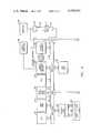
- FIG. 4is a schematic diagram of the electronic processor employed by the apparatus of FIG. 1.
- the apparatus shown thereinincludes a body 10 containing a flow chamber having an inlet 12 for a blood sample and an outlet 14 with a passageway 16 extending between them past an imaging area 18.
- the passageway 16has an inlet with a conduit 20 adapted to be connected to a volume of saline solution 22.
- the inlet 12 for the blood samplehas a needle 24 in the passageway 16 downstream from the conduit 20 with the needle 24 connected to a container 26 adapted to hold the blood sample to be analyzed.
- the cross-sectional area of the passageway 16becomes progressively smaller as the passageway extends from the blood inlet 12 to the outlet 14 while at the same time the passageway 16 becomes much shallower and much wider.
- the passageway 16has a width and depth of about 5,000 microns at the blood inlet 12 and a width and depth of about 500 microns at midpoint 28, and a depth of 100 microns with a width exceeding 5,000 microns at the examination area 18.
- the flow stream through the examination area 18is many times deeper than the largest cell which have a maximum dimension of about 20 microns, but with the flow passageway shaped in this way the blood stream entering through the opening 12 is confined to a stable flow path of minimum shear in the examination area 18, and the disc-like cells are oriented in that area with their maximum cross-sectional area visible in the plane of FIG. 2.
- the flow characteristics in the passageway 16may be controlled by adjusting the fluid pressure in containers 22 and 26 either automatically or by adjusting the static heights thereof.
- a microscope 30is focused on the examination area 18 and the examination area 18 is illuminated from below by a strobe light 32 which is preferably a U.S. Scientific Instrument Corporation Model 3018 containing a 2UP1.5 lamp.
- the output of the microscope 30is focused on a CCD camera 34 which is preferably a CCD camera model number TC1160BD manufactured by RCA.
- the output of the CCD camerais converted to a series of still frame images, and suitable electronic processors are employed for evaluating those images.
- One processor which may be employedis the processor marketed as Image Analysis System Model C-1285 by Hamamatsu Systems, Inc., Waltham, Mass.
- the output of the CCD camerais connected to an electronic processor 36 which is illustrated in greater detail in FIG.
- the frame grabberis preferably a Model FG08 frame grabber made by the Matrox Corporation of Montreal, the output of which is supplied to a video refresh memory 42 Model RGB 256 made by Matrox Corporation which are both coupled to the multibus 44 of the central processing unit 46 which is preferably an Intel 80/20 computer.
- the multibusis also coupled to a 48K random access memory 48 of Electronic Solutions, Inc., and a 16K dual port random access memory 50 Model RM 117 of Data Cube Corporation.
- the output of the video refresh memoryis also coupled to a color monitor 52 which may be used to provide digitally enhanced video images of individual still frames for human examination.
- the second output of the dual port ram 50is connected to a multibus 54 which is connected to an Applied Micro Devices central processing unit 56, a 48K random access memory of Electronic Solutions, Inc. 58 and removable storage in the form of a floppy disc controller 60, such as an Advanced Micro Devices Model 8/8 and two units of Shugart floppy disc storage 62.
- a floppy disc controller 60such as an Advanced Micro Devices Model 8/8 and two units of Shugart floppy disc storage 62.
- a wide variety of programmingmay be employed for processing pictures with the apparatus of FIG. 4 depending upon the particular task which user wishes to perform.
- the programming of the Hamamatsu System 1285may be employed. Preferably, however, the programming is performed as follows:
- the tasksare first divided into those which must address each pixel in a given image and those which only address a small subset of the total. Since much time will be spent in the first class of tasks, they are programmed in assembly language on the interface processor 46 (the Intel 80/20 in FIG. 4). The output of these operations are then transferred to the host machine 56 via the dual ported ram 50. On the host side almost all of the necessary programming is more suitably done in a high level language such as Pascal (BASIC or FORTRAN could be in principal be used also).
- the types of tasks that are done in the assembly languageincludes greyscale transformations, convolutions, and greyscale histogram calculations.
- the types of tasks done on the host sideinclude overall control of the other devices, identification and segmentation of object of interest in the field of view, calculation of parameters associated with objects thus found, and formating the output of results. Another way of considering this separation of tasks in this fashion is that tasks which must be performed at speeds great compared to a human operator are done in assembly. Tasks which are either complicated or which can operate at less than the maximum speed can be programmed in the higher language. Objects are found in a field of view primarily by setting a greyscale window function for values known to be characteristic of the desired object. These values can be established by prior knowledge or by well-known histogram techniques.
- an edge tracing programis invoked to outline the whole object associated with that pixel. Once the edge has been found, then many relevant parameters such as location, area, integrated optical density, and various moments can easily be calculated. Probability of membership in previously defined subgroups can be determined from these derived parameters by means of standard decision theory. Definitions of blood cell morphoplogy classifications are established by trained observers. These definitions are then used as the basis of the selected algorithms. Accuracy of the method is determined by comparison of machine results with those of trained observers examining the same samples. Output of the results can be programmed to be any of a variety of formats. Histograms, line plots, and tabular summaries are available for particular needs.
Landscapes
- Chemical & Material Sciences (AREA)
- Dispersion Chemistry (AREA)
- Physics & Mathematics (AREA)
- Health & Medical Sciences (AREA)
- Life Sciences & Earth Sciences (AREA)
- Analytical Chemistry (AREA)
- Biochemistry (AREA)
- General Health & Medical Sciences (AREA)
- General Physics & Mathematics (AREA)
- Immunology (AREA)
- Pathology (AREA)
- Investigating Or Analysing Biological Materials (AREA)
- Optical Measuring Cells (AREA)
- Motor Or Generator Frames (AREA)
- Investigating Or Analysing Materials By Optical Means (AREA)
Abstract
Description
Claims (16)
Priority Applications (8)
| Application Number | Priority Date | Filing Date | Title |
|---|---|---|---|
| US06/146,064US4338024A (en) | 1980-05-02 | 1980-05-02 | Flow analyzer and system for analysis of fluids with particles |
| AU71776/81AAU546258B2 (en) | 1980-05-02 | 1981-04-09 | Flow through optical analyzer |
| EP81901339AEP0050666B1 (en) | 1980-05-02 | 1981-04-09 | Flow-through optical analyzer |
| JP56501743AJPH0352573B2 (en) | 1980-05-02 | 1981-04-09 | |
| GB8138017AGB2090427B (en) | 1980-05-02 | 1981-04-09 | Flow-through optical analyzer |
| PCT/US1981/000479WO1981003224A1 (en) | 1980-05-02 | 1981-04-09 | Flow-through optical analyzer |
| DE813146423TDE3146423T1 (en) | 1980-05-02 | 1981-04-09 | FLOW-THROUGH OPTICAL ANALYZER |
| CA000376531ACA1157157A (en) | 1980-05-02 | 1981-04-29 | Flow through optical analyzer |
Applications Claiming Priority (1)
| Application Number | Priority Date | Filing Date | Title |
|---|---|---|---|
| US06/146,064US4338024A (en) | 1980-05-02 | 1980-05-02 | Flow analyzer and system for analysis of fluids with particles |
Publications (1)
| Publication Number | Publication Date |
|---|---|
| US4338024Atrue US4338024A (en) | 1982-07-06 |
Family
ID=22515717
Family Applications (1)
| Application Number | Title | Priority Date | Filing Date |
|---|---|---|---|
| US06/146,064Expired - LifetimeUS4338024A (en) | 1980-05-02 | 1980-05-02 | Flow analyzer and system for analysis of fluids with particles |
Country Status (8)
| Country | Link |
|---|---|
| US (1) | US4338024A (en) |
| EP (1) | EP0050666B1 (en) |
| JP (1) | JPH0352573B2 (en) |
| AU (1) | AU546258B2 (en) |
| CA (1) | CA1157157A (en) |
| DE (1) | DE3146423T1 (en) |
| GB (1) | GB2090427B (en) |
| WO (1) | WO1981003224A1 (en) |
Cited By (114)
| Publication number | Priority date | Publication date | Assignee | Title |
|---|---|---|---|---|
| US4408877A (en)* | 1979-04-10 | 1983-10-11 | Ernst Leitz Wetzlar Gmbh | Device for hydrodynamic focussing of a particle-suspension in a liquid flow cytophotometer |
| US4428669A (en) | 1980-06-06 | 1984-01-31 | Institut Nationale De La Sante Et De La Recherche Medicale | Method and device for measuring the deformability of living cells, notably of red blood corpusles |
| DE3406618A1 (en)* | 1983-02-28 | 1984-08-30 | International Remote Imaging Systems Inc., Chatsworth, Calif. | METHOD AND DEVICE FOR DETERMINING THE LIMIT OF AN OBJECT |
| US4476231A (en)* | 1981-07-22 | 1984-10-09 | International Remote Imaging Systems, Inc. | Method of analyzing the distribution of a reagent between particles and liquid in a suspension |
| EP0134976A3 (en)* | 1983-07-11 | 1986-01-02 | International Remote Imaging Systems, Inc. | Method of analyzing particles in a fluid sample |
| US4564444A (en)* | 1983-07-28 | 1986-01-14 | Hitachi, Ltd. | Filamentous microorganism detector and an apparatus with the detector for controlling the process using microorganisms |
| US4596036A (en)* | 1983-08-31 | 1986-06-17 | The United States Of America As Represented By The United States Department Of Energy | Method and apparatus for fringe-scanning chromosome analysis |
| US4661913A (en)* | 1984-09-11 | 1987-04-28 | Becton, Dickinson And Company | Apparatus and method for the detection and classification of articles using flow cytometry techniques |
| EP0252762A1 (en)* | 1986-07-10 | 1988-01-13 | Greenfield Scientific Inc. | System for microscopically analyzing fluids |
| US4751188A (en)* | 1982-10-15 | 1988-06-14 | Max-Planck-Gesellschaft Zur Foerderung Der Wissenschaften E.V. | Method for the simultaneous quantitative determination of cells and reagent therefor |
| US4786165A (en)* | 1986-07-10 | 1988-11-22 | Toa Medical Electronics Co., Ltd. | Flow cytometry and apparatus therefor |
| US4794453A (en)* | 1986-09-09 | 1988-12-27 | Web Printing Controls Co. | Method and apparatus for stroboscopic video inspection of an asynchronous event |
| US4833382A (en)* | 1986-06-06 | 1989-05-23 | Gibbs David L | Method and apparatus for use in microscope investigations |
| US4955720A (en)* | 1989-01-05 | 1990-09-11 | International Paper Company | On-line fiber orientation distribution measurement |
| WO1991002330A1 (en)* | 1989-08-10 | 1991-02-21 | International Remote Imaging Systems, Inc. | A method of differentiating particles based upon a dynamically changing threshold |
| US5000554A (en)* | 1990-05-23 | 1991-03-19 | Gibbs David L | Method and apparatus for use in microscope investigations with a carrier having exactly one x-y coordinate system reference mark |
| US5050808A (en)* | 1989-12-13 | 1991-09-24 | Satake Engineering Co., Ltd. | Milling apparatus and system therefor |
| EP0468100A1 (en)* | 1990-07-24 | 1992-01-29 | Toa Medical Electronics Co., Ltd. | Automatic focal-point adjustment method in flow imaging cytometer |
| US5088816A (en)* | 1989-09-19 | 1992-02-18 | Toa Medical Electronics Co., Ltd. | Process and apparatus for analyzing cells |
| US5101978A (en)* | 1989-11-27 | 1992-04-07 | The United States Of America As Represented By The Secretary Of The Army | Fluidic sorting device for two or more materials suspended in a fluid |
| EP0466168A3 (en)* | 1990-07-13 | 1992-10-21 | Toa Medical Electronics Co., Ltd. | Particle image analyzing apparatus |
| US5159398A (en)* | 1991-02-27 | 1992-10-27 | Toa Medical Electronics Co., Ltd. | Flow imaging cytometer |
| US5159397A (en)* | 1991-02-27 | 1992-10-27 | Toa Medical Electronics Co., Ltd. | Flow imaging cytometer |
| US5170286A (en)* | 1991-02-19 | 1992-12-08 | The United States Of America As Represented By The Secretary Of The Department Of Health And Human Services | Rapid exchange imaging chamber for stop-flow microscopy |
| US5229849A (en)* | 1984-09-17 | 1993-07-20 | University Of Delaware | Laser doppler spectrometer for the statistical study of the behavior of microscopic organisms |
| US5247339A (en)* | 1991-02-27 | 1993-09-21 | Toa Medical Electronics Co., Ltd. | Flow imaging cytometer |
| US5247340A (en)* | 1991-02-27 | 1993-09-21 | Toa Medical Electronics Co., Ltd. | Flow imaging cytometer |
| US5268966A (en)* | 1989-08-10 | 1993-12-07 | International Remote Imaging Systems, Inc. | Method of differentiating particles based upon a dynamically changing threshold |
| US5290701A (en)* | 1991-08-28 | 1994-03-01 | Wilkins Judd R | Microbial detection system and process |
| US5325168A (en)* | 1991-05-14 | 1994-06-28 | Toa Medical Electronics Co., Ltd. | Apparatus and method for analyzing cells in urine |
| US5325169A (en)* | 1991-05-14 | 1994-06-28 | Toa Medical Electronics Co., Ltd. | Apparatus and method for analyzing cells in urine |
| US5335293A (en)* | 1992-06-16 | 1994-08-02 | Key Technology, Inc. | Product inspection method and apparatus |
| US5412466A (en)* | 1991-07-26 | 1995-05-02 | Toa Medical Electronics Co., Ltd. | Apparatus for forming flattened sample flow for analyzing particles |
| US5426499A (en)* | 1992-10-21 | 1995-06-20 | Toa Medical Electronics Co., Ltd. | Particle analyzing apparatus and method wherein a one dimension image sensor optically tracks a particle |
| US5436978A (en)* | 1989-08-10 | 1995-07-25 | International Remote Imaging Systems, Inc. | Method and an apparatus for differentiating a sample of biological cells |
| EP0556971A3 (en)* | 1992-02-18 | 1995-08-09 | Hitachi Ltd | An apparatus for investigating particles in a fluid, and a method of operation thereof |
| US5448349A (en)* | 1992-10-21 | 1995-09-05 | Toe Medical Electronics Co., Ltd. | Particle analyzing apparatus and method wherein an optical deflector optically tracks a particle |
| US5471294A (en)* | 1991-10-24 | 1995-11-28 | Toa Medical Electronics Co., Ltd. | Flow imaging cytometer |
| EP0678742A3 (en)* | 1994-04-21 | 1996-01-31 | Hitachi Ltd | Method for monitoring a coloring solution for particle analysis and calibration method for particle analysis. |
| WO1996020456A1 (en)* | 1994-12-23 | 1996-07-04 | International Remote Imaging Systems, Inc. | Method and apparatus of analyzing particles in a fluid sample and displaying same |
| WO1996022556A1 (en)* | 1995-01-18 | 1996-07-25 | International Remote Imaging Systems, Inc. | Best focal position determining apparatus based on a plurality of focal positions |
| EP0725268A3 (en)* | 1995-02-01 | 1996-10-16 | Hitachi Ltd | Particle analysis method in flow cytometry and device for displaying particle images by classifying the particles according to their properties |
| US5594544A (en)* | 1993-10-21 | 1997-01-14 | Hitachi, Ltd. | Flow type particle image analyzing method and apparatus |
| US5625709A (en)* | 1994-12-23 | 1997-04-29 | International Remote Imaging Systems, Inc. | Method and apparatus for identifying characteristics of an object in a field of view |
| US5656501A (en)* | 1993-08-11 | 1997-08-12 | Yissum Research Development Company Of The Hebrew University Of Jerusalem | Flow cell device for monitoring blood or other cell suspension under flow |
| US5684584A (en)* | 1991-05-14 | 1997-11-04 | Toa Medical Electronics Co., Ltd. | Apparatus for analyzing cells in urine |
| WO1997043638A1 (en)* | 1996-05-15 | 1997-11-20 | International Remote Imaging Systems, Inc. | Method and apparatus for verifying uniform flow of a fluid sample through a flow cell |
| US5690895A (en)* | 1993-01-26 | 1997-11-25 | Hitachi, Ltd. | Flow cell apparatus |
| US5693484A (en)* | 1991-05-14 | 1997-12-02 | Toa Medical Electronics Co., Ltd. | Method of classifying and counting cells in urine |
| US5715182A (en)* | 1993-08-19 | 1998-02-03 | Hitachi, Ltd. | Device for the classification and examination of particles in fluid |
| US5717778A (en)* | 1993-02-26 | 1998-02-10 | Chu; Albert E. | Optical specimen analysis system and method |
| US5736404A (en)* | 1995-12-27 | 1998-04-07 | Zia Yassinzadeh | Flow detection appartus and method |
| US5768412A (en)* | 1994-09-19 | 1998-06-16 | Hitachi, Ltd. | Region segmentation method for particle images and apparatus thereof |
| US5814468A (en)* | 1996-03-27 | 1998-09-29 | Coulter International Corp. | Methods of enumerating receptor molecules for specific binding partners on formed bodies and in solution |
| US5825477A (en)* | 1995-12-22 | 1998-10-20 | Toa Medical Electronics Co., Ltd. | Apparatus for measuring particle morphology and method thereof |
| US5851835A (en)* | 1995-12-18 | 1998-12-22 | Center For Laboratory Technology, Inc. | Multiparameter hematology apparatus and method |
| US5983120A (en)* | 1995-10-23 | 1999-11-09 | Cytometrics, Inc. | Method and apparatus for reflected imaging analysis |
| US6141624A (en)* | 1997-05-13 | 2000-10-31 | International Remote Imaging Systems | Fluid sample for analysis controlled by total fluid volume and by total particle counts |
| EP1125105A2 (en)* | 1998-11-05 | 2001-08-22 | ChemoMetec A/S | A method for the assessment of particles and a system and a device for use in the method |
| US6330350B1 (en) | 1997-05-22 | 2001-12-11 | Korea Institute Of Science And Technology | Method and apparatus for automatically recognizing blood cells |
| US6414321B1 (en) | 1999-09-10 | 2002-07-02 | Rudolf Grosskopf | Arrangement for three-dimensional image recording of particles using flow-through systems |
| US6473172B1 (en) | 2000-09-20 | 2002-10-29 | International Remote Imaging Systems, Inc. | Flow cell and method of operating therefor |
| US6610973B1 (en)* | 1999-07-27 | 2003-08-26 | Davis, Iii John Merrill | Pill counting aid using a planar light diffusing panel for receipt and retention of the pills |
| US20030228038A1 (en)* | 1995-11-30 | 2003-12-11 | Chroma Vision Medical Systems, Inc., A California Corporation | Method and apparatus for automated image analysis of biological specimens |
| US20030231791A1 (en)* | 2002-06-12 | 2003-12-18 | Torre-Bueno Jose De La | Automated system for combining bright field and fluorescent microscopy |
| US20040047502A1 (en)* | 2002-09-09 | 2004-03-11 | Renliang Xu | Simultaneous measurement and display of 3-D size distributions of particulate materials in suspensions |
| US20040071331A1 (en)* | 1993-02-26 | 2004-04-15 | Ey Laboratories, Inc. | Reflectometry system with compensation for specimen holder topography and with lock-rejection of system noise |
| US20040095574A1 (en)* | 2002-11-19 | 2004-05-20 | Turner Richard H. | Flow cell for urinalysis diagnostic system and method of making same |
| WO2004046736A2 (en) | 2002-11-18 | 2004-06-03 | International Remote Imaging Systems, Inc. | Particle extraction for automatic flow microscope |
| WO2004046553A2 (en) | 2002-11-18 | 2004-06-03 | International Remote Imaging Systems, Inc. | Uniform flow displacement pump |
| US20040109386A1 (en)* | 2002-11-18 | 2004-06-10 | Gold Kenneth S. | Particle analyzer with specimen tube in-line mixer and fluid detector |
| EP1431743A1 (en)* | 2002-12-20 | 2004-06-23 | J.M. Canty Inc. | Granular product inspection device |
| US20040126008A1 (en)* | 2000-04-24 | 2004-07-01 | Eric Chapoulaud | Analyte recognition for urinalysis diagnostic system |
| US20040136581A1 (en)* | 1996-11-27 | 2004-07-15 | Chroma Vision Medical Systems, Inc., A California Corporation | Method and apparatus for automated image analysis of biological specimens |
| US20040189988A1 (en)* | 2003-03-31 | 2004-09-30 | C&L Instruments | Sample chamber for microscopy |
| US20040202357A1 (en)* | 2003-04-11 | 2004-10-14 | Perz Cynthia B. | Silhouette image acquisition |
| WO2004090511A1 (en)* | 2003-04-10 | 2004-10-21 | Endress+Hauser Conducta Gesellschaft Für Mess- Und Regeltechnik Mbh + Co. Kg | Cuvette for a photometer or a spectrometer |
| US20050008228A1 (en)* | 2003-05-23 | 2005-01-13 | Eric Chapoulaud | Fluid sample analysis using class weights |
| US20050033455A1 (en)* | 2002-11-18 | 2005-02-10 | Harvey Kasdan | Multi-level controller system |
| US20050037406A1 (en)* | 2002-06-12 | 2005-02-17 | De La Torre-Bueno Jose | Methods and apparatus for analysis of a biological specimen |
| US20050069175A1 (en)* | 2003-09-29 | 2005-03-31 | Tropicana Products, Inc. | Image/optical analysis of citrus pulp |
| US6947586B2 (en) | 2000-04-24 | 2005-09-20 | International Remote Imaging Systems, Inc. | Multi-neural net imaging apparatus and method |
| US20060002636A1 (en)* | 2004-06-30 | 2006-01-05 | Torre-Bueno Jose D L | Data structure of an image storage and retrieval system |
| US20060088196A1 (en)* | 2004-10-25 | 2006-04-27 | Popovich Joseph Jr | Embedded imaging and control system |
| WO2006089074A2 (en) | 2005-02-17 | 2006-08-24 | Iris International, Inc. | Method and apparatus for analyzing body fluids |
| US20070031043A1 (en)* | 2005-08-02 | 2007-02-08 | Perz Cynthia B | System for and method of intelligently directed segmentation analysis for automated microscope systems |
| US20070087442A1 (en)* | 2005-10-19 | 2007-04-19 | Wardlaw Stephen C | Apparatus and method for performing counts within a biologic fluid sample |
| US20070243117A1 (en)* | 2004-04-07 | 2007-10-18 | Wardlaw Stephen C | Disposable Chamber for Analyzing Biologic Fluids |
| WO2008010761A1 (en)* | 2006-07-19 | 2008-01-24 | Hemocue Ab | A measurement apparatus, method and computer program |
| US20080044312A1 (en)* | 2006-08-15 | 2008-02-21 | The Government Of The Us, As Represented By The Secretary Of The Navy | Method and apparatus for attaching a fluid cell to a planar substrate |
| US20080100840A1 (en)* | 2006-10-30 | 2008-05-01 | Peter Oma | Method and Apparatus for Analyzing Particles in a Fluid |
| US20090219530A1 (en)* | 2007-12-04 | 2009-09-03 | John Mitchell | Non-orthogonal particle detection systems and methods |
| US7653260B2 (en) | 2004-06-17 | 2010-01-26 | Carl Zeis MicroImaging GmbH | System and method of registering field of view |
| US20110096157A1 (en)* | 2009-10-28 | 2011-04-28 | Alan Marc Fine | Microscopy imaging |
| US20110271746A1 (en)* | 2010-05-06 | 2011-11-10 | Sony Corporation | Microparticle sorting apparatus, microchip and microchip module |
| US8645167B2 (en) | 2008-02-29 | 2014-02-04 | Dakocytomation Denmark A/S | Systems and methods for tracking and providing workflow information |
| CN103558153A (en)* | 2013-10-31 | 2014-02-05 | 长春迪瑞医疗科技股份有限公司 | Particle imaging chamber and design method thereof |
| US8676509B2 (en) | 2001-11-13 | 2014-03-18 | Dako Denmark A/S | System for tracking biological samples |
| WO2014146062A3 (en)* | 2013-03-15 | 2014-12-18 | Iris International, Inc. | Flowcell, sheath fluid, and autofocus systems and methods for particle analysis in urine samples |
| US9075225B2 (en) | 2009-10-28 | 2015-07-07 | Alentic Microscience Inc. | Microscopy imaging |
| US9316635B2 (en) | 2013-03-15 | 2016-04-19 | Iris International, Inc. | Sheath fluid systems and methods for particle analysis in blood samples |
| US9322752B2 (en) | 2013-03-15 | 2016-04-26 | Iris International, Inc. | Flowcell systems and methods for particle analysis in blood samples |
| US9989750B2 (en) | 2013-06-26 | 2018-06-05 | Alentic Microscience Inc. | Sample processing improvements for microscopy |
| US10416060B1 (en) | 2019-06-04 | 2019-09-17 | Horiba Instruments Incorporated | Apparatus and method for three-dimensional dynamic image analysis for particle volume determination |
| US10502666B2 (en) | 2013-02-06 | 2019-12-10 | Alentic Microscience Inc. | Sample processing improvements for quantitative microscopy |
| CN111684279A (en)* | 2018-08-02 | 2020-09-18 | 深圳迈瑞生物医疗电子股份有限公司 | Cell analysis method, cell analysis device and storage medium |
| US11080512B2 (en)* | 2018-10-29 | 2021-08-03 | Arkray, Inc. | Information processing device, information processing method, measurement system and non-transitory storage medium |
| US11125675B2 (en) | 2019-10-18 | 2021-09-21 | Roger Lawrence Deran | Fluid suspended particle classifier |
| US11181465B2 (en) | 2018-02-01 | 2021-11-23 | Toray Industries, Inc. | Device for evaluating particles in liquid and method for operating same |
| US11382548B2 (en)* | 2014-12-22 | 2022-07-12 | Renalsense Ltd. | Apparatus, system, and methods for urinalysis |
| US11579063B2 (en) | 2017-01-31 | 2023-02-14 | Fujifilm Corporation | Cell culture apparatus, imaging unit, and culture monitoring method |
| CN116067852A (en)* | 2022-11-09 | 2023-05-05 | 四川东鹏农海科技有限公司 | Device for measuring suspended pollen particle number and application method thereof |
| US20230227768A1 (en)* | 2016-04-25 | 2023-07-20 | Renascent Diagnostics, Llc | Method and systems for increasing the capacity of flow cytometter bacteria detection and antibiotic susceptibility testing systems |
| US12022236B2 (en) | 2009-10-28 | 2024-06-25 | Alentic Microscience Inc. | Detecting and using light representative of a sample |
Families Citing this family (21)
| Publication number | Priority date | Publication date | Assignee | Title |
|---|---|---|---|---|
| DE3141984A1 (en)* | 1981-10-22 | 1983-05-05 | International Remote Imaging Systems, 91311 Chatsworth, Calif. | Method of analysing for particles |
| FR2515352B1 (en)* | 1981-10-22 | 1987-07-17 | Int Remote Imaging Systems Inc | METHOD FOR THE ANALYSIS OF PARTICLES CONTAINED IN A SAMPLE OF A DILUTED FLUID |
| DE3315195A1 (en)* | 1982-04-29 | 1983-11-03 | International Remote Imaging Systems Inc., 91311 Chatsworth, Calif. | METHOD FOR ALIGNING PARTICLES IN A FLUID SAMPLE |
| DE3315194A1 (en)* | 1982-04-29 | 1983-11-03 | International Remote Imaging Systems Inc., 91311 Chatsworth, Calif. | METHOD FOR SEPARATING PARTICLES FLOWING IN A FLUID SAMPLE |
| US4896966A (en)* | 1986-08-15 | 1990-01-30 | Hamilton-Thorn Research | Motility scanner and method |
| US4775515A (en)* | 1986-11-18 | 1988-10-04 | Cottingham Hugh V | Agglutinographic slide |
| FR2664983B1 (en)* | 1990-07-17 | 1994-04-15 | Centre Tech Ind Papiers Cartons | DEVICE FOR THE CONTINUOUS DETECTION OF CONTRAST IMPURITIES CONTAINED IN A MOVING FLUID MATERIAL. |
| JP2874746B2 (en)* | 1990-11-22 | 1999-03-24 | シスメックス株式会社 | Flow cell mechanism in flow imaging cytometer |
| FR2681693A1 (en)* | 1991-09-24 | 1993-03-26 | Barrat Bertrand | DEVICE FOR DIMENSIONAL ANALYSIS OF PARTICLES POSITIONED IN A PLANE. |
| US5598842A (en)* | 1993-09-03 | 1997-02-04 | Toa Medical Electronics Co., Ltd. | Non-invasive blood analyzer and method using the same |
| JP2826449B2 (en)* | 1993-09-17 | 1998-11-18 | 株式会社日立製作所 | Flow type particle image analysis method and flow type particle image analysis device |
| JP3039594B2 (en)* | 1993-10-08 | 2000-05-08 | 株式会社日立製作所 | Staining reagent and method of use |
| JP3364323B2 (en)* | 1994-05-17 | 2003-01-08 | 謙 石原 | Non-invasive blood analyzer |
| JPH0991430A (en)* | 1995-09-27 | 1997-04-04 | Hitachi Ltd | Pattern recognizer |
| US6549661B1 (en) | 1996-12-25 | 2003-04-15 | Hitachi, Ltd. | Pattern recognition apparatus and pattern recognition method |
| DE19738626C2 (en)* | 1997-09-04 | 2001-02-08 | Erhard Wendlandt | Micro flow and culture cuvette |
| DE19919608A1 (en)* | 1999-05-27 | 2000-11-30 | Roche Diagnostics Gmbh | Sample holder for the IR spectroscopy of sample liquids |
| DE19932870A1 (en)* | 1999-07-09 | 2001-04-05 | Friedrich Schiller Uni Jena Bu | Device for optical particle and particle flow analysis |
| WO2004099773A1 (en)* | 2003-04-30 | 2004-11-18 | Pfizer Products Inc. | Automated in vitro cellular imaging assays for micronuclei and other target objects |
| JP5032792B2 (en) | 2006-05-22 | 2012-09-26 | 浜松ホトニクス株式会社 | Cell sorter |
| JP2012504764A (en)* | 2008-10-02 | 2012-02-23 | ピクセル メディカル テクノロジーズ リミテッド | Optical imaging based on viscoelastic focusing |
Citations (10)
| Publication number | Priority date | Publication date | Assignee | Title |
|---|---|---|---|---|
| US2480312A (en)* | 1947-02-20 | 1949-08-30 | Glenn C Wolf | Apparatus for the observation and counting of microscopic bodies |
| US2791150A (en)* | 1952-02-16 | 1957-05-07 | Daniel S Stevens | Device for determining the red blood cell count |
| US3390229A (en)* | 1962-11-01 | 1968-06-25 | Raytheon Eduction Company | Particle measuring and counting system |
| US3560754A (en)* | 1965-11-17 | 1971-02-02 | Ibm | Photoelectric particle separator using time delay |
| US3819270A (en)* | 1972-10-02 | 1974-06-25 | Block Engineering | Blood cell analyzer |
| US3976862A (en)* | 1975-03-18 | 1976-08-24 | Block Engineering, Inc. | Flow stream processor |
| US4075462A (en)* | 1975-01-08 | 1978-02-21 | William Guy Rowe | Particle analyzer apparatus employing light-sensitive electronic detector array |
| US4097845A (en)* | 1976-11-01 | 1978-06-27 | Rush-Presbyterian-St. Luke's Medical Center | Method of and an apparatus for automatic classification of red blood cells |
| US4175860A (en)* | 1977-05-31 | 1979-11-27 | Rush-Presbyterian-St. Luke's Medical Center | Dual resolution method and apparatus for use in automated classification of pap smear and other samples |
| US4199748A (en)* | 1976-11-01 | 1980-04-22 | Rush-Presbyterian-St. Luke's Medical Center | Automated method and apparatus for classification of cells with application to the diagnosis of anemia |
Family Cites Families (4)
| Publication number | Priority date | Publication date | Assignee | Title |
|---|---|---|---|---|
| US3822095A (en)* | 1972-08-14 | 1974-07-02 | Block Engineering | System for differentiating particles |
| US3924947A (en)* | 1973-10-19 | 1975-12-09 | Coulter Electronics | Apparatus for preservation and identification of particles analyzed by flow-through apparatus |
| DE2656263A1 (en)* | 1976-12-11 | 1978-08-24 | Max Planck Gesellschaft | Measuring device for properties of suspended particles - has medium flow path to measurement orifice which narrows differently in two orthogonal planes |
| JPS548312A (en)* | 1977-06-22 | 1979-01-22 | Tokyu Kensetsu Kk | Method of changeeover construction of road* track* etc* to underground line |
- 1980
- 1980-05-02USUS06/146,064patent/US4338024A/ennot_activeExpired - Lifetime
- 1981
- 1981-04-09WOPCT/US1981/000479patent/WO1981003224A1/enactiveIP Right Grant
- 1981-04-09EPEP81901339Apatent/EP0050666B1/ennot_activeExpired
- 1981-04-09GBGB8138017Apatent/GB2090427B/ennot_activeExpired
- 1981-04-09DEDE813146423Tpatent/DE3146423T1/enactiveGranted
- 1981-04-09JPJP56501743Apatent/JPH0352573B2/janot_activeExpired - Lifetime
- 1981-04-09AUAU71776/81Apatent/AU546258B2/ennot_activeExpired
- 1981-04-29CACA000376531Apatent/CA1157157A/ennot_activeExpired
Patent Citations (10)
| Publication number | Priority date | Publication date | Assignee | Title |
|---|---|---|---|---|
| US2480312A (en)* | 1947-02-20 | 1949-08-30 | Glenn C Wolf | Apparatus for the observation and counting of microscopic bodies |
| US2791150A (en)* | 1952-02-16 | 1957-05-07 | Daniel S Stevens | Device for determining the red blood cell count |
| US3390229A (en)* | 1962-11-01 | 1968-06-25 | Raytheon Eduction Company | Particle measuring and counting system |
| US3560754A (en)* | 1965-11-17 | 1971-02-02 | Ibm | Photoelectric particle separator using time delay |
| US3819270A (en)* | 1972-10-02 | 1974-06-25 | Block Engineering | Blood cell analyzer |
| US4075462A (en)* | 1975-01-08 | 1978-02-21 | William Guy Rowe | Particle analyzer apparatus employing light-sensitive electronic detector array |
| US3976862A (en)* | 1975-03-18 | 1976-08-24 | Block Engineering, Inc. | Flow stream processor |
| US4097845A (en)* | 1976-11-01 | 1978-06-27 | Rush-Presbyterian-St. Luke's Medical Center | Method of and an apparatus for automatic classification of red blood cells |
| US4199748A (en)* | 1976-11-01 | 1980-04-22 | Rush-Presbyterian-St. Luke's Medical Center | Automated method and apparatus for classification of cells with application to the diagnosis of anemia |
| US4175860A (en)* | 1977-05-31 | 1979-11-27 | Rush-Presbyterian-St. Luke's Medical Center | Dual resolution method and apparatus for use in automated classification of pap smear and other samples |
Non-Patent Citations (1)
| Title |
|---|
| Edited by Melamed, et al.; Flow Cytometry and Sorting, 1979.* |
Cited By (235)
| Publication number | Priority date | Publication date | Assignee | Title |
|---|---|---|---|---|
| US4408877A (en)* | 1979-04-10 | 1983-10-11 | Ernst Leitz Wetzlar Gmbh | Device for hydrodynamic focussing of a particle-suspension in a liquid flow cytophotometer |
| US4428669A (en) | 1980-06-06 | 1984-01-31 | Institut Nationale De La Sante Et De La Recherche Medicale | Method and device for measuring the deformability of living cells, notably of red blood corpusles |
| US4612614A (en)* | 1980-09-12 | 1986-09-16 | International Remote Imaging Systems, Inc. | Method of analyzing particles in a fluid sample |
| US4476231A (en)* | 1981-07-22 | 1984-10-09 | International Remote Imaging Systems, Inc. | Method of analyzing the distribution of a reagent between particles and liquid in a suspension |
| US4751188A (en)* | 1982-10-15 | 1988-06-14 | Max-Planck-Gesellschaft Zur Foerderung Der Wissenschaften E.V. | Method for the simultaneous quantitative determination of cells and reagent therefor |
| DE3406618A1 (en)* | 1983-02-28 | 1984-08-30 | International Remote Imaging Systems Inc., Chatsworth, Calif. | METHOD AND DEVICE FOR DETERMINING THE LIMIT OF AN OBJECT |
| EP0134976A3 (en)* | 1983-07-11 | 1986-01-02 | International Remote Imaging Systems, Inc. | Method of analyzing particles in a fluid sample |
| US4564444A (en)* | 1983-07-28 | 1986-01-14 | Hitachi, Ltd. | Filamentous microorganism detector and an apparatus with the detector for controlling the process using microorganisms |
| US4596036A (en)* | 1983-08-31 | 1986-06-17 | The United States Of America As Represented By The United States Department Of Energy | Method and apparatus for fringe-scanning chromosome analysis |
| US4661913A (en)* | 1984-09-11 | 1987-04-28 | Becton, Dickinson And Company | Apparatus and method for the detection and classification of articles using flow cytometry techniques |
| US5229849A (en)* | 1984-09-17 | 1993-07-20 | University Of Delaware | Laser doppler spectrometer for the statistical study of the behavior of microscopic organisms |
| US4833382A (en)* | 1986-06-06 | 1989-05-23 | Gibbs David L | Method and apparatus for use in microscope investigations |
| US4786165A (en)* | 1986-07-10 | 1988-11-22 | Toa Medical Electronics Co., Ltd. | Flow cytometry and apparatus therefor |
| EP0252762A1 (en)* | 1986-07-10 | 1988-01-13 | Greenfield Scientific Inc. | System for microscopically analyzing fluids |
| US4794453A (en)* | 1986-09-09 | 1988-12-27 | Web Printing Controls Co. | Method and apparatus for stroboscopic video inspection of an asynchronous event |
| US4955720A (en)* | 1989-01-05 | 1990-09-11 | International Paper Company | On-line fiber orientation distribution measurement |
| US5268966A (en)* | 1989-08-10 | 1993-12-07 | International Remote Imaging Systems, Inc. | Method of differentiating particles based upon a dynamically changing threshold |
| US5436978A (en)* | 1989-08-10 | 1995-07-25 | International Remote Imaging Systems, Inc. | Method and an apparatus for differentiating a sample of biological cells |
| WO1991002330A1 (en)* | 1989-08-10 | 1991-02-21 | International Remote Imaging Systems, Inc. | A method of differentiating particles based upon a dynamically changing threshold |
| US5088816A (en)* | 1989-09-19 | 1992-02-18 | Toa Medical Electronics Co., Ltd. | Process and apparatus for analyzing cells |
| USRE35227E (en)* | 1989-09-19 | 1996-05-07 | Toa Medical Electronics Co., Ltd. | Process and apparatus for analyzing cells |
| US5101978A (en)* | 1989-11-27 | 1992-04-07 | The United States Of America As Represented By The Secretary Of The Army | Fluidic sorting device for two or more materials suspended in a fluid |
| US5050808A (en)* | 1989-12-13 | 1991-09-24 | Satake Engineering Co., Ltd. | Milling apparatus and system therefor |
| US5000554A (en)* | 1990-05-23 | 1991-03-19 | Gibbs David L | Method and apparatus for use in microscope investigations with a carrier having exactly one x-y coordinate system reference mark |
| EP0466168A3 (en)* | 1990-07-13 | 1992-10-21 | Toa Medical Electronics Co., Ltd. | Particle image analyzing apparatus |
| US5159642A (en)* | 1990-07-13 | 1992-10-27 | Toa Medical Electronics Co., Ltd. | Particle image analyzing apparatus |
| EP0468100A1 (en)* | 1990-07-24 | 1992-01-29 | Toa Medical Electronics Co., Ltd. | Automatic focal-point adjustment method in flow imaging cytometer |
| US5170286A (en)* | 1991-02-19 | 1992-12-08 | The United States Of America As Represented By The Secretary Of The Department Of Health And Human Services | Rapid exchange imaging chamber for stop-flow microscopy |
| EP0501008A3 (en)* | 1991-02-27 | 1992-11-04 | Toa Medical Electronics Co., Ltd. | Flow imaging cytometer |
| US5247339A (en)* | 1991-02-27 | 1993-09-21 | Toa Medical Electronics Co., Ltd. | Flow imaging cytometer |
| US5247340A (en)* | 1991-02-27 | 1993-09-21 | Toa Medical Electronics Co., Ltd. | Flow imaging cytometer |
| US5159397A (en)* | 1991-02-27 | 1992-10-27 | Toa Medical Electronics Co., Ltd. | Flow imaging cytometer |
| US5159398A (en)* | 1991-02-27 | 1992-10-27 | Toa Medical Electronics Co., Ltd. | Flow imaging cytometer |
| US5325168A (en)* | 1991-05-14 | 1994-06-28 | Toa Medical Electronics Co., Ltd. | Apparatus and method for analyzing cells in urine |
| US5325169A (en)* | 1991-05-14 | 1994-06-28 | Toa Medical Electronics Co., Ltd. | Apparatus and method for analyzing cells in urine |
| US5693484A (en)* | 1991-05-14 | 1997-12-02 | Toa Medical Electronics Co., Ltd. | Method of classifying and counting cells in urine |
| US5684584A (en)* | 1991-05-14 | 1997-11-04 | Toa Medical Electronics Co., Ltd. | Apparatus for analyzing cells in urine |
| US5412466A (en)* | 1991-07-26 | 1995-05-02 | Toa Medical Electronics Co., Ltd. | Apparatus for forming flattened sample flow for analyzing particles |
| US5290701A (en)* | 1991-08-28 | 1994-03-01 | Wilkins Judd R | Microbial detection system and process |
| US5471294A (en)* | 1991-10-24 | 1995-11-28 | Toa Medical Electronics Co., Ltd. | Flow imaging cytometer |
| WO1993014599A1 (en)* | 1992-01-15 | 1993-07-22 | University Of Delaware | Laster doppler spectrometer for the statistical study of the behavior of microscopic organisms |
| EP0556971A3 (en)* | 1992-02-18 | 1995-08-09 | Hitachi Ltd | An apparatus for investigating particles in a fluid, and a method of operation thereof |
| US5335293A (en)* | 1992-06-16 | 1994-08-02 | Key Technology, Inc. | Product inspection method and apparatus |
| US5426499A (en)* | 1992-10-21 | 1995-06-20 | Toa Medical Electronics Co., Ltd. | Particle analyzing apparatus and method wherein a one dimension image sensor optically tracks a particle |
| CN1041128C (en)* | 1992-10-21 | 1998-12-09 | 东亚医用电子株式会社 | Particle analysing equipment |
| CN1040796C (en)* | 1992-10-21 | 1998-11-18 | 东亚医用电子株式会社 | Particle analysing equipment |
| US5448349A (en)* | 1992-10-21 | 1995-09-05 | Toe Medical Electronics Co., Ltd. | Particle analyzing apparatus and method wherein an optical deflector optically tracks a particle |
| US5690895A (en)* | 1993-01-26 | 1997-11-25 | Hitachi, Ltd. | Flow cell apparatus |
| US7031508B2 (en) | 1993-02-26 | 2006-04-18 | E Y Laboratories, Inc. | Reflectometry system with compensation for specimen holder topography and with lock-rejection of system noise |
| US5717778A (en)* | 1993-02-26 | 1998-02-10 | Chu; Albert E. | Optical specimen analysis system and method |
| US20040071331A1 (en)* | 1993-02-26 | 2004-04-15 | Ey Laboratories, Inc. | Reflectometry system with compensation for specimen holder topography and with lock-rejection of system noise |
| US5656501A (en)* | 1993-08-11 | 1997-08-12 | Yissum Research Development Company Of The Hebrew University Of Jerusalem | Flow cell device for monitoring blood or other cell suspension under flow |
| US5715182A (en)* | 1993-08-19 | 1998-02-03 | Hitachi, Ltd. | Device for the classification and examination of particles in fluid |
| US5594544A (en)* | 1993-10-21 | 1997-01-14 | Hitachi, Ltd. | Flow type particle image analyzing method and apparatus |
| US5728582A (en)* | 1994-04-21 | 1998-03-17 | Hitachi, Ltd. | Monitoring method of stain solution for particle analysis and calibration method of particle analysis |
| EP0678742A3 (en)* | 1994-04-21 | 1996-01-31 | Hitachi Ltd | Method for monitoring a coloring solution for particle analysis and calibration method for particle analysis. |
| US5768412A (en)* | 1994-09-19 | 1998-06-16 | Hitachi, Ltd. | Region segmentation method for particle images and apparatus thereof |
| US5822447A (en)* | 1994-12-23 | 1998-10-13 | International Remote Imaging Systems, Inc. | Method of analyzing particles in a sample and displaying same |
| US5625709A (en)* | 1994-12-23 | 1997-04-29 | International Remote Imaging Systems, Inc. | Method and apparatus for identifying characteristics of an object in a field of view |
| WO1996020456A1 (en)* | 1994-12-23 | 1996-07-04 | International Remote Imaging Systems, Inc. | Method and apparatus of analyzing particles in a fluid sample and displaying same |
| WO1996022556A1 (en)* | 1995-01-18 | 1996-07-25 | International Remote Imaging Systems, Inc. | Best focal position determining apparatus based on a plurality of focal positions |
| US5633491A (en)* | 1995-01-18 | 1997-05-27 | International Remote Imaging Systems, Inc. | Method and apparatus for automatically selecting the best focal position from a plurality of focal positions for a focusing apparatus |
| US5619032A (en)* | 1995-01-18 | 1997-04-08 | International Remote Imaging Systems, Inc. | Method and apparatus for automatically selecting the best focal position from a plurality of focal positions for a focusing apparatus |
| EP0725268A3 (en)* | 1995-02-01 | 1996-10-16 | Hitachi Ltd | Particle analysis method in flow cytometry and device for displaying particle images by classifying the particles according to their properties |
| US5878160A (en)* | 1995-02-01 | 1999-03-02 | Hitachi, Ltd. | Flow type particle image analyzing method and apparatus for displaying particle images by classifying them based on their configurational features |
| US6104939A (en)* | 1995-10-23 | 2000-08-15 | Cytometrics, Inc. | Method and apparatus for reflected imaging analysis |
| US5983120A (en)* | 1995-10-23 | 1999-11-09 | Cytometrics, Inc. | Method and apparatus for reflected imaging analysis |
| US20040066960A1 (en)* | 1995-11-30 | 2004-04-08 | Chromavision Medical Systems, Inc., A California Corporation | Automated detection of objects in a biological sample |
| US6920239B2 (en)* | 1995-11-30 | 2005-07-19 | Chromavision Medical Systems, Inc. | Method and apparatus for automated image analysis of biological specimens |
| US20070053569A1 (en)* | 1995-11-30 | 2007-03-08 | Douglass James W | Method and apparatus for automated image analysis of biological specimens |
| US7133545B2 (en) | 1995-11-30 | 2006-11-07 | Clarient, Inc. | Method and apparatus for automated image analysis of biological specimens |
| US7783098B2 (en) | 1995-11-30 | 2010-08-24 | Carl Zeiss Microimaging Gmbh | Method and apparatus for automated image analysis of biological specimens |
| US7177454B2 (en) | 1995-11-30 | 2007-02-13 | Clarient, Inc. | Automated detection of objects in a biological sample |
| US20090060303A1 (en)* | 1995-11-30 | 2009-03-05 | Carl Zeiss Microlmaging Ais, Inc. | Method and apparatus for automated image analysis of biological specimens |
| US7359548B2 (en) | 1995-11-30 | 2008-04-15 | Carl Zeiss Microimaging Ais, Inc. | Method and apparatus for automated image analysis of biological specimens |
| US20030228038A1 (en)* | 1995-11-30 | 2003-12-11 | Chroma Vision Medical Systems, Inc., A California Corporation | Method and apparatus for automated image analysis of biological specimens |
| US7558415B2 (en) | 1995-11-30 | 2009-07-07 | Carl Zeiss Microimaging Ais, Inc. | Automated detection of objects in a biological sample |
| US5851835A (en)* | 1995-12-18 | 1998-12-22 | Center For Laboratory Technology, Inc. | Multiparameter hematology apparatus and method |
| US5825477A (en)* | 1995-12-22 | 1998-10-20 | Toa Medical Electronics Co., Ltd. | Apparatus for measuring particle morphology and method thereof |
| US5736404A (en)* | 1995-12-27 | 1998-04-07 | Zia Yassinzadeh | Flow detection appartus and method |
| US5814468A (en)* | 1996-03-27 | 1998-09-29 | Coulter International Corp. | Methods of enumerating receptor molecules for specific binding partners on formed bodies and in solution |
| US6424415B1 (en) | 1996-05-15 | 2002-07-23 | International Remote Imaging Systems, Inc. | Method and apparatus for verifying uniform flow of a fluid sample through a flow cell and distribution on a slide |
| WO1997043638A1 (en)* | 1996-05-15 | 1997-11-20 | International Remote Imaging Systems, Inc. | Method and apparatus for verifying uniform flow of a fluid sample through a flow cell |
| US6184978B1 (en) | 1996-05-15 | 2001-02-06 | International Remote Imaging Systems, Inc. | Method and apparatus for verifying uniform flow of a fluid sample through a flow cell and distribution on a slide |
| US7190818B2 (en)* | 1996-11-27 | 2007-03-13 | Clarient, Inc. | Method and apparatus for automated image analysis of biological specimens |
| US20040136581A1 (en)* | 1996-11-27 | 2004-07-15 | Chroma Vision Medical Systems, Inc., A California Corporation | Method and apparatus for automated image analysis of biological specimens |
| US20070206843A1 (en)* | 1996-11-27 | 2007-09-06 | Douglass James W | Method and Apparatus for Automated Image Analysis of Biological Specimens |
| US7428325B2 (en) | 1996-11-27 | 2008-09-23 | Carl Zeiss Microimaging Ais, Inc. | Method and apparatus for automated image analysis of biological specimens |
| US6141624A (en)* | 1997-05-13 | 2000-10-31 | International Remote Imaging Systems | Fluid sample for analysis controlled by total fluid volume and by total particle counts |
| US6330350B1 (en) | 1997-05-22 | 2001-12-11 | Korea Institute Of Science And Technology | Method and apparatus for automatically recognizing blood cells |
| EP1125105A2 (en)* | 1998-11-05 | 2001-08-22 | ChemoMetec A/S | A method for the assessment of particles and a system and a device for use in the method |
| US8906697B2 (en) | 1998-11-05 | 2014-12-09 | Chemometec A/S | Method for the assessment of particles and a system and device for use in the method |
| US6610973B1 (en)* | 1999-07-27 | 2003-08-26 | Davis, Iii John Merrill | Pill counting aid using a planar light diffusing panel for receipt and retention of the pills |
| US6414321B1 (en) | 1999-09-10 | 2002-07-02 | Rudolf Grosskopf | Arrangement for three-dimensional image recording of particles using flow-through systems |
| US7236623B2 (en) | 2000-04-24 | 2007-06-26 | International Remote Imaging Systems, Inc. | Analyte recognition for urinalysis diagnostic system |
| US6947586B2 (en) | 2000-04-24 | 2005-09-20 | International Remote Imaging Systems, Inc. | Multi-neural net imaging apparatus and method |
| US20040126008A1 (en)* | 2000-04-24 | 2004-07-01 | Eric Chapoulaud | Analyte recognition for urinalysis diagnostic system |
| US6473172B1 (en) | 2000-09-20 | 2002-10-29 | International Remote Imaging Systems, Inc. | Flow cell and method of operating therefor |
| US8676509B2 (en) | 2001-11-13 | 2014-03-18 | Dako Denmark A/S | System for tracking biological samples |
| US7272252B2 (en) | 2002-06-12 | 2007-09-18 | Clarient, Inc. | Automated system for combining bright field and fluorescent microscopy |
| US20050037406A1 (en)* | 2002-06-12 | 2005-02-17 | De La Torre-Bueno Jose | Methods and apparatus for analysis of a biological specimen |
| US20030231791A1 (en)* | 2002-06-12 | 2003-12-18 | Torre-Bueno Jose De La | Automated system for combining bright field and fluorescent microscopy |
| US6873725B2 (en) | 2002-09-09 | 2005-03-29 | Coulter International Corp. | Simultaneous measurement and display of 3-D size distributions of particulate materials in suspensions |
| US20040047502A1 (en)* | 2002-09-09 | 2004-03-11 | Renliang Xu | Simultaneous measurement and display of 3-D size distributions of particulate materials in suspensions |
| US7702172B2 (en) | 2002-11-18 | 2010-04-20 | Iris Internation, Inc. | Particle extraction for automatic flow microscope |
| US20080140224A1 (en)* | 2002-11-18 | 2008-06-12 | Harvey Kasdan | Multi-level controller system and method |
| WO2004046736A2 (en) | 2002-11-18 | 2004-06-03 | International Remote Imaging Systems, Inc. | Particle extraction for automatic flow microscope |
| US7079244B2 (en) | 2002-11-18 | 2006-07-18 | International Remote Imaging Systems, Inc. | Particle analyzer with specimen tube in-line mixer |
| US20050033455A1 (en)* | 2002-11-18 | 2005-02-10 | Harvey Kasdan | Multi-level controller system |
| US20060186899A1 (en)* | 2002-11-18 | 2006-08-24 | Gold Kenneth S | Particle analyzer with specimen tube fluid detector |
| US8447417B2 (en) | 2002-11-18 | 2013-05-21 | Iris International, Inc. | Multi-level controller system and method |
| US7150607B2 (en) | 2002-11-18 | 2006-12-19 | International Remote Imaging Systems, Inc. | Uniform flow displacement pump |
| US7161674B2 (en) | 2002-11-18 | 2007-01-09 | International Remote Imaging Systems, Inc. | Particle analyzer with specimen tube fluid detector |
| US7319907B2 (en) | 2002-11-18 | 2008-01-15 | International Remote Imaging Systems, Inc. | Multi-level controller system |
| US20110184537A1 (en)* | 2002-11-18 | 2011-07-28 | Harvey Kasdan | Multi-Level Controller System |
| US20040136593A1 (en)* | 2002-11-18 | 2004-07-15 | Eric Chapoulaud | Particle extraction for automatic flow microscope |
| WO2004046736A3 (en)* | 2002-11-18 | 2004-07-15 | Int Remote Imaging Systems Inc | Particle extraction for automatic flow microscope |
| US20070077158A1 (en)* | 2002-11-18 | 2007-04-05 | Pelmulder John P | Uniform flow displacement pump |
| CN100353368C (en)* | 2002-11-18 | 2007-12-05 | 国际遥距成象系统公司 | Particle extraction for automatic flow microscope |
| WO2004046553A2 (en) | 2002-11-18 | 2004-06-03 | International Remote Imaging Systems, Inc. | Uniform flow displacement pump |
| AU2003294351B2 (en)* | 2002-11-18 | 2007-10-04 | Iris International, Inc. | Particle extraction for automatic flow microscope |
| US20040109386A1 (en)* | 2002-11-18 | 2004-06-10 | Gold Kenneth S. | Particle analyzer with specimen tube in-line mixer and fluid detector |
| US20040095574A1 (en)* | 2002-11-19 | 2004-05-20 | Turner Richard H. | Flow cell for urinalysis diagnostic system and method of making same |
| US6825926B2 (en) | 2002-11-19 | 2004-11-30 | International Remote Imaging Systems, Inc. | Flow cell for urinalysis diagnostic system and method of making same |
| EP1431743A1 (en)* | 2002-12-20 | 2004-06-23 | J.M. Canty Inc. | Granular product inspection device |
| US20040184649A1 (en)* | 2002-12-20 | 2004-09-23 | J.M. Canty Inc. | Granular product inspection device |
| US7518716B2 (en) | 2002-12-20 | 2009-04-14 | J.M. Canty Inc. | Granular product inspection device |
| US20040189988A1 (en)* | 2003-03-31 | 2004-09-30 | C&L Instruments | Sample chamber for microscopy |
| US7245368B2 (en) | 2003-03-31 | 2007-07-17 | C & L Instruments | Sample chamber for microscopy |
| WO2004090511A1 (en)* | 2003-04-10 | 2004-10-21 | Endress+Hauser Conducta Gesellschaft Für Mess- Und Regeltechnik Mbh + Co. Kg | Cuvette for a photometer or a spectrometer |
| US20040202357A1 (en)* | 2003-04-11 | 2004-10-14 | Perz Cynthia B. | Silhouette image acquisition |
| US8369591B2 (en) | 2003-04-11 | 2013-02-05 | Carl Zeiss Microimaging Gmbh | Silhouette image acquisition |
| US7324694B2 (en) | 2003-05-23 | 2008-01-29 | International Remote Imaging Systems, Inc. | Fluid sample analysis using class weights |
| US20050008228A1 (en)* | 2003-05-23 | 2005-01-13 | Eric Chapoulaud | Fluid sample analysis using class weights |
| US20050069175A1 (en)* | 2003-09-29 | 2005-03-31 | Tropicana Products, Inc. | Image/optical analysis of citrus pulp |
| US8241572B2 (en) | 2004-04-07 | 2012-08-14 | Abbott Point Of Care, Inc. | Disposable chamber for analyzing biologic fluids |
| US7850916B2 (en) | 2004-04-07 | 2010-12-14 | Abbott Laboratories | Disposable chamber for analyzing biologic fluids |
| US9084995B2 (en) | 2004-04-07 | 2015-07-21 | Abbott Laboratories | Disposable chamber for analyzing biologic fluids |
| US10578602B2 (en) | 2004-04-07 | 2020-03-03 | Abbott Laboratories | Disposable chamber for analyzing biologic fluids |
| US20100216248A1 (en)* | 2004-04-07 | 2010-08-26 | Abbott Laboratories | Disposable chamber for analyzing biologic fluids |
| US20070243117A1 (en)* | 2004-04-07 | 2007-10-18 | Wardlaw Stephen C | Disposable Chamber for Analyzing Biologic Fluids |
| US7653260B2 (en) | 2004-06-17 | 2010-01-26 | Carl Zeis MicroImaging GmbH | System and method of registering field of view |
| US20060002636A1 (en)* | 2004-06-30 | 2006-01-05 | Torre-Bueno Jose D L | Data structure of an image storage and retrieval system |
| US8582924B2 (en) | 2004-06-30 | 2013-11-12 | Carl Zeiss Microimaging Gmbh | Data structure of an image storage and retrieval system |
| US20060088196A1 (en)* | 2004-10-25 | 2006-04-27 | Popovich Joseph Jr | Embedded imaging and control system |
| US8121392B2 (en)* | 2004-10-25 | 2012-02-21 | Parata Systems, Llc | Embedded imaging and control system |
| US20110027824A1 (en)* | 2005-02-17 | 2011-02-03 | Turner Richard H | Method And Apparatus For Analyzing Body Fluids |
| US8391608B2 (en) | 2005-02-17 | 2013-03-05 | Iris International, Inc. | Method and apparatus for analyzing body fluids |
| US7822276B2 (en) | 2005-02-17 | 2010-10-26 | Iris International, Inc. | Method and apparatus for analyzing body fluids |
| EP2541466A1 (en) | 2005-02-17 | 2013-01-02 | Iris International, Inc. | Method and Apparatus For Analyzing Body Fluids |
| WO2006089074A2 (en) | 2005-02-17 | 2006-08-24 | Iris International, Inc. | Method and apparatus for analyzing body fluids |
| US8116543B2 (en) | 2005-08-02 | 2012-02-14 | Carl Zeiss Microimaging Gmbh | System for and method of intelligently directed segmentation analysis for automated microscope systems |
| US20070031043A1 (en)* | 2005-08-02 | 2007-02-08 | Perz Cynthia B | System for and method of intelligently directed segmentation analysis for automated microscope systems |
| US20100040266A1 (en)* | 2005-08-02 | 2010-02-18 | Perz Cynthia B | System for and method of intelligently directed segmentation analysis for automated microscope systems |
| US9696252B2 (en) | 2005-10-19 | 2017-07-04 | Abbott Laboratories | Apparatus for performing counts within a biologic fluid sample |
| US20100273244A1 (en)* | 2005-10-19 | 2010-10-28 | Abbott Laboratories | Apparatus for performing counts within a biologic fluid sample |
| US7731901B2 (en)* | 2005-10-19 | 2010-06-08 | Abbott Laboratories | Apparatus and method for performing counts within a biologic fluid sample |
| US20100272345A1 (en)* | 2005-10-19 | 2010-10-28 | Abbott Laboratories | Method for performing counts within a biologic fluid sample |
| US8158434B2 (en) | 2005-10-19 | 2012-04-17 | Abbott Laboratories | Method for performing counts within a biologic fluid sample |
| US20070087442A1 (en)* | 2005-10-19 | 2007-04-19 | Wardlaw Stephen C | Apparatus and method for performing counts within a biologic fluid sample |
| AU2006304717B2 (en)* | 2005-10-19 | 2010-05-27 | Levine, Robert A. | Apparatus and method for performing counts within a biologic fluid sample |
| US8224058B2 (en) | 2006-07-19 | 2012-07-17 | Hemocue Ab | Measurement apparatus, method and computer program |
| RU2402006C1 (en)* | 2006-07-19 | 2010-10-20 | Хемокуэ Аб | Device, method and computer program for measuring |
| US20080019584A1 (en)* | 2006-07-19 | 2008-01-24 | Stellan Lindberg | Measurement apparatus, method and computer program |
| CN101490529B (en)* | 2006-07-19 | 2013-11-13 | 海莫库公司 | Measuring equipment and methods |
| AU2007275927B2 (en)* | 2006-07-19 | 2011-06-09 | Hemocue Ab | A measurement apparatus, method and computer program |
| WO2008010761A1 (en)* | 2006-07-19 | 2008-01-24 | Hemocue Ab | A measurement apparatus, method and computer program |
| US8137624B2 (en) | 2006-08-15 | 2012-03-20 | The United States Of America As Represented By The Secretary Of The Navy | Method and apparatus for attaching a fluid cell to a planar substrate |
| US20080044312A1 (en)* | 2006-08-15 | 2008-02-21 | The Government Of The Us, As Represented By The Secretary Of The Navy | Method and apparatus for attaching a fluid cell to a planar substrate |
| US20080100840A1 (en)* | 2006-10-30 | 2008-05-01 | Peter Oma | Method and Apparatus for Analyzing Particles in a Fluid |
| US7605919B2 (en) | 2006-10-30 | 2009-10-20 | Brightwell Technologies Inc. | Method and apparatus for analyzing particles in a fluid |
| US7916293B2 (en) | 2007-12-04 | 2011-03-29 | Particle Measuring Systems, Inc. | Non-orthogonal particle detection systems and methods |
| US8427642B2 (en) | 2007-12-04 | 2013-04-23 | Particle Measuring Systems, Inc. | Two-dimensional optical imaging methods and systems for particle detection |
| US20090244536A1 (en)* | 2007-12-04 | 2009-10-01 | John Mitchell | Two-dimensional optical imaging methods and systems for particle detection |
| US8174697B2 (en) | 2007-12-04 | 2012-05-08 | Particle Measuring Systems, Inc. | Non-orthogonal particle detection systems and methods |
| US8027035B2 (en) | 2007-12-04 | 2011-09-27 | Particle Measuring Systems, Inc. | Non-orthogonal particle detection systems and methods |
| US20090219530A1 (en)* | 2007-12-04 | 2009-09-03 | John Mitchell | Non-orthogonal particle detection systems and methods |
| US8154724B2 (en) | 2007-12-04 | 2012-04-10 | Particle Measuring Systems, Inc. | Two-dimensional optical imaging methods and systems for particle detection |
| US9767425B2 (en) | 2008-02-29 | 2017-09-19 | Dako Denmark A/S | Systems and methods for tracking and providing workflow information |
| US8645167B2 (en) | 2008-02-29 | 2014-02-04 | Dakocytomation Denmark A/S | Systems and methods for tracking and providing workflow information |
| US10832199B2 (en) | 2008-02-29 | 2020-11-10 | Agilent Technologies, Inc. | Systems and methods for tracking and providing workflow information |
| US11947096B2 (en) | 2009-10-28 | 2024-04-02 | Alentic Microscience Inc. | Microscopy imaging |
| US11294160B2 (en) | 2009-10-28 | 2022-04-05 | Alentic Microscience Inc. | Microscopy imaging |
| US9041790B2 (en) | 2009-10-28 | 2015-05-26 | Alentic Microscience Inc. | Microscopy imaging |
| US9075225B2 (en) | 2009-10-28 | 2015-07-07 | Alentic Microscience Inc. | Microscopy imaging |
| US11635447B2 (en) | 2009-10-28 | 2023-04-25 | Alentic Microscience Inc. | Microscopy imaging |
| US10900999B2 (en) | 2009-10-28 | 2021-01-26 | Alentic Microscience Inc. | Microscopy imaging |
| US10866395B2 (en) | 2009-10-28 | 2020-12-15 | Alentic Microscience Inc. | Microscopy imaging |
| US12388957B2 (en) | 2009-10-28 | 2025-08-12 | Alentic Microscience Inc. | Detecting and using light representative of a sample |
| US10620234B2 (en) | 2009-10-28 | 2020-04-14 | Alentic Microscience Inc. | Microscopy imaging |
| US12022236B2 (en) | 2009-10-28 | 2024-06-25 | Alentic Microscience Inc. | Detecting and using light representative of a sample |
| US20110096157A1 (en)* | 2009-10-28 | 2011-04-28 | Alan Marc Fine | Microscopy imaging |
| US9720217B2 (en) | 2009-10-28 | 2017-08-01 | Alentic Microscience Inc. | Microscopy imaging |
| US10114203B2 (en) | 2009-10-28 | 2018-10-30 | Alentic Microscience Inc. | Microscopy imaging |
| US10520711B2 (en) | 2009-10-28 | 2019-12-31 | Alentic Microscience Inc. | Microscopy imaging |
| US10345564B2 (en) | 2009-10-28 | 2019-07-09 | Alentic Microscience Inc. | Microscopy imaging |
| US20110271746A1 (en)* | 2010-05-06 | 2011-11-10 | Sony Corporation | Microparticle sorting apparatus, microchip and microchip module |
| CN102284429A (en)* | 2010-05-06 | 2011-12-21 | 索尼公司 | Microparticle sorting apparatus, microchip and microchip module |
| US8657121B2 (en)* | 2010-05-06 | 2014-02-25 | Sony Corporation | Microparticle sorting apparatus, microchip and microchip module |
| CN102284429B (en)* | 2010-05-06 | 2015-05-13 | 索尼公司 | Microparticle sorting apparatus and method, microchip and microchip module |
| US10502666B2 (en) | 2013-02-06 | 2019-12-10 | Alentic Microscience Inc. | Sample processing improvements for quantitative microscopy |
| US11598699B2 (en) | 2013-02-06 | 2023-03-07 | Alentic Microscience Inc. | Sample processing improvements for quantitative microscopy |
| US10768078B2 (en) | 2013-02-06 | 2020-09-08 | Alentic Microscience Inc. | Sample processing improvements for quantitative microscopy |
| US11543340B2 (en) | 2013-03-15 | 2023-01-03 | Iris International, Inc. | Autofocus systems and methods for particle analysis in blood samples |
| US9909973B2 (en) | 2013-03-15 | 2018-03-06 | Iris International, Inc. | Flowcell systems and methods for particle analysis in blood samples |
| US10451612B2 (en) | 2013-03-15 | 2019-10-22 | Iris International, Inc. | Sheath fluid systems and methods for particle analysis in blood samples |
| US9857361B2 (en) | 2013-03-15 | 2018-01-02 | Iris International, Inc. | Flowcell, sheath fluid, and autofocus systems and methods for particle analysis in urine samples |
| US9702806B2 (en) | 2013-03-15 | 2017-07-11 | Iris International, Inc. | Hematology systems and methods |
| US9470618B2 (en) | 2013-03-15 | 2016-10-18 | Iris International, Inc. | Sheath fluid systems and methods for particle analysis in blood samples |
| US10705008B2 (en) | 2013-03-15 | 2020-07-07 | Iris International, Inc. | Autofocus systems and methods for particle analysis in blood samples |
| US10345217B2 (en) | 2013-03-15 | 2019-07-09 | Iris International, Inc. | Flowcell systems and methods for particle analysis in blood samples |
| US10429292B2 (en) | 2013-03-15 | 2019-10-01 | Iris International, Inc. | Dynamic range extension systems and methods for particle analysis in blood samples |
| US10060846B2 (en) | 2013-03-15 | 2018-08-28 | Iris International, Inc. | Hematology systems and methods |
| US10794900B2 (en) | 2013-03-15 | 2020-10-06 | Iris International, Inc. | Flowcell, sheath fluid, and autofocus systems and methods for particle analysis in urine samples |
| US11525766B2 (en) | 2013-03-15 | 2022-12-13 | Iris International, Inc. | Dynamic range extension systems and methods for particle analysis in blood samples |
| WO2014146062A3 (en)* | 2013-03-15 | 2014-12-18 | Iris International, Inc. | Flowcell, sheath fluid, and autofocus systems and methods for particle analysis in urine samples |
| US9322752B2 (en) | 2013-03-15 | 2016-04-26 | Iris International, Inc. | Flowcell systems and methods for particle analysis in blood samples |
| US9316635B2 (en) | 2013-03-15 | 2016-04-19 | Iris International, Inc. | Sheath fluid systems and methods for particle analysis in blood samples |
| US11874452B2 (en) | 2013-06-26 | 2024-01-16 | Alentic Microscience Inc. | Sample processing improvements for microscopy |
| US10809512B2 (en) | 2013-06-26 | 2020-10-20 | Alentic Microscience Inc. | Sample processing improvements for microscopy |
| US9989750B2 (en) | 2013-06-26 | 2018-06-05 | Alentic Microscience Inc. | Sample processing improvements for microscopy |
| US10459213B2 (en) | 2013-06-26 | 2019-10-29 | Alentic Microscience Inc. | Sample processing improvements for microscopy |
| US10746979B2 (en) | 2013-06-26 | 2020-08-18 | Alentic Microscience Inc. | Sample processing improvements for microscopy |
| CN103558153A (en)* | 2013-10-31 | 2014-02-05 | 长春迪瑞医疗科技股份有限公司 | Particle imaging chamber and design method thereof |
| CN103558153B (en)* | 2013-10-31 | 2016-05-25 | 长春迪瑞医疗科技股份有限公司 | Particle imaging chamber and method for designing thereof |
| US11382548B2 (en)* | 2014-12-22 | 2022-07-12 | Renalsense Ltd. | Apparatus, system, and methods for urinalysis |
| US20230227768A1 (en)* | 2016-04-25 | 2023-07-20 | Renascent Diagnostics, Llc | Method and systems for increasing the capacity of flow cytometter bacteria detection and antibiotic susceptibility testing systems |
| US11579063B2 (en) | 2017-01-31 | 2023-02-14 | Fujifilm Corporation | Cell culture apparatus, imaging unit, and culture monitoring method |
| US11181465B2 (en) | 2018-02-01 | 2021-11-23 | Toray Industries, Inc. | Device for evaluating particles in liquid and method for operating same |
| CN111684279A (en)* | 2018-08-02 | 2020-09-18 | 深圳迈瑞生物医疗电子股份有限公司 | Cell analysis method, cell analysis device and storage medium |
| US11080512B2 (en)* | 2018-10-29 | 2021-08-03 | Arkray, Inc. | Information processing device, information processing method, measurement system and non-transitory storage medium |
| US10416060B1 (en) | 2019-06-04 | 2019-09-17 | Horiba Instruments Incorporated | Apparatus and method for three-dimensional dynamic image analysis for particle volume determination |
| US11125675B2 (en) | 2019-10-18 | 2021-09-21 | Roger Lawrence Deran | Fluid suspended particle classifier |
| CN116067852A (en)* | 2022-11-09 | 2023-05-05 | 四川东鹏农海科技有限公司 | Device for measuring suspended pollen particle number and application method thereof |
| CN116067852B (en)* | 2022-11-09 | 2023-10-03 | 四川东鹏农海科技有限公司 | Device for measuring suspended pollen particle number and application method thereof |
Also Published As
| Publication number | Publication date |
|---|---|
| DE3146423T1 (en) | 1982-07-15 |
| JPS57500995A (en) | 1982-06-03 |
| EP0050666A1 (en) | 1982-05-05 |
| EP0050666B1 (en) | 1986-10-01 |
| WO1981003224A1 (en) | 1981-11-12 |
| CA1157157A (en) | 1983-11-15 |
| EP0050666A4 (en) | 1982-09-09 |
| AU7177681A (en) | 1981-11-26 |
| GB2090427B (en) | 1985-01-30 |
| GB2090427A (en) | 1982-07-07 |
| JPH0352573B2 (en) | 1991-08-12 |
| AU546258B2 (en) | 1985-08-22 |
| DE3146423C2 (en) | 1993-09-02 |
Similar Documents
| Publication | Publication Date | Title |
|---|---|---|
| US4338024A (en) | Flow analyzer and system for analysis of fluids with particles | |
| US4393466A (en) | Method of analyzing particles in a dilute fluid sample | |
| GB2122369A (en) | A method of separating particles in a moving fluid sample | |
| GB2121976A (en) | A method of aligning particles in a moving fluid sample | |
| KR102095617B1 (en) | Autofocus systems and methods for particle analysis in blood samples | |
| US4612614A (en) | Method of analyzing particles in a fluid sample | |
| US4667335A (en) | Method of determining the diagnostic significance of the content of a volume of biological sample containing particles | |
| Bacus et al. | Image processing for automated erythrocyte classification. | |
| EP0146621B1 (en) | A method of determining the diagnostic significance of the content of a volume of biological sample containing particles | |
| EP1849125B1 (en) | Method and apparatus for analyzing body fluids | |
| JPH0341783B2 (en) | ||
| CA1164992A (en) | Method of analyzing particles in a dilute fluid sample | |
| FR2526164A1 (en) | Calibration of optical analyser of particles in blood, urine etc. - using microscopic particles of selected parameter to set standard in computer | |
| GB2107548A (en) | Method of analyzing particles in a dilute fluid sample | |
| Eden et al. | Feasibility of computer screening of blood films for the detection of malaria parasites | |
| Lewis | Automated differential leucocyte counting: Present status and future trends | |
| CN120126125A (en) | White blood cell recognition AI training method, processing and storage device based on hemolysis | |
| HK40009534A (en) | Flowcell systems and methods for particle analysis in biological fluid samples | |
| HK40002305A (en) | Autofocus systems and methods for particle analysis in body fluid samples | |
| HK40002305B (en) | Autofocus systems and methods for particle analysis in body fluid samples |
Legal Events
| Date | Code | Title | Description |
|---|---|---|---|
| STCF | Information on status: patent grant | Free format text:PATENTED CASE | |
| AS | Assignment | Owner name:TOA MEDICAL ELECTRONICS CO., LTD., A CORP. OF JAPA Free format text:SECURITY INTEREST;ASSIGNOR:INTERNATIONAL REMOTE IMAGING SYSTEMS INC., A CORP. OF DE.;REEL/FRAME:005006/0008 Effective date:19880715 | |
| AS | Assignment | Owner name:INTERNATIONAL REMOTE IMAGING SYSTEMS, INC., CALIFO Free format text:ASSIGNMENT OF ASSIGNORS INTEREST;ASSIGNOR:INTERNATIONAL REMOTE IMAGING SYSTEMS, INC., A CALIFORNIA CORPORATION;REEL/FRAME:008085/0522 Effective date:19960813 | |
| AS | Assignment | Owner name:CITY NATIONAL BANK, CALIFORNIA Free format text:SECURITY INTEREST;ASSIGNOR:INTERNATIONAL REMOTE IMAGING SYSTEMS, INC.;REEL/FRAME:008376/0858 Effective date:19970103 | |
| AS | Assignment | Owner name:INTERNATIONAL REMOTE IMAGING SYSTEMS, INC., CALIFO Free format text:TERMINATION, RELEASE AND REASSIGNMENT OF SECURITY INTEREST;ASSIGNOR:TOA MEDICAL ELECTRONICS, LTD.;REEL/FRAME:008820/0681 Effective date:19971030 | |
| AS | Assignment | Owner name:FOOTHILL CAPITAL CORPORATION, CALIFORNIA Free format text:SECURITY AGREEMENT;ASSIGNOR:INTERNATIONAL REMOTE IMAGING SYSTEMS, INC.;REEL/FRAME:009257/0255 Effective date:19980505 | |
| AS | Assignment | Owner name:CITY NATIONAL BANK, CALIFORNIA Free format text:RELEASE OF SECURITY AGREEMENT;ASSIGNOR:INTERNATIONAL REMOTE IMAGING SYSTEMS, INC.;REEL/FRAME:009214/0371 Effective date:19970103 |