US4335427A - Method of selecting a preferred difference image - Google Patents
Method of selecting a preferred difference imageDownload PDFInfo
- Publication number
- US4335427A US4335427AUS06/142,188US14218880AUS4335427AUS 4335427 AUS4335427 AUS 4335427AUS 14218880 AUS14218880 AUS 14218880AUS 4335427 AUS4335427 AUS 4335427A
- Authority
- US
- United States
- Prior art keywords
- images
- region
- difference image
- image
- sample difference
- Prior art date
- Legal status (The legal status is an assumption and is not a legal conclusion. Google has not performed a legal analysis and makes no representation as to the accuracy of the status listed.)
- Expired - Lifetime
Links
Images
Classifications
- G—PHYSICS
- G06—COMPUTING OR CALCULATING; COUNTING
- G06T—IMAGE DATA PROCESSING OR GENERATION, IN GENERAL
- G06T5/00—Image enhancement or restoration
- G06T5/50—Image enhancement or restoration using two or more images, e.g. averaging or subtraction
- H—ELECTRICITY
- H04—ELECTRIC COMMUNICATION TECHNIQUE
- H04N—PICTORIAL COMMUNICATION, e.g. TELEVISION
- H04N5/00—Details of television systems
- H04N5/30—Transforming light or analogous information into electric information
- H04N5/32—Transforming X-rays
- H04N5/3205—Transforming X-rays using subtraction imaging techniques
Definitions
- This inventionrelates to dynamic image enhancement of low contrast anatomic regions utilizing a contrast material introduced into a patient during the acquisition of a series of radiographic images.
- the inventionrelates to a method of selecting a preferred difference image representing the differential between a reference or mask image acquired before the contrast material takes effect and a later acquired post-contrast image.
- a digital subtraction radiographic systemfor image enhancement of low contrast objects utilizing venous injection of a contrast material to visualize a difficult-to-image anatomic region of interest.
- a radiographic digitized composite mask image of a region of interest of a patientis acquired and stored in a first digital memory.
- a quantity of contrast materialpreferably an X-ray opaque dye, is introduced into the patient.
- the time for the contrast material to take effectvaries, typically between approximately 10 and 15 seconds.
- a second radiographic image substantially of the same region of interest as the reference imageis acquired and stored in a digitized form in a second digital memory.
- Alternative techniquesare disclosed for subtractively combining the two digitized images to cancel the overlapping background, thereby enhancing the region of interest which shows the contrast material.
- the resultant subtracted imageknown as a difference or differential image, devoid of a significant amount of non-useful background information, permits superior clinical evaluation of the difficult-to-image region.
- a clinical investigator about to perform a diagnostic study involving the acquisition of a series of radiographic imagesgenerally will not know beforehand the length of time for the contrast material to take effect in the anatomic region of interest after injection since there are so many factors in this determination.
- an image acquired early in the studytypically the first such image, will be designated the reference or mask image.
- Each subsequently acquired imagethen becomes a candidate for the post contrast image to be subtracted from the mask to yield the desired difference image.
- the ideal difference imageis generated from the pair that permits the greatest amount of cancellation of background or non-useful information and yields the most information in the region of interest.
- the first conditionrequires a high degree of overlap of the two images which is determined principally by the degree of movement in the imaged region.
- the second conditionis a function of the concentration of contrast material in the region of interest and this is a function of time.
- Patient movementboth voluntary and involuntary, has posed a sufficient clinical problem that the investigator would prefer to use as the mask, an image which comes as close in time as possible prior to the appearance of a substantial quantity of contrast material in the region of interest to significantly minimize motion between the time of the acquisition of the mask and the acquisition of the post contrast image.
- the resultant differencewould provide the desired high contrast in the region of interest with maximum overlap and hence cancellation of the non-useful background.
- the system disclosed in the aforementioned copending applicationpermits generating a series of difference images by subtracting from the mask, independently and sequentially, any other image, i.e. a post contrast image, and viewing the difference.
- any other imagei.e. a post contrast image
- the resultwill not necessarily yield the best difference image available in the study since the original choice of mask may not have been the best since the choice was at best an informed guess.
- this processwere generally acceptable when performed by an experienced investigator, the technique is not likely to consistently yield acceptable clinical results when operated on a grand scale.
- the procedure of introducing contrast material into the object studiedis complex and must be coordinated with the operation of an X-ray system and data collection system. To require concurrent analysis and decision making on the quality of images acquiring burdens the operators and the system.
- the present inventionpermits the data to be collected over a long enough time interval to insure the collection of the best mask and data (contrast) image. Then, as a leisurely post-processing operation, one can select the best pair of images to subtractively combine. Furthermore, since the contrast material does not necessarily reach all parts of the image at one time, it is doubtful whether the a priori selection of a single mask would be adequate to an analysis of the entire image.
- Applicantshave discovered a "remasking" technique for consistently generating a preferred difference image from a series of images acquired during a dynamic imaging study.
- the improved methodrequires a minimum of training and a minimum of operator time.
- the methodis applied in a radiographic, digital subtraction system for imaging a low contrast anatomic region of interest of a patient by acquiring a plurality of mask or reference images of the region of interest, introducing into the patient a contrast material suitable for enhancement of a clinically relevant region, acquiring a plurality of post contrast images of the anatomic region and generating a plurality of difference images in which overlapping portions are significantly eliminated by digitally subtractively combining a plurality of pairs of reference and post contrast images.
- the improved methodcomprises storing the plurality of reference and post contrast images acquired during the study. As a practical matter, the series of images need not even be designated as pre or post contrast, but merely stored.
- the methodfurther comprises selecting a sample difference image which shows contrast material in the clinically relevant region of anatomic interest.
- the sample difference imageis viewed on a video display and a first area from the region showing contrast material is identified. Similarly, a second area is identified from the background portion of the image, the background portion showing at most a minimal amount of contrast material.
- Each area from either regionis recorded, preferably electro-optically, such as by a light pen or joystick.
- the stored reference and post contrast imagesare then searched to select those pairs which when subtractively combined yield the best difference images in terms of maximum overlap of background. Finally, from the difference images so selected, the one which exhibits the greatest activity in the contrast region is selected as a preferred difference image for clinical analysis.
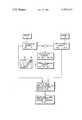
- the single FIGUREis a schematic flow diagram showing steps of the present invention.
- a series of radiographic digitized reference or mask imagesare chronologically acquired and serially stored in a Memory A. These images visually represent a clinically relevant region of interest of a patient acquired before a contrast agent introduced into the patient takes effect. After the contrast agent or material takes effect another series of digitized images are chronologically acquired and serially stored in Memory B.
- the total study per patientincludes a series of n composite frames where n is typically approximately 30. Of the n frames in a study the pre-contrast portion is designated x 1 . . . x i and the post-contrast portion is designated x j . . . x n .
- the operator of a multi-memory radiographic digital subtraction systemmay be remote computer control, view a sample difference image generated by subtracting a post-contrast image (taken from the group x j . . . x n ) from a pre-contrast or reference image (taken from the group x 1 . . . x i ). While all possible difference images (x 1 -x j ) . . . (x i -x n ) can conceivably be generated and stored, such a procedure would be inefficient and costly as well as requiring an undue length of time for a clinician to review.
- a sample difference imagefor example, x 1 -x j
- the sample difference image x l -x jmust be one that includes both a portion of the clinically relevant region of interest showing contrast material as indicated by the diagonally cross-hatched portion of the illustrated sample difference image on the drawing and the background region showing little or no contrast material indicated by the vertical lines on the drawing representative of the motion artifact.
- an area A.sub. 1is selected from the background portion and an area A 2 is selected from the clinically relevant region of interest showing contrast material.
- Enclosed regions A 1 and A 2represent some random number of addresses of the, for example, 256 ⁇ 256 pixel array of the video screen (not shown) that is provided in a radiographic digital subtraction system.
- the selected areas A 1 and A 2are electro-optically recorded, preferably by a light pen or joystick. Once these regions A 1 and A 2 are identified and recorded all address points falling therewithin can be quickly searched on any stored image.
- nare stored on a mass storage device, such as on tape or disk.
- the next step in the preferred methodis to search through the n images on all points falling within areas A 1 and A 2 to select a predetermined number of pairs y of images (one from the group x 1 . . . x i and one from the group x j . . . x n ) which yield the best overlap in region A 1 .
- the best overlapis provided by those pairs which when subtractively combined yield difference images in which region A 1 has the lowest composite values denoting the least level of motion artifact.
- the y difference images generatedare searched to determine which one has the greatest activity, i.e. showing the most contrast material in region A 2 , as indicated by the highest cumulative values generated in region A 2 after subtraction.
- the difference image which is selected in this stepis a preferred difference image.
- the post contrast image showing the most contrast material in region A 2can be pre-selected.
- Region A 1 of that pre-selected imagecan be compared with each of the remaining stored images to select the one which when subtractively combined with the pre-selected post-contrast image will yield the greatest cancellation in the background region.
Landscapes
- Engineering & Computer Science (AREA)
- Multimedia (AREA)
- Signal Processing (AREA)
- Physics & Mathematics (AREA)
- General Physics & Mathematics (AREA)
- Theoretical Computer Science (AREA)
- Apparatus For Radiation Diagnosis (AREA)
- Image Analysis (AREA)
Abstract
Description
This invention relates to dynamic image enhancement of low contrast anatomic regions utilizing a contrast material introduced into a patient during the acquisition of a series of radiographic images. In particular, the invention relates to a method of selecting a preferred difference image representing the differential between a reference or mask image acquired before the contrast material takes effect and a later acquired post-contrast image.
In a copending application Ser. No. 138,400 of Robert Henry McCarthy, filed Apr. 8, 1980, for DYNAMIC IMAGE ENHANCEMENT AND APPARATUS THEREFOR and assigned to the assignee of the present invention, there is disclosed a digital subtraction radiographic system for image enhancement of low contrast objects utilizing venous injection of a contrast material to visualize a difficult-to-image anatomic region of interest. In the digital subtraction system, disclosed in the copending application, a radiographic digitized composite mask image of a region of interest of a patient is acquired and stored in a first digital memory. During the clinical procedure, a quantity of contrast material, preferably an X-ray opaque dye, is introduced into the patient. Depending on several factors including the type and quantity of contrast material, physical characteristics of the patient, and the distance between the remote vein and the region of interest, such as a carotid artery, the time for the contrast material to take effect varies, typically between approximately 10 and 15 seconds. After the contrast material takes effect, a second radiographic image substantially of the same region of interest as the reference image is acquired and stored in a digitized form in a second digital memory. Alternative techniques are disclosed for subtractively combining the two digitized images to cancel the overlapping background, thereby enhancing the region of interest which shows the contrast material. The resultant subtracted image, known as a difference or differential image, devoid of a significant amount of non-useful background information, permits superior clinical evaluation of the difficult-to-image region.
A clinical investigator about to perform a diagnostic study involving the acquisition of a series of radiographic images generally will not know beforehand the length of time for the contrast material to take effect in the anatomic region of interest after injection since there are so many factors in this determination. Thus, an image acquired early in the study, typically the first such image, will be designated the reference or mask image. Each subsequently acquired image then becomes a candidate for the post contrast image to be subtracted from the mask to yield the desired difference image.
The ideal difference image is generated from the pair that permits the greatest amount of cancellation of background or non-useful information and yields the most information in the region of interest. The first condition requires a high degree of overlap of the two images which is determined principally by the degree of movement in the imaged region. The second condition is a function of the concentration of contrast material in the region of interest and this is a function of time. Patient movement, both voluntary and involuntary, has posed a sufficient clinical problem that the investigator would prefer to use as the mask, an image which comes as close in time as possible prior to the appearance of a substantial quantity of contrast material in the region of interest to significantly minimize motion between the time of the acquisition of the mask and the acquisition of the post contrast image. Hence, the resultant difference would provide the desired high contrast in the region of interest with maximum overlap and hence cancellation of the non-useful background.
For any given mask, the system disclosed in the aforementioned copending application, permits generating a series of difference images by subtracting from the mask, independently and sequentially, any other image, i.e. a post contrast image, and viewing the difference. Though a trained physician can typically and rather expeditiously select the best difference image once a mask is preselected, the result will not necessarily yield the best difference image available in the study since the original choice of mask may not have been the best since the choice was at best an informed guess. Moreover, even if this process were generally acceptable when performed by an experienced investigator, the technique is not likely to consistently yield acceptable clinical results when operated on a grand scale.
The procedure of introducing contrast material into the object studied is complex and must be coordinated with the operation of an X-ray system and data collection system. To require concurrent analysis and decision making on the quality of images acquiring burdens the operators and the system. The present invention permits the data to be collected over a long enough time interval to insure the collection of the best mask and data (contrast) image. Then, as a leisurely post-processing operation, one can select the best pair of images to subtractively combine. Furthermore, since the contrast material does not necessarily reach all parts of the image at one time, it is doubtful whether the a priori selection of a single mask would be adequate to an analysis of the entire image.
Applicants have discovered a "remasking" technique for consistently generating a preferred difference image from a series of images acquired during a dynamic imaging study. The improved method requires a minimum of training and a minimum of operator time.
The method is applied in a radiographic, digital subtraction system for imaging a low contrast anatomic region of interest of a patient by acquiring a plurality of mask or reference images of the region of interest, introducing into the patient a contrast material suitable for enhancement of a clinically relevant region, acquiring a plurality of post contrast images of the anatomic region and generating a plurality of difference images in which overlapping portions are significantly eliminated by digitally subtractively combining a plurality of pairs of reference and post contrast images.
The improved method comprises storing the plurality of reference and post contrast images acquired during the study. As a practical matter, the series of images need not even be designated as pre or post contrast, but merely stored. The method further comprises selecting a sample difference image which shows contrast material in the clinically relevant region of anatomic interest. The sample difference image is viewed on a video display and a first area from the region showing contrast material is identified. Similarly, a second area is identified from the background portion of the image, the background portion showing at most a minimal amount of contrast material. Each area from either region is recorded, preferably electro-optically, such as by a light pen or joystick. The stored reference and post contrast images are then searched to select those pairs which when subtractively combined yield the best difference images in terms of maximum overlap of background. Finally, from the difference images so selected, the one which exhibits the greatest activity in the contrast region is selected as a preferred difference image for clinical analysis.
The single FIGURE is a schematic flow diagram showing steps of the present invention.
Referring to the drawing, a series of radiographic digitized reference or mask images are chronologically acquired and serially stored in a Memory A. These images visually represent a clinically relevant region of interest of a patient acquired before a contrast agent introduced into the patient takes effect. After the contrast agent or material takes effect another series of digitized images are chronologically acquired and serially stored in Memory B. The total study per patient includes a series of n composite frames where n is typically approximately 30. Of the n frames in a study the pre-contrast portion is designated x1 . . . xi and the post-contrast portion is designated xj . . . xn.
The operator of a multi-memory radiographic digital subtraction system to which reference has been made hereinabove under the Background Art portion of this application, may be remote computer control, view a sample difference image generated by subtracting a post-contrast image (taken from the group xj . . . xn) from a pre-contrast or reference image (taken from the group x1 . . . xi). While all possible difference images (x1 -xj) . . . (xi -xn) can conceivably be generated and stored, such a procedure would be inefficient and costly as well as requiring an undue length of time for a clinician to review. As an alternative, a sample difference image, for example, x1 -xj, is generated and displayed on a video screen for viewing. The sample difference image xl -xj must be one that includes both a portion of the clinically relevant region of interest showing contrast material as indicated by the diagonally cross-hatched portion of the illustrated sample difference image on the drawing and the background region showing little or no contrast material indicated by the vertical lines on the drawing representative of the motion artifact. On such a sample difference image x1 -xj an area A.sub. 1 is selected from the background portion and an area A2 is selected from the clinically relevant region of interest showing contrast material. Enclosed regions A1 and A2 represent some random number of addresses of the, for example, 256×256 pixel array of the video screen (not shown) that is provided in a radiographic digital subtraction system. The selected areas A1 and A2 are electro-optically recorded, preferably by a light pen or joystick. Once these regions A1 and A2 are identified and recorded all address points falling therewithin can be quickly searched on any stored image.
During a study all of the images n are stored on a mass storage device, such as on tape or disk. The next step in the preferred method is to search through the n images on all points falling within areas A1 and A2 to select a predetermined number of pairs y of images (one from the group x1 . . . xi and one from the group xj . . . xn) which yield the best overlap in region A1. The best overlap is provided by those pairs which when subtractively combined yield difference images in which region A1 has the lowest composite values denoting the least level of motion artifact. This is done, for example, by summing the values of the differences of each address falling within the region A1 and selecting the lowest cumulative totals, or by finding the mean difference or some other convenient index. The predetermined number of such pairs y may, for example, be 10. After this search and select step the y difference images generated are searched to determine which one has the greatest activity, i.e. showing the most contrast material in region A2, as indicated by the highest cumulative values generated in region A2 after subtraction. The difference image which is selected in this step is a preferred difference image.
Alternatively, the post contrast image showing the most contrast material in region A2 can be pre-selected. Region A1 of that pre-selected image can be compared with each of the remaining stored images to select the one which when subtractively combined with the pre-selected post-contrast image will yield the greatest cancellation in the background region.
Claims (8)
1. A method of selecting one or more x-ray difference images from a group of x-ray images, which comprises the steps of:
(a) obtaining first and second pluralities of x-ray images of a patient or subject, each x-ray image being obtained at a different time;
(b) obtaining a sample difference image representing the difference between one of the images of the first plurality of images and one of the images of the second plurality of images;
(c) selecting a region of the sample difference image, the region being less than the entire area of the sample difference image;
(d) pairing images selected from the first plurality of images with images selected from the second plurality of images;
(e) obtaining a plurality of difference images each representing the difference between the images of one of the pairs of images;
(f) searching regions of the difference images, the searched regions corresponding, respectively, to the selected region of the sample difference image; and
(g) selecting one or more difference images, the selected difference images having searched regions that satisfy a predetermined condition or index.
2. A method according to claim 1, wherein the region of the sample difference image is selected at a time subsequent to the time at which the first of the images is obtained.
3. A method according to claim 1, wherein the images are digital video images each formed by a matrix of pixels.
4. A method according to claim 3, wherein the pixels of each image are stored on a mass storage device, the searching of regions of the images being accomplished by processing of pixel data assigned to computer memory address locations related to the assigned computer memory addresses of pixels in the selected region of the sample difference image.
5. A method according to claim 4 wherein the pixel values in the searched regions are mathematically processed to obtain a result which is compared with a reference value based upon pixel values in the selected region of the sample difference image.
6. A method according to claim 1, wherein a second region of the sample difference image is selected, the second region being less than the entire area of the sample difference image, the first-mentioned region having substantially different contrast characteristics than the second region.
7. A method according to claim 1, which further includes the steps of:
(h) selecting a second region of the sample difference image, the second region being less than the entire area of the sample difference image and different than the first-mentioned selected region;
(i) in the difference images selected according to step (g) of claim 9, searching such selected difference images in regions thereof corresponding, respectively, to the second selected region of the sample difference image; and
(j) selecting one or more difference images, the selected difference images having searched regions, corresponding to the second selected region of the sample difference image, that satisfy a predetermined condition or index.
8. A method according to claim 7, wherein one of the selected regions of the sample difference image is a region having a high degree of contrast with the background of the sample difference image and wherein the other of the selected regions of the sample difference image is a region having a low degree of contrast with the background of the sample difference image.
Priority Applications (4)
| Application Number | Priority Date | Filing Date | Title |
|---|---|---|---|
| US06/142,188US4335427A (en) | 1980-04-21 | 1980-04-21 | Method of selecting a preferred difference image |
| EP81301632AEP0038663B1 (en) | 1980-04-21 | 1981-04-14 | Method of selecting a preferred difference image |
| DE8181301632TDE3173228D1 (en) | 1980-04-21 | 1981-04-14 | Method of selecting a preferred difference image |
| JP5866681AJPS56166839A (en) | 1980-04-21 | 1981-04-20 | Method of selecting prior image difference |
Applications Claiming Priority (1)
| Application Number | Priority Date | Filing Date | Title |
|---|---|---|---|
| US06/142,188US4335427A (en) | 1980-04-21 | 1980-04-21 | Method of selecting a preferred difference image |
Publications (1)
| Publication Number | Publication Date |
|---|---|
| US4335427Atrue US4335427A (en) | 1982-06-15 |
Family
ID=22498905
Family Applications (1)
| Application Number | Title | Priority Date | Filing Date |
|---|---|---|---|
| US06/142,188Expired - LifetimeUS4335427A (en) | 1980-04-21 | 1980-04-21 | Method of selecting a preferred difference image |
Country Status (4)
| Country | Link |
|---|---|
| US (1) | US4335427A (en) |
| EP (1) | EP0038663B1 (en) |
| JP (1) | JPS56166839A (en) |
| DE (1) | DE3173228D1 (en) |
Cited By (71)
| Publication number | Priority date | Publication date | Assignee | Title |
|---|---|---|---|---|
| US4394688A (en)* | 1981-08-25 | 1983-07-19 | Hamamatsu Systems, Inc. | Video system having an adjustable digital gamma correction for contrast enhancement |
| US4415980A (en)* | 1981-03-02 | 1983-11-15 | Lockheed Missiles & Space Co., Inc. | Automated radiographic inspection system |
| US4425580A (en) | 1980-11-19 | 1984-01-10 | Siemens Aktiengesellschaft | Diagnostic x-ray installation for providing subtraction images |
| US4430749A (en) | 1981-06-30 | 1984-02-07 | Siemens Gammasonics, Inc. | Medical imaging apparatus and method for furnishing difference images |
| US4436095A (en) | 1981-12-22 | 1984-03-13 | Thomson-Csf Broadcast, Inc. | Method and apparatus for imaging a body |
| US4437161A (en) | 1981-06-29 | 1984-03-13 | Siemens Gammasonics Inc. | Medical imaging apparatus |
| US4444196A (en)* | 1982-06-21 | 1984-04-24 | Diagnostic Technology, Inc. | Digital intravenous subtraction angiography |
| US4448200A (en)* | 1978-03-27 | 1984-05-15 | University Of Southern California | System and method for dynamic background subtraction |
| WO1984002643A1 (en)* | 1983-01-10 | 1984-07-19 | Robert Thomas Gordon | Method for enhancing nmr imaging; and diagnostic use |
| US4516261A (en)* | 1979-11-08 | 1985-05-07 | U.S. Philips Corporation | Device for reducing faults in layer images of a three-dimensional object formed by means of penetrating radiation |
| DE3426933A1 (en)* | 1983-07-26 | 1985-05-09 | Elscint Ltd., Haifa | ARRANGEMENT FOR AUTOMATICALLY CORRECTING ERROR TRANSMISSIONS |
| US4521861A (en)* | 1982-04-30 | 1985-06-04 | Texas Instruments Incorporated | Method and apparatus for enhancing radiometric imaging |
| US4536790A (en)* | 1982-11-26 | 1985-08-20 | Thomson-Csf Broadcast, Inc. | Apparatus and method for fluoroscopic imaging of a body |
| US4542459A (en)* | 1982-11-26 | 1985-09-17 | General Electric Company | Matched filter for x-ray hybrid subtraction |
| US4551800A (en)* | 1982-11-26 | 1985-11-05 | General Electric Company | Integrated hybrid image remasking in a subtraction angiography method |
| US4559557A (en)* | 1984-06-01 | 1985-12-17 | General Electric Company | Region-of-interest digital subtraction angiography |
| US4578765A (en)* | 1982-11-02 | 1986-03-25 | Cambridge Instruments Limited | Image comparison systems |
| US4636850A (en)* | 1984-09-07 | 1987-01-13 | Adac Laboratories, Inc. | Apparatus and method for enhancement of video images |
| US4641352A (en)* | 1984-07-12 | 1987-02-03 | Paul Fenster | Misregistration correction |
| US4644582A (en)* | 1983-01-28 | 1987-02-17 | Hitachi, Ltd. | Image registration method |
| US4720843A (en)* | 1985-04-24 | 1988-01-19 | U.S. Philips Corporation | Method for separating moving structures from a fixed background in a sequence of X-ray projection images and equipment for implementing this method |
| US4878115A (en)* | 1987-09-25 | 1989-10-31 | University Of Kentucky Research Foundation | Dynamic coronary roadmapping |
| US4916745A (en)* | 1986-02-07 | 1990-04-10 | Hart Hiram E | Bayesian image processing method and apparatus |
| US4941192A (en)* | 1987-04-20 | 1990-07-10 | Hitachi, Ltd. | Method and apparatus for recognizing pattern of gray level image |
| US4947323A (en)* | 1986-05-22 | 1990-08-07 | University Of Tennessee Research Corporation | Method and apparatus for measuring small spatial dimensions of an object |
| US5003616A (en)* | 1985-04-17 | 1991-03-26 | Hitachi, Ltd. | Image processing apparatus |
| US5008185A (en)* | 1985-11-04 | 1991-04-16 | Cell Analysis Systems, Inc. | Methods and apparatus for the quantitation of nuclear proteins |
| US5048110A (en)* | 1984-03-30 | 1991-09-10 | Fuji Photo Film Co., Ltd. | Method and apparatus for automatically correcting subtraction image density |
| US5049748A (en)* | 1989-10-19 | 1991-09-17 | Fuji Photo Film Co. Ltd. | Method and apparatus for forming energy subtraction images |
| US5133020A (en)* | 1989-07-21 | 1992-07-21 | Arch Development Corporation | Automated method and system for the detection and classification of abnormal lesions and parenchymal distortions in digital medical images |
| US5212737A (en)* | 1991-09-30 | 1993-05-18 | Imatron, Inc. | Real time data averager for use in computed tomography scanning |
| US5250933A (en)* | 1989-03-02 | 1993-10-05 | Hewlett-Packard Company | Method and apparatus for the simultaneous display of one or more selected images |
| US5258928A (en)* | 1990-05-03 | 1993-11-02 | Rca Thomson Licensing Corporation | Parts efficient memory based functional circuit having selectable transfer characteristics |
| EP0604291A1 (en)* | 1992-12-24 | 1994-06-29 | Sopha Medical | Processing method for a set of images |
| AU650657B2 (en)* | 1990-06-08 | 1994-06-30 | Smith & Nephew, Inc. | Dynamic elbow support |
| US5467404A (en)* | 1991-08-14 | 1995-11-14 | Agfa-Gevaert | Method and apparatus for contrast enhancement |
| EP0704823A3 (en)* | 1994-09-22 | 1996-06-05 | Sanyo Electric Co | Method of judging back-and-forth positional relationship between subjects and method of converting two-dimensional images into three-dimensional images |
| US5563720A (en)* | 1992-07-17 | 1996-10-08 | International Business Machines Corporation | Expert system for image enhancement |
| US5717602A (en)* | 1996-02-05 | 1998-02-10 | Kenning; Gregory G. | Automated electrophoresis and analysis system |
| US5717778A (en)* | 1993-02-26 | 1998-02-10 | Chu; Albert E. | Optical specimen analysis system and method |
| GB2331440A (en)* | 1997-11-15 | 1999-05-19 | Elekta Ab | Analysis of radiographic images |
| US5976088A (en)* | 1998-06-24 | 1999-11-02 | Ecton, Inc. | Ultrasound imaging systems and methods of increasing the effective acquisition frame rate |
| US6004270A (en)* | 1998-06-24 | 1999-12-21 | Ecton, Inc. | Ultrasound system for contrast agent imaging and quantification in echocardiography using template image for image alignment |
| USD445189S1 (en) | 1999-09-14 | 2001-07-17 | Ecton, Inc. | Medical diagnostic ultrasound system |
| US6312381B1 (en) | 1999-09-14 | 2001-11-06 | Acuson Corporation | Medical diagnostic ultrasound system and method |
| US6436039B1 (en) | 1999-09-14 | 2002-08-20 | Ecton, Inc. | Medicial diagnostic ultrasound system and method |
| US6468213B1 (en) | 1999-09-14 | 2002-10-22 | Ecton, Inc. | Medical diagnostic ultrasound system and method |
| US6488625B1 (en) | 1999-09-14 | 2002-12-03 | Ecton, Inc. | Medical diagnostic ultrasound system and method |
| US6497664B1 (en) | 1999-09-14 | 2002-12-24 | Ecton, Inc. | Medical diagnostic ultrasound system and method |
| US6508763B1 (en) | 1999-09-14 | 2003-01-21 | Ecton, Inc. | Medical diagnostic ultrasound system and method |
| US6524244B1 (en) | 1999-09-14 | 2003-02-25 | Ecton Inc. | Medical diagnostic ultrasound system and method |
| US6561979B1 (en) | 1999-09-14 | 2003-05-13 | Acuson Corporation | Medical diagnostic ultrasound system and method |
| US20030210762A1 (en)* | 2002-03-26 | 2003-11-13 | Martin Spahn | Method for suppressing ghost image artifacts in x-ray images and x-ray device for performing this method |
| US6714621B2 (en)* | 2000-07-07 | 2004-03-30 | Ge Medical Systems Global Technology Company Llc | Method and apparatus for radiological examination by injection of a contrast medium |
| US20040071331A1 (en)* | 1993-02-26 | 2004-04-15 | Ey Laboratories, Inc. | Reflectometry system with compensation for specimen holder topography and with lock-rejection of system noise |
| US20040116804A1 (en)* | 1998-10-23 | 2004-06-17 | Hassan Mostafavi | Method and system for radiation application |
| US20050054916A1 (en)* | 2003-09-05 | 2005-03-10 | Varian Medical Systems Technologies, Inc. | Systems and methods for gating medical procedures |
| US20050201510A1 (en)* | 1998-10-23 | 2005-09-15 | Hassan Mostafavi | Method and system for predictive physiological gating |
| US20050259891A1 (en)* | 2004-04-05 | 2005-11-24 | Fuji Photo Film Co., Ltd. | Apparatus, method, and program for producing subtraction images |
| US6990368B2 (en) | 2002-04-04 | 2006-01-24 | Surgical Navigation Technologies, Inc. | Method and apparatus for virtual digital subtraction angiography |
| US20090022263A1 (en)* | 2007-07-18 | 2009-01-22 | Ge Medical Systems Global Technology Company, Llc. | X-ray ct apparatus and method of generating an image |
| US20090060311A1 (en)* | 2003-09-05 | 2009-03-05 | Varian Medical Systems, Inc. | Systems and methods for processing x-ray images |
| US20100061596A1 (en)* | 2008-09-05 | 2010-03-11 | Varian Medical Systems Technologies, Inc. | Video-Based Breathing Monitoring Without Fiducial Tracking |
| US20100063419A1 (en)* | 2008-09-05 | 2010-03-11 | Varian Medical Systems Technologies, Inc. | Systems and methods for determining a state of a patient |
| US7678048B1 (en) | 1999-09-14 | 2010-03-16 | Siemens Medical Solutions Usa, Inc. | Medical diagnostic ultrasound system and method |
| US9020192B2 (en) | 2012-04-11 | 2015-04-28 | Access Business Group International Llc | Human submental profile measurement |
| US9123100B2 (en)* | 2007-11-20 | 2015-09-01 | Olea Medical | Method and system for processing multiple series of biological images obtained from a patient |
| US9740949B1 (en)* | 2007-06-14 | 2017-08-22 | Hrl Laboratories, Llc | System and method for detection of objects of interest in imagery |
| US20180271469A1 (en)* | 2017-03-22 | 2018-09-27 | Konica Minolta, Inc. | Radiographic moving image processing apparatus |
| US20180279983A1 (en)* | 2017-03-28 | 2018-10-04 | Canon Medical Systems Corporation | Medical image processing apparatus, medical image processing method, and x-ray diagnostic apparatus |
| US20230251212A1 (en)* | 2022-02-07 | 2023-08-10 | GM Global Technology Operations LLC | Image processing and detection of discontinuities in battery cells |
Families Citing this family (6)
| Publication number | Priority date | Publication date | Assignee | Title |
|---|---|---|---|---|
| US4504908A (en)* | 1982-03-15 | 1985-03-12 | General Electric Company | Matched filter for X-ray temporal subtraction |
| DE3215552C1 (en)* | 1982-04-26 | 1983-10-13 | Siemens AG, 1000 Berlin und 8000 München | X-ray diagnostic device for angiographic X-ray examinations |
| EP0101746A1 (en)* | 1982-07-26 | 1984-03-07 | University Of Southern California | System and method of dynamic background subtraction |
| JPS59151941A (en)* | 1983-02-16 | 1984-08-30 | 株式会社東芝 | Radiological diagnostic equipment |
| JPS59207791A (en)* | 1983-05-10 | 1984-11-24 | Toshiba Corp | Image correcting device |
| JPS60194936A (en)* | 1984-03-19 | 1985-10-03 | 株式会社日立メデイコ | X-ray vessel image fluoroscopic apparatus |
Citations (6)
| Publication number | Priority date | Publication date | Assignee | Title |
|---|---|---|---|---|
| US3283071A (en)* | 1963-06-04 | 1966-11-01 | Motorola Inc | Method of examining x-rays |
| US3894181A (en)* | 1973-06-14 | 1975-07-08 | Wisconsin Alumni Res Found | Differential enhancement of periodically variable images |
| US3974386A (en)* | 1974-07-12 | 1976-08-10 | Wisconsin Alumni Research Foundation | Differential X-ray method and apparatus |
| US4204226A (en)* | 1978-05-16 | 1980-05-20 | Wisconsin Alumni Research Foundation | Real-time digital X-ray time interval difference imaging |
| US4204225A (en)* | 1978-05-16 | 1980-05-20 | Wisconsin Alumni Research Foundation | Real-time digital X-ray subtraction imaging |
| US4212062A (en)* | 1977-10-21 | 1980-07-08 | Hitachi Medical Corporation | Tomographic imaging system |
Family Cites Families (4)
| Publication number | Priority date | Publication date | Assignee | Title |
|---|---|---|---|---|
| US3582651A (en)* | 1968-08-22 | 1971-06-01 | Westinghouse Electric Corp | X-ray image storage,reproduction and comparison system |
| US3588502A (en)* | 1970-03-03 | 1971-06-28 | George B Greenfield | Radiographic image subtraction technique |
| US3854049A (en)* | 1973-12-10 | 1974-12-10 | Wisconsin Alumni Res Found | Compensation for patient thickness variations in differential x-ray transmission imaging |
| NL184298C (en)* | 1979-07-19 | 1989-06-01 | Philips Nv | DEVICE FOR DIFFERENCE IMAGE DETERMINATION. |
- 1980
- 1980-04-21USUS06/142,188patent/US4335427A/ennot_activeExpired - Lifetime
- 1981
- 1981-04-14DEDE8181301632Tpatent/DE3173228D1/ennot_activeExpired
- 1981-04-14EPEP81301632Apatent/EP0038663B1/ennot_activeExpired
- 1981-04-20JPJP5866681Apatent/JPS56166839A/enactivePending
Patent Citations (6)
| Publication number | Priority date | Publication date | Assignee | Title |
|---|---|---|---|---|
| US3283071A (en)* | 1963-06-04 | 1966-11-01 | Motorola Inc | Method of examining x-rays |
| US3894181A (en)* | 1973-06-14 | 1975-07-08 | Wisconsin Alumni Res Found | Differential enhancement of periodically variable images |
| US3974386A (en)* | 1974-07-12 | 1976-08-10 | Wisconsin Alumni Research Foundation | Differential X-ray method and apparatus |
| US4212062A (en)* | 1977-10-21 | 1980-07-08 | Hitachi Medical Corporation | Tomographic imaging system |
| US4204226A (en)* | 1978-05-16 | 1980-05-20 | Wisconsin Alumni Research Foundation | Real-time digital X-ray time interval difference imaging |
| US4204225A (en)* | 1978-05-16 | 1980-05-20 | Wisconsin Alumni Research Foundation | Real-time digital X-ray subtraction imaging |
Non-Patent Citations (1)
| Title |
|---|
| Ort et al.; "An Improved Technique for Enhancing Small Periodic Contrast Changes in TV Fluoroscopy"; Optical Engineering; vol. 12, No. 5, 1973; pp. 169-175.* |
Cited By (94)
| Publication number | Priority date | Publication date | Assignee | Title |
|---|---|---|---|---|
| US4448200A (en)* | 1978-03-27 | 1984-05-15 | University Of Southern California | System and method for dynamic background subtraction |
| US4516261A (en)* | 1979-11-08 | 1985-05-07 | U.S. Philips Corporation | Device for reducing faults in layer images of a three-dimensional object formed by means of penetrating radiation |
| US4425580A (en) | 1980-11-19 | 1984-01-10 | Siemens Aktiengesellschaft | Diagnostic x-ray installation for providing subtraction images |
| US4415980A (en)* | 1981-03-02 | 1983-11-15 | Lockheed Missiles & Space Co., Inc. | Automated radiographic inspection system |
| US4437161A (en) | 1981-06-29 | 1984-03-13 | Siemens Gammasonics Inc. | Medical imaging apparatus |
| US4430749A (en) | 1981-06-30 | 1984-02-07 | Siemens Gammasonics, Inc. | Medical imaging apparatus and method for furnishing difference images |
| US4394688A (en)* | 1981-08-25 | 1983-07-19 | Hamamatsu Systems, Inc. | Video system having an adjustable digital gamma correction for contrast enhancement |
| US4436095A (en) | 1981-12-22 | 1984-03-13 | Thomson-Csf Broadcast, Inc. | Method and apparatus for imaging a body |
| US4521861A (en)* | 1982-04-30 | 1985-06-04 | Texas Instruments Incorporated | Method and apparatus for enhancing radiometric imaging |
| US4444196A (en)* | 1982-06-21 | 1984-04-24 | Diagnostic Technology, Inc. | Digital intravenous subtraction angiography |
| US4578765A (en)* | 1982-11-02 | 1986-03-25 | Cambridge Instruments Limited | Image comparison systems |
| US4536790A (en)* | 1982-11-26 | 1985-08-20 | Thomson-Csf Broadcast, Inc. | Apparatus and method for fluoroscopic imaging of a body |
| US4542459A (en)* | 1982-11-26 | 1985-09-17 | General Electric Company | Matched filter for x-ray hybrid subtraction |
| US4551800A (en)* | 1982-11-26 | 1985-11-05 | General Electric Company | Integrated hybrid image remasking in a subtraction angiography method |
| WO1984002643A1 (en)* | 1983-01-10 | 1984-07-19 | Robert Thomas Gordon | Method for enhancing nmr imaging; and diagnostic use |
| US4644582A (en)* | 1983-01-28 | 1987-02-17 | Hitachi, Ltd. | Image registration method |
| US4685146A (en)* | 1983-07-26 | 1987-08-04 | Elscint Ltd. | Automatic misregistration correction |
| DE3426933A1 (en)* | 1983-07-26 | 1985-05-09 | Elscint Ltd., Haifa | ARRANGEMENT FOR AUTOMATICALLY CORRECTING ERROR TRANSMISSIONS |
| US5048110A (en)* | 1984-03-30 | 1991-09-10 | Fuji Photo Film Co., Ltd. | Method and apparatus for automatically correcting subtraction image density |
| US4559557A (en)* | 1984-06-01 | 1985-12-17 | General Electric Company | Region-of-interest digital subtraction angiography |
| US4641352A (en)* | 1984-07-12 | 1987-02-03 | Paul Fenster | Misregistration correction |
| US4636850A (en)* | 1984-09-07 | 1987-01-13 | Adac Laboratories, Inc. | Apparatus and method for enhancement of video images |
| US5003616A (en)* | 1985-04-17 | 1991-03-26 | Hitachi, Ltd. | Image processing apparatus |
| US4720843A (en)* | 1985-04-24 | 1988-01-19 | U.S. Philips Corporation | Method for separating moving structures from a fixed background in a sequence of X-ray projection images and equipment for implementing this method |
| US5008185A (en)* | 1985-11-04 | 1991-04-16 | Cell Analysis Systems, Inc. | Methods and apparatus for the quantitation of nuclear proteins |
| US4916745A (en)* | 1986-02-07 | 1990-04-10 | Hart Hiram E | Bayesian image processing method and apparatus |
| US4947323A (en)* | 1986-05-22 | 1990-08-07 | University Of Tennessee Research Corporation | Method and apparatus for measuring small spatial dimensions of an object |
| US4941192A (en)* | 1987-04-20 | 1990-07-10 | Hitachi, Ltd. | Method and apparatus for recognizing pattern of gray level image |
| US4878115A (en)* | 1987-09-25 | 1989-10-31 | University Of Kentucky Research Foundation | Dynamic coronary roadmapping |
| US5367318A (en)* | 1989-03-02 | 1994-11-22 | Hewlett-Packard Company | Method and apparatus for the simultaneous display of one or more selected images |
| US5250933A (en)* | 1989-03-02 | 1993-10-05 | Hewlett-Packard Company | Method and apparatus for the simultaneous display of one or more selected images |
| US5133020A (en)* | 1989-07-21 | 1992-07-21 | Arch Development Corporation | Automated method and system for the detection and classification of abnormal lesions and parenchymal distortions in digital medical images |
| US5049748A (en)* | 1989-10-19 | 1991-09-17 | Fuji Photo Film Co. Ltd. | Method and apparatus for forming energy subtraction images |
| US5258928A (en)* | 1990-05-03 | 1993-11-02 | Rca Thomson Licensing Corporation | Parts efficient memory based functional circuit having selectable transfer characteristics |
| AU650657B2 (en)* | 1990-06-08 | 1994-06-30 | Smith & Nephew, Inc. | Dynamic elbow support |
| US5467404A (en)* | 1991-08-14 | 1995-11-14 | Agfa-Gevaert | Method and apparatus for contrast enhancement |
| US5212737A (en)* | 1991-09-30 | 1993-05-18 | Imatron, Inc. | Real time data averager for use in computed tomography scanning |
| US5563720A (en)* | 1992-07-17 | 1996-10-08 | International Business Machines Corporation | Expert system for image enhancement |
| US5553157A (en)* | 1992-12-24 | 1996-09-03 | Sopha Medical | Method for the processing of the images of a batch of images |
| FR2700038A1 (en)* | 1992-12-24 | 1994-07-01 | Sopha Medical | A method of processing images of a batch of images |
| EP0604291A1 (en)* | 1992-12-24 | 1994-06-29 | Sopha Medical | Processing method for a set of images |
| US5717778A (en)* | 1993-02-26 | 1998-02-10 | Chu; Albert E. | Optical specimen analysis system and method |
| US7031508B2 (en) | 1993-02-26 | 2006-04-18 | E Y Laboratories, Inc. | Reflectometry system with compensation for specimen holder topography and with lock-rejection of system noise |
| US20040071331A1 (en)* | 1993-02-26 | 2004-04-15 | Ey Laboratories, Inc. | Reflectometry system with compensation for specimen holder topography and with lock-rejection of system noise |
| EP0704823A3 (en)* | 1994-09-22 | 1996-06-05 | Sanyo Electric Co | Method of judging back-and-forth positional relationship between subjects and method of converting two-dimensional images into three-dimensional images |
| US5699443A (en)* | 1994-09-22 | 1997-12-16 | Sanyo Electric Co., Ltd. | Method of judging background/foreground position relationship between moving subjects and method of converting two-dimensional images into three-dimensional images |
| US5717602A (en)* | 1996-02-05 | 1998-02-10 | Kenning; Gregory G. | Automated electrophoresis and analysis system |
| GB2331440B (en)* | 1997-11-15 | 2001-08-29 | Elekta Ab | Analysis of radiographic images |
| GB2331440A (en)* | 1997-11-15 | 1999-05-19 | Elekta Ab | Analysis of radiographic images |
| US6333991B1 (en) | 1997-11-15 | 2001-12-25 | Elekta Ab | Analysis of radiographic images |
| US5976088A (en)* | 1998-06-24 | 1999-11-02 | Ecton, Inc. | Ultrasound imaging systems and methods of increasing the effective acquisition frame rate |
| US6056691A (en)* | 1998-06-24 | 2000-05-02 | Ecton, Inc. | System for collecting ultrasound imaging data at an adjustable collection image frame rate |
| US6228030B1 (en) | 1998-06-24 | 2001-05-08 | Ecton, Inc. | Method of using ultrasound energy to locate the occurrence of predetermined event in the heart cycle or other physiologic cycle of the body |
| US6004270A (en)* | 1998-06-24 | 1999-12-21 | Ecton, Inc. | Ultrasound system for contrast agent imaging and quantification in echocardiography using template image for image alignment |
| US6086537A (en)* | 1998-06-24 | 2000-07-11 | Ecton, Inc. | System for reducing speckle in full motion ultrasound image data by filtering across physiologic cycles |
| US8788020B2 (en) | 1998-10-23 | 2014-07-22 | Varian Medical Systems, Inc. | Method and system for radiation application |
| US20040116804A1 (en)* | 1998-10-23 | 2004-06-17 | Hassan Mostafavi | Method and system for radiation application |
| US10646188B2 (en) | 1998-10-23 | 2020-05-12 | Varian Medical Systems, Inc. | Method and system for radiation application |
| US9232928B2 (en) | 1998-10-23 | 2016-01-12 | Varian Medical Systems, Inc. | Method and system for predictive physiological gating |
| US7620146B2 (en) | 1998-10-23 | 2009-11-17 | Varian Medical Systems, Inc. | Systems and methods for processing x-ray images |
| US20050201510A1 (en)* | 1998-10-23 | 2005-09-15 | Hassan Mostafavi | Method and system for predictive physiological gating |
| US6312381B1 (en) | 1999-09-14 | 2001-11-06 | Acuson Corporation | Medical diagnostic ultrasound system and method |
| US6508763B1 (en) | 1999-09-14 | 2003-01-21 | Ecton, Inc. | Medical diagnostic ultrasound system and method |
| US7678048B1 (en) | 1999-09-14 | 2010-03-16 | Siemens Medical Solutions Usa, Inc. | Medical diagnostic ultrasound system and method |
| US6436039B1 (en) | 1999-09-14 | 2002-08-20 | Ecton, Inc. | Medicial diagnostic ultrasound system and method |
| USD454954S1 (en) | 1999-09-14 | 2002-03-26 | Ecton, Inc. | Medical diagnostic ultrasound system |
| US6524244B1 (en) | 1999-09-14 | 2003-02-25 | Ecton Inc. | Medical diagnostic ultrasound system and method |
| US6468213B1 (en) | 1999-09-14 | 2002-10-22 | Ecton, Inc. | Medical diagnostic ultrasound system and method |
| US6497664B1 (en) | 1999-09-14 | 2002-12-24 | Ecton, Inc. | Medical diagnostic ultrasound system and method |
| US6488625B1 (en) | 1999-09-14 | 2002-12-03 | Ecton, Inc. | Medical diagnostic ultrasound system and method |
| US6561979B1 (en) | 1999-09-14 | 2003-05-13 | Acuson Corporation | Medical diagnostic ultrasound system and method |
| USD445189S1 (en) | 1999-09-14 | 2001-07-17 | Ecton, Inc. | Medical diagnostic ultrasound system |
| US6714621B2 (en)* | 2000-07-07 | 2004-03-30 | Ge Medical Systems Global Technology Company Llc | Method and apparatus for radiological examination by injection of a contrast medium |
| US7277568B2 (en)* | 2002-03-26 | 2007-10-02 | Siemens Aktiengesellschaft | Method for suppressing ghost image artifacts in x-ray device for performing this method |
| US20030210762A1 (en)* | 2002-03-26 | 2003-11-13 | Martin Spahn | Method for suppressing ghost image artifacts in x-ray images and x-ray device for performing this method |
| US6990368B2 (en) | 2002-04-04 | 2006-01-24 | Surgical Navigation Technologies, Inc. | Method and apparatus for virtual digital subtraction angiography |
| US8838199B2 (en) | 2002-04-04 | 2014-09-16 | Medtronic Navigation, Inc. | Method and apparatus for virtual digital subtraction angiography |
| US20050054916A1 (en)* | 2003-09-05 | 2005-03-10 | Varian Medical Systems Technologies, Inc. | Systems and methods for gating medical procedures |
| US8571639B2 (en) | 2003-09-05 | 2013-10-29 | Varian Medical Systems, Inc. | Systems and methods for gating medical procedures |
| US20090060311A1 (en)* | 2003-09-05 | 2009-03-05 | Varian Medical Systems, Inc. | Systems and methods for processing x-ray images |
| US20050259891A1 (en)* | 2004-04-05 | 2005-11-24 | Fuji Photo Film Co., Ltd. | Apparatus, method, and program for producing subtraction images |
| US9740949B1 (en)* | 2007-06-14 | 2017-08-22 | Hrl Laboratories, Llc | System and method for detection of objects of interest in imagery |
| US7639773B2 (en) | 2007-07-18 | 2009-12-29 | Ge Medical Systems Global Technology Company, Llc | X-ray CT apparatus and method of generating an image |
| US20090022263A1 (en)* | 2007-07-18 | 2009-01-22 | Ge Medical Systems Global Technology Company, Llc. | X-ray ct apparatus and method of generating an image |
| US9123100B2 (en)* | 2007-11-20 | 2015-09-01 | Olea Medical | Method and system for processing multiple series of biological images obtained from a patient |
| US20100063419A1 (en)* | 2008-09-05 | 2010-03-11 | Varian Medical Systems Technologies, Inc. | Systems and methods for determining a state of a patient |
| US20100061596A1 (en)* | 2008-09-05 | 2010-03-11 | Varian Medical Systems Technologies, Inc. | Video-Based Breathing Monitoring Without Fiducial Tracking |
| US10667727B2 (en) | 2008-09-05 | 2020-06-02 | Varian Medical Systems, Inc. | Systems and methods for determining a state of a patient |
| US9020192B2 (en) | 2012-04-11 | 2015-04-28 | Access Business Group International Llc | Human submental profile measurement |
| US20180271469A1 (en)* | 2017-03-22 | 2018-09-27 | Konica Minolta, Inc. | Radiographic moving image processing apparatus |
| US20180279983A1 (en)* | 2017-03-28 | 2018-10-04 | Canon Medical Systems Corporation | Medical image processing apparatus, medical image processing method, and x-ray diagnostic apparatus |
| US11793478B2 (en)* | 2017-03-28 | 2023-10-24 | Canon Medical Systems Corporation | Medical image processing apparatus, medical image processing method, and X-ray diagnostic apparatus |
| US20230251212A1 (en)* | 2022-02-07 | 2023-08-10 | GM Global Technology Operations LLC | Image processing and detection of discontinuities in battery cells |
| US12203880B2 (en)* | 2022-02-07 | 2025-01-21 | GM Global Technology Operations LLC | Image processing and detection of discontinuities in battery cells |
Also Published As
| Publication number | Publication date |
|---|---|
| DE3173228D1 (en) | 1986-01-30 |
| JPS56166839A (en) | 1981-12-22 |
| EP0038663A1 (en) | 1981-10-28 |
| EP0038663B1 (en) | 1985-12-18 |
Similar Documents
| Publication | Publication Date | Title |
|---|---|---|
| US4335427A (en) | Method of selecting a preferred difference image | |
| US4450478A (en) | Digital fluorographic method and system | |
| JP3540347B2 (en) | Method of automatically displaying X-ray frame sequence and method of displaying X-ray frame sequence | |
| KR900005434B1 (en) | Region - of - interest digital subtraction angiography | |
| US5956435A (en) | Automatic analysis of two different images of the same object | |
| US5768405A (en) | Digital image processing method for local determination of the center and the width of objects in the form of contrasting bands on a background | |
| US7672517B2 (en) | Method for encoding image pixels a method for processing images and a method for processing images aimed at qualitative recognition of the object reproduced by one or more image pixels | |
| JPH0679343B2 (en) | Device and method for compensating for movement between figures in an imaging system | |
| US5095906A (en) | Image processing system | |
| JPH0731739B2 (en) | Method and apparatus for extracting faces in a two-dimensional tomographic slice | |
| JPS63125241A (en) | Image processor | |
| EP0037722A1 (en) | Dynamic image enhancement method and apparatus therefor | |
| JPH05205018A (en) | Image preservation communication system | |
| EP0244111A2 (en) | Imaging systems | |
| JP3502513B2 (en) | Ultrasonic image processing method and ultrasonic image processing apparatus | |
| JP3005540B1 (en) | Calculation image creation method | |
| JPS63168152A (en) | Medical image processing method | |
| CA1205215A (en) | Method and apparatus for performing digital intravenous subtraction angiography | |
| GB2210533A (en) | Highlighting subtle contrast in graphical images | |
| US7298884B2 (en) | Method and apparatus for classification of pixels in medical imaging | |
| JPH07175922A (en) | Method for extracting tissue region of medical diagnostic image | |
| Silva et al. | Your-lead: Yolo and u-net for reconstruction of ecg lead signals | |
| JPH0792587A (en) | Radiograph reproducing device | |
| JPH07222741A (en) | Computed tomography equipment | |
| KR100709806B1 (en) | Image processing device |
Legal Events
| Date | Code | Title | Description |
|---|---|---|---|
| STCF | Information on status: patent grant | Free format text:PATENTED CASE | |
| CC | Certificate of correction |