US4114603A - Intracranial pressure monitoring catheter - Google Patents
Intracranial pressure monitoring catheterDownload PDFInfo
- Publication number
- US4114603A US4114603AUS05/712,132US71213276AUS4114603AUS 4114603 AUS4114603 AUS 4114603AUS 71213276 AUS71213276 AUS 71213276AUS 4114603 AUS4114603 AUS 4114603A
- Authority
- US
- United States
- Prior art keywords
- catheter
- recess
- patient
- arachnoid membrane
- skull
- Prior art date
- Legal status (The legal status is an assumption and is not a legal conclusion. Google has not performed a legal analysis and makes no representation as to the accuracy of the status listed.)
- Expired - Lifetime
Links
- 238000012544monitoring processMethods0.000titleclaimsdescription19
- 238000007917intracranial administrationMethods0.000titleclaimsdescription10
- 239000012528membraneSubstances0.000claimsabstractdescription31
- 210000000576arachnoidAnatomy0.000claimsabstractdescription29
- 239000012530fluidSubstances0.000claimsabstractdescription22
- 210000003625skullAnatomy0.000claimsabstractdescription22
- 210000004761scalpAnatomy0.000claimsabstractdescription21
- 210000001951dura materAnatomy0.000claimsabstractdescription10
- 238000007789sealingMethods0.000claimsdescription12
- 208000015181infectious diseaseDiseases0.000claimsdescription10
- 238000004891communicationMethods0.000claimsdescription8
- 210000000701subdural spaceAnatomy0.000claimsdescription8
- 238000007920subcutaneous administrationMethods0.000claimsdescription7
- 238000000034methodMethods0.000claimsdescription4
- 210000001175cerebrospinal fluidAnatomy0.000abstractdescription14
- 238000012806monitoring deviceMethods0.000abstractdescription7
- 239000000243solutionSubstances0.000description4
- 210000002330subarachnoid spaceAnatomy0.000description4
- FAPWRFPIFSIZLT-UHFFFAOYSA-MSodium chlorideChemical compound[Na+].[Cl-]FAPWRFPIFSIZLT-UHFFFAOYSA-M0.000description3
- 210000004556brainAnatomy0.000description3
- 238000002347injectionMethods0.000description3
- 239000007924injectionSubstances0.000description3
- 238000005259measurementMethods0.000description3
- 239000011780sodium chlorideSubstances0.000description3
- XLYOFNOQVPJJNP-UHFFFAOYSA-NwaterSubstancesOXLYOFNOQVPJJNP-UHFFFAOYSA-N0.000description3
- 230000006931brain damageEffects0.000description2
- 231100000874brain damageToxicity0.000description2
- 208000029028brain injuryDiseases0.000description2
- 210000004289cerebral ventricleAnatomy0.000description2
- 210000003128headAnatomy0.000description2
- 230000029058respiratory gaseous exchangeEffects0.000description2
- 108010001478BacitracinProteins0.000description1
- 206010048962Brain oedemaDiseases0.000description1
- 241001269524DuraSpecies0.000description1
- 206010072170Skin woundDiseases0.000description1
- 208000007536ThrombosisDiseases0.000description1
- 208000027418Wounds and injuryDiseases0.000description1
- 230000005856abnormalityEffects0.000description1
- 239000003242anti bacterial agentSubstances0.000description1
- 229960003071bacitracinDrugs0.000description1
- 229930184125bacitracinNatural products0.000description1
- CLKOFPXJLQSYAH-ABRJDSQDSA-Nbacitracin AChemical groupC1SC([C@@H](N)[C@@H](C)CC)=N[C@@H]1C(=O)N[C@@H](CC(C)C)C(=O)N[C@H](CCC(O)=O)C(=O)N[C@@H]([C@@H](C)CC)C(=O)N[C@@H]1C(=O)N[C@H](CCCN)C(=O)N[C@@H]([C@@H](C)CC)C(=O)N[C@H](CC=2C=CC=CC=2)C(=O)N[C@@H](CC=2N=CNC=2)C(=O)N[C@H](CC(O)=O)C(=O)N[C@@H](CC(N)=O)C(=O)NCCCC1CLKOFPXJLQSYAH-ABRJDSQDSA-N0.000description1
- 230000000721bacterilogical effectEffects0.000description1
- 230000003115biocidal effectEffects0.000description1
- 208000006752brain edemaDiseases0.000description1
- 238000010276constructionMethods0.000description1
- 238000007428craniotomyMethods0.000description1
- 230000001747exhibiting effectEffects0.000description1
- 208000024200hematopoietic and lymphoid system neoplasmDiseases0.000description1
- 238000003780insertionMethods0.000description1
- 230000037431insertionEffects0.000description1
- 239000000463materialSubstances0.000description1
- 229920002529medical grade siliconePolymers0.000description1
- 239000002184metalSubstances0.000description1
- 239000002674ointmentSubstances0.000description1
- 229920000260silasticPolymers0.000description1
- 239000004945silicone rubberSubstances0.000description1
- 230000003068static effectEffects0.000description1
- 239000000126substanceSubstances0.000description1
- 208000024891symptomDiseases0.000description1
Images
Classifications
- A—HUMAN NECESSITIES
- A61—MEDICAL OR VETERINARY SCIENCE; HYGIENE
- A61M—DEVICES FOR INTRODUCING MEDIA INTO, OR ONTO, THE BODY; DEVICES FOR TRANSDUCING BODY MEDIA OR FOR TAKING MEDIA FROM THE BODY; DEVICES FOR PRODUCING OR ENDING SLEEP OR STUPOR
- A61M25/00—Catheters; Hollow probes
- A61M25/0021—Catheters; Hollow probes characterised by the form of the tubing
- A—HUMAN NECESSITIES
- A61—MEDICAL OR VETERINARY SCIENCE; HYGIENE
- A61B—DIAGNOSIS; SURGERY; IDENTIFICATION
- A61B5/00—Measuring for diagnostic purposes; Identification of persons
- A61B5/03—Measuring fluid pressure within the body other than blood pressure, e.g. cerebral pressure ; Measuring pressure in body tissues or organs
- A61B5/031—Intracranial pressure
- A—HUMAN NECESSITIES
- A61—MEDICAL OR VETERINARY SCIENCE; HYGIENE
- A61M—DEVICES FOR INTRODUCING MEDIA INTO, OR ONTO, THE BODY; DEVICES FOR TRANSDUCING BODY MEDIA OR FOR TAKING MEDIA FROM THE BODY; DEVICES FOR PRODUCING OR ENDING SLEEP OR STUPOR
- A61M27/00—Drainage appliance for wounds or the like, i.e. wound drains, implanted drains
- A61M27/002—Implant devices for drainage of body fluids from one part of the body to another
- A61M27/006—Cerebrospinal drainage; Accessories therefor, e.g. valves
- A—HUMAN NECESSITIES
- A61—MEDICAL OR VETERINARY SCIENCE; HYGIENE
- A61M—DEVICES FOR INTRODUCING MEDIA INTO, OR ONTO, THE BODY; DEVICES FOR TRANSDUCING BODY MEDIA OR FOR TAKING MEDIA FROM THE BODY; DEVICES FOR PRODUCING OR ENDING SLEEP OR STUPOR
- A61M27/00—Drainage appliance for wounds or the like, i.e. wound drains, implanted drains
- A61M27/002—Implant devices for drainage of body fluids from one part of the body to another
- A61M2027/004—Implant devices for drainage of body fluids from one part of the body to another with at least a part of the circuit outside the body
Definitions
- This inventionrelates to catheters useful for continuously monitoring the pressure of cerebro spinal fluid in a patient.
- CSFcerebro spinal fluid
- Various devicesare currently available for monitoring intracranial cerebro spinal fluid (CSF) pressure in patients exhibiting symptoms of brain damage.
- CSF pressureis indicated where the surgeon suspects the presence of cerebral edema, an obstruction of normal CSF flow, blood clots, tumors, or other causes of increased pressure in the head of a patient.
- measurement of intracranial CSF pressure, as well as fluctuations in the pressurecan be exceedingly useful as a diagnostic tool, and that such measurements can yield valuable information when made either prior to or after craniotomy.
- One type of pressure monitoring devices currently availablecomprises a fluid-filled, pressure indicating bladder which is placed within the skull, and is connected via a fluid couple to a pressure monitoring device. Examples of this type of device are disclosed in the Journal of Neurosurgery, Vol. 39 (Dec. 1973) at page 784, and in U.S. Pat. No. 3,877,137 to Hakim et al. Minaturized electronic transducers which may be implanted in the brain and electrically connected to the outside, as well as telemetric devices which require no direct connection to the exterior of the skull have also been proposed. These latter types of devices, having no fluid couple, are characterized by a potentially reduced infection risk, but also by the liklihood of electronic drift which results in instability and unreliability of the measurements they produce. Further, use of the telemetric type of device typically requires the head to be opened twice, once for insertion and once for withdrawal.
- a serious problem in all of these devicesis the possibility of infection.
- a staph or other infectionmay occur at the locus of the incision. Accordingly, when a short, direct tunnel through the scalp and skull is maintained over the period of monitoring, a path may be provided for the infection to migrate to the subdural space. Even more seriously, if the arachnoid membrane encasing the brain is penetrated, there is a possibility of infection within the brain itself.
- FIG. 39Another type of monitoring device is described in the Journal of Neurosurgery, Vol. 39 (Sept. 1973) at page 416, wherein John K. Vries et al disclose a subarachnoid screw for monitoring intracranial pressures.
- This devicecomprises a hollow tubular metal structure designed to communicate between the subarachnoid space and the outside of the scalp.
- the proximal end of the screwconsists of a standard luer lock and a hexogonal collar.
- the distal endhas an open tip. Threads are provided adjacent the distal opening.
- an incisionis made in the scalp and a 1/4 inch hole is drilled through the skull. Prior to inserting the screw, the exposed dura is nicked with a knife and removed with a small angled curette.
- This maneuverusually also opens the arachnoid membrane, and a small amount of CSF is usually seen.
- the screwis then threaded into the hole so that its distal opening is in direct communication with the patient's CSF.
- the arachnoid membraneis allowed to bridge the end of the hollow screw.
- the proximal end of the screwis connected to a stopcock assembly via a saline-filled extension tube.
- the stopcock connectionsinclude a pressure transducer, a 20 cc syringe filled with saline, and a water manometer which is open to the air through a bacteriologic filter.
- the output of the transduceris displayed on an oscilloscope and recorded on chart paper.
- the systemis calibrated by zero balancing the transducer to the water manometer after matching up the height of the water manometer to the level of the end of the screw in the subarachnoid space.
- the transduceris then opened to the subarachnoid space via the saline-filled extension tube, and a calibrated intracranial pressure is recorded.
- the instant inventionprovides an intracranial pressure monitoring catheter which employs the arachnoid membrane as a sensing membrane for monitoring CSF pressure.
- the cathetercomprises a flexible, generally flat, ribbon-like, elongate distal portion having front and back surfaces and an essentially constant cross-sectional shape.
- a recessis provided near the distal tip of the front surface of the flat portion, and the catheter has a longitudinal lumen for providing a fluid couple between the recess and a pressure monitoring device.
- a first incisionis made in the scalp and an aperture is formed which penetrates the skull and dura mater, but not the arachnoid membrane.
- a second scalp incision, spaced apart from the first,is then opened and a subcutaneous tunnel is formed between the incisions.
- the catheteris then introduced through the second incision, passed through the subcutaneous tunnel and the aperture in the skull, and is inserted into the subdural space, i.e., between the arachnoid membrane and dura mater, with the opening of the recess being bridged by the arachnoid membrane.
- the back of the catheteris supported by the patient's skull.
- the incision above the skull holeis then closed, and the proximal end of the catheter is attached to a suitable pressure measuring device.
- a fluid coupleis provided between the portion of the arachnoid membrane bridging the sensing recess and the pressure measuring device. Fluctuations in CSF pressure, even those caused by heart beats and normal respiration, are readily detectable.
- the catheterOn conclusion of the pressure monitoring, the catheter is withdrawn by pulling on a portion of the catheter external to the scalp. Since the cross-section of the distal portion is essentially constant, it may be withdrawn relatively easily.
- a primary object of the inventionis to provide a pressure monitoring catheter which significantly reduces the possibility of infection. This object is realized because the catheter of the invention does not require that the arachnoid membrane be penetrated and because the scalp entrance incision and skull entrance hole are spaced apart along the skull. Thus, use of catheter does not require that a short passage be maintained through both the scalp and the skull at one location.
- Another object of the inventionis to provide a simple and inexpensive catheter which can measure static pressure, is sensitive to small pressures changes, and employs the arachnoid membrane as a pressure responsive membrane.
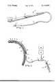
- FIG. 1is a partially broken away perspective view of the catheter of the invention.
- FIG. 2is a cross section of the catheter of the invention as installed in the cranium of a patient.
- a catheterwhich comprises a generally flat or ribbon-like distal portion 10 connected to a length of tubing 12 which terminates, at the proximal end of the catheter, with a connection luer hub 11.
- the distal portion 10is preferably fabricated from a flexible, easily sterilizable material, e.g., medical grade silicone rubber such as that commercially available under the trademark "Silastic".
- at least parts of the catheterare made radio-opaque to permit its location to be determined by x-ray.
- the distal portion 10has front and back surfaces 14, 16 and includes a cup-like recess 18, positioned in the front surface 14 adjacent the catheter's distal tip.
- the flat front surface 14defines a sealing surface 19 about recess 18.
- a lumen 20passes through the distal portion 10 and communicates between the recess 18 and the interior of the tubing 12.
- a three-way stopcock 22is attached to a luer hub 11 and may be adjusted to communicate with a pressure measuring device 24, i.e., a standard physiological transducer, or a fluid injection means 26, which may comprise a luer hub 27 fitted with a syringe 28.
- the pressure measuring device 24is preferably connected to a pressure display and recording device 30, e.g., an instrument sold under the trademark "Datascope 850".
- the distal portion 10 of the catheterapart from the portion housing the recess 18, is of substantially uniform cross section. As will be explained more fully below, this characteristic facilitates removal of the catheter at the conclusion of monitoring.
- the ribbon-like shapee.g., rectangular cross-section, is preferred since it stabilizes the orientation of the catheter, i.e., it minimizes tipping of the distal tip of the catheter such as might disturb the seal formed between sealing portion 19 and the patient's arachnoid membrane 40.
- the width of the back surface 16 at a point behind the recess 18 and of the distal portion 10should be about 8 millimeters.
- the thickness of the distal portion 10is preferably between about 2 and 4 millimeters. This dimension is important insofar as proper operation of the catheter depends upon sealing contact being effected between sealing surface 19 and the patient's arachnoid membrane, the back of the catheter being supported by the patient's skull 38 through the dura mater 39.
- the distal portionshould be long enough to be passed through a subcutaneous tunnel of significant length and still have a portion remaining exterior to the skull. Its length should generally be between fifteen and thirty centimeters, preferably between about fifteen and twenty centimeters.
- a catheter for pediatric usemay be provided by appropriately scaling down the foregoing dimensions.
- the particular dimensions of such a catheterwill depend on the size of the patient for which it is designed. The necessary scaling will be within the skill of those in the art.
- a pair of incisions spaced preferably between about 8 and 12 centimeters apartare made through the patient's scalp 35.
- a burr hole 32is drilled through the skull 38 at the site of one of the incisions, and the dura mater 39, but not the arachnoid membrane 40, is penetrated.
- a subcutaneous tunnel 36is then formed between the catheter entrance incision 34 and the incision (not shown) adjacent the burr hole 32.
- the catheterWith the stopcock 22 open to the luer hub 27, the catheter is filled with sterilized Ringer's lactated solution containing 100,000 units of bacitracin per liter, and its distal end is introduced into the incision 34 and through the tunnel 36 to the site of the burr hole 32.
- the catheter lumen 20is then flushed with more Ringer's solution to clear it and the recess 18 of any substances that may have been introduced while the catheter was being passed through the tunnel 36.
- the recessshould also be visually inspected to make certain that it is clear.
- the recess 18is then completely filled with Ringer's lactated solution and the stopcock 22 is closed to prevent fluid leakage from the catheter lumen.
- the distal end of the catheteris then inserted through the burr hole 32 and dura mater 39 into the subdural space with the sealing portion 19 in contact with the arachnoid membrane 40.
- the scalp incision directly above the burr hole 32is closed, and the catheter is secured to the scalp by sutures.
- a suturecan be placed at the incision 34 but left untied for later use after the cup catheter has been withdrawn.
- An antibiotic ointment with a nonaqueous baseis applied liberally at the catheter exit site to prevent infection.
- the pressure measuring deviceshould be positioned at the same height as the patient's cerebral ventricle, and if it becomes necessary for the patient to sit or stand up, either the transducer 24 should be kept level with the patient's cerebral ventricles, or the stopcock should be closed to the syringe to prevent aspiration of fluid into the subdural space.
- the catheteris now set for monitoring CSF pressure, and any fluctuations will be detected by pressure measuring device 24 and recorded on the pressure recording device 30.
- the patient's arachnoid membraneacts as a pressure responsive diaphragm, the steady-state level of the CSF pressure as well as pressure fluctuations affecting the arachnoid membrane being transmitted through the fluid couple to pressure measuring device 24.
- Pressure waves transmitted through the fluid couplecan be caused by normal changes in CSF pressure such as are characteristic of heart beats and respiration, as well as by abnormalities symptomatic of brain damage.
- the stopcock 20should be opened to provide communication between the syringe 28 and the catheter so that, over a period of 5 seconds, a maximum of 0.25 ml of fluid may be injected in the patient's subdural space.
- the seal between the sealing surface 19 and arachnoid membrane 40is temporarily opened and a small amount of fluid leaks into the subdural space.
- the stopcockshould be returned to its original position. This procedure ensures that the recess 18 remains optimally filled with fluid, thereby maintaining the fluid couple from the arachnoid membrane to the pressure measuring device 24.
- the retaining sutures connecting the catheter to the scalpare taken out and the catheter is removed by slowly and carefully withdrawing it through the subcutaneous tunnel 36 by pulling on a portion of the catheter external to the scalp.
- a culture swabshould be taken at the cup chamber and at the incision 34.
- the untied skin sutures at the exit sitecan now be tied.
Landscapes
- Health & Medical Sciences (AREA)
- Life Sciences & Earth Sciences (AREA)
- Engineering & Computer Science (AREA)
- Biomedical Technology (AREA)
- Veterinary Medicine (AREA)
- Heart & Thoracic Surgery (AREA)
- Hematology (AREA)
- Animal Behavior & Ethology (AREA)
- General Health & Medical Sciences (AREA)
- Public Health (AREA)
- Biophysics (AREA)
- Anesthesiology (AREA)
- Neurosurgery (AREA)
- Pulmonology (AREA)
- Physics & Mathematics (AREA)
- Pathology (AREA)
- Medical Informatics (AREA)
- Molecular Biology (AREA)
- Surgery (AREA)
- Neurology (AREA)
- Ophthalmology & Optometry (AREA)
- Otolaryngology (AREA)
- Measuring And Recording Apparatus For Diagnosis (AREA)
- Media Introduction/Drainage Providing Device (AREA)
Abstract
Description
Claims (7)
Priority Applications (9)
| Application Number | Priority Date | Filing Date | Title |
|---|---|---|---|
| US05/712,132US4114603A (en) | 1976-08-06 | 1976-08-06 | Intracranial pressure monitoring catheter |
| CA280,781ACA1068514A (en) | 1976-08-06 | 1977-06-17 | Intracranial pressure monitoring catheter |
| DE19772728430DE2728430A1 (en) | 1976-08-06 | 1977-06-24 | CATHETER FOR MONITORING BRAIN PRESSURE |
| NLAANVRAGE7707470,ANL182197C (en) | 1976-08-06 | 1977-07-05 | CATHETER FOR MONITORING THE PRESSURE OF THE CEREBROSPINAL HUMIDITY. |
| SE7708865ASE429297B (en) | 1976-08-06 | 1977-08-03 | Catheters for monitoring intracranial pressure |
| JP9303877AJPS5334386A (en) | 1976-08-06 | 1977-08-04 | Catheter for monitoring pressure in cranium |
| CH960377ACH615819A5 (en) | 1976-08-06 | 1977-08-04 | |
| FR7724065AFR2360289A1 (en) | 1976-08-06 | 1977-08-04 | INTRACRANIAL PRESSURE CONTROL CATHETER |
| GB32883/77AGB1547064A (en) | 1976-08-06 | 1977-08-05 | Intracranial pressure monitoring catheter |
Applications Claiming Priority (1)
| Application Number | Priority Date | Filing Date | Title |
|---|---|---|---|
| US05/712,132US4114603A (en) | 1976-08-06 | 1976-08-06 | Intracranial pressure monitoring catheter |
Publications (1)
| Publication Number | Publication Date |
|---|---|
| US4114603Atrue US4114603A (en) | 1978-09-19 |
Family
ID=24860881
Family Applications (1)
| Application Number | Title | Priority Date | Filing Date |
|---|---|---|---|
| US05/712,132Expired - LifetimeUS4114603A (en) | 1976-08-06 | 1976-08-06 | Intracranial pressure monitoring catheter |
Country Status (9)
| Country | Link |
|---|---|
| US (1) | US4114603A (en) |
| JP (1) | JPS5334386A (en) |
| CA (1) | CA1068514A (en) |
| CH (1) | CH615819A5 (en) |
| DE (1) | DE2728430A1 (en) |
| FR (1) | FR2360289A1 (en) |
| GB (1) | GB1547064A (en) |
| NL (1) | NL182197C (en) |
| SE (1) | SE429297B (en) |
Cited By (62)
| Publication number | Priority date | Publication date | Assignee | Title |
|---|---|---|---|---|
| US4195637A (en)* | 1977-10-21 | 1980-04-01 | Schneider Medintag Ag | Catheter arrangement, method of catheterization, and method of manufacturing a dilatation element |
| US4246908A (en)* | 1976-10-19 | 1981-01-27 | Kabushiki Kaisha Toyota Kenkyusho | Intracranial pressure transducer |
| US4407301A (en)* | 1981-01-27 | 1983-10-04 | C. R. Bard, Inc. | Disc membrane catheter for performing cystometrograms and urethral profiles |
| US4735208A (en)* | 1987-01-09 | 1988-04-05 | Ad-Tech Medical Instrument Corp. | Subdural strip electrode for determining epileptogenic foci |
| US4796641A (en)* | 1987-07-06 | 1989-01-10 | Data Sciences, Inc. | Device and method for chronic in-vivo measurement of internal body pressure |
| US4817629A (en)* | 1986-01-03 | 1989-04-04 | Stryker Corporation | Process and apparatus for measurement and monitoring of muscle compartment pressure |
| US4869265A (en)* | 1987-04-03 | 1989-09-26 | Western Clinical Engineering Ltd. | Biomedical pressure transducer |
| US5048536A (en)* | 1987-04-03 | 1991-09-17 | Mcewen James A | Tourniquet for regulating applied pressures |
| US5117836A (en)* | 1990-10-22 | 1992-06-02 | Millar Instruments, Inc. | Method for measuring intracranial fluid characteristics |
| US5191898A (en)* | 1990-10-22 | 1993-03-09 | Millar Instruments, Inc. | Method and assembly for measuring intracranial fluid characateristics |
| US5312357A (en)* | 1991-11-04 | 1994-05-17 | Drager Medical Electonic B.V. | Catheter |
| US5406958A (en)* | 1992-09-19 | 1995-04-18 | Smiths Industries Public Limited Company | Medico-surgical sensor assemblies |
| US5564425A (en)* | 1993-08-24 | 1996-10-15 | Tonokura Ika Kogyo K.K. | Catheter with built-in-display |
| US5851206A (en)* | 1990-03-13 | 1998-12-22 | The Regents Of The University Of California | Method and apparatus for endovascular thermal thrombosis and thermal cancer treatment |
| DE20116879U1 (en) | 2001-10-13 | 2001-12-20 | REHAU AG + Co., 95111 Rehau | Device for determining the intracerebral pressure gradient |
| US6453185B1 (en)* | 2000-03-17 | 2002-09-17 | Integra Lifesciences, Inc. | Ventricular catheter with reduced size connector and method of use |
| EP1165161A4 (en)* | 1999-03-03 | 2003-02-05 | Uab Research Foundation | DIRECT CENTRAL NERVOUS CATHETER AND TEMPERATURE CONTROL SYSTEM |
| US6537232B1 (en) | 1997-05-15 | 2003-03-25 | Regents Of The University Of Minnesota | Intracranial pressure monitoring device and method for use in MR-guided drug delivery |
| US6731976B2 (en) | 1997-09-03 | 2004-05-04 | Medtronic, Inc. | Device and method to measure and communicate body parameters |
| US6923799B1 (en) | 1999-06-04 | 2005-08-02 | Wilson T. Asfora | Subdural evacuating port system |
| US6939299B1 (en)* | 1999-12-13 | 2005-09-06 | Kurt Petersen | Implantable continuous intraocular pressure sensor |
| EP1600186A1 (en)* | 1999-03-03 | 2005-11-30 | UAB Research Foundation | Direct central nervous system catheter and temperature control system |
| US20060089589A1 (en)* | 2004-10-21 | 2006-04-27 | Portnoy Harold D | Resistive shunt valve |
| US20060149161A1 (en)* | 2004-12-29 | 2006-07-06 | Wilson Stephen F | System and method for measuring the pressure of a fluid system within a patient |
| WO2006091164A1 (en)* | 2005-02-28 | 2006-08-31 | Jan Malm | Method and device for determining the hydrodynamics of the cerebrospinal fluid system |
| US20060199997A1 (en)* | 2005-02-24 | 2006-09-07 | Ethicon Endo-Surgery, Inc. | Monitoring of a food intake restriction device |
| US20060211914A1 (en)* | 2005-02-24 | 2006-09-21 | Hassler William L Jr | System and method for determining implanted device positioning and obtaining pressure data |
| US20060211913A1 (en)* | 2005-02-24 | 2006-09-21 | Dlugos Daniel F | Non-invasive pressure measurement in a fluid adjustable restrictive device |
| US20070213837A1 (en)* | 2005-02-24 | 2007-09-13 | Ferreri Annie L | System and Method for Determining Implanted Device Orientation |
| US20070235083A1 (en)* | 2005-02-24 | 2007-10-11 | Dlugos Daniel F | Apparatus for Adjustment and Sensing of Gastric Band Pressure |
| US20070244411A1 (en)* | 2006-04-03 | 2007-10-18 | Mimosa Acoustics, Inc. | Method and system for monitoring intracranial pressure |
| US20080160535A1 (en)* | 1997-12-15 | 2008-07-03 | Somalogic, Inc. | Methods and Reagents for Detecting Target Binding by Nucleic Acid Ligands |
| US20090112103A1 (en)* | 2007-10-31 | 2009-04-30 | Codman & Shurtleff, Inc. | Wireless Pressure Sensing Shunts |
| US20090107233A1 (en)* | 2007-10-31 | 2009-04-30 | Codman Shurleff, Inc. | Wireless Flow Sensor |
| US20090112147A1 (en)* | 2007-10-31 | 2009-04-30 | Codman Shurleff, Inc. | Wireless Pressure Setting Indicator |
| US7553290B1 (en) | 1999-06-04 | 2009-06-30 | Medtronic Ps Medical, Inc. | Subdural evacuating port aspiration system |
| US7694821B1 (en) | 1999-06-04 | 2010-04-13 | Medtronic Ps Medical, Inc. | Subdural evacuating port system |
| US20100249727A1 (en)* | 2008-02-06 | 2010-09-30 | Intravena, Llc | Convenience IV kits and methods of use |
| WO2011003909A1 (en)* | 2009-07-06 | 2011-01-13 | Likvor Ab | Device and method for measuring and regulating cerebrospinal fluid parameters |
| US20110054518A1 (en)* | 2009-08-27 | 2011-03-03 | Boston Scientific Neuromodulation Corporation | Burr hole sealing device for preventing brain shift |
| US7927270B2 (en) | 2005-02-24 | 2011-04-19 | Ethicon Endo-Surgery, Inc. | External mechanical pressure sensor for gastric band pressure measurements |
| USRE42625E1 (en) | 1990-03-13 | 2011-08-16 | The Regents Of The University Of California | Endovascular electrolytically detachable wire and tip for the formation of thrombus in arteries, veins, aneurysms, vascular malformations and arteriovenous fistulas |
| USRE42662E1 (en) | 1990-03-13 | 2011-08-30 | The Regents Of The University Of California | Endovascular electrolytically detachable wire and tip for the formation of thrombus in arteries, veins, aneurysms, vascular malformations and arteriovenous fistulas |
| US8016744B2 (en) | 2005-02-24 | 2011-09-13 | Ethicon Endo-Surgery, Inc. | External pressure-based gastric band adjustment system and method |
| USRE42756E1 (en) | 1990-03-13 | 2011-09-27 | The Regents Of The University Of California | Endovascular electrolytically detachable wire and tip for the formation of thrombus in arteries, veins, aneurysms, vascular malformations and arteriovenous fistulas |
| US8152710B2 (en) | 2006-04-06 | 2012-04-10 | Ethicon Endo-Surgery, Inc. | Physiological parameter analysis for an implantable restriction device and a data logger |
| US8480612B2 (en) | 2007-10-31 | 2013-07-09 | DePuy Synthes Products, LLC | Wireless shunts with storage |
| WO2013154606A1 (en)* | 2012-04-13 | 2013-10-17 | Branchpoint Technologies, LLC | Sensor, circuitry, and method for wireless intracranial pressure monitoring |
| US8870742B2 (en) | 2006-04-06 | 2014-10-28 | Ethicon Endo-Surgery, Inc. | GUI for an implantable restriction device and a data logger |
| US9050436B2 (en) | 2013-03-14 | 2015-06-09 | DePuy Synthes Products, LLC | Adjustable resistance, gravitationally activated, anti-syphon valve |
| US9265577B2 (en) | 2007-05-18 | 2016-02-23 | The Johns Hopkins University | Methods and systems for providing planning and dispensation of research and/or treatment for brain disease |
| US9636070B2 (en) | 2013-03-14 | 2017-05-02 | DePuy Synthes Products, Inc. | Methods, systems, and devices for monitoring and displaying medical parameters for a patient |
| US20170265920A1 (en)* | 2014-08-20 | 2017-09-21 | Tecres S.P.A. | Device for supplying fluid substances in the body of a patient |
| US9848789B2 (en) | 2014-04-17 | 2017-12-26 | Branchpoint Technologies, Inc. | Wireless intracranial monitoring system |
| US9901269B2 (en) | 2014-04-17 | 2018-02-27 | Branchpoint Technologies, Inc. | Wireless intracranial monitoring system |
| US10232169B2 (en) | 2015-07-23 | 2019-03-19 | Boston Scientific Neuromodulation Corporation | Burr hole plugs for electrical stimulation systems and methods of making and using |
| CN111228629A (en)* | 2020-01-21 | 2020-06-05 | 济南市传染病医院 | Craniocerebral drainage tube submerged implantation device |
| US10702174B2 (en) | 2007-06-27 | 2020-07-07 | Integra Lifesciences Corporation | Medical monitor user interface |
| US11013913B2 (en) | 2018-03-16 | 2021-05-25 | Boston Scientific Neuromodulation Corporation | Kits and methods for securing a burr hole plugs for stimulation systems |
| US11058870B2 (en) | 2018-03-09 | 2021-07-13 | Boston Scientific Neuromodulation Corporation | Burr hole plugs for electrical stimulation systems and methods of making and using |
| US11103716B2 (en) | 2017-11-13 | 2021-08-31 | Boston Scientific Neuromodulation Corporation | Systems and methods for making and using a low-profile control module for an electrical stimulation system |
| US11497914B2 (en) | 2018-01-16 | 2022-11-15 | Boston Scientific Neuromodulation Corporation | Systems and methods for making and using an electrical stimulation system with a case-neutral battery |
Families Citing this family (4)
| Publication number | Priority date | Publication date | Assignee | Title |
|---|---|---|---|---|
| CA1160525A (en)* | 1980-03-07 | 1984-01-17 | Alfred R. Perlin | Esophageal probe |
| DE3500822A1 (en)* | 1985-01-10 | 1986-07-10 | Spiegelberg, Andreas, Dipl.-Ing., 1000 Berlin | PRESSURE MEASUREMENT DEVICE |
| AT386117B (en)* | 1986-02-03 | 1988-07-11 | Ludwig Dr Med Auer | Device for determining the pressure profile on instruments for surgical interventions |
| DE20009854U1 (en) | 2000-05-31 | 2000-08-24 | REHAU AG + Co., 95111 Rehau | Device for pressure measurements |
Citations (4)
| Publication number | Priority date | Publication date | Assignee | Title |
|---|---|---|---|---|
| US3669094A (en)* | 1970-07-06 | 1972-06-13 | Heyer Schulte Corp | Device and method for measuring intracranial pressure |
| US3789667A (en)* | 1972-02-14 | 1974-02-05 | Ladd Res Ind Inc | Fiber optic pressure detector |
| US3877137A (en)* | 1974-05-30 | 1975-04-15 | Hakim Co Ltd | Method of making implantable pressure sensor |
| US3886948A (en)* | 1972-08-14 | 1975-06-03 | Hakim Co Ltd | Ventricular shunt having a variable pressure valve |
Family Cites Families (2)
| Publication number | Priority date | Publication date | Assignee | Title |
|---|---|---|---|---|
| US3686958A (en)* | 1971-02-22 | 1972-08-29 | Ladd Res Ind | Fiber optic pressure detector |
| US4027661A (en)* | 1974-07-16 | 1977-06-07 | Hittman Corporation | Pressure sensor |
- 1976
- 1976-08-06USUS05/712,132patent/US4114603A/ennot_activeExpired - Lifetime
- 1977
- 1977-06-17CACA280,781Apatent/CA1068514A/ennot_activeExpired
- 1977-06-24DEDE19772728430patent/DE2728430A1/enactiveGranted
- 1977-07-05NLNLAANVRAGE7707470,Apatent/NL182197C/ennot_activeIP Right Cessation
- 1977-08-03SESE7708865Apatent/SE429297B/ennot_activeIP Right Cessation
- 1977-08-04JPJP9303877Apatent/JPS5334386A/enactiveGranted
- 1977-08-04CHCH960377Apatent/CH615819A5/frnot_activeIP Right Cessation
- 1977-08-04FRFR7724065Apatent/FR2360289A1/enactiveGranted
- 1977-08-05GBGB32883/77Apatent/GB1547064A/ennot_activeExpired
Patent Citations (4)
| Publication number | Priority date | Publication date | Assignee | Title |
|---|---|---|---|---|
| US3669094A (en)* | 1970-07-06 | 1972-06-13 | Heyer Schulte Corp | Device and method for measuring intracranial pressure |
| US3789667A (en)* | 1972-02-14 | 1974-02-05 | Ladd Res Ind Inc | Fiber optic pressure detector |
| US3886948A (en)* | 1972-08-14 | 1975-06-03 | Hakim Co Ltd | Ventricular shunt having a variable pressure valve |
| US3877137A (en)* | 1974-05-30 | 1975-04-15 | Hakim Co Ltd | Method of making implantable pressure sensor |
Non-Patent Citations (2)
| Title |
|---|
| Journal of Neurosurgery, vol. 39 (Sep. 1973), p. 416, by John K. Vries.* |
| Numoto et al., "Pressure Indicating Bag for Monitoring Intracranial Pressure", J. Neurosurg., vol. 39, p. 784-787.* |
Cited By (95)
| Publication number | Priority date | Publication date | Assignee | Title |
|---|---|---|---|---|
| US4246908A (en)* | 1976-10-19 | 1981-01-27 | Kabushiki Kaisha Toyota Kenkyusho | Intracranial pressure transducer |
| US4195637A (en)* | 1977-10-21 | 1980-04-01 | Schneider Medintag Ag | Catheter arrangement, method of catheterization, and method of manufacturing a dilatation element |
| US4407301A (en)* | 1981-01-27 | 1983-10-04 | C. R. Bard, Inc. | Disc membrane catheter for performing cystometrograms and urethral profiles |
| US4817629A (en)* | 1986-01-03 | 1989-04-04 | Stryker Corporation | Process and apparatus for measurement and monitoring of muscle compartment pressure |
| US4735208A (en)* | 1987-01-09 | 1988-04-05 | Ad-Tech Medical Instrument Corp. | Subdural strip electrode for determining epileptogenic foci |
| US4869265A (en)* | 1987-04-03 | 1989-09-26 | Western Clinical Engineering Ltd. | Biomedical pressure transducer |
| US5048536A (en)* | 1987-04-03 | 1991-09-17 | Mcewen James A | Tourniquet for regulating applied pressures |
| US4796641A (en)* | 1987-07-06 | 1989-01-10 | Data Sciences, Inc. | Device and method for chronic in-vivo measurement of internal body pressure |
| US5851206A (en)* | 1990-03-13 | 1998-12-22 | The Regents Of The University Of California | Method and apparatus for endovascular thermal thrombosis and thermal cancer treatment |
| USRE42625E1 (en) | 1990-03-13 | 2011-08-16 | The Regents Of The University Of California | Endovascular electrolytically detachable wire and tip for the formation of thrombus in arteries, veins, aneurysms, vascular malformations and arteriovenous fistulas |
| USRE42662E1 (en) | 1990-03-13 | 2011-08-30 | The Regents Of The University Of California | Endovascular electrolytically detachable wire and tip for the formation of thrombus in arteries, veins, aneurysms, vascular malformations and arteriovenous fistulas |
| USRE42756E1 (en) | 1990-03-13 | 2011-09-27 | The Regents Of The University Of California | Endovascular electrolytically detachable wire and tip for the formation of thrombus in arteries, veins, aneurysms, vascular malformations and arteriovenous fistulas |
| US5919187A (en)* | 1990-03-13 | 1999-07-06 | The Regents Of The University Of California | Method and apparatus for endovascular thermal thrombosis and thermal cancer treatment |
| US5191898A (en)* | 1990-10-22 | 1993-03-09 | Millar Instruments, Inc. | Method and assembly for measuring intracranial fluid characateristics |
| US5117836A (en)* | 1990-10-22 | 1992-06-02 | Millar Instruments, Inc. | Method for measuring intracranial fluid characteristics |
| US5312357A (en)* | 1991-11-04 | 1994-05-17 | Drager Medical Electonic B.V. | Catheter |
| US5406958A (en)* | 1992-09-19 | 1995-04-18 | Smiths Industries Public Limited Company | Medico-surgical sensor assemblies |
| US5564425A (en)* | 1993-08-24 | 1996-10-15 | Tonokura Ika Kogyo K.K. | Catheter with built-in-display |
| US6537232B1 (en) | 1997-05-15 | 2003-03-25 | Regents Of The University Of Minnesota | Intracranial pressure monitoring device and method for use in MR-guided drug delivery |
| US6731976B2 (en) | 1997-09-03 | 2004-05-04 | Medtronic, Inc. | Device and method to measure and communicate body parameters |
| US20080160535A1 (en)* | 1997-12-15 | 2008-07-03 | Somalogic, Inc. | Methods and Reagents for Detecting Target Binding by Nucleic Acid Ligands |
| EP1600186A1 (en)* | 1999-03-03 | 2005-11-30 | UAB Research Foundation | Direct central nervous system catheter and temperature control system |
| EP1165161A4 (en)* | 1999-03-03 | 2003-02-05 | Uab Research Foundation | DIRECT CENTRAL NERVOUS CATHETER AND TEMPERATURE CONTROL SYSTEM |
| US8343138B2 (en) | 1999-06-04 | 2013-01-01 | Medtronic Xomed, Inc. | Subdural evacuation port aspiration device |
| US7553290B1 (en) | 1999-06-04 | 2009-06-30 | Medtronic Ps Medical, Inc. | Subdural evacuating port aspiration system |
| US7694821B1 (en) | 1999-06-04 | 2010-04-13 | Medtronic Ps Medical, Inc. | Subdural evacuating port system |
| US6923799B1 (en) | 1999-06-04 | 2005-08-02 | Wilson T. Asfora | Subdural evacuating port system |
| US6939299B1 (en)* | 1999-12-13 | 2005-09-06 | Kurt Petersen | Implantable continuous intraocular pressure sensor |
| US6453185B1 (en)* | 2000-03-17 | 2002-09-17 | Integra Lifesciences, Inc. | Ventricular catheter with reduced size connector and method of use |
| US6749574B2 (en) | 2000-03-17 | 2004-06-15 | Integra Lifesciences Inc. | Ventricular catheter with reduced size connector |
| DE20116879U1 (en) | 2001-10-13 | 2001-12-20 | REHAU AG + Co., 95111 Rehau | Device for determining the intracerebral pressure gradient |
| US20060089589A1 (en)* | 2004-10-21 | 2006-04-27 | Portnoy Harold D | Resistive shunt valve |
| US9220424B2 (en)* | 2004-12-29 | 2015-12-29 | DePuy Synthes Products, Inc. | System and method for measuring the pressure of a fluid system within a patient |
| US20060149161A1 (en)* | 2004-12-29 | 2006-07-06 | Wilson Stephen F | System and method for measuring the pressure of a fluid system within a patient |
| US20090270759A1 (en)* | 2004-12-29 | 2009-10-29 | Codman & Shurtleff, Inc. | System and Method for Measuring the Pressure of a Fluid System Within a Patient |
| US7585280B2 (en) | 2004-12-29 | 2009-09-08 | Codman & Shurtleff, Inc. | System and method for measuring the pressure of a fluid system within a patient |
| US9931043B2 (en) | 2004-12-29 | 2018-04-03 | Integra Lifesciences Switzerland Sàrl | System and method for measuring the pressure of a fluid system within a patient |
| US20060211914A1 (en)* | 2005-02-24 | 2006-09-21 | Hassler William L Jr | System and method for determining implanted device positioning and obtaining pressure data |
| US7775215B2 (en) | 2005-02-24 | 2010-08-17 | Ethicon Endo-Surgery, Inc. | System and method for determining implanted device positioning and obtaining pressure data |
| US8066629B2 (en) | 2005-02-24 | 2011-11-29 | Ethicon Endo-Surgery, Inc. | Apparatus for adjustment and sensing of gastric band pressure |
| US8016744B2 (en) | 2005-02-24 | 2011-09-13 | Ethicon Endo-Surgery, Inc. | External pressure-based gastric band adjustment system and method |
| US7658196B2 (en) | 2005-02-24 | 2010-02-09 | Ethicon Endo-Surgery, Inc. | System and method for determining implanted device orientation |
| US7927270B2 (en) | 2005-02-24 | 2011-04-19 | Ethicon Endo-Surgery, Inc. | External mechanical pressure sensor for gastric band pressure measurements |
| US7775966B2 (en) | 2005-02-24 | 2010-08-17 | Ethicon Endo-Surgery, Inc. | Non-invasive pressure measurement in a fluid adjustable restrictive device |
| US20060199997A1 (en)* | 2005-02-24 | 2006-09-07 | Ethicon Endo-Surgery, Inc. | Monitoring of a food intake restriction device |
| US20070235083A1 (en)* | 2005-02-24 | 2007-10-11 | Dlugos Daniel F | Apparatus for Adjustment and Sensing of Gastric Band Pressure |
| US8016745B2 (en) | 2005-02-24 | 2011-09-13 | Ethicon Endo-Surgery, Inc. | Monitoring of a food intake restriction device |
| US20070213837A1 (en)* | 2005-02-24 | 2007-09-13 | Ferreri Annie L | System and Method for Determining Implanted Device Orientation |
| US20060211913A1 (en)* | 2005-02-24 | 2006-09-21 | Dlugos Daniel F | Non-invasive pressure measurement in a fluid adjustable restrictive device |
| US8784331B2 (en) | 2005-02-28 | 2014-07-22 | Likvor Ab | Method and device for determining the hydrodynamics of the cerebrospinal fluid system |
| WO2006091164A1 (en)* | 2005-02-28 | 2006-08-31 | Jan Malm | Method and device for determining the hydrodynamics of the cerebrospinal fluid system |
| US20080306403A1 (en)* | 2005-02-28 | 2008-12-11 | Jan Malm | Method and Device for Determining the Hydrodynamics of the Cerebrospinal Fluid System |
| US20070244411A1 (en)* | 2006-04-03 | 2007-10-18 | Mimosa Acoustics, Inc. | Method and system for monitoring intracranial pressure |
| US8152710B2 (en) | 2006-04-06 | 2012-04-10 | Ethicon Endo-Surgery, Inc. | Physiological parameter analysis for an implantable restriction device and a data logger |
| US8870742B2 (en) | 2006-04-06 | 2014-10-28 | Ethicon Endo-Surgery, Inc. | GUI for an implantable restriction device and a data logger |
| US9265577B2 (en) | 2007-05-18 | 2016-02-23 | The Johns Hopkins University | Methods and systems for providing planning and dispensation of research and/or treatment for brain disease |
| US10702174B2 (en) | 2007-06-27 | 2020-07-07 | Integra Lifesciences Corporation | Medical monitor user interface |
| US8870768B2 (en) | 2007-10-31 | 2014-10-28 | DePuy Synthes Products, LLC | Wireless flow sensor methods |
| US7842004B2 (en) | 2007-10-31 | 2010-11-30 | Codman & Shurtleff, Inc. | Wireless pressure setting indicator |
| US10265509B2 (en) | 2007-10-31 | 2019-04-23 | Integra LifeSciences Switzerland Sarl | Wireless shunts with storage |
| US20090112103A1 (en)* | 2007-10-31 | 2009-04-30 | Codman & Shurtleff, Inc. | Wireless Pressure Sensing Shunts |
| US20090112147A1 (en)* | 2007-10-31 | 2009-04-30 | Codman Shurleff, Inc. | Wireless Pressure Setting Indicator |
| US8454524B2 (en) | 2007-10-31 | 2013-06-04 | DePuy Synthes Products, LLC | Wireless flow sensor |
| US8480612B2 (en) | 2007-10-31 | 2013-07-09 | DePuy Synthes Products, LLC | Wireless shunts with storage |
| US20090107233A1 (en)* | 2007-10-31 | 2009-04-30 | Codman Shurleff, Inc. | Wireless Flow Sensor |
| US8579847B2 (en) | 2007-10-31 | 2013-11-12 | Codman & Shurtleff, Inc. | Wireless pressure setting indicator |
| US9204812B2 (en) | 2007-10-31 | 2015-12-08 | DePuy Synthes Products, LLC | Wireless pressure sensing shunts |
| US8864666B2 (en) | 2007-10-31 | 2014-10-21 | DePuy Synthes Products, LLC | Wireless flow sensor |
| US20100249727A1 (en)* | 2008-02-06 | 2010-09-30 | Intravena, Llc | Convenience IV kits and methods of use |
| WO2011003909A1 (en)* | 2009-07-06 | 2011-01-13 | Likvor Ab | Device and method for measuring and regulating cerebrospinal fluid parameters |
| CN102481435B (en)* | 2009-07-06 | 2015-07-29 | 莱克沃有限公司 | Device for measuring and regulating cerebrospinal fluid parameters |
| US20110021991A1 (en)* | 2009-07-06 | 2011-01-27 | Sundstroem Nina | Fully automated method of measuring and regulating cerebrospinal fluid parameters using disposable tube-set |
| US8109899B2 (en) | 2009-07-06 | 2012-02-07 | Likvor Ab | Fully automated method of measuring and regulating cerebrospinal fluid parameters using disposable tube-set |
| CN102481435A (en)* | 2009-07-06 | 2012-05-30 | 莱克沃有限公司 | Device and method for measuring and regulating cerebrospinal fluid parameters |
| US20110054518A1 (en)* | 2009-08-27 | 2011-03-03 | Boston Scientific Neuromodulation Corporation | Burr hole sealing device for preventing brain shift |
| US8313453B2 (en)* | 2009-08-27 | 2012-11-20 | Boston Scientific Neuromodulation Corporation | Burr hole sealing device for preventing brain shift |
| US11564585B2 (en) | 2011-04-13 | 2023-01-31 | Branchpoint Technologies, Inc. | Sensor, circuitry, and method for wireless intracranial pressure monitoring |
| US10420479B2 (en) | 2011-04-13 | 2019-09-24 | Branchpoint Technologies, Inc. | Sensor, circuitry, and method for wireless intracranial pressure monitoring |
| US9901268B2 (en) | 2011-04-13 | 2018-02-27 | Branchpoint Technologies, Inc. | Sensor, circuitry, and method for wireless intracranial pressure monitoring |
| WO2013154606A1 (en)* | 2012-04-13 | 2013-10-17 | Branchpoint Technologies, LLC | Sensor, circuitry, and method for wireless intracranial pressure monitoring |
| US9731102B2 (en) | 2013-03-14 | 2017-08-15 | DePuy Synthes Products, Inc. | Adjustable resistance, gravitationally activated, anti-siphon valve |
| US9050436B2 (en) | 2013-03-14 | 2015-06-09 | DePuy Synthes Products, LLC | Adjustable resistance, gravitationally activated, anti-syphon valve |
| US9636070B2 (en) | 2013-03-14 | 2017-05-02 | DePuy Synthes Products, Inc. | Methods, systems, and devices for monitoring and displaying medical parameters for a patient |
| US11083386B2 (en) | 2014-04-17 | 2021-08-10 | Branchpoint Technologies, Inc. | Wireless intracranial monitoring system |
| US9848789B2 (en) | 2014-04-17 | 2017-12-26 | Branchpoint Technologies, Inc. | Wireless intracranial monitoring system |
| US11197622B2 (en) | 2014-04-17 | 2021-12-14 | Branchpoint Technologies, Inc. | Wireless intracranial monitoring system |
| US9901269B2 (en) | 2014-04-17 | 2018-02-27 | Branchpoint Technologies, Inc. | Wireless intracranial monitoring system |
| US20170265920A1 (en)* | 2014-08-20 | 2017-09-21 | Tecres S.P.A. | Device for supplying fluid substances in the body of a patient |
| US10232169B2 (en) | 2015-07-23 | 2019-03-19 | Boston Scientific Neuromodulation Corporation | Burr hole plugs for electrical stimulation systems and methods of making and using |
| US11103716B2 (en) | 2017-11-13 | 2021-08-31 | Boston Scientific Neuromodulation Corporation | Systems and methods for making and using a low-profile control module for an electrical stimulation system |
| US11497914B2 (en) | 2018-01-16 | 2022-11-15 | Boston Scientific Neuromodulation Corporation | Systems and methods for making and using an electrical stimulation system with a case-neutral battery |
| US11058870B2 (en) | 2018-03-09 | 2021-07-13 | Boston Scientific Neuromodulation Corporation | Burr hole plugs for electrical stimulation systems and methods of making and using |
| US11013913B2 (en) | 2018-03-16 | 2021-05-25 | Boston Scientific Neuromodulation Corporation | Kits and methods for securing a burr hole plugs for stimulation systems |
| CN111228629B (en)* | 2020-01-21 | 2021-11-16 | 济南市传染病医院 | Craniocerebral drainage tube submerged implantation device |
| CN111228629A (en)* | 2020-01-21 | 2020-06-05 | 济南市传染病医院 | Craniocerebral drainage tube submerged implantation device |
Also Published As
| Publication number | Publication date |
|---|---|
| SE429297B (en) | 1983-08-29 |
| SE7708865L (en) | 1978-02-07 |
| NL7707470A (en) | 1978-02-08 |
| DE2728430C2 (en) | 1987-07-23 |
| JPS5334386A (en) | 1978-03-30 |
| NL182197C (en) | 1988-02-01 |
| DE2728430A1 (en) | 1978-02-09 |
| GB1547064A (en) | 1979-06-06 |
| CH615819A5 (en) | 1980-02-29 |
| NL182197B (en) | 1987-09-01 |
| FR2360289B1 (en) | 1984-01-27 |
| CA1068514A (en) | 1979-12-25 |
| JPS6232935B2 (en) | 1987-07-17 |
| FR2360289A1 (en) | 1978-03-03 |
Similar Documents
| Publication | Publication Date | Title |
|---|---|---|
| US4114603A (en) | Intracranial pressure monitoring catheter | |
| US5191898A (en) | Method and assembly for measuring intracranial fluid characateristics | |
| US5117836A (en) | Method for measuring intracranial fluid characteristics | |
| US6537232B1 (en) | Intracranial pressure monitoring device and method for use in MR-guided drug delivery | |
| US5935083A (en) | Device for body fluid pressure measurement | |
| Vries et al. | A subarachnoid screw for monitoring intracranial pressure | |
| US5284138A (en) | Apparatus and method for positioning a sensor away from the blood vessel wall | |
| US4160448A (en) | Blood pressure measuring catheter | |
| US5396897A (en) | Method for locating tumors prior to needle biopsy | |
| EP0774919B1 (en) | Gas column pressure-monitoring catheters | |
| US5788631A (en) | Hollow viscus and solid organ tonometry | |
| US5383896A (en) | Vascular sealing device | |
| US5218965A (en) | Apparatus for carrying a sensor in a connector for a catheter adapter | |
| EP0540783A1 (en) | Catheter | |
| US20080171990A1 (en) | Multimodal Catheter for Focal Brain Monitoring and Ventriculostomy | |
| US5478326A (en) | Arterial device for control of bleeding from a puncture in an artery wall | |
| Tanaka et al. | Nonoperative measurement of pancreatic and common bile duct pressures with a microtransducer catheter and effects of duodenoscopic sphincterotomy | |
| US5526809A (en) | Hollow viscous and soild organ tonometry | |
| Coroneos et al. | Measurement of extradural pressure and its relationship to other intracranial pressures: an experimental and clinical study | |
| US5599317A (en) | Externalized sealed catheter with leakproof access | |
| Kroin et al. | Long-term testing of an intracranial pressure monitoring device | |
| Langfitt | Clinical methods for monitoring intracranial pressure and measuring cerebral blood flow | |
| VENNES | INTRAHEPATIC PRESSURE:: AN ACCURATE REFLECTION OF PORTAL PRESSURE | |
| Gaab et al. | Miniaturized methods of monitoring intracranial pressure in craniocerebral trauma before and after operation | |
| De Jong et al. | Epidural pressure monitoring with the so-called Rotterdam transducer. Further in vivo results |
Legal Events
| Date | Code | Title | Description |
|---|---|---|---|
| AS | Assignment | Owner name:ELEKTA AB, SWEDEN Free format text:ASSIGNMENT OF ASSIGNORS INTEREST;ASSIGNOR:CORDIS CORPORATION;REEL/FRAME:008478/0819 Effective date:19970411 | |
| AS | Assignment | Owner name:NMT NEUROSCIENCES (IP), INC., A DELAWARE CORPORATI Free format text:ASSIGNMENT OF ASSIGNORS INTEREST;ASSIGNOR:ELEKTA AB (PUBL) A SWEDISH CORPORATION;REEL/FRAME:009375/0712 Effective date:19980708 Owner name:J.H. WHITNEY & CO., CONNECTICUT Free format text:SECURITY INTEREST;ASSIGNORS:NITINOL MEDICAL TECHNOLOGIES, INC.;NMT NEUROSCIENCES (IP) INC.;NMT NEUROSCIENCES (INTERNATIONAL) INC.;AND OTHERS;REEL/FRAME:009375/0116 Effective date:19980708 | |
| AS | Assignment | Owner name:NMT MEDICAL, INC., MASSACHUSETTS Free format text:CHANGE OF NAME;ASSIGNOR:NMT NEUROSCIENCES (IP), INC. (A DELAWARE CORPORATION);REEL/FRAME:010206/0089 Effective date:19990603 | |
| AS | Assignment | Owner name:NMT NEUROSCIENCES (INTERNATIONAL), INC., MASSACHUS Free format text:SECURITY INTEREST TERMINATION;ASSIGNOR:J.H. WHITNEY & CO.;REEL/FRAME:010668/0425 Effective date:19991020 Owner name:NMT INVESTMENTS CORP., MASSACHUSETTS Free format text:SECURITY INTEREST TERMINATION;ASSIGNOR:J.H. WHITNEY & CO.;REEL/FRAME:010668/0425 Effective date:19991020 Owner name:NMT NEUROSCIENCES (U.S.), INC., MASSACHUSETTS Free format text:SECURITY INTEREST TERMINATION;ASSIGNOR:J.H. WHITNEY & CO.;REEL/FRAME:010668/0425 Effective date:19991020 Owner name:NMT NEUROSCIENCES (IP), INC., MASSACHUSETTS Free format text:SECURITY INTEREST TERMINATION;ASSIGNOR:J.H. WHITNEY & CO.;REEL/FRAME:010668/0425 Effective date:19991020 Owner name:NMT HEART, INC., MASSACHUSETTS Free format text:SECURITY INTEREST TERMINATION;ASSIGNOR:J.H. WHITNEY & CO.;REEL/FRAME:010668/0425 Effective date:19991020 Owner name:NMT MEDICAL, INC. (F/K/A NITINOL MEDICAL TECHNOLOG Free format text:SECURITY INTEREST TERMINATION;ASSIGNOR:J.H. WHITNEY & CO.;REEL/FRAME:010668/0425 Effective date:19991020 Owner name:NMT NEUROSCIENCES INNOVASIVE SYSTEMS, INC. (F/K/A Free format text:SECURITY INTEREST TERMINATION;ASSIGNOR:J.H. WHITNEY & CO.;REEL/FRAME:010668/0425 Effective date:19991020 |