US3669094A - Device and method for measuring intracranial pressure - Google Patents
Device and method for measuring intracranial pressureDownload PDFInfo
- Publication number
- US3669094A US3669094AUS52654AUS3669094DAUS3669094AUS 3669094 AUS3669094 AUS 3669094AUS 52654 AUS52654 AUS 52654AUS 3669094D AUS3669094D AUS 3669094DAUS 3669094 AUS3669094 AUS 3669094A
- Authority
- US
- United States
- Prior art keywords
- drainage
- passage
- tube
- branch
- tip
- Prior art date
- Legal status (The legal status is an assumption and is not a legal conclusion. Google has not performed a legal analysis and makes no representation as to the accuracy of the status listed.)
- Expired - Lifetime
Links
Images
Classifications
- A—HUMAN NECESSITIES
- A61—MEDICAL OR VETERINARY SCIENCE; HYGIENE
- A61B—DIAGNOSIS; SURGERY; IDENTIFICATION
- A61B5/00—Measuring for diagnostic purposes; Identification of persons
- A61B5/03—Measuring fluid pressure within the body other than blood pressure, e.g. cerebral pressure ; Measuring pressure in body tissues or organs
- A61B5/031—Intracranial pressure
- A—HUMAN NECESSITIES
- A61—MEDICAL OR VETERINARY SCIENCE; HYGIENE
- A61M—DEVICES FOR INTRODUCING MEDIA INTO, OR ONTO, THE BODY; DEVICES FOR TRANSDUCING BODY MEDIA OR FOR TAKING MEDIA FROM THE BODY; DEVICES FOR PRODUCING OR ENDING SLEEP OR STUPOR
- A61M27/00—Drainage appliance for wounds or the like, i.e. wound drains, implanted drains
- A61M27/002—Implant devices for drainage of body fluids from one part of the body to another
- A61M27/006—Cerebrospinal drainage; Accessories therefor, e.g. valves
- A—HUMAN NECESSITIES
- A61—MEDICAL OR VETERINARY SCIENCE; HYGIENE
- A61M—DEVICES FOR INTRODUCING MEDIA INTO, OR ONTO, THE BODY; DEVICES FOR TRANSDUCING BODY MEDIA OR FOR TAKING MEDIA FROM THE BODY; DEVICES FOR PRODUCING OR ENDING SLEEP OR STUPOR
- A61M27/00—Drainage appliance for wounds or the like, i.e. wound drains, implanted drains
- A61M27/002—Implant devices for drainage of body fluids from one part of the body to another
- A61M2027/004—Implant devices for drainage of body fluids from one part of the body to another with at least a part of the circuit outside the body
Definitions
- ABSTRACTA physiological drainage catheter and a method of using the same to provide a means for measuring intracranial pressure without substantially affecting the existing pressure by introduction of the measuring and drainage means.
- Drainage catheters for use in the human bodyare widely used in various applications, perhaps the best known of which is the drainage of fluids from the cranium of a child afflicted with hydrocephalus. These devices are customarily thrust into the ventricles of the brain where openings through the catheter permit drainage of the fluid to some region where the fluids are disposed of, such as in the heart.
- the intracranial pressureis a matter of considerable importance in the treatment of the hydrocephalus. It is important that the pressure not be reduced to an abnormally low level, and it is of interest to determine what the initial level would be.
- the existing techniques for inserting cathetershave ordinarily involved the loss of substantial ventricular fluid, and with this loss, of course, there has been a drop in pressure such that the original intracranial pressure was not known and, worse, was not discoverable.
- Still another problem in the prior art of measuring intracranial pressureis the fact that the insertion of a body such as a sensor into a region already overtaxed by liquids under pressure to the extent that the brain itself has begun to be shrunk, or the skull distended, affects the pressure by changing the volume of relatively incompressible material within an enclosure, ie., the cranium. Accordingly, an element of uncertainty is introduced into the measurements simply by the introduction of the measuring instrument itself.
- a physiological drainage cathetercomprises an elongated drainage tube with a central drainage passage extending along a central axis, a peripheral wall bounding said passage, an entry port passing through the wall to the central passage, and a branch tube integrally formed with the drainage tube and having a branch passage therein, opening in fluid communication to the drainage passage.
- an insertion rodwhich has an outer diameter substantially equal to the inner diameter of the drainage passage, such that the rod is slidable therein. It makes a close fit with the peripheral walls so as to close the inlet port and the branch passage when opposed to their openings in said peripheral wall. Then the catheter can be inserted into the body by a means of an endwise axial force exerted on the tip by the rod, and the passages will remain closed during that time. After the catheter is placed, the rod may be withdrawn to open and interconnect the inlet port and branch passage, while keeping the drainage passage closed on the side of the branch passage opposite from the tip so as to enable pressure in the cranium to be measured at the branch passage.
- the dimensions of the catheterare selected such that the net volume of the catheter inserted into the cranium is substantially equal to the volume of the brain passage and branch passage outside the skull, whereby no net change of material volume is caused in the cranium as a consequence of insertion of the catheter into the fluid-filled body cavity (the space between the cranium and the brain).
- Still another feature of the inventionresides in the method of inserting the catheter with the rod by exerting the axial endwise force, withdrawing the rod to interconnect the two passages, and taking a pressure reading at the branch passage prior to cutting the drainage tube a a location such that the branch tube is discarded.
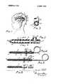
- FIG. 1shows the presently preferred embodiment of the invention installed in the head of a child suffering from hydrocephalus
- F IG. 2is a fragmentary axial cross-section of the presently preferred embodiment of the invention.
- FIG. 3is a cross-section taken at line 3-3 of FIG. 2;
- FIG. 4shows the device of FIG. 2 being inserted into the cranium
- FIG. 5shows a sequential measuring step and FIG. 6 shows a final cutting and discarding operation.
- FIG. Ishows a drainage system 10 installed in the cranium ll of a child.
- the child's brain 12is shown in general schematic notation, and a catheter [3, according to the invention, is thrust through a hole 14 formed in the skull into the ventricles of the brain so as to drain excess fluid therefrom.
- a shunt tube of this general classificationforms no part of this invention, but for general information, it is shown in Heyer US. Pat. No. 3,020,913 issued Feb. l3, i962.
- This shunt tubecustomarily includes a check valve means, and the pump itself also comprises check valve means, the combination assuring a unidirectional flow from the ventricles of the brain to the region into which the fluid is discharged.
- the pumpwhich is optional, provides pumping means.
- this inventionit is the purpose of this invention to permit intracranial pressure to be measured in the region of tip 20 of catheter [3. It is evident that since the brain is already compacted by excess fluid, and that this fluid exerts pressure against the cranium so as to distend the same, insertion of still another solid object will change the pressure unless steps are taken to prevent this change.'lt is further obvious that it is desirable to avoid loss of fluid as much as possible should an accurate pressure reading be desired. This invention accomplishes these objectives.
- Catheter I3is best shown in FIG. 2. It includes a drainage tube 21 having a drainage passage 22 extending axially along an axis 23. Tip 20 is shown, and spaced somewhat from it, is a plurality of inlet ports 24 which opens onto the peripheral wall 25 of the tube. These inlet ports thereby provide perforations to permit flow of fluid from the outside of the tube into the drainage passage.
- a branch tube 26is formed integrally with the drainage tube and has a branch passage 27 opening on wall 25. It is spaced from the inlet ports and on the opposite side therefrom from the tip.
- the catheter structureis readily molded from medical grade silicone rubber, is generally flexible, and is nonreactive with body tissue.
- An insertion rod 30(FIG. 4) includes a handle loop 31, and extended length 32 with an outer cylindrical surface 33 and has an outer diameter substantially equal to the inner diameter of the cylindrical peripheral wall 25 so that it may readily be slidable in the peripheral wall but is in close, nearly fluid-sealing contact therewith. It will be noted in the position of FIG. 4 that when end 34 of rod 30 is past the inlet ports toward the tip, the inlet ports and also the branch passage are closed by this rod.
- the insertion rodWith the insertion rod in this position, it is pushed to exert an endwise axial force on the tip end of the catheter so as to ress it through hole 14 and through the brain to the desired terminal location for the tip. At this time, the insertion rod is withdrawn to the position shown in FIG. 5 which will permit fluid to flow into the drainage passage and into the branch passage, but not through the end 35 of the drainage tube.
- a pressure gauge 36may be connected to the branch tube and will reliably measure the intracranial pressure.
- catheters of this classare generally approximately known, and catheters of this sort are generally provided in excess length so that they can be snipped off at any desired length before being connected to the pump.
- the drainage tubemay be pinched off at any location such as location 37 in FIG. 5 and cut at that point, and then this cut portion connected to the pump in the manner described in the aforesaid Schulte patent.
- the branch tube with the remainder of the drainage tubewill be discarded.
- This inventionthereby provides a ready means for insertion of a physiological drainage catheter, a means for measuring intracranial pressure without uncertainty derived from insertion of the measuring instrument, and a device and technique which causes little risk of loss offluid, because the compressed material of the brain near the skull will tend to seal around the catheter upon its insertion so as to prevent loss at that point.
- a physiological drainage catheterfor use in measuring fluid pressure in an enclosed body cavity, comprising an elongated drainage tube with a tip end, a central drainage passage extending along a central axis, a peripheral inner wall bounding said passage, a peripheral outer wall bounding said tube, an entry port passing through the walls to the central passage, and a branch tube integrally formed with the drainage tube spaced from the entry port on the opposite side thereof from the tip end and having a branch passage therein in fluid communication with the drainage passage, said branch tube being adapted to be connected to a means for measuring pressure in said passages, said means terminating said branch passage, the outer surface of the tip and that portion of the outer wall which is intended to be inserted into the cavity enclosing a first volume which is substantially equal to a second volume defined by the entry port, branch passage, and the inner wall from the tip end to its junction with the branch passage, whereby the fluid which is displaced by insertion of the tube in the amount of the first volume can be received substantially totally in the region defined as the second volume, so that pressure
- a physiological drainage catheterfor use in measuring fluid pressure in an enclosed body cavity, comprising an elongated drainage tube with a tip, a central drainage passage extending alon a central axis, a peripheral wall bounding said passage and aving an inner diameter, a
- peripheral outer wallbounding said tube, an entry port passing through the walls to open into the drainage passage, and a branch tube integrally formed with the drainage tube and having a branch passage therein opening into the drainage passage at a point spaced from the entry port and on the opposite side thereof from the tip, and an insertion rod having an outer diameter substantially equal to the said inner diameter such that the rod is slidable therein, but makes a close fit with said peripheral wall so as to close said inlet port and said branch passage when opposed to their opening onto said peripheral wall, said branch tube being adapted to be connected to a means for measuring pressure in said passages, said means terminating said branch passage, the outer surface of the tip, and that portion of the outer wall which is intended to be inserted into the cavity enclosing a first volume which is substantially equal to a second volume defined by the entry port, branch passage, and the inner wall from the tip end to its junction with the branch passage, whereby the fluid which is displaced by insertion of the tube in the amount of the first volume can be received substantially totally in the region defined as
Landscapes
- Health & Medical Sciences (AREA)
- Life Sciences & Earth Sciences (AREA)
- Engineering & Computer Science (AREA)
- Biomedical Technology (AREA)
- Animal Behavior & Ethology (AREA)
- Veterinary Medicine (AREA)
- Hematology (AREA)
- Public Health (AREA)
- General Health & Medical Sciences (AREA)
- Heart & Thoracic Surgery (AREA)
- Biophysics (AREA)
- Molecular Biology (AREA)
- Surgery (AREA)
- Medical Informatics (AREA)
- Pathology (AREA)
- Physics & Mathematics (AREA)
- Neurosurgery (AREA)
- Neurology (AREA)
- Ophthalmology & Optometry (AREA)
- Otolaryngology (AREA)
- Anesthesiology (AREA)
- External Artificial Organs (AREA)
- Media Introduction/Drainage Providing Device (AREA)
Abstract
Description
United States Patent Heyer [4 1 June 13, 1972 [54] DEVICE AND METHOD F OR 3.128.769 4/l964 Scislowicz ..|28/348 MEASURING TR C 3,333,588 8/1967 Schulte ..l28/350 R PRESSURE OTHER PUBLICATIONS [72] Inventor: William T. Beyer, Santa Barbara, Calif.
[73] Assignee: Heyer-Schulte Corporation, Santa Barbara, Calif.
[22] Filed: July 6, 1970 [21] Appl. No.: 52,654
52 us. c1 ..128 2, 128/350 581 Field of Stitch 128/2, 348, 349 8,349 BV, 128/350 [56] References Clted UNITED STATES PATENTS 2,930,378 3/l960 Buyers 138/350 R A Ventriculostomy Reservoir," In British Medical Journal, p. 173,.Iuly18, 1964.
Primary ExaminerChanning L. Pace AttorneyAngus & Mon
[57] ABSTRACT A physiological drainage catheter and a method of using the same to provide a means for measuring intracranial pressure without substantially affecting the existing pressure by introduction of the measuring and drainage means.
8 6 Figures DEVICE AND METHOD FOR MEASURING INTRACRANIAL PRESSURE This invention relates to a physiological drainage catheter, including means to enable intracranial pressure to be measured without substantially affecting the pressure in the region being measured, together with a method for accomplishing this objective.
Drainage catheters for use in the human body are widely used in various applications, perhaps the best known of which is the drainage of fluids from the cranium of a child afflicted with hydrocephalus. These devices are customarily thrust into the ventricles of the brain where openings through the catheter permit drainage of the fluid to some region where the fluids are disposed of, such as in the heart.
The intracranial pressure is a matter of considerable importance in the treatment of the hydrocephalus. It is important that the pressure not be reduced to an abnormally low level, and it is of interest to determine what the initial level would be. Unfortunately, the existing techniques for inserting catheters have ordinarily involved the loss of substantial ventricular fluid, and with this loss, of course, there has been a drop in pressure such that the original intracranial pressure was not known and, worse, was not discoverable. Still another problem in the prior art of measuring intracranial pressure is the fact that the insertion of a body such as a sensor into a region already overtaxed by liquids under pressure to the extent that the brain itself has begun to be shrunk, or the skull distended, affects the pressure by changing the volume of relatively incompressible material within an enclosure, ie., the cranium. Accordingly, an element of uncertainty is introduced into the measurements simply by the introduction of the measuring instrument itself.
It is an object of this invention to provide a means of inserting a catheter whereby the net volume of the catheter itself will not have an adverse effect upon the pressure in the region to be measured, to provide a catheter with means for attachment of measuring devices, and to provide for a means of insertion of the catheter followed by the steps of a pressure measurement and discarding of surplus portions of the catheter itself.
A physiological drainage catheter according to this invention comprises an elongated drainage tube with a central drainage passage extending along a central axis, a peripheral wall bounding said passage, an entry port passing through the wall to the central passage, and a branch tube integrally formed with the drainage tube and having a branch passage therein, opening in fluid communication to the drainage passage.
According to a preferred feature of the invention, an insertion rod is provided which has an outer diameter substantially equal to the inner diameter of the drainage passage, such that the rod is slidable therein. It makes a close fit with the peripheral walls so as to close the inlet port and the branch passage when opposed to their openings in said peripheral wall. Then the catheter can be inserted into the body by a means of an endwise axial force exerted on the tip by the rod, and the passages will remain closed during that time. After the catheter is placed, the rod may be withdrawn to open and interconnect the inlet port and branch passage, while keeping the drainage passage closed on the side of the branch passage opposite from the tip so as to enable pressure in the cranium to be measured at the branch passage.
According to still another preferred, but optional, feature of the invention, the dimensions of the catheter are selected such that the net volume of the catheter inserted into the cranium is substantially equal to the volume of the brain passage and branch passage outside the skull, whereby no net change of material volume is caused in the cranium as a consequence of insertion of the catheter into the fluid-filled body cavity (the space between the cranium and the brain).
Still another feature of the invention resides in the method of inserting the catheter with the rod by exerting the axial endwise force, withdrawing the rod to interconnect the two passages, and taking a pressure reading at the branch passage prior to cutting the drainage tube a a location such that the branch tube is discarded.
The above and other features of this invention will be fully understood from the following detailed description and the accompanying drawings in which:
FIG. 1 shows the presently preferred embodiment of the invention installed in the head of a child suffering from hydrocephalus;
F IG. 2 is a fragmentary axial cross-section of the presently preferred embodiment of the invention;
FIG. 3 is a cross-section taken at line 3-3 of FIG. 2;
FIG. 4 shows the device of FIG. 2 being inserted into the cranium;
FIG. 5 shows a sequential measuring step and FIG. 6 shows a final cutting and discarding operation.
FIG. I shows a drainage system 10 installed in the cranium ll of a child. The child'sbrain 12 is shown in general schematic notation, and a catheter [3, according to the invention, is thrust through ahole 14 formed in the skull into the ventricles of the brain so as to drain excess fluid therefrom.
Apump 15 of the general type shown in Schulte US. Pat. No. 3,1 1 1,125, issued Nov. 19, 1963, is placed against the outside of the skull beneath the scalp and accepts fluid from the physiological drainage catheter and discharges it into a shunt tube 16, which is only incompletely shown, but which leads to some other region of the body, such as the heart, to dispose of the fluid. A shunt tube of this general classification forms no part of this invention, but for general information, it is shown in Heyer US. Pat. No. 3,020,913 issued Feb. l3, i962. This shunt tube customarily includes a check valve means, and the pump itself also comprises check valve means, the combination assuring a unidirectional flow from the ventricles of the brain to the region into which the fluid is discharged. The pump, which is optional, provides pumping means.
it is the purpose of this invention to permit intracranial pressure to be measured in the region oftip 20 of catheter [3. It is evident that since the brain is already compacted by excess fluid, and that this fluid exerts pressure against the cranium so as to distend the same, insertion of still another solid object will change the pressure unless steps are taken to prevent this change.'lt is further obvious that it is desirable to avoid loss of fluid as much as possible should an accurate pressure reading be desired. This invention accomplishes these objectives.
Catheter I3 is best shown in FIG. 2. It includes adrainage tube 21 having adrainage passage 22 extending axially along anaxis 23.Tip 20 is shown, and spaced somewhat from it, is a plurality ofinlet ports 24 which opens onto theperipheral wall 25 of the tube. These inlet ports thereby provide perforations to permit flow of fluid from the outside of the tube into the drainage passage.
Abranch tube 26 is formed integrally with the drainage tube and has abranch passage 27 opening onwall 25. It is spaced from the inlet ports and on the opposite side therefrom from the tip.
The catheter structure is readily molded from medical grade silicone rubber, is generally flexible, and is nonreactive with body tissue.
An insertion rod 30 (FIG. 4) includes ahandle loop 31, and extendedlength 32 with an outercylindrical surface 33 and has an outer diameter substantially equal to the inner diameter of the cylindricalperipheral wall 25 so that it may readily be slidable in the peripheral wall but is in close, nearly fluid-sealing contact therewith. It will be noted in the position of FIG. 4 that whenend 34 ofrod 30 is past the inlet ports toward the tip, the inlet ports and also the branch passage are closed by this rod.
With the insertion rod in this position, it is pushed to exert an endwise axial force on the tip end of the catheter so as to ress it throughhole 14 and through the brain to the desired terminal location for the tip. At this time, the insertion rod is withdrawn to the position shown in FIG. 5 which will permit fluid to flow into the drainage passage and into the branch passage, but not through theend 35 of the drainage tube.
Therefore, apressure gauge 36 may be connected to the branch tube and will reliably measure the intracranial pressure.
The depth of insertion of catheters of this class are generally approximately known, and catheters of this sort are generally provided in excess length so that they can be snipped off at any desired length before being connected to the pump. One may readily approximate the net volume (of silicone rubber) of the catheter in the intracranial region by calculating the cross-section area of the tube and multiplying it by the length with appropriate correction for the particular shape of the tip and the volume occupied by the entry ports. This net volume must be compensated for by removing to regions outside the cranium but within the pressure-measuring system an equal amount of fluid unless a net change of pressure is to be caused in the cranium. Accordingly, one may readily select the location along the branch tube at which the pressure gauge is to be attached so that the volume of the branch passage used and of the drainage passage outside the cranium will approximately equal the net volume of catheter inside the cranium. Then no substantial uncertainty will be introduced into the measurement, and such as may exist will be well within the tolerance limits of measurement.
The foregoing thereby provides a ready means for accurately determining the initial intracranical pressure, which is a pressure reading quite important to the individual patient. After this is done, the drainage tube may be pinched off at any location such aslocation 37 in FIG. 5 and cut at that point, and then this cut portion connected to the pump in the manner described in the aforesaid Schulte patent. The branch tube with the remainder of the drainage tube will be discarded.
This invention thereby provides a ready means for insertion of a physiological drainage catheter, a means for measuring intracranial pressure without uncertainty derived from insertion of the measuring instrument, and a device and technique which causes little risk of loss offluid, because the compressed material of the brain near the skull will tend to seal around the catheter upon its insertion so as to prevent loss at that point.
This invention is not to be limited by the embodiment shown in the drawing and described in the description which is given by way of example and not of limitation but only in accordance with the scope of the appended claims.
Iclaim:
1. A physiological drainage catheter for use in measuring fluid pressure in an enclosed body cavity, comprising an elongated drainage tube with a tip end, a central drainage passage extending along a central axis, a peripheral inner wall bounding said passage, a peripheral outer wall bounding said tube, an entry port passing through the walls to the central passage, and a branch tube integrally formed with the drainage tube spaced from the entry port on the opposite side thereof from the tip end and having a branch passage therein in fluid communication with the drainage passage, said branch tube being adapted to be connected to a means for measuring pressure in said passages, said means terminating said branch passage, the outer surface of the tip and that portion of the outer wall which is intended to be inserted into the cavity enclosing a first volume which is substantially equal to a second volume defined by the entry port, branch passage, and the inner wall from the tip end to its junction with the branch passage, whereby the fluid which is displaced by insertion of the tube in the amount of the first volume can be received substantially totally in the region defined as the second volume, so that pressure in the cavity as measured in the branch passage will not have been changed substantially from the pressure in the cavity prior to insertion of the tube.
2. A physiological drainage catheter according to claim 1 in which the catheter is flexible.
3. In combination: a physiological drainage catheter for use in measuring fluid pressure in an enclosed body cavity, comprising an elongated drainage tube with a tip, a central drainage passage extending alon a central axis, a peripheral wall bounding said passage and aving an inner diameter, a
peripheral outer wall bounding said tube, an entry port passing through the walls to open into the drainage passage, and a branch tube integrally formed with the drainage tube and having a branch passage therein opening into the drainage passage at a point spaced from the entry port and on the opposite side thereof from the tip, and an insertion rod having an outer diameter substantially equal to the said inner diameter such that the rod is slidable therein, but makes a close fit with said peripheral wall so as to close said inlet port and said branch passage when opposed to their opening onto said peripheral wall, said branch tube being adapted to be connected to a means for measuring pressure in said passages, said means terminating said branch passage, the outer surface of the tip, and that portion of the outer wall which is intended to be inserted into the cavity enclosing a first volume which is substantially equal to a second volume defined by the entry port, branch passage, and the inner wall from the tip end to its junction with the branch passage, whereby the fluid which is displaced by insertion of the tube in the amount of the first volume can be received substantially totally in the region defined as the second volume, so that pressure in the cavity as measured in the branch passage will not have been changed substantially from the pressure in the cavity prior to insertion of the tube, whereby the catheter can be inserted into the body by means of an endwise axial force exerted on the tip by the rod, and the passages remain closed, and after the catheter is placed, the rod may be withdrawn to open and interconnect the inlet port and branch passage while closing the drainage passage on the side of the branch passage opposite from the tip so as to enable pressure in the cranium to be measured in the branch passage.
4. A combination according toclaim 3 in which both tubes are flexible, whereby either may be pinched to shut off fluid flow therethrough.
5. A combination according to claim 4 in which the tubes are made of medical grade silicone rubber.
6. The method, utilizing a flexible drainage catheter having a tip, a peripheral inner wall defining a drainage passage extending along a central axis, a peripheral outer wall an inlet port passing through the walls and opening into said drainage passage, a branch tube integral with the drainage tube having a branch passage opening into the drainage passage through the peripheral wall at a location spaced from the entry port, and a rod making an axially sliding close fit with the peripheral wall so as to close the inlet port and the branch passage when positioned contiguous to them, of inserting a physiological drainage catheter into an enclosed body cavity and measuring the fluid pressure in said cavity prior to draining the same, comprising the following steps in the order recited:
a. inserting the rod into the drainage passage so that it abuts the tip and is capable of exerting an endwise axial force thereon,
b. inserting the catheter into a region to be drained by exerting said endwise axial force on the rod in order to press the tip through the body to a desired location in said cavity,
c. leaving the catheter in place and withdrawing the rod until its end is on the side of the branch tube opposite from the tip, and
d. measuring the fluid pressure in the branch tube.
7. A method according to claim 6 succeeded by the following additional step:
e. cutting the drainage tube between the branch tube and the tip and discarding the portion which is cut off.
8. The method according to claim 6, comprising admitting to the passages fluid in a volume substantially equal to the volume displaced by the outer boundary of that portion of the catheter which is inserted into the cavity.
1: m a: =0- tr UNITED STATES PATENT OFFICE CERTIFICATE OF CORRECTION Patent No. 3,669,094 Dated June 1.3, 1.972
Inventor(s) WILLIAM T HEYER It: is certified that error appears in the above-identified patent and that said Letters Patent are hereby corrected as shown below:
Col. 2, line 1, "a a" should read --at a Col. 2,line 14, "step" should read -step;-
Col. 2, line 59, "and" should read --an- Col. 4, line 42, "wall" should read -wall, Claim 6,line 3 Signed and sealed this 12th day of November 1974.
(SEAL) Attest:
McCOY M. GIBSON JR. C. MARSHALL DANN Attesting Officer Commissioner of Patents
Claims (8)
1. A physiological drainage catheter for use in measuring fluid pressure in an enclosed body cavity, comprising an elongated drainage tube with a tip end, a central drainage passage extending along a central axis, a peripheral inner wall bounding said passage, a peripheral outer wall bounding said tube, an entry port passing througH the walls to the central passage, and a branch tube integrally formed with the drainage tube spaced from the entry port on the opposite side thereof from the tip end and having a branch passage therein in fluid communication with the drainage passage, said branch tube being adapted to be connected to a means for measuring pressure in said passages, said means terminating said branch passage, the outer surface of the tip and that portion of the outer wall which is intended to be inserted into the cavity enclosing a first volume which is substantially equal to a second volume defined by the entry port, branch passage, and the inner wall from the tip end to its junction with the branch passage, whereby the fluid which is displaced by insertion of the tube in the amount of the first volume can be received substantially totally in the region defined as the second volume, so that pressure in the cavity as measured in the branch passage will not have been changed substantially from the pressure in the cavity prior to insertion of the tube.
2. A physiological drainage catheter according to claim 1 in which the catheter is flexible.
3. In combination: a physiological drainage catheter for use in measuring fluid pressure in an enclosed body cavity, comprising an elongated drainage tube with a tip, a central drainage passage extending along a central axis, a peripheral wall bounding said passage and having an inner diameter, a peripheral outer wall bounding said tube, an entry port passing through the walls to open into the drainage passage, and a branch tube integrally formed with the drainage tube and having a branch passage therein opening into the drainage passage at a point spaced from the entry port and on the opposite side thereof from the tip, and an insertion rod having an outer diameter substantially equal to the said inner diameter such that the rod is slidable therein, but makes a close fit with said peripheral wall so as to close said inlet port and said branch passage when opposed to their opening onto said peripheral wall, said branch tube being adapted to be connected to a means for measuring pressure in said passages, said means terminating said branch passage, the outer surface of the tip, and that portion of the outer wall which is intended to be inserted into the cavity enclosing a first volume which is substantially equal to a second volume defined by the entry port, branch passage, and the inner wall from the tip end to its junction with the branch passage, whereby the fluid which is displaced by insertion of the tube in the amount of the first volume can be received substantially totally in the region defined as the second volume, so that pressure in the cavity as measured in the branch passage will not have been changed substantially from the pressure in the cavity prior to insertion of the tube, whereby the catheter can be inserted into the body by means of an endwise axial force exerted on the tip by the rod, and the passages remain closed, and after the catheter is placed, the rod may be withdrawn to open and interconnect the inlet port and branch passage while closing the drainage passage on the side of the branch passage opposite from the tip so as to enable pressure in the cranium to be measured in the branch passage.
4. A combination according to claim 3 in which both tubes are flexible, whereby either may be pinched to shut off fluid flow therethrough.
5. A combination according to claim 4 in which the tubes are made of medical grade silicone rubber.
6. The method, utilizing a flexible drainage catheter having a tip, a peripheral inner wall defining a drainage passage extending along a central axis, a peripheral outer wall an inlet port passing through the walls and opening into said drainage passage, a branch tube integral with the drainage tube having a branch passage opening into the drainage passage through the peripheral wall at a location spaced from the entry port, and a rod making an axially sliding close fit with the peripheral wall so aS to close the inlet port and the branch passage when positioned contiguous to them, of inserting a physiological drainage catheter into an enclosed body cavity and measuring the fluid pressure in said cavity prior to draining the same, comprising the following steps in the order recited: a. inserting the rod into the drainage passage so that it abuts the tip and is capable of exerting an endwise axial force thereon, b. inserting the catheter into a region to be drained by exerting said endwise axial force on the rod in order to press the tip through the body to a desired location in said cavity, c. leaving the catheter in place and withdrawing the rod until its end is on the side of the branch tube opposite from the tip, and d. measuring the fluid pressure in the branch tube.
7. A method according to claim 6 succeeded by the following additional step: e. cutting the drainage tube between the branch tube and the tip and discarding the portion which is cut off.
8. The method according to claim 6, comprising admitting to the passages fluid in a volume substantially equal to the volume displaced by the outer boundary of that portion of the catheter which is inserted into the cavity.
Applications Claiming Priority (1)
| Application Number | Priority Date | Filing Date | Title |
|---|---|---|---|
| US5265470A | 1970-07-06 | 1970-07-06 |
Publications (1)
| Publication Number | Publication Date |
|---|---|
| US3669094Atrue US3669094A (en) | 1972-06-13 |
Family
ID=21979017
Family Applications (1)
| Application Number | Title | Priority Date | Filing Date |
|---|---|---|---|
| US52654AExpired - LifetimeUS3669094A (en) | 1970-07-06 | 1970-07-06 | Device and method for measuring intracranial pressure |
Country Status (1)
| Country | Link |
|---|---|
| US (1) | US3669094A (en) |
Cited By (33)
| Publication number | Priority date | Publication date | Assignee | Title |
|---|---|---|---|---|
| US3894541A (en)* | 1974-02-27 | 1975-07-15 | El Shafei Ismail Lotfy | Method of treating hydrocephalus |
| US3958562A (en)* | 1974-05-30 | 1976-05-25 | Hakim Company Limited | Implantable pressure sensor |
| FR2360289A1 (en)* | 1976-08-06 | 1978-03-03 | Wilkinson Harold | INTRACRANIAL PRESSURE CONTROL CATHETER |
| US4246908A (en)* | 1976-10-19 | 1981-01-27 | Kabushiki Kaisha Toyota Kenkyusho | Intracranial pressure transducer |
| US4261341A (en)* | 1979-06-08 | 1981-04-14 | Hakim Company Limited | Method and apparatus for the treatment of ascites |
| US4378797A (en)* | 1980-04-14 | 1983-04-05 | Thomas Jefferson University | Extravascular circulation of oxygenated synthetic nutrients to treat tissue hypoxic and ischemic disorders |
| US4445887A (en)* | 1982-03-03 | 1984-05-01 | Thomas Jefferson University | Stroke treatment utilizing extravascular circulation of oxygenated synthetic nutrients to treat tissue hypoxic and ischemic disorders |
| US4446155A (en)* | 1982-03-03 | 1984-05-01 | Thomas Jefferson University | Stroke treatment utilizing extravascular circulation of oxygenated synthetic nutrients to treat tissue hypoxic and ischemic disorders |
| US4446154A (en)* | 1982-03-03 | 1984-05-01 | Thomas Jefferson University | Stroke treatment utilizing extravascular circulation of oxygenated synthetic nutrients to treat tissue hypoxic and ischemic disorders |
| US4445514A (en)* | 1980-04-14 | 1984-05-01 | Thomas Jefferson University | Extravascular circulation of oxygenated synthetic nutrients to treat tissue hypoxic and ischemic disorders |
| US4445888A (en)* | 1982-03-03 | 1984-05-01 | Thomas Jefferson University | Stroke treatment utilizing extravascular circulation of oxygenated synthetic nutrients to treat tissue hypoxic and ischemic disorders |
| US4445886A (en)* | 1980-04-14 | 1984-05-01 | Thomas Jefferson University | Stroke treatment utilizing extravascular circulation of oxygenated synthetic nutrients to treat tissue hypoxic and ischemic disorders |
| US4445500A (en)* | 1982-03-03 | 1984-05-01 | Thomas Jefferson University | Stroke treatment utilizing extravascular circulation of oxygenated synthetic nutrients to treat tissue hypoxic and ischemic disorders |
| US4450841A (en)* | 1982-03-03 | 1984-05-29 | Thomas Jefferson University | Stroke treatment utilizing extravascular circulation of oxygenated synthetic nutrients to treat tissue hypoxic and ischemic disorders |
| US4451251A (en)* | 1982-03-03 | 1984-05-29 | Thomas Jefferson University | Stroke treatment utilizing extravascular circulation of oxygenated synthetic nutrients to treat tissue hypoxic and ischemic disorders |
| US4657532A (en)* | 1985-07-19 | 1987-04-14 | Thomas Jefferson University | Intra-peritoneal perfusion of oxygenated fluorocarbon |
| US4686085A (en)* | 1980-04-14 | 1987-08-11 | Thomas Jefferson University | Stroke treatment utilizing extravascular circulation of oxygenated synthetic nutrients to treat tissue hypoxic and ischemic disorders |
| US4723556A (en)* | 1986-04-14 | 1988-02-09 | Cordis Corporation | Intracranial ventricular catheter assembly |
| US4758431A (en)* | 1980-04-14 | 1988-07-19 | Thomas Jefferson University | Extravascular circulation of oxygenated synthetic nutrients to treat tissue hypoxic and ischemic disorders |
| US4795423A (en)* | 1980-04-14 | 1989-01-03 | Thomas Jefferson University | Oxygenated perfluorinated perfusion of the ocular globe to treat ischemic retinopathy |
| US4830849A (en)* | 1980-04-14 | 1989-05-16 | Thomas Jefferson University | Extravascular circulation of oxygenated synthetic nutrients to treat tissue hypoxic and ischemic disorders |
| US4840617A (en)* | 1980-04-14 | 1989-06-20 | Thomas Jefferson University | Cerebral and lumbar perfusion catheterization apparatus for use in treating hypoxic/ischemic neurologic tissue |
| US4903707A (en)* | 1988-04-22 | 1990-02-27 | Camino Laboratories | Ventricular catheter assembly |
| US5312357A (en)* | 1991-11-04 | 1994-05-17 | Drager Medical Electonic B.V. | Catheter |
| US5499974A (en)* | 1993-10-22 | 1996-03-19 | Siemens Elema Ab | External pump stroke indicator for use with an implanted medication infusion system |
| US5564425A (en)* | 1993-08-24 | 1996-10-15 | Tonokura Ika Kogyo K.K. | Catheter with built-in-display |
| WO1999052585A1 (en)* | 1998-04-16 | 1999-10-21 | Camino Neurocare, Inc. | Catheter having distal stylet opening and connector |
| US6248080B1 (en) | 1997-09-03 | 2001-06-19 | Medtronic, Inc. | Intracranial monitoring and therapy delivery control device, system and method |
| US6363273B1 (en) | 1999-12-22 | 2002-03-26 | Codman & Shurtleff, Inc. | Introducer element and method of using same |
| US6453185B1 (en)* | 2000-03-17 | 2002-09-17 | Integra Lifesciences, Inc. | Ventricular catheter with reduced size connector and method of use |
| US6731976B2 (en) | 1997-09-03 | 2004-05-04 | Medtronic, Inc. | Device and method to measure and communicate body parameters |
| US20070142792A1 (en)* | 2005-12-16 | 2007-06-21 | Terrill Matthew J | Medical device for delivery of liquids |
| US10307566B2 (en)* | 2017-07-05 | 2019-06-04 | Duke University | Drainage or infusion catheter and method of use |
- 1970
- 1970-07-06USUS52654Apatent/US3669094A/ennot_activeExpired - Lifetime
Cited By (37)
| Publication number | Priority date | Publication date | Assignee | Title |
|---|---|---|---|---|
| US3894541A (en)* | 1974-02-27 | 1975-07-15 | El Shafei Ismail Lotfy | Method of treating hydrocephalus |
| US3958562A (en)* | 1974-05-30 | 1976-05-25 | Hakim Company Limited | Implantable pressure sensor |
| FR2360289A1 (en)* | 1976-08-06 | 1978-03-03 | Wilkinson Harold | INTRACRANIAL PRESSURE CONTROL CATHETER |
| US4114603A (en)* | 1976-08-06 | 1978-09-19 | Wilkinson Harold A | Intracranial pressure monitoring catheter |
| US4246908A (en)* | 1976-10-19 | 1981-01-27 | Kabushiki Kaisha Toyota Kenkyusho | Intracranial pressure transducer |
| US4261341A (en)* | 1979-06-08 | 1981-04-14 | Hakim Company Limited | Method and apparatus for the treatment of ascites |
| US4445514A (en)* | 1980-04-14 | 1984-05-01 | Thomas Jefferson University | Extravascular circulation of oxygenated synthetic nutrients to treat tissue hypoxic and ischemic disorders |
| US4686085A (en)* | 1980-04-14 | 1987-08-11 | Thomas Jefferson University | Stroke treatment utilizing extravascular circulation of oxygenated synthetic nutrients to treat tissue hypoxic and ischemic disorders |
| US4830849A (en)* | 1980-04-14 | 1989-05-16 | Thomas Jefferson University | Extravascular circulation of oxygenated synthetic nutrients to treat tissue hypoxic and ischemic disorders |
| US4795423A (en)* | 1980-04-14 | 1989-01-03 | Thomas Jefferson University | Oxygenated perfluorinated perfusion of the ocular globe to treat ischemic retinopathy |
| US4840617A (en)* | 1980-04-14 | 1989-06-20 | Thomas Jefferson University | Cerebral and lumbar perfusion catheterization apparatus for use in treating hypoxic/ischemic neurologic tissue |
| US4758431A (en)* | 1980-04-14 | 1988-07-19 | Thomas Jefferson University | Extravascular circulation of oxygenated synthetic nutrients to treat tissue hypoxic and ischemic disorders |
| US4445886A (en)* | 1980-04-14 | 1984-05-01 | Thomas Jefferson University | Stroke treatment utilizing extravascular circulation of oxygenated synthetic nutrients to treat tissue hypoxic and ischemic disorders |
| US4378797A (en)* | 1980-04-14 | 1983-04-05 | Thomas Jefferson University | Extravascular circulation of oxygenated synthetic nutrients to treat tissue hypoxic and ischemic disorders |
| US4445500A (en)* | 1982-03-03 | 1984-05-01 | Thomas Jefferson University | Stroke treatment utilizing extravascular circulation of oxygenated synthetic nutrients to treat tissue hypoxic and ischemic disorders |
| US4451251A (en)* | 1982-03-03 | 1984-05-29 | Thomas Jefferson University | Stroke treatment utilizing extravascular circulation of oxygenated synthetic nutrients to treat tissue hypoxic and ischemic disorders |
| US4450841A (en)* | 1982-03-03 | 1984-05-29 | Thomas Jefferson University | Stroke treatment utilizing extravascular circulation of oxygenated synthetic nutrients to treat tissue hypoxic and ischemic disorders |
| US4445887A (en)* | 1982-03-03 | 1984-05-01 | Thomas Jefferson University | Stroke treatment utilizing extravascular circulation of oxygenated synthetic nutrients to treat tissue hypoxic and ischemic disorders |
| US4445888A (en)* | 1982-03-03 | 1984-05-01 | Thomas Jefferson University | Stroke treatment utilizing extravascular circulation of oxygenated synthetic nutrients to treat tissue hypoxic and ischemic disorders |
| US4446154A (en)* | 1982-03-03 | 1984-05-01 | Thomas Jefferson University | Stroke treatment utilizing extravascular circulation of oxygenated synthetic nutrients to treat tissue hypoxic and ischemic disorders |
| US4446155A (en)* | 1982-03-03 | 1984-05-01 | Thomas Jefferson University | Stroke treatment utilizing extravascular circulation of oxygenated synthetic nutrients to treat tissue hypoxic and ischemic disorders |
| US4657532A (en)* | 1985-07-19 | 1987-04-14 | Thomas Jefferson University | Intra-peritoneal perfusion of oxygenated fluorocarbon |
| US4723556A (en)* | 1986-04-14 | 1988-02-09 | Cordis Corporation | Intracranial ventricular catheter assembly |
| US4903707A (en)* | 1988-04-22 | 1990-02-27 | Camino Laboratories | Ventricular catheter assembly |
| US5312357A (en)* | 1991-11-04 | 1994-05-17 | Drager Medical Electonic B.V. | Catheter |
| US5564425A (en)* | 1993-08-24 | 1996-10-15 | Tonokura Ika Kogyo K.K. | Catheter with built-in-display |
| US5499974A (en)* | 1993-10-22 | 1996-03-19 | Siemens Elema Ab | External pump stroke indicator for use with an implanted medication infusion system |
| US6731976B2 (en) | 1997-09-03 | 2004-05-04 | Medtronic, Inc. | Device and method to measure and communicate body parameters |
| US6248080B1 (en) | 1997-09-03 | 2001-06-19 | Medtronic, Inc. | Intracranial monitoring and therapy delivery control device, system and method |
| WO1999052585A1 (en)* | 1998-04-16 | 1999-10-21 | Camino Neurocare, Inc. | Catheter having distal stylet opening and connector |
| US6363273B1 (en) | 1999-12-22 | 2002-03-26 | Codman & Shurtleff, Inc. | Introducer element and method of using same |
| US6453185B1 (en)* | 2000-03-17 | 2002-09-17 | Integra Lifesciences, Inc. | Ventricular catheter with reduced size connector and method of use |
| US6749574B2 (en) | 2000-03-17 | 2004-06-15 | Integra Lifesciences Inc. | Ventricular catheter with reduced size connector |
| US20070142792A1 (en)* | 2005-12-16 | 2007-06-21 | Terrill Matthew J | Medical device for delivery of liquids |
| US10307566B2 (en)* | 2017-07-05 | 2019-06-04 | Duke University | Drainage or infusion catheter and method of use |
| US11291797B2 (en) | 2017-07-05 | 2022-04-05 | Duke University | Drainage or infusion catheter and method of use |
| US12090260B2 (en) | 2017-07-05 | 2024-09-17 | Duke University | Drainage or infusion catheter and method of use |
Similar Documents
| Publication | Publication Date | Title |
|---|---|---|
| US3669094A (en) | Device and method for measuring intracranial pressure | |
| US5454374A (en) | Pressure-measuring method and needle system for hemodialysis | |
| US4233983A (en) | Catheter provided with a safety-fixing member, remotely adjustable and expandible by introducing fluids | |
| US4248234A (en) | Catheter with variable flexural modulus and method of using same | |
| US2646042A (en) | Medical apparatus | |
| US3999538A (en) | Method of blood viscosity determination | |
| US4723556A (en) | Intracranial ventricular catheter assembly | |
| CA1068514A (en) | Intracranial pressure monitoring catheter | |
| US4160448A (en) | Blood pressure measuring catheter | |
| Stef et al. | Intraluminal esophageal manometry: an analysis of variables affecting recording fidelity of peristaltic pressures | |
| US4621646A (en) | Blood flow measuring method | |
| US4741345A (en) | Continuous flow tissue pressure measurement | |
| US3399668A (en) | Disposable cholangiography catheter | |
| US4500313A (en) | Urethral catheter | |
| US4739769A (en) | Tissue pressure measurement transducer system | |
| DE102018208892A1 (en) | A sensor head device for a minimally invasive cardiac assist system and method of manufacturing a sensor head device for a cardiac assist system | |
| SE8000638L (en) | CATHETER | |
| US3807389A (en) | Medical instrument for measuring fluid pressure | |
| JP2005095603A (en) | Intraventricular pressure detecting catheter | |
| US3565056A (en) | Body fluid pressure measuring apparatus | |
| US3062202A (en) | Body fluid pressure measuring device | |
| BROWN et al. | Tooth pulp tissue pressure and hydraulic permeability | |
| US3957050A (en) | Ventricular drainage apparatus | |
| DE4201259A1 (en) | DEVICE FOR PERFUSING AN INSULATED HEART | |
| US3890962A (en) | Disposable manometer |
Legal Events
| Date | Code | Title | Description |
|---|---|---|---|
| AS | Assignment | Owner name:AMERICAN HOSPITAL SUPPLY CORPORATION; ONE AMERICAN Free format text:ASSIGNMENT OF ASSIGNORS INTEREST.;ASSIGNOR:AMERICAN HEYER- SCHULTE CORPORATION;REEL/FRAME:004099/0695 Effective date:19830121 | |
| AS | Assignment | Owner name:BAXTER TRAVENOL LABORATORIES, INC. A CORP. OF DE Free format text:MERGER;ASSIGNOR:AMERICAN HOSPITAL SUPPLY CORPORATION INTO;REEL/FRAME:004760/0345 Effective date:19870126 | |
| AS | Assignment | Owner name:BAXTER INTERNATIONAL INC. Free format text:CHANGE OF NAME;ASSIGNOR:BAXTER TRAVENOL LABORATORIES, INC., A CORP. OF DE;REEL/FRAME:005050/0870 Effective date:19880518 |