US20220301111A1 - Image pre-processing method and image processing apparatus for fundoscopic image - Google Patents
Image pre-processing method and image processing apparatus for fundoscopic imageDownload PDFInfo
- Publication number
- US20220301111A1 US20220301111A1US17/235,938US202117235938AUS2022301111A1US 20220301111 A1US20220301111 A1US 20220301111A1US 202117235938 AUS202117235938 AUS 202117235938AUS 2022301111 A1US2022301111 A1US 2022301111A1
- Authority
- US
- United States
- Prior art keywords
- image
- fundoscopic
- region
- interest
- eyeball
- Prior art date
- Legal status (The legal status is an assumption and is not a legal conclusion. Google has not performed a legal analysis and makes no representation as to the accuracy of the status listed.)
- Granted
Links
Images


Classifications
- G—PHYSICS
- G06—COMPUTING OR CALCULATING; COUNTING
- G06T—IMAGE DATA PROCESSING OR GENERATION, IN GENERAL
- G06T5/00—Image enhancement or restoration
- G06T5/90—Dynamic range modification of images or parts thereof
- G—PHYSICS
- G06—COMPUTING OR CALCULATING; COUNTING
- G06T—IMAGE DATA PROCESSING OR GENERATION, IN GENERAL
- G06T5/00—Image enhancement or restoration
- G06T5/73—Deblurring; Sharpening
- G—PHYSICS
- G06—COMPUTING OR CALCULATING; COUNTING
- G06T—IMAGE DATA PROCESSING OR GENERATION, IN GENERAL
- G06T5/00—Image enhancement or restoration
- G06T5/70—Denoising; Smoothing
- G06T5/002—
- G06K9/3233—
- G—PHYSICS
- G06—COMPUTING OR CALCULATING; COUNTING
- G06N—COMPUTING ARRANGEMENTS BASED ON SPECIFIC COMPUTATIONAL MODELS
- G06N20/00—Machine learning
- G—PHYSICS
- G06—COMPUTING OR CALCULATING; COUNTING
- G06T—IMAGE DATA PROCESSING OR GENERATION, IN GENERAL
- G06T5/00—Image enhancement or restoration
- G06T5/50—Image enhancement or restoration using two or more images, e.g. averaging or subtraction
- G—PHYSICS
- G06—COMPUTING OR CALCULATING; COUNTING
- G06T—IMAGE DATA PROCESSING OR GENERATION, IN GENERAL
- G06T5/00—Image enhancement or restoration
- G06T5/90—Dynamic range modification of images or parts thereof
- G06T5/94—Dynamic range modification of images or parts thereof based on local image properties, e.g. for local contrast enhancement
- G—PHYSICS
- G06—COMPUTING OR CALCULATING; COUNTING
- G06T—IMAGE DATA PROCESSING OR GENERATION, IN GENERAL
- G06T7/00—Image analysis
- G06T7/0002—Inspection of images, e.g. flaw detection
- G06T7/0012—Biomedical image inspection
- G—PHYSICS
- G06—COMPUTING OR CALCULATING; COUNTING
- G06T—IMAGE DATA PROCESSING OR GENERATION, IN GENERAL
- G06T7/00—Image analysis
- G06T7/10—Segmentation; Edge detection
- G06T7/11—Region-based segmentation
- G—PHYSICS
- G06—COMPUTING OR CALCULATING; COUNTING
- G06T—IMAGE DATA PROCESSING OR GENERATION, IN GENERAL
- G06T7/00—Image analysis
- G06T7/10—Segmentation; Edge detection
- G06T7/13—Edge detection
- G—PHYSICS
- G06—COMPUTING OR CALCULATING; COUNTING
- G06V—IMAGE OR VIDEO RECOGNITION OR UNDERSTANDING
- G06V10/00—Arrangements for image or video recognition or understanding
- G06V10/20—Image preprocessing
- G06V10/25—Determination of region of interest [ROI] or a volume of interest [VOI]
- G—PHYSICS
- G06—COMPUTING OR CALCULATING; COUNTING
- G06T—IMAGE DATA PROCESSING OR GENERATION, IN GENERAL
- G06T2207/00—Indexing scheme for image analysis or image enhancement
- G06T2207/10—Image acquisition modality
- G06T2207/10004—Still image; Photographic image
- G—PHYSICS
- G06—COMPUTING OR CALCULATING; COUNTING
- G06T—IMAGE DATA PROCESSING OR GENERATION, IN GENERAL
- G06T2207/00—Indexing scheme for image analysis or image enhancement
- G06T2207/20—Special algorithmic details
- G06T2207/20004—Adaptive image processing
- G06T2207/20012—Locally adaptive
- G—PHYSICS
- G06—COMPUTING OR CALCULATING; COUNTING
- G06T—IMAGE DATA PROCESSING OR GENERATION, IN GENERAL
- G06T2207/00—Indexing scheme for image analysis or image enhancement
- G06T2207/20—Special algorithmic details
- G06T2207/20024—Filtering details
- G—PHYSICS
- G06—COMPUTING OR CALCULATING; COUNTING
- G06T—IMAGE DATA PROCESSING OR GENERATION, IN GENERAL
- G06T2207/00—Indexing scheme for image analysis or image enhancement
- G06T2207/20—Special algorithmic details
- G06T2207/20081—Training; Learning
- G—PHYSICS
- G06—COMPUTING OR CALCULATING; COUNTING
- G06T—IMAGE DATA PROCESSING OR GENERATION, IN GENERAL
- G06T2207/00—Indexing scheme for image analysis or image enhancement
- G06T2207/20—Special algorithmic details
- G06T2207/20172—Image enhancement details
- G06T2207/20182—Noise reduction or smoothing in the temporal domain; Spatio-temporal filtering
- G—PHYSICS
- G06—COMPUTING OR CALCULATING; COUNTING
- G06T—IMAGE DATA PROCESSING OR GENERATION, IN GENERAL
- G06T2207/00—Indexing scheme for image analysis or image enhancement
- G06T2207/20—Special algorithmic details
- G06T2207/20172—Image enhancement details
- G06T2207/20192—Edge enhancement; Edge preservation
- G—PHYSICS
- G06—COMPUTING OR CALCULATING; COUNTING
- G06T—IMAGE DATA PROCESSING OR GENERATION, IN GENERAL
- G06T2207/00—Indexing scheme for image analysis or image enhancement
- G06T2207/30—Subject of image; Context of image processing
- G06T2207/30004—Biomedical image processing
- G06T2207/30041—Eye; Retina; Ophthalmic
- G—PHYSICS
- G06—COMPUTING OR CALCULATING; COUNTING
- G06T—IMAGE DATA PROCESSING OR GENERATION, IN GENERAL
- G06T2207/00—Indexing scheme for image analysis or image enhancement
- G06T2207/30—Subject of image; Context of image processing
- G06T2207/30004—Biomedical image processing
- G06T2207/30096—Tumor; Lesion
- G—PHYSICS
- G06—COMPUTING OR CALCULATING; COUNTING
- G06V—IMAGE OR VIDEO RECOGNITION OR UNDERSTANDING
- G06V40/00—Recognition of biometric, human-related or animal-related patterns in image or video data
- G06V40/10—Human or animal bodies, e.g. vehicle occupants or pedestrians; Body parts, e.g. hands
- G06V40/18—Eye characteristics, e.g. of the iris
- G06V40/193—Preprocessing; Feature extraction
Definitions
- the disclosurerelates to an image processing technology, and particularly relates to an image pre-processing method and an image processing apparatus for a fundoscopic image.
- Medical imagesare images taken of specific parts of organisms, and these images may be used to assess the risk or severity of a disease. For example, through fundoscopic photographic examinations, diseases such as retinopathy, glaucoma, macular disease or the like may be detected early. Generally speaking, most doctors judge lesions in the medical images manually. Although computer-assisted evaluation of medical images is now possible, breakthroughs are needed in terms of indicators such as efficiency, complexity, and accuracy.
- Embodiments of the disclosureprovide an image pre-processing method and an image processing apparatus for a fundoscopic image, in which features can be enhanced, thereby improving accuracy of subsequent identification of lesions or other features.
- the image pre-processing methodincludes (but is not limited to) the following steps.
- a region of interestis obtained from a fundoscopic image to generate a first image.
- the region of interestis focused on an eyeball in the fundoscopic image.
- a smoothing processis performed on the first image to generate a second image.
- a value difference between multiple neighboring pixelsis increased to generate a third image.
- the third imageis used for image recognition.
- the image processing apparatusincludes (but is not limited to) a storage and a processor.
- the storagestores a code.
- the processoris coupled to the storage.
- the processorloads and executes the code to be configured to obtain a region of interest from a fundoscopic image to generate a first image, perform a smoothing process on the first image to generate a second image, and increase a value difference between multiple neighboring pixels in the second image to generate a third image.
- the region of interestis focused on an eyeball in the fundoscopic image.
- the third imageis used for image recognition.
- the image pre-processing method and the image processing apparatus for a fundoscopic imageaccording to the embodiments of the disclosure, before image recognition is performed, the region of interest is cut out from an initial fundoscopic image, and the smoothing process and value enhancement are further performed. In this way, features may be enhanced, noises may be reduced, and accuracy of the subsequent image recognition may be improved.
- FIG. 1is a block diagram of components of an image processing apparatus according to an embodiment of the disclosure.
- FIG. 2is a flowchart of an image pre-processing method according to an embodiment of the disclosure.
- FIG. 3is a schematic diagram of a first image according to an embodiment of the disclosure.
- FIG. 4is a schematic diagram of a second image according to an embodiment of the disclosure.
- FIG. 5is a schematic diagram of a third image according to an embodiment of the disclosure.
- FIG. 1is a block diagram of components of an image processing apparatus 100 according to an embodiment of the disclosure.
- the image processing apparatus 100includes (but is not limited to) a storage 110 and a processor 130 .
- the image processing apparatus 100may be a desktop computer, a notebook computer, a smart phone, a tablet computer, a server, a medical testing instrument, or other computing apparatuses.
- the storage 110may be any type of fixed or removable random access memory (RAM), read only memory (ROM), flash memory, hard disk drive (HDD), solid-state drive (SSD) or similar components.
- the storage 110is configured to record a code, a software module, a configuration, data (for example, an image, a value, a reference value, a distance, etc.) or a file, and the embodiments thereof will be described in detail later.
- the processor 130is coupled to the storage 110 .
- the processor 130may be a central processing unit (CPU), a graphic processing unit (GPU), or other programmable general-purpose or special-purpose microprocessor, digital signal processor (DSP), programmable controller, field programmable gate array (FPGA), application-specific integrated circuit (ASIC), neural network accelerator or other similar components or a combination of the above components.
- the processor 130is configured to execute all or part of operations of the image processing apparatus 100 , and may load and execute each code, the software module, the file, and the data recorded by the storage 110 .
- FIG. 2is a flowchart of an image pre-processing method according to an embodiment of the disclosure.
- the processor 130may obtain a region of interest from a fundoscopic image to generate a first image (step S 210 ).
- the fundoscopic imageis an image obtained by fundoscopic photography of a human or other organism.
- the processor 130may obtain the fundoscopic image through a built-in or external image capturing apparatus, or the processor 130 may download the fundoscopic image from a server, a computer, or a storage medium. It should be noted that fundoscopic images from different sources may have different shapes or sizes. In order to normalize these fundoscopic images, the processor 130 may first cut out the region of interest considered as important or useful information.
- the region of interest according to the embodiments of the disclosureis focused on an eyeball in the fundoscopic image.
- the processor 130may locate a center of the eyeball from the fundoscopic image.
- the processor 130may take a point where the most straight lines in a gradient direction of pixels intersect in the fundoscopic image as a position of the eyeball.
- the processor 130may perform the Hough transformation on the fundoscopic image to select a circle that best meets the requirements, and determine a center of circle of a contour of the region of interest accordingly.
- the processor 130may further determine the region of interest according to the center of the eyeball.
- the processor 130may set a circle that relatively or most conforms to the contour of the eyeball according to the center, and use the contour of the circle as a boundary of the region of interest.
- a circle obtained by the Hough transformationmay be used as the region of interest by the processor 130 .
- the processor 130may search for a boundary of the eyeball from the outside to the center of the fundoscopic image. For example, the processor 130 scans the fundoscopic image sequentially from four sides toward the center, and determines lightness of a scanned region. It should be noted that a lightness value (or brightness, that is, the lightness of a color) on one side of the boundary of the eyeball is higher than the other side of the boundary of the eyeball. Generally speaking, a region outside the eyeball in the fundoscopic image has a low lightness value and may be in black.
- the processor 130may determine that an outermost edge of the region of interest on this side has been found.
- the processor 130may use the outermost edges respectively found on the four sides of the fundoscopic image as boundary lines. That is, a quadrilateral is formed.
- the processor 130may use a length of a shortest side of the quadrilateral as a diameter of a circle (taken as the eyeball), and use the circle formed by the diameter as the boundary of the eyeball.
- the center of the circleis the center of the quadrilateral.
- the processor 130multiplies half the length of the shortest side of the quadrilateral by a floating point number greater than 0 and less than 1 to obtain a radius length, and uses the radius length to obtain a circle. In addition, the center of the circle is still at the center of the quadrilateral. Then, the processor 130 may determine the region of interest according to the boundary of the eyeball. That is, the boundary of the eyeball is taken as the boundary of the region of interest.
- the processor 130may cut out the region of interest from the fundoscopic image. That is, the processor 130 may delete a region in the fundoscopic image that is not the region of interest.
- the processor 130may further add a background color outside the region of interest to form the first image. This background color will be considered as useless information in subsequent image recognition (for example, for lesion identification or for severity identification) of the fundoscopic image.
- the useless informationmay be excluded by feature extraction or may have a relatively low value.
- red, green and blue background colorsall consist of values of 128, 64, or 0, but are not limited thereto.
- the size, shape, and/or ratio of the first imagemay be fixed, thereby normalizing different fundoscopic images.
- the circlemay be changed to an ellipse or other geometric figures.
- FIG. 3is a schematic diagram of the first image according to an embodiment of the disclosure.
- a circular regioni.e., the region of interest
- the eyeballcorresponds to the eyeball.
- the processor 130may perform a smoothing process on the first image to generate a second image (step S 230 ).
- the smoothing processis a spatial domain filtering technology capable of directly blurring a pixel in an image and removing noises. For example, a value difference (also referred to as distance) between neighboring pixels may be reduced.
- the smoothing processis Gaussian blur.
- the processor 130may perform Gaussian blur on the first image. For example, the processor 130 performs a convolution operation on each pixel in the first image by using a Gaussian convolution kernel, and then sums convolution results to obtain the second image.
- FIG. 4is a schematic diagram of the second image according to an embodiment of the disclosure.
- FIG. 4shows that details of some noises are blurred by Gaussian blur, while edges of blood vessels, macula, veins and/or arteries remain visible.
- the smoothing processingmay also be median filtering, mean filtering, box filtering or other processing.
- the processor 130may increase a value difference between multiple neighboring pixels in the second image to generate a third image (step S 250 ). Specifically, the smoothing process may narrow the value difference between the neighboring pixels.
- the processor 130may increase (i.e., update or change) the value difference in proportion to a distance between the value difference between the neighboring pixels and a reference value. For example, when the reference value is 128 , the processor 130 may calculate an original value difference of red, green, and blue channel values between each pixel and its neighboring pixel, and compare the distances between the original value differences and the reference value. As the distance is increased, the processor 130 may increase the magnitude of an increase in the value difference.
- the processor 130may reduce the magnitude of an increase in the value difference.
- the proportionmay be 1, 2, 5, or 10 times.
- the processor 130may change the values of the corresponding pixels so that the value difference between two pixels matches the updated value difference.
- a value to be changedhas an upper limit or lower limit.
- the upper limitmay be 255 and the lower limit may be 0.
- the changed valueexceeds the upper limit or the lower limit, it may be set as a specific value (for example, the upper limit, the lower limit or other values).
- a mathematical relationship between the original value difference and the updated value differenceis not limited to a proportional relationship.
- the processor 130may also adopt a linear relationship, an exponential relationship, or other mathematical relationships depending on actual needs.
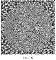
- FIG. 5is a schematic diagram of the third image according to an embodiment of the disclosure.
- the blood vessels, the macula, the optic disc, the veins, the arteries or other objects in FIG. 5are more clear.
- the third imagemay be used for image recognition.
- the processor 130inputs the third image to a detection model based on a machine learning algorithm (for example, deep neural network (DNN), multi-layer perceptron (MLP), support vector machine (SVM) or other machine learning models).
- the detection modelmay be used for image recognition.
- the third imagemay be used in pre-processing in a training phase and/or inference phase of the detection model.
- an initial fundoscopic imageis usually immediately subjected to feature extraction without undergoing image pre-processing.
- the image pre-processing according to the embodiments of the disclosuremay facilitate identification of a part such as blood vessels, macula, or veins, but the disclosure is not limited to the above.
- the image recognitionmay be based on scale-invariant feature transform (SIFT), a Haar-like feature, AdaBoost, or other recognition technologies.
- SIFTscale-invariant feature transform
- AdaBoostAdaBoost
- the region of interest in the fundoscopic imageis determined, the smoothing process is performed on the image, and the value difference is increased. In this way, features may be enhanced, and the subsequent image recognition, model training, or other image applications may be facilitated.
Landscapes
- Engineering & Computer Science (AREA)
- Theoretical Computer Science (AREA)
- Physics & Mathematics (AREA)
- General Physics & Mathematics (AREA)
- Computer Vision & Pattern Recognition (AREA)
- Medical Informatics (AREA)
- Software Systems (AREA)
- Health & Medical Sciences (AREA)
- General Health & Medical Sciences (AREA)
- Nuclear Medicine, Radiotherapy & Molecular Imaging (AREA)
- Radiology & Medical Imaging (AREA)
- Quality & Reliability (AREA)
- Artificial Intelligence (AREA)
- Data Mining & Analysis (AREA)
- Evolutionary Computation (AREA)
- Computing Systems (AREA)
- General Engineering & Computer Science (AREA)
- Mathematical Physics (AREA)
- Multimedia (AREA)
- Eye Examination Apparatus (AREA)
- Image Processing (AREA)
- Image Analysis (AREA)
- Apparatus For Radiation Diagnosis (AREA)
- Color Image Communication Systems (AREA)
- Measuring And Recording Apparatus For Diagnosis (AREA)
Abstract
Description
- This application claims the priority benefit of Taiwanese application serial no. 110109989, filed on Mar. 19, 2021. The entirety of the above-mentioned patent application is hereby incorporated by reference herein and made a part of this specification.
- The disclosure relates to an image processing technology, and particularly relates to an image pre-processing method and an image processing apparatus for a fundoscopic image.
- Medical images are images taken of specific parts of organisms, and these images may be used to assess the risk or severity of a disease. For example, through fundoscopic photographic examinations, diseases such as retinopathy, glaucoma, macular disease or the like may be detected early. Generally speaking, most doctors judge lesions in the medical images manually. Although computer-assisted evaluation of medical images is now possible, breakthroughs are needed in terms of indicators such as efficiency, complexity, and accuracy.
- Embodiments of the disclosure provide an image pre-processing method and an image processing apparatus for a fundoscopic image, in which features can be enhanced, thereby improving accuracy of subsequent identification of lesions or other features.
- The image pre-processing method according to an embodiment of the disclosure includes (but is not limited to) the following steps. A region of interest is obtained from a fundoscopic image to generate a first image. The region of interest is focused on an eyeball in the fundoscopic image. A smoothing process is performed on the first image to generate a second image. A value difference between multiple neighboring pixels is increased to generate a third image. The third image is used for image recognition.
- The image processing apparatus according to an embodiment of the disclosure includes (but is not limited to) a storage and a processor. The storage stores a code. The processor is coupled to the storage. The processor loads and executes the code to be configured to obtain a region of interest from a fundoscopic image to generate a first image, perform a smoothing process on the first image to generate a second image, and increase a value difference between multiple neighboring pixels in the second image to generate a third image. The region of interest is focused on an eyeball in the fundoscopic image. The third image is used for image recognition.
- Based on the above, according to the image pre-processing method and the image processing apparatus for a fundoscopic image according to the embodiments of the disclosure, before image recognition is performed, the region of interest is cut out from an initial fundoscopic image, and the smoothing process and value enhancement are further performed. In this way, features may be enhanced, noises may be reduced, and accuracy of the subsequent image recognition may be improved.
- To make the aforementioned more comprehensible, several embodiments accompanied with drawings are described in detail as follows.
- The accompanying drawings are included to provide a further understanding of the disclosure, and are incorporated in and constitute a part of this specification. The drawings illustrate exemplary embodiments of the disclosure and, together with the description, serve to explain the principles of the disclosure.
FIG. 1 is a block diagram of components of an image processing apparatus according to an embodiment of the disclosure.FIG. 2 is a flowchart of an image pre-processing method according to an embodiment of the disclosure.FIG. 3 is a schematic diagram of a first image according to an embodiment of the disclosure.FIG. 4 is a schematic diagram of a second image according to an embodiment of the disclosure.FIG. 5 is a schematic diagram of a third image according to an embodiment of the disclosure.FIG. 1 is a block diagram of components of animage processing apparatus 100 according to an embodiment of the disclosure. Referring toFIG. 1 , theimage processing apparatus 100 includes (but is not limited to) astorage 110 and aprocessor 130. Theimage processing apparatus 100 may be a desktop computer, a notebook computer, a smart phone, a tablet computer, a server, a medical testing instrument, or other computing apparatuses.- The
storage 110 may be any type of fixed or removable random access memory (RAM), read only memory (ROM), flash memory, hard disk drive (HDD), solid-state drive (SSD) or similar components. In one embodiment, thestorage 110 is configured to record a code, a software module, a configuration, data (for example, an image, a value, a reference value, a distance, etc.) or a file, and the embodiments thereof will be described in detail later. - The
processor 130 is coupled to thestorage 110. Theprocessor 130 may be a central processing unit (CPU), a graphic processing unit (GPU), or other programmable general-purpose or special-purpose microprocessor, digital signal processor (DSP), programmable controller, field programmable gate array (FPGA), application-specific integrated circuit (ASIC), neural network accelerator or other similar components or a combination of the above components. In one embodiment, theprocessor 130 is configured to execute all or part of operations of theimage processing apparatus 100, and may load and execute each code, the software module, the file, and the data recorded by thestorage 110. - Hereinafter, the method according to the embodiments of the disclosure will be described with reference to each apparatus, component, and module in the
image processing apparatus 100. Each process of the method may be adjusted according to actual implementation, and is not limited to those described herein. FIG. 2 is a flowchart of an image pre-processing method according to an embodiment of the disclosure. Referring toFIG. 2 , theprocessor 130 may obtain a region of interest from a fundoscopic image to generate a first image (step S210). Specifically, the fundoscopic image is an image obtained by fundoscopic photography of a human or other organism. Theprocessor 130 may obtain the fundoscopic image through a built-in or external image capturing apparatus, or theprocessor 130 may download the fundoscopic image from a server, a computer, or a storage medium. It should be noted that fundoscopic images from different sources may have different shapes or sizes. In order to normalize these fundoscopic images, theprocessor 130 may first cut out the region of interest considered as important or useful information.- The region of interest according to the embodiments of the disclosure is focused on an eyeball in the fundoscopic image. In one embodiment, the
processor 130 may locate a center of the eyeball from the fundoscopic image. For example, theprocessor 130 may take a point where the most straight lines in a gradient direction of pixels intersect in the fundoscopic image as a position of the eyeball. For another example, theprocessor 130 may perform the Hough transformation on the fundoscopic image to select a circle that best meets the requirements, and determine a center of circle of a contour of the region of interest accordingly. Theprocessor 130 may further determine the region of interest according to the center of the eyeball. For example, theprocessor 130 may set a circle that relatively or most conforms to the contour of the eyeball according to the center, and use the contour of the circle as a boundary of the region of interest. For another example, a circle obtained by the Hough transformation may be used as the region of interest by theprocessor 130. - In another embodiment, the
processor 130 may search for a boundary of the eyeball from the outside to the center of the fundoscopic image. For example, theprocessor 130 scans the fundoscopic image sequentially from four sides toward the center, and determines lightness of a scanned region. It should be noted that a lightness value (or brightness, that is, the lightness of a color) on one side of the boundary of the eyeball is higher than the other side of the boundary of the eyeball. Generally speaking, a region outside the eyeball in the fundoscopic image has a low lightness value and may be in black. When a lightness difference between neighboring pixels on any side is higher than a difference threshold, or the lightness value of one or more pixels is higher than a lightness threshold (that is, the lightness value on one side is higher than that on the other side), theprocessor 130 may determine that an outermost edge of the region of interest on this side has been found. Theprocessor 130 may use the outermost edges respectively found on the four sides of the fundoscopic image as boundary lines. That is, a quadrilateral is formed. Theprocessor 130 may use a length of a shortest side of the quadrilateral as a diameter of a circle (taken as the eyeball), and use the circle formed by the diameter as the boundary of the eyeball. The center of the circle is the center of the quadrilateral. In another embodiment, in order to first filter out interference that often appears on a periphery, theprocessor 130 multiplies half the length of the shortest side of the quadrilateral by a floating point number greater than 0 and less than 1 to obtain a radius length, and uses the radius length to obtain a circle. In addition, the center of the circle is still at the center of the quadrilateral. Then, theprocessor 130 may determine the region of interest according to the boundary of the eyeball. That is, the boundary of the eyeball is taken as the boundary of the region of interest. - In one embodiment, the
processor 130 may cut out the region of interest from the fundoscopic image. That is, theprocessor 130 may delete a region in the fundoscopic image that is not the region of interest. Theprocessor 130 may further add a background color outside the region of interest to form the first image. This background color will be considered as useless information in subsequent image recognition (for example, for lesion identification or for severity identification) of the fundoscopic image. The useless information may be excluded by feature extraction or may have a relatively low value. For example, red, green and blue background colors all consist of values of 128, 64, or 0, but are not limited thereto. In addition, the size, shape, and/or ratio of the first image may be fixed, thereby normalizing different fundoscopic images. In some embodiments, the circle may be changed to an ellipse or other geometric figures. - For example,
FIG. 3 is a schematic diagram of the first image according to an embodiment of the disclosure. Referring toFIG. 3 , a circular region (i.e., the region of interest) shown in the figure corresponds to the eyeball. - The
processor 130 may perform a smoothing process on the first image to generate a second image (step S230). Specifically, the smoothing process is a spatial domain filtering technology capable of directly blurring a pixel in an image and removing noises. For example, a value difference (also referred to as distance) between neighboring pixels may be reduced. - In one embodiment, the smoothing process is Gaussian blur. The
processor 130 may perform Gaussian blur on the first image. For example, theprocessor 130 performs a convolution operation on each pixel in the first image by using a Gaussian convolution kernel, and then sums convolution results to obtain the second image. - For example,
FIG. 4 is a schematic diagram of the second image according to an embodiment of the disclosure. Referring toFIG. 3 andFIG. 4 , compared withFIG. 3 ,FIG. 4 shows that details of some noises are blurred by Gaussian blur, while edges of blood vessels, macula, veins and/or arteries remain visible. - In other embodiments, the smoothing processing may also be median filtering, mean filtering, box filtering or other processing.
- The
processor 130 may increase a value difference between multiple neighboring pixels in the second image to generate a third image (step S250). Specifically, the smoothing process may narrow the value difference between the neighboring pixels. In order to further enhance a feature, in one embodiment, theprocessor 130 may increase (i.e., update or change) the value difference in proportion to a distance between the value difference between the neighboring pixels and a reference value. For example, when the reference value is128, theprocessor 130 may calculate an original value difference of red, green, and blue channel values between each pixel and its neighboring pixel, and compare the distances between the original value differences and the reference value. As the distance is increased, theprocessor 130 may increase the magnitude of an increase in the value difference. As the distance is reduced, theprocessor 130 may reduce the magnitude of an increase in the value difference. The proportion may be 1, 2, 5, or 10 times. Then, according to an increased value difference (i.e., updated value difference), theprocessor 130 may change the values of the corresponding pixels so that the value difference between two pixels matches the updated value difference. - In some embodiments, a value to be changed has an upper limit or lower limit. For example, the upper limit may be 255 and the lower limit may be 0. When the changed value exceeds the upper limit or the lower limit, it may be set as a specific value (for example, the upper limit, the lower limit or other values).
- It should be noted that a mathematical relationship between the original value difference and the updated value difference is not limited to a proportional relationship. In other embodiments, the
processor 130 may also adopt a linear relationship, an exponential relationship, or other mathematical relationships depending on actual needs. - For example,
FIG. 5 is a schematic diagram of the third image according to an embodiment of the disclosure. Referring toFIG. 4 andFIG. 5 , the blood vessels, the macula, the optic disc, the veins, the arteries or other objects inFIG. 5 are more clear. - It should also be noted that the third image according to the embodiments of the disclosure may be used for image recognition. In one embodiment, the
processor 130 inputs the third image to a detection model based on a machine learning algorithm (for example, deep neural network (DNN), multi-layer perceptron (MLP), support vector machine (SVM) or other machine learning models). In one embodiment, the detection model may be used for image recognition. It should be noted that the third image may be used in pre-processing in a training phase and/or inference phase of the detection model. Generally speaking, in the detection model, an initial fundoscopic image is usually immediately subjected to feature extraction without undergoing image pre-processing. Through the image pre-processing according to the embodiments of the disclosure, a relatively accurate recognition result of an image of a lesion such as bleeding, exudates, and edema can be obtained. Alternatively, the image pre-processing according to the embodiments of the disclosure may facilitate identification of a part such as blood vessels, macula, or veins, but the disclosure is not limited to the above. - In another embodiment, the image recognition may be based on scale-invariant feature transform (SIFT), a Haar-like feature, AdaBoost, or other recognition technologies.
- To sum up, in the image pre-processing method and the image processing apparatus for a fundoscopic image according to the embodiments of the disclosure, the region of interest in the fundoscopic image is determined, the smoothing process is performed on the image, and the value difference is increased. In this way, features may be enhanced, and the subsequent image recognition, model training, or other image applications may be facilitated.
- It will be apparent to those skilled in the art that various modifications and variations can be made to the disclosed embodiments without departing from the scope or spirit of the disclosure. In view of the foregoing, it is intended that the disclosure covers modifications and variations provided that they fall within the scope of the following claims and their equivalents.
Claims (14)
Applications Claiming Priority (2)
| Application Number | Priority Date | Filing Date | Title |
|---|---|---|---|
| TW110109989ATWI775356B (en) | 2021-03-19 | 2021-03-19 | Image pre-processing method and image processing apparatus for fundoscopic image |
| TW110109989 | 2021-03-19 |
Publications (2)
| Publication Number | Publication Date |
|---|---|
| US20220301111A1true US20220301111A1 (en) | 2022-09-22 |
| US11954824B2 US11954824B2 (en) | 2024-04-09 |
Family
ID=77739002
Family Applications (1)
| Application Number | Title | Priority Date | Filing Date |
|---|---|---|---|
| US17/235,938Active2042-02-19US11954824B2 (en) | 2021-03-19 | 2021-04-21 | Image pre-processing method and image processing apparatus for fundoscopic image |
Country Status (6)
| Country | Link |
|---|---|
| US (1) | US11954824B2 (en) |
| EP (1) | EP4060601A1 (en) |
| JP (1) | JP7337124B2 (en) |
| CN (1) | CN115115528B (en) |
| PH (1) | PH12021050543A1 (en) |
| TW (1) | TWI775356B (en) |
Families Citing this family (1)
| Publication number | Priority date | Publication date | Assignee | Title |
|---|---|---|---|---|
| EP4557213A1 (en)* | 2023-11-15 | 2025-05-21 | Koninklijke Philips N.V. | Enhancing image quality of medical images and providing medical image data to a user |
Citations (6)
| Publication number | Priority date | Publication date | Assignee | Title |
|---|---|---|---|---|
| JP2004046329A (en)* | 2002-07-09 | 2004-02-12 | Pentax Corp | Image contour enhancement device |
| US20060147094A1 (en)* | 2003-09-08 | 2006-07-06 | Woong-Tuk Yoo | Pupil detection method and shape descriptor extraction method for a iris recognition, iris feature extraction apparatus and method, and iris recognition system and method using its |
| US20080317339A1 (en)* | 2004-10-28 | 2008-12-25 | Fotonation Ireland Limited | Method and apparatus for red-eye detection using preview or other reference images |
| US20150110368A1 (en)* | 2013-10-22 | 2015-04-23 | Eyenuk, Inc. | Systems and methods for processing retinal images for screening of diseases or abnormalities |
| US20170076146A1 (en)* | 2015-09-11 | 2017-03-16 | EyeVerify Inc. | Fusing ocular-vascular with facial and/or sub-facial information for biometric systems |
| US20190236803A1 (en)* | 2016-12-15 | 2019-08-01 | Tencent Technology (Shenzhen) Company Limited | Pupil localizing method and system |
Family Cites Families (12)
| Publication number | Priority date | Publication date | Assignee | Title |
|---|---|---|---|---|
| JP3618877B2 (en) | 1996-02-05 | 2005-02-09 | キヤノン株式会社 | Ophthalmic image processing device |
| US6915024B1 (en)* | 2000-09-29 | 2005-07-05 | Hewlett-Packard Development Company, L.P. | Image sharpening by variable contrast mapping |
| JP4636841B2 (en)* | 2004-09-29 | 2011-02-23 | キヤノン株式会社 | Ophthalmic image photographing apparatus and photographing method |
| JP2006263127A (en) | 2005-03-24 | 2006-10-05 | Gifu Univ | Fundus image diagnosis support system and fundus image diagnosis support program |
| JP2007117154A (en) | 2005-10-25 | 2007-05-17 | Pentax Corp | Electronic endoscope system |
| US20070248277A1 (en)* | 2006-04-24 | 2007-10-25 | Scrofano Michael A | Method And System For Processing Image Data |
| JP2011035477A (en)* | 2009-07-29 | 2011-02-17 | Kyocera Corp | Image processing apparatus, imaging apparatus, noise elimination method, and program |
| JP6124868B2 (en)* | 2012-03-27 | 2017-05-10 | 株式会社日立製作所 | Image processing apparatus and image processing method |
| JP2015051054A (en) | 2013-09-05 | 2015-03-19 | キヤノン株式会社 | Image processing apparatus, image processing system, and image processing method |
| WO2019013779A1 (en)* | 2017-07-12 | 2019-01-17 | Mohammed Alauddin Bhuiyan | Automated blood vessel feature detection and quantification for retinal image grading and disease screening |
| CN108537155B (en)* | 2018-03-29 | 2021-01-26 | Oppo广东移动通信有限公司 | Image processing method, apparatus, electronic device, and computer-readable storage medium |
| CN111833334A (en)* | 2020-07-16 | 2020-10-27 | 上海志唐健康科技有限公司 | A method of fundus image feature processing and analysis based on twin network architecture |
- 2021
- 2021-03-19TWTW110109989Apatent/TWI775356B/enactive
- 2021-04-16CNCN202110411128.7Apatent/CN115115528B/enactiveActive
- 2021-04-21USUS17/235,938patent/US11954824B2/enactiveActive
- 2021-07-20JPJP2021119515Apatent/JP7337124B2/enactiveActive
- 2021-09-13EPEP21196275.8Apatent/EP4060601A1/enactivePending
- 2021-10-25PHPH1/2021/050543Apatent/PH12021050543A1/enunknown
Patent Citations (6)
| Publication number | Priority date | Publication date | Assignee | Title |
|---|---|---|---|---|
| JP2004046329A (en)* | 2002-07-09 | 2004-02-12 | Pentax Corp | Image contour enhancement device |
| US20060147094A1 (en)* | 2003-09-08 | 2006-07-06 | Woong-Tuk Yoo | Pupil detection method and shape descriptor extraction method for a iris recognition, iris feature extraction apparatus and method, and iris recognition system and method using its |
| US20080317339A1 (en)* | 2004-10-28 | 2008-12-25 | Fotonation Ireland Limited | Method and apparatus for red-eye detection using preview or other reference images |
| US20150110368A1 (en)* | 2013-10-22 | 2015-04-23 | Eyenuk, Inc. | Systems and methods for processing retinal images for screening of diseases or abnormalities |
| US20170076146A1 (en)* | 2015-09-11 | 2017-03-16 | EyeVerify Inc. | Fusing ocular-vascular with facial and/or sub-facial information for biometric systems |
| US20190236803A1 (en)* | 2016-12-15 | 2019-08-01 | Tencent Technology (Shenzhen) Company Limited | Pupil localizing method and system |
Also Published As
| Publication number | Publication date |
|---|---|
| JP2022145411A (en) | 2022-10-04 |
| TW202238514A (en) | 2022-10-01 |
| JP7337124B2 (en) | 2023-09-01 |
| US11954824B2 (en) | 2024-04-09 |
| TWI775356B (en) | 2022-08-21 |
| CN115115528A (en) | 2022-09-27 |
| CN115115528B (en) | 2025-02-28 |
| EP4060601A1 (en) | 2022-09-21 |
| PH12021050543A1 (en) | 2023-01-09 |
Similar Documents
| Publication | Publication Date | Title |
|---|---|---|
| Adem | Exudate detection for diabetic retinopathy with circular Hough transformation and convolutional neural networks | |
| Sevastopolsky | Optic disc and cup segmentation methods for glaucoma detection with modification of U-Net convolutional neural network | |
| CN111127425B (en) | Target detection positioning method and device based on retina fundus image | |
| Rebouças Filho et al. | Automatic histologically-closer classification of skin lesions | |
| BahadarKhan et al. | A morphological hessian based approach for retinal blood vessels segmentation and denoising using region based otsu thresholding | |
| Zhao et al. | Saliency driven vasculature segmentation with infinite perimeter active contour model | |
| Hsu et al. | Chronic wound assessment and infection detection method | |
| CN107835654B (en) | Image processing apparatus, image processing method, and recording medium | |
| Cavalcanti et al. | Macroscopic pigmented skin lesion segmentation and its influence on lesion classification and diagnosis | |
| US20220374947A1 (en) | Artificial intelligence-based system and method for grading collectible trading cards | |
| US11954824B2 (en) | Image pre-processing method and image processing apparatus for fundoscopic image | |
| CN111144413B (en) | Iris positioning method and computer readable storage medium | |
| Dharmawan et al. | Design of optimal adaptive filters for two-dimensional filamentary structures segmentation | |
| Iyyanar et al. | Hybrid Approach for Effective Segmentation and Classification of Glaucoma Disease Using UNet++ and CapsNet. | |
| US11690569B2 (en) | Blood vessel detecting apparatus and image-based blood vessel detecting method | |
| WO2020140380A1 (en) | Method and device for quickly dividing optical coherence tomography image | |
| Chiamaka Okafor et al. | The effect of image preprocessing algorithms on diabetic foot ulcer classification | |
| Santos et al. | Deep neural network model based on one-stage detector for identifying fundus lesions | |
| CN118135292A (en) | Fundus image classification method and device, electronic equipment and storage medium | |
| EP4398188A1 (en) | Vessel recognition using weighted gabor technique | |
| Suman et al. | Automatic grading of non-proliferative diabetic retinopathy | |
| CN116523829A (en) | Image recognition method, device, equipment and application in Alzheimer's disease prediction | |
| CN113269756B (en) | Method and device for optimizing retinal vessel segmentation based on multi-scale matched filtering and particle swarm | |
| CN115100178A (en) | Method, device, medium and equipment for evaluating morphological characteristics of fundus blood vessels | |
| Silva et al. | A data-centric approach for pectoral muscle deep learning segmentation enhancements in mammography images |
Legal Events
| Date | Code | Title | Description |
|---|---|---|---|
| AS | Assignment | Owner name:ACER INCORPORATED, TAIWAN Free format text:ASSIGNMENT OF ASSIGNORS INTEREST;ASSIGNORS:HUANG, YI-JIN;TSAI, CHIN-HAN;CHEN, MING-KE;SIGNING DATES FROM 20210420 TO 20210421;REEL/FRAME:055980/0806 | |
| FEPP | Fee payment procedure | Free format text:ENTITY STATUS SET TO UNDISCOUNTED (ORIGINAL EVENT CODE: BIG.); ENTITY STATUS OF PATENT OWNER: LARGE ENTITY | |
| STPP | Information on status: patent application and granting procedure in general | Free format text:DOCKETED NEW CASE - READY FOR EXAMINATION | |
| AS | Assignment | Owner name:ACER MEDICAL INC., TAIWAN Free format text:ASSIGNMENT OF ASSIGNORS INTEREST;ASSIGNOR:ACER INCORPORATED;REEL/FRAME:059124/0724 Effective date:20220209 | |
| STPP | Information on status: patent application and granting procedure in general | Free format text:NON FINAL ACTION MAILED | |
| STPP | Information on status: patent application and granting procedure in general | Free format text:RESPONSE TO NON-FINAL OFFICE ACTION ENTERED AND FORWARDED TO EXAMINER | |
| STPP | Information on status: patent application and granting procedure in general | Free format text:FINAL REJECTION MAILED | |
| STPP | Information on status: patent application and granting procedure in general | Free format text:DOCKETED NEW CASE - READY FOR EXAMINATION | |
| STPP | Information on status: patent application and granting procedure in general | Free format text:NOTICE OF ALLOWANCE MAILED -- APPLICATION RECEIVED IN OFFICE OF PUBLICATIONS | |
| STCF | Information on status: patent grant | Free format text:PATENTED CASE |