US20120014578A1 - Computer Aided Detection Of Abnormalities In Volumetric Breast Ultrasound Scans And User Interface - Google Patents
Computer Aided Detection Of Abnormalities In Volumetric Breast Ultrasound Scans And User InterfaceDownload PDFInfo
- Publication number
- US20120014578A1 US20120014578A1US12/839,371US83937110AUS2012014578A1US 20120014578 A1US20120014578 A1US 20120014578A1US 83937110 AUS83937110 AUS 83937110AUS 2012014578 A1US2012014578 A1US 2012014578A1
- Authority
- US
- United States
- Prior art keywords
- image
- interest
- ultrasound
- user
- breast tissue
- Prior art date
- Legal status (The legal status is an assumption and is not a legal conclusion. Google has not performed a legal analysis and makes no representation as to the accuracy of the status listed.)
- Abandoned
Links
Images


Classifications
- G—PHYSICS
- G06—COMPUTING OR CALCULATING; COUNTING
- G06T—IMAGE DATA PROCESSING OR GENERATION, IN GENERAL
- G06T7/00—Image analysis
- G06T7/0002—Inspection of images, e.g. flaw detection
- G06T7/0012—Biomedical image inspection
- G—PHYSICS
- G06—COMPUTING OR CALCULATING; COUNTING
- G06T—IMAGE DATA PROCESSING OR GENERATION, IN GENERAL
- G06T2207/00—Indexing scheme for image analysis or image enhancement
- G06T2207/10—Image acquisition modality
- G06T2207/10132—Ultrasound image
- G—PHYSICS
- G06—COMPUTING OR CALCULATING; COUNTING
- G06T—IMAGE DATA PROCESSING OR GENERATION, IN GENERAL
- G06T2207/00—Indexing scheme for image analysis or image enhancement
- G06T2207/30—Subject of image; Context of image processing
- G06T2207/30004—Biomedical image processing
- G06T2207/30068—Mammography; Breast
Definitions
- This patent specificationrelates to medical imaging systems and processes.
- the present inventionrelates to the computer aided detection of breast abnormalities in volumetric breast ultrasound scans, and devices and methods of interactive display of such computer aided detection results.
- an ABUS deviceis used to image a whole breast volume with up to three partially overlapping scans per breast, which would generate several hundred images or slices.
- image acquisition with ABUScan generally be performed by technicians, radiologists are still required to read the hundreds of images, thus breast imaging with ABUS can still be relatively time consuming.
- a complete screening examconsists of hundreds of images or slices, and the information content of each of the images is high. Abnormalities can therefore be easily overlooked when the images are being inspected slice by slice.
- Multi-modality CADxROC study of the effect on radiologists' accuracy in characterizing breast masses on mammograms and 3D ultrasound images,” Acad Radiol, 2009, 16, 810-818; and Cui, J.; Sahiner, B.; Chan, H.-P.; Nees, A.; Paramagul, C.; Hadjiiski, L. M.; Zhou, C. & Shi, J; “A new automated method for the segmentation and characterization of breast masses on ultrasound images,” Med Phys, 2009, 36, 1553-1565.
- This workis based on images from a targeted 3D ultrasound scanning system. Only a small volume holding the lesion is imaged and analyzed. The purpose is distinguishing benign and malignant lesions.
- Features used in the work aboveinclude morphology (e.g. height to width ration), posterior acoustic shadowing, lesion and margin contrast.
- U.S. Pat. No. 7,556,602discusses the use of ultrasound mammography in which an automated transducer scans the patient's breast to generate images of thin slices that are processed into fewer thick slices simultaneously for rapid assessment of the breast. Computer aided detection or diagnosis can be preformed on images and resulting mark and/or other information can be displayed as well.
- the '602 patentdiscusses extracting and applying a classifier algorithm to known two-dimensional features such as spiculation metrics, density metrics, eccentricity metrics and sphericity metrics. However, spiculation is not identified or suggested as a criterion used for candidate detection (which is referred to as the ROI location algorithm).
- the '602 patentalso discusses correlating regions of interest in an x-ray mammogram view to an adjunctive ultrasound view.
- the disclosed algorithmsare applicable to cases where the mammogram view and the ultrasound view are taken from the same standard view (e.g. CC or MLO) or at least where the breast tissue is compressed, in both ultrasound and mammography, in directions perpendicular to an axis that is perpendicular to the chest wall and passes through the nipple.
- a computer aided detection methodthat helps radiologists in searching and interpretation of abnormalities would be very useful.
- a novel CAD systemis provided for detection of breast cancer in volumetric ultrasound scans.
- a method of analyzing ultrasound images of breast tissueincludes receiving and processing a digitized ultrasound image of the breast tissue so as to generate a three-dimensional image composed of view slices that are approximately perpendicular to the direction of compression of the breast tissue during ultrasound scanning.
- the 3D imageis further processed using one or more computer aided detection algorithms so as to identify locations of one or more regions of interest within the image based at least in part on identified areas of spiculation in portions of one or more of the view slices.
- the compression directionis towards the chest wall and the areas of spiculation are identified in portions of coronal view slices being approximately parallel to the skin surface.
- features extracted from the 3D imagesuch as based on gradient convergence, local contrast, and/or posterior shadowing can also be used to identified regions of interest in the image, in combination with spiculation.
- the featuresare computed at regularly spaced locations in the image, at each location using computations that include voxel values in a local 3D subvolume.
- a likelihood of malignancy for each of the regions of interestcan be estimated and displaying to a user. The method can be used for screening and/or diagnostic purposes
- a method of analyzing ultrasound images of breast tissue of a patientincludes receiving and processing two digitized three-dimensional ultrasound images of breast tissue of the patient so as to generate a region of interest in each image. A likelihood of malignancy is then evaluated based at least in part on the estimated distance between the locations of the regions of interest in the two images.
- the two imagescan be of the same breast of the patient as in the first image, such as two offset scans of the same breast taken during the same scanning procedure, or of the same breast during a prior year screening.
- the two imagescan be the left and right breast of the patient using a reference point such as the nipple, so as to evaluate symmetry when evaluating the likelihood of malignancy.
- a method of analyzing digital images of breast tissue of a patientincludes receiving and processing a digitized ultrasound image of breast tissue compressed in a direction towards a chest wall using one or more computer aided detection algorithms thereby generating a region of interest in the ultrasound image; receiving and processing a digitized mammographic image, such as a CC or MLO view, of the same breast tissue using one or more computer aided detection algorithms thereby generating a region of interest in the mammographic image; and evaluating the likelihood of malignancy based at least in part on the estimated distance between a location of the region of interest in the ultrasound image and a location the region of interest in the mammographic image.
- the mammographic imageis a tomographic mammographic image.
- a method of interactively displaying ultrasound and mammographic images of breast tissue to a userincludes receiving one or more digitized mammographic images, such as CC or MLO views, of the breast tissue, and a digitized three-dimensional ultrasound image of the breast tissue compressed in a direction towards a chest wall of the patient.
- the useridentifies a location or locations on the one or more mammographic images of a user identified region of interest in the breast tissue.
- One or more locationsare estimated on the ultrasound image that correspond to the location or locations on the one or more mammographic images of the user identified region of interest, and portions of the digitized ultrasound image are displayed to the user so as to indicate the one or more estimated locations corresponding to the region of interest.
- an estimated position of the user identified region of interest relative to an identified nipple on the breast tissueis displayed to the user using a clock position and distance from the nipple.
- the mammographic imageis a three-dimensional mammographic image.
- the user identified region of interestis located by the user first in the ultrasound image, then the location or locations on the mammographic image or images are estimated and displayed to the user.
- a method of interactively displaying computer aided detection results of medical ultrasound images to a userincludes receiving a digitized ultrasound image of breast tissue; processing the image using one or more computer aided detection algorithms thereby generating one or more regions of interest; displaying the digitized image along with one or more marks tending to indicate location on the tissue of the regions of interest and information relating to an estimated likelihood of malignancy, such as a percentage or a color indicating a percentage range, for each displayed region of interest.
- related systemsfor analyzing digital images of breast tissue, and for displaying ultrasound and mammographic images of a breast tissue to a user are provided.
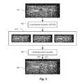
- FIG. 1is a flow chart illustrating the detection method according to some embodiments
- FIGS. 2A-Care examples of a cross section through a malignant lesion showing features identified according to some embodiments
- FIG. 3is a flowchart showing combination of features at a voxel level using context, resulting in a likelihood of abnormality, according to some embodiments;
- FIG. 4is a matrix of coronal views and cross sections of a malignant lesion at three different depths, according to some embodiments
- FIG. 5illustrates methods of presenting CAD results to users, according to some embodiments
- FIG. 6is a plot showing detection sensitivity as a function of the number of false positives per 3D image volume, according to some embodiments.
- FIG. 7is a flowchart showing steps in carrying out CAD analysis of ultrasound breast images, according to some embodiments.
- FIGS. 8A-Cshow further detail of view correlation procedures, according to some embodiments.
- FIGS. 9A-Bshow further detail of left and right symmetry checking, according to some embodiments.
- FIGS. 10A-Bshow further detail of temporal correlation procedures, according to some embodiments.
- FIGS. 11A-Cshow further detail of correlation procedures between ultrasound images and cranio-caudal (CC) view of a mammographic image, according to some embodiments;
- FIGS. 12A-Cshow further detail of correlation procedures between ultrasound images and mediolateral oblique (MLO) view of a mammographic image, according to some embodiments;
- FIG. 13shows a user interface which relates positions in mammographic and ultrasound images, according to some embodiments.
- FIGS. 14A-Dshow x-ray tomosynthesis imaging displayed views, according to some embodiments.
- a processmay correspond to a method, a function, a procedure, a subroutine, a subprogram, etc. When a process corresponds to a function, its termination corresponds to a return of the function to the calling function or the main function.
- embodiments of the inventionmay be implemented, at least in part, either manually or automatically.
- Manual or automatic implementationsmay be executed, or at least assisted, through the use of machines, hardware, software, firmware, middleware, microcode, hardware description languages, or any combination thereof.
- the program code or code segments to perform the necessary tasksmay be stored in a machine readable medium.
- a processor(s)may perform the necessary tasks.
- a systemfor detection of breast cancer in 3D ultrasound imaging data.
- Volumetric ultrasound imagesare obtained by an automated breast ultrasound scanning (ABUS) device.
- ABUSautomated breast ultrasound scanning
- this deviceis used to image a whole breast volume with up to three partially overlapping scans.
- Breast cancer screening with ABUSis time consuming as up to six scans per patient (three per breast) have to be read slice by slice. By using effective computer aided detection methods feasibility of breast cancer screening with ABUS may be increased.
- ABUS imagesbreast cancers appear as dark lesions. When viewed in transversal and sagital planes, lesions and normal tissue appear similar as in traditional 2D ultrasound. However, when viewing ABUS images in coronal planes (in parallel to the skin surface) images look remarkably different. In particular, it appears that architectural distortion and spiculation are frequently seen in the coronal views, and these are strong indicators of the presence of cancer. Therefore, in the computer aided detection (CAD) system according to some embodiments, combine a dark lesion detector operating in 3D is combined with a detector for spiculation and architectural distortion operating on 2D coronal slices. In this way a sensitive detection method is obtained.
- CADcomputer aided detection
- FIG. 1is a flow chart illustrating the detection method according to some embodiments.
- image data 110 from the scanning deviceis converted to a coronal representation. This comprises (1) artifact removal (step 112 ), (2) re-sampling of the data to isotropic resolution (step 114 ), and (3) rotation of the data to coronal orientation (step 114 ).
- Artifact removal, step 112addresses correction of scan line artifacts due to signal transfer variation during scanning. Lines with an outlying mean value are corrected using the mean value of neighboring lines as a reference.
- the image datais converted to cubic voxels of 0.5 mm.
- the (x,y) planeshold coronal slices, which are in parallel to the skin surface during scanning, while the z coordinate represents depth.
- the image volumeis segmented in four classes: background, fatty breast tissue, dense breast tissue, and other tissue.
- backgroundis labelled using feature based voxel classification and morphological operators.
- Featuresinclude texture and voxel value. If the voxel value is low and texture indicates a homogeneous neighborhood voxels are labelled as background.
- the chest-wallis detected using dynamic programming in transversal and sagittal slices, and by subsequently fitting a parameterized surface through the set of obtained boundary points. Voxels between the chest wall and skin are labelled as breast tissue. Using Otsu's method a threshold is determined to label fatty and dense tissue voxels in the breast.
- images voxel valuesare normalized in step 118 , using the segmented breast tissue volume.
- mean values of voxels labelled as dense and fatty tissueare computed. These are denoted by mean_fat and mean_dense respectively.
- contrast of the imageis normalized by:
- y_originalbeing the voxel value before normalization
- y and y_fatthe voxel value and the mean voxel value of fatty tissue after contrast normalization.
- Voxel feature extractiontakes place using modules 126 .
- the breast tissue regionis processed to extract local features.
- Three modulesare used, each targeting a different characteristic of breast cancers in ultrasound. Cancers appear as dark regions with relatively compact shapes. The mean voxel value in malignant lesions is lower than that the surrounding fatty tissue, and often a dark shadow is present under the lesion. Finally, in coronal slices through, or near, malignant lesions spiculation or architectural distortion is often visible. This a new radiological sign, which is not observed in traditional hand-held ultrasound because this modality was not able to show the coronal plane.
- the three modules 126are developed to capture the characteristic features and are described below.
- the first module 120computes volumetric gradient convergence features that give a high response at the center of compact regions. It operates on the 3D gradient vector field derived from the image at a chosen scale. The module computes the number of gradient vectors directed to the center of a spherical neighborhood covered by the filter. This number is normalized by subtracting its expected value and by dividing the result by its standard deviation, both determined for a reference pattern of random gradient directions. At any given location, the filter output is obtained as a function of the neighborhood radius R, making the filter equally responsive to both small and large lesions. At each location the maximum filter output and corresponding neighborhood size are determined. Apart from the integrated measure of convergence, also the isotropy of convergence is determined.
- Karssemeijer(1996)
- KarssemeijerN., “Local orientation distribution as a function of spatial scale for detection of masses in mammograms,” Information Processing in Medical Imaging, LNCS 2082 (Springer), pp. 280-293 (1999) (hereinafter “Karssemeijer (1999)”).
- analysis of local line and gradient direction patternsforms the basis for computation of the local features that are used. Further detail of this method will now be described.
- the size of the neighborhood in which orientations patterns are evaluatedis one of the most important parameters in the computation of these features. Variation of this size can have a dramatic effect on the detection of individual cancers, although the influence of this parameter on the overall performance measured on a large database tends to be less.
- the output of a local contrast operatorhas been used to set the size of the neighborhood adaptively.
- featuresare computed as a continuous function of the neighborhood size, only slightly increasing the computational load. The method is described here for 2D application, but can be used in higher dimensions as well.
- Orientation mapsare computed using first and second order Gaussian derivatives. When there is a concentration of gradient orientation towards a certain point this indicates the presence of a mass. A concentration of line orientations computed from second order directional derivatives indicates the presence of spiculation or architectural distortion. These concentration or convergence features will be denoted by g 1 and l 1 , respectively for gradient and line orientations.
- concentration or convergence featureswill be denoted by g 1 and l 1 , respectively for gradient and line orientations.
- features representing radial uniformity measure whether or not increase of pixels oriented to a centercomes from the whole surrounding area or from a few directions only. These will be denoted by g 2 and l 2 .
- a circular neighborhoodis used in the 2D case and a spherical neighbourhood in the 3D case.
- the term voxelis used for image samples in both the 2D and 3D case. All voxels j located within a distance r min ⁇ r ij ⁇ r max from i are selected when the magnitude of the orientation operator exceeds a small threshold. This selected set of voxels is denoted by S i .
- the featuresare based on a statistic x j defined by
- Voxels that are oriented to the centercan be determined by evaluating:
- weight factorscan be chosen as a function of the distance r ij , for instance to give voxels closer to the center a larger weight.
- variance of this sumcan be estimated when it is assumed that all voxel contributions are independent:
- each ring (or shell) kthe number of voxels hitting the center N k, hit can be counted, allowing the sum to be rewritten as
- N k and Nthe number of voxels in ring (shell) k and in total, respectively.
- the normalization factorwhich can be written as (N( p ⁇ p 2 )) ⁇ 1/2 .
- Multiscale methodsthat have been proposed for detection of masses in mammograms include: for wavelets, see, A. F. Laine, S. Schuler, J. Fan, and W.
- a methodis described that allows very efficient computation of a class of local image features as a continuous function of scale, only slightly increasing the computational effort needed for computation at the largest scale considered.
- the non-linear features described in the previous subsectionbelong to this class.
- an ordered listis constructed in which each element represents a neighbor j within distance r ij of the central location i.
- positional information of the neighbor that is needed for the computationis stored, here the x j , y j offset, orientation and distance r ij with respect to center.
- This listis constructed by visiting all voxels in any order, and by subsequently sorting its elements by distance to the center.
- the actual computation of the featurestakes place, at each voxel or at a given fraction of voxels using a sampling scheme.
- the ordered list of neighborsis used to collect the data from the neighborhood.
- the x j , y j offsets in the listare used to address the voxel data and precomputed derivatives or orientations at the location of the neighbor.
- the orientation with respect to iis used to compute orientation related features. Because the neighbors are ordered with increasing distance to the center, computation of the features from the collected data can be carried out at given intervals, for instance each time the number of neighbors has increased by some fixed number. As the computational effort lies in collection of the data, this only slightly increases the computational load.
- module 124is designed to find spiculation or architectural distortion in coronal planes, using the method above in 2D.
- a line orientation patternforms the basis for feature computation. Line orientations are obtained using a basis of second order Gaussian directional derivatives.
- Module 122computes local contrast as a function of scale. At each location in the image the mean voxel value m(x,y,z) in a neighborhood is computed. The neighborhood is defined by all voxels within distance R 1 from the central location. Contrast is computed by subtracting this mean value from the mean value of voxels labeled as fatty tissue just outside the neighborhood, i.e. voxels with distance to the center within an interval [R 1 ,R 1 + ⁇ R].
- local contrast featuresare computed for various neighborhood types: (1) A spherical neighborhood with a fixed radius, (2) A spherical neighborhood with radius estimated from the image data, for instance by taking the radius at which the gradient concentration filter g 1 has the highest output maximum response, (3) a semi-spherical neighborhood including only superficial voxels, i.e those that are closer to the transducer (or skin) than the central location, (4) a semi-spheric neighborhood including only deeper voxels (further away from the transducer than the central location), and (5) the spherical neighborhood with the radius that gives the highest local contrast.
- the ultrasound imagescan result from breast tissue compressions in directions other than toward the chest wall.
- the ultrasound imagecan result from a scan in which the breast is compressed in a direction such as with conventional mammography (e.g. as in CC and/or MLO views.
- the module 124is designed to find spiculations and/or architectural distortions in plans perpendicular to the direction of compression. For example, if the compression direction of the ultrasound scan is as in a CC mammography view, then the module 124 would look for spiculations and/or architectual distortions in a transverse plane.
- modules 126including spiculation module 124 are used in candidate detection (i.e. to locate the regions of interest). This is in contrast to techniques such as discussed in the '602 patent where features such as spiculation metrics are only used for classification of regions of interest.
- FIGS. 2A-Care examples of a cross section through a malignant lesion showing features identified according to some embodiments.
- the skin surfaceis on top in each of the views.
- FIG. 2Athe original cross section image 210 is shown.
- FIG. 2Bshows the response 220 of gradient convergence module 120 (as described with respect to FIG. 1 ). The highest values are shown outlined in white such as region 222 , and the next highest values are shown outlined in black such as region 224 .
- FIG. 2Cshows the response 230 of coronal spiculation module 124 (as described with respect to FIG. 1 ). The highest values are shown outlined in white such as region 232 , and the next highest values are shown outlined in black such as region 234 .
- 2Dshows the response 240 of local contrast module 122 (as described with respect to FIG. 1 ).
- the highest valuesare shown outlined in white such as region 242 , and the next highest values are shown outlined in black such as region 244 . It can be seen that the maxima of the responses are not aligned. In particular, the coronal spiculation feature is strongest in the upper part of the lesion.
- FIG. 3is a flowchart showing combination of features at a voxel level using context, resulting in a likelihood of abnormality, according to some embodiments.
- Input image 310is input to the local feature extraction 312 which corresponds to the modules 120 , 122 and 124 as described with respect to FIG. 1 .
- Examples of the cross sections highlighted according to the three modulesis shown in 314 which correspond to the cross section examples shown in FIG. 2 .
- the results of the modulesare combined in the contextual voxel classifier 316 , which corresponds to the step 126 of FIG. 1 .
- the resultis the likelihood map 320 which shows the highest values outlined in white such as region 322 and the next highest values outlined in black, such as region 324 .
- FIG. 4is a matrix of coronal views and cross sections of a malignant lesion at three different depths, according to some embodiments. Depth increases from column 410 being coronal views shallowest (closest to the skin), column 412 being coronal views of medium depth, and column 414 being coronal views being the deepest (furthest from the skin). Column 416 are transversal plane views. Row 420 shows the original image. Row 422 shows an overlay of the results of gradient convergence module 120 (as described with respect to FIG. 1 ). Row 424 shows an overlay of the results of coronal spiculation module 124 (as described with respect to FIG. 1 ). Row 426 shows the overlay of lesion likelihood as a result of the contextual voxel classification step 128 . In the rows 422 , 424 and 426 , the highest values (most likely to be malignant) is outlined in white, and the next highest values is outlined in black.
- step 128selected voxels on a regular 3D grid of locations covering the breast tissue are classified using a feature vector that comprises information extracted by the feature extraction modules 126 at the location (x, y, z) of the voxel itself and its surroundings.
- the latteris essential because it has been observed that in 3D breast ultrasound imaging the central locations of lesions often do not coincide with focal points of spiculation patterns associated with the lesions.
- a contextual voxel or pixel classification method 128is designed that brings together information extracted in nearby locations.
- a contextual Markov Random Fieldcan be defined to represent relations between features in a neighborhood of r.
- a feature vectorcan also be defined as the concatenation of feature vectors in a neighborhood of r.
- supervised learningis used to determine a set of candidate locations that are most representative of cancer.
- a set of training casesis used in which locations of relevant abnormalities.
- the training setincludes both malignant and benign lesions (e.g. cysts and fibroadenoma).
- voxels and associated feature vectors in the center of annotated lesionsare taken as abnormal patterns, while voxels sampled outside the lesions and/or in normal cases are used as normal samples.
- supervised learninga classifier is trained to relate the input to a probability that an abnormality is present at a given location.
- the output of the contextual voxel classifier 128is a volume representing likelihood of abnormality L(r). See, e.g. output view 320 in FIG. 3 and column 426 in FIG. 4 .
- local maximaa determined in step 132 .
- step 134candidate classification is carried out.
- a multi-stage modelis employed. By thresholding, the most relevant candidate locations are selected and processed further. Typically, this processing includes a segmentation step in which the lesion boundary is localized.
- New featuresare computed, with the aim of representing relevant characteristics of the lesion by a numerical feature vector.
- Features for characterizing breast ultrasound lesionshave been described in the literature for 2D handheld ultrasound and extension to 3D is straightforward. They include lesion contrast, margin contrast, margin sharpness, boundary smoothness, shadowing, width-to height ratio, and moments of the distribution of voxel values inside the lesion.
- a new set of features represented in coronal spiculationis added. These are computed from the distribution of coronal spiculation features computed in the candidate detection stage inside the lesion, e.g. mean variance and percentile values.
- the number of false positive candidatescan be reduced, and/or the probability that a lesion is malignant or benign can be assessed.
- Three configurations of the CAD systemare described below according to some embodiments, although other configurations are possible. The three described configurations are: false positive reduction; false positive reduction and subsequent lesion classification; and multi-class classification.
- false positivesare defined as non-lesion locations.
- the detection systemis trained with both benign and malignant lesions as target training patterns and it learns to distinguish those lesions from normal breast tissue. The task of deciding whether a lesion is more likely to be benign or malignant is left to the radiologist.
- the detection systemis combined with a classification system trained to distinguish benign lesions from malignant lesions.
- the classification systemis a feature-based system trained in the traditional way as a 2-class supervised classifier. The system is applied to regions surviving the false positive reduction step of the CAD system. In this way, each region detected by the CAD system has two numerical values assigned to it: one to indicate the probability that a lesion is present, and another to indicate the probability that the lesion is malignant.
- non lesion locationsform one class in a multi-class classification system.
- the systemis trained to distinguish non-lesion locations, cancer, and benign lesions.
- the CAD systemcomputes a likelihood value for each of the classes. It is noted that these likelihood values depend on prevalence and characteristics of the classes in the training set, which is dependent on the case sample and on the threshold applied in the candidate lesion detector. This has to be taken into account when information is displayed to the radiologist.
- FIG. 5illustrates methods of presenting CAD results to users, according to some embodiments.
- Column 522shows lateral coronal views and column 520 shows medial coronal views.
- the woman in this casehas an invasive ductal cancer.
- the images 510shows the slice at the skin level.
- White dashed circlessuch as circle 524 are interactive CAD finding projections.
- the coronal view at the depth where the selected finding is locatedis shown. If depth of the displayed view corresponds with the location of the CAD finding the prompt is displayed in a solid white circle, such as circle 540 .
- the images 512are the coronal views at the depth corresponding to the lesions marked by solid white circles 540 and 542 . Where there are lesions that do not correspond with the slice depth, white dashed circles are shown for the CAD marks, such as mark 544 . Images 514 show the coronal slices viewed a depth corresponding to a lesion as shown by the CAD mark 530 displayed in a solid white circle. According to some embodiments, colors are used in the display and green circles denote the slice depth does not correspond to the lesion depth and red circles are used when the view corresponds to the lesion depth.
- a functionis available that allows the user to move the display automatically to the slice in which CAD identified a suspicious region.
- This slice, or depthcan be determined by taking the maximum of the likelihood image, or the center of the segmentation.
- This functioncan be activated by clicking on the marked location with a mouse pointer, such as pointer 550 .
- the displaycan automatically synchronize the displayed slices, to the same depth in all displayed views. In this way, radiologists can more efficiently make comparisons between views, which is usually done at the same depth.
- the views of column 522 (lateral coronal views) and column 520 (medial coronal views)are synchronized for each depth.
- the likelihood of malignancy computed by the CAD systemwhen activating a CAD mark, can be displayed.
- the computed likelihoods for the marks 540 and 542are 10% and 90% respectively.
- the displaycan include an indication that a lesion is malignant or benign.
- a functionis available that allows the user to move the display automatically to the slice in which CAD identified a suspicious region exists when viewing slices from the original scanning acquired images.
- FIGS. 2A-Dcould be the taken from the original scanned images such as acquired in step 110 of FIG. 1 .
- the original scanned 2D imagescan be displayed to the user in a cine fashion, which automatically stops or pauses when the image contains a CAD identified a suspicious region.
- Current users of hand held breast ultrasound, such as radiologistsmay be more familiar and/or feel more comfortable with viewing original 2D acquired image.
- the displaycan also be interactive when displaying and automatically stopping at 2D images containing a CAD identified suspicious region.
- the CAD identified suspicious regioncan be highlighted using solid and/or dashed circles such as shown in FIG. 5 , and in response to a user's selection with a pointer, the system can interactively display information, such as likelihood of malignancy as shown in FIG. 5 .
- FIG. 6is a plot showing free response operating characteristic (FROC) demonstrating detection performance of the candidate detection stage, according to some embodiments.
- Plot 610shows detection sensitivity as a function of the number of false positives per 3D image volume. The plot 610 shows the result of applying the candidate detection method as described herein to a series of test and training cases.
- FROCfree response operating characteristic
- FIG. 7is a flowchart showing steps in carrying out CAD analysis of ultrasound breast images, according to some embodiments.
- a 3D ultrasound volumeis input, artifacts are removed and resampling for coronal view reconstruction is carried out. This step corresponds to steps 112 and 114 in FIG. 1 .
- the imageis segmented to identify the breast tissue, and the image is normalized. This step corresponds to steps 116 and 118 in FIG. 1 .
- each voxelis analyzed using modules for gradient convergence, spiculation in coronal planes and local contrast. This step corresponds to using modules 126 in FIG. 1 .
- each pixel or voxelis classified, which corresponds to step 128 in FIG. 1 .
- groups of voxelsare segmented having similar properties and classified according to characteristics of the region such as size, shape, lesion contrast, margin contrast, margin sharpness, boundary smoothness, shadowing, width-to height ratio, moments of the distribution of voxel values inside the lesion, and coronal spiculation. This step corresponds to step 134 in FIG. 1 .
- steps 720 , 722 , 724 , and 726relate to image information from a single view of a single breast.
- steps 720 , 722 , 724 , and 726the information is compared to other views, other breasts (i.e. left vs. right), scans at other times, and images from other modalities such as mammography.
- the steps 720 , 722 , 724 and 726are used to adjust the likelihood of malignancy.
- step 720correlation between different views is carried out. Ordinarily, more than one ultrasound scan is used to cover a breast. If a lesion occurs in an overlap area, then correlation between different views of the same breast can be carried out.
- step 722left versus right breast symmetry check is carried out which can identify false positives due.
- step 724a temporal comparison is carried out, for example between ultrasound scans of the same breast taken at different times, such as separated by one or more years.
- step 726a comparison with other modalities such as mammography is carried out. According to some embodiments, one or more of the comparison steps 720 , 722 , 724 and 726 are not carried out, are performed in a different order than shown in FIG. 7 , and/or performed in parallel with each other.
- FIGS. 8A-Cshow further detail of view correlation procedures, according to some embodiments.
- a first scanshown in coronal view 820 , is made of breast 810 having a nipple 812 .
- a region of interestis identified which is shown by the mark 822 on coronal view 820 .
- the region of interestis identified, for example, using the techniques discussed with respect to FIG. 1 .
- the position of the nipple 812 in the first scanis determined either manually by an operator, or alternatively the nipple position can be automatically determined as is known in the art.
- the position of the region of interest relative to the nipple using x, y, z coordinatescan therefore be determined.
- FIG. 8Bis a transversal slice that shows the depth z 1 of the region of interest shown by the spot 842 as measured from the skin surface 846 . Chest wall 844 is also shown.
- a region of interesthaving a location relative to the nipple of x 2 , y 2 and z 2 .
- FIG. 8Athe coronal view 830 is shown for the second scan of the breast 810 , with the corresponding region of interest 832 marked by the dashed circle.
- the position x 2 and y 2can be shown in the coronal view.
- the depth z 2is shown in FIG. 8C as the distance between skin surface 856 and region of interest marked by dashed circle 852 in transversal view 850 .
- a threshold distance conditioncan be applied for the maximum distance between the regions of interest in first and second scans:
- k 1 , k 2 and k 3 . . .are weighting factors for each of the features of the regions of interest.
- Examples of feature — 1, feature — 2, etcare features such as size, shape, coronal spiculation, contrast, etc.
- the values for the threshold for maximum distance and/or the weighting factors k 1 , k 2 and k 3can be determined using a free response operating characteristic (FROC) curve, where the values are adjusted so as to yield the highest sensitivity for given false positive rates per volume.
- FROCfree response operating characteristic
- FIGS. 9A-Bshow further detail of left and right symmetry checking, according to some embodiments.
- FIG. 9Ais a coronal view of a scan of a patent's right breast 910
- FIG. 9Bis a coronal view of a scan of a patient's left breast 920 .
- the region marked 914 on the right breastis shown in coronal view 910 having a position relative to the nipple 912 of x r , y r and Z r .
- a region marked 924 on the left breastis shown in coronal view 920 having a position relative to the nipple 922 of x l , y l and z l .
- the same or similar threshold as shown in equation (10) and the error evaluation of equation (11)can be used.
- y l⁇ y r
- greater correlationindicates decreased likelihood of malignancy.
- FIGS. 10A-Bshow further detail of temporal correlation procedures, according to some embodiments.
- FIG. 10Ais a coronal view of a scan of a patient's breast 1010 at one time (t 1 ).
- FIG. 10Bis a coronal view of a scan of a patient's breast 1020 at an earlier time (t 0 ).
- screening scansare performed at regular intervals, such as one year to two years, which would be the difference between t 0 and t 1 .
- the region marked 1014 on the later scan of the breastis shown in coronal view 1010 having a position relative to the nipple 1012 of X t1 , y t2 and z t2 .
- a region marked 1024 on the earlier scan of the breastis shown in coronal view 1020 having a position relative to the nipple 1022 of x t0 , y t0 and z t0 .
- the same or similar threshold as shown in equation (10) and the error evaluation of equation (11)can be used. However, for temporal comparisons, a finding smaller differences (or greater similarity) between two scans at different times tends to decrease the likelihood of malignancy.
- FIGS. 11A-Cshow further detail of correlation procedures between ultrasound images and cranio-caudal (CC) view of a mammographic image, according to some embodiments.
- FIG. 11Aillustrates breast tissue as compressed for a CC mammography view. The uncompressed breast tissue 1110 is compressed as shown by outline 1112 , against a platen 1114 .
- FIG. 11Bshows a coronal view 1120 of an ultrasound scan having a region of interest 1122 , as well as a CC view 1130 from a mammography scan having a region of interest 1132 .
- the position of region 1122 relative to the nipple 1124 in the ultrasound imagecan be determined to be x u , y u and z u , as has been explained previously.
- the distance x m relative to the nipple 1134can be determined and is:
- the distance y mcan be estimated from the ratio of the depth of the corresponding lesion in a transverse or sagittal slice of the ultrasound scan.
- FIG. 11Cshows a transverse slice 1140 of and ultrasound scan where the region of interest 1142 is at depth z u form the skin surface 1144 .
- the total thickness of the breast tissue in slice 1140 from skin surface 1144 and the chest wall 1146is denoted as T u .
- the distance y m in the CC mammography imageis therefore:
- C mis the total distance from the nipple 1134 to the chest wall 1136 in FIG. 11B .
- FIGS. 12A-Cshow further detail of correlation procedures between ultrasound images and mediolateral oblique (MLO) view of a mammographic image, according to some embodiments.
- FIG. 12Aillustrates breast tissue as compressed for a MLO mammography view.
- the uncompressed breast tissue 1210is compressed as shown by outline 1212 , against a platen 1214 .
- 1210also represents a coronal view of an ultrasound scan having a region of interest 1222 .
- FIG. 12Bis an MLO view 1230 from a mammography scan having a region of interest 1232 .
- the position of region 1222 relative to the nipple 1224 in the ultrasound imagecan be determined to be x u , y u and z u , as has been explained previously.
- the distance x mcan be estimated from the ratio of the depth of the corresponding lesion in a transverse or sagittal slice of the ultrasound scan.
- FIG. 12Cshows a transverse slice 1240 of and ultrasound scan where the region of interest 1242 is at depth z u form the skin surface 1244 .
- the total thickness of the breast tissue in slice 1240 from skin surface 1244 and the chest wall 1246is denoted as T u .
- the distance x m in the MLO mammography imageis therefore:
- the distance y m in the MLO mammography imagecan be related to position of the region of interest 1222 in relation to the nipple 1224 and the angles ⁇ u , which is the angular position of the region 1222 , and the oblique imaging angle ⁇ m which can be determined, for example from the mammography image (DICOM) (Digital Imaging and Communications in Medicine standard) header.
- DICOMmammography image
- the distance y m in the MLO mammography imagecan be estimated as:
- the correlation of CAD results between ultrasound and mammographic imagesis applied to x-ray tomographic breast images.
- the breast tissueis compressed as in the standard CC and MLO views, and multiple x-ray images are taken a different angles.
- a computer processthen synthesizes the 3D mammographic image of the breast.
- FIGS. 14A-Dshow x-ray tomosynthesis imaging displayed views, according to some embodiments.
- FIG. 14Aillustrates breast tissue 1410 being compressed and imaged for x-ray tomosynthesis imaging of a CC view. The tissue is imaged at multiple angles centered around the direction 1412 .
- x m and y mcan be related to ultrasound images of the same breast using the equations (12) and (13) as described above.
- the distance z mwhich was not available in standard mammography, is the distance perpendicular to the CC view 1420 , and can be related to ultrasound images using the simple relationship:
- Z mis the total thickness of the tomosynthesis image (see FIGS. 14A and 14C ), which can be retrieved from the DICOM header or directly measured from the image volume
- R uis the radius of the breast measured from the coronal ultrasound image, an example of which is shown in FIG. 8A . If the ultrasound image or images result from multiple scans, R u is preferably measured in a region of the image that is common to both scans 820 and 830 as shown in FIG. 8A .
- FIG. 14Cillustrates breast tissue 1430 being compressed and imaged for x-ray tomosynthesis imaging of a MLO view.
- the tissueis imaged at multiple angles centered around the direction 1432 .
- FIG. 14Dis an example of a MLO view 1440 of a tomosynthesis mammagraphic image.
- x m and y mcan be related to ultrasound images of the same breast using the equations (14) and (15) as described above.
- the distance z mwhich was not available in standard mammography, is the distance perpendicular to the MLO view 1440 , and can be related to ultrasound images using the relationship:
- the described techniques for correlating locations in mammography images and ultrasound imagesare more robust than those such as discussed in the '602 patent which are applicable only in cases where the breast tissue is compressed, in both ultrasound and mammography, in directions perpendicular to an axis that is perpendicular to the chest wall and passes through the nipple.
- the techniques disclosed according to some embodiments hereinare applicable to cases where the ultrasound image is made with a breast compressed in a direction perpendicular to the chest wall (i.e. the breast tissue is compressed directly towards the chest wall), and the mammography image compression is according to a standard view (e.g. CC or MLO).
- FIG. 13shows a user interface which relates positions in mammographic and ultrasound images, according to some embodiments.
- the user interface 1310includes a display 1312 , input devices such as keyboard 1362 and mouse 1360 , and a processing system 1370 .
- input devicessuch as keyboard 1362 and mouse 1360
- processing system 1370a processing system 1370 .
- other user input methodssuch as touch sensitive screen screens can be used.
- Processing system 1370can be a suitable personal computer or a workstation that includes one or more processing units 1342 , input/output devices such as CD and/or DVD drives, internal storage 1372 such as RAM, PROM, EPROM, and magnetic type storage media such as one or more hard disks for storing the medical images and related databases and other information, as well as graphics processors suitable to power the graphics being displayed on display 1312 .
- processing units 1342input/output devices such as CD and/or DVD drives
- internal storage 1372such as RAM, PROM, EPROM, and magnetic type storage media such as one or more hard disks for storing the medical images and related databases and other information, as well as graphics processors suitable to power the graphics being displayed on display 1312 .
- graphics processorssuitable to power the graphics being displayed on display 1312 .
- the display 1312is shown displaying two areas.
- Mammographic display area 1314is similar to a mammography workstation for viewing digital mammography images.
- Ultrasound display area 1316is similar to an ultrasound image workstation for viewing 3D ultrasound breast images.
- Mammographic display area 1314is shown displaying four mammographic images, namely right MLO view 1330 , left MLO view 1332 , right CC view 1334 , and left CC view 1336 . Shown on right MLO view 1330 is a region of interest 1320 , and on right CC view 1336 is region of interest 1322 .
- the userselects both regions 1320 and 1322 in mammographic display area 1314 , for example, by clicking with mouse pointer 1324 .
- the useris indicating to the system that the user believes the two regions 1320 and 1322 are the same suspicious lesion.
- the systemestimates the corresponding location in the ultrasound image and automatically displays suitable ultrasound images to the user.
- the systemdisplays a coronal view 1340 of an ultrasound scan of the patient's right breast, at the depth associated with the suspected lesion, as well as a mark indicator, such as dashed circle 1344 at a position on the coronal view 1340 .
- transversal view 1350 and sagittal view 1354both at locations corresponding to the estimated location of the user selected lesion.
- the mark indicators 1352 and 1356indicate the estimated locations of the lesion in views 1350 and 1354 respectively.
- the systemuses relationships such as shown in equations (12), (13), (14) and (15) to relate the user selected locations on the mammographic images to the displayed estimated locations on the ultrasound images.
- the user interface system 1310can operate in an inverse fashion as described above. Namely, the user selects a location on any of the ultrasound views, and in response the system displays the estimated corresponding locations on the mammographic images. For example, the user selects a location on the coronal image 1340 . In response, the system estimates and automatically highlights the corresponding locations on the CC and MLO views of the mammographic image. Note that since the 3D coordinates can be determined from a single selection on one of the ultrasound images, the system can estimate the mammographic locations in response to making a selection on only one ultrasound image view. As described above, the system can use relationships such as shown in equations (12), (13), (14) and (15) to relate the user selected location on the ultrasound image to the corresponding estimated locations on the mammographic images.
- the user interface system as described with respect to FIG. 13is applied to three-dimensional mammographic images such as tomosynthesis mammography images.
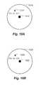
- the systemestimates and displays a clock position and radial distance from the nipple. Such estimation and display to the user can be helpful to the user in describing the location of the suspicious lesion to others.
- the clock position and radial distanceis shown in window 1358 .
Landscapes
- Engineering & Computer Science (AREA)
- Quality & Reliability (AREA)
- General Health & Medical Sciences (AREA)
- Medical Informatics (AREA)
- Nuclear Medicine, Radiotherapy & Molecular Imaging (AREA)
- Radiology & Medical Imaging (AREA)
- Health & Medical Sciences (AREA)
- Computer Vision & Pattern Recognition (AREA)
- Physics & Mathematics (AREA)
- General Physics & Mathematics (AREA)
- Theoretical Computer Science (AREA)
- Apparatus For Radiation Diagnosis (AREA)
- Ultra Sonic Daignosis Equipment (AREA)
Abstract
Description
- 1. Field
- This patent specification relates to medical imaging systems and processes. In particular, the present invention relates to the computer aided detection of breast abnormalities in volumetric breast ultrasound scans, and devices and methods of interactive display of such computer aided detection results.
- 2. Related Art
- Breast cancer screening programs currently use x-ray mammography to find cancers in an early stage when treatment is most effective. However, in dense breasts mammography is known to be insensitive. Therefore, new screening modalities are being investigated that may complement or replace mammography in women with high breast density. The most promising new technologies for screening dense breasts are dynamic contrast enhanced MRI, and automated breast ultrasound scanning. The latter technique is less sensitive than MRI, but has the advantage that it is relatively inexpensive that it does not require the use of a contrast agent. The use of gadolinium in contrast agent enhanced MRI may not be acceptable in a screening population. Effectiveness of handheld breast ultrasound screening has been demonstrated in several trials. See, Kolb T M, Lichy J, Newhouse J H: Comparison of the performance of screening mammography, physical examination, and breast US and evaluation of factors that influence them: An analysis of 27,825 patient evaluations.Radiology(2002); 225(1): 165-75; and Berg W A, et al., Combined Screening With Ultrasound and Mammography vs. Mammography Alone in Women at Elevated Risk of Breast Cancer. JAMA, May 14, 2008; 299: 2151-2163 (2008). However, the fact that this screening exam, requiring radiologists to perform by hand and read the images as being generated, is time consuming for radiologists makes it less attractive. This is being somewhat alleviated however, with volumetric ultrasound images obtained by an automated breast ultrasound scanning (ABUS) technology. Typically an ABUS device is used to image a whole breast volume with up to three partially overlapping scans per breast, which would generate several hundred images or slices. Although the image acquisition with ABUS can generally be performed by technicians, radiologists are still required to read the hundreds of images, thus breast imaging with ABUS can still be relatively time consuming. A complete screening exam consists of hundreds of images or slices, and the information content of each of the images is high. Abnormalities can therefore be easily overlooked when the images are being inspected slice by slice.
- There have been publications on development of computer aided diagnosis in ultrasound. The majority deals with traditional 2D handheld ultrasound and aim at helping the radiologist to diagnose lesions. In these papers, the detection of a lesion refers to an automatic segmentation of lesions in a 2D image selected by the radiologist. As the radiologist determines the target lesion, it is usually located in the center of the image. After segmentation of the lesion, features are extracted and a classifier is trained to distinguish benign and malignant lesions. See, Drukker, K.; Giger, M. L.; Horsch, K.; Kupinski, M. A.; Vyborny, C. J. & Mendelson, E. B. “Computerized lesion detection on breast ultrasound,” Med Phys, 2002, 29, 1438-1446; Drukker and M. L. Giger, “Computerized analysis of shadowing on breast ultrasound for improved lesion detection,” Med. Phys. 30, 1833-1842 (2003); K. Horsch, M. L. Giger, C. J. Vyborny, and L. A. Venta, “Performance of computer-aided diagnosis in the interpretation of lesions on breast sonography,” Acad. Radiol. 11, 272-280 (2004); V. Mogatadakala, K. D. Donohue, C. W. Piccoli, and F. Forsberg, “Detection of breast lesion regions in ultrasound images using wavelets and order statistics,” Med. Phys. 33, 840-849 (2006); and Drukker, C. A. Sennett, and M. L. Giger, “Automated method for improving system performance of computer-aided diagnosis in breast ultrasound,” IEEE Trans. Med. Imaging 28, 122-128 (2009).
- Characterisation of breast lesions in 3D ultrasound has been explored. See, Sahiner, B.; Chan, H.-P.; Roubidoux, M. A.; Helvie, M. A.; Hadjiiski, L. M.; Ramachandran, A.; Paramagul, C.; LeCarpentier, G. L.; Nees, A. & Blane, C, “Computerized characterization of breast masses on three-dimensional ultrasound volumes,” Med Phys, 2004, 31, 744-754; Sahiner, B.; Chan, H.-P.; Roubidoux, M. A.; Hadjiiski, L. M.; Helvie, M. A.; Paramagul, C.; Bailey, J.; Nees, A. V. & Blane, C, “Malignant and benign breast masses on 3D US volumetric images: effect of computer-aided diagnosis on radiologist accuracy,” Radiology, 2007, 242, 716-724; Sahiner, B.; Chan, H.-P.; Hadjiiski, L. M.; Roubidoux, M. A.; Paramagul, C.; Bailey, J. E.; Nees, A. V.; Blane, C. E.; Adler, D. D.; Patterson, S. K.; Klein, K. A.; Pinsky, R. W. & Helvie, M. A, “Multi-modality CADx: ROC study of the effect on radiologists' accuracy in characterizing breast masses on mammograms and 3D ultrasound images,” Acad Radiol, 2009, 16, 810-818; and Cui, J.; Sahiner, B.; Chan, H.-P.; Nees, A.; Paramagul, C.; Hadjiiski, L. M.; Zhou, C. & Shi, J; “A new automated method for the segmentation and characterization of breast masses on ultrasound images,” Med Phys, 2009, 36, 1553-1565. This work is based on images from a targeted 3D ultrasound scanning system. Only a small volume holding the lesion is imaged and analyzed. The purpose is distinguishing benign and malignant lesions. Features used in the work above include morphology (e.g. height to width ration), posterior acoustic shadowing, lesion and margin contrast.
- Computer aided detection in whole breast ultrasound with the aim of assisting in screening has been described in a few publications. See, Ikedo, D. Fukouka, T. Hara, H. Fujita, E. Takada, T. Endo, and T. Morita, “Development of a fully automatic scheme for detection of masses in whole breast ultrasound images,” Med. Phys. 34, 4378-4388 (2007); and Chang R et al. “Rapid image stitching and computer-aided detection for multipass automated breast ultrasound,” Med. Phys. 37 (5) 2010. This work describes a method in which serial 2D images are analyzed separately by the CAD system.
- U.S. Pat. No. 7,556,602 (hereinafter the “the '602 patent”) discusses the use of ultrasound mammography in which an automated transducer scans the patient's breast to generate images of thin slices that are processed into fewer thick slices simultaneously for rapid assessment of the breast. Computer aided detection or diagnosis can be preformed on images and resulting mark and/or other information can be displayed as well. The '602 patent discusses extracting and applying a classifier algorithm to known two-dimensional features such as spiculation metrics, density metrics, eccentricity metrics and sphericity metrics. However, spiculation is not identified or suggested as a criterion used for candidate detection (which is referred to as the ROI location algorithm). The '602 patent also discusses correlating regions of interest in an x-ray mammogram view to an adjunctive ultrasound view. However the disclosed algorithms are applicable to cases where the mammogram view and the ultrasound view are taken from the same standard view (e.g. CC or MLO) or at least where the breast tissue is compressed, in both ultrasound and mammography, in directions perpendicular to an axis that is perpendicular to the chest wall and passes through the nipple.
- Accordingly, a computer aided detection method that helps radiologists in searching and interpretation of abnormalities would be very useful. According to some embodiments, a novel CAD system is provided for detection of breast cancer in volumetric ultrasound scans.
- According to some embodiments, a method of analyzing ultrasound images of breast tissue is provided. The method includes receiving and processing a digitized ultrasound image of the breast tissue so as to generate a three-dimensional image composed of view slices that are approximately perpendicular to the direction of compression of the breast tissue during ultrasound scanning. The 3D image is further processed using one or more computer aided detection algorithms so as to identify locations of one or more regions of interest within the image based at least in part on identified areas of spiculation in portions of one or more of the view slices. According to some embodiments, the compression direction is towards the chest wall and the areas of spiculation are identified in portions of coronal view slices being approximately parallel to the skin surface. Features extracted from the 3D image such as based on gradient convergence, local contrast, and/or posterior shadowing can also be used to identified regions of interest in the image, in combination with spiculation. According to some embodiments, the features are computed at regularly spaced locations in the image, at each location using computations that include voxel values in a local 3D subvolume. According to some embodiments, a likelihood of malignancy for each of the regions of interest can be estimated and displaying to a user. The method can be used for screening and/or diagnostic purposes
- According to some embodiments, a method of analyzing ultrasound images of breast tissue of a patient is provided that includes receiving and processing two digitized three-dimensional ultrasound images of breast tissue of the patient so as to generate a region of interest in each image. A likelihood of malignancy is then evaluated based at least in part on the estimated distance between the locations of the regions of interest in the two images. According to some embodiments, the two images can be of the same breast of the patient as in the first image, such as two offset scans of the same breast taken during the same scanning procedure, or of the same breast during a prior year screening. According to some embodiments, the two images can be the left and right breast of the patient using a reference point such as the nipple, so as to evaluate symmetry when evaluating the likelihood of malignancy.
- According to some embodiments, a method of analyzing digital images of breast tissue of a patient is provided that includes receiving and processing a digitized ultrasound image of breast tissue compressed in a direction towards a chest wall using one or more computer aided detection algorithms thereby generating a region of interest in the ultrasound image; receiving and processing a digitized mammographic image, such as a CC or MLO view, of the same breast tissue using one or more computer aided detection algorithms thereby generating a region of interest in the mammographic image; and evaluating the likelihood of malignancy based at least in part on the estimated distance between a location of the region of interest in the ultrasound image and a location the region of interest in the mammographic image. According to some embodiments, the mammographic image is a tomographic mammographic image.
- According to some embodiments, a method of interactively displaying ultrasound and mammographic images of breast tissue to a user is provided. The method includes receiving one or more digitized mammographic images, such as CC or MLO views, of the breast tissue, and a digitized three-dimensional ultrasound image of the breast tissue compressed in a direction towards a chest wall of the patient. The user identifies a location or locations on the one or more mammographic images of a user identified region of interest in the breast tissue. One or more locations are estimated on the ultrasound image that correspond to the location or locations on the one or more mammographic images of the user identified region of interest, and portions of the digitized ultrasound image are displayed to the user so as to indicate the one or more estimated locations corresponding to the region of interest. According to some embodiments an estimated position of the user identified region of interest relative to an identified nipple on the breast tissue is displayed to the user using a clock position and distance from the nipple. According to some embodiments, the mammographic image is a three-dimensional mammographic image. According to some embodiments, the user identified region of interest is located by the user first in the ultrasound image, then the location or locations on the mammographic image or images are estimated and displayed to the user.
- According to some embodiments, a method of interactively displaying computer aided detection results of medical ultrasound images to a user is provided that includes receiving a digitized ultrasound image of breast tissue; processing the image using one or more computer aided detection algorithms thereby generating one or more regions of interest; displaying the digitized image along with one or more marks tending to indicate location on the tissue of the regions of interest and information relating to an estimated likelihood of malignancy, such as a percentage or a color indicating a percentage range, for each displayed region of interest.
- According to some embodiments related systems for analyzing digital images of breast tissue, and for displaying ultrasound and mammographic images of a breast tissue to a user are provided.
- Further features and advantages will become more readily apparent from the following detailed description when taken in conjunction with the accompanying drawings.
- The present disclosure is further described in the detailed description which follows, in reference to the noted plurality of drawings by way of non-limiting examples of exemplary embodiments, in which like reference numerals represent similar parts throughout the several views of the drawings, and wherein:
FIG. 1 is a flow chart illustrating the detection method according to some embodiments;FIGS. 2A-C are examples of a cross section through a malignant lesion showing features identified according to some embodiments;FIG. 3 is a flowchart showing combination of features at a voxel level using context, resulting in a likelihood of abnormality, according to some embodiments;FIG. 4 is a matrix of coronal views and cross sections of a malignant lesion at three different depths, according to some embodiments;FIG. 5 illustrates methods of presenting CAD results to users, according to some embodiments;FIG. 6 is a plot showing detection sensitivity as a function of the number of false positives per 3D image volume, according to some embodiments;FIG. 7 is a flowchart showing steps in carrying out CAD analysis of ultrasound breast images, according to some embodiments;FIGS. 8A-C show further detail of view correlation procedures, according to some embodiments;FIGS. 9A-B show further detail of left and right symmetry checking, according to some embodiments;FIGS. 10A-B show further detail of temporal correlation procedures, according to some embodiments;FIGS. 11A-C show further detail of correlation procedures between ultrasound images and cranio-caudal (CC) view of a mammographic image, according to some embodiments;FIGS. 12A-C show further detail of correlation procedures between ultrasound images and mediolateral oblique (MLO) view of a mammographic image, according to some embodiments;FIG. 13 shows a user interface which relates positions in mammographic and ultrasound images, according to some embodiments; andFIGS. 14A-D show x-ray tomosynthesis imaging displayed views, according to some embodiments.- The following description provides exemplary embodiments only, and is not intended to limit the scope, applicability, or configuration of the disclosure. Rather, the following description of the exemplary embodiments will provide those skilled in the art with an enabling description for implementing one or more exemplary embodiments. It being understood that various changes may be made in the function and arrangement of elements without departing from the spirit and scope of the invention as set forth in the appended claims.
- Specific details are given in the following description to provide a thorough understanding of the embodiments. However, it will be understood by one of ordinary skill in the art that the embodiments may be practiced without these specific details. For example, systems, processes, and other elements in the invention may be shown as components in block diagram form in order not to obscure the embodiments in unnecessary detail. In other instances, well-known processes, structures, and techniques may be shown without unnecessary detail in order to avoid obscuring the embodiments. Further, like reference numbers and designations in the various drawings indicated like elements.
- Also, it is noted that individual embodiments may be described as a process which is depicted as a flowchart, a flow diagram, a data flow diagram, a structure diagram, or a block diagram. Although a flowchart may describe the operations as a sequential process, many of the operations can be performed in parallel or concurrently. In addition, the order of the operations may be re-arranged. A process may be terminated when its operations are completed, but could have additional steps not discussed or included in a figure. Furthermore, not all operations in any particularly described process may occur in all embodiments. A process may correspond to a method, a function, a procedure, a subroutine, a subprogram, etc. When a process corresponds to a function, its termination corresponds to a return of the function to the calling function or the main function.
- Furthermore, embodiments of the invention may be implemented, at least in part, either manually or automatically. Manual or automatic implementations may be executed, or at least assisted, through the use of machines, hardware, software, firmware, middleware, microcode, hardware description languages, or any combination thereof. When implemented in software, firmware, middleware or microcode, the program code or code segments to perform the necessary tasks may be stored in a machine readable medium. A processor(s) may perform the necessary tasks.
- According to some embodiments, a system is described for detection of breast cancer in 3D ultrasound imaging data. Volumetric ultrasound images are obtained by an automated breast ultrasound scanning (ABUS) device. Typically this device is used to image a whole breast volume with up to three partially overlapping scans. Breast cancer screening with ABUS is time consuming as up to six scans per patient (three per breast) have to be read slice by slice. By using effective computer aided detection methods feasibility of breast cancer screening with ABUS may be increased.
- In ABUS images breast cancers appear as dark lesions. When viewed in transversal and sagital planes, lesions and normal tissue appear similar as in traditional 2D ultrasound. However, when viewing ABUS images in coronal planes (in parallel to the skin surface) images look remarkably different. In particular, it appears that architectural distortion and spiculation are frequently seen in the coronal views, and these are strong indicators of the presence of cancer. Therefore, in the computer aided detection (CAD) system according to some embodiments, combine a dark lesion detector operating in 3D is combined with a detector for spiculation and architectural distortion operating on 2D coronal slices. In this way a sensitive detection method is obtained.
- To effectively use the CAD system to guide search and interpretation in 3D breast ultrasound screening a new system for presenting CAD information is presented as well, based on coronal viewing and interactive CAD marker projections.
- Image segmentation and normalization.
FIG. 1 is a flow chart illustrating the detection method according to some embodiments. First,image data 110 from the scanning device is converted to a coronal representation. This comprises (1) artifact removal (step112), (2) re-sampling of the data to isotropic resolution (step114), and (3) rotation of the data to coronal orientation (step114). Artifact removal,step 112, addresses correction of scan line artifacts due to signal transfer variation during scanning. Lines with an outlying mean value are corrected using the mean value of neighboring lines as a reference. In theresampling step 114, the image data is converted to cubic voxels of 0.5 mm. In coronal views the (x,y) planes hold coronal slices, which are in parallel to the skin surface during scanning, while the z coordinate represents depth. - In
step 116, the image volume is segmented in four classes: background, fatty breast tissue, dense breast tissue, and other tissue. First, the background is labelled using feature based voxel classification and morphological operators. Features include texture and voxel value. If the voxel value is low and texture indicates a homogeneous neighborhood voxels are labelled as background. Next, the chest-wall is detected using dynamic programming in transversal and sagittal slices, and by subsequently fitting a parameterized surface through the set of obtained boundary points. Voxels between the chest wall and skin are labelled as breast tissue. Using Otsu's method a threshold is determined to label fatty and dense tissue voxels in the breast. - Before further processing, images voxel values are normalized in
step 118, using the segmented breast tissue volume. From the labelled image, mean values of voxels labelled as dense and fatty tissue are computed. These are denoted by mean_fat and mean_dense respectively. Using the mean values, contrast of the image is normalized by:
y=y_fat+constant*(y_original−mean_fat)/(mean_dense−mean_fat)- with y_original being the voxel value before normalization, and y and y_fat the voxel value and the mean voxel value of fatty tissue after contrast normalization.
- Voxel feature extraction takes
place using modules 126. After normalization, the breast tissue region is processed to extract local features. Three modules are used, each targeting a different characteristic of breast cancers in ultrasound. Cancers appear as dark regions with relatively compact shapes. The mean voxel value in malignant lesions is lower than that the surrounding fatty tissue, and often a dark shadow is present under the lesion. Finally, in coronal slices through, or near, malignant lesions spiculation or architectural distortion is often visible. This a new radiological sign, which is not observed in traditional hand-held ultrasound because this modality was not able to show the coronal plane. The threemodules 126 are developed to capture the characteristic features and are described below. - The
first module 120 computes volumetric gradient convergence features that give a high response at the center of compact regions. It operates on the 3D gradient vector field derived from the image at a chosen scale. The module computes the number of gradient vectors directed to the center of a spherical neighborhood covered by the filter. This number is normalized by subtracting its expected value and by dividing the result by its standard deviation, both determined for a reference pattern of random gradient directions. At any given location, the filter output is obtained as a function of the neighborhood radius R, making the filter equally responsive to both small and large lesions. At each location the maximum filter output and corresponding neighborhood size are determined. Apart from the integrated measure of convergence, also the isotropy of convergence is determined. For further detail of this method applied in a 2D application, See: Brake, G. M. & Karssemeijer, N. (1999), “Single and multiscale detection of masses in digital mammograms,” IEEE Transactions on Medical Imaging. Vol. 18(7), pp. 628-639; Karssemeijer, N. & to Brake, G. M., “Detection of stellate distortions in mammograms,” IEEE Transactions on Medical Imaging. Vol. 15(5), pp. 611-619 (1996) (hereinafter “Karssemeijer (1996)”); and Karssemeijer, N., “Local orientation distribution as a function of spatial scale for detection of masses in mammograms,” Information Processing in Medical Imaging, LNCS 2082 (Springer), pp. 280-293 (1999) (hereinafter “Karssemeijer (1999)”). - According to some embodiments, analysis of local line and gradient direction patterns forms the basis for computation of the local features that are used. Further detail of this method will now be described. According to some embodiments, the size of the neighborhood in which orientations patterns are evaluated is one of the most important parameters in the computation of these features. Variation of this size can have a dramatic effect on the detection of individual cancers, although the influence of this parameter on the overall performance measured on a large database tends to be less. In the past, the output of a local contrast operator has been used to set the size of the neighborhood adaptively. In the method presented herein features are computed as a continuous function of the neighborhood size, only slightly increasing the computational load. The method is described here for 2D application, but can be used in higher dimensions as well.
- Local orientation distributions. It has been shown that features representing local orientation distributions are well suited for detection of masses in mammograms. See, N. Karssemeijer, “Detection of stellate distortions in mammograms using scale space operators,” In Y Bizais, C Barrilot, and R Di Paola, editors,Information Processing in Medical Imaging, pages 335-346. Kluwer, Dordrecht, 1995; Karssemeijer (1996); and G M to Brake and N Karssemeijer, “Detection of stellate breast abnormalities,” In K Doi, M L Giger, R M Nishikawa, and R A Schmidt, editors,Digital Mammography, pages 341-346. Elsevier,Amsterdam,1996 (hereinafter “te Brake (1996)”). The fact that such features are very insensitive to changes in contrast is a major advantage when processing large datasets of images of various origin, because one has to deal with unknown non-linear variation of the greyscale. Orientation maps are computed using first and second order Gaussian derivatives. When there is a concentration of gradient orientation towards a certain point this indicates the presence of a mass. A concentration of line orientations computed from second order directional derivatives indicates the presence of spiculation or architectural distortion. These concentration or convergence features will be denoted by g1 and l1, respectively for gradient and line orientations. In addition, features representing radial uniformity measure whether or not increase of pixels oriented to a center comes from the whole surrounding area or from a few directions only. These will be denoted by g2 and l2.
- In Karssemeijer (1996), features for orientation concentration were computed by counting the number of pixels pointing to a center, and were defined to measure deviations of this number from the expected value in a random orientation pattern. The assumption was made that a binomial distribution of this number with mean probability
p of a pixel pointing to a center can be used for normalization. As the probability p of hitting the center varies with the distance, this normalization may not be best choice. A more general definition of the features was given in Karssemeijer (1999), which discusses how to deal with varying values ofp properly. Note that the papers referred to above present the method for the 2D case and has applied to the techniques described herein they are extended to 3D. - For computation of the features at a given voxel i a circular neighborhood is used in the 2D case and a spherical neighbourhood in the 3D case. The term voxel is used for image samples in both the 2D and 3D case. All voxels j located within a distance rmin<rij<rmaxfrom i are selected when the magnitude of the orientation operator exceeds a small threshold. This selected set of voxels is denoted by Si. The features are based on a statistic xjdefined by
- with pjthe probability that voxel j is oriented towards the center given a random pattern of orientations. Voxels that are oriented to the center can be determined by evaluating:
arc cos(v·r)<D/(2rij) (2)- with r the unit vector in the direction from j to i, v a unit vector with the voxel orientation at j and D a constant determining the accuracy with which voxels should be directed to the center to be counted. Alternative ways of determining when a voxel is oriented to the center can also be used. In the application presented here we use the equation above for mass detection, where voxel orientations are 3D gradient vectors. However, we use arc cos (|v·r|)<D/(2rij) for spiculation detection, because in that case the pixel orientations are 2D line orientation estimates. After computing xjfor voxels in the neighborhood of i weighted sum Xiis computed by
- where the weight factors can be chosen as a function of the distance rij, for instance to give voxels closer to the center a larger weight. For a noise pattern, the variance of this sum can be estimated when it is assumed that all voxel contributions are independent:
- Normalizing the sum Xiby the square root of the variance the value of the concentration feature f1is defined by
- When no weight factors are used and the neighborhood Siis subdivided in K rings (or spherical shells in 3D) around i in which the probability pkcan be considered constant, the sum Xican be written as
- In each ring (or shell) k the number of voxels hitting the center Nk, hitcan be counted, allowing the sum to be rewritten as
- with Nkand N the number of voxels in ring (shell) k and in total, respectively. The normalization factor which can be written as (N(
p −p2 ))−1/2. - If weight factors are used that only depend on pj, the sum Xican be written as
- the expected value of f1remains zero.
- It is noted that the approximation that is made by assuming all voxels to have independent directions is clearly incorrect, even when voxels have independent random values. Orientations of neighboring voxels become correlated by the use of convolution kernels for estimation. This leads to underestimation of the variance, which becomes larger with larger kernels. However, it seems that this effect is similar for normal and abnormal areas. For the purpose of removing dependency of the size of the neighborhood and compensating unwanted effects at the breast edge boundary the method is effective.
- Features g2 and l2 that measure radial uniformity of the orientation patterns around site i are computed by subdividing the neighborhood Siin L directional bins, that is like a pie. The statistic Xiis computed now for each bin. When there is only noise the expected value of Xiin each bin is zero. In previous work, the number of bins was counted in which the number of voxels pointing to the center was larger than the median of a binomial distribution determined by Nland
p , with Nlandp lthe number of voxels in bin l andp lthe average probability of hitting the center. This definition had some problems, as the median of a binomial distribution is not exactly defined. With the approach described here, it is sufficient to compute the number of bins n+ in which the sum of Xi,kis positive. The radial uniformity feature is defined by - with Kithe number of sectors at i. The standard deviation of n+ for random noise √{square root over (Kl/4)} is used for normalization, which is important to avoid problems at the edge of the breast where not all sectors can be used.
- Computation of features as a function of scale. In multiscale methods one tries to match the scale of feature extraction to the scale of the abnormality in order to optimize detection performance. Generally, the value of features used for mass detection depend strongly on the size of the abnormality, which makes multiscale approaches attractive. However, most multiscale methods are computationally intensive, because features have to be computed repeatedly at a number of scales. Usually only a very limited number of scales are chosen, which reduces accuracy. Multiscale methods that have been proposed for detection of masses in mammograms include: for wavelets, see, A. F. Laine, S. Schuler, J. Fan, and W. Huda, “Mammographic feature enhancement by multiscale enhancement,”IEEE Trans on Med Imag,13:725-740, (1994); for maximum entropy, see L. Miller and N. Ramsey, “The detection of malignant masses by non-linear multiscale analysis,” In K. Doi, M. L. Giger, R. M. Nishikawa, and R. A. Schmidt, editors, Digital Mammography, pages 335-340, Elsevier, Amsterdam (1996); and for multi-resolution texture analysis, see, D. Wei, H. P. Chan, M. A. Helvie, B. Sahiner, N. Petrick, D. D. Adler, and M. M. Goodsitt, “Classification of mass and normal breast tissue on digital mammograms: multiresolution texture analysis,”Med Phys,22:1501-1513, 9 (1995). Also line concentration measured at a number of scales was used in previous work on detection of stellate lesions, where the maximum over the scales was used Karssemeijer (1996).
- According to some embodiments, a method is described that allows very efficient computation of a class of local image features as a continuous function of scale, only slightly increasing the computational effort needed for computation at the largest scale considered. The non-linear features described in the previous subsection belong to this class. In the first step of the algorithm an ordered list is constructed in which each element represents a neighbor j within distance rijof the central location i. In this list, positional information of the neighbor that is needed for the computation is stored, here the xj, yjoffset, orientation and distance rijwith respect to center. This list is constructed by visiting all voxels in any order, and by subsequently sorting its elements by distance to the center. In the second step the actual computation of the features takes place, at each voxel or at a given fraction of voxels using a sampling scheme. The ordered list of neighbors is used to collect the data from the neighborhood. The xj, yjoffsets in the list are used to address the voxel data and precomputed derivatives or orientations at the location of the neighbor. The orientation with respect to i is used to compute orientation related features. Because the neighbors are ordered with increasing distance to the center, computation of the features from the collected data can be carried out at given intervals, for instance each time the number of neighbors has increased by some fixed number. As the computational effort lies in collection of the data, this only slightly increases the computational load. We use intervals in which the number of neighbors increases quadratically for 2D and with a power of three for 3D. Thus, features are computed at regularly spaced distances from the center. In a similar way, a contrast feature can be computed by collecting the sum of voxel values as a function of distance to the center, and by and subtracting the mean of the last interval from the mean of the previous intervals. The curves that represent features as a function of the distance to the center reveal aspects of the neighborhood patterns that can be useful for differentiation of true and false positive detections.
- Referring again to
FIG. 1 ,module 124 is designed to find spiculation or architectural distortion in coronal planes, using the method above in 2D. In contrast tomodule 120, where gradient vectors are used, in this module124 a line orientation pattern forms the basis for feature computation. Line orientations are obtained using a basis of second order Gaussian directional derivatives. By applying the method to each coronal slice independently, a response for each breast tissue voxel is obtained. Module 122 computes local contrast as a function of scale. At each location in the image the mean voxel value m(x,y,z) in a neighborhood is computed. The neighborhood is defined by all voxels within distance R1 from the central location. Contrast is computed by subtracting this mean value from the mean value of voxels labeled as fatty tissue just outside the neighborhood, i.e. voxels with distance to the center within an interval [R1,R1+ΔR]. According to some embodiments, local contrast features are computed for various neighborhood types: (1) A spherical neighborhood with a fixed radius, (2) A spherical neighborhood with radius estimated from the image data, for instance by taking the radius at which the gradient concentration filter g1 has the highest output maximum response, (3) a semi-spherical neighborhood including only superficial voxels, i.e those that are closer to the transducer (or skin) than the central location, (4) a semi-spheric neighborhood including only deeper voxels (further away from the transducer than the central location), and (5) the spherical neighborhood with the radius that gives the highest local contrast.- In the examples discussed thus far, it is assumed that the breast tissue has been compressed towards the chest wall during the ultrasound scanning process. Note that the ROI location detection schemes described herein can also apply to other compression directions. According to some embodiments, the ultrasound images can result from breast tissue compressions in directions other than toward the chest wall. For example, the ultrasound image can result from a scan in which the breast is compressed in a direction such as with conventional mammography (e.g. as in CC and/or MLO views. In general, according to some embodiments, the
module 124 is designed to find spiculations and/or architectural distortions in plans perpendicular to the direction of compression. For example, if the compression direction of the ultrasound scan is as in a CC mammography view, then themodule 124 would look for spiculations and/or architectual distortions in a transverse plane. - Note that the
modules 126, includingspiculation module 124 are used in candidate detection (i.e. to locate the regions of interest). This is in contrast to techniques such as discussed in the '602 patent where features such as spiculation metrics are only used for classification of regions of interest. FIGS. 2A-C are examples of a cross section through a malignant lesion showing features identified according to some embodiments. The skin surface is on top in each of the views. InFIG. 2A , the originalcross section image 210 is shown.FIG. 2B shows theresponse 220 of gradient convergence module120 (as described with respect toFIG. 1 ). The highest values are shown outlined in white such asregion 222, and the next highest values are shown outlined in black such asregion 224.FIG. 2C shows theresponse 230 of coronal spiculation module124 (as described with respect toFIG. 1 ). The highest values are shown outlined in white such asregion 232, and the next highest values are shown outlined in black such as region234.FIG. 2D shows theresponse 240 of local contrast module122 (as described with respect toFIG. 1 ). The highest values are shown outlined in white such asregion 242, and the next highest values are shown outlined in black such asregion 244. It can be seen that the maxima of the responses are not aligned. In particular, the coronal spiculation feature is strongest in the upper part of the lesion.FIG. 3 is a flowchart showing combination of features at a voxel level using context, resulting in a likelihood of abnormality, according to some embodiments.Input image 310 is input to thelocal feature extraction 312 which corresponds to themodules FIG. 1 . Examples of the cross sections highlighted according to the three modules is shown in314 which correspond to the cross section examples shown inFIG. 2 . The results of the modules are combined in thecontextual voxel classifier 316, which corresponds to thestep 126 ofFIG. 1 . The result is thelikelihood map 320 which shows the highest values outlined in white such asregion 322 and the next highest values outlined in black, such asregion 324.FIG. 4 is a matrix of coronal views and cross sections of a malignant lesion at three different depths, according to some embodiments. Depth increases fromcolumn 410 being coronal views shallowest (closest to the skin),column 412 being coronal views of medium depth, andcolumn 414 being coronal views being the deepest (furthest from the skin).Column 416 are transversal plane views. Row420 shows the original image. Row422 shows an overlay of the results of gradient convergence module120 (as described with respect toFIG. 1 ). Row424 shows an overlay of the results of coronal spiculation module124 (as described with respect toFIG. 1 ). Row426 shows the overlay of lesion likelihood as a result of the contextualvoxel classification step 128. In therows - Further detail of the contextual voxel classification and candidate detection steps will now be provided, according to some embodiments. In
step 128, selected voxels on a regular 3D grid of locations covering the breast tissue are classified using a feature vector that comprises information extracted by thefeature extraction modules 126 at the location (x, y, z) of the voxel itself and its surroundings. The latter is essential because it has been observed that in 3D breast ultrasound imaging the central locations of lesions often do not coincide with focal points of spiculation patterns associated with the lesions. In particular, for example, it has been found that spiculation patterns in coronal planes are often are stronger in a region in the upper part of a lesion (closer to the skin) or even outside the lesion, as can be seen inFIGS. 2A-D . Therefore, a contextual voxel orpixel classification method 128 is designed that brings together information extracted in nearby locations. According to some embodiments the feature vector f(r) at a given location r=(x, y, z,)Tis augmented with the maximum of selected features in a neighborhood of r. For instance, the maximum of each of the spiculation features in a column centered at r and oriented in the z direction can be added to the feature vector. According to other embodiments, a contextual Markov Random Field can be defined to represent relations between features in a neighborhood of r. A feature vector can also be defined as the concatenation of feature vectors in a neighborhood of r. - According to some embodiments, to determine a set of candidate locations that are most representative of cancer, supervised learning is used. A set of training cases is used in which locations of relevant abnormalities. The training set includes both malignant and benign lesions (e.g. cysts and fibroadenoma). For training of classifiers, voxels and associated feature vectors in the center of annotated lesions are taken as abnormal patterns, while voxels sampled outside the lesions and/or in normal cases are used as normal samples. By supervised learning a classifier is trained to relate the input to a probability that an abnormality is present at a given location. Thus, the output of the
contextual voxel classifier 128 is a volume representing likelihood of abnormality L(r). See,e.g. output view 320 inFIG. 3 andcolumn 426 inFIG. 4 . Referring again toFIG. 1 , after smoothing instep 130, local maxima a determined instep 132. These are candidate locations used in further processing steps. - In
step 134, candidate classification is carried out. As is common in most CAD systems a multi-stage model is employed. By thresholding, the most relevant candidate locations are selected and processed further. Typically, this processing includes a segmentation step in which the lesion boundary is localized. New features are computed, with the aim of representing relevant characteristics of the lesion by a numerical feature vector. Features for characterizing breast ultrasound lesions have been described in the literature for 2D handheld ultrasound and extension to 3D is straightforward. They include lesion contrast, margin contrast, margin sharpness, boundary smoothness, shadowing, width-to height ratio, and moments of the distribution of voxel values inside the lesion. Here a new set of features represented in coronal spiculation is added. These are computed from the distribution of coronal spiculation features computed in the candidate detection stage inside the lesion, e.g. mean variance and percentile values. - By supervised classification, the number of false positive candidates can be reduced, and/or the probability that a lesion is malignant or benign can be assessed. Three configurations of the CAD system are described below according to some embodiments, although other configurations are possible. The three described configurations are: false positive reduction; false positive reduction and subsequent lesion classification; and multi-class classification.
- False positive reduction. According to some embodiments, false positives are defined as non-lesion locations. The detection system is trained with both benign and malignant lesions as target training patterns and it learns to distinguish those lesions from normal breast tissue. The task of deciding whether a lesion is more likely to be benign or malignant is left to the radiologist.
- False positive reduction and subsequent lesion classification. According to some embodiments, the detection system is combined with a classification system trained to distinguish benign lesions from malignant lesions. The classification system is a feature-based system trained in the traditional way as a 2-class supervised classifier. The system is applied to regions surviving the false positive reduction step of the CAD system. In this way, each region detected by the CAD system has two numerical values assigned to it: one to indicate the probability that a lesion is present, and another to indicate the probability that the lesion is malignant.
- Multi-class classification. According to some embodiments, non lesion locations form one class in a multi-class classification system. The system is trained to distinguish non-lesion locations, cancer, and benign lesions. The CAD system computes a likelihood value for each of the classes. It is noted that these likelihood values depend on prevalence and characteristics of the classes in the training set, which is dependent on the case sample and on the threshold applied in the candidate lesion detector. This has to be taken into account when information is displayed to the radiologist.
- Users can use CAD marks to increase quality of their reading. By using CAD marks as guidance, image volumes may be more efficiently searched for abnormalities, without overlooking lesions.
FIG. 5 illustrates methods of presenting CAD results to users, according to some embodiments.Column 522 shows lateral coronal views andcolumn 520 shows medial coronal views. The woman in this case has an invasive ductal cancer. Theimages 510 shows the slice at the skin level. White dashed circles such ascircle 524 are interactive CAD finding projections. By activating a mark, such a by selecting it with a pointer, the coronal view at the depth where the selected finding is located is shown. If depth of the displayed view corresponds with the location of the CAD finding the prompt is displayed in a solid white circle, such ascircle 540. - The
images 512 are the coronal views at the depth corresponding to the lesions marked by solidwhite circles mark 544.Images 514 show the coronal slices viewed a depth corresponding to a lesion as shown by theCAD mark 530 displayed in a solid white circle. According to some embodiments, colors are used in the display and green circles denote the slice depth does not correspond to the lesion depth and red circles are used when the view corresponds to the lesion depth. - According to some embodiments, a function is available that allows the user to move the display automatically to the slice in which CAD identified a suspicious region. This slice, or depth, can be determined by taking the maximum of the likelihood image, or the center of the segmentation. This function can be activated by clicking on the marked location with a mouse pointer, such as
pointer 550. Optionally, the display can automatically synchronize the displayed slices, to the same depth in all displayed views. In this way, radiologists can more efficiently make comparisons between views, which is usually done at the same depth. In the case ofFIG. 5 , the views of column522 (lateral coronal views) and column520 (medial coronal views) are synchronized for each depth. The top row,images 510 show slices at the skin surface (both having a depth=0).Images 512 are slices both having a depth of the solidmarked lesions Images 514 are deeper slices, both at the depth of the lesion ofCAD mark 530. - According to some embodiments, when activating a CAD mark, the likelihood of malignancy computed by the CAD system can be displayed. In
images 512 for example, the computed likelihoods for themarks - According to some embodiments, a function is available that allows the user to move the display automatically to the slice in which CAD identified a suspicious region exists when viewing slices from the original scanning acquired images. For example,
FIGS. 2A-D could be the taken from the original scanned images such as acquired instep 110 ofFIG. 1 . According to such embodiments, the original scanned 2D images can be displayed to the user in a cine fashion, which automatically stops or pauses when the image contains a CAD identified a suspicious region. Current users of hand held breast ultrasound, such as radiologists, may be more familiar and/or feel more comfortable with viewing original 2D acquired image. According to some embodiments, the display can also be interactive when displaying and automatically stopping at 2D images containing a CAD identified suspicious region. For example, the CAD identified suspicious region can be highlighted using solid and/or dashed circles such as shown inFIG. 5 , and in response to a user's selection with a pointer, the system can interactively display information, such as likelihood of malignancy as shown inFIG. 5 . FIG. 6 is a plot showing free response operating characteristic (FROC) demonstrating detection performance of the candidate detection stage, according to some embodiments. Plot610 shows detection sensitivity as a function of the number of false positives per 3D image volume. Theplot 610 shows the result of applying the candidate detection method as described herein to a series of test and training cases.FIG. 7 is a flowchart showing steps in carrying out CAD analysis of ultrasound breast images, according to some embodiments. In step710 a 3D ultrasound volume is input, artifacts are removed and resampling for coronal view reconstruction is carried out. This step corresponds tosteps FIG. 1 . Instep 712, the image is segmented to identify the breast tissue, and the image is normalized. This step corresponds tosteps FIG. 1 . Instep 714, each voxel is analyzed using modules for gradient convergence, spiculation in coronal planes and local contrast. This step corresponds to usingmodules 126 inFIG. 1 . Instep 716, each pixel or voxel is classified, which corresponds to step128 inFIG. 1 . Instep 718, groups of voxels are segmented having similar properties and classified according to characteristics of the region such as size, shape, lesion contrast, margin contrast, margin sharpness, boundary smoothness, shadowing, width-to height ratio, moments of the distribution of voxel values inside the lesion, and coronal spiculation. This step corresponds to step134 inFIG. 1 .- Up until now, the processing steps described in
FIG. 7 relate to image information from a single view of a single breast. Insteps steps - In
step 720, correlation between different views is carried out. Ordinarily, more than one ultrasound scan is used to cover a breast. If a lesion occurs in an overlap area, then correlation between different views of the same breast can be carried out. Instep 722, left versus right breast symmetry check is carried out which can identify false positives due. Instep 724, a temporal comparison is carried out, for example between ultrasound scans of the same breast taken at different times, such as separated by one or more years. Instep 726, a comparison with other modalities such as mammography is carried out. According to some embodiments, one or more of the comparison steps720,722,724 and726 are not carried out, are performed in a different order than shown inFIG. 7 , and/or performed in parallel with each other. FIGS. 8A-C show further detail of view correlation procedures, according to some embodiments. InFIG. 8A , a first scan, shown incoronal view 820, is made ofbreast 810 having a nipple812. From the first scan, a region of interest is identified which is shown by themark 822 oncoronal view 820. The region of interest is identified, for example, using the techniques discussed with respect toFIG. 1 . The position of the nipple812 in the first scan is determined either manually by an operator, or alternatively the nipple position can be automatically determined as is known in the art. The position of the region of interest relative to the nipple using x, y, z coordinates can therefore be determined. The x1and y1position of the region marked822 in the first scan can be shown in thecoronal view 820 ofFIG. 8A .FIG. 8B is a transversal slice that shows the depth z1of the region of interest shown by thespot 842 as measured from theskin surface 846.Chest wall 844 is also shown. In the second scan, a region of interest having a location relative to the nipple of x2, y2and z2. InFIG. 8A , thecoronal view 830 is shown for the second scan of thebreast 810, with the corresponding region ofinterest 832 marked by the dashed circle. The position x2and y2can be shown in the coronal view. The depth z2is shown inFIG. 8C as the distance betweenskin surface 856 and region of interest marked by dashedcircle 852 intransversal view 850. According to some embodiments, a threshold distance condition can be applied for the maximum distance between the regions of interest in first and second scans:
√{square root over (Δx2+Δy2+Δz2)}≦Maximum Distance. (10)- Then the correlation between regions of interest in the first and second scans can be calculated as:
error=√{square root over (k1Δfeature—1+k2Δfeature—2+k3Δfeature—3)} (11)- where k1, k2and k3. . . are weighting factors for each of the features of the regions of interest. Examples of
feature —1, feature—2, etc are features such as size, shape, coronal spiculation, contrast, etc. According to some embodiments, the values for the threshold for maximum distance and/or the weighting factors k1, k2and k3can be determined using a free response operating characteristic (FROC) curve, where the values are adjusted so as to yield the highest sensitivity for given false positive rates per volume. FIGS. 9A-B show further detail of left and right symmetry checking, according to some embodiments. InFIG. 9A , is a coronal view of a scan of a patent'sright breast 910 andFIG. 9B is a coronal view of a scan of a patient'sleft breast 920. The region marked914 on the right breast is shown incoronal view 910 having a position relative to thenipple 912 of xr, yrand Zr. Similarly, a region marked924 on the left breast is shown incoronal view 920 having a position relative to thenipple 922 of xl, yland zl. The same or similar threshold as shown in equation (10) and the error evaluation of equation (11) can be used. However, as symmetry about the sagittal plane is being checked, yl=−yr, and greater correlation indicates decreased likelihood of malignancy.FIGS. 10A-B show further detail of temporal correlation procedures, according to some embodiments.FIG. 10A , is a coronal view of a scan of a patient'sbreast 1010 at one time (t1).FIG. 10B is a coronal view of a scan of a patient'sbreast 1020 at an earlier time (t0). Ordinarily, screening scans are performed at regular intervals, such as one year to two years, which would be the difference between t0and t1. The region marked1014 on the later scan of the breast is shown incoronal view 1010 having a position relative to thenipple 1012 of Xt1, yt2and zt2. Similarly, a region marked1024 on the earlier scan of the breast is shown incoronal view 1020 having a position relative to thenipple 1022 of xt0, yt0and zt0. The same or similar threshold as shown in equation (10) and the error evaluation of equation (11) can be used. However, for temporal comparisons, a finding smaller differences (or greater similarity) between two scans at different times tends to decrease the likelihood of malignancy.FIGS. 11A-C show further detail of correlation procedures between ultrasound images and cranio-caudal (CC) view of a mammographic image, according to some embodiments.FIG. 11A illustrates breast tissue as compressed for a CC mammography view. Theuncompressed breast tissue 1110 is compressed as shown byoutline 1112, against aplaten 1114.FIG. 11B shows acoronal view 1120 of an ultrasound scan having a region ofinterest 1122, as well as aCC view 1130 from a mammography scan having a region ofinterest 1132. The position ofregion 1122 relative to thenipple 1124 in the ultrasound image can be determined to be xu, yuand zu, as has been explained previously. In theCC mammography image 1130, the distance xmrelative to thenipple 1134 can be determined and is:
xm=xu (12)- provided the image scales are normalized. The distance ymcan be estimated from the ratio of the depth of the corresponding lesion in a transverse or sagittal slice of the ultrasound scan.
FIG. 11C shows atransverse slice 1140 of and ultrasound scan where the region ofinterest 1142 is at depth zuform theskin surface 1144. The total thickness of the breast tissue inslice 1140 fromskin surface 1144 and thechest wall 1146 is denoted as Tu. The distance ymin the CC mammography image is therefore:
ym=Cm(zu/Tu) (13)- where the Cmis the total distance from the
nipple 1134 to thechest wall 1136 inFIG. 11B . FIGS. 12A-C show further detail of correlation procedures between ultrasound images and mediolateral oblique (MLO) view of a mammographic image, according to some embodiments.FIG. 12A illustrates breast tissue as compressed for a MLO mammography view. Theuncompressed breast tissue 1210 is compressed as shown byoutline 1212, against aplaten 1214.1210 also represents a coronal view of an ultrasound scan having a region ofinterest 1222.FIG. 12B is anMLO view 1230 from a mammography scan having a region ofinterest 1232. The position ofregion 1222 relative to thenipple 1224 in the ultrasound image can be determined to be xu, yuand zu, as has been explained previously. The distance xmcan be estimated from the ratio of the depth of the corresponding lesion in a transverse or sagittal slice of the ultrasound scan.FIG. 12C shows atransverse slice 1240 of and ultrasound scan where the region ofinterest 1242 is at depth zuform theskin surface 1244. The total thickness of the breast tissue inslice 1240 fromskin surface 1244 and thechest wall 1246 is denoted as Tu. The distance xmin the MLO mammography image is therefore:
xm=Cm(zu/Tu) (14)- where the Cmis the total distance from the
nipple 1234 to thechest wall 1236. The distance ymin the MLO mammography image can be related to position of the region ofinterest 1222 in relation to thenipple 1224 and the angles αu, which is the angular position of theregion 1222, and the oblique imaging angle θmwhich can be determined, for example from the mammography image (DICOM) (Digital Imaging and Communications in Medicine standard) header. The distance ymin the MLO mammography image can be estimated as:
ym=rucos(αu−θm) (15)- where ruis the radial distance of the
region 1222 from thenipple 1224 in the ultrasoundcoronal view 1210, and can be related to xuand yuby ru=√{square root over (xu2+yu2)}. Once the equivalent coordinates in the mammography view (CC and MLO) are found as described herein, the same or similar threshold as shown in equation (10) and the error evaluation of equation (11) can be used in correlating regions of interest ultrasound and mammographic images. - According to some embodiments, the correlation of CAD results between ultrasound and mammographic images is applied to x-ray tomographic breast images. For example, in x-ray tomosynthesis mammography, the breast tissue is compressed as in the standard CC and MLO views, and multiple x-ray images are taken a different angles. A computer process then synthesizes the 3D mammographic image of the breast.
FIGS. 14A-D show x-ray tomosynthesis imaging displayed views, according to some embodiments.FIG. 14A illustratesbreast tissue 1410 being compressed and imaged for x-ray tomosynthesis imaging of a CC view. The tissue is imaged at multiple angles centered around thedirection 1412.FIG. 14B is an example of aCC view 1420 of a tomosynthesis mammagraphic image. Using the coordinates shown, xmand ymcan be related to ultrasound images of the same breast using the equations (12) and (13) as described above. The distance zm, which was not available in standard mammography, is the distance perpendicular to theCC view 1420, and can be related to ultrasound images using the simple relationship: - where Zmis the total thickness of the tomosynthesis image (see
FIGS. 14A and 14C ), which can be retrieved from the DICOM header or directly measured from the image volume, and Ruis the radius of the breast measured from the coronal ultrasound image, an example of which is shown inFIG. 8A . If the ultrasound image or images result from multiple scans, Ruis preferably measured in a region of the image that is common to bothscans FIG. 8A . FIG. 14C illustratesbreast tissue 1430 being compressed and imaged for x-ray tomosynthesis imaging of a MLO view. The tissue is imaged at multiple angles centered around thedirection 1432.FIG. 14D is an example of aMLO view 1440 of a tomosynthesis mammagraphic image. Using the coordinates shown, xmand ymcan be related to ultrasound images of the same breast using the equations (14) and (15) as described above. The distance zm, which was not available in standard mammography, is the distance perpendicular to theMLO view 1440, and can be related to ultrasound images using the relationship:- where ruand αuand θmare defined as described above with respect to
FIG. 12A . - The described techniques for correlating locations in mammography images and ultrasound images are more robust than those such as discussed in the '602 patent which are applicable only in cases where the breast tissue is compressed, in both ultrasound and mammography, in directions perpendicular to an axis that is perpendicular to the chest wall and passes through the nipple. In contrast, the techniques disclosed according to some embodiments herein, are applicable to cases where the ultrasound image is made with a breast compressed in a direction perpendicular to the chest wall (i.e. the breast tissue is compressed directly towards the chest wall), and the mammography image compression is according to a standard view (e.g. CC or MLO).
- According to some embodiments, the techniques described here for relating positions in ultrasound images and mammography images are used to provide a useful user interface for users such as radiologists who are reading, reviewing or otherwise analyzing the images.
FIG. 13 shows a user interface which relates positions in mammographic and ultrasound images, according to some embodiments. Theuser interface 1310 includes adisplay 1312, input devices such askeyboard 1362 andmouse 1360, and aprocessing system 1370. According to some embodiments, other user input methods such as touch sensitive screen screens can be used. Processing system 1370 can be a suitable personal computer or a workstation that includes one or more processing units1342, input/output devices such as CD and/or DVD drives,internal storage 1372 such as RAM, PROM, EPROM, and magnetic type storage media such as one or more hard disks for storing the medical images and related databases and other information, as well as graphics processors suitable to power the graphics being displayed ondisplay 1312.- The
display 1312 is shown displaying two areas.Mammographic display area 1314 is similar to a mammography workstation for viewing digital mammography images.Ultrasound display area 1316 is similar to an ultrasound image workstation for viewing 3D ultrasound breast images.Mammographic display area 1314 is shown displaying four mammographic images, namelyright MLO view 1330, leftMLO view 1332,right CC view 1334, and leftCC view 1336. Shown onright MLO view 1330 is a region ofinterest 1320, and onright CC view 1336 is region ofinterest 1322. - According to some embodiments, the user selects both
regions mammographic display area 1314, for example, by clicking withmouse pointer 1324. By selecting bothregions regions regions coronal view 1340 of an ultrasound scan of the patient's right breast, at the depth associated with the suspected lesion, as well as a mark indicator, such as dashedcircle 1344 at a position on thecoronal view 1340. Also displayed aretransversal view 1350 andsagittal view 1354, both at locations corresponding to the estimated location of the user selected lesion. Themark indicators views - According to some embodiments, the
user interface system 1310 can operate in an inverse fashion as described above. Namely, the user selects a location on any of the ultrasound views, and in response the system displays the estimated corresponding locations on the mammographic images. For example, the user selects a location on thecoronal image 1340. In response, the system estimates and automatically highlights the corresponding locations on the CC and MLO views of the mammographic image. Note that since the 3D coordinates can be determined from a single selection on one of the ultrasound images, the system can estimate the mammographic locations in response to making a selection on only one ultrasound image view. As described above, the system can use relationships such as shown in equations (12), (13), (14) and (15) to relate the user selected location on the ultrasound image to the corresponding estimated locations on the mammographic images. - According to some embodiments the user interface system as described with respect to
FIG. 13 is applied to three-dimensional mammographic images such as tomosynthesis mammography images. - According to some embodiments, in response to the user selecting a location on either
display area display 1312, the clock position and radial distance is shown inwindow 1358. - Whereas many alterations and modifications of the present disclosure will no doubt become apparent to a person of ordinary skill in the art after having read the foregoing description, it is to be understood that the particular embodiments shown and described by way of illustration are in no way intended to be considered limiting. Further, the disclosure has been described with reference to particular preferred embodiments, but variations within the spirit and scope of the disclosure will occur to those skilled in the art. It is noted that the foregoing examples have been provided merely for the purpose of explanation and are in no way to be construed as limiting of the present disclosure. While the present disclosure has been described with reference to exemplary embodiments, it is understood that the words, which have been used herein, are words of description and illustration, rather than words of limitation. Changes may be made, within the purview of the appended claims, as presently stated and as amended, without departing from the scope and spirit of the present disclosure in its aspects. Although the present disclosure has been described herein with reference to particular means, materials and embodiments, the present disclosure is not intended to be limited to the particulars disclosed herein; rather, the present disclosure extends to all functionally equivalent structures, methods and uses, such as are within the scope of the appended claims.
Claims (63)
Priority Applications (8)
| Application Number | Priority Date | Filing Date | Title |
|---|---|---|---|
| US12/839,371US20120014578A1 (en) | 2010-07-19 | 2010-07-19 | Computer Aided Detection Of Abnormalities In Volumetric Breast Ultrasound Scans And User Interface |
| US14/044,842US9826958B2 (en) | 2009-11-27 | 2013-10-02 | Automated detection of suspected abnormalities in ultrasound breast images |
| US14/084,589US20140082542A1 (en) | 2010-07-19 | 2013-11-19 | Viewing and correlating between breast ultrasound and mammogram or breast tomosynthesis images |
| US14/448,607US9439621B2 (en) | 2009-11-27 | 2014-07-31 | Reduced image reading time and improved patient flow in automated breast ultrasound using enchanced, whole breast navigator overview images |
| US14/555,408US10251621B2 (en) | 2010-07-19 | 2014-11-26 | Automated breast ultrasound equipment and methods using enhanced navigator aids |
| US15/716,650US10603007B2 (en) | 2009-11-27 | 2017-09-27 | Automated breast ultrasound equipment and methods using enhanced navigator aids |
| US16/667,526US11439362B2 (en) | 2010-07-19 | 2019-10-29 | Automated ultrasound equipment and methods using enhanced navigator aids |
| US17/942,749US12390185B2 (en) | 2010-07-19 | 2022-09-12 | Automated breast ultrasound equipment and methods using enhanced navigator aids |
Applications Claiming Priority (1)
| Application Number | Priority Date | Filing Date | Title |
|---|---|---|---|
| US12/839,371US20120014578A1 (en) | 2010-07-19 | 2010-07-19 | Computer Aided Detection Of Abnormalities In Volumetric Breast Ultrasound Scans And User Interface |
Related Parent Applications (4)
| Application Number | Title | Priority Date | Filing Date |
|---|---|---|---|
| PCT/US2009/066020Continuation-In-PartWO2011065950A1 (en) | 2009-11-27 | 2009-11-27 | Interactive display of computer aided detection radiological screening results combined with quantitative prompts |
| US201213512164AContinuation-In-Part | 2009-11-27 | 2012-11-09 | |
| US14/044,842Continuation-In-PartUS9826958B2 (en) | 2009-11-27 | 2013-10-02 | Automated detection of suspected abnormalities in ultrasound breast images |
| PCT/US2014/048897Continuation-In-PartWO2015017542A1 (en) | 2009-11-27 | 2014-07-30 | Reduced image reading time and improved patient flow in automated breast ultrasound using enhanced, whole-breast navigator overview images |
Related Child Applications (5)
| Application Number | Title | Priority Date | Filing Date |
|---|---|---|---|
| PCT/US2009/066020Continuation-In-PartWO2011065950A1 (en) | 2009-11-27 | 2009-11-27 | Interactive display of computer aided detection radiological screening results combined with quantitative prompts |
| US14/044,842Continuation-In-PartUS9826958B2 (en) | 2009-11-27 | 2013-10-02 | Automated detection of suspected abnormalities in ultrasound breast images |
| US14/076,989Continuation-In-PartUS9498184B2 (en) | 2005-09-01 | 2013-11-11 | Breast ultrasound scanning device |
| US14/084,589Continuation-In-PartUS20140082542A1 (en) | 2009-11-27 | 2013-11-19 | Viewing and correlating between breast ultrasound and mammogram or breast tomosynthesis images |
| US14/448,607Continuation-In-PartUS9439621B2 (en) | 2009-11-27 | 2014-07-31 | Reduced image reading time and improved patient flow in automated breast ultrasound using enchanced, whole breast navigator overview images |
Publications (1)
| Publication Number | Publication Date |
|---|---|
| US20120014578A1true US20120014578A1 (en) | 2012-01-19 |
Family
ID=45467034
Family Applications (1)
| Application Number | Title | Priority Date | Filing Date |
|---|---|---|---|
| US12/839,371AbandonedUS20120014578A1 (en) | 2009-11-27 | 2010-07-19 | Computer Aided Detection Of Abnormalities In Volumetric Breast Ultrasound Scans And User Interface |
Country Status (1)
| Country | Link |
|---|---|
| US (1) | US20120014578A1 (en) |
Cited By (66)
| Publication number | Priority date | Publication date | Assignee | Title |
|---|---|---|---|---|
| US20110255781A1 (en)* | 2010-04-20 | 2011-10-20 | Qualcomm Incorporated | Efficient descriptor extraction over multiple levels of an image scale space |
| US20130144167A1 (en)* | 2011-12-02 | 2013-06-06 | Jae-Cheol Lee | Lesion diagnosis apparatus and method using lesion peripheral zone information |
| US20130148875A1 (en)* | 2011-12-13 | 2013-06-13 | Glen William Brooksby | Methods and systems for processing images for inspection of an object |
| US20130229409A1 (en)* | 2010-06-08 | 2013-09-05 | Junyong Song | Image processing method and image display device according to the method |
| US20130261447A1 (en)* | 2012-04-02 | 2013-10-03 | Fujifilm Corporation | Ultrasound diagnostic apparatus |
| US20140010429A1 (en)* | 2010-11-30 | 2014-01-09 | Ralph Highnam | Imaging Technique and Imaging System |
| US20140010344A1 (en)* | 2011-03-23 | 2014-01-09 | Konica Minolta, Inc. | Medical image display system |
| US20140028717A1 (en)* | 2011-03-29 | 2014-01-30 | Fujifilm Corporation | Radiation image displaying apparatus and radiation image displaying method |
| WO2015017542A1 (en)* | 2013-07-31 | 2015-02-05 | Qview Medical, Inc. | Reduced image reading time and improved patient flow in automated breast ultrasound using enhanced, whole-breast navigator overview images |
| US20150097868A1 (en)* | 2012-03-21 | 2015-04-09 | Koninklijkie Philips N.V. | Clinical workstation integrating medical imaging and biopsy data and methods using same |
| US20150139518A1 (en)* | 2012-07-09 | 2015-05-21 | Kabushiki Kaisha Toshiba | Image processing apparatus |
| JP2015104465A (en)* | 2013-11-29 | 2015-06-08 | コニカミノルタ株式会社 | Medical image system and program |
| US20150279064A1 (en)* | 2014-03-27 | 2015-10-01 | Siemens Aktiengesellschaft | Imaging tomosynthesis system, in particular mammography system |
| US20150282782A1 (en)* | 2014-04-08 | 2015-10-08 | General Electric Company | System and method for detection of lesions |
| WO2015084681A3 (en)* | 2013-10-02 | 2015-10-29 | QView Medical Inc. | Automated breast ultrasound equipment and methods using enhanced navigator aids |
| US20160314587A1 (en)* | 2014-01-10 | 2016-10-27 | Canon Kabushiki Kaisha | Processing apparatus, processing method, and non-transitory computer-readable storage medium |
| US20170055929A1 (en)* | 2015-08-27 | 2017-03-02 | Canon Kabushiki Kaisha | Image processing apparatus, image processing method, radiation imaging system, and non-transitory computer-readable storage medium |
| JP2017099769A (en)* | 2015-12-03 | 2017-06-08 | 東芝メディカルシステムズ株式会社 | Medical information processing device |
| WO2018013703A1 (en)* | 2016-07-12 | 2018-01-18 | Mindshare Medical, Inc. | Medical analytics system |
| US9912840B2 (en) | 2014-06-16 | 2018-03-06 | Samsung Electronics Co., Ltd. | Apparatus and method for sampling images |
| JP2018043001A (en)* | 2016-09-12 | 2018-03-22 | キヤノンメディカルシステムズ株式会社 | Medical information processing system |
| US9959617B2 (en) | 2016-01-28 | 2018-05-01 | Taihao Medical Inc. | Medical image processing apparatus and breast image processing method thereof |
| US20180158228A1 (en)* | 2016-11-25 | 2018-06-07 | Screenpoint Medical | Displaying system for displaying digital breast tomosynthesis data |
| US10037601B1 (en) | 2017-02-02 | 2018-07-31 | International Business Machines Corporation | Systems and methods for automatic detection of architectural distortion in two dimensional mammographic images |
| US10070845B2 (en) | 2012-09-25 | 2018-09-11 | Fujifilm Corporation | Ultrasound diagnostic apparatus displaying body marks each of which indicates an examination position by the ultrasound probe |
| USD837876S1 (en) | 2013-10-08 | 2019-01-08 | First Data Corporation | Docking stand for point-of-sale device |
| US20190050983A1 (en)* | 2017-08-09 | 2019-02-14 | Canon Kabushiki Kaisha | Image processing system, apparatus, method and storage medium |
| US10238368B2 (en) | 2013-09-21 | 2019-03-26 | General Electric Company | Method and system for lesion detection in ultrasound images |
| US10251626B2 (en)* | 2016-04-11 | 2019-04-09 | Toshiba Medical Systems Corporation | Medical image processing apparatus and non-transitory computer-readable storage medium |
| WO2019102043A1 (en)* | 2017-11-27 | 2019-05-31 | Deciphex | Automated screening of histopathology tissue samples via analysis of a normal model |
| US20190311182A1 (en)* | 2018-04-05 | 2019-10-10 | International Business Machines Corporation | Automated and unsupervised curation of image datasets |
| CN110491480A (en)* | 2019-05-22 | 2019-11-22 | 腾讯科技(深圳)有限公司 | A kind of medical image processing method, device, electromedical equipment and storage medium |
| CN110584709A (en)* | 2019-08-14 | 2019-12-20 | 深圳市德力凯医疗设备股份有限公司 | Brain blood flow data acquisition method, storage medium and ultrasonic equipment |
| CN111275617A (en)* | 2020-01-09 | 2020-06-12 | 云南大学 | A kind of automatic stitching method, system and storage medium of ABUS breast ultrasound panorama |
| CN111372520A (en)* | 2017-10-16 | 2020-07-03 | 皇家飞利浦有限公司 | Ultrasound imaging system and method |
| US20200305835A1 (en)* | 2019-03-29 | 2020-10-01 | Fujifilm Corporation | Control device, medical imaging system, control method, and control program |
| JP2020163137A (en)* | 2019-03-26 | 2020-10-08 | コニカミノルタジャパン株式会社 | Medical image generation device and medical image generation program |
| US20210100518A1 (en)* | 2017-03-30 | 2021-04-08 | Hologic, Inc. | System and method for targeted object enhancement to generate synthetic breast tissue images |
| CN113229847A (en)* | 2020-01-23 | 2021-08-10 | 佳能医疗系统株式会社 | Image processing device, ultrasonic diagnostic device, and image processing program |
| WO2021195370A1 (en)* | 2020-03-27 | 2021-09-30 | Hologic, Inc. | Systems and methods for correlating regions of interest in multiple imaging modalities |
| US11160528B2 (en)* | 2015-12-18 | 2021-11-02 | General Electric Company | System and method for visualization of ultrasound volumes |
| CN113645905A (en)* | 2019-03-12 | 2021-11-12 | 三星麦迪森株式会社 | Method for displaying ultrasound images, ultrasound diagnostic apparatus and computer program product |
| US11403483B2 (en) | 2017-06-20 | 2022-08-02 | Hologic, Inc. | Dynamic self-learning medical image method and system |
| US11399790B2 (en) | 2017-03-30 | 2022-08-02 | Hologic, Inc. | System and method for hierarchical multi-level feature image synthesis and representation |
| US11406332B2 (en) | 2011-03-08 | 2022-08-09 | Hologic, Inc. | System and method for dual energy and/or contrast enhanced breast imaging for screening, diagnosis and biopsy |
| US11419565B2 (en) | 2014-02-28 | 2022-08-23 | IIologic, Inc. | System and method for generating and displaying tomosynthesis image slabs |
| US11439362B2 (en)* | 2010-07-19 | 2022-09-13 | Qview Medical, Inc. | Automated ultrasound equipment and methods using enhanced navigator aids |
| US11455754B2 (en)* | 2017-03-30 | 2022-09-27 | Hologic, Inc. | System and method for synthesizing low-dimensional image data from high-dimensional image data using an object grid enhancement |
| US11452486B2 (en) | 2006-02-15 | 2022-09-27 | Hologic, Inc. | Breast biopsy and needle localization using tomosynthesis systems |
| US20220351368A1 (en)* | 2021-05-03 | 2022-11-03 | PAIGE.AI, Inc. | Systems and methods to process electronic images to identify attributes |
| WO2022235375A1 (en)* | 2021-05-03 | 2022-11-10 | PAIGE.AI, Inc. | Systems and methods to process electronic images to identify attributes |
| US11508340B2 (en) | 2011-11-27 | 2022-11-22 | Hologic, Inc. | System and method for generating a 2D image using mammography and/or tomosynthesis image data |
| US11562511B2 (en)* | 2019-05-20 | 2023-01-24 | Canon Medical Systems Corporation | Medical image processing apparatus, x-ray diagnostic apparatus, and storage medium |
| US11589944B2 (en) | 2013-03-15 | 2023-02-28 | Hologic, Inc. | Tomosynthesis-guided biopsy apparatus and method |
| US20230125385A1 (en)* | 2021-10-25 | 2023-04-27 | Hologic, Inc. | Auto-focus tool for multimodality image review |
| US11663780B2 (en) | 2012-02-13 | 2023-05-30 | Hologic Inc. | System and method for navigating a tomosynthesis stack using synthesized image data |
| CN116194050A (en)* | 2020-09-25 | 2023-05-30 | 奥利芙医疗保健公司 | Breast Cancer Diagnosis System |
| US11701199B2 (en) | 2009-10-08 | 2023-07-18 | Hologic, Inc. | Needle breast biopsy system and method of use |
| US20230230679A1 (en)* | 2013-03-15 | 2023-07-20 | Hologic, Inc. | System and method for navigating a tomosynthesis stack including automatic focusing |
| US11775156B2 (en) | 2010-11-26 | 2023-10-03 | Hologic, Inc. | User interface for medical image review workstation |
| EP4218601A4 (en)* | 2020-09-23 | 2024-03-27 | FUJIFILM Corporation | ULTRASONIC SYSTEM AND METHOD FOR CONTROLLING THE ULTRASONIC SYSTEM |
| US12029602B2 (en) | 2013-10-24 | 2024-07-09 | Hologic, Inc. | System and method for navigating x-ray guided breast biopsy |
| EP4306060A4 (en)* | 2021-03-08 | 2024-08-28 | FUJIFILM Corporation | DISPLAY DEVICE AND CONTROL METHOD FOR THE DISPLAY DEVICE |
| US12236597B2 (en) | 2021-11-29 | 2025-02-25 | Hologic, Inc. | Systems and methods for correlating objects of interest |
| US12236582B2 (en) | 2018-09-24 | 2025-02-25 | Hologic, Inc. | Breast mapping and abnormality localization |
| WO2025070512A1 (en)* | 2023-09-29 | 2025-04-03 | 富士フイルム株式会社 | Image processing device, medical image capture system, image processing method, and image processing program |
Citations (18)
| Publication number | Priority date | Publication date | Assignee | Title |
|---|---|---|---|---|
| US5003979A (en)* | 1989-02-21 | 1991-04-02 | University Of Virginia | System and method for the noninvasive identification and display of breast lesions and the like |
| US5904653A (en)* | 1997-05-07 | 1999-05-18 | General Electric Company | Method and apparatus for three-dimensional ultrasound imaging combining intensity data with color flow velocity or power data |
| US6413219B1 (en)* | 1999-03-31 | 2002-07-02 | General Electric Company | Three-dimensional ultrasound data display using multiple cut planes |
| US6434262B2 (en)* | 1993-09-29 | 2002-08-13 | Shih-Ping Wang | Computer-aided diagnosis system and method |
| US6459925B1 (en)* | 1998-11-25 | 2002-10-01 | Fischer Imaging Corporation | User interface system for mammographic imager |
| US6461298B1 (en)* | 1993-11-29 | 2002-10-08 | Life Imaging Systems | Three-dimensional imaging system |
| US20030007598A1 (en)* | 2000-11-24 | 2003-01-09 | U-Systems, Inc. | Breast cancer screening with adjunctive ultrasound mammography |
| US6574499B1 (en)* | 1998-11-25 | 2003-06-03 | Xdata Corporation | Mammography method and apparatus |
| US20030194121A1 (en)* | 2002-04-15 | 2003-10-16 | General Electric Company | Computer aided detection (CAD) for 3D digital mammography |
| US20050171430A1 (en)* | 2000-11-24 | 2005-08-04 | Wei Zhang | Processing and displaying breast ultrasound information |
| US20060177125A1 (en)* | 2005-02-08 | 2006-08-10 | Regents Of The University Of Michigan | Computerized detection of breast cancer on digital tomosynthesis mammograms |
| US7103205B2 (en)* | 2000-11-24 | 2006-09-05 | U-Systems, Inc. | Breast cancer screening with ultrasound image overlays |
| US7315640B1 (en)* | 1999-03-01 | 2008-01-01 | Mirada Solutions Limited | X-ray image processing |
| US7597663B2 (en)* | 2000-11-24 | 2009-10-06 | U-Systems, Inc. | Adjunctive ultrasound processing and display for breast cancer screening |
| US7640051B2 (en)* | 2003-06-25 | 2009-12-29 | Siemens Medical Solutions Usa, Inc. | Systems and methods for automated diagnosis and decision support for breast imaging |
| US20100158332A1 (en)* | 2008-12-22 | 2010-06-24 | Dan Rico | Method and system of automated detection of lesions in medical images |
| US7940966B2 (en)* | 2000-11-24 | 2011-05-10 | U-Systems, Inc. | Full-field breast image data processing and archiving |
| US8051386B2 (en)* | 2006-12-21 | 2011-11-01 | Sectra Ab | CAD-based navigation of views of medical image data stacks or volumes |
- 2010
- 2010-07-19USUS12/839,371patent/US20120014578A1/ennot_activeAbandoned
Patent Citations (19)
| Publication number | Priority date | Publication date | Assignee | Title |
|---|---|---|---|---|
| US5003979A (en)* | 1989-02-21 | 1991-04-02 | University Of Virginia | System and method for the noninvasive identification and display of breast lesions and the like |
| US6434262B2 (en)* | 1993-09-29 | 2002-08-13 | Shih-Ping Wang | Computer-aided diagnosis system and method |
| US6461298B1 (en)* | 1993-11-29 | 2002-10-08 | Life Imaging Systems | Three-dimensional imaging system |
| US5904653A (en)* | 1997-05-07 | 1999-05-18 | General Electric Company | Method and apparatus for three-dimensional ultrasound imaging combining intensity data with color flow velocity or power data |
| US6574499B1 (en)* | 1998-11-25 | 2003-06-03 | Xdata Corporation | Mammography method and apparatus |
| US6459925B1 (en)* | 1998-11-25 | 2002-10-01 | Fischer Imaging Corporation | User interface system for mammographic imager |
| US7315640B1 (en)* | 1999-03-01 | 2008-01-01 | Mirada Solutions Limited | X-ray image processing |
| US6413219B1 (en)* | 1999-03-31 | 2002-07-02 | General Electric Company | Three-dimensional ultrasound data display using multiple cut planes |
| US20030007598A1 (en)* | 2000-11-24 | 2003-01-09 | U-Systems, Inc. | Breast cancer screening with adjunctive ultrasound mammography |
| US20050171430A1 (en)* | 2000-11-24 | 2005-08-04 | Wei Zhang | Processing and displaying breast ultrasound information |
| US7103205B2 (en)* | 2000-11-24 | 2006-09-05 | U-Systems, Inc. | Breast cancer screening with ultrasound image overlays |
| US7597663B2 (en)* | 2000-11-24 | 2009-10-06 | U-Systems, Inc. | Adjunctive ultrasound processing and display for breast cancer screening |
| US7828733B2 (en)* | 2000-11-24 | 2010-11-09 | U-Systems Inc. | Coronal and axial thick-slice ultrasound images derived from ultrasonic scans of a chestwardly-compressed breast |
| US7940966B2 (en)* | 2000-11-24 | 2011-05-10 | U-Systems, Inc. | Full-field breast image data processing and archiving |
| US20030194121A1 (en)* | 2002-04-15 | 2003-10-16 | General Electric Company | Computer aided detection (CAD) for 3D digital mammography |
| US7640051B2 (en)* | 2003-06-25 | 2009-12-29 | Siemens Medical Solutions Usa, Inc. | Systems and methods for automated diagnosis and decision support for breast imaging |
| US20060177125A1 (en)* | 2005-02-08 | 2006-08-10 | Regents Of The University Of Michigan | Computerized detection of breast cancer on digital tomosynthesis mammograms |
| US8051386B2 (en)* | 2006-12-21 | 2011-11-01 | Sectra Ab | CAD-based navigation of views of medical image data stacks or volumes |
| US20100158332A1 (en)* | 2008-12-22 | 2010-06-24 | Dan Rico | Method and system of automated detection of lesions in medical images |
Cited By (118)
| Publication number | Priority date | Publication date | Assignee | Title |
|---|---|---|---|---|
| US12193853B2 (en) | 2006-02-15 | 2025-01-14 | Hologic, Inc. | Breast biopsy and needle localization using tomosynthesis systems |
| US11918389B2 (en) | 2006-02-15 | 2024-03-05 | Hologic, Inc. | Breast biopsy and needle localization using tomosynthesis systems |
| US11452486B2 (en) | 2006-02-15 | 2022-09-27 | Hologic, Inc. | Breast biopsy and needle localization using tomosynthesis systems |
| US12193886B2 (en) | 2009-10-08 | 2025-01-14 | Hologic, Inc. | Needle breast biopsy system and method of use |
| US11701199B2 (en) | 2009-10-08 | 2023-07-18 | Hologic, Inc. | Needle breast biopsy system and method of use |
| US9530073B2 (en)* | 2010-04-20 | 2016-12-27 | Qualcomm Incorporated | Efficient descriptor extraction over multiple levels of an image scale space |
| US20110255781A1 (en)* | 2010-04-20 | 2011-10-20 | Qualcomm Incorporated | Efficient descriptor extraction over multiple levels of an image scale space |
| US20130229409A1 (en)* | 2010-06-08 | 2013-09-05 | Junyong Song | Image processing method and image display device according to the method |
| US11439362B2 (en)* | 2010-07-19 | 2022-09-13 | Qview Medical, Inc. | Automated ultrasound equipment and methods using enhanced navigator aids |
| US11775156B2 (en) | 2010-11-26 | 2023-10-03 | Hologic, Inc. | User interface for medical image review workstation |
| US20140010429A1 (en)* | 2010-11-30 | 2014-01-09 | Ralph Highnam | Imaging Technique and Imaging System |
| US9361683B2 (en)* | 2010-11-30 | 2016-06-07 | Ralph Highnam | Imaging technique and imaging system |
| US12239471B2 (en) | 2011-03-08 | 2025-03-04 | Hologic, Inc. | System and method for dual energy and/or contrast enhanced breast imaging for screening, diagnosis and biopsy |
| US11406332B2 (en) | 2011-03-08 | 2022-08-09 | Hologic, Inc. | System and method for dual energy and/or contrast enhanced breast imaging for screening, diagnosis and biopsy |
| US20140010344A1 (en)* | 2011-03-23 | 2014-01-09 | Konica Minolta, Inc. | Medical image display system |
| US20140028717A1 (en)* | 2011-03-29 | 2014-01-30 | Fujifilm Corporation | Radiation image displaying apparatus and radiation image displaying method |
| US11508340B2 (en) | 2011-11-27 | 2022-11-22 | Hologic, Inc. | System and method for generating a 2D image using mammography and/or tomosynthesis image data |
| US12183309B2 (en) | 2011-11-27 | 2024-12-31 | Hologic, Inc. | System and method for generating a 2D image using mammography and/or tomosynthesis image data |
| US11837197B2 (en) | 2011-11-27 | 2023-12-05 | Hologic, Inc. | System and method for generating a 2D image using mammography and/or tomosynthesis image data |
| US20130144167A1 (en)* | 2011-12-02 | 2013-06-06 | Jae-Cheol Lee | Lesion diagnosis apparatus and method using lesion peripheral zone information |
| US8942465B2 (en)* | 2011-12-13 | 2015-01-27 | General Electric Company | Methods and systems for processing images for inspection of an object |
| US20130148875A1 (en)* | 2011-12-13 | 2013-06-13 | Glen William Brooksby | Methods and systems for processing images for inspection of an object |
| US12307604B2 (en) | 2012-02-13 | 2025-05-20 | Hologic, Inc. | System and method for navigating a tomosynthesis stack using synthesized image data |
| US11663780B2 (en) | 2012-02-13 | 2023-05-30 | Hologic Inc. | System and method for navigating a tomosynthesis stack using synthesized image data |
| US9798856B2 (en)* | 2012-03-21 | 2017-10-24 | Koninklijke Philips N.V. | Clinical workstation integrating medical imaging and biopsy data and methods using same |
| US20150097868A1 (en)* | 2012-03-21 | 2015-04-09 | Koninklijkie Philips N.V. | Clinical workstation integrating medical imaging and biopsy data and methods using same |
| US20160081660A1 (en)* | 2012-04-02 | 2016-03-24 | Fujifilm Corporation | Ultrasound diagnostic apparatus |
| US9526474B2 (en)* | 2012-04-02 | 2016-12-27 | Fujifilm Corporation | Ultrasound diagnostic apparatus |
| US9289186B2 (en)* | 2012-04-02 | 2016-03-22 | Fujifilm Corporation | Ultrasound diagnostic apparatus |
| CN103356235A (en)* | 2012-04-02 | 2013-10-23 | 富士胶片株式会社 | Ultrasound diagnostic equipment |
| US20130261447A1 (en)* | 2012-04-02 | 2013-10-03 | Fujifilm Corporation | Ultrasound diagnostic apparatus |
| US20150139518A1 (en)* | 2012-07-09 | 2015-05-21 | Kabushiki Kaisha Toshiba | Image processing apparatus |
| US10070845B2 (en) | 2012-09-25 | 2018-09-11 | Fujifilm Corporation | Ultrasound diagnostic apparatus displaying body marks each of which indicates an examination position by the ultrasound probe |
| US10206660B2 (en) | 2012-09-25 | 2019-02-19 | Fujifilm Corporation | Ultrasound diagnostic method displaying body marks each of which indicates an examination position by the ultrasound probe |
| US12324707B2 (en) | 2013-03-15 | 2025-06-10 | Hologic, Inc. | Tomosynthesis-guided biopsy in prone |
| US20230230679A1 (en)* | 2013-03-15 | 2023-07-20 | Hologic, Inc. | System and method for navigating a tomosynthesis stack including automatic focusing |
| US11589944B2 (en) | 2013-03-15 | 2023-02-28 | Hologic, Inc. | Tomosynthesis-guided biopsy apparatus and method |
| US12211608B2 (en) | 2013-03-15 | 2025-01-28 | Hologic, Inc. | System and method for navigating a tomosynthesis stack including automatic focusing |
| US12064291B2 (en) | 2013-03-15 | 2024-08-20 | Hologic, Inc. | Tomosynthesis-guided biopsy in prone |
| EP3552551A2 (en) | 2013-07-31 | 2019-10-16 | Qview Medical, Inc. | Reduced image reading time and improved patient flow in automated breast ultrasound using enhanced, whole-breast navigator overview image |
| WO2015017542A1 (en)* | 2013-07-31 | 2015-02-05 | Qview Medical, Inc. | Reduced image reading time and improved patient flow in automated breast ultrasound using enhanced, whole-breast navigator overview images |
| US10238368B2 (en) | 2013-09-21 | 2019-03-26 | General Electric Company | Method and system for lesion detection in ultrasound images |
| WO2015084681A3 (en)* | 2013-10-02 | 2015-10-29 | QView Medical Inc. | Automated breast ultrasound equipment and methods using enhanced navigator aids |
| EP3116402B1 (en)* | 2013-10-02 | 2022-08-10 | Qview Medical Inc. | Automated breast ultrasound equipment and methods using enhanced navigator aids |
| USD837876S1 (en) | 2013-10-08 | 2019-01-08 | First Data Corporation | Docking stand for point-of-sale device |
| USD852268S1 (en) | 2013-10-08 | 2019-06-25 | First Data Corporation | Point-of-sale device |
| US12029602B2 (en) | 2013-10-24 | 2024-07-09 | Hologic, Inc. | System and method for navigating x-ray guided breast biopsy |
| JP2015104465A (en)* | 2013-11-29 | 2015-06-08 | コニカミノルタ株式会社 | Medical image system and program |
| US20160314587A1 (en)* | 2014-01-10 | 2016-10-27 | Canon Kabushiki Kaisha | Processing apparatus, processing method, and non-transitory computer-readable storage medium |
| US10102622B2 (en)* | 2014-01-10 | 2018-10-16 | Canon Kabushiki Kaisha | Processing apparatus, processing method, and non-transitory computer-readable storage medium |
| US11801025B2 (en) | 2014-02-28 | 2023-10-31 | Hologic, Inc. | System and method for generating and displaying tomosynthesis image slabs |
| US11419565B2 (en) | 2014-02-28 | 2022-08-23 | IIologic, Inc. | System and method for generating and displaying tomosynthesis image slabs |
| US9401019B2 (en)* | 2014-03-27 | 2016-07-26 | Siemens Aktiengesellschaft | Imaging tomosynthesis system, in particular mammography system |
| US20150279064A1 (en)* | 2014-03-27 | 2015-10-01 | Siemens Aktiengesellschaft | Imaging tomosynthesis system, in particular mammography system |
| US20150282782A1 (en)* | 2014-04-08 | 2015-10-08 | General Electric Company | System and method for detection of lesions |
| US9912840B2 (en) | 2014-06-16 | 2018-03-06 | Samsung Electronics Co., Ltd. | Apparatus and method for sampling images |
| US10092264B2 (en)* | 2015-08-27 | 2018-10-09 | Canon Kabushiki Kaisha | Image processing apparatus, image processing method, radiation imaging system, and non-transitory computer-readable storage medium |
| US20170055929A1 (en)* | 2015-08-27 | 2017-03-02 | Canon Kabushiki Kaisha | Image processing apparatus, image processing method, radiation imaging system, and non-transitory computer-readable storage medium |
| JP2017099769A (en)* | 2015-12-03 | 2017-06-08 | 東芝メディカルシステムズ株式会社 | Medical information processing device |
| US11160528B2 (en)* | 2015-12-18 | 2021-11-02 | General Electric Company | System and method for visualization of ultrasound volumes |
| US9959617B2 (en) | 2016-01-28 | 2018-05-01 | Taihao Medical Inc. | Medical image processing apparatus and breast image processing method thereof |
| US10251626B2 (en)* | 2016-04-11 | 2019-04-09 | Toshiba Medical Systems Corporation | Medical image processing apparatus and non-transitory computer-readable storage medium |
| US10255997B2 (en) | 2016-07-12 | 2019-04-09 | Mindshare Medical, Inc. | Medical analytics system |
| WO2018013703A1 (en)* | 2016-07-12 | 2018-01-18 | Mindshare Medical, Inc. | Medical analytics system |
| JP7432296B2 (en) | 2016-09-12 | 2024-02-16 | キヤノンメディカルシステムズ株式会社 | Medical information processing system |
| JP2018043001A (en)* | 2016-09-12 | 2018-03-22 | キヤノンメディカルシステムズ株式会社 | Medical information processing system |
| US20180158228A1 (en)* | 2016-11-25 | 2018-06-07 | Screenpoint Medical | Displaying system for displaying digital breast tomosynthesis data |
| US10242490B2 (en)* | 2016-11-25 | 2019-03-26 | Screenpoint Medical | Displaying system for displaying digital breast tomosynthesis data |
| US10037601B1 (en) | 2017-02-02 | 2018-07-31 | International Business Machines Corporation | Systems and methods for automatic detection of architectural distortion in two dimensional mammographic images |
| US20230082494A1 (en)* | 2017-03-30 | 2023-03-16 | Hologic, Inc. | System and method for targeted object enhancement to generate synthetic breast tissue images |
| US11399790B2 (en) | 2017-03-30 | 2022-08-02 | Hologic, Inc. | System and method for hierarchical multi-level feature image synthesis and representation |
| US11445993B2 (en)* | 2017-03-30 | 2022-09-20 | Hologic, Inc. | System and method for targeted object enhancement to generate synthetic breast tissue images |
| US11983799B2 (en) | 2017-03-30 | 2024-05-14 | Hologic, Inc. | System and method for synthesizing low-dimensional image data from high-dimensional image data using an object grid enhancement |
| US12211124B2 (en) | 2017-03-30 | 2025-01-28 | Hologic, Inc. | System and method for synthesizing low-dimensional image data from high-dimensional image data using an object grid enhancement |
| US11957497B2 (en) | 2017-03-30 | 2024-04-16 | Hologic, Inc | System and method for hierarchical multi-level feature image synthesis and representation |
| US20210100518A1 (en)* | 2017-03-30 | 2021-04-08 | Hologic, Inc. | System and method for targeted object enhancement to generate synthetic breast tissue images |
| US11455754B2 (en)* | 2017-03-30 | 2022-09-27 | Hologic, Inc. | System and method for synthesizing low-dimensional image data from high-dimensional image data using an object grid enhancement |
| US12070349B2 (en)* | 2017-03-30 | 2024-08-27 | Hologic, Inc. | System and method for targeted object enhancement to generate synthetic breast tissue images |
| US11850021B2 (en) | 2017-06-20 | 2023-12-26 | Hologic, Inc. | Dynamic self-learning medical image method and system |
| US11403483B2 (en) | 2017-06-20 | 2022-08-02 | Hologic, Inc. | Dynamic self-learning medical image method and system |
| US10748282B2 (en)* | 2017-08-09 | 2020-08-18 | Canon Kabushiki Kaisha | Image processing system, apparatus, method and storage medium |
| US20190050983A1 (en)* | 2017-08-09 | 2019-02-14 | Canon Kabushiki Kaisha | Image processing system, apparatus, method and storage medium |
| CN111372520A (en)* | 2017-10-16 | 2020-07-03 | 皇家飞利浦有限公司 | Ultrasound imaging system and method |
| WO2019102043A1 (en)* | 2017-11-27 | 2019-05-31 | Deciphex | Automated screening of histopathology tissue samples via analysis of a normal model |
| US20200151435A1 (en)* | 2018-04-05 | 2020-05-14 | International Business Machines Corporation | Automated and unsupervised curation of image datasets |
| US10628662B2 (en)* | 2018-04-05 | 2020-04-21 | International Business Machines Corporation | Automated and unsupervised curation of image datasets |
| US10943098B2 (en)* | 2018-04-05 | 2021-03-09 | International Business Machines Corporation | Automated and unsupervised curation of image datasets |
| US20190311182A1 (en)* | 2018-04-05 | 2019-10-10 | International Business Machines Corporation | Automated and unsupervised curation of image datasets |
| US12236582B2 (en) | 2018-09-24 | 2025-02-25 | Hologic, Inc. | Breast mapping and abnormality localization |
| US12191036B2 (en) | 2019-03-12 | 2025-01-07 | Samsung Medison Co., Ltd. | Method for displaying ultrasonic image, ultrasonic diagnostic device, and computer program product |
| CN113645905A (en)* | 2019-03-12 | 2021-11-12 | 三星麦迪森株式会社 | Method for displaying ultrasound images, ultrasound diagnostic apparatus and computer program product |
| JP7331749B2 (en) | 2019-03-26 | 2023-08-23 | コニカミノルタ株式会社 | Medical image generation device and medical image generation program |
| JP2020163137A (en)* | 2019-03-26 | 2020-10-08 | コニカミノルタジャパン株式会社 | Medical image generation device and medical image generation program |
| US20200305835A1 (en)* | 2019-03-29 | 2020-10-01 | Fujifilm Corporation | Control device, medical imaging system, control method, and control program |
| US11744546B2 (en)* | 2019-03-29 | 2023-09-05 | Fujifilm Corporation | Control device, medical imaging system, control method, and control program |
| US11562511B2 (en)* | 2019-05-20 | 2023-01-24 | Canon Medical Systems Corporation | Medical image processing apparatus, x-ray diagnostic apparatus, and storage medium |
| US11984225B2 (en) | 2019-05-22 | 2024-05-14 | Tencent Technology (Shenzhen) Company Limited | Medical image processing method and apparatus, electronic medical device, and storage medium |
| CN110491480A (en)* | 2019-05-22 | 2019-11-22 | 腾讯科技(深圳)有限公司 | A kind of medical image processing method, device, electromedical equipment and storage medium |
| CN110584709A (en)* | 2019-08-14 | 2019-12-20 | 深圳市德力凯医疗设备股份有限公司 | Brain blood flow data acquisition method, storage medium and ultrasonic equipment |
| CN111275617A (en)* | 2020-01-09 | 2020-06-12 | 云南大学 | A kind of automatic stitching method, system and storage medium of ABUS breast ultrasound panorama |
| JP7368247B2 (en) | 2020-01-23 | 2023-10-24 | キヤノンメディカルシステムズ株式会社 | Ultrasound diagnostic equipment and image processing program |
| JP2021115160A (en)* | 2020-01-23 | 2021-08-10 | キヤノンメディカルシステムズ株式会社 | Image processing equipment, ultrasonic diagnostic equipment and image processing programs |
| US12076180B2 (en) | 2020-01-23 | 2024-09-03 | Canon Medical Systems Corporation | Image processing apparatus, ultrasound diagnostic apparatus, and image processing method |
| CN113229847A (en)* | 2020-01-23 | 2021-08-10 | 佳能医疗系统株式会社 | Image processing device, ultrasonic diagnostic device, and image processing program |
| WO2021195370A1 (en)* | 2020-03-27 | 2021-09-30 | Hologic, Inc. | Systems and methods for correlating regions of interest in multiple imaging modalities |
| JP2023519878A (en)* | 2020-03-27 | 2023-05-15 | ホロジック, インコーポレイテッド | Systems and methods for correlating regions of interest in multiple imaging modalities |
| US12383232B2 (en) | 2020-09-23 | 2025-08-12 | Fujifilm Corporation | Ultrasound system and control method of ultrasound system |
| EP4218601A4 (en)* | 2020-09-23 | 2024-03-27 | FUJIFILM Corporation | ULTRASONIC SYSTEM AND METHOD FOR CONTROLLING THE ULTRASONIC SYSTEM |
| CN116194050A (en)* | 2020-09-25 | 2023-05-30 | 奥利芙医疗保健公司 | Breast Cancer Diagnosis System |
| US12336859B2 (en) | 2021-03-08 | 2025-06-24 | Fujifilm Corporation | Display device and control method of display device |
| EP4306060A4 (en)* | 2021-03-08 | 2024-08-28 | FUJIFILM Corporation | DISPLAY DEVICE AND CONTROL METHOD FOR THE DISPLAY DEVICE |
| US12182996B2 (en)* | 2021-05-03 | 2024-12-31 | PAIGE.AI, Inc. | Systems and methods to process electronic images to identify attributes |
| WO2022235375A1 (en)* | 2021-05-03 | 2022-11-10 | PAIGE.AI, Inc. | Systems and methods to process electronic images to identify attributes |
| US20220351368A1 (en)* | 2021-05-03 | 2022-11-03 | PAIGE.AI, Inc. | Systems and methods to process electronic images to identify attributes |
| US20230125385A1 (en)* | 2021-10-25 | 2023-04-27 | Hologic, Inc. | Auto-focus tool for multimodality image review |
| US12254586B2 (en)* | 2021-10-25 | 2025-03-18 | Hologic, Inc. | Auto-focus tool for multimodality image review |
| US12236597B2 (en) | 2021-11-29 | 2025-02-25 | Hologic, Inc. | Systems and methods for correlating objects of interest |
| WO2025070512A1 (en)* | 2023-09-29 | 2025-04-03 | 富士フイルム株式会社 | Image processing device, medical image capture system, image processing method, and image processing program |
Similar Documents
| Publication | Publication Date | Title |
|---|---|---|
| US20120014578A1 (en) | Computer Aided Detection Of Abnormalities In Volumetric Breast Ultrasound Scans And User Interface | |
| Sechopoulos et al. | Artificial intelligence for breast cancer detection in mammography and digital breast tomosynthesis: State of the art | |
| US8634622B2 (en) | Computer-aided detection of regions of interest in tomographic breast imagery | |
| US7646902B2 (en) | Computerized detection of breast cancer on digital tomosynthesis mammograms | |
| US7653263B2 (en) | Method and system for volumetric comparative image analysis and diagnosis | |
| US8340388B2 (en) | Systems, computer-readable media, methods, and medical imaging apparatus for the automated detection of suspicious regions of interest in noise normalized X-ray medical imagery | |
| US8774479B2 (en) | System and method for automated segmentation, characterization, and classification of possibly malignant lesions and stratification of malignant tumors | |
| US6553356B1 (en) | Multi-view computer-assisted diagnosis | |
| AU2005207310B2 (en) | System and method for filtering a medical image | |
| US8139832B2 (en) | Processing medical images of the breast to detect anatomical abnormalities therein | |
| US20110026791A1 (en) | Systems, computer-readable media, and methods for classifying and displaying breast density | |
| US20080031506A1 (en) | Texture analysis for mammography computer aided diagnosis | |
| US20100183210A1 (en) | Computer-assisted analysis of colonic polyps by morphology in medical images | |
| Azhari et al. | Tumor detection in medical imaging: a survey | |
| EP2493381B1 (en) | Three-dimensional analysis of lesions represented by image data | |
| Hasan et al. | Automated screening of MRI brain scanning using grey level statistics | |
| Caroline et al. | Computer aided detection of masses in digital breast tomosynthesis: A review | |
| US20070003118A1 (en) | Method and system for projective comparative image analysis and diagnosis | |
| US20150065868A1 (en) | System, method, and computer accessible medium for volumetric texture analysis for computer aided detection and diagnosis of polyps | |
| Harrison et al. | State-of-the-art of breast cancer diagnosis in medical images via convolutional neural networks (cnns) | |
| US20070014448A1 (en) | Method and system for lateral comparative image analysis and diagnosis | |
| WO2005078635A1 (en) | Method and arrangement relating to x-ray imaging | |
| Lo et al. | Feasibility testing: Three-dimensional tumor mapping in different orientations of automated breast ultrasound | |
| CN113298824A (en) | DBT (DBT tumor volume) automatic segmentation method based on expansion depth convolution neural network | |
| Salman et al. | Breast Cancer Classification as Malignant or Benign based on Texture Features using Multilayer Perceptron |
Legal Events
| Date | Code | Title | Description |
|---|---|---|---|
| AS | Assignment | Owner name:QVIEW MEDICAL, INC., CALIFORNIA Free format text:ASSIGNMENT OF ASSIGNORS INTEREST;ASSIGNORS:KARSSEMEIJER, NICO;ZHANG, WEI;SIGNING DATES FROM 20100823 TO 20100928;REEL/FRAME:025063/0679 | |
| AS | Assignment | Owner name:QVIEW, INC.,, CALIFORNIA Free format text:ASSIGNMENT OF ASSIGNORS INTEREST;ASSIGNORS:ZHANG, WEI;WANG, SHIH-PING;SCHNEIDER, ALEXANDER;AND OTHERS;REEL/FRAME:032351/0493 Effective date:20140214 | |
| AS | Assignment | Owner name:QVIEW, MEDICAL INC, CALIFORNIA Free format text:ASSIGNMENT OF ASSIGNORS INTEREST;ASSIGNORS:ZHANG, WEI;WANG, SHIH-PING;SCHNEIDER, ALEXANDER;AND OTHERS;REEL/FRAME:034445/0969 Effective date:20140829 | |
| AS | Assignment | Owner name:QVIEW MEDICAL, INC.,, CALIFORNIA Free format text:ASSIGNMENT OF ASSIGNORS INTEREST;ASSIGNORS:ZHANG, WEI;WANG, SHIH-PING;SCHNEIDER, ALEXANDER;AND OTHERS;SIGNING DATES FROM 20150121 TO 20150127;REEL/FRAME:034930/0920 | |
| AS | Assignment | Owner name:QVIEW, MEDICAL INC., CALIFORNIA Free format text:CORRECTIVE ASSIGNMENT TO CORRECT THE INCORRECT APPL. NO. 12/839,917 PREVIOUSLY RECORDED AT REEL: 034445 FRAME: 0969. ASSIGNOR(S) HEREBY CONFIRMS THE ASSIGNMENT;ASSIGNORS:ZHANG, WEI;WANG, SHIH-PING;SCHNEIDER, ALEXANDER;AND OTHERS;REEL/FRAME:044217/0709 Effective date:20140829 | |
| STPP | Information on status: patent application and granting procedure in general | Free format text:FINAL REJECTION MAILED | |
| STCB | Information on status: application discontinuation | Free format text:ABANDONED -- FAILURE TO RESPOND TO AN OFFICE ACTION |