US20100104167A1 - X-ray diagnosis apparatus and image processing apparatus - Google Patents
X-ray diagnosis apparatus and image processing apparatusDownload PDFInfo
- Publication number
- US20100104167A1 US20100104167A1US12/605,857US60585709AUS2010104167A1US 20100104167 A1US20100104167 A1US 20100104167A1US 60585709 AUS60585709 AUS 60585709AUS 2010104167 A1US2010104167 A1US 2010104167A1
- Authority
- US
- United States
- Prior art keywords
- image
- unit
- correction
- display
- feature point
- Prior art date
- Legal status (The legal status is an assumption and is not a legal conclusion. Google has not performed a legal analysis and makes no representation as to the accuracy of the status listed.)
- Granted
Links
Images


Classifications
- A—HUMAN NECESSITIES
- A61—MEDICAL OR VETERINARY SCIENCE; HYGIENE
- A61B—DIAGNOSIS; SURGERY; IDENTIFICATION
- A61B6/00—Apparatus or devices for radiation diagnosis; Apparatus or devices for radiation diagnosis combined with radiation therapy equipment
- A61B6/12—Arrangements for detecting or locating foreign bodies
- A—HUMAN NECESSITIES
- A61—MEDICAL OR VETERINARY SCIENCE; HYGIENE
- A61B—DIAGNOSIS; SURGERY; IDENTIFICATION
- A61B6/00—Apparatus or devices for radiation diagnosis; Apparatus or devices for radiation diagnosis combined with radiation therapy equipment
- A61B6/44—Constructional features of apparatus for radiation diagnosis
- A61B6/4429—Constructional features of apparatus for radiation diagnosis related to the mounting of source units and detector units
- A61B6/4435—Constructional features of apparatus for radiation diagnosis related to the mounting of source units and detector units the source unit and the detector unit being coupled by a rigid structure
- A61B6/4441—Constructional features of apparatus for radiation diagnosis related to the mounting of source units and detector units the source unit and the detector unit being coupled by a rigid structure the rigid structure being a C-arm or U-arm
- A—HUMAN NECESSITIES
- A61—MEDICAL OR VETERINARY SCIENCE; HYGIENE
- A61B—DIAGNOSIS; SURGERY; IDENTIFICATION
- A61B6/00—Apparatus or devices for radiation diagnosis; Apparatus or devices for radiation diagnosis combined with radiation therapy equipment
- A61B6/46—Arrangements for interfacing with the operator or the patient
- A61B6/461—Displaying means of special interest
- A—HUMAN NECESSITIES
- A61—MEDICAL OR VETERINARY SCIENCE; HYGIENE
- A61B—DIAGNOSIS; SURGERY; IDENTIFICATION
- A61B6/00—Apparatus or devices for radiation diagnosis; Apparatus or devices for radiation diagnosis combined with radiation therapy equipment
- A61B6/48—Diagnostic techniques
- A61B6/486—Diagnostic techniques involving generating temporal series of image data
- A—HUMAN NECESSITIES
- A61—MEDICAL OR VETERINARY SCIENCE; HYGIENE
- A61B—DIAGNOSIS; SURGERY; IDENTIFICATION
- A61B6/00—Apparatus or devices for radiation diagnosis; Apparatus or devices for radiation diagnosis combined with radiation therapy equipment
- A61B6/50—Apparatus or devices for radiation diagnosis; Apparatus or devices for radiation diagnosis combined with radiation therapy equipment specially adapted for specific body parts; specially adapted for specific clinical applications
- A61B6/504—Apparatus or devices for radiation diagnosis; Apparatus or devices for radiation diagnosis combined with radiation therapy equipment specially adapted for specific body parts; specially adapted for specific clinical applications for diagnosis of blood vessels, e.g. by angiography
- A—HUMAN NECESSITIES
- A61—MEDICAL OR VETERINARY SCIENCE; HYGIENE
- A61B—DIAGNOSIS; SURGERY; IDENTIFICATION
- A61B6/00—Apparatus or devices for radiation diagnosis; Apparatus or devices for radiation diagnosis combined with radiation therapy equipment
- A61B6/52—Devices using data or image processing specially adapted for radiation diagnosis
- A61B6/5205—Devices using data or image processing specially adapted for radiation diagnosis involving processing of raw data to produce diagnostic data
- G—PHYSICS
- G06—COMPUTING OR CALCULATING; COUNTING
- G06T—IMAGE DATA PROCESSING OR GENERATION, IN GENERAL
- G06T5/00—Image enhancement or restoration
- G06T5/50—Image enhancement or restoration using two or more images, e.g. averaging or subtraction
- G—PHYSICS
- G06—COMPUTING OR CALCULATING; COUNTING
- G06T—IMAGE DATA PROCESSING OR GENERATION, IN GENERAL
- G06T7/00—Image analysis
- G06T7/0002—Inspection of images, e.g. flaw detection
- G06T7/0012—Biomedical image inspection
- G—PHYSICS
- G06—COMPUTING OR CALCULATING; COUNTING
- G06T—IMAGE DATA PROCESSING OR GENERATION, IN GENERAL
- G06T7/00—Image analysis
- G06T7/10—Segmentation; Edge detection
- G06T7/11—Region-based segmentation
- G—PHYSICS
- G06—COMPUTING OR CALCULATING; COUNTING
- G06T—IMAGE DATA PROCESSING OR GENERATION, IN GENERAL
- G06T7/00—Image analysis
- G06T7/20—Analysis of motion
- G06T7/246—Analysis of motion using feature-based methods, e.g. the tracking of corners or segments
- A—HUMAN NECESSITIES
- A61—MEDICAL OR VETERINARY SCIENCE; HYGIENE
- A61B—DIAGNOSIS; SURGERY; IDENTIFICATION
- A61B5/00—Measuring for diagnostic purposes; Identification of persons
- A61B5/24—Detecting, measuring or recording bioelectric or biomagnetic signals of the body or parts thereof
- A61B5/316—Modalities, i.e. specific diagnostic methods
- A61B5/318—Heart-related electrical modalities, e.g. electrocardiography [ECG]
- A—HUMAN NECESSITIES
- A61—MEDICAL OR VETERINARY SCIENCE; HYGIENE
- A61B—DIAGNOSIS; SURGERY; IDENTIFICATION
- A61B6/00—Apparatus or devices for radiation diagnosis; Apparatus or devices for radiation diagnosis combined with radiation therapy equipment
- A61B6/48—Diagnostic techniques
- A61B6/481—Diagnostic techniques involving the use of contrast agents
- G—PHYSICS
- G06—COMPUTING OR CALCULATING; COUNTING
- G06T—IMAGE DATA PROCESSING OR GENERATION, IN GENERAL
- G06T2207/00—Indexing scheme for image analysis or image enhancement
- G06T2207/10—Image acquisition modality
- G06T2207/10116—X-ray image
- G—PHYSICS
- G06—COMPUTING OR CALCULATING; COUNTING
- G06T—IMAGE DATA PROCESSING OR GENERATION, IN GENERAL
- G06T2207/00—Indexing scheme for image analysis or image enhancement
- G06T2207/10—Image acquisition modality
- G06T2207/10116—X-ray image
- G06T2207/10121—Fluoroscopy
- G—PHYSICS
- G06—COMPUTING OR CALCULATING; COUNTING
- G06T—IMAGE DATA PROCESSING OR GENERATION, IN GENERAL
- G06T2207/00—Indexing scheme for image analysis or image enhancement
- G06T2207/20—Special algorithmic details
- G06T2207/20092—Interactive image processing based on input by user
- G06T2207/20104—Interactive definition of region of interest [ROI]
- G—PHYSICS
- G06—COMPUTING OR CALCULATING; COUNTING
- G06T—IMAGE DATA PROCESSING OR GENERATION, IN GENERAL
- G06T2207/00—Indexing scheme for image analysis or image enhancement
- G06T2207/20—Special algorithmic details
- G06T2207/20112—Image segmentation details
- G06T2207/20164—Salient point detection; Corner detection
- G—PHYSICS
- G06—COMPUTING OR CALCULATING; COUNTING
- G06T—IMAGE DATA PROCESSING OR GENERATION, IN GENERAL
- G06T2207/00—Indexing scheme for image analysis or image enhancement
- G06T2207/30—Subject of image; Context of image processing
- G06T2207/30004—Biomedical image processing
- G06T2207/30021—Catheter; Guide wire
- G—PHYSICS
- G06—COMPUTING OR CALCULATING; COUNTING
- G06T—IMAGE DATA PROCESSING OR GENERATION, IN GENERAL
- G06T2207/00—Indexing scheme for image analysis or image enhancement
- G06T2207/30—Subject of image; Context of image processing
- G06T2207/30004—Biomedical image processing
- G06T2207/30048—Heart; Cardiac
- G—PHYSICS
- G06—COMPUTING OR CALCULATING; COUNTING
- G06T—IMAGE DATA PROCESSING OR GENERATION, IN GENERAL
- G06T2207/00—Indexing scheme for image analysis or image enhancement
- G06T2207/30—Subject of image; Context of image processing
- G06T2207/30004—Biomedical image processing
- G06T2207/30052—Implant; Prosthesis
- G—PHYSICS
- G06—COMPUTING OR CALCULATING; COUNTING
- G06T—IMAGE DATA PROCESSING OR GENERATION, IN GENERAL
- G06T2207/00—Indexing scheme for image analysis or image enhancement
- G06T2207/30—Subject of image; Context of image processing
- G06T2207/30004—Biomedical image processing
- G06T2207/30101—Blood vessel; Artery; Vein; Vascular
- G—PHYSICS
- G06—COMPUTING OR CALCULATING; COUNTING
- G06T—IMAGE DATA PROCESSING OR GENERATION, IN GENERAL
- G06T2207/00—Indexing scheme for image analysis or image enhancement
- G06T2207/30—Subject of image; Context of image processing
- G06T2207/30204—Marker
Definitions
- the present inventionrelates to an X-ray diagnosis apparatus and an image processing apparatus.
- a treatment method called vascular intervention treatmenthas been conventionally performed on a stenosed portion occurring in a blood vessel caused by, for example, a thrombus.
- a balloon-tip catheteris inserted by a doctor up to a stenosed portion.
- a liquidis then injected into the balloon through the catheter, so that the balloon is expanded, as a result, the stenosed portion is mechanically expanded.
- the balloon-tip catheteris withdrawn by a doctor to the outside of the body.
- vascular intervention treatmentwith the use of a balloon-tip catheter tightly attached with a stent strut of metal mesh around the outer side of the balloon is also performed.
- the balloon-tip catheteris withdrawn to the outside of the body by sucking the liquid in the balloon. Consequently, the expanded stent strut is retained in the stenosed portion, thereby reducing a re-coarctation rate in stenosed portion.
- an X-ray diagnosis apparatusperforms fluroscopic imaging of a treatment target portion, and a doctor remotely executes a series of processing with the use of a balloon-tip catheter and a stent while referring to an X-ray image displayed on a monitor.
- the vascular intervention treatmentrequires precisely moving a balloon-tip catheter and a stent inserted in a blood vessel to a treatment target portion. Particularly when retaining a stent strut, it is required to position the stent precisely by millimeter. For this reason, the balloon part is attached with an X-ray impermeable metal at two points (or one point in some cases) as a marker that indicates the position of a balloon-tip catheter or a stent (stent marker), and a doctor performs treatment while confirming the position of the balloon-tip catheter or the stent by referring to a stent marker on a displayed X-ray image.
- edge parts of a stet strutare important for a doctor to determine an extent of expansion of the stent strut, X-ray impermeability of a stent strut is very low compared with X-ray impermeability of a stent marker. For this reason, the edge parts of a stent strut is less clear than a stent marker.
- a stent-highlighted display technologyfor example, see JP 2005-510288 (KOKAI) is proposed.
- a plurality of frames of X-ray images of a treatment target portionis taken along a time sequence, and correction is performed on the taken X-ray images so as to match up the position of a moving stent by using a stent marker as a reference.
- Processingsuch as adding and averaging, is then performed on the X-ray images on which movement correction is performed, and then a highlighted image on which the stent strut is highlighted is created.
- correction processingis performed on a second frame so as to match up the positions of stent markers in the second frame with the positions of the stent markers in a first frame.
- Such correction processingis performed on a plurality of frames (for example, up to a 30th frame), and adding and averaging processing is performed on a plurality of images on which the positions of the stent markers match up.
- FIG. 22a highlighted image on which the stent strut is highlighted and the whole stent is clearly rendered is created, and the created highlighted image is displayed on a monitor.
- FIG. 22is a schematic diagram for explaining the conventional technology.
- the conventional technology described abovehas a problem that an X-ray image that ensures visibility of treatment equipment, such as a stent, cannot be instantly displayed at the time of execution of vascular intervention treatment performed with reference to an X-ray image.
- a waiting time(for example, tens seconds of waiting time) arises from imaging of an X-ray image until display of a highlighted image.
- temporal resolution of a displayed highlighted imageis lower than temporal resolution of taken X-ray images.
- any case of treatment performed by a doctor referring to an X-ray image with the use of treatment equipment (for example, rotablator) attached with X-ray impermeable marker and arranged in a treatment portion that moves continuously due to throbshas a problem of incapability of instantly displaying an X-ray image that ensures visibility of the treatment equipment, even by using the conventional technology described above.
- treatment equipmentfor example, rotablator
- an object of the present inventionis to provide an X-ray diagnosis apparatus and an image processing apparatus that can instantly display an X-ray image that ensures visibility of treatment equipment at the time of execution of a treatment performed with reference to an X-ray image.
- an X-ray diagnosis apparatusincludes an image-data creating unit that creates X-ray images along a time sequence by detecting X-rays radiated from an X-ray tube and passed through a subject; a feature point detecting unit that detects a position of a feature point included in a certain object on a new image created by the image creating unit each time when the image creating unit creates new image as a new one of X-ray images along a time sequence; a correction-image creating unit that creates a correction image from the new image through at least one of image shift and image transformation, so as to match up the position of the feature point detected on the new image by the feature point detecting unit, with a reference position that is a position of a feature point already detected by the feature point detecting unit on a reference image that is a certain X-ray image created before the new image; and a display control unit that performs control of displaying newly created correction image as an image for display so as to be sequentially displayed as a moving
- an X-ray diagnosis apparatusincludes an image-data creating unit that creates X-ray images along a time sequence by detecting X-rays radiated from an X-ray tube and passed through a subject; a cardiographic-information acquiring unit that acquires an electrocardiogram waveform of the subject; a feature point detecting unit that detects a position of a feature point included in a certain object on each of a plurality of preparatory images that is a plurality of X-ray images preliminarily created by the image creating unit; a cyclical trace-information acquiring unit that acquires cyclical trace information about the feature point along a time sequence, based on respective positions of the feature point on the preparatory images detected by the feature point detecting unit, and cardiac phases at respective time points of creation of the preparatory images, the cardiac phases being estimated from an electrocardiogram waveform acquired by the cardiographic-information acquiring unit; a cyclical trace-information storage unit that stores the cyclical trace information acquired by
- an image processing apparatusincludes a feature point detecting unit that detects a position of a feature point included in a certain object on a new image each time when new image as a new one of X-ray images is created along a time sequence by detecting X-rays radiated from an X-ray tube and passed through a subject; a correction-image creating unit that creates a correction image from the new image through at least one of image shift and image transformation, so as to match up the position of the feature point detected on the new image by the feature point detecting unit, with a reference position that is a position of a feature point already detected by the feature point detecting unit on a reference image that is a certain X-ray image created before the new image; and a display control unit that performs control of displaying newly created correction image as an image for display so as to be sequentially displayed as a moving image onto a certain display unit, each time when the correction-image creating unit newly creates the correction image along a time sequence.
- an image processing apparatusincludes a cardiographic-information acquiring unit that acquires an electrocardiogram waveform of a subject; a feature point detecting unit that detects a position of a feature point included in a certain object on each of a plurality of preparatory images that is a plurality of X-ray images preliminarily created by detecting X-rays radiated from an X-ray tube and passed through the subject; a cyclical trace-information acquiring unit that acquires cyclical trace information about the feature point along a time sequence, based on respective positions of the feature point on the preparatory images detected by the feature point detecting unit, and cardiac phases at respective time points of creation of the preparatory images, the cardiac phases being estimated from an electrocardiogram waveform acquired by the cardiographic-information acquiring unit; a cyclical trace-information storage unit that stores the cyclical trace information acquired by the cyclical trace-information acquiring unit; a correction-image creating unit that creates a correction image from
- FIG. 1is a schematic diagram for explaining a configuration of an X-ray diagnosis apparatus according to a first embodiment of the present invention
- FIG. 2is a schematic diagram for explaining an image-data storage unit according to the first embodiment
- FIG. 3is a schematic diagram for explaining a configuration of an image processing unit according to the first embodiment
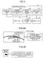
- FIGS. 4A and 4Bare schematic diagrams for explaining a marker-coordinate detecting unit according to the first embodiment
- FIGS. 5A and 5Bare schematic diagrams for explaining a correction-image creating unit according to the first embodiment
- FIG. 6is a schematic diagram for explaining an image post-processing unit according to the first embodiment
- FIGS. 7A and 7Bare schematic diagrams for explaining display modes
- FIG. 8is a flowchart for explaining processing performed by the X-ray diagnosis apparatus according to the first embodiment
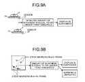
- FIGS. 9A and 9Bare schematic diagrams for explaining a modification 1 of the first embodiment
- FIGS. 10A to 10Care schematic diagrams for explaining a modification 2 of the first embodiment
- FIGS. 11A and 11Bare schematic diagrams for explaining a modification 3 of the first embodiment
- FIG. 12is a schematic diagram for explaining a configuration of an image processing unit according to a second embodiment of the present invention.
- FIG. 13is a schematic diagram for explaining X-ray images according to the second embodiment
- FIGS. 14A and 14Bare schematic diagrams for explaining a marker-coordinate detecting unit according to the second embodiment
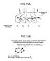
- FIGS. 15A and 15Bare schematic diagrams for explaining a cyclical trace-data acquiring unit
- FIG. 16is a schematic diagram for explaining new images according to the second embodiment.
- FIG. 17is a schematic diagram for explaining a correction-image creating unit according to the second embodiment.
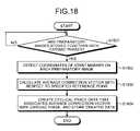
- FIG. 18is a flowchart for explaining cyclical trace-data creation processing performed by an X-ray diagnosis apparatus according to the second embodiment
- FIG. 19is a flowchart for explaining image processing with the use of cyclical trace data performed by the X-ray diagnosis apparatus according to the second embodiment
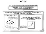
- FIGS. 20 and 21are schematic diagrams for explaining a third embodiment of the present invention.
- FIG. 22is a schematic diagram for explaining a conventional technology.
- FIG. 1is a schematic diagram for explaining a configuration of the X-ray diagnosis apparatus according to the first embodiment.
- the X-ray diagnosis apparatus 100includes a high-voltage generator 11 , an X-ray tube 12 , an X-ray beam-limiting device 13 , a top plate 14 , a C-arm 15 , an X-ray detector 16 , a C-arm turning-moving mechanism 17 , a top-plate moving mechanism 18 , a C-arm and top-plate mechanism control unit 19 , a diaphragm control unit 20 , a system control unit 21 , an input unit 22 , a display unit 23 , an image-data creating unit 24 , an image-data storage unit 25 , and an image processing unit 26 .
- the high-voltage generator 11is a device that generates a high voltage and supplies the generated high voltage to the X-ray tube 12 ; and the X-ray tube 12 is a device that generates X-rays by using a high voltage supplied by the high-voltage generator 11 .
- the high-voltage generator 11controls an adjustment in X-ray dosage to be radiated onto a subject P, and ON/OFF of X-ray radiation to the subject P, by regulating a voltage supplied to the X-ray tube 12 .
- the X-ray beam-limiting device 13is a device that limits an X-ray generated by the X-ray tube 12 so as to be radiated selectively onto a region of interest of the subject P.
- the X-ray beam-limiting device 13includes four slidable diaphragm blades, and causes an X-ray generated by the X-ray tube 12 to be limited and radiated onto the subject P by sliding the diaphragm blades.
- the top plate 14is a bed on which the subject P to be placed, and is arranged on a not-shown couch.
- the X-ray detector 16is a device in which X-ray detecting elements for detecting an X-ray passed through the subject P are arranged in a matrix, and each of the X-ray detecting elements converts an X-ray passed through the subject P into an electric signal and stores it, and transmits the stored electric signal to the image-data creating unit 24 , which will be described later.
- the C-arm 15is an arm that supports the X-ray tube 12 , the X-ray beam-limiting device 13 , and the X-ray detector 16 , so that “the X-ray tube 12 and the X-ray beam-limiting device 13 ” and the X-ray detector 16 are arranged with the C-arm 15 on opposite sides of the subject P.
- the C-arm turning-moving mechanism 17is a device that turns and moves the C-arm 15
- the top-plate moving mechanism 18is a device that turns and moves the top plate 14 .
- the C-arm and top-plate mechanism control unit 19performs turn control and movement control of the C-arm 15 and movement control of the top plate 14 by controlling the C-arm turning-moving mechanism 17 and the top-plate moving mechanism 18 , respectively.
- the beam-limit control unit 20controls a radiation area of X-rays by adjusting the aperture of the diaphragm blades included in the X-ray beam-limiting device 13 .
- the image-data creating unit 24creates an X-ray image by using an electric signal converted by the X-ray detector 16 from an X-ray passed through the subject P, and stores the created X-ray image into the image-data storage unit 25 . Specifically, the image-data creating unit 24 creates an X-ray image by performing a current-voltage conversion, an analog-to-digital (A/D) conversion, and a parallel-serial conversion, on an electric signal received from the X-ray detector 16 .
- A/Danalog-to-digital
- the image-data storage unit 25stores an X-ray image created by the image-data creating unit 24 .
- the image processing unit 26is a processing unit that executes various image processing on an X-ray image stored by the image-data storage unit 25 , and will be explained later in detail.
- the input unit 22includes a mouse, a keyboard, a button, a trackball, a joystick, and the like, which are configured for an operator who operates the X-ray diagnosis apparatus 100 , such as a doctor or an engineer, to input various commands, and transfers a command received from the operator to the system control unit 21 .
- the display unit 23includes a monitor that displays a Graphical User Interface (GUI) for receiving a command from the operator via the input unit 22 , and displays an X-ray image stored in the image-data storage unit 25 , an X-ray image processed through image processing by the image processing unit 26 , and the like.
- GUIGraphical User Interface
- the display unit 23can include a plurality of monitors.
- the system control unit 21controls operations of the X-ray diagnosis apparatus 100 overall. Precisely, the system control unit 21 performs an adjustment in X-ray dosage, control of ON/OFF of X-ray radiation, turn and movement control of the C-arm 15 , and movement control of the top plate 14 , by controlling the high-voltage generator 11 , the C-arm and top-plate mechanism control unit 19 , and the diaphragm control unit 20 based on a command from the operator transferred from the input unit 22 .
- system control unit 21controls image creating processing to be performed by the image-data creating unit 24 , and image processing to be performed by the image processing unit 26 , which will be described later, based on a command from the operator. Furthermore, the system control unit 21 performs control of displaying a GUI for receiving a command from the operator, an X-ray image stored by the image-data storage unit 25 , an X-ray image processed through image processing performed by the image processing unit 26 , and the like, onto the monitor of the display unit 23 .
- the X-ray diagnosis apparatus 100executes fluroscopic imaging of an X-ray image of the stenosed portion as a region of interest in which a stent is to be inserted, along a time sequence, based on a command from the operator.
- the present inventioncan be applied to a case where one piece of X-ray impermeable metal is attached as a stent marker to the center of the balloon of the stent.
- the X-ray diagnosis apparatus 100radiates an X-ray from the X-ray tube 12 onto a stenosed portion of the subject P on which vascular intervention treatment is performed, detects the X-ray passed through the subject P with the X-ray detector 16 , thereby storing X-ray images that are sequentially created along a time sequence into the image-data storage unit 25 .
- FIG. 2is a schematic diagram for explaining the image-data storage unit according to the first embodiment.
- FIG. 3is a schematic diagram for explaining a configuration of the image processing unit according to the first embodiment
- FIGS. 4A and 4Bare schematic diagrams for explaining a marker-coordinate detecting unit
- FIGS. 5A and 5Bare schematic diagrams for explaining a correction-image creating unit according to the first embodiment
- FIG. 6is a schematic diagram for explaining an image post-processing unit
- FIGS. 7A and 7Bare schematic diagrams for explaining display modes.
- the image processing unit 26includes a marker-coordinate detecting unit 26 a , a correction-image creating unit 26 b , and an image post-processing unit 26 c.
- the marker-coordinate detecting unit 26 adetects coordinates of stent markers attached to a stent on the new image.
- the system control unit 21performs control of displaying an X-ray image that is created at first and stored in the image-data storage unit 25 (a first frame), onto the monitor of the display unit 23 .
- a doctor who refers to the first framespecifies two stent markers in the first frame via the input unit 22 as shown in FIG. 4A . Accordingly, the marker-coordinate detecting unit 26 a detects respective coordinates of the two stent markers in the first frame.
- the marker-coordinate detecting unit 26 asets Regions Of Interest (ROIs) to rectangles in each of which the coordinates of each of the two stent markers specified in the first frame is centered; extracts a pattern similar to each pattern in each of the set ROIs through a cross correlation method from each of new images that are sequentially created; and then detects coordinates representing the highest cross correlation value as the coordinates of each of the stent markers.
- ROIsRegions Of Interest
- FIG. 4Ais explained above in a case where the doctor sets stent markers at two points, the present invention is not limited to this, and can be in a case where a doctor specifies a stent marker at one point.
- the marker-coordinate detecting unit 26 aexecutes the cross correlation method also in the first frame by using an ROI set from the coordinates of the specified stent marker, and detects coordinates of the other stent marker.
- the marker-coordinate detecting unit 26 adetects coordinates of the stent markers by using a teaching image that indicates characteristics of a stent marker attached to a stent used in treatment in practice, for example, the shape and the brightness of a stent marker observed on an X-ray image.
- an X-ray image of a stent markeris preliminarily and separately stored as a teaching image, and the marker-coordinate detecting unit 26 a extracts a pattern similar to the teaching image from each new image, and then detects coordinates of a stent marker by searching for the most similar region from among extracted candidate regions for the stent marker.
- the correction-image creating unit 26 bcreates a correction image from each new image in a second frame and later through image shift processing, such as parallel translation and/or turn movement, and/or image transformation processing, such as affine transformation, so as to match up the coordinates of the stent markers detected on each new image in the second frame and later by the marker-coordinate detecting unit 26 a , with reference coordinates that are coordinates of the stent markers already detected by the marker-coordinate detecting unit 26 a in the first frame that is an X-ray image created at first.
- image shift processingsuch as parallel translation and/or turn movement
- image transformation processingsuch as affine transformation
- the correction-image creating unit 26 bcreates a correction image 2 from the second frame through image transformation, so as to match up coordinates of the stent markers detected on an X-ray image in the second frame created as a new image, with coordinates of the stent markers already detected in the first frame (reference position).
- the correction-image creating unit 26 bthen creates a correction image from each new image in a third frame and later with reference to coordinates of the stent markers on each correction image created by itself from the previous X-ray image created immediately before the new image as the reference coordinates. For example, as shown in FIG. 5B , the correction-image creating unit 26 b creates a correction image 3 from the third frame through image transformation so as to match up coordinates of the stent markers detected in the third frame with the coordinates of the stent markers on the correction image 2 created from the second frame.
- the present inventionis not limited to this, and can be in a case where the reference coordinates are fixed to coordinates of the stent markers detected in the first frame, and then each correction image is created from each new image in the second frame and later.
- a correction imageis to be used for creating an image for display to be used when displaying a moving image
- the image post-processing unit 26 cperforms post-processing on a correction image created by the correction-image creating unit 26 b . Specifically, as shown in FIG. 6 , the image post-processing unit 26 c creates a filtered correction image by executing high-frequency noise reduction filtering-processing and low-frequency component removal filtering-processing on a correction image of which the positions of the stent markers match up with those of the first frame, and creates a logarithmic image by further calculating logarithmic values of the natural logarithm base from respective pixel values of pixels included in the filtered correction image. The image post-processing unit 26 c also executes the post-processing described above on the first frame.
- the image post-processing unit 26 cexecutes high-frequency noise reduction filtering-processing with the use of a spatial filter, for example, described in Nambu K, Iseki H., “A noise reduction method based on a statistical test of high dimensional pixel vectors for dynamic and volumetric images”, Riv Neuroradiol 2005, 18, 21-33, and Nishiki, “Method for reducing noise in X-ray images by averaging pixels based on the normalized difference with the relevant pixel”, Radiological Physics and Technology, Vol 2, 2008.
- the spatial filteris high-frequency noise reduction filtering-processing of performing smoothing processing within a single frame by measuring a difference value between pixel values in frames of different time axes, and changing weighting in accordance with an extent of the difference value, and can reduce a high frequency noise without influence on the other frames.
- a correction imagecan be processed through a strong spatial filter because the coordinates of the stent markers match up, thereby reducing a high frequency noise on a stent portion and improving visibility of the stent on the correction image.
- the image post-processing unit 26 ccan execute high-frequency noise reduction filtering-processing, for example, with the use of a recursive filter.
- a recursive filteris a filter that reduces a high frequency noise by adding pixel values of pixels included in a past frame on which a certain weighting is performed, to pixel values of pixels included in a frame to be processed. Because the coordinates of the stent markers match up on a correction image, a high frequency noise on a stent portion can be reduced through the recursive filter that uses a past frame for processing, so that visibility of the stent on the correction image can be improved.
- the image post-processing unit 26 cperforms low-frequency component removal filtering-processing by using a high-pass filter. Accordingly, a difference in the contrast can be reduced in the background area other than the stent portion on a correction image.
- the image post-processing unit 26 ccan make signal components in the whole image to a certain level by executing logarithmic-image creating processing on the filtered correction image.
- the system control unit 21performs control of displaying sequentially each of newly created logarithmic images as an image for display, onto the monitor of the display unit 23 , each time when the image post-processing unit 26 c newly creates a logarithmic image along a time sequence.
- the system control unit 21performs control of displaying a moving image of images for display on which the coordinates of the stent markers match up. Accordingly, even though the background area other than the stent is blurred on the image, the X-ray images can be displayed as a moving image on which the stent portion is stationary.
- the system control unit 21displays images for display in various modes in accordance with a display-mode instruction command received from the operator via the input unit 22 .
- the system control unit 21performs control of displaying a set region that is set based on coordinates of the stent markers on the logarithmic image, as an image for display, in accordance with a display-mode instruction command.
- the system control unit 21controls display such that when coordinates of the two stent markers on a logarithmic image are (X 1 , Y 1 ) and (X 2 , Y 2 ), respectively; a set region is set to a rectangle of which the center is (X 1 +X 2 )/2, (Y 1 +Y 2 )/2), the width is “2 ⁇
- system control unit 21performs control of displaying an enlarged image enlarged from the set region as an image for display.
- the system control unit 21controls the positions of the stent markers on the images for display so as to be centered on the monitor of the display unit 23 .
- the system control unit 21performs control of displaying an image for display in parallel with an original image of the image for display, in accordance with a display-mode instruction command. Furthermore, when displaying them in parallel, if the set region or the enlarged image is the image for display, the system control unit 21 performs control of displaying a region corresponding to the set region on the original image.
- the system control unit 21performs control of displaying a moving image of the set region in parallel with a moving image of the original image added with a frame corresponding to the set region, onto the monitor of the display unit 23 .
- the system control unit 21performs control of displaying a moving image of the enlarged image in parallel with a moving image of the original image added with a frame corresponding to the set region, onto the monitor of the display unit 23 .
- the stent strutis more clearly displayed on the set region and the enlarge image, compared with the original image, and a difference in the contrast of the background area is reduced, so that visibility of the whole stent is improved.
- the frame displayed on the original imagemoves along with a movement of the positions of the stent markers.
- the first embodimentis explained above in a case where an image for display is a logarithmic image, a set region, or an enlarged image.
- the present inventionis not limited to this, and can be in a case where an image for display is a correction image itself, or an image on which post-processing is performed through processing of an arbitrary combination set by the operator from among high-frequency noise reduction filtering-processing, low-frequency component removal filtering-processing, and logarithmic-image creating processing.
- FIG. 8is a flowchart for explaining processing performed by the X-ray diagnosis apparatus according to the first embodiment.
- the X-ray diagnosis apparatus 100starts fluroscopic imaging of X-ray image to a stenosed portion of the subject P into which the stent is inserted, and the image-data storage unit 25 stores the first X-ray image (the first frame) (Yes at Step S 801 ), the marker-coordinate detecting unit 26 a detects coordinates of the stent markers in the first frame (Step S 802 ).
- the image post-processing unit 26 cthen creates an image for display by performing post-processing on the first X-ray image (the first frame) (Step S 803 ), and the system control unit 21 performs control of displaying an enlarged image of a set region set in the image for display together with an original image (Step S 804 ).
- the marker-coordinate detecting unit 26 adetects coordinates of the stent markers on the new image (Step S 806 ).
- the correction-image creating unit 26 bcreates a correction image from the new image through image transformation, so as to match up the detected coordinates on the new image with reference coordinates that are the coordinates of the stent markers already detected in the first frame by the marker-coordinate detecting unit 26 a (Step S 807 ).
- the image post-processing unit 26 ccreates an image for display through post-processing that includes high-frequency noise reduction filtering-processing, low-frequency component removal filtering-processing, and logarithmic-image creating processing, onto the correction image created by the correction-image creating unit 26 b (Step S 808 ).
- the system control unit 21then performs control of displaying an enlarged image of a set region set in the image for display together with an original image (Step S 809 ).
- the system control unit 21determines whether a display termination request is input from the operator via the input unit 22 (Step S 810 ).
- Step S 810If the display termination request is not input (No at Step S 810 ), the system control unit 21 goes back to Step S 805 , and controls the marker-coordinate detecting unit 26 a so as to detect coordinates of the stent markers as soon as a new image is stored.
- the marker-coordinate detecting unit 26 adetects coordinates of the stent markers on the new image
- the correction-image creating unit 26 bcreates a correction image from the new image through image transformation so as to match up the coordinates of the stent markers on the new image with the reference coordinates that are coordinates of the stent markers already detected in the first frame by the marker-coordinate detecting unit 26 a.
- the image post-processing unit 26 cthen creates an image for display by performing post-processing on the correction image created by the correction-image creating unit 26 b , through the post-processing including high-frequency noise reduction filtering-processing, low-frequency component removal filtering-processing, and logarithmic-image creating processing; and then the system control unit 21 performs control of displaying an enlarged image of a set region that is set in the image for display together with an original image.
- the first embodimenteven though the background area other than the stent portion slightly moves, a moving image of X-ray images on which the stent portion is stationary can be displayed, and an X-ray image that ensures visibility of the stent can be instantly displayed at the time of execution of vascular intervention treatment performed with reference to an X-ray image, as described above as a main feature. Moreover, according to the first embodiment, because the stent portion is stationary on the X-ray images displayed as a moving image, the doctor can easily grasp a process in which the stent strut is extended.
- a blood vessel in which the stent is insertedis also stationary on the X-ray images displayed as a moving image; accordingly, when a treatment is performed under a condition that the subject P is given with a contrast agent, the doctor can easily grasp the state of a blood flow in the blood vessel in which the stent is inserted.
- FIGS. 9A and 9Bare schematic diagrams for explaining a modification 1 of the first embodiment
- FIGS. 10A to 10Care schematic diagrams for explaining a modification 2 of the first embodiment
- FIGS. 11A and 11Bare schematic diagrams for explaining a modification 3 of the first embodiment.
- a sensor 27 for detecting a movement of the top plate 14is attached to the top plate 14 on which the subject P lies, so that the system control unit 21 performs control of suspending display of an image for display during a period in which a movement (the amount of movement) of the top late 14 (i.e., the couch on which the top plate 14 is arranged) detected by the sensor 27 is equal to or larger than a threshold.
- the system control unit 21when the amount of movement of coordinates of the stent markers in a k+1th frame currently detected by the marker-coordinate detecting unit 26 a from already-detected coordinates of the stent markers in a k-th frame is equal to or larger than a threshold, the system control unit 21 performs control of suspending display of an image for display.
- the system control unit 21can perform control of displaying onto the monitor a warning indicating that the amount of movement of the stent markers between frames is equal to or larger than a threshold.
- the system control unit 21stops correction-image creating processing, and continuously displays an image for display created from the previous X-ray image (the k-the frame).
- the system control unit 21changes imaging conditions by reducing the width of an X-ray radiation pulse, and further controlling the high-voltage generator 11 so as to increase a tube current to be supplied to the X-ray tube 12 , as shown in FIG. 10B .
- the system control unit 21performs control of displaying an imaging-condition change notice for advising changing imaging conditions so as to reduce the width of an X-ray radiation pulse, and to increase a tube current to be supplied to the X-ray tube 12 .
- an “X-ray dosage”is expressed by ‘“width of X-ray radiation pulse” ⁇ “tube current” ⁇ “X-ray radiation interval”’; movement blurring of the subject P on an image is reduced while maintaining the same X-ray dosage, by not changing “X-ray radiation interval (frame rate)”, reducing “width of X-ray radiation pulse”, and increasing “tube current” for compensating the reduced “width of X-ray radiation pulse”. Accordingly, when it is difficult to detect the stent markers due to movement blurring, the operation can be recovered to a state in which an image for display (correction image) can be created, by increasing the sensitivity for detecting the stent markers while maintaining the same X-ray dosage radiated at certain intervals.
- a target of the control of changing imaging conditions or the control of displaying an imaging-condition change noticecan be selected only one from among reduction in the width of X-ray radiation pulse and increase in the tube current.
- an execution of the control of changing imaging conditions or the display control of an imaging-condition change noticecan be limited to a case where the stent markers are not extracted, and an X-ray dosage during fluroscopic imaging is not higher than a safety level.
- the system control unit 21stops continuous display of the image for display created from the k-th frame.
- the system control unit 21performs control of displaying, for example, only an original image as a moving image.
- the doctorcan be notified that X-ray image imaging conditions are inappropriate, or of a possibility that the subject P has heart beat fluctuations.
- the marker-coordinate detecting unit 26 acontinues extraction processing of the stent markers on a new image, and can resume display of a new image for display when extraction processing of the stent markers is continuously succeeded again.
- the system control unit 21can perform control of displaying a warning message onto the monitor of the display unit 23 .
- the system control unit 21causes display of a failure mark indicating a failure on a display position of the image for display created from the previous frame. Accordingly, for example, a doctor can recognize a low degree of reliability of a displayed image when failure marks are displayed in succession.
- a failure markis desirably displayed in a quiet color in an unnoticeable position.
- a style of a warning messagecan be a display style of indicating the number of frames in successive failures in stent-marker extraction, or a display style of gradually changing the color from blue to red along with increase in the number of successive failures in stent-marker extraction.
- a kind of a progress barcan be used as a warning message.
- a plurality of stentsis sometimes inserted simultaneously in some cases.
- the system control unit 21performs the following control explained below based on a distance between the two stents.
- the distance between the stentscan be calculated by the marker-coordinate detecting unit 26 a by using coordinates specified via the input unit 22 by a doctor who refers to the first frame (original image), or can be calculated by the marker-coordinate detecting unit 26 a by using coordinates of the stent markers detected in the first frame by using a teaching image.
- the system control unit 21controls the correction-image creating unit 26 b to create a correction image through image transformation so as to match up respective coordinates of stent markers originating in each of the stents with corresponding reference positions, respectively.
- the correction-image creating unit 26 bperforms image transformation so as to match up coordinates of the two stent markers detected on a new image with the reference coordinates (X 1 , Y 1 ) and (X 2 , Y 2 ) of the respective two stents, as shown in the upper part of FIG. 11A .
- the correction-image creating unit 26 bWhen using a stent to which two stent markers are attached to the both ends of the balloon, under the control of the system control unit 21 , the correction-image creating unit 26 b performs image transformation so as to match up coordinates of the four stent markers detected on a new image with the reference coordinates (X 1 , Y 1 ) and (X 2 , Y 2 ), and (X 3 , Y 3 ) and (X 4 , Y 4 ) of the respective two stents, as shown in the lower part of FIG. 11A .
- the system control unit 21controls the image processing unit 26 such that two stents (a stent 1 and a stent 2 ) are individually processed.
- the system control unit 21controls the image processing unit 26 so as to create two images for display in order to execute display of a moving image of images for display on which the position of the stent 1 matches up, and display of a moving image of images for display on which the position of the stent 2 matches up, in two sub-windows on the monitor.
- a region-of-interest specifying screen for specifying one of the two stents as a region of interestis displayed, and when one of the stents is specified as a region of interest via the input unit 22 , the system control unit 21 controls the image processing unit 26 so as to perform processing only on the specified stent, as shown in FIG. 11B .
- the system control unit 21controls the image processing unit 26 so as to create only an image for display on which the position of the stent 1 that is specified matches up.
- an optimal image for displaycan be displayed as a moving image in accordance with a distance between the stents.
- FIG. 12is a schematic diagram for explaining a configuration of the image processing unit according to the second embodiment.
- the image processing unit 26further includes a cardiographic-information acquiring unit 26 f , a cyclical trace-data acquiring unit 26 d , and a cyclical trace-data storage unit 26 e , which are different from the image processing unit 26 according to the first embodiment shown in FIG. 3 .
- a cardiographic-information acquiring unit 26 fa cardiographic-information acquiring unit 26 f
- a cyclical trace-data acquiring unit 26 da cyclical trace-data storage unit 26 e
- Such differencesare mainly explained below.
- an electrocardiograph 28that acquires an electrocardiogram waveform is attached to the subject P.
- the cardiographic-information acquiring unit 26 facquires an electrocardiogram waveform of the subject P inserted with a stent from the electrocardiograph 28 .
- the cardiographic-information acquiring unit 26 fcan transfer the electrocardiogram waveform acquired from the electrocardiograph 28 to each of the image-data storage unit 25 and the cyclical trace-data acquiring unit 26 d.
- the X-ray diagnosis apparatus 100creates X-ray images along a time sequence by radiating X-rays from the X-ray tube 12 , and detecting X-rays passed through the subject P with the X-ray detector 16 similarly to the first embodiment, and further acquires a cardiac phase of the subject P at the time of creation of each X-ray image, as the cardiographic-information acquiring unit 26 f acquires an electrocardiogram waveform from the electrocardiograph 28 attached to the subject P.
- FIG. 13is a schematic diagram for explaining X-ray images according to the second embodiment.
- the X-ray diagnosis apparatus 100performs preliminarily imaging over a predetermined period (for example, a period of three hear beats) from the start of display processing of an image for display. Accordingly, the image-data storage unit 25 stores X-ray images in a period of three heart beats added with information about cardiac phases as preparatory images. Preparatory images are images to collect the cyclical trace-data described later, and the images displayed for the diagnosis can be used as preparatory images. Moreover, the images of the imaging performed before this imaging can be used as preparatory images.
- the marker-coordinate detecting unit 26 aacquires coordinates of the stent markers on each preparatory image.
- FIGS. 14A and 14Bare schematic diagrams for explaining the marker-coordinate detecting unit according to the second embodiment.
- the system control unit 21performs control of displaying, for example, a plurality of preparatory images along a time sequence equivalent to one heart beat, onto the monitor of the display unit 23 .
- the system control unit 21causes display such that the operator can grasp in which position (cardiac phase) on an electrocardiogram waveform each of the preparatory images is created.
- the operatorthen specifies a marker at one point on a preparatory image, for example, in an ending period of a systole, among preparatory images displayed on the monitor, and further specifies a corresponding marker at one point on a preparatory image in an ending period of a diastole.
- a preparatory image at a time of 30% of an R wave interval (30% RR interval)is specified as a preparatory image in an ending period of systole
- a preparatory image at a time of 70% of an R wave interval (70% RR interval)is specified as a preparatory image in an ending period of diastole.
- the marker-coordinate detecting unit 26 adetects coordinates of the stent marker specified on the two preparatory images, and sets a rectangle in which coordinates of the specified stent marker are centered. As shown in FIG. 14B , the marker-coordinate detecting unit 26 a then extracts a pattern similar to a pattern in a rectangle set on another preparatory image, for example, through the cross correlation method, and detects coordinates having the highest correlation value as the coordinates of the stent marker.
- the system control unit 21can perform control of displaying a result of the processing performed on preparatory images by the marker-coordinate detecting unit 26 a onto the monitor, and then the operator can correct the detected coordinates of the stent marker via the mouse of the input unit 22 .
- the marker-coordinate detecting unit 26 acan execute processing by using a teaching image.
- the marker-coordinate detecting processingcan be repeatedly executed on preparatory images in each period of one heart beat, or executed at once on preparatory images in each period of three heart beats.
- the cyclical trace-data acquiring unit 26 d shown in FIG. 12acquires cyclical trace data of the stent marker along a time sequence, based on the coordinates of the stent marker detected by the marker-coordinate detecting unit 26 a on each of the preparatory images, and a cardiac phase at creation of each of the preparatory images.
- the cyclical trace-data acquiring unit 26 dcalculates a difference between coordinates of the stent marker detected on each of the preparatory images by the marker-coordinate detecting unit 26 a and the reference point on “the preparatory image at 70% RR interval”, as a correction vector.
- the cyclical trace-data acquiring unit 26 dcalculates an average correction vector in each cardiac phase from the correction vectors calculated on all of the preparatory images in a period of three heart beats.
- the cyclical trace-data acquiring unit 26 dcreates cyclical trace data that associates an average correction vector with a cardiac phase by calculating an average correction vector (vector C M ) of “cardiac phase: M % RR interval” with respect to the reference point of “cardiac phase: 70% RR interval”.
- the cyclical trace-data storage unit 26 estores cyclical trace data created by the cyclical trace-data acquiring unit 26 d.
- the present inventionis not limited to this, and can be in a case where the marker-coordinate detecting unit 26 a detects only coordinates of a stent marker in the lower side of a preparatory image, or a case where the marker-coordinate detecting unit 26 a detects coordinates of two stent markers.
- the X-ray diagnosis apparatus 100executes fluroscopic imaging of a new image to be subjected to image processing, in accordance with an instruction by the operator.
- the image-data storage unit 25sequentially stores the new image to be subjected to image processing together with a cardiac phase estimated from an electrocardiogram waveform.
- FIG. 16is a schematic diagram for explaining new images according to the second embodiment.
- the correction-image creating unit 26 beach time when a new image is created along a time sequence, creates a correction image from the new image based on cyclical trace data stored by the cyclical trace-data storage unit 26 e and a cardiac phase at the time of creation of the new image.
- the correction-image creating unit 26 bwhen a new image is stored, acquires a stored average correction vector corresponding to a cardiac phase at the time of creation of the new image from the cyclical trace data, and creates a correction image by using the acquired average vector.
- FIG. 18is a flowchart for explaining cyclical trace-data creation processing performed by the X-ray diagnosis apparatus according to the second embodiment.
- the marker-coordinate detecting unit 26 adetects coordinates of the stent marker on each preparatory image (Step S 1802 , see FIGS. 14A and 14B ).
- the cyclical trace-data acquiring unit 26 dthen calculates an average correction vector at each coordinate of the stent marker detected on the preparatory images by the marker-coordinate detecting unit 26 a with respect to a reference point specified by the operator (Step S 1803 ), and creates cyclical trace data that associates an average correction vector with a cardiac phase; and the cyclical trace-data storage unit 26 e stores the created data (Step S 1804 ), then the processing is terminated.
- FIG. 19is a flowchart for explaining image processing with the use of cyclical trace data performed by the X-ray diagnosis apparatus according to the second embodiment.
- the X-ray diagnosis apparatus 100executes fluroscopic imaging of an X-ray image onto a stenosed portion of the subject P inserted with a stent, and when the image-data storage unit 25 stores a new image to be subjected to image processing together with a cardiac phase (Yes at Step S 1091 ), the correction-image creating unit 26 b creates a correction image by acquiring a correction vector corresponding to a cardiac phase at the time of creation of the new image from cyclical trace data (Step S 1902 ).
- the image post-processing unit 26 ccreates an image for display through post-processing that includes high-frequency noise reduction filtering-processing, low-frequency component removal filtering-processing, and logarithmic-image creating processing, onto the correction image created by the correction-image creating unit 26 b (Step S 1903 ).
- the system control unit 21then performs control of displaying an enlarged image of a set region set in the image for display together with an original image (Step S 1904 ).
- the system control unit 21determines whether a display termination request is input from the operator via the input unit 22 (Step S 1905 ).
- Step S 1905If the display termination request is not input (No at Step S 1905 ), the system control unit 21 goes back to Step S 1901 , and controls the correction-image creating unit 26 b so as to cerate a correction image as soon as a new image is stored.
- a correction image and an image for displayare created from a new image that is sequentially created, without using the marker-coordinate detecting unit 26 a , accordingly, the load of processing on the image processing unit 26 can be reduced and a processing time can be reduced, and an X-ray image that ensures visibility of the stent can be more instantly displayed as a moving image.
- FIGS. 20 and 21are schematic diagrams for explaining an X-ray diagnosis apparatus according to the third embodiment.
- the X-ray diagnosis apparatus 100receives from the operator via the input unit 22 an instruction to execute one of the following two modes: namely, “a realtime marker-coordinate detection use mode” explained in the first embodiment, in which correction-image creating processing is executed by using coordinates of a stent marker on a new image detected by the marker-coordinate detecting unit 26 a ; and “a cyclical trace-data use mode” explained in the second embodiment, in which correction-image creating processing is executed by using cyclical trace data.
- the system control unit 21While executing the “realtime marker-coordinate detection use mode”, if the marker-coordinate detecting unit 26 a does not detect the stent marker on a new image, the system control unit 21 according to the third embodiment controls the correction-image creating unit 26 b so as to create a correction image by switching the mode to the “cyclical trace-data use mode”.
- the correction-image creating unit 26 bacquires a correction vector “vector C M ” of the “cardiac phase: M % RR interval” with respect to the reference point (70% RR interval) from cyclical trace data.
- the correction-image creating unit 26 bestimates coordinates of the stent marker in the k-th frame from the coordinates of the stent marker already detected by the marker-coordinate detecting unit 26 a on a new image created at 70% RR interval while executing the “realtime marker-coordinate detection use mode”, and creates a correction image.
- the system control unit 21controls such that cyclical trace data is corrected and renewed by using the function of the marker-coordinate detecting unit 26 a in the “realtime marker-coordinate detection use mode” even while executing the “cyclical trace-data use mode.
- the marker-coordinate detecting unit 26 adetects coordinates of the stent marker on each of selected images that are selected at certain intervals (for example, once every five frames) from among sequentially created new images.
- the cyclical trace-data acquiring unit 26 dthen corrects and renews cyclical trace data stored by the cyclical trace-data storage unit 26 e based on the coordinates of the stent marker on each of the selected images detected by the marker-coordinate detecting unit 26 a , and a cardiac phase at the time of creation of each of the selected images.
- the cyclical trace-data acquiring unit 26 dcorrects and renews cyclical trace data once every 100 milliseconds by the control of the system control unit 21 .
- the correction-image creating unit 26 bthen executes correction-image creating processing by using the renewed cyclical trace data stored by the cyclical trace-data storage unit 26 e by the control of the system control unit 21 .
- correction-image creating processingcan be executed by using cyclical trace data, so that X-ray images of high visibility of the stent can be displayed as a moving image without interruption.
- cyclical trace datacan be corrected and renewed with detected marker coordinates, so that visibility of the stent on an X-ray image displayed by an image display method with high immediacy can be further ensured.
- the present functionWhether or not to activate the function of displaying a moving image of X-ray images on which the stent is stationary (hereinafter, “the present function”) explained above in the first to third embodiments can be determined by an operator of the X-ray diagnosis apparatus 100 (a doctor or an engineer).
- a button for determining ON/OFF of the present functionis provided in the input unit 22 or in the vicinity of the couch, a moving image of X-ray images on which the stent is stationary can be displayed only when such display is desired by a doctor who performs a treatment.
- the system control unit 21can control processing explained below. Precisely, the system control unit 21 reduces a radiation rate (for example, a pulse rate or a frame rate) of X-ray radiated from the X-ray tube 12 at the start of operation of the present function, and then after the operation of the present function is terminated, the system control unit 21 turns back the X-ray radiation rate to the previous rate.
- a radiation ratefor example, a pulse rate or a frame rate
- the frame rateis 15 to 30 frames/sec, so that when the present function is activated, the frame rate is turned to, for example, a half of it.
- the system control unit 21increases or decreases the X-ray radiation rate from the rate at the start of operation, in accordance with a result of stent marker detection. Specifically, when detection of the stent markers is continuously failed a certain number of times, the system control unit 21 increases again the X-ray radiation rate. By contrast, when detection of the stent markers is continuously succeeded a certain number of times, the system control unit 21 decreases the X-ray radiation rate. According to such processing, an exposure to X-ray can be reduced.
- the correction-image creating unit 26 bfurther performs a turn correction such that the direction of the stent rendered on a correction image is to be in the horizontal direction or the vertical direction, based on positional information about the two stent markers. Accordingly, the doctor can refer to an image on which the direction of the stent is constantly in the horizontal direction or the vertical direction, thereby more easily recognizing, for example, the state of stent expansion.
- a doctorwhen performing vascular intervention treatment, a doctor often performs fluroscopic imaging intermittently in many cases. For example, the doctor performs fluroscopic imaging for 30 seconds, then suspends fluroscopic imaging, and resumes fluroscopic imaging after 30 seconds. For example, it is assumed that the first fluroscopic imaging for the first 30 seconds is “A”, while the next fluroscopic imaging is “B”, and “A” and “B” are independently performed without correlation.
- the correction-image creating unit 26 bperforms correction-image creating processing such that displayed angles of the stent become the same angle, by using information acquired during the processing of “A” while performing the processing of “B”. Specifically, the correction-image creating unit 26 b performs image transformation on X-ray images created through the fluroscopic imaging of “B” so as to match up coordinates of the stent markers on such images with those extracted on X-ray images created through the fluroscopic imaging of “A”. Accordingly, for example, when fluroscopic imaging is resumed within one minute, the doctor can observe the stent in the same angle, thereby continuing manual operation without uncomfortable feeling. Such function can be turned ON/OFF, and a certain time (for example, one minute) can be set by a user.
- the “function of displaying a moving image of X-ray images on which the stent is stationary”that is explained above in the first to third embodiments can be used for processing to be executed simultaneously with X-ray radiation in real time, or can be used for processing to be executed on X-ray images that were created in the past along a time sequence.
- the first to third embodimentsare explained in a case of performing vascular intervention treatment as a treatment performed with reference to an X-ray image, and using a stent as a treatment instrument; however, the present invention is not limited to this, and can be applied to various treatment instruments used for various treatments to be executed with reference to an X-ray image.
- the present inventioncan be applied to a treatment with the use of any of the following treatment equipment by using it as a marker, for example, an electrode of an electrophysiological catheter used for treatment for arrhythmia, a drill of rotablator used for performing treatment on a hard stenosed portion that is difficult for a balloon or a stent to expand, a metal cylinder with holes configured to be attached on a tip end of a catheter and to be used for directional coronary arterectomy, or a catheter with an ultrasound-wave transmitting-receiving function for checking a situation inside a blood vessel of a stenosed portion.
- a markerfor example, an electrode of an electrophysiological catheter used for treatment for arrhythmia, a drill of rotablator used for performing treatment on a hard stenosed portion that is difficult for a balloon or a stent to expand, a metal cylinder with holes configured to be attached on a tip end of a catheter and to be used for directional coronary arterectomy, or a catheter with
- an angioscopevascular ultrasound, vascular Magnetic Resonance Imaging (MRI), Optical Coherence Tomography (OCT), a device for engrafting a stem cell used in a tissue-engineering field, an artificial valve, and a vascular graft
- MRIMagnetic Resonance Imaging
- OCTOptical Coherence Tomography
- the present inventioncan be applied to various clinical practices, for example, a hybrid treatment of surgical and internal treatments, and a guidance of needling for biopsy in a surgical treatment.
Landscapes
- Engineering & Computer Science (AREA)
- Health & Medical Sciences (AREA)
- Life Sciences & Earth Sciences (AREA)
- Medical Informatics (AREA)
- Physics & Mathematics (AREA)
- General Health & Medical Sciences (AREA)
- Radiology & Medical Imaging (AREA)
- Nuclear Medicine, Radiotherapy & Molecular Imaging (AREA)
- Heart & Thoracic Surgery (AREA)
- Veterinary Medicine (AREA)
- Optics & Photonics (AREA)
- Pathology (AREA)
- Biophysics (AREA)
- Biomedical Technology (AREA)
- High Energy & Nuclear Physics (AREA)
- Molecular Biology (AREA)
- Surgery (AREA)
- Animal Behavior & Ethology (AREA)
- Public Health (AREA)
- Computer Vision & Pattern Recognition (AREA)
- Theoretical Computer Science (AREA)
- General Physics & Mathematics (AREA)
- Vascular Medicine (AREA)
- Dentistry (AREA)
- Oral & Maxillofacial Surgery (AREA)
- Multimedia (AREA)
- Human Computer Interaction (AREA)
- Quality & Reliability (AREA)
- Apparatus For Radiation Diagnosis (AREA)
Abstract
Description
- This application is based upon and claims the benefit of priority from the prior Japanese Patent Application No. 2008-275348, filed on Oct. 27, 2008; the entire contents of which are incorporated herein by reference.
- 1. Field of the Invention
- The present invention relates to an X-ray diagnosis apparatus and an image processing apparatus.
- 2. Description of the Related Art
- A treatment method called vascular intervention treatment has been conventionally performed on a stenosed portion occurring in a blood vessel caused by, for example, a thrombus.
- According to vascular intervention treatment, a balloon-tip catheter is inserted by a doctor up to a stenosed portion. A liquid is then injected into the balloon through the catheter, so that the balloon is expanded, as a result, the stenosed portion is mechanically expanded. After the liquid in the balloon is sucked, the balloon-tip catheter is withdrawn by a doctor to the outside of the body.
- To avoid re-stenosis of the stenosed portion that is expanded with the balloon, vascular intervention treatment with the use of a balloon-tip catheter tightly attached with a stent strut of metal mesh around the outer side of the balloon is also performed. According to such treatment method, after the stent strut is expanded along with expansion of the balloon, the balloon-tip catheter is withdrawn to the outside of the body by sucking the liquid in the balloon. Consequently, the expanded stent strut is retained in the stenosed portion, thereby reducing a re-coarctation rate in stenosed portion. A device that includes two parts, namely, a stent strut and a balloon-tip catheter, is called a “stent”.
- According to the vascular intervention treatment described above, an X-ray diagnosis apparatus performs fluroscopic imaging of a treatment target portion, and a doctor remotely executes a series of processing with the use of a balloon-tip catheter and a stent while referring to an X-ray image displayed on a monitor.
- The vascular intervention treatment requires precisely moving a balloon-tip catheter and a stent inserted in a blood vessel to a treatment target portion. Particularly when retaining a stent strut, it is required to position the stent precisely by millimeter. For this reason, the balloon part is attached with an X-ray impermeable metal at two points (or one point in some cases) as a marker that indicates the position of a balloon-tip catheter or a stent (stent marker), and a doctor performs treatment while confirming the position of the balloon-tip catheter or the stent by referring to a stent marker on a displayed X-ray image.
- However, when performing vascular intervention treatment on a blood vessel in an organ that is constantly throbbing, such as a heart, the position of a balloon-tip catheter and the position of a stent on an X-ray image constantly move, therefore, it is a very high-technique operation for a doctor to perform positioning by referring to an X-ray image.
- Although edge parts of a stet strut are important for a doctor to determine an extent of expansion of the stent strut, X-ray impermeability of a stent strut is very low compared with X-ray impermeability of a stent marker. For this reason, the edge parts of a stent strut is less clear than a stent marker.
- Therefore, as a technology for improving visibility of a stent on an X-ray image, a stent-highlighted display technology (for example, see JP 2005-510288 (KOKAI)) is proposed.
- According to the stent-highlighted display technology, a plurality of frames of X-ray images of a treatment target portion is taken along a time sequence, and correction is performed on the taken X-ray images so as to match up the position of a moving stent by using a stent marker as a reference. Processing, such as adding and averaging, is then performed on the X-ray images on which movement correction is performed, and then a highlighted image on which the stent strut is highlighted is created.
- Specifically, as shown in
FIG. 22 , correction processing is performed on a second frame so as to match up the positions of stent markers in the second frame with the positions of the stent markers in a first frame. Such correction processing is performed on a plurality of frames (for example, up to a 30th frame), and adding and averaging processing is performed on a plurality of images on which the positions of the stent markers match up. Accordingly, as shown inFIG. 22 , a highlighted image on which the stent strut is highlighted and the whole stent is clearly rendered is created, and the created highlighted image is displayed on a monitor.FIG. 22 is a schematic diagram for explaining the conventional technology. - The conventional technology described above has a problem that an X-ray image that ensures visibility of treatment equipment, such as a stent, cannot be instantly displayed at the time of execution of vascular intervention treatment performed with reference to an X-ray image.
- In other words, according to the stent-highlighted display technology described above, visibility of a stent can be improved; however, because tracking processing of stent marker, correction processing, and creating processing of highlighted image are performed as post-processing after a plurality of X-ray images is created along a time sequence, a waiting time (for example, tens seconds of waiting time) arises from imaging of an X-ray image until display of a highlighted image. Furthermore, because a highlighted image is created only one frame from a plurality of images (for example, 30 frames) on which correction processing is performed, temporal resolution of a displayed highlighted image is lower than temporal resolution of taken X-ray images.
- Similarly to the case of performing vascular intervention treatment, any case of treatment performed by a doctor referring to an X-ray image with the use of treatment equipment (for example, rotablator) attached with X-ray impermeable marker and arranged in a treatment portion that moves continuously due to throbs has a problem of incapability of instantly displaying an X-ray image that ensures visibility of the treatment equipment, even by using the conventional technology described above.
- For this reason, the present invention has been made to solve the problems of the conventional technology described above, and an object of the present invention is to provide an X-ray diagnosis apparatus and an image processing apparatus that can instantly display an X-ray image that ensures visibility of treatment equipment at the time of execution of a treatment performed with reference to an X-ray image.
- According to an aspect of the present invention, an X-ray diagnosis apparatus includes an image-data creating unit that creates X-ray images along a time sequence by detecting X-rays radiated from an X-ray tube and passed through a subject; a feature point detecting unit that detects a position of a feature point included in a certain object on a new image created by the image creating unit each time when the image creating unit creates new image as a new one of X-ray images along a time sequence; a correction-image creating unit that creates a correction image from the new image through at least one of image shift and image transformation, so as to match up the position of the feature point detected on the new image by the feature point detecting unit, with a reference position that is a position of a feature point already detected by the feature point detecting unit on a reference image that is a certain X-ray image created before the new image; and a display control unit that performs control of displaying newly created correction image as an image for display so as to be sequentially displayed as a moving image onto a certain display unit, each time when the correction-image creating unit newly creates the correction image along a time sequence.
- According to another aspect of the present invention, an X-ray diagnosis apparatus includes an image-data creating unit that creates X-ray images along a time sequence by detecting X-rays radiated from an X-ray tube and passed through a subject; a cardiographic-information acquiring unit that acquires an electrocardiogram waveform of the subject; a feature point detecting unit that detects a position of a feature point included in a certain object on each of a plurality of preparatory images that is a plurality of X-ray images preliminarily created by the image creating unit; a cyclical trace-information acquiring unit that acquires cyclical trace information about the feature point along a time sequence, based on respective positions of the feature point on the preparatory images detected by the feature point detecting unit, and cardiac phases at respective time points of creation of the preparatory images, the cardiac phases being estimated from an electrocardiogram waveform acquired by the cardiographic-information acquiring unit; a cyclical trace-information storage unit that stores the cyclical trace information acquired by the cyclical trace-information acquiring unit; a correction-image creating unit that creates a correction image from a new image created by the image creating unit through at least one of image shift and image transformation each time when the image creating unit creates new image as a new one of X-ray images along a time sequence, based on the cyclical trace information stored by the cyclical trace-information storage unit and a cardiac phase estimated from an electrocardiogram waveform at the time of creation of the new image acquired by the cardiographic-information acquiring unit; and a display control unit that performs control of displaying newly created correction image as an image for display so as to be sequentially displayed as a moving image onto a certain display unit, each time when the correction-image creating unit newly creates the correction image along a time sequence.
- According to still another aspect of the present invention, an image processing apparatus includes a feature point detecting unit that detects a position of a feature point included in a certain object on a new image each time when new image as a new one of X-ray images is created along a time sequence by detecting X-rays radiated from an X-ray tube and passed through a subject; a correction-image creating unit that creates a correction image from the new image through at least one of image shift and image transformation, so as to match up the position of the feature point detected on the new image by the feature point detecting unit, with a reference position that is a position of a feature point already detected by the feature point detecting unit on a reference image that is a certain X-ray image created before the new image; and a display control unit that performs control of displaying newly created correction image as an image for display so as to be sequentially displayed as a moving image onto a certain display unit, each time when the correction-image creating unit newly creates the correction image along a time sequence.
- According to still another aspect of the present invention, an image processing apparatus includes a cardiographic-information acquiring unit that acquires an electrocardiogram waveform of a subject; a feature point detecting unit that detects a position of a feature point included in a certain object on each of a plurality of preparatory images that is a plurality of X-ray images preliminarily created by detecting X-rays radiated from an X-ray tube and passed through the subject; a cyclical trace-information acquiring unit that acquires cyclical trace information about the feature point along a time sequence, based on respective positions of the feature point on the preparatory images detected by the feature point detecting unit, and cardiac phases at respective time points of creation of the preparatory images, the cardiac phases being estimated from an electrocardiogram waveform acquired by the cardiographic-information acquiring unit; a cyclical trace-information storage unit that stores the cyclical trace information acquired by the cyclical trace-information acquiring unit; a correction-image creating unit that creates a correction image from a new image through at least one of image shift and image transformation each time when new image as a new one of X-ray images is created along a time sequence, based on the cyclical trace information stored by the cyclical trace-information storage unit and a cardiac phase estimated from an electrocardiogram waveform at the time of creation of the new image acquired by the cardiographic-information acquiring unit; and a display control unit that performs control of displaying newly created correction image as an image for display so as to be sequentially displayed as a moving image onto a certain display unit, each time when the correction-image creating unit newly creates the correction image along a time sequence.
FIG. 1 is a schematic diagram for explaining a configuration of an X-ray diagnosis apparatus according to a first embodiment of the present invention;FIG. 2 is a schematic diagram for explaining an image-data storage unit according to the first embodiment;FIG. 3 is a schematic diagram for explaining a configuration of an image processing unit according to the first embodiment;FIGS. 4A and 4B are schematic diagrams for explaining a marker-coordinate detecting unit according to the first embodiment;FIGS. 5A and 5B are schematic diagrams for explaining a correction-image creating unit according to the first embodiment;FIG. 6 is a schematic diagram for explaining an image post-processing unit according to the first embodiment;FIGS. 7A and 7B are schematic diagrams for explaining display modes;FIG. 8 is a flowchart for explaining processing performed by the X-ray diagnosis apparatus according to the first embodiment;FIGS. 9A and 9B are schematic diagrams for explaining amodification 1 of the first embodiment;FIGS. 10A to 10C are schematic diagrams for explaining amodification 2 of the first embodiment;FIGS. 11A and 11B are schematic diagrams for explaining amodification 3 of the first embodiment;FIG. 12 is a schematic diagram for explaining a configuration of an image processing unit according to a second embodiment of the present invention;FIG. 13 is a schematic diagram for explaining X-ray images according to the second embodiment;FIGS. 14A and 14B are schematic diagrams for explaining a marker-coordinate detecting unit according to the second embodiment;FIGS. 15A and 15B are schematic diagrams for explaining a cyclical trace-data acquiring unit;FIG. 16 is a schematic diagram for explaining new images according to the second embodiment;FIG. 17 is a schematic diagram for explaining a correction-image creating unit according to the second embodiment;FIG. 18 is a flowchart for explaining cyclical trace-data creation processing performed by an X-ray diagnosis apparatus according to the second embodiment;FIG. 19 is a flowchart for explaining image processing with the use of cyclical trace data performed by the X-ray diagnosis apparatus according to the second embodiment;FIGS. 20 and 21 are schematic diagrams for explaining a third embodiment of the present invention; andFIG. 22 is a schematic diagram for explaining a conventional technology.- Exemplary embodiments of an X-ray diagnosis apparatus and an image processing apparatus according to the present invention will be explained below in detail with reference to the accompanying drawings. The following embodiments are explained in a case where the present invention is applied to an X-ray diagnosis apparatus.
- First of all, a configuration of an X-ray diagnosis apparatus according to a first embodiment of the present invention is explained below.
FIG. 1 is a schematic diagram for explaining a configuration of the X-ray diagnosis apparatus according to the first embodiment. - As shown in
FIG. 1 , theX-ray diagnosis apparatus 100 according to the first embodiment includes a high-voltage generator 11, anX-ray tube 12, an X-ray beam-limitingdevice 13, atop plate 14, a C-arm 15, anX-ray detector 16, a C-arm turning-movingmechanism 17, a top-plate moving mechanism 18, a C-arm and top-platemechanism control unit 19, adiaphragm control unit 20, asystem control unit 21, aninput unit 22, adisplay unit 23, an image-data creating unit 24, an image-data storage unit 25, and animage processing unit 26. - The high-
voltage generator 11 is a device that generates a high voltage and supplies the generated high voltage to theX-ray tube 12; and theX-ray tube 12 is a device that generates X-rays by using a high voltage supplied by the high-voltage generator 11. In other words, the high-voltage generator 11 controls an adjustment in X-ray dosage to be radiated onto a subject P, and ON/OFF of X-ray radiation to the subject P, by regulating a voltage supplied to theX-ray tube 12. - The X-ray beam-limiting
device 13 is a device that limits an X-ray generated by theX-ray tube 12 so as to be radiated selectively onto a region of interest of the subject P. For example, the X-ray beam-limitingdevice 13 includes four slidable diaphragm blades, and causes an X-ray generated by theX-ray tube 12 to be limited and radiated onto the subject P by sliding the diaphragm blades. - The
top plate 14 is a bed on which the subject P to be placed, and is arranged on a not-shown couch. - The
X-ray detector 16 is a device in which X-ray detecting elements for detecting an X-ray passed through the subject P are arranged in a matrix, and each of the X-ray detecting elements converts an X-ray passed through the subject P into an electric signal and stores it, and transmits the stored electric signal to the image-data creating unit 24, which will be described later. - The C-
arm 15 is an arm that supports theX-ray tube 12, the X-ray beam-limitingdevice 13, and theX-ray detector 16, so that “theX-ray tube 12 and the X-ray beam-limitingdevice 13” and theX-ray detector 16 are arranged with the C-arm 15 on opposite sides of the subject P. - The C-arm turning-moving
mechanism 17 is a device that turns and moves the C-arm 15, and the top-plate moving mechanism 18 is a device that turns and moves thetop plate 14. - The C-arm and top-plate
mechanism control unit 19 performs turn control and movement control of the C-arm 15 and movement control of thetop plate 14 by controlling the C-arm turning-movingmechanism 17 and the top-plate moving mechanism 18, respectively. - The beam-
limit control unit 20 controls a radiation area of X-rays by adjusting the aperture of the diaphragm blades included in the X-ray beam-limitingdevice 13. - The image-
data creating unit 24 creates an X-ray image by using an electric signal converted by theX-ray detector 16 from an X-ray passed through the subject P, and stores the created X-ray image into the image-data storage unit 25. Specifically, the image-data creating unit 24 creates an X-ray image by performing a current-voltage conversion, an analog-to-digital (A/D) conversion, and a parallel-serial conversion, on an electric signal received from theX-ray detector 16. - The image-
data storage unit 25 stores an X-ray image created by the image-data creating unit 24. - The
image processing unit 26 is a processing unit that executes various image processing on an X-ray image stored by the image-data storage unit 25, and will be explained later in detail. - The
input unit 22 includes a mouse, a keyboard, a button, a trackball, a joystick, and the like, which are configured for an operator who operates theX-ray diagnosis apparatus 100, such as a doctor or an engineer, to input various commands, and transfers a command received from the operator to thesystem control unit 21. - The
display unit 23 includes a monitor that displays a Graphical User Interface (GUI) for receiving a command from the operator via theinput unit 22, and displays an X-ray image stored in the image-data storage unit 25, an X-ray image processed through image processing by theimage processing unit 26, and the like. Thedisplay unit 23 can include a plurality of monitors. - The
system control unit 21 controls operations of theX-ray diagnosis apparatus 100 overall. Precisely, thesystem control unit 21 performs an adjustment in X-ray dosage, control of ON/OFF of X-ray radiation, turn and movement control of the C-arm 15, and movement control of thetop plate 14, by controlling the high-voltage generator 11, the C-arm and top-platemechanism control unit 19, and thediaphragm control unit 20 based on a command from the operator transferred from theinput unit 22. - Moreover, the
system control unit 21 controls image creating processing to be performed by the image-data creating unit 24, and image processing to be performed by theimage processing unit 26, which will be described later, based on a command from the operator. Furthermore, thesystem control unit 21 performs control of displaying a GUI for receiving a command from the operator, an X-ray image stored by the image-data storage unit 25, an X-ray image processed through image processing performed by theimage processing unit 26, and the like, onto the monitor of thedisplay unit 23. - When performing vascular intervention treatment with the use of a stent strut and a balloon-tip catheter onto a stenosed portion in a blood vessel of a heart of the subject P, the
X-ray diagnosis apparatus 100 according to the first embodiment executes fluroscopic imaging of an X-ray image of the stenosed portion as a region of interest in which a stent is to be inserted, along a time sequence, based on a command from the operator. According to the first embodiment, explained below is a case where two pieces of X-ray impermeable metal are attached as stent markers to the both ends of a balloon of the stent; however, the present invention can be applied to a case where one piece of X-ray impermeable metal is attached as a stent marker to the center of the balloon of the stent. - In other words, as shown in
FIG. 2 , theX-ray diagnosis apparatus 100 according to the first embodiment radiates an X-ray from theX-ray tube 12 onto a stenosed portion of the subject P on which vascular intervention treatment is performed, detects the X-ray passed through the subject P with theX-ray detector 16, thereby storing X-ray images that are sequentially created along a time sequence into the image-data storage unit 25.FIG. 2 is a schematic diagram for explaining the image-data storage unit according to the first embodiment. - A main feature in the
X-ray diagnosis apparatus 100 according to the first embodiment is that an X-ray image that ensures visibility of a stent can be instantly displayed at the time of execution of vascular intervention treatment performed with reference to an X-ray image, by executing processing by theimage processing unit 26, which is explained below in detail with reference toFIGS. 3 to 7 .FIG. 3 is a schematic diagram for explaining a configuration of the image processing unit according to the first embodiment;FIGS. 4A and 4B are schematic diagrams for explaining a marker-coordinate detecting unit;FIGS. 5A and 5B are schematic diagrams for explaining a correction-image creating unit according to the first embodiment;FIG. 6 is a schematic diagram for explaining an image post-processing unit; andFIGS. 7A and 7B are schematic diagrams for explaining display modes. - As shown in
FIG. 3 , theimage processing unit 26 includes a marker-coordinate detectingunit 26a, a correction-image creating unit 26b, and animage post-processing unit 26c. - Each time when a new image that is a new X-ray image is stored in the image-
data storage unit 25, the marker-coordinate detectingunit 26adetects coordinates of stent markers attached to a stent on the new image. - For example, as shown in
FIG. 4A , thesystem control unit 21 performs control of displaying an X-ray image that is created at first and stored in the image-data storage unit25 (a first frame), onto the monitor of thedisplay unit 23. - A doctor who refers to the first frame specifies two stent markers in the first frame via the
input unit 22 as shown inFIG. 4A . Accordingly, the marker-coordinate detectingunit 26adetects respective coordinates of the two stent markers in the first frame. - After that, as shown in
FIG. 4A , the marker-coordinate detectingunit 26asets Regions Of Interest (ROIs) to rectangles in each of which the coordinates of each of the two stent markers specified in the first frame is centered; extracts a pattern similar to each pattern in each of the set ROIs through a cross correlation method from each of new images that are sequentially created; and then detects coordinates representing the highest cross correlation value as the coordinates of each of the stent markers. - Although
FIG. 4A is explained above in a case where the doctor sets stent markers at two points, the present invention is not limited to this, and can be in a case where a doctor specifies a stent marker at one point. In such case, the marker-coordinate detectingunit 26aexecutes the cross correlation method also in the first frame by using an ROI set from the coordinates of the specified stent marker, and detects coordinates of the other stent marker. - Alternatively, the marker-coordinate detecting
unit 26adetects coordinates of the stent markers by using a teaching image that indicates characteristics of a stent marker attached to a stent used in treatment in practice, for example, the shape and the brightness of a stent marker observed on an X-ray image. - For example, as shown in
FIG. 4B , an X-ray image of a stent marker is preliminarily and separately stored as a teaching image, and the marker-coordinate detectingunit 26aextracts a pattern similar to the teaching image from each new image, and then detects coordinates of a stent marker by searching for the most similar region from among extracted candidate regions for the stent marker. - Returning to
FIG. 3 , the correction-image creating unit 26bcreates a correction image from each new image in a second frame and later through image shift processing, such as parallel translation and/or turn movement, and/or image transformation processing, such as affine transformation, so as to match up the coordinates of the stent markers detected on each new image in the second frame and later by the marker-coordinate detectingunit 26a, with reference coordinates that are coordinates of the stent markers already detected by the marker-coordinate detectingunit 26ain the first frame that is an X-ray image created at first. - For example, as shown in
FIG. 5A , the correction-image creating unit 26bcreates acorrection image 2 from the second frame through image transformation, so as to match up coordinates of the stent markers detected on an X-ray image in the second frame created as a new image, with coordinates of the stent markers already detected in the first frame (reference position). - The correction-
image creating unit 26bthen creates a correction image from each new image in a third frame and later with reference to coordinates of the stent markers on each correction image created by itself from the previous X-ray image created immediately before the new image as the reference coordinates. For example, as shown inFIG. 5B , the correction-image creating unit 26bcreates acorrection image 3 from the third frame through image transformation so as to match up coordinates of the stent markers detected in the third frame with the coordinates of the stent markers on thecorrection image 2 created from the second frame. - Although the first embodiment is explained above in a case of using coordinates of the stent markers on a correction image created from the previous frame of the new image as the reference coordinates, the present invention is not limited to this, and can be in a case where the reference coordinates are fixed to coordinates of the stent markers detected in the first frame, and then each correction image is created from each new image in the second frame and later.
- However, as described below, because a correction image is to be used for creating an image for display to be used when displaying a moving image, it is desirable to create a correction image from a new image by using the previous correction image, in order to ensure an execution of display of a moving image on which the positions of the stent markers are not blurred.
- Returning to
FIG. 3 , theimage post-processing unit 26cperforms post-processing on a correction image created by the correction-image creating unit 26b. Specifically, as shown inFIG. 6 , theimage post-processing unit 26ccreates a filtered correction image by executing high-frequency noise reduction filtering-processing and low-frequency component removal filtering-processing on a correction image of which the positions of the stent markers match up with those of the first frame, and creates a logarithmic image by further calculating logarithmic values of the natural logarithm base from respective pixel values of pixels included in the filtered correction image. Theimage post-processing unit 26calso executes the post-processing described above on the first frame. - The
image post-processing unit 26cexecutes high-frequency noise reduction filtering-processing with the use of a spatial filter, for example, described in Nambu K, Iseki H., “A noise reduction method based on a statistical test of high dimensional pixel vectors for dynamic and volumetric images”,Riv Neuroradiol 2005, 18, 21-33, and Nishiki, “Method for reducing noise in X-ray images by averaging pixels based on the normalized difference with the relevant pixel”, Radiological Physics and Technology,Vol 2, 2008. - The spatial filter is high-frequency noise reduction filtering-processing of performing smoothing processing within a single frame by measuring a difference value between pixel values in frames of different time axes, and changing weighting in accordance with an extent of the difference value, and can reduce a high frequency noise without influence on the other frames. A correction image can be processed through a strong spatial filter because the coordinates of the stent markers match up, thereby reducing a high frequency noise on a stent portion and improving visibility of the stent on the correction image.
- Alternatively, the
image post-processing unit 26ccan execute high-frequency noise reduction filtering-processing, for example, with the use of a recursive filter. - A recursive filter is a filter that reduces a high frequency noise by adding pixel values of pixels included in a past frame on which a certain weighting is performed, to pixel values of pixels included in a frame to be processed. Because the coordinates of the stent markers match up on a correction image, a high frequency noise on a stent portion can be reduced through the recursive filter that uses a past frame for processing, so that visibility of the stent on the correction image can be improved.
- Moreover, the
image post-processing unit 26cperforms low-frequency component removal filtering-processing by using a high-pass filter. Accordingly, a difference in the contrast can be reduced in the background area other than the stent portion on a correction image. - Furthermore, the
image post-processing unit 26ccan make signal components in the whole image to a certain level by executing logarithmic-image creating processing on the filtered correction image. - Returning to
FIG. 3 , thesystem control unit 21 performs control of displaying sequentially each of newly created logarithmic images as an image for display, onto the monitor of thedisplay unit 23, each time when theimage post-processing unit 26cnewly creates a logarithmic image along a time sequence. - In other words, the
system control unit 21 performs control of displaying a moving image of images for display on which the coordinates of the stent markers match up. Accordingly, even though the background area other than the stent is blurred on the image, the X-ray images can be displayed as a moving image on which the stent portion is stationary. - The
system control unit 21 displays images for display in various modes in accordance with a display-mode instruction command received from the operator via theinput unit 22. - Specifically, the
system control unit 21 performs control of displaying a set region that is set based on coordinates of the stent markers on the logarithmic image, as an image for display, in accordance with a display-mode instruction command. For example, thesystem control unit 21 controls display such that when coordinates of the two stent markers on a logarithmic image are (X1, Y1) and (X2, Y2), respectively; a set region is set to a rectangle of which the center is (X1+X2)/2, (Y1+Y2)/2), the width is “2×|X1−X2|”, and the height is “2×|Y1−Y2”; and the logarithmic image other than the set region is masked and displayed. - Moreover, the
system control unit 21 performs control of displaying an enlarged image enlarged from the set region as an image for display. - When displaying only one from among the logarithmic image, the set region, and the enlarged image, the
system control unit 21 controls the positions of the stent markers on the images for display so as to be centered on the monitor of thedisplay unit 23. - Otherwise, the
system control unit 21 performs control of displaying an image for display in parallel with an original image of the image for display, in accordance with a display-mode instruction command. Furthermore, when displaying them in parallel, if the set region or the enlarged image is the image for display, thesystem control unit 21 performs control of displaying a region corresponding to the set region on the original image. - In other words, as shown in
FIG. 7A , thesystem control unit 21 performs control of displaying a moving image of the set region in parallel with a moving image of the original image added with a frame corresponding to the set region, onto the monitor of thedisplay unit 23. Alternatively, as shown inFIG. 7B , thesystem control unit 21 performs control of displaying a moving image of the enlarged image in parallel with a moving image of the original image added with a frame corresponding to the set region, onto the monitor of thedisplay unit 23. - As shown in
FIGS. 7A and 7B , because of the post-processing described above, the stent strut is more clearly displayed on the set region and the enlarge image, compared with the original image, and a difference in the contrast of the background area is reduced, so that visibility of the whole stent is improved. The frame displayed on the original image moves along with a movement of the positions of the stent markers. Although an original image and an image for display can be displayed in parallel on one monitor included in thedisplay unit 23; when thedisplay unit 23 includes a plurality of monitors, an original image and an image for display can be separately displayed on two different monitors. - The first embodiment is explained above in a case where an image for display is a logarithmic image, a set region, or an enlarged image. However, the present invention is not limited to this, and can be in a case where an image for display is a correction image itself, or an image on which post-processing is performed through processing of an arbitrary combination set by the operator from among high-frequency noise reduction filtering-processing, low-frequency component removal filtering-processing, and logarithmic-image creating processing.
- Processing performed by the
X-ray diagnosis apparatus 100 according to the first embodiment is explained below with reference toFIG. 8 .FIG. 8 is a flowchart for explaining processing performed by the X-ray diagnosis apparatus according to the first embodiment. - As shown in
FIG. 8 , when theX-ray diagnosis apparatus 100 according to the first embodiment starts fluroscopic imaging of X-ray image to a stenosed portion of the subject P into which the stent is inserted, and the image-data storage unit 25 stores the first X-ray image (the first frame) (Yes at Step S801), the marker-coordinate detectingunit 26adetects coordinates of the stent markers in the first frame (Step S802). - The
image post-processing unit 26cthen creates an image for display by performing post-processing on the first X-ray image (the first frame) (Step S803), and thesystem control unit 21 performs control of displaying an enlarged image of a set region set in the image for display together with an original image (Step S804). - Subsequently, when the image-
data storage unit 25 stores a new image (Yes at Step805), the marker-coordinate detectingunit 26adetects coordinates of the stent markers on the new image (Step S806). - After that, the correction-
image creating unit 26bcreates a correction image from the new image through image transformation, so as to match up the detected coordinates on the new image with reference coordinates that are the coordinates of the stent markers already detected in the first frame by the marker-coordinate detectingunit 26a(Step S807). - Furthermore, the
image post-processing unit 26ccreates an image for display through post-processing that includes high-frequency noise reduction filtering-processing, low-frequency component removal filtering-processing, and logarithmic-image creating processing, onto the correction image created by the correction-image creating unit 26b(Step S808). - The
system control unit 21 then performs control of displaying an enlarged image of a set region set in the image for display together with an original image (Step S809). - After that, the
system control unit 21 determines whether a display termination request is input from the operator via the input unit22 (Step S810). - If the display termination request is not input (No at Step S810), the
system control unit 21 goes back to Step S805, and controls the marker-coordinate detectingunit 26aso as to detect coordinates of the stent markers as soon as a new image is stored. - On the other hand, when the display termination request is input (Yes at Step S810), the
system control unit 21 terminates the processing. - As described above, according to the first embodiment, when a new image is stored in the image-
data storage unit 25; the marker-coordinate detectingunit 26adetects coordinates of the stent markers on the new image; and the correction-image creating unit 26bcreates a correction image from the new image through image transformation so as to match up the coordinates of the stent markers on the new image with the reference coordinates that are coordinates of the stent markers already detected in the first frame by the marker-coordinate detectingunit 26a. - The
image post-processing unit 26cthen creates an image for display by performing post-processing on the correction image created by the correction-image creating unit 26b, through the post-processing including high-frequency noise reduction filtering-processing, low-frequency component removal filtering-processing, and logarithmic-image creating processing; and then thesystem control unit 21 performs control of displaying an enlarged image of a set region that is set in the image for display together with an original image. Therefore, according to the first embodiment, even though the background area other than the stent portion slightly moves, a moving image of X-ray images on which the stent portion is stationary can be displayed, and an X-ray image that ensures visibility of the stent can be instantly displayed at the time of execution of vascular intervention treatment performed with reference to an X-ray image, as described above as a main feature. Moreover, according to the first embodiment, because the stent portion is stationary on the X-ray images displayed as a moving image, the doctor can easily grasp a process in which the stent strut is extended. Furthermore, according to the first embodiment, as well as the stent portion, a blood vessel in which the stent is inserted is also stationary on the X-ray images displayed as a moving image; accordingly, when a treatment is performed under a condition that the subject P is given with a contrast agent, the doctor can easily grasp the state of a blood flow in the blood vessel in which the stent is inserted. - Moreover, according to the first embodiment, because high-frequency noise reduction filtering-processing, low-frequency component removal filtering-processing, and logarithmic-image creating processing are executed on a correction image, visibility of the stent on an X-ray image can be further improved.
- The first embodiment described above can be implemented by applying various different modifications. Three different modifications are explained below with reference to
FIGS. 9A to 11B .FIGS. 9A and 9B are schematic diagrams for explaining amodification 1 of the first embodiment;FIGS. 10A to 10C are schematic diagrams for explaining amodification 2 of the first embodiment; andFIGS. 11A and 11B are schematic diagrams for explaining amodification 3 of the first embodiment. - As shown in
FIG. 9A , in theX-ray diagnosis apparatus 100 according to the first embodiment, asensor 27 for detecting a movement of thetop plate 14 is attached to thetop plate 14 on which the subject P lies, so that thesystem control unit 21 performs control of suspending display of an image for display during a period in which a movement (the amount of movement) of the top late14 (i.e., the couch on which thetop plate 14 is arranged) detected by thesensor 27 is equal to or larger than a threshold. - Otherwise, in the
X-ray diagnosis apparatus 100 according to the first embodiment, as shown inFIG. 9B , when the amount of movement of coordinates of the stent markers in a k+1th frame currently detected by the marker-coordinate detectingunit 26afrom already-detected coordinates of the stent markers in a k-th frame is equal to or larger than a threshold, thesystem control unit 21 performs control of suspending display of an image for display. When display of an image for display is suspended, for example, only an original image is displayed as a moving image. Alternatively, as shown inFIG. 9B , instead of suspending display, thesystem control unit 21 can perform control of displaying onto the monitor a warning indicating that the amount of movement of the stent markers between frames is equal to or larger than a threshold. - Accordingly, it can avoid displaying an excessively transformed image for display caused by a large movement of the positions of the stent markers.
- In the
X-ray diagnosis apparatus 100 according to the first embodiment, as shown inFIG. 10A , when the stent markers are not extracted on a new image (the k+1th frame), thesystem control unit 21 stops correction-image creating processing, and continuously displays an image for display created from the previous X-ray image (the k-the frame). - Usually, during fluroscopic imaging, 15 to 30 frames of X-ray images are created for one second. When coordinates of the stent markers are not detected on a new image, the
system control unit 21 continuously displays an image for display created from the previous frame. When coordinates of the stent markers are then detected again on a new image, thesystem control unit 21 performs control of displaying an image for display by executing correction-image creating processing. Accordingly, images for display on which the stent portion matches up can be displayed as a moving image, while not giving uncomfortable feeling to the doctor who refers to the monitor. - Otherwise, in the
X-ray diagnosis apparatus 100 according to the first embodiment, when the marker-coordinate detectingunit 26acannot extract the stent markers on a new image; thesystem control unit 21 changes imaging conditions by reducing the width of an X-ray radiation pulse, and further controlling the high-voltage generator 11 so as to increase a tube current to be supplied to theX-ray tube 12, as shown inFIG. 10B . Alternatively, thesystem control unit 21 performs control of displaying an imaging-condition change notice for advising changing imaging conditions so as to reduce the width of an X-ray radiation pulse, and to increase a tube current to be supplied to theX-ray tube 12. - Precisely, where an “X-ray dosage” is expressed by ‘“width of X-ray radiation pulse”דtube current”דX-ray radiation interval”’; movement blurring of the subject P on an image is reduced while maintaining the same X-ray dosage, by not changing “X-ray radiation interval (frame rate)”, reducing “width of X-ray radiation pulse”, and increasing “tube current” for compensating the reduced “width of X-ray radiation pulse”. Accordingly, when it is difficult to detect the stent markers due to movement blurring, the operation can be recovered to a state in which an image for display (correction image) can be created, by increasing the sensitivity for detecting the stent markers while maintaining the same X-ray dosage radiated at certain intervals. A target of the control of changing imaging conditions or the control of displaying an imaging-condition change notice can be selected only one from among reduction in the width of X-ray radiation pulse and increase in the tube current. Moreover, an execution of the control of changing imaging conditions or the display control of an imaging-condition change notice can be limited to a case where the stent markers are not extracted, and an X-ray dosage during fluroscopic imaging is not higher than a safety level.
- If no extraction of the stent markers continues, display of a static image of an image for display continues. For this reason, according to the
X-ray diagnosis apparatus 100 according to the first embodiment, when “the stent markers are not extracted a certain number of times, for example, ten times continuously from the k+1th frame to the k+10th frame” as shown inFIG. 10C , thesystem control unit 21 stops continuous display of the image for display created from the k-th frame. When display of an image for display is stopped, thesystem control unit 21 performs control of displaying, for example, only an original image as a moving image. - Accordingly, uncomfortable feeling can be avoided being given to the doctor who is executing a treatment by referring to the monitor, and the doctor can be notified that X-ray image imaging conditions are inappropriate, or of a possibility that the subject P has heart beat fluctuations. Even when display of an image for display is stopped, the marker-coordinate detecting
unit 26acontinues extraction processing of the stent markers on a new image, and can resume display of a new image for display when extraction processing of the stent markers is continuously succeeded again. - If the stent markers are not extracted, and an image for display created from the previous frame is being continuously displayed, the
system control unit 21 can perform control of displaying a warning message onto the monitor of thedisplay unit 23. For example, when extraction of the stent markers is failed, as a warning message, thesystem control unit 21 causes display of a failure mark indicating a failure on a display position of the image for display created from the previous frame. Accordingly, for example, a doctor can recognize a low degree of reliability of a displayed image when failure marks are displayed in succession. A failure mark is desirably displayed in a quiet color in an unnoticeable position. Moreover, in addition to a failure mark, a style of a warning message can be a display style of indicating the number of frames in successive failures in stent-marker extraction, or a display style of gradually changing the color from blue to red along with increase in the number of successive failures in stent-marker extraction. Alternatively, a kind of a progress bar can be used as a warning message. - According to vascular intervention treatment, a plurality of stents is sometimes inserted simultaneously in some cases. For example, when two stents are inserted, according to the
X-ray diagnosis apparatus 100 of the first embodiment, thesystem control unit 21 performs the following control explained below based on a distance between the two stents. - The distance between the stents can be calculated by the marker-coordinate detecting
unit 26aby using coordinates specified via theinput unit 22 by a doctor who refers to the first frame (original image), or can be calculated by the marker-coordinate detectingunit 26aby using coordinates of the stent markers detected in the first frame by using a teaching image. - Specifically, when it is determined that two stents are adjacent to each other because a distance between the two stents is within a certain distance (for example, within 50 millimeters) on a real-space plane, the
system control unit 21 controls the correction-image creating unit 26bto create a correction image through image transformation so as to match up respective coordinates of stent markers originating in each of the stents with corresponding reference positions, respectively. - For example, when using stents each attached with one stent marker at the center of the balloon, under the control of the
system control unit 21, the correction-image creating unit 26bperforms image transformation so as to match up coordinates of the two stent markers detected on a new image with the reference coordinates (X1, Y1) and (X2, Y2) of the respective two stents, as shown in the upper part ofFIG. 11A . - When using a stent to which two stent markers are attached to the both ends of the balloon, under the control of the
system control unit 21, the correction-image creating unit 26bperforms image transformation so as to match up coordinates of the four stent markers detected on a new image with the reference coordinates (X1, Y1) and (X2, Y2), and (X3, Y3) and (X4, Y4) of the respective two stents, as shown in the lower part ofFIG. 11A . - By contrast, if it is determined that two stents are distant because a distance between the two stents on a real-space plane is longer than a certain distance (for example, longer than 50 millimeters), excessive image transformation needs to be performed to match up respective positions of the two stents on one image. To avoid this, when stents are distant, as shown in
FIG. 11B , thesystem control unit 21 controls theimage processing unit 26 such that two stents (astent 1 and a stent2) are individually processed. For example, thesystem control unit 21 controls theimage processing unit 26 so as to create two images for display in order to execute display of a moving image of images for display on which the position of thestent 1 matches up, and display of a moving image of images for display on which the position of thestent 2 matches up, in two sub-windows on the monitor. - Alternatively, a region-of-interest specifying screen for specifying one of the two stents as a region of interest is displayed, and when one of the stents is specified as a region of interest via the
input unit 22, thesystem control unit 21 controls theimage processing unit 26 so as to perform processing only on the specified stent, as shown inFIG. 11B . For example, thesystem control unit 21 controls theimage processing unit 26 so as to create only an image for display on which the position of thestent 1 that is specified matches up. - Consequently, even when executing a treatment by using a plurality of stents, an optimal image for display can be displayed as a moving image in accordance with a distance between the stents.
- Although the first embodiment is explained above in a case of creating a correction image by detecting coordinates of stent markers on each new image, a second embodiment of the present invention is explained below in a case of creating a correction image without detecting coordinates of stent markers on a new image.
- A configuration of the
image processing unit 26 according to the second embodiment is explained below with reference toFIG. 12 .FIG. 12 is a schematic diagram for explaining a configuration of the image processing unit according to the second embodiment. - Although the
X-ray diagnosis apparatus 100 according to the second embodiment has a similar configuration to that of theX-ray diagnosis apparatus 100 according to the first embodiment shown inFIG. 1 , as shown inFIG. 12 , theimage processing unit 26 further includes a cardiographic-information acquiring unit 26f, a cyclical trace-data acquiring unit 26d, and a cyclical trace-data storage unit 26e, which are different from theimage processing unit 26 according to the first embodiment shown inFIG. 3 . Such differences are mainly explained below. - According to the second embodiment, as shown in
FIG. 12 , anelectrocardiograph 28 that acquires an electrocardiogram waveform is attached to the subject P. The cardiographic-information acquiring unit 26facquires an electrocardiogram waveform of the subject P inserted with a stent from theelectrocardiograph 28. The cardiographic-information acquiring unit 26fcan transfer the electrocardiogram waveform acquired from theelectrocardiograph 28 to each of the image-data storage unit 25 and the cyclical trace-data acquiring unit 26d. - In other words, as shown in
FIG. 13 , theX-ray diagnosis apparatus 100 according to the second embodiment creates X-ray images along a time sequence by radiating X-rays from theX-ray tube 12, and detecting X-rays passed through the subject P with theX-ray detector 16 similarly to the first embodiment, and further acquires a cardiac phase of the subject P at the time of creation of each X-ray image, as the cardiographic-information acquiring unit 26facquires an electrocardiogram waveform from theelectrocardiograph 28 attached to the subject P.FIG. 13 is a schematic diagram for explaining X-ray images according to the second embodiment. - The
X-ray diagnosis apparatus 100 according to the second embodiment performs preliminarily imaging over a predetermined period (for example, a period of three hear beats) from the start of display processing of an image for display. Accordingly, the image-data storage unit 25 stores X-ray images in a period of three heart beats added with information about cardiac phases as preparatory images. Preparatory images are images to collect the cyclical trace-data described later, and the images displayed for the diagnosis can be used as preparatory images. Moreover, the images of the imaging performed before this imaging can be used as preparatory images. - The marker-coordinate detecting
unit 26aaccording to the second embodiment then acquires coordinates of the stent markers on each preparatory image. - An example of marker-coordinate detecting processing performed on preparatory images by the marker-coordinate detecting
unit 26aaccording to the second embodiment is explained below with reference toFIGS. 14A and 14B .FIGS. 14A and 14B are schematic diagrams for explaining the marker-coordinate detecting unit according to the second embodiment. - To begin with, when preparatory images are stored in the image-
data storage unit 25, thesystem control unit 21 performs control of displaying, for example, a plurality of preparatory images along a time sequence equivalent to one heart beat, onto the monitor of thedisplay unit 23. When performing such display control, as shown inFIG. 14A , thesystem control unit 21 causes display such that the operator can grasp in which position (cardiac phase) on an electrocardiogram waveform each of the preparatory images is created. - As shown in
FIG. 14A , the operator then specifies a marker at one point on a preparatory image, for example, in an ending period of a systole, among preparatory images displayed on the monitor, and further specifies a corresponding marker at one point on a preparatory image in an ending period of a diastole. It is assumed in the following explanations that a preparatory image at a time of 30% of an R wave interval (30% RR interval) is specified as a preparatory image in an ending period of systole, and a preparatory image at a time of 70% of an R wave interval (70% RR interval) is specified as a preparatory image in an ending period of diastole. - The marker-coordinate detecting
unit 26adetects coordinates of the stent marker specified on the two preparatory images, and sets a rectangle in which coordinates of the specified stent marker are centered. As shown inFIG. 14B , the marker-coordinate detectingunit 26athen extracts a pattern similar to a pattern in a rectangle set on another preparatory image, for example, through the cross correlation method, and detects coordinates having the highest correlation value as the coordinates of the stent marker. - After the processing performed by the marker-coordinate detecting
unit 26ais finished, thesystem control unit 21 can perform control of displaying a result of the processing performed on preparatory images by the marker-coordinate detectingunit 26aonto the monitor, and then the operator can correct the detected coordinates of the stent marker via the mouse of theinput unit 22. - Alternatively, as explained in the first embodiment, also according to the second embodiment, the marker-coordinate detecting
unit 26acan execute processing by using a teaching image. - In addition, the marker-coordinate detecting processing can be repeatedly executed on preparatory images in each period of one heart beat, or executed at once on preparatory images in each period of three heart beats.
- The cyclical trace-
data acquiring unit 26dshown inFIG. 12 acquires cyclical trace data of the stent marker along a time sequence, based on the coordinates of the stent marker detected by the marker-coordinate detectingunit 26aon each of the preparatory images, and a cardiac phase at creation of each of the preparatory images. - For example, it is assumed that the operator selects as a reference point the coordinates of the stent marker on “a preparatory image at 70% RR interval” among two preparatory images on which the stent marker is specified. In such case, as shown in
FIG. 15A , the cyclical trace-data acquiring unit 26dcalculates a difference between coordinates of the stent marker detected on each of the preparatory images by the marker-coordinate detectingunit 26aand the reference point on “the preparatory image at 70% RR interval”, as a correction vector. - Furthermore, the cyclical trace-
data acquiring unit 26dcalculates an average correction vector in each cardiac phase from the correction vectors calculated on all of the preparatory images in a period of three heart beats. - For example, as shown in
FIG. 15B , the cyclical trace-data acquiring unit 26dcreates cyclical trace data that associates an average correction vector with a cardiac phase by calculating an average correction vector (vector CM) of “cardiac phase: M % RR interval” with respect to the reference point of “cardiac phase: 70% RR interval”. - Returning to
FIG. 12 , the cyclical trace-data storage unit 26estores cyclical trace data created by the cyclical trace-data acquiring unit 26d. - As shown in
FIGS. 14A and 14B , although the second embodiment is explained above in a case where the marker-coordinate detectingunit 26adetects only coordinates of a stent marker in the upper side of a preparatory image, the present invention is not limited to this, and can be in a case where the marker-coordinate detectingunit 26adetects only coordinates of a stent marker in the lower side of a preparatory image, or a case where the marker-coordinate detectingunit 26adetects coordinates of two stent markers. - When cyclical trace data is stored in the cyclical trace-
data storage unit 26e, theX-ray diagnosis apparatus 100 according to the second embodiment executes fluroscopic imaging of a new image to be subjected to image processing, in accordance with an instruction by the operator. - Accordingly, as shown in
FIG. 16 , the image-data storage unit 25 sequentially stores the new image to be subjected to image processing together with a cardiac phase estimated from an electrocardiogram waveform.FIG. 16 is a schematic diagram for explaining new images according to the second embodiment. - Returning to
FIG. 12 , each time when a new image is created along a time sequence, the correction-image creating unit 26baccording to the second embodiment creates a correction image from the new image based on cyclical trace data stored by the cyclical trace-data storage unit 26eand a cardiac phase at the time of creation of the new image. - In other words, as shown in
FIG. 17 , when a new image is stored, the correction-image creating unit 26baccording to the second embodiment acquires a stored average correction vector corresponding to a cardiac phase at the time of creation of the new image from the cyclical trace data, and creates a correction image by using the acquired average vector. - Cyclical trace-data creation processing performed by the
X-ray diagnosis apparatus 100 according to the second embodiment is explained below with reference toFIG. 18 .FIG. 18 is a flowchart for explaining cyclical trace-data creation processing performed by the X-ray diagnosis apparatus according to the second embodiment. - As shown in
FIG. 18 , according to theX-ray diagnosis apparatus 100 of the second embodiment, when preparatory images (for example, preparatory images taken through a period of three heart beats) are stored together with respective cardiac phases by the image-data storage unit25 (Yes at Step S1801), the marker-coordinate detectingunit 26adetects coordinates of the stent marker on each preparatory image (Step S1802, seeFIGS. 14A and 14B ). - The cyclical trace-
data acquiring unit 26dthen calculates an average correction vector at each coordinate of the stent marker detected on the preparatory images by the marker-coordinate detectingunit 26awith respect to a reference point specified by the operator (Step S1803), and creates cyclical trace data that associates an average correction vector with a cardiac phase; and the cyclical trace-data storage unit 26estores the created data (Step S1804), then the processing is terminated. - Image processing with the use of cyclical trace data performed by the
X-ray diagnosis apparatus 100 according to the second embodiment is explained below with reference toFIG. 19 .FIG. 19 is a flowchart for explaining image processing with the use of cyclical trace data performed by the X-ray diagnosis apparatus according to the second embodiment. - As shown in
FIG. 19 , theX-ray diagnosis apparatus 100 according to the second embodiment executes fluroscopic imaging of an X-ray image onto a stenosed portion of the subject P inserted with a stent, and when the image-data storage unit 25 stores a new image to be subjected to image processing together with a cardiac phase (Yes at Step S1091), the correction-image creating unit 26bcreates a correction image by acquiring a correction vector corresponding to a cardiac phase at the time of creation of the new image from cyclical trace data (Step S1902). - Furthermore, the
image post-processing unit 26ccreates an image for display through post-processing that includes high-frequency noise reduction filtering-processing, low-frequency component removal filtering-processing, and logarithmic-image creating processing, onto the correction image created by the correction-image creating unit 26b(Step S1903). - The
system control unit 21 then performs control of displaying an enlarged image of a set region set in the image for display together with an original image (Step S1904). - After that, the
system control unit 21 determines whether a display termination request is input from the operator via the input unit22 (Step S1905). - If the display termination request is not input (No at Step S1905), the
system control unit 21 goes back to Step S1901, and controls the correction-image creating unit 26bso as to cerate a correction image as soon as a new image is stored. - By contrast, if the display termination request is input (Yes at Step S1905), the
system control unit 21 terminates the processing. - As described above, according to the second embodiment, a correction image and an image for display are created from a new image that is sequentially created, without using the marker-coordinate detecting
unit 26a, accordingly, the load of processing on theimage processing unit 26 can be reduced and a processing time can be reduced, and an X-ray image that ensures visibility of the stent can be more instantly displayed as a moving image. - According to a third embodiment of the present invention, a case of using the functions of the
X-ray diagnosis apparatus 100 according to the first embodiment and the functions of theX-ray diagnosis apparatus 100 according to the second embodiment in combination is explained below with reference toFIGS. 20 to 21 .FIGS. 20 and 21 are schematic diagrams for explaining an X-ray diagnosis apparatus according to the third embodiment. - The
X-ray diagnosis apparatus 100 according to the third embodiment receives from the operator via theinput unit 22 an instruction to execute one of the following two modes: namely, “a realtime marker-coordinate detection use mode” explained in the first embodiment, in which correction-image creating processing is executed by using coordinates of a stent marker on a new image detected by the marker-coordinate detectingunit 26a; and “a cyclical trace-data use mode” explained in the second embodiment, in which correction-image creating processing is executed by using cyclical trace data. - While executing the “realtime marker-coordinate detection use mode”, if the marker-coordinate detecting
unit 26adoes not detect the stent marker on a new image, thesystem control unit 21 according to the third embodiment controls the correction-image creating unit 26bso as to create a correction image by switching the mode to the “cyclical trace-data use mode”. - For example, as shown in
FIG. 20 , when the stent marker is not extracted in the k-th frame (cardiac phase: M % RR interval), in accordance with the control by thesystem control unit 21, the correction-image creating unit 26bacquires a correction vector “vector CM” of the “cardiac phase: M % RR interval” with respect to the reference point (70% RR interval) from cyclical trace data. - As shown in
FIG. 20 , by using the correction vector “vector CM”, the correction-image creating unit 26bthen estimates coordinates of the stent marker in the k-th frame from the coordinates of the stent marker already detected by the marker-coordinate detectingunit 26aon a new image created at 70% RR interval while executing the “realtime marker-coordinate detection use mode”, and creates a correction image. - On the other hand, the
system control unit 21 according to the third embodiment controls such that cyclical trace data is corrected and renewed by using the function of the marker-coordinate detectingunit 26ain the “realtime marker-coordinate detection use mode” even while executing the “cyclical trace-data use mode. - For example, as shown in
FIG. 21 , by the control of thesystem control unit 21, the marker-coordinate detectingunit 26adetects coordinates of the stent marker on each of selected images that are selected at certain intervals (for example, once every five frames) from among sequentially created new images. - As shown in
FIG. 21 , by the control of thesystem control unit 21, the cyclical trace-data acquiring unit 26dthen corrects and renews cyclical trace data stored by the cyclical trace-data storage unit 26ebased on the coordinates of the stent marker on each of the selected images detected by the marker-coordinate detectingunit 26a, and a cardiac phase at the time of creation of each of the selected images. For example, the cyclical trace-data acquiring unit 26dcorrects and renews cyclical trace data once every 100 milliseconds by the control of thesystem control unit 21. - The correction-
image creating unit 26bthen executes correction-image creating processing by using the renewed cyclical trace data stored by the cyclical trace-data storage unit 26eby the control of thesystem control unit 21. - As described above, according to the third embodiment, while executing the “realtime marker-coordinate detection use mode”, for example, even if coordinates of the stent marker are not extracted due to reduction in image quality, correction-image creating processing can be executed by using cyclical trace data, so that X-ray images of high visibility of the stent can be displayed as a moving image without interruption. Moreover, according to the third embodiment, while executing the “cyclical trace-data use mode”, cyclical trace data can be corrected and renewed with detected marker coordinates, so that visibility of the stent on an X-ray image displayed by an image display method with high immediacy can be further ensured.
- Whether or not to activate the function of displaying a moving image of X-ray images on which the stent is stationary (hereinafter, “the present function”) explained above in the first to third embodiments can be determined by an operator of the X-ray diagnosis apparatus100 (a doctor or an engineer). In other words, as a button for determining ON/OFF of the present function is provided in the
input unit 22 or in the vicinity of the couch, a moving image of X-ray images on which the stent is stationary can be displayed only when such display is desired by a doctor who performs a treatment. - While the present function is activated, the stent displayed on the monitor looks substantially stationary. Therefore, X-ray radiation of fluroscopic imaging to be executed when the present function is active does not need to be performed at a high rate. For this reason, the
system control unit 21 can control processing explained below. Precisely, thesystem control unit 21 reduces a radiation rate (for example, a pulse rate or a frame rate) of X-ray radiated from theX-ray tube 12 at the start of operation of the present function, and then after the operation of the present function is terminated, thesystem control unit 21 turns back the X-ray radiation rate to the previous rate. For example, in a case of a treatment on a coronary artery, usually the frame rate is 15 to 30 frames/sec, so that when the present function is activated, the frame rate is turned to, for example, a half of it. Moreover, thesystem control unit 21 increases or decreases the X-ray radiation rate from the rate at the start of operation, in accordance with a result of stent marker detection. Specifically, when detection of the stent markers is continuously failed a certain number of times, thesystem control unit 21 increases again the X-ray radiation rate. By contrast, when detection of the stent markers is continuously succeeded a certain number of times, thesystem control unit 21 decreases the X-ray radiation rate. According to such processing, an exposure to X-ray can be reduced. - Moreover, according to any one of the first to third embodiments, if two stent markers are attached to the stent, when creating a correction image, the following processing explained below can be added. Precisely, the correction-
image creating unit 26bfurther performs a turn correction such that the direction of the stent rendered on a correction image is to be in the horizontal direction or the vertical direction, based on positional information about the two stent markers. Accordingly, the doctor can refer to an image on which the direction of the stent is constantly in the horizontal direction or the vertical direction, thereby more easily recognizing, for example, the state of stent expansion. - Generally, when performing vascular intervention treatment, a doctor often performs fluroscopic imaging intermittently in many cases. For example, the doctor performs fluroscopic imaging for 30 seconds, then suspends fluroscopic imaging, and resumes fluroscopic imaging after 30 seconds. For example, it is assumed that the first fluroscopic imaging for the first 30 seconds is “A”, while the next fluroscopic imaging is “B”, and “A” and “B” are independently performed without correlation. In such case, comparing a stent static image displayed during the fluroscopic imaging of “A” and a stent static image displayed during the fluroscopic imaging of “B”, it is highly possible that displayed angles of the stent are different, consequently, it is difficult for the doctor to see the stent even if referring to an image displayed while executing the fluroscopic imaging of “B”.
- For this reason, when “A” and “B” are performed within a certain time (for example, within one minute), the correction-
image creating unit 26bperforms correction-image creating processing such that displayed angles of the stent become the same angle, by using information acquired during the processing of “A” while performing the processing of “B”. Specifically, the correction-image creating unit 26bperforms image transformation on X-ray images created through the fluroscopic imaging of “B” so as to match up coordinates of the stent markers on such images with those extracted on X-ray images created through the fluroscopic imaging of “A”. Accordingly, for example, when fluroscopic imaging is resumed within one minute, the doctor can observe the stent in the same angle, thereby continuing manual operation without uncomfortable feeling. Such function can be turned ON/OFF, and a certain time (for example, one minute) can be set by a user. - The “function of displaying a moving image of X-ray images on which the stent is stationary” that is explained above in the first to third embodiments can be used for processing to be executed simultaneously with X-ray radiation in real time, or can be used for processing to be executed on X-ray images that were created in the past along a time sequence.
- Furthermore, the first to third embodiments are explained in a case of performing vascular intervention treatment as a treatment performed with reference to an X-ray image, and using a stent as a treatment instrument; however, the present invention is not limited to this, and can be applied to various treatment instruments used for various treatments to be executed with reference to an X-ray image.
- The present invention can be applied to a treatment with the use of any of the following treatment equipment by using it as a marker, for example, an electrode of an electrophysiological catheter used for treatment for arrhythmia, a drill of rotablator used for performing treatment on a hard stenosed portion that is difficult for a balloon or a stent to expand, a metal cylinder with holes configured to be attached on a tip end of a catheter and to be used for directional coronary arterectomy, or a catheter with an ultrasound-wave transmitting-receiving function for checking a situation inside a blood vessel of a stenosed portion. Moreover, as treatment equipment, an angioscope, vascular ultrasound, vascular Magnetic Resonance Imaging (MRI), Optical Coherence Tomography (OCT), a device for engrafting a stem cell used in a tissue-engineering field, an artificial valve, and a vascular graft, can be listed. Furthermore, the present invention can be applied to various clinical practices, for example, a hybrid treatment of surgical and internal treatments, and a guidance of needling for biopsy in a surgical treatment.
Claims (20)
Priority Applications (7)
| Application Number | Priority Date | Filing Date | Title |
|---|---|---|---|
| US14/058,906US9532754B2 (en) | 2008-10-27 | 2013-10-21 | X-ray diagnosis apparatus and image processing apparatus |
| US15/357,568US10028711B2 (en) | 2008-10-27 | 2016-11-21 | X-ray diagnosis apparatus and image processing apparatus |
| US16/026,920US10456095B2 (en) | 2008-10-27 | 2018-07-03 | X-ray diagnosis apparatus and image processing apparatus |
| US16/565,061US10827991B2 (en) | 2008-10-27 | 2019-09-09 | X-ray diagnosis apparatus and image processing apparatus |
| US17/073,736US11540790B2 (en) | 2008-10-27 | 2020-10-19 | X-ray diagnosis apparatus and image processing apparatus |
| US18/068,983US11937959B2 (en) | 2008-10-27 | 2022-12-20 | X-ray diagnosis apparatus and image processing apparatus |
| US18/584,941US12310771B2 (en) | 2008-10-27 | 2024-02-22 | X-ray diagnosis apparatus and image processing apparatus |
Applications Claiming Priority (2)
| Application Number | Priority Date | Filing Date | Title |
|---|---|---|---|
| JP2008275348 | 2008-10-27 | ||
| JP2008-275348 | 2008-10-27 |
Related Child Applications (1)
| Application Number | Title | Priority Date | Filing Date |
|---|---|---|---|
| US14/058,906DivisionUS9532754B2 (en) | 2008-10-27 | 2013-10-21 | X-ray diagnosis apparatus and image processing apparatus |
Publications (2)
| Publication Number | Publication Date |
|---|---|
| US20100104167A1true US20100104167A1 (en) | 2010-04-29 |
| US8594271B2 US8594271B2 (en) | 2013-11-26 |
Family
ID=41587052
Family Applications (8)
| Application Number | Title | Priority Date | Filing Date |
|---|---|---|---|
| US12/605,857Active2031-11-17US8594271B2 (en) | 2008-10-27 | 2009-10-26 | X-ray diagnosis apparatus and image processing apparatus |
| US14/058,906Active2030-03-30US9532754B2 (en) | 2008-10-27 | 2013-10-21 | X-ray diagnosis apparatus and image processing apparatus |
| US15/357,568ActiveUS10028711B2 (en) | 2008-10-27 | 2016-11-21 | X-ray diagnosis apparatus and image processing apparatus |
| US16/026,920ActiveUS10456095B2 (en) | 2008-10-27 | 2018-07-03 | X-ray diagnosis apparatus and image processing apparatus |
| US16/565,061ActiveUS10827991B2 (en) | 2008-10-27 | 2019-09-09 | X-ray diagnosis apparatus and image processing apparatus |
| US17/073,736ActiveUS11540790B2 (en) | 2008-10-27 | 2020-10-19 | X-ray diagnosis apparatus and image processing apparatus |
| US18/068,983ActiveUS11937959B2 (en) | 2008-10-27 | 2022-12-20 | X-ray diagnosis apparatus and image processing apparatus |
| US18/584,941ActiveUS12310771B2 (en) | 2008-10-27 | 2024-02-22 | X-ray diagnosis apparatus and image processing apparatus |
Family Applications After (7)
| Application Number | Title | Priority Date | Filing Date |
|---|---|---|---|
| US14/058,906Active2030-03-30US9532754B2 (en) | 2008-10-27 | 2013-10-21 | X-ray diagnosis apparatus and image processing apparatus |
| US15/357,568ActiveUS10028711B2 (en) | 2008-10-27 | 2016-11-21 | X-ray diagnosis apparatus and image processing apparatus |
| US16/026,920ActiveUS10456095B2 (en) | 2008-10-27 | 2018-07-03 | X-ray diagnosis apparatus and image processing apparatus |
| US16/565,061ActiveUS10827991B2 (en) | 2008-10-27 | 2019-09-09 | X-ray diagnosis apparatus and image processing apparatus |
| US17/073,736ActiveUS11540790B2 (en) | 2008-10-27 | 2020-10-19 | X-ray diagnosis apparatus and image processing apparatus |
| US18/068,983ActiveUS11937959B2 (en) | 2008-10-27 | 2022-12-20 | X-ray diagnosis apparatus and image processing apparatus |
| US18/584,941ActiveUS12310771B2 (en) | 2008-10-27 | 2024-02-22 | X-ray diagnosis apparatus and image processing apparatus |
Country Status (4)
| Country | Link |
|---|---|
| US (8) | US8594271B2 (en) |
| EP (3) | EP2595114B1 (en) |
| JP (7) | JP5523791B2 (en) |
| CN (1) | CN101721220B (en) |
Cited By (26)
| Publication number | Priority date | Publication date | Assignee | Title |
|---|---|---|---|---|
| US20110103674A1 (en)* | 2009-10-29 | 2011-05-05 | Chang Ti-Chiun | Marker detection in x-ray images |
| US20110249029A1 (en)* | 2010-04-07 | 2011-10-13 | Siemens Medical Solutions Usa, Inc. | System for Manipulating a Detected Object within an Angiographic X-ray Acquisition |
| US8126113B2 (en) | 2009-01-29 | 2012-02-28 | Kabushiki Kaisha Toshiba | X-ray diagnosis apparatus |
| US20130044563A1 (en)* | 2011-08-08 | 2013-02-21 | Canon Kabushiki Kaisha | Object information acquisition apparatus, object information acquisition system, display control method, display method, and program |
| US20130170617A1 (en)* | 2011-12-28 | 2013-07-04 | Canon Kabushiki Kaisha | Imaging control apparatus, radiographic imaging system, and imaging control method |
| US20130168558A1 (en)* | 2011-12-28 | 2013-07-04 | Canon Kabushiki Kaisha | Imaging control apparatus, radiographic imaging system, and imaging control method |
| US20140065639A1 (en)* | 2012-09-06 | 2014-03-06 | Olympus Corporation | Method of analyzing image of cell in laminated structure and method of evaluating laminated structure for corneal transplantation |
| EP2873967A1 (en)* | 2013-11-19 | 2015-05-20 | Samsung Electronics Co., Ltd | X-ray imaging apparatus and method of controlling the same |
| US9189848B2 (en) | 2012-02-01 | 2015-11-17 | Kabushiki Kaisha Toshiba | Medical image processing apparatus, medical image processing method and x-ray imaging apparatus |
| US20150366529A1 (en)* | 2014-06-19 | 2015-12-24 | Kabushiki Kaisha Toshiba | X-ray diagnostic apparatus and x-ray diagnostic method |
| US20160022239A1 (en)* | 2013-02-27 | 2016-01-28 | Kabushiki Kaisha Toshiba | X-ray diagnostic apparatus and image processing apparatus |
| US20160029992A1 (en)* | 2014-08-04 | 2016-02-04 | Kabushiki Kaisha Toshiba | X-ray diagnostic apparatus |
| US20160029989A1 (en)* | 2014-08-01 | 2016-02-04 | Kabushiki Kaisha Toshiba | X-ray diagnostic apparatus |
| US20160086319A1 (en)* | 2013-06-05 | 2016-03-24 | National Institute of Technology | Image processing apparatus, medical image diagnostic apparatus, and x-ray diagnostic apparatus |
| US20160135787A1 (en)* | 2014-11-14 | 2016-05-19 | Volcano Corporation | Percutaneous coronary intervention (pci) planning interface and associated devices, systems, and methods |
| US20160270750A1 (en)* | 2013-11-08 | 2016-09-22 | Canon Kabushiki Kaisha | Control device, control method, and program |
| US20170164916A1 (en)* | 2015-12-15 | 2017-06-15 | Canon Kabushiki Kaisha | Radiation imaging apparatus, control method for radiation imaging apparatus, and program |
| US20180293772A1 (en)* | 2017-04-10 | 2018-10-11 | Fujifilm Corporation | Automatic layout apparatus, automatic layout method, and automatic layout program |
| JP2019187679A (en)* | 2018-04-23 | 2019-10-31 | 株式会社島津製作所 | Radiographic system |
| US10736598B2 (en) | 2017-03-31 | 2020-08-11 | Canon Medical Systems Corporation | Image processing apparatus, x-ray diagnostic apparatus, and image processing method |
| US11151726B2 (en) | 2018-01-10 | 2021-10-19 | Canon Medical Systems Corporation | Medical image processing apparatus, X-ray diagnostic apparatus, and medical image processing method |
| US11324461B2 (en) | 2019-05-14 | 2022-05-10 | Shimadzu Corporation | X-ray imaging apparatus |
| CN114677268A (en)* | 2020-12-24 | 2022-06-28 | 株式会社岛津制作所 | X-ray image processing device and X-ray image processing method |
| CN115424082A (en)* | 2022-10-31 | 2022-12-02 | 杭州三坛医疗科技有限公司 | Image processing method and device and electronic equipment |
| US11801020B2 (en) | 2019-11-29 | 2023-10-31 | Canon Medical Systems Corporation | X-ray diagnostic apparatus, marker detecting method, and computer-readable non-transitory recording medium |
| US12039005B2 (en) | 2020-09-15 | 2024-07-16 | Fujifilm Corporation | Learning device, learning method, learning program, trained model, radiographic image processing device, radiographic image processing method, and radiographic image processing program |
Families Citing this family (69)
| Publication number | Priority date | Publication date | Assignee | Title |
|---|---|---|---|---|
| JP5523791B2 (en) | 2008-10-27 | 2014-06-18 | 株式会社東芝 | X-ray diagnostic apparatus and image processing apparatus |
| JP5836047B2 (en) | 2010-10-08 | 2015-12-24 | 株式会社東芝 | Medical image processing device |
| WO2012143971A1 (en)* | 2011-04-19 | 2012-10-26 | 株式会社島津製作所 | Radiographic imaging apparatus |
| JP2013126530A (en)* | 2011-11-15 | 2013-06-27 | Toshiba Corp | Image processing device and method |
| JP6327780B2 (en)* | 2012-04-05 | 2018-05-23 | 株式会社島津製作所 | X-ray fluoroscopic equipment |
| JP6057546B2 (en)* | 2012-05-29 | 2017-01-11 | 古野電気株式会社 | Ultrasonic image processing apparatus, ultrasonic image processing program, and ultrasonic image processing method |
| JP5867306B2 (en)* | 2012-06-20 | 2016-02-24 | 株式会社島津製作所 | X-ray fluoroscopic equipment |
| EP2680677A1 (en)* | 2012-06-25 | 2014-01-01 | General Electric Company | Image display method |
| US9530186B2 (en)* | 2012-10-05 | 2016-12-27 | Koninklijke Philips N.V. | Real-time image processing for optimizing sub-images views |
| JP5928296B2 (en)* | 2012-10-24 | 2016-06-01 | 株式会社島津製作所 | X-ray fluoroscopic imaging apparatus and X-ray fluoroscopic imaging method |
| DE102013205469A1 (en)* | 2013-03-27 | 2014-10-02 | Siemens Aktiengesellschaft | Method for image support and X-ray machine |
| JP6176832B2 (en)* | 2013-04-18 | 2017-08-09 | 東芝メディカルシステムズ株式会社 | Support device and X-ray diagnostic apparatus |
| JP6286140B2 (en)* | 2013-05-28 | 2018-02-28 | 株式会社日立製作所 | Radiographic image analysis apparatus, radiotherapy system, marker part detection method, and program |
| JP6002324B2 (en)* | 2013-07-03 | 2016-10-05 | 株式会社日立製作所 | Radiation image generating apparatus and image processing method |
| US9613289B2 (en) | 2013-12-02 | 2017-04-04 | Toshiba Medical Systems Corporation | X-ray diagnosis apparatus and image processing apparatus |
| JP2015136416A (en)* | 2014-01-21 | 2015-07-30 | 株式会社東芝 | Catheter and image diagnostic apparatus |
| JP6123695B2 (en)* | 2014-02-12 | 2017-05-10 | 株式会社島津製作所 | X-ray equipment |
| JP6472606B2 (en)* | 2014-05-15 | 2019-02-20 | キヤノンメディカルシステムズ株式会社 | X-ray diagnostic equipment |
| JP6274005B2 (en)* | 2014-05-15 | 2018-02-07 | 株式会社島津製作所 | X-ray imaging apparatus and method of operating X-ray imaging apparatus |
| WO2015177845A1 (en)* | 2014-05-19 | 2015-11-26 | 株式会社島津製作所 | Image-processing device |
| US10349907B2 (en)* | 2014-07-23 | 2019-07-16 | Shimadzu Corporation | Radiographic imaging device |
| CN105426927B (en)* | 2014-08-26 | 2019-05-10 | 东芝医疗系统株式会社 | Medical image processing apparatus, medical image processing method and medical image equipment |
| KR20160033546A (en)* | 2014-09-18 | 2016-03-28 | 삼성전자주식회사 | X-ray apparatus and method for scanning thereof |
| JP6537797B2 (en)* | 2014-09-29 | 2019-07-03 | キヤノンメディカルシステムズ株式会社 | Medical image processing apparatus and X-ray diagnostic apparatus |
| KR102328119B1 (en)* | 2014-10-16 | 2021-11-18 | 삼성전자주식회사 | X-ray apparatus and method for scanning thereof |
| WO2016060473A1 (en) | 2014-10-16 | 2016-04-21 | Samsung Electronics Co., Ltd. | X-ray apparatus and x-ray imaging method |
| JP6287817B2 (en)* | 2014-12-25 | 2018-03-07 | 株式会社島津製作所 | X-ray fluoroscopic equipment |
| JP6552831B2 (en)* | 2015-02-03 | 2019-07-31 | キヤノンメディカルシステムズ株式会社 | Image processing apparatus and X-ray diagnostic apparatus |
| JP6583649B2 (en)* | 2015-02-24 | 2019-10-02 | 株式会社島津製作所 | X-ray fluoroscopic equipment |
| JP6636535B2 (en)* | 2015-03-16 | 2020-01-29 | コーニンクレッカ フィリップス エヌ ヴェKoninklijke Philips N.V. | Automatic motion detection |
| JP6509025B2 (en)* | 2015-05-11 | 2019-05-08 | 株式会社日立製作所 | Image processing apparatus and method thereof |
| JP6517087B2 (en)* | 2015-06-05 | 2019-05-22 | キヤノンメディカルシステムズ株式会社 | X-ray diagnostic apparatus and image processing program |
| EP3346923B1 (en) | 2015-09-10 | 2020-12-16 | Koninklijke Philips N.V. | Enhanced imaging of a vascular treatment |
| JP2017080042A (en) | 2015-10-27 | 2017-05-18 | 東芝メディカルシステムズ株式会社 | X-ray diagnostic imaging equipment |
| JP6894896B2 (en)* | 2015-11-18 | 2021-06-30 | ライトラボ・イメージング・インコーポレーテッド | X-ray image feature detection and alignment systems and methods |
| WO2017115432A1 (en)* | 2015-12-28 | 2017-07-06 | 株式会社島津製作所 | Radiography apparatus |
| US10163211B2 (en)* | 2016-01-26 | 2018-12-25 | General Electric Company | System and method for enhanced visualization of multiple low contrast objects within an imaged subject |
| JP2017196037A (en)* | 2016-04-26 | 2017-11-02 | 東芝メディカルシステムズ株式会社 | X-ray diagnostic equipment |
| JP6783547B2 (en) | 2016-04-28 | 2020-11-11 | キヤノンメディカルシステムズ株式会社 | X-ray diagnostic equipment |
| CN109310387B (en)* | 2016-06-22 | 2023-07-04 | Sync-Rx有限公司 | Estimating an intraluminal path of an intraluminal device along a lumen |
| JP6886260B2 (en)* | 2016-09-07 | 2021-06-16 | キヤノン株式会社 | Image processing equipment, its control method, and programs |
| CN106343957A (en)* | 2016-09-09 | 2017-01-25 | 深圳市中科微光医疗器械技术有限公司 | Three dimensional OCT scan imaging system for cardiovascular applications and imaging method thereof |
| JP6750425B2 (en)* | 2016-09-20 | 2020-09-02 | 株式会社島津製作所 | Radiation image processing apparatus and radiation image processing method |
| JP2018114203A (en) | 2017-01-20 | 2018-07-26 | 株式会社島津製作所 | X-ray fluoroscopic imaging apparatus |
| JP6812815B2 (en) | 2017-01-31 | 2021-01-13 | 株式会社島津製作所 | X-ray imaging device and X-ray image analysis method |
| US10467786B2 (en)* | 2017-02-28 | 2019-11-05 | General Electric Company | Systems and methods of stent image enhancement |
| JP6729798B2 (en) | 2017-04-13 | 2020-07-22 | 株式会社島津製作所 | X-ray equipment |
| JP2020150964A (en)* | 2017-07-19 | 2020-09-24 | 株式会社島津製作所 | X-ray equipment |
| US10607108B2 (en) | 2018-04-30 | 2020-03-31 | International Business Machines Corporation | Techniques for example-based affine registration |
| JP7091919B2 (en)* | 2018-08-02 | 2022-06-28 | 株式会社島津製作所 | Radiation imaging device |
| JP6879277B2 (en) | 2018-09-03 | 2021-06-02 | コニカミノルタ株式会社 | Image display device, radiography system, image display program and image display method |
| JP7233874B2 (en)* | 2018-09-21 | 2023-03-07 | キヤノンメディカルシステムズ株式会社 | Medical information processing device, X-ray diagnostic device and medical information processing program |
| JP7262968B2 (en)* | 2018-10-19 | 2023-04-24 | キヤノンメディカルシステムズ株式会社 | Medical image processing device, X-ray diagnostic device and medical image processing program |
| DE102018220758B4 (en)* | 2018-11-30 | 2023-02-16 | Siemens Healthcare Gmbh | Device and method for controlling an X-ray machine |
| US11037280B2 (en)* | 2019-03-12 | 2021-06-15 | GE Precision Healthcare LLC | System and method for simulating bilateral injection of contrast agent into a patient |
| DE102019116381A1 (en)* | 2019-06-17 | 2020-12-17 | Schölly Fiberoptic GmbH | Method for determining the image position of a marking point in an image of an image sequence |
| EP4029450B1 (en)* | 2019-09-10 | 2025-06-25 | FUJIFILM Corporation | Image inspection device, console, and radiation imaging system |
| JP7471792B2 (en)* | 2019-10-01 | 2024-04-22 | キヤノンメディカルシステムズ株式会社 | X-ray diagnostic equipment |
| EP3811867A1 (en)* | 2019-10-21 | 2021-04-28 | Koninklijke Philips N.V. | System for image processing |
| JP7402314B2 (en)* | 2020-04-02 | 2023-12-20 | 富士フイルム株式会社 | Medical image processing system, operating method of medical image processing system |
| CN111513740B (en)* | 2020-04-13 | 2023-09-12 | 北京东软医疗设备有限公司 | Angiography machine control method, angiography machine control device, electronic device, and storage medium |
| US12109059B2 (en)* | 2020-05-19 | 2024-10-08 | Konica Minolta, Inc. | Dynamic analysis system, correction apparatus, storage medium, and dynamic imaging apparatus |
| JP7366870B2 (en)* | 2020-09-15 | 2023-10-23 | 富士フイルム株式会社 | Learning device, method and program, trained model, and radiographic image processing device, method and program |
| JP2022164532A (en)* | 2021-04-15 | 2022-10-27 | キヤノン株式会社 | Information processing apparatus, information processing method, and program |
| JP7656495B2 (en) | 2021-06-17 | 2025-04-03 | 富士フイルム株式会社 | Medical image processing apparatus and medical image processing method |
| CN114757851A (en)* | 2022-05-11 | 2022-07-15 | 深圳芯启航科技有限公司 | Fingerprint image processing method and device, electronic equipment and storage medium |
| KR102448069B1 (en)* | 2022-05-27 | 2022-09-27 | 주식회사 디알텍 | Image processing method and apparatus for reducing noise included in an image obtained by radiography |
| CN119947651A (en) | 2022-08-26 | 2025-05-06 | 朝日英达科株式会社 | Surgery assisting device, angiography device, surgery assisting system, control method thereof, and computer program |
| KR102796956B1 (en) | 2022-12-26 | 2025-04-17 | 주식회사 디알텍 | Image processing method and apparatus for reducing noise of image obtained by radiography |
Citations (30)
| Publication number | Priority date | Publication date | Assignee | Title |
|---|---|---|---|---|
| US4448200A (en)* | 1978-03-27 | 1984-05-15 | University Of Southern California | System and method for dynamic background subtraction |
| US4450478A (en)* | 1981-09-09 | 1984-05-22 | Georgetown University | Digital fluorographic method and system |
| US4870692A (en)* | 1986-12-01 | 1989-09-26 | U.S. Philips Corporation | Device for and method of subtraction imaging with reduction of motion artefacts |
| US5579358A (en)* | 1995-05-26 | 1996-11-26 | General Electric Company | Compensation for movement in computed tomography equipment |
| US6067373A (en)* | 1998-04-02 | 2000-05-23 | Arch Development Corporation | Method, system and computer readable medium for iterative image warping prior to temporal subtraction of chest radiographs in the detection of interval changes |
| US6097833A (en)* | 1993-11-26 | 2000-08-01 | U.S. Philips Corporation | Image composition method and imaging apparatus for performing said method |
| US6154518A (en)* | 1996-10-28 | 2000-11-28 | General Electric Company | Three dimensional locally adaptive warping for volumetric registration of images |
| US6275560B1 (en)* | 1998-12-22 | 2001-08-14 | General Electric Company | Cardiac gated computed tomography system |
| US20020025017A1 (en)* | 1999-06-17 | 2002-02-28 | Stergios Stergiopoulos | Method for tracing organ motion and removing artifacts for computed tomography imaging systems |
| US20020146158A1 (en)* | 2001-01-23 | 2002-10-10 | Cyril Allouche | Following the deformation of a structure per unit length defined on an image of a sequence of images of an organ which is deformable over time |
| US6532380B1 (en)* | 2000-06-30 | 2003-03-11 | Cedars Sinai Medical Center | Image guidance for coronary stent deployment |
| US6728394B1 (en)* | 2000-02-14 | 2004-04-27 | Siemens Medical Solutions Usa, Inc. | Dynamic measurement of object parameters |
| US20040260175A1 (en)* | 2001-11-21 | 2004-12-23 | Raoul Florent | Medical viewing system and method for detecting and enhancing structures in noisy images |
| US20050002546A1 (en)* | 2001-11-30 | 2005-01-06 | Raoul Florent | Medical viewing system and method for enhancing structures in noisy images |
| US6879711B2 (en)* | 1999-12-02 | 2005-04-12 | Ge Medical Systems Sa | Method of automatic registration of three-dimensional images |
| US6915003B2 (en)* | 2000-05-12 | 2005-07-05 | Fuji Photo Film Co., Ltd. | Method and apparatus for matching positions of images |
| US20060133567A1 (en)* | 2003-01-29 | 2006-06-22 | Raoul Florent | System and method for enhancing an object of interest in noisy medical images |
| US20060155184A1 (en)* | 2002-12-04 | 2006-07-13 | Raoul Florent | Medical viewing system and method for detecting borders of an object of interest in noisy images |
| US20060241413A1 (en)* | 2005-02-21 | 2006-10-26 | Siemens Aktiengesellschaft | Method for determining the position of an instrument with an x-ray system |
| US20060251300A1 (en)* | 2003-06-16 | 2006-11-09 | Koninklijke Philips Electronics N.V. | Imaging system for interventional radiology |
| US7298881B2 (en)* | 2004-02-13 | 2007-11-20 | University Of Chicago | Method, system, and computer software product for feature-based correlation of lesions from multiple images |
| US20080045827A1 (en)* | 2004-04-29 | 2008-02-21 | Koninklijke Philips Electronics, N.V. | Viewing System for Control of Ptca Angiograms |
| US20080069418A1 (en)* | 2005-01-28 | 2008-03-20 | Koninklijke Philips Electronics N.V. | User Interface for Motion Analysis in Kinematic Mr Studies |
| US20080118109A1 (en)* | 2003-09-30 | 2008-05-22 | Gianni Pedrizzetti | Method of tracking position and velocity of objects' borders in two or three dimensional digital images, particularly in echographic images |
| US20080137934A1 (en)* | 2006-12-07 | 2008-06-12 | Takuya Sakaguchi | Three dimensional image processing apparatus and x-ray diagnosis apparatus |
| US20080188739A1 (en)* | 2005-04-26 | 2008-08-07 | Koninklijke Philips Electronics, N.V. | Medical Viewing System and Method For Detecting and Enhancing Static Structures In Noisy Images Using Motion of the Image Acquisition Means |
| US20080267475A1 (en)* | 2007-04-24 | 2008-10-30 | Markus Lendl | Method for high-resolution presentation of filigree vessel implants in angiographic images |
| US20080279476A1 (en)* | 2007-05-11 | 2008-11-13 | Koninklijke Philips Electronics, N.V. | Method for producing an image and system for producing an image |
| US20090052613A1 (en)* | 2007-08-23 | 2009-02-26 | Takuya Sakaguchi | Three dimensional image processing apparatus and x-ray diagnosis apparatus |
| US20090240136A9 (en)* | 2001-10-04 | 2009-09-24 | Ying Sun | Contrast-invariant registration of cardiac and renal magnetic resonance perfusion images |
Family Cites Families (36)
| Publication number | Priority date | Publication date | Assignee | Title |
|---|---|---|---|---|
| JPS58191003U (en)* | 1982-06-16 | 1983-12-19 | 株式会社日立メデイコ | Marker for detecting movement of subject |
| JPH03139336A (en) | 1989-10-26 | 1991-06-13 | Toshiba Corp | image display device |
| JP3163875B2 (en)* | 1993-09-30 | 2001-05-08 | 株式会社島津製作所 | Medical image processing equipment |
| JP3661797B2 (en)* | 1994-03-18 | 2005-06-22 | 富士写真フイルム株式会社 | Method for determining corresponding points for alignment of radiographic images |
| JPH08255238A (en)* | 1995-03-17 | 1996-10-01 | Ge Yokogawa Medical Syst Ltd | Image processing method, image processor and x-ray radiographing device |
| JPH0956707A (en)* | 1995-08-29 | 1997-03-04 | Ge Yokogawa Medical Syst Ltd | Method for image processing, processor x-ray fluoroscopic equipment and radiographic equipment |
| US6283917B1 (en)* | 1998-10-01 | 2001-09-04 | Atl Ultrasound | Ultrasonic diagnostic imaging system with blurring corrected spatial compounding |
| JP2002058665A (en)* | 2000-06-09 | 2002-02-26 | Ge Yokogawa Medical Systems Ltd | X-ray tv apparatus |
| JP2002008008A (en) | 2000-06-27 | 2002-01-11 | Fuji Photo Film Co Ltd | Medical image output method and device therefor |
| JP4045422B2 (en) | 2002-07-23 | 2008-02-13 | 株式会社日立メディコ | Image processing device |
| JP4389081B2 (en) | 2002-09-12 | 2009-12-24 | 株式会社日立メディコ | Region-of-interest tracking control method, diagnostic imaging apparatus using the method, and region-of-interest tracking control program |
| US8167802B2 (en)* | 2002-09-12 | 2012-05-01 | Hitachi Medical Corporation | Biological tissue motion trace method and image diagnosis device using the trace method |
| JP2004105643A (en) | 2002-09-20 | 2004-04-08 | Toshiba Corp | X-ray diagnostic equipment |
| US7991453B2 (en) | 2002-11-13 | 2011-08-02 | Koninklijke Philips Electronics N.V | Medical viewing system and method for detecting boundary structures |
| JP2003220057A (en) | 2002-11-22 | 2003-08-05 | Konica Corp | Image display apparatus |
| US7778490B2 (en)* | 2003-01-13 | 2010-08-17 | Koninklijke Philips Electronics N.V. | Method of image registration and medical image data processing apparatus |
| US20040151358A1 (en)* | 2003-01-31 | 2004-08-05 | Akiko Yanagita | Medical image processing system and method for processing medical image |
| JP4356327B2 (en) | 2003-01-31 | 2009-11-04 | コニカミノルタホールディングス株式会社 | Medical image processing apparatus and medical image processing method |
| JP4454244B2 (en)* | 2003-04-01 | 2010-04-21 | 株式会社日立メディコ | X-ray CT system |
| DE602004030110D1 (en) | 2003-05-21 | 2010-12-30 | Philips Intellectual Property | DEVICE FOR NAVIGATING A CATHETER |
| JP4493408B2 (en)* | 2003-06-06 | 2010-06-30 | 富士フイルム株式会社 | Image interpretation support method, apparatus and program |
| JP4393137B2 (en)* | 2003-08-25 | 2010-01-06 | 株式会社東芝 | X-ray computed tomography system |
| JP2005176965A (en)* | 2003-12-17 | 2005-07-07 | Shimadzu Corp | Surgical X-ray TV system |
| JP2005177215A (en) | 2003-12-22 | 2005-07-07 | Canon Inc | Image processing device |
| DE602005023833D1 (en) | 2004-01-20 | 2010-11-11 | Philips Intellectual Property | DEVICE AND METHOD FOR NAVIGATING A CATHETER |
| US7243408B2 (en)* | 2004-02-09 | 2007-07-17 | Boston Scientific Scimed, Inc. | Process method for attaching radio opaque markers to shape memory stent |
| DE202004018245U1 (en)* | 2004-11-24 | 2005-07-07 | Riesinger, Birgit | Drainage device for treating wounds using reduced pressure has absorption body with layer(s) of textile section enriched with super-absorbents enclosed by liquid transmissive sleeve; absorbed wound secretions remain in absorption body |
| CN101065772B (en) | 2004-11-24 | 2010-08-18 | 皇家飞利浦电子股份有限公司 | Multi-feature time filtering for enhancing structures in noisy images |
| JP3919799B2 (en)* | 2005-07-25 | 2007-05-30 | キヤノン株式会社 | Radiographic image processing apparatus, radiographic image apparatus, radiographic image system, program, computer-readable storage medium, and radiographic image processing method |
| JP4638330B2 (en) | 2005-11-10 | 2011-02-23 | イマグノーシス株式会社 | MEDICAL 3D IMAGE DISPLAY DIRECTION CORRECTION DEVICE, CORRECTION METHOD, AND CORRECTION PROGRAM |
| JP4878828B2 (en)* | 2005-12-09 | 2012-02-15 | 株式会社日立メディコ | X-ray fluoroscopic equipment |
| JP5254810B2 (en)* | 2006-02-28 | 2013-08-07 | コーニンクレッカ フィリップス エレクトロニクス エヌ ヴィ | Local motion compensation based on list mode data |
| JP5070787B2 (en) | 2006-09-27 | 2012-11-14 | 凸版印刷株式会社 | Surface acoustic wave measuring apparatus and method |
| JP4854546B2 (en) | 2007-03-06 | 2012-01-18 | キヤノン株式会社 | Image processing apparatus and image processing method |
| JP4792016B2 (en) | 2007-08-31 | 2011-10-12 | 京セラミタ株式会社 | Inkjet recording device |
| JP5523791B2 (en)* | 2008-10-27 | 2014-06-18 | 株式会社東芝 | X-ray diagnostic apparatus and image processing apparatus |
- 2009
- 2009-10-23JPJP2009244074Apatent/JP5523791B2/enactiveActive
- 2009-10-26USUS12/605,857patent/US8594271B2/enactiveActive
- 2009-10-27CNCN2009102091150Apatent/CN101721220B/enactiveActive
- 2009-10-27EPEP13153555.1Apatent/EP2595114B1/enactiveActive
- 2009-10-27EPEP09013539Apatent/EP2180448B1/enactiveActive
- 2009-10-27EPEP22165797.6Apatent/EP4047555A1/enactivePending
- 2013
- 2013-10-21USUS14/058,906patent/US9532754B2/enactiveActive
- 2013-11-11JPJP2013233549Apatent/JP5752211B2/enactiveActive
- 2014
- 2014-08-21JPJP2014168791Apatent/JP5886390B2/enactiveActive
- 2015
- 2015-05-27JPJP2015107966Apatent/JP6054472B2/enactiveActive
- 2016
- 2016-11-21USUS15/357,568patent/US10028711B2/enactiveActive
- 2016-11-30JPJP2016232040Apatent/JP6297661B2/enactiveActive
- 2018
- 2018-02-19JPJP2018027091Apatent/JP6491375B2/enactiveActive
- 2018-07-03USUS16/026,920patent/US10456095B2/enactiveActive
- 2019
- 2019-02-28JPJP2019035249Apatent/JP6744440B2/enactiveActive
- 2019-09-09USUS16/565,061patent/US10827991B2/enactiveActive
- 2020
- 2020-10-19USUS17/073,736patent/US11540790B2/enactiveActive
- 2022
- 2022-12-20USUS18/068,983patent/US11937959B2/enactiveActive
- 2024
- 2024-02-22USUS18/584,941patent/US12310771B2/enactiveActive
Patent Citations (30)
| Publication number | Priority date | Publication date | Assignee | Title |
|---|---|---|---|---|
| US4448200A (en)* | 1978-03-27 | 1984-05-15 | University Of Southern California | System and method for dynamic background subtraction |
| US4450478A (en)* | 1981-09-09 | 1984-05-22 | Georgetown University | Digital fluorographic method and system |
| US4870692A (en)* | 1986-12-01 | 1989-09-26 | U.S. Philips Corporation | Device for and method of subtraction imaging with reduction of motion artefacts |
| US6097833A (en)* | 1993-11-26 | 2000-08-01 | U.S. Philips Corporation | Image composition method and imaging apparatus for performing said method |
| US5579358A (en)* | 1995-05-26 | 1996-11-26 | General Electric Company | Compensation for movement in computed tomography equipment |
| US6154518A (en)* | 1996-10-28 | 2000-11-28 | General Electric Company | Three dimensional locally adaptive warping for volumetric registration of images |
| US6067373A (en)* | 1998-04-02 | 2000-05-23 | Arch Development Corporation | Method, system and computer readable medium for iterative image warping prior to temporal subtraction of chest radiographs in the detection of interval changes |
| US6275560B1 (en)* | 1998-12-22 | 2001-08-14 | General Electric Company | Cardiac gated computed tomography system |
| US20020025017A1 (en)* | 1999-06-17 | 2002-02-28 | Stergios Stergiopoulos | Method for tracing organ motion and removing artifacts for computed tomography imaging systems |
| US6879711B2 (en)* | 1999-12-02 | 2005-04-12 | Ge Medical Systems Sa | Method of automatic registration of three-dimensional images |
| US6728394B1 (en)* | 2000-02-14 | 2004-04-27 | Siemens Medical Solutions Usa, Inc. | Dynamic measurement of object parameters |
| US6915003B2 (en)* | 2000-05-12 | 2005-07-05 | Fuji Photo Film Co., Ltd. | Method and apparatus for matching positions of images |
| US6532380B1 (en)* | 2000-06-30 | 2003-03-11 | Cedars Sinai Medical Center | Image guidance for coronary stent deployment |
| US20020146158A1 (en)* | 2001-01-23 | 2002-10-10 | Cyril Allouche | Following the deformation of a structure per unit length defined on an image of a sequence of images of an organ which is deformable over time |
| US20090240136A9 (en)* | 2001-10-04 | 2009-09-24 | Ying Sun | Contrast-invariant registration of cardiac and renal magnetic resonance perfusion images |
| US20040260175A1 (en)* | 2001-11-21 | 2004-12-23 | Raoul Florent | Medical viewing system and method for detecting and enhancing structures in noisy images |
| US20050002546A1 (en)* | 2001-11-30 | 2005-01-06 | Raoul Florent | Medical viewing system and method for enhancing structures in noisy images |
| US20060155184A1 (en)* | 2002-12-04 | 2006-07-13 | Raoul Florent | Medical viewing system and method for detecting borders of an object of interest in noisy images |
| US20060133567A1 (en)* | 2003-01-29 | 2006-06-22 | Raoul Florent | System and method for enhancing an object of interest in noisy medical images |
| US20060251300A1 (en)* | 2003-06-16 | 2006-11-09 | Koninklijke Philips Electronics N.V. | Imaging system for interventional radiology |
| US20080118109A1 (en)* | 2003-09-30 | 2008-05-22 | Gianni Pedrizzetti | Method of tracking position and velocity of objects' borders in two or three dimensional digital images, particularly in echographic images |
| US7298881B2 (en)* | 2004-02-13 | 2007-11-20 | University Of Chicago | Method, system, and computer software product for feature-based correlation of lesions from multiple images |
| US20080045827A1 (en)* | 2004-04-29 | 2008-02-21 | Koninklijke Philips Electronics, N.V. | Viewing System for Control of Ptca Angiograms |
| US20080069418A1 (en)* | 2005-01-28 | 2008-03-20 | Koninklijke Philips Electronics N.V. | User Interface for Motion Analysis in Kinematic Mr Studies |
| US20060241413A1 (en)* | 2005-02-21 | 2006-10-26 | Siemens Aktiengesellschaft | Method for determining the position of an instrument with an x-ray system |
| US20080188739A1 (en)* | 2005-04-26 | 2008-08-07 | Koninklijke Philips Electronics, N.V. | Medical Viewing System and Method For Detecting and Enhancing Static Structures In Noisy Images Using Motion of the Image Acquisition Means |
| US20080137934A1 (en)* | 2006-12-07 | 2008-06-12 | Takuya Sakaguchi | Three dimensional image processing apparatus and x-ray diagnosis apparatus |
| US20080267475A1 (en)* | 2007-04-24 | 2008-10-30 | Markus Lendl | Method for high-resolution presentation of filigree vessel implants in angiographic images |
| US20080279476A1 (en)* | 2007-05-11 | 2008-11-13 | Koninklijke Philips Electronics, N.V. | Method for producing an image and system for producing an image |
| US20090052613A1 (en)* | 2007-08-23 | 2009-02-26 | Takuya Sakaguchi | Three dimensional image processing apparatus and x-ray diagnosis apparatus |
Cited By (50)
| Publication number | Priority date | Publication date | Assignee | Title |
|---|---|---|---|---|
| US8126113B2 (en) | 2009-01-29 | 2012-02-28 | Kabushiki Kaisha Toshiba | X-ray diagnosis apparatus |
| US8411927B2 (en)* | 2009-10-29 | 2013-04-02 | Siemens Aktiengesellschaft | Marker detection in X-ray images |
| US20110103674A1 (en)* | 2009-10-29 | 2011-05-05 | Chang Ti-Chiun | Marker detection in x-ray images |
| US20110249029A1 (en)* | 2010-04-07 | 2011-10-13 | Siemens Medical Solutions Usa, Inc. | System for Manipulating a Detected Object within an Angiographic X-ray Acquisition |
| US20130044563A1 (en)* | 2011-08-08 | 2013-02-21 | Canon Kabushiki Kaisha | Object information acquisition apparatus, object information acquisition system, display control method, display method, and program |
| US9723994B2 (en)* | 2011-08-08 | 2017-08-08 | Canon Kabushiki Kaisha | Object information acquisition apparatus, object information acquisition system, display control method, display method, and program |
| US9097643B2 (en)* | 2011-12-28 | 2015-08-04 | Canon Kabushiki Kaisha | Imaging control apparatus, radiographic imaging system, and imaging control method |
| US20130170617A1 (en)* | 2011-12-28 | 2013-07-04 | Canon Kabushiki Kaisha | Imaging control apparatus, radiographic imaging system, and imaging control method |
| US20130168558A1 (en)* | 2011-12-28 | 2013-07-04 | Canon Kabushiki Kaisha | Imaging control apparatus, radiographic imaging system, and imaging control method |
| US8835859B2 (en)* | 2011-12-28 | 2014-09-16 | Canon Kabushiki Kaisha | Imaging control apparatus, radiographic imaging system, and imaging control method |
| US9189848B2 (en) | 2012-02-01 | 2015-11-17 | Kabushiki Kaisha Toshiba | Medical image processing apparatus, medical image processing method and x-ray imaging apparatus |
| US9086378B2 (en)* | 2012-09-06 | 2015-07-21 | Olympus Corporation | Method of analyzing image of cell in laminated structure and method of evaluating laminated structure for corneal transplantation |
| US20140065639A1 (en)* | 2012-09-06 | 2014-03-06 | Olympus Corporation | Method of analyzing image of cell in laminated structure and method of evaluating laminated structure for corneal transplantation |
| US9423352B2 (en) | 2012-09-06 | 2016-08-23 | Olympus Corporation | Method of analyzing image of cell in laminated structure and method of evaluating laminated structure for corneal transplantation |
| US20160022239A1 (en)* | 2013-02-27 | 2016-01-28 | Kabushiki Kaisha Toshiba | X-ray diagnostic apparatus and image processing apparatus |
| US10758199B2 (en)* | 2013-02-27 | 2020-09-01 | Canon Medical Systems Corporation | X-ray diagnostic apparatus and image processing apparatus |
| US10007976B2 (en)* | 2013-06-05 | 2018-06-26 | National Institute of Technology | Image processing apparatus, medical image diagnostic apparatus, and x-ray diagnostic apparatus |
| US20160086319A1 (en)* | 2013-06-05 | 2016-03-24 | National Institute of Technology | Image processing apparatus, medical image diagnostic apparatus, and x-ray diagnostic apparatus |
| US20160270750A1 (en)* | 2013-11-08 | 2016-09-22 | Canon Kabushiki Kaisha | Control device, control method, and program |
| US10736591B2 (en)* | 2013-11-08 | 2020-08-11 | Canon Kabushiki Kaisha | Control device, control method, and program |
| US9743901B2 (en) | 2013-11-19 | 2017-08-29 | Samsung Electronics Co., Ltd. | X-ray imaging apparatus and method of controlling the same |
| EP2873967A1 (en)* | 2013-11-19 | 2015-05-20 | Samsung Electronics Co., Ltd | X-ray imaging apparatus and method of controlling the same |
| US20150366529A1 (en)* | 2014-06-19 | 2015-12-24 | Kabushiki Kaisha Toshiba | X-ray diagnostic apparatus and x-ray diagnostic method |
| US9681849B2 (en)* | 2014-06-19 | 2017-06-20 | Toshiba Medical Systems Corporation | X-ray diagnostic apparatus and X-ray diagnostic method |
| US9801602B2 (en)* | 2014-08-01 | 2017-10-31 | Toshiba Medical Systems Corporation | X-ray diagnostic apparatus to identify a target in x-ray images |
| US20160029989A1 (en)* | 2014-08-01 | 2016-02-04 | Kabushiki Kaisha Toshiba | X-ray diagnostic apparatus |
| US10278667B2 (en)* | 2014-08-04 | 2019-05-07 | Toshiba Medical Systems Corporation | X-ray diagnostic apparatus |
| US20160029992A1 (en)* | 2014-08-04 | 2016-02-04 | Kabushiki Kaisha Toshiba | X-ray diagnostic apparatus |
| US20160135787A1 (en)* | 2014-11-14 | 2016-05-19 | Volcano Corporation | Percutaneous coronary intervention (pci) planning interface and associated devices, systems, and methods |
| US12087427B2 (en)* | 2014-11-14 | 2024-09-10 | Philips Image Guided Therapy Corporation | Percutaneous coronary intervention (PCI) planning interface and associated devices, systems, and methods |
| US10667775B2 (en)* | 2014-11-14 | 2020-06-02 | Philips Image Guided Therapy Corporation | Percutaneous coronary intervention (PCI) planning interface and associated devices, systems, and methods |
| US11688502B2 (en)* | 2014-11-14 | 2023-06-27 | Philips Image Guided Therapy Corporation | Percutaneous coronary intervention (PCI) planning interface and associated devices, systems, and methods |
| US12237067B2 (en)* | 2014-11-14 | 2025-02-25 | Philips Image Guided Therapy Corporation | Percutaneous coronary intervention (PCI) planning interface and associated devices, systems, and methods |
| US20240047038A1 (en)* | 2014-11-14 | 2024-02-08 | Philips Image Guided Therapy Corporation | Percutaneous coronary intervention (pci) planning interface and associated devices, systems, and methods |
| US20230352145A1 (en)* | 2014-11-14 | 2023-11-02 | Philips Image Guided Therapy Corporation | Percutaneous coronary intervention (pci) planning interface and associated devices, systems, and methods |
| US11205507B2 (en)* | 2014-11-14 | 2021-12-21 | Philips Image Guided Therapy Corporation | Percutaneous coronary intervention (PCI) planning interface and associated devices, systems, and methods |
| US20220115112A1 (en)* | 2014-11-14 | 2022-04-14 | Philips Image Guided Therapy Corporation | Percutaneous coronary intervention (pci) planning interface and associated devices, systems, and methods |
| US10758196B2 (en)* | 2015-12-15 | 2020-09-01 | Canon Kabushiki Kaisha | Radiation imaging apparatus, control method for radiation imaging apparatus, and program |
| US20170164916A1 (en)* | 2015-12-15 | 2017-06-15 | Canon Kabushiki Kaisha | Radiation imaging apparatus, control method for radiation imaging apparatus, and program |
| US10736598B2 (en) | 2017-03-31 | 2020-08-11 | Canon Medical Systems Corporation | Image processing apparatus, x-ray diagnostic apparatus, and image processing method |
| US10950019B2 (en)* | 2017-04-10 | 2021-03-16 | Fujifilm Corporation | Automatic layout apparatus, automatic layout method, and automatic layout program |
| US20180293772A1 (en)* | 2017-04-10 | 2018-10-11 | Fujifilm Corporation | Automatic layout apparatus, automatic layout method, and automatic layout program |
| US11151726B2 (en) | 2018-01-10 | 2021-10-19 | Canon Medical Systems Corporation | Medical image processing apparatus, X-ray diagnostic apparatus, and medical image processing method |
| US10973481B2 (en) | 2018-04-23 | 2021-04-13 | Shimadzu Corporation | Radiographic system |
| JP2019187679A (en)* | 2018-04-23 | 2019-10-31 | 株式会社島津製作所 | Radiographic system |
| US11324461B2 (en) | 2019-05-14 | 2022-05-10 | Shimadzu Corporation | X-ray imaging apparatus |
| US11801020B2 (en) | 2019-11-29 | 2023-10-31 | Canon Medical Systems Corporation | X-ray diagnostic apparatus, marker detecting method, and computer-readable non-transitory recording medium |
| US12039005B2 (en) | 2020-09-15 | 2024-07-16 | Fujifilm Corporation | Learning device, learning method, learning program, trained model, radiographic image processing device, radiographic image processing method, and radiographic image processing program |
| CN114677268A (en)* | 2020-12-24 | 2022-06-28 | 株式会社岛津制作所 | X-ray image processing device and X-ray image processing method |
| CN115424082A (en)* | 2022-10-31 | 2022-12-02 | 杭州三坛医疗科技有限公司 | Image processing method and device and electronic equipment |
Also Published As
Similar Documents
| Publication | Publication Date | Title |
|---|---|---|
| US12310771B2 (en) | X-ray diagnosis apparatus and image processing apparatus | |
| US9801602B2 (en) | X-ray diagnostic apparatus to identify a target in x-ray images | |
| USRE48583E1 (en) | X-ray diagnosis apparatus and image processing apparatus | |
| US10278667B2 (en) | X-ray diagnostic apparatus | |
| US20170316579A1 (en) | Image processing apparatus and x-ray diagnosis apparatus | |
| US10736598B2 (en) | Image processing apparatus, x-ray diagnostic apparatus, and image processing method |
Legal Events
| Date | Code | Title | Description |
|---|---|---|---|
| AS | Assignment | Owner name:KABUSHIKI KAISHA TOSHIBA,JAPAN Free format text:ASSIGNMENT OF ASSIGNORS INTEREST;ASSIGNORS:SAKAGUCHI, TAKUYA;NAMBU, KYOJIRO;TAKEMOTO, HISATO;REEL/FRAME:023751/0449 Effective date:20091218 Owner name:TOSHIBA MEDICAL SYSTEMS CORPORATION,JAPAN Free format text:ASSIGNMENT OF ASSIGNORS INTEREST;ASSIGNORS:SAKAGUCHI, TAKUYA;NAMBU, KYOJIRO;TAKEMOTO, HISATO;REEL/FRAME:023751/0449 Effective date:20091218 Owner name:TOSHIBA MEDICAL SYSTEMS CORPORATION, JAPAN Free format text:ASSIGNMENT OF ASSIGNORS INTEREST;ASSIGNORS:SAKAGUCHI, TAKUYA;NAMBU, KYOJIRO;TAKEMOTO, HISATO;REEL/FRAME:023751/0449 Effective date:20091218 Owner name:KABUSHIKI KAISHA TOSHIBA, JAPAN Free format text:ASSIGNMENT OF ASSIGNORS INTEREST;ASSIGNORS:SAKAGUCHI, TAKUYA;NAMBU, KYOJIRO;TAKEMOTO, HISATO;REEL/FRAME:023751/0449 Effective date:20091218 | |
| STCF | Information on status: patent grant | Free format text:PATENTED CASE | |
| FEPP | Fee payment procedure | Free format text:PAYOR NUMBER ASSIGNED (ORIGINAL EVENT CODE: ASPN); ENTITY STATUS OF PATENT OWNER: LARGE ENTITY | |
| AS | Assignment | Owner name:TOSHIBA MEDICAL SYSTEMS CORPORATION, JAPAN Free format text:ASSIGNMENT OF ASSIGNORS INTEREST;ASSIGNOR:KABUSHIKI KAISHA TOSHIBA;REEL/FRAME:038891/0693 Effective date:20160316 | |
| FPAY | Fee payment | Year of fee payment:4 | |
| MAFP | Maintenance fee payment | Free format text:PAYMENT OF MAINTENANCE FEE, 8TH YEAR, LARGE ENTITY (ORIGINAL EVENT CODE: M1552); ENTITY STATUS OF PATENT OWNER: LARGE ENTITY Year of fee payment:8 | |
| MAFP | Maintenance fee payment | Free format text:PAYMENT OF MAINTENANCE FEE, 12TH YEAR, LARGE ENTITY (ORIGINAL EVENT CODE: M1553); ENTITY STATUS OF PATENT OWNER: LARGE ENTITY Year of fee payment:12 |