US20080107231A1 - X-Ray Ct Apparatus - Google Patents
X-Ray Ct ApparatusDownload PDFInfo
- Publication number
- US20080107231A1 US20080107231A1US11/884,099US88409906AUS2008107231A1US 20080107231 A1US20080107231 A1US 20080107231A1US 88409906 AUS88409906 AUS 88409906AUS 2008107231 A1US2008107231 A1US 2008107231A1
- Authority
- US
- United States
- Prior art keywords
- ray
- projection data
- transmission length
- image
- tube current
- Prior art date
- Legal status (The legal status is an assumption and is not a legal conclusion. Google has not performed a legal analysis and makes no representation as to the accuracy of the status listed.)
- Granted
Links
- 238000004458analytical methodMethods0.000claimsabstractdescription34
- 238000007405data analysisMethods0.000claimsabstractdescription25
- 238000003384imaging methodMethods0.000claimsabstractdescription24
- 230000001678irradiating effectEffects0.000claimsabstractdescription5
- 230000005540biological transmissionEffects0.000claimsdescription53
- 238000012545processingMethods0.000claimsdescription34
- 210000000988bone and boneAnatomy0.000claimsdescription28
- 210000004872soft tissueAnatomy0.000claimsdescription15
- 238000004364calculation methodMethods0.000claimsdescription11
- XLYOFNOQVPJJNP-UHFFFAOYSA-NwaterSubstancesOXLYOFNOQVPJJNP-UHFFFAOYSA-N0.000claimsdescription10
- 238000010521absorption reactionMethods0.000claimsdescription9
- 238000010191image analysisMethods0.000claimsdescription9
- 210000000056organAnatomy0.000claimsdescription9
- 239000012925reference materialSubstances0.000claimsdescription3
- 238000005259measurementMethods0.000description17
- 238000000034methodMethods0.000description10
- 230000008569processEffects0.000description7
- 238000010586diagramMethods0.000description5
- 239000000463materialSubstances0.000description5
- 230000006870functionEffects0.000description4
- 210000001519tissueAnatomy0.000description4
- 238000012546transferMethods0.000description4
- 238000006243chemical reactionMethods0.000description3
- 238000012937correctionMethods0.000description3
- 210000004072lungAnatomy0.000description3
- 230000009467reductionEffects0.000description3
- 238000013144data compressionMethods0.000description2
- 238000011156evaluationMethods0.000description2
- 230000006872improvementEffects0.000description2
- 239000011159matrix materialSubstances0.000description2
- 230000008901benefitEffects0.000description1
- 238000007906compressionMethods0.000description1
- 230000006835compressionEffects0.000description1
- 238000013480data collectionMethods0.000description1
- 238000003745diagnosisMethods0.000description1
- 238000005516engineering processMethods0.000description1
- 238000002789length controlMethods0.000description1
- 230000003252repetitive effectEffects0.000description1
- 230000004044responseEffects0.000description1
- 238000005070samplingMethods0.000description1
- 230000009466transformationEffects0.000description1
Images


Classifications
- A—HUMAN NECESSITIES
- A61—MEDICAL OR VETERINARY SCIENCE; HYGIENE
- A61B—DIAGNOSIS; SURGERY; IDENTIFICATION
- A61B6/00—Apparatus or devices for radiation diagnosis; Apparatus or devices for radiation diagnosis combined with radiation therapy equipment
- A61B6/02—Arrangements for diagnosis sequentially in different planes; Stereoscopic radiation diagnosis
- A61B6/03—Computed tomography [CT]
- A61B6/032—Transmission computed tomography [CT]
- A—HUMAN NECESSITIES
- A61—MEDICAL OR VETERINARY SCIENCE; HYGIENE
- A61B—DIAGNOSIS; SURGERY; IDENTIFICATION
- A61B6/00—Apparatus or devices for radiation diagnosis; Apparatus or devices for radiation diagnosis combined with radiation therapy equipment
- A61B6/54—Control of apparatus or devices for radiation diagnosis
- A61B6/542—Control of apparatus or devices for radiation diagnosis involving control of exposure
Definitions
- the material used as a parameter of the X-ray absorption coefficientis not limited to water, but any material such as bone and soft tissue can be selected. Because it is sufficient if a object can be converted in a form of a transmission length of a predetermined material.
Landscapes
- Health & Medical Sciences (AREA)
- Life Sciences & Earth Sciences (AREA)
- Engineering & Computer Science (AREA)
- Medical Informatics (AREA)
- Optics & Photonics (AREA)
- Biomedical Technology (AREA)
- Biophysics (AREA)
- High Energy & Nuclear Physics (AREA)
- Veterinary Medicine (AREA)
- Nuclear Medicine, Radiotherapy & Molecular Imaging (AREA)
- Public Health (AREA)
- Pathology (AREA)
- Radiology & Medical Imaging (AREA)
- Physics & Mathematics (AREA)
- Heart & Thoracic Surgery (AREA)
- Molecular Biology (AREA)
- Surgery (AREA)
- Animal Behavior & Ethology (AREA)
- General Health & Medical Sciences (AREA)
- Pulmonology (AREA)
- Theoretical Computer Science (AREA)
- Apparatus For Radiation Diagnosis (AREA)
Abstract
Description
- The present invention relates to an improvement in an X-ray CT apparatus that permits to control X-ray source current (herein after will be called as X-ray tube current) fed to an X-ray source (herein after will be called as X-ray tube) which determines X-ray irradiation density to an imaging region of an object to be examined depending on positions in body axial direction of the object.
- The present application is an application claiming Paris Convention priority based on Japanese Patent Application No. 2005-051497 under Japanese Patent Law and is an application, which enjoys the benefit of Japanese Patent Application No. 2005-051497 by reference.
- Quality of images obtained by an X-ray CT apparatus is determined by factors such as spatial resolution and noises contained in the images. Among these factors, the image noises primarily depend on the intensity of transmitting X-rays. The intensity of transmitting X-rays is determined by the intensity of irradiating X-rays and X-ray transmission length across the cross section of an imaging portion of an object. The intensity of irradiating X-rays is determined by a current fed to an X-ray tube. An absorption of the cross section of the imaging portion of the object is determined, when assuming configuration of a human body as an ellipse, depending on difference of the transmission length between front face-back face direction (herein after may be called as front and rear direction) thereof and side face to side face direction (herein after may be called as right to left direction). A reduction of transmitting X-ray intensity increases noise rate with respect to transmitting X-ray intensity signals.
- Technology for improving the reduction of transmitting X-ray intensity is, for example, disclosed in
patent document 1. In thepatent document 1, the improvement for the reduction of the transmitting X-ray intensity is achieved by the following steps. At first, scanogram image data which are taken prior to measurement of tomographic images (also called as real scanning) used for diagnosis are analyzed and with the analyzed scanogram image data, a three dimensional like transmission length model of a object is prepared. Subsequently, a variation pattern of X-ray tube current depending on an imaging portion of the object is set based on the previously prepared three dimensional like transmission length model and scanning conditions in the real scanning. - Patent document 1: JP-A-2002-263097
- However, the conventional art is confined to the setting of the X-ray tube current control values based on the prepared scanogram and does not take in to account a fact that X-ray absorption amount in scanogram differs depending on respective tissues of the object such as bones where X-ray attenuation is large and soft tissues. For example, at a portion such as a shoulder having many bones, the transmission X-ray intensity shorts. In the case of such shorting of the transmission X-ray intensity, the amount of noises with respect to the transmission X-ray intensity signals increases. Namely, even if the conventional art is employed, a problem still remains unsolved that the quality of tomographic images reduces at portions of a object containing many bones because of the noise amount increase due to shortage of the transmission X-ray intensity.
- An object of the present invention is to provide an X-ray CT apparatus, which permits to obtain tomographic images of high quality even at portions of a object where variation of transmission X-ray intensity is large.
- An X-ray CT apparatus according to the present invention comprising an X-ray source irradiating X-rays to an object, an X-ray detector that is disposed oppositely to the X-ray source in a manner placing the object therebetween and is to detect transmitting X-rays through the object as projection data, a rotating means that rotates the X-ray source and the X-ray detector, a control means that collects the projection data from plural angular directions obtained through rotation of the X-ray tube and the X-ray detector by the rotating means, performs reconstruction computation of these collected projection data to produce tomographic images of the object as well as controls the X-ray source and the rotating means and a display means that displays the produced tomographic images, characterized in that the X-ray CT apparatus further comprising a projection data analysis means that reconstructs a tomographic image at an imaging portion of the object used for analysis from the projection data and produces a control profile by reprojecting the reconstructed tomographic image and a tube current control means that controls value of current to be fed to the X-ray tube based on the produced control profile.
- According to the present invention, tomographic images of high quality can be obtained even at portions of an object where variation of transmission X-ray intensity is large.
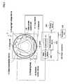
FIG. 1 is a schematic constitutional diagram of an X-ray CT apparatus to which the present invention is applied.FIG. 2 is a block diagram for explaining a first embodiment of a projection data analysis device, which is applied for a tube current control unit in the present invention.FIG. 3 is a flowchart for explaining a control processing flow of tube current applied in the present invention.FIG. 4 (A) is a reconstructed tomographic image used for analysis that is reconstructed based on projection data of an imaging portion of a object having a comparatively less bones by the projection data analysis device according to the present invention as shown inFIG. 2 .FIG. 4 (B) is an image of the reconstructed tomographic image used for analysis after being objected to a threshold processing and a diagram for explaining an attenuation profile B by bones in X direction obtained by reprojecting the image in X direction.FIG. 5 (A) is a reconstructed tomographic image used for analysis that is reconstructed based on projection data of an imaging portion of a object having many bones by the projection data analysis device according to the present invention as shown inFIG. 2 .FIG. 5 (B) is an image of the reconstructed tomographic image used for analysis after being objected to a threshold processing and a diagram for explaining an attenuation profile B by bones in X direction obtained by reprojecting the image in X direction.FIG. 6 is a timing chart for explaining respective processes performed by the projection data analysis device according to the present invention as shown inFIG. 2 , when a real scanning is started with the X-ray CT apparatus according to the present invention.FIG. 7 is a model diagram showing a reprojected result in X direction and Y direction obtained along the body axis direction of a object with regard to three respective tissues of soft tissue, bones and lung field from a reconstructed image of the object used for analysis obtained by a projection data analysis device together with a scanogram of the corresponding portions of the object.- Herein below, an embodiment of an X-ray CT apparatus according to the present invention will be explained with reference to the accompanied drawings. An
X-ray CT apparatus 1 according to the present embodiment measures projection data of a object while controlling X-ray tube current depending on an X-ray tube position (θ, Z). Further, in all of the drawings for explaining the embodiment of the present invention, ones having the same functions are designated with the same reference numerals and repetitive explanation thereof is omitted. FIG. 1 shows a constitution of theX-ray CT apparatus 1 according to the present embodiment. TheX-ray CT apparatus 1 is constituted by ascanner 10, ahost computer 20 connected to thescanner 10, anoperation unit 24 connected to thehost computer 20 and adisplay unit 30.- At first the constitutional elements of the
scanner 10 will be explained. - An
X-ray tube 11 irradiates X-rays to an object. AnX-ray detector 12 is disposed oppositely to theX-ray tube 11 and detects X-rays transmitted through the object. A data measurement device (DAS (Data Acquisition System))13 performs a predetermined data processing to the transmitted X-rays detected by theX-ray detector 12 and calculates projection data. An X-rayhigh voltage device 14 is a power source feeding for theX-ray tube 11 and of which voltage, current and power supply time (corresponding to an X-ray irradiation time) is designed to be able to set by theoperation unit 24 which will be explained later. A voltage applied from the power source is called as tube voltage and a current therefrom is called as a tube current. Adata transmission device 15 transfers data between a rotation system—a stationary system and includes a slip ring and blushes or a rotary transformer. Ascanner control device 16 controls amount of rotation of rotary plate (a scanner) to which theX-ray tube 11 and theX-ray detector 12 are attached. A projectiondata analysis device 17 reconstructs tomographic images of the object from the projection data calculated by thedata measurement device 13. A tubecurrent control unit 18 controls the tube current of theX-ray tube 11. These respective constitutional elements are carried on a rotary body rotatable around the object. - Now, the
host computer 20 performs overall control on theX-ray CT apparatus 1 and includes the following constitutional elements. Acontrol device 21 is constituted by such as a CUP and a memory for performing the overall control on theX-ray CT apparatus 1. Adata reception device 22 receives measurement data from thetransmission device 15 in thescanner 10. Animage processing device 23 performs image reconstruction computation based on the measurement data received by thedata reception device 22 to produce the tomographic images. Theoperation unit 24 is such as a track ball, a mouse and a keyboard for providing such as an input command with regard to ON/OFF of a tube current control mode. - Further, the
display device 30 is provided with a function of displaying produced tomographic images. Although the illustration is omitted, theX-ray CT apparatus 1 is provided with a patient table for laying the object during the scanning. In theX-ray CT apparatus 1 according to the present embodiment, ON/OFF of the tube current control mode can be performed by the input command from theoperation unit 24. During the tube current control mode OFF, the real scanning is performed with a constant irradiation X-ray intensity. The data of transmitted X-ray intensity measured by theX-ray detector 12 are, after being converted into digital data by theDAS 13, transferred from thedata transmission device 15 in the rotary system to the stationary system. In the stationary system, the projection data are acquired by a reception unit in thedata reception device 22, send to theimage processing device 23 and, after being reconstructed as tomographic images, the images are displayed on thedisplay device 30 for image interpretation. - The constitution of the projection
data analysis device 17 according to the first embodiment will be explained based onFIG. 2 . - The projection
data analysis device 17 determines a control value for the tube current to be flown to theX-ray tube 11. The projectiondata analysis device 17 includes an analysis useimage reconstruction unit 17a, a reconstructedimage analysis unit 17band a tube current controlvalue calculation unit 17c. The analysis useimage reconstruction unit 17a, when projection data are inputted, starts the reconstruction processing of the projection data and calculates a tomographic image for every predetermined view interval. The reconstructedimage analysis unit 17banalyzes the reconstructed images and calculates respective maximum reprojection values at, for example, bones and soft tissue of the cross section, the maximum reconstruction value obtained by totaling these and converted transmission lengths including longitudinal and lateral width of the images. The tube current controlvalue calculation unit 17ccalculates an optimum tube current control value depending on the obtained converted transmission lengths and inputs imaging conditions including new tube current value to the X-rayhigh voltage device 14. - Further, The projection
data analysis device 17 can be provided with a portion weight determination means17d. - More specifically, as shown by a dotted portion in
FIG. 2 , the portionweight determination unit 17dcan be added. Information about organ portions is obtained by evaluating an absolute amount, ratio or variation of the reprojection value. The portionweight determination unit 17dmodifies the current control value obtained by the current controlvalue calculation unit 17cfor every organ portions by making use of the obtained organ portion information and outputs a new current control value to the X-rayhigh voltage device 14. For example, while weight coefficients are determined beforehand for every organ portions, and a new current control value is determined by multiplying a weight with the calculated current value. For example, when the object is a female, the weight is set to a small value for a pelvic cavity (hypogastrium) and the amount of irradiation is controlled lower than that obtained from the transmission length. - In the tube
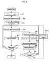
current control unit 18 in theX-ray CT apparatus 1, the tube current is controlled in the following steps. At first, during a real scanning an analysis use tomographic image is at any time reconstructed from the projection data measured (measurement data). The analysis use tomographic image reconstructed at any time is analyzed and a new tube current value is determined according to the analysis. The newly determined tube current value is directly fed back to theX-ray tube 11. In the series of these processing flow, after imaging a scanogram and determining an imaging range and original values of X-ray condition, the process moves to a real scanning to start projection data measurement for every view (view data measurement). The X-ray conditions including the tube current value are renewed at any time during the real scanning. - Herein below, the flow of tube current control processing will be explained in detail based on
FIG. 3 - At first, in step S1, a scanogram imaging is performed with the
X-ray CT apparatus 1. In step S2, based on the scanogram image taken in step S1 the imaging range is set. In step S3, conditions for X-ray imaging including the tube current value are set. The tube current value set herein is the original value for the imaging condition. In step S4, the view data measurement is performed according to the imaging range and the imaging conditions set at steps S2 and S3. - In step S5, by means of a data compression device provided at the input stage in the projection
data analysis device 17, compression processing of the projection data is performed. - In step S6, the projection data compressed in step S5 are input to the analysis use image reconstruction means17ain the projection
data analysis device 17. The analysis use image reconstruction means17aperforms a reconstruction processing for the image used for analysis. In step S7, based on the analysis use image prepared in step S6, the reconstructed image analysis means17banalyzes the reconstructed image and calculates a converted transmission length in the analysis use image according to a predetermined reference material (for example, water). In step S8, the tube current control value calculation means17ccalculates an optimum tube current value based on the obtained converted transmission length and by making use of such as a transmission length-control value conversion table. In step S9, based on the optimum tube current value calculated by the tube current control value calculation means17cwhether or not the tube current value for the X-rayhigh voltage device 14 is to be modified is judged based on the deviation from the original conditions. When the tube current is not modified, the process returns to step S4 and the view data measurement is continued based on the instant tube current value. When the tube current value is modified, the process advances to step S10, the X-ray conditions including the tube current are reset and the new reset tube current value is input to the X-rayhigh voltage device 14. Then the process returns to step S4 and the view data measurement is performed according to X-ray conditions with the new current control value. - In step S11, based on the projection data measured at step S4, the
image processing device 23 performs the image reconstruction processing. The step S11 and the steps S5˜S10 can be performed in parallel. Through this parallel processing a speed of the series of measurement and data processing can be increased. The projection data obtained in step S4 are output through thedata transfer device 15 in the rotary system to thehost computer 20. In step S11, thedata transfer device 22 in thehost computer 20 receives the projection data and outputs the same to theimage processing device 23. Theimage processing device 23 performs the image reconstruction processing based on the projection data and outputs a tomographic image to thedisplay device 30. In step S12, whether the measurement is completed or not is judged and when the answer is “Yes”, the measurement ends. When the answer is “No”, the process returns to step S4. - Now, the reconstruction image analysis means17bwill be explained in further detail. The reconstruction image analysis means17bis primarily for performing the reprojection processing of the analysis use reconstruction image and can calculate the converted transmission length information in the reprojection direction. Further, the reconstruction image analysis means17baccording to the present embodiment can perform a threshold processing at the time of reprojecting. For example, reprojection data B of bones as shown in
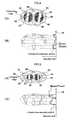
FIGS. 4 and 5 can be calculated. FIG. 4 (A) shows an example of atomographic image 40 containing bone portions (B)41,42,43,44,46 and49 and soft tissues (T)45,46 and47. InFIG. 4 (A), thetomographic image 40 is objected to a threshold processing with a predetermined CT value and a variety of regions such as bones and soft tissues are extracted. Then, an X-ray transmission profile of region extractedtomographic image 40 from X direction is calculated as4A. Among the calculated profile the maximum reprojection value Bmax is determined. In the same manner the maximum reprojection values Bmax other than the X direction are determined and all of the values around the object are added to thereby obtain a control profile, which is reflected to the X-ray tube current.FIG. 5 is an example wherein a converted transmission length at another portion than that inFIG. 4 is obtained. Like inFIG. 4 , an example of atomographic image 50 containing bone portions (B)51,52,53,54,56 and59 and soft tissues (T)55,56 and57 is shown. In this example an X-ray transmission profile is calculated as5A. As will be understand when comparing4A inFIG. 4 with5A inFIG. 5 , the maximum reprojection values Bmax vary largely depending on the measurement positions of the object.- The tube current control value calculation means17cconverts the obtained control profile to a converted transmission length and calculates an optimum tube current control value from the converted transmission length. When calculating the tube current control value, the object is approximated to an ellipse filled with a uniform material (for example, water) and the conversion is performed by using an equivalent transmission length of the material (for example, water) having an ellipse approximated configuration as the reference. For example, since Σ F (j) is an integrated value of entire CT values in reprojection direction (for example, X direction), a water equivalent transmission length Dw[mm] is determined with the following formula (1). Wherein CT value of air is assumed as 0 and water as 1000.
Dw=ΣF(j)*p/N*1000 (1) - Herein, p is a pixel size {mm} and N is a number of reprojection beams, which covers the object (corresponding to a width of the object when viewed from the reprojection direction). Current control values Q for the tube current are provided beforehand as a function of Dw (alternatively, in a form of a table) and are determined according to formula (2). Since as the analysis object reconstructed images are used instead of the projection data, an evaluation accuracy of cross sections of the object is enhanced.
Q=f(Dw) (2) - When the reprojection processing with threshold value is performed, the transmission lengths of soft tissue and bones further can be calculated according to formulae (3) and (4). Wherein Nt and Nb are numbers of reprojection beams containing respectively soft tissue and bones, and the CT value of bones is assumed as 2000 and CT value of soft tissue as 1040.
Db=ΣF(j)*p/N*2000 (3)
Dt=ΣF(j)*p/N*1040 (4) - The current control value is determined according to formula (2) after determining water transmission length while weighting respectively the transmission lengths of the tissues and bones according to formula (5).
- Although weight coefficients Wt and Wb can be assumed as 1.0 in common, however, since when the tube voltage is low, an influence of bones increases, an adjustment is possible to set the tube current at comparatively high by increasing the bone weight Wb large.
- Further, in the case of an infant, since the influence of bones is small, it is preferable to set the tube current as low as possible so as to suppress X-ray exposure. In such instance, the weight of bones Wb can be adjusted low.
- Further, as weights for every portions of imaging organs, Wt and Wb can be used.
Dw=Wt*Dt+Wb*Dt (5) - Further, in a method of not using the weight coefficients, a relationship between transmission length of soft tissue and transmission length of bones and optimum tube current values are determined beforehand. In such instance, the current control value Q for the optimum tube current is expressed as formula (6).
Q=f(Dt,Db) (6) - In such instance, while analyzing clinical data likely, a two dimensional table can be determined statistically.
- In any events, since a cross section of a object of which the transmission length of bones is taken into account can be calculated, measurement accuracy of a cross section of the object is further enhanced.
- The reprojection processing can be performed on one sheet of analysis use reconstruction image in plural directions and when the processing is performed in more than one directions, in that X direction (3 o'clock direction) and Y direction (0 o'clock direction), the transmission length at the advanced phase in 6 o'clock and 9 o'clock directions can be estimated. When controlling the tube current in a sinusoidal wave or in any function, it is sufficient if the maximum value and the minimum value thereof are given, an alternate computation in X direction and Y direction can be performed in order to reduce computation amount.
- Now, an operation of the
X-ray CT apparatus 1 will be explained. When theX-ray detector 12 in theX-ray CT apparatus 1 according to the present embodiment includes, for example, detector elements for 1024 channels, view data are captured 1024 time for every one rotation. Further, with regard to the projection data from theDAS 13, an average value of center two rows in the multi-slice detector is constituted to be inputted to the projectiondata analysis device 17 through a data compression device provided at the input stage in the projectiondata analysis device 17. Further, while adding 8 sample data both in view direction and channel direction, view data of 128 channels are inputted 128 times for every one revolution. When assuming that sampling interval of the original data is 0.5 mm, an analysis use image having a resolution of about 4 mm can be reconstructed. Further, when assuming that the matrix of the analysis use reconstructed image is 128 pixels, the maximum FOV (region of interest) will be 512 mm at resolution of 4 mm. When comparing with a reading use reconstructed image, since the reconstructed matrix is 1/16 and the number of views is ⅛, computation scale thereof will become about 1/128. In this instance, a correction processing with filter is performed by means of a blurring correction filter, which is stored in advance by performing Fourier transformation at 256 points. Further, a reverse projection processing is performed after application of the blurring correction filter. When completing the reverse projection processing of the view data necessary for the reconstruction, the analysis use tomographic image is reconstructed. In addition, although it is necessary to hold data of air and offset data together with data of water, since the amount of such as the data of air and the offset data is sufficient those for one view, the small memory capacity required for the respective data is sufficient. - In the case of multi-slice CT, when the number of rows to be arranged increases, a three dimensional back projection computation such as using Feldkamp method is necessary, however, according to the present embodiment, even if only specific rows are reconstructed as they are with a conventional two dimensional reconstruction method, the results show a sufficient practicality.
- Further, the estimation accuracy of the converted transmission length of the cross section for evaluation can be enhanced, if, in addition to the reconstruction only of the images at the rows near the center, images at the edge rows are reconstructed together and the converted transmission lengths in the body axis direction are obtained at plural points.
- Prior to the real scanning, an initial value X0 of X-ray condition and time interval Δt for control command are inputted. The X0 can be an optimum value obtained from an analysis result of the scanogram or value determined empirically by an operator.
- Further, although the time interval Δt for control command can be determined arbitrary, however, if the tube current value is renewed so frequently and the X-ray exposure is not optimized, the command renewal is meaningless, therefore, the time interval is determined in view of such as the response time of the control system and, of course, the time interval can be defined by such as the number of views and an angular interval. For example, when assuming that the number of views for one rotation is 128 and the tube current value is outputted for every 32 views, the command can be renewed four times for every one rotation.
FIG. 6 shows a timing chart of the respective processes during scanning. When the scanning is started at t0 and X-ray exposure is started, in synchronism thereto a data collection operation is started. Herein the initial value of the tube current is assumed as X0. All of the measured data are sent to thedata transfer device 15 and are transferred to the stationary system. The data captured in thehost computer 20 are reconstructed in theimage processing device 23 as a reading use tomographic image.- On the other hand, the projection
data analysis device 17 performs the analysis use image reconstruction, the reconstructed image analysis and the optimum tube current control value calculation processing so as to determine a tube current control value. The analysis use image reconstruction, the reconstructed image analysis and the optimum tube current control value calculation processing are performed respectively in a pipeline manner. A new tube current control value obtained through the optimum tube current control value calculation processing is sent to the X-rayhigh voltage device 14 and the feedback is for the first time effective at t6. The timing when the feedback becomes effective can be used as a substantial imaging start point. - The tube current value obtained here is what was set based on an analysis use image reconstructed from the projection data obtained around time t1-t2 and contains corresponding phase (angular) information. Herein, the X-ray
high voltage device 14 alters the actual control value at the corresponding phase t6. - In the case of determining the tube current control value by directly processing the projection data as in the conventional art, when such as a preamplifier gain, log conversion gain and distance between focal point-detector vary, parameters for the tube current had to be reviewed, however, in the present embodiment, since the tube current control value is set based on the CT value representing an absolute value, a stable control can be achieved.
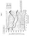
FIG. 7 shows reprojection results in X and Y directions of respective three tissues of soft tissue, bones and lung field. The horizontal axis represents body axis direction and corresponds to the positions of the scanogram shown in the background. Herein, when noting to the reprojection result of the soft tissue, only the lung field existing position shows a high reprojection value in both X and Y directions.- On the other hand, the reprojection value of the bones increases near the shoulder. In this manner, it is understood that the results show the respective features of the imaged portions. Thereby, according to the present embodiment, respective portions can be judged by making use of the respective reprojection values.
- In the present embodiment, although exposure doses are constituted to be automatically controllable for every portion, through setting a portion particularly desired to be lowered of the exposure doses by an operator using such as a scanogram, control of the exposure doses can be preformed by making use of such information, however, when the tube current value is set so as to follow according to the measurement portions of the tomographic image, such is further desirable in view of its user friendliness.
- Further, in the present embodiment, an example where the transmission length is converted into X-ray absorption coefficient of water was explained, however, the material used as a parameter of the X-ray absorption coefficient is not limited to water, but any material such as bone and soft tissue can be selected. Because it is sufficient if a object can be converted in a form of a transmission length of a predetermined material.
Claims (10)
Applications Claiming Priority (3)
| Application Number | Priority Date | Filing Date | Title |
|---|---|---|---|
| JP2005-051497 | 2005-02-25 | ||
| JP2005051497 | 2005-02-25 | ||
| JP2006003624 | 2006-02-27 |
Publications (2)
| Publication Number | Publication Date |
|---|---|
| US20080107231A1true US20080107231A1 (en) | 2008-05-08 |
| US7636416B2 US7636416B2 (en) | 2009-12-22 |
Family
ID=36927507
Family Applications (1)
| Application Number | Title | Priority Date | Filing Date |
|---|---|---|---|
| US11/884,099Expired - Fee RelatedUS7636416B2 (en) | 2005-02-25 | 2006-02-27 | X-ray CT apparatus comprising a tube current control unit |
Country Status (4)
| Country | Link |
|---|---|
| US (1) | US7636416B2 (en) |
| JP (1) | JP5001142B2 (en) |
| CN (1) | CN101128153B (en) |
| WO (1) | WO2006090877A1 (en) |
Cited By (25)
| Publication number | Priority date | Publication date | Assignee | Title |
|---|---|---|---|---|
| US20090041381A1 (en)* | 2007-08-06 | 2009-02-12 | Georgiev Todor G | Method and Apparatus for Radiance Processing by Demultiplexing in the Frequency Domain |
| US20090268970A1 (en)* | 2008-04-29 | 2009-10-29 | Sevket Derin Babacan | Method and Apparatus for Block-Based Compression of Light-field Images |
| US20100020187A1 (en)* | 2006-04-04 | 2010-01-28 | Georgiev Todor G | Plenoptic camera |
| US20110211824A1 (en)* | 2008-01-23 | 2011-09-01 | Georgiev Todor G | Methods and Apparatus for Full-Resolution Light-Field Capture and Rendering |
| US8189065B2 (en) | 2008-01-23 | 2012-05-29 | Adobe Systems Incorporated | Methods and apparatus for full-resolution light-field capture and rendering |
| US8189089B1 (en) | 2009-01-20 | 2012-05-29 | Adobe Systems Incorporated | Methods and apparatus for reducing plenoptic camera artifacts |
| US8228417B1 (en) | 2009-07-15 | 2012-07-24 | Adobe Systems Incorporated | Focused plenoptic camera employing different apertures or filtering at different microlenses |
| US8244058B1 (en)* | 2008-05-30 | 2012-08-14 | Adobe Systems Incorporated | Method and apparatus for managing artifacts in frequency domain processing of light-field images |
| US8290358B1 (en) | 2007-06-25 | 2012-10-16 | Adobe Systems Incorporated | Methods and apparatus for light-field imaging |
| US8315476B1 (en) | 2009-01-20 | 2012-11-20 | Adobe Systems Incorporated | Super-resolution with the focused plenoptic camera |
| US8345144B1 (en) | 2009-07-15 | 2013-01-01 | Adobe Systems Incorporated | Methods and apparatus for rich image capture with focused plenoptic cameras |
| US8358366B1 (en) | 2010-05-28 | 2013-01-22 | Adobe Systems Incorporate | Methods and apparatus for high-speed digital imaging |
| US8400555B1 (en) | 2009-12-01 | 2013-03-19 | Adobe Systems Incorporated | Focused plenoptic camera employing microlenses with different focal lengths |
| US8665341B2 (en) | 2010-08-27 | 2014-03-04 | Adobe Systems Incorporated | Methods and apparatus for rendering output images with simulated artistic effects from focused plenoptic camera data |
| US8724000B2 (en) | 2010-08-27 | 2014-05-13 | Adobe Systems Incorporated | Methods and apparatus for super-resolution in integral photography |
| US8749694B2 (en) | 2010-08-27 | 2014-06-10 | Adobe Systems Incorporated | Methods and apparatus for rendering focused plenoptic camera data using super-resolved demosaicing |
| US8803918B2 (en) | 2010-08-27 | 2014-08-12 | Adobe Systems Incorporated | Methods and apparatus for calibrating focused plenoptic camera data |
| US8817015B2 (en) | 2010-03-03 | 2014-08-26 | Adobe Systems Incorporated | Methods, apparatus, and computer-readable storage media for depth-based rendering of focused plenoptic camera data |
| WO2015065843A1 (en)* | 2013-10-31 | 2015-05-07 | Hexagon Metrology, Inc. | Parametric control of object scanning |
| US9030550B2 (en) | 2011-03-25 | 2015-05-12 | Adobe Systems Incorporated | Thin plenoptic cameras using solid immersion lenses |
| US20160302756A1 (en)* | 2013-12-18 | 2016-10-20 | General Electric Company | System and method of computed tomography signal restoration via noise reduction |
| US10092183B2 (en) | 2014-08-31 | 2018-10-09 | Dr. John Berestka | Systems and methods for analyzing the eye |
| US10255698B2 (en)* | 2017-08-28 | 2019-04-09 | Shenzhen United Imaging Healthcare Co., Ltd. | System and method for removing hard tissue in CT image |
| US11298097B2 (en) | 2020-04-07 | 2022-04-12 | Siemens Healthcare Gmbh | Automated determination of an X-ray tube-current profile |
| US12440102B2 (en) | 2025-03-28 | 2025-10-14 | Lightfield Medical Inc. | Systems and methods for analyzing the eye |
Families Citing this family (25)
| Publication number | Priority date | Publication date | Assignee | Title |
|---|---|---|---|---|
| EP1972276B1 (en)* | 2007-03-20 | 2017-03-01 | Cefla Societa' Cooperativa | Method for activation of an emitter of a computed tomography scanner |
| US8135186B2 (en)* | 2008-01-25 | 2012-03-13 | Purdue Research Foundation | Method and system for image reconstruction |
| DE102008014738A1 (en)* | 2008-03-18 | 2009-09-24 | Siemens Aktiengesellschaft | Medical imaging method and medical imaging device |
| EP2328477B1 (en)* | 2008-08-04 | 2018-05-16 | Koninklijke Philips N.V. | Interventional imaging and data processing |
| JP5317612B2 (en)* | 2008-09-26 | 2013-10-16 | ジーイー・メディカル・システムズ・グローバル・テクノロジー・カンパニー・エルエルシー | Tomographic image processing apparatus, X-ray CT apparatus, and program |
| JP5484788B2 (en)* | 2009-05-25 | 2014-05-07 | ジーイー・メディカル・システムズ・グローバル・テクノロジー・カンパニー・エルエルシー | X-ray CT system |
| CN102498438B (en)* | 2009-07-17 | 2014-11-26 | D·P·罗勒 | Extended low contrast detectability for radiographic imaging systems |
| US9168016B2 (en)* | 2010-01-29 | 2015-10-27 | Fujifilm Corporation | Radiographic image capturing apparatus, radiographic image capturing system, and method of supplying electric power to radiographic image capturing apparatus |
| JP5642444B2 (en)* | 2010-07-15 | 2014-12-17 | 三菱重工業株式会社 | Radiotherapy apparatus operating method and radiotherapy apparatus control apparatus |
| JP5806812B2 (en)* | 2010-10-05 | 2015-11-10 | 株式会社日立メディコ | X-ray CT system |
| CN102440792B (en)* | 2010-10-11 | 2015-12-02 | 株式会社东芝 | X-ray ct system |
| JP6257948B2 (en)* | 2012-08-07 | 2018-01-10 | 東芝メディカルシステムズ株式会社 | X-ray imaging system |
| CN103040481B (en)* | 2012-12-25 | 2015-08-26 | 深圳先进技术研究院 | A kind of system and method reducing x-ray diagnostic equipment x-ray dose |
| JP6242683B2 (en)* | 2012-12-27 | 2017-12-06 | 東芝メディカルシステムズ株式会社 | X-ray CT apparatus and control method |
| JP6328387B2 (en)* | 2013-07-18 | 2018-05-23 | 株式会社日立製作所 | X-ray measuring device |
| DE102013219249A1 (en)* | 2013-09-25 | 2015-03-26 | Siemens Aktiengesellschaft | Method and system for automatic selection of a scan protocol |
| WO2015151948A1 (en)* | 2014-03-31 | 2015-10-08 | 株式会社 日立メディコ | X-ray ct device and imaging method |
| CN104287768A (en)* | 2014-09-30 | 2015-01-21 | 沈阳东软医疗系统有限公司 | Method and system for controlling CT scan dose |
| CN105391368B (en)* | 2015-10-13 | 2018-03-20 | 沈阳东软医疗系统有限公司 | A kind of system for measuring linear accelerator treatment head stop position |
| US10085698B2 (en)* | 2016-01-26 | 2018-10-02 | Genereal Electric Company | Methods and systems for automated tube current modulation |
| JP6906905B2 (en) | 2016-06-29 | 2021-07-21 | キヤノンメディカルシステムズ株式会社 | X-ray diagnostic equipment |
| WO2018032095A1 (en)* | 2016-08-18 | 2018-02-22 | Hydro-Quebec | Apparatus and method for inspecting a power line |
| CN106725570B (en)* | 2016-12-30 | 2019-12-20 | 上海联影医疗科技有限公司 | Imaging method and system |
| US10973489B2 (en)* | 2017-09-29 | 2021-04-13 | General Electric Company | CT imaging system and method using a task-based image quality metric to achieve a desired image quality |
| CN117761089A (en)* | 2023-12-21 | 2024-03-26 | 同方威视技术股份有限公司 | CT equipment and detection methods |
Citations (19)
| Publication number | Priority date | Publication date | Assignee | Title |
|---|---|---|---|---|
| US5379333A (en)* | 1993-11-19 | 1995-01-03 | General Electric Company | Variable dose application by modulation of x-ray tube current during CT scanning |
| US5400378A (en)* | 1993-11-19 | 1995-03-21 | General Electric Company | Dynamic dose control in multi-slice CT scan |
| US5822393A (en)* | 1997-04-01 | 1998-10-13 | Siemens Aktiengesellschaft | Method for adaptively modulating the power level of an x-ray tube of a computer tomography (CT) system |
| US5867555A (en)* | 1997-03-04 | 1999-02-02 | Siemens Aktiengesellschaft | Adaptive dose modulation during CT scanning |
| US6385280B1 (en)* | 1998-08-18 | 2002-05-07 | Siemens Aktiengesellschaft | X-ray computed tomography apparatus with modulation of the x-ray power of the x-ray source |
| US6490337B1 (en)* | 2000-04-03 | 2002-12-03 | Hitachi Medical Corporation | X-ray CT apparatus |
| US20040086076A1 (en)* | 2001-03-09 | 2004-05-06 | Takayuki Nagaoka | X-ray ct device and image displaying method therefor |
| US6754301B2 (en)* | 2001-08-28 | 2004-06-22 | Ge Medical Systems Global Technology Company, Llc | X-ray CT system, gantry apparatus and operation console |
| US6775352B2 (en)* | 2002-08-16 | 2004-08-10 | Ge Medical Systems Global Technology Company, Llc | Method and system for implementing variable x-ray intensity modulation schemes for imaging systems |
| US6901129B2 (en)* | 2001-07-04 | 2005-05-31 | Kabushiki Kaisha Toshiba | X-ray computer tomography apparatus |
| US6954513B2 (en)* | 2002-12-20 | 2005-10-11 | Ge Medical Systems Global Technology Company, Llc | X-ray CT Apparatus and exposure dose calculating method |
| US6987828B2 (en)* | 2002-03-27 | 2006-01-17 | Ge Medical Systems Global Technology Company, Llc | Transmitted X-ray data acquisition system and X-ray computed tomography system |
| US6990172B2 (en)* | 2004-02-19 | 2006-01-24 | General Electric Company | Method and apparatus to determine tube current modulation profile for radiographic imaging |
| US7031423B2 (en)* | 2003-05-09 | 2006-04-18 | Kabushiki Kaisha Toshiba | X-ray computed tomography apparatus and picture quality simulation apparatus |
| US7072437B2 (en)* | 2003-12-26 | 2006-07-04 | Ge Medical Systems Global Technology Company, Llc | Radiation tomography system and tomography method |
| US7106824B2 (en)* | 2004-04-26 | 2006-09-12 | Kabushiki Kaisha Toshiba | X-ray computed tomographic apparatus |
| US7142630B2 (en)* | 2001-10-22 | 2006-11-28 | Kabushiki Kaisha Toshiba | X-ray computed tomography apparatus with X-ray intensity control |
| US7203270B2 (en)* | 2003-04-09 | 2007-04-10 | Kabushiki Kaisha Toshiba | X-ray computed tomographic apparatus |
| US7215733B2 (en)* | 2004-07-23 | 2007-05-08 | Kabushiki Kaisha Toshiba | X-ray computed tomography apparatus |
Family Cites Families (1)
| Publication number | Priority date | Publication date | Assignee | Title |
|---|---|---|---|---|
| JPH08166995A (en)* | 1994-12-13 | 1996-06-25 | Toshiba Corp | Medical diagnosis support system |
- 2006
- 2006-02-27JPJP2007504834Apatent/JP5001142B2/ennot_activeExpired - Fee Related
- 2006-02-27CNCN2006800062088Apatent/CN101128153B/ennot_activeExpired - Fee Related
- 2006-02-27USUS11/884,099patent/US7636416B2/ennot_activeExpired - Fee Related
- 2006-02-27WOPCT/JP2006/303624patent/WO2006090877A1/enactiveApplication Filing
Patent Citations (20)
| Publication number | Priority date | Publication date | Assignee | Title |
|---|---|---|---|---|
| US5379333A (en)* | 1993-11-19 | 1995-01-03 | General Electric Company | Variable dose application by modulation of x-ray tube current during CT scanning |
| US5400378A (en)* | 1993-11-19 | 1995-03-21 | General Electric Company | Dynamic dose control in multi-slice CT scan |
| US5867555A (en)* | 1997-03-04 | 1999-02-02 | Siemens Aktiengesellschaft | Adaptive dose modulation during CT scanning |
| US5822393A (en)* | 1997-04-01 | 1998-10-13 | Siemens Aktiengesellschaft | Method for adaptively modulating the power level of an x-ray tube of a computer tomography (CT) system |
| US6385280B1 (en)* | 1998-08-18 | 2002-05-07 | Siemens Aktiengesellschaft | X-ray computed tomography apparatus with modulation of the x-ray power of the x-ray source |
| US6490337B1 (en)* | 2000-04-03 | 2002-12-03 | Hitachi Medical Corporation | X-ray CT apparatus |
| US7103139B2 (en)* | 2001-03-09 | 2006-09-05 | Hitachi Medical Corporation | X-ray CT device and image displaying method therefor |
| US20040086076A1 (en)* | 2001-03-09 | 2004-05-06 | Takayuki Nagaoka | X-ray ct device and image displaying method therefor |
| US6901129B2 (en)* | 2001-07-04 | 2005-05-31 | Kabushiki Kaisha Toshiba | X-ray computer tomography apparatus |
| US6754301B2 (en)* | 2001-08-28 | 2004-06-22 | Ge Medical Systems Global Technology Company, Llc | X-ray CT system, gantry apparatus and operation console |
| US7142630B2 (en)* | 2001-10-22 | 2006-11-28 | Kabushiki Kaisha Toshiba | X-ray computed tomography apparatus with X-ray intensity control |
| US6987828B2 (en)* | 2002-03-27 | 2006-01-17 | Ge Medical Systems Global Technology Company, Llc | Transmitted X-ray data acquisition system and X-ray computed tomography system |
| US6775352B2 (en)* | 2002-08-16 | 2004-08-10 | Ge Medical Systems Global Technology Company, Llc | Method and system for implementing variable x-ray intensity modulation schemes for imaging systems |
| US6954513B2 (en)* | 2002-12-20 | 2005-10-11 | Ge Medical Systems Global Technology Company, Llc | X-ray CT Apparatus and exposure dose calculating method |
| US7203270B2 (en)* | 2003-04-09 | 2007-04-10 | Kabushiki Kaisha Toshiba | X-ray computed tomographic apparatus |
| US7031423B2 (en)* | 2003-05-09 | 2006-04-18 | Kabushiki Kaisha Toshiba | X-ray computed tomography apparatus and picture quality simulation apparatus |
| US7072437B2 (en)* | 2003-12-26 | 2006-07-04 | Ge Medical Systems Global Technology Company, Llc | Radiation tomography system and tomography method |
| US6990172B2 (en)* | 2004-02-19 | 2006-01-24 | General Electric Company | Method and apparatus to determine tube current modulation profile for radiographic imaging |
| US7106824B2 (en)* | 2004-04-26 | 2006-09-12 | Kabushiki Kaisha Toshiba | X-ray computed tomographic apparatus |
| US7215733B2 (en)* | 2004-07-23 | 2007-05-08 | Kabushiki Kaisha Toshiba | X-ray computed tomography apparatus |
Cited By (44)
| Publication number | Priority date | Publication date | Assignee | Title |
|---|---|---|---|---|
| US20100020187A1 (en)* | 2006-04-04 | 2010-01-28 | Georgiev Todor G | Plenoptic camera |
| US8238738B2 (en) | 2006-04-04 | 2012-08-07 | Adobe Systems Incorporated | Plenoptic camera |
| US8290358B1 (en) | 2007-06-25 | 2012-10-16 | Adobe Systems Incorporated | Methods and apparatus for light-field imaging |
| US20090041381A1 (en)* | 2007-08-06 | 2009-02-12 | Georgiev Todor G | Method and Apparatus for Radiance Processing by Demultiplexing in the Frequency Domain |
| US8559756B2 (en) | 2007-08-06 | 2013-10-15 | Adobe Systems Incorporated | Radiance processing by demultiplexing in the frequency domain |
| US20110211824A1 (en)* | 2008-01-23 | 2011-09-01 | Georgiev Todor G | Methods and Apparatus for Full-Resolution Light-Field Capture and Rendering |
| US8189065B2 (en) | 2008-01-23 | 2012-05-29 | Adobe Systems Incorporated | Methods and apparatus for full-resolution light-field capture and rendering |
| US8160439B2 (en) | 2008-01-23 | 2012-04-17 | Adobe Systems Incorporated | Methods and apparatus for full-resolution light-field capture and rendering |
| US8379105B2 (en) | 2008-01-23 | 2013-02-19 | Adobe Systems Incorporated | Methods and apparatus for full-resolution light-field capture and rendering |
| US8380060B2 (en) | 2008-01-23 | 2013-02-19 | Adobe Systems Incorporated | Methods and apparatus for full-resolution light-field capture and rendering |
| US8155456B2 (en) | 2008-04-29 | 2012-04-10 | Adobe Systems Incorporated | Method and apparatus for block-based compression of light-field images |
| US20090268970A1 (en)* | 2008-04-29 | 2009-10-29 | Sevket Derin Babacan | Method and Apparatus for Block-Based Compression of Light-field Images |
| US8401316B2 (en) | 2008-04-29 | 2013-03-19 | Adobe Systems Incorporated | Method and apparatus for block-based compression of light-field images |
| US8611693B2 (en) | 2008-05-30 | 2013-12-17 | Adobe Systems Incorporated | Managing artifacts in frequency domain processing of light-field images |
| US8244058B1 (en)* | 2008-05-30 | 2012-08-14 | Adobe Systems Incorporated | Method and apparatus for managing artifacts in frequency domain processing of light-field images |
| US8315476B1 (en) | 2009-01-20 | 2012-11-20 | Adobe Systems Incorporated | Super-resolution with the focused plenoptic camera |
| US8189089B1 (en) | 2009-01-20 | 2012-05-29 | Adobe Systems Incorporated | Methods and apparatus for reducing plenoptic camera artifacts |
| US9316840B2 (en) | 2009-01-20 | 2016-04-19 | Adobe Systems Incorporated | Methods and apparatus for reducing plenoptic camera artifacts |
| US8345144B1 (en) | 2009-07-15 | 2013-01-01 | Adobe Systems Incorporated | Methods and apparatus for rich image capture with focused plenoptic cameras |
| US8471920B2 (en) | 2009-07-15 | 2013-06-25 | Adobe Systems Incorporated | Focused plenoptic camera employing different apertures or filtering at different microlenses |
| US8228417B1 (en) | 2009-07-15 | 2012-07-24 | Adobe Systems Incorporated | Focused plenoptic camera employing different apertures or filtering at different microlenses |
| US8400555B1 (en) | 2009-12-01 | 2013-03-19 | Adobe Systems Incorporated | Focused plenoptic camera employing microlenses with different focal lengths |
| US8817015B2 (en) | 2010-03-03 | 2014-08-26 | Adobe Systems Incorporated | Methods, apparatus, and computer-readable storage media for depth-based rendering of focused plenoptic camera data |
| US8860833B2 (en) | 2010-03-03 | 2014-10-14 | Adobe Systems Incorporated | Blended rendering of focused plenoptic camera data |
| US8358366B1 (en) | 2010-05-28 | 2013-01-22 | Adobe Systems Incorporate | Methods and apparatus for high-speed digital imaging |
| US8665341B2 (en) | 2010-08-27 | 2014-03-04 | Adobe Systems Incorporated | Methods and apparatus for rendering output images with simulated artistic effects from focused plenoptic camera data |
| US8749694B2 (en) | 2010-08-27 | 2014-06-10 | Adobe Systems Incorporated | Methods and apparatus for rendering focused plenoptic camera data using super-resolved demosaicing |
| US8724000B2 (en) | 2010-08-27 | 2014-05-13 | Adobe Systems Incorporated | Methods and apparatus for super-resolution in integral photography |
| US8803918B2 (en) | 2010-08-27 | 2014-08-12 | Adobe Systems Incorporated | Methods and apparatus for calibrating focused plenoptic camera data |
| US9030550B2 (en) | 2011-03-25 | 2015-05-12 | Adobe Systems Incorporated | Thin plenoptic cameras using solid immersion lenses |
| US9197798B2 (en) | 2011-03-25 | 2015-11-24 | Adobe Systems Incorporated | Thin plenoptic cameras using microspheres |
| WO2015065843A1 (en)* | 2013-10-31 | 2015-05-07 | Hexagon Metrology, Inc. | Parametric control of object scanning |
| US9857163B2 (en) | 2013-10-31 | 2018-01-02 | Hexagon Metrology, Inc. | Parametric control of object scanning |
| US10595808B2 (en)* | 2013-12-18 | 2020-03-24 | General Electric Company | System and method of computed tomography signal restoration via noise reduction |
| US20160302756A1 (en)* | 2013-12-18 | 2016-10-20 | General Electric Company | System and method of computed tomography signal restoration via noise reduction |
| US10092183B2 (en) | 2014-08-31 | 2018-10-09 | Dr. John Berestka | Systems and methods for analyzing the eye |
| US10687703B2 (en) | 2014-08-31 | 2020-06-23 | John Berestka | Methods for analyzing the eye |
| US11452447B2 (en) | 2014-08-31 | 2022-09-27 | John Berestka | Methods for analyzing the eye |
| US11911109B2 (en) | 2014-08-31 | 2024-02-27 | Dr. John Berestka | Methods for analyzing the eye |
| US10593073B2 (en) | 2017-08-28 | 2020-03-17 | Shenzhen United Imaging Healthcare Co., Ltd. | System and method for removing hard tissue in CT image |
| US10255698B2 (en)* | 2017-08-28 | 2019-04-09 | Shenzhen United Imaging Healthcare Co., Ltd. | System and method for removing hard tissue in CT image |
| US11094094B2 (en) | 2017-08-28 | 2021-08-17 | Shanghai United Imaging Healthcare Co., Ltd | System and method for removing hard tissue in CT image |
| US11298097B2 (en) | 2020-04-07 | 2022-04-12 | Siemens Healthcare Gmbh | Automated determination of an X-ray tube-current profile |
| US12440102B2 (en) | 2025-03-28 | 2025-10-14 | Lightfield Medical Inc. | Systems and methods for analyzing the eye |
Also Published As
| Publication number | Publication date |
|---|---|
| JP5001142B2 (en) | 2012-08-15 |
| WO2006090877A1 (en) | 2006-08-31 |
| CN101128153B (en) | 2010-09-29 |
| JPWO2006090877A1 (en) | 2008-07-24 |
| US7636416B2 (en) | 2009-12-22 |
| CN101128153A (en) | 2008-02-20 |
Similar Documents
| Publication | Publication Date | Title |
|---|---|---|
| US7636416B2 (en) | X-ray CT apparatus comprising a tube current control unit | |
| EP2243020B1 (en) | System and method for quantitative imaging of chemical composition to decompose more than two materials | |
| US7885373B2 (en) | System and method for quantitative imaging of chemical composition to decompose multiple materials | |
| US9254107B2 (en) | X-ray CT apparatus and tube current determination method | |
| US7873141B2 (en) | X-ray tomographic imaging apparatus | |
| JP5028528B2 (en) | X-ray CT system | |
| JP4152649B2 (en) | Method and apparatus for CT scout image processing | |
| JP6713860B2 (en) | Image reconstruction apparatus, X-ray CT apparatus, and image reconstruction method | |
| JP2004113791A (en) | Automatic exposure control method and apparatus in CT scan | |
| JPH07246200A (en) | X-ray ct scanner | |
| EP1762976A2 (en) | Computerized tomography device and image processing method for identifying brown adipose tissue | |
| WO2007074772A1 (en) | X-ray ct device | |
| JP2002531199A (en) | Method and apparatus for calcification leveling | |
| JP7617915B2 (en) | Radiological Imaging Methods | |
| US7239730B2 (en) | Method and apparatus for volume scoring calcification concentrations of a CT scan | |
| JP4429694B2 (en) | X-ray CT system | |
| JP4554185B2 (en) | X-ray CT system | |
| CN102652676A (en) | Method to reduce radiation dose delivered by imaging system | |
| JP2011177396A (en) | X-ray ct apparatus | |
| JP5677889B2 (en) | X-ray CT apparatus and X-ray CT system | |
| EP3657443A1 (en) | Tomographic image processing apparatus and method, and computer program product | |
| JP3789728B2 (en) | Projection data correction method and apparatus, and radiation tomography apparatus | |
| JP2004081394A (en) | In-tissue fat evaluating method, image processor, and x-ray computerized tomography (ct) system | |
| CN1231182C (en) | Method and apparatus for optimizing CT image quality by means of optimized data collection | |
| JP2002153454A (en) | X-ray ct device |
Legal Events
| Date | Code | Title | Description |
|---|---|---|---|
| AS | Assignment | Owner name:HITACHI MEDICAL CORPORATION, JAPAN Free format text:ASSIGNMENT OF ASSIGNORS INTEREST;ASSIGNORS:MIYAZAKI, OSAMU;HIROKAWA, KOICHI;IRIE, TOSHIYUKI;REEL/FRAME:019727/0638 Effective date:20070627 | |
| STCF | Information on status: patent grant | Free format text:PATENTED CASE | |
| FPAY | Fee payment | Year of fee payment:4 | |
| AS | Assignment | Owner name:HITACHI, LTD., JAPAN Free format text:ASSIGNMENT OF ASSIGNORS INTEREST;ASSIGNOR:HITACHI MEDICAL CORPORATION;REEL/FRAME:040545/0254 Effective date:20160401 | |
| FPAY | Fee payment | Year of fee payment:8 | |
| FEPP | Fee payment procedure | Free format text:MAINTENANCE FEE REMINDER MAILED (ORIGINAL EVENT CODE: REM.); ENTITY STATUS OF PATENT OWNER: LARGE ENTITY | |
| LAPS | Lapse for failure to pay maintenance fees | Free format text:PATENT EXPIRED FOR FAILURE TO PAY MAINTENANCE FEES (ORIGINAL EVENT CODE: EXP.); ENTITY STATUS OF PATENT OWNER: LARGE ENTITY | |
| STCH | Information on status: patent discontinuation | Free format text:PATENT EXPIRED DUE TO NONPAYMENT OF MAINTENANCE FEES UNDER 37 CFR 1.362 | |
| FP | Lapsed due to failure to pay maintenance fee | Effective date:20211222 |