US20070263172A1 - Reflection Microscope for Examination of the Corneal Endothelium and Method of Operating Same - Google Patents
Reflection Microscope for Examination of the Corneal Endothelium and Method of Operating SameDownload PDFInfo
- Publication number
- US20070263172A1 US20070263172A1US11/632,084US63208405AUS2007263172A1US 20070263172 A1US20070263172 A1US 20070263172A1US 63208405 AUS63208405 AUS 63208405AUS 2007263172 A1US2007263172 A1US 2007263172A1
- Authority
- US
- United States
- Prior art keywords
- endothelium
- center
- sensor
- light spot
- camera
- Prior art date
- Legal status (The legal status is an assumption and is not a legal conclusion. Google has not performed a legal analysis and makes no representation as to the accuracy of the status listed.)
- Granted
Links
- 238000000034methodMethods0.000titleclaimsabstractdescription33
- 210000000871endothelium cornealAnatomy0.000titledescription2
- 230000003287optical effectEffects0.000claimsabstractdescription53
- 210000003038endotheliumAnatomy0.000claimsabstractdescription38
- 238000003384imaging methodMethods0.000claimsabstractdescription9
- 230000001960triggered effectEffects0.000claimsabstract2
- 210000004087corneaAnatomy0.000claimsdescription20
- 210000001508eyeAnatomy0.000claimsdescription16
- 210000005252bulbus oculiAnatomy0.000claimsdescription6
- 230000004044responseEffects0.000claimsdescription2
- 230000003213activating effectEffects0.000claims1
- 230000004913activationEffects0.000claims1
- 210000004027cellAnatomy0.000description17
- 238000012360testing methodMethods0.000description11
- 230000001413cellular effectEffects0.000description7
- 239000012141concentrateSubstances0.000description3
- 238000010586diagramMethods0.000description3
- 210000000981epitheliumAnatomy0.000description3
- 238000011156evaluationMethods0.000description3
- 229910052736halogenInorganic materials0.000description3
- 150000002367halogensChemical class0.000description3
- 239000010410layerSubstances0.000description3
- 238000007491morphometric analysisMethods0.000description3
- 238000001514detection methodMethods0.000description2
- 201000010099diseaseDiseases0.000description2
- 208000037265diseases, disorders, signs and symptomsDiseases0.000description2
- 230000006870functionEffects0.000description2
- 210000003128headAnatomy0.000description2
- 241000283973Oryctolagus cuniculusSpecies0.000description1
- 230000009471actionEffects0.000description1
- 230000008859changeEffects0.000description1
- 230000007423decreaseEffects0.000description1
- 210000002555descemet membraneAnatomy0.000description1
- 238000013461designMethods0.000description1
- 230000008753endothelial functionEffects0.000description1
- 230000036571hydrationEffects0.000description1
- 238000006703hydration reactionMethods0.000description1
- 238000005286illuminationMethods0.000description1
- 238000010191image analysisMethods0.000description1
- 230000002757inflammatory effectEffects0.000description1
- 230000010354integrationEffects0.000description1
- 230000003834intracellular effectEffects0.000description1
- 238000005088metallographyMethods0.000description1
- 238000000386microscopyMethods0.000description1
- 238000012986modificationMethods0.000description1
- 230000004048modificationEffects0.000description1
- 230000004660morphological changeEffects0.000description1
- 230000003562morphometric effectEffects0.000description1
- 238000013425morphometryMethods0.000description1
- 210000001316polygonal cellAnatomy0.000description1
- 230000008569processEffects0.000description1
- 238000012545processingMethods0.000description1
- 239000002356single layerSubstances0.000description1
- 238000001356surgical procedureMethods0.000description1
- 230000002123temporal effectEffects0.000description1
- 238000002560therapeutic procedureMethods0.000description1
- 210000001519tissueAnatomy0.000description1
- 230000000007visual effectEffects0.000description1
- 238000012800visualizationMethods0.000description1
- XLYOFNOQVPJJNP-UHFFFAOYSA-NwaterSubstancesOXLYOFNOQVPJJNP-UHFFFAOYSA-N0.000description1
Images


Classifications
- A—HUMAN NECESSITIES
- A61—MEDICAL OR VETERINARY SCIENCE; HYGIENE
- A61B—DIAGNOSIS; SURGERY; IDENTIFICATION
- A61B3/00—Apparatus for testing the eyes; Instruments for examining the eyes
- A61B3/10—Objective types, i.e. instruments for examining the eyes independent of the patients' perceptions or reactions
- A61B3/13—Ophthalmic microscopes
- A61B3/135—Slit-lamp microscopes
- A—HUMAN NECESSITIES
- A61—MEDICAL OR VETERINARY SCIENCE; HYGIENE
- A61B—DIAGNOSIS; SURGERY; IDENTIFICATION
- A61B3/00—Apparatus for testing the eyes; Instruments for examining the eyes
- A61B3/10—Objective types, i.e. instruments for examining the eyes independent of the patients' perceptions or reactions
- A61B3/14—Arrangements specially adapted for eye photography
Definitions
- the present inventionrefers to a new non-contact endothelium reflection microscope apparatus that permits to automatically obtain the endothelium image and to display clinically useful parameters such as the number and density of the cells, shape, surface, minimum, maximum and medium area, standard deviation, variation coefficient, percentage of cells of various shapes, area distribution histogram, perimeter distribution histogram.
- the endotheliumis the most internal layer of the tissues forming the cornea, and consists of a single layer of flat polygonal cells.
- the endothelium functionis to adjust the water contents, permitting a suitable hydration of the cornea.
- the shape and amount of the cellsinfluence the quality of the vision.
- the cornea transparencyis subjected to a very delicate balance, and a number of diseases can produce a loss of the transparency.
- the endothelium cellsare of hexagonal shape in the children and in the young people. They do not reproduce themselves and. At birth, the density is about 4000 cells per square millimeter but as years pass the number decreases and the cells change their shape. The average density in an adult becomes of 2700 cells per square millimeter, in a range from 1600 to 3200 cells for square millimeter. The loss of cells brings about two main morphologic changes: the presence of cells with different surface area, and the increase in the amount of cells shaped differently from the basic hexagonal shape.
- the evaluation of the cornea endotheliumis useful to have a first clinic indication regarding the risks of a surgical step, and for checking a diagnostic assumption or a therapy effectiveness.
- itis very important to observe heterogeneous parts, such as intracellular and intercellular areas of no reflectance (dark spots), hyper reflective areas (bright spots), empty areas in the cells layer (guttae), bubbles, Descemet's membrane rupture lines.
- Said partscan be checked in relationship with the evolution of different endothelium diseases of inflammatory or dystrophic nature.
- the quantity evaluationpermits to assign to a determined photographic field a numeric parameter useful for the study of the endothelium variations in time, or for the comparison among different patients.
- the most easily accessible parameteris the average cellular density, obtained for comparison or and by counting the cellular elements.
- the first methodis carried out by comparing the cellular dimensions with the dimensions of the hexagonal reticules that correspond to determined densities.
- the counting of the cellular elementsinstead, is carried out by using fixed or variable reticules.
- the two methodsgive no information on the evolution of the cellular dimensions. This can be obtained by identifying, above and beyond the dimension of the average cellular area and its variability, also the perimeters of the cells.
- the endothelium reflection microscopic observationwas first introduced in the opthalmologic practice around 1960 by David Maurice who, by modifying a metallography microscope, was able to obtain photographic images of a rabbit corneal endothelium. Exploiting the same theoretic principles, a microscope was subsequently proposed capable of taking photographs of the endothelium without contacting the eye.
- the non-contact reflection microscope apparatusare generally derived from normal slit lamps with a high magnification microscope.
- the technical principle on which they are basedis the visualization of a determined structure in relation to its capability of reflecting an incident ray of light used for the illumination.
- the observation angleis of about 45°, the microscope being placed such that the bisector axis of the angle of view is perpendicular to the plane tangent to the corneal surface.
- the non-contact endothelium microscopyis particular indicated in all cases where the contact with the cornea can be dangerous, and therefore immediately after surgery or when there is an extreme structural fragility of the cornea.
- the apparatusis able to give also a quantitative description of the endothelium tissue, expressed by the average cellular density and specific morphometric parameters.
- a non-contact endothelium microscope according to the prior artis shown for example in European Patent Application n. EP628281.
- the optical unit in this apparatuscomprises an illuminating system, for obliquely illuminating through a slit an eyeball surface of a subject eye, and an eye-front observation optical system in which alignment-use indicator light for positional adjustment of the imaging optical axis is projected towards the eye and the resulting reflected light is received and imaged by a TV camera.
- An enlarged-imaging optical systemis also provided for enlarged observation or enlarged photographing of the subject part by the TV camera based on slit illuminating light with which the eyeball surface has been illuminated.
- a photo-detectoris arranged so as to detect a position at which the enlarged-imaging optical system has been focused on the subject part, via a reflected optical path other than that via which the enlarged image has been formed by the enlarged-imaging optical system.
- the whole optical unitis automatically moved both in a transversal direction and in a direction toward the eye, in response to the location of the above mentioned indicator light as displayed on a screen of a video monitor, so that the location chases a specified position on the screen.
- the enlarged visual image of the subject portion of the corneais thus photographed via the TV camera when the photo-detector detects the focusing.
- the apparatus according to the inventionpermits to have the endothelium test without use of sensors, photosensors or other devices placed onto a reflected optical path.
- a higher quality endothelium imageis obtained with a reduced use of electronic components and so with greater reliability, completeness and use flexibility in comparison with the prior art.
- FIG. 1is an optical path diagram of a first embodiment of the apparatus according to the invention
- FIG. 2is an optical path diagram of a second embodiment of the apparatus according to the invention.
- FIG. 3is a block diagram representing the hardware configuration of an apparatus according to the invention.
- FIGS. 4 and 5are explanatory views showing respective images displayed on a monitor screen during the image acquisition procedures according to the invention.
- FIG. 6schematically represents exemplifying reflections obtained with the apparatus.
- FIGS. 7 to 9are flowcharts showing the procedures for image acquisition with the apparatus according to the invention.
- the apparatuscomprises a movable optical head or microscope 1 provided with a CCD high speed camera 2 , i.e. a monochrome digital camera with shooting capacity of at least one hundred frames per second with FireWire high speed data output, i.e. with IEEE 1394 port or equivalent.
- a CCD high speed camera 2i.e. a monochrome digital camera with shooting capacity of at least one hundred frames per second with FireWire high speed data output, i.e. with IEEE 1394 port or equivalent.
- the high speed camera 2is directly connected to a CPU unit 3 .
- the unit 3comprises a controller 4 , e.g. a 65XX type controller produced by the company National Instruments (United States, Texas) or equivalent.
- the controller 4controls a power driver board 5 , so that the signal coming from the CPU unit 3 is suitable for driving electric DC motors 6 as described hereinafter.
- the function of the motors 6is to set in position the microscope 1 with the camera 2 , following to automatic control by the CPU unit 3 so that the eye center 7 to be examined is found. Such a finding is obtained via a reflection onto the cornea surface of the light emitted by an infrared LED 8 mounted onto the mobile head of the apparatus, consisting of the microscope 1 with the camera 2 .
- the cited electronic componentsare connected each other according to known configurations. Considering instead more in detail the optical scheme of FIG. 1 , a second LED 9 with associated optics 10 is arranged nearby the infrared LED 8 for providing the fixation point in association with a semireflecting mirror 11 and a semireflecting mirror 12 , necessary to arrange the microscope in a way to center the patient eye and to obtain the triangulation necessary for the test.
- These componentslike the other that follow and that form the optical scheme, are triangulation elements for the endothelium test, known and already in use for this kind of applications.
- the optical schemecomprises then a side projection axis 13 , a side reflection axis 14 and a central channel 15 .
- a halogen lamp 16is arranged with a lamp condenser 17 and a slit 18 .
- a semireflecting mirror 19receiving the light beam generated by the halogen lamp 16 and the beam that can be generated by a photoflash 20 located at the start of the side projection axis 13 .
- the photoflash 20is followed by a photoflash condenser 21 , a slit 22 and, beyond the mirror 19 , by a an optical unit 23 that concentrates the beam onto the patient eye 7 .
- the lamp 16 , the condenser 17 , the slit 18 , the semireflecting mirror 19 and the photoflash 20are replaced by a stroboscopic lamp 36 activated analogously and with the same function to the previous elements.
- a side reflection optical unit 24that concentrates the reflected beam and the endothelium image to a mirror 25 , from which the beam and the image signal are reflected to the central channel 15 passing through a filter 26 and a magnifying optical unit 27 .
- the central channel 15also provides for, starting from the examined eye 7 , the above mentioned semireflecting mirror 12 and a central optical unit 29 that concentrates the image of the eye 7 and of the LED 8 to the high speed camera 2 , passing through the dichroic mirror 28 .
- the systemis controlled by two pulses 30 and 31 coming from the controller 4 .
- the first pulse 30transmits the on/off signal to the LEDs 8 and 9 , to the photoflash 20 and to the halogen lamp 16 .
- the second pulse 31transmits the signal for the operation of the motors 6 .
- the optical headis driven by the motors along three Cartesian directions where the low-high direction corresponds to a Y-direction, the direction of horizontally approaching to and mowing away from the eye corresponds to a Z-direction, and the transversal sideways direction corresponds to a X-direction.
- the microscope according to the inventionworks in the following way.
- the testAfter arranging the optical head at the desired position, the test starts with the turning on of LED 9 giving the fixation point for the patient. At the same time, the infrared LED 8 is switched on, projecting via the reflecting mirror 12 a spot of light onto the cornea surface. This spot is detected by the camera 2 along the central channel 15 . Camera 2 starts then acquiring images, with a resolution of at least 656 ⁇ 400 pixels, taken continuously with a frequency of about 100 Hz.
- data acquisition proceduresare carried out for identifying the points (pixels) in which the grey level is inside a certain predetermined range, so as to eliminate the darker and the clearer points of the prefixed range, and to identify all the points that belong to the light spot reflected by the cornea, and thus to precisely outline the same spot.
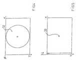
- the X- and Y-coordinatesare calculated, with reference to the upper left angle of the image that coincides with the same position on the sensor of the camera 2 (point ⁇ in FIG. 4 ).
- average, variance and standard deviation of the X-, Y-coordinatesare calculated so as to define the center of the reflected spot and to identify the interference of possible remote luminous signals that could be mistakenly associated with the spot.
- the driver board 5is continuously operated to make the luminous spot given by the LED 8 coincide with the center of the sensor of the camera 2 , as a result of the action of the electric motors 6 .
- the apparatus according to the inventionmakes the center position of the eye 7 coincide with the center of the CCD sensor of the camera and of the video signal processed by the FireWire IEEE 1394 port and the controller 4 , with a feedback control loop to automatically drive the electric motors 6 .
- the CPU unit 3determines two concentric areas 32 and 33 (see FIGS. 4 and 5 ).
- a bigger area 32is the area of the image useful to the test, the borders of the image being discarded due to the fact that they are often affected by undesired external reflections.
- the area 2can be circular, as in the example, or shaped differently (oval, squared etc.)
- the radius of the area 32may be defined by the medical operator, or established as a design parameter, the center coinciding with the CCD camera sensor center.
- a smaller area 33is instead the optimal area for the centering, i.e. the target area to be reached by the center of the spot in order to deem the eye 7 and camera sensor centered with respect to each other.
- the system automationis therefore to calculate the center position of the reflected spot with respect to the area center 33 so as to instruct the motors accordingly. In this way, through the driver board 5 and the motors 6 placed on two X- Y-directions, the movement of the optical head is driven with a frequency equal to that with which the frames are taken, i.e. every ten milliseconds.
- the lamp 16When the reflected image (spot) is deemed centered to the sensor (step A in FIGS. 7 and 8 ), through a suitable TTL signal that activates the driver board 5 , the lamp 16 is switched on. Said lamp 16 illuminates the slit 18 through the lamp condenser 17 . The luminous slit that is formed is projected on the eye along the axis 13 through the mirror 19 and the lens 23 . The optical head is now moved along the Z-direction, until the triangulation takes place, i.e. until the luminous slit, due to the geometric conditions that regulate the optical reflection, can be reflected by the corneal surface via the reflection axis 14 .
- the image of the slitbecomes superimposed to the image acquired by the camera 2 coming from the central channel 15 .
- the same geometric conditions just mentionedare such that the advancement of the optical head along the Z-direction corresponds to a shifting, from the left towards the right (considering the camera sensor as seen in FIGS. 4 and 5 ) of the image of the slit reflected by the cornea.
- the apparatusproceeds in the following manner.
- a check area or band 34( FIG. 5 ) is established on the image taken by the CCD camera sensor, in the left part thereof.
- the check area 34is a five pixels wide band starting from the left border of the sensor, but it may be less displaced with respect to the center, and be less wide and long according to the circumstances.
- the image in the check band 34is generally composed by a grey background with a low intensity value.
- the area 34is constantly checked, during the advancement along the -Z direction, with the maximum frequency allowed by the characteristics of the camera (for example around 100 frames per second).
- the maximum frequency allowed by the characteristics of the camerafor example around 100 frames per second.
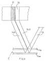
- the reflected beam 14 Bis captured by the camera as a luminous strip 35 (the above mentioned image of the slit) moving from left to right.
- the grey level intensity detected thereinincreases to a bigger value than a predetermined threshold value; this time t 0 is fixed like a temporal reference.
- the grey level intensity detection in the check areais carried out by average calculations over all the pixels forming the area.
- a suitable delay ⁇ tis set to control the acquisition.
- ⁇ tthe delay after the image 35 reflected by the epithelium Cep has been detected in the check area 34 , that an image reflected by the endothelium comes to an optimal position for being taken by the camera 2 .
- This situationis clearly represented in the same FIG. 6 , where the beam 14 A reflected by the endothelium Cend produces a strip image 37 which is displaced rearward with respect to the image 35 reflected by the epithelium Cep.
- the period of time ⁇ t that passes between to (reference) and the time in which the image of the endothelium is takenis then fundamental, and is evaluated on the basis of the advancement speed and the average thickness of the human cornea.
- the delay time ⁇ tcan in any case be adjusted manually or automatically.
- the photoflash 20is turned on, illuminating the cornea, and the image of the endothelium is taken through the camera 2 .
- a number of different imagescan also be taken, so that the one having the best quality can be chosen.
- the imagesare stored in a database for possible further processing or treatment. As the acquisition cycle is closed, the apparatus returns in the start configuration awaiting a new test to be done.
- both the ⁇ t delay and the position of the check area 34can be changed so as to give to the medical operator the possibility to obtain better images also in case of corneas with particular morphologies.
- the photoflash lamp 20thanks to its supplementary luminous impulse, permits to lower the gain of the camera 2 and so to have less noisy images. Said photoflash can be activated with a certain advance with respect to the lapse of ⁇ t, considering the intrinsical lag of the device.
- the advantageous characteristics of the apparatus according to the inventionattain the object stated in the introductory part.
- the quality of the endothelium imagescan be increased even further, with respect to known apparatus using conventional focusing techniques, by the possibility of taking a number of frames, and then choosing the highest quality one.
- the patients, the tests and the captured imagesare stored in a database, permitting to work on the taken data even after the test.
- Thispermits to rely on useful clinical parameters and, subsequently, to process the same so as to define the number and the density of the cells, their shape, their surface, i.e. their maximum, minimum and average area, the deviation from the standard parameters, a variance coefficient, the ratio of cells of various form, graphics of the distribution, of the dimension of cells areas and graphics of the perimeters distribution.
- the testcan be carried out with a reduced assistance by the medical operator, thanks to the automatic control of the same test as described above.
Landscapes
- Life Sciences & Earth Sciences (AREA)
- Health & Medical Sciences (AREA)
- Medical Informatics (AREA)
- Biophysics (AREA)
- Ophthalmology & Optometry (AREA)
- Engineering & Computer Science (AREA)
- Biomedical Technology (AREA)
- Heart & Thoracic Surgery (AREA)
- Physics & Mathematics (AREA)
- Molecular Biology (AREA)
- Surgery (AREA)
- Animal Behavior & Ethology (AREA)
- General Health & Medical Sciences (AREA)
- Public Health (AREA)
- Veterinary Medicine (AREA)
- Eye Examination Apparatus (AREA)
- Microscoopes, Condenser (AREA)
Abstract
Description
- The present invention refers to a new non-contact endothelium reflection microscope apparatus that permits to automatically obtain the endothelium image and to display clinically useful parameters such as the number and density of the cells, shape, surface, minimum, maximum and medium area, standard deviation, variation coefficient, percentage of cells of various shapes, area distribution histogram, perimeter distribution histogram.
- The endothelium is the most internal layer of the tissues forming the cornea, and consists of a single layer of flat polygonal cells. The endothelium function is to adjust the water contents, permitting a suitable hydration of the cornea. The shape and amount of the cells influence the quality of the vision. The cornea transparency is subjected to a very delicate balance, and a number of diseases can produce a loss of the transparency.
- The endothelium cells are of hexagonal shape in the children and in the young people. They do not reproduce themselves and. At birth, the density is about 4000 cells per square millimeter but as years pass the number decreases and the cells change their shape. The average density in an adult becomes of 2700 cells per square millimeter, in a range from 1600 to 3200 cells for square millimeter. The loss of cells brings about two main morphologic changes: the presence of cells with different surface area, and the increase in the amount of cells shaped differently from the basic hexagonal shape.
- The evaluation of the cornea endothelium is useful to have a first clinic indication regarding the risks of a surgical step, and for checking a diagnostic assumption or a therapy effectiveness. In this kind of evaluation, it is very important to observe heterogeneous parts, such as intracellular and intercellular areas of no reflectance (dark spots), hyper reflective areas (bright spots), empty areas in the cells layer (guttae), bubbles, Descemet's membrane rupture lines.
- Said parts can be checked in relationship with the evolution of different endothelium diseases of inflammatory or dystrophic nature. The quantity evaluation permits to assign to a determined photographic field a numeric parameter useful for the study of the endothelium variations in time, or for the comparison among different patients.
- The most easily accessible parameter is the average cellular density, obtained for comparison or and by counting the cellular elements. The first method is carried out by comparing the cellular dimensions with the dimensions of the hexagonal reticules that correspond to determined densities. The counting of the cellular elements, instead, is carried out by using fixed or variable reticules.
- The two methods give no information on the evolution of the cellular dimensions. This can be obtained by identifying, above and beyond the dimension of the average cellular area and its variability, also the perimeters of the cells. The endothelium reflection microscopic observation was first introduced in the opthalmologic practice around 1960 by David Maurice who, by modifying a metallography microscope, was able to obtain photographic images of a rabbit corneal endothelium. Exploiting the same theoretic principles, a microscope was subsequently proposed capable of taking photographs of the endothelium without contacting the eye.
- The non-contact reflection microscope apparatus are generally derived from normal slit lamps with a high magnification microscope. The technical principle on which they are based is the visualization of a determined structure in relation to its capability of reflecting an incident ray of light used for the illumination. In the commonly used technique (triangulation), the observation angle is of about 45°, the microscope being placed such that the bisector axis of the angle of view is perpendicular to the plane tangent to the corneal surface.
- The non-contact endothelium microscopy is particular indicated in all cases where the contact with the cornea can be dangerous, and therefore immediately after surgery or when there is an extreme structural fragility of the cornea. With the integration of the microscope with techniques of image analysis, the apparatus is able to give also a quantitative description of the endothelium tissue, expressed by the average cellular density and specific morphometric parameters.
- A non-contact endothelium microscope according to the prior art is shown for example in European Patent Application n. EP628281. The optical unit in this apparatus comprises an illuminating system, for obliquely illuminating through a slit an eyeball surface of a subject eye, and an eye-front observation optical system in which alignment-use indicator light for positional adjustment of the imaging optical axis is projected towards the eye and the resulting reflected light is received and imaged by a TV camera. An enlarged-imaging optical system is also provided for enlarged observation or enlarged photographing of the subject part by the TV camera based on slit illuminating light with which the eyeball surface has been illuminated.
- A photo-detector is arranged so as to detect a position at which the enlarged-imaging optical system has been focused on the subject part, via a reflected optical path other than that via which the enlarged image has been formed by the enlarged-imaging optical system. The whole optical unit is automatically moved both in a transversal direction and in a direction toward the eye, in response to the location of the above mentioned indicator light as displayed on a screen of a video monitor, so that the location chases a specified position on the screen. The enlarged visual image of the subject portion of the cornea is thus photographed via the TV camera when the photo-detector detects the focusing.
- The above described system, with the use of a focusing detection photo-detector placed along a supplementary reflected optical path, renders the apparatus sophisticated, and thus costly to be produced and maintained in order to have reliable results.
- The apparatus according to the invention permits to have the endothelium test without use of sensors, photosensors or other devices placed onto a reflected optical path. A higher quality endothelium image is obtained with a reduced use of electronic components and so with greater reliability, completeness and use flexibility in comparison with the prior art.
- The essential characteristics of the microscope apparatus for the morphometric analysis of the cornea endothelium with direct image acquisition according to the invention are defined by the first of the annexed claims.
- The characteristics and advantages of the microscope apparatus for the morphometric analysis of the cornea endothelium with direct image acquisition according to the present invention will be made clearer by the following description of embodiments thereof, given purely as a an example and not limitative, with reference to the accompanying drawings, wherein:
FIG. 1 is an optical path diagram of a first embodiment of the apparatus according to the invention;FIG. 2 is an optical path diagram of a second embodiment of the apparatus according to the invention;FIG. 3 is a block diagram representing the hardware configuration of an apparatus according to the invention;FIGS. 4 and 5 are explanatory views showing respective images displayed on a monitor screen during the image acquisition procedures according to the invention;FIG. 6 schematically represents exemplifying reflections obtained with the apparatus; and- FIGS.7 to9 are flowcharts showing the procedures for image acquisition with the apparatus according to the invention.
- Referring to FIGS.1 to3, the apparatus according to the invention comprises a movable optical head or
microscope 1 provided with a CCDhigh speed camera 2, i.e. a monochrome digital camera with shooting capacity of at least one hundred frames per second with FireWire high speed data output, i.e. with IEEE 1394 port or equivalent. - The
high speed camera 2 is directly connected to aCPU unit 3. Theunit 3 comprises acontroller 4, e.g. a 65XX type controller produced by the company National Instruments (United States, Texas) or equivalent. Thecontroller 4 controls apower driver board 5, so that the signal coming from theCPU unit 3 is suitable for driving electric DC motors6 as described hereinafter. - The function of the motors6 is to set in position the
microscope 1 with thecamera 2, following to automatic control by theCPU unit 3 so that the eye center7 to be examined is found. Such a finding is obtained via a reflection onto the cornea surface of the light emitted by aninfrared LED 8 mounted onto the mobile head of the apparatus, consisting of themicroscope 1 with thecamera 2. - The cited electronic components are connected each other according to known configurations. Considering instead more in detail the optical scheme of
FIG. 1 , a second LED9 with associatedoptics 10 is arranged nearby theinfrared LED 8 for providing the fixation point in association with asemireflecting mirror 11 and asemireflecting mirror 12, necessary to arrange the microscope in a way to center the patient eye and to obtain the triangulation necessary for the test. These components, like the other that follow and that form the optical scheme, are triangulation elements for the endothelium test, known and already in use for this kind of applications. - The optical scheme comprises then a
side projection axis 13, aside reflection axis 14 and acentral channel 15. In the embodiment ofFIG. 1 , transversally to theside projection axis 13, ahalogen lamp 16 is arranged with alamp condenser 17 and aslit 18. Along theside projection axis 13 there is also placed asemireflecting mirror 19 receiving the light beam generated by thehalogen lamp 16 and the beam that can be generated by a photoflash20 located at the start of theside projection axis 13. On the same axis, the photoflash20 is followed by aphotoflash condenser 21, aslit 22 and, beyond themirror 19, by a anoptical unit 23 that concentrates the beam onto the patient eye7. In the embodiment ofFIG. 2 thelamp 16, thecondenser 17, theslit 18, thesemireflecting mirror 19 and the photoflash20 are replaced by astroboscopic lamp 36 activated analogously and with the same function to the previous elements. - Along the
side reflection axis 14 there is arranged a side reflectionoptical unit 24 that concentrates the reflected beam and the endothelium image to amirror 25, from which the beam and the image signal are reflected to thecentral channel 15 passing through afilter 26 and a magnifyingoptical unit 27. The beam, and the endothelium image conveyed therewith, joins thecentral channel 15 in a point where adichroic mirror 28 is arranged. - The
central channel 15 also provides for, starting from the examined eye7, the above mentionedsemireflecting mirror 12 and a centraloptical unit 29 that concentrates the image of the eye7 and of theLED 8 to thehigh speed camera 2, passing through thedichroic mirror 28. - The system is controlled by two
pulses controller 4. Thefirst pulse 30 transmits the on/off signal to theLEDs 8 and9, to the photoflash20 and to thehalogen lamp 16. Thesecond pulse 31 transmits the signal for the operation of the motors6. - The optical head is driven by the motors along three Cartesian directions where the low-high direction corresponds to a Y-direction, the direction of horizontally approaching to and mowing away from the eye corresponds to a Z-direction, and the transversal sideways direction corresponds to a X-direction.
- With reference also to FIGS.4 to6 and to the self-explanatory flowcharts of FIGS.7 to9, the microscope according to the invention works in the following way.
- After arranging the optical head at the desired position, the test starts with the turning on of LED9 giving the fixation point for the patient. At the same time, the
infrared LED 8 is switched on, projecting via the reflecting mirror12 a spot of light onto the cornea surface. This spot is detected by thecamera 2 along thecentral channel 15.Camera 2 starts then acquiring images, with a resolution of at least 656×400 pixels, taken continuously with a frequency of about 100 Hz. - On each acquired frame, data acquisition procedures are carried out for identifying the points (pixels) in which the grey level is inside a certain predetermined range, so as to eliminate the darker and the clearer points of the prefixed range, and to identify all the points that belong to the light spot reflected by the cornea, and thus to precisely outline the same spot.
- Of all the pixels that form the image of the reflected spot the X- and Y-coordinates are calculated, with reference to the upper left angle of the image that coincides with the same position on the sensor of the camera2 (point ø in
FIG. 4 ). - Subsequently, average, variance and standard deviation of the X-, Y-coordinates are calculated so as to define the center of the reflected spot and to identify the interference of possible remote luminous signals that could be mistakenly associated with the spot.
- The
driver board 5 is continuously operated to make the luminous spot given by theLED 8 coincide with the center of the sensor of thecamera 2, as a result of the action of the electric motors6. In practice, the apparatus according to the invention makes the center position of the eye7 coincide with the center of the CCD sensor of the camera and of the video signal processed by the FireWire IEEE 1394 port and thecontroller 4, with a feedback control loop to automatically drive the electric motors6. - In greater detail, the
CPU unit 3 determines twoconcentric areas 32 and33 (seeFIGS. 4 and 5 ). Abigger area 32 is the area of the image useful to the test, the borders of the image being discarded due to the fact that they are often affected by undesired external reflections. When the center of the above mentioned light spot is outside thearea 32, the continuation of the test is not permitted. Thearea 2 can be circular, as in the example, or shaped differently (oval, squared etc.) - The radius of the
area 32 may be defined by the medical operator, or established as a design parameter, the center coinciding with the CCD camera sensor center. Asmaller area 33 is instead the optimal area for the centering, i.e. the target area to be reached by the center of the spot in order to deem the eye7 and camera sensor centered with respect to each other. - After that the center of the reflected spot has been calculated as mentioned, the distance of this from the center of the smaller area33 (which can even be a single pixel), and the motors are continuously operated to drive the
optical head 1 along the X- and Y-directions until such distance is minimized, that is to say the center of the reflected spot is brought (and kept) inside thearea 33. In practice, the system automation is therefore to calculate the center position of the reflected spot with respect to thearea center 33 so as to instruct the motors accordingly. In this way, through thedriver board 5 and the motors6 placed on two X- Y-directions, the movement of the optical head is driven with a frequency equal to that with which the frames are taken, i.e. every ten milliseconds. - When the reflected image (spot) is deemed centered to the sensor (step A in
FIGS. 7 and 8 ), through a suitable TTL signal that activates thedriver board 5, thelamp 16 is switched on. Saidlamp 16 illuminates theslit 18 through thelamp condenser 17. The luminous slit that is formed is projected on the eye along theaxis 13 through themirror 19 and thelens 23. The optical head is now moved along the Z-direction, until the triangulation takes place, i.e. until the luminous slit, due to the geometric conditions that regulate the optical reflection, can be reflected by the corneal surface via thereflection axis 14. When this reflection occurs, the image of the slit becomes superimposed to the image acquired by thecamera 2 coming from thecentral channel 15. The same geometric conditions just mentioned are such that the advancement of the optical head along the Z-direction corresponds to a shifting, from the left towards the right (considering the camera sensor as seen inFIGS. 4 and 5 ) of the image of the slit reflected by the cornea. - In order to have high quality images of the endothelium, it is important that the images be captured, and also (preferably) the cornea be illuminated by the photoflash20, in the time in which the incident beam coming from the
side projection axis 13 is in the optimal position to create the necessary reflection on the layer of the endothelium cells. To this purpose, the apparatus according to the invention proceeds in the following manner. - A check area or band34 (
FIG. 5 ) is established on the image taken by the CCD camera sensor, in the left part thereof. In the example thecheck area 34 is a five pixels wide band starting from the left border of the sensor, but it may be less displaced with respect to the center, and be less wide and long according to the circumstances. In the absence of a triangulation, the image in thecheck band 34 is generally composed by a grey background with a low intensity value. - The
area 34 is constantly checked, during the advancement along the -Z direction, with the maximum frequency allowed by the characteristics of the camera (for example around 100 frames per second). With particular reference also toFIG. 6 , there is represented a beam14B reflected by the cornea C, and more precisely by the superficial part thereof, the epithelium Cep. The reflected beam14B is captured by the camera as a luminous strip35 (the above mentioned image of the slit) moving from left to right. - When the
luminous strip 35 enters thecheck area 34 the grey level intensity detected therein increases to a bigger value than a predetermined threshold value; this time t0is fixed like a temporal reference. The grey level intensity detection in the check area is carried out by average calculations over all the pixels forming the area. - From the time t0a suitable delay Δt is set to control the acquisition. In fact, considering the advancement speed of the head along the Z-direction and above all the thickness of the cornea, it is only with a certain delay after the
image 35 reflected by the epithelium Cep has been detected in thecheck area 34, that an image reflected by the endothelium comes to an optimal position for being taken by thecamera 2. This situation is clearly represented in the sameFIG. 6 , where thebeam 14A reflected by the endothelium Cend produces astrip image 37 which is displaced rearward with respect to theimage 35 reflected by the epithelium Cep. - The period of time Δt that passes between to (reference) and the time in which the image of the endothelium is taken is then fundamental, and is evaluated on the basis of the advancement speed and the average thickness of the human cornea. The delay time Δt can in any case be adjusted manually or automatically. As the delay time Δt passes, the photoflash20 is turned on, illuminating the cornea, and the image of the endothelium is taken through the
camera 2. A number of different images can also be taken, so that the one having the best quality can be chosen. The images are stored in a database for possible further processing or treatment. As the acquisition cycle is closed, the apparatus returns in the start configuration awaiting a new test to be done. - As mentioned, both the Δt delay and the position of the
check area 34 can be changed so as to give to the medical operator the possibility to obtain better images also in case of corneas with particular morphologies. The photoflash lamp20, thanks to its supplementary luminous impulse, permits to lower the gain of thecamera 2 and so to have less noisy images. Said photoflash can be activated with a certain advance with respect to the lapse of Δt, considering the intrinsical lag of the device. - The advantageous characteristics of the apparatus according to the invention attain the object stated in the introductory part. The absence of a photosensor or of a linear sensor along an optical reflection path; the acquisition procedure, controlled and realized by means of simple software instructions given to the apparatus as described above, ensures a better reliability, lower costs and a better use flexibility. Furthermore, the quality of the endothelium images can be increased even further, with respect to known apparatus using conventional focusing techniques, by the possibility of taking a number of frames, and then choosing the highest quality one.
- The patients, the tests and the captured images are stored in a database, permitting to work on the taken data even after the test. This permits to rely on useful clinical parameters and, subsequently, to process the same so as to define the number and the density of the cells, their shape, their surface, i.e. their maximum, minimum and average area, the deviation from the standard parameters, a variance coefficient, the ratio of cells of various form, graphics of the distribution, of the dimension of cells areas and graphics of the perimeters distribution. The test can be carried out with a reduced assistance by the medical operator, thanks to the automatic control of the same test as described above.
- Variations and/or modifications can be brought to the endothelium reflection microscope for morphometric analysis with direct image acquisition according to the invention, without for this reason departing from the scope of the invention itself as defined in the annexed claims.
Claims (12)
Applications Claiming Priority (4)
| Application Number | Priority Date | Filing Date | Title |
|---|---|---|---|
| EP04425498.5 | 2004-07-08 | ||
| EP04425498 | 2004-07-08 | ||
| EP04425498 | 2004-07-08 | ||
| PCT/IB2005/001908WO2006006048A1 (en) | 2004-07-08 | 2005-07-06 | Reflection microscope for examination of the corneal endothelium and method of operating same |
Publications (2)
| Publication Number | Publication Date |
|---|---|
| US20070263172A1true US20070263172A1 (en) | 2007-11-15 |
| US7726814B2 US7726814B2 (en) | 2010-06-01 |
Family
ID=34932614
Family Applications (2)
| Application Number | Title | Priority Date | Filing Date |
|---|---|---|---|
| US11/632,084Active2026-09-09US7726814B2 (en) | 2004-07-08 | 2005-07-06 | Reflection microscope and method |
| US12/661,748ActiveUS7896495B2 (en) | 2004-07-08 | 2010-03-23 | Reflection microscope apparatus |
Family Applications After (1)
| Application Number | Title | Priority Date | Filing Date |
|---|---|---|---|
| US12/661,748ActiveUS7896495B2 (en) | 2004-07-08 | 2010-03-23 | Reflection microscope apparatus |
Country Status (8)
| Country | Link |
|---|---|
| US (2) | US7726814B2 (en) |
| EP (1) | EP1786314B1 (en) |
| JP (1) | JP4990133B2 (en) |
| AU (1) | AU2005261423B2 (en) |
| CA (1) | CA2572664C (en) |
| ES (1) | ES2541305T3 (en) |
| PT (1) | PT1786314E (en) |
| WO (1) | WO2006006048A1 (en) |
Cited By (21)
| Publication number | Priority date | Publication date | Assignee | Title |
|---|---|---|---|---|
| US20080055544A1 (en)* | 2006-08-31 | 2008-03-06 | Tomey Corporation | Cornea imaging apparatus |
| US20130253406A1 (en)* | 2010-11-15 | 2013-09-26 | Aquesys, Inc. | Methods for deploying intraocular shunts |
| US9393153B2 (en) | 2010-11-15 | 2016-07-19 | Aquesys, Inc. | Methods for intraocular shunt placement |
| US9585790B2 (en) | 2013-11-14 | 2017-03-07 | Aquesys, Inc. | Intraocular shunt inserter |
| US9592154B2 (en) | 2011-12-08 | 2017-03-14 | Aquesys, Inc. | Intraocular shunt manufacture |
| US9610195B2 (en) | 2013-02-27 | 2017-04-04 | Aquesys, Inc. | Intraocular shunt implantation methods and devices |
| US9636254B2 (en) | 2006-06-30 | 2017-05-02 | Aquesys, Inc. | Systems for reducing pressure in an organ |
| US20170161892A1 (en)* | 2014-07-02 | 2017-06-08 | Si14 S.P.A. | A method for acquiring and processing images of an ocular fundus by means of a portable electronic device |
| US9693901B2 (en) | 2010-11-15 | 2017-07-04 | Aquesys, Inc. | Shunt placement through the sclera |
| US9808373B2 (en) | 2013-06-28 | 2017-11-07 | Aquesys, Inc. | Intraocular shunt implantation |
| US9877866B2 (en) | 2010-11-15 | 2018-01-30 | Aquesys, Inc. | Intraocular shunt placement |
| US9883969B2 (en) | 2011-12-08 | 2018-02-06 | Aquesys, Inc. | Intrascleral shunt placement |
| US10004638B2 (en) | 2010-11-15 | 2018-06-26 | Aquesys, Inc. | Intraocular shunt delivery |
| US10080682B2 (en) | 2011-12-08 | 2018-09-25 | Aquesys, Inc. | Intrascleral shunt placement |
| US10085884B2 (en) | 2006-06-30 | 2018-10-02 | Aquesys, Inc. | Intraocular devices |
| US10463537B2 (en) | 2015-06-03 | 2019-11-05 | Aquesys Inc. | Ab externo intraocular shunt placement |
| US10667947B2 (en) | 2016-06-02 | 2020-06-02 | Aquesys, Inc. | Intraocular drug delivery |
| US10842671B2 (en) | 2010-11-15 | 2020-11-24 | Aquesys, Inc. | Intraocular shunt placement in the suprachoroidal space |
| US10952898B2 (en) | 2018-03-09 | 2021-03-23 | Aquesys, Inc. | Intraocular shunt inserter |
| US11135089B2 (en) | 2018-03-09 | 2021-10-05 | Aquesys, Inc. | Intraocular shunt inserter |
| US11246753B2 (en) | 2017-11-08 | 2022-02-15 | Aquesys, Inc. | Manually adjustable intraocular flow regulation |
Families Citing this family (8)
| Publication number | Priority date | Publication date | Assignee | Title |
|---|---|---|---|---|
| JP5026134B2 (en)* | 2007-03-30 | 2012-09-12 | 株式会社トーメーコーポレーション | Corneal imaging apparatus and cornea imaging method |
| US8718335B2 (en)* | 2007-11-29 | 2014-05-06 | Wavefront Biometric Technologies Pty Limited | Biometric authentication using the eye |
| JP6301246B2 (en)* | 2011-03-25 | 2018-04-11 | ノバルティス エージー | Method and system for imaging optical elements |
| CN102871644B (en)* | 2012-09-28 | 2014-12-10 | 北京锐视觉科技有限公司 | Slit lamp and information management system thereof |
| EP2777628B1 (en)* | 2013-03-15 | 2018-02-28 | Neos Surgery, S.L. | Device for repairing an intervertebral disc |
| JP6355930B2 (en)* | 2014-01-30 | 2018-07-11 | 株式会社トーメーコーポレーション | Corneal imaging device |
| ES2951291T3 (en)* | 2014-03-14 | 2023-10-19 | Lkc Tech Inc | System and procedure for the detection of retinopathy |
| CN115135228A (en)* | 2020-02-21 | 2022-09-30 | 卡尔蔡司医疗技术公司 | OCT suspensory ligament imaging |
Citations (4)
| Publication number | Priority date | Publication date | Assignee | Title |
|---|---|---|---|---|
| US5381194A (en)* | 1992-02-07 | 1995-01-10 | Kabushiki Kaisha Topcon | Apparatus for photographing a corneal endothelium |
| US5471261A (en)* | 1993-09-02 | 1995-11-28 | Konan Inc. | Apparatus for obtaining images of cornea endothelium |
| US5757461A (en)* | 1993-09-22 | 1998-05-26 | Konan Inc. | Method for displaying a photographing point of a cornea in an apparatus for obtaining video images of corneal cells |
| US6164778A (en)* | 1998-12-24 | 2000-12-26 | Kabushiki Kaisha Topcon | Corneal endothelial cell photographing apparatus |
Family Cites Families (11)
| Publication number | Priority date | Publication date | Assignee | Title |
|---|---|---|---|---|
| JPH06160727A (en)* | 1992-11-18 | 1994-06-07 | Koonan:Kk | Eyeball microscope |
| US5548354A (en) | 1993-06-10 | 1996-08-20 | Konan Common Co., Ltd. | Method for observing and photographing a cornea and apparatus for the same |
| JP3415226B2 (en)* | 1993-07-15 | 2003-06-09 | 株式会社甲南コモン | Corneal cell imaging device |
| JP2607216B2 (en)* | 1993-06-30 | 1997-05-07 | 株式会社コーナン | Centering device in eyeball imaging device |
| JPH0767837A (en)* | 1993-09-02 | 1995-03-14 | Konan:Kk | Cornea endothelium photographing device |
| TW272311B (en)* | 1994-01-12 | 1996-03-11 | At & T Corp | |
| JPH08173387A (en)* | 1994-12-26 | 1996-07-09 | Konan:Kk | Cornea cell photographing system with cornea thickness measuring function |
| JPH10179523A (en)* | 1996-12-26 | 1998-07-07 | Canon Inc | Digital camera |
| JP3710601B2 (en)* | 1997-07-15 | 2005-10-26 | 株式会社トプコン | Ophthalmic equipment |
| JP3854888B2 (en)* | 2002-04-09 | 2006-12-06 | 株式会社ビー・エム・エル | Atherosclerosis detection system |
| DE10138158A1 (en) | 2001-08-09 | 2003-02-20 | Rhine Tec Ges Fuer Virtuelle I | Contact free magnified imaging of a section of the epithelium or endothelium of the eye using a slit illumination arrangement that is used to record multiple images of the cornea |
- 2005
- 2005-07-06EPEP05785108.1Apatent/EP1786314B1/ennot_activeExpired - Lifetime
- 2005-07-06ESES05785108.1Tpatent/ES2541305T3/ennot_activeExpired - Lifetime
- 2005-07-06PTPT57851081Tpatent/PT1786314E/enunknown
- 2005-07-06WOPCT/IB2005/001908patent/WO2006006048A1/enactiveApplication Filing
- 2005-07-06USUS11/632,084patent/US7726814B2/enactiveActive
- 2005-07-06AUAU2005261423Apatent/AU2005261423B2/ennot_activeExpired
- 2005-07-06CACA2572664Apatent/CA2572664C/ennot_activeExpired - Lifetime
- 2005-07-06JPJP2007519904Apatent/JP4990133B2/ennot_activeExpired - Lifetime
- 2010
- 2010-03-23USUS12/661,748patent/US7896495B2/enactiveActive
Patent Citations (4)
| Publication number | Priority date | Publication date | Assignee | Title |
|---|---|---|---|---|
| US5381194A (en)* | 1992-02-07 | 1995-01-10 | Kabushiki Kaisha Topcon | Apparatus for photographing a corneal endothelium |
| US5471261A (en)* | 1993-09-02 | 1995-11-28 | Konan Inc. | Apparatus for obtaining images of cornea endothelium |
| US5757461A (en)* | 1993-09-22 | 1998-05-26 | Konan Inc. | Method for displaying a photographing point of a cornea in an apparatus for obtaining video images of corneal cells |
| US6164778A (en)* | 1998-12-24 | 2000-12-26 | Kabushiki Kaisha Topcon | Corneal endothelial cell photographing apparatus |
Cited By (34)
| Publication number | Priority date | Publication date | Assignee | Title |
|---|---|---|---|---|
| US9636254B2 (en) | 2006-06-30 | 2017-05-02 | Aquesys, Inc. | Systems for reducing pressure in an organ |
| US10085884B2 (en) | 2006-06-30 | 2018-10-02 | Aquesys, Inc. | Intraocular devices |
| US7572010B2 (en)* | 2006-08-31 | 2009-08-11 | Tomey Corporation | Cornea imaging apparatus |
| US20080055544A1 (en)* | 2006-08-31 | 2008-03-06 | Tomey Corporation | Cornea imaging apparatus |
| US9326891B2 (en)* | 2010-11-15 | 2016-05-03 | Aquesys, Inc. | Methods for deploying intraocular shunts |
| US10940040B2 (en) | 2010-11-15 | 2021-03-09 | Aquesys, Inc. | Intraocular shunt placement |
| US10842671B2 (en) | 2010-11-15 | 2020-11-24 | Aquesys, Inc. | Intraocular shunt placement in the suprachoroidal space |
| US10307293B2 (en) | 2010-11-15 | 2019-06-04 | Aquesys, Inc. | Methods for intraocular shunt placement |
| US9393153B2 (en) | 2010-11-15 | 2016-07-19 | Aquesys, Inc. | Methods for intraocular shunt placement |
| US9980854B2 (en) | 2010-11-15 | 2018-05-29 | Aquesys, Inc. | Shunt placement through the sclera |
| US9693901B2 (en) | 2010-11-15 | 2017-07-04 | Aquesys, Inc. | Shunt placement through the sclera |
| US20130253406A1 (en)* | 2010-11-15 | 2013-09-26 | Aquesys, Inc. | Methods for deploying intraocular shunts |
| US9877866B2 (en) | 2010-11-15 | 2018-01-30 | Aquesys, Inc. | Intraocular shunt placement |
| US10004638B2 (en) | 2010-11-15 | 2018-06-26 | Aquesys, Inc. | Intraocular shunt delivery |
| US10080682B2 (en) | 2011-12-08 | 2018-09-25 | Aquesys, Inc. | Intrascleral shunt placement |
| US9883969B2 (en) | 2011-12-08 | 2018-02-06 | Aquesys, Inc. | Intrascleral shunt placement |
| US9592154B2 (en) | 2011-12-08 | 2017-03-14 | Aquesys, Inc. | Intraocular shunt manufacture |
| US10524959B2 (en) | 2013-02-27 | 2020-01-07 | Aquesys, Inc. | Intraocular shunt implantation methods and devices |
| US9610195B2 (en) | 2013-02-27 | 2017-04-04 | Aquesys, Inc. | Intraocular shunt implantation methods and devices |
| US10369048B2 (en) | 2013-06-28 | 2019-08-06 | Aquesys, Inc. | Intraocular shunt implantation |
| US9808373B2 (en) | 2013-06-28 | 2017-11-07 | Aquesys, Inc. | Intraocular shunt implantation |
| US11298264B2 (en) | 2013-06-28 | 2022-04-12 | Aquesys, Inc. | Intraocular shunt implantation |
| US10470928B2 (en) | 2013-11-14 | 2019-11-12 | Aquesys, Inc. | Intraocular shunt inserter |
| US11938059B2 (en) | 2013-11-14 | 2024-03-26 | Aquesys, Inc. | Intraocular shunt insertion techniques |
| US10653555B2 (en) | 2013-11-14 | 2020-05-19 | Aquesys, Inc. | Intraocular shunt insertion techniques |
| US9585790B2 (en) | 2013-11-14 | 2017-03-07 | Aquesys, Inc. | Intraocular shunt inserter |
| US20170161892A1 (en)* | 2014-07-02 | 2017-06-08 | Si14 S.P.A. | A method for acquiring and processing images of an ocular fundus by means of a portable electronic device |
| US10463537B2 (en) | 2015-06-03 | 2019-11-05 | Aquesys Inc. | Ab externo intraocular shunt placement |
| US11612517B2 (en) | 2015-06-03 | 2023-03-28 | Aquesys, Inc. | Ab externo intraocular shunt placement |
| US10470927B2 (en) | 2015-06-03 | 2019-11-12 | Aquesys, Inc. | AB externo intraocular shunt placement |
| US10667947B2 (en) | 2016-06-02 | 2020-06-02 | Aquesys, Inc. | Intraocular drug delivery |
| US11246753B2 (en) | 2017-11-08 | 2022-02-15 | Aquesys, Inc. | Manually adjustable intraocular flow regulation |
| US10952898B2 (en) | 2018-03-09 | 2021-03-23 | Aquesys, Inc. | Intraocular shunt inserter |
| US11135089B2 (en) | 2018-03-09 | 2021-10-05 | Aquesys, Inc. | Intraocular shunt inserter |
Also Published As
| Publication number | Publication date |
|---|---|
| ES2541305T3 (en) | 2015-07-17 |
| US7896495B2 (en) | 2011-03-01 |
| JP2008505673A (en) | 2008-02-28 |
| PT1786314E (en) | 2015-08-21 |
| CA2572664A1 (en) | 2006-01-19 |
| JP4990133B2 (en) | 2012-08-01 |
| EP1786314B1 (en) | 2015-04-08 |
| EP1786314A1 (en) | 2007-05-23 |
| AU2005261423B2 (en) | 2011-03-03 |
| US20100259724A1 (en) | 2010-10-14 |
| AU2005261423A1 (en) | 2006-01-19 |
| US7726814B2 (en) | 2010-06-01 |
| CA2572664C (en) | 2016-02-23 |
| WO2006006048A1 (en) | 2006-01-19 |
Similar Documents
| Publication | Publication Date | Title |
|---|---|---|
| EP1786314B1 (en) | Reflection microscope for examination of the corneal endothelium and method of operating same | |
| JP4113005B2 (en) | Eye examination equipment | |
| US8857987B2 (en) | Ophthalmic apparatus | |
| JP4937840B2 (en) | Ophthalmic equipment | |
| JP4458935B2 (en) | Perimeter | |
| JP2000333905A (en) | Ophthalmic device | |
| JP2012100713A (en) | Ophthalmologic apparatus | |
| JP2014083358A (en) | Ophthalmologic apparatus, ophthalmology control method, and program | |
| JP2016185192A (en) | Ophthalmic apparatus and method for controlling ophthalmic apparatus | |
| JP4408640B2 (en) | Ophthalmic measuring device | |
| JP2014079392A (en) | Ophthalmology imaging apparatus | |
| JPH11276439A (en) | Ophthalmologic photographing apparatus | |
| JP3880475B2 (en) | Ophthalmic equipment | |
| JP2001017459A (en) | Surgical microscope | |
| JP3636917B2 (en) | Eye refractive power measurement device | |
| WO2020032128A1 (en) | Ophthalmic photographing device | |
| RU2352244C2 (en) | Method of measurement of fast movements of eyes and deviations of solid vision and device for its realisation | |
| CN114615922A (en) | Ophthalmologic apparatus and control method therefor | |
| JPH07136119A (en) | Ophthalmic equipment | |
| JP3995590B2 (en) | Ophthalmic equipment | |
| JPH0975308A (en) | Corneal endothelial cell imaging system | |
| JP2003024280A (en) | Anterior segment cross-sectional analyzer and anterior segment cross-sectional analyzing program | |
| JPH0654807A (en) | Ophthalmic device | |
| JP2001025459A (en) | Ophthalmic measurement device | |
| JP2003038442A (en) | Corneal shape measuring device |
Legal Events
| Date | Code | Title | Description |
|---|---|---|---|
| AS | Assignment | Owner name:CONSTRUZIONI STRUMENTI OFTALMICI C.S.O. S.R.L., IT Free format text:ASSIGNMENT OF ASSIGNORS INTEREST;ASSIGNOR:MURA, SERGIO;REEL/FRAME:018782/0390 Effective date:20061206 Owner name:CONSTRUZIONI STRUMENTI OFTALMICI C.S.O. S.R.L.,ITA Free format text:ASSIGNMENT OF ASSIGNORS INTEREST;ASSIGNOR:MURA, SERGIO;REEL/FRAME:018782/0390 Effective date:20061206 | |
| AS | Assignment | Owner name:COSTRUZIONI STRUMENTI OFTALMICHI S.R.L.,ITALY Free format text:CORRECTIVE ASSIGNMENT TO CORRECT ASSIGNEE NAME ON REEL 18782 FRAME 390 THROUGHT 391;ASSIGNOR:MURA, SERGIO;REEL/FRAME:024347/0881 Effective date:20061206 | |
| STCF | Information on status: patent grant | Free format text:PATENTED CASE | |
| FEPP | Fee payment procedure | Free format text:PAYOR NUMBER ASSIGNED (ORIGINAL EVENT CODE: ASPN); ENTITY STATUS OF PATENT OWNER: SMALL ENTITY | |
| FPAY | Fee payment | Year of fee payment:4 | |
| MAFP | Maintenance fee payment | Free format text:PAYMENT OF MAINTENANCE FEE, 8TH YR, SMALL ENTITY (ORIGINAL EVENT CODE: M2552) Year of fee payment:8 | |
| MAFP | Maintenance fee payment | Free format text:PAYMENT OF MAINTENANCE FEE, 12TH YR, SMALL ENTITY (ORIGINAL EVENT CODE: M2553); ENTITY STATUS OF PATENT OWNER: SMALL ENTITY Year of fee payment:12 |