US20070249933A1 - Method and device for automatically differentiating types of kidney stones by means of computed tomography - Google Patents
Method and device for automatically differentiating types of kidney stones by means of computed tomographyDownload PDFInfo
- Publication number
- US20070249933A1 US20070249933A1US11/730,274US73027407AUS2007249933A1US 20070249933 A1US20070249933 A1US 20070249933A1US 73027407 AUS73027407 AUS 73027407AUS 2007249933 A1US2007249933 A1US 2007249933A1
- Authority
- US
- United States
- Prior art keywords
- ray attenuation
- attenuation values
- voxel
- prescribed
- kidney stones
- Prior art date
- Legal status (The legal status is an assumption and is not a legal conclusion. Google has not performed a legal analysis and makes no representation as to the accuracy of the status listed.)
- Granted
Links
Images


Classifications
- A—HUMAN NECESSITIES
- A61—MEDICAL OR VETERINARY SCIENCE; HYGIENE
- A61B—DIAGNOSIS; SURGERY; IDENTIFICATION
- A61B6/00—Apparatus or devices for radiation diagnosis; Apparatus or devices for radiation diagnosis combined with radiation therapy equipment
- A61B6/02—Arrangements for diagnosis sequentially in different planes; Stereoscopic radiation diagnosis
- A61B6/03—Computed tomography [CT]
- A61B6/032—Transmission computed tomography [CT]
- A—HUMAN NECESSITIES
- A61—MEDICAL OR VETERINARY SCIENCE; HYGIENE
- A61B—DIAGNOSIS; SURGERY; IDENTIFICATION
- A61B5/00—Measuring for diagnostic purposes; Identification of persons
- A61B5/48—Other medical applications
- A61B5/4869—Determining body composition
- A—HUMAN NECESSITIES
- A61—MEDICAL OR VETERINARY SCIENCE; HYGIENE
- A61B—DIAGNOSIS; SURGERY; IDENTIFICATION
- A61B6/00—Apparatus or devices for radiation diagnosis; Apparatus or devices for radiation diagnosis combined with radiation therapy equipment
- A61B6/48—Diagnostic techniques
- A61B6/482—Diagnostic techniques involving multiple energy imaging
- G—PHYSICS
- G06—COMPUTING OR CALCULATING; COUNTING
- G06T—IMAGE DATA PROCESSING OR GENERATION, IN GENERAL
- G06T7/00—Image analysis
- G06T7/0002—Inspection of images, e.g. flaw detection
- G06T7/0012—Biomedical image inspection
- G—PHYSICS
- G06—COMPUTING OR CALCULATING; COUNTING
- G06T—IMAGE DATA PROCESSING OR GENERATION, IN GENERAL
- G06T7/00—Image analysis
- G06T7/10—Segmentation; Edge detection
- G06T7/11—Region-based segmentation
- G—PHYSICS
- G06—COMPUTING OR CALCULATING; COUNTING
- G06T—IMAGE DATA PROCESSING OR GENERATION, IN GENERAL
- G06T7/00—Image analysis
- G06T7/97—Determining parameters from multiple pictures
- A—HUMAN NECESSITIES
- A61—MEDICAL OR VETERINARY SCIENCE; HYGIENE
- A61B—DIAGNOSIS; SURGERY; IDENTIFICATION
- A61B90/00—Instruments, implements or accessories specially adapted for surgery or diagnosis and not covered by any of the groups A61B1/00 - A61B50/00, e.g. for luxation treatment or for protecting wound edges
- A61B90/36—Image-producing devices or illumination devices not otherwise provided for
- A61B2090/364—Correlation of different images or relation of image positions in respect to the body
- A—HUMAN NECESSITIES
- A61—MEDICAL OR VETERINARY SCIENCE; HYGIENE
- A61B—DIAGNOSIS; SURGERY; IDENTIFICATION
- A61B6/00—Apparatus or devices for radiation diagnosis; Apparatus or devices for radiation diagnosis combined with radiation therapy equipment
- A61B6/40—Arrangements for generating radiation specially adapted for radiation diagnosis
- A61B6/4007—Arrangements for generating radiation specially adapted for radiation diagnosis characterised by using a plurality of source units
- A61B6/4014—Arrangements for generating radiation specially adapted for radiation diagnosis characterised by using a plurality of source units arranged in multiple source-detector units
- A—HUMAN NECESSITIES
- A61—MEDICAL OR VETERINARY SCIENCE; HYGIENE
- A61B—DIAGNOSIS; SURGERY; IDENTIFICATION
- A61B6/00—Apparatus or devices for radiation diagnosis; Apparatus or devices for radiation diagnosis combined with radiation therapy equipment
- A61B6/40—Arrangements for generating radiation specially adapted for radiation diagnosis
- A61B6/4035—Arrangements for generating radiation specially adapted for radiation diagnosis the source being combined with a filter or grating
- G—PHYSICS
- G06—COMPUTING OR CALCULATING; COUNTING
- G06T—IMAGE DATA PROCESSING OR GENERATION, IN GENERAL
- G06T2207/00—Indexing scheme for image analysis or image enhancement
- G06T2207/10—Image acquisition modality
- G06T2207/10072—Tomographic images
- G06T2207/10081—Computed x-ray tomography [CT]
- G—PHYSICS
- G06—COMPUTING OR CALCULATING; COUNTING
- G06T—IMAGE DATA PROCESSING OR GENERATION, IN GENERAL
- G06T2207/00—Indexing scheme for image analysis or image enhancement
- G06T2207/10—Image acquisition modality
- G06T2207/10141—Special mode during image acquisition
- G06T2207/10152—Varying illumination
- G—PHYSICS
- G06—COMPUTING OR CALCULATING; COUNTING
- G06T—IMAGE DATA PROCESSING OR GENERATION, IN GENERAL
- G06T2207/00—Indexing scheme for image analysis or image enhancement
- G06T2207/20—Special algorithmic details
- G06T2207/20004—Adaptive image processing
- G06T2207/20012—Locally adaptive
- G—PHYSICS
- G06—COMPUTING OR CALCULATING; COUNTING
- G06T—IMAGE DATA PROCESSING OR GENERATION, IN GENERAL
- G06T2207/00—Indexing scheme for image analysis or image enhancement
- G06T2207/30—Subject of image; Context of image processing
- G06T2207/30004—Biomedical image processing
- G06T2207/30084—Kidney; Renal
Definitions
- Embodiments of the present applicationgenerally relate to a method and/or a device for automatically differentiating types of kidney stones by way of computed tomography.
- itmay relate to one in which two computed tomography pictures of an object area including kidney stones are recorded in the context of a different spectral distribution of the X-radiation, and there are reconstructed from raw data of the two computed tomography pictures two image data records of the object area that include X-ray attenuation values of voxels of the object area in the context of the respective spectral distribution of the X-radiation.
- Identifying and distinguishing different types of kidney stones in the human or animal bodyis very important in deciding on a therapy.
- Uric acid stones, cystine stones, oxalate stones or hydroxyapatite stonesfor example count among the different types of kidney stones'. It is chiefly the differentiation between uric acid stones and other types of kidney stones that is of great importance here, since the therapy of uric acid stones differs substantially from those of the other types.
- the uric acid concentration in the bloodis determined chemically. A high concentration indicates that a uric acid stone could be present.
- solid constituentsare filtered out of the urine.
- constituents of kidney stonescan sometimes be detected in the urine after bodily movement, for example, through climbing steps, and be suitably investigated.
- a surgical interventionis performed in which the kidney stone is already removed for the purpose of simultaneous diagnosis and therapy. It is possible to classify kidney stones with the aid of conventional computed tomography only with difficulty, since kidney stones do not occur as pure, compact substances.
- pure calcium oxalateis clearly distinguished from crystallized uric acid in the X-ray attenuation value, the X-ray attenuation values can nevertheless be very similar in reality.
- At least one embodiment of the present inventionspecifies a method and/or a device for differentiating types of kidney stones that enable automatic differentiation of at least two types of kidney stones without surgical intervention.
- two computed tomography pictures of an object area that includes the kidney stones to be differentiatedare recorded in the context of a different spectral distribution of the X-radiation, and two image data records of the object area are reconstructed from the raw data of the two computed tomography pictures.
- the two image data recordsinclude the X-ray attenuation values of the voxels of the object area in the context of the respective spectral distribution of the X-radiation.
- X-ray attenuation valuescan be understood here both as the attenuation coefficients ⁇ and as values derived therefrom, such as the CT value.
- the two computed tomography picturesare recorded by using a multi-imaging computer tomograph, for example a so-called dual energy computer tomograph, with the aid of which it is possible simultaneously or at least virtually simultaneously to record two computed tomography pictures with a different spectral distribution of the X-radiation or different X-ray energy.
- a multi-imaging computer tomographfor example a so-called dual energy computer tomograph
- Different techniques for generating two computed tomography pictures with a different spectral distribution of the X-radiationare fundamentally known to the specialist.
- a ratio ris calculated from the two image data records for each voxel of at least one interesting slice of the object area if the mean value from the two assigned X-ray attenuation values of the respective voxel lies above a prescribed threshold value (X min ) that is characteristic of kidney stones.
- x 1represents either the measured X-ray attenuation value of the voxel in the context of one of the two different X-ray energies, or an X-ray attenuation value averaged for this X-ray energy, which will be examined more closely later.
- x 2represents either the measured X-ray attenuation value of the voxel in the context of the other X-ray energy, or an appropriately averaged X-ray attenuation value.
- the two X-ray attenuation values x 1 , x 2can either be extracted directly from the two image data records, or be calculated therefrom.
- the values o 1 and o 2 that also occurrepresent the X-ray attenuation values of pure urine in the context of the two X-ray energies. These values are prescribed. They are either already known, or can be determined in advance.
- the basis for the calculation of the ratio ris a 3-material decomposition in which the respective voxel is interpreted from a mixture of the base materials of urine, first type of kidney stone, in particular uric acid stone, and another type of kidney stone.
- high values of rcharacterize calcium-containing stones such as, for example, hydroxyapatite stones or oxalate stones.
- Medium values of rare measured for cystine stones (high sulfur content).
- Low values of rare yielded for the uric acid stone.
- different types of kidney stonescan be distinguished, in particular, uric acid stones can be distinguished from other types of kidney stones, by appropriately prescribing threshold values or value ranges for r.
- the respective voxelis therefore assigned to one of at least two types of kidney stones as a function of the magnitude of r.
- These appropriately classified voxelscan then be displayed with colored highlighting in a computed tomography image, for example.
- a differently colored display for different types of kidney stonesthat is to say as a function of the magnitude of r. The viewer can immediately identify the location and the type of kidney stone in the CT images, in particular whether what is involved here is a uric acid stone or another type of kidney stone.
- At least one embodiment of the present method and/or the associated devicetherefore enable types of kidney stones to be automatically differentiated by way of computed tomography, that is to say without surgical intervention. At least one embodiment of the method requires neither an analysis of eliminated stone material nor an analysis of blood values.
- the X-ray attenuation values x 1 , x 2 of the respective voxelare obtained by averaging.
- a three-dimensional volume area with a prescribed extent around the relevant voxel, also denoted below as central voxelis firstly formed.
- the three-dimensional volume areapreferably constitutes a spherical volume, but can also exhibit another shape, for example, a cuboid. All the voxels whose X-ray attenuation values fulfill a prescribed criterion that is characteristic of kidney stones are selected inside this volume area.
- An average X-ray attenuation value of the selected voxelsis then calculated separately for each image data record, the two averaged X-ray attenuation values yielded therefrom being used to form the ratio r.
- This stepenables the selection of neighboring voxels of the central voxel that are highly likely to constitute kidney stone voxels, without leading to smearing with constant range.
- the steps for determining the ratio rare not carried out for all the voxels, but only for a portion of these voxels that is determined in the following way.
- the number of the selected voxelsis determined in the three-dimensional volume area whose X-ray attenuation values fulfill the prescribed criterion. If this number lies above a prescribed threshold value for the number of the selected voxels, the two X-ray attenuation values or averaged X-ray attenuation values for the central voxel are used or determined, and the ratio r is calculated.
- the value r ua,maxis a prescribed threshold value that specifies the upper threshold of the ratio r for uric acid.
- This valueis known or can be determined in advance.
- the voxelsare then selected inside the three-dimensional volume area on the basis of the combined X-ray attenuation value x m . All the voxels for which this combined X-ray attenuation value x m lies above a threshold value that represents a lower limit for the presence of kidney stones are selected.
- This mode of procedure based on the combined X-ray attenuation value that represents a weighted mean value dependent on the image noise ratio qsubstantially reduces the risk of an erroneous selection, caused by the image noise, in the vicinity of the threshold value of x m , and so a more reliable result is attained.
- the ratio q of the image noise of the two image data records that is required for this purposecan already be known for the computed tomography installation being used, or be determined in advance from the two image data records, or else other image data records, for example topograms recorded in advance.
- the device for automatically differentiating types of kidney stones by way of computed tomography picturesincludes, in addition to a memory unit for the two image data records as main constituent, a determination module that carries out the calculations and determinations in accordance with the previously described method and, if appropriate, the individual developments of this method.
- the determination moduleis in this case preferably implemented in the image computer or a computed tomography installation that can supply the raw data for the two computed tomography pictures in the context of a different spectral distribution of the X-radiation.
- the devicealso includes an image reconstruction module that reconstructs the two image data records of the object area from the raw data of the two computed tomography pictures.
- At least one embodiment of the devicecan, however, also include only the determination module with the memory unit, and an interface via which already reconstructed image data records from the two computed tomography pictures are received.
- the determination moduleis, for example, connected to an image display module that enables a colored image display of the voxels representing the kidney stones on an appropriate image display unit.
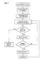
- FIG. 1shows an example of a method cycle in carrying out an embodiment of the present method
- FIG. 2shows an illustration for the 3-material decomposition carried out in an embodiment of the method.
- first, second, etc.may be used herein to describe various elements, components, regions, layers and/or sections, it should be understood that these elements, components, regions, layers and/or sections should not be limited by these terms. These terms are used only to distinguish one element, component, region, layer, or section from another region, layer, or section. Thus, a first element, component, region, layer, or section discussed below could be termed a second element, component, region, layer, or section without departing from the teachings of the present invention.
- a dual energy computer tomographis used to carry out a dual energy CT scan of the object, in which raw data are simultaneously obtained in the context of two different X-ray energies. These different X-ray energies are obtained by means of a different tube voltage of the X-ray tubes used, 80 kV and 140 kV in the present example.
- Two CT imagesare reconstructed independently of one another from the raw data via known reconstruction algorithms.
- Each of the two image data records obtained in this casecomprises for each voxel of the investigation volume a corresponding HU value for the respective X-ray energy.
- the ratio q of the image noise between the image for 80 kV and the image for 140 kVis not known for this slice, this ratio q can, for example, be determined approximately from the object diameter or the measured noise of the HU values of air. It is possible to this end, for example, to calculate for both tube voltages the mean noise for all the pixels of the slice below a certain threshold, for example, below ⁇ 950 HU, in the upper half of the image, and to form the ratio subsequently. It is likewise possible to determine this ratio from a previously recorded topogram, for example.
- a number of voxel slices above and below itare also required for the main portion of the processing.
- averaged HU valueis calculated as the arithmetic mean from the HU values for 80 kV and 140 kV, x 80 and x 140 .
- Selection stepa three-dimensional spherical environment of the investigated voxel is considered. Use is made only of voxels whose combined HU value lies above the threshold for kidney stones. In this way, all the neighboring voxels possibly having the same chemical composition are firstly selected. If the averaged HU value lies above the HU threshold for kidney stones for fewer than n min voxels in the volume considered, the following steps are omitted and no material assignment is made. Otherwise, a mean HU value x8 0 is calculated for this selected voxel for 80 kV, and a mean HU value x 140 is calculated for 140 kV, this being done in each case by averaging over the HU values of all the selected voxels.
- a radius of 7 voxelscan be adopted as an example of the spherical environment, and a value of 100 voxels can be adopted as an example of the threshold value n min . Of course, these values can, however, also be selected otherwise, depending on the application and image quality.
- High values of rcharacterize calcium-containing stones (hydroxyapatite stones, oxalate stones); medium values of r are measured for cystine stones (high sulfur content); low values of r are yielded for uric acid stones. It is very possible to distinguish uric acid stones and all other types of kidney stones on the basis of a clear difference in the value of r, since uric acid contains no relatively heavy atoms. Depending on image quality, however, an embodiment of the present method also enables other types of kidney stones to be distinguished.
- the material determined via the value ris now assigned to the central voxel. Once the image data record, or the interesting slice therein is completely processed, the material map thus prepared can be used to mark the types of kidney stones found in color in the CT image.
- FIG. 2shows the X-ray attenuation values for urine, pure uric acid, pure cystine and pure calcium oxalate in a diagram where the X-ray attenuation values for 80 kV are plotted against the X-ray attenuation values for 140 kV.
- the X-ray attenuation values of real kidney stoneslie in the hatched zone.
- the ratio r used in an embodiment of the present methodcorresponds to the gradient of the connecting line between the data point for pure urine and the data point that is yielded from the two measured (or averaged) X-ray attenuation values for a type of kidney stone.
- the types of kidney stonescan be differentiated in an embodiment of the present method on the basis of the different gradients of the connecting lines for the different types of kidney stones.
- x min Lower threshold (HU) for kidney stone voxels stake Radius of the volume considered n min Minimum number of voxels above the kidney stone threshold o 80 HU value of urine for 80 kV o 140 HU value of urine for 140 kV r ua,max Upper threshold of r for uric acid
- variable rwas calculated in the present example on the basis of a spherical volume, it is, of course, also possible to calculate on the basis of other fundamentals.
- rcan be calculated for the voxels of a cuboid volume that contains the complete stone.
Landscapes
- Engineering & Computer Science (AREA)
- Health & Medical Sciences (AREA)
- Life Sciences & Earth Sciences (AREA)
- Medical Informatics (AREA)
- Physics & Mathematics (AREA)
- General Health & Medical Sciences (AREA)
- Theoretical Computer Science (AREA)
- Biophysics (AREA)
- Surgery (AREA)
- Nuclear Medicine, Radiotherapy & Molecular Imaging (AREA)
- General Physics & Mathematics (AREA)
- Pathology (AREA)
- Radiology & Medical Imaging (AREA)
- Biomedical Technology (AREA)
- Heart & Thoracic Surgery (AREA)
- Molecular Biology (AREA)
- Computer Vision & Pattern Recognition (AREA)
- Animal Behavior & Ethology (AREA)
- Veterinary Medicine (AREA)
- Public Health (AREA)
- High Energy & Nuclear Physics (AREA)
- Optics & Photonics (AREA)
- Pulmonology (AREA)
- Quality & Reliability (AREA)
- Apparatus For Radiation Diagnosis (AREA)
Abstract
Description
- The present application hereby claims priority under 35 U.S.C. §119 on German patent application numbers DE 10 2006 015 454.1 filed Mar, 31, 2006, the entire contents of each of which is hereby incorporated herein by reference.
- Embodiments of the present application generally relate to a method and/or a device for automatically differentiating types of kidney stones by way of computed tomography. For example, it may relate to one in which two computed tomography pictures of an object area including kidney stones are recorded in the context of a different spectral distribution of the X-radiation, and there are reconstructed from raw data of the two computed tomography pictures two image data records of the object area that include X-ray attenuation values of voxels of the object area in the context of the respective spectral distribution of the X-radiation.
- Identifying and distinguishing different types of kidney stones in the human or animal body is very important in deciding on a therapy. Uric acid stones, cystine stones, oxalate stones or hydroxyapatite stones, for example count among the different types of kidney stones'. It is chiefly the differentiation between uric acid stones and other types of kidney stones that is of great importance here, since the therapy of uric acid stones differs substantially from those of the other types.
- Three different methods have been known to date for differentiating types of kidney stones. In the first method, the uric acid concentration in the blood is determined chemically. A high concentration indicates that a uric acid stone could be present. In the second method, solid constituents are filtered out of the urine. Thus, constituents of kidney stones can sometimes be detected in the urine after bodily movement, for example, through climbing steps, and be suitably investigated. In the third method, a surgical intervention is performed in which the kidney stone is already removed for the purpose of simultaneous diagnosis and therapy. It is possible to classify kidney stones with the aid of conventional computed tomography only with difficulty, since kidney stones do not occur as pure, compact substances. Although, for example, pure calcium oxalate is clearly distinguished from crystallized uric acid in the X-ray attenuation value, the X-ray attenuation values can nevertheless be very similar in reality.
- At least one embodiment of the present invention specifies a method and/or a device for differentiating types of kidney stones that enable automatic differentiation of at least two types of kidney stones without surgical intervention.
- In at least one embodiment of the present method, two computed tomography pictures of an object area that includes the kidney stones to be differentiated are recorded in the context of a different spectral distribution of the X-radiation, and two image data records of the object area are reconstructed from the raw data of the two computed tomography pictures. The two image data records include the X-ray attenuation values of the voxels of the object area in the context of the respective spectral distribution of the X-radiation. X-ray attenuation values can be understood here both as the attenuation coefficients μ and as values derived therefrom, such as the CT value.
- The two computed tomography pictures are recorded by using a multi-imaging computer tomograph, for example a so-called dual energy computer tomograph, with the aid of which it is possible simultaneously or at least virtually simultaneously to record two computed tomography pictures with a different spectral distribution of the X-radiation or different X-ray energy. Different techniques for generating two computed tomography pictures with a different spectral distribution of the X-radiation are fundamentally known to the specialist. It is possible to this end, for example, to make use of a number of X-ray sources with a different X-radiation, different detectors of different spectral sensitivity, different filters in front of the X-ray sources and/or X-ray detectors, or else of a combination of said techniques.
- In at least one embodiment of the present method, a ratio r is calculated from the two image data records for each voxel of at least one interesting slice of the object area if the mean value from the two assigned X-ray attenuation values of the respective voxel lies above a prescribed threshold value (Xmin) that is characteristic of kidney stones. The ratio r is yielded in the following way:
- In this equation, x1represents either the measured X-ray attenuation value of the voxel in the context of one of the two different X-ray energies, or an X-ray attenuation value averaged for this X-ray energy, which will be examined more closely later. In the same way, x2represents either the measured X-ray attenuation value of the voxel in the context of the other X-ray energy, or an appropriately averaged X-ray attenuation value. The two X-ray attenuation values x1, x2can either be extracted directly from the two image data records, or be calculated therefrom. The values o1and o2that also occur represent the X-ray attenuation values of pure urine in the context of the two X-ray energies. These values are prescribed. They are either already known, or can be determined in advance.
- The basis for the calculation of the ratio r is a 3-material decomposition in which the respective voxel is interpreted from a mixture of the base materials of urine, first type of kidney stone, in particular uric acid stone, and another type of kidney stone. A cystine stone, an oxalate stone or a hydroxyapatite stone, for example, comes into consideration as other type of kidney stone. It has been found on the basis of simulations that the ratio r for a stone of constant chemical composition depends only slightly on the object diameter.
- In this case, high values of r characterize calcium-containing stones such as, for example, hydroxyapatite stones or oxalate stones. Medium values of r are measured for cystine stones (high sulfur content). Low values of r are yielded for the uric acid stone. In this way, different types of kidney stones can be distinguished, in particular, uric acid stones can be distinguished from other types of kidney stones, by appropriately prescribing threshold values or value ranges for r.
- After r has been determined, the respective voxel is therefore assigned to one of at least two types of kidney stones as a function of the magnitude of r. These appropriately classified voxels can then be displayed with colored highlighting in a computed tomography image, for example. Also possible in this case is a differently colored display for different types of kidney stones, that is to say as a function of the magnitude of r. The viewer can immediately identify the location and the type of kidney stone in the CT images, in particular whether what is involved here is a uric acid stone or another type of kidney stone.
- At least one embodiment of the present method and/or the associated device therefore enable types of kidney stones to be automatically differentiated by way of computed tomography, that is to say without surgical intervention. At least one embodiment of the method requires neither an analysis of eliminated stone material nor an analysis of blood values.
- In an advantageous development of at least one embodiment of the method, the X-ray attenuation values x1, x2of the respective voxel are obtained by averaging. In this case, a three-dimensional volume area with a prescribed extent around the relevant voxel, also denoted below as central voxel, is firstly formed. The three-dimensional volume area preferably constitutes a spherical volume, but can also exhibit another shape, for example, a cuboid. All the voxels whose X-ray attenuation values fulfill a prescribed criterion that is characteristic of kidney stones are selected inside this volume area. An average X-ray attenuation value of the selected voxels is then calculated separately for each image data record, the two averaged X-ray attenuation values yielded therefrom being used to form the ratio r. This step enables the selection of neighboring voxels of the central voxel that are highly likely to constitute kidney stone voxels, without leading to smearing with constant range.
- In an example refinement of at least one embodiment of the method, the steps for determining the ratio r are not carried out for all the voxels, but only for a portion of these voxels that is determined in the following way. Here, the number of the selected voxels is determined in the three-dimensional volume area whose X-ray attenuation values fulfill the prescribed criterion. If this number lies above a prescribed threshold value for the number of the selected voxels, the two X-ray attenuation values or averaged X-ray attenuation values for the central voxel are used or determined, and the ratio r is calculated. If, however, the number of the selected voxels lies below the threshold value, no further kind of calculation is carried out for the central voxel. It is then assumed that this voxel does not constitute a site with a kidney stone in the object area investigated.
- In one refinement of at least one embodiment of the present method, it is possible to use as criterion in accordance with which the voxels are selected inside the three-dimensional volume area the fact that the mean value of the two X-ray attenuation values of the respective voxel must lie above a threshold value that constitutes a lower limit for the presence of kidney stones.
- However, it is preferred in the case of this criterion for the basis to be not the mean value, but a combined X-ray attenuation value that represents a weighted value xmdependent on the image noise ratio q between the two computed tomography pictures, and is calculated using the following rule:
- The image noise ratio q is yielded from q=dx1/dx2, where dx1and dx2represent the statistical errors, that is to say the standard deviation, of the X-ray attenuation values x1and x2. The value rua,maxis a prescribed threshold value that specifies the upper threshold of the ratio r for uric acid.
- This value is known or can be determined in advance. The voxels are then selected inside the three-dimensional volume area on the basis of the combined X-ray attenuation value xm. All the voxels for which this combined X-ray attenuation value xmlies above a threshold value that represents a lower limit for the presence of kidney stones are selected. This mode of procedure based on the combined X-ray attenuation value that represents a weighted mean value dependent on the image noise ratio q substantially reduces the risk of an erroneous selection, caused by the image noise, in the vicinity of the threshold value of xm, and so a more reliable result is attained. The ratio q of the image noise of the two image data records that is required for this purpose can already be known for the computed tomography installation being used, or be determined in advance from the two image data records, or else other image data records, for example topograms recorded in advance.
- The device for automatically differentiating types of kidney stones by way of computed tomography pictures includes, in addition to a memory unit for the two image data records as main constituent, a determination module that carries out the calculations and determinations in accordance with the previously described method and, if appropriate, the individual developments of this method. The determination module is in this case preferably implemented in the image computer or a computed tomography installation that can supply the raw data for the two computed tomography pictures in the context of a different spectral distribution of the X-radiation. In this case, the device also includes an image reconstruction module that reconstructs the two image data records of the object area from the raw data of the two computed tomography pictures.
- In one refinement, at least one embodiment of the device can, however, also include only the determination module with the memory unit, and an interface via which already reconstructed image data records from the two computed tomography pictures are received. The determination module is, for example, connected to an image display module that enables a colored image display of the voxels representing the kidney stones on an appropriate image display unit.
- The present method is explained once again briefly below with the aid of an example embodiment in conjunction with the drawings, in which:
FIG. 1 shows an example of a method cycle in carrying out an embodiment of the present method; andFIG. 2 shows an illustration for the 3-material decomposition carried out in an embodiment of the method.- Although the terms first, second, etc. may be used herein to describe various elements, components, regions, layers and/or sections, it should be understood that these elements, components, regions, layers and/or sections should not be limited by these terms. These terms are used only to distinguish one element, component, region, layer, or section from another region, layer, or section. Thus, a first element, component, region, layer, or section discussed below could be termed a second element, component, region, layer, or section without departing from the teachings of the present invention.
- The terminology used herein is for the purpose of describing particular embodiments only and is not intended to be limiting of the present invention. As used herein, the singular forms “a”, “an”, and “the” are intended to include the plural forms as well, unless the context clearly indicates otherwise. It will be further understood that the terms “includes” and/or “including”, when used in this specification, specify the presence of stated features, integers, steps, operations, elements, and/or components, but do not preclude the presence or addition of one or more other features, integers, steps, operations, elements, components, and/or groups thereof.
- In describing example embodiments illustrated in the drawings, specific terminology is employed for the sake of clarity. However, the disclosure of this patent specification is not intended to be limited to the specific terminology so selected and it is to be understood that each specific element includes all technical equivalents that operate in a similar manner.
- Referencing the drawings, wherein like reference numerals designate identical or corresponding parts throughout the several views, example embodiments of the present patent application are hereafter described.
- In the present example embodiment, a dual energy computer tomograph is used to carry out a dual energy CT scan of the object, in which raw data are simultaneously obtained in the context of two different X-ray energies. These different X-ray energies are obtained by means of a different tube voltage of the X-ray tubes used, 80 kV and 140 kV in the present example. Two CT images are reconstructed independently of one another from the raw data via known reconstruction algorithms. Each of the two image data records obtained in this case comprises for each voxel of the investigation volume a corresponding HU value for the respective X-ray energy.
- Irrespective of the data recording and the computer tomograph used, it should be ensured in this case that the HU values for the body materials to be differentiated are to some extent stable when they occur or are positioned at different sites inside the object being investigated. This is, however, the case for most commercially available computer tomographs.
- In the present example embodiment, only an axial slice is considered during preprocessing. If the ratio q of the image noise between the image for 80 kV and the image for 140 kV is not known for this slice, this ratio q can, for example, be determined approximately from the object diameter or the measured noise of the HU values of air. It is possible to this end, for example, to calculate for both tube voltages the mean noise for all the pixels of the slice below a certain threshold, for example, below −950 HU, in the upper half of the image, and to form the ratio subsequently. It is likewise possible to determine this ratio from a previously recorded topogram, for example.
- In addition to the slice being investigated, a number of voxel slices above and below it are also required for the main portion of the processing. The term “combined HU value” used below denotes the weighted mean value xm, dependent on the image noise ratio, of the HU values for 80 kV and 140 kV (x80and x140, respectively). This can be calculated from the ratio q and the prescribed upper threshold value rua,maxused later, for uric acid:
- By contrast therewith the term “averaged HU value” is calculated as the arithmetic mean from the HU values for 80 kV and 140 kV, x80and x140.
- The following two steps are then carried out (cf.
FIG. 1 ) for each voxel in the slice being investigated, given that the averaged HU value of this voxel lies above a typical threshold for kidney stones: - 1. Selection step: a three-dimensional spherical environment of the investigated voxel is considered. Use is made only of voxels whose combined HU value lies above the threshold for kidney stones. In this way, all the neighboring voxels possibly having the same chemical composition are firstly selected. If the averaged HU value lies above the HU threshold for kidney stones for fewer than nminvoxels in the volume considered, the following steps are omitted and no material assignment is made. Otherwise, a mean HU value x80is calculated for this selected voxel for 80 kV, and a mean HU value x140is calculated for 140 kV, this being done in each case by averaging over the HU values of all the selected voxels. A radius of 7 voxels can be adopted as an example of the spherical environment, and a value of 100 voxels can be adopted as an example of the threshold value nmin. Of course, these values can, however, also be selected otherwise, depending on the application and image quality.
2. 3-material decomposition: the selected voxels are interpreted as a mixture of the base materials of urine (HU values: o80and o-140), uric acid stone and cystine stone or oxalate/hydroxyapatite stone. The ratio
is calculated. - High values of r characterize calcium-containing stones (hydroxyapatite stones, oxalate stones); medium values of r are measured for cystine stones (high sulfur content); low values of r are yielded for uric acid stones. It is very possible to distinguish uric acid stones and all other types of kidney stones on the basis of a clear difference in the value of r, since uric acid contains no relatively heavy atoms. Depending on image quality, however, an embodiment of the present method also enables other types of kidney stones to be distinguished. The material determined via the value r is now assigned to the central voxel. Once the image data record, or the interesting slice therein is completely processed, the material map thus prepared can be used to mark the types of kidney stones found in color in the CT image.
- For illustrative purposes,
FIG. 2 shows the X-ray attenuation values for urine, pure uric acid, pure cystine and pure calcium oxalate in a diagram where the X-ray attenuation values for 80 kV are plotted against the X-ray attenuation values for 140 kV. The X-ray attenuation values of real kidney stones lie in the hatched zone. The ratio r used in an embodiment of the present method corresponds to the gradient of the connecting line between the data point for pure urine and the data point that is yielded from the two measured (or averaged) X-ray attenuation values for a type of kidney stone. The types of kidney stones can be differentiated in an embodiment of the present method on the basis of the different gradients of the connecting lines for the different types of kidney stones. - The following parameters are required in this example to carry out an embodiment of the method:
Parameter Meaning xmin Lower threshold (HU) for kidney stone voxels stake Radius of the volume considered nmin Minimum number of voxels above the kidney stone threshold o80 HU value of urine for 80 kV o140 HU value of urine for 140 kV rua,max Upper threshold of r for uric acid - Even though the variable r was calculated in the present example on the basis of a spherical volume, it is, of course, also possible to calculate on the basis of other fundamentals. Thus, for example, r can be calculated for the voxels of a cuboid volume that contains the complete stone. Likewise, it is also possible to analyze only the central region of the stone, or even only single voxels.
- Example embodiments being thus described, it will be obvious that the same may be varied in many ways. Such variations are not to be regarded as a departure from the spirit and scope of the present invention, and all such modifications as would be obvious to one skilled in the art are intended to be included within the scope of the following claims.
Claims (20)
Applications Claiming Priority (3)
| Application Number | Priority Date | Filing Date | Title |
|---|---|---|---|
| DE102006015454.1 | 2006-03-31 | ||
| DE102006015454ADE102006015454A1 (en) | 2006-03-31 | 2006-03-31 | Method and device for automatic differentiation of kidney stone types by means of computed tomography |
| DE102006015454 | 2006-03-31 |
Publications (2)
| Publication Number | Publication Date |
|---|---|
| US20070249933A1true US20070249933A1 (en) | 2007-10-25 |
| US8364240B2 US8364240B2 (en) | 2013-01-29 |
Family
ID=38514361
Family Applications (1)
| Application Number | Title | Priority Date | Filing Date |
|---|---|---|---|
| US11/730,274Expired - Fee RelatedUS8364240B2 (en) | 2006-03-31 | 2007-03-30 | Method and device for automatically differentiating types of kidney stones by means of computed tomography |
Country Status (4)
| Country | Link |
|---|---|
| US (1) | US8364240B2 (en) |
| JP (1) | JP5207650B2 (en) |
| CN (1) | CN101044988A (en) |
| DE (1) | DE102006015454A1 (en) |
Cited By (11)
| Publication number | Priority date | Publication date | Assignee | Title |
|---|---|---|---|---|
| US20100128844A1 (en)* | 2008-11-26 | 2010-05-27 | Brian Thomsen | System and Method for Material Segmentation Utilizing Computed Tomography Scans |
| JP2011110245A (en)* | 2009-11-27 | 2011-06-09 | Ge Medical Systems Global Technology Co Llc | Image display device, x-ray ct apparatus, and program |
| US20140064587A1 (en)* | 2012-08-31 | 2014-03-06 | Klinikum Der Universitat Munchen | Method for detecting damage to silicone implants and computed tomography device |
| US20150031992A1 (en)* | 2013-07-29 | 2015-01-29 | The Cleveland Clinic Foundation | Identifying kidney stone composition from medical imaging |
| US9204847B2 (en) | 2011-02-15 | 2015-12-08 | Siemens Aktiengesellschaft | Method, image data record processing facility, X-ray system and computer program product for correcting image data of an examination object |
| WO2015193521A1 (en)* | 2014-06-18 | 2015-12-23 | Universitat Autònoma De Barcelona | Method and system for the automatic classification of kidney stones, computer program, and computer program product |
| US9256942B1 (en)* | 2014-10-17 | 2016-02-09 | Lite-Med Inc. | Method for determining variations among multiple three-dimensional stone images extracorporeally and computer program using the same |
| US20160123904A1 (en)* | 2014-11-04 | 2016-05-05 | Kabushiki Kaisha Toshiba | Method of, and apparatus for, material classification in multi-energy image data |
| US20160307340A1 (en)* | 2015-04-14 | 2016-10-20 | Thomas Allmendinger | Multispectral ct imaging |
| US20180317743A1 (en)* | 2017-05-04 | 2018-11-08 | Boston Scientific Scimed, Inc. | Medical system and related methods |
| CN112656483A (en)* | 2020-12-21 | 2021-04-16 | 中南大学湘雅医院 | Visual portable choledochoscope lithotomy forceps |
Families Citing this family (5)
| Publication number | Priority date | Publication date | Assignee | Title |
|---|---|---|---|---|
| JP2009178493A (en)* | 2008-02-01 | 2009-08-13 | Ge Medical Systems Global Technology Co Llc | X-ray ct apparatus |
| WO2014126189A1 (en)* | 2013-02-14 | 2014-08-21 | 株式会社テレシステムズ | X-ray imaging device and x-ray imaging method |
| CN109685796B (en)* | 2018-12-26 | 2021-05-18 | 上海联影智能医疗科技有限公司 | Medical image processing method, apparatus, device and storage medium |
| CN112220441A (en)* | 2020-10-14 | 2021-01-15 | 泰州国安医疗用品有限公司 | Minimally invasive in vivo data acquisition system |
| KR102744742B1 (en)* | 2022-09-14 | 2024-12-18 | 계명대학교 산학협력단 | Urinary stone analysis method and analysis appratus |
Citations (4)
| Publication number | Priority date | Publication date | Assignee | Title |
|---|---|---|---|---|
| US4792900A (en)* | 1986-11-26 | 1988-12-20 | Picker International, Inc. | Adaptive filter for dual energy radiographic imaging |
| US5838758A (en)* | 1990-08-10 | 1998-11-17 | Vivid Technologies | Device and method for inspection of baggage and other objects |
| US6597759B2 (en)* | 2000-09-29 | 2003-07-22 | Ge-Lunar Corporation | Method of inspecting meat for bone content using dual energy x-ray attenuation |
| US20050195936A1 (en)* | 2003-12-03 | 2005-09-08 | Raghav Raman | Quantification method of vessel calcification |
- 2006
- 2006-03-31DEDE102006015454Apatent/DE102006015454A1/ennot_activeWithdrawn
- 2007
- 2007-03-30JPJP2007090878Apatent/JP5207650B2/ennot_activeExpired - Fee Related
- 2007-03-30USUS11/730,274patent/US8364240B2/ennot_activeExpired - Fee Related
- 2007-04-02CNCNA2007100921069Apatent/CN101044988A/enactivePending
Patent Citations (4)
| Publication number | Priority date | Publication date | Assignee | Title |
|---|---|---|---|---|
| US4792900A (en)* | 1986-11-26 | 1988-12-20 | Picker International, Inc. | Adaptive filter for dual energy radiographic imaging |
| US5838758A (en)* | 1990-08-10 | 1998-11-17 | Vivid Technologies | Device and method for inspection of baggage and other objects |
| US6597759B2 (en)* | 2000-09-29 | 2003-07-22 | Ge-Lunar Corporation | Method of inspecting meat for bone content using dual energy x-ray attenuation |
| US20050195936A1 (en)* | 2003-12-03 | 2005-09-08 | Raghav Raman | Quantification method of vessel calcification |
Cited By (18)
| Publication number | Priority date | Publication date | Assignee | Title |
|---|---|---|---|---|
| US7983382B2 (en)* | 2008-11-26 | 2011-07-19 | General Electric Company | System and method for material segmentation utilizing computed tomography scans |
| US20100128844A1 (en)* | 2008-11-26 | 2010-05-27 | Brian Thomsen | System and Method for Material Segmentation Utilizing Computed Tomography Scans |
| JP2011110245A (en)* | 2009-11-27 | 2011-06-09 | Ge Medical Systems Global Technology Co Llc | Image display device, x-ray ct apparatus, and program |
| US9204847B2 (en) | 2011-02-15 | 2015-12-08 | Siemens Aktiengesellschaft | Method, image data record processing facility, X-ray system and computer program product for correcting image data of an examination object |
| US20140064587A1 (en)* | 2012-08-31 | 2014-03-06 | Klinikum Der Universitat Munchen | Method for detecting damage to silicone implants and computed tomography device |
| US9211066B2 (en)* | 2012-08-31 | 2015-12-15 | Siemens Aktiengesellschaft | Method for detecting damage to silicone implants and computed tomography device |
| US9706970B2 (en)* | 2013-07-29 | 2017-07-18 | The Cleveland Clinic Foundation | Identifying kidney stone composition from medical imaging |
| US20150031992A1 (en)* | 2013-07-29 | 2015-01-29 | The Cleveland Clinic Foundation | Identifying kidney stone composition from medical imaging |
| WO2015193521A1 (en)* | 2014-06-18 | 2015-12-23 | Universitat Autònoma De Barcelona | Method and system for the automatic classification of kidney stones, computer program, and computer program product |
| US9256942B1 (en)* | 2014-10-17 | 2016-02-09 | Lite-Med Inc. | Method for determining variations among multiple three-dimensional stone images extracorporeally and computer program using the same |
| US20160123904A1 (en)* | 2014-11-04 | 2016-05-05 | Kabushiki Kaisha Toshiba | Method of, and apparatus for, material classification in multi-energy image data |
| US9964499B2 (en)* | 2014-11-04 | 2018-05-08 | Toshiba Medical Systems Corporation | Method of, and apparatus for, material classification in multi-energy image data |
| US20160307340A1 (en)* | 2015-04-14 | 2016-10-20 | Thomas Allmendinger | Multispectral ct imaging |
| US10290122B2 (en)* | 2015-04-14 | 2019-05-14 | Siemens Aktiengesellschaft | Multi-spectral CT imaging |
| US20180317743A1 (en)* | 2017-05-04 | 2018-11-08 | Boston Scientific Scimed, Inc. | Medical system and related methods |
| US11089941B2 (en)* | 2017-05-04 | 2021-08-17 | Boston Scientific Scimed, Inc. | Medical system for identifying material to be removed from a patient and related methods |
| US12082882B2 (en) | 2017-05-04 | 2024-09-10 | Boston Scientific Scimed, Inc. | Medical systems and related methods |
| CN112656483A (en)* | 2020-12-21 | 2021-04-16 | 中南大学湘雅医院 | Visual portable choledochoscope lithotomy forceps |
Also Published As
| Publication number | Publication date |
|---|---|
| CN101044988A (en) | 2007-10-03 |
| JP5207650B2 (en) | 2013-06-12 |
| US8364240B2 (en) | 2013-01-29 |
| DE102006015454A1 (en) | 2007-10-18 |
| JP2007268274A (en) | 2007-10-18 |
Similar Documents
| Publication | Publication Date | Title |
|---|---|---|
| US8364240B2 (en) | Method and device for automatically differentiating types of kidney stones by means of computed tomography | |
| US7936909B2 (en) | Method and device for detecting chemical anomalies and/or salient features in soft tissue of an object area | |
| US11116471B2 (en) | Methods and apparatus for extended low contrast detectability for radiographic imaging systems | |
| CN100431482C (en) | Method for automatically calibrating filling parameter picture | |
| US7778380B2 (en) | Data handling and analysis in computed tomography with multiple energy windows | |
| US7920735B2 (en) | Method and device for automatically differentiating bones or other calcium-containing materials and contrast agent in soft tissue | |
| JP4469594B2 (en) | System for measuring disease-related tissue changes | |
| US8186880B1 (en) | Extended and fixed INTable simultaneously imaged calibration and correction methods and references for 3-D imaging devices | |
| US8068578B2 (en) | Method for recognizing and marking contrast agents in blood vessels of the lung with the aid of a CT examination and an image evaluation unit of a CT system | |
| US10932741B2 (en) | Assessing a condition of a subject using non-contrast dual energy computed tomography | |
| US20090208084A1 (en) | System and method for quantitative imaging of chemical composition to decompose more than two materials | |
| US7319739B2 (en) | Imaging method based on two different x-ray spectra | |
| US7050533B2 (en) | Method and device for determining the type of fluid in a fluid mass in an object | |
| US20100034734A1 (en) | System and method for quantitative molecular breast imaging | |
| JP2004160228A (en) | Method and apparatus for detecting tissue abnormality, perfusion abnormality and functional abnormality | |
| CA2768296A1 (en) | Extended low contrast detectability for radiographic imaging systems | |
| US7482592B2 (en) | Method for combining PET with MR perfusion and diffusion | |
| Hammers et al. | Balancing bias, reliability, noise properties and the need for parametric maps in quantitative ligand PET:[11C] diprenorphine test–retest data | |
| US8128907B2 (en) | CSF biomarker dilution factor corrections by MRI imaging and algorithm | |
| US20090127451A1 (en) | Devices and Methods for Calibrating Nuclear Medical and Radiological Images | |
| JP2021513054A (en) | Correction of standard capture value (SUV) scaling differences in serial positron emission tomography (PET) examinations using image alignment and regression analysis | |
| US7068827B2 (en) | System and method of measuring fat content in target organ and recording medium of recording fat content measuring program | |
| Arnold et al. | Peak SNR in automated coronary calcium scoring: Selecting CT scan parameters and statistically defined scoring thresholds a | |
| Judy | Multidetector-row CT image quality and radiation dose: imaging the lung | |
| Primak et al. | Kidney stones |
Legal Events
| Date | Code | Title | Description |
|---|---|---|---|
| AS | Assignment | Owner name:SIEMENS AKTIENGESELLSCHAFT, GERMANY Free format text:ASSIGNMENT OF ASSIGNORS INTEREST;ASSIGNOR:KRAUSS, BERNHARD;REEL/FRAME:019534/0008 Effective date:20070404 | |
| STCF | Information on status: patent grant | Free format text:PATENTED CASE | |
| FPAY | Fee payment | Year of fee payment:4 | |
| AS | Assignment | Owner name:SIEMENS HEALTHCARE GMBH, GERMANY Free format text:ASSIGNMENT OF ASSIGNORS INTEREST;ASSIGNOR:SIEMENS AKTIENGESELLSCHAFT;REEL/FRAME:039271/0561 Effective date:20160610 | |
| MAFP | Maintenance fee payment | Free format text:PAYMENT OF MAINTENANCE FEE, 8TH YEAR, LARGE ENTITY (ORIGINAL EVENT CODE: M1552); ENTITY STATUS OF PATENT OWNER: LARGE ENTITY Year of fee payment:8 | |
| AS | Assignment | Owner name:SIEMENS HEALTHINEERS AG, GERMANY Free format text:ASSIGNMENT OF ASSIGNORS INTEREST;ASSIGNOR:SIEMENS HEALTHCARE GMBH;REEL/FRAME:066088/0256 Effective date:20231219 | |
| AS | Assignment | Owner name:SIEMENS HEALTHINEERS AG, GERMANY Free format text:CORRECTIVE ASSIGNMENT TO CORRECT THE ASSIGNEE PREVIOUSLY RECORDED AT REEL: 066088 FRAME: 0256. ASSIGNOR(S) HEREBY CONFIRMS THE ASSIGNMENT;ASSIGNOR:SIEMENS HEALTHCARE GMBH;REEL/FRAME:071178/0246 Effective date:20231219 | |
| FEPP | Fee payment procedure | Free format text:MAINTENANCE FEE REMINDER MAILED (ORIGINAL EVENT CODE: REM.); ENTITY STATUS OF PATENT OWNER: LARGE ENTITY | |
| LAPS | Lapse for failure to pay maintenance fees | Free format text:PATENT EXPIRED FOR FAILURE TO PAY MAINTENANCE FEES (ORIGINAL EVENT CODE: EXP.); ENTITY STATUS OF PATENT OWNER: LARGE ENTITY | |
| STCH | Information on status: patent discontinuation | Free format text:PATENT EXPIRED DUE TO NONPAYMENT OF MAINTENANCE FEES UNDER 37 CFR 1.362 | |
| FP | Lapsed due to failure to pay maintenance fee | Effective date:20250129 |