US20070167697A1 - Method and apparatus for automatically characterizing a malignancy - Google Patents
Method and apparatus for automatically characterizing a malignancyDownload PDFInfo
- Publication number
- US20070167697A1 US20070167697A1US11/291,830US29183005AUS2007167697A1US 20070167697 A1US20070167697 A1US 20070167697A1US 29183005 AUS29183005 AUS 29183005AUS 2007167697 A1US2007167697 A1US 2007167697A1
- Authority
- US
- United States
- Prior art keywords
- image data
- functional
- structural
- imaging
- interest
- Prior art date
- Legal status (The legal status is an assumption and is not a legal conclusion. Google has not performed a legal analysis and makes no representation as to the accuracy of the status listed.)
- Granted
Links
- 206010028980NeoplasmDiseases0.000titleclaimsabstractdescription55
- 201000011510cancerDiseases0.000titleclaimsabstractdescription47
- 230000036210malignancyEffects0.000titleclaimsabstractdescription39
- 238000000034methodMethods0.000titleclaimsdescription53
- 230000003902lesionEffects0.000claimsabstractdescription29
- 238000002591computed tomographyMethods0.000claimsdescription67
- 238000002600positron emission tomographyMethods0.000claimsdescription59
- 238000003384imaging methodMethods0.000claimsdescription50
- 238000004458analytical methodMethods0.000claimsdescription41
- 230000008569processEffects0.000claimsdescription6
- 238000002595magnetic resonance imagingMethods0.000claimsdescription5
- 230000002308calcificationEffects0.000claimsdescription4
- 206010021143HypoxiaDiseases0.000claimsdescription3
- 230000001195anabolic effectEffects0.000claimsdescription3
- 230000001925catabolic effectEffects0.000claimsdescription3
- 238000000799fluorescence microscopyMethods0.000claimsdescription3
- 238000002599functional magnetic resonance imagingMethods0.000claimsdescription3
- 230000007954hypoxiaEffects0.000claimsdescription3
- 238000010801machine learningMethods0.000claimsdescription3
- 230000017074necrotic cell deathEffects0.000claimsdescription3
- 238000002603single-photon emission computed tomographyMethods0.000claimsdescription3
- 230000002792vascularEffects0.000claimsdescription3
- 230000006820DNA synthesisEffects0.000claimsdescription2
- 230000004153glucose metabolismEffects0.000claimsdescription2
- 210000000056organAnatomy0.000claimsdescription2
- 238000002604ultrasonographyMethods0.000claimsdescription2
- 239000002872contrast mediaSubstances0.000claims2
- 238000003748differential diagnosisMethods0.000claims2
- 238000011282treatmentMethods0.000claims2
- 238000011179visual inspectionMethods0.000abstractdescription3
- 238000010191image analysisMethods0.000description17
- 238000012545processingMethods0.000description13
- 238000012937correctionMethods0.000description8
- 238000001514detection methodMethods0.000description8
- 238000006243chemical reactionMethods0.000description7
- 210000001519tissueAnatomy0.000description7
- 238000004364calculation methodMethods0.000description6
- 238000011156evaluationMethods0.000description4
- 239000000700radioactive tracerSubstances0.000description4
- 230000000241respiratory effectEffects0.000description4
- 201000010099diseaseDiseases0.000description3
- 208000037265diseases, disorders, signs and symptomsDiseases0.000description3
- 230000002503metabolic effectEffects0.000description3
- 238000010606normalizationMethods0.000description3
- WQZGKKKJIJFFOK-GASJEMHNSA-NGlucoseNatural productsOC[C@H]1OC(O)[C@H](O)[C@@H](O)[C@@H]1OWQZGKKKJIJFFOK-GASJEMHNSA-N0.000description2
- 239000008103glucoseSubstances0.000description2
- 239000002207metaboliteSubstances0.000description2
- 238000012986modificationMethods0.000description2
- 230000004048modificationEffects0.000description2
- 238000009206nuclear medicineMethods0.000description2
- 230000003287optical effectEffects0.000description2
- 239000000126substanceSubstances0.000description2
- VRYALKFFQXWPIH-PBXRRBTRSA-N(3r,4s,5r)-3,4,5,6-tetrahydroxyhexanalChemical compoundOC[C@@H](O)[C@@H](O)[C@H](O)CC=OVRYALKFFQXWPIH-PBXRRBTRSA-N0.000description1
- 206010027476MetastasesDiseases0.000description1
- 238000009825accumulationMethods0.000description1
- 210000003484anatomyAnatomy0.000description1
- 230000033115angiogenesisEffects0.000description1
- 230000015572biosynthetic processEffects0.000description1
- 210000000988bone and boneAnatomy0.000description1
- 230000008859changeEffects0.000description1
- 150000001875compoundsChemical class0.000description1
- 238000013170computed tomography imagingMethods0.000description1
- 238000002059diagnostic imagingMethods0.000description1
- 230000005251gamma rayEffects0.000description1
- 230000002068genetic effectEffects0.000description1
- 239000012216imaging agentSubstances0.000description1
- 230000004807localizationEffects0.000description1
- 230000005741malignant processEffects0.000description1
- 238000005259measurementMethods0.000description1
- 230000004060metabolic processEffects0.000description1
- 238000004445quantitative analysisMethods0.000description1
- 238000009877renderingMethods0.000description1
- 230000004044responseEffects0.000description1
- 238000012552reviewMethods0.000description1
- 230000011218segmentationEffects0.000description1
- 239000000758substrateSubstances0.000description1
- 230000002195synergetic effectEffects0.000description1
- 230000002123temporal effectEffects0.000description1
- 238000012285ultrasound imagingMethods0.000description1
- 230000000007visual effectEffects0.000description1
Images


Classifications
- G—PHYSICS
- G01—MEASURING; TESTING
- G01T—MEASUREMENT OF NUCLEAR OR X-RADIATION
- G01T1/00—Measuring X-radiation, gamma radiation, corpuscular radiation, or cosmic radiation
- G01T1/16—Measuring radiation intensity
- G01T1/161—Applications in the field of nuclear medicine, e.g. in vivo counting
- G01T1/1611—Applications in the field of nuclear medicine, e.g. in vivo counting using both transmission and emission sources sequentially
- A—HUMAN NECESSITIES
- A61—MEDICAL OR VETERINARY SCIENCE; HYGIENE
- A61B—DIAGNOSIS; SURGERY; IDENTIFICATION
- A61B6/00—Apparatus or devices for radiation diagnosis; Apparatus or devices for radiation diagnosis combined with radiation therapy equipment
- A61B6/02—Arrangements for diagnosis sequentially in different planes; Stereoscopic radiation diagnosis
- A61B6/03—Computed tomography [CT]
- A61B6/037—Emission tomography
- G—PHYSICS
- G01—MEASURING; TESTING
- G01R—MEASURING ELECTRIC VARIABLES; MEASURING MAGNETIC VARIABLES
- G01R33/00—Arrangements or instruments for measuring magnetic variables
- G01R33/20—Arrangements or instruments for measuring magnetic variables involving magnetic resonance
- G01R33/44—Arrangements or instruments for measuring magnetic variables involving magnetic resonance using nuclear magnetic resonance [NMR]
- G01R33/48—NMR imaging systems
- G01R33/4808—Multimodal MR, e.g. MR combined with positron emission tomography [PET], MR combined with ultrasound or MR combined with computed tomography [CT]
- A—HUMAN NECESSITIES
- A61—MEDICAL OR VETERINARY SCIENCE; HYGIENE
- A61B—DIAGNOSIS; SURGERY; IDENTIFICATION
- A61B34/00—Computer-aided surgery; Manipulators or robots specially adapted for use in surgery
- A61B34/10—Computer-aided planning, simulation or modelling of surgical operations
- G—PHYSICS
- G01—MEASURING; TESTING
- G01R—MEASURING ELECTRIC VARIABLES; MEASURING MAGNETIC VARIABLES
- G01R33/00—Arrangements or instruments for measuring magnetic variables
- G01R33/20—Arrangements or instruments for measuring magnetic variables involving magnetic resonance
- G01R33/44—Arrangements or instruments for measuring magnetic variables involving magnetic resonance using nuclear magnetic resonance [NMR]
- G01R33/48—NMR imaging systems
- G01R33/4808—Multimodal MR, e.g. MR combined with positron emission tomography [PET], MR combined with ultrasound or MR combined with computed tomography [CT]
- G01R33/481—MR combined with positron emission tomography [PET] or single photon emission computed tomography [SPECT]
- G—PHYSICS
- G01—MEASURING; TESTING
- G01R—MEASURING ELECTRIC VARIABLES; MEASURING MAGNETIC VARIABLES
- G01R33/00—Arrangements or instruments for measuring magnetic variables
- G01R33/20—Arrangements or instruments for measuring magnetic variables involving magnetic resonance
- G01R33/44—Arrangements or instruments for measuring magnetic variables involving magnetic resonance using nuclear magnetic resonance [NMR]
- G01R33/48—NMR imaging systems
- G01R33/4808—Multimodal MR, e.g. MR combined with positron emission tomography [PET], MR combined with ultrasound or MR combined with computed tomography [CT]
- G01R33/4812—MR combined with X-ray or computed tomography [CT]
- G—PHYSICS
- G01—MEASURING; TESTING
- G01R—MEASURING ELECTRIC VARIABLES; MEASURING MAGNETIC VARIABLES
- G01R33/00—Arrangements or instruments for measuring magnetic variables
- G01R33/20—Arrangements or instruments for measuring magnetic variables involving magnetic resonance
- G01R33/44—Arrangements or instruments for measuring magnetic variables involving magnetic resonance using nuclear magnetic resonance [NMR]
- G01R33/48—NMR imaging systems
- G01R33/4808—Multimodal MR, e.g. MR combined with positron emission tomography [PET], MR combined with ultrasound or MR combined with computed tomography [CT]
- G01R33/4814—MR combined with ultrasound
Definitions
- the present inventionrelates generally to the field of medical imaging and more specifically to the evaluation of features of interest in image data acquired using different imaging modalities.
- the present inventionrelates to the evaluation of malignancies observable in computed tomography (CT) and positron emission tomography (PET) image data.
- CTcomputed tomography
- PETpositron emission tomography
- Non-invasive imagingbroadly encompasses techniques for generating images of the internal structures or regions of a person that are otherwise inaccessible for visual inspection.
- One of the best known uses of non-invasive imagingis in the medical arts where these techniques are used to generate images of organs and/or bones inside a patient which would otherwise not be visible.
- One class of medical non-invasive imaging techniquesis based on the generation of structural images of internal structures which depict the physical arrangement, composition, or properties of the imaged region.
- Example of such modalitiesinclude X-ray based techniques, such as CT and tomosynthesis. In these X-ray based techniques, the attenuation of X-rays by the patient is measured at different view angles and this information is used to reconstruct two-dimensional images and/or three-dimensional volumes of the imaged region.
- MRImagnetic resonance imaging
- ultrasound imagingin which the differential reflection of acoustic waves by the internal structures of a patient is used to reconstruct images of the internal anatomy.
- Examples, of such functional imaging modalitiesinclude nuclear medicine, single-photon emission computed tomography (SPECT), and PET.
- SPECTsingle-photon emission computed tomography
- PETPET
- These modalitiestypically detect photons or gamma rays, either directly or indirectly, which are generated by a radioactive tracer introduced into the patient. Based on the type of metaboland, sugar, or other compound into which the radioactive tracer is incorporated, the radioactive tracer is accumulated in different parts of the patient and measurement of the resulting gamma rays can be used to localize and image the accumulation of the tracer.
- tumorsmay disproportionately utilize glucose or other substrates relative to other tissues such that the tumors may be detected and localized using radioactively tagged deoxyglucose.
- functional imaging modalitiesinclude functional MRI, in which chemical composition information is obtained, and fluorescence imaging.
- the different functionalities of structural and functional imagingmay be combined to provide more information to a diagnostician than either modality alone.
- a clinicianis able to acquire both PET and CT image data that can be used in conjunction to detect tumors or to evaluate the progression of a tumor.
- the cliniciantypically evaluates different malignancy characteristics that can be measured in each type of image data.
- the PET image dataprovides useful metabolic information, such as the molecular signature of disease
- the CT image dataprovides useful anatomic and geometric information in the form of high-resolution images and volume renderings.
- the malignancy characteristics derived from each type of datamay then be considered together and utilized to characterize suspicious areas as well as to accurately assess cancer stages.
- a methodfor automatically evaluating a lesion.
- the methodincludes the step of detecting a lesion.
- One or more malignancy characteristics for the lesionare assessed in a set of functional image data and in a set of structural image data.
- a probability of malignancy for the lesionis automatically calculated based on the malignancy characteristics assessed in the set of structural image data and on the malignancy characteristics assessed in the set of functional image data.
- a computer-readable mediais also provided that affords some or all of the functionality of the type defined by this method.
- the image analysis systemcomprises analysis circuitry configured to assess one or more malignancy characteristics for a lesion in a set of structural image data and to assess one or more malignancy characteristics for the lesion in a set of functional image data.
- the analysis circuitryis also configured to calculate a probability of malignancy for the lesion based on the malignancy characteristics assessed in the set of structural image data and on the malignancy characteristics assessed in the set of functional image data.
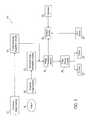
- FIG. 1is a diagrammatical view of an exemplary imaging system for use in accordance with the present technique
- FIG. 2is a diagrammatical view of an exemplary PET/CT imaging system for use in accordance with one embodiment of the present technique.
- FIG. 3is a flowchart depicting a technique for assessing lesion malignancy, in accordance with the present technique.
- the present inventionprovides for the automatic or semi-automatic assessment of cancerous or potentially cancerous tissues using multiple sets of image data, such as a set of functional image data and a set of structural image data.
- the respective sets of image datamay be concurrently acquired such as using a positron emission tomography/computed tomography (PET/CT) system, or may be acquired serially using combined or separate imaging systems.
- PET/CTpositron emission tomography/computed tomography
- the automated analysis routines employedallow for a quantitative analysis of malignancy characteristics of identified regions of interest within the functional and/or structural image data sets. These regions of interest may be identified in an automatic fashion. In this manner accurate, quantifiable results may be obtained to assist a clinician in the evaluation of a patient.
- an exemplary image analysis system 10for use in accordance with the present technique is provided.
- the image analysis system 10is depicted as comprising both functional and structural imaging modalities and combined image processing circuitry.
- these imaging modalities and/or their associated image processing circuitrymay be separate from one another with their respective image data being jointly provided for analysis as provided herein.
- more than one structural and/or functional imaging modalitymay be present.
- a single structural imaging modality and a single functional imaging modalityare depicted in FIG. 1 .
- the image analysis system 10is depicted as including a functional image scanner 12 configured to acquire data for generating functional images of a patient 14 .
- the functional image scanner 12represents the data acquisition components of a functional imaging modality, such as a PET, nuclear medicine, single-photon emission computed tomography (SPECT), fluorescence imaging, or functional magnetic resonance imaging system.
- SPECTsingle-photon emission computed tomography
- functional image acquisition circuitry 16is depicted.
- the acquisition circuitry 16is configured to acquire signals from the functional image scanner 12 and to provide any conversion (such as analog to digital conversion) or processing (such as image normalization, gain correction, artifact correction, and so forth) typically performed to facilitate the generation of suitable functional images.
- image processing circuitry 18receives the acquired signals from the functional image acquisition circuitry 16 and, via suitable reconstruction techniques, generates functional images and/or volumes from the acquired functional image data.
- the generated images or volumesmay be provided to image display circuitry 20 configured to display the functional images or volumes in a suitable format, such as on a display 22 or as an image printed by printer 24 .
- the functional images and/or volumes generated by the image processing circuitry 18are provided to analysis circuitry 26 in the depicted embodiment.
- the analysis circuitry 26analyzes the functional images and/or volumes in accordance with analysis routines, such as computer executable routines that may be run on general purpose or dedicated circuitry.
- the analysis circuitrymay receive operator inputs via one or more input devices 28 , such as a keyboard and/or mouse. These inputs may include configuration information or other inputs that may select the analysis routine to be executed or that may affect the operation of such an analysis routine, such as by specifying variables or factors taken into account by the analysis routines.
- inputsmay be provided to the analysis circuitry 26 from a database 30 or other source of medical history that may contain information or factors incorporated into the analysis of the functional images and/or volumes.
- the image analysis system 10also includes a structural image scanner 30 and associated structural image acquisition circuitry 32 .
- the structural image scanner 30is an imaging modality configured to acquire image data useful in generating structural, i.e., anatomic, images of the patient 14 . Examples of such structural imaging modalities include CT, tomosynthesis, and other X-ray based imaging techniques, magnetic resonance imaging (MRI) and ultrasound. As with the functional image scanner 12 described above, the structural image scanner 30 includes the data acquisition components of the structural imaging modality.
- the structural image acquisition circuitry 32is configured to acquire signals from the structural image scanner 30 and to provide any conversion (such as analog to digital conversion) or processing (such as image normalization, gain correction, artifact correction, and so forth) typically performed to facilitate the generation of suitable structural images.
- the acquired structural image datais provided to image processing circuitry 18 , which generates structural images and/or volumes.
- the structural images and/or volumesare in turn provided to image display circuitry 20 for display or printing and/or to the analysis circuitry 26 for analysis, as described above.
- the exemplary PET/CT image analysis system 50includes CT scanning components, including an X-ray source 56 configured to emit X-rays through an imaging volume containing the patient 14 and X-ray control circuitry 58 configured to control the operation of the X-ray source 56 via timing and control signals.
- the included CT scanning componentsinclude a CT detector 60 configured to detect X-rays emitted by the source 56 after attenuation by the patient 14 .
- the source 56 and CT detector 60may be structurally associated in a number of ways.
- the source 56 and CT detector 60may both be mounted on a rotatable gantry, as in third-generation CT systems.
- one or both of the source 56 and detector 60may be formed as mechanically stationary structures, as in fourth and fifth-generation CT systems.
- signalsare acquired from the CT detector 60 by the CT detector acquisition circuitry 62 .
- the CT detector acquisition circuitry 62is configured to provide any conversion (such as analog to digital conversion) or processing (such as image normalization, gain correction, artifact correction, and so forth) typically performed to facilitate the generation of suitable CT images.
- the CT detector acquisition circuitry 62may be configured to acquire diagnostic quality CT images, such as by utilizing prospective or retrospective gating techniques that compensate for respiratory motion or by otherwise acquiring CT image data during periods of respiratory stillness, such as during a breath hold.
- higher quality CT imagesare acquired than in embodiments in which the patient 14 breathes and no compensation or correction is made for the respiratory motion.
- the higher quality CT imagesmay be more useful in providing anatomic localization and/or attenuation correction of the PET signals (discussed below).
- the exemplary PET/CT image analysis system 50also includes PET scanning components, including a PET detector 52 .
- the PET detector 52may include a scintillator and associated optical sensing elements as well as timing circuits configured to differentiate coincident gamma ray pairs from spurious signals.
- the exemplary PET/CT image analysis system 50includes PET detector acquisition circuitry 54 configured to acquire signals from the PET detector 52 .
- the PET detector acquisition circuitry 54is configured to provide any conversion or processing typically performed to facilitate the generation of suitable PET images.
- the acquired PET and CT signalsare provided to PET/CT image processing circuitry 64 .
- the PET/CT image processing circuitry 64is depicted as a single component though, as will be appreciated by those of ordinary skill in the arts, this circuitry may actually be implemented as discrete or distinct circuitries for each imaging modality.
- the provided circuitrymay be configured to process both the PET and the CT image signals and to generate respective PET and CT images and/or volumes therefrom.
- the generated PET and CT images and/or volumesmay be provided to image display circuitry 20 for viewing on a display 22 or print out from a printer 24 , as discussed above with regard to FIG. 1 .
- the PET and CT imagesare provided to PET/CT analysis circuitry 66 .
- the PET/CT analysis circuitry 66analyzes the PET and CT images and/or volumes in accordance with analysis routines, such as computer executable routines that may be run on general purpose or dedicated circuitry.
- the PET/CT analysis circuitry 66is configured to identify and measure malignancy characteristics of a lesion that are visually or automatically identifiable in the respective PET and CT images or in the combined PET/CT image data.
- the PET/CT analysis circuitry 66may identify and/or measure malignancy characteristics such as vascular properties, calcification, and/or solidity with regard to a lesion observed in the CT image data.
- the PET/CT analysis circuitry 66may identify and/or measure malignancy characteristics such as the metabolism of glucose or other metabolites, anabolic activity, catabolic activity, and/or tissue necrosis with regard to a lesion observed in the PET image data.
- the PET/CT analysis circuitry 66may automatically detect the lesions for which malignancy characteristics are measured, such as by using threshold criteria or other techniques known in the art for segmenting regions of interest.
- a clinician or other viewermay manually detect the lesions or other regions of interest in either or both of the PET or CT images and/or volumes (such as in images viewed on the display 22 ). The clinician may then, via input device 28 (such as a keyboard and/or mouse), identify the lesions for analysis by the PET/CT analysis circuitry 66 .
- input device 28such as a keyboard and/or mouse
- image processing circuitry 64may register the PET and CT images such that respective regions in each image correspond to one another or are aligned.
- deformable registration routinesmay be executed by the PET/CT image processing circuitry 64 or by the PET/CT analysis circuitry 66 to properly rotate, translate, and/or deform the respective images to achieve the desired correspondence of regions.
- deformable registrationmay be desirable where the PET and CT data is acquired serially or where the data acquisition period for one of the modalities, such as PET, is longer than for the other modality, such as CT.
- other registration techniquessuch as rigid registration techniques, that achieve the desired degree of registration or correspondence can also be used in conjunction with the present technique.
- the input device 28may be used to allow a clinician to identify regions of interest in the PET or CT images
- the input device 28may also be used to provide operator inputs to the PET/CT image analysis circuitry 66 .
- These inputsmay include configuration information or other inputs that may select the analysis routine to be executed or that may affect the operation of such an analysis routine, such as by specifying variables or factors taken into account by the analysis routines.
- inputsmay be provided to the PET/CT image analysis circuitry 66 from a database 30 or other source of medical history that may contain information or factors incorporated into the analysis of the PET and CT images and/or volumes.
- FIG. 3a flowchart is provided describing steps performed in the automated assessment of cancer, in accordance with the present technique, and as may be performed by the exemplary systems described in FIGS. 1 and 2 .
- the techniqueis discussed in the context of an analysis of a set of functional image data and a set of structural image data.
- more than one set of functional and/or structural image datamay be employed in addition to or instead of the combination of functional and structural image data.
- the underlying principles of analysisare the same regardless of whether two or more sets of functional image data, two or more sets of structural image data, or a combination of structural and functional image data are employed.
- a detection step 70is provided in which one or more lesions are identified in structural and/or functional images of a patient or in the combined functional and structural images.
- the detection step 70is performed by automated detection routines, such as computer assisted detection routines, which identify lesions based on threshold or other segmentation/classification criteria from the surrounding image data.
- the detection step 70is performed by a clinician or other viewer based on a visual review of one or both of the functional or structural images and/or volumes.
- a registration step 72may also be performed to register or align corresponding regions within the structural and functional images. Though the depicted embodiment suggests that the registration step 72 is performed subsequent to the detection step 70 , in practice the registration step 72 may be performed before or after the detection step 70 .
- the registration step 72utilizes an automated deformable registration technique that accounts for patient motion, such as after the acquisition of a set of CT image data but prior to the completion of the acquisition of a set of PET image data during an examination.
- the registration techniquetransforms reconstructed regions such that corresponding regions of interest in the structural and functional images are registered, i.e., aligned.
- a variety of registration techniquescan be employed to suitably register concurrently or sequentially acquired functional and structural images.
- Malignancy characteristics for the one or more detected lesionsare automatically measured in the structural image data at step 74 .
- vascular propertiessuch as spiculation and angiogenesis
- malignancy characteristicssuch as calcification and solidity may be measured for a lesion automatically at step 74 .
- the malignancy characteristics that are measured at step 74will depend on the imaging modality employed and the malignancy characteristics that are typically evaluated in images generated by that modality.
- malignancy characteristics for the one or more detected lesionsare automatically measured in the functional image data.
- the functional image datais PET data
- glucose metabolism, DNA synthesis, tumor hypoxia, and/or tissue necrosismay be measured for an identified lesion.
- these and/or other types of metabolic activitysuch as catabolic and/or anabolic activity, and metabolite concentrations may be measured, depending on what malignancy characteristics are typically ascertained from images obtained using the respective functional imaging modality and imaging agent.
- malignancy characteristics for the one or more detected lesionsare automatically measured in the combined structural and functional image data.
- characteristics or other factors which are not apparent in either the structural or functional data alone, but which are apparent (or more easily quantified) in the combined datamay be measured.
- the malignancy characteristics measured at steps 74 , 76 , and/or 78are used to automatically calculate the probability of malignancy at step 80 .
- the routines employed to perform this calculationmay vary.
- the calculationwill be based, in full or in part, upon a clinical model of disease presentation.
- the calculationwill be based, in full or in part, upon machine learning methods operating on databases of prior clinical data, i.e., data where the clinical outcomes are known.
- the calculationwill be based, in full or in part, on prior clinical information, such as genetic and family history, clinical history, habits, and so forth. In practice, some or all of these techniques, as well as other suitable techniques, may be employed for automatically calculating the probability of malignancy for the lesion or lesions detected at step 70 .
- an optional step 82 of automatically calculating a cancer stageis provided in the depicted embodiment.
- the optional calculation of cancer stage at step 82is based upon the locations and malignancy probabilities calculated for each lesion. For example, in one embodiment, a probability is calculated for each of the stages of the particular cancer stage model employed (such as the tumor, node, metastases (TNM) model). In this manner, a probability is calculated for the patient for each of the possible cancer stages provided by the model employed.
- TPMtumor, node, metastases
- the processes for measuring malignancy characteristics and calculating malignancy and cancer stage probabilities described hereinmay be provided as one or more routines executable by the analysis circuitry or by processor-based components of the image analysis system 10 described herein.
- the routinesmay be stored or accessed on one or more computer-readable media, such as magnetic or optical media, which may be local to the image analysis system 10 or may be remotely accessible via a network connection, such as via the Internet or a local area network.
- access to or operation of the routinesmay be provided to an operator as part of the normal operation of the image analysis system 10 .
Landscapes
- Health & Medical Sciences (AREA)
- Physics & Mathematics (AREA)
- Engineering & Computer Science (AREA)
- Life Sciences & Earth Sciences (AREA)
- Nuclear Medicine, Radiotherapy & Molecular Imaging (AREA)
- Medical Informatics (AREA)
- High Energy & Nuclear Physics (AREA)
- General Health & Medical Sciences (AREA)
- Radiology & Medical Imaging (AREA)
- Molecular Biology (AREA)
- Biomedical Technology (AREA)
- General Physics & Mathematics (AREA)
- Optics & Photonics (AREA)
- Theoretical Computer Science (AREA)
- Animal Behavior & Ethology (AREA)
- Pathology (AREA)
- Condensed Matter Physics & Semiconductors (AREA)
- Heart & Thoracic Surgery (AREA)
- Pulmonology (AREA)
- Surgery (AREA)
- Biophysics (AREA)
- Public Health (AREA)
- Veterinary Medicine (AREA)
- Spectroscopy & Molecular Physics (AREA)
- Apparatus For Radiation Diagnosis (AREA)
- Nuclear Medicine (AREA)
- Measuring And Recording Apparatus For Diagnosis (AREA)
Abstract
Description
- The present invention relates generally to the field of medical imaging and more specifically to the evaluation of features of interest in image data acquired using different imaging modalities. In particular, the present invention relates to the evaluation of malignancies observable in computed tomography (CT) and positron emission tomography (PET) image data.
- Non-invasive imaging broadly encompasses techniques for generating images of the internal structures or regions of a person that are otherwise inaccessible for visual inspection. One of the best known uses of non-invasive imaging is in the medical arts where these techniques are used to generate images of organs and/or bones inside a patient which would otherwise not be visible. One class of medical non-invasive imaging techniques is based on the generation of structural images of internal structures which depict the physical arrangement, composition, or properties of the imaged region. Example of such modalities include X-ray based techniques, such as CT and tomosynthesis. In these X-ray based techniques, the attenuation of X-rays by the patient is measured at different view angles and this information is used to reconstruct two-dimensional images and/or three-dimensional volumes of the imaged region.
- Another modality used to generate structural images is magnetic resonance imaging (MRI). In MRI, the tissues undergoing imaging are subjected to strong magnetic fields and to radio wave perturbations which produce measurable signals as the tissues of the body align and realign themselves based upon their composition. These signals may then be used to reconstruct structural images that reflect the physical arrangement of tissues based on these different gyromagnetic responses. Another example of a structural imaging modality is ultrasound imaging, in which the differential reflection of acoustic waves by the internal structures of a patient is used to reconstruct images of the internal anatomy.
- While structural imaging modalities generate images of the physical composition or arrangement of a region of interest, functional imaging modalities generate images reflecting the chemical composition or metabolic activity of the region of interest. Examples, of such functional imaging modalities include nuclear medicine, single-photon emission computed tomography (SPECT), and PET. These modalities typically detect photons or gamma rays, either directly or indirectly, which are generated by a radioactive tracer introduced into the patient. Based on the type of metaboland, sugar, or other compound into which the radioactive tracer is incorporated, the radioactive tracer is accumulated in different parts of the patient and measurement of the resulting gamma rays can be used to localize and image the accumulation of the tracer. For example, tumors may disproportionately utilize glucose or other substrates relative to other tissues such that the tumors may be detected and localized using radioactively tagged deoxyglucose. Other examples of functional imaging modalities include functional MRI, in which chemical composition information is obtained, and fluorescence imaging.
- The different functionalities of structural and functional imaging may be combined to provide more information to a diagnostician than either modality alone. For example, in the case of combined PET/CT scanners, a clinician is able to acquire both PET and CT image data that can be used in conjunction to detect tumors or to evaluate the progression of a tumor. In such an example, the clinician typically evaluates different malignancy characteristics that can be measured in each type of image data. In particular, the PET image data provides useful metabolic information, such as the molecular signature of disease, while the CT image data provides useful anatomic and geometric information in the form of high-resolution images and volume renderings. The malignancy characteristics derived from each type of data may then be considered together and utilized to characterize suspicious areas as well as to accurately assess cancer stages.
- While the availability and analysis of both functional and structural image data (such as PET and CT images) provides diagnostic opportunities, several challenges to such techniques still exist. For example, in the case of combined PET/CT systems the image data is typically visually inspected by a clinician who provides a subjective assessment based on the visual inspection. However, the presentation of subtle disease state presentations, in either PET or CT image data, may be problematic. For example, a clinician may not know how to quantitatively determine whether a slight increase in a PET signal is due to a benign process or to a malignant process. Proper interpretation of this data typically requires a thorough understanding of the physics processes and image formation techniques involved, which may not be information available to or known by the average practicing clinician. Furthermore, even if this information were known by the clinician, the calculations involved to quantify and assess the significance of a signal change would be too laborious to manually perform on a regular basis.
- Furthermore, few clinicians have the knowledge or experience to fully understand and interpret the combined PET and CT data. Typically a clinician is primarily trained in the interpretation of image data from one type of image modality, but not both. Furthermore, synergies exist in the combined PET and CT image data such that the combined data may contain critical information that is not obvious or apparent in the uncombined image data. Apprehension of this synergistic information may not be possible by a clinician trained with respect to only one of the image modalities or inexperienced in the evaluation of such combined image data sets.
- In accordance with an exemplary embodiment of the present technique, a method is provided for automatically evaluating a lesion. The method includes the step of detecting a lesion. One or more malignancy characteristics for the lesion are assessed in a set of functional image data and in a set of structural image data. A probability of malignancy for the lesion is automatically calculated based on the malignancy characteristics assessed in the set of structural image data and on the malignancy characteristics assessed in the set of functional image data. A computer-readable media is also provided that affords some or all of the functionality of the type defined by this method.
- An image analysis system is provided. The image analysis system comprises analysis circuitry configured to assess one or more malignancy characteristics for a lesion in a set of structural image data and to assess one or more malignancy characteristics for the lesion in a set of functional image data. The analysis circuitry is also configured to calculate a probability of malignancy for the lesion based on the malignancy characteristics assessed in the set of structural image data and on the malignancy characteristics assessed in the set of functional image data.
- The foregoing and other advantages and features of the invention will become apparent upon reading the following detailed description and upon reference to the drawings in which:
FIG. 1 is a diagrammatical view of an exemplary imaging system for use in accordance with the present technique;FIG. 2 is a diagrammatical view of an exemplary PET/CT imaging system for use in accordance with one embodiment of the present technique; andFIG. 3 is a flowchart depicting a technique for assessing lesion malignancy, in accordance with the present technique.- The present invention provides for the automatic or semi-automatic assessment of cancerous or potentially cancerous tissues using multiple sets of image data, such as a set of functional image data and a set of structural image data. The respective sets of image data may be concurrently acquired such as using a positron emission tomography/computed tomography (PET/CT) system, or may be acquired serially using combined or separate imaging systems. The automated analysis routines employed allow for a quantitative analysis of malignancy characteristics of identified regions of interest within the functional and/or structural image data sets. These regions of interest may be identified in an automatic fashion. In this manner accurate, quantifiable results may be obtained to assist a clinician in the evaluation of a patient.
- In
FIG. 1 , an exemplaryimage analysis system 10 for use in accordance with the present technique is provided. For simplicity, theimage analysis system 10 is depicted as comprising both functional and structural imaging modalities and combined image processing circuitry. However, as noted above, these imaging modalities and/or their associated image processing circuitry may be separate from one another with their respective image data being jointly provided for analysis as provided herein. Likewise, as noted above, more than one structural and/or functional imaging modality may be present. However, for simplicity and comprehensiveness, a single structural imaging modality and a single functional imaging modality are depicted inFIG. 1 . - Returning to
FIG. 1 , theimage analysis system 10 is depicted as including afunctional image scanner 12 configured to acquire data for generating functional images of apatient 14. Thefunctional image scanner 12 represents the data acquisition components of a functional imaging modality, such as a PET, nuclear medicine, single-photon emission computed tomography (SPECT), fluorescence imaging, or functional magnetic resonance imaging system. Likewise, functionalimage acquisition circuitry 16 is depicted. Theacquisition circuitry 16 is configured to acquire signals from thefunctional image scanner 12 and to provide any conversion (such as analog to digital conversion) or processing (such as image normalization, gain correction, artifact correction, and so forth) typically performed to facilitate the generation of suitable functional images. In the depicted embodiment,image processing circuitry 18 receives the acquired signals from the functionalimage acquisition circuitry 16 and, via suitable reconstruction techniques, generates functional images and/or volumes from the acquired functional image data. The generated images or volumes may be provided toimage display circuitry 20 configured to display the functional images or volumes in a suitable format, such as on adisplay 22 or as an image printed byprinter 24. - In addition, the functional images and/or volumes generated by the
image processing circuitry 18 are provided toanalysis circuitry 26 in the depicted embodiment. Theanalysis circuitry 26 analyzes the functional images and/or volumes in accordance with analysis routines, such as computer executable routines that may be run on general purpose or dedicated circuitry. In addition to the functional images and/or volumes, the analysis circuitry may receive operator inputs via one ormore input devices 28, such as a keyboard and/or mouse. These inputs may include configuration information or other inputs that may select the analysis routine to be executed or that may affect the operation of such an analysis routine, such as by specifying variables or factors taken into account by the analysis routines. Furthermore, inputs may be provided to theanalysis circuitry 26 from adatabase 30 or other source of medical history that may contain information or factors incorporated into the analysis of the functional images and/or volumes. - In the depicted embodiment, the
image analysis system 10 also includes astructural image scanner 30 and associated structuralimage acquisition circuitry 32. Thestructural image scanner 30 is an imaging modality configured to acquire image data useful in generating structural, i.e., anatomic, images of thepatient 14. Examples of such structural imaging modalities include CT, tomosynthesis, and other X-ray based imaging techniques, magnetic resonance imaging (MRI) and ultrasound. As with thefunctional image scanner 12 described above, thestructural image scanner 30 includes the data acquisition components of the structural imaging modality. Similarly, the structuralimage acquisition circuitry 32 is configured to acquire signals from thestructural image scanner 30 and to provide any conversion (such as analog to digital conversion) or processing (such as image normalization, gain correction, artifact correction, and so forth) typically performed to facilitate the generation of suitable structural images. As discussed above with regard to the functional imaging components of the depictedimage analysis system 10, the acquired structural image data is provided toimage processing circuitry 18, which generates structural images and/or volumes. The structural images and/or volumes are in turn provided to imagedisplay circuitry 20 for display or printing and/or to theanalysis circuitry 26 for analysis, as described above. - Referring now to
FIG. 2 , an exemplary PET/CTimage analysis system 50 is depicted as a specific example of theimage analysis system 10 ofFIG. 1 . The exemplary PET/CTimage analysis system 50 includes CT scanning components, including anX-ray source 56 configured to emit X-rays through an imaging volume containing the patient14 andX-ray control circuitry 58 configured to control the operation of theX-ray source 56 via timing and control signals. In addition, the included CT scanning components include aCT detector 60 configured to detect X-rays emitted by thesource 56 after attenuation by thepatient 14. As will be appreciated by those of ordinary skill in the art, thesource 56 andCT detector 60 may be structurally associated in a number of ways. For example, thesource 56 andCT detector 60 may both be mounted on a rotatable gantry, as in third-generation CT systems. Alternatively, one or both of thesource 56 anddetector 60 may be formed as mechanically stationary structures, as in fourth and fifth-generation CT systems. - In the depicted system, signals are acquired from the
CT detector 60 by the CTdetector acquisition circuitry 62. The CTdetector acquisition circuitry 62, as noted with regard to the structuralimage acquisition circuitry 32 ofFIG. 1 , is configured to provide any conversion (such as analog to digital conversion) or processing (such as image normalization, gain correction, artifact correction, and so forth) typically performed to facilitate the generation of suitable CT images. Furthermore, the CTdetector acquisition circuitry 62 may be configured to acquire diagnostic quality CT images, such as by utilizing prospective or retrospective gating techniques that compensate for respiratory motion or by otherwise acquiring CT image data during periods of respiratory stillness, such as during a breath hold. In such embodiments, higher quality CT images are acquired than in embodiments in which thepatient 14 breathes and no compensation or correction is made for the respiratory motion. Furthermore, in embodiments where respiratory motion is accounted for or not allowed, the higher quality CT images may be more useful in providing anatomic localization and/or attenuation correction of the PET signals (discussed below). - The exemplary PET/CT
image analysis system 50 also includes PET scanning components, including aPET detector 52. As will be appreciated by those of ordinary skill in the arts, thePET detector 52 may include a scintillator and associated optical sensing elements as well as timing circuits configured to differentiate coincident gamma ray pairs from spurious signals. In addition, the exemplary PET/CTimage analysis system 50 includes PETdetector acquisition circuitry 54 configured to acquire signals from thePET detector 52. The PETdetector acquisition circuitry 54, as noted with regard to the functionalimage acquisition circuitry 16 ofFIG. 1 , is configured to provide any conversion or processing typically performed to facilitate the generation of suitable PET images. - In the depicted embodiment, the acquired PET and CT signals are provided to PET/CT
image processing circuitry 64. For simplicity, the PET/CTimage processing circuitry 64 is depicted as a single component though, as will be appreciated by those of ordinary skill in the arts, this circuitry may actually be implemented as discrete or distinct circuitries for each imaging modality. Conversely, the provided circuitry may be configured to process both the PET and the CT image signals and to generate respective PET and CT images and/or volumes therefrom. The generated PET and CT images and/or volumes may be provided to imagedisplay circuitry 20 for viewing on adisplay 22 or print out from aprinter 24, as discussed above with regard toFIG. 1 . - In addition, in the depicted embodiment, the PET and CT images are provided to PET/
CT analysis circuitry 66. The PET/CT analysis circuitry 66 analyzes the PET and CT images and/or volumes in accordance with analysis routines, such as computer executable routines that may be run on general purpose or dedicated circuitry. In particular, the PET/CT analysis circuitry 66 is configured to identify and measure malignancy characteristics of a lesion that are visually or automatically identifiable in the respective PET and CT images or in the combined PET/CT image data. For example, the PET/CT analysis circuitry 66 may identify and/or measure malignancy characteristics such as vascular properties, calcification, and/or solidity with regard to a lesion observed in the CT image data. Likewise, the PET/CT analysis circuitry 66 may identify and/or measure malignancy characteristics such as the metabolism of glucose or other metabolites, anabolic activity, catabolic activity, and/or tissue necrosis with regard to a lesion observed in the PET image data. - Furthermore, the PET/
CT analysis circuitry 66 may automatically detect the lesions for which malignancy characteristics are measured, such as by using threshold criteria or other techniques known in the art for segmenting regions of interest. Alternatively, a clinician or other viewer may manually detect the lesions or other regions of interest in either or both of the PET or CT images and/or volumes (such as in images viewed on the display22). The clinician may then, via input device28 (such as a keyboard and/or mouse), identify the lesions for analysis by the PET/CT analysis circuitry 66. In addition, to facilitate analysis either the PET/CT analysis circuitry 66 orimage processing circuitry 64 may register the PET and CT images such that respective regions in each image correspond to one another or are aligned. In this manner, a region identified in an image of one modality may be properly identified in images generated by the other modality as well. For example, deformable registration routines (or other registration routines which account for patient motion) may be executed by the PET/CTimage processing circuitry 64 or by the PET/CT analysis circuitry 66 to properly rotate, translate, and/or deform the respective images to achieve the desired correspondence of regions. Such deformable registration may be desirable where the PET and CT data is acquired serially or where the data acquisition period for one of the modalities, such as PET, is longer than for the other modality, such as CT. As will be appreciated by those of ordinary skill in the art, other registration techniques, such as rigid registration techniques, that achieve the desired degree of registration or correspondence can also be used in conjunction with the present technique. - While the
input device 28 may be used to allow a clinician to identify regions of interest in the PET or CT images, theinput device 28 may also be used to provide operator inputs to the PET/CTimage analysis circuitry 66. These inputs may include configuration information or other inputs that may select the analysis routine to be executed or that may affect the operation of such an analysis routine, such as by specifying variables or factors taken into account by the analysis routines. Furthermore, inputs may be provided to the PET/CTimage analysis circuitry 66 from adatabase 30 or other source of medical history that may contain information or factors incorporated into the analysis of the PET and CT images and/or volumes. - Turning now to
FIG. 3 , a flowchart is provided describing steps performed in the automated assessment of cancer, in accordance with the present technique, and as may be performed by the exemplary systems described inFIGS. 1 and 2 . For illustrative purposes, the technique is discussed in the context of an analysis of a set of functional image data and a set of structural image data. As noted above, however, in other embodiments, more than one set of functional and/or structural image data may be employed in addition to or instead of the combination of functional and structural image data. As will be appreciated by those of ordinary skill in the art, however, the underlying principles of analysis are the same regardless of whether two or more sets of functional image data, two or more sets of structural image data, or a combination of structural and functional image data are employed. - As provided in the flowchart (and as noted above) a
detection step 70 is provided in which one or more lesions are identified in structural and/or functional images of a patient or in the combined functional and structural images. In one embodiment, thedetection step 70 is performed by automated detection routines, such as computer assisted detection routines, which identify lesions based on threshold or other segmentation/classification criteria from the surrounding image data. In an alternative embodiment, thedetection step 70 is performed by a clinician or other viewer based on a visual review of one or both of the functional or structural images and/or volumes. - Based on the temporal and/or spatial variations in the underlying functional and structural image data, a
registration step 72 may also be performed to register or align corresponding regions within the structural and functional images. Though the depicted embodiment suggests that theregistration step 72 is performed subsequent to thedetection step 70, in practice theregistration step 72 may be performed before or after thedetection step 70. In one embodiment, theregistration step 72 utilizes an automated deformable registration technique that accounts for patient motion, such as after the acquisition of a set of CT image data but prior to the completion of the acquisition of a set of PET image data during an examination. In such an embodiment, the registration technique transforms reconstructed regions such that corresponding regions of interest in the structural and functional images are registered, i.e., aligned. As will be appreciated by those of ordinary skill in the art, a variety of registration techniques can be employed to suitably register concurrently or sequentially acquired functional and structural images. - Malignancy characteristics for the one or more detected lesions are automatically measured in the structural image data at
step 74. For example, in one embodiment where the structural image data is CT image data, vascular properties, such as spiculation and angiogenesis, are measured by automated routines atstep 74. Similarly, in this embodiment, malignancy characteristics such as calcification and solidity may be measured for a lesion automatically atstep 74. As will be appreciated by those of ordinary skill in the art, the malignancy characteristics that are measured atstep 74 will depend on the imaging modality employed and the malignancy characteristics that are typically evaluated in images generated by that modality. - Similarly, at
step 76, malignancy characteristics for the one or more detected lesions are automatically measured in the functional image data. For example, in an embodiment where the functional image data is PET data, glucose metabolism, DNA synthesis, tumor hypoxia, and/or tissue necrosis may be measured for an identified lesion. Similarly, in other functional imaging modalities, these and/or other types of metabolic activity, such as catabolic and/or anabolic activity, and metabolite concentrations may be measured, depending on what malignancy characteristics are typically ascertained from images obtained using the respective functional imaging modality and imaging agent. - Optionally, at
step 78, malignancy characteristics for the one or more detected lesions are automatically measured in the combined structural and functional image data. In embodiments where such detection occurs within the combined data, characteristics or other factors which are not apparent in either the structural or functional data alone, but which are apparent (or more easily quantified) in the combined data, may be measured. - In the depicted embodiment, the malignancy characteristics measured at
steps step 80. As will be appreciated by those of ordinary skill in the art, the routines employed to perform this calculation may vary. In one embodiment, the calculation will be based, in full or in part, upon a clinical model of disease presentation. In another embodiment, the calculation will be based, in full or in part, upon machine learning methods operating on databases of prior clinical data, i.e., data where the clinical outcomes are known. In a further embodiment, the calculation will be based, in full or in part, on prior clinical information, such as genetic and family history, clinical history, habits, and so forth. In practice, some or all of these techniques, as well as other suitable techniques, may be employed for automatically calculating the probability of malignancy for the lesion or lesions detected atstep 70. - In addition, an
optional step 82 of automatically calculating a cancer stage is provided in the depicted embodiment. In one embodiment, the optional calculation of cancer stage atstep 82 is based upon the locations and malignancy probabilities calculated for each lesion. For example, in one embodiment, a probability is calculated for each of the stages of the particular cancer stage model employed (such as the tumor, node, metastases (TNM) model). In this manner, a probability is calculated for the patient for each of the possible cancer stages provided by the model employed. - As one of ordinary skill in the art will appreciate, the processes for measuring malignancy characteristics and calculating malignancy and cancer stage probabilities described herein may be provided as one or more routines executable by the analysis circuitry or by processor-based components of the
image analysis system 10 described herein. The routines may be stored or accessed on one or more computer-readable media, such as magnetic or optical media, which may be local to theimage analysis system 10 or may be remotely accessible via a network connection, such as via the Internet or a local area network. Furthermore, access to or operation of the routines may be provided to an operator as part of the normal operation of theimage analysis system 10. - While the invention may be susceptible to various modifications and alternative forms, specific embodiments have been shown by way of example in the drawings and have been described in detail herein. However, it should be understood that the invention is not intended to be limited to the particular forms disclosed. Rather, the invention is to cover all modifications,.equivalents, and alternatives falling within the spirit and scope of the invention as defined by the following appended claims.
Claims (30)
Priority Applications (3)
| Application Number | Priority Date | Filing Date | Title |
|---|---|---|---|
| US11/291,830US8010184B2 (en) | 2005-11-30 | 2005-11-30 | Method and apparatus for automatically characterizing a malignancy |
| CN200610064497.9ACN101011259B (en) | 2005-11-30 | 2006-11-30 | Method and apparatus for automatically characterizing a malignancy |
| JP2006324326AJP5068519B2 (en) | 2005-11-30 | 2006-11-30 | Machine-readable medium and apparatus including routines for automatically characterizing malignant tumors |
Applications Claiming Priority (1)
| Application Number | Priority Date | Filing Date | Title |
|---|---|---|---|
| US11/291,830US8010184B2 (en) | 2005-11-30 | 2005-11-30 | Method and apparatus for automatically characterizing a malignancy |
Publications (2)
| Publication Number | Publication Date |
|---|---|
| US20070167697A1true US20070167697A1 (en) | 2007-07-19 |
| US8010184B2 US8010184B2 (en) | 2011-08-30 |
Family
ID=38237164
Family Applications (1)
| Application Number | Title | Priority Date | Filing Date |
|---|---|---|---|
| US11/291,830Expired - Fee RelatedUS8010184B2 (en) | 2005-11-30 | 2005-11-30 | Method and apparatus for automatically characterizing a malignancy |
Country Status (3)
| Country | Link |
|---|---|
| US (1) | US8010184B2 (en) |
| JP (1) | JP5068519B2 (en) |
| CN (1) | CN101011259B (en) |
Cited By (16)
| Publication number | Priority date | Publication date | Assignee | Title |
|---|---|---|---|---|
| US20080118132A1 (en)* | 2006-11-21 | 2008-05-22 | General Electric Company | Methods and apparatus for automatically registering lesions between examinations |
| WO2009102930A3 (en)* | 2008-02-13 | 2009-12-03 | Kitware, Inc. | Method and system for measuring tissue damage and disease risk |
| WO2011046807A3 (en)* | 2009-10-12 | 2011-09-01 | Ventana Medical Systems, Inc. | Multi-modality contrast and brightfield context rendering for enhanced pathology determination and multi-analyte detection in tissue |
| WO2012071119A1 (en)* | 2010-11-26 | 2012-05-31 | General Electric Company | Systems and methods for comparing different medical images to analyze a structure-of-interest |
| CN102622743A (en)* | 2010-11-26 | 2012-08-01 | 美国西门子医疗解决公司 | Methods and apparatus for comparing 3d and 2d image data |
| US8466420B2 (en) | 2010-06-04 | 2013-06-18 | General Electric Company | Charge loss correction |
| US20130208970A1 (en)* | 2011-01-19 | 2013-08-15 | Toshiba Medical Systems Corporation | Medical image processing apparatus, an x-ray ct scanner, and a medical image processing program |
| WO2015123536A1 (en)* | 2014-02-14 | 2015-08-20 | Memorial Sloan Kettering Cancer Center | Providing assessment of tissue biopsy samples |
| WO2015126023A1 (en)* | 2014-02-21 | 2015-08-27 | 전북대학교산학협력단 | Method for detecting/selecting region of interest in medical image and system employing same |
| KR101576058B1 (en) | 2014-03-26 | 2015-12-10 | 전북대학교산학협력단 | Method of Region Of Interest Selection and System using characteristics of MRI/MRA |
| CN107800970A (en)* | 2017-11-16 | 2018-03-13 | 杨国墉 | A kind of IMAQ control system for being beneficial to carry out image procossing |
| US10311971B2 (en) | 2009-03-26 | 2019-06-04 | Koninklijke Philips N.V. | PET/CT based monitoring system supported by a clinical guideline navigator |
| US20210153838A1 (en)* | 2019-11-21 | 2021-05-27 | Hsiao-Ching Nien | Method and Apparatus of Intelligent Analysis for Liver Tumor |
| US11054534B1 (en) | 2020-04-24 | 2021-07-06 | Ronald Nutt | Time-resolved positron emission tomography encoder system for producing real-time, high resolution, three dimensional positron emission tomographic image without the necessity of performing image reconstruction |
| US11300695B2 (en) | 2020-04-24 | 2022-04-12 | Ronald Nutt | Time-resolved positron emission tomography encoder system for producing event-by-event, real-time, high resolution, three-dimensional positron emission tomographic image without the necessity of performing image reconstruction |
| US20220254023A1 (en)* | 2019-07-31 | 2022-08-11 | Google Llc | System and Method for Interpretation of Multiple Medical Images Using Deep Learning |
Families Citing this family (9)
| Publication number | Priority date | Publication date | Assignee | Title |
|---|---|---|---|---|
| RU2011118457A (en)* | 2008-10-10 | 2012-11-20 | Конинклейке Филипс Электроникс, Н.В. (Nl) | DEFINITION AND / OR PRESENTATION OF THE RISK INDICATOR FOR HEALTH |
| CN101721201B (en)* | 2008-10-15 | 2015-11-25 | 株式会社东芝 | Medical image-processing apparatus, X ray CT device, MRI device, ultrasonic image diagnostic apparatus and medical image processing method |
| JP5618129B2 (en)* | 2010-02-26 | 2014-11-05 | 学校法人東京理科大学 | Medical image display control device and program |
| EP2603136B1 (en)* | 2010-08-13 | 2023-07-12 | Smith & Nephew, Inc. | Detection of anatomical landmarks |
| JP5541005B2 (en)* | 2010-08-30 | 2014-07-09 | 株式会社島津製作所 | Radiation tomography equipment |
| JP2013527503A (en)* | 2010-09-20 | 2013-06-27 | ザ ボード オブ リージェンツ オブ ザ ユニバーシティー オブ テキサス システム | Advanced multimedia structured report |
| US8712124B2 (en) | 2011-06-21 | 2014-04-29 | General Electric Company | Artifact removal in nuclear images |
| US8675944B2 (en)* | 2012-01-12 | 2014-03-18 | Kabushiki Kaisha Toshiba | Method of registering image data |
| WO2025004951A1 (en)* | 2023-06-26 | 2025-01-02 | 国立研究開発法人国立がん研究センター | Dose evaluation method and dose evaluation system |
Citations (3)
| Publication number | Priority date | Publication date | Assignee | Title |
|---|---|---|---|---|
| US6490476B1 (en)* | 1999-10-14 | 2002-12-03 | Cti Pet Systems, Inc. | Combined PET and X-ray CT tomograph and method for using same |
| US20070081712A1 (en)* | 2005-10-06 | 2007-04-12 | Xiaolei Huang | System and method for whole body landmark detection, segmentation and change quantification in digital images |
| US20100032575A1 (en)* | 2008-08-08 | 2010-02-11 | Andrei Iagaru | Methods and systems for pet/ct scanning for evaluation of malignancy |
Family Cites Families (4)
| Publication number | Priority date | Publication date | Assignee | Title |
|---|---|---|---|---|
| JP2000105279A (en)* | 1998-09-30 | 2000-04-11 | Sumitomo Heavy Ind Ltd | Radiotherapic region setting method, therapic instrument, therapic plan preparing device, and data base system for therapy |
| US7912528B2 (en)* | 2003-06-25 | 2011-03-22 | Siemens Medical Solutions Usa, Inc. | Systems and methods for automated diagnosis and decision support for heart related diseases and conditions |
| WO2005001740A2 (en) | 2003-06-25 | 2005-01-06 | Siemens Medical Solutions Usa, Inc. | Systems and methods for automated diagnosis and decision support for breast imaging |
| EP1893077A4 (en)* | 2005-06-02 | 2011-02-09 | Medipattern Corp | SYSTEM AND METHOD FOR COMPUTER-ASSISTED DETECTION |
- 2005
- 2005-11-30USUS11/291,830patent/US8010184B2/ennot_activeExpired - Fee Related
- 2006
- 2006-11-30CNCN200610064497.9Apatent/CN101011259B/ennot_activeExpired - Fee Related
- 2006-11-30JPJP2006324326Apatent/JP5068519B2/ennot_activeExpired - Fee Related
Patent Citations (7)
| Publication number | Priority date | Publication date | Assignee | Title |
|---|---|---|---|---|
| US6490476B1 (en)* | 1999-10-14 | 2002-12-03 | Cti Pet Systems, Inc. | Combined PET and X-ray CT tomograph and method for using same |
| US20030004405A1 (en)* | 1999-10-14 | 2003-01-02 | Cti Pet Systems, Inc. | Combined PET and X-Ray CT tomograph |
| US6631284B2 (en)* | 1999-10-14 | 2003-10-07 | Cti Pet Systems, Inc. | Combined PET and X-ray CT tomograph |
| US20040030246A1 (en)* | 1999-10-14 | 2004-02-12 | Cti Pet Systems, Inc. | Combined PET and X-ray CT tomograph |
| US7603165B2 (en)* | 1999-10-14 | 2009-10-13 | Siemens Medical Solutions Usa, Inc. | Method for acquiring PET and CT images |
| US20070081712A1 (en)* | 2005-10-06 | 2007-04-12 | Xiaolei Huang | System and method for whole body landmark detection, segmentation and change quantification in digital images |
| US20100032575A1 (en)* | 2008-08-08 | 2010-02-11 | Andrei Iagaru | Methods and systems for pet/ct scanning for evaluation of malignancy |
Cited By (26)
| Publication number | Priority date | Publication date | Assignee | Title |
|---|---|---|---|---|
| US20080118132A1 (en)* | 2006-11-21 | 2008-05-22 | General Electric Company | Methods and apparatus for automatically registering lesions between examinations |
| US8788012B2 (en)* | 2006-11-21 | 2014-07-22 | General Electric Company | Methods and apparatus for automatically registering lesions between examinations |
| US8465437B2 (en) | 2008-02-13 | 2013-06-18 | Kitware, Inc. | Method and system for measuring lung tissue damage and disease risk |
| WO2009102930A3 (en)* | 2008-02-13 | 2009-12-03 | Kitware, Inc. | Method and system for measuring tissue damage and disease risk |
| US20100063410A1 (en)* | 2008-02-13 | 2010-03-11 | Avila Ricardo S | Method and system for measuring lung tissue damage and disease risk |
| US10311971B2 (en) | 2009-03-26 | 2019-06-04 | Koninklijke Philips N.V. | PET/CT based monitoring system supported by a clinical guideline navigator |
| WO2011046807A3 (en)* | 2009-10-12 | 2011-09-01 | Ventana Medical Systems, Inc. | Multi-modality contrast and brightfield context rendering for enhanced pathology determination and multi-analyte detection in tissue |
| KR101388291B1 (en)* | 2009-10-12 | 2014-04-22 | 벤타나 메디컬 시스템즈, 인코포레이티드 | Multi-modality contrast and brightfield context rendering for enhanced pathology determination and multi-analyte detection in tissue |
| US9310302B2 (en) | 2009-10-12 | 2016-04-12 | Ventana Medical Systems, Inc. | Multi-modality contrast and brightfield context rendering for enhanced pathology determination and multi-analyte detection in tissue |
| US8466420B2 (en) | 2010-06-04 | 2013-06-18 | General Electric Company | Charge loss correction |
| CN102622743A (en)* | 2010-11-26 | 2012-08-01 | 美国西门子医疗解决公司 | Methods and apparatus for comparing 3d and 2d image data |
| WO2012071119A1 (en)* | 2010-11-26 | 2012-05-31 | General Electric Company | Systems and methods for comparing different medical images to analyze a structure-of-interest |
| US8929624B2 (en) | 2010-11-26 | 2015-01-06 | General Electric Company | Systems and methods for comparing different medical images to analyze a structure-of-interest |
| US9066654B2 (en)* | 2011-01-19 | 2015-06-30 | Kabushiki Kaisha Toshiba | Medical image processing apparatus, an X-ray CT scanner, and a medical image processing program |
| US20130208970A1 (en)* | 2011-01-19 | 2013-08-15 | Toshiba Medical Systems Corporation | Medical image processing apparatus, an x-ray ct scanner, and a medical image processing program |
| WO2015123536A1 (en)* | 2014-02-14 | 2015-08-20 | Memorial Sloan Kettering Cancer Center | Providing assessment of tissue biopsy samples |
| US20160367228A1 (en)* | 2014-02-14 | 2016-12-22 | Memorial Sloan Kettering Cancer Center | System and method for providing assessment of tumor and other biological components contained in tissue biopsy samples |
| US11109846B2 (en) | 2014-02-14 | 2021-09-07 | Memorial Sloan-Kettering Cancer Center | System and method for providing assessment of tumor and other biological components contained in tissue biopsy samples |
| WO2015126023A1 (en)* | 2014-02-21 | 2015-08-27 | 전북대학교산학협력단 | Method for detecting/selecting region of interest in medical image and system employing same |
| KR101576058B1 (en) | 2014-03-26 | 2015-12-10 | 전북대학교산학협력단 | Method of Region Of Interest Selection and System using characteristics of MRI/MRA |
| CN107800970A (en)* | 2017-11-16 | 2018-03-13 | 杨国墉 | A kind of IMAQ control system for being beneficial to carry out image procossing |
| US20220254023A1 (en)* | 2019-07-31 | 2022-08-11 | Google Llc | System and Method for Interpretation of Multiple Medical Images Using Deep Learning |
| US12380992B2 (en)* | 2019-07-31 | 2025-08-05 | Google Llc | System and method for interpretation of multiple medical images using deep learning |
| US20210153838A1 (en)* | 2019-11-21 | 2021-05-27 | Hsiao-Ching Nien | Method and Apparatus of Intelligent Analysis for Liver Tumor |
| US11054534B1 (en) | 2020-04-24 | 2021-07-06 | Ronald Nutt | Time-resolved positron emission tomography encoder system for producing real-time, high resolution, three dimensional positron emission tomographic image without the necessity of performing image reconstruction |
| US11300695B2 (en) | 2020-04-24 | 2022-04-12 | Ronald Nutt | Time-resolved positron emission tomography encoder system for producing event-by-event, real-time, high resolution, three-dimensional positron emission tomographic image without the necessity of performing image reconstruction |
Also Published As
| Publication number | Publication date |
|---|---|
| CN101011259B (en) | 2015-04-01 |
| JP2007152104A (en) | 2007-06-21 |
| US8010184B2 (en) | 2011-08-30 |
| JP5068519B2 (en) | 2012-11-07 |
| CN101011259A (en) | 2007-08-08 |
Similar Documents
| Publication | Publication Date | Title |
|---|---|---|
| US8010184B2 (en) | Method and apparatus for automatically characterizing a malignancy | |
| US9597041B2 (en) | Sequential image acquisition with updating method and system | |
| JP5241397B2 (en) | A method to determine attenuation values for patient positron emission tomography data | |
| US7596401B2 (en) | Method for expanding the domain of imaging software in a diagnostic work-up | |
| US7935055B2 (en) | System and method of measuring disease severity of a patient before, during and after treatment | |
| US20100317967A1 (en) | Computer assisted therapy monitoring | |
| US20120078089A1 (en) | Method and apparatus for generating medical images | |
| JP2004174263A (en) | Method and system for airway measurement | |
| EP2577604B1 (en) | Processing system for medical scan images | |
| US10445878B2 (en) | Image enhancement system for bone disease evaluation | |
| US20110148861A1 (en) | Pet data processing system, an arrangement, a method and a computer program product for determining a distribution of a tracer uptake | |
| CN111312373A (en) | An automatic labeling method for PET/CT image fusion | |
| US20120230556A1 (en) | Method and apparatus for motion correcting medical images | |
| Shiri et al. | Artificial intelligence–driven single-shot PET image artifact detection and disentanglement: toward routine clinical image quality assurance | |
| Cao et al. | Improving image quality and lung nodule detection for low-dose chest CT by using generative adversarial network reconstruction | |
| US8693741B2 (en) | Methods and apparatus for analyzing medical imaging data | |
| US10052076B2 (en) | Diagnostic brain imaging | |
| Ceballos Inza et al. | Colonic content assessment from MRI imaging using a semi-automatic approach | |
| van Driest et al. | Automatic Quantification of Local Plaque Thickness Differences as Assessed by Serial Coronary Computed Tomography Angiography Using Scan-Quality-Based Vessel-Specific Thresholds | |
| Alozai et al. | Comparative Diagnostic Accuracy of Cardiac MRI vs. CT Angiography in Suspected Coronary Artery Disease (CAD): A Systematic Review and Meta-Analysis | |
| Roy et al. | 5 Enhancing with Modality-Based Patient Care Image Registration in Modern Healthcare | |
| CN116831603A (en) | A method, medium and system for judging false positive or false negative in nuclear medicine detection | |
| Quan | Quantifying Airway Dilatation in the Lungs from Computed Tomography | |
| Li et al. | Application of CT perfusion imaging technology in the diagnosis of hepatitis and liver cirrhosis | |
| Henrysson | Evaluation of Quantitative PET/CT Usage for Cancer Treatment |
Legal Events
| Date | Code | Title | Description |
|---|---|---|---|
| AS | Assignment | Owner name:GENERAL ELECTRIC COMPANY, NEW YORK Free format text:ASSIGNMENT OF ASSIGNORS INTEREST;ASSIGNORS:JANSEN, FLORIBERTUS HEUKENSFELDT;TROTTER, DINKO GONZALEZ;MILLER, JAMES;AND OTHERS;SIGNING DATES FROM 20051103 TO 20061018;REEL/FRAME:018440/0080 Owner name:GENERAL ELECTRIC COMPANY, NEW YORK Free format text:ASSIGNMENT OF ASSIGNORS INTEREST;ASSIGNORS:JANSEN, FLORIBERTUS HEUKENSFELDT;TROTTER, DINKO GONZALEZ;MILLER, JAMES;AND OTHERS;REEL/FRAME:018440/0080;SIGNING DATES FROM 20051103 TO 20061018 | |
| FEPP | Fee payment procedure | Free format text:PAYOR NUMBER ASSIGNED (ORIGINAL EVENT CODE: ASPN); ENTITY STATUS OF PATENT OWNER: LARGE ENTITY | |
| ZAAA | Notice of allowance and fees due | Free format text:ORIGINAL CODE: NOA | |
| ZAAB | Notice of allowance mailed | Free format text:ORIGINAL CODE: MN/=. | |
| STCF | Information on status: patent grant | Free format text:PATENTED CASE | |
| CC | Certificate of correction | ||
| FPAY | Fee payment | Year of fee payment:4 | |
| MAFP | Maintenance fee payment | Free format text:PAYMENT OF MAINTENANCE FEE, 8TH YEAR, LARGE ENTITY (ORIGINAL EVENT CODE: M1552); ENTITY STATUS OF PATENT OWNER: LARGE ENTITY Year of fee payment:8 | |
| FEPP | Fee payment procedure | Free format text:MAINTENANCE FEE REMINDER MAILED (ORIGINAL EVENT CODE: REM.); ENTITY STATUS OF PATENT OWNER: LARGE ENTITY | |
| LAPS | Lapse for failure to pay maintenance fees | Free format text:PATENT EXPIRED FOR FAILURE TO PAY MAINTENANCE FEES (ORIGINAL EVENT CODE: EXP.); ENTITY STATUS OF PATENT OWNER: LARGE ENTITY | |
| STCH | Information on status: patent discontinuation | Free format text:PATENT EXPIRED DUE TO NONPAYMENT OF MAINTENANCE FEES UNDER 37 CFR 1.362 | |
| FP | Lapsed due to failure to pay maintenance fee | Effective date:20230830 |