US20050154376A1 - Robot for minimally invasive interventions - Google Patents
Robot for minimally invasive interventionsDownload PDFInfo
- Publication number
- US20050154376A1 US20050154376A1US10/982,670US98267004AUS2005154376A1US 20050154376 A1US20050154376 A1US 20050154376A1US 98267004 AUS98267004 AUS 98267004AUS 2005154376 A1US2005154376 A1US 2005154376A1
- Authority
- US
- United States
- Prior art keywords
- robot
- organ
- proximal
- control system
- locomotion
- Prior art date
- Legal status (The legal status is an assumption and is not a legal conclusion. Google has not performed a legal analysis and makes no representation as to the accuracy of the status listed.)
- Granted
Links
- 238000000034methodMethods0.000claimsabstractdescription31
- 230000033001locomotionEffects0.000claimsabstractdescription30
- 210000000056organAnatomy0.000claimsabstractdescription27
- 230000000007visual effectEffects0.000claimsdescription8
- 210000001519tissueAnatomy0.000claimsdescription5
- 210000004209hairAnatomy0.000claimsdescription4
- 238000012377drug deliveryMethods0.000claimsdescription3
- 230000001225therapeutic effectEffects0.000claimsdescription2
- 238000002604ultrasonographyMethods0.000claimsdescription2
- 238000004873anchoringMethods0.000claims2
- 210000004872soft tissueAnatomy0.000claims2
- 210000002216heartAnatomy0.000abstractdescription30
- 210000003516pericardiumAnatomy0.000abstractdescription22
- 241001465754MetazoaSpecies0.000abstractdescription2
- 230000009193crawlingEffects0.000abstractdescription2
- 238000010009beatingMethods0.000description11
- 238000012360testing methodMethods0.000description10
- 238000013461designMethods0.000description9
- 238000013459approachMethods0.000description8
- 210000004072lungAnatomy0.000description8
- 238000002347injectionMethods0.000description7
- 239000007924injectionSubstances0.000description7
- 239000012636effectorSubstances0.000description6
- 210000003281pleural cavityAnatomy0.000description5
- 238000007675cardiac surgeryMethods0.000description4
- 210000004351coronary vesselAnatomy0.000description4
- 230000006378damageEffects0.000description4
- 238000002474experimental methodMethods0.000description4
- 210000005240left ventricleAnatomy0.000description4
- 238000001356surgical procedureMethods0.000description4
- 206010002091AnaesthesiaDiseases0.000description3
- 238000002679ablationMethods0.000description3
- 230000037005anaesthesiaEffects0.000description3
- 230000001746atrial effectEffects0.000description3
- 230000008901benefitEffects0.000description3
- 230000000747cardiac effectEffects0.000description3
- 230000008859changeEffects0.000description3
- 238000011161developmentMethods0.000description3
- 125000006850spacer groupChemical group0.000description3
- 230000006641stabilisationEffects0.000description3
- 238000011105stabilizationMethods0.000description3
- 239000003381stabilizerSubstances0.000description3
- 238000009125cardiac resynchronization therapyMethods0.000description2
- 230000002612cardiopulmonary effectEffects0.000description2
- 210000000038chestAnatomy0.000description2
- 230000006835compressionEffects0.000description2
- 238000007906compressionMethods0.000description2
- 238000012790confirmationMethods0.000description2
- 238000010276constructionMethods0.000description2
- 230000000694effectsEffects0.000description2
- 238000001415gene therapyMethods0.000description2
- 230000000004hemodynamic effectEffects0.000description2
- 238000002513implantationMethods0.000description2
- 230000003993interactionEffects0.000description2
- 230000003601intercostal effectEffects0.000description2
- 210000005248left atrial appendageAnatomy0.000description2
- 210000005246left atriumAnatomy0.000description2
- 230000003137locomotive effectEffects0.000description2
- 238000012544monitoring processMethods0.000description2
- 230000002107myocardial effectEffects0.000description2
- 210000004165myocardiumAnatomy0.000description2
- HLXZNVUGXRDIFK-UHFFFAOYSA-Nnickel titaniumChemical compound[Ti].[Ti].[Ti].[Ti].[Ti].[Ti].[Ti].[Ti].[Ti].[Ti].[Ti].[Ni].[Ni].[Ni].[Ni].[Ni].[Ni].[Ni].[Ni].[Ni].[Ni].[Ni].[Ni].[Ni].[Ni]HLXZNVUGXRDIFK-UHFFFAOYSA-N0.000description2
- 229910001000nickel titaniumInorganic materials0.000description2
- 239000002245particleSubstances0.000description2
- 210000005241right ventricleAnatomy0.000description2
- 239000000243solutionSubstances0.000description2
- 210000001562sternumAnatomy0.000description2
- 238000002560therapeutic procedureMethods0.000description2
- 238000002054transplantationMethods0.000description2
- 238000009423ventilationMethods0.000description2
- 230000002861ventricularEffects0.000description2
- 210000002417xiphoid boneAnatomy0.000description2
- 241000777300CongiopodidaeSpecies0.000description1
- 206010058467Lung neoplasm malignantDiseases0.000description1
- 208000005228Pericardial EffusionDiseases0.000description1
- FAPWRFPIFSIZLT-UHFFFAOYSA-MSodium chlorideChemical compound[Na+].[Cl-]FAPWRFPIFSIZLT-UHFFFAOYSA-M0.000description1
- 241000256247Spodoptera exiguaSpecies0.000description1
- 230000004913activationEffects0.000description1
- 239000000853adhesiveSubstances0.000description1
- 230000001070adhesive effectEffects0.000description1
- 230000033115angiogenesisEffects0.000description1
- 230000003466anti-cipated effectEffects0.000description1
- 210000001367arteryAnatomy0.000description1
- 230000005540biological transmissionEffects0.000description1
- 230000000740bleeding effectEffects0.000description1
- 238000013153catheter ablationMethods0.000description1
- 230000001276controlling effectEffects0.000description1
- 230000006735deficitEffects0.000description1
- 238000001035dryingMethods0.000description1
- 230000002526effect on cardiovascular systemEffects0.000description1
- 238000002674endoscopic surgeryMethods0.000description1
- 238000011156evaluationMethods0.000description1
- 238000000605extractionMethods0.000description1
- 210000003195fasciaAnatomy0.000description1
- 229920002457flexible plasticPolymers0.000description1
- 239000012530fluidSubstances0.000description1
- 238000002594fluoroscopyMethods0.000description1
- 230000025561forward locomotionEffects0.000description1
- 239000007903gelatin capsuleSubstances0.000description1
- 239000003193general anesthetic agentSubstances0.000description1
- 239000011521glassSubstances0.000description1
- 210000005003heart tissueAnatomy0.000description1
- 238000003384imaging methodMethods0.000description1
- 238000001802infusionMethods0.000description1
- 208000014674injuryDiseases0.000description1
- 238000003780insertionMethods0.000description1
- 230000009545invasionEffects0.000description1
- 210000004185liverAnatomy0.000description1
- 238000002690local anesthesiaMethods0.000description1
- 238000005461lubricationMethods0.000description1
- 201000005202lung cancerDiseases0.000description1
- 208000020816lung neoplasmDiseases0.000description1
- 210000004379membraneAnatomy0.000description1
- 239000012528membraneSubstances0.000description1
- 238000012986modificationMethods0.000description1
- 230000004048modificationEffects0.000description1
- 239000002086nanomaterialSubstances0.000description1
- 239000013307optical fiberSubstances0.000description1
- 239000006187pillSubstances0.000description1
- 239000004417polycarbonateSubstances0.000description1
- 229920000515polycarbonatePolymers0.000description1
- 238000011205postoperative examinationMethods0.000description1
- 244000144977poultrySpecies0.000description1
- 230000008569processEffects0.000description1
- 238000002694regional anesthesiaMethods0.000description1
- 230000001105regulatory effectEffects0.000description1
- 238000011160researchMethods0.000description1
- 230000004044responseEffects0.000description1
- 230000000284resting effectEffects0.000description1
- 230000000250revascularizationEffects0.000description1
- 238000002432robotic surgeryMethods0.000description1
- 210000002784stomachAnatomy0.000description1
- 210000000115thoracic cavityAnatomy0.000description1
- 230000000451tissue damageEffects0.000description1
- 231100000827tissue damageToxicity0.000description1
- 238000013519translationMethods0.000description1
- 230000008733traumaEffects0.000description1
- 238000012800visualizationMethods0.000description1
Images


Classifications
- A—HUMAN NECESSITIES
- A61—MEDICAL OR VETERINARY SCIENCE; HYGIENE
- A61B—DIAGNOSIS; SURGERY; IDENTIFICATION
- A61B34/00—Computer-aided surgery; Manipulators or robots specially adapted for use in surgery
- A61B34/30—Surgical robots
- A—HUMAN NECESSITIES
- A61—MEDICAL OR VETERINARY SCIENCE; HYGIENE
- A61B—DIAGNOSIS; SURGERY; IDENTIFICATION
- A61B1/00—Instruments for performing medical examinations of the interior of cavities or tubes of the body by visual or photographical inspection, e.g. endoscopes; Illuminating arrangements therefor
- A61B1/00147—Holding or positioning arrangements
- A61B1/00156—Holding or positioning arrangements using self propulsion
- A—HUMAN NECESSITIES
- A61—MEDICAL OR VETERINARY SCIENCE; HYGIENE
- A61B—DIAGNOSIS; SURGERY; IDENTIFICATION
- A61B1/00—Instruments for performing medical examinations of the interior of cavities or tubes of the body by visual or photographical inspection, e.g. endoscopes; Illuminating arrangements therefor
- A61B1/005—Flexible endoscopes
- A—HUMAN NECESSITIES
- A61—MEDICAL OR VETERINARY SCIENCE; HYGIENE
- A61B—DIAGNOSIS; SURGERY; IDENTIFICATION
- A61B17/00—Surgical instruments, devices or methods
- A61B17/04—Surgical instruments, devices or methods for suturing wounds; Holders or packages for needles or suture materials
- A61B17/0469—Suturing instruments for use in minimally invasive surgery, e.g. endoscopic surgery
- A—HUMAN NECESSITIES
- A61—MEDICAL OR VETERINARY SCIENCE; HYGIENE
- A61B—DIAGNOSIS; SURGERY; IDENTIFICATION
- A61B34/00—Computer-aided surgery; Manipulators or robots specially adapted for use in surgery
- A61B34/70—Manipulators specially adapted for use in surgery
- A61B34/71—Manipulators operated by drive cable mechanisms
- A—HUMAN NECESSITIES
- A61—MEDICAL OR VETERINARY SCIENCE; HYGIENE
- A61B—DIAGNOSIS; SURGERY; IDENTIFICATION
- A61B34/00—Computer-aided surgery; Manipulators or robots specially adapted for use in surgery
- A61B34/70—Manipulators specially adapted for use in surgery
- A61B34/72—Micromanipulators
- A—HUMAN NECESSITIES
- A61—MEDICAL OR VETERINARY SCIENCE; HYGIENE
- A61B—DIAGNOSIS; SURGERY; IDENTIFICATION
- A61B17/00—Surgical instruments, devices or methods
- A61B17/00234—Surgical instruments, devices or methods for minimally invasive surgery
- A61B2017/00238—Type of minimally invasive operation
- A61B2017/00243—Type of minimally invasive operation cardiac
- A—HUMAN NECESSITIES
- A61—MEDICAL OR VETERINARY SCIENCE; HYGIENE
- A61B—DIAGNOSIS; SURGERY; IDENTIFICATION
- A61B17/00—Surgical instruments, devices or methods
- A61B2017/00681—Aspects not otherwise provided for
- A61B2017/00694—Aspects not otherwise provided for with means correcting for movement of or for synchronisation with the body
- A61B2017/00703—Aspects not otherwise provided for with means correcting for movement of or for synchronisation with the body correcting for movement of heart, e.g. ECG-triggered
- A—HUMAN NECESSITIES
- A61—MEDICAL OR VETERINARY SCIENCE; HYGIENE
- A61B—DIAGNOSIS; SURGERY; IDENTIFICATION
- A61B17/00—Surgical instruments, devices or methods
- A61B17/04—Surgical instruments, devices or methods for suturing wounds; Holders or packages for needles or suture materials
- A61B17/0469—Suturing instruments for use in minimally invasive surgery, e.g. endoscopic surgery
- A61B2017/0474—Knot pushers
- A—HUMAN NECESSITIES
- A61—MEDICAL OR VETERINARY SCIENCE; HYGIENE
- A61B—DIAGNOSIS; SURGERY; IDENTIFICATION
- A61B17/00—Surgical instruments, devices or methods
- A61B17/04—Surgical instruments, devices or methods for suturing wounds; Holders or packages for needles or suture materials
- A61B17/0469—Suturing instruments for use in minimally invasive surgery, e.g. endoscopic surgery
- A61B2017/0475—Suturing instruments for use in minimally invasive surgery, e.g. endoscopic surgery using sutures having a slip knot
- A—HUMAN NECESSITIES
- A61—MEDICAL OR VETERINARY SCIENCE; HYGIENE
- A61B—DIAGNOSIS; SURGERY; IDENTIFICATION
- A61B17/00—Surgical instruments, devices or methods
- A61B17/04—Surgical instruments, devices or methods for suturing wounds; Holders or packages for needles or suture materials
- A61B17/06—Needles ; Sutures; Needle-suture combinations; Holders or packages for needles or suture materials
- A61B17/06066—Needles, e.g. needle tip configurations
- A61B2017/06076—Needles, e.g. needle tip configurations helically or spirally coiled
- A—HUMAN NECESSITIES
- A61—MEDICAL OR VETERINARY SCIENCE; HYGIENE
- A61B—DIAGNOSIS; SURGERY; IDENTIFICATION
- A61B17/00—Surgical instruments, devices or methods
- A61B17/30—Surgical pincettes, i.e. surgical tweezers without pivotal connections
- A61B2017/306—Surgical pincettes, i.e. surgical tweezers without pivotal connections holding by means of suction
- A—HUMAN NECESSITIES
- A61—MEDICAL OR VETERINARY SCIENCE; HYGIENE
- A61B—DIAGNOSIS; SURGERY; IDENTIFICATION
- A61B34/00—Computer-aided surgery; Manipulators or robots specially adapted for use in surgery
- A61B34/30—Surgical robots
- A61B2034/301—Surgical robots for introducing or steering flexible instruments inserted into the body, e.g. catheters or endoscopes
- A—HUMAN NECESSITIES
- A61—MEDICAL OR VETERINARY SCIENCE; HYGIENE
- A61B—DIAGNOSIS; SURGERY; IDENTIFICATION
- A61B90/00—Instruments, implements or accessories specially adapted for surgery or diagnosis and not covered by any of the groups A61B1/00 - A61B50/00, e.g. for luxation treatment or for protecting wound edges
- A61B90/36—Image-producing devices or illumination devices not otherwise provided for
- A61B90/361—Image-producing devices, e.g. surgical cameras
Definitions
- Heart surgeryparticularly the types addressed here (e.g., epicardial electrode placement, atrial ablation) is typically done via either an open approach, or a minimally invasive approach using hand-held rigid endoscopic tools.
- a commercially available robotic system for cardiac surgeryis the da Vinci System available from Intuitive Surgical of Mountain View, Calif. That system is teleoperative, meaning that the motions of the surgeons hands on input devices are mirrored by laparoscopic manipulators located within the body. While such a system can offer superior dexterity to conventional laparoscopic instruments, it requires some form of stabilization for the heart, requires collapsing a lung, has a limited operative field, and is bulky and expensive.
- the prior art solutionsaddress a problem that exists only because the tools are held by a surgeon or a robot that is fixed to the table or standing on the floor.
- the present disclosuretakes a different approach. Rather than trying to immobilize the heart surface to stabilize it in the fixed frame of reference of a table-mounted robotic device, we mounted the device in the moving reference frame of the beating heart. That task was accomplished with a miniature crawling robotic device designed to be introduced into the pericardium through a port, attach itself to the epicardial surface, and then, under the direct control of the surgeon, travel to the desired location for treatment.
- the problem of beating-heart motionwas largely avoided by attaching the device directly to the epicardium.
- the problem of accesswas resolved by incorporating the capability for locomotion.
- Port access for minimally invasive cardiac surgeryhas typically been transthoracic, largely to accommodate the rigid endoscopes generally used for both manual and robot-assisted procedures.
- Transthoracic access to the heartrequires deflation of the left lung, general endotracheal anesthesia, and differential lung ventilation.
- a variety of current and upcoming procedurescan conceivably be performed transpericardially, without invasion of the pleural space, with appropriate instrumentation. Examples include, but are not limited to cell transplantation, gene therapy for angiogenesis, epicardial electrode placement for resynchronization, epicardial atrial ablation, intrapericardial drug delivery, and ventricle-to-coronary artery bypass, among others.
- the ability of the device to move to any desired location on the epicardium from any starting pointenables minimally invasive cardiac surgery to become independent of the location of the pericardial incision.
- Use of the devicealso allows a subxiphoid transpericardial approach to any intrapericardial procedure, regardless of the location of the treatment site.
- deflation of the left lungis no longer needed, and it becomes feasible to use local or regional rather than general anesthetic techniques.
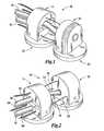
- FIG. 1is a forward isometric view of the distal body and proximal body which, together with the control wires and suction lines, makeup one embodiment of the robot;
- FIG. 2is a rearward isometric view of the distal and proximal bodies of FIG. 1 ;
- FIGS. 3A-3Dillustrate forward locomotion of the robot
- FIGS. 4A-4Cillustrate side to side turns of the robot
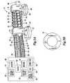
- FIG. 5Aillustrates another embodiment of a robot and control system according to the present disclosure while FIG. 5B illustrates a washer-like support spacer;
- FIG. 6Ais an example of one type of end effector, a semicircular needle, retracted into a recessed storage location in the distal body while FIG. 6B illustrates the needle in operation;
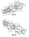
- FIGS. 7A and 7Bare an example of another type of end effector
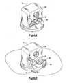
- FIG. 8illustrates a self contained embodiment of the device of the present disclosure
- FIGS. 9A and 9Billustrate one example of a streamlined device of the present disclosure
- FIGS. 10A and 10Billustrate another example of a streamlined device of the present disclosure
- FIGS. 11A and 11Billustrate an embodiment of a device of the present disclosure having stabilization struts
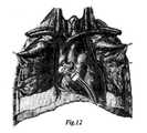
- FIG. 12is a conceptual illustration of the robot maneuvering on the surface of a heart to perform a procedure.
- FIGS. 1 and 2illustrate a prototype device 10 designed and constructed in the Medical Instrumentation Lab at Camegie Mellon University, which will now be described for purposes of illustration and not limitation.
- the device 10consists of two glass-filled polycarbonate shells forming a distal body 12 and a proximal body 14 , each body having a 13 mm circular footprint and a height of 14 mm. That size allows the device 10 to fit within a standard 20 mm diameter cannula or port.
- Each of the body sections 12 , 14is equipped with an independent suction line 16 , 18 and suction pad 20 , 22 , respectively, for gripping to biological tissue.
- the suction lines 16 , 18 and suction pads 20 , 22illustrate one type of means for prehension.
- the translation and rotation of the body sections 12 , 14 relative to one anotherare controlled from an external control system, in this embodiment a handle 15 (shown in FIGS. 3A-3D and FIGS. 4A-4C ), by manually adjusting the lengths of three nitinol wires 24 , 25 , 26 running along the longitudinal axis of the device 10 .
- the super-elasticity of nitinolallows the wires to support tension and compression (i.e. pulling and pushing) without permanently deforming. That eliminates the need for shape restoring components (like springs) that are required in some other systems.
- the axes of these wires 24 , 25 , 26intersect the body sections 12 , 14 at the perimeter of a 10 mm diameter circle at 120-degree intervals.
- the wires 24 , 25 , 26are fixed to the distal body 12 and pass freely through the proximal body 14 out to the handle 15 of the device. Between the proximal body 14 and the handle 15 , the wires 24 , 25 , 26 are contained within sheathes 24 ′, 25 ′, 26 ′, respectively, e.g. flexible plastic tubing, whose ends are attached to the proximal body 14 and the handle 15 .
- the three independently actuated wires 24 , 25 , 26provide three degrees of freedom between the distal body 12 and the proximal body 14 , two angular and one tanslational. The two angular degrees of freedom allow the device 10 to adapt to the curvature of the heart (or other organ) as well as turn laterally (i.e. yaw).
- the sheaths 24 ′, 25 ′, 26 ′prevent bowing of the wires 24 , 25 , 26 so as to transmit the forces applied to the wires 24 , 25 , 26 , respectively, at the handle 15 to either proximal body 14 or distal body 12 and ensure that the length of wires 24 , 25 , 26 between the handle 15 and proximal body 14 remains constant.
- the length of a wire exiting its sheath at the handleis changed, the length of that wire between the proximal body 14 and the distal body 12 changes by the same amount.
- Inchworm-like locomotionis achieved by alternating the suction force exerted by the two body sections, while changing the lengths of the wires at the fixed handle, as shown in FIGS. 3A-3D .
- the configuration of the sheaths 24 ′, 25 ′ 26 ′and enclosed wires 24 . 25 , 26does not affect the locomotion of the device 10 as long as there is slack between the handle 15 and the proximal body 14 , some of which will be taken up with each forward step.
- the heavy black lineindicates which suction pad is active. Note that the configuration of the sheaths and enclosed wires between the handle 15 and proximal body 14 changes with each forward step, but the lengths remain constant.
- FIGS. 3A and 3BWhile the proximal suction pad 22 is turned on, the wires 24 , 25 , 26 are moved forward causing distal body 12 to move forward by the same amount.
- FIG. 3Cthe proximal suction pad 22 is turned off and the distal suction pad 20 is turned on.
- FIG. 4Dthe compression in the sheaths 24 ′, 25 ′, 26 ′ is released, causing the proximal body 14 to “catch up” with the distal body 12 .
- Another forward stepcan now be taken by repeating the process. Turning can be achieved by differentially changing the lengths of the side wires as shown in FIGS. 4A-4C .
- the actuation of the wires at the handlemay be performed manually, along with the opening and closing of the valves to the suction lines.
- FIG. 5Another embodiment is illustrated in FIG. 5 .
- components having similar functions and construction to those of FIGS. 1 and 2have like reference numbers.
- the embodiment of FIG. 5differs from the previous embodiment in several ways.
- the wires 24 , 25 , 26may be attached to a support spring 27 by eyelets 28 to prevent the wires 24 , 25 , 26 from bowing during turning and to ensure that the wires maintain an equal distance from one another.
- a plurality of flat, washer-like structures 31may be provided to maintain the proper spacing between wires 24 , 25 , 26 .
- the plurality of washer-like structures 31may be separated from one another by springs (not shown).
- a 1.6 mm diameter commercial fiberscope 29may be fixed on the distal body 12 to provide visual feedback, with or without the use of an adjustable mirror 40 .
- the images from the fiberscope 29may be captured with a digital video camera 42 and displayed as a part of the graphical user interface (GUI) 44 , both of which are part of a control system 46 .
- the control system 46may include sensors 48 for monitoring the vacuum supplied by suction lines 16 , 18 , electronically controlled valves 50 for determining which suction pad 20 , 22 is operative, and vacuum source 52 .
- the control system 46may also include motors 54 for controlling movement of wires 24 , 25 26 .
- a computer 55may be provided to control the various components in response to information input by the surgeon via the GUI 44 to control locomotion and other functions.
- Such a designallows for the motors 54 , solenoid valves 50 , etc. to be located outside the device 10 . It is anticipated that the robot 10 may be either a disposable device or a reusable, sterilizable device.
- the suction pads 20 , 22are connected to the bodies 12 , 14 by means of flexible feet 56 , 58 , respectively. That enables the suction pads 20 , 22 more freedom to conform to the surface of the organ.
- Meshesmay cover the bottom of the suction pads to keep out large particles, while suction filters 60 may be provided to remove fluids and small particles.
- An aspect of the present inventionis changing the frame of reference of the robot from that of the surgeon to that of the moving organ.
- the exact form and construction of the robot used to bring about that change of referenceis not critical to this aspect of the invention.
- locomotionis achieved through the advancement of wires, either manually or through the activation of motors, others means of locomotion may be provided such as local (i.e. positioned on the robot) electric motors (operated with or without a tether), local ultrasonic motors (operated with or without a tether), as well as pneumatic actuators (typically operated with a tether).
- the means for prehension in the disclosed embodimentis suction.
- Alternative means of prehensionmay include synthetic gecko foot hair [Sitti M, Fearing RS (2003) Synthetic gecko foot-hair micro/nano-structures as dry adhesives. J Adhesion Sci Technol 17(8): 1055-1073] or a “tacky” foot.
- the actuation for treatmentmay include all the same alternatives as for locomotion.
- the devicemay operate with a tether having wires and pneumatic lines as disclosed above, with a tether having electric wires for local motors or video from a camera, or the device may operate without a tether.
- Tetherless modelscould be powered by a battery, the transcutaneous charging of a coil, etc., and could be controlled by local computing or through radio frequency transmissions.
- a loosely bounded body cavityrefers to that space surrounding an organ such as, for example, the peritoneal space surrounding the liver, the pleural space surrounding the lungs, the pericardial space surrounding the heart, etc., in addition to the space within certain organs such as the heart or stomach.
- FIG. 6Aillustrates an end effector (tool), which in this example is a needle 30 carried within a recess 32 in distal body 12 .
- Distal body 12also carries a means for providing images such as a fiberscope or camera, with or without some combination of lenses, mirrors, fiberoptics, etc.
- the needle 30may used to perform epicardial electrode lead placement for cardiac resynchronization therapy (CRT) via subxiphoid videopericardioscopic access.
- CTRcardiac resynchronization therapy
- a robot 10 equipped with the needle 30can perform a minimally invasive suturing technique that can be used with a variety of epicardial pacing leads, both permanent and temporary.
- Needle 30is a high-strength needle for suturing that has a drive shaft (not shown) that runs along the long axis of the device 10 , entered laterally and located below the midline of distal body 12 .
- the needle 30At the distal (working) end of this drive shaft is the needle 30 which is a segment (roughly 5 mm) that is bent 90° with respect to the drive shaft, forming the radius of a circle.
- the needle 30will then terminate in a semicircular suturing portion.
- the lower half of the front end of the distal body 12has a semicircular channel 32 into which the needle 30 recedes when it is not in use, protecting both the cardiac tissue and the needle 30 .
- the proximal end of the suture thread 36remains outside the body.
- the distal end of the thread 36is connected to a sharpened cap 38 , which will fit snugly over the end of the needle 30 .
- suturingwill be performed by advancing the needle 30 from its recessed storage channel 32 (see FIG. 5B ) and then rotating the drive shaft, forcing the semicircular needle 30 and the sharpened cap 38 with its suture thread 36 to pass through the tissue in an arc and exit again with the suture cap 38 still on the tip of the needle 30 .
- a minimally invasive forcepspassing through an off-center working port of the robot 10 will be used to grasp the thread 36 , lift the thread 36 and its cap 38 from the tip of the needle 30 , and retract the cap 38 and distal end of the thread 36 all the way back through the cannula to the outside of the body.
- the surgeonwill knot the suture with his own hands, tying a single throw (or half of a square knot) in the thread. Once the single throw has been tied, the surgeon will place a wire with a slightly forked tip against the knot, with the knot resting in the notch of the fork, and will use the wire to push the knot all the way back against the epicardium.
- the robot 10will have a separate electrode channel that will allow passage of the electrode and its wire lead from outside the body into the pericardium to be sutured to the heart.
- the needle 30 , forceps, wire “fork”, suture with sharpened cap, and all supporting instrumentation needed for the suturing techniquemay be designed for sterilizability. Actuation from outside the body is the most feasible option for the forceps, because the forceps must be fully retractable to bring the tip of the suture thread back to the hand of the surgeon. Actuation of the needle for suturing may be performed locally by motors inside the robot, or from outside the body using a wire running through the cannula. Visual feedback for suturing may be provided by the same device used during locomotion.
- FIGS. 7A and 7BAnother end effector (tool) is illustrated in FIGS. 7A and 7B .
- Like componentscarry the same reference numbers as used in the previous figures.
- the washer-like structure 31 and spacer springshave been eliminated for purposes of clarity. The reader will understand that a plurality of washer-like structures 31 and spacer springs may be used in the embodiment of FIGS. 7A and 7B .
- An experiment using this end effector, needle 70was performed on a pig. Through a 15 mm pericardial opening at the junction between the pericardium and the diaphragm at the midline, the device 10 was manually introduced inside the intact pericardium. Suction was applied to both suction pads.
- Stable contact with the epicardium of the beating anterior wall of the right ventriclewas visually confirmed for a period of 30 seconds.
- the device 10was then advanced across the left anterior descending coronary artery over the anterior wall of the beating left ventricle. Consistent stable contract with the epicardium of the beating left ventricle was observed.
- the device 10was then further advanced over the left atrial appendage and stable fixation to the surface of the beating left atrium was confirmed.
- Two myocardial injections of tissue-marking dyewere performed during the experiment.
- the device 10walked to the desired site, locked down both bodies using suction, and then the surgeon performed the injection manually by advancing the 27G custom needle 70 through a working port.
- the device 10was positioned over the bifurcation of the left anterior descending coronary artery and the takeoff of the diagonal branch; the needle 70 was advanced into the left ventricular myocardium for 2-3 mm and 0.5 cc of dye was injected.
- the maximum force applied during injectionwas 0.72 N.
- the device 10was then moved over the diagonal coronary artery and another injection of 0.5 cc of dye was made over the anterolateral wall of the left ventricle.
- the maximum force applied to the needle during the second injectionwas 1.15 N. No bleeding was observed after the needle 70 was withdrawn. Confirmation of successful injection was made at postoperative examination.
- pulling the tetherprovided a feasible method in the preliminary experiments to apply a tangential force to dislodge the device, without requiring the development of additional hardware that could be inserted somehow into the pericardial sac for testing.
- the force necessary to dislodge the device from the epicardium by pulling on the tetherwas measured with a force gauge while the device held onto the heart.
- a small clampwas applied to the part of the tether consisting of the three drive wires and their sheaths, and this clamp was attached to a handheld digital force gauge. The surgeon then pulled on the force gauge until the device was dislodged, and the gauge recorded the maximum force encountered during each trial.
- the ease with which the device can be retrieved from the pericardiumwas tested by measuring the maximum force encountered during extraction.
- the devicewas positioned normally inside the pericardium, standing upright on its feet with the distal body of the device near the left atrium, roughly 10 cm from the entry incision. All suction was turned off. The device was then retracted completely from the pericardium by pulling on the tether. This test was repeated three times, and the peak force measured during retrieval was recorded during each trial. The mean peak force measured was 2.49 ⁇ 0.51 N.
- FIG. 8illustrates a self-contained design of the device 10 , with the proximal body 14 shown at the upper left of the figure and the distal body 12 at the lower right.
- This designinvolves two motors for locomotion.
- One motorwould be located in the vertical cylindrical body of the proximal body 14 and the other motor located in the horizontal cylinder 74 visible in the arm connecting the proximal 14 and distal 12 bodies.
- a streamline housingdiscussed below, is not shown in FIG. 8 .
- the motorscould receive power and instructions through a tether (not shown) or from an onboard battery and an onboard computer (not shown).
- FIGS. 9A and 9Billustrate a device 10 with a capsular or pill-like design for streamline interaction with the pericardial sac.
- the device 10has a two piece hard covering, one piece of which slides inside the other like a gelatin capsule of the sort often used for pills.
- FIG. 9Aillustrates the device 10 in an extended phase of a step, i.e. maximum distance between the distal body 12 and proximal body 14
- FIG. 9Billustrates the contracted phase of the step, i.e. minimal distance between the proximal body 14 and distal body 12 .
- FIGS. 10A and 10Billustrate another design of the device 10 with a one-piece outer shell designed for streamline interaction with the pericardial sac. This design may be used in place of the design of FIGS. 9A and 9B should that design be found to cause pinching of the pericardium during locomotion.
- FIG. 10Aillustrates the extended phase of a step while FIG. 10B illustrates the contracted phase of the step.
- FIGS. 11A and 11Billustrate a design in which deployable outrigger-like struts 80 , 82 are used if stronger safeguards against tipping become necessary.
- the outrigger-like struts 80 , 82would be foldable from the horizontal positions shown to vertical positions adjacent distal body 12 on both sides of the device 10 .
- the struts 80 , 82may be moved from the horizontal to the vertical position shown in the figures to guard against tipping. If the struts 80 , 82 evidence any tendency to snag the pericardium, a stretchable membrane 84 may be employed to cover both the device 10 and the struts 80 , 82 as shown in the figures.
- FIG. 11Aillustrates the extended phase of a step while FIG. 111B illustrates the contracted phase of the step.
- the device 10will enter the pericardium and be placed on the epicardial surface of the heart using a rigid or flexible endoscope with a working port.
- the endoscopewill be introduced into the pericardial sac through a port or limited incision beneath the xiphoid process of the sternum.
- the device 10will grasp the epicardium using suction.
- the suction forcesare applied through the two independent suction pads 20 , 22 (see FIG. 5 ) that may be attached directly to bodies 12 , 14 or through compliant or flexible feet 56 , 58 , respectively.
- the vacuum pressureis supplied to the suction pads 20 , 22 by the vacuum source 52 through the operation of valves 50 and suction lines 16 , 18 respectively.
- the vacuum sourceprovides a vacuum pressure of ⁇ 0.08 N/mm 2 , which was found to be effective and safe for use in FDA approved cardiac stabilizers. The suction forces generated by this pressure have proven effective for our application, and did not damage the epicardial tissue.
- the vacuum pressureis monitored by the external pressure sensors 48 and regulated by computer-controlled solenoid valves 50 , both located within the control system 46 . Based on this pressure, the normal and tangential forces calculated to dislodge one of the bodies 12 , 14 are 1.76 N and 0.87 N, respectively. Bench testing using a force gauge to dislodge the device from a poultry model verified normal and tangential forces of 2.01 N and 0.86 N. The tangential force that can be resisted by the device will be increased significantly by reducing the profile.
- the device 10will provide visual feedback to the surgeon during locomotion and administration of therapy. That can be accomplished using fiberoptics to relay the image from the distal end 12 of the device 10 to the camera 42 located in the control system 46 .
- a CCD video cameracan be mounted directly to the distal end 12 of the device 10 . It may be possible to provide all of the necessary vision with a single visual sensor on a fixed mount. More likely, however, either the viewing head will be actuated for motion, or two imaging devices will be incorporated: one tangential to the surface of the organ (looking forward) for providing information for navigation, and the other normal to the surface (looking down) for providing a view of the area to receive attention. “Attention” is intended to be a broad term that includes all types of interventions in addition to all forms of testing, viewing or inspecting a site, etc., or any other activity that results in consideration being devoted to an organ or a portion of an organ.
- the device 10differs from prior art robotic surgical systems in several fundamental ways: (1) it operates within the reference frame of the heart rather than that of an operating table, (2) it will be introduced using a sub-xiphoid rather than an intercostal approach, obviating general endotracheal anesthesia (GETA), (3) it has locomotive capabilities, and (4) it will be relatively inexpensive and possibly disposable. For surgical procedures that can be performed completely within the pericardium, the device 10 will eliminate many of the limitations of these surgical systems.
- Therapies administered from the device 10will not require stabilization of the heart because the device 10 will be located in the same reference frame as the surface of the heart, rather than that of a fixed operating table. This eliminates the need for either endoscopic stabilizers, which require additional incisions, or cardiopulmonary bypass, which increases the complexity and risk of the procedure.
- the teleoperative surgical systems in use todayutilize laparoscopic manipulators and cameras and are introduced to the pericardial sac through several intercostal (between rib) incisions. These instruments must then pass through the pleural space before reaching the heart, which requires the collapsing of a lung.
- the delivery of the device 10 onto the heartwill not require collapsing a lung because it will be introduced to the thoracic cavity through an incision made directly below the xiphoid process.
- the endoscopewill then be pushed through the tissue and fascia beneath the sternum until the bare area of the pericardium is reached, never entering the pleural space.
- the scopewill also be used to breach the pericardium, thus delivering the device 10 directly to the epicardium.
- the device 10will not require the collapsing of a lung, it will also not require differential ventilation of the patient, and it is therefore possible that local or regional anesthesia could be used instead of general endotracheal anesthesia (GETA).
- GETAgeneral endotracheal anesthesia
- a potential benefitis that the device 10 may enable certain cardiovascular interventions to be performed on an ambulatory outpatient basis, something that has never been done before.
- the locomotive capabilities of the device 10will enable it to reach virtually any position and orientation on the epicardium. This is not the case with rigid laparoscopes, which are limited to a relatively small workspace near the entry incision. In addition, these systems require the removal and re-insertion of the tools to change the operative field within a single procedure.
- the device 10can easily change its workspace by simply moving to another region of the heart.
- the da Vinci surgical systemis very expensive and consists of a surgeon's computerized console and a patient-side cart with multiple large robotic arms. For procedures that can be performed within the pericardium, the device 10 will provide a small, extremely low-cost alternative to this system.
- the TEMwill be able to perform epicardial cardiac procedures such as: cell transplantation, gene therapy, atrial ablation, and electrode placement for resynchronization myocardial revascularization.
- Devicessuch as an ultrasound transducer, diagnostic aid or other sensor, drug delivery system, therapeutic device, optical fiber, camera or surgical tool(s) may be carried by the device 10 .
Landscapes
- Health & Medical Sciences (AREA)
- Life Sciences & Earth Sciences (AREA)
- Surgery (AREA)
- Engineering & Computer Science (AREA)
- General Health & Medical Sciences (AREA)
- Veterinary Medicine (AREA)
- Public Health (AREA)
- Animal Behavior & Ethology (AREA)
- Nuclear Medicine, Radiotherapy & Molecular Imaging (AREA)
- Molecular Biology (AREA)
- Biomedical Technology (AREA)
- Heart & Thoracic Surgery (AREA)
- Medical Informatics (AREA)
- Robotics (AREA)
- Biophysics (AREA)
- Radiology & Medical Imaging (AREA)
- Physics & Mathematics (AREA)
- Pathology (AREA)
- Optics & Photonics (AREA)
- Surgical Instruments (AREA)
- Endoscopes (AREA)
- Manipulator (AREA)
Abstract
Description
- The present application claims priority from U.S. application Ser. No. 60/518,582 filed Nov. 7, 2003 and entitled Inchworm Robot for Minimally Invasive Cardiac Interventions, which is hereby incorporated by reference in its entirety.
- Heart surgery, particularly the types addressed here (e.g., epicardial electrode placement, atrial ablation) is typically done via either an open approach, or a minimally invasive approach using hand-held rigid endoscopic tools.
- Several recent development efforts center around robots intended to perform heart surgery, among other procedures. A commercially available robotic system for cardiac surgery is the da Vinci System available from Intuitive Surgical of Mountain View, Calif. That system is teleoperative, meaning that the motions of the surgeons hands on input devices are mirrored by laparoscopic manipulators located within the body. While such a system can offer superior dexterity to conventional laparoscopic instruments, it requires some form of stabilization for the heart, requires collapsing a lung, has a limited operative field, and is bulky and expensive.
- Closed-chest endoscopic visualization of the epicardium was first described by Santos et al. (Ann Thorac Surg 1977; 23: 467-470); subsequent reports have utilized the technique for evaluation of blunt chest trauma, pericardial effusion and lung cancer staging. Lattouf et al have utilized the technique for epicardial implantation of left ventricular pacing leads. In each of these reports, endoscope access required thoracotomy with breach of the left pleural space. Direct access to the pericardial space via subxiphoid puncture is an increasingly practiced technique during catheter ablation procedures. In these reports, once access was achieved, catheter manipulation was guided solely by fluoroscopy. We are aware of cursory attempts at standard pacing lead implantation using this approach which have failed due to inability to achieve fixation.
- The challenges of minimally invasive access are further complicated by the goal of avoiding cardiopulmonary bypass, and this goal necessitates surgery on a beating heart. Thus instrumentation is needed that allows stable manipulation of an arbitrary location on the epicardium while the heart is beating. See, for example, published application number 20040172033. Local immobilization of the heart is the approach generally followed with endoscopic stabilizers such as the Endostab device and the endo-Octopus device, which operate with pressure or suction. However, the resulting forces exerted on the myocardium can cause changes in the electrophysiological and hemodynamic performance of the heart, and there has been discussion in the literature regarding the care that must be taken to avoid hemodynamic impairment [Falk, et al., Endoscopic coronary artery bypass grafting on the beating heart using a computer enhanced telemanipulation system. Heart Surg Forum 2: 199-205, 1999]. As an alternative, several researchers in robot-assisted endoscopic surgery are investigating active compensation of heartbeat motion by visually tracking the epicardium and moving the tool tips accordingly [avu
 o{haeck over (g)}lu MC, et al., Robotics for telesurgery: second generation Berkeley/UCSF laparoscopic telesurgical workstation and looking towards the future applications. Industrial Robot 30: 22-29, 2003; Ortmaier T J. Motion compensation in minimally invasive robotic surgery. Ph.D. dissertation, Technical University of Munich, Germany, 2003.], but this research problem remains open. The motion of the beating heart is complex. In addition to the challenges of modeling or tracking the heart surface, active compensation will require considerable expense for high-bandwidth actuation to enable manipulation in at least three degrees of freedom over a relatively large workspace (See Cavusoglu, supra).
- The prior art solutions address a problem that exists only because the tools are held by a surgeon or a robot that is fixed to the table or standing on the floor. The present disclosure takes a different approach. Rather than trying to immobilize the heart surface to stabilize it in the fixed frame of reference of a table-mounted robotic device, we mounted the device in the moving reference frame of the beating heart. That task was accomplished with a miniature crawling robotic device designed to be introduced into the pericardium through a port, attach itself to the epicardial surface, and then, under the direct control of the surgeon, travel to the desired location for treatment. The problem of beating-heart motion was largely avoided by attaching the device directly to the epicardium. The problem of access was resolved by incorporating the capability for locomotion.
- Improved access and precise manipulation are not the only benefits of this approach. Port access for minimally invasive cardiac surgery has typically been transthoracic, largely to accommodate the rigid endoscopes generally used for both manual and robot-assisted procedures. Transthoracic access to the heart requires deflation of the left lung, general endotracheal anesthesia, and differential lung ventilation. A variety of current and upcoming procedures, however, can conceivably be performed transpericardially, without invasion of the pleural space, with appropriate instrumentation. Examples include, but are not limited to cell transplantation, gene therapy for angiogenesis, epicardial electrode placement for resynchronization, epicardial atrial ablation, intrapericardial drug delivery, and ventricle-to-coronary artery bypass, among others.
- The ability of the device to move to any desired location on the epicardium from any starting point enables minimally invasive cardiac surgery to become independent of the location of the pericardial incision. Use of the device also allows a subxiphoid transpericardial approach to any intrapericardial procedure, regardless of the location of the treatment site. As a result, deflation of the left lung is no longer needed, and it becomes feasible to use local or regional rather than general anesthetic techniques. These advantages have the potential for opening the way to ambulatory outpatient cardiac surgery. The opportunity for “synergy” (e.g. multiple procedures during a single operative session) may prove particularly valuable. The techniques disclosed herein are applicable to other organs within a living body and need not be limited to the human heart, which is merely our first application.
- For the present disclosure to be easily understood and readily practiced, the present disclosure will now be described, for purposes of illustration and not limitation, in connection with the following figures wherein:
FIG. 1 is a forward isometric view of the distal body and proximal body which, together with the control wires and suction lines, makeup one embodiment of the robot;FIG. 2 is a rearward isometric view of the distal and proximal bodies ofFIG. 1 ;FIGS. 3A-3D illustrate forward locomotion of the robot;FIGS. 4A-4C illustrate side to side turns of the robot;FIG. 5A illustrates another embodiment of a robot and control system according to the present disclosure whileFIG. 5B illustrates a washer-like support spacer;FIG. 6A is an example of one type of end effector, a semicircular needle, retracted into a recessed storage location in the distal body whileFIG. 6B illustrates the needle in operation;FIGS. 7A and 7B are an example of another type of end effector;FIG. 8 illustrates a self contained embodiment of the device of the present disclosure;FIGS. 9A and 9B illustrate one example of a streamlined device of the present disclosure;FIGS. 10A and 10B illustrate another example of a streamlined device of the present disclosure;FIGS. 11A and 11B illustrate an embodiment of a device of the present disclosure having stabilization struts; andFIG. 12 is a conceptual illustration of the robot maneuvering on the surface of a heart to perform a procedure.- One embodiment of a robot constructed according to the present disclosure is illustrated in
FIGS. 1 and 2 .FIGS. 1 and 2 illustrate aprototype device 10 designed and constructed in the Medical Instrumentation Lab at Camegie Mellon University, which will now be described for purposes of illustration and not limitation. Thedevice 10 consists of two glass-filled polycarbonate shells forming adistal body 12 and aproximal body 14, each body having a 13 mm circular footprint and a height of 14 mm. That size allows thedevice 10 to fit within a standard 20 mm diameter cannula or port. Each of thebody sections independent suction line suction pad suction pads - The translation and rotation of the
body sections FIGS. 3A-3D andFIGS. 4A-4C ), by manually adjusting the lengths of threenitinol wires device 10. The super-elasticity of nitinol allows the wires to support tension and compression (i.e. pulling and pushing) without permanently deforming. That eliminates the need for shape restoring components (like springs) that are required in some other systems. The axes of thesewires body sections wires distal body 12 and pass freely through theproximal body 14 out to thehandle 15 of the device. Between theproximal body 14 and thehandle 15, thewires proximal body 14 and thehandle 15. The three independently actuatedwires distal body 12 and theproximal body 14, two angular and one tanslational. The two angular degrees of freedom allow thedevice 10 to adapt to the curvature of the heart (or other organ) as well as turn laterally (i.e. yaw). - The
sheaths 24′,25′,26′ prevent bowing of thewires wires handle 15 to eitherproximal body 14 ordistal body 12 and ensure that the length ofwires handle 15 andproximal body 14 remains constant. Thus, when the length of a wire exiting its sheath at the handle is changed, the length of that wire between theproximal body 14 and thedistal body 12 changes by the same amount. - Inchworm-like locomotion is achieved by alternating the suction force exerted by the two body sections, while changing the lengths of the wires at the fixed handle, as shown in
FIGS. 3A-3D . The configuration of thesheaths 24′,25′26′andenclosed wires 24.25,26 does not affect the locomotion of thedevice 10 as long as there is slack between thehandle 15 and theproximal body 14, some of which will be taken up with each forward step. In the figure, the heavy black line indicates which suction pad is active. Note that the configuration of the sheaths and enclosed wires between thehandle 15 andproximal body 14 changes with each forward step, but the lengths remain constant. - Between
FIGS. 3A and 3B , while theproximal suction pad 22 is turned on, thewires distal body 12 to move forward by the same amount. InFIG. 3C , theproximal suction pad 22 is turned off and thedistal suction pad 20 is turned on. InFIG. 4D , the compression in thesheaths 24′,25′,26′ is released, causing theproximal body 14 to “catch up” with thedistal body 12. Another forward step can now be taken by repeating the process. Turning can be achieved by differentially changing the lengths of the side wires as shown inFIGS. 4A-4C . The actuation of the wires at the handle may be performed manually, along with the opening and closing of the valves to the suction lines. - Another embodiment is illustrated in
FIG. 5 . InFIG. 5 , components having similar functions and construction to those ofFIGS. 1 and 2 have like reference numbers. The embodiment ofFIG. 5 differs from the previous embodiment in several ways. For example, between theproximal body 14 anddistal body 12 thewires support spring 27 byeyelets 28 to prevent thewires support spring 27 may have a very low spring constant (e.g. k=0.012 N/mm) such that the restoring force is negligible as compared to that of thewires spring 27 and eyelets28, a plurality of flat, washer-like structures31 (SeeFIG. 5B ) may be provided to maintain the proper spacing betweenwires like structures 31 may be separated from one another by springs (not shown). - A 1.6 mm diameter
commercial fiberscope 29, running longitudinally through the length of the device, may be fixed on thedistal body 12 to provide visual feedback, with or without the use of anadjustable mirror 40. The images from thefiberscope 29 may be captured with adigital video camera 42 and displayed as a part of the graphical user interface (GUI)44, both of which are part of acontrol system 46. Thecontrol system 46 may includesensors 48 for monitoring the vacuum supplied bysuction lines valves 50 for determining whichsuction pad vacuum source 52. Thecontrol system 46 may also includemotors 54 for controlling movement ofwires computer 55 may be provided to control the various components in response to information input by the surgeon via theGUI 44 to control locomotion and other functions. Such a design allows for themotors 54,solenoid valves 50, etc. to be located outside thedevice 10. It is anticipated that therobot 10 may be either a disposable device or a reusable, sterilizable device. - In the embodiment of
FIG. 5 , thesuction pads bodies flexible feet suction pads - An aspect of the present invention is changing the frame of reference of the robot from that of the surgeon to that of the moving organ. The exact form and construction of the robot used to bring about that change of reference is not critical to this aspect of the invention. For example, although in the disclosed embodiments locomotion is achieved through the advancement of wires, either manually or through the activation of motors, others means of locomotion may be provided such as local (i.e. positioned on the robot) electric motors (operated with or without a tether), local ultrasonic motors (operated with or without a tether), as well as pneumatic actuators (typically operated with a tether). The means for prehension in the disclosed embodiment is suction. Alternative means of prehension may include synthetic gecko foot hair [Sitti M, Fearing RS (2003) Synthetic gecko foot-hair micro/nano-structures as dry adhesives. J Adhesion Sci Technol 17(8): 1055-1073] or a “tacky” foot. The actuation for treatment may include all the same alternatives as for locomotion. Finally, the device may operate with a tether having wires and pneumatic lines as disclosed above, with a tether having electric wires for local motors or video from a camera, or the device may operate without a tether. Tetherless models could be powered by a battery, the transcutaneous charging of a coil, etc., and could be controlled by local computing or through radio frequency transmissions. It will be understood by those of ordinary skill in the art that changing the frame of reference of the robot from that of the surgeon to that of the moving organ can be brought about by a wide variety of robots designed so as to be able to move within a loosely bounded body cavity. A loosely bounded body cavity refers to that space surrounding an organ such as, for example, the peritoneal space surrounding the liver, the pleural space surrounding the lungs, the pericardial space surrounding the heart, etc., in addition to the space within certain organs such as the heart or stomach.
FIG. 6A illustrates an end effector (tool), which in this example is aneedle 30 carried within arecess 32 indistal body 12.Distal body 12 also carries a means for providing images such as a fiberscope or camera, with or without some combination of lenses, mirrors, fiberoptics, etc. Theneedle 30 may used to perform epicardial electrode lead placement for cardiac resynchronization therapy (CRT) via subxiphoid videopericardioscopic access. Arobot 10 equipped with theneedle 30 can perform a minimally invasive suturing technique that can be used with a variety of epicardial pacing leads, both permanent and temporary.Needle 30 is a high-strength needle for suturing that has a drive shaft (not shown) that runs along the long axis of thedevice 10, entered laterally and located below the midline ofdistal body 12. At the distal (working) end of this drive shaft is theneedle 30 which is a segment (roughly 5 mm) that is bent 90° with respect to the drive shaft, forming the radius of a circle. Theneedle 30 will then terminate in a semicircular suturing portion. The lower half of the front end of thedistal body 12 has asemicircular channel 32 into which theneedle 30 recedes when it is not in use, protecting both the cardiac tissue and theneedle 30.- The proximal end of the
suture thread 36 remains outside the body. The distal end of thethread 36 is connected to a sharpenedcap 38, which will fit snugly over the end of theneedle 30. When the surgeon has positioned thedistal body 12 at a desired work site, suturing will be performed by advancing theneedle 30 from its recessed storage channel32 (seeFIG. 5B ) and then rotating the drive shaft, forcing thesemicircular needle 30 and the sharpenedcap 38 with itssuture thread 36 to pass through the tissue in an arc and exit again with thesuture cap 38 still on the tip of theneedle 30. A minimally invasive forceps (not shown), passing through an off-center working port of therobot 10 will be used to grasp thethread 36, lift thethread 36 and itscap 38 from the tip of theneedle 30, and retract thecap 38 and distal end of thethread 36 all the way back through the cannula to the outside of the body. Here, the surgeon will knot the suture with his own hands, tying a single throw (or half of a square knot) in the thread. Once the single throw has been tied, the surgeon will place a wire with a slightly forked tip against the knot, with the knot resting in the notch of the fork, and will use the wire to push the knot all the way back against the epicardium. He will then tie a second throw and use the wire to push it forward until it meets the first throw, completing the knot. Given an ample supply of sutures fitted with sharpened caps, this technique can be repeated as many times as necessary, by placing each sharpened cap on the tip of the needle using the same forceps that is used to retrieve it from the needle. - The
robot 10 will have a separate electrode channel that will allow passage of the electrode and its wire lead from outside the body into the pericardium to be sutured to the heart. Theneedle 30, forceps, wire “fork”, suture with sharpened cap, and all supporting instrumentation needed for the suturing technique may be designed for sterilizability. Actuation from outside the body is the most feasible option for the forceps, because the forceps must be fully retractable to bring the tip of the suture thread back to the hand of the surgeon. Actuation of the needle for suturing may be performed locally by motors inside the robot, or from outside the body using a wire running through the cannula. Visual feedback for suturing may be provided by the same device used during locomotion. - Another end effector (tool) is illustrated in
FIGS. 7A and 7B . Like components carry the same reference numbers as used in the previous figures. In the device ofFIGS. 7A and 7B , the washer-like structure 31 and spacer springs have been eliminated for purposes of clarity. The reader will understand that a plurality of washer-like structures 31 and spacer springs may be used in the embodiment ofFIGS. 7A and 7B . An experiment using this end effector,needle 70, was performed on a pig. Through a 15 mm pericardial opening at the junction between the pericardium and the diaphragm at the midline, thedevice 10 was manually introduced inside the intact pericardium. Suction was applied to both suction pads. Stable contact with the epicardium of the beating anterior wall of the right ventricle was visually confirmed for a period of 30 seconds. Thedevice 10 was then advanced across the left anterior descending coronary artery over the anterior wall of the beating left ventricle. Consistent stable contract with the epicardium of the beating left ventricle was observed. Thedevice 10 was then further advanced over the left atrial appendage and stable fixation to the surface of the beating left atrium was confirmed. - Successful locomotion of the
device 10 inside the intact pericardium was confirmed on the following areas: anterior wall of the beating right ventricle, anterolateral wall of the beating left ventricle, and anterior wall of the left atrial appendage. No gross epicardial or pericardial damage was observed. Because the present prototype has no outer shell between thebodies proximal body 12, which was not tapered, then tended to snag somewhat on the pericardium. The pericardial sac also showed a tendency to adhere to all surfaces of thedevice 10 that it contacted, no matter how smooth. This seemed to be largely caused by drying of the sac due to exposure to air. Occasional infusions of normal saline solution were used for lubrication, which succeeded in alleviating the problem. - Two myocardial injections of tissue-marking dye were performed during the experiment. In each case, the
device 10 walked to the desired site, locked down both bodies using suction, and then the surgeon performed the injection manually by advancing the27G custom needle 70 through a working port. For the first injection, thedevice 10 was positioned over the bifurcation of the left anterior descending coronary artery and the takeoff of the diagonal branch; theneedle 70 was advanced into the left ventricular myocardium for 2-3 mm and 0.5 cc of dye was injected. The maximum force applied during injection was 0.72 N. Thedevice 10 was then moved over the diagonal coronary artery and another injection of 0.5 cc of dye was made over the anterolateral wall of the left ventricle. The maximum force applied to the needle during the second injection was 1.15 N. No bleeding was observed after theneedle 70 was withdrawn. Confirmation of successful injection was made at postoperative examination. - Because the device is tethered, pulling the tether provided a feasible method in the preliminary experiments to apply a tangential force to dislodge the device, without requiring the development of additional hardware that could be inserted somehow into the pericardial sac for testing. The force necessary to dislodge the device from the epicardium by pulling on the tether was measured with a force gauge while the device held onto the heart. A small clamp was applied to the part of the tether consisting of the three drive wires and their sheaths, and this clamp was attached to a handheld digital force gauge. The surgeon then pulled on the force gauge until the device was dislodged, and the gauge recorded the maximum force encountered during each trial. This test was performed three times with suction applied only to the distal body, three times with suction applied only to the proximal body, and three times with suction applied to both bodies. The results are presented in Table 1. No damage to the device resulted from these tests.
TABLE 1 Body with Number of Standard suction applied trials Mean (N) deviation (N) distal 3 1.62 0.37 proximal 3 3.23 0.70 both 3 4.48 0.43 - During this experiment we demonstrated a technique for reattachment that can be used if the device is accidentally detached from the epicardium. This test was performed while the device was inside the intact pericardium. In this test, all suction was turned off, and, by manually twisting the tether, the device was intentionally rotated and was left lying on its right side on the epicardium. The suction in both
pads device 10, and by its presence lifted the pericardium somewhat, so that it did not lie as low on and around thedevice 10 as it normally would. Therefore, to avoid this effect, the video device was removed from the pericardium, and the test was repeated, this time with only the external video recorder monitoring the trial. Righting and reattaching of the device was performed successfully in both trials with no tissue damage and no damage to the device. - The ease with which the device can be retrieved from the pericardium was tested by measuring the maximum force encountered during extraction. The device was positioned normally inside the pericardium, standing upright on its feet with the distal body of the device near the left atrium, roughly 10 cm from the entry incision. All suction was turned off. The device was then retracted completely from the pericardium by pulling on the tether. This test was repeated three times, and the peak force measured during retrieval was recorded during each trial. The mean peak force measured was 2.49±0.51 N.
FIG. 8 illustrates a self-contained design of thedevice 10, with theproximal body 14 shown at the upper left of the figure and thedistal body 12 at the lower right. This design involves two motors for locomotion. One motor would be located in the vertical cylindrical body of theproximal body 14 and the other motor located in thehorizontal cylinder 74 visible in the arm connecting the proximal14 and distal12 bodies. For clarity, a streamline housing, discussed below, is not shown inFIG. 8 . The motors could receive power and instructions through a tether (not shown) or from an onboard battery and an onboard computer (not shown).FIGS. 9A and 9B illustrate adevice 10 with a capsular or pill-like design for streamline interaction with the pericardial sac. Thedevice 10 has a two piece hard covering, one piece of which slides inside the other like a gelatin capsule of the sort often used for pills.FIG. 9A illustrates thedevice 10 in an extended phase of a step, i.e. maximum distance between thedistal body 12 andproximal body 14, whileFIG. 9B illustrates the contracted phase of the step, i.e. minimal distance between theproximal body 14 anddistal body 12.FIGS. 10A and 10B illustrate another design of thedevice 10 with a one-piece outer shell designed for streamline interaction with the pericardial sac. This design may be used in place of the design ofFIGS. 9A and 9B should that design be found to cause pinching of the pericardium during locomotion.FIG. 10A illustrates the extended phase of a step whileFIG. 10B illustrates the contracted phase of the step.FIGS. 11A and 11B illustrate a design in which deployable outrigger-like struts like struts distal body 12 on both sides of thedevice 10. Once thedevice 10 is deployed within the pericardium, thestruts struts stretchable membrane 84 may be employed to cover both thedevice 10 and thestruts FIG. 11A illustrates the extended phase of a step whileFIG. 111B illustrates the contracted phase of the step.- Turning to
FIG. 12 , in operation according to one aspect of the present invention, thedevice 10 will enter the pericardium and be placed on the epicardial surface of the heart using a rigid or flexible endoscope with a working port. The endoscope will be introduced into the pericardial sac through a port or limited incision beneath the xiphoid process of the sternum. - Once positioned appropriately with the endoscope under direct visual confirmation, the
device 10 will grasp the epicardium using suction. The suction forces are applied through the twoindependent suction pads 20,22 (seeFIG. 5 ) that may be attached directly tobodies flexible feet suction pads vacuum source 52 through the operation ofvalves 50 andsuction lines external pressure sensors 48 and regulated by computer-controlledsolenoid valves 50, both located within thecontrol system 46. Based on this pressure, the normal and tangential forces calculated to dislodge one of thebodies - The
device 10 will provide visual feedback to the surgeon during locomotion and administration of therapy. That can be accomplished using fiberoptics to relay the image from thedistal end 12 of thedevice 10 to thecamera 42 located in thecontrol system 46. Alternatively, a CCD video camera can be mounted directly to thedistal end 12 of thedevice 10. It may be possible to provide all of the necessary vision with a single visual sensor on a fixed mount. More likely, however, either the viewing head will be actuated for motion, or two imaging devices will be incorporated: one tangential to the surface of the organ (looking forward) for providing information for navigation, and the other normal to the surface (looking down) for providing a view of the area to receive attention. “Attention” is intended to be a broad term that includes all types of interventions in addition to all forms of testing, viewing or inspecting a site, etc., or any other activity that results in consideration being devoted to an organ or a portion of an organ. - The
device 10 differs from prior art robotic surgical systems in several fundamental ways: (1) it operates within the reference frame of the heart rather than that of an operating table, (2) it will be introduced using a sub-xiphoid rather than an intercostal approach, obviating general endotracheal anesthesia (GETA), (3) it has locomotive capabilities, and (4) it will be relatively inexpensive and possibly disposable. For surgical procedures that can be performed completely within the pericardium, thedevice 10 will eliminate many of the limitations of these surgical systems. - Therapies administered from the
device 10 will not require stabilization of the heart because thedevice 10 will be located in the same reference frame as the surface of the heart, rather than that of a fixed operating table. This eliminates the need for either endoscopic stabilizers, which require additional incisions, or cardiopulmonary bypass, which increases the complexity and risk of the procedure. - The teleoperative surgical systems in use today utilize laparoscopic manipulators and cameras and are introduced to the pericardial sac through several intercostal (between rib) incisions. These instruments must then pass through the pleural space before reaching the heart, which requires the collapsing of a lung. The delivery of the
device 10 onto the heart will not require collapsing a lung because it will be introduced to the thoracic cavity through an incision made directly below the xiphoid process. The endoscope will then be pushed through the tissue and fascia beneath the sternum until the bare area of the pericardium is reached, never entering the pleural space. The scope will also be used to breach the pericardium, thus delivering thedevice 10 directly to the epicardium. Because thedevice 10 will not require the collapsing of a lung, it will also not require differential ventilation of the patient, and it is therefore possible that local or regional anesthesia could be used instead of general endotracheal anesthesia (GETA). As a result, a potential benefit is that thedevice 10 may enable certain cardiovascular interventions to be performed on an ambulatory outpatient basis, something that has never been done before. - The locomotive capabilities of the
device 10 will enable it to reach virtually any position and orientation on the epicardium. This is not the case with rigid laparoscopes, which are limited to a relatively small workspace near the entry incision. In addition, these systems require the removal and re-insertion of the tools to change the operative field within a single procedure. Thedevice 10, on the other hand, can easily change its workspace by simply moving to another region of the heart. - The da Vinci surgical system is very expensive and consists of a surgeon's computerized console and a patient-side cart with multiple large robotic arms. For procedures that can be performed within the pericardium, the
device 10 will provide a small, extremely low-cost alternative to this system. - With proper development of end-effectors, the TEM will be able to perform epicardial cardiac procedures such as: cell transplantation, gene therapy, atrial ablation, and electrode placement for resynchronization myocardial revascularization. Devices such as an ultrasound transducer, diagnostic aid or other sensor, drug delivery system, therapeutic device, optical fiber, camera or surgical tool(s) may be carried by the
device 10. - Procedures for organs other than the heart can be developed while remaining within the teachings of the present disclosure. Additionally, procedures on living bodies other than humans, e.g. pets, farm animals, race horses, etc. can be developed while remaining within the teachings of the present disclosure. Thus, while the present invention has been described in connection with preferred embodiments thereof, those of ordinary skill in the art will recognize that many modifications and variations are possible. The present invention is intended to be limited only by the following claims and not by the foregoing description which is intended to set forth the presently preferred embodiment.


Claims (21)
Priority Applications (2)
| Application Number | Priority Date | Filing Date | Title |
|---|---|---|---|
| US10/982,670US8162925B2 (en) | 2003-11-07 | 2004-11-05 | Robot for minimally invasive interventions |
| US13/424,732US9265582B2 (en) | 2003-11-07 | 2012-03-20 | Robot for minimally invasive interventions |
Applications Claiming Priority (2)
| Application Number | Priority Date | Filing Date | Title |
|---|---|---|---|
| US51858203P | 2003-11-07 | 2003-11-07 | |
| US10/982,670US8162925B2 (en) | 2003-11-07 | 2004-11-05 | Robot for minimally invasive interventions |
Related Child Applications (1)
| Application Number | Title | Priority Date | Filing Date |
|---|---|---|---|
| US13/424,732DivisionUS9265582B2 (en) | 2003-11-07 | 2012-03-20 | Robot for minimally invasive interventions |
Publications (2)
| Publication Number | Publication Date |
|---|---|
| US20050154376A1true US20050154376A1 (en) | 2005-07-14 |
| US8162925B2 US8162925B2 (en) | 2012-04-24 |
Family
ID=34590277
Family Applications (2)
| Application Number | Title | Priority Date | Filing Date |
|---|---|---|---|
| US10/982,670Active2028-01-26US8162925B2 (en) | 2003-11-07 | 2004-11-05 | Robot for minimally invasive interventions |
| US13/424,732Expired - LifetimeUS9265582B2 (en) | 2003-11-07 | 2012-03-20 | Robot for minimally invasive interventions |
Family Applications After (1)
| Application Number | Title | Priority Date | Filing Date |
|---|---|---|---|
| US13/424,732Expired - LifetimeUS9265582B2 (en) | 2003-11-07 | 2012-03-20 | Robot for minimally invasive interventions |
Country Status (4)
| Country | Link |
|---|---|
| US (2) | US8162925B2 (en) |
| EP (1) | EP1689289B1 (en) |
| JP (2) | JP2007510470A (en) |
| WO (1) | WO2005046461A1 (en) |
Cited By (100)
| Publication number | Priority date | Publication date | Assignee | Title |
|---|---|---|---|---|
| US20070123748A1 (en)* | 2005-07-14 | 2007-05-31 | Dwight Meglan | Robot for minimally invasive interventions |
| US20070238985A1 (en)* | 2006-02-16 | 2007-10-11 | Catholic Healthcare West (D/B/A St. Joseph's Hospital And Medical Center) | System utilizing radio frequency signals for tracking and improving navigation of slender instruments during insertion in the body |
| US20070249999A1 (en)* | 2006-03-31 | 2007-10-25 | Automated Medical Instruments, Inc. | System and method for advancing, orienting, and immobilizing on internal body tissue a catheter or other therapeutic device |
| US20080215181A1 (en)* | 2007-02-16 | 2008-09-04 | Catholic Healthcare West (D/B/A St. Joseph's Hospital And Medical Center) | Method and system for performing invasive medical procedures using a surgical robot |
| US20080221591A1 (en)* | 2007-02-20 | 2008-09-11 | Board Of Regents Of The University Of Nebraska | Methods, systems, and devices for surgical visualization and device manipulation |
| US20100199077A1 (en)* | 2009-01-30 | 2010-08-05 | Freescale Semiconductor, Inc. | Authenticated debug access for field returns |
| US7828810B2 (en) | 2003-10-09 | 2010-11-09 | Sentreheart, Inc. | Apparatus and method for the ligation of tissue |
| US20110020216A1 (en)* | 2007-06-21 | 2011-01-27 | David James Mooney | Scaffolds for cell collection or elimination |
| US7918865B2 (en) | 2005-04-07 | 2011-04-05 | Sentreheart, Inc. | Apparatus and method for the ligation of tissue |
| US20110117170A1 (en)* | 2008-05-30 | 2011-05-19 | Lan Cao | Controlled Release of Growth Factors and Signaling Molecules for Promoting Angiogenesis |
| EP2347774A1 (en) | 2005-12-13 | 2011-07-27 | The President and Fellows of Harvard College | Scaffolds for cell transplantation |
| US8012143B1 (en) | 2006-12-12 | 2011-09-06 | Pacesetter, Inc. | Intrapericardial delivery tools and methods |
| US8219177B2 (en) | 2006-02-16 | 2012-07-10 | Catholic Healthcare West | Method and system for performing invasive medical procedures using a surgical robot |
| US8282565B2 (en) | 2007-03-19 | 2012-10-09 | University Of Virginia Patent Foundation | Access needle pressure sensor device and method of use |
| US20130027531A1 (en)* | 2011-07-29 | 2013-01-31 | Olympus Corporation | Operation method of endoscope |
| US8469983B2 (en) | 2007-09-20 | 2013-06-25 | Sentreheart, Inc. | Devices and methods for remote suture management |
| CN103190877A (en)* | 2013-02-01 | 2013-07-10 | 上海交通大学 | Flexible endoscope robot with adsorption capability |
| WO2013106852A1 (en) | 2012-01-13 | 2013-07-18 | President And Fellows Of Harvard College | Controlled delivery of tlr agonists in structural polymeric devices |
| US8604742B2 (en) | 2003-07-08 | 2013-12-10 | Board Of Regents Of The University Of Nebraska | Robotic devices with arms and related methods |
| US8673295B2 (en) | 2009-04-30 | 2014-03-18 | University of Pittsburgh—of the Commonwealth System of Higher Education | Thermoresponsive, biodegradable, elastomeric material and uses therefor |
| US8679096B2 (en) | 2007-06-21 | 2014-03-25 | Board Of Regents Of The University Of Nebraska | Multifunctional operational component for robotic devices |
| US8721663B2 (en) | 1999-05-20 | 2014-05-13 | Sentreheart, Inc. | Methods and apparatus for transpericardial left atrial appendage closure |
| US8728456B2 (en) | 2009-07-31 | 2014-05-20 | President And Fellows Of Harvard College | Programming of cells for tolerogenic therapies |
| US8771297B2 (en) | 2007-03-30 | 2014-07-08 | Sentreheart, Inc. | Devices, systems, and methods for closing the left atrial appendage |
| US8828024B2 (en) | 2007-07-12 | 2014-09-09 | Board Of Regents Of The University Of Nebraska | Methods, systems, and devices for surgical access and procedures |
| US8834488B2 (en) | 2006-06-22 | 2014-09-16 | Board Of Regents Of The University Of Nebraska | Magnetically coupleable robotic surgical devices and related methods |
| US8894633B2 (en) | 2009-12-17 | 2014-11-25 | Board Of Regents Of The University Of Nebraska | Modular and cooperative medical devices and related systems and methods |
| US8968267B2 (en) | 2010-08-06 | 2015-03-03 | Board Of Regents Of The University Of Nebraska | Methods and systems for handling or delivering materials for natural orifice surgery |
| US8974440B2 (en) | 2007-08-15 | 2015-03-10 | Board Of Regents Of The University Of Nebraska | Modular and cooperative medical devices and related systems and methods |
| US9010214B2 (en) | 2012-06-22 | 2015-04-21 | Board Of Regents Of The University Of Nebraska | Local control robotic surgical devices and related methods |
| US9060781B2 (en) | 2011-06-10 | 2015-06-23 | Board Of Regents Of The University Of Nebraska | Methods, systems, and devices relating to surgical end effectors |
| US9089353B2 (en) | 2011-07-11 | 2015-07-28 | Board Of Regents Of The University Of Nebraska | Robotic surgical devices, systems, and related methods |
| WO2015154082A1 (en) | 2014-04-04 | 2015-10-08 | President And Fellows Of Harvard College | Refillable drug delivery devices and methods of use thereof |
| US9198664B2 (en) | 2009-04-01 | 2015-12-01 | Sentreheart, Inc. | Tissue ligation devices and controls therefor |
| US9211405B2 (en) | 2007-03-22 | 2015-12-15 | University Of Virginia Patent Foundation | Electrode catheter for ablation purposes and related method thereof |
| US9218752B2 (en) | 2010-02-18 | 2015-12-22 | University Of Virginia Patent Foundation | System, method, and computer program product for simulating epicardial electrophysiology procedures |
| US9297005B2 (en) | 2009-04-13 | 2016-03-29 | President And Fellows Of Harvard College | Harnessing cell dynamics to engineer materials |
| US9370558B2 (en) | 2008-02-13 | 2016-06-21 | President And Fellows Of Harvard College | Controlled delivery of TLR agonists in structural polymeric devices |
| US9408608B2 (en) | 2013-03-12 | 2016-08-09 | Sentreheart, Inc. | Tissue ligation devices and methods therefor |
| US9415189B2 (en) | 2010-07-13 | 2016-08-16 | Blue Belt Technologies Inc. | Method and apparatus for intraoperative cardiac tissue injection |
| US9468396B2 (en) | 2007-03-19 | 2016-10-18 | University Of Virginia Patent Foundation | Systems and methods for determining location of an access needle in a subject |
| US9486281B2 (en) | 2010-04-13 | 2016-11-08 | Sentreheart, Inc. | Methods and devices for accessing and delivering devices to a heart |
| US9486512B2 (en) | 2011-06-03 | 2016-11-08 | President And Fellows Of Harvard College | In situ antigen-generating cancer vaccine |
| US9498206B2 (en) | 2011-06-08 | 2016-11-22 | Sentreheart, Inc. | Tissue ligation devices and tensioning devices therefor |
| US9498292B2 (en) | 2012-05-01 | 2016-11-22 | Board Of Regents Of The University Of Nebraska | Single site robotic device and related systems and methods |
| US20170027424A1 (en)* | 2006-04-12 | 2017-02-02 | Gearbox, Llc | Parameter-based control of a lumen traveling device |
| US9603894B2 (en) | 2010-11-08 | 2017-03-28 | President And Fellows Of Harvard College | Materials presenting notch signaling molecules to control cell behavior |
| US9610328B2 (en) | 2010-03-05 | 2017-04-04 | President And Fellows Of Harvard College | Enhancement of skeletal muscle stem cell engraftment by dual delivery of VEGF and IGF-1 |
| EP3153155A1 (en) | 2015-10-08 | 2017-04-12 | President and Fellows of Harvard College | Refillable drug delivery devices and methods of use thereof |
| US9642534B2 (en) | 2009-09-11 | 2017-05-09 | University Of Virginia Patent Foundation | Systems and methods for determining location of an access needle in a subject |
| US9675561B2 (en) | 2011-04-28 | 2017-06-13 | President And Fellows Of Harvard College | Injectable cryogel vaccine devices and methods of use thereof |
| US9693954B2 (en) | 2010-06-25 | 2017-07-04 | President And Fellows Of Harvard College | Co-delivery of stimulatory and inhibitory factors to create temporally stable and spatially restricted zones |
| US9743987B2 (en) | 2013-03-14 | 2017-08-29 | Board Of Regents Of The University Of Nebraska | Methods, systems, and devices relating to robotic surgical devices, end effectors, and controllers |
| US9770305B2 (en) | 2012-08-08 | 2017-09-26 | Board Of Regents Of The University Of Nebraska | Robotic surgical devices, systems, and related methods |
| US9821045B2 (en) | 2008-02-13 | 2017-11-21 | President And Fellows Of Harvard College | Controlled delivery of TLR3 agonists in structural polymeric devices |
| US9888966B2 (en) | 2013-03-14 | 2018-02-13 | Board Of Regents Of The University Of Nebraska | Methods, systems, and devices relating to force control surgical systems |
| US9907603B2 (en) | 2010-05-05 | 2018-03-06 | Ablacor Medical Corporation | Anchored cardiac ablation catheter |
| US9924997B2 (en) | 2010-05-05 | 2018-03-27 | Ablacor Medical Corporation | Anchored ablation catheter |
| CN107877315A (en)* | 2017-09-22 | 2018-04-06 | 南通职业大学 | End effector mechanism for rocket storage incubator surface polyurethane adiabatic layer of polishing |
| US9937249B2 (en) | 2012-04-16 | 2018-04-10 | President And Fellows Of Harvard College | Mesoporous silica compositions for modulating immune responses |
| US9936956B2 (en) | 2015-03-24 | 2018-04-10 | Sentreheart, Inc. | Devices and methods for left atrial appendage closure |
| US10045947B2 (en) | 2011-04-28 | 2018-08-14 | President And Fellows Of Harvard College | Injectable preformed macroscopic 3-dimensional scaffolds for minimally invasive administration |
| US10130369B2 (en) | 2015-03-24 | 2018-11-20 | Sentreheart, Inc. | Tissue ligation devices and methods therefor |
| US10166066B2 (en) | 2007-03-13 | 2019-01-01 | University Of Virginia Patent Foundation | Epicardial ablation catheter and method of use |
| US10258408B2 (en) | 2013-10-31 | 2019-04-16 | Sentreheart, Inc. | Devices and methods for left atrial appendage closure |
| US10292710B2 (en) | 2016-02-26 | 2019-05-21 | Sentreheart, Inc. | Devices and methods for left atrial appendage closure |
| US10335024B2 (en) | 2007-08-15 | 2019-07-02 | Board Of Regents Of The University Of Nebraska | Medical inflation, attachment and delivery devices and related methods |
| US10342561B2 (en) | 2014-09-12 | 2019-07-09 | Board Of Regents Of The University Of Nebraska | Quick-release end effectors and related systems and methods |
| US10376322B2 (en) | 2014-11-11 | 2019-08-13 | Board Of Regents Of The University Of Nebraska | Robotic device with compact joint design and related systems and methods |
| US10582973B2 (en) | 2012-08-08 | 2020-03-10 | Virtual Incision Corporation | Robotic surgical devices, systems, and related methods |
| US10589002B2 (en) | 2014-04-14 | 2020-03-17 | University of Pittsburgh—of the Commonwealth System of Higher Education | Biodegradable, thermally responsive injectable hydrogel for treatment of ischemic cardiomyopathy |
| US10647959B2 (en) | 2011-04-27 | 2020-05-12 | President And Fellows Of Harvard College | Cell-friendly inverse opal hydrogels for cell encapsulation, drug and protein delivery, and functional nanoparticle encapsulation |
| US10667883B2 (en) | 2013-03-15 | 2020-06-02 | Virtual Incision Corporation | Robotic surgical devices, systems, and related methods |
| US10682400B2 (en) | 2014-04-30 | 2020-06-16 | President And Fellows Of Harvard College | Combination vaccine devices and methods of killing cancer cells |
| US10702347B2 (en) | 2016-08-30 | 2020-07-07 | The Regents Of The University Of California | Robotic device with compact joint design and an additional degree of freedom and related systems and methods |
| US10722319B2 (en) | 2016-12-14 | 2020-07-28 | Virtual Incision Corporation | Releasable attachment device for coupling to medical devices and related systems and methods |
| US10751136B2 (en) | 2016-05-18 | 2020-08-25 | Virtual Incision Corporation | Robotic surgical devices, systems and related methods |
| US10806538B2 (en) | 2015-08-03 | 2020-10-20 | Virtual Incision Corporation | Robotic surgical devices, systems, and related methods |
| US10966700B2 (en) | 2013-07-17 | 2021-04-06 | Virtual Incision Corporation | Robotic surgical devices, systems and related methods |
| US11013564B2 (en) | 2018-01-05 | 2021-05-25 | Board Of Regents Of The University Of Nebraska | Single-arm robotic device with compact joint design and related systems and methods |
| US11051894B2 (en) | 2017-09-27 | 2021-07-06 | Virtual Incision Corporation | Robotic surgical devices with tracking camera technology and related systems and methods |
| US11058354B2 (en) | 2007-03-19 | 2021-07-13 | University Of Virginia Patent Foundation | Access needle with direct visualization and related methods |
| US11150242B2 (en) | 2015-04-10 | 2021-10-19 | President And Fellows Of Harvard College | Immune cell trapping devices and methods for making and using the same |
| US11173617B2 (en) | 2016-08-25 | 2021-11-16 | Board Of Regents Of The University Of Nebraska | Quick-release end effector tool interface |
| US11202759B2 (en) | 2010-10-06 | 2021-12-21 | President And Fellows Of Harvard College | Injectable, pore-forming hydrogels for materials-based cell therapies |
| US11284958B2 (en) | 2016-11-29 | 2022-03-29 | Virtual Incision Corporation | User controller with user presence detection and related systems and methods |
| US11357595B2 (en) | 2016-11-22 | 2022-06-14 | Board Of Regents Of The University Of Nebraska | Gross positioning device and related systems and methods |
| US11555177B2 (en) | 2016-07-13 | 2023-01-17 | President And Fellows Of Harvard College | Antigen-presenting cell-mimetic scaffolds and methods for making and using the same |
| US11590162B2 (en) | 2016-10-07 | 2023-02-28 | University of Pittsburgh—of the Commonwealth System of Higher Education | Biodegradable, antioxidant, thermally responsive injectable hydrogel and uses therefor |
| US11723718B2 (en)* | 2015-06-02 | 2023-08-15 | Heartlander Surgical, Inc. | Therapy delivery system that operates on the surface of an anatomical entity |
| US11752238B2 (en) | 2016-02-06 | 2023-09-12 | President And Fellows Of Harvard College | Recapitulating the hematopoietic niche to reconstitute immunity |
| US11786457B2 (en) | 2015-01-30 | 2023-10-17 | President And Fellows Of Harvard College | Peritumoral and intratumoral materials for cancer therapy |
| US11883065B2 (en) | 2012-01-10 | 2024-01-30 | Board Of Regents Of The University Of Nebraska | Methods, systems, and devices for surgical access and insertion |
| US11903658B2 (en) | 2019-01-07 | 2024-02-20 | Virtual Incision Corporation | Robotically assisted surgical system and related devices and methods |
| US11951303B2 (en) | 2007-11-09 | 2024-04-09 | University Of Virginia Patent Foundation | Steerable epicardial pacing catheter system placed via the subxiphoid process |
| US12150722B2 (en) | 2020-07-06 | 2024-11-26 | Virtual Incision Corporation | Surgical robot positioning system and related devices and methods |
| US12156710B2 (en) | 2011-10-03 | 2024-12-03 | Virtual Incision Corporation | Robotic surgical devices, systems and related methods |
| US12258430B2 (en) | 2018-09-19 | 2025-03-25 | President And Fellows Of Harvard College | Compositions and methods for labeling and modulation of cells in vitro and in vivo |
| US12274744B2 (en) | 2016-08-02 | 2025-04-15 | President And Fellows Of Harvard College | Biomaterials for modulating immune responses |
| US12295680B2 (en) | 2012-08-08 | 2025-05-13 | Board Of Regents Of The University Of Nebraska | Robotic surgical devices, systems and related methods |
Families Citing this family (10)
| Publication number | Priority date | Publication date | Assignee | Title |
|---|---|---|---|---|
| CN101626735A (en)* | 2007-03-07 | 2010-01-13 | 皇家飞利浦电子股份有限公司 | Positioning device for positioning an object on a surface |
| WO2009037945A1 (en)* | 2007-08-28 | 2009-03-26 | National University Corporation Chiba University | Laparoscopic surgery assisting robot |
| JP5470636B2 (en)* | 2009-01-30 | 2014-04-16 | 国立大学法人 千葉大学 | Laparoscopic surgery support robot |
| JP5652445B2 (en)* | 2012-08-31 | 2015-01-14 | 株式会社安川電機 | robot |
| US10736703B2 (en) | 2014-05-29 | 2020-08-11 | Carnegie Mellon University | Deployable polygonal manipulator for minimally invasive surgical interventions |
| US11096754B2 (en) | 2017-10-04 | 2021-08-24 | Mako Surgical Corp. | Sterile drape assembly for surgical robot |
| EP3709921A1 (en)* | 2017-11-13 | 2020-09-23 | Koninklijke Philips N.V. | Multi-stage robot for anatomical structure interventions |
| USD1022197S1 (en) | 2020-11-19 | 2024-04-09 | Auris Health, Inc. | Endoscope |
| EP4255334A1 (en)* | 2020-12-03 | 2023-10-11 | Heartlander Surgical, Inc. | Medical diagnosis and treatment system |
| CN112704569B (en)* | 2020-12-23 | 2022-03-01 | 华中科技大学同济医学院附属协和医院 | Endoscopy operation simulation hand |
Citations (38)
| Publication number | Priority date | Publication date | Assignee | Title |
|---|---|---|---|---|
| US1128746A (en)* | 1914-01-13 | 1915-02-16 | Fred N Bierce | Creeper. |
| US4148307A (en)* | 1975-12-26 | 1979-04-10 | Olympus Optical Company Limited | Tubular medical instrument having a flexible sheath driven by a plurality of cuffs |
| US4522129A (en)* | 1980-05-28 | 1985-06-11 | Nitro Nobel Ab | Device for charging drillholes |
| US4848168A (en)* | 1987-04-13 | 1989-07-18 | Bridgestone Corporation | Traveling device moving along elongated member |
| US4938081A (en)* | 1987-04-13 | 1990-07-03 | Bridgestone Corporation | Traveling device moving along elongated member |
| US5018451A (en)* | 1990-01-05 | 1991-05-28 | The United States Of America As Represented By The United States Department Of Energy | Extendable pipe crawler |
| US5077510A (en)* | 1989-10-31 | 1991-12-31 | Portsmouth Technology Consultants Limited | Four sided scaling robot |
| US5080020A (en)* | 1989-07-14 | 1992-01-14 | Nihon Kohden Corporation | Traveling device having elastic contractible body moving along elongated member |
| US5121694A (en)* | 1991-04-02 | 1992-06-16 | Zollinger William T | Pipe crawler with extendable legs |
| US5293823A (en)* | 1992-09-23 | 1994-03-15 | Box W Donald | Robotic vehicle |
| US5382885A (en)* | 1993-08-09 | 1995-01-17 | The University Of British Columbia | Motion scaling tele-operating system with force feedback suitable for microsurgery |
| US5386741A (en)* | 1993-06-07 | 1995-02-07 | Rennex; Brian G. | Robotic snake |
| US5467813A (en)* | 1991-03-27 | 1995-11-21 | Vermaat Technics B.V. | Robot with suction cup attachment to steam generator partition |
| US5473953A (en)* | 1993-07-09 | 1995-12-12 | The United States Of America As Represented By The United States Department Of Energy | Device for inspecting vessel surfaces |
| US5497707A (en)* | 1992-09-23 | 1996-03-12 | Martin Marietta Energy Systems, Inc. | Robotic vehicle |
| US5601025A (en)* | 1992-09-23 | 1997-02-11 | Martin Marietta Energy Systems, Inc. | Robotic vehicle |
| US5662587A (en)* | 1992-09-16 | 1997-09-02 | Cedars Sinai Medical Center | Robotic endoscopy |
| US5906591A (en)* | 1996-10-22 | 1999-05-25 | Scuola Superiore Di Studi Universitari E Di Perfezionamento S. Anna | Endoscopic robot |
| US5906607A (en)* | 1996-02-20 | 1999-05-25 | Cardiothoracic Systems, Inc. | Surgical devices for imposing a negative pressure to stabilize cardiac tissue during surgery |
| US6240312B1 (en)* | 1997-10-23 | 2001-05-29 | Robert R. Alfano | Remote-controllable, micro-scale device for use in in vivo medical diagnosis and/or treatment |
| US6309346B1 (en)* | 2000-06-29 | 2001-10-30 | Ashkan Farhadi | Creeping colonoscope |
| US20020107478A1 (en)* | 2000-01-27 | 2002-08-08 | Wendlandt Jeffrey Michael | Catheter introducer system for exploration of body cavities |
| US6450104B1 (en)* | 2000-04-28 | 2002-09-17 | North Carolina State University | Modular observation crawler and sensing instrument and method for operating same |
| US20020168317A1 (en)* | 2000-03-03 | 2002-11-14 | Intramedical Imaging, Llc | Methods and devices to expand applications of intraoperative radiation probes |
| US20020173700A1 (en)* | 2001-05-19 | 2002-11-21 | Korea Institute Of Science And Technology | Micro robot |
| US6484727B1 (en)* | 1996-10-22 | 2002-11-26 | Epicor, Inc. | Apparatus and method for diagnosis and therapy of electrophysiological disease |
| US20030018358A1 (en)* | 1999-06-25 | 2003-01-23 | Vahid Saadat | Apparatus and methods for treating tissue |
| US20030065250A1 (en)* | 2001-09-17 | 2003-04-03 | Case Western Reserve University | Peristaltically Self-propelled endoscopic device |
| US6589166B2 (en)* | 2001-02-16 | 2003-07-08 | Ethicon, Inc. | Cardiac stabilizer device having multiplexed vacuum ports and method of stabilizing a beating heart |
| US20030167000A1 (en)* | 2000-02-08 | 2003-09-04 | Tarun Mullick | Miniature ingestible capsule |
| US6633150B1 (en)* | 2000-05-02 | 2003-10-14 | Personal Robotics, Inc. | Apparatus and method for improving traction for a mobile robot |
| US6648814B2 (en)* | 2001-04-24 | 2003-11-18 | Korean Institute Of Science And Technology | Micro-robot for colonoscope with motor locomotion and system for colonoscope using the same |
| US6702734B2 (en)* | 2001-02-10 | 2004-03-09 | Korea Institute Of Science And Technology | Self-propelled endoscopic micro-robot and system for intestinal endoscopy using the same |
| US6719684B2 (en)* | 2001-11-12 | 2004-04-13 | Korea Institute Of Science And Technology | Micro capsule type robot |
| US20040092798A1 (en)* | 1997-09-17 | 2004-05-13 | Spence Paul A. | Device to permit offpump beating heart coronary bypass surgery |
| US6824508B2 (en)* | 2001-05-19 | 2004-11-30 | Korea Institute Of Science And Technology | Micro robot |
| US20050119640A1 (en)* | 2003-10-03 | 2005-06-02 | The Regents Of The University Of California | Surgical instrument for adhering to tissues |
| US6936001B1 (en)* | 1999-10-01 | 2005-08-30 | Computer Motion, Inc. | Heart stabilizer |
Family Cites Families (9)
| Publication number | Priority date | Publication date | Assignee | Title |
|---|---|---|---|---|
| JPH0543114U (en)* | 1990-12-20 | 1993-06-11 | オリンパス光学工業株式会社 | Self-propelled device and self-propelled endoscope equipped with the same |
| JPH0543114A (en) | 1991-08-19 | 1993-02-23 | Matsushita Electric Ind Co Ltd | Paper discharge tray installation device |
| JPH05293077A (en)* | 1992-04-20 | 1993-11-09 | Olympus Optical Co Ltd | Intraluminal inserting apparatus |
| GB9619470D0 (en) | 1996-09-18 | 1996-10-30 | Univ London | Imaging apparatus |
| US7052493B2 (en)* | 1996-10-22 | 2006-05-30 | Epicor Medical, Inc. | Methods and devices for ablation |
| US6517477B1 (en) | 2000-01-27 | 2003-02-11 | Scimed Life Systems, Inc. | Catheter introducer system for exploration of body cavities |
| US6663622B1 (en)* | 2000-02-11 | 2003-12-16 | Iotek, Inc. | Surgical devices and methods for use in tissue ablation procedures |
| US6786898B2 (en)* | 2003-01-15 | 2004-09-07 | Medtronic, Inc. | Methods and tools for accessing an anatomic space |
| EP1370320B1 (en) | 2001-02-28 | 2006-08-30 | KIST(Korean Institute of Science and Technology) | Endoscopic device for locomotion through the gastro-intestinal tract |
- 2004
- 2004-11-05WOPCT/US2004/036910patent/WO2005046461A1/enactiveApplication Filing
- 2004-11-05EPEP04818635.7Apatent/EP1689289B1/ennot_activeExpired - Lifetime
- 2004-11-05JPJP2006538503Apatent/JP2007510470A/enactivePending
- 2004-11-05USUS10/982,670patent/US8162925B2/enactiveActive
- 2011
- 2011-08-22JPJP2011180809Apatent/JP5869803B2/ennot_activeExpired - Fee Related
- 2012
- 2012-03-20USUS13/424,732patent/US9265582B2/ennot_activeExpired - Lifetime
Patent Citations (41)
| Publication number | Priority date | Publication date | Assignee | Title |
|---|---|---|---|---|
| US1128746A (en)* | 1914-01-13 | 1915-02-16 | Fred N Bierce | Creeper. |
| US4148307A (en)* | 1975-12-26 | 1979-04-10 | Olympus Optical Company Limited | Tubular medical instrument having a flexible sheath driven by a plurality of cuffs |
| US4522129A (en)* | 1980-05-28 | 1985-06-11 | Nitro Nobel Ab | Device for charging drillholes |
| US4848168A (en)* | 1987-04-13 | 1989-07-18 | Bridgestone Corporation | Traveling device moving along elongated member |
| US4938081A (en)* | 1987-04-13 | 1990-07-03 | Bridgestone Corporation | Traveling device moving along elongated member |
| US5080020A (en)* | 1989-07-14 | 1992-01-14 | Nihon Kohden Corporation | Traveling device having elastic contractible body moving along elongated member |
| US5077510A (en)* | 1989-10-31 | 1991-12-31 | Portsmouth Technology Consultants Limited | Four sided scaling robot |
| US5018451A (en)* | 1990-01-05 | 1991-05-28 | The United States Of America As Represented By The United States Department Of Energy | Extendable pipe crawler |
| US5467813A (en)* | 1991-03-27 | 1995-11-21 | Vermaat Technics B.V. | Robot with suction cup attachment to steam generator partition |
| US5121694A (en)* | 1991-04-02 | 1992-06-16 | Zollinger William T | Pipe crawler with extendable legs |
| US5662587A (en)* | 1992-09-16 | 1997-09-02 | Cedars Sinai Medical Center | Robotic endoscopy |
| US5601025A (en)* | 1992-09-23 | 1997-02-11 | Martin Marietta Energy Systems, Inc. | Robotic vehicle |
| US5497707A (en)* | 1992-09-23 | 1996-03-12 | Martin Marietta Energy Systems, Inc. | Robotic vehicle |
| US5791255A (en)* | 1992-09-23 | 1998-08-11 | Lockheed Martin Energy Systems, Inc. | Robotic vehicle |
| US5293823A (en)* | 1992-09-23 | 1994-03-15 | Box W Donald | Robotic vehicle |
| US5386741A (en)* | 1993-06-07 | 1995-02-07 | Rennex; Brian G. | Robotic snake |
| US5473953A (en)* | 1993-07-09 | 1995-12-12 | The United States Of America As Represented By The United States Department Of Energy | Device for inspecting vessel surfaces |
| US5382885A (en)* | 1993-08-09 | 1995-01-17 | The University Of British Columbia | Motion scaling tele-operating system with force feedback suitable for microsurgery |
| US5906607A (en)* | 1996-02-20 | 1999-05-25 | Cardiothoracic Systems, Inc. | Surgical devices for imposing a negative pressure to stabilize cardiac tissue during surgery |
| US6484727B1 (en)* | 1996-10-22 | 2002-11-26 | Epicor, Inc. | Apparatus and method for diagnosis and therapy of electrophysiological disease |
| US5906591A (en)* | 1996-10-22 | 1999-05-25 | Scuola Superiore Di Studi Universitari E Di Perfezionamento S. Anna | Endoscopic robot |
| US20040092798A1 (en)* | 1997-09-17 | 2004-05-13 | Spence Paul A. | Device to permit offpump beating heart coronary bypass surgery |
| US6240312B1 (en)* | 1997-10-23 | 2001-05-29 | Robert R. Alfano | Remote-controllable, micro-scale device for use in in vivo medical diagnosis and/or treatment |
| US20030018358A1 (en)* | 1999-06-25 | 2003-01-23 | Vahid Saadat | Apparatus and methods for treating tissue |
| US6936001B1 (en)* | 1999-10-01 | 2005-08-30 | Computer Motion, Inc. | Heart stabilizer |
| US20020107478A1 (en)* | 2000-01-27 | 2002-08-08 | Wendlandt Jeffrey Michael | Catheter introducer system for exploration of body cavities |
| US6699179B2 (en)* | 2000-01-27 | 2004-03-02 | Scimed Life Systems, Inc. | Catheter introducer system for exploration of body cavities |
| US20030167000A1 (en)* | 2000-02-08 | 2003-09-04 | Tarun Mullick | Miniature ingestible capsule |
| US20020168317A1 (en)* | 2000-03-03 | 2002-11-14 | Intramedical Imaging, Llc | Methods and devices to expand applications of intraoperative radiation probes |
| US6450104B1 (en)* | 2000-04-28 | 2002-09-17 | North Carolina State University | Modular observation crawler and sensing instrument and method for operating same |
| US6633150B1 (en)* | 2000-05-02 | 2003-10-14 | Personal Robotics, Inc. | Apparatus and method for improving traction for a mobile robot |
| US6309346B1 (en)* | 2000-06-29 | 2001-10-30 | Ashkan Farhadi | Creeping colonoscope |
| US6702734B2 (en)* | 2001-02-10 | 2004-03-09 | Korea Institute Of Science And Technology | Self-propelled endoscopic micro-robot and system for intestinal endoscopy using the same |
| US6589166B2 (en)* | 2001-02-16 | 2003-07-08 | Ethicon, Inc. | Cardiac stabilizer device having multiplexed vacuum ports and method of stabilizing a beating heart |
| US6648814B2 (en)* | 2001-04-24 | 2003-11-18 | Korean Institute Of Science And Technology | Micro-robot for colonoscope with motor locomotion and system for colonoscope using the same |
| US6824508B2 (en)* | 2001-05-19 | 2004-11-30 | Korea Institute Of Science And Technology | Micro robot |
| US20020173700A1 (en)* | 2001-05-19 | 2002-11-21 | Korea Institute Of Science And Technology | Micro robot |
| US20030065250A1 (en)* | 2001-09-17 | 2003-04-03 | Case Western Reserve University | Peristaltically Self-propelled endoscopic device |
| US6764441B2 (en)* | 2001-09-17 | 2004-07-20 | Case Western Reserve University | Peristaltically self-propelled endoscopic device |
| US6719684B2 (en)* | 2001-11-12 | 2004-04-13 | Korea Institute Of Science And Technology | Micro capsule type robot |
| US20050119640A1 (en)* | 2003-10-03 | 2005-06-02 | The Regents Of The University Of California | Surgical instrument for adhering to tissues |
Cited By (219)
| Publication number | Priority date | Publication date | Assignee | Title |
|---|---|---|---|---|
| US8974473B2 (en) | 1999-05-20 | 2015-03-10 | Sentreheart, Inc. | Methods and apparatus for transpericardial left atrial appendage closure |
| US8721663B2 (en) | 1999-05-20 | 2014-05-13 | Sentreheart, Inc. | Methods and apparatus for transpericardial left atrial appendage closure |
| US9724105B2 (en) | 1999-05-20 | 2017-08-08 | Sentreheart, Inc. | Methods and apparatus for transpericardial left atrial appendage closure |
| US9403281B2 (en) | 2003-07-08 | 2016-08-02 | Board Of Regents Of The University Of Nebraska | Robotic devices with arms and related methods |
| US8604742B2 (en) | 2003-07-08 | 2013-12-10 | Board Of Regents Of The University Of Nebraska | Robotic devices with arms and related methods |
| US9271819B2 (en) | 2003-10-09 | 2016-03-01 | Sentreheart, Inc. | Apparatus and method for the ligation of tissue |
| US10806460B2 (en) | 2003-10-09 | 2020-10-20 | Sentreheart Llc | Apparatus and method for the ligation of tissue |
| US7828810B2 (en) | 2003-10-09 | 2010-11-09 | Sentreheart, Inc. | Apparatus and method for the ligation of tissue |
| US7846168B2 (en) | 2003-10-09 | 2010-12-07 | Sentreheart, Inc. | Apparatus and method for the ligation of tissue |
| US11350944B2 (en) | 2003-10-09 | 2022-06-07 | Sentreheart Llc | Apparatus and method for the ligation of tissue |
| US8795297B2 (en) | 2003-10-09 | 2014-08-05 | Sentreheart, Inc. | Apparatus and method for the ligation of tissue |
| US10327780B2 (en) | 2003-10-09 | 2019-06-25 | Sentreheart, Inc. | Apparatus and method for the ligation of tissue |
| US7918865B2 (en) | 2005-04-07 | 2011-04-05 | Sentreheart, Inc. | Apparatus and method for the ligation of tissue |
| US9522006B2 (en) | 2005-04-07 | 2016-12-20 | Sentreheart, Inc. | Apparatus and method for the ligation of tissue |
| US20070123748A1 (en)* | 2005-07-14 | 2007-05-31 | Dwight Meglan | Robot for minimally invasive interventions |
| US9132210B2 (en) | 2005-12-13 | 2015-09-15 | President And Fellows Of Harvard College | Scaffolds for cell transplantation |
| US8932583B2 (en) | 2005-12-13 | 2015-01-13 | President And Fellows Of Harvard College | Scaffolds for cell transplantation |
| US11096997B2 (en) | 2005-12-13 | 2021-08-24 | President And Fellows Of Harvard College | Scaffolds for cell transplantation |
| EP2347774A1 (en) | 2005-12-13 | 2011-07-27 | The President and Fellows of Harvard College | Scaffolds for cell transplantation |
| US10149897B2 (en) | 2005-12-13 | 2018-12-11 | President And Fellows Of Harvard College | Scaffolds for cell transplantation |
| US10137184B2 (en) | 2005-12-13 | 2018-11-27 | President And Fellows Of Harvard College | Scaffolds for cell transplantation |
| US9446107B2 (en) | 2005-12-13 | 2016-09-20 | President And Fellows Of Harvard College | Scaffolds for cell transplantation |
| EP2347775A1 (en) | 2005-12-13 | 2011-07-27 | The President and Fellows of Harvard College | Scaffolds for cell transplantation |
| US8067237B2 (en) | 2005-12-13 | 2011-11-29 | President And Fellows Of Harvard College | Scaffolds for cell transplantation |
| US8010181B2 (en) | 2006-02-16 | 2011-08-30 | Catholic Healthcare West | System utilizing radio frequency signals for tracking and improving navigation of slender instruments during insertion in the body |
| US20070238985A1 (en)* | 2006-02-16 | 2007-10-11 | Catholic Healthcare West (D/B/A St. Joseph's Hospital And Medical Center) | System utilizing radio frequency signals for tracking and improving navigation of slender instruments during insertion in the body |
| US8219177B2 (en) | 2006-02-16 | 2012-07-10 | Catholic Healthcare West | Method and system for performing invasive medical procedures using a surgical robot |
| WO2007123770A3 (en)* | 2006-03-31 | 2008-05-02 | Automated Medical Instr Inc | System and method for advancing, orienting, and immobilizing on internal body tissue a catheter or therapeutic device |
| US20170135763A1 (en)* | 2006-03-31 | 2017-05-18 | Ablacor Medical Corporation | System and method for advancing, orienting, and immobilizing on internal body tissue a catheter or other therapeutic device |
| US11426236B2 (en)* | 2006-03-31 | 2022-08-30 | Electrophysiology Frontiers S.P.A. | System and method for advancing, orienting, and immobilizing on internal body tissue a catheter or other therapeutic device |
| US20130317385A1 (en)* | 2006-03-31 | 2013-11-28 | Ablacor Medical Corporation | System and method for advancing, orienting, and immobilizing on internal body tissue a catheter or other therapeutic device |
| US8535304B2 (en)* | 2006-03-31 | 2013-09-17 | Ablacor Medical Corporation | System and method for advancing, orienting, and immobilizing on internal body tissue a catheter or other therapeutic device |
| US20220346872A1 (en)* | 2006-03-31 | 2022-11-03 | Electrophysiology Frontiers S.P.A. | System and method for advancing, orienting, and immobilizing on internal body tissue a catheter or other therapeutic device |
| US10376313B2 (en)* | 2006-03-31 | 2019-08-13 | Ablacor Medical Corporation | System and method for advancing, orienting, and immobilizing on internal body tissue a catheter or other therapeutic device |
| US9554851B2 (en)* | 2006-03-31 | 2017-01-31 | Ablacor Medical Corporation | System and method for advancing, orienting, and immobilizing on internal body tissue a catheter or other therapeutic device |
| US20070249999A1 (en)* | 2006-03-31 | 2007-10-25 | Automated Medical Instruments, Inc. | System and method for advancing, orienting, and immobilizing on internal body tissue a catheter or other therapeutic device |
| US20170027424A1 (en)* | 2006-04-12 | 2017-02-02 | Gearbox, Llc | Parameter-based control of a lumen traveling device |
| US8968332B2 (en) | 2006-06-22 | 2015-03-03 | Board Of Regents Of The University Of Nebraska | Magnetically coupleable robotic surgical devices and related methods |
| US10959790B2 (en) | 2006-06-22 | 2021-03-30 | Board Of Regents Of The University Of Nebraska | Multifunctional operational component for robotic devices |
| US10376323B2 (en) | 2006-06-22 | 2019-08-13 | Board Of Regents Of The University Of Nebraska | Multifunctional operational component for robotic devices |
| US10307199B2 (en) | 2006-06-22 | 2019-06-04 | Board Of Regents Of The University Of Nebraska | Robotic surgical devices and related methods |
| US9883911B2 (en) | 2006-06-22 | 2018-02-06 | Board Of Regents Of The University Of Nebraska | Multifunctional operational component for robotic devices |
| US8834488B2 (en) | 2006-06-22 | 2014-09-16 | Board Of Regents Of The University Of Nebraska | Magnetically coupleable robotic surgical devices and related methods |
| US8012143B1 (en) | 2006-12-12 | 2011-09-06 | Pacesetter, Inc. | Intrapericardial delivery tools and methods |
| US20080215181A1 (en)* | 2007-02-16 | 2008-09-04 | Catholic Healthcare West (D/B/A St. Joseph's Hospital And Medical Center) | Method and system for performing invasive medical procedures using a surgical robot |
| US8219178B2 (en)* | 2007-02-16 | 2012-07-10 | Catholic Healthcare West | Method and system for performing invasive medical procedures using a surgical robot |
| US20080221591A1 (en)* | 2007-02-20 | 2008-09-11 | Board Of Regents Of The University Of Nebraska | Methods, systems, and devices for surgical visualization and device manipulation |
| US9579088B2 (en)* | 2007-02-20 | 2017-02-28 | Board Of Regents Of The University Of Nebraska | Methods, systems, and devices for surgical visualization and device manipulation |
| US10702335B2 (en) | 2007-03-13 | 2020-07-07 | University Of Virginia Patent Foundation | Electrode catheter for ablation purposes and related method thereof |
| US10166066B2 (en) | 2007-03-13 | 2019-01-01 | University Of Virginia Patent Foundation | Epicardial ablation catheter and method of use |
| US11937872B2 (en) | 2007-03-13 | 2024-03-26 | University Of Virginia Patent Foundation | Epicardial ablation catheter and method of use |
| US8282565B2 (en) | 2007-03-19 | 2012-10-09 | University Of Virginia Patent Foundation | Access needle pressure sensor device and method of use |
| US9314265B2 (en) | 2007-03-19 | 2016-04-19 | University Of Virginia Patent Foundation | Access needle pressure sensor device and method of use |
| US11058354B2 (en) | 2007-03-19 | 2021-07-13 | University Of Virginia Patent Foundation | Access needle with direct visualization and related methods |
| US9468396B2 (en) | 2007-03-19 | 2016-10-18 | University Of Virginia Patent Foundation | Systems and methods for determining location of an access needle in a subject |
| US9211405B2 (en) | 2007-03-22 | 2015-12-15 | University Of Virginia Patent Foundation | Electrode catheter for ablation purposes and related method thereof |
| US10966725B2 (en) | 2007-03-30 | 2021-04-06 | Sentreheart Llc | Devices and systems for closing the left atrial appendage |
| US8771297B2 (en) | 2007-03-30 | 2014-07-08 | Sentreheart, Inc. | Devices, systems, and methods for closing the left atrial appendage |
| US8986325B2 (en) | 2007-03-30 | 2015-03-24 | Sentreheart, Inc. | Devices, systems, and methods for closing the left atrial appendage |
| US11020122B2 (en) | 2007-03-30 | 2021-06-01 | Sentreheart Llc | Methods for closing the left atrial appendage |
| US11826050B2 (en) | 2007-03-30 | 2023-11-28 | Atricure, Inc. | Devices, systems, and methods for closing the left atrial appendage |
| US9498223B2 (en) | 2007-03-30 | 2016-11-22 | Sentreheart, Inc. | Devices for closing the left atrial appendage |
| US8679096B2 (en) | 2007-06-21 | 2014-03-25 | Board Of Regents Of The University Of Nebraska | Multifunctional operational component for robotic devices |
| US9179981B2 (en) | 2007-06-21 | 2015-11-10 | Board Of Regents Of The University Of Nebraska | Multifunctional operational component for robotic devices |
| US20110020216A1 (en)* | 2007-06-21 | 2011-01-27 | David James Mooney | Scaffolds for cell collection or elimination |
| US10695468B2 (en) | 2007-06-21 | 2020-06-30 | President And Fellows Of Harvard College | Scaffolds for cell collection or elimination |
| US9770535B2 (en) | 2007-06-21 | 2017-09-26 | President And Fellows Of Harvard College | Scaffolds for cell collection or elimination |
| US8828024B2 (en) | 2007-07-12 | 2014-09-09 | Board Of Regents Of The University Of Nebraska | Methods, systems, and devices for surgical access and procedures |
| US9956043B2 (en) | 2007-07-12 | 2018-05-01 | Board Of Regents Of The University Of Nebraska | Methods, systems, and devices for surgical access and procedures |
| US10695137B2 (en) | 2007-07-12 | 2020-06-30 | Board Of Regents Of The University Of Nebraska | Methods, systems, and devices for surgical access and procedures |
| US8974440B2 (en) | 2007-08-15 | 2015-03-10 | Board Of Regents Of The University Of Nebraska | Modular and cooperative medical devices and related systems and methods |
| US10335024B2 (en) | 2007-08-15 | 2019-07-02 | Board Of Regents Of The University Of Nebraska | Medical inflation, attachment and delivery devices and related methods |
| US8469983B2 (en) | 2007-09-20 | 2013-06-25 | Sentreheart, Inc. | Devices and methods for remote suture management |
| US11951303B2 (en) | 2007-11-09 | 2024-04-09 | University Of Virginia Patent Foundation | Steerable epicardial pacing catheter system placed via the subxiphoid process |
| US10258677B2 (en) | 2008-02-13 | 2019-04-16 | President And Fellows Of Harvard College | Continuous cell programming devices |
| US9370558B2 (en) | 2008-02-13 | 2016-06-21 | President And Fellows Of Harvard College | Controlled delivery of TLR agonists in structural polymeric devices |
| US10568949B2 (en) | 2008-02-13 | 2020-02-25 | President And Fellows Of Harvard College | Method of eliciting an anti-tumor immune response with controlled delivery of TLR agonists in porous polymerlc devices |
| US10328133B2 (en) | 2008-02-13 | 2019-06-25 | President And Fellows Of Harvard College | Continuous cell programming devices |
| US9821045B2 (en) | 2008-02-13 | 2017-11-21 | President And Fellows Of Harvard College | Controlled delivery of TLR3 agonists in structural polymeric devices |
| US20110117170A1 (en)* | 2008-05-30 | 2011-05-19 | Lan Cao | Controlled Release of Growth Factors and Signaling Molecules for Promoting Angiogenesis |
| US9539309B2 (en) | 2008-05-30 | 2017-01-10 | President And Fellows Of Harvard College | Controlled release of growth factors and signaling molecules for promoting angiogenesis |
| US9012399B2 (en) | 2008-05-30 | 2015-04-21 | President And Fellows Of Harvard College | Controlled release of growth factors and signaling molecules for promoting angiogenesis |
| US20100199077A1 (en)* | 2009-01-30 | 2010-08-05 | Freescale Semiconductor, Inc. | Authenticated debug access for field returns |
| US10799241B2 (en) | 2009-04-01 | 2020-10-13 | Sentreheart Llc | Tissue ligation devices and controls therefor |
| US9198664B2 (en) | 2009-04-01 | 2015-12-01 | Sentreheart, Inc. | Tissue ligation devices and controls therefor |
| US11950784B2 (en) | 2009-04-01 | 2024-04-09 | Atricure, Inc. | Tissue ligation devices and controls therefor |
| US9297005B2 (en) | 2009-04-13 | 2016-03-29 | President And Fellows Of Harvard College | Harnessing cell dynamics to engineer materials |
| US9408855B2 (en) | 2009-04-30 | 2016-08-09 | University of Pittsburgh—of the Commonwealth System of Higher Education | Thermoresponsive, biodegradable, elastomeric material and uses therefor |
| US8673295B2 (en) | 2009-04-30 | 2014-03-18 | University of Pittsburgh—of the Commonwealth System of Higher Education | Thermoresponsive, biodegradable, elastomeric material and uses therefor |
| US10080789B2 (en) | 2009-07-31 | 2018-09-25 | President And Fellows Of Harvard College | Programming of cells for tolerogenic therapies |
| US8728456B2 (en) | 2009-07-31 | 2014-05-20 | President And Fellows Of Harvard College | Programming of cells for tolerogenic therapies |
| US9381235B2 (en) | 2009-07-31 | 2016-07-05 | President And Fellows Of Harvard College | Programming of cells for tolerogenic therapies |
| US11083381B2 (en) | 2009-09-11 | 2021-08-10 | University Of Virginia Patent Foundation | Systems and methods for determining pressure frequency changes in a subject |
| US9642534B2 (en) | 2009-09-11 | 2017-05-09 | University Of Virginia Patent Foundation | Systems and methods for determining location of an access needle in a subject |
| US8894633B2 (en) | 2009-12-17 | 2014-11-25 | Board Of Regents Of The University Of Nebraska | Modular and cooperative medical devices and related systems and methods |
| US9218752B2 (en) | 2010-02-18 | 2015-12-22 | University Of Virginia Patent Foundation | System, method, and computer program product for simulating epicardial electrophysiology procedures |
| US9610328B2 (en) | 2010-03-05 | 2017-04-04 | President And Fellows Of Harvard College | Enhancement of skeletal muscle stem cell engraftment by dual delivery of VEGF and IGF-1 |
| US10405919B2 (en) | 2010-04-13 | 2019-09-10 | Sentreheart, Inc. | Methods and devices for treating atrial fibrillation |
| US9486281B2 (en) | 2010-04-13 | 2016-11-08 | Sentreheart, Inc. | Methods and devices for accessing and delivering devices to a heart |
| US9924996B2 (en) | 2010-05-05 | 2018-03-27 | Ablacor Medical Corporation | Anchored cardiac ablation catheter |
| US9924997B2 (en) | 2010-05-05 | 2018-03-27 | Ablacor Medical Corporation | Anchored ablation catheter |
| US9924995B2 (en) | 2010-05-05 | 2018-03-27 | Ablacor Medical Corporation | Anchored cardiac ablation catheter |
| US9924994B2 (en) | 2010-05-05 | 2018-03-27 | Ablacor Medical Corporation | Anchored cardiac ablation catheter |
| US9907603B2 (en) | 2010-05-05 | 2018-03-06 | Ablacor Medical Corporation | Anchored cardiac ablation catheter |
| US9693954B2 (en) | 2010-06-25 | 2017-07-04 | President And Fellows Of Harvard College | Co-delivery of stimulatory and inhibitory factors to create temporally stable and spatially restricted zones |
| US9415189B2 (en) | 2010-07-13 | 2016-08-16 | Blue Belt Technologies Inc. | Method and apparatus for intraoperative cardiac tissue injection |
| US8968267B2 (en) | 2010-08-06 | 2015-03-03 | Board Of Regents Of The University Of Nebraska | Methods and systems for handling or delivering materials for natural orifice surgery |
| US11202759B2 (en) | 2010-10-06 | 2021-12-21 | President And Fellows Of Harvard College | Injectable, pore-forming hydrogels for materials-based cell therapies |
| US9603894B2 (en) | 2010-11-08 | 2017-03-28 | President And Fellows Of Harvard College | Materials presenting notch signaling molecules to control cell behavior |
| US10647959B2 (en) | 2011-04-27 | 2020-05-12 | President And Fellows Of Harvard College | Cell-friendly inverse opal hydrogels for cell encapsulation, drug and protein delivery, and functional nanoparticle encapsulation |
| US10045947B2 (en) | 2011-04-28 | 2018-08-14 | President And Fellows Of Harvard College | Injectable preformed macroscopic 3-dimensional scaffolds for minimally invasive administration |
| US12427118B2 (en) | 2011-04-28 | 2025-09-30 | President And Fellows Of Harvard College | Injectable cryogel vaccine devices and methods of use thereof |
| US9675561B2 (en) | 2011-04-28 | 2017-06-13 | President And Fellows Of Harvard College | Injectable cryogel vaccine devices and methods of use thereof |
| US10406216B2 (en) | 2011-06-03 | 2019-09-10 | President And Fellows Of Harvard College | In situ antigen-generating cancer vaccine |
| US9486512B2 (en) | 2011-06-03 | 2016-11-08 | President And Fellows Of Harvard College | In situ antigen-generating cancer vaccine |
| US11026690B2 (en) | 2011-06-08 | 2021-06-08 | Sentreheart Llc | Tissue ligation devices and tensioning devices therefor |
| US9498206B2 (en) | 2011-06-08 | 2016-11-22 | Sentreheart, Inc. | Tissue ligation devices and tensioning devices therefor |
| US11065050B2 (en) | 2011-06-10 | 2021-07-20 | Board Of Regents Of The University Of Nebraska | Methods, systems, and devices relating to surgical end effectors |
| US11832871B2 (en) | 2011-06-10 | 2023-12-05 | Board Of Regents Of The University Of Nebraska | Methods, systems, and devices relating to surgical end effectors |
| US9757187B2 (en) | 2011-06-10 | 2017-09-12 | Board Of Regents Of The University Of Nebraska | Methods, systems, and devices relating to surgical end effectors |
| US9060781B2 (en) | 2011-06-10 | 2015-06-23 | Board Of Regents Of The University Of Nebraska | Methods, systems, and devices relating to surgical end effectors |
| US10350000B2 (en) | 2011-06-10 | 2019-07-16 | Board Of Regents Of The University Of Nebraska | Methods, systems, and devices relating to surgical end effectors |
| US11032125B2 (en) | 2011-07-11 | 2021-06-08 | Board Of Regents Of The University Of Nebraska | Robotic surgical devices, systems and related methods |
| US11909576B2 (en) | 2011-07-11 | 2024-02-20 | Board Of Regents Of The University Of Nebraska | Robotic surgical devices, systems, and related methods |
| US9089353B2 (en) | 2011-07-11 | 2015-07-28 | Board Of Regents Of The University Of Nebraska | Robotic surgical devices, systems, and related methods |
| US10111711B2 (en) | 2011-07-11 | 2018-10-30 | Board Of Regents Of The University Of Nebraska | Robotic surgical devices, systems, and related methods |
| US12323289B2 (en) | 2011-07-11 | 2025-06-03 | Board Of Regents Of The University Of Nebraska | Robotic surgical devices, systems, and related methods |
| US11595242B2 (en) | 2011-07-11 | 2023-02-28 | Board Of Regents Of The University Of Nebraska | Robotic surgical devices, systems and related methods |
| US9393001B2 (en)* | 2011-07-29 | 2016-07-19 | Olympus Corporation | Operation method of endoscope |
| US20130027531A1 (en)* | 2011-07-29 | 2013-01-31 | Olympus Corporation | Operation method of endoscope |
| US12156710B2 (en) | 2011-10-03 | 2024-12-03 | Virtual Incision Corporation | Robotic surgical devices, systems and related methods |
| US11883065B2 (en) | 2012-01-10 | 2024-01-30 | Board Of Regents Of The University Of Nebraska | Methods, systems, and devices for surgical access and insertion |
| WO2013106852A1 (en) | 2012-01-13 | 2013-07-18 | President And Fellows Of Harvard College | Controlled delivery of tlr agonists in structural polymeric devices |
| US11278604B2 (en) | 2012-04-16 | 2022-03-22 | President And Fellows Of Harvard College | Mesoporous silica compositions comprising inflammatory cytokines comprising inflammatory cytokines for modulating immune responses |
| US9937249B2 (en) | 2012-04-16 | 2018-04-10 | President And Fellows Of Harvard College | Mesoporous silica compositions for modulating immune responses |
| US12171512B2 (en) | 2012-05-01 | 2024-12-24 | Board Of Regents Of The University Of Nebraska | Single site robotic device and related systems and methods |
| US10219870B2 (en) | 2012-05-01 | 2019-03-05 | Board Of Regents Of The University Of Nebraska | Single site robotic device and related systems and methods |
| US11529201B2 (en) | 2012-05-01 | 2022-12-20 | Board Of Regents Of The University Of Nebraska | Single site robotic device and related systems and methods |
| US11819299B2 (en) | 2012-05-01 | 2023-11-21 | Board Of Regents Of The University Of Nebraska | Single site robotic device and related systems and methods |
| US9498292B2 (en) | 2012-05-01 | 2016-11-22 | Board Of Regents Of The University Of Nebraska | Single site robotic device and related systems and methods |
| US10470828B2 (en) | 2012-06-22 | 2019-11-12 | Board Of Regents Of The University Of Nebraska | Local control robotic surgical devices and related methods |
| US11484374B2 (en) | 2012-06-22 | 2022-11-01 | Board Of Regents Of The University Of Nebraska | Local control robotic surgical devices and related methods |
| US9010214B2 (en) | 2012-06-22 | 2015-04-21 | Board Of Regents Of The University Of Nebraska | Local control robotic surgical devices and related methods |
| US11051895B2 (en) | 2012-08-08 | 2021-07-06 | Board Of Regents Of The University Of Nebraska | Robotic surgical devices, systems, and related methods |
| US10624704B2 (en) | 2012-08-08 | 2020-04-21 | Board Of Regents Of The University Of Nebraska | Robotic devices with on board control and related systems and devices |
| US9770305B2 (en) | 2012-08-08 | 2017-09-26 | Board Of Regents Of The University Of Nebraska | Robotic surgical devices, systems, and related methods |
| US11832902B2 (en) | 2012-08-08 | 2023-12-05 | Virtual Incision Corporation | Robotic surgical devices, systems, and related methods |
| US10582973B2 (en) | 2012-08-08 | 2020-03-10 | Virtual Incision Corporation | Robotic surgical devices, systems, and related methods |
| US11617626B2 (en) | 2012-08-08 | 2023-04-04 | Board Of Regents Of The University Of Nebraska | Robotic surgical devices, systems and related methods |
| US12295680B2 (en) | 2012-08-08 | 2025-05-13 | Board Of Regents Of The University Of Nebraska | Robotic surgical devices, systems and related methods |
| CN103190877A (en)* | 2013-02-01 | 2013-07-10 | 上海交通大学 | Flexible endoscope robot with adsorption capability |
| US11207073B2 (en) | 2013-03-12 | 2021-12-28 | Sentreheart Llc | Tissue ligation devices and methods therefor |
| US10251650B2 (en) | 2013-03-12 | 2019-04-09 | Sentreheart, Inc. | Tissue litigation devices and methods therefor |
| US9408608B2 (en) | 2013-03-12 | 2016-08-09 | Sentreheart, Inc. | Tissue ligation devices and methods therefor |
| US9888966B2 (en) | 2013-03-14 | 2018-02-13 | Board Of Regents Of The University Of Nebraska | Methods, systems, and devices relating to force control surgical systems |
| US11806097B2 (en) | 2013-03-14 | 2023-11-07 | Board Of Regents Of The University Of Nebraska | Methods, systems, and devices relating to robotic surgical devices, end effectors, and controllers |
| US12336777B2 (en) | 2013-03-14 | 2025-06-24 | Board Of Regents Of The University Of Nebraska | Methods, systems, and devices relating to robotic surgical devices, end effectors, and controllers |
| US10743949B2 (en) | 2013-03-14 | 2020-08-18 | Board Of Regents Of The University Of Nebraska | Methods, systems, and devices relating to force control surgical systems |
| US12070282B2 (en) | 2013-03-14 | 2024-08-27 | Board Of Regents Of The University Of Nebraska | Methods, systems, and devices relating to force control surgical systems |
| US10603121B2 (en) | 2013-03-14 | 2020-03-31 | Board Of Regents Of The University Of Nebraska | Methods, systems, and devices relating to robotic surgical devices, end effectors, and controllers |
| US9743987B2 (en) | 2013-03-14 | 2017-08-29 | Board Of Regents Of The University Of Nebraska | Methods, systems, and devices relating to robotic surgical devices, end effectors, and controllers |
| US11633253B2 (en) | 2013-03-15 | 2023-04-25 | Virtual Incision Corporation | Robotic surgical devices, systems, and related methods |
| US10667883B2 (en) | 2013-03-15 | 2020-06-02 | Virtual Incision Corporation | Robotic surgical devices, systems, and related methods |
| US10966700B2 (en) | 2013-07-17 | 2021-04-06 | Virtual Incision Corporation | Robotic surgical devices, systems and related methods |
| US11826032B2 (en) | 2013-07-17 | 2023-11-28 | Virtual Incision Corporation | Robotic surgical devices, systems and related methods |
| US10258408B2 (en) | 2013-10-31 | 2019-04-16 | Sentreheart, Inc. | Devices and methods for left atrial appendage closure |
| US10799288B2 (en) | 2013-10-31 | 2020-10-13 | Sentreheart Llc | Devices and methods for left atrial appendage closure |
| US11844566B2 (en) | 2013-10-31 | 2023-12-19 | Atricure, Inc. | Devices and methods for left atrial appendage closure |
| WO2015154082A1 (en) | 2014-04-04 | 2015-10-08 | President And Fellows Of Harvard College | Refillable drug delivery devices and methods of use thereof |
| US11123454B2 (en) | 2014-04-14 | 2021-09-21 | University of Pittsburgh—of the Commonwealth System of Higher Education | Biodegradable, thermally responsive injectable hydrogel for treatment of ischemic cardiomyopathy |
| US10589002B2 (en) | 2014-04-14 | 2020-03-17 | University of Pittsburgh—of the Commonwealth System of Higher Education | Biodegradable, thermally responsive injectable hydrogel for treatment of ischemic cardiomyopathy |
| US11998657B2 (en) | 2014-04-14 | 2024-06-04 | University of Pittsburgh—of the Commonwealth System of Higher Education | Biodegradable, thermally responsive injectable hydrogel for treatment of ischemic cardiomyopathy |
| US10682400B2 (en) | 2014-04-30 | 2020-06-16 | President And Fellows Of Harvard College | Combination vaccine devices and methods of killing cancer cells |
| US11998593B2 (en) | 2014-04-30 | 2024-06-04 | President And Fellows Of Harvard College | Combination vaccine devices and methods of killing cancer cells |
| US11576695B2 (en) | 2014-09-12 | 2023-02-14 | Virtual Incision Corporation | Quick-release end effectors and related systems and methods |
| US12390240B2 (en) | 2014-09-12 | 2025-08-19 | Virtual Incision Corporation | Quick-release end effectors and related systems and methods |
| US10342561B2 (en) | 2014-09-12 | 2019-07-09 | Board Of Regents Of The University Of Nebraska | Quick-release end effectors and related systems and methods |
| US11406458B2 (en) | 2014-11-11 | 2022-08-09 | Board Of Regents Of The University Of Nebraska | Robotic device with compact joint design and related systems and methods |
| US12096999B2 (en) | 2014-11-11 | 2024-09-24 | Board Of Regents Of The University Of Nebraska | Robotic device with compact joint design and related systems and methods |
| US10376322B2 (en) | 2014-11-11 | 2019-08-13 | Board Of Regents Of The University Of Nebraska | Robotic device with compact joint design and related systems and methods |
| US11786457B2 (en) | 2015-01-30 | 2023-10-17 | President And Fellows Of Harvard College | Peritumoral and intratumoral materials for cancer therapy |
| US9936956B2 (en) | 2015-03-24 | 2018-04-10 | Sentreheart, Inc. | Devices and methods for left atrial appendage closure |
| US10959734B2 (en) | 2015-03-24 | 2021-03-30 | Sentreheart Llc | Tissue ligation devices and methods therefor |
| US10716571B2 (en) | 2015-03-24 | 2020-07-21 | Sentreheart Llc | Devices and methods for left atrial appendage closure |
| US10130369B2 (en) | 2015-03-24 | 2018-11-20 | Sentreheart, Inc. | Tissue ligation devices and methods therefor |
| US11150242B2 (en) | 2015-04-10 | 2021-10-19 | President And Fellows Of Harvard College | Immune cell trapping devices and methods for making and using the same |
| US11723718B2 (en)* | 2015-06-02 | 2023-08-15 | Heartlander Surgical, Inc. | Therapy delivery system that operates on the surface of an anatomical entity |
| US11872090B2 (en) | 2015-08-03 | 2024-01-16 | Virtual Incision Corporation | Robotic surgical devices, systems, and related methods |
| US10806538B2 (en) | 2015-08-03 | 2020-10-20 | Virtual Incision Corporation | Robotic surgical devices, systems, and related methods |
| EP3153155A1 (en) | 2015-10-08 | 2017-04-12 | President and Fellows of Harvard College | Refillable drug delivery devices and methods of use thereof |
| US11752238B2 (en) | 2016-02-06 | 2023-09-12 | President And Fellows Of Harvard College | Recapitulating the hematopoietic niche to reconstitute immunity |
| US10292710B2 (en) | 2016-02-26 | 2019-05-21 | Sentreheart, Inc. | Devices and methods for left atrial appendage closure |
| US11389167B2 (en) | 2016-02-26 | 2022-07-19 | Atricure, Inc. | Devices and methods for left atrial appendage closure |
| US12383355B2 (en) | 2016-05-18 | 2025-08-12 | Virtual Incision Corporation | Robotic surgical devices, systems and related methods |
| US10751136B2 (en) | 2016-05-18 | 2020-08-25 | Virtual Incision Corporation | Robotic surgical devices, systems and related methods |
| US11826014B2 (en) | 2016-05-18 | 2023-11-28 | Virtual Incision Corporation | Robotic surgical devices, systems and related methods |
| US11555177B2 (en) | 2016-07-13 | 2023-01-17 | President And Fellows Of Harvard College | Antigen-presenting cell-mimetic scaffolds and methods for making and using the same |
| US12274744B2 (en) | 2016-08-02 | 2025-04-15 | President And Fellows Of Harvard College | Biomaterials for modulating immune responses |
| US11173617B2 (en) | 2016-08-25 | 2021-11-16 | Board Of Regents Of The University Of Nebraska | Quick-release end effector tool interface |
| US10702347B2 (en) | 2016-08-30 | 2020-07-07 | The Regents Of The University Of California | Robotic device with compact joint design and an additional degree of freedom and related systems and methods |
| US12274517B2 (en) | 2016-08-30 | 2025-04-15 | Board Of Regents Of The University Of Nebraska | Robotic device with compact joint design and an additional degree of freedom and related systems and methods |
| US11590162B2 (en) | 2016-10-07 | 2023-02-28 | University of Pittsburgh—of the Commonwealth System of Higher Education | Biodegradable, antioxidant, thermally responsive injectable hydrogel and uses therefor |
| US11813124B2 (en) | 2016-11-22 | 2023-11-14 | Board Of Regents Of The University Of Nebraska | Gross positioning device and related systems and methods |
| US12109079B2 (en) | 2016-11-22 | 2024-10-08 | Board Of Regents Of The University Of Nebraska | Gross positioning device and related systems and methods |
| US11357595B2 (en) | 2016-11-22 | 2022-06-14 | Board Of Regents Of The University Of Nebraska | Gross positioning device and related systems and methods |
| US11284958B2 (en) | 2016-11-29 | 2022-03-29 | Virtual Incision Corporation | User controller with user presence detection and related systems and methods |
| US10722319B2 (en) | 2016-12-14 | 2020-07-28 | Virtual Incision Corporation | Releasable attachment device for coupling to medical devices and related systems and methods |
| US11786334B2 (en) | 2016-12-14 | 2023-10-17 | Virtual Incision Corporation | Releasable attachment device for coupling to medical devices and related systems and methods |
| CN107877315A (en)* | 2017-09-22 | 2018-04-06 | 南通职业大学 | End effector mechanism for rocket storage incubator surface polyurethane adiabatic layer of polishing |
| US12343098B2 (en) | 2017-09-27 | 2025-07-01 | Virtual Incision Corporation | Robotic surgical devices with tracking camera technology and related systems and methods |
| US11974824B2 (en) | 2017-09-27 | 2024-05-07 | Virtual Incision Corporation | Robotic surgical devices with tracking camera technology and related systems and methods |
| US11051894B2 (en) | 2017-09-27 | 2021-07-06 | Virtual Incision Corporation | Robotic surgical devices with tracking camera technology and related systems and methods |
| US11013564B2 (en) | 2018-01-05 | 2021-05-25 | Board Of Regents Of The University Of Nebraska | Single-arm robotic device with compact joint design and related systems and methods |
| US11950867B2 (en) | 2018-01-05 | 2024-04-09 | Board Of Regents Of The University Of Nebraska | Single-arm robotic device with compact joint design and related systems and methods |
| US12303221B2 (en) | 2018-01-05 | 2025-05-20 | Board Of Regents Of The University Of Nebraska | Single-arm robotic device with compact joint design and related systems and methods |
| US11504196B2 (en) | 2018-01-05 | 2022-11-22 | Board Of Regents Of The University Of Nebraska | Single-arm robotic device with compact joint design and related systems and methods |
| US12258430B2 (en) | 2018-09-19 | 2025-03-25 | President And Fellows Of Harvard College | Compositions and methods for labeling and modulation of cells in vitro and in vivo |
| US11903658B2 (en) | 2019-01-07 | 2024-02-20 | Virtual Incision Corporation | Robotically assisted surgical system and related devices and methods |
| US12150722B2 (en) | 2020-07-06 | 2024-11-26 | Virtual Incision Corporation | Surgical robot positioning system and related devices and methods |
Also Published As
| Publication number | Publication date |
|---|---|
| JP2007510470A (en) | 2007-04-26 |
| JP2012011215A (en) | 2012-01-19 |
| US9265582B2 (en) | 2016-02-23 |
| EP1689289B1 (en) | 2016-12-21 |
| JP5869803B2 (en) | 2016-02-24 |
| WO2005046461B1 (en) | 2005-07-28 |
| US20120271318A1 (en) | 2012-10-25 |
| WO2005046461A1 (en) | 2005-05-26 |
| US8162925B2 (en) | 2012-04-24 |
| EP1689289A1 (en) | 2006-08-16 |
Similar Documents
| Publication | Publication Date | Title |
|---|---|---|
| US8162925B2 (en) | Robot for minimally invasive interventions | |
| US20070123748A1 (en) | Robot for minimally invasive interventions | |
| US8771173B2 (en) | Access device for surgery | |
| CN112135571B (en) | Devices and methods for left atrial appendage closure | |
| US7186214B2 (en) | Instruments and methods for accessing an anatomic space | |
| US6837848B2 (en) | Methods and apparatus for accessing and stabilizing an area of the heart | |
| US5836311A (en) | Method and apparatus for temporarily immobilizing a local area of tissue | |
| JP2024020623A (en) | Access devices and methods for treatment of medical conditions and delivery of injectables | |
| US6231585B1 (en) | Device for stabilizing a treatment site and method of use | |
| US20100331854A1 (en) | Device and method for performing treatment in a pericardial space | |
| WO2008031077A9 (en) | Robotic surgical system with forward-oriented field of view guide instrument navigation | |
| JPH10229988A (en) | Least fracture method for forming blood vessel rebuilding passage and its device | |
| Ota et al. | A novel highly articulated robotic surgical system for epicardial ablation | |
| KR102378428B1 (en) | Apparatus and system for medical robot | |
| Riviere et al. | Prototype epicardial crawling device for intrapericardial intervention on the beating heart | |
| Choset et al. | Enabling medical robotics for the next generation of minimally invasive procedures: Minimally invasive cardiac surgery with single port access | |
| US20240099767A1 (en) | Medical diagnosis and treatment system | |
| JP2022036316A (en) | Retractor for intracardiac surgery | |
| Zenati et al. | Articulated Robotic MedProbe snake robot for single port surgery | |
| AU2011343870A1 (en) | Access device for surgery |
Legal Events
| Date | Code | Title | Description |
|---|---|---|---|
| AS | Assignment | Owner name:UNIVERSITY OF PITTSBURGH OF THE COMMONWEALTH SYSTE Free format text:ASSIGNMENT OF ASSIGNORS INTEREST;ASSIGNOR:ZENATI, MARCO;REEL/FRAME:027875/0827 Effective date:20120316 | |
| AS | Assignment | Owner name:CARNEGIE MELLON UNIVERSITY, PENNSYLVANIA Free format text:ASSIGNMENT OF ASSIGNORS INTEREST;ASSIGNORS:RIVIERE, CAMERON;PATRONIK, NICHOLAS;STETTEN, GEORGE;SIGNING DATES FROM 20120315 TO 20120319;REEL/FRAME:027884/0729 | |
| STCF | Information on status: patent grant | Free format text:PATENTED CASE | |
| CC | Certificate of correction | ||
| FPAY | Fee payment | Year of fee payment:4 | |
| MAFP | Maintenance fee payment | Free format text:PAYMENT OF MAINTENANCE FEE, 8TH YR, SMALL ENTITY (ORIGINAL EVENT CODE: M2552); ENTITY STATUS OF PATENT OWNER: SMALL ENTITY Year of fee payment:8 | |
| MAFP | Maintenance fee payment | Free format text:PAYMENT OF MAINTENANCE FEE, 12TH YR, SMALL ENTITY (ORIGINAL EVENT CODE: M2553); ENTITY STATUS OF PATENT OWNER: SMALL ENTITY Year of fee payment:12 |