US20040167742A1 - Examination system and examination method - Google Patents
Examination system and examination methodDownload PDFInfo
- Publication number
- US20040167742A1 US20040167742A1US10/704,647US70464703AUS2004167742A1US 20040167742 A1US20040167742 A1US 20040167742A1US 70464703 AUS70464703 AUS 70464703AUS 2004167742 A1US2004167742 A1US 2004167742A1
- Authority
- US
- United States
- Prior art keywords
- image
- measuring head
- optical
- tissue
- data
- Prior art date
- Legal status (The legal status is an assumption and is not a legal conclusion. Google has not performed a legal analysis and makes no representation as to the accuracy of the status listed.)
- Granted
Links
- 238000000034methodMethods0.000titleclaimsabstractdescription12
- 230000003287optical effectEffects0.000claimsabstractdescription50
- 238000001514detection methodMethods0.000claimsdescription8
- 238000002604ultrasonographyMethods0.000claimsdescription7
- 238000012014optical coherence tomographyMethods0.000claimsdescription3
- 238000001069Raman spectroscopyMethods0.000claimsdescription2
- 238000004611spectroscopical analysisMethods0.000claims1
- 210000003128headAnatomy0.000description60
- 206010028980NeoplasmDiseases0.000description19
- 238000011156evaluationMethods0.000description10
- 230000008878couplingEffects0.000description3
- 238000010168coupling processMethods0.000description3
- 238000005859coupling reactionMethods0.000description3
- 239000003550markerSubstances0.000description3
- WQZGKKKJIJFFOK-GASJEMHNSA-NGlucoseNatural productsOC[C@H]1OC(O)[C@H](O)[C@@H](O)[C@@H]1OWQZGKKKJIJFFOK-GASJEMHNSA-N0.000description2
- DGAQECJNVWCQMB-PUAWFVPOSA-MIlexoside XXIXChemical compoundC[C@@H]1CC[C@@]2(CC[C@@]3(C(=CC[C@H]4[C@]3(CC[C@@H]5[C@@]4(CC[C@@H](C5(C)C)OS(=O)(=O)[O-])C)C)[C@@H]2[C@]1(C)O)C)C(=O)O[C@H]6[C@@H]([C@H]([C@@H]([C@H](O6)CO)O)O)O.[Na+]DGAQECJNVWCQMB-PUAWFVPOSA-M0.000description2
- ZLMJMSJWJFRBEC-UHFFFAOYSA-NPotassiumChemical compound[K]ZLMJMSJWJFRBEC-UHFFFAOYSA-N0.000description2
- 230000005540biological transmissionEffects0.000description2
- 239000008280bloodSubstances0.000description2
- 210000004369bloodAnatomy0.000description2
- 239000008103glucoseSubstances0.000description2
- 229910052700potassiumInorganic materials0.000description2
- 239000011591potassiumSubstances0.000description2
- 238000005070samplingMethods0.000description2
- 229910052708sodiumInorganic materials0.000description2
- 239000011734sodiumSubstances0.000description2
- 210000001124body fluidAnatomy0.000description1
- 239000010839body fluidSubstances0.000description1
- 230000006870functionEffects0.000description1
- 238000003384imaging methodMethods0.000description1
- 238000001000micrographMethods0.000description1
- 210000005036nerveAnatomy0.000description1
Images


Classifications
- A—HUMAN NECESSITIES
- A61—MEDICAL OR VETERINARY SCIENCE; HYGIENE
- A61B—DIAGNOSIS; SURGERY; IDENTIFICATION
- A61B34/00—Computer-aided surgery; Manipulators or robots specially adapted for use in surgery
- A61B34/20—Surgical navigation systems; Devices for tracking or guiding surgical instruments, e.g. for frameless stereotaxis
- A—HUMAN NECESSITIES
- A61—MEDICAL OR VETERINARY SCIENCE; HYGIENE
- A61B—DIAGNOSIS; SURGERY; IDENTIFICATION
- A61B17/00—Surgical instruments, devices or methods
- A61B2017/00017—Electrical control of surgical instruments
- A61B2017/00022—Sensing or detecting at the treatment site
- A61B2017/00106—Sensing or detecting at the treatment site ultrasonic
- A—HUMAN NECESSITIES
- A61—MEDICAL OR VETERINARY SCIENCE; HYGIENE
- A61B—DIAGNOSIS; SURGERY; IDENTIFICATION
- A61B17/00—Surgical instruments, devices or methods
- A61B2017/00017—Electrical control of surgical instruments
- A61B2017/00221—Electrical control of surgical instruments with wireless transmission of data, e.g. by infrared radiation or radiowaves
- A—HUMAN NECESSITIES
- A61—MEDICAL OR VETERINARY SCIENCE; HYGIENE
- A61B—DIAGNOSIS; SURGERY; IDENTIFICATION
- A61B34/00—Computer-aided surgery; Manipulators or robots specially adapted for use in surgery
- A61B34/20—Surgical navigation systems; Devices for tracking or guiding surgical instruments, e.g. for frameless stereotaxis
- A61B2034/2046—Tracking techniques
- A61B2034/2055—Optical tracking systems
- A—HUMAN NECESSITIES
- A61—MEDICAL OR VETERINARY SCIENCE; HYGIENE
- A61B—DIAGNOSIS; SURGERY; IDENTIFICATION
- A61B34/00—Computer-aided surgery; Manipulators or robots specially adapted for use in surgery
- A61B34/20—Surgical navigation systems; Devices for tracking or guiding surgical instruments, e.g. for frameless stereotaxis
- A61B2034/2046—Tracking techniques
- A61B2034/2065—Tracking using image or pattern recognition
- A—HUMAN NECESSITIES
- A61—MEDICAL OR VETERINARY SCIENCE; HYGIENE
- A61B—DIAGNOSIS; SURGERY; IDENTIFICATION
- A61B90/00—Instruments, implements or accessories specially adapted for surgery or diagnosis and not covered by any of the groups A61B1/00 - A61B50/00, e.g. for luxation treatment or for protecting wound edges
- A61B90/36—Image-producing devices or illumination devices not otherwise provided for
- A61B2090/364—Correlation of different images or relation of image positions in respect to the body
- A61B2090/365—Correlation of different images or relation of image positions in respect to the body augmented reality, i.e. correlating a live optical image with another image
- A—HUMAN NECESSITIES
- A61—MEDICAL OR VETERINARY SCIENCE; HYGIENE
- A61B—DIAGNOSIS; SURGERY; IDENTIFICATION
- A61B90/00—Instruments, implements or accessories specially adapted for surgery or diagnosis and not covered by any of the groups A61B1/00 - A61B50/00, e.g. for luxation treatment or for protecting wound edges
- A61B90/36—Image-producing devices or illumination devices not otherwise provided for
- A61B90/37—Surgical systems with images on a monitor during operation
- A61B2090/378—Surgical systems with images on a monitor during operation using ultrasound
- A—HUMAN NECESSITIES
- A61—MEDICAL OR VETERINARY SCIENCE; HYGIENE
- A61B—DIAGNOSIS; SURGERY; IDENTIFICATION
- A61B90/00—Instruments, implements or accessories specially adapted for surgery or diagnosis and not covered by any of the groups A61B1/00 - A61B50/00, e.g. for luxation treatment or for protecting wound edges
- A61B90/39—Markers, e.g. radio-opaque or breast lesions markers
- A61B2090/3937—Visible markers
- A61B2090/3945—Active visible markers, e.g. light emitting diodes
- A—HUMAN NECESSITIES
- A61—MEDICAL OR VETERINARY SCIENCE; HYGIENE
- A61B—DIAGNOSIS; SURGERY; IDENTIFICATION
- A61B90/00—Instruments, implements or accessories specially adapted for surgery or diagnosis and not covered by any of the groups A61B1/00 - A61B50/00, e.g. for luxation treatment or for protecting wound edges
- A61B90/50—Supports for surgical instruments, e.g. articulated arms
- A61B2090/502—Headgear, e.g. helmet, spectacles
- A—HUMAN NECESSITIES
- A61—MEDICAL OR VETERINARY SCIENCE; HYGIENE
- A61B—DIAGNOSIS; SURGERY; IDENTIFICATION
- A61B34/00—Computer-aided surgery; Manipulators or robots specially adapted for use in surgery
- A61B34/10—Computer-aided planning, simulation or modelling of surgical operations
- A—HUMAN NECESSITIES
- A61—MEDICAL OR VETERINARY SCIENCE; HYGIENE
- A61B—DIAGNOSIS; SURGERY; IDENTIFICATION
- A61B90/00—Instruments, implements or accessories specially adapted for surgery or diagnosis and not covered by any of the groups A61B1/00 - A61B50/00, e.g. for luxation treatment or for protecting wound edges
- A61B90/20—Surgical microscopes characterised by non-optical aspects
- A—HUMAN NECESSITIES
- A61—MEDICAL OR VETERINARY SCIENCE; HYGIENE
- A61B—DIAGNOSIS; SURGERY; IDENTIFICATION
- A61B90/00—Instruments, implements or accessories specially adapted for surgery or diagnosis and not covered by any of the groups A61B1/00 - A61B50/00, e.g. for luxation treatment or for protecting wound edges
- A61B90/36—Image-producing devices or illumination devices not otherwise provided for
- A61B90/361—Image-producing devices, e.g. surgical cameras
- Y—GENERAL TAGGING OF NEW TECHNOLOGICAL DEVELOPMENTS; GENERAL TAGGING OF CROSS-SECTIONAL TECHNOLOGIES SPANNING OVER SEVERAL SECTIONS OF THE IPC; TECHNICAL SUBJECTS COVERED BY FORMER USPC CROSS-REFERENCE ART COLLECTIONS [XRACs] AND DIGESTS
- Y10—TECHNICAL SUBJECTS COVERED BY FORMER USPC
- Y10S—TECHNICAL SUBJECTS COVERED BY FORMER USPC CROSS-REFERENCE ART COLLECTIONS [XRACs] AND DIGESTS
- Y10S128/00—Surgery
- Y10S128/92—Computer assisted medical diagnostics
- Y10S128/922—Computer assisted medical diagnostics including image analysis
Definitions
- the present inventionrelates to an examination system and an examination method for displaying a tissue region to a user.
- the tissue regionis in particular a portion of a patient to undergo a surgical operation, and the user is in particular a surgeon performing such surgical operation.
- a near-surface tumoris to be excised from the tissue region, and the surgical operation is performed by a surgeon viewing the tissue region through a surgical microscope.
- An ultrasound systemmay be used for localizing the tumor to be excised. The surgeon scans the tissue region in which the tumor is presumed with a measuring head of the ultrasound system, and the surgeon views sampled ultrasonic depth scans on a display device of the ultrasound system. From the depth scans the surgeon can conclude, whether there is any tumor tissue at the location at which the measuring head is presently located. The surgeon then looks through the surgical microscope to perceive such location in the microscope image and memorize the same. By multiply repeating this procedure, the surgeon perceives the extension of the tumor, memorizes the same and can then perform the surgical operation to excise the tumor while looking through the surgical microscope.
- the inventionprovides an examination system for displaying a tissue region to a viewer, comprising: an optical system for generating an optical image of the tissue region; a tissue qualifying system having a measuring head for receiving tissue data, and having a computer for analyzing the obtained tissue data and discriminating the obtained tissue data into at least two data categories; a position detecting system configured to determine a position of the measuring head relative to the component of the optical system; and an indicating system for optically marking portions of the optical image of the tissue region based on the detected position of the measuring head and on the discriminated data category.
- the data categoriesmay characterize the tissue at those locations at which the measuring head samples tissue data at a particular time.
- the at least two data categoriescomprise a “tumor tissue” and a “non-tumor tissue”.
- tissue qualifying systemdiscriminates a higher number or other types of tissue, such as nerves and supporting tissue.
- the examination systemmay enable the user to perceive an extension of a specific type of tissue in the tissue region under examination, without having to avert his view from the image of the tissue region generated by the optical system. This is because the viewer may move the measuring head across the tissue region while observing the optical image thereof, and the examination system may provide the marking or indication of the image at those locations of the image where the tissue qualifying system discriminates a certain data category from the data sampled by the measuring head. Since these markings or indications may be provided directly in the image generated by the optical system, the viewer does not have to avert his view from, e.g., a display for generating the optical image of the tissue region, as this was necessary in the conventional system.
- the marking or indication displayed at a certain location in the imageis continued to be displayed at that location even when the measuring head has been further moved and is already sampling data at another location of the tissue region.
- an extended partial region of the image of the tissue region, in which the tissue type of interest is presentis marked by e.g. highlighting, a particular color or other means.
- the measuring headmay comprise a fluorescence measuring head, a Raman spectroscopy head, an acousto-optical measuring head, an optical coherence tomography (OCT) measuring head, an ultrasound measuring head, such as an ultrasonic Doppler measuring head, a concentration measuring head, such as a blood glucose concentration measuring head, a sodium concentration measuring head and a potassium concentration measuring head, and a temperature measuring head.
- OCToptical coherence tomography
- the optical systemmay comprise a microscope having an objective lens.
- the position detection systemis preferably configured to determine the position of the measuring head relative to the objective lens.
- the microscopepreferably comprises a camera to record image data which represent the optical image of the tissue region.
- a computercan then, by image data processing, recognize the measuring head in the image and calculate its position within the image. At this calculated position, the marking is then displayed, if the tissue data fall into a designated data category.
- a position detection systemindependent of the microscope for evaluating the relative position between the measuring head and the microscope.
- a conventional triangulation system tracking suitable markings provided at the measuring headis an example for such position detection system.
- the indication systempreferably comprises a display for displaying the marking and a first beam splitter for coupling an image of the displayed marking into a beam path of the microscope, whereby a simultaneous perceptibility of both the optical image generated by the microscope and the marking is made possible.
- the image of the displayed markingis preferably coupled into the beam path between the objective lens and the ocular lens by a first beam splitter.
- the optical systemcomprises, e.g. for archive purposes, a camera for recording an image coupled out of the beam path between the objective lens and the ocular lens by a second beam splitter.
- the second beam splitteris preferably provided downstream of the first beam splitter in the beam path, so that in the image recorded by the camera the generated markings are also included.
- the camerafirst records an image of the tissue region free of the markings, and that the markings are then electronically introduced into the image recorded by the camera.
- the position detection systemevaluates data of the optical image of the tissue region, in which data markings are provided, it is preferable to provide the markings using color components which are different from those color components which the position detection system evaluates for determining the position of the measuring head.
- the optical image of the examined tissue regionto the viewer by means of ocular lenses
- the optical imagecan be displayed on a monitor.
- the measuring headcan be adapted to sample data at only one location at a time. However, it is also possible that the measuring head samples data at plural locations simultaneously, which are, e.g., arranged on a line or distributed in a two-dimensional array. For the multiple locations, a discrimination according to tissue categories is respectively performed, and a corresponding marking is provided in the optical image of the tissue region.
- FIG. 1is a schematic representation of an examination system according to a first embodiment of the present invention
- FIG. 2is a schematic representation of an optical image of an examined tissue region obtained with the examination system of FIG. 1, with markings displayed therein;
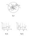
- FIG. 3 ais a schematic representation of tissue data belonging to two different data categories
- FIG. 3 bis a schematic representation of further tissue data belonging to two different data categories.
- FIG. 4is a schematic representation of an examination system according to a second embodiment of the present invention.
- An exemplary examination system 1 shown in FIG. 1comprises a surgical microscope 3 for generating a stereo-microscopic image of a tissue area 7 arranged in an object plane 5 .
- the operation microscope 3comprises an objective lens 8 and a beam path with two zoom systems 9 for imaging the image of the object plane 5 via two ocular lenses 11 to both the left and right eyes of the viewer.
- the examination system 1comprises a tissue qualifying system 13 , having an ultrasound measuring head 15 for sampling tissue data and a discriminating device 17 for evaluating the tissue data.
- the discriminating device 17may be realized as a software component in a computer 16 .
- the ultrasound measuring head 15emits ultrasonic waves from a tip 19 of the measuring head and receives ultrasonic waves reflected by the tissue and generates data therefrom, such as depicted by way of example in FIG. 3 a for healthy tissue.
- a data curve 21is depicted, which represents the intensity I of ultrasonic waves reflected from the tissue in dependence of the tissue depth T starting from the tissue surface.
- FIG. 3 bdata are depicted which are sampled by the measuring head when same is located above tumor tissue.
- the discriminating device 17evaluates the data provided by the measuring head and discriminates same into two data categories, namely “tumor tissue” and “non-tumor tissue”.
- the discriminationcan be performed, e.g. in such a manner that the computer averages data from regions labeled I and II, respectively, in FIGS. 3 a and 3 b , and calculates a ratio of the averaged intensity in region I over the averaged intensity in region II. If this ratio is less than one, discrimination as “non-tumor tissue” is made, and if this ratio is equal to or larger than one, discrimination as “tumor tissue” is made.
- a beam splitter 23is provided between the zoom system 9 and the ocular lens 11 .
- the beam splitter 23couples a partial beam out of the beam path and supplies it to a camera 25 in such a way that camera 25 detects an image of the object plane 5 .
- the detected imageis supplied as image data to an image evaluation device 27 , which may be realized as a software component in the computer 16 .
- the measuring head 15comprises two light emitting diodes 29 at a distance from its tip 19 . Light emitted from the diodes 29 is also imaged onto the camera 25 by the left beam path, such that the diodes 29 are also represented in the image data supplied to the image evaluation device 27 .
- the image evaluation device 27determines the locations of the light emitting diodes 29 relative to the microscope 3 from the image data. Using a predetermined geometrical relation of the two light emitting diodes relative to each other and relative to the tip 19 of the measuring head 15 , the position of the measuring head 19 relative to the microscope 3 is determined finally.
- the detected position of the measuring head 15is supplied by the image evaluation device 27 to a marker display device 31 , which may also be realized as a software component in the computer 16 .
- the marker display device 31further receives a signal from the discrimination device 17 , whenever same discriminates the data sampled by the measuring head 15 as “tumor tissue”.
- the device 31then generates image data representing an image which has a marking, such as a green spot, at the position detected by the position detecting device 27 .
- These image dataare then submitted to an LCD-display 33 displaying an image representing the image data.
- the imageis coupled into the beam path of the microscope on the right in FIG. 1, by a beam splitter 35 disposed between the zoom system 9 and the ocular lens 11 .
- the right eye of the viewersees a green marking superimposed onto the optical image of the object plane 5 at the position of the tip 19 of the measuring head 15 , if the tissue data sampled by same have been discriminated by the discrimination device 17 as “tumor tissue”.
- FIG. 2an image of an object region 41 of the microscope 3 is shown schematically as perceived by a viewer with his right eye when looking through the ocular lens 11 .
- linesindicate tissue structures as generated by the microscope 3 as an optical image of the examined tissue region.
- a part of the measuring head 15 with its tip 19 and the two light emitting diodes 29are visible in the image.
- the tip 19 of the measuring head 15has already been scanned systematically over the examined tissue region, and markings 45 indicated as small circles are visible and represent tumor tissue.
- the markings 45are visible at such locations where the system has detected tissue data of the category “tumor tissue”.
- a line, indicated as a dashed line 47 in FIG. 2is also possible to display a line, indicated as a dashed line 47 in FIG. 2 as a marking.
- the line 47has been calculated by the device 31 so as to represent a periphery of a group of markings 45 . This has the advantage that the image of the optically perceivable structures 45 is not excessively overlaid when the markings 45 are not shown in the image.
- the beam splitters 23 and 35are arranged in the left and right beam path, respectively, of the microscope 3 .
- the image recorded by the camera 25includes the markings generated by the display 33 for e.g. recording purposes or in order to enable further viewers, which have no access to the ocular lenses 11 , to view the same image as the viewer looking into the ocular lenses 11 .
- the markings 45 coupled inmay not disturb evaluation of the position of the light emitting diodes 29 by the image evaluation device 27 , the markings may be displayed in green, while the evaluation device 27 uses only red image components for evaluation of the position of the measuring head, namely those of the color which the light emitting diodes 29 emit.
- An examination system 1 a shown schematically in FIG. 4comprises a microscope 3 a with an objective lens 8 a , a camera 25 a and a head-mounted display 51 , which can be carried by a viewer directly on his head and which provides both eyes of the viewer with images of a tissue region 7 a recorded by the camera 25 a through the objective lens 8 a .
- the corresponding image dataare supplied by a wireless transmission to the head-mounted display 51 from a computer 16 a .
- the computer 16 areceives the data recorded by the camera 25 a and is connected to a sender 53 of the wireless transmission.
- an ultrasonic measuring head 15 a of the examination system 1 ais a measuring head which samples data not only at its tip, but at plural locations distributed along an extended line 19 a . Data sampled by the measuring head 15 a at the plural locations along the line 19 a are also supplied to the computer 16 a for discrimination into different data categories.
- a determination of the position of the measuring head 15 a relative to the objective lens 8 a of the microscope 3 ais performed in the examination system 1 a not by evaluating the image recorded by the camera 25 a , but by evaluating an image recorded by a further camera 55 arranged at a distance from the objective lens 8 a .
- the camera 55tracks a marker pattern 57 in the form of a square arranged inside of another square provided on a surface of the measuring head 15 a , and the computer 16 a determines a position and an orientation of the line 19 a , along which the measuring head 15 a samples data, based on an evaluation of the image recorded by the camera 55 .
- the computer 16 adisplays a marking at the corresponding image location in the image shown by the head-mounted display 51 , as has been illustrated above with reference to FIG. 2.
- Such shift of the examined tissue region in the image area of the microscopemay occur on the one hand due to movements of the patient, and on the other hand due to the surgeon moving the microscope in the operating room relative to the patient.
- the shiftcan be detected, e.g. by the computer analyzing the image of the object area 41 (compare FIG. 2), determining the position of tissue structures (compare lines 43 in FIG. 2) as patterns and detecting shifts of such patterns.
- the surgeonis enabled, starting out from a viewing situation as exemplarily shown in FIG. 2 where a region 47 is already provided with markings 45 , to shift the microscope so far that the region 47 is no longer within the image area 41 , and then to move back the microscope. After moving back the microscope, the markings 45 appear at the correct location in relation to the examined tissue region.
- the markingsare preferably stored in an image memory, the contents of which represent an extended image area which extends beyond the momentarily viewed image area 41 (compare FIG. 2) or the image area displayed by the display 33 , respectively.
- the image area actually displayedthen corresponds to a partial region of the image memory, which is reproduced by the display 33 .
- a shift of the image area or the microscope, respectively,then corresponds to shifting or scrolling of the memory contents displayed in relation to the extended image area 41 .
- the optical systemmay alternatively or in addition to the microscope also include an endoscope or any video-optical system.
- the measuring headis an ultrasonic measuring head, it may also be of a flow detecting Doppler type, or of a duplex type.
- the measuring headmay also be adapted to detect biochemical or electrophysical parameters, such as a blood glucose concentration, a sodium or/and potassium concentration or a ratio of same, or a tissue or body fluid temperature.
- the inventionmay provide an examination system and a corresponding examination method, comprising: Arranging a tissue region in a vicinity of an object plane of a microscope and generating an optical image of the tissue region, receiving tissue data from a measuring head separate from the microscope, discriminating the tissue data into at least two data categories and displaying markings in the optical image of the tissue region in dependence of the discriminated data category and a relative position between a component of the microscope and the measuring head.
Landscapes
- Health & Medical Sciences (AREA)
- Surgery (AREA)
- Engineering & Computer Science (AREA)
- Life Sciences & Earth Sciences (AREA)
- Biomedical Technology (AREA)
- Robotics (AREA)
- Nuclear Medicine, Radiotherapy & Molecular Imaging (AREA)
- Heart & Thoracic Surgery (AREA)
- Medical Informatics (AREA)
- Molecular Biology (AREA)
- Animal Behavior & Ethology (AREA)
- General Health & Medical Sciences (AREA)
- Public Health (AREA)
- Veterinary Medicine (AREA)
- Microscoopes, Condenser (AREA)
Abstract
Description
- 1. Field of the Invention[0001]
- The present invention relates to an examination system and an examination method for displaying a tissue region to a user. The tissue region is in particular a portion of a patient to undergo a surgical operation, and the user is in particular a surgeon performing such surgical operation.[0002]
- 2. Brief Description of Related Art[0003]
- In a conventional surgical system and method, e.g., a near-surface tumor is to be excised from the tissue region, and the surgical operation is performed by a surgeon viewing the tissue region through a surgical microscope. An ultrasound system may be used for localizing the tumor to be excised. The surgeon scans the tissue region in which the tumor is presumed with a measuring head of the ultrasound system, and the surgeon views sampled ultrasonic depth scans on a display device of the ultrasound system. From the depth scans the surgeon can conclude, whether there is any tumor tissue at the location at which the measuring head is presently located. The surgeon then looks through the surgical microscope to perceive such location in the microscope image and memorize the same. By multiply repeating this procedure, the surgeon perceives the extension of the tumor, memorizes the same and can then perform the surgical operation to excise the tumor while looking through the surgical microscope.[0004]
- This procedure is complicated and requires high concentration on the part of the surgeon, because the success of the operation depends on the extent to which the surgeon can memorize the extension of the tumor.[0005]
- It is an object of the present invention to provide a system and a method that may simplify localizing specific tissue types in an image of a microscope.[0006]
- To solve this object, the invention provides an examination system for displaying a tissue region to a viewer, comprising: an optical system for generating an optical image of the tissue region; a tissue qualifying system having a measuring head for receiving tissue data, and having a computer for analyzing the obtained tissue data and discriminating the obtained tissue data into at least two data categories; a position detecting system configured to determine a position of the measuring head relative to the component of the optical system; and an indicating system for optically marking portions of the optical image of the tissue region based on the detected position of the measuring head and on the discriminated data category.[0007]
- The data categories may characterize the tissue at those locations at which the measuring head samples tissue data at a particular time. According to an exemplary embodiment, the at least two data categories comprise a “tumor tissue” and a “non-tumor tissue”. However, it is also possible that the tissue qualifying system discriminates a higher number or other types of tissue, such as nerves and supporting tissue.[0008]
- The examination system may enable the user to perceive an extension of a specific type of tissue in the tissue region under examination, without having to avert his view from the image of the tissue region generated by the optical system. This is because the viewer may move the measuring head across the tissue region while observing the optical image thereof, and the examination system may provide the marking or indication of the image at those locations of the image where the tissue qualifying system discriminates a certain data category from the data sampled by the measuring head. Since these markings or indications may be provided directly in the image generated by the optical system, the viewer does not have to avert his view from, e.g., a display for generating the optical image of the tissue region, as this was necessary in the conventional system.[0009]
- According to a preferred embodiment, it is possible that the marking or indication displayed at a certain location in the image is continued to be displayed at that location even when the measuring head has been further moved and is already sampling data at another location of the tissue region. After systematically scanning the tissue region to be examined, an extended partial region of the image of the tissue region, in which the tissue type of interest is present, is marked by e.g. highlighting, a particular color or other means.[0010]
- In order to sample the tissue data, various known methods may be employed. According to preferred exemplary embodiments, the measuring head may comprise a fluorescence measuring head, a Raman spectroscopy head, an acousto-optical measuring head, an optical coherence tomography (OCT) measuring head, an ultrasound measuring head, such as an ultrasonic Doppler measuring head, a concentration measuring head, such as a blood glucose concentration measuring head, a sodium concentration measuring head and a potassium concentration measuring head, and a temperature measuring head.[0011]
- According to a preferred exemplary embodiment, the optical system may comprise a microscope having an objective lens. Herein, the position detection system is preferably configured to determine the position of the measuring head relative to the objective lens.[0012]
- The microscope preferably comprises a camera to record image data which represent the optical image of the tissue region. A computer can then, by image data processing, recognize the measuring head in the image and calculate its position within the image. At this calculated position, the marking is then displayed, if the tissue data fall into a designated data category.[0013]
- It is, however, also preferred to additionally provide a position detection system independent of the microscope for evaluating the relative position between the measuring head and the microscope. A conventional triangulation system tracking suitable markings provided at the measuring head is an example for such position detection system. Here it is also possible to evaluate both the position of the microscope relative to the position detection system, and the position of the measuring head relative to the position detection system, and to calculate the relative position between the microscope and the measuring head from the positions of the microscope and the measuring head.[0014]
- The indication system preferably comprises a display for displaying the marking and a first beam splitter for coupling an image of the displayed marking into a beam path of the microscope, whereby a simultaneous perceptibility of both the optical image generated by the microscope and the marking is made possible.[0015]
- Where the microscope comprises an ocular lens, the image of the displayed marking is preferably coupled into the beam path between the objective lens and the ocular lens by a first beam splitter.[0016]
- According to a preferred embodiment the optical system comprises, e.g. for archive purposes, a camera for recording an image coupled out of the beam path between the objective lens and the ocular lens by a second beam splitter. The second beam splitter is preferably provided downstream of the first beam splitter in the beam path, so that in the image recorded by the camera the generated markings are also included.[0017]
- However, it is also possible that the camera first records an image of the tissue region free of the markings, and that the markings are then electronically introduced into the image recorded by the camera.[0018]
- In particular, in a case where the position detection system evaluates data of the optical image of the tissue region, in which data markings are provided, it is preferable to provide the markings using color components which are different from those color components which the position detection system evaluates for determining the position of the measuring head.[0019]
- Besides displaying the optical image of the examined tissue region to the viewer by means of ocular lenses, it is also preferred to display the image to the viewer by a display device which the viewer can wear on his head, such as a head mounted display. Furthermore, the optical image can be displayed on a monitor.[0020]
- The measuring head can be adapted to sample data at only one location at a time. However, it is also possible that the measuring head samples data at plural locations simultaneously, which are, e.g., arranged on a line or distributed in a two-dimensional array. For the multiple locations, a discrimination according to tissue categories is respectively performed, and a corresponding marking is provided in the optical image of the tissue region.[0021]
- The forgoing as well as other advantageous features of the invention will be more apparent from the following detailed description of exemplary embodiments of the invention with reference to the accompanying drawings, wherein:[0022]
- FIG. 1 is a schematic representation of an examination system according to a first embodiment of the present invention;[0023]
- FIG. 2 is a schematic representation of an optical image of an examined tissue region obtained with the examination system of FIG. 1, with markings displayed therein;[0024]
- FIG. 3[0025]ais a schematic representation of tissue data belonging to two different data categories;
- FIG. 3[0026]bis a schematic representation of further tissue data belonging to two different data categories; and
- FIG. 4 is a schematic representation of an examination system according to a second embodiment of the present invention.[0027]
- In the embodiments described below, components which are identical in function and structure are designated as far as possible by the same reference numerals. Therefore, to understand the features of the individual components of a specific embodiment, the descriptions of other embodiments should be referred to.[0028]
- An exemplary examination system[0029]1 shown in FIG. 1 comprises a
surgical microscope 3 for generating a stereo-microscopic image of a tissue area7 arranged in an object plane5. To this end, theoperation microscope 3 comprises anobjective lens 8 and a beam path with twozoom systems 9 for imaging the image of the object plane5 via two ocular lenses11 to both the left and right eyes of the viewer. - Furthermore, the examination system[0030]1 comprises a
tissue qualifying system 13, having anultrasound measuring head 15 for sampling tissue data and adiscriminating device 17 for evaluating the tissue data. Thediscriminating device 17 may be realized as a software component in acomputer 16. Theultrasound measuring head 15 emits ultrasonic waves from atip 19 of the measuring head and receives ultrasonic waves reflected by the tissue and generates data therefrom, such as depicted by way of example in FIG. 3afor healthy tissue. Therein, adata curve 21 is depicted, which represents the intensity I of ultrasonic waves reflected from the tissue in dependence of the tissue depth T starting from the tissue surface. - In FIG. 3[0031]b, data are depicted which are sampled by the measuring head when same is located above tumor tissue.
- The discriminating[0032]
device 17 evaluates the data provided by the measuring head and discriminates same into two data categories, namely “tumor tissue” and “non-tumor tissue”. The discrimination can be performed, e.g. in such a manner that the computer averages data from regions labeled I and II, respectively, in FIGS. 3aand3b, and calculates a ratio of the averaged intensity in region I over the averaged intensity in region II. If this ratio is less than one, discrimination as “non-tumor tissue” is made, and if this ratio is equal to or larger than one, discrimination as “tumor tissue” is made. - In the beam path of the stereo-[0033]
microscope 3 on the left in FIG. 1, abeam splitter 23 is provided between thezoom system 9 and the ocular lens11. Thebeam splitter 23 couples a partial beam out of the beam path and supplies it to acamera 25 in such a way thatcamera 25 detects an image of the object plane5. The detected image is supplied as image data to animage evaluation device 27, which may be realized as a software component in thecomputer 16. The measuringhead 15 comprises two light emittingdiodes 29 at a distance from itstip 19. Light emitted from thediodes 29 is also imaged onto thecamera 25 by the left beam path, such that thediodes 29 are also represented in the image data supplied to theimage evaluation device 27. Theimage evaluation device 27 determines the locations of thelight emitting diodes 29 relative to themicroscope 3 from the image data. Using a predetermined geometrical relation of the two light emitting diodes relative to each other and relative to thetip 19 of the measuringhead 15, the position of the measuringhead 19 relative to themicroscope 3 is determined finally. - The detected position of the measuring[0034]
head 15 is supplied by theimage evaluation device 27 to amarker display device 31, which may also be realized as a software component in thecomputer 16. Themarker display device 31 further receives a signal from thediscrimination device 17, whenever same discriminates the data sampled by the measuringhead 15 as “tumor tissue”. Thedevice 31 then generates image data representing an image which has a marking, such as a green spot, at the position detected by theposition detecting device 27. These image data are then submitted to an LCD-display 33 displaying an image representing the image data. The image is coupled into the beam path of the microscope on the right in FIG. 1, by abeam splitter 35 disposed between thezoom system 9 and the ocular lens11. Thereby, the right eye of the viewer sees a green marking superimposed onto the optical image of the object plane5 at the position of thetip 19 of the measuringhead 15, if the tissue data sampled by same have been discriminated by thediscrimination device 17 as “tumor tissue”. - In FIG. 2, an image of an[0035]
object region 41 of themicroscope 3 is shown schematically as perceived by a viewer with his right eye when looking through the ocular lens11. Therein, lines indicate tissue structures as generated by themicroscope 3 as an optical image of the examined tissue region. Further, a part of the measuringhead 15 with itstip 19 and the twolight emitting diodes 29 are visible in the image. In the situation shown in FIG. 2, thetip 19 of the measuringhead 15 has already been scanned systematically over the examined tissue region, andmarkings 45 indicated as small circles are visible and represent tumor tissue. Themarkings 45 are visible at such locations where the system has detected tissue data of the category “tumor tissue”. These markings are stored by thedevice 31 and thus remain visible in the image even when thetip 19 of the measuringhead 15 is no longer in the corresponding place. However, the option is provided to delete the markings previously stored, in order to re-start the examination all over. - Alternatively, or additionally, to the display of the individual markings as circles, it is also possible to display a line, indicated as a dashed[0036]
line 47 in FIG. 2 as a marking. Theline 47 has been calculated by thedevice 31 so as to represent a periphery of a group ofmarkings 45. This has the advantage that the image of the opticallyperceivable structures 45 is not excessively overlaid when themarkings 45 are not shown in the image. - In the embodiment shown in FIG. 1, the[0037]
beam splitters microscope 3. Alternatively, it is also possible to arrange both beam splitters in one beam path of the microscope in such a way that thebeam splitter 35 for coupling in the image generated by thedisplay 33 is arranged between thebeam splitter 23 for coupling out the image of the object area5, and thezoom system 9. In such embodiment, the image recorded by thecamera 25 includes the markings generated by thedisplay 33 for e.g. recording purposes or in order to enable further viewers, which have no access to the ocular lenses11, to view the same image as the viewer looking into the ocular lenses11. - In order that the[0038]
markings 45 coupled in may not disturb evaluation of the position of thelight emitting diodes 29 by theimage evaluation device 27, the markings may be displayed in green, while theevaluation device 27 uses only red image components for evaluation of the position of the measuring head, namely those of the color which thelight emitting diodes 29 emit. - An[0039]
examination system 1ashown schematically in FIG. 4 comprises a microscope3awith anobjective lens 8a, acamera 25aand a head-mounteddisplay 51, which can be carried by a viewer directly on his head and which provides both eyes of the viewer with images of atissue region 7arecorded by thecamera 25athrough theobjective lens 8a. The corresponding image data are supplied by a wireless transmission to the head-mounteddisplay 51 from acomputer 16a. Thecomputer 16areceives the data recorded by thecamera 25aand is connected to asender 53 of the wireless transmission. - Different from the embodiment illustrated with reference to FIG. 1, an[0040]
ultrasonic measuring head 15aof theexamination system 1ais a measuring head which samples data not only at its tip, but at plural locations distributed along anextended line 19a. Data sampled by the measuringhead 15aat the plural locations along theline 19aare also supplied to thecomputer 16afor discrimination into different data categories. - A determination of the position of the measuring[0041]
head 15arelative to theobjective lens 8aof the microscope3ais performed in theexamination system 1anot by evaluating the image recorded by thecamera 25a, but by evaluating an image recorded by afurther camera 55 arranged at a distance from theobjective lens 8a. Thecamera 55 tracks amarker pattern 57 in the form of a square arranged inside of another square provided on a surface of the measuringhead 15a, and thecomputer 16adetermines a position and an orientation of theline 19a, along which the measuringhead 15asamples data, based on an evaluation of the image recorded by thecamera 55. If the evaluation of the data results in a discrimination of the data as “tumor tissue” for one of the plural locations alongline 19a, thecomputer 16adisplays a marking at the corresponding image location in the image shown by the head-mounteddisplay 51, as has been illustrated above with reference to FIG. 2. - In the previously described embodiments, it is possible to detect a shift of the examined tissue region within the image field of the microscope by image processing. The markings displayed in the image may then be displaced in accordance with the determined shift, such that re-scanning the examined tissue region with the measuring head may not be necessary after such a shift.[0042]
- Such shift of the examined tissue region in the image area of the microscope may occur on the one hand due to movements of the patient, and on the other hand due to the surgeon moving the microscope in the operating room relative to the patient. The shift can be detected, e.g. by the computer analyzing the image of the object area[0043]41 (compare FIG. 2), determining the position of tissue structures (compare
lines 43 in FIG. 2) as patterns and detecting shifts of such patterns. Thereby, the surgeon is enabled, starting out from a viewing situation as exemplarily shown in FIG. 2 where aregion 47 is already provided withmarkings 45, to shift the microscope so far that theregion 47 is no longer within theimage area 41, and then to move back the microscope. After moving back the microscope, themarkings 45 appear at the correct location in relation to the examined tissue region. - Alternatively to the determination of the shift by image processing as described above, it is also possible in a simplified embodiment only to record the coordinates of the microscope in the operating room and to presume that the patient does not move. The shift to be detected then results from variations of the coordinates of the microscope in the operating room.[0044]
- The markings are preferably stored in an image memory, the contents of which represent an extended image area which extends beyond the momentarily viewed image area[0045]41 (compare FIG. 2) or the image area displayed by the
display 33, respectively. The image area actually displayed then corresponds to a partial region of the image memory, which is reproduced by thedisplay 33. A shift of the image area or the microscope, respectively, then corresponds to shifting or scrolling of the memory contents displayed in relation to theextended image area 41. - The optical system may alternatively or in addition to the microscope also include an endoscope or any video-optical system. Furthermore, if the measuring head is an ultrasonic measuring head, it may also be of a flow detecting Doppler type, or of a duplex type. However, the measuring head may also be adapted to detect biochemical or electrophysical parameters, such as a blood glucose concentration, a sodium or/and potassium concentration or a ratio of same, or a tissue or body fluid temperature.[0046]
- Summarized, the invention may provide an examination system and a corresponding examination method, comprising: Arranging a tissue region in a vicinity of an object plane of a microscope and generating an optical image of the tissue region, receiving tissue data from a measuring head separate from the microscope, discriminating the tissue data into at least two data categories and displaying markings in the optical image of the tissue region in dependence of the discriminated data category and a relative position between a component of the microscope and the measuring head.[0047]
- Therefore, while the present invention has been shown and described herein in what is believed to be the most practical and preferred embodiments, it is recognized that departures can be made therefrom within the scope of the invention, which is therefore not be limited to the details disclosed herein but is to be accorded the full scope of the claims so as to embrace any and all equivalent methods and apparatus.[0048]
Claims (17)
Applications Claiming Priority (2)
| Application Number | Priority Date | Filing Date | Title |
|---|---|---|---|
| DE10252837ADE10252837B4 (en) | 2002-11-13 | 2002-11-13 | Examination system and examination procedure |
| DEDE10252837.3 | 2002-11-13 |
Publications (2)
| Publication Number | Publication Date |
|---|---|
| US20040167742A1true US20040167742A1 (en) | 2004-08-26 |
| US7477764B2 US7477764B2 (en) | 2009-01-13 |
Family
ID=32240015
Family Applications (1)
| Application Number | Title | Priority Date | Filing Date |
|---|---|---|---|
| US10/704,647Active2026-08-24US7477764B2 (en) | 2002-11-13 | 2003-11-12 | Examination system and examination method |
Country Status (4)
| Country | Link |
|---|---|
| US (1) | US7477764B2 (en) |
| JP (1) | JP4524353B2 (en) |
| CH (1) | CH697546B1 (en) |
| DE (1) | DE10252837B4 (en) |
Cited By (8)
| Publication number | Priority date | Publication date | Assignee | Title |
|---|---|---|---|---|
| US20050228281A1 (en)* | 2004-03-31 | 2005-10-13 | Nefos Thomas P | Handheld diagnostic ultrasound system with head mounted display |
| DE102005051405A1 (en)* | 2005-10-27 | 2007-05-03 | Forschungszentrum Rossendorf E.V. | measuring sensor |
| US20090054788A1 (en)* | 2007-04-19 | 2009-02-26 | Carl Zeiss Surgical Gmbh | Method and apparatus for displaying a field of a brain of a patient and navigation system for brain surgery |
| US20110181702A1 (en)* | 2009-07-28 | 2011-07-28 | Carl Zeiss Surgical Gmbh | Method and system for generating a representation of an oct data set |
| US20130164865A1 (en)* | 2011-06-29 | 2013-06-27 | Panasonic Corporation | Method of manufacturing light-emitting device and apparatus for manufacturing light-emitting device |
| EP2949285A1 (en)* | 2014-05-27 | 2015-12-02 | Carl Zeiss Meditec AG | Surgical microscope |
| US11181728B2 (en)* | 2017-10-31 | 2021-11-23 | Samantree Medical Sa | Imaging systems with micro optical element arrays and methods of specimen imaging |
| USRE49023E1 (en) | 2009-09-30 | 2022-04-12 | Nidek Co., Ltd. | Fundus observation apparatus |
Families Citing this family (23)
| Publication number | Priority date | Publication date | Assignee | Title |
|---|---|---|---|---|
| CA2595324C (en)* | 2005-01-21 | 2015-08-11 | Massachusetts Institute Of Technology | Methods and apparatus for optical coherence tomography scanning |
| EP1905377B1 (en)* | 2006-09-28 | 2013-05-29 | BrainLAB AG | Preoperative planing of the position of surgical instruments |
| DE102007054450A1 (en)* | 2006-11-13 | 2008-05-15 | Eberhard-Karls-Universität Universitätsklinikum Tübingen | Image providing device for surgeon, has common camera i.e. digital camera, provided for image acquisition of operation field and for determining camera position and orientation by navigation system |
| DE112008002383T5 (en) | 2007-09-06 | 2010-06-24 | LenSx Lasers, Inc., Aliso Viejo | Precise targeting of surgical photodisruption |
| US9492322B2 (en) | 2009-11-16 | 2016-11-15 | Alcon Lensx, Inc. | Imaging surgical target tissue by nonlinear scanning |
| US8265364B2 (en)* | 2010-02-05 | 2012-09-11 | Alcon Lensx, Inc. | Gradient search integrated with local imaging in laser surgical systems |
| US8414564B2 (en)* | 2010-02-18 | 2013-04-09 | Alcon Lensx, Inc. | Optical coherence tomographic system for ophthalmic surgery |
| US8398236B2 (en) | 2010-06-14 | 2013-03-19 | Alcon Lensx, Inc. | Image-guided docking for ophthalmic surgical systems |
| US9532708B2 (en) | 2010-09-17 | 2017-01-03 | Alcon Lensx, Inc. | Electronically controlled fixation light for ophthalmic imaging systems |
| US8459794B2 (en) | 2011-05-02 | 2013-06-11 | Alcon Lensx, Inc. | Image-processor-controlled misalignment-reduction for ophthalmic systems |
| US9622913B2 (en) | 2011-05-18 | 2017-04-18 | Alcon Lensx, Inc. | Imaging-controlled laser surgical system |
| US8398238B1 (en) | 2011-08-26 | 2013-03-19 | Alcon Lensx, Inc. | Imaging-based guidance system for ophthalmic docking using a location-orientation analysis |
| KR101274736B1 (en) | 2011-10-17 | 2013-06-17 | 큐렉소 주식회사 | A monitoring system of the tissue movement during a surgery |
| US9023016B2 (en) | 2011-12-19 | 2015-05-05 | Alcon Lensx, Inc. | Image processor for intra-surgical optical coherence tomographic imaging of laser cataract procedures |
| US9066784B2 (en) | 2011-12-19 | 2015-06-30 | Alcon Lensx, Inc. | Intra-surgical optical coherence tomographic imaging of cataract procedures |
| US8937769B2 (en)* | 2012-06-07 | 2015-01-20 | Alcon Research, Ltd. | Orthogonal light beam splitting for microscopes |
| KR101446173B1 (en)* | 2013-02-21 | 2014-10-01 | 주식회사 고영테크놀러지 | Tracking system and method for tracking using the same |
| WO2016055422A1 (en)* | 2014-10-06 | 2016-04-14 | Carl Zeiss Meditec Ag | Surgical system with an oct device |
| DE102014224044A1 (en)* | 2014-11-25 | 2016-05-25 | Carl Zeiss Meditec Ag | Tool-controlled multimodal magnifier in the field of view of a surgical microscope |
| US20190175301A1 (en)* | 2016-06-09 | 2019-06-13 | Shimadzu Corporation | Near-nfrared imaging apparatus and marker member for near-infrared imaging apparatus |
| US11103695B2 (en) | 2018-09-14 | 2021-08-31 | Neuralink Corp. | Device implantation using a cartridge |
| CA3112749A1 (en)* | 2018-09-14 | 2020-03-19 | Neuralink Corp. | Computer vision techniques |
| DE102020102476B4 (en)* | 2020-01-31 | 2024-02-08 | Carl Zeiss Meditec Ag | Method for marking an area of a tumor |
Citations (7)
| Publication number | Priority date | Publication date | Assignee | Title |
|---|---|---|---|---|
| US4786155A (en)* | 1986-12-16 | 1988-11-22 | Fantone Stephen D | Operating microscope providing an image of an obscured object |
| US5982532A (en)* | 1995-10-12 | 1999-11-09 | Carl Zeiss-Stiftung | Process for the operation of an operation microscope |
| US6006126A (en)* | 1991-01-28 | 1999-12-21 | Cosman; Eric R. | System and method for stereotactic registration of image scan data |
| US6167296A (en)* | 1996-06-28 | 2000-12-26 | The Board Of Trustees Of The Leland Stanford Junior University | Method for volumetric image navigation |
| US6191862B1 (en)* | 1999-01-20 | 2001-02-20 | Lightlab Imaging, Llc | Methods and apparatus for high speed longitudinal scanning in imaging systems |
| US20020120424A1 (en)* | 2001-01-03 | 2002-08-29 | Christoph Hauger | Method and apparatus for fixing a location |
| US6763259B1 (en)* | 1999-07-02 | 2004-07-13 | Carl-Zeiss-Stiftung (De) | Surgical system supported by optical coherence tomography |
Family Cites Families (3)
| Publication number | Priority date | Publication date | Assignee | Title |
|---|---|---|---|---|
| DD241485A1 (en)* | 1985-10-02 | 1986-12-10 | Zeiss Jena Veb Carl | OPERATING MICROSCOPE WITH RADIATION DIVIDER PROVIDED IN THE ILLUMINATION RADIATION |
| EP0999785A4 (en)* | 1997-06-27 | 2007-04-25 | Univ Leland Stanford Junior | METHOD AND APPARATUS FOR GENERATING THREE-DIMENSIONAL IMAGES FOR "NAVIGATION" PURPOSES |
| DE10027827A1 (en)* | 2000-06-05 | 2001-12-06 | Sonem Gmbh | Adaptive classifying device for classifying reflective/absorbent structures or acoustic barrier layers with similar properties has n channels of a spectrum to separate response signals ready to be evaluated by a classifying unit. |
- 2002
- 2002-11-13DEDE10252837Apatent/DE10252837B4/ennot_activeExpired - Lifetime
- 2003
- 2003-11-04CHCH01879/03Apatent/CH697546B1/ennot_activeIP Right Cessation
- 2003-11-12USUS10/704,647patent/US7477764B2/enactiveActive
- 2003-11-13JPJP2003383840Apatent/JP4524353B2/ennot_activeExpired - Lifetime
Patent Citations (8)
| Publication number | Priority date | Publication date | Assignee | Title |
|---|---|---|---|---|
| US4786155A (en)* | 1986-12-16 | 1988-11-22 | Fantone Stephen D | Operating microscope providing an image of an obscured object |
| US6006126A (en)* | 1991-01-28 | 1999-12-21 | Cosman; Eric R. | System and method for stereotactic registration of image scan data |
| US5982532A (en)* | 1995-10-12 | 1999-11-09 | Carl Zeiss-Stiftung | Process for the operation of an operation microscope |
| US6167296A (en)* | 1996-06-28 | 2000-12-26 | The Board Of Trustees Of The Leland Stanford Junior University | Method for volumetric image navigation |
| US6191862B1 (en)* | 1999-01-20 | 2001-02-20 | Lightlab Imaging, Llc | Methods and apparatus for high speed longitudinal scanning in imaging systems |
| US6763259B1 (en)* | 1999-07-02 | 2004-07-13 | Carl-Zeiss-Stiftung (De) | Surgical system supported by optical coherence tomography |
| US20020120424A1 (en)* | 2001-01-03 | 2002-08-29 | Christoph Hauger | Method and apparatus for fixing a location |
| US6741948B2 (en)* | 2001-01-03 | 2004-05-25 | Carl-Zeiss-Stiftung | Method and apparatus for fixing a location |
Cited By (14)
| Publication number | Priority date | Publication date | Assignee | Title |
|---|---|---|---|---|
| US20050228281A1 (en)* | 2004-03-31 | 2005-10-13 | Nefos Thomas P | Handheld diagnostic ultrasound system with head mounted display |
| DE102005051405A1 (en)* | 2005-10-27 | 2007-05-03 | Forschungszentrum Rossendorf E.V. | measuring sensor |
| DE102005051405B4 (en)* | 2005-10-27 | 2007-08-23 | Forschungszentrum Dresden - Rossendorf E.V. | measuring sensor |
| US20090054788A1 (en)* | 2007-04-19 | 2009-02-26 | Carl Zeiss Surgical Gmbh | Method and apparatus for displaying a field of a brain of a patient and navigation system for brain surgery |
| US20110181702A1 (en)* | 2009-07-28 | 2011-07-28 | Carl Zeiss Surgical Gmbh | Method and system for generating a representation of an oct data set |
| USRE49023E1 (en) | 2009-09-30 | 2022-04-12 | Nidek Co., Ltd. | Fundus observation apparatus |
| USRE49024E1 (en)* | 2009-09-30 | 2022-04-12 | Nidek Co., Ltd. | Fundus observation apparatus |
| US20130164865A1 (en)* | 2011-06-29 | 2013-06-27 | Panasonic Corporation | Method of manufacturing light-emitting device and apparatus for manufacturing light-emitting device |
| US9241388B2 (en)* | 2011-06-29 | 2016-01-19 | Panasonic Intellectual Property Management Co., Ltd. | Method and apparatus for manufacturing a light-emitting device including correction of an application amount of a fluorescent resin based on a fluorescent particle concentration |
| EP2949285A1 (en)* | 2014-05-27 | 2015-12-02 | Carl Zeiss Meditec AG | Surgical microscope |
| US9933606B2 (en) | 2014-05-27 | 2018-04-03 | Carl Zeiss Meditec Ag | Surgical microscope |
| US20220035147A1 (en)* | 2017-10-31 | 2022-02-03 | Samantree Medical Sa | Imaging systems with micro optical element arrays and methods of specimen imaging |
| US11181728B2 (en)* | 2017-10-31 | 2021-11-23 | Samantree Medical Sa | Imaging systems with micro optical element arrays and methods of specimen imaging |
| US11609416B2 (en)* | 2017-10-31 | 2023-03-21 | Samantree Medical Sa | Imaging systems with micro optical element arrays and methods of specimen imaging |
Also Published As
| Publication number | Publication date |
|---|---|
| US7477764B2 (en) | 2009-01-13 |
| JP4524353B2 (en) | 2010-08-18 |
| CH697546B1 (en) | 2008-11-28 |
| DE10252837B4 (en) | 2005-03-24 |
| JP2004202221A (en) | 2004-07-22 |
| DE10252837A1 (en) | 2004-06-03 |
Similar Documents
| Publication | Publication Date | Title |
|---|---|---|
| US7477764B2 (en) | Examination system and examination method | |
| JP4091143B2 (en) | OCT-assisted surgical microscope including a multi-coordinate manipulator | |
| US6456769B1 (en) | Fiber bundle and endoscope apparatus | |
| AU759282B2 (en) | Systems and methods for optical examination of samples | |
| US5513005A (en) | Method of operating a surgical microscope arrangement for computer-supported stereotactic microsurgery on a patient | |
| US6968127B2 (en) | Fundus camera | |
| US12402775B2 (en) | Depth and contour detection for anatomical targets | |
| US20110178395A1 (en) | Imaging method and system | |
| US20100069747A1 (en) | Diagnostic imaging apparatus | |
| EP2805304B1 (en) | Imaging apparatus | |
| US20070146719A1 (en) | Scanning apparatus for optically scanning surfaces | |
| JP2013258627A (en) | Image processing apparatus and three-dimensional image observation system | |
| EP2830485B1 (en) | Integration delayed optical feedback in image guidance | |
| US20160081712A1 (en) | Device, system, and method for insertion of a medical device into a subject | |
| CN105431077A (en) | Image processing apparatus and image processing method | |
| JP3446272B2 (en) | Endoscope with measurement function | |
| WO2009157229A1 (en) | Scatterer interior observation device and scatterer interior observation method | |
| CN110945315B (en) | OCT image acquisition equipment | |
| US7055955B2 (en) | Eye fundus examination apparatus | |
| EP0724858B1 (en) | Ophthalmic measuring apparatus | |
| CN118215968B (en) | System for characterizing regions of interest in biological tissue | |
| JP2005224430A (en) | Anterior eye measurement device | |
| JP2005013514A (en) | Optical imaging apparatus |
Legal Events
| Date | Code | Title | Description |
|---|---|---|---|
| AS | Assignment | Owner name:CARL-ZEISS-STIFTUNG TRADING AS CARL ZEISS, GERMANY Free format text:ASSIGNMENT OF ASSIGNORS INTEREST;ASSIGNOR:HAISCH, MICHAEL;REEL/FRAME:015255/0823 Effective date:20040301 | |
| FEPP | Fee payment procedure | Free format text:PAYOR NUMBER ASSIGNED (ORIGINAL EVENT CODE: ASPN); ENTITY STATUS OF PATENT OWNER: LARGE ENTITY | |
| STCF | Information on status: patent grant | Free format text:PATENTED CASE | |
| AS | Assignment | Owner name:CARL ZEISS AG, GERMANY Free format text:ASSIGNMENT OF ASSIGNORS INTEREST;ASSIGNOR:CARL ZEISS STIFTUNG;REEL/FRAME:022064/0164 Effective date:20081128 | |
| AS | Assignment | Owner name:CARL ZEISS SURGICAL GMBH, GERMANY Free format text:ASSIGNMENT OF ASSIGNORS INTEREST;ASSIGNOR:CARL ZEISS AG;REEL/FRAME:022075/0806 Effective date:20081201 | |
| FPAY | Fee payment | Year of fee payment:4 | |
| FPAY | Fee payment | Year of fee payment:8 | |
| MAFP | Maintenance fee payment | Free format text:PAYMENT OF MAINTENANCE FEE, 12TH YEAR, LARGE ENTITY (ORIGINAL EVENT CODE: M1553); ENTITY STATUS OF PATENT OWNER: LARGE ENTITY Year of fee payment:12 |