US20040136582A1 - Apparatus for remote control of a microscope - Google Patents
Apparatus for remote control of a microscopeDownload PDFInfo
- Publication number
- US20040136582A1 US20040136582A1US10/751,771US75177104AUS2004136582A1US 20040136582 A1US20040136582 A1US 20040136582A1US 75177104 AUS75177104 AUS 75177104AUS 2004136582 A1US2004136582 A1US 2004136582A1
- Authority
- US
- United States
- Prior art keywords
- image
- images
- microscope
- digitized
- specimen
- Prior art date
- Legal status (The legal status is an assumption and is not a legal conclusion. Google has not performed a legal analysis and makes no representation as to the accuracy of the status listed.)
- Granted
Links
Images


Classifications
- G—PHYSICS
- G06—COMPUTING OR CALCULATING; COUNTING
- G06T—IMAGE DATA PROCESSING OR GENERATION, IN GENERAL
- G06T3/00—Geometric image transformations in the plane of the image
- G06T3/40—Scaling of whole images or parts thereof, e.g. expanding or contracting
- G06T3/4038—Image mosaicing, e.g. composing plane images from plane sub-images
- G—PHYSICS
- G01—MEASURING; TESTING
- G01N—INVESTIGATING OR ANALYSING MATERIALS BY DETERMINING THEIR CHEMICAL OR PHYSICAL PROPERTIES
- G01N15/00—Investigating characteristics of particles; Investigating permeability, pore-volume or surface-area of porous materials
- G01N15/10—Investigating individual particles
- G01N15/14—Optical investigation techniques, e.g. flow cytometry
- G01N15/1429—Signal processing
- G01N15/1433—Signal processing using image recognition
- G—PHYSICS
- G06—COMPUTING OR CALCULATING; COUNTING
- G06T—IMAGE DATA PROCESSING OR GENERATION, IN GENERAL
- G06T7/00—Image analysis
- G06T7/0002—Inspection of images, e.g. flaw detection
- G06T7/0012—Biomedical image inspection
- G—PHYSICS
- G06—COMPUTING OR CALCULATING; COUNTING
- G06V—IMAGE OR VIDEO RECOGNITION OR UNDERSTANDING
- G06V20/00—Scenes; Scene-specific elements
- G06V20/60—Type of objects
- G06V20/69—Microscopic objects, e.g. biological cells or cellular parts
- G—PHYSICS
- G16—INFORMATION AND COMMUNICATION TECHNOLOGY [ICT] SPECIALLY ADAPTED FOR SPECIFIC APPLICATION FIELDS
- G16H—HEALTHCARE INFORMATICS, i.e. INFORMATION AND COMMUNICATION TECHNOLOGY [ICT] SPECIALLY ADAPTED FOR THE HANDLING OR PROCESSING OF MEDICAL OR HEALTHCARE DATA
- G16H10/00—ICT specially adapted for the handling or processing of patient-related medical or healthcare data
- G16H10/40—ICT specially adapted for the handling or processing of patient-related medical or healthcare data for data related to laboratory analysis, e.g. patient specimen analysis
- G—PHYSICS
- G16—INFORMATION AND COMMUNICATION TECHNOLOGY [ICT] SPECIALLY ADAPTED FOR SPECIFIC APPLICATION FIELDS
- G16H—HEALTHCARE INFORMATICS, i.e. INFORMATION AND COMMUNICATION TECHNOLOGY [ICT] SPECIALLY ADAPTED FOR THE HANDLING OR PROCESSING OF MEDICAL OR HEALTHCARE DATA
- G16H30/00—ICT specially adapted for the handling or processing of medical images
- G16H30/20—ICT specially adapted for the handling or processing of medical images for handling medical images, e.g. DICOM, HL7 or PACS
- G—PHYSICS
- G16—INFORMATION AND COMMUNICATION TECHNOLOGY [ICT] SPECIALLY ADAPTED FOR SPECIFIC APPLICATION FIELDS
- G16H—HEALTHCARE INFORMATICS, i.e. INFORMATION AND COMMUNICATION TECHNOLOGY [ICT] SPECIALLY ADAPTED FOR THE HANDLING OR PROCESSING OF MEDICAL OR HEALTHCARE DATA
- G16H30/00—ICT specially adapted for the handling or processing of medical images
- G16H30/40—ICT specially adapted for the handling or processing of medical images for processing medical images, e.g. editing
- G—PHYSICS
- G16—INFORMATION AND COMMUNICATION TECHNOLOGY [ICT] SPECIALLY ADAPTED FOR SPECIFIC APPLICATION FIELDS
- G16H—HEALTHCARE INFORMATICS, i.e. INFORMATION AND COMMUNICATION TECHNOLOGY [ICT] SPECIALLY ADAPTED FOR THE HANDLING OR PROCESSING OF MEDICAL OR HEALTHCARE DATA
- G16H40/00—ICT specially adapted for the management or administration of healthcare resources or facilities; ICT specially adapted for the management or operation of medical equipment or devices
- G16H40/60—ICT specially adapted for the management or administration of healthcare resources or facilities; ICT specially adapted for the management or operation of medical equipment or devices for the operation of medical equipment or devices
- G16H40/63—ICT specially adapted for the management or administration of healthcare resources or facilities; ICT specially adapted for the management or operation of medical equipment or devices for the operation of medical equipment or devices for local operation
Definitions
- This inventionrelates to a method of, and an apparatus for, acquiring and constructing tiled digital images from a specimen on a support, such as a microscope slide, and for storing, and transferring the image for viewing by another at a local or remote location.
- tissue analysisat its current state-of-the-art, is that it is not completely automated, for example, with regard to finding structural regions such as glands, basal layers or other important diagnostic regions.
- structural regionssuch as glands, basal layers or other important diagnostic regions.
- Ser. 701,974filed Aug. 23, 1996, if these regions are located, important very sensitive diagnostic measurements can be performed, which patent application is incorporated by reference as if fully reproduced herein.
- assaysare made of a variety of tissue types, both human and animal for analysis of neoplasia in tissue, for pre-invasive cancer in tissue, and the effects on the tissue of chemopreventive agents.
- tissue typeshaving various architectural features, such as breast tissue, colon tissue, prostate tissue, esophageal tissue, skin tissue, cervix tissue, etc. These tissues have different morphologies, and they undergo different neoplasias usually resulting from a cellular mutation, as maybe enhanced by a carcinogen, or resulting from a cellular proliferation rate enhanced by hormones, growth factors, or other inducers of abnormal tissue growth. Often it is desired to quantify small changes in the neoplasia when it is incipient or through a series of analyses performed at close time intervals to measure whether the neoplasia progression is increasing or has been slowed, stopped or regressed.
- the tissue specimensare cut to expose the basal layer for review under the microscope.
- the quantitative measurementsare performed at 40 ⁇ to obtain 100 to 400 tissue images.
- the 40 ⁇ objectiveprovides a narrow field of view of a very small portion of the entire basal layer.
- the basal layeris somewhat elongated and generally linear such as a basal layer in a rat esophagus; and the analysis of the basal layer requires examining it along its length.
- the basal layer in a mouse colonis more in the form of an irregular, circular shape; and the analysis of this basal layer requires traveling about this circular shape.
- suspected tumor areasmay be at widely-spaced points in the stained tissue; and one wants to be able to navigate and travel to these specific suspected areas and to do the 40 ⁇ analysis at these areas in an efficient manner.
- an interactive mannerthat would be familiar and consistent with the use of a microscope manually, with high power magnification and low power magnification simultaneously available, but performed on a computer terminal.
- Such a level of interactionis different than the interaction with the system disclosed in the above-listed Bacus patents.
- the computer-automated systemwill turn the lens turret to switch in the high magnification automatically and adjusts the lens and also automatically adjusts the illumination to eliminate glare and to provide the proper light illumination including light density. Further, the focus is adjusted, and the proper diaphragm openings are automatically reset.
- the computer-controlled, automated subsystemsautomatically reset to values stored and predetermined for each selected objective lens and the analysis being done.
- microscopescan be used to view various objects or specimens, but are most typically used to view and to take still photographs of biological specimens, such as tissues and cells.
- Those particular microscopeslack a computer-controlled X and Y stage for translating a specimen-carrying slide with respect to the field of view of the selected objective lens.
- pathologists and others who use such microscopeswant to view the specimen images in full color or in enhanced colors using fluorescent illumination and/or monochromatic images using the automated filter subsystems on the microscopes.
- the microscopic field of viewreduces very substantially as the magnification increases.
- the skill level of the clinician and/or pathologistis important to locate viewing the most suspicious areas or points of interest on the specimen.
- a technicianwill do a first assay and analysis.
- a pathologistwill return to the selected points of interest or other points of interest for review and analysis.
- One concern with respect to a quantitative analysis of breast cancer tissue or prostate biopsy tissue samples to pap smears or other tests for various cancers or the likeis that a particularly suspicious point in the tissue may be overlooked and missed during the visual assay or for selection for an automated review analysis.
- the field of viewis limited to very small area of the specimen. Hence, the observer has difficulty in knowing and remembering the actual, exact location of this small periscopic view within the very large whole specimen.
- U.S. Pat. No. 5,428,690 to Bacusdiscloses a system for prescreening of a field of cells on a specimen slide at low magnification before the viewer. When seeing a point of interest to be viewed at high magnification, the viewer will operate a switch or the like to select and record the address of these selected prescreened points of interest. Later, these prescreened points of interest are then brought into position to be analyzed at high magnification. This system is fairly or too slow for many uses.
- a very expensive systemis currently in use in which a pathologist located at a diagnostic center is able to make a diagnostic opinion with respect to specimens under a microscope at a remote center.
- the pathologist at the diagnostic centermanipulates robotic controls to send telepathy signals via a special, dedicated satellite or other large bandwidth channel to control the microscope at the remote site in approximately real time.
- the pathologistthen can maneuver the remote microscope to shift the microscope's field of view and to send, by telepathy, a highly magnified, very small image back to the pathologist.
- This systemrequires each subscriber to have a special microscope operable by manipulation of the robotic controls at the diagnostic center and a dedicated or large bandwidth channel to convey real time video signals and hence results in a very high cost for the assay being done.
- a peripheral edge or map of the specimenis made using a second video camera and a light box or by using computerized scanning equipment to trace the outline or peripheral edge of the specimen boundaries.
- a small circle of lightis displayed within the map of the specimen so that the pathologist at the diagnostic center knows the location of the field of view of the highly magnified image within the specimen.
- the pathologistis operating in real time in the same way that he would use his own microscope at his diagnostic center except for a slight transmission delay used to transmit the video signals of the highly magnified image over large bandwidth channel.
- the pathologisthas a small map or peripheral outline of the specimen, the pathologist's field of view of the actual specimen is only the small circle of view that is coming through the microscope objective lens. This does not help the pathologist locate suspicious areas of interest as in a prescreening of the entire tissue.
- the pathologistmay switch to the lowest magnification to get the largest field of view of a small section of the specimen, but he never views the entire specimen at any magnification. Also, there is no image analysis quantitative testing from the received images at the diagnostic center; and no quantitative assaying is done with these images at the diagnostic center.
- a personsuch as a pathologist at a computer terminal may control an automated microscope to acquire on a computer screen or window images of the specimen at different magnifications as selected by the person. Further, the person may receive on the screen a low magnification, reconstructed image of the entire specimen to aid the person in interactively selecting points of interest on the specimen, such as along a basal layer of a tissue specimen.
- the microscope's small field of view limitation of a specimenis overcome by providing to the viewer a reconstructed, digitized and magnified image of the entire specimen (or a very large portion of the specimen) for performing a visual analysis of the entire tissue in full color to aid in the selection of points of interest to be viewed at a higher magnification.
- Thisis achieved by acquiring a large number of low magnification images of the specimen through a microscopic scanning system, e.g., 35 image tiles of the specimen at 1.25 ⁇ , and then assembling and coordinating the tiles to form an overall, low magnified image of the specimen, i.e., a macro image of the specimen.
- the digitized macro imageis reduced in size by a software system to a smaller size, e.g., a 1 ⁇ 4 size image that is displayed on a local screen or is sent over a low band width or a high band width channel to a remote screen.
- a software systeme.g., a 1 ⁇ 4 size image that is displayed on a local screen or is sent over a low band width or a high band width channel to a remote screen.
- the pathologistnot only does not need to have others do a slow laborious prescreening to locate suspicious areas for analysis or for viewing at high magnification, he can use his own experiences to go directly to the most suspicious areas which he sees on the macro image. He can, on a priority basis, do the most suspicious area first, followed by lower priority areas of interest.
- a new and improved automated, computer-controlled microscopethat displays the low magnification composite image of the specimen to allow the user to view and to interactively select points of interest, each of which may be displayed at high magnification. This is achieved by providing the user with a marker, such as a cursor or the like, to select the defined area of interest; and to acquire reproduced, spatially adjacent high magnification, digitized images of the selected area of interest. More specifically, the specimen, when it was first scanned at low magnification to provide a macro view of the specimen, the addresses or locations of the tile images and/or pixels for the composite image were acquired.
- any selected region of interest in the macro imagehas locations to which the microscopic stage may be automatically repositioned under a changed, higher magnification lens to acquire higher magnification, digitized image tiles that can be assembled into a micro image.
- both the macro and micro imagesare formed of adjacent digitized image tiles which have been coordinated to reproduce spatially the original image that was scanned.
- the pathologistmay select a larger region for analysis at high resolution than can be accommodated at this magnification on his high magnification viewing screen. He can, though, view all of the adjacent, highly magnified, digitized image tiles on this high magnification screen by scrolling up or down or right to left to shift these digitized, adjacent image tiles into view on the screen.
- the pathologistis able to obtain a much larger view than the small field for the objective lens in use of adjacent tissue or cells to give him a broader, overall perspective of what is happening or what has happened in specific section of a specimen.
- a pathologistmay want to see at high magnification and to assay at this high magnification, a suspicious area, the pathologist can draw a mark about the area and cause it to then be assayed and displayed.
- the pathologistBy having displayed on a low magnification screen or split screen of the full composite area, by having the high magnification region being marked on the low image screen, and by having the region being scrolled to view adjacent high magnification images on the high magnification screen the pathologist has available information to guide him in the sense of helping to navigate intelligently within the specimen in his search for cancerous tissue or the like for further inspection or measurement of malignant characteristics. Often, when restricted to a field of view of an objective microscope, the pathologist has a difficult time, in the words of a cliche, of seeing the forest for the trees. In the present invention, the pathologist has a full, magnified, reduced in size specimen view with the higher image area marked thereon to guide him and to let him see the forest. He can also see a region of the forest at higher magnification by scrolling adjacent tree images onto the high magnification screen.
- the usermay elect to change to an intermediate magnification being viewed by either switching automatically to a new objective lens and acquiring new digitized image tiles at the intermediate magnification or by using software to reconstruct from the existing high and low magnification, digitized images a new intermediate digitized image.
- the preferred, low magnification imagewhich is reduced in size, can be transmitted over narrow band width channels such as a local area network or over the Internet through various servers and computers.
- the reconstructed, high magnification imagescan be transmitted over such narrow band width channels.
- a pathologist or other person having a split screen computersuch as a PC, can be connected to the microscope and operate it from a remote location to obtain the macro image and to navigate to points of interest and obtain the desired micro images.
- FIG. 1is a screen view of a system embodying the present invention showing a low magnification image of a specimen on a microscope slide in one window, a high magnification image of a portion of the low magnification image selected by a region marker and a control window;
- FIG. 2is a view of a display screen of the apparatus embodying the present invention showing the control window a low magnification window having a plurality of high magnification micro image regions delineated therein and a high magnification window including one or more of the micro image regions;
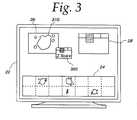
- FIG. 3is a view similar to FIG. 2 including the control window but also including a low magnification region from the slide showing regions marked by a histology grade or structure through automatic analysis of tissue and a high magnification window showing markings related to the grading or histology grade yielded by the automatic analysis of tissue in combination with a window showing a numerical score;
- FIG. 4is a block diagram of the apparatus embodying the present invention.
- FIG. 5is a block diagram of a portion of the apparatus shown in FIG. 4 showing details of a mechanical arrangement of a microscope
- FIG. 6is a flow diagram related to operation of the apparatus
- FIG. 7is a flow diagram of details of one of the steps in FIG. 6;
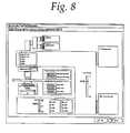
- FIG. 8is a display screen showing control parameters to be manipulated thereon
- FIG. 9is a flow chart for a region outlying routine
- FIG. 10is a flow chart for a scanning and analyzing routine
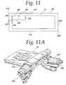
- FIG. 11is a schematic showing of the limits of travel of the microscope stage with respect to the image tiles
- FIG. 11Ais a perspective view of the microscope stage and stepper motors and encoders providing a closed loop drive for the motors;
- FIG. 12is a block diagram of a networked system allowing multiple workstations to obtain access to diagnostic image information and to manipulate such information locally at each workstation;
- FIG. 12Ais a view of the system described in connection with FIG. 5;
- FIG. 13is a block diagram of a remote networked system for distributing and accessing diagnostic images and data through a hypertext transport protocol based server directly or over a packet network;
- FIG. 14is a view of a low magnification, reconstructed image from a basal layer of rat esophagus
- FIG. 14Ais a view of a high magnification, reconstructed image from a selected point of interest from FIG. 14;
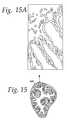
- FIG. 15is a view of a low magnification image of a mouse colon having a basal layer
- FIG. 15Ais a view of a reconstructed macro image of a mouse colon
- FIG. 16is a schematic view of an analysis from regions of a basal layer
- FIG. 16Ais a schematic view of an analysis to provide to a Z score.
- FIG. 17is a schematic view showing texture analysis tests for regions.
- the systemincludes a computer 12 which is a dual Pentium Pro personal computer in combination with a Hitachi HV-C20 video camera 14 associated with a Zeiss Axioplan 2 microscope 16 .
- the computer system 12is able to receive signals from the camera 14 which captures light from the microscope 16 having a microscope slide 18 positioned on an LUDL encoded motorized stage 20 .
- the encoded motorized stage 20includes a MAC 2000 stage controller for controlling the stage in response to the computer 12 .
- a microscope slide 18includes a biological specimen 21 which is to be viewed by the microscope and whose image is to be digitized both at low magnification and at high magnification as selected by a user.
- the low magnification digitized imageis then displayed on a 21 inch Iiyama video display monitor 22 having resolution of 1600 by 1200 to provide display screens of the type shown in FIGS. 1 through 3 including a low magnification image 24 , for instance, at 1.25 power, a high magnification image 26 , for instance at 40 power and a control window or image 28 .
- the low magnification imagemay have identified therein a region 30 which is reproduced at high magnification in high magnification screen or window 26 so that a pathologist or other operator of the system can review architectural regions of interest in low magnification image 24 and simultaneously view them in high magnification in the high magnification screen or window 26 to determine whether the cells forming a portion of the architectural feature need be examined further for cancer or the like or not.
- the computer 10is constructed around a PCI system bus 40 and has a first Pentium Pro microprocessor 42 and a second pentium pro microprocessor 44 connected thereto.
- the system bus 40has connected to it a PCI bus 50 and an ISA bus 52 .
- the PCI bus 50has a SCSI controller 60 connected thereto to send and receive information from a hard disk 62 .
- the hard disk 62also is coupled in daisy chain SCSI fashion to a high capacity removal disk and to a CD Rom drive 66 .
- the hard disks 62contains the programs for operating the system for controlling the microscope 16 and for processing the images as well as for doing a quantitative analysis of the selected portions of the histological specimens being viewed on the slide 18 .
- the system bus 40also has connected to it a random access memory 70 within which portions of the program being executed are stored as well as a read only memory 72 for holding a bootstrap loader as well as portions of the basic input/output operating system.
- a floppy disk controller 74is coupled to the system bus 40 and has connected to it a floppy disk drive 76 for reading and writing information to a floppy disk as appropriate.
- a mouse controller 80is coupled to the system bus and has a mouse 82 which operates as a pointing device for controlling manipulations on the screen 22 and within the windows 24 , 26 and 28 .
- a keyboard controller 90is connected to the system bus and has a keyboard 92 connected thereto. The keyboard 92 may be used to send and receive alpha numeric signals to other portions of the computer.
- An audio controller 100has a plurality of speakers 102 and a microphone 104 connected thereto for audio input and output and is coupled to the system bus 40 .
- a network interfacesuch as a network interface card 104 , is connected to the system bus and can provide signals via a channel 106 to other portions of a network or internet to which the system may be connected. Likewise, signals can be sent out of the system through a modem 110 connected to the ISA bus 52 and may be sent via a channel 112 , for instance, to the internet.
- a printer 116is connected via a parallel I/O controller 118 to the system bus in order to provide printouts as appropriate of screens and other information as it is generated.
- a serial I/O controller 122is connected to the system bus and has connected to it a camera controller 124 which is coupled to CCD sensors 126 in the cameras.
- the CCD sensors 126supply pixel or image signals representative of what is found on the slide 18 to an Epix pixci image acquisition controller 130 coupled to the PCI bus 50 .
- the microscope 16includes a base 140 having a stage 20 positioned thereon as well as an objective turret 142 having a plurality of objectives 144 , 146 and 148 thereon.
- the objective 144may be of 1.25 ⁇ objective.
- the objective 146may be a 20 ⁇ objective.
- the objective 148may be a 40 ⁇ objective. Signals from the camera sensors and controller are supplied over a bus 128 to the image acquisition system where they are digitized and supplied to the PCI bus for storage in RAM or for backing storage on the hard disk 62 .
- stage 20When a specimen is on the slide 18 the stage 20 may be manipulated under the control of the computer through a stage controller 160 coupled to the serial I/O controller 122 .
- a microscope controller 162controls aspects of the microscope such as the illumination, the color temperature or spectral output of a lamp 168 and the like.
- the processors 42 or 44send a command through the system bus to cause the serial I/O controller 122 to signal the microscope controller to change magnification to 1.25 ⁇ in a step 202 .
- the controllersets the color temperature of the lamp 168 , sets a pair of neutral density filter wheels 170 and 172 and sets a field diaphragm 174 for the correct illumination.
- a condenser diaphragm 176is also controlled and a color filter wheel 180 may also be controlled to apply the appropriate filter color to the CCD censors 126 in the camera.
- the entire slideis then scanned in a step 204 .
- the imagesare tiled and melded together into the overall image 24 supplied on the screen 22 to provide the operator in the step 206 with a visually inspectable macro image of relevant regions of the slide of interest.
- the mousemay be moved to identify a marker segment or region which, for instance, may be a rectangular region (as shown as 30 in FIG. 1) which will cause the microscope to change magnification as at step 208 to 4 ⁇ , 20 ⁇ , 40 ⁇ , etc., by rotating the turret to bring the appropriate objective lens system into viewing position.
- a marker segment or regionwhich, for instance, may be a rectangular region (as shown as 30 in FIG. 1) which will cause the microscope to change magnification as at step 208 to 4 ⁇ , 20 ⁇ , 40 ⁇ , etc., by rotating the turret to bring the appropriate objective lens system into viewing position.
- a testis made to determine whether the user has commanded continued inspection. If the user has, a test is made in a step 209 c to determine if the magnification is to be changed by changing the selected objective. In the event the magnification is to be changed control is transferred to the step 208 . If the magnification is to remain unchanged control is transferred to the step 209 a . In the event inspection is not to continue the region selected is outlined for higher magnification scan in a step 209 d . In a step 209 e , a command may be received to scan or acquire the higher magnification image for display in screen 26 . The image may then be archived for later analysis, displayed or analyzed immediately.
- the overall illumination and control of the microscopewill be controlled so that in a step 210 the objective turret 142 will be rotated to place the higher power objective above the slide 18 .
- voltage to the lampwill be changed to adjust the lamp 168 to provide the proper illumination and color temperature as predetermined for the selected objective.
- the condenser diaphragm 176will have its opening selected as appropriate to provide the proper illumination for that objective.
- the filter turret 180will select the proper light wavelength filter to be supplied to the camera sensors. For instance, a red, blue or green filter, as appropriate, particularly if the specimen has been stained.
- a step 218the field diaphragm 174 will have its opening changed.
- the neutral density filter wheel 170will select a neutral density filter and in a step 222 the neutral density filter wheel 172 will also select a neutral density filter.
- the X, Y and Z offsetswill be used for reconstruction of the recorded image at the magnification and in a step 226 the current position will be read from encoders in the stage which are accurate to 0.10 micron.
- the mouseIn order to identify the selected region the mouse is moved to that area of the region in a pointing operation in a step 240 as shown in FIG. 9. The mouse may be moved to draw a box around the region selected.
- the X and Y screen pointsare computed for the edges of the regions selected and the computed image or pixel points are translated to stage coordinate points in order to control the stage of the microscope.
- a list of all of the X fields for positioning the stage for the objectiveis stored in random access memory and may be backed up on the hard disk. The information from the X offsets for the objective and the stage offsets is used as well as the size of the field to position the slide properly under the objective to capture the micro image.
- stageis positioned for each of the X and Y coordinate values in stage coordinate values and the digitized image is captured by the cameras and stored in RAM and backed up on the hard disk.
- the imagemay be then analyzed quantitatively in various manners such as those set forth in the previously-identified United States application.
- the imagemay be stored for archival purposes in a step 254 .
- FIG. 8is a view of the settings of the microscope objective properties of the Axioplan 2, computer-controlled microscope.
- the X and Y positioningis specifically carried out as shown in FIG. 1 where the slide 18 is shown with a slide boundary 270 , 272 , 274 and 276 .
- Stage boundary for limits of the stage travel for purposes of the stagethe stage can be moved all the way from an upper left hand corner of travel 276 to a lower right hand corner of travel 280 .
- At the upper left hand bounded corner of travel 278limits which a signal that the end of travel has been reached and the stage is then translated a short distance 282 in the extra action and a short distance 284 in the Y direction to define the first tile 288 in terms of a reference point 290 at its upper left hand corner.
- the next macro image tile 292may be placed contiguous with it by moving the stage appropriately and by measuring the location of the stage from the stage in counters without the necessity of performing any image manipulation.
- the image tiles 288 and 292may be abutted without any substantial overlap or they may be overlapped slightly, such as a one pixel with overlap, which is negligible insofar as blurring of any adjacent edges of abutted image tiles.
- the upper left hand corner 300 of the tile 292defines the rest of 292 and other tiles can be so defined.
- Micro image tilescan likewise be defined so that they are contiguous but not substantially overlapping, as would interfere with the composite image.
- the low power image 24has a plurality of micro images defined therein which are tiled and which are shown in higher magnification as individual tiles 312 , 314 , 316 and the like in FIG. 2.
- the region 310 when magnified as shown in the window 26may exceed the bounds of the window and thus the window may include scroll bars or other means for allowing the image 310 which is larger than the window 26 to be examined from within the window 26 .
- the stage 200is best seen in FIG. 11A and includes the X and Y stepper motors 279 and 281 with their respective encoders, which provide a closed loop system to give the 0.1 micron accuracy versus the usual 5 or 6 micron accuracy of most microscope stages without a closed loop system.
- This closed loop system and this very high accuracyallow the abutting of the tile images for both high magnification and low magnification images without the substantial overlap and the time-consuming and expensive software currently used to eliminate the overlap and blurriness at the overlapping edges of adjacent image tiles.
- the tilesmay be positioned precisely in a horizontal row and precisely in vertical rows to reconstruct the macro image and the micro image. This reconstruction is done without the use, as in the prior art, of extensive software manipulation to eliminate overlapping image tiles, horizontally or vertically or the haphazard orientation of image tiles.
- the low power window 24 , high power window 26 and control window 28can be used in conjunction with reporting of quantitative analysis data, histograms, etc. for the specimen being viewed; and such analysis information may be provided as a visual output in a window 320 .
- Each of the various regions 30 that a pathologist may follow in marking various features in the low power window 24 and the high power window 26may be reflected in both windows in order that an audit trail is provided for the system.
- the present inventionalso includes the facility for allowing remote diagnostics to occur by being able to couple the system either over a network communication facility to an intranet, for instance via the network interface, or via a modem or other suitable connection, to an internet so that once the image has been scanned and stored in memory on hard disks or other storage, remote users may be able to access the low magnification image as well as the high magnification image and move around within both images to make determinations as to the histological characteristics of the samples via Z scores.
- An additional feature of the systemincludes a plurality of networked workstations coupled to a first computer console 12 having a display screen 22 connected to the microscope 14 .
- Satellite work stations 350 and 352are substantially identical to the work station 12 including respective computers 354 and 356 coupled to displays 358 and 360 .
- the devicescan be manipulated through input devices 360 and 362 which may include a keyboard, mouse and the like.
- a third devicecan be connected including a work station 370 , having a display 372 , a computer 374 and an input device 376 .
- Each of the devicesis connected over respective network lines 380 , 382 , 384 to the computer 12 which transmission may be via either net or the like.
- Each of the different operators at the physically separate viewing stationscan locate regions from the view of entire tissue cross sections via a macro view and label the regions for subsequent scanning and/or quantitative analysis.
- a single operator at the instrument station 12can locate regions to view the entire tissue cross section. Those regions can be labeled for subsequent scanning and/or quantitative analysis with subsequent review and physically remote viewing stations, for instance, in an operating room or in individual pathologists' signout areas in order to review analysis results while still maintaining and reviewing the entire macro view of the tissue and/or the individual stored images from which the quantitative results were obtained.
- the viewing stations 350 , 352 and 370can comprise desk top computers, laptops, etc. There is no need for a microscope at the network stations 350 , 352 and 370 .
- remote workstations 400 , 402 , 404 , 406 and 408may be connected through a server 410 which may be supplied via a packet switched network.
- the server 410and may be a hypertext transport protocol based server of the type used for the World Wide Web or may be a telnet type server as used previously in internet remote operation applications.
- the server 410communicates via a communications channel 414 with a local computer 416 having a display 418 associated therewith, the local computer 416 being connected to the microscope 420 .
- Each of the remote work stations 400 , 402 , 404 , 406 and 408may perform the same operations as the stations 350 , 352 and 370 although they do it from nearby buildings or even from around the world, thus providing additional flexibility for others to make use of the specimen obtained and being viewed under the microscope 420 .
- stored imagesmay be disseminated through the server 410 to the remote servers 400 through 408 for further analysis and review.
- FIG. 14there is illustrated on screen 28 a basal layer 431 a of a cut cross-section of a rat esophagus.
- the basal layeris elongated and linear in a downward direction, and the selected point of interest is shown as a box 30 on the basal layer on the composite, low magnification image.
- the high magnification image 26 of this selected point of interestis shown on screen 26 in FIG. 14A.
- FIG. 15is shown a mouse colon as a reconstructed, low magnification macro image 28 which has been reduced ⁇ fraction (1/16) ⁇ th in size.
- the micro image 26is shown in FIG. 15A, and the marking therefore is shown in FIG. 15.
Landscapes
- Engineering & Computer Science (AREA)
- Health & Medical Sciences (AREA)
- General Health & Medical Sciences (AREA)
- Medical Informatics (AREA)
- General Physics & Mathematics (AREA)
- Physics & Mathematics (AREA)
- Public Health (AREA)
- Primary Health Care (AREA)
- Epidemiology (AREA)
- Theoretical Computer Science (AREA)
- Biomedical Technology (AREA)
- Radiology & Medical Imaging (AREA)
- Nuclear Medicine, Radiotherapy & Molecular Imaging (AREA)
- Life Sciences & Earth Sciences (AREA)
- Chemical & Material Sciences (AREA)
- Dispersion Chemistry (AREA)
- Immunology (AREA)
- General Business, Economics & Management (AREA)
- Quality & Reliability (AREA)
- Computer Vision & Pattern Recognition (AREA)
- Signal Processing (AREA)
- Business, Economics & Management (AREA)
- Analytical Chemistry (AREA)
- Molecular Biology (AREA)
- Biochemistry (AREA)
- Multimedia (AREA)
- Pathology (AREA)
- Microscoopes, Condenser (AREA)
- Investigating Or Analysing Biological Materials (AREA)
- Investigating Or Analysing Materials By Optical Means (AREA)
Abstract
Description
- This is a continuation of a co-pending application, U.S. patent application Ser. No. 10/008,519, filed Nov. 13, 2001, which is a continuation of U.S. patent application Ser. No. 09/752,022 filed Dec. 28, 2000, which is a divisional application of U.S. patent application Ser. No. 09/395,694, filed Sep. 13, 1999, now U.S. Pat. No. 6,226,392, which is a continuation of U.S. patent application Ser. No. 08/805,856, filed Mar. 3, 1997, which is now abandoned, which is a continuation-in-part of U.S. patent application Ser. No. 08/701,974 filed on Aug. 24, 1996, now U.S. Pat. No. 6,031,930.[0001]
- This invention relates to a method of, and an apparatus for, acquiring and constructing tiled digital images from a specimen on a support, such as a microscope slide, and for storing, and transferring the image for viewing by another at a local or remote location.[0002]
- In the image analysis and quantification of DNA from tissue sections as disclosed in U.S. Pat. No. 4,741,031, and also especially in the immunohisto-chemistry assays on the kinds of cell analysis systems disclosed in U.S. Pat. Nos. 5,086,476; 5,202,931; and 5,252,487 issued to Bacus, there is a problem of first locating the cancer regions for analysis under low power and then remembering them when performing the analysis under higher power. Specifically, the problem is that once the microscope is set up for quantification by image analysis under, e.g. 40×, where all of the diaphragms are set and light adjusted, etc., if the operator needs to move to another tissue area, it is first desirable to locate it at e.g. 10×. In fact, often regions can only be located at this power. In order to do so however, all of the settings (diaphragms, light levels, wavelengths of light, etc.) have to be changed to view the tissue at this magnification. Currently, there is no way to ensure that one could go back to the settings at the previous 40× magnification and continue on with the quantitative image analysis of that same specimen. This necessitates finding those areas under 40×, without changing objectives, which is very slow and time-consuming, and often important cancer areas can be missed.[0003]
- Also, another problem with tissue analysis, at its current state-of-the-art, is that it is not completely automated, for example, with regard to finding structural regions such as glands, basal layers or other important diagnostic regions. However, as set forth in my co-pending patent application Serial No. 701,974, filed Aug. 23, 1996, if these regions are located, important very sensitive diagnostic measurements can be performed, which patent application is incorporated by reference as if fully reproduced herein. For example, as disclosed in the aforesaid patent application, assays are made of a variety of tissue types, both human and animal for analysis of neoplasia in tissue, for pre-invasive cancer in tissue, and the effects on the tissue of chemopreventive agents. A quantitative analysis by image processing techniques is performed on tissue types, having various architectural features, such as breast tissue, colon tissue, prostate tissue, esophageal tissue, skin tissue, cervix tissue, etc. These tissues have different morphologies, and they undergo different neoplasias usually resulting from a cellular mutation, as maybe enhanced by a carcinogen, or resulting from a cellular proliferation rate enhanced by hormones, growth factors, or other inducers of abnormal tissue growth. Often it is desired to quantify small changes in the neoplasia when it is incipient or through a series of analyses performed at close time intervals to measure whether the neoplasia progression is increasing or has been slowed, stopped or regressed.[0004]
- Usually, the tissue specimens are cut to expose the basal layer for review under the microscope. Typically, the quantitative measurements are performed at 40× to obtain 100 to 400 tissue images. The 40× objective provides a narrow field of view of a very small portion of the entire basal layer. Often, the basal layer is somewhat elongated and generally linear such as a basal layer in a rat esophagus; and the analysis of the basal layer requires examining it along its length. The basal layer in a mouse colon is more in the form of an irregular, circular shape; and the analysis of this basal layer requires traveling about this circular shape. In breast tissue samples, suspected tumor areas may be at widely-spaced points in the stained tissue; and one wants to be able to navigate and travel to these specific suspected areas and to do the 40× analysis at these areas in an efficient manner. There is a need to allow an experienced operator to interact with the analysis to locate and identify such regions in an interactive manner. Especially, an interactive manner that would be familiar and consistent with the use of a microscope manually, with high power magnification and low power magnification simultaneously available, but performed on a computer terminal. Such a level of interaction is different than the interaction with the system disclosed in the above-listed Bacus patents.[0005]
- There is a need to take the level of interaction to a higher level and let each component, the human and the computer, perform the part that it does best, in the most cost-effective manner. There are available on the market computer-controlled, automated microscopes such as those sold by Carl Zeiss, Inc., Thornwood, N.J., under the name Axioplan 2 for taking photographic images of a specimen in the microscopic field of view. Those particular microscopes have computer-controlled and automatically adjusted subsystems, such as an illumination subsystem, a focusing subsystem, a diaphragm or optical stops subsystem, an objective lens subsystem, or a filtering subsystem. As an operator selects changes from one objective lens, such as one providing low magnification, e.g., 4×, to a higher magnification, e.g., 40×, the computer-automated system will turn the lens turret to switch in the high magnification automatically and adjusts the lens and also automatically adjusts the illumination to eliminate glare and to provide the proper light illumination including light density. Further, the focus is adjusted, and the proper diaphragm openings are automatically reset. Thus, the computer-controlled, automated subsystems automatically reset to values stored and predetermined for each selected objective lens and the analysis being done.[0006]
- Those particular microscopes can be used to view various objects or specimens, but are most typically used to view and to take still photographs of biological specimens, such as tissues and cells. Those particular microscopes lack a computer-controlled X and Y stage for translating a specimen-carrying slide with respect to the field of view of the selected objective lens. Currently, pathologists and others who use such microscopes want to view the specimen images in full color or in enhanced colors using fluorescent illumination and/or monochromatic images using the automated filter subsystems on the microscopes. Currently trained pathologists or clinicians are accustomed to manually adjust and have a microscope available to them to view larger areas of the specimen at low magnification, and then to momentarily switch in a new higher magnification lens to obtain a more highly magnified image of a portion of the specimen viewed at low magnification. Pathologists and those working in this area have created in themselves a desire to view suspect tissue through a microscope and appear to resist analysis systems that do not provide them this ability.[0007]
- The microscopic field of view reduces very substantially as the magnification increases. The skill level of the clinician and/or pathologist is important to locate viewing the most suspicious areas or points of interest on the specimen. Sometimes, a technician will do a first assay and analysis. A pathologist will return to the selected points of interest or other points of interest for review and analysis. One concern with respect to a quantitative analysis of breast cancer tissue or prostate biopsy tissue samples to pap smears or other tests for various cancers or the like is that a particularly suspicious point in the tissue may be overlooked and missed during the visual assay or for selection for an automated review analysis. When observing at high magnifications, the field of view is limited to very small area of the specimen. Hence, the observer has difficulty in knowing and remembering the actual, exact location of this small periscopic view within the very large whole specimen.[0008]
- Often, also the problem is finding or locating the tissue or cells for view at high magnification so that artifacts and/or blank spaces on the slide are not viewed. A number of approaches have been proposed to prescreen and locate by an X and Y address the cells or small points of interest from a very large number of potential points of interest.[0009]
- There are currently available commercial services for prescreening pap smears where one can mail in slides and the service will do a microscopic prescan at high magnification for suspected or suspicious areas of interest which are marked and given address locations, and also a video tape of the slide specimen is returned by this service to the sender. The sender then reviews the areas of interest located during the prescreening and/or the video tape to complete the analysis.[0010]
- In an attempt to locate and allow review of specified points of interest, U.S. Pat. No. 5,428,690 to Bacus discloses a system for prescreening of a field of cells on a specimen slide at low magnification before the viewer. When seeing a point of interest to be viewed at high magnification, the viewer will operate a switch or the like to select and record the address of these selected prescreened points of interest. Later, these prescreened points of interest are then brought into position to be analyzed at high magnification. This system is fairly or too slow for many uses.[0011]
- A very expensive system is currently in use in which a pathologist located at a diagnostic center is able to make a diagnostic opinion with respect to specimens under a microscope at a remote center. The pathologist at the diagnostic center manipulates robotic controls to send telepathy signals via a special, dedicated satellite or other large bandwidth channel to control the microscope at the remote site in approximately real time. The pathologist then can maneuver the remote microscope to shift the microscope's field of view and to send, by telepathy, a highly magnified, very small image back to the pathologist. This system requires each subscriber to have a special microscope operable by manipulation of the robotic controls at the diagnostic center and a dedicated or large bandwidth channel to convey real time video signals and hence results in a very high cost for the assay being done. To assist the pathologist in staying within the specimen at the remote site, a peripheral edge or map of the specimen is made using a second video camera and a light box or by using computerized scanning equipment to trace the outline or peripheral edge of the specimen boundaries. A small circle of light is displayed within the map of the specimen so that the pathologist at the diagnostic center knows the location of the field of view of the highly magnified image within the specimen. In a sense, the pathologist is operating in real time in the same way that he would use his own microscope at his diagnostic center except for a slight transmission delay used to transmit the video signals of the highly magnified image over large bandwidth channel. Although the pathologist has a small map or peripheral outline of the specimen, the pathologist's field of view of the actual specimen is only the small circle of view that is coming through the microscope objective lens. This does not help the pathologist locate suspicious areas of interest as in a prescreening of the entire tissue. The pathologist may switch to the lowest magnification to get the largest field of view of a small section of the specimen, but he never views the entire specimen at any magnification. Also, there is no image analysis quantitative testing from the received images at the diagnostic center; and no quantitative assaying is done with these images at the diagnostic center.[0012]
- There is a particular interest today in using the Internet system because it is so readily accessible by users at a low cost and using a computer and viewing screen connected to the computer. One problem with trying to do any transmission of digitized, microscopic, highly magnified images over the Internet is that the bandwidth is too narrow to accommodate the tremendous amount of stored data which needs to be transmitted. There is a need for a system which would allow a pathologist or another person, to be able to perform tissue analysis or quantitative assays using a standard computer terminal from a location remote from the automated microscope.[0013]
- In accordance with the present invention, a person such as a pathologist at a computer terminal may control an automated microscope to acquire on a computer screen or window images of the specimen at different magnifications as selected by the person. Further, the person may receive on the screen a low magnification, reconstructed image of the entire specimen to aid the person in interactively selecting points of interest on the specimen, such as along a basal layer of a tissue specimen.[0014]
- More specifically, and in accordance with the present invention, the microscope's small field of view limitation of a specimen is overcome by providing to the viewer a reconstructed, digitized and magnified image of the entire specimen (or a very large portion of the specimen) for performing a visual analysis of the entire tissue in full color to aid in the selection of points of interest to be viewed at a higher magnification. This is achieved by acquiring a large number of low magnification images of the specimen through a microscopic scanning system, e.g., 35 image tiles of the specimen at 1.25×, and then assembling and coordinating the tiles to form an overall, low magnified image of the specimen, i.e., a macro image of the specimen. Preferably, the digitized macro image is reduced in size by a software system to a smaller size, e.g., a ¼ size image that is displayed on a local screen or is sent over a low band width or a high band width channel to a remote screen. Thus, the pathologist not only does not need to have others do a slow laborious prescreening to locate suspicious areas for analysis or for viewing at high magnification, he can use his own experiences to go directly to the most suspicious areas which he sees on the macro image. He can, on a priority basis, do the most suspicious area first, followed by lower priority areas of interest.[0015]
- In accordance with the present invention, there is provided a new and improved automated, computer-controlled microscope that displays the low magnification composite image of the specimen to allow the user to view and to interactively select points of interest, each of which may be displayed at high magnification. This is achieved by providing the user with a marker, such as a cursor or the like, to select the defined area of interest; and to acquire reproduced, spatially adjacent high magnification, digitized images of the selected area of interest. More specifically, the specimen, when it was first scanned at low magnification to provide a macro view of the specimen, the addresses or locations of the tile images and/or pixels for the composite image were acquired. Therefore, any selected region of interest in the macro image has locations to which the microscopic stage may be automatically repositioned under a changed, higher magnification lens to acquire higher magnification, digitized image tiles that can be assembled into a micro image. Herein, both the macro and micro images are formed of adjacent digitized image tiles which have been coordinated to reproduce spatially the original image that was scanned.[0016]
- It is the high magnification images, usually at 40×, that were analyzed using image processing techniques as disclosed in the aforesaid patent application, to provide an assay or numerical histological data for the specimen.[0017]
- In accordance with the preferred embodiment of the invention, the pathologist may select a larger region for analysis at high resolution than can be accommodated at this magnification on his high magnification viewing screen. He can, though, view all of the adjacent, highly magnified, digitized image tiles on this high magnification screen by scrolling up or down or right to left to shift these digitized, adjacent image tiles into view on the screen. Thus, even at a higher magnification of a region the pathologist is able to obtain a much larger view than the small field for the objective lens in use of adjacent tissue or cells to give him a broader, overall perspective of what is happening or what has happened in specific section of a specimen. For instance, a pathologist may want to see at high magnification and to assay at this high magnification, a suspicious area, the pathologist can draw a mark about the area and cause it to then be assayed and displayed.[0018]
- By having displayed on a low magnification screen or split screen of the full composite area, by having the high magnification region being marked on the low image screen, and by having the region being scrolled to view adjacent high magnification images on the high magnification screen the pathologist has available information to guide him in the sense of helping to navigate intelligently within the specimen in his search for cancerous tissue or the like for further inspection or measurement of malignant characteristics. Often, when restricted to a field of view of an objective microscope, the pathologist has a difficult time, in the words of a cliche, of seeing the forest for the trees. In the present invention, the pathologist has a full, magnified, reduced in size specimen view with the higher image area marked thereon to guide him and to let him see the forest. He can also see a region of the forest at higher magnification by scrolling adjacent tree images onto the high magnification screen.[0019]
- In accordance with a further aspect of the invention, the user may elect to change to an intermediate magnification being viewed by either switching automatically to a new objective lens and acquiring new digitized image tiles at the intermediate magnification or by using software to reconstruct from the existing high and low magnification, digitized images a new intermediate digitized image.[0020]
- The preferred, low magnification image, which is reduced in size, can be transmitted over narrow band width channels such as a local area network or over the Internet through various servers and computers. Likewise, the reconstructed, high magnification images can be transmitted over such narrow band width channels. Because the microscope is fully computer controlled, a pathologist or other person having a split screen computer such as a PC, can be connected to the microscope and operate it from a remote location to obtain the macro image and to navigate to points of interest and obtain the desired micro images. With the present invention, there is no need for a specialized microscope at each remote location nor for a broad band channel to send video signals in real time between the diagnostic center and the remote location.[0021]
- FIG. 1 is a screen view of a system embodying the present invention showing a low magnification image of a specimen on a microscope slide in one window, a high magnification image of a portion of the low magnification image selected by a region marker and a control window;[0022]
- FIG. 2 is a view of a display screen of the apparatus embodying the present invention showing the control window a low magnification window having a plurality of high magnification micro image regions delineated therein and a high magnification window including one or more of the micro image regions;[0023]
- FIG. 3 is a view similar to FIG. 2 including the control window but also including a low magnification region from the slide showing regions marked by a histology grade or structure through automatic analysis of tissue and a high magnification window showing markings related to the grading or histology grade yielded by the automatic analysis of tissue in combination with a window showing a numerical score;[0024]
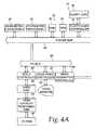
- FIG. 4 is a block diagram of the apparatus embodying the present invention;[0025]
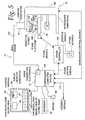
- FIG. 5 is a block diagram of a portion of the apparatus shown in FIG. 4 showing details of a mechanical arrangement of a microscope;[0026]
- FIG. 6 is a flow diagram related to operation of the apparatus;[0027]
- FIG. 7 is a flow diagram of details of one of the steps in FIG. 6;[0028]
- FIG. 8 is a display screen showing control parameters to be manipulated thereon;[0029]
- FIG. 9 is a flow chart for a region outlying routine;[0030]
- FIG. 10 is a flow chart for a scanning and analyzing routine;[0031]
- FIG. 11 is a schematic showing of the limits of travel of the microscope stage with respect to the image tiles;[0032]
- FIG. 11A is a perspective view of the microscope stage and stepper motors and encoders providing a closed loop drive for the motors;[0033]
- FIG. 12 is a block diagram of a networked system allowing multiple workstations to obtain access to diagnostic image information and to manipulate such information locally at each workstation;[0034]
- FIG. 12A is a view of the system described in connection with FIG. 5;[0035]
- FIG. 13 is a block diagram of a remote networked system for distributing and accessing diagnostic images and data through a hypertext transport protocol based server directly or over a packet network;[0036]
- FIG. 14 is a view of a low magnification, reconstructed image from a basal layer of rat esophagus;[0037]
- FIG. 14A is a view of a high magnification, reconstructed image from a selected point of interest from FIG. 14;[0038]
- FIG. 15 is a view of a low magnification image of a mouse colon having a basal layer;[0039]
- FIG. 15A is a view of a reconstructed macro image of a mouse colon;[0040]
- FIG. 16 is a schematic view of an analysis from regions of a basal layer;[0041]
- FIG. 16A is a schematic view of an analysis to provide to a Z score; and[0042]
- FIG. 17 is a schematic view showing texture analysis tests for regions.[0043]
- Referring now to the drawings, and especially to FIGS. 4 and 5, apparatus for synthesizing low magnification and high magnification microscopic images is shown therein and generally identified by[0044]
reference numeral 10. The system includes acomputer 12 which is a dual Pentium Pro personal computer in combination with a Hitachi HV-C20 video camera 14 associated with aZeiss Axioplan 2microscope 16. Thecomputer system 12 is able to receive signals from thecamera 14 which captures light from themicroscope 16 having amicroscope slide 18 positioned on an LUDL encodedmotorized stage 20. The encodedmotorized stage 20 includes a MAC 2000 stage controller for controlling the stage in response to thecomputer 12. Amicroscope slide 18 includes abiological specimen 21 which is to be viewed by the microscope and whose image is to be digitized both at low magnification and at high magnification as selected by a user. The low magnification digitized image is then displayed on a 21 inch Iiyama video display monitor22 having resolution of 1600 by 1200 to provide display screens of the type shown in FIGS. 1 through 3 including alow magnification image 24, for instance, at 1.25 power, ahigh magnification image 26, for instance at 40 power and a control window orimage 28. The low magnification image may have identified therein aregion 30 which is reproduced at high magnification in high magnification screen orwindow 26 so that a pathologist or other operator of the system can review architectural regions of interest inlow magnification image 24 and simultaneously view them in high magnification in the high magnification screen orwindow 26 to determine whether the cells forming a portion of the architectural feature need be examined further for cancer or the like or not. - The[0045]
computer 10 is constructed around aPCI system bus 40 and has a firstPentium Pro microprocessor 42 and a second pentium promicroprocessor 44 connected thereto. Thesystem bus 40 has connected to it aPCI bus 50 and anISA bus 52. ThePCI bus 50 has aSCSI controller 60 connected thereto to send and receive information from ahard disk 62. Thehard disk 62 also is coupled in daisy chain SCSI fashion to a high capacity removal disk and to aCD Rom drive 66. Thehard disks 62 contains the programs for operating the system for controlling themicroscope 16 and for processing the images as well as for doing a quantitative analysis of the selected portions of the histological specimens being viewed on theslide 18. Thesystem bus 40 also has connected to it arandom access memory 70 within which portions of the program being executed are stored as well as a read onlymemory 72 for holding a bootstrap loader as well as portions of the basic input/output operating system. Afloppy disk controller 74 is coupled to thesystem bus 40 and has connected to it afloppy disk drive 76 for reading and writing information to a floppy disk as appropriate. Amouse controller 80 is coupled to the system bus and has amouse 82 which operates as a pointing device for controlling manipulations on thescreen 22 and within thewindows keyboard controller 90 is connected to the system bus and has akeyboard 92 connected thereto. Thekeyboard 92 may be used to send and receive alpha numeric signals to other portions of the computer. Anaudio controller 100 has a plurality of speakers102 and amicrophone 104 connected thereto for audio input and output and is coupled to thesystem bus 40. A network interface, such as anetwork interface card 104, is connected to the system bus and can provide signals via achannel 106 to other portions of a network or internet to which the system may be connected. Likewise, signals can be sent out of the system through amodem 110 connected to theISA bus 52 and may be sent via achannel 112, for instance, to the internet. Aprinter 116 is connected via a parallel I/O controller 118 to the system bus in order to provide printouts as appropriate of screens and other information as it is generated. A serial I/O controller 122 is connected to the system bus and has connected to it acamera controller 124 which is coupled toCCD sensors 126 in the cameras. TheCCD sensors 126 supply pixel or image signals representative of what is found on theslide 18 to an Epix pixciimage acquisition controller 130 coupled to thePCI bus 50. - The[0046]
microscope 16 includes a base140 having astage 20 positioned thereon as well as anobjective turret 142 having a plurality ofobjectives bus 128 to the image acquisition system where they are digitized and supplied to the PCI bus for storage in RAM or for backing storage on thehard disk 62. - When a specimen is on the[0047]
slide 18 thestage 20 may be manipulated under the control of the computer through astage controller 160 coupled to the serial I/O controller 122. Likewise, amicroscope controller 162 controls aspects of the microscope such as the illumination, the color temperature or spectral output of alamp 168 and the like. For instance, in normal operation, when a specimen is placed on the slide,specimen slide 18 is placed on thestage 20 in astep 200, as shown in FIG. 6, theprocessors O controller 122 to signal the microscope controller to change magnification to 1.25× in astep 202. This is done by rotating the objective turret of theAxioplan 2 microscope to select theobjective 144. Likewise, the controller sets the color temperature of thelamp 168, sets a pair of neutraldensity filter wheels field diaphragm 174 for the correct illumination. Acondenser diaphragm 176 is also controlled and acolor filter wheel 180 may also be controlled to apply the appropriate filter color to the CCD censors126 in the camera. The entire slide is then scanned in astep 204. The images are tiled and melded together into theoverall image 24 supplied on thescreen 22 to provide the operator in thestep 206 with a visually inspectable macro image of relevant regions of the slide of interest. - In order to provide the magnified image, the mouse may be moved to identify a marker segment or region which, for instance, may be a rectangular region (as shown as[0048]30 in FIG. 1) which will cause the microscope to change magnification as at
step 208 to 4×, 20×, 40×, etc., by rotating the turret to bring the appropriate objective lens system into viewing position. - Next the user, in a[0049]
step 209a, uses the mouse to select the region on the macro image in order to select the micro image to be viewed on thescreen 22. In astep 209ba test is made to determine whether the user has commanded continued inspection. If the user has, a test is made in astep 209cto determine if the magnification is to be changed by changing the selected objective. In the event the magnification is to be changed control is transferred to thestep 208. If the magnification is to remain unchanged control is transferred to thestep 209a. In the event inspection is not to continue the region selected is outlined for higher magnification scan in astep 209d. In astep 209e, a command may be received to scan or acquire the higher magnification image for display inscreen 26. The image may then be archived for later analysis, displayed or analyzed immediately. - In order to perform the magnification called for in[0050]
step 208, the overall illumination and control of the microscope will be controlled so that in astep 210 theobjective turret 142 will be rotated to place the higher power objective above theslide 18. In astep 212 voltage to the lamp will be changed to adjust thelamp 168 to provide the proper illumination and color temperature as predetermined for the selected objective. In astep 214, thecondenser diaphragm 176 will have its opening selected as appropriate to provide the proper illumination for that objective. In astep 216, thefilter turret 180 will select the proper light wavelength filter to be supplied to the camera sensors. For instance, a red, blue or green filter, as appropriate, particularly if the specimen has been stained. In astep 218 thefield diaphragm 174 will have its opening changed. In astep 220 the neutraldensity filter wheel 170 will select a neutral density filter and in astep 222 the neutraldensity filter wheel 172 will also select a neutral density filter. In astep 224 the X, Y and Z offsets will be used for reconstruction of the recorded image at the magnification and in astep 226 the current position will be read from encoders in the stage which are accurate to 0.10 micron. - In order to identify the selected region the mouse is moved to that area of the region in a pointing operation in a[0051]
step 240 as shown in FIG. 9. The mouse may be moved to draw a box around the region selected. In astep 242 the X and Y screen points are computed for the edges of the regions selected and the computed image or pixel points are translated to stage coordinate points in order to control the stage of the microscope. In a step244 a list of all of the X fields for positioning the stage for the objective is stored in random access memory and may be backed up on the hard disk. The information from the X offsets for the objective and the stage offsets is used as well as the size of the field to position the slide properly under the objective to capture the micro image. - When the slide has been positioned properly, as shown in FIG. 10 in a[0052]
step 250 the stage is positioned for each of the X and Y coordinate values in stage coordinate values and the digitized image is captured by the cameras and stored in RAM and backed up on the hard disk. The image may be then analyzed quantitatively in various manners such as those set forth in the previously-identified United States application. Optionally the image may be stored for archival purposes in astep 254. - In order to override the specific control functions that take place as shown in FIG. 7, a screen is provided as shown in FIG. 8 wherein the XY step size can be edited, the X, Y and Z offset can be edited, the lamp voltage can be selected, the neutral density filter can be selected as well as the opening of the field diaphragm and several other microscopic characteristics. FIG. 8 is a view of the settings of the microscope objective properties of the[0053]
Axioplan 2, computer-controlled microscope. - The X and Y positioning is specifically carried out as shown in FIG. 1 where the[0054]
slide 18 is shown with aslide boundary travel 276 to a lower right hand corner oftravel 280. At the upper left hand bounded corner oftravel 278 limits which a signal that the end of travel has been reached and the stage is then translated ashort distance 282 in the extra action and ashort distance 284 in the Y direction to define thefirst tile 288 in terms of areference point 290 at its upper left hand corner. Since the size of themacro image tile 288 is known, the nextmacro image tile 292 may be placed contiguous with it by moving the stage appropriately and by measuring the location of the stage from the stage in counters without the necessity of performing any image manipulation. Theimage tiles left hand corner 300 of thetile 292 defines the rest of292 and other tiles can be so defined. Micro image tiles can likewise be defined so that they are contiguous but not substantially overlapping, as would interfere with the composite image. This avoids the problems encountered with having to perform extended computations on digital images in a frame storer or multiple frame storage in order to match or bring the images into contiguity without blurriness at the edges of contiguous image tiles. It may be appreciated as shown in FIG. 2 that thelow power image 24 has a plurality of micro images defined therein which are tiled and which are shown in higher magnification asindividual tiles region 310 when magnified as shown in thewindow 26 may exceed the bounds of the window and thus the window may include scroll bars or other means for allowing theimage 310 which is larger than thewindow 26 to be examined from within thewindow 26. - The[0055]
stage 200 is best seen in FIG. 11A and includes the X andY stepper motors point 278 is always taken from the same point, the tiles may be positioned precisely in a horizontal row and precisely in vertical rows to reconstruct the macro image and the micro image. This reconstruction is done without the use, as in the prior art, of extensive software manipulation to eliminate overlapping image tiles, horizontally or vertically or the haphazard orientation of image tiles. - Furthermore, as shown in FIG. 3, the[0056]
low power window 24,high power window 26 andcontrol window 28 can be used in conjunction with reporting of quantitative analysis data, histograms, etc. for the specimen being viewed; and such analysis information may be provided as a visual output in a window320. Each of thevarious regions 30 that a pathologist may follow in marking various features in thelow power window 24 and thehigh power window 26 may be reflected in both windows in order that an audit trail is provided for the system. - The present invention also includes the facility for allowing remote diagnostics to occur by being able to couple the system either over a network communication facility to an intranet, for instance via the network interface, or via a modem or other suitable connection, to an internet so that once the image has been scanned and stored in memory on hard disks or other storage, remote users may be able to access the low magnification image as well as the high magnification image and move around within both images to make determinations as to the histological characteristics of the samples via Z scores.[0057]
- An additional feature of the system includes a plurality of networked workstations coupled to a[0058]
first computer console 12 having adisplay screen 22 connected to themicroscope 14.Satellite work stations work station 12 includingrespective computers displays input devices 360 and362 which may include a keyboard, mouse and the like. Also a third device can be connected including awork station 370, having adisplay 372, acomputer 374 and an input device376. Each of the devices is connected overrespective network lines computer 12 which transmission may be via either net or the like. Each of the different operators at the physically separate viewing stations can locate regions from the view of entire tissue cross sections via a macro view and label the regions for subsequent scanning and/or quantitative analysis. A single operator at theinstrument station 12 can locate regions to view the entire tissue cross section. Those regions can be labeled for subsequent scanning and/or quantitative analysis with subsequent review and physically remote viewing stations, for instance, in an operating room or in individual pathologists' signout areas in order to review analysis results while still maintaining and reviewing the entire macro view of the tissue and/or the individual stored images from which the quantitative results were obtained. Theviewing stations network stations - In a still further alternative embodiment,[0059]
remote workstations server 410 which may be supplied via a packet switched network. Theserver 410 and may be a hypertext transport protocol based server of the type used for the World Wide Web or may be a telnet type server as used previously in internet remote operation applications. Theserver 410 communicates via acommunications channel 414 with alocal computer 416 having adisplay 418 associated therewith, thelocal computer 416 being connected to themicroscope 420. Each of theremote work stations stations microscope 420. In addition, stored images may be disseminated through theserver 410 to theremote servers 400 through408 for further analysis and review. - In FIG. 14, there is illustrated on screen[0060]28 a
basal layer 431aof a cut cross-section of a rat esophagus. The basal layer is elongated and linear in a downward direction, and the selected point of interest is shown as abox 30 on the basal layer on the composite, low magnification image. Thehigh magnification image 26 of this selected point of interest is shown onscreen 26 in FIG. 14A. In FIG. 15 is shown a mouse colon as a reconstructed, low magnificationmacro image 28 which has been reduced {fraction (1/16)}th in size. Themicro image 26 is shown in FIG. 15A, and the marking therefore is shown in FIG. 15. - The analysis for texture and for morphological features used to analyze a series of[0061]
regions 30 on the elongated basal layer that were analyzed at high magnification are shown in FIGS. 16, 16A and17. The manner of doing these tests and of obtaining a Z score or grade is disclosed in the aforesaid patent application. - While there has been illustrated and described a particular embodiment of the present invention, it will be appreciated that numerous changes and modifications will occur to those skilled in the art, and it is intended in the appended claims to cover all those changes and modifications which followed in the true spirit and scope of the present invention.[0062]
Claims (37)
Priority Applications (1)
| Application Number | Priority Date | Filing Date | Title |
|---|---|---|---|
| US10/751,771US7110586B2 (en) | 1996-08-23 | 2004-01-05 | Apparatus for remote control of a microscope |
Applications Claiming Priority (6)
| Application Number | Priority Date | Filing Date | Title |
|---|---|---|---|
| US08/701,974US6031930A (en) | 1996-08-23 | 1996-08-23 | Method and apparatus for testing a progression of neoplasia including cancer chemoprevention testing |
| US08/805,856US6101265A (en) | 1996-08-23 | 1997-03-03 | Method and apparatus for acquiring and reconstructing magnified specimen images from a computer-controlled microscope |
| US09/395,694US6226392B1 (en) | 1996-08-23 | 1999-09-13 | Method and apparatus for acquiring and reconstructing magnified specimen images from a computer-controlled microscope |
| US09/752,022US6404906B2 (en) | 1997-03-03 | 2000-12-28 | Method and apparatus for acquiring and reconstructing magnified specimen images from a computer-controlled microscope |
| US10/008,519US6674884B2 (en) | 1996-08-23 | 2001-11-13 | Apparatus for remote control of a microscope |
| US10/751,771US7110586B2 (en) | 1996-08-23 | 2004-01-05 | Apparatus for remote control of a microscope |
Related Parent Applications (1)
| Application Number | Title | Priority Date | Filing Date |
|---|---|---|---|
| US10/008,519ContinuationUS6674884B2 (en) | 1996-08-23 | 2001-11-13 | Apparatus for remote control of a microscope |
Publications (2)
| Publication Number | Publication Date |
|---|---|
| US20040136582A1true US20040136582A1 (en) | 2004-07-15 |
| US7110586B2 US7110586B2 (en) | 2006-09-19 |
Family
ID=27015227
Family Applications (3)
| Application Number | Title | Priority Date | Filing Date |
|---|---|---|---|
| US09/752,022Expired - LifetimeUS6404906B2 (en) | 1996-08-23 | 2000-12-28 | Method and apparatus for acquiring and reconstructing magnified specimen images from a computer-controlled microscope |
| US10/008,519Expired - LifetimeUS6674884B2 (en) | 1996-08-23 | 2001-11-13 | Apparatus for remote control of a microscope |
| US10/751,771Expired - Fee RelatedUS7110586B2 (en) | 1996-08-23 | 2004-01-05 | Apparatus for remote control of a microscope |
Family Applications Before (2)
| Application Number | Title | Priority Date | Filing Date |
|---|---|---|---|
| US09/752,022Expired - LifetimeUS6404906B2 (en) | 1996-08-23 | 2000-12-28 | Method and apparatus for acquiring and reconstructing magnified specimen images from a computer-controlled microscope |
| US10/008,519Expired - LifetimeUS6674884B2 (en) | 1996-08-23 | 2001-11-13 | Apparatus for remote control of a microscope |
Country Status (1)
| Country | Link |
|---|---|
| US (3) | US6404906B2 (en) |
Cited By (19)
| Publication number | Priority date | Publication date | Assignee | Title |
|---|---|---|---|---|
| US20040008894A1 (en)* | 1998-06-01 | 2004-01-15 | Zeineh Jack A. | Compression packaged image transmission for telemicroscopy |
| US20040083085A1 (en)* | 1998-06-01 | 2004-04-29 | Zeineh Jack A. | Integrated virtual slide and live microscope system |
| US20040250216A1 (en)* | 1999-11-04 | 2004-12-09 | Roman Kendyl A. | Graphical user interface including zoom control box representing image and magnification of displayed image |
| US20050089208A1 (en)* | 2003-07-22 | 2005-04-28 | Rui-Tao Dong | System and method for generating digital images of a microscope slide |
| US20050136509A1 (en)* | 2003-09-10 | 2005-06-23 | Bioimagene, Inc. | Method and system for quantitatively analyzing biological samples |
| US20060066734A1 (en)* | 2004-09-28 | 2006-03-30 | Leica Microsystems Cms Gmbh | Method for superimposing optical information in a scanning microscope |
| US20080024860A1 (en)* | 2006-06-30 | 2008-01-31 | The General Hospital Corporation | Device and method for wide-field and high resolution imaging of tissue |
| US7653260B2 (en)* | 2004-06-17 | 2010-01-26 | Carl Zeis MicroImaging GmbH | System and method of registering field of view |
| RU2408931C1 (en)* | 2009-05-18 | 2011-01-10 | Учреждение Российской академии наук Институт проблем управления им. В.А. Трапезникова РАН | Automated multifunctional system for analysis of object images |
| WO2010148152A3 (en)* | 2009-06-16 | 2011-03-31 | Ikonisys, Inc. | System and method for remote control of a microscope |
| US20110181688A1 (en)* | 2008-09-11 | 2011-07-28 | Atsushi Miyamoto | Method and device for synthesizing panorama image using scanning charged-particle microscope |
| US20120098953A1 (en)* | 2009-07-17 | 2012-04-26 | Go Kotaki | Scanning electron microscope device and pattern dimension measuring method using same |
| US20120296599A1 (en)* | 2011-05-20 | 2012-11-22 | Hon Hai Precision Industry Co., Ltd. | Temperature sensing system and method |
| WO2013016825A1 (en)* | 2011-08-02 | 2013-02-07 | Viewsiq Inc. | Apparatus and method for digital microscopy imaging |
| US8428887B2 (en) | 2003-09-08 | 2013-04-23 | Ventana Medical Systems, Inc. | Method for automated processing of digital images of tissue micro-arrays (TMA) |
| CN103180770A (en)* | 2011-08-26 | 2013-06-26 | 索尼公司 | Information processing system and information processing method |
| US20150379328A1 (en)* | 2011-07-20 | 2015-12-31 | Mikroscan Technologies, Inc. | Network-based pathology system with desktop slide scanner |
| US10119901B2 (en) | 2013-11-15 | 2018-11-06 | Mikroscan Technologies, Inc. | Geological scanner |
| US10162166B2 (en) | 2014-10-28 | 2018-12-25 | Mikroscan Technologies, Inc. | Microdissection viewing system |
Families Citing this family (135)
| Publication number | Priority date | Publication date | Assignee | Title |
|---|---|---|---|---|
| US6718053B1 (en)* | 1996-11-27 | 2004-04-06 | Chromavision Medical Systems, Inc. | Method and apparatus for automated image analysis of biological specimens |
| EP1300713A3 (en)* | 1995-11-30 | 2004-11-03 | Chromavision Medical Systems, Inc. | Method and apparatus for automated image analysis of biological specimens |
| US6272235B1 (en)* | 1997-03-03 | 2001-08-07 | Bacus Research Laboratories, Inc. | Method and apparatus for creating a virtual microscope slide |
| JP4018218B2 (en)* | 1997-12-25 | 2007-12-05 | キヤノン株式会社 | Optical apparatus and distance measuring point selection method |
| US6674894B1 (en)* | 1999-04-20 | 2004-01-06 | University Of Utah Research Foundation | Method and apparatus for enhancing an image using data optimization and segmentation |
| US6847729B1 (en) | 1999-04-21 | 2005-01-25 | Fairfield Imaging Limited | Microscopy |
| US6993170B2 (en)* | 1999-06-23 | 2006-01-31 | Icoria, Inc. | Method for quantitative analysis of blood vessel structure |
| JP2001016622A (en)* | 1999-06-30 | 2001-01-19 | Agilent Technologies Japan Ltd | Debug device and test method for image pickup device |
| US7505614B1 (en)* | 2000-04-03 | 2009-03-17 | Carl Zeiss Microimaging Ais, Inc. | Remote interpretation of medical images |
| DK1146480T3 (en)* | 2000-04-06 | 2003-12-01 | European Molecular Biology Lab Embl | Computer controlled microscope |
| US20030163031A1 (en)* | 2000-06-26 | 2003-08-28 | Adlabs, Inc. | Method and system for providing centralized anatomic pathology services |
| DE10043992B4 (en)* | 2000-09-05 | 2013-12-24 | Leica Microsystems Cms Gmbh | Method for examining a sample and confocal scanning microscope |
| US7292251B1 (en)* | 2000-10-06 | 2007-11-06 | The Research Foundation Of State University Of New York | Virtual telemicroscope |
| US7027628B1 (en) | 2000-11-14 | 2006-04-11 | The United States Of America As Represented By The Department Of Health And Human Services | Automated microscopic image acquisition, compositing, and display |
| WO2002076282A2 (en)* | 2001-01-05 | 2002-10-03 | Tissueinformatics, Inc. | Method for quantitative analysis of blood vessel structure |
| JP4865147B2 (en)* | 2001-05-29 | 2012-02-01 | エスアイアイ・ナノテクノロジー株式会社 | Pattern measuring method and measuring system using display microscope image |
| US20030081216A1 (en)* | 2001-11-01 | 2003-05-01 | Martin Ebert | Graphical user interface for sample positioning |
| US20030093164A1 (en)* | 2001-11-02 | 2003-05-15 | Martin Ebert | System for providing communication between a GUI and metrology control software |
| US6884341B2 (en)* | 2002-10-02 | 2005-04-26 | G6 Science Corp. | Filter device to capture a desired amount of material |
| US6905594B2 (en)* | 2002-10-11 | 2005-06-14 | G6 Science Corp. | Filter apparatus and methods to capture a desired amount of material from a sample suspension for monolayer deposition, analysis or other uses |
| US20040076346A1 (en)* | 2002-10-18 | 2004-04-22 | Russon Virgil Kay | Imaging system and method |
| US7034317B2 (en)* | 2002-12-17 | 2006-04-25 | Dmetrix, Inc. | Method and apparatus for limiting scanning imaging array data to characteristics of interest |
| GB2398196B (en)* | 2003-02-05 | 2005-06-01 | Fairfield Imaging Ltd | Microscope system and method |
| US20040202357A1 (en) | 2003-04-11 | 2004-10-14 | Perz Cynthia B. | Silhouette image acquisition |
| US20040224279A1 (en)* | 2003-05-06 | 2004-11-11 | Ormco Corporation | Dental display system |
| US6909151B2 (en)* | 2003-06-27 | 2005-06-21 | Intel Corporation | Nonplanar device with stress incorporation layer and method of fabrication |
| US7106502B1 (en) | 2003-08-21 | 2006-09-12 | The United States Of America As Represented By The Administrator Of National Aeronautics And Space Administration | Operation of a Cartesian robotic system in a compact microscope imaging system with intelligent controls |
| US20050058330A1 (en)* | 2003-09-16 | 2005-03-17 | Sysmex Corporation | Method of displaying smear image and retrieving method employing the same, surveillance method, system of displaying smear image, program for displaying smear image and recording medium recording the program |
| WO2005104039A2 (en)* | 2004-03-23 | 2005-11-03 | Google, Inc. | A digital mapping system |
| US7865301B2 (en)* | 2004-03-23 | 2011-01-04 | Google Inc. | Secondary map in digital mapping system |
| US7599790B2 (en)* | 2004-03-23 | 2009-10-06 | Google Inc. | Generating and serving tiles in a digital mapping system |
| US7925070B2 (en)* | 2004-03-30 | 2011-04-12 | Sysmex Corporation | Method for displaying virtual slide and terminal device for displaying virtual slide |
| DE602004018112D1 (en)* | 2004-06-07 | 2009-01-15 | Milestone Srl | Method and apparatus for microwave assisted automatic tissue treatment |
| US7199712B2 (en)* | 2004-06-17 | 2007-04-03 | Tafas Triantafyllos P | System for automatically locating and manipulating positions on an object |
| JP4542386B2 (en)* | 2004-07-30 | 2010-09-15 | シスメックス株式会社 | Image display system, image providing apparatus, image display apparatus, and computer program |
| US7792338B2 (en)* | 2004-08-16 | 2010-09-07 | Olympus America Inc. | Method and apparatus of mechanical stage positioning in virtual microscopy image capture |
| US20060133657A1 (en)* | 2004-08-18 | 2006-06-22 | Tripath Imaging, Inc. | Microscopy system having automatic and interactive modes for forming a magnified mosaic image and associated method |
| US8094914B2 (en)* | 2004-09-22 | 2012-01-10 | Nikon Corporation | Microscope system and image processing method used for observation of a specimen |
| US7796815B2 (en)* | 2005-06-10 | 2010-09-14 | The Cleveland Clinic Foundation | Image analysis of biological objects |
| WO2006138672A2 (en)* | 2005-06-17 | 2006-12-28 | Fei Company | Combined hardware and software instrument simulator for use as a teaching aid |
| US20070031043A1 (en)* | 2005-08-02 | 2007-02-08 | Perz Cynthia B | System for and method of intelligently directed segmentation analysis for automated microscope systems |
| US7627153B2 (en)* | 2005-08-04 | 2009-12-01 | Carl Zeiss Microimaging Gmbh | Repositioning inaccuracies in an automated imaging system |
| US20070091109A1 (en)* | 2005-09-13 | 2007-04-26 | Roscoe Atkinson | Image quality |
| JP4915071B2 (en)* | 2005-09-22 | 2012-04-11 | 株式会社ニコン | Microscope and virtual slide creation system |
| CN101278190B (en)* | 2005-09-29 | 2012-12-19 | 奥林巴斯株式会社 | Focal position determining method, focal position determining apparatus, feeble light detecting apparatus and feeble light detecting method |
| US7929738B2 (en)* | 2005-10-11 | 2011-04-19 | Olympus Corporation | Microscope apparatus and microscope system |
| US7933897B2 (en) | 2005-10-12 | 2011-04-26 | Google Inc. | Entity display priority in a distributed geographic information system |
| JP4639135B2 (en)* | 2005-10-19 | 2011-02-23 | 株式会社ミツトヨ | Probe observation device, surface texture measurement device |
| JP4917331B2 (en)* | 2006-03-01 | 2012-04-18 | 浜松ホトニクス株式会社 | Image acquisition apparatus, image acquisition method, and image acquisition program |
| JP4917330B2 (en)* | 2006-03-01 | 2012-04-18 | 浜松ホトニクス株式会社 | Image acquisition apparatus, image acquisition method, and image acquisition program |
| JP4917329B2 (en)* | 2006-03-01 | 2012-04-18 | 浜松ホトニクス株式会社 | Image acquisition apparatus, image acquisition method, and image acquisition program |
| JP4890096B2 (en)* | 2006-05-19 | 2012-03-07 | 浜松ホトニクス株式会社 | Image acquisition apparatus, image acquisition method, and image acquisition program |
| DE102006042157B4 (en) | 2006-09-06 | 2013-03-21 | Leica Microsystems Cms Gmbh | Method and microscope system for scanning a sample |
| US8244021B2 (en)* | 2006-12-20 | 2012-08-14 | Ventana Medical Systems, Inc. | Quantitative, multispectral image analysis of tissue specimens stained with quantum dots |
| AU2016208425A1 (en)* | 2007-04-17 | 2016-08-18 | Emd Millipore Corporation | Graphical user interface for analysis and comparison of location-specific multiparameter data sets |
| AU2008242910A1 (en)* | 2007-04-17 | 2008-10-30 | Emd Millipore Corporation | Graphical user interface for analysis and comparison of location-specific multiparameter data sets |
| WO2008137912A1 (en)* | 2007-05-07 | 2008-11-13 | Ge Healthcare Bio-Sciences Corp. | System and method for the automated analysis of cellular assays and tissues |
| WO2008143849A2 (en) | 2007-05-14 | 2008-11-27 | Historx, Inc. | Compartment segregation by pixel characterization using image data clustering |
| ES2599902T3 (en)* | 2007-06-15 | 2017-02-06 | Novartis Ag | Microscope system and method to obtain standardized sample data |
| CA2604317C (en) | 2007-08-06 | 2017-02-28 | Historx, Inc. | Methods and system for validating sample images for quantitative immunoassays |
| CA2596204C (en)* | 2007-08-07 | 2019-02-26 | Historx, Inc. | Method and system for determining an optimal dilution of a reagent |
| US7978258B2 (en)* | 2007-08-31 | 2011-07-12 | Historx, Inc. | Automatic exposure time selection for imaging tissue |
| US20090136098A1 (en)* | 2007-11-27 | 2009-05-28 | Honeywell International, Inc. | Context sensitive pacing for effective rapid serial visual presentation |
| US8059136B2 (en)* | 2007-12-11 | 2011-11-15 | Honeywell International Inc. | Hierarchichal rapid serial visual presentation for robust target identification |
| AU2008343568B2 (en) | 2007-12-27 | 2015-05-14 | Siemens Heathcare Diagnostics Inc. | Method and apparatus for remote multiple-process graphical monitoring |
| US20090298172A1 (en)* | 2008-05-28 | 2009-12-03 | Steven Paul Wheeler | Histological specimen treatment apparatus and method |
| US8310544B2 (en)* | 2008-06-13 | 2012-11-13 | The United States Of America As Represented By The Secretary Of Agriculture | Hand-held inspection tool and method |
| US7980200B2 (en) | 2008-06-19 | 2011-07-19 | Troutman C Michael | Ectoparasite diagnostic apparatus and method of use thereof |
| US8446463B2 (en)* | 2008-08-22 | 2013-05-21 | Genprime, Inc. | Apparatus, method and article to perform assays using assay strips |
| ES2573945T3 (en) | 2008-09-16 | 2016-06-13 | Novartis Ag | Reproducible quantification of biomarker expression |
| WO2010035414A1 (en)* | 2008-09-26 | 2010-04-01 | オリンパス株式会社 | Microscope system, storage medium in which control program therefor is stored, and control method therefor |
| DK2406679T3 (en) | 2009-03-11 | 2017-04-18 | Sakura Finetek Usa Inc | AUTO FOCUS PROCEDURE AND AUTO FOCUS DEVICE |
| EP2418473B1 (en) | 2009-04-10 | 2022-01-26 | Nikon Corporation | Organism sample observation device |
| US20100294821A1 (en)* | 2009-05-20 | 2010-11-25 | Laci Szabo | Welding/cutting torch system holster |
| JP5561027B2 (en) | 2009-11-30 | 2014-07-30 | ソニー株式会社 | Information processing apparatus, information processing method, and program thereof |
| JP5617233B2 (en)* | 2009-11-30 | 2014-11-05 | ソニー株式会社 | Information processing apparatus, information processing method, and program thereof |
| JP5573145B2 (en)* | 2009-12-16 | 2014-08-20 | ソニー株式会社 | Image processing system, image processing apparatus, image processing method, and program |
| US20110228071A1 (en)* | 2010-02-10 | 2011-09-22 | Rohde Mitchell M | Microscope for Forensic Examination |
| JP5526921B2 (en)* | 2010-03-29 | 2014-06-18 | ソニー株式会社 | IMAGING DEVICE, ELECTRONIC DEVICE, AND DISPLAY CONTROL METHOD |
| US8396269B2 (en)* | 2010-04-08 | 2013-03-12 | Digital Pathco LLC | Image quality assessment including comparison of overlapped margins |
| JP5663979B2 (en) | 2010-06-29 | 2015-02-04 | ソニー株式会社 | Image management server, image display device, image providing method, image acquisition method, program, and image management system |
| JP5560966B2 (en) | 2010-07-01 | 2014-07-30 | ソニー株式会社 | Microscope control apparatus, image management server, image processing method, program, and image management system |
| JP5703609B2 (en) | 2010-07-02 | 2015-04-22 | ソニー株式会社 | Microscope and region determination method |
| JP5655557B2 (en) | 2010-07-12 | 2015-01-21 | ソニー株式会社 | Microscope control device, image display device, image management server, focus position information generation method, image display method, image management method, and microscope image management system |
| JP5324534B2 (en)* | 2010-07-29 | 2013-10-23 | 株式会社日立ハイテクノロジーズ | Inspection method and apparatus |
| US10139613B2 (en) | 2010-08-20 | 2018-11-27 | Sakura Finetek U.S.A., Inc. | Digital microscope and method of sensing an image of a tissue sample |
| JP2012177805A (en)* | 2011-02-25 | 2012-09-13 | Olympus Corp | Microscopic imaging device |
| CN103298393B (en)* | 2011-05-17 | 2015-09-16 | 奥林巴斯医疗株式会社 | Labelling display control method in armarium, medical image and medical processor |
| JP2013011856A (en)* | 2011-06-01 | 2013-01-17 | Canon Inc | Imaging system and control method thereof |
| US9625258B2 (en)* | 2011-06-06 | 2017-04-18 | 3Shape A/S | Dual-resolution 3D scanner |
| JP5963009B2 (en)* | 2011-12-08 | 2016-08-03 | パナソニックIpマネジメント株式会社 | Digital specimen preparation apparatus, digital specimen preparation method, and digital specimen preparation server |
| FR2983963B1 (en)* | 2011-12-12 | 2016-05-06 | Tribvn | METHOD FOR ANALYZING A BIOLOGICAL SAMPLE, AND DEVICE USING THE SAME |
| JP2013134574A (en)* | 2011-12-26 | 2013-07-08 | Canon Inc | Image data generation device, image data display system, and image data generation method |
| JP5832281B2 (en)* | 2011-12-27 | 2015-12-16 | キヤノン株式会社 | Image processing apparatus, image processing system, image processing method, and program |
| CN104080534B (en)* | 2011-12-30 | 2017-05-24 | 艾博特健康公司 | Method for rapid imaging of biologic fluid samples |
| US20130215146A1 (en)* | 2012-02-17 | 2013-08-22 | Canon Kabushiki Kaisha | Image-drawing-data generation apparatus, method for generating image drawing data, and program |
| SG11201406104QA (en) | 2012-03-30 | 2014-10-30 | Clarient Diagnostic Services Inc | Methods for generating an image of a biological sample |
| AU2013240127B2 (en) | 2012-03-30 | 2018-07-12 | Leica Microsystems Cms Gmbh | Immunofluorescence and fluorescent-based nucleic acid analysis on a single sample |
| US8838968B2 (en)* | 2012-05-14 | 2014-09-16 | Ca, Inc. | System and method for virtual machine data protection in a public cloud |
| EP2857884B1 (en)* | 2012-05-28 | 2021-05-05 | Sony Corporation | Information processing device, information processing method, program and microscope system |
| WO2013181418A2 (en) | 2012-05-30 | 2013-12-05 | Clarient Diagnostic Services, Inc. | Multiplexed diagnosis method for classical hodgkin lymphoma |
| US20150141278A1 (en) | 2012-05-30 | 2015-05-21 | Clarient Diagnostic Services, Inc. | Multiplexed assay method for lung cancer classification |
| JP6516673B2 (en)* | 2012-09-27 | 2019-05-22 | ライカ バイオシステムズ イメージング インコーポレイテッド | Medical image based collaboration |
| US20150226743A1 (en) | 2012-10-28 | 2015-08-13 | Clarient Diagnostics Services, Inc. | Multiplexed method for diagnosing classical hodgkin lymphoma |
| AU2014230824B2 (en)* | 2013-03-15 | 2019-04-18 | Ventana Medical Systems, Inc. | Tissue object-based machine learning system for automated scoring of digital whole slides |
| JP6455829B2 (en)* | 2013-04-01 | 2019-01-23 | キヤノン株式会社 | Image processing apparatus, image processing method, and program |
| WO2014165989A1 (en)* | 2013-04-08 | 2014-10-16 | Wdi Wise Device Inc. | Method and apparatus for small and large format histology sample examination |
| DE102013103971A1 (en) | 2013-04-19 | 2014-11-06 | Sensovation Ag | Method for generating an overall picture of an object composed of several partial images |
| US9830501B2 (en)* | 2013-07-23 | 2017-11-28 | Northwestern University | High throughput partial wave spectroscopic microscopy and associated systems and methods |
| US9412162B2 (en)* | 2013-08-21 | 2016-08-09 | Sectra Ab | Methods, systems and circuits for generating magnification-dependent images suitable for whole slide images |
| US10007102B2 (en) | 2013-12-23 | 2018-06-26 | Sakura Finetek U.S.A., Inc. | Microscope with slide clamping assembly |
| WO2015109270A1 (en)* | 2014-01-17 | 2015-07-23 | Campbell William Eugene | Methods and systems for slide processing |
| US10564172B2 (en) | 2014-05-08 | 2020-02-18 | The Cleveland Clinic Foundation | Systems and methods for detection, analysis, isolation and/or harvesting of biological objects |
| CN104567815B (en)* | 2014-12-26 | 2017-04-19 | 北京航天控制仪器研究所 | Image-matching-based automatic reconnaissance system of unmanned aerial vehicle mounted photoelectric stabilization platform |
| US10365189B2 (en) | 2015-05-07 | 2019-07-30 | Steven Wheeler | Histological specimen treatment |
| JP6333871B2 (en)* | 2016-02-25 | 2018-05-30 | ファナック株式会社 | Image processing apparatus for displaying an object detected from an input image |
| US10268032B2 (en) | 2016-07-07 | 2019-04-23 | The Board Of Regents Of The University Of Texas System | Systems and method for imaging devices with angular orientation indications |
| US10576475B2 (en) | 2016-09-15 | 2020-03-03 | Genprime, Inc. | Diagnostic assay strip cassette |
| US10489633B2 (en) | 2016-09-27 | 2019-11-26 | Sectra Ab | Viewers and related methods, systems and circuits with patch gallery user interfaces |
| US11280803B2 (en) | 2016-11-22 | 2022-03-22 | Sakura Finetek U.S.A., Inc. | Slide management system |
| US10712548B2 (en) | 2017-06-08 | 2020-07-14 | Microscope International, LLC | Systems and methods for rapid scanning of images in digital microscopes |
| US10444486B2 (en) | 2017-09-04 | 2019-10-15 | Microscopes International, Llc | Systems and methods for detection of blank fields in digital microscopes |
| CN107860700B (en)* | 2017-12-12 | 2024-04-12 | 东北大学 | Device and method for precisely positioning and quantitatively researching shale microstructure |
| US11055844B2 (en)* | 2018-02-21 | 2021-07-06 | Case Western Reserve University | Predicting response to immunotherapy using computer extracted features of cancer nuclei from hematoxylin and eosin (HandE) stained images of non-small cell lung cancer (NSCLC) |
| US11112952B2 (en) | 2018-03-26 | 2021-09-07 | Microscopes International, Llc | Interface for display of multi-layer images in digital microscopy |
| KR102105489B1 (en)* | 2018-12-31 | 2020-04-28 | (주)유아이엠디 | Microscopy based slide scanning system for bone marrow interpretation |
| EP3914953A4 (en) | 2019-01-22 | 2022-12-07 | Applied Materials, Inc. | Capture and storage of magnified images |
| US11494901B2 (en)* | 2019-05-21 | 2022-11-08 | Saint Louis University | Digital telepathology and virtual control of a microscope using edge computing |
| WO2020243550A1 (en)* | 2019-05-31 | 2020-12-03 | PAIGE.AI, Inc. | Systems and methods for processing images of slides to automatically prioritize the processed images of slides for digital pathology |
| US11630067B2 (en) | 2019-12-17 | 2023-04-18 | Applied Materials, Inc. | System for acquisition and processing of multiplexed fluorescence in-situ hybridization images |
| JP7432182B2 (en)* | 2020-10-18 | 2024-02-16 | エイアイエックスメド・インコーポレイテッド | Method and system for acquiring cytology images in cytopathology examination |
| JP2022121167A (en)* | 2021-02-08 | 2022-08-19 | キヤノン株式会社 | IMAGE PROCESSING DEVICE, CONTROL METHOD AND PROGRAM FOR IMAGE PROCESSING DEVICE |
| IT202100025109A1 (en)* | 2021-09-30 | 2023-03-30 | Univ Degli Studi Magna Graecia Di Catanzaro | System and method for remote control of a microscope |
| CN114323585B (en)* | 2021-12-28 | 2024-04-12 | 梅卡曼德(北京)机器人科技有限公司 | Method for batch calculation of modulation transfer function, electronic device and storage medium |
| CN114758346B (en)* | 2022-04-22 | 2025-05-06 | 张学清 | High-precision global microscopic image printing document identification system |
Citations (66)
| Publication number | Priority date | Publication date | Assignee | Title |
|---|---|---|---|---|
| US3999047A (en)* | 1972-09-05 | 1976-12-21 | Green James E | Method and apparatus utilizing color algebra for analyzing scene regions |
| US4071759A (en)* | 1975-12-19 | 1978-01-31 | Nihon Denshi Kabushiki Kaisha | Scanning electron device |
| US4150360A (en)* | 1975-05-29 | 1979-04-17 | Grumman Aerospace Corporation | Method and apparatus for classifying biological cells |
| US4175860A (en)* | 1977-05-31 | 1979-11-27 | Rush-Presbyterian-St. Luke's Medical Center | Dual resolution method and apparatus for use in automated classification of pap smear and other samples |
| US4199748A (en)* | 1976-11-01 | 1980-04-22 | Rush-Presbyterian-St. Luke's Medical Center | Automated method and apparatus for classification of cells with application to the diagnosis of anemia |
| US4213036A (en)* | 1977-12-27 | 1980-07-15 | Grumman Aerospace Corporation | Method for classifying biological cells |
| US4485408A (en)* | 1981-07-11 | 1984-11-27 | Ricoh Company, Ltd. | Halftone processing method for digital facsmile apparatus |
| US4523278A (en)* | 1979-02-01 | 1985-06-11 | Prof. Dr.-Ing. Werner H. Bloss | Method of automatic detection of cells and determination of cell features from cytological smear preparations |
| US4558302A (en)* | 1983-06-20 | 1985-12-10 | Sperry Corporation | High speed data compression and decompression apparatus and method |
| US4672559A (en)* | 1984-12-26 | 1987-06-09 | E. I. Du Pont De Nemours And Company | Method for operating a microscopical mapping system |
| US4673988A (en)* | 1985-04-22 | 1987-06-16 | E.I. Du Pont De Nemours And Company | Electronic mosaic imaging process |
| US4700298A (en)* | 1984-09-14 | 1987-10-13 | Branko Palcic | Dynamic microscope image processing scanner |
| US4741043A (en)* | 1985-11-04 | 1988-04-26 | Cell Analysis Systems, Inc. | Method of and an apparatus for image analyses of biological specimens |
| US4742558A (en)* | 1984-02-14 | 1988-05-03 | Nippon Telegraph & Telephone Public Corporation | Image information retrieval/display apparatus |
| US4760385A (en)* | 1985-04-22 | 1988-07-26 | E. I. Du Pont De Nemours And Company | Electronic mosaic imaging process |
| US4777525A (en)* | 1985-12-23 | 1988-10-11 | Preston Jr Kendall | Apparatus and method for a multi-resolution electro-optical imaging, display and storage/retrieval system |
| US4914022A (en)* | 1987-10-21 | 1990-04-03 | Amc Cancer Research Center | Method for preparing multiple tissue samples for microscopic investigation and testing |
| US5068906A (en)* | 1988-04-22 | 1991-11-26 | Toa Medical Electronics Co., Ltd. | Processor for extracting and memorizing cell images |
| US5072382A (en)* | 1989-10-02 | 1991-12-10 | Kamentsky Louis A | Methods and apparatus for measuring multiple optical properties of biological specimens |
| US5073857A (en)* | 1989-06-01 | 1991-12-17 | Accuron Corporation | Method and apparatus for cell analysis |
| US5086477A (en)* | 1990-08-07 | 1992-02-04 | Northwest Technology Corp. | Automated system for extracting design and layout information from an integrated circuit |
| US5099521A (en)* | 1988-03-24 | 1992-03-24 | Toa Medical Electronics Co., Ltd. | Cell image processing method and apparatus therefor |
| US5107422A (en)* | 1989-10-02 | 1992-04-21 | Kamentsky Louis A | Method and apparatus for measuring multiple optical properties of biological specimens |
| US5123056A (en)* | 1990-02-02 | 1992-06-16 | Siemens Medical Systems, Inc. | Whole-leg x-ray image processing and display techniques |
| US5163095A (en)* | 1988-04-22 | 1992-11-10 | Toa Medical Electronics Co., Ltd. | Processor for extracting and memorizing cell images, and method of practicing same |
| US5216500A (en)* | 1991-07-15 | 1993-06-01 | Rj Lee Group, Inc. | Simultaneously recording of video image and microscope stage position data |
| US5216596A (en)* | 1987-04-30 | 1993-06-01 | Corabi International Telemetrics, Inc. | Telepathology diagnostic network |
| US5218645A (en)* | 1991-03-29 | 1993-06-08 | Cell Analysis Systems, Inc. | Method and apparatus for separating cell objects for analysis |
| US5252487A (en)* | 1989-05-19 | 1993-10-12 | Cell Analysis Systems, Inc. | Method and apparatus for determining the amount of oncogene protein product in a cell sample |
| US5257182A (en)* | 1991-01-29 | 1993-10-26 | Neuromedical Systems, Inc. | Morphological classification system and method |
| US5260871A (en)* | 1991-07-31 | 1993-11-09 | Mayo Foundation For Medical Education And Research | Method and apparatus for diagnosis of breast tumors |
| US5268966A (en)* | 1989-08-10 | 1993-12-07 | International Remote Imaging Systems, Inc. | Method of differentiating particles based upon a dynamically changing threshold |
| US5287272A (en)* | 1988-04-08 | 1994-02-15 | Neuromedical Systems, Inc. | Automated cytological specimen classification system and method |
| US5313532A (en)* | 1990-01-23 | 1994-05-17 | Massachusetts Institute Of Technology | Recognition of patterns in images |
| US5333207A (en)* | 1990-11-07 | 1994-07-26 | Neuromedical Systems, Inc. | Inspection apparatus and method with inspection auditing for images presented on a display |
| US5428690A (en)* | 1991-09-23 | 1995-06-27 | Becton Dickinson And Company | Method and apparatus for automated assay of biological specimens |
| US5473706A (en)* | 1991-09-23 | 1995-12-05 | Becton Dickinson And Company | Method and apparatus for automated assay of biological specimens |
| US5499097A (en)* | 1994-09-19 | 1996-03-12 | Neopath, Inc. | Method and apparatus for checking automated optical system performance repeatability |
| US5505946A (en)* | 1994-04-01 | 1996-04-09 | Trustees Of Univ Of Pa | Bowman-birk inhibitor concentrate compositions and methods for the treatment of pre-malignant tissue |
| US5544650A (en)* | 1988-04-08 | 1996-08-13 | Neuromedical Systems, Inc. | Automated specimen classification system and method |
| US5625765A (en)* | 1993-09-03 | 1997-04-29 | Criticom Corp. | Vision systems including devices and methods for combining images for extended magnification schemes |
| US5655029A (en)* | 1990-11-07 | 1997-08-05 | Neuromedical Systems, Inc. | Device and method for facilitating inspection of a specimen |
| US5687251A (en)* | 1993-02-09 | 1997-11-11 | Cedars-Sinai Medical Center | Method and apparatus for providing preferentially segmented digital images |
| US5710835A (en)* | 1995-11-14 | 1998-01-20 | The Regents Of The University Of California, Office Of Technology Transfer | Storage and retrieval of large digital images |
| US5784162A (en)* | 1993-08-18 | 1998-07-21 | Applied Spectral Imaging Ltd. | Spectral bio-imaging methods for biological research, medical diagnostics and therapy |
| US5793969A (en)* | 1993-07-09 | 1998-08-11 | Neopath, Inc. | Network review and analysis of computer encoded slides |
| US5796861A (en)* | 1996-07-12 | 1998-08-18 | Frim International, Inc. | Mosaic construction, processing, and review of very large electronic micrograph composites |
| US5838837A (en)* | 1995-04-10 | 1998-11-17 | Sharp Kabushiki Kaisha | Image synthesizing device |
| US5836877A (en)* | 1997-02-24 | 1998-11-17 | Lucid Inc | System for facilitating pathological examination of a lesion in tissue |
| US5968120A (en)* | 1997-05-02 | 1999-10-19 | Olivr Corporation Ltd. | Method and system for providing on-line interactivity over a server-client network |
| US5978804A (en)* | 1996-04-11 | 1999-11-02 | Dietzman; Gregg R. | Natural products information system |
| US5993001A (en)* | 1997-06-05 | 1999-11-30 | Joslin Diabetes Center, Inc. | Stereoscopic imaging system for retinal examination with remote examination unit |
| US6005964A (en)* | 1996-01-24 | 1999-12-21 | The Board Of Trustees Of The University Of Illinois | Automatic machine vision microscope slide inspection system and method |
| US6049421A (en)* | 1995-07-19 | 2000-04-11 | Morphometrix Technologies Inc. | Automated scanning of microscope slides |
| US6078681A (en)* | 1996-03-18 | 2000-06-20 | Marine Biological Laboratory | Analytical imaging system and process |
| US6091930A (en)* | 1997-03-04 | 2000-07-18 | Case Western Reserve University | Customizable interactive textbook |
| US6091842A (en)* | 1996-10-25 | 2000-07-18 | Accumed International, Inc. | Cytological specimen analysis system with slide mapping and generation of viewing path information |
| US6148096A (en)* | 1995-09-15 | 2000-11-14 | Accumed International, Inc. | Specimen preview and inspection system |
| US6151405A (en)* | 1996-11-27 | 2000-11-21 | Chromavision Medical Systems, Inc. | System and method for cellular specimen grading |
| US6246785B1 (en)* | 1996-04-27 | 2001-06-12 | Roche Diagnostics Gmbh | Automated, microscope-assisted examination process of tissue or bodily fluid samples |
| US6272235B1 (en)* | 1997-03-03 | 2001-08-07 | Bacus Research Laboratories, Inc. | Method and apparatus for creating a virtual microscope slide |
| US6418444B1 (en)* | 1997-12-11 | 2002-07-09 | Sun Microsystems, Inc. | Method and apparatus for selective excution of a computer program |
| US6430309B1 (en)* | 1995-09-15 | 2002-08-06 | Monogen, Inc. | Specimen preview and inspection system |
| US6711283B1 (en)* | 2000-05-03 | 2004-03-23 | Aperio Technologies, Inc. | Fully automatic rapid microscope slide scanner |
| US6870547B1 (en)* | 1999-12-16 | 2005-03-22 | Eastman Kodak Company | Method and apparatus for rendering a low-resolution thumbnail image suitable for a low resolution display having a reference back to an original digital negative and an edit list of operations |
| US6924929B2 (en)* | 2002-03-29 | 2005-08-02 | National Institute Of Radiological Sciences | Microscope apparatus |
Family Cites Families (14)
| Publication number | Priority date | Publication date | Assignee | Title |
|---|---|---|---|---|
| US4485409A (en) | 1982-03-29 | 1984-11-27 | Measuronics Corporation | Data acquisition system for large format video display |
| FR2583545B1 (en) | 1985-06-13 | 1987-08-07 | Primat Didier | IMPROVEMENT IN METHODS AND DEVICES FOR AUTOMATIC DIGITALIZATION OF A SCENE COMPRISING DISCRETE SIGNIFICANT ELEMENTS |
| GB8611554D0 (en) | 1986-05-12 | 1986-06-18 | Crosfield Electronics Ltd | Image display |
| JP2749069B2 (en) | 1988-04-26 | 1998-05-13 | オリンパス光学工業株式会社 | Fluorescence microscope equipment |
| US5016173A (en)* | 1989-04-13 | 1991-05-14 | Vanguard Imaging Ltd. | Apparatus and method for monitoring visually accessible surfaces of the body |
| JP3018733B2 (en) | 1992-05-13 | 2000-03-13 | 株式会社ニコン | Still image transmission device |
| JPH06118307A (en) | 1992-10-02 | 1994-04-28 | Nikon Corp | Speculum support device |
| JPH0715712A (en) | 1993-06-23 | 1995-01-17 | Matsushita Electric Ind Co Ltd | Electronic conference system and conference image display method |
| JPH0715721A (en) | 1993-06-28 | 1995-01-17 | Nikon Corp | Image transmission equipment |
| US5863877A (en) | 1993-10-13 | 1999-01-26 | Church & Dwight Co., Inc. | Carbonate built cleaning composition containing added magnesium |
| AU3490695A (en)* | 1994-09-20 | 1996-04-09 | Neopath, Inc. | Cytological slide scoring apparatus |
| US5627908A (en)* | 1994-09-20 | 1997-05-06 | Neopath, Inc. | Method for cytological system dynamic normalization |
| US5668660A (en)* | 1994-11-29 | 1997-09-16 | Hunt; Gary D. | Microscope with plural zoom lens assemblies in series |
| US6014451A (en)* | 1997-10-17 | 2000-01-11 | Pioneer Hi-Bred International, Inc. | Remote imaging system for plant diagnosis |
- 2000
- 2000-12-28USUS09/752,022patent/US6404906B2/ennot_activeExpired - Lifetime
- 2001
- 2001-11-13USUS10/008,519patent/US6674884B2/ennot_activeExpired - Lifetime
- 2004
- 2004-01-05USUS10/751,771patent/US7110586B2/ennot_activeExpired - Fee Related
Patent Citations (73)
| Publication number | Priority date | Publication date | Assignee | Title |
|---|---|---|---|---|
| US3999047A (en)* | 1972-09-05 | 1976-12-21 | Green James E | Method and apparatus utilizing color algebra for analyzing scene regions |
| US4150360A (en)* | 1975-05-29 | 1979-04-17 | Grumman Aerospace Corporation | Method and apparatus for classifying biological cells |
| US4071759A (en)* | 1975-12-19 | 1978-01-31 | Nihon Denshi Kabushiki Kaisha | Scanning electron device |
| US4199748A (en)* | 1976-11-01 | 1980-04-22 | Rush-Presbyterian-St. Luke's Medical Center | Automated method and apparatus for classification of cells with application to the diagnosis of anemia |
| US4175860A (en)* | 1977-05-31 | 1979-11-27 | Rush-Presbyterian-St. Luke's Medical Center | Dual resolution method and apparatus for use in automated classification of pap smear and other samples |
| US4213036A (en)* | 1977-12-27 | 1980-07-15 | Grumman Aerospace Corporation | Method for classifying biological cells |
| US4523278A (en)* | 1979-02-01 | 1985-06-11 | Prof. Dr.-Ing. Werner H. Bloss | Method of automatic detection of cells and determination of cell features from cytological smear preparations |
| US4485408A (en)* | 1981-07-11 | 1984-11-27 | Ricoh Company, Ltd. | Halftone processing method for digital facsmile apparatus |
| US4558302A (en)* | 1983-06-20 | 1985-12-10 | Sperry Corporation | High speed data compression and decompression apparatus and method |
| US4558302B1 (en)* | 1983-06-20 | 1994-01-04 | Unisys Corp | |
| US4742558A (en)* | 1984-02-14 | 1988-05-03 | Nippon Telegraph & Telephone Public Corporation | Image information retrieval/display apparatus |
| US4700298A (en)* | 1984-09-14 | 1987-10-13 | Branko Palcic | Dynamic microscope image processing scanner |
| US4672559A (en)* | 1984-12-26 | 1987-06-09 | E. I. Du Pont De Nemours And Company | Method for operating a microscopical mapping system |
| US4673988A (en)* | 1985-04-22 | 1987-06-16 | E.I. Du Pont De Nemours And Company | Electronic mosaic imaging process |
| US4760385A (en)* | 1985-04-22 | 1988-07-26 | E. I. Du Pont De Nemours And Company | Electronic mosaic imaging process |
| US4741043A (en)* | 1985-11-04 | 1988-04-26 | Cell Analysis Systems, Inc. | Method of and an apparatus for image analyses of biological specimens |
| US4887892A (en)* | 1985-11-04 | 1989-12-19 | Cell Analysis Systems, Inc. | Method and method and apparatus for control of light intensity for image analysis |
| US5018209A (en)* | 1985-11-04 | 1991-05-21 | Cell Analysis Systems, Inc. | Analysis method and apparatus for biological specimens |
| US4741043B1 (en)* | 1985-11-04 | 1994-08-09 | Cell Analysis Systems Inc | Method of and apparatus for image analyses of biological specimens |
| US4777525A (en)* | 1985-12-23 | 1988-10-11 | Preston Jr Kendall | Apparatus and method for a multi-resolution electro-optical imaging, display and storage/retrieval system |
| US5297034A (en)* | 1987-04-30 | 1994-03-22 | Corabi International Telemetrics, Inc. | Telepathology diagnostic network |
| US5216596A (en)* | 1987-04-30 | 1993-06-01 | Corabi International Telemetrics, Inc. | Telepathology diagnostic network |
| US4914022A (en)* | 1987-10-21 | 1990-04-03 | Amc Cancer Research Center | Method for preparing multiple tissue samples for microscopic investigation and testing |
| US5099521A (en)* | 1988-03-24 | 1992-03-24 | Toa Medical Electronics Co., Ltd. | Cell image processing method and apparatus therefor |
| US5287272A (en)* | 1988-04-08 | 1994-02-15 | Neuromedical Systems, Inc. | Automated cytological specimen classification system and method |
| US5544650A (en)* | 1988-04-08 | 1996-08-13 | Neuromedical Systems, Inc. | Automated specimen classification system and method |
| US5287272B1 (en)* | 1988-04-08 | 1996-08-27 | Neuromedical Systems Inc | Automated cytological specimen classification system and method |
| US5163095A (en)* | 1988-04-22 | 1992-11-10 | Toa Medical Electronics Co., Ltd. | Processor for extracting and memorizing cell images, and method of practicing same |
| US5068906A (en)* | 1988-04-22 | 1991-11-26 | Toa Medical Electronics Co., Ltd. | Processor for extracting and memorizing cell images |
| US5252487A (en)* | 1989-05-19 | 1993-10-12 | Cell Analysis Systems, Inc. | Method and apparatus for determining the amount of oncogene protein product in a cell sample |
| US5073857A (en)* | 1989-06-01 | 1991-12-17 | Accuron Corporation | Method and apparatus for cell analysis |
| US5268966A (en)* | 1989-08-10 | 1993-12-07 | International Remote Imaging Systems, Inc. | Method of differentiating particles based upon a dynamically changing threshold |
| US5072382A (en)* | 1989-10-02 | 1991-12-10 | Kamentsky Louis A | Methods and apparatus for measuring multiple optical properties of biological specimens |
| US5107422A (en)* | 1989-10-02 | 1992-04-21 | Kamentsky Louis A | Method and apparatus for measuring multiple optical properties of biological specimens |
| US5313532A (en)* | 1990-01-23 | 1994-05-17 | Massachusetts Institute Of Technology | Recognition of patterns in images |
| US5123056A (en)* | 1990-02-02 | 1992-06-16 | Siemens Medical Systems, Inc. | Whole-leg x-ray image processing and display techniques |
| US5086477A (en)* | 1990-08-07 | 1992-02-04 | Northwest Technology Corp. | Automated system for extracting design and layout information from an integrated circuit |
| US5333207A (en)* | 1990-11-07 | 1994-07-26 | Neuromedical Systems, Inc. | Inspection apparatus and method with inspection auditing for images presented on a display |
| US5655029A (en)* | 1990-11-07 | 1997-08-05 | Neuromedical Systems, Inc. | Device and method for facilitating inspection of a specimen |
| US5257182A (en)* | 1991-01-29 | 1993-10-26 | Neuromedical Systems, Inc. | Morphological classification system and method |
| US5257182B1 (en)* | 1991-01-29 | 1996-05-07 | Neuromedical Systems Inc | Morphological classification system and method |
| US5218645A (en)* | 1991-03-29 | 1993-06-08 | Cell Analysis Systems, Inc. | Method and apparatus for separating cell objects for analysis |
| US5216500A (en)* | 1991-07-15 | 1993-06-01 | Rj Lee Group, Inc. | Simultaneously recording of video image and microscope stage position data |
| US5260871A (en)* | 1991-07-31 | 1993-11-09 | Mayo Foundation For Medical Education And Research | Method and apparatus for diagnosis of breast tumors |
| US5428690A (en)* | 1991-09-23 | 1995-06-27 | Becton Dickinson And Company | Method and apparatus for automated assay of biological specimens |
| US5473706A (en)* | 1991-09-23 | 1995-12-05 | Becton Dickinson And Company | Method and apparatus for automated assay of biological specimens |
| US5687251A (en)* | 1993-02-09 | 1997-11-11 | Cedars-Sinai Medical Center | Method and apparatus for providing preferentially segmented digital images |
| US5793969A (en)* | 1993-07-09 | 1998-08-11 | Neopath, Inc. | Network review and analysis of computer encoded slides |
| US5784162A (en)* | 1993-08-18 | 1998-07-21 | Applied Spectral Imaging Ltd. | Spectral bio-imaging methods for biological research, medical diagnostics and therapy |
| US5625765A (en)* | 1993-09-03 | 1997-04-29 | Criticom Corp. | Vision systems including devices and methods for combining images for extended magnification schemes |
| US5505946A (en)* | 1994-04-01 | 1996-04-09 | Trustees Of Univ Of Pa | Bowman-birk inhibitor concentrate compositions and methods for the treatment of pre-malignant tissue |
| US5499097A (en)* | 1994-09-19 | 1996-03-12 | Neopath, Inc. | Method and apparatus for checking automated optical system performance repeatability |
| US5838837A (en)* | 1995-04-10 | 1998-11-17 | Sharp Kabushiki Kaisha | Image synthesizing device |
| US6049421A (en)* | 1995-07-19 | 2000-04-11 | Morphometrix Technologies Inc. | Automated scanning of microscope slides |
| US6148096A (en)* | 1995-09-15 | 2000-11-14 | Accumed International, Inc. | Specimen preview and inspection system |
| US6430309B1 (en)* | 1995-09-15 | 2002-08-06 | Monogen, Inc. | Specimen preview and inspection system |
| US5710835A (en)* | 1995-11-14 | 1998-01-20 | The Regents Of The University Of California, Office Of Technology Transfer | Storage and retrieval of large digital images |
| US6005964A (en)* | 1996-01-24 | 1999-12-21 | The Board Of Trustees Of The University Of Illinois | Automatic machine vision microscope slide inspection system and method |
| US6078681A (en)* | 1996-03-18 | 2000-06-20 | Marine Biological Laboratory | Analytical imaging system and process |
| US5978804A (en)* | 1996-04-11 | 1999-11-02 | Dietzman; Gregg R. | Natural products information system |
| US6246785B1 (en)* | 1996-04-27 | 2001-06-12 | Roche Diagnostics Gmbh | Automated, microscope-assisted examination process of tissue or bodily fluid samples |
| US5796861A (en)* | 1996-07-12 | 1998-08-18 | Frim International, Inc. | Mosaic construction, processing, and review of very large electronic micrograph composites |
| US6091842A (en)* | 1996-10-25 | 2000-07-18 | Accumed International, Inc. | Cytological specimen analysis system with slide mapping and generation of viewing path information |
| US6151405A (en)* | 1996-11-27 | 2000-11-21 | Chromavision Medical Systems, Inc. | System and method for cellular specimen grading |
| US5836877A (en)* | 1997-02-24 | 1998-11-17 | Lucid Inc | System for facilitating pathological examination of a lesion in tissue |
| US6272235B1 (en)* | 1997-03-03 | 2001-08-07 | Bacus Research Laboratories, Inc. | Method and apparatus for creating a virtual microscope slide |
| US6091930A (en)* | 1997-03-04 | 2000-07-18 | Case Western Reserve University | Customizable interactive textbook |
| US5968120A (en)* | 1997-05-02 | 1999-10-19 | Olivr Corporation Ltd. | Method and system for providing on-line interactivity over a server-client network |
| US5993001A (en)* | 1997-06-05 | 1999-11-30 | Joslin Diabetes Center, Inc. | Stereoscopic imaging system for retinal examination with remote examination unit |
| US6418444B1 (en)* | 1997-12-11 | 2002-07-09 | Sun Microsystems, Inc. | Method and apparatus for selective excution of a computer program |
| US6870547B1 (en)* | 1999-12-16 | 2005-03-22 | Eastman Kodak Company | Method and apparatus for rendering a low-resolution thumbnail image suitable for a low resolution display having a reference back to an original digital negative and an edit list of operations |
| US6711283B1 (en)* | 2000-05-03 | 2004-03-23 | Aperio Technologies, Inc. | Fully automatic rapid microscope slide scanner |
| US6924929B2 (en)* | 2002-03-29 | 2005-08-02 | National Institute Of Radiological Sciences | Microscope apparatus |
Cited By (49)
| Publication number | Priority date | Publication date | Assignee | Title |
|---|---|---|---|---|
| US20060251331A1 (en)* | 1998-06-01 | 2006-11-09 | Trestle Acquisition Corp. | System and method for reconstructing a diagnostic trajectory |
| US7505616B2 (en) | 1998-06-01 | 2009-03-17 | Carl Zeiss Microimaging Ais, Inc. | System and method for reconstructing a diagnostic trajectory |
| US7916913B2 (en) | 1998-06-01 | 2011-03-29 | Carl Zeiss Microimaging Gmbh | System and method for reconstructing a diagnostic trajectory |
| US20100073472A1 (en)* | 1998-06-01 | 2010-03-25 | Carl Zeiss Microlmaging Ais, Inc. | Intergrated virtual slide and live microscope system |
| US20040008894A1 (en)* | 1998-06-01 | 2004-01-15 | Zeineh Jack A. | Compression packaged image transmission for telemicroscopy |
| US20100067759A1 (en)* | 1998-06-01 | 2010-03-18 | Zeineh Jack A | System and Method for Remote Navigation of a Specimen |
| US8036868B2 (en) | 1998-06-01 | 2011-10-11 | Carl Zeiss Microimaging Gmbh | Intergrated systems and methods of virtual or live microscope slide presentation |
| US20060251309A1 (en)* | 1998-06-01 | 2006-11-09 | Trestle Acquisition Corp. | System and method for remote navigation of a specimen |
| US20040083085A1 (en)* | 1998-06-01 | 2004-04-29 | Zeineh Jack A. | Integrated virtual slide and live microscope system |
| US7224839B2 (en) | 1998-06-01 | 2007-05-29 | Clarient, Inc. | Compression packaged image transmission for telemicroscopy |
| US20100034437A1 (en)* | 1998-06-01 | 2010-02-11 | Zeineh Jack A | System and method for reconstructing a diagnostic trajectory |
| US7916916B2 (en) | 1998-06-01 | 2011-03-29 | Carl Zeiss Microimaging Gmbh | System and method for remote navigation of a specimen |
| US7391894B2 (en) | 1998-06-01 | 2008-06-24 | Carl Zeiss Microimaging Ais, Inc. | System and method for remote navigation of a specimen |
| US7698653B2 (en)* | 1999-11-04 | 2010-04-13 | Roman Kendyl A | Graphical user interface including zoom control box representing image and magnification of displayed image |
| US20040250216A1 (en)* | 1999-11-04 | 2004-12-09 | Roman Kendyl A. | Graphical user interface including zoom control box representing image and magnification of displayed image |
| US20110187846A1 (en)* | 2003-07-14 | 2011-08-04 | Carl Zeiss Microimaging Gmbh | System for remote viewing and display of a slide |
| US20060276974A1 (en)* | 2003-07-14 | 2006-12-07 | Trestle Acquisition Corp. | System for remote viewing and display of a slide |
| US20050089208A1 (en)* | 2003-07-22 | 2005-04-28 | Rui-Tao Dong | System and method for generating digital images of a microscope slide |
| US8428887B2 (en) | 2003-09-08 | 2013-04-23 | Ventana Medical Systems, Inc. | Method for automated processing of digital images of tissue micro-arrays (TMA) |
| US8515683B2 (en) | 2003-09-10 | 2013-08-20 | Ventana Medical Systems, Inc. | Method and system for automated detection of immunohistochemical (IHC) patterns |
| US20050136509A1 (en)* | 2003-09-10 | 2005-06-23 | Bioimagene, Inc. | Method and system for quantitatively analyzing biological samples |
| US7941275B2 (en) | 2003-09-10 | 2011-05-10 | Ventana Medical Systems, Inc. | Method and system for automated detection of immunohistochemical (IHC) patterns |
| US7979212B2 (en) | 2003-09-10 | 2011-07-12 | Ventana Medical Systems, Inc. | Method and system for morphology based mitosis identification and classification of digital images |
| US20050266395A1 (en)* | 2003-09-10 | 2005-12-01 | Bioimagene, Inc. | Method and system for morphology based mitosis identification and classification of digital images |
| US7653260B2 (en)* | 2004-06-17 | 2010-01-26 | Carl Zeis MicroImaging GmbH | System and method of registering field of view |
| US7873241B2 (en)* | 2004-09-28 | 2011-01-18 | Leica Microsystems Cms Gmbh | Method for superimposing optical information in a scanning microscope |
| US20060066734A1 (en)* | 2004-09-28 | 2006-03-30 | Leica Microsystems Cms Gmbh | Method for superimposing optical information in a scanning microscope |
| US8045263B2 (en) | 2006-06-30 | 2011-10-25 | The General Hospital Corporation | Device and method for wide-field and high resolution imaging of tissue |
| EP2041613A4 (en)* | 2006-06-30 | 2010-04-07 | Gen Hospital Corp | DEVICE AND METHOD FOR HIGH RESOLUTION TISSUE IMAGING AND LARGE FIELD |
| US20080024860A1 (en)* | 2006-06-30 | 2008-01-31 | The General Hospital Corporation | Device and method for wide-field and high resolution imaging of tissue |
| US20110181688A1 (en)* | 2008-09-11 | 2011-07-28 | Atsushi Miyamoto | Method and device for synthesizing panorama image using scanning charged-particle microscope |
| US8767038B2 (en)* | 2008-09-11 | 2014-07-01 | Hitachi High-Technologies Corporation | Method and device for synthesizing panorama image using scanning charged-particle microscope |
| RU2408931C1 (en)* | 2009-05-18 | 2011-01-10 | Учреждение Российской академии наук Институт проблем управления им. В.А. Трапезникова РАН | Automated multifunctional system for analysis of object images |
| US20160048013A1 (en)* | 2009-06-16 | 2016-02-18 | lkonisys Inc. | System and method for remote control of a microscope |
| WO2010148152A3 (en)* | 2009-06-16 | 2011-03-31 | Ikonisys, Inc. | System and method for remote control of a microscope |
| US9343264B2 (en)* | 2009-07-17 | 2016-05-17 | Hitachi High-Technologies Corporation | Scanning electron microscope device and pattern dimension measuring method using same |
| US20120098953A1 (en)* | 2009-07-17 | 2012-04-26 | Go Kotaki | Scanning electron microscope device and pattern dimension measuring method using same |
| TWI506388B (en)* | 2011-05-20 | 2015-11-01 | Hon Hai Prec Ind Co Ltd | Automatic temperature testing system and temperature testing method |
| US20120296599A1 (en)* | 2011-05-20 | 2012-11-22 | Hon Hai Precision Industry Co., Ltd. | Temperature sensing system and method |
| US20150379328A1 (en)* | 2011-07-20 | 2015-12-31 | Mikroscan Technologies, Inc. | Network-based pathology system with desktop slide scanner |
| US20170078555A1 (en)* | 2011-07-20 | 2017-03-16 | Mikroscan Technologies, Inc. | Network-based pathology system with desktop slide scanner |
| US9871960B2 (en)* | 2011-07-20 | 2018-01-16 | Mikroscan Technologies, Inc. | Network-based pathology system with desktop slide scanner |
| US9883093B2 (en)* | 2011-07-20 | 2018-01-30 | Mikroscan Technologies, Inc. | Network-based pathology system with desktop slide scanner |
| US9224063B2 (en) | 2011-08-02 | 2015-12-29 | Viewsiq Inc. | Apparatus and method for digital microscopy imaging |
| WO2013016825A1 (en)* | 2011-08-02 | 2013-02-07 | Viewsiq Inc. | Apparatus and method for digital microscopy imaging |
| US9008388B2 (en) | 2011-08-26 | 2015-04-14 | Sony Corporation | Information processing system and information processing method |
| CN103180770A (en)* | 2011-08-26 | 2013-06-26 | 索尼公司 | Information processing system and information processing method |
| US10119901B2 (en) | 2013-11-15 | 2018-11-06 | Mikroscan Technologies, Inc. | Geological scanner |
| US10162166B2 (en) | 2014-10-28 | 2018-12-25 | Mikroscan Technologies, Inc. | Microdissection viewing system |
Also Published As
| Publication number | Publication date |
|---|---|
| US20020061127A1 (en) | 2002-05-23 |
| US7110586B2 (en) | 2006-09-19 |
| US6674884B2 (en) | 2004-01-06 |
| US20010050999A1 (en) | 2001-12-13 |
| US6404906B2 (en) | 2002-06-11 |
Similar Documents
| Publication | Publication Date | Title |
|---|---|---|
| US6674884B2 (en) | Apparatus for remote control of a microscope | |
| US6226392B1 (en) | Method and apparatus for acquiring and reconstructing magnified specimen images from a computer-controlled microscope | |
| EP1016031B1 (en) | Method and apparatus for acquiring and reconstructing magnified specimen images from a computer-controlled microscope | |
| WO1998044446A9 (en) | Method and apparatus for acquiring and reconstructing magnified specimen images from a computer-controlled microscope | |
| US7149332B2 (en) | Method and apparatus for internet, intranet, and local viewing of virtual microscope slides | |
| JP2009053701A (en) | Method and apparatus for creating virtual microscope slide |
Legal Events
| Date | Code | Title | Description |
|---|---|---|---|
| AS | Assignment | Owner name:OLYMPUS AMERICA INC., PENNSYLVANIA Free format text:ASSIGNMENT OF ASSIGNORS INTEREST;ASSIGNOR:BACUS LABORATORIES, INC.;REEL/FRAME:022277/0607 Effective date:20090210 | |
| AS | Assignment | Owner name:BACUS RESEARCH LABORATORIES, INC., ILLINOIS Free format text:ASSIGNMENT OF ASSIGNORS INTEREST;ASSIGNORS:BACUS, JAMES V.;BACUS, JAMES W.;REEL/FRAME:022813/0466 Effective date:19970707 | |
| FPAY | Fee payment | Year of fee payment:4 | |
| FPAY | Fee payment | Year of fee payment:8 | |
| FEPP | Fee payment procedure | Free format text:MAINTENANCE FEE REMINDER MAILED (ORIGINAL EVENT CODE: REM.) | |
| LAPS | Lapse for failure to pay maintenance fees | Free format text:PATENT EXPIRED FOR FAILURE TO PAY MAINTENANCE FEES (ORIGINAL EVENT CODE: EXP.); ENTITY STATUS OF PATENT OWNER: LARGE ENTITY | |
| STCH | Information on status: patent discontinuation | Free format text:PATENT EXPIRED DUE TO NONPAYMENT OF MAINTENANCE FEES UNDER 37 CFR 1.362 | |
| FP | Lapsed due to failure to pay maintenance fee | Effective date:20180919 | |
| AS | Assignment | Owner name:EVIDENT SCIENTIFIC, INC., MASSACHUSETTS Free format text:CONFIRMATORY ASSIGNMENT;ASSIGNOR:OLYMPUS AMERICA INC.;REEL/FRAME:066143/0724 Effective date:20231130 |