US20040015075A1 - Radioactive emission detector equipped with a position tracking system and utilization thereof with medical systems and in medical procedures - Google Patents
Radioactive emission detector equipped with a position tracking system and utilization thereof with medical systems and in medical proceduresDownload PDFInfo
- Publication number
- US20040015075A1 US20040015075A1US10/343,792US34379201AUS2004015075A1US 20040015075 A1US20040015075 A1US 20040015075A1US 34379201 AUS34379201 AUS 34379201AUS 2004015075 A1US2004015075 A1US 2004015075A1
- Authority
- US
- United States
- Prior art keywords
- coordinates
- detector
- count rate
- position tracking
- positional information
- Prior art date
- Legal status (The legal status is an assumption and is not a legal conclusion. Google has not performed a legal analysis and makes no representation as to the accuracy of the status listed.)
- Granted
Links
- 230000002285radioactive effectEffects0.000titleclaimsabstractdescription230
- 238000000034methodMethods0.000titleclaimsdescription201
- 230000005855radiationEffects0.000claimsdescription198
- 239000012217radiopharmaceuticalSubstances0.000claimsdescription120
- 229940121896radiopharmaceuticalDrugs0.000claimsdescription120
- 230000002799radiopharmaceutical effectEffects0.000claimsdescription120
- 239000000523sampleSubstances0.000claimsdescription118
- 238000003384imaging methodMethods0.000claimsdescription93
- 206010028980NeoplasmDiseases0.000claimsdescription48
- 230000003287optical effectEffects0.000claimsdescription31
- 238000012545processingMethods0.000claimsdescription31
- 238000012544monitoring processMethods0.000claimsdescription28
- 238000001574biopsyMethods0.000claimsdescription25
- 230000006870functionEffects0.000claimsdescription23
- 238000001356surgical procedureMethods0.000claimsdescription23
- 201000011510cancerDiseases0.000claimsdescription20
- 238000002604ultrasonographyMethods0.000claimsdescription18
- 230000000007visual effectEffects0.000claimsdescription12
- -1201TlClChemical compound0.000claimsdescription8
- 206010061218InflammationDiseases0.000claimsdescription8
- 206010000269abscessDiseases0.000claimsdescription8
- 239000000835fiberSubstances0.000claimsdescription8
- 230000004054inflammatory processEffects0.000claimsdescription8
- 230000007246mechanismEffects0.000claimsdescription8
- 230000002792vascularEffects0.000claimsdescription8
- 229940012952fibrinogenDrugs0.000claimsdescription7
- 238000003325tomographyMethods0.000claimsdescription7
- 229960002700octreotideDrugs0.000claimsdescription6
- 238000012546transferMethods0.000claimsdescription6
- CEYVKTKJMLCDGD-UHFFFAOYSA-N1-isocyano-1-methoxy-2-methylpropaneChemical compoundCOC([N+]#[C-])C(C)CCEYVKTKJMLCDGD-UHFFFAOYSA-N0.000claimsdescription5
- 230000000747cardiac effectEffects0.000claimsdescription4
- 238000011065in-situ storageMethods0.000claimsdescription4
- 230000002262irrigationEffects0.000claimsdescription4
- 238000003973irrigationMethods0.000claimsdescription4
- 238000002357laparoscopic surgeryMethods0.000claimsdescription4
- 238000005070samplingMethods0.000claimsdescription4
- ZMEWRPBAQVSBBB-GOTSBHOMSA-N(2s)-2-[[(2s)-2-[(2-aminoacetyl)amino]-3-(4-hydroxyphenyl)propanoyl]amino]-6-[[2-[2-[2-[bis(carboxymethyl)amino]ethyl-(carboxymethyl)amino]ethyl-(carboxymethyl)amino]acetyl]amino]hexanoic acidChemical compoundOC(=O)CN(CC(O)=O)CCN(CC(O)=O)CCN(CC(O)=O)CC(=O)NCCCC[C@@H](C(O)=O)NC(=O)[C@@H](NC(=O)CN)CC1=CC=C(O)C=C1ZMEWRPBAQVSBBB-GOTSBHOMSA-N0.000claimsdescription3
- QZZYPHBVOQMBAT-LRAGLOQXSA-N(2s)-2-amino-3-[4-(2-fluoranylethoxy)phenyl]propanoic acidChemical compoundOC(=O)[C@@H](N)CC1=CC=C(OCC[18F])C=C1QZZYPHBVOQMBAT-LRAGLOQXSA-N0.000claimsdescription3
- AOYNUTHNTBLRMT-MXWOLSILSA-N2-Deoxy-2(F-18)fluoro-2-D-glucoseChemical compoundOC[C@@H](O)[C@@H](O)[C@H](O)[C@@H]([18F])C=OAOYNUTHNTBLRMT-MXWOLSILSA-N0.000claimsdescription3
- 229940034605capromab pendetideDrugs0.000claimsdescription3
- 229950011098pendetideDrugs0.000claimsdescription3
- 229960003465pentetreotideDrugs0.000claimsdescription3
- 229950007308satumomabDrugs0.000claimsdescription3
- 238000007670refiningMethods0.000claims26
- 230000006496vascular abnormalityEffects0.000claims7
- ZCXUVYAZINUVJD-AHXZWLDOSA-N2-deoxy-2-((18)F)fluoro-alpha-D-glucoseChemical compoundOC[C@H]1O[C@H](O)[C@H]([18F])[C@@H](O)[C@@H]1OZCXUVYAZINUVJD-AHXZWLDOSA-N0.000claims4
- 230000003467diminishing effectEffects0.000claims1
- 210000001519tissueAnatomy0.000description44
- 238000004422calculation algorithmMethods0.000description28
- 238000002059diagnostic imagingMethods0.000description16
- 238000005259measurementMethods0.000description16
- 238000012216screeningMethods0.000description14
- 210000004027cellAnatomy0.000description13
- 230000033001locomotionEffects0.000description12
- 230000008901benefitEffects0.000description11
- 238000009826distributionMethods0.000description11
- 238000011282treatmentMethods0.000description11
- 238000013459approachMethods0.000description10
- 238000003745diagnosisMethods0.000description9
- 238000002595magnetic resonance imagingMethods0.000description9
- 210000000056organAnatomy0.000description9
- 238000001514detection methodMethods0.000description8
- 206010027476MetastasesDiseases0.000description7
- 238000013461designMethods0.000description7
- 230000003902lesionEffects0.000description7
- 230000004807localizationEffects0.000description7
- 238000012978minimally invasive surgical procedureMethods0.000description7
- 230000008569processEffects0.000description7
- 238000012935AveragingMethods0.000description6
- 238000001839endoscopyMethods0.000description6
- 230000005251gamma rayEffects0.000description6
- 230000002601intratumoral effectEffects0.000description6
- 210000002751lymphAnatomy0.000description6
- 230000009401metastasisEffects0.000description6
- 230000010412perfusionEffects0.000description6
- 239000007787solidSubstances0.000description6
- ZCYVEMRRCGMTRW-UHFFFAOYSA-N7553-56-2Chemical compound[I]ZCYVEMRRCGMTRW-UHFFFAOYSA-N0.000description5
- 238000002679ablationMethods0.000description5
- 230000015572biosynthetic processEffects0.000description5
- 210000000038chestAnatomy0.000description5
- 230000006378damageEffects0.000description5
- 229910052740iodineInorganic materials0.000description5
- 239000011630iodineSubstances0.000description5
- 230000005415magnetizationEffects0.000description5
- 210000004872soft tissueAnatomy0.000description5
- 230000001225therapeutic effectEffects0.000description5
- 230000035899viabilityEffects0.000description5
- 208000007536ThrombosisDiseases0.000description4
- 238000004458analytical methodMethods0.000description4
- 210000004369bloodAnatomy0.000description4
- 239000008280bloodSubstances0.000description4
- 238000002725brachytherapyMethods0.000description4
- 239000013078crystalSubstances0.000description4
- 208000037265diseases, disorders, signs and symptomsDiseases0.000description4
- 239000003814drugSubstances0.000description4
- 230000000694effectsEffects0.000description4
- 238000002594fluoroscopyMethods0.000description4
- 230000002496gastric effectEffects0.000description4
- 208000015181infectious diseaseDiseases0.000description4
- 239000000463materialSubstances0.000description4
- 230000035945sensitivityEffects0.000description4
- 239000000126substanceSubstances0.000description4
- 238000012285ultrasound imagingMethods0.000description4
- 208000003174Brain NeoplasmsDiseases0.000description3
- 206010006187Breast cancerDiseases0.000description3
- 208000026310Breast neoplasmDiseases0.000description3
- 206010008342Cervix carcinomaDiseases0.000description3
- 206010058467Lung neoplasm malignantDiseases0.000description3
- 206010060862Prostate cancerDiseases0.000description3
- 208000000236Prostatic NeoplasmsDiseases0.000description3
- 208000006105Uterine Cervical NeoplasmsDiseases0.000description3
- 230000004075alterationEffects0.000description3
- 210000004204blood vesselAnatomy0.000description3
- 238000004364calculation methodMethods0.000description3
- 201000010881cervical cancerDiseases0.000description3
- 239000003795chemical substances by applicationSubstances0.000description3
- 238000010276constructionMethods0.000description3
- 201000010099diseaseDiseases0.000description3
- 238000005315distribution functionMethods0.000description3
- 238000005516engineering processMethods0.000description3
- 238000007667floatingMethods0.000description3
- 229910052731fluorineInorganic materials0.000description3
- 239000011737fluorineSubstances0.000description3
- 230000014509gene expressionEffects0.000description3
- 208000014674injuryDiseases0.000description3
- 230000001788irregularEffects0.000description3
- 201000005202lung cancerDiseases0.000description3
- 208000020816lung neoplasmDiseases0.000description3
- 238000013507mappingMethods0.000description3
- 238000002324minimally invasive surgeryMethods0.000description3
- 238000012986modificationMethods0.000description3
- 230000004048modificationEffects0.000description3
- 208000010125myocardial infarctionDiseases0.000description3
- 239000002245particleSubstances0.000description3
- 230000007170pathologyEffects0.000description3
- 210000000664rectumAnatomy0.000description3
- 230000004044responseEffects0.000description3
- 229910052710siliconInorganic materials0.000description3
- 230000008733traumaEffects0.000description3
- 210000001215vaginaAnatomy0.000description3
- AOYNUTHNTBLRMT-SLPGGIOYSA-N2-deoxy-2-fluoro-aldehydo-D-glucoseChemical compoundOC[C@@H](O)[C@@H](O)[C@H](O)[C@@H](F)C=OAOYNUTHNTBLRMT-SLPGGIOYSA-N0.000description2
- KKJUPNGICOCCDW-UHFFFAOYSA-N7-N,N-Dimethylamino-1,2,3,4,5-pentathiocyclooctaneChemical compoundCN(C)C1CSSSSSC1KKJUPNGICOCCDW-UHFFFAOYSA-N0.000description2
- 208000003200AdenomaDiseases0.000description2
- 206010001233Adenoma benignDiseases0.000description2
- 206010005003Bladder cancerDiseases0.000description2
- 206010005949Bone cancerDiseases0.000description2
- 208000018084Bone neoplasmDiseases0.000description2
- 229910004613CdTeInorganic materials0.000description2
- 229910004611CdZnTeInorganic materials0.000description2
- 206010009944Colon cancerDiseases0.000description2
- 102000008946FibrinogenHuman genes0.000description2
- 108010049003FibrinogenProteins0.000description2
- 229910001218Gallium arsenideInorganic materials0.000description2
- 241001151024GiuliaSpecies0.000description2
- 208000008839Kidney NeoplasmsDiseases0.000description2
- 208000000172MedulloblastomaDiseases0.000description2
- 208000010191Osteitis DeformansDiseases0.000description2
- 206010031256Osteomyelitis chronicDiseases0.000description2
- 206010033128Ovarian cancerDiseases0.000description2
- 206010061535Ovarian neoplasmDiseases0.000description2
- 208000027868Paget diseaseDiseases0.000description2
- 206010038389Renal cancerDiseases0.000description2
- 208000024770Thyroid neoplasmDiseases0.000description2
- 208000007097Urinary Bladder NeoplasmsDiseases0.000description2
- 230000005856abnormalityEffects0.000description2
- 210000003484anatomyAnatomy0.000description2
- 230000002238attenuated effectEffects0.000description2
- 230000017531blood circulationEffects0.000description2
- 210000000988bone and boneAnatomy0.000description2
- 208000029742colonic neoplasmDiseases0.000description2
- 239000012141concentrateSubstances0.000description2
- 238000000315cryotherapyMethods0.000description2
- 238000011161developmentMethods0.000description2
- 230000018109developmental processEffects0.000description2
- 238000002405diagnostic procedureMethods0.000description2
- 238000010586diagramMethods0.000description2
- 230000003292diminished effectEffects0.000description2
- 238000002592echocardiographyMethods0.000description2
- 238000002474experimental methodMethods0.000description2
- 229910052733galliumInorganic materials0.000description2
- 229910052732germaniumInorganic materials0.000description2
- 210000002758humerusAnatomy0.000description2
- 229910052738indiumInorganic materials0.000description2
- APFVFJFRJDLVQX-UHFFFAOYSA-Nindium atomChemical compound[In]APFVFJFRJDLVQX-UHFFFAOYSA-N0.000description2
- 230000009545invasionEffects0.000description2
- 201000010982kidney cancerDiseases0.000description2
- 201000007270liver cancerDiseases0.000description2
- 208000014018liver neoplasmDiseases0.000description2
- 210000004072lungAnatomy0.000description2
- 208000027202mammary Paget diseaseDiseases0.000description2
- 238000009607mammographyMethods0.000description2
- 239000011159matrix materialSubstances0.000description2
- 229910052751metalInorganic materials0.000description2
- 239000002184metalSubstances0.000description2
- 230000001613neoplastic effectEffects0.000description2
- 238000002355open surgical procedureMethods0.000description2
- 230000001151other effectEffects0.000description2
- 230000009257reactivityEffects0.000description2
- 238000011084recoveryMethods0.000description2
- 238000002271resectionMethods0.000description2
- 230000029058respiratory gaseous exchangeEffects0.000description2
- 210000005005sentinel lymph nodeAnatomy0.000description2
- 238000002603single-photon emission computed tomographyMethods0.000description2
- 229910052713technetiumInorganic materials0.000description2
- GKLVYJBZJHMRIY-UHFFFAOYSA-Ntechnetium atomChemical compound[Tc]GKLVYJBZJHMRIY-UHFFFAOYSA-N0.000description2
- 229910052716thalliumInorganic materials0.000description2
- 201000002510thyroid cancerDiseases0.000description2
- 210000001685thyroid glandAnatomy0.000description2
- 201000005112urinary bladder cancerDiseases0.000description2
- 206010046885vaginal cancerDiseases0.000description2
- 208000013139vaginal neoplasmDiseases0.000description2
- 229910017115AlSbInorganic materials0.000description1
- 229910004829CaWO4Inorganic materials0.000description1
- 208000017667Chronic DiseaseDiseases0.000description1
- 229910005543GaSeInorganic materials0.000description1
- 206010017993Gastrointestinal neoplasmsDiseases0.000description1
- 206010019280Heart failuresDiseases0.000description1
- DGAQECJNVWCQMB-PUAWFVPOSA-MIlexoside XXIXChemical compoundC[C@@H]1CC[C@@]2(CC[C@@]3(C(=CC[C@H]4[C@]3(CC[C@@H]5[C@@]4(CC[C@@H](C5(C)C)OS(=O)(=O)[O-])C)C)[C@@H]2[C@]1(C)O)C)C(=O)O[C@H]6[C@@H]([C@H]([C@@H]([C@H](O6)CO)O)O)O.[Na+]DGAQECJNVWCQMB-PUAWFVPOSA-M0.000description1
- 206010061216InfarctionDiseases0.000description1
- 206010036618Premenstrual syndromeDiseases0.000description1
- XUIMIQQOPSSXEZ-UHFFFAOYSA-NSiliconChemical compound[Si]XUIMIQQOPSSXEZ-UHFFFAOYSA-N0.000description1
- 208000005718Stomach NeoplasmsDiseases0.000description1
- 208000002847Surgical WoundDiseases0.000description1
- 229910007709ZnTeInorganic materials0.000description1
- 210000001015abdomenAnatomy0.000description1
- 238000010521absorption reactionMethods0.000description1
- 230000002411adverseEffects0.000description1
- 229910021417amorphous siliconInorganic materials0.000description1
- 230000003321amplificationEffects0.000description1
- 238000002399angioplastyMethods0.000description1
- 210000000709aortaAnatomy0.000description1
- 238000003491arrayMethods0.000description1
- 210000001367arteryAnatomy0.000description1
- 239000005441auroraSubstances0.000description1
- 230000004888barrier functionEffects0.000description1
- 238000010009beatingMethods0.000description1
- 210000000941bileAnatomy0.000description1
- 230000005540biological transmissionEffects0.000description1
- 230000000903blocking effectEffects0.000description1
- 210000004556brainAnatomy0.000description1
- UHYPYGJEEGLRJD-UHFFFAOYSA-Ncadmium(2+);selenium(2-)Chemical compound[Se-2].[Cd+2]UHYPYGJEEGLRJD-UHFFFAOYSA-N0.000description1
- 206010007625cardiogenic shockDiseases0.000description1
- 230000008859changeEffects0.000description1
- 238000002512chemotherapyMethods0.000description1
- 239000012829chemotherapy agentSubstances0.000description1
- 238000004891communicationMethods0.000description1
- 150000001875compoundsChemical class0.000description1
- 238000002591computed tomographyMethods0.000description1
- 238000005094computer simulationMethods0.000description1
- 230000008878couplingEffects0.000description1
- 238000010168coupling processMethods0.000description1
- 238000005859coupling reactionMethods0.000description1
- 238000005520cutting processMethods0.000description1
- 238000000354decomposition reactionMethods0.000description1
- 230000001066destructive effectEffects0.000description1
- 229910003460diamondInorganic materials0.000description1
- 239000010432diamondSubstances0.000description1
- 208000035475disorderDiseases0.000description1
- 230000005670electromagnetic radiationEffects0.000description1
- 210000003743erythrocyteAnatomy0.000description1
- 238000011156evaluationMethods0.000description1
- 238000001914filtrationMethods0.000description1
- 206010017758gastric cancerDiseases0.000description1
- 239000012216imaging agentSubstances0.000description1
- 230000007574infarctionEffects0.000description1
- 238000007689inspectionMethods0.000description1
- 230000010354integrationEffects0.000description1
- 230000003993interactionEffects0.000description1
- 201000002313intestinal cancerDiseases0.000description1
- 210000000936intestineAnatomy0.000description1
- 238000012977invasive surgical procedureMethods0.000description1
- 208000028867ischemiaDiseases0.000description1
- 210000003734kidneyAnatomy0.000description1
- 238000000608laser ablationMethods0.000description1
- HTUMBQDCCIXGCV-UHFFFAOYSA-Nlead oxideChemical compound[O-2].[Pb+2]HTUMBQDCCIXGCV-UHFFFAOYSA-N0.000description1
- RQQRAHKHDFPBMC-UHFFFAOYSA-Llead(ii) iodideChemical compoundI[Pb]IRQQRAHKHDFPBMC-UHFFFAOYSA-L0.000description1
- 239000007788liquidSubstances0.000description1
- 201000010453lymph node cancerDiseases0.000description1
- 230000001926lymphatic effectEffects0.000description1
- 238000004519manufacturing processMethods0.000description1
- 201000001441melanomaDiseases0.000description1
- YFDLHELOZYVNJE-UHFFFAOYSA-Lmercury diiodideChemical compoundI[Hg]IYFDLHELOZYVNJE-UHFFFAOYSA-L0.000description1
- 150000002739metalsChemical class0.000description1
- 238000012806monitoring deviceMethods0.000description1
- 230000008450motivationEffects0.000description1
- 230000002107myocardial effectEffects0.000description1
- 238000013188needle biopsyMethods0.000description1
- ORQBXQOJMQIAOY-UHFFFAOYSA-NnobeliumChemical compound[No]ORQBXQOJMQIAOY-UHFFFAOYSA-N0.000description1
- 238000012633nuclear imagingMethods0.000description1
- 238000009206nuclear medicineMethods0.000description1
- 238000003199nucleic acid amplification methodMethods0.000description1
- 239000013307optical fiberSubstances0.000description1
- 239000003973paintSubstances0.000description1
- 230000035515penetrationEffects0.000description1
- 230000010363phase shiftEffects0.000description1
- 230000001766physiological effectEffects0.000description1
- 239000004033plasticSubstances0.000description1
- 229920003023plasticPolymers0.000description1
- 239000013308plastic optical fiberSubstances0.000description1
- 238000002600positron emission tomographyMethods0.000description1
- 210000002307prostateAnatomy0.000description1
- 230000005258radioactive decayEffects0.000description1
- 239000012857radioactive materialSubstances0.000description1
- 239000000941radioactive substanceSubstances0.000description1
- 239000000700radioactive tracerSubstances0.000description1
- 238000007674radiofrequency ablationMethods0.000description1
- 210000002254renal arteryAnatomy0.000description1
- SBIBMFFZSBJNJF-UHFFFAOYSA-Nselenium;zincChemical compound[Se]=[Zn]SBIBMFFZSBJNJF-UHFFFAOYSA-N0.000description1
- 239000010703siliconSubstances0.000description1
- 238000004088simulationMethods0.000description1
- 201000002314small intestine cancerDiseases0.000description1
- 229910052708sodiumInorganic materials0.000description1
- 239000011734sodiumSubstances0.000description1
- 230000005236sound signalEffects0.000description1
- 238000001228spectrumMethods0.000description1
- 238000007619statistical methodMethods0.000description1
- 210000002784stomachAnatomy0.000description1
- 201000011549stomach cancerDiseases0.000description1
- 229910052715tantalumInorganic materials0.000description1
- GUVRBAGPIYLISA-UHFFFAOYSA-Ntantalum atomChemical compound[Ta]GUVRBAGPIYLISA-UHFFFAOYSA-N0.000description1
- 230000008685targetingEffects0.000description1
- 108010023586technetium Tc 99m P280Proteins0.000description1
- 238000002560therapeutic procedureMethods0.000description1
- 201000003957thoracic cancerDiseases0.000description1
- KOECRLKKXSXCPB-UHFFFAOYSA-KtriiodobismuthaneChemical compoundI[Bi](I)IKOECRLKKXSXCPB-UHFFFAOYSA-K0.000description1
- WFKWXMTUELFFGS-UHFFFAOYSA-NtungstenChemical compound[W]WFKWXMTUELFFGS-UHFFFAOYSA-N0.000description1
- 229910052721tungstenInorganic materials0.000description1
- 239000010937tungstenSubstances0.000description1
- 230000002485urinary effectEffects0.000description1
- 210000003462veinAnatomy0.000description1
- 238000012800visualizationMethods0.000description1
Images


Classifications
- G—PHYSICS
- G01—MEASURING; TESTING
- G01T—MEASUREMENT OF NUCLEAR OR X-RADIATION
- G01T1/00—Measuring X-radiation, gamma radiation, corpuscular radiation, or cosmic radiation
- G01T1/16—Measuring radiation intensity
- G01T1/161—Applications in the field of nuclear medicine, e.g. in vivo counting
- A—HUMAN NECESSITIES
- A61—MEDICAL OR VETERINARY SCIENCE; HYGIENE
- A61B—DIAGNOSIS; SURGERY; IDENTIFICATION
- A61B5/00—Measuring for diagnostic purposes; Identification of persons
- A61B5/06—Devices, other than using radiation, for detecting or locating foreign bodies ; Determining position of diagnostic devices within or on the body of the patient
- A61B5/061—Determining position of a probe within the body employing means separate from the probe, e.g. sensing internal probe position employing impedance electrodes on the surface of the body
- A61B5/064—Determining position of a probe within the body employing means separate from the probe, e.g. sensing internal probe position employing impedance electrodes on the surface of the body using markers
- A—HUMAN NECESSITIES
- A61—MEDICAL OR VETERINARY SCIENCE; HYGIENE
- A61B—DIAGNOSIS; SURGERY; IDENTIFICATION
- A61B5/00—Measuring for diagnostic purposes; Identification of persons
- A61B5/41—Detecting, measuring or recording for evaluating the immune or lymphatic systems
- A61B5/414—Evaluating particular organs or parts of the immune or lymphatic systems
- A61B5/415—Evaluating particular organs or parts of the immune or lymphatic systems the glands, e.g. tonsils, adenoids or thymus
- A—HUMAN NECESSITIES
- A61—MEDICAL OR VETERINARY SCIENCE; HYGIENE
- A61B—DIAGNOSIS; SURGERY; IDENTIFICATION
- A61B5/00—Measuring for diagnostic purposes; Identification of persons
- A61B5/41—Detecting, measuring or recording for evaluating the immune or lymphatic systems
- A61B5/414—Evaluating particular organs or parts of the immune or lymphatic systems
- A61B5/418—Evaluating particular organs or parts of the immune or lymphatic systems lymph vessels, ducts or nodes
- A—HUMAN NECESSITIES
- A61—MEDICAL OR VETERINARY SCIENCE; HYGIENE
- A61B—DIAGNOSIS; SURGERY; IDENTIFICATION
- A61B6/00—Apparatus or devices for radiation diagnosis; Apparatus or devices for radiation diagnosis combined with radiation therapy equipment
- A61B6/02—Arrangements for diagnosis sequentially in different planes; Stereoscopic radiation diagnosis
- A61B6/03—Computed tomography [CT]
- A61B6/037—Emission tomography
- A—HUMAN NECESSITIES
- A61—MEDICAL OR VETERINARY SCIENCE; HYGIENE
- A61B—DIAGNOSIS; SURGERY; IDENTIFICATION
- A61B6/00—Apparatus or devices for radiation diagnosis; Apparatus or devices for radiation diagnosis combined with radiation therapy equipment
- A61B6/40—Arrangements for generating radiation specially adapted for radiation diagnosis
- A61B6/4057—Arrangements for generating radiation specially adapted for radiation diagnosis by using radiation sources located in the interior of the body
- A—HUMAN NECESSITIES
- A61—MEDICAL OR VETERINARY SCIENCE; HYGIENE
- A61B—DIAGNOSIS; SURGERY; IDENTIFICATION
- A61B6/00—Apparatus or devices for radiation diagnosis; Apparatus or devices for radiation diagnosis combined with radiation therapy equipment
- A61B6/50—Apparatus or devices for radiation diagnosis; Apparatus or devices for radiation diagnosis combined with radiation therapy equipment specially adapted for specific body parts; specially adapted for specific clinical applications
- A61B6/507—Apparatus or devices for radiation diagnosis; Apparatus or devices for radiation diagnosis combined with radiation therapy equipment specially adapted for specific body parts; specially adapted for specific clinical applications for determination of haemodynamic parameters, e.g. perfusion CT
- A—HUMAN NECESSITIES
- A61—MEDICAL OR VETERINARY SCIENCE; HYGIENE
- A61B—DIAGNOSIS; SURGERY; IDENTIFICATION
- A61B6/00—Apparatus or devices for radiation diagnosis; Apparatus or devices for radiation diagnosis combined with radiation therapy equipment
- A61B6/52—Devices using data or image processing specially adapted for radiation diagnosis
- A61B6/5211—Devices using data or image processing specially adapted for radiation diagnosis involving processing of medical diagnostic data
- A61B6/5229—Devices using data or image processing specially adapted for radiation diagnosis involving processing of medical diagnostic data combining image data of a patient, e.g. combining a functional image with an anatomical image
- A61B6/5235—Devices using data or image processing specially adapted for radiation diagnosis involving processing of medical diagnostic data combining image data of a patient, e.g. combining a functional image with an anatomical image combining images from the same or different ionising radiation imaging techniques, e.g. PET and CT
- A—HUMAN NECESSITIES
- A61—MEDICAL OR VETERINARY SCIENCE; HYGIENE
- A61B—DIAGNOSIS; SURGERY; IDENTIFICATION
- A61B6/00—Apparatus or devices for radiation diagnosis; Apparatus or devices for radiation diagnosis combined with radiation therapy equipment
- A61B6/52—Devices using data or image processing specially adapted for radiation diagnosis
- A61B6/5211—Devices using data or image processing specially adapted for radiation diagnosis involving processing of medical diagnostic data
- A61B6/5229—Devices using data or image processing specially adapted for radiation diagnosis involving processing of medical diagnostic data combining image data of a patient, e.g. combining a functional image with an anatomical image
- A61B6/5247—Devices using data or image processing specially adapted for radiation diagnosis involving processing of medical diagnostic data combining image data of a patient, e.g. combining a functional image with an anatomical image combining images from an ionising-radiation diagnostic technique and a non-ionising radiation diagnostic technique, e.g. X-ray and ultrasound
- A—HUMAN NECESSITIES
- A61—MEDICAL OR VETERINARY SCIENCE; HYGIENE
- A61P—SPECIFIC THERAPEUTIC ACTIVITY OF CHEMICAL COMPOUNDS OR MEDICINAL PREPARATIONS
- A61P35/00—Antineoplastic agents
- A—HUMAN NECESSITIES
- A61—MEDICAL OR VETERINARY SCIENCE; HYGIENE
- A61P—SPECIFIC THERAPEUTIC ACTIVITY OF CHEMICAL COMPOUNDS OR MEDICINAL PREPARATIONS
- A61P9/00—Drugs for disorders of the cardiovascular system
- A61P9/10—Drugs for disorders of the cardiovascular system for treating ischaemic or atherosclerotic diseases, e.g. antianginal drugs, coronary vasodilators, drugs for myocardial infarction, retinopathy, cerebrovascula insufficiency, renal arteriosclerosis
- A—HUMAN NECESSITIES
- A61—MEDICAL OR VETERINARY SCIENCE; HYGIENE
- A61B—DIAGNOSIS; SURGERY; IDENTIFICATION
- A61B90/00—Instruments, implements or accessories specially adapted for surgery or diagnosis and not covered by any of the groups A61B1/00 - A61B50/00, e.g. for luxation treatment or for protecting wound edges
- A61B90/39—Markers, e.g. radio-opaque or breast lesions markers
- A61B2090/392—Radioactive markers
- A—HUMAN NECESSITIES
- A61—MEDICAL OR VETERINARY SCIENCE; HYGIENE
- A61B—DIAGNOSIS; SURGERY; IDENTIFICATION
- A61B5/00—Measuring for diagnostic purposes; Identification of persons
- A61B5/05—Detecting, measuring or recording for diagnosis by means of electric currents or magnetic fields; Measuring using microwaves or radio waves
- A61B5/055—Detecting, measuring or recording for diagnosis by means of electric currents or magnetic fields; Measuring using microwaves or radio waves involving electronic [EMR] or nuclear [NMR] magnetic resonance, e.g. magnetic resonance imaging
- A—HUMAN NECESSITIES
- A61—MEDICAL OR VETERINARY SCIENCE; HYGIENE
- A61B—DIAGNOSIS; SURGERY; IDENTIFICATION
- A61B6/00—Apparatus or devices for radiation diagnosis; Apparatus or devices for radiation diagnosis combined with radiation therapy equipment
- A61B6/02—Arrangements for diagnosis sequentially in different planes; Stereoscopic radiation diagnosis
- A61B6/03—Computed tomography [CT]
- A—HUMAN NECESSITIES
- A61—MEDICAL OR VETERINARY SCIENCE; HYGIENE
- A61B—DIAGNOSIS; SURGERY; IDENTIFICATION
- A61B6/00—Apparatus or devices for radiation diagnosis; Apparatus or devices for radiation diagnosis combined with radiation therapy equipment
- A61B6/12—Arrangements for detecting or locating foreign bodies
Definitions
- the present inventionrelates to a radioactive emission detector equipped with a position tracking system. More particularly, the present invention relates to the functional integration of a radioactive emission detector equipped with a position tracking system as above with medical imaging modalities and/or with guided minimally-invasive surgical instruments. The present invention is therefore useful for calculating the position of a concentrated radiopharmaceutical in the body in positional context of imaged portions of the body, which information can be used, for example, for performing an efficient minimally invasive surgical procedure.
- the present inventionfurther relates to a surgical instrument equipped with a position tracking system and a radioactive emission detector for fine in situ localization during resection and/or biopsy procedures, which surgical instrument is operated in concert with other aspects of the invention.
- minimally invasive surgical procedurescause little blunt trauma or blood loss and minimize the risk of infection by maintaining the body's natural barriers to infection substantially intact.
- Minimally invasive surgical proceduresresult in faster recovery and cause fewer complications than conventional, open, surgical procedures.
- Minimally invasive surgical proceduressuch as laparoscopic, endoscopic, or cystoscopic surgeries, have replaced more invasive surgical procedures in all areas of surgical medicine. Due to technological advancements in areas such as fiber optics, micro-tool fabrication, imaging and material science, the physician performing the operation has easier-to-operate and more cost effective tools for use in minimally invasive procedures.
- there still exist a host of technical hurdles that limit the efficacy and increase the difficulty of minimally invasive proceduressome of which were overcome by the development of sophisticated imaging techniques.
- the present inventionoffers a yet further advantage in this respect.
- Radionuclide imagingis one of the most important applications of radioactivity in medicine.
- the purpose of radionuclide imagingis to obtain a distribution image of a radioactively labeled substance, e.g., a radiopharmaceutical, within the body following administration thereof to a patient.
- radiopharmaceuticalsinclude monoclonal antibodies or other agents, e.g., fibrinogen or fluorodeoxyglucose, tagged with a radioactive isotope, e.g., 99M technetium, 67 gallium, 201 thallium, 111 indium, 123 iodine, 125 iodine and 18 fluorine, which may be administered orally or intravenously.
- the radiopharmaceuticalsare designed to concentrate in the area of a tumor, and the uptake of such radiopharmaceuticals in the active part of a tumor, or other pathologies such as an inflammation, is higher and more rapid than in the tissue that neighbors the tumor. Thereafter, a radiation emission detector, typically an invasive detector or a gamma camera (see below), is employed for locating the position of the active area.
- a radiation emission detectortypically an invasive detector or a gamma camera (see below)
- Another applicationis the detection of blood clots with radiopharmaceuticals such as ACUTECT from Nycomed Amersham for the detection of newly formed thrombosis in veins, or clots in arteries of the heart or brain, in an emergency or operating room.
- Yet other applicationsinclude radioimaging of myocardial infarct using agents such as radioactive anti-myosin antibodies, radioimaging specific cell types using radioactively tagged molecules (also known as molecular imaging), etc.
- the distribution image of the radiopharmaceutical in and around a tumor, or another body structureis obtained by recording the radioactive emission of the radiopharmaceutical with an external radiation detector placed at different locations outside the patient.
- the usual preferred emission for such applicationsis that of gamma rays, which emission is in the energy range of approximately 20-511 KeV.
- beta radiation and positronsmay also be detected.
- U.S. Pat. No. 4,959,547 to Carroll et al.describes a probe used to map or provide imaging of radiation within a patient.
- the probecomprises a radiation detector and an adjustment mechanism for adjusting the solid angle through which radiation may pass to the detector, the solid angle being continuously variable.
- the probeis constructed so that the only radiation reaching the detector is that which is within the solid angle. By adjusting the solid angle from a maximum to a minimum while moving the probe adjacent the source of radiation and sensing the detected radiation, one is able to locate the probe at the source of radiation.
- the probecan be used to determine the location of the radioactivity and to provide a point-by-point image of the radiation source or data for mapping the same.
- U.S. Pat. No. 5,246,005 to Carroll et al.describes a radiation detector or probe, which uses statistically valid signals to detect radiation signals from tissue.
- the output of a radiation detectoris a series of pulses, which are counted for a predetermined amount of time. At least two count ranges are defined by circuitry in the apparatus and the count range which includes the input count is determined. For each count range, an audible signal is produced which is audibly discriminable from the audible signal produced for every other count range.
- the mean values of each count rangeare chosen to be statistically different, e.g., 1, 2, or 3 standard deviations, from the mean of adjacent lower or higher count ranges.
- the parameters of the audible signalsuch as frequency, voice, repetition rate, and/or intensity are changed for each count range to provide a signal which is discriminable from the signals of any other count range.
- U.S. Pat. No. 5,475,219 to Olsondescribes a system for detecting photon emissions wherein a detector serves to derive electrical parameter signals having amplitudes corresponding with the detected energy of the photon emissions and other signal generating events.
- Two comparator networksemployed within an energy window, which define a function to develop an output, L, when an event-based signal amplitude is equal to or above a threshold value, and to develop an output, H, when such signal amplitude additionally extends above an upper limit.
- Improved reliability and accuracyis achieved with a discriminator circuit which, in response to these outputs L and H, derives an event output upon the occurrence of an output L in the absence of an output H.
- This discriminator circuitis an asynchronous, sequential, fundamental mode discriminator circuit with three stable states.
- U.S. Pat. Nos. 5,694,933 and 6,135,955 to Madden et al.describe a system and method for diagnostic testing of a structure within a patient's body that has been provided with a radioactive imaging agent, e.g., a radiotracer, to cause the structure to produce gamma rays, associated characteristic x rays, and a continuum of Compton-scattered photons.
- the systemincludes a radiation receiving device, e.g., a hand-held probe or camera, an associated signal processor, and an analyzer.
- the radiation receiving deviceis arranged to be located adjacent the body and the structure for receiving gamma rays and characteristic X-rays emitted from the structure and for providing a processed electrical signal representative thereof.
- the processed electrical signalincludes a first portion representing the characteristic X-rays received and a second portion representing the gamma rays received.
- the signal processorremoves the signal corresponding to the Compton-scattered photons from the electrical signal in the region of the full-energy gamma ray and the characteristic X-ray.
- the analyzeris arranged to selectively use the X-ray portion of the processed signal to provide near-field information about the structure, to selectively use both the X-ray and the gamma-ray portions of the processed signal to provide near-field and far-field information about the structure, and to selectively use the gamma-ray portion of the processed signal to provide extended field information about the structure.
- U.S. Pat. No. 5,732,704 to Thurston et al.describes a method for identifying a sentinel lymph node located within a grouping of regional nodes at a lymph drainage basin associated with neoplastic tissue wherein a radiopharmaceutical is injected at the situs of the neoplastic tissue. This radiopharmaceutical migrates along a lymph duct towards the drainage basin containing the sentinel node.
- a hand-held probe with a forwardly disposed radiation detector crystalis maneuvered along the duct while the clinician observes a graphical readout of count rate amplitudes to determine when the probe is aligned with the duct.
- the region containing the sentinel nodeis identified when the count rate at the probe substantially increases.
- the probeis maneuvered utilizing a sound output in connection with actuation of the probe to establish increasing count rate thresholds followed by incremental movements until the threshold is not reached and no sound cue is given to the surgeon.
- the probe detectorwill be in adjacency with the sentinel node, which then may be removed.
- U.S. Pat. No. 5,857,463 to Thurston et al.describes further apparatus for tracking a radiopharmaceutical present within the lymph duct and for locating the sentinel node within which the radiopharmaceutical has concentrated.
- a smaller, straight, hand-held probeis employed carrying two hand actuable switches.
- the probeis moved in an undulatory manner, wherein the location of the radiopharmaceutical-containing duct is determined by observing a graphics readout.
- a switch on the probe deviceis actuated by the surgeon to carry out a sequence of squelching operations until a small node locating region is defined.
- U.S. Pat. Nos. 5,916,167 to Kramer et al. and 5,987,350 to Thurstondescribe surgical probes wherein a heat-sterilizable and reusable detector component is combined with a disposable handle and cable assembly.
- the reusable detector componentincorporates a detector crystal and associated mountings along with preamplifier components.
- U.S. Pat. No. 5,928,150 to Calldescribes a system for detecting emissions from a radiopharmaceutical injected within a lymph duct wherein a hand-held probe is utilized.
- a hand-held probeWhen employed to locate sentinel lymph nodes, supplementary features are provided including a function for treating validated photon event pulses to determine count rate level signals.
- the systemincludes a function for count-rate based ranging as well as an adjustable thresholding feature.
- a post-threshold amplification circuitdevelops full-scale aural and visual outputs.
- U.S. Pat. Nos. 5,932,879 and 6,076,009 to Raylman et al.describe an intraoperative system for preferentially detecting beta radiation over gamma radiation emitted from a radiopharmaceutical.
- the systemhas ion-implanted silicon charged-particle detectors for generating signals in response to received beta particles.
- a preamplifieris located in proximity to the detector filters and amplifies the signal.
- the probeis coupled to a processing unit for amplifying and filtering the signal.
- U.S. Pat. No. 6,144,876 to Boutondescribes a system for detecting and locating sources of radiation, with particular applicability to interoperative lymphatic mapping (ILM) procedures.
- the scanning probe employed with the systemperforms with both an audible as well as a visual perceptive output.
- a desirable stabilityis achieved in the readouts from the system through a signal processing approach which establishes a floating or dynamic window analysis of validated photon event counts.
- This floating windowis defined between an upper edge and a lower edge.
- the values of these window edgesvary during the analysis in response to compiled count sum values. In general, the upper and lower edges are spaced apart a value corresponding with about four standard deviations.
- count sumsare collected over successive short scan intervals of 50 milliseconds and the count segments resulting therefrom are located in a succession of bins within a circular buffer memory.
- the count sumis generated as the sum of the memory segment count values of a certain number of the bins or segments of memory. Alteration of the floating window occurs when the count sum either exceeds its upper edge or falls below its lower edge.
- a reported mean, computed with respect to the window edge that is crossed,is developed for each scan interval which, in turn, is utilized to derive a mean count rate signal.
- the resulting perceptive outputexhibits a desirable stability, particularly under conditions wherein the probe detector is in a direct confrontational geometry with a radiation source.
- U.S. Pat. No. 5,846,513teaches a system for detecting and destroying living tumor tissue within the body of a living being.
- the systemis arranged to be used with a tumor localizing radiopharmaceutical.
- the systemincludes a percutaneously insertable radiation detecting probe, an associated analyzer, and a percutaneously insertable tumor removing instrument, e.g., a resectoscope.
- the radiation detecting probeincludes a needle unit having a radiation sensor component therein and a handle to which the needle unit is releasably mounted.
- the needleis arranged to be inserted through a small percutaneous portal into the patient's body and is movable to various positions within the suspected tumor to detect the presence of radiation indicative of cancerous tissue.
- the probecan then be removed and the tumor removing instrument inserted through the portal to destroy and/or remove the cancerous tissue.
- the instrumentnot only destroys the tagged tissue, but also removes it from the body of the being so that it can be assayed for radiation to confirm that the removed tissue is cancerous and not healthy tissue.
- a collimatormay be used with the probe to establish the probe's field of view.
- the main limitation of the systemis that once the body is penetrated, scanning capabilities are limited to a translational movement along the line of penetration.
- An effective collimator for gamma radiationmust be several mm in thickness and therefore an effective collimator for high energy gamma radiation cannot be engaged with a fine surgical instrument such as a surgical needle.
- beta radiationis absorbed mainly due to its chemical reactivity after passage of about 0.2-3 mm through biological tissue.
- the system described in U.S. Pat. No. 5,846,513cannot efficiently employ high energy gamma detection because directionality will to a great extent be lost and it also cannot efficiently employ beta radiation because too high proximity to the radioactive source is required, whereas body tissue limits the degree of maneuvering the instrument.
- CTcomputerized tomography
- X-ray fluoroscopyX-ray fluoroscopy
- MRImagnetic resonance imaging
- optical endoscopymammography or ultrasound which distinguish the borders and shapes of soft tissue organs or masses.
- medical imaginghas become a vital part in the early detection, diagnosis and treatment of cancer and other diseases. In some cases medical imaging is the first step in preventing the spread of cancer through early detection and in many cases medical imaging makes it possible to cure or eliminate the cancer altogether via subsequent treatment.
- radioactivity tagged materialsgenerally known as radiopharmaceuticals, which are administered orally or intravenously and which tend to concentrate in such areas, as the uptake of such radiopharmaceuticals in the active part of a tumor is higher and more rapid than in the neighboring tumor tissue.
- a radiation emission detectortypically an invasive detector, is employed for locating the position of the active area.
- Imagingis often used to build computer models which allow doctors to, for example, guide exact radiation in the treatment of cancer, and to design minimally-invasive or open surgical procedures.
- imaging modalitiesare also used to guide surgeons to the target area inside the patient's body, in the operation room during the surgical procedure.
- Such proceduresmay include, for example, biopsies, inserting a localized radiation source for direct treatment of a cancerous lesion, known as brachytherapy (so as to prevent radiation damage to tissues near the lesion), injecting a chemotherapy agent into the cancerous site or removing a cancerous or other lesions.
- the aim of all such proceduresis to pin-point the target area as precisely as possible in order to get the most precise biopsy results, preferably from the most active part of a tumor, or to remove such a tumor in its entirety on the one hand with minimal damage to the surrounding, non affected tissues, on the other hand.
- prior art radiation emission detectors and/or biopsy probeswhile being suitable for identifying the location of the radiation site, they leave something to be desired from the standpoint of facilitating the removal or other destruction of the detected cancerous tissue with minimum invasion of the patient.
- the combination of modalitiescan reduce the margin of error in positioning such tumors.
- the possibility of demonstrating the position of the active part of a tumor superimposed on a scan from an imaging modality that shows the organ or tumor, coupled with the possibility to follow a surgical tool in reference to the afflicted area during a surgical procedurewill allow for a more precise and controlled surgical procedures to take place, minimizing the aforementioned problems.
- the present inventionaddresses these and other issues which are further elaborated hereinbelow, and offers the physicians and patients more reliable targeting, that in turn will result in less invasive and less destructive surgical procedures and less cases of mistaken diagnosis.
- a system for calculating a position of a radioactivity emitting source in a system-of-coordinatescomprising (a) a radioactive emission detector; (b) a position tracking system being connected to and/or communicating with the radioactive emission detector; and (c) a data processor being designed and configured for receiving data inputs from the position tracking system and from the radioactive emission detector and for calculating the position of the radioactivity emitting source in the system-of-coordinates.
- a system for calculating a position of a radioactivity emitting source in a system-of-coordinatescomprising (a) at least two radioactive emission detectors; (b) a position tracking system being connected to and/or communicating with at least two radioactive emission detectors; and (c) a data processor being designed and configured for receiving data inputs from the position tracking system and from the at least two radioactive emission detectors and for calculating the position of the radioactivity emitting source in the system-of-coordinates.
- a method for defining a position of a radioactivity emitting source in a system-of-coordinatescomprising the steps of (a) providing a radioactive emission detector being connected to or communicating with a position tracking system; and (b) monitoring radioactivity being emitted from the radioactivity emitting source, while at the same time, monitoring the position of the radioactive emission detector in the system-of-coordinates thereby defining the position of the radioactivity emitting source in the system-of-coordinates.
- a method for defining a position of a radioactivity emitting source in a system-of-coordinatescomprising the steps of (a) providing at least one radioactive emission detector being connected to or communicating with a position tracking system; and (b) monitoring radioactivity being emitted from the radioactivity emitting source, while at the same time, monitoring the position of the at least one radioactive emission detector in the system-of-coordinates, thereby defining the position of the radioactivity emitting source in the system-of-coordinates.
- a system for calculating a position of a radioactivity emitting source in a first system-of-coordinates and further of projecting the position of the radioactivity emitting source onto a second system-of-coordinatescomprising (a) a radioactive emission detector; (b) a position tracking system being connected to and/or communicating with the radioactive detector; and (c) a data processor being designed and configured for (i) receiving data inputs from the position tracking system and from the radioactive emission detector; (ii) calculating the position of the radioactivity emitting source in the first system-of-coordinates; and (iii) projecting the position of the radioactivity emitting source onto the second system-of-coordinates.
- a system for calculating a position of a radioactivity emitting source in a first system-of-coordinates and further of projecting the position of the radioactivity emitting source onto a second system-of-coordinatescomprising (a) at least two radioactive emission detectors; (b) a position tracking system being connected to and/or communicating with the at least two radioactive emission detectors; and (c) a data processor being designed and configured for (i) receiving data inputs from the position tracking system and from the at least two radioactive emission detectors; (ii) calculating the position of the radioactivity emitting source in the first system-of-coordinates; and (iii) projecting the position of the radioactivity emitting source onto the second system-of-coordinates.
- a method for calculating a position of a radioactivity emitting source in a first system-of-coordinates and for projecting the position of the radioactivity emitting source onto a second system-of-coordinatescomprising the steps of (a) providing a radioactive emission detector being connected to or communicating with a position tracking system; and (b) monitoring radioactivity being emitted from the radioactivity emitting source, while at the same time, monitoring the position of the radioactive emission detector in the first system-of-coordinates, thereby defining the position of the radioactivity emitting source in the first system-of-coordinates and projecting the position of the radioactivity emitting source onto the second system-of-coordinates.
- a method for calculating a position of a radioactivity emitting source in a first system-of-coordinates and for projecting the position of the radioactivity emitting source onto a second system-of-coordinatescomprising the steps of (a) providing at least one radioactive emission detector being connected to or communicating with a position tracking system; and (b) monitoring radioactivity being emitted from the radioactivity emitting source, while at the same time, monitoring the position of the at least one radioactive emission detector in the first system-of-coordinates, thereby defining the position of the radioactivity emitting source in the first system-of-coordinates and projecting the position of the radioactivity emitting source onto the second system-of-coordinates.
- a system for calculating a position of a body component and a position of a radiopharmaceutical uptaking portion of the body component within a subjectcomprising (a) a two-dimensional (projectional or cross-sectional) or a three-dimensional (consequtive cross-sectional) imaging modality being connected to and/or communicating with a first position tracking system for calculating the position of the body component in a first system-of-coordinates; (b) a radioactive emission detector being connected to and/or communicating with a second position tracking system for tracking a position of the radiopharmaceutical uptaking portion of the body component in a second system-of-coordinates; and (c) at least one data processor being designed and configured for receiving data inputs from the three-dimensional imaging modality, the first position tracking system, the radioactive emission detector and the second position tracking system and calculating the position of the body component and the position of the radiopharmaceutical uptaking portion of the body component in
- a method for calculating a position of a body component and a position of a radiopharmaceutical uptaking portion of the body component within a subjectcomprising the steps of (a) providing a two-dimensional or a three-dimensional imaging modality being connected to and/or communicating with a first position tracking system and calculating the position of the body component in a first system-of-coordinates; (b) providing a radioactive emission detector being connected to and/or communicating with a second position tracking system and tracking a position of the radiopharmaceutical uptaking portion of the body component in a second system-of-coordinates; and (c) receiving data inputs from the three-dimensional imaging modality, the first position tracking system, the radioactive emission detector and the second position tracking system and calculating the position of the body component and the position of the radiopharmaceutical uptaking portion of the body component in a common system-of-coordinates.
- a system for performing an intrabody surgical procedure on a radiopharmaceutical uptaking portion of a body component within a subjectcomprising (a) a radioactive emission detector being connected to and/or communicating with a first position tracking system for tracking a position of the radiopharmaceutical uptaking portion of the body component in a first system-of-coordinates; (b) a surgical instrument being connected to and/or communicating with a second position tracking system for tracking a position of the surgical instrument in a second system-of-coordinates; and (c) at least one data processor being designed and configured for receiving data inputs from the first position tracking system, the radioactive emission detector and the second position tracking system and for calculating the position of the surgical instrument and the radiopharmaceutical uptaking portion of the body component in a common system-of-coordinates.
- a method for performing an intrabody surgical procedure on a radiopharmaceutical uptaking portion of a body component within a subjectcomprising the steps of (a) providing a radioactive emission detector being connected to and/or communicating with a first position tracking system and tracking a position of the radiopharmaceutical uptaking portion of the body component in a first system-of-coordinates; (b) providing a surgical instrument being connected to and/or communicating with a second position tracking system and tracking a position of the surgical instrument in a second system-of-coordinates while performing the intrabody surgical procedure; and (c) receiving data inputs from the first position tracking system, the radioactive emission detector and the second position tracking system and calculating the position of the surgical instrument and the radiopharmaceutical uptaking portion of the body component in a common system-of-coordinates while performing the intrabody surgical procedure.
- the second system-of-coordinatesserves as the common system-of-coordinates and therefore the position of the radiopharmaceutical uptaking portion of the body component in the first system-of-coordinates is projected onto the second system-of-coordinates.
- the first system-of-coordinatesserves as the common system-of-coordinates and therefore the position of the surgical instrument in the second system-of-coordinates is projected onto the first system-of-coordinates.
- the second system-of-coordinates, the first system-of-coordinates and the common system-of-coordinatesare a single system-of-coordinates.
- the first system-of-coordinates, the second system-of-coordinates and the common system-of-coordinatesare each a separate system-of-coordinates and therefore the position of the surgical instrument in the second system-of-coordinates and the position of the radiopharmaceutical uptaking portion of the body component in the first system-of-coordinates are both projected onto the common system-of-coordinates.
- the first position tracking system and the second position tracking systemare a single position tracking system.
- an image presentation deviceserves for visual co-presentation of the position of the surgical instrument and the radiopharmaceutical uptaking portion of the body component.
- the radioactive emission detectoris selected from the group consisting of a narrow angle radioactive emission detector, a wide angle radioactive emission detector, a plurality of individual narrow angle radiation emission detectors and a spatially sensitive radioactivity detector, such as a gamma camera employed in nuclear imaging.
- the first and the second position tracking systemsmay include, but are not limited to, any combination of an articulated arm position tracking system, an accelerometers based position tracking system, a potentiometers based position tracking system, a sound wave based position tracking system, a radio frequency based position tracking system, a magnetic field based position tracking system and an optical (e.g., optical encoder) based position tracking system.
- the surgical instrumentmay include, but is not limited to, any coin bination of laser probe, cardiac catheter, angioplasty catheter, endoscopic probe, biopsy needle, ultrasonic probe, fiber optic scopes, aspiration tubes, laparoscopy probe, thermal probe and suction/irrigation probe.
- the radiopharmaceuticalmay include, but is not limited to, 131 I, 67 Ga (which may be administered as Ga-citrate), 99M Tc methoxyisobutyl isonitrile, 201 TlCl, 18 F-fluorodeeoxyglucose, 125 I-fibrinogen and 111 In-octreotide, to name a few.
- the two- or three-dimensional imaging modalityis connected to and/or communicating with a third position tracking system and is used for calculating the position of a body component in a third system-of-coordinates.
- data inputsare received from the two- or three-dimensional imaging modality and the third position tracking system and are used for calculating the position of the surgical instrument and the position of the radiopharmaceutical uptaking portion of a body component and the position of the body component in a common system-of-coordinates.
- the first position tracking system, the second position tracking system and the third position tracking systemare a single position tracking system.
- the position of the surgical instrument, the radiopharmaceutical uptaking portion of the body component and the body componentare co-represented by a visual presentation device.
- each of the first, the second and the third position tracking systemis independently selected from the group consisting of an articulated arm position tracking system, an accelerometers based position tracking system, a sound wave based position tracking system, a radio frequency based position tracking system, a magnetic field based position tracking system and an optical based position tracking system.
- the second system-of-coordinatesserves as the common system-of-coordinates and therefore the position of the radiopharmaceutical uptaking portion of the body component in the first system-of-coordinates and the position of the body component in the third system-of-coordinates are projected onto the second system-of-coordinates.
- the first system-of-coordinatesserves as the common system-of-coordinates and therefore the position of the surgical instrument in the second system-of-coordinates and the position of the body component in the third system-of-coordinates are projected onto the first system-of-coordinates.
- the third system-of-coordinatesserves as the common system-of-coordinates and therefore the position of the surgical instrument in the second system-of-coordinates and the position of the radiopharmaceutical uptaking portion of the body component in the first system-of-coordinates are projected onto the third system-of-coordinates.
- the second system-of-coordinates, the first system-of-coordinates, the third system-of-coordinates and the common system-of-coordinatesare a single system-of-coordinates.
- the second system-of-coordinates, the first system-of-coordinates, the third system-of-coordinates and the common system-of-coordinatesare each a separate system-of-coordinates and therefore the position of the surgical instrument in the second system-of-coordinates and the position of the radiopharmaceutical uptaking portion of the body component in the first system-of-coordinates and the position of the body component in the third system-of-coordinates are all projected onto the common system-of-coordinates.
- a system for generating a two- or three-dimensional image of a radioactivity emitting source in a bodycomprising (a) a radioactive emission detector; (b) a position tracking system being connected to and/or communicating with the radioactive emission detector; and (c) a data processor being designed and configured for receiving data inputs from the position tracking system and from the radioactive emission detector and for generating the two- or three-dimensional image of the radioactivity emitting source.
- a method of generating a two- or three-dimensional image of a radioactivity emitting source in a bodycomprising (a) scanning the body with a radioactive emission detector; (b) using a position tracking system being connected to and/or communicating with the radioactive emission detector for determining a position in a two- or three-dimensional system of coordinates of the radioactive emission detector; and (c) data processing inputs from the position tracking system and from the radioactive emission detector for generating the two- or three-dimensional image of the radioactivity emitting source.
- a system for performing an intrabody surgical procedure on a radiopharmaceutical uptaking portion of a body component within a subjectcomprising a surgical instrument being connected to and/or communicating with a position tracking system for tracking a position of the surgical instrument in a system-of-coordinates, the surgical instrument including a radioactive emission detector coupled thereto for monitoring the radiopharmaceutical in situ.
- radioactive emission detectoris sensitive to beta radiation and/or positron radiation.
- itis sensitive to low energy (10-30 KeV) or gamma radiation.
- the surgical instrumentpreferably includes a tissue resecting mechanism and/or a tissue sampling mechanism, such as an aspiration mechanism.
- a system for calculating a position of a radioactivity emitting source in a system-of-coordinatescomprising (a) a surgical instrument designed and constructed for invading a body of a subject, the surgical instrument including a radioactive emission detector connected thereto or integrated therein; (b) a position tracking system being connected to and/or communicating with the surgical instrument; and (c) a data processor being designed and configured for receiving data inputs from the position tracking system and from the radioactive emission detector and for calculating the position of the radioactivity emitting source in the system-of-coordinates.
- a system for calculating a position of a radioactivity emitting source in a first system-of-coordinates and further of projecting the position of the radioactivity emitting source onto a second system-of-coordinatescomprising (a) a surgical instrument designed and constructed for invading a body of a subject, the surgical instrument including a radioactive emission detector connected thereto or integrated therein; (b) a position tracking system being connected to and/or communicating with the surgical instrument; and (c) a data processor being designed and configured for (i) receiving data inputs from the position tracking system and from the radioactive emission detector; (ii) calculating the position of the radioactivity emitting source in the first system-of-coordinates; (iii) calculating the position of the surgical instrument in the first system-of-coordinates; and (iii) projecting the position of the radioactivity emitting source and of the surgical instrument onto the second system-of-coordinates.
- a method for calculating a position of a radioactivity emitting source in a first system-of-coordinates and for projecting the position of the radioactivity emitting source onto a second system-of-coordinatescomprising the steps of (a) providing a surgical instrument designed and constructed for invading a body of a subject, the surgical instrument including a radioactive emission detector connected thereto or integrated therein, the surgical instrument being connected to or communicating with a position tracking system; and (b) monitoring radioactivity being emitted from the radioactivity emitting source, while at the same time, monitoring the position of the radioactive emission detector in the first system-of-coordinates, thereby defining the positions of the radioactivity emitting source and of the surgical instrument in the first system-of-coordinates and projecting the position of the radioactivity emitting source onto the second system-of-coordinates.
- a system for calculating a position of a body component and a position of a radiopharmaceutical uptaking portion of the body component within a subjectcomprising (a) a two- or three-dimensional imaging modality being connected to and/or communicating with a first position tracking system for calculating the position of the body component in a first system-of-coordinates; (b) a surgical instrument designed and constructed for invading the body, the surgical instrument including a radioactive emission detector connected thereto or integrated therein, the surgical instrument being connected to and/or communicating with a second position tracking system for tracking a position of the radiopharmaceutical uptaking portion of the body component in a second system-of-coordinates; and (c) at least one data processor being designed and configured for receiving data inputs from the three-dimensional imaging modality, the first position tracking system, the radioactive emission detector and the second position tracking system and calculating the position of the body component, the position of the radiopharmaceutical
- a method for calculating a position of a body component and a position of a radiopharmaceutical uptaking portion of the body component within a subjectcomprising the steps of (a) providing a two- or three-dimensional imaging modality being connected to and/or communicating with a first position tracking system and calculating the position of the body component in a first system-of-coordinates; (b) providing a surgical instrument designed and constructed for invading the body, the surgical instrument including a radioactive emission detector connected thereto or integrated therein, the surgical instrument being connected to and/or communicating with a second position tracking system for tracking a position of the radiopharmaceutical uptaking portion of the body component in a second system-of-coordinates; and (c) receiving data inputs from the two- or three-dimensional imaging modality, the first position tracking system, the radioactive emission detector and the second position tracking system and calculating the position of the body component, the position of the surgical instrument and the position of
- the present inventionseeks to improve and expand upon generation of one, two- or three-dimensional images of radioactivity emitting sources. Specifically, the present invention seeks to provide an improved method and system for imaging and guiding a diagnostic or therapeutic instrument towards a target region inside the patient's body, particularly by means of a nuclear radiation detector with a position tracking system.
- a radiation probeis housed in a collimator and attached to a position tracking system. As the probe moves in two- or three-dimensional space about the patient being examined, data is collected and an image of the radiation patterns emanating from within the patient are mapped.
- One advantage of a two- or three-dimensional scanis that higher safety and accuracy are achieved through a greater number of directional searches and in turn a better localization of the radiation source.
- the inventionenables mapping radiation source regions and surrounding uncertainty regions.
- One way of accomplishing thisis by means of a feedback system that employs statistical analysis to determine the bounds of an uncertainty region, and which guides medical personnel to conduct additional scans in these uncertainty regions to improve accuracy, reduce error, and hence minimize the bounds of the uncertainty regions.
- the present inventionsuccessfully addresses the shortcomings of the presently known configurations by providing a radioactive emission detector per se and/or integrated in a surgical instrument connected to or communicating with a position tracking system and the use thereof in a variety of systems and methods used for medical imaging and/or medical procedures.
- the present inventionhas many other applications in the direction of therapeutics, such as, but not limited to, implanting brachytherapy seeds, ultrasound microwave radio-frequency cryotherapy and localized radiation ablations.
- Implementation of the methods and systems of the present inventioninvolves performing or completing selected tasks or steps manually, automatically, or a combination thereof.
- several selected stepscould be implemented by hardware or by software on any operating system of any firmware or a combination thereof
- selected steps of the inventioncould be implemented as a chip a circuit.
- selected steps of the inventioncould be implemented as a plurality of software instructions being executed by a computer using any suitable algorithms.
- selected steps of the method and system of the inventioncould be described as being performed by a data processor, such as a computing platform for executing a plurality of instructions.
- FIG. 1is a black box diagram of a system according to the teachings of the present invention.
- FIG. 2is a perspective view of an articulated arm which serves as a position tracking system shown carrying a radioactive emission detector in accordance with the teachings of the present invention
- FIG. 3is a schematic depiction of a radioactive emission detector carrying a pair of three coaxially aligned accelerometers which serve as a position tracking system in accordance with the teachings of the present invention
- FIG. 4is a schematic presentation of a radioactive emission detector communicating with yet another type of a position tracking system in accordance with the teachings of the present invention
- FIG. 5is a simplified cross-sectional view of a narrow or wide angle radioactive emission detector used to implement an embodiment of the present invention
- FIG. 6is a presentation of a scanning protocol which can be effected with the detector of FIG. 5;
- FIG. 7is a simplified cross-sectional view of a spatially sensitive radioactive emission detector, e.g., a gamma camera, used to implement another embodiment of the present invention.
- a spatially sensitive radioactive emission detectore.g., a gamma camera
- FIG. 8is a presentation of a scanning protocol which can be effected with the detector of FIG. 7;
- FIG. 9demonstrates a system in accordance with the teachings of the present invention which employs four position tracking systems for co-tracking the positions of a patient, a radioactive emission detector, an imaging modality and a surgical instrument;
- FIG. 10demonstrates the use of a pair of radiation emission detectors connected therebetween via a connector, preferably a flexible connector or a flexible connection to the connector according to the present invention
- FIG. 11is a schematic diagram of a surgical instrument and accompanying system elements according to the teachings of the present invention.
- FIG. 12is a simplified pictorial illustration of an imaging system constructed and operative in accordance with a preferred embodiment of the present invention, including a radiation probe and position sensor, position tracking system, medical imaging system and coordinate registration system;
- FIG. 13is a simplified pictorial illustration of a single dimension image formation with a nuclear radiation probe attached to a position tracking system of the system of FIG. 12, in accordance with a preferred embodiment of the present invention
- FIG. 14is a simplified pictorial plot of detecting a radiation point source with the nuclear radiation probe of the system of FIG. 12, without further processing, in accordance with a preferred embodiment of the present invention
- FIG. 15is a simplified flow chart of an averaging algorithm used in the imaging system of FIG. 12, in accordance with a preferred embodiment of the present invention.
- FIG. 16is a simplified pictorial plot of detecting a radiation point source with the nuclear radiation probe of the system of FIG. 12, with averaging processing, in accordance with a preferred embodiment of the present invention
- FIGS. 17 and 18are simplified pictorial illustrations of hot cross and hot bar phantom images, respectively, of images produced by a gamma radiation probe of the system of FIG. 12;
- FIG. 19is a simplified flow chart of a minimizing algorithm used in the imaging system of FIG. 12, in accordance with a preferred embodiment of the present invention.
- FIG. 20is a simplified pictorial plot of detecting a radiation point source with the nuclear radiation probe of the system of FIG. 12, with minimizing processing, in accordance with a preferred embodiment of the present invention
- FIG. 21is a simplified pictorial illustration of an image reconstruction system constructed and operative in accordance with a preferred embodiment of the present invention, which produces a combined image made up of medical images, the position of the peak radiation location and the location of a therapeutic instrument;
- FIG. 22is a simplified flow chart of a radiation map reconstruction algorithm, in accordance with a preferred embodiment of the present invention.
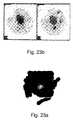
- FIGS. 23A and 23Bare illustrations of radiolabeled patterns observed in images produced by the system of the invention and by a conventional gamma camera, respectively, of an autonomous adenoma of a thyroid;
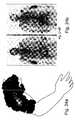
- FIGS. 24A and 24Bare illustrations of radiolabeled patterns observed in images produced by the system of the invention and by a conventional gamma camera, respectively, of suspected Paget's disease of a humerus;
- FIGS. 25A and 25Bare illustrations of radiolabeled patterns observed in images produced by the system of the invention and by a conventional gamma camera, respectively, of chronic osteomyelitis;
- FIGS. 26A and 26Bare illustrations of radiolabeled patterns observed in images produced by the system of the invention and by a conventional gamma camera, respectively, of skeletal metastasis from medulloblastoma.
- FIGS. 27 A-Gdemonstrate the operation of an algorithm provided by the present invention for estimating the distribution of radiation sources in a control volume.
- the present inventionis of a radioactive emission detector equipped with a position tracking system which can be functionally integrated with medical two- or three-dimensional imaging modalities and/or with guided minimally-invasive or other surgical tools.
- the present inventioncan be used for calculating the position of a concentrated radiopharmaceutical in the body in positional context of imaged portions of the body, which information can be used, for example, for performing an efficient and highly accurate minimally invasive surgical procedure.
- radioactive materialsto tag physiologically active tissue within the body of a patient, for determining the tissue's localization and demarcation by radioactive emission detectors has been disclosed in the medical literature for at least forty years. Significant developments in the localization and demarcation of tissue bearing radioactive isotope tags for diagnostic and/or therapeutic purposes have occurred since that time.
- Such radiopharmaceuticalstend to localize in particular tissue or cell type, whereas uptake or binding of the specific radiopharmaceutical is increased in more “physiologically active” tissue such as the active core of a cancerous tissue, so that the radiation emitted following nuclear disintegrations of the isotope can be detected by a radiation detector to better allocate the active portion of a tumor.
- Such radiationmay be, for example, ⁇ , ⁇ ⁇ , ⁇ + and/or ⁇ radiation.
- radioactive substancesare used to determine the level of flow of blood in blood vessels and the level of perfusion thereof into a tissue, e.g., coronary flow and myocardial perfusion.
- FIG. 1illustrates a system for calculating a position of a radioactivity emitting source in a system-of-coordinates, in accordance with the teachings of the present invention, which system is referred to hereinbelow as system 20 .
- System 20includes a radioactivity emission detector 22 .
- System 20according to the present invention further includes a position tracking system 24 .
- System 24is connected to and/or communicating with radioactive emission detector 22 so as to monitor the position of detector 22 in a two- or three-dimensional space defined by a system-of-coordinates 28 in two, three or more, say four, five or preferably six degrees-of-freedom (x, y, z, ⁇ , ⁇ and ⁇ ).
- System 20further includes a data processor 26 .
- Data processor 26is designed and configured for receiving data inputs from position tracking system 24 and from radioactive emission detector 22 and, as is further detailed below, for calculating the position of the radioactivity emitting source in system-of-coordinates 28 .
- system-of-coordinatesand “three-dimensional space” are used herein interchangeably.
- detectors 22are preferably connected there between via a connector 29 .
- Connector 29is preferably flexible. In the alternative, the connections of detectors 22 to connector 29 provide the required flexibility.
- Position tracking systemsper se are well known in the art and may use any one of a plurality of approaches for the determination of position in a two- or three-dimensional space as is defined by a system-of-coordinates in two, three and up to six degrees-of-freedom.
- Some position tracking systemsemploy movable physical connections and appropriate movement monitoring devices (e.g., potentiometers) to keep track of positional changes.
- movement monitoring devicese.g., potentiometers
- Such systemsonce zeroed, keep track of position changes to thereby determine actual positions at all times.
- One example for such a position tracking systemis an articulated arm.
- FIG. 2shows an articulated arm 30 which includes six arm members 32 and a base 34 , which can therefore provide positional data in six degrees-of-freedom.
- Monitoring positional changesmay be effected in any one of several different ways. For example, providing each arm member 32 with, e.g., potentiometers or optical encoders 38 used to monitor the angle between adjacent arm members 32 , to thereby monitor the angular change of each such arm member with respect to adjacent arm members, so as to determine the position in space of radioactive emission detector 22 , which is physically connected to articulated arm 30 .
- FIG. 3other position tracking systems can be attached directly to radioactive emission detector 22 in order to monitor its position in space.
- An example of such a position tracking systemis an assortment of three triaxially (e.g., co-orthogonally) oriented accelerometers 36 which may be used to monitor the positional changes of radioactive emission detector 22 with respect to a space.
- a pair of such assortmentsas is specifically shown in FIG. 3, can be used to determine the position of detector 22 in six-degrees of freedom.
- FIGS. 4 and 10other position tracking systems re-determine a position irrespective of previous positions, to keep track of positional changes.
- Such systemstypically employ an array of receivers/transmitters 40 which are spread in known positions in a three-dimensional space and transmitter(s)/receiver(s) 42 , respectively, which are in physical connection with the object whose position being monitored. Time based triangulation and/or phase shift triangulation are used in such cases to periodically determine the position of the monitored object, radioactive emission detector 22 in this case.
- Radioactive emission detectorsare well known in the art and may use any one of a number of approaches for the determination of the amount of radioactive emission emanating from an object or portion thereof. Depending on the type of radiation, such detectors typically include substances which when interacting with radioactive decay emitted particles emit either electrons or photons in a level which is proportional over a wide linear range of operation to the level of radiation impinging thereon. The emission of electrons or photons is measurable and therefore serves to quantitatively determine radiation levels.
- Solid-state detectors in the form of N-type, P-type, PIN-type pixellated or unpixellatedinclude, for example, Ge, Si, CdTe, CdZnTe, CdSe, CdZnSe, HgI 2 , TlBrI, GaAs, InI, GaSe, Diamond, TlBr, PbI 2 , InP, ZnTe, HgBrI, a-Si, a-Se, BP, GaP, CdS, SiC, AlSb, PbO, BiI 3 and ZnSe detectors.
- Gas (e.g., CO 2 CH 4 ) filled detectorsinclude ionization chamber detectors, proportional chamber detectors and geiger chamber detectors.
- Scintillation detectorsinclude organic scintillators crystals and liquids, such as C 14 H 10 , C 14 H 12 , C 10 H 8 , etc., Plastics, NE102A, NE104, NE 110, Pilot U and inorganic scintillators, such as NaI, CsI, BGO, LSO, YSO, BaF, ZnS, ZnO, CaWO 4 and CdWO 4 . Also known are scintillation fiber detectors.
- Scintillator couplinginclude photomultiplier tube (PMT) of the following types: side-on type, head-on type, hemispherical type, position sensitive type, icrochannel plate-photomultiplier (MCP-PMTs) and electron multipliers, or photodiodes (and photodiodes arrays), such as Si photodiodes, Si PIN photodiodes, Si APD, GaAs(P) photodiodes, GaP and CCD.
- PMTphotomultiplier tube
- MCP-PMTsicrochannel plate-photomultiplier
- electron multipliersor photodiodes (and photodiodes arrays), such as Si photodiodes, Si PIN photodiodes, Si APD, GaAs(P) photodiodes, GaP and CCD.
- FIG. 5shows a narrow angle or wide angle radioactive emission detector 22 ′.
- Narrow or wide angle radioactive emission detector 22 ′includes a narrow slit (collimator) so as to allow only radiation arriving from a predetermined angular direction (e.g., 1°-280°-wide angle, preferably 1°-80°-narrow angle) to enter the detector.
- Narrow or wide angle radioactive emission detectors especially suitable for the configuration shown in FIG. 10are manufactured, for example, by Neoprobe, Dublin, Ohio (www.neoprobe.com), USA, Nuclear Fields, USA (www.nufi.com) IntraMedical Imaging, Los Angeles, Calif., USA (www.gammaprobe.com).
- such a detectoris typically used to measure radioactivity, point by point, by scanning over the surface of a radioactive object from a plurality of directions and distances. In the example shown, scans from four different directions are employed. It will be appreciated that if sufficient radioactivity records are collected from different angles and distances, and the orientation and position in space of detector 22 ′ is simultaneously monitored and recorded during such scans, a three-dimensional model of a radioactive region can be reconstituted and its position in space determined. If two or more detectors are co-employed, as shown in the configuration of FIG. 10, the results may be collected faster.
- FIG. 7shows another example of a radioactive emission detector, a spatially sensitive (pixelated) radioactive emission detector 22 ′′ (such as a gamma camera).
- Detector 22 ′′in effect, includes an array of multitude narrow angle detector units 23 .
- Such an arrangementis used in accordance with the teachings of the present invention to reduce the amount of measurements and angles necessary to acquire sufficient data so as to reconstitute a three-dimensional model of the radioactive object.
- Examples of spatially sensitive radioactive emission detectors employed in a variety of contextsare disclosed in, for example, U.S. Pat. Nos. 4,019,057; 4,550,250; 4,831,262; and 5,521,373; which are incorporated by reference as if set forth herein.
- FIG. 8shows a scan optionally made by spatially sensitive radioactive emission detector 22 ′′ (such as a gamma camera).
- a radioactive emmission detector of particular advantages for use in context of the present inventionis the Compton gamma probe, since, in the Compton gamma probe, spatial resolution is independent of sensitivity and it appears possible to exceed the noise equivalent sensitivity of collimated imaging systems especially for systems with high spatial resolution.
- the Compton probeis a novel type of gamma-probe that makes use of the kinematics of Compton scattering to construct a source image without the aid of mechanical collimators.
- Compton imaging telescopeswere first built in the 1970s for astronomical observations [V. Schoenfelder et al., Astrophysical Journal 217 (1977) 306]. The first medical imaging laboratory instrument was proposed in the early 1980s [M. Singh, Med. Phys.
- the potential advantages of the Compton gamma probeinclude higher efficiency, 3-D imaging without detector motion, and more compact and lightweight system.
- high-energy gamma raysare scattered from a first detector layer (or detectors array) into a second detector layer array.
- the deposited energyis measured in both detectors.
- the Compton scattering equationcan be solved to determine the cone of possible direction about this axis on which the gamma ray must have entered the first detector. The intersection of cones from many events is then developed to locate gamma ray sources in the probe's field-of-view.
- the probe's electronic systemis combining coincidence measurements across many detectors and detectors layers with a very good energy resolution.
- the choice of the geometry and the material of the first layer detectorplays a major role in the system imaging capability and depends on (i) material efficiency of single Compton events, in relation to other interactions; (ii) detector energy resolution; and (iii) detector position resolution.
- the overall angular resolutionresults from the combination of two components, related to the energy resolution and to the pixel volume of the detector.
- the present inventionthus provides a method for defining a position of a radioactivity emitting source in a system-of-coordinates.
- the methodis effected by (a) providing a radioactive emission detector which is connected to or communicating with a position tracking system; and (b) monitoring radioactivity emitted from the radioactivity emitting source, while at the same time, monitoring the position of radioactive emission detector in the system-of-coordinates, thereby defining the position of the radioactivity emitting source in the system-of-coordinates.
- model produced by system 20is projectable onto any of the other systems-of-coordinates, or alternatively, the system-of-coordinates defined by position tracking system 24 may be shared by other position tracking systems, as is further detailed hereinbelow, such that no such projection is required.
- system 20 of the present inventioncan be used for calculating a position of a radioactivity emitting source in a first system-of-coordinates 28 and further for projecting the position of the radioactivity emitting source onto a second system-of-coordinates 28 ′.
- the systemincludes radioactive emission detector 22 , position tracking system 24 which is connected to and/or communicating with radioactive emission detector 22 , and data processor 26 which is designed and configured for (i) receiving data inputs from position tracking system 24 and from radioactive emission detector 22 ; (ii) calculating the position of the radioactivity emitting source in the first system-of-coordinates; and (iii) projecting the position of the radioactivity emitting source onto the second system-of-coordinates.
- a method for calculating a position of a radioactivity emitting source in a first system-of-coordinates and for projecting the position of the radioactivity emitting source onto a second system-of-coordinatesis also offered by the present invention.
- This methodis effected by (a) providing a radioactive emission detector being connected to or communicating with a position tracking system; and (b) monitoring radioactivity being emitted from the radioactivity emitting source, while at the same time, monitoring the position of the radioactive emission detector in the first system-of-coordinates, thereby defining the position of the radioactivity emitting source in the first system-of-coordinates and projecting the position of the radioactivity emitting source onto the second system-of-coordinates.
- a radioactive emission detector and a position tracking system connected thereto and/or communicating therewithallows a suitable data processor to generate a two- or three-dimensional image of the radioactivity emitting source.
- An algorithmcan be used to calculate image intensity based on, for example, a probability function which averages radiation counts and generates an image in which the shorter the time interval between radioactive counts, the brighter the image and vise versa, while down-compensating when a location is re-scanned.
- a free-hand scanning with a directional detectorcan be employed for this purpose.
- the detectorwhen scanning a body area with the detector, the detector is made to follow a three-dimensional surface which defines the body curvature and in effect is used also as a position tracking pointer.
- This informationcan be used to define the position of the radioactive source with respect to the outer surface of the body, so as to create a three-dimensional map of both the radioactive source and of the body curvature.
- This approachcan also be undertaken in open surgeries, such as open chest surgeries so as to provide the surgeon in real time with information concerning the functionality of a tissue.
- the radioactive emission detectorwhich can be used in context of the present invention can be a beta emission detector, a gamma emission detector, a positron emission detector or any combination thereof.
- a detector that is sensitive to both beta (and/or positron) and gamma emissioncan be used to improve localization by sensing for example gamma emission distant from the source and sensing beta or positrons emission closer to the source.
- a beta detectoris dedicated for the detection of either electrons from sources such as 131 Iodine, or positrons from sources such as 18 Fluorine.
- a gamma detectorcan be designed as a single energy detector or as a detector that can distinguish between different types of energies, using the light intensity in the scintilator as a relative measure of the gamma energy. Also, the detector can be designed to utilize coincidence detection by using detectors facing one another (180 degrees) with the examined organ or tissue in-between.
- the radiation detectorcan have different collimators with different diameters. A large bore will be used for high sensitivity with lower resolution while a small bore collimator will have higher resolution at the expense of lower sensitivity.
- Another possibilityis to have a the collimator moving or rotating with the opening eccentric so that a different solid angle is exposed to the incoming photons at any one time, thus gathering the photons from overlapping volumes at different time intervals.
- the rest of the image processingis similar if the probe moves or if the collimator eccentric opening moves.
- System 20 of the present inventioncan be used in concert with other medical devices, such as, but not limited to, any one of a variety of imaging modalities and/or surgical instruments.
- Imaging modalitiesare well known in the art, the main modalities that serve for two- (projectional or cross sectional) or three- (cosequtive cross sectional) dimensional imaging are a fluoroscope, a computerized tomography scanner, a magnetic resonance imager an ultrasound imager and an optical camera.
- Medical images taken of the human bodyare typically acquired or displayed in three main orientations (i) coronal orientation: in a cross section (plane), for example, across the shoulders, dividing the body into front and back halves; (ii) sagittal orientation: in a cross section (plane), for example, down the middle, dividing the body into left and right halves; and (iii) axial orientation: in a cross section (plane), perpendicular to the long axis of the body, dividing the body into upper and lower halves.
- Oblique viewscan also be acquired and displayed.
- X-ray imagingresults in a projection of the integrated density of column-voxels defined by the X-rays as they pass through the body.
- Fluoroscopyis a method based on the principals of film X-ray that is useful for detecting disorders and tumors in the upper gastro-intestinal (GI) system (for example, the stomach and intestines). Fluoroscopic imaging yields a moving X-ray picture. The physician can watch the screen and see an image of the patient's body (for example the beating heart). Fluoroscopic technology improved greatly with the addition of television cameras and fluoroscopic “image intensifiers”. Today, many conventional X-ray systems have the ability to switch back and forth between the radiographic and fluoroscopic modes. The latest X-ray systems have the ability to acquire the radiograph or fluoroscopic movie using digital acquisition.
- GIgastro-intestinal
- Computed Tomographyis based on the X-ray principal, where the film is replaced by a detector that measures the X-ray profile.
- CTComputed Tomography
- Inside the covers of the CT scanneris a rotating frame which has an X-ray tube mounted on one side and the detector mounted on the opposite side.
- a fan beam of X-rayis created as the rotating frame spins the X-ray tube and detector around the patient.
- an image or “slice”has been acquired. This “slice” is collimated to a thickness between 1 mm and 10 mm using lead shutters in front of the X-ray tube and X-ray detector.
- the detectortakes numerous profiles of the attenuated X-ray beam. Typically, in one 360° lap, about 1,000 profiles are sampled. Each profile is subdivided spatially by the detectors and fed into about 700 individual channels. Each profile is then backwards reconstructed (or “back projected”) by a dedicated computer into a two-dimensional image of the “slice” that was scanned.
- the CT gantry and tablehave multiple microprocessors that control the rotation of the gantry, movement of the table (up/down and in/out), tilting of the gantry for angled images, and other functions such as turning the X-ray beam on an off.
- the CTcontains a slip ring that allows electric power to be transferred from a stationary power source onto the continuously rotating gantry.
- the innovation of the power slip ringhas created a renaissance in CT called spiral or helical scanning.
- spiral CTacquires a volume of data with the patient anatomy all in one position. This volume data set can then be computer-reconstructed to provide three-dimensional models such as of complex blood vessels like the renal arteries or aorta. Spiral CT allows the acquisition of CT data that is perfectly suited for three-dimensional reconstruction.
- MR Imagingis superior to CT in detecting soft tissue lesions such as tumors as it has excellent contrast resolution, meaning it can show subtle soft-tissue changes with exceptional clarity. Thus, MR is often the method of choice for diagnosing tumors and for searching for metastases.
- MRuses magnetic energy and radio waves to create single or consequtive cross-sectional images or “slices” of the human body.
- the main component of most MR systemsis a large tube shaped or cylindrical magnet.
- the strength of the MR systems magnetic fieldis measured in metric units called “Tesla”. Most of the cylindrical magnets have a strength between 0.5 and 1.5 Tesla and most of the open or C-shaped magnets have a magnetic strength between 0.01 and 0.35 Tesla.
- Each total MR examinationtypically is comprised of a series of 2 to 6 sequences.
- An “MR sequence”is an acquisition of data that yields a specific image orientation and a specific type of image appearance or “contrast”.
- MR sequenceis an acquisition of data that yields a specific image orientation and a specific type of image appearance or “contrast”.
- a radio signalis turned on and off, and subsequently the energy which is absorbed by different atoms in the body is echoed or reflected back out of the body.
- These echoesare continuously measured by “gradient coils” that are switched on and off to measure the MR signal reflecting back.
- the net magnetization vectorrotate from a longitudinal position a distance proportional to the time length of the radio frequency pulse.
- Radio frequency coilsare the “antenna” of the MRI system that broadcasts the RF signal to the patient and/or receives the return signal.
- RF coilscan be receive-only, in which case the body coil is used as a transmitter; or transmit and receive (transceiver).
- Surface coilsare the simplest design of coil. They are simply a loop of wire, either circular or rectangular, that is placed over the region of interest.
- a digital computerreconstructs these echoes into images of the body.
- a benefit of MRIis that it can easily acquire direct views of the body in almost any orientation, while CT scanners typically acquire cross-sectional images perpendicular or nearly perpendicular to the long body axis.
- Ultrasound imagingis a versatile scanning technique that uses sound waves to create images of organs or anatomical structures in order to make a diagnosis.
- the ultrasound processinvolves placing a small device called a transducer, against the skin of the patient near the region of interest, for example, against the back to image the kidneys.
- the ultrasound transducercombines functions of emitting and receiving sound. This transducer produces a stream of inaudible, high frequency sound waves which penetrate into the body and echo off the organs inside.
- the transducerdetects sound waves as they echo back from the internal structures and contours of the organs. Different tissues reflect these sound waves differently, causing a signature which can be measured and transformed into an image.
- These wavesare received by the ultrasound machine and turned into live pictures with the use of computers and reconstruction software.
- Ultrasound scanninghas many uses, including: diagnosis of disease and structural abnormalities, helping to conduct other diagnostic procedures, such as needle biopsies etc.
- imaging modalitieseither inherently include (e.g., fluoroscope, CT, MRI) and/or are integrated with position-tracking-systems, which enable the use of such systems to reconstruct three-dimensional image models and provide their position in a three-dimensional space.
- fluoroscopee.g., fluoroscope, CT, MRI
- position-tracking-systemswhich enable the use of such systems to reconstruct three-dimensional image models and provide their position in a three-dimensional space.
- an optical cameracan be used to generate three-dimensional imagery date according to the present invention by imaging a body from a plurality (at least two) directions. This type of imaging is especially applicable in open chest surgeries or other open surgeries. Software for calculating a three-dimensional image from a pair of stereoscopic images is well known in the art.
- three-dimensional imaging modalityrefers to any type of imaging equipment which includes software and hardware for generating a three-dimensional image.
- Such an equipmentcan generate a three-dimensional image by imaging successive cross-sections of a body, e.g., as if viewed from a single direction.
- such an equipmentcan generate a three-dimensional image by imaging a body from different angles or directions (typically two angles) and thereafter combining the data into a three-dimensional image.
- Surgical instrumentsare also well known in the art and may use any one of a plurality of configurations in order to perform minimally-invasive surgical procedures. Examples include laser probes, cardiac and angioplastic catheters, endoscopic probes, biopsy needles, aspiration tubes or needles, resecting devices, ultrasonic probes, fiber optic scopes, laparoscopy probes, thermal probes and suction/irrigation probes. Examples of such surgical instruments employed in a variety of medical contexts are disclosed in, for example, U.S. Pat. Nos.
- surgical instrumentsare integrated with position-tracking-systems, which enable to monitor the position of such instruments while placed in and guided through the body of a treated patient.
- the surgical instrumentis equipped with an additional radioactive emission detector attached thereto or placed therein.
- This additional detectoris used, according to preferred embodiments of the invention, to fine tune the location of radioactive emission from within the body, and in closer proximity to the radioactive source. Since the surgical tool is preferably connected to or communicating with a position-tracking system, the position of the additional detector can be monitored and its readouts used to fine tune the position of the radioactive source within the body.
- at least one extracorporeal detector and an intracorporeal detectorare used in concert to determine the position of a radioactive source in the body in highest precision.
- the extracorporeal detectorprovides the general position of the source and is used for directing the surgical instrument thereto, whereas the intracorporeal detector is used for reassuring prior to application of treatment or retrieval of biopsy that indeed the source was correctly targeted at the highest precision.
- intracorporeal and extracorporeal detectorscalls for careful choice of the radioactive isotope employed with the radiopharmaceutical. While the extracorporeal detector can be constructed with a suitable collimator for handling strong radiation, such as gamma radiation, the intracorporeal detector is miniature by nature and is limited in design and construction by the construction of the surgical instrument with which it is employed. Since collimators for high energy (80-511 KeV) gamma radiation are robust in nature, they are not readily engageable with miniature detectors.
- Electron (beta) and positron radiationare characterized by: (i) they highly absorbed by biological tissue as they are of lower energy and higher chemical reactivity; and (ii) they are readily collimated and focused by thin metal collimators. It is also possible to use low energy gamma radiation (10-30 KeV) for intracorporal applications since the collimation of these gamma photons can be achieved with thin layers of Tantalum or Tungsten. As such, the radio pharmaceutical of choice is selected to emit both gamma and beta and/or positron radiation, whereas the extracorporeal detector is set to detect the high energy gamma radiation, whereas the intracorporeal detector is set to detect the low energy gamma, beta and/or positron radiation.
- Isotopes that emit both high energy gamma and/or low energy gamma, beta and/or positron radiation and which can be used per se or as a part of a compound as radiopharmaceuticalsinclude, without limitation, 18 F, 111 In and 123 I in radiopharmaceuticals, such as, but not limited to, 2-[ 18 F]fluoro-2-deoxy-D-glucose ( 18 FDG), 111 In-Pentetreotide ([ 111 In-DTPA-DPhe 1 ]-octreotide), L-3-[ 123 I]-Iodo-alpha methyl-tyrosine (IMT), O-(2-[ 18 F]fluoroethyl)-L-tyrosine (L-[ 18 F]FET), 111 In-Capromab Pendetide (CYT-356, Prostascint) and 111 In-Satumomab Pendetide (Oncoscint).
- FIG. 11illustrates a system in accordance with this aspect of the present invention.
- a surgical instrument 100is shown connected to a resection/aspiration control element 102 as well known in the art.
- Surgical instrument 100includes a radioactive emission detector 104 , which has a collimator 106 for collimating low energy gamma, beta and/or positron radiation.
- detector 104may be translated within instrument 100 .
- a position tracking systemhaving one element thereof 110 attached to instrument 100 and another element thereof 112 at a fixed location serves to monitor the position of instrument 100 at all times in two, three and up to six degrees of freedom.
- Radioactive emission detector 104communicates with a counter 114 for counting low energy gamma, beta and/or positron radiation. All the data is communicated to, and processed by, a processor 116 .
- the 2D or 3D datamay be projected and displayed along with 2D or 3D imaging data derived from an imaging modality using a shared presentation device as described elsewhere herein. A real or virtual image of the surgical instrument itself may also be co-displayed.
- Examples of commercially available radiation emission detectors that can fit inside, for example, a biopsy needleinclude scintillating plastic optical fibers like S101 and S104, manufactured by PPLASTIFO or an optical fiber communicating with a scintillator (either detector paint or scintillation crystal) at the fiber edge.
- the level of detected radiationcan be reported visually or by an audio signal, as is well known in the art.
- a surgical instrument equipped with a radiation emission detector and which is connected to and/or communicating with a position tracking systemforms one embodiment of this aspect of the present invention.
- Such a design acting in concert with either conventional imaging modalities and/or extracorporeal radiation emission detectorsform other embodiments of this aspect of the invention.
- a surgical instrument equipped with a radiation emission detector and which is connected to and/or communicating with a position tracking systemserves for in situ fine tuning of a radioactive source in the body.
- FIG. 9An example to this desired outcome is shown in FIG. 9.
- four independent position tracking systems 50 , 52 , 54 and 56are used to track the positions of a patient 58 , an imaging modality 60 , a radioactive emission detector 62 and a surgical instrument 64 in four independent systems-of-coordinates 66 , 68 , 70 and 72 , respectively. If the patient is still, no tracking of the patient's position is required.
- any subset or all of the position tracking systems employedmay be integrated into one or more common position tracking systems, and/or that any subset or all of the position tracking systems employed may share one or more systems-of-coordinates, and further that any positional data obtained by any of the position tracking systems described in any of the systems-of coordinates may be projected to any other system of coordinates or to an independent (fifth) system of coordinates 74 .
- the system of coordinatesis a dynamic system of coordinates which takes into account the chest breathing movements of the patient during the procedure.
- the raw data collected by detector 62is recorded and, as indicated at 78 , the position and the radioactive data records are used to generate a three-dimensional model of a radiopharmaceutical uptaking portion of a body component of the patient.
- imagery data collected by imaging modality 60is recorded and the position and the imagery data records are used to generate a three-dimensional model of the imaged body component of the patient.
- All the data collectedis then fed into a data processor 82 which processes the data and, as indicated at 84 , generates a combined or superimposed presentation of the radioactive data and the imagery data, which is in positional context with patient 58 and surgical instrument 64 .
- Instrument 64which by itself can be presented in context of the combined presentation, may then be used to perform the procedure most accurately.
- Processor 82may be a single entity or may include a plurality of data processing stations which directly communicate with, or even integral to, any one or more of the devices described.
- the present inventionprovides a major advantage over prior art designs because it positionally integrates data pertaining to a body portion as retrieved by two independent imaging techniques, conventional imaging and radioactive imaging, to thereby provide a surgeon with the ability the fine point the portion of the body to be sampled or treated.
- subsets of the devices described in FIG. 9may be used as stand-alone systems.
- a combination of detector 62 with its position-tracking system and instrument 64 with its position-tracking-systemmay in some instances be sufficient to perform intrabody procedures.
- a combination of detector 62 position-tracking-system and modality 60 position-tracking-systemare sufficient.
- Imaging system 200preferably includes a radiation probe 202 , such as the narrow angle radioactive emission detector 22 ′ described hereinabove with reference to FIGS. 5 and 10.
- a position sensor 204is provided for sensing the position of radiation probe 202 .
- Position sensor 204may be physically attached to radiation probe 202 , or may be distanced therefrom.
- Position sensor 204transmits the sensed position data to a position tracking system 206 .
- Position tracking system 206may be a system like position tracking system 24 , described hereinabove with reference to FIG. 1, and position sensor 204 may be any kind of sensor applicable for such position tracking systems.
- Another method which can be used to locate the source of radiation emissionis by using a small hand held gamma camera 205 (such as the DigiRad 2020tc Imager TM, 9350 Trade Place, San Diego, Calif. 92126-6334, USA), attached to position sensor 204 .
- a small hand held gamma camera 205such as the DigiRad 2020tc Imager TM, 9350 Trade Place, San Diego, Calif. 92126-6334, USA
- Position tracking system 206enables radiation probe 202 to freely scan back and forth in two- or three-dimensions over the area of interest of the patient, preferably incrementing a short distance between each scan pass. Position tracking system 206 tracks the position of radiation probe 202 with respect to a position tracking coordinate system, such as X p , Y p and Z p, with an origin O p .
- Imaging system 200also includes a medical imaging system 208 , such as, but not limited to, computed or computerized tomography (CT), magnetic resonance imaging (MRI), ultrasound imaging, positron emission tomography (PET) and single photon emission computed tomography (SPECT), for example.
- Medical imaging system 208provides images of a patient 209 with respect to a medical imaging coordinate system, such as X m , Y m and Z m, with an origin O m .
- Imaging system 200also includes a coordinate registration system 210 , such as that described in U.S. patent application Ser. No. 09/610,490, the disclosure of which is incorporated herein by reference.
- Coordinate registration system 210is adapted to register the coordinates of the position tracking coordinate system with those of the medical imaging coordinate system.
- Position tracking system 206 , medical imaging system 208 and coordinate registration system 210are preferably in wired or wireless communication with a processing unit 212 (also referred to as a data processor 212 ).
- a clinician/physician/surgeonmay move or scan radiation probe 202 about a target area under examination.
- a physiological activity map of the target areais obtained by measuring the radiation count rate with radiation probe 202 , and by correlating the count rate with the count rate direction with position tracking system 206 , which follows the motion of the moving or scanning radiation probe 202 .
- FIG. 13illustrates image formation with radiation probe 202 , in accordance with a preferred embodiment of the present invention.
- the example shown in FIG. 13is for a single dimension image formation, but it is readily understood that the same principles hold true for any other dimensional image formation.
- radiation probe 202may be a gamma ray detector probe that comprises a collimator 211 and radiation detector 213 .
- Gamma raysare projected through the probe collimator 211 onto radiation detector 213 , which produces electronic signals in accordance with the radiation detected.
- Radiation probe 202sends pulses to a probe counter 215 which may include a pulse height analyzer circuit (not shown).
- the pulse height analyzer circuitanalyzes the electronic signals produced by radiation detector 213 . If the electronic signals are within a selected energy window, the level of radiation, i.e., number of radiation counts, is counted by probe counter 215 .
- suitable radiation detectorsinclude a solid state detector (SSD) (CdZnTe, CdTe, HgI, Si, Ge, and the like), a scintillation detector (NaI(Tl), LSO, GSO, CsI, CaF, and the like), a gas detector, or a scintillating fiber detector (S101, S104, and the like), for example.
- SSDsolid state detector
- CaI(Tl)CdZnTe, CdTe, HgI, Si, Ge, and the like
- NaI(Tl)scintillation detector
- LSOLSO
- GSOGSO
- CsICsI
- CaFgas detector
- S101, S104, and the likescintillating fiber detector
- the position sensor 204 associated with the radiation probe 202senses the position of radiation probe 202
- position tracking system 206calculates and monitors the motion of radiation probe 202 with respect to the position tracking coordinate system.
- the motionis calculated and monitored in two, three and up to six dimensions—the linear directions of the X, Y and Z axes as well as rotations about the X, Y and Z axes (i.e., rotational angles ⁇ , ⁇ and ⁇ , respectively).
- Suitable position tracking systemsinclude a measurement mechanical arm (FaroArm, http://www.faro.com/products/faroarm.asp), optical tracking systems (Northern Digital Inc., Ontario, Canada NDI-POLARIS passive or active systems), magnetic tracking systems (NDI-AURORA), infrared tracking systems (E-PEN system, http://www.e-pen.com), and ultrasonic tracking systems (E-PEN system), for example.
- FaroArmhttp://www.faro.com/products/faroarm.asp
- optical tracking systemsNorthern Digital Inc., Ontario, Canada
- NDI-AURORAmagnetic tracking systems
- E-PEN systemhttp://www.e-pen.com
- ultrasonic tracking systemsE-PEN system
- Processing unit 212combines the radiation probe count rate from probe counter 215 together with the positional information from position tracking system 206 , and uses an imaging software algorithm 217 to form a two-dimensional or three-dimensional radiotracer-spread image of the target area inside the patient's body.
- the spatial probe positions together with the spatial count ratesmay be stored in memory or displayed on a computer monitor 214 as a pattern of marks corresponding to the spatial and count rate position.
- FIG. 14illustrates a single-dimensional, unprocessed simulation of a radiation point source 218 (FIG. 13), 30 mm deep inside the human body, detected by using a 10 mm nuclear radiation probe 202 coupled to position tracking system 206 .
- the graph of FIG. 14indicates to a physician that there is a peak count rate of about 500 in the probe position of about 50 mm.
- the imaging software algorithm 217employs an averaging process to refine the curve of FIG. 14. This averaging process will now be described with reference to FIG. 15.
- Probe counter 215feeds probe count rate information N(Xc, Yc, Zc, ⁇ , ⁇ , ⁇ ) to processing unit 212 (step 301 ).
- Position sensor 204feeds probe position information (Xc, Yc, Zc, ⁇ , ⁇ , ⁇ ) to processing unit 212 (step 302 ).
- Probe parameters(such as its physical size, dx, dy, dz) are also input into processing unit 212 (step 303 ).
- Processing unit 212finds all the voxels (i.e., volume pixels) that represent the probe volume in the processing unit memory (step 304 ), i.e., Xc+dx, Yc+dy, Zc+dz. Processing unit 212 calculates the number of times that the calculation process has been done in each voxel from the beginning of the image formation (step 305 ), i.e., M(Xc+dx, Yc+dy, Zc+dz).
- Processing unit 212then corrects the display image that represents the perceived voxels at N(Xc+dx, Yc+dy, Zc+dz) (step 307 ). The algorithm then repeats itself for the next probe position (step 308 ).
- FIGS. 17 and 18respectively show examples of a hot cross phantom image and a hot 4.77 mm bar phantom image, produced by a gamma radiation probe coupled with position tracking system 206 and the averaging algorithm of FIG. 15.
- the position tracking system usedwas the Ascension miniBIRD, commercially available from Ascension Technology Corporation, P.O. Box 527, Burlington, Vt. 05402 USA (http://www.ascension-tech.com/graphic.htm).
- the magnetic tracking and location systems of Ascension Technology Corporationuse DC magnetic fields to overcome blocking and distortion from nearby conductive metals. Signals pass through the human body without attenuation.
- the imaging software algorithm 217may employ a minimizing process to refine the curve of FIG. 14 as is now described with reference to FIG. 19.
- Probe counter 215feeds probe count rate information N(Xc, Yc, Zc, ⁇ , ⁇ , ⁇ ) to processing unit 212 (step 401 ).
- Position sensor 204feeds probe position information (Xc, Yc, Zc, ⁇ , ⁇ , ⁇ ) to processing unit 212 (step 402 ).
- Probe parameters(such as its physical size, dx, dy, dz) are also input into processing unit 212 (step 403 ).
- Processing unit 212finds all the voxels that represent the probe volume in the processing unit memory (step 404 ), i.e., Xc+dx, Yc+dy, Zc+dz. From the voxels that represent the probe volume in the processing unit memory, processing unit 212 finds those that have a higher count rate value N(Xc+dx, Yc+dy, Zc+dz) than the inputted probe count rate N(Xc, Yc, Zc, ⁇ , ⁇ , ⁇ ) (step 405 ).
- Processing unit 212then changes the higher count rate voxels to that of inputted probe count rate N(Xc, Yc, Zc, ⁇ , ⁇ , ⁇ ) (step 406 ), and corrects the display image at the higher count rate voxels N(Xc+dx, Yc+dy, Zc+dz) (step 407 ). The algorithm then repeats itself for the next probe position (step 408 ).
- Another algorithmis provided by the invention for estimating the distribution of radiation sources in a control volume, and is described with reference to FIGS. 27 A- 27 G.
- the radiation sourcescomprise dot sources that radiate uniformly in all directions, and that the radiation sources are localized and smoothly distributed in a bounded volume.
- FIGS. 27A and 27Billustrate a radiation sensor 600 , preferably generally shaped as a tube collimator.
- Radiation quanta 602are registered by the radiation sensor 600 , as described hereinabove, thereby providing the average number of quanta per unit time.
- the radiation sensor 600may be moved around a volume of interest 604 .
- the position of the sensor 600 and its direction (as well as the position of the investigated volume 604 )are assumed to be known at any given moment (FIG. 27A).
- the tube collimatoris preferably provided with a plane circular detector 606 of radiation quanta.
- the quanta detector 606is preferably disposed on a rear end 608 of the tube and radiation quanta can reach the detector 606 only through an open front end 610 of the tube (FIG. 27B)
- FIG. 27Cillustrates a system of coordinates (x, y, z) with the origin O in the center of the radiation sensor 600 , the (x, y) plane being the plane of the detector, and the z axis being in the center of the collimator tube.
- the radiation sourcesare distributed in a rectangular region V in a plane. Two systems of coordinates are considered. The first one is the sensor coordinate system (x, y, z) corresponding to the sensor 600 . The second one is the radiation source coordinate system (u, v, w) corresponding to the radiation sources plane (u, v).
- the radiation sourcesare considered to be distributed in accordance with the distribution function I(Q)) in some bounded, given rectangle V on the plane (u, v).
- the function I(Q)will be considered to be given from some finite dimensional space H of functions defined in V. In other words, the function I(Q) itself will not be estimated but rather some finite dimensional approximation of the distribution I(Q).
- the space His naturally isomorphic to the m-dimensional space of n ⁇ n matrixes (with its natural scalar product ⁇ •, •>).
- ⁇ kare results of measurements of the unknown element I of the space H
- ⁇ 1 , ⁇ 2 , . . . , ⁇ mbe eigenvectors of operator M:H ⁇ H corresponding to eigenvalues ⁇ 1 ⁇ 2 ⁇ . . . ⁇ m ⁇ 0.
- Rbe some natural number, 1 ⁇ R ⁇ m (R is the “regularization parameter”).
- H (R)be the subspace of the space H spanned by the first R eigenvectors ⁇ 1 , . . . , ⁇ R .
- the regularized estimate Î Rmay be obtained as follows:
- an irregular subdivision of the rectangle Vis used. This irregular approach may significantly decrease the dimension of the problem and facilitate computer calculations.
- FIG. 27Gillustrates an advantageous irregular cell subdivision, in accordance with a preferred embodiment of the present invention.
- the large cellswhich have an intensity larger than some threshold, are subdivided into 4 equal subcells (or 8 subcells in the 3D case).
- a suitable thresholdmay be obtained by taking the average intensity (of all large cells) minus two (or three) sigmas (standard deviation), for example. Measurements and estimations are made in these subdivisions as described hereinabove.
- the 3D problemmay be treated in the same way as the 2D case, the only difference being that instead of rectangle V, there is a parallelepiped V (FIG. 27D). Accordingly, the cells in each subdivision are also parallelepipeds.
- the algorithms described hereinabovemay be used for a variety of imaging systems.
- the algorithmsmay be used with single radiation detector probe, an array of radiation detector probes, large gamma cameras of various design, such as multi head cameras, general purpose cameras, and automatic white balance (AWB) scanners.
- the algorithmsare suitable for SPECT and planar imaging, and may be used for all types of isotopes of with any type of photon energy.
- the algorithmsthus comprise a feedback system that employs analysis to determine the bounds of an uncertainty region about the radiation source, and which guides medical personnel to conduct additional scans in these uncertainty regions to improve accuracy, reduce error, and hence minimize the bounds of the uncertainty regions.
- Continuous sampling with radiation probe 202may provide localization of a tumor and a physiological radiation activity map of the tumor region. Higher safety and accuracy are gained by a greater number of scans.
- FIG. 21illustrates an image reconstruction system 450 , constructed and operative in accordance with a preferred embodiment of the present invention.
- Image reconstruction system 450produces a combined image 451 made up of the images coming from the medical imaging system 208 with the position of the peak radiation location (and its uncertainty area) from processing unit 212 , together with the location of a therapeutic instrument 452 , such as a biopsy needle.
- the combined image 451allows the physician to better assess the relative position of therapeutic instrument 452 in relation to the anatomical image (from medical imaging system 208 ) and the position of the radioactive area as inferred by the radiation detection algorithm.
- FIG. 22illustrates a flow chart of a radiation map reconstruction algorithm, in accordance with a preferred embodiment of the present invention.
- Deconvolution methodsare often used in image processing procedures. Examples of such deconvolution methods are described in U.S. Pat. No. 6,166,853 to Sapia et al., the disclosure of which is incorporated herein by reference. (However, it is appreciated that these are just examples and the present invention is not limited to the deconvolution methods mentioned in U.S. Pat. No. 6,166,853)
- a system transfer-functionmay be generally obtained directly by taking the Fourier transform of the aperture.
- the blurring effects due to convolutiongenerally exist in two-dimensions only, i.e., the x-y planes.
- a point-spread-functionis an expression used to describe the convolutional blurring in two-dimensions. The PSF physically results from imaging a point source.
- the Fourier transform of the PSFis the system transfer-function, which is obtained by convolving the system transfer-function with a Dirac-delta function.
- a point sourceis the physical equivalent of a Dirac-delta function, and, in the frequency domain, the Dirac-delta function is a unity operator across the spectrum. Therefore, the Fourier transform of the PSF should be the Fourier transform of the aperture.
- the PSFcontains noise and blurring due to other effects such as aberrations.
- the PSF contribution to the overall blurrinessmay be diminished or eliminated by deconvolution.
- the transfer function of the radiation detectormay be determined by taking the Fourier transform of the aperture of the detector, and taking into account the noise and blurring due to other effects such as aberrations (step 500 ).
- An example of a transfer functionmay be a normal distribution.
- the deconvolution of the transfer functionmay be determined (step 502 ).
- the count readings of each spatial location of the detectorconstitute the sum of radiation counts from all the voxels (or pixels in the case of two-dimensional maps, the term “voxel” being used herein to include both pixels and voxels) within the detector's field of view. At least one voxel, or preferably each such voxel, may be assigned a count value based on the deconvolution of the unique transfer function of the radiation detector in use (step 504 ). An additional mathematical procedure may treat the various values that each voxel receives due to the multiple readings from viewpoints of different detectors (step 506 ).
- This treatmentmay constitute for example a simple algebraic average, minimum value or reciprocal of averaged reciprocals in order to produce a single value of readings in each voxel.
- the deconvolutionis then used to reconstruct the voxels of the radiation map with diminished or no blurriness (step 508 ).
- the algorithms described hereinare applicable not only to the analysis of readings obtained using a directional radioactivity detector, rather they also apply for spatially sensitive (pixelated) radioactivity detectors.
- the readings of each pixelare algorithmically treated as described herein like for a directional radioactivity detector.
- the motivation behind using a spatially sensitive detectoris to save on measurement time by receiving readings from a multitude of directions in parallel. This, in essence, creates a number of overlapping low resolution images which can then be processed to form a high resolution image.
- the spatially sensitive detectorcan be scanned to improve even further the resolution using the algorithms described hereinabove.
- each pixelcan be seen as a separate detector with an angle of acceptance dictated by the geometry of a segmented collimator employed thereby.
- Each of the pixelsoccupies a different position in space and hence can be seen as a new position of a single directional probe by the algorithm described herein. It is also possible, like with the directional detector, to scan the whole set of pixels by scanning the spatially sensitive detector and to acquire a new set of data points from the new position.
- a super resolution algorithmcan be employed to generate an image of higher resolution.
- Suitable super resolution algorithmsare described in, for example, J. Acoust. Soc. Am., Vol. 77, No. 2, February 1985 Pages 567 - 572; Yokota and Sato, IEEE Trans. Acoust. Speech Signal Process. (April 1984); Yokota and Sato, Acoustical Imaging (Plenum, New York, 1982, Vol. 12; H. Shekarforoush and R. Chellappa, “Data-Driven Multi-channel Super-resolution with Application to Video Sequences”, Journal of Optical Society of America-A, vol. 16, no. 3, pp. 481-492, 1999; H.
- FIGS. 23A and 23Billustrate radiolabeled patterns observed in images produced by the system of the invention and by a conventional gamma camera, respectively, of an autonomous adenoma of a thyroid of a 58 year-old male.
- FIGS. 24A and 24Billustrate radiolabeled patterns observed in images produced by the system of the invention and by a conventional gamma camera, respectively, of suspected Paget's disease of a humerus in an 89 year-old female.
- FIGS. 25A and 25Billustrate radiolabeled patterns observed in images produced by the system of the invention and by a conventional gamma camera, respectively, of chronic osteomyelitis in a 19 year-old female.
- FIGS. 26A and 26Billustrate radiolabeled patterns observed in images produced by the system of the invention and by a conventional gamma camera, respectively, of skeletal metastasis from medulloblastoma in an 18 year-old male.
- the system and method of the present inventioncan find uses for screening for cancer and/or directing invasive diagnosis (biopsies) either from outside the body or by way of endoscopic approach.
- biopsiesinclude, but are not limited to, lung cancer biopsy, breast cancer biopsy, prostate cancer biopsy, cervical cancer biopsy, liver cancer biopsy, lymph node cancer biopsy, thyroid cancer biopsy, brain cancer biopsy, bone cancer biopsy, colon cancer biopsy, gastro intestine cancer endoscopy and biopsy, endoscopic screening for vaginal cancer, endoscopic screening for prostate cancer (by way of the rectum), endoscopic screening for ovarian cancer (by way of the vagina), endoscopic screening for cervical cancer (by way of the vagina), endoscopic screening for bladder cancer (by way of the urinary track), endoscopic screening for bile cancer (by way of the gastrointestinal track), screening for lung cancer, screening for breast cancer, screening for melanoma, screening for brain cancer, screening for lymph cancer, screening for kidney cancer, screening for gastro intestinal cancer (from the outside).
- the radiation detectorcan be combined and packaged together with a small RF coil for the transmission and reception or reception only of the MRI signals in a rectal probe configuration for prostate diagnosis and treatment or any other close confinement position such as the vagina, airways, the uper portion of the gastrointestinal track, etc)
- Procedures known as directing localized treatment of cancercan also benefit from the system and method of the present invention.
- Examplesinclude, but are not limited to, intra tumoral chemotherapy, intra tumoral brachytherapy, intra tumoral cryogenic ablation, intra tumoral radio frequency ablation, intra tumoral ultrasound ablation, and intra tumoral laser ablation, in cases of, for example, lung cancer, breast cancer, prostate cancer, cervical cancer, liver cancer, lymph cancer, thyroid cancer, brain cancer, bone cancer, colon cancer (by way of endoscopy through the rectum), gastric cancer (by way of endoscopy through the thorax), thoracic cancer, small intestine cancer (by way of endoscopy through the rectum or, by way of endoscopy through the thorax), bladder cancer, kidney cancer, vaginal cancer and ovarian cancer.
- the following procedurescan take advantage of the present invention wherein the method and system can be used to assess tissue perfusion, tissue viability and blood flow intra operatively during PTCA procedure (balloon alone or in conjunction with the placement of a stent), in cases of cardiogenic shock to asses damage to the heart, following myocardial infarct to asses damage to the heart, in assessing heart failure condition tissue in terms of tissue viability and tissue perfusion, in intra vascular tissue viability and perfusion assessment prior to CABG operation.
- the radioactivity detectorcan be mounted on a catheter that is entered through the blood vessels to the heart to evaluate ischemia from within the heart in order to guide ablation probes or another type of treatment to the appropriate location within the heart.
- Another application which may benefit from the present inventionis the localization of blood clots.
- a radioactivity detector as described hereincan be used to asses and differentiate between new clots and old clots.
- the radioactivity detectorcan be placed on a very small caliber wire such as a guide wire that is used during PTCA in order to image intrabloodvessel clots. Intrabloodvessel clots can be searched for in the aortic arc as clots therein are responsible for about 75% of stroke cases.
- tissue viability and blood flow intra operativelycan also be employed in the following: during CABG operation to asses tissue viability, to mark infarct areas, during CABG operations to asses the success of the re vascularization.
- the present inventionhas many other applications in the direction of therapeutics, such as, but not limited to, implanting brachytherapy seeds, ultrasound microwave radio-frequency cryotherapy and localized radiation ablations.
Landscapes
- Health & Medical Sciences (AREA)
- Life Sciences & Earth Sciences (AREA)
- Engineering & Computer Science (AREA)
- Medical Informatics (AREA)
- Physics & Mathematics (AREA)
- General Health & Medical Sciences (AREA)
- Molecular Biology (AREA)
- Biomedical Technology (AREA)
- Animal Behavior & Ethology (AREA)
- Public Health (AREA)
- Veterinary Medicine (AREA)
- Heart & Thoracic Surgery (AREA)
- Pathology (AREA)
- Surgery (AREA)
- Biophysics (AREA)
- Nuclear Medicine, Radiotherapy & Molecular Imaging (AREA)
- High Energy & Nuclear Physics (AREA)
- Optics & Photonics (AREA)
- Radiology & Medical Imaging (AREA)
- Vascular Medicine (AREA)
- Human Computer Interaction (AREA)
- Immunology (AREA)
- Computer Vision & Pattern Recognition (AREA)
- Dentistry (AREA)
- Endocrinology (AREA)
- Spectroscopy & Molecular Physics (AREA)
- Oral & Maxillofacial Surgery (AREA)
- General Physics & Mathematics (AREA)
- Chemical Kinetics & Catalysis (AREA)
- Medicinal Chemistry (AREA)
- Organic Chemistry (AREA)
- Pharmacology & Pharmacy (AREA)
- General Chemical & Material Sciences (AREA)
- Chemical & Material Sciences (AREA)
- Bioinformatics & Cheminformatics (AREA)
- Urology & Nephrology (AREA)
- Cardiology (AREA)
- Nuclear Medicine (AREA)
Abstract
Description
- The present invention relates to a radioactive emission detector equipped with a position tracking system. More particularly, the present invention relates to the functional integration of a radioactive emission detector equipped with a position tracking system as above with medical imaging modalities and/or with guided minimally-invasive surgical instruments. The present invention is therefore useful for calculating the position of a concentrated radiopharmaceutical in the body in positional context of imaged portions of the body, which information can be used, for example, for performing an efficient minimally invasive surgical procedure. The present invention further relates to a surgical instrument equipped with a position tracking system and a radioactive emission detector for fine in situ localization during resection and/or biopsy procedures, which surgical instrument is operated in concert with other aspects of the invention.[0001]
- The use of minimally invasive surgical techniques has dramatically affected the methods and outcomes of surgical procedures. Physically cutting through tissue and organs to visually expose surgical sites in conventional “open surgical” procedures causes tremendous blunt trauma and blood loss. Exposure of internal tissues and organs in this manner also dramatically increases the risk of infection. Trauma, blood loss, and infection all combine to extend recovery times, increase the rate of complications, and require a more intensive care and monitoring regiment. The result of such open surgical procedures is more pain and suffering, higher procedural costs, and greater risk of adverse outcomes.[0002]
- In sharp contrast, minimally invasive surgical procedures cause little blunt trauma or blood loss and minimize the risk of infection by maintaining the body's natural barriers to infection substantially intact. Minimally invasive surgical procedures result in faster recovery and cause fewer complications than conventional, open, surgical procedures. Minimally invasive surgical procedures, such as laparoscopic, endoscopic, or cystoscopic surgeries, have replaced more invasive surgical procedures in all areas of surgical medicine. Due to technological advancements in areas such as fiber optics, micro-tool fabrication, imaging and material science, the physician performing the operation has easier-to-operate and more cost effective tools for use in minimally invasive procedures. However, there still exist a host of technical hurdles that limit the efficacy and increase the difficulty of minimally invasive procedures, some of which were overcome by the development of sophisticated imaging techniques. As is further detailed below the present invention offers a yet further advantage in this respect.[0003]
- Radionuclide imaging is one of the most important applications of radioactivity in medicine. The purpose of radionuclide imaging is to obtain a distribution image of a radioactively labeled substance, e.g., a radiopharmaceutical, within the body following administration thereof to a patient. Examples of radiopharmaceuticals include monoclonal antibodies or other agents, e.g., fibrinogen or fluorodeoxyglucose, tagged with a radioactive isotope, e.g.,[0004]99Mtechnetium,67gallium,201thallium,111indium,123iodine,125iodine and18fluorine, which may be administered orally or intravenously. The radiopharmaceuticals are designed to concentrate in the area of a tumor, and the uptake of such radiopharmaceuticals in the active part of a tumor, or other pathologies such as an inflammation, is higher and more rapid than in the tissue that neighbors the tumor. Thereafter, a radiation emission detector, typically an invasive detector or a gamma camera (see below), is employed for locating the position of the active area. Another application is the detection of blood clots with radiopharmaceuticals such as ACUTECT from Nycomed Amersham for the detection of newly formed thrombosis in veins, or clots in arteries of the heart or brain, in an emergency or operating room. Yet other applications include radioimaging of myocardial infarct using agents such as radioactive anti-myosin antibodies, radioimaging specific cell types using radioactively tagged molecules (also known as molecular imaging), etc.
- The distribution image of the radiopharmaceutical in and around a tumor, or another body structure, is obtained by recording the radioactive emission of the radiopharmaceutical with an external radiation detector placed at different locations outside the patient. The usual preferred emission for such applications is that of gamma rays, which emission is in the energy range of approximately 20-511 KeV. When the probe is placed in contact with the tissue, beta radiation and positrons may also be detected.[0005]
- The first attempts at radionuclide “imaging” were in the late 1940's. An array of radiation detectors was positioned mechanically on a matrix of measuring points around the head of a patient. Alternatively, a single detector was positioned mechanically for separate measurements at each point on the matrix.[0006]
- A significant advance occurred in the early 1950's with the introduction of the rectilinear scanner by Ben Cassen. With this instrument, the detector was scanned mechanically in a predetermined pattern over the area of interest.[0007]
- The first gamma camera capable of recording all points of the image at one time was described by Hal Anger in 1953. Anger used a detector comprised of a NaI(Tl) screen and a sheet of X-ray film. In the late 1950's, Anger replaced the film screen with a photomultiplier tube assembly. The Anger camera is described in Hal O. Anger, “Radioisotope camera in Hine G J”, Instrumentation in Nuclear Medicine, New York, Academic Press 1967, chapter 19. U.S. Pat. No. 2,776,377 to Anger, issued in 1957, also describes such a radiation detector assembly.[0008]
- U.S. Pat. No. 4,959,547 to Carroll et al. describes a probe used to map or provide imaging of radiation within a patient. The probe comprises a radiation detector and an adjustment mechanism for adjusting the solid angle through which radiation may pass to the detector, the solid angle being continuously variable. The probe is constructed so that the only radiation reaching the detector is that which is within the solid angle. By adjusting the solid angle from a maximum to a minimum while moving the probe adjacent the source of radiation and sensing the detected radiation, one is able to locate the probe at the source of radiation. The probe can be used to determine the location of the radioactivity and to provide a point-by-point image of the radiation source or data for mapping the same.[0009]
- U.S. Pat. No. 5,246,005 to Carroll et al. describes a radiation detector or probe, which uses statistically valid signals to detect radiation signals from tissue. The output of a radiation detector is a series of pulses, which are counted for a predetermined amount of time. At least two count ranges are defined by circuitry in the apparatus and the count range which includes the input count is determined. For each count range, an audible signal is produced which is audibly discriminable from the audible signal produced for every other count range. The mean values of each count range are chosen to be statistically different, e.g., 1, 2, or 3 standard deviations, from the mean of adjacent lower or higher count ranges. The parameters of the audible signal, such as frequency, voice, repetition rate, and/or intensity are changed for each count range to provide a signal which is discriminable from the signals of any other count range.[0010]
- U.S. Pat. No. 5,475,219 to Olson describes a system for detecting photon emissions wherein a detector serves to derive electrical parameter signals having amplitudes corresponding with the detected energy of the photon emissions and other signal generating events. Two comparator networks employed within an energy window, which define a function to develop an output, L, when an event-based signal amplitude is equal to or above a threshold value, and to develop an output, H, when such signal amplitude additionally extends above an upper limit. Improved reliability and accuracy is achieved with a discriminator circuit which, in response to these outputs L and H, derives an event output upon the occurrence of an output L in the absence of an output H. This discriminator circuit is an asynchronous, sequential, fundamental mode discriminator circuit with three stable states.[0011]
- U.S. Pat. Nos. 5,694,933 and 6,135,955 to Madden et al. describe a system and method for diagnostic testing of a structure within a patient's body that has been provided with a radioactive imaging agent, e.g., a radiotracer, to cause the structure to produce gamma rays, associated characteristic x rays, and a continuum of Compton-scattered photons. The system includes a radiation receiving device, e.g., a hand-held probe or camera, an associated signal processor, and an analyzer. The radiation receiving device is arranged to be located adjacent the body and the structure for receiving gamma rays and characteristic X-rays emitted from the structure and for providing a processed electrical signal representative thereof. The processed electrical signal includes a first portion representing the characteristic X-rays received and a second portion representing the gamma rays received. The signal processor removes the signal corresponding to the Compton-scattered photons from the electrical signal in the region of the full-energy gamma ray and the characteristic X-ray. The analyzer is arranged to selectively use the X-ray portion of the processed signal to provide near-field information about the structure, to selectively use both the X-ray and the gamma-ray portions of the processed signal to provide near-field and far-field information about the structure, and to selectively use the gamma-ray portion of the processed signal to provide extended field information about the structure.[0012]
- U.S. Pat. No. 5,732,704 to Thurston et al. describes a method for identifying a sentinel lymph node located within a grouping of regional nodes at a lymph drainage basin associated with neoplastic tissue wherein a radiopharmaceutical is injected at the situs of the neoplastic tissue. This radiopharmaceutical migrates along a lymph duct towards the drainage basin containing the sentinel node. A hand-held probe with a forwardly disposed radiation detector crystal is maneuvered along the duct while the clinician observes a graphical readout of count rate amplitudes to determine when the probe is aligned with the duct. The region containing the sentinel node is identified when the count rate at the probe substantially increases. Following surgical incision, the probe is maneuvered utilizing a sound output in connection with actuation of the probe to establish increasing count rate thresholds followed by incremental movements until the threshold is not reached and no sound cue is given to the surgeon. At this point of the maneuvering of the probe, the probe detector will be in adjacency with the sentinel node, which then may be removed.[0013]
- U.S. Pat. No. 5,857,463 to Thurston et al. describes further apparatus for tracking a radiopharmaceutical present within the lymph duct and for locating the sentinel node within which the radiopharmaceutical has concentrated. A smaller, straight, hand-held probe is employed carrying two hand actuable switches. For tracking procedures, the probe is moved in an undulatory manner, wherein the location of the radiopharmaceutical-containing duct is determined by observing a graphics readout. When the region of the sentinel node is approached, a switch on the probe device is actuated by the surgeon to carry out a sequence of squelching operations until a small node locating region is defined.[0014]
- U.S. Pat. Nos. 5,916,167 to Kramer et al. and 5,987,350 to Thurston describe surgical probes wherein a heat-sterilizable and reusable detector component is combined with a disposable handle and cable assembly. The reusable detector component incorporates a detector crystal and associated mountings along with preamplifier components.[0015]
- U.S. Pat. No. 5,928,150 to Call describes a system for detecting emissions from a radiopharmaceutical injected within a lymph duct wherein a hand-held probe is utilized. When employed to locate sentinel lymph nodes, supplementary features are provided including a function for treating validated photon event pulses to determine count rate level signals. The system includes a function for count-rate based ranging as well as an adjustable thresholding feature. A post-threshold amplification circuit develops full-scale aural and visual outputs.[0016]
- U.S. Pat. Nos. 5,932,879 and 6,076,009 to Raylman et al. describe an intraoperative system for preferentially detecting beta radiation over gamma radiation emitted from a radiopharmaceutical. The system has ion-implanted silicon charged-particle detectors for generating signals in response to received beta particles. A preamplifier is located in proximity to the detector filters and amplifies the signal. The probe is coupled to a processing unit for amplifying and filtering the signal.[0017]
- U.S. Pat. No. 6,144,876 to Bouton describes a system for detecting and locating sources of radiation, with particular applicability to interoperative lymphatic mapping (ILM) procedures. The scanning probe employed with the system performs with both an audible as well as a visual perceptive output. A desirable stability is achieved in the readouts from the system through a signal processing approach which establishes a floating or dynamic window analysis of validated photon event counts. This floating window is defined between an upper edge and a lower edge. The values of these window edges vary during the analysis in response to compiled count sum values. In general, the upper and lower edges are spaced apart a value corresponding with about four standard deviations.[0018]
- To compute these count sums, counts are collected over successive short scan intervals of 50 milliseconds and the count segments resulting therefrom are located in a succession of bins within a circular buffer memory. The count sum is generated as the sum of the memory segment count values of a certain number of the bins or segments of memory. Alteration of the floating window occurs when the count sum either exceeds its upper edge or falls below its lower edge. A reported mean, computed with respect to the window edge that is crossed, is developed for each scan interval which, in turn, is utilized to derive a mean count rate signal. The resulting perceptive output exhibits a desirable stability, particularly under conditions wherein the probe detector is in a direct confrontational geometry with a radiation source.[0019]
- U.S. Pat. No. 5,846,513 teaches a system for detecting and destroying living tumor tissue within the body of a living being. The system is arranged to be used with a tumor localizing radiopharmaceutical. The system includes a percutaneously insertable radiation detecting probe, an associated analyzer, and a percutaneously insertable tumor removing instrument, e.g., a resectoscope. The radiation detecting probe includes a needle unit having a radiation sensor component therein and a handle to which the needle unit is releasably mounted. The needle is arranged to be inserted through a small percutaneous portal into the patient's body and is movable to various positions within the suspected tumor to detect the presence of radiation indicative of cancerous tissue. The probe can then be removed and the tumor removing instrument inserted through the portal to destroy and/or remove the cancerous tissue. The instrument not only destroys the tagged tissue, but also removes it from the body of the being so that it can be assayed for radiation to confirm that the removed tissue is cancerous and not healthy tissue. A collimator may be used with the probe to establish the probe's field of view.[0020]
- The main limitation of the system is that once the body is penetrated, scanning capabilities are limited to a translational movement along the line of penetration.[0021]
- An effective collimator for gamma radiation must be several mm in thickness and therefore an effective collimator for high energy gamma radiation cannot be engaged with a fine surgical instrument such as a surgical needle. On the other hand, beta radiation is absorbed mainly due to its chemical reactivity after passage of about 0.2-3 mm through biological tissue. Thus, the system described in U.S. Pat. No. 5,846,513 cannot efficiently employ high energy gamma detection because directionality will to a great extent be lost and it also cannot efficiently employ beta radiation because too high proximity to the radioactive source is required, whereas body tissue limits the degree of maneuvering the instrument.[0022]
- The manipulation of soft tissue organs requires visualization (imaging) techniques such as computerized tomography (CT), fluoroscopy (X-ray fluoroscopy), magnetic resonance imaging (MRI), optical endoscopy, mammography or ultrasound which distinguish the borders and shapes of soft tissue organs or masses. Over the years, medical imaging has become a vital part in the early detection, diagnosis and treatment of cancer and other diseases. In some cases medical imaging is the first step in preventing the spread of cancer through early detection and in many cases medical imaging makes it possible to cure or eliminate the cancer altogether via subsequent treatment.[0023]
- An evaluation of the presence or absence of tumor metastasis or invasion has been a major determinant for the achievement of an effective treatment for cancer patients. Studies have determined that about 30% of patients with essentially newly diagnosed tumor will exhibit clinically detectable metastasis. Of the remaining 70% of such patients who are deemed “clinically free” of metastasis, about one-half are curable by local tumor therapy alone. However, some of these metastasis or even early stage primary tumors do not show with the imaging tools described above. Moreover often enough the most important part of a tumor to be removed for biopsy or surgically removed is the active, i.e., growing part, whereas using only conventional imaging cannot distinguish this specific part of a tumor from other parts thereof and/or adjacent non affected tissue.[0024]
- A common practice in order to locate this active part is to mark it with radioactivity tagged materials generally known as radiopharmaceuticals, which are administered orally or intravenously and which tend to concentrate in such areas, as the uptake of such radiopharmaceuticals in the active part of a tumor is higher and more rapid than in the neighboring tumor tissue. Thereafter, a radiation emission detector, typically an invasive detector, is employed for locating the position of the active area.[0025]
- Medical imaging is often used to build computer models which allow doctors to, for example, guide exact radiation in the treatment of cancer, and to design minimally-invasive or open surgical procedures. Moreover, imaging modalities are also used to guide surgeons to the target area inside the patient's body, in the operation room during the surgical procedure. Such procedures may include, for example, biopsies, inserting a localized radiation source for direct treatment of a cancerous lesion, known as brachytherapy (so as to prevent radiation damage to tissues near the lesion), injecting a chemotherapy agent into the cancerous site or removing a cancerous or other lesions.[0026]
- The aim of all such procedures is to pin-point the target area as precisely as possible in order to get the most precise biopsy results, preferably from the most active part of a tumor, or to remove such a tumor in its entirety on the one hand with minimal damage to the surrounding, non affected tissues, on the other hand.[0027]
- However, in the current state of the prior art this goal is yet to be achieved, as most of the common imaging modalities such as fluoroscopy, CT, MRI, mammography or ultrasound demonstrate the position and appearance of the entire lesion with anatomical modifications that the lesion causes to its surrounding tissue, without differentiating between the non-active mass from the physiologically active part thereof.[0028]
- On the other hand, prior art radiation emission detectors and/or biopsy probes, while being suitable for identifying the location of the radiation site, they leave something to be desired from the standpoint of facilitating the removal or other destruction of the detected cancerous tissue with minimum invasion of the patient.[0029]
- The combination of modalities, as is offered by the present invention, can reduce the margin of error in positioning such tumors. In addition, the possibility of demonstrating the position of the active part of a tumor superimposed on a scan from an imaging modality that shows the organ or tumor, coupled with the possibility to follow a surgical tool in reference to the afflicted area during a surgical procedure will allow for a more precise and controlled surgical procedures to take place, minimizing the aforementioned problems.[0030]
- The present invention addresses these and other issues which are further elaborated hereinbelow, and offers the physicians and patients more reliable targeting, that in turn will result in less invasive and less destructive surgical procedures and less cases of mistaken diagnosis.[0031]
- According to one aspect of the present invention there is provided a system for calculating a position of a radioactivity emitting source in a system-of-coordinates, the system comprising (a) a radioactive emission detector; (b) a position tracking system being connected to and/or communicating with the radioactive emission detector; and (c) a data processor being designed and configured for receiving data inputs from the position tracking system and from the radioactive emission detector and for calculating the position of the radioactivity emitting source in the system-of-coordinates.[0032]
- According to another aspect of the present invention there is provided a system for calculating a position of a radioactivity emitting source in a system-of-coordinates, the system comprising (a) at least two radioactive emission detectors; (b) a position tracking system being connected to and/or communicating with at least two radioactive emission detectors; and (c) a data processor being designed and configured for receiving data inputs from the position tracking system and from the at least two radioactive emission detectors and for calculating the position of the radioactivity emitting source in the system-of-coordinates.[0033]
- According to still another aspect of the present invention there is provided a method for defining a position of a radioactivity emitting source in a system-of-coordinates, the method comprising the steps of (a) providing a radioactive emission detector being connected to or communicating with a position tracking system; and (b) monitoring radioactivity being emitted from the radioactivity emitting source, while at the same time, monitoring the position of the radioactive emission detector in the system-of-coordinates thereby defining the position of the radioactivity emitting source in the system-of-coordinates.[0034]
- According to yet another aspect of the present invention there is provided a method for defining a position of a radioactivity emitting source in a system-of-coordinates, the method comprising the steps of (a) providing at least one radioactive emission detector being connected to or communicating with a position tracking system; and (b) monitoring radioactivity being emitted from the radioactivity emitting source, while at the same time, monitoring the position of the at least one radioactive emission detector in the system-of-coordinates, thereby defining the position of the radioactivity emitting source in the system-of-coordinates.[0035]
- According to yet another aspect of the present invention there is provided a system for calculating a position of a radioactivity emitting source in a first system-of-coordinates and further of projecting the position of the radioactivity emitting source onto a second system-of-coordinates, the system comprising (a) a radioactive emission detector; (b) a position tracking system being connected to and/or communicating with the radioactive detector; and (c) a data processor being designed and configured for (i) receiving data inputs from the position tracking system and from the radioactive emission detector; (ii) calculating the position of the radioactivity emitting source in the first system-of-coordinates; and (iii) projecting the position of the radioactivity emitting source onto the second system-of-coordinates.[0036]
- According to still another aspect of the present invention there is provided a system for calculating a position of a radioactivity emitting source in a first system-of-coordinates and further of projecting the position of the radioactivity emitting source onto a second system-of-coordinates, the system comprising (a) at least two radioactive emission detectors; (b) a position tracking system being connected to and/or communicating with the at least two radioactive emission detectors; and (c) a data processor being designed and configured for (i) receiving data inputs from the position tracking system and from the at least two radioactive emission detectors; (ii) calculating the position of the radioactivity emitting source in the first system-of-coordinates; and (iii) projecting the position of the radioactivity emitting source onto the second system-of-coordinates.[0037]
- According to still another aspect of the present invention there is provided a method for calculating a position of a radioactivity emitting source in a first system-of-coordinates and for projecting the position of the radioactivity emitting source onto a second system-of-coordinates, the method comprising the steps of (a) providing a radioactive emission detector being connected to or communicating with a position tracking system; and (b) monitoring radioactivity being emitted from the radioactivity emitting source, while at the same time, monitoring the position of the radioactive emission detector in the first system-of-coordinates, thereby defining the position of the radioactivity emitting source in the first system-of-coordinates and projecting the position of the radioactivity emitting source onto the second system-of-coordinates.[0038]
- According to an additional aspect of the present invention there is provided a method for calculating a position of a radioactivity emitting source in a first system-of-coordinates and for projecting the position of the radioactivity emitting source onto a second system-of-coordinates, the method comprising the steps of (a) providing at least one radioactive emission detector being connected to or communicating with a position tracking system; and (b) monitoring radioactivity being emitted from the radioactivity emitting source, while at the same time, monitoring the position of the at least one radioactive emission detector in the first system-of-coordinates, thereby defining the position of the radioactivity emitting source in the first system-of-coordinates and projecting the position of the radioactivity emitting source onto the second system-of-coordinates.[0039]
- According to yet an additional aspect of the present invention there is provided a system for calculating a position of a body component and a position of a radiopharmaceutical uptaking portion of the body component within a subject, the system comprising (a) a two-dimensional (projectional or cross-sectional) or a three-dimensional (consequtive cross-sectional) imaging modality being connected to and/or communicating with a first position tracking system for calculating the position of the body component in a first system-of-coordinates; (b) a radioactive emission detector being connected to and/or communicating with a second position tracking system for tracking a position of the radiopharmaceutical uptaking portion of the body component in a second system-of-coordinates; and (c) at least one data processor being designed and configured for receiving data inputs from the three-dimensional imaging modality, the first position tracking system, the radioactive emission detector and the second position tracking system and calculating the position of the body component and the position of the radiopharmaceutical uptaking portion of the body component in a common system-of-coordinates.[0040]
- According to yet an additional aspect of the present invention there is provided a method for calculating a position of a body component and a position of a radiopharmaceutical uptaking portion of the body component within a subject, the method comprising the steps of (a) providing a two-dimensional or a three-dimensional imaging modality being connected to and/or communicating with a first position tracking system and calculating the position of the body component in a first system-of-coordinates; (b) providing a radioactive emission detector being connected to and/or communicating with a second position tracking system and tracking a position of the radiopharmaceutical uptaking portion of the body component in a second system-of-coordinates; and (c) receiving data inputs from the three-dimensional imaging modality, the first position tracking system, the radioactive emission detector and the second position tracking system and calculating the position of the body component and the position of the radiopharmaceutical uptaking portion of the body component in a common system-of-coordinates.[0041]
- According to still an additional aspect of the present invention there is provided a system for performing an intrabody surgical procedure on a radiopharmaceutical uptaking portion of a body component within a subject, the system comprising (a) a radioactive emission detector being connected to and/or communicating with a first position tracking system for tracking a position of the radiopharmaceutical uptaking portion of the body component in a first system-of-coordinates; (b) a surgical instrument being connected to and/or communicating with a second position tracking system for tracking a position of the surgical instrument in a second system-of-coordinates; and (c) at least one data processor being designed and configured for receiving data inputs from the first position tracking system, the radioactive emission detector and the second position tracking system and for calculating the position of the surgical instrument and the radiopharmaceutical uptaking portion of the body component in a common system-of-coordinates.[0042]
- According to a further aspect of the present invention there is provided a method for performing an intrabody surgical procedure on a radiopharmaceutical uptaking portion of a body component within a subject, the method comprising the steps of (a) providing a radioactive emission detector being connected to and/or communicating with a first position tracking system and tracking a position of the radiopharmaceutical uptaking portion of the body component in a first system-of-coordinates; (b) providing a surgical instrument being connected to and/or communicating with a second position tracking system and tracking a position of the surgical instrument in a second system-of-coordinates while performing the intrabody surgical procedure; and (c) receiving data inputs from the first position tracking system, the radioactive emission detector and the second position tracking system and calculating the position of the surgical instrument and the radiopharmaceutical uptaking portion of the body component in a common system-of-coordinates while performing the intrabody surgical procedure.[0043]
- According to further features in preferred embodiments of the invention described below, the second system-of-coordinates serves as the common system-of-coordinates and therefore the position of the radiopharmaceutical uptaking portion of the body component in the first system-of-coordinates is projected onto the second system-of-coordinates.[0044]
- According to still further features in the described preferred embodiments the first system-of-coordinates serves as the common system-of-coordinates and therefore the position of the surgical instrument in the second system-of-coordinates is projected onto the first system-of-coordinates.[0045]
- According to still further features in the described preferred embodiments the second system-of-coordinates, the first system-of-coordinates and the common system-of-coordinates are a single system-of-coordinates.[0046]
- According to still further features in the described preferred embodiments the first system-of-coordinates, the second system-of-coordinates and the common system-of-coordinates are each a separate system-of-coordinates and therefore the position of the surgical instrument in the second system-of-coordinates and the position of the radiopharmaceutical uptaking portion of the body component in the first system-of-coordinates are both projected onto the common system-of-coordinates.[0047]
- According to still further features in the described preferred embodiments the first position tracking system and the second position tracking system are a single position tracking system.[0048]
- According to still further features in the described preferred embodiments an image presentation device serves for visual co-presentation of the position of the surgical instrument and the radiopharmaceutical uptaking portion of the body component.[0049]
- According to still further features in the described preferred embodiments the radioactive emission detector is selected from the group consisting of a narrow angle radioactive emission detector, a wide angle radioactive emission detector, a plurality of individual narrow angle radiation emission detectors and a spatially sensitive radioactivity detector, such as a gamma camera employed in nuclear imaging.[0050]
- According to still further features in the described preferred embodiments the first and the second position tracking systems may include, but are not limited to, any combination of an articulated arm position tracking system, an accelerometers based position tracking system, a potentiometers based position tracking system, a sound wave based position tracking system, a radio frequency based position tracking system, a magnetic field based position tracking system and an optical (e.g., optical encoder) based position tracking system.[0051]
- According to still further features in the described preferred embodiments the surgical instrument may include, but is not limited to, any coin bination of laser probe, cardiac catheter, angioplasty catheter, endoscopic probe, biopsy needle, ultrasonic probe, fiber optic scopes, aspiration tubes, laparoscopy probe, thermal probe and suction/irrigation probe.[0052]
- According to still further features in the described preferred embodiments the radiopharmaceutical may include, but is not limited to,[0053]131I,67Ga (which may be administered as Ga-citrate),99MTc methoxyisobutyl isonitrile,201TlCl,18F-fluorodeeoxyglucose,125I-fibrinogen and111In-octreotide, to name a few.
- According to still further features in the described preferred embodiments the two- or three-dimensional imaging modality is connected to and/or communicating with a third position tracking system and is used for calculating the position of a body component in a third system-of-coordinates.[0054]
- According to still further features in the described preferred embodiments data inputs are received from the two- or three-dimensional imaging modality and the third position tracking system and are used for calculating the position of the surgical instrument and the position of the radiopharmaceutical uptaking portion of a body component and the position of the body component in a common system-of-coordinates.[0055]
- According to still further features in the described preferred embodiments the first position tracking system, the second position tracking system and the third position tracking system are a single position tracking system.[0056]
- According to still further features in the described preferred embodiments the position of the surgical instrument, the radiopharmaceutical uptaking portion of the body component and the body component are co-represented by a visual presentation device.[0057]
- According to still further features in the described preferred embodiments each of the first, the second and the third position tracking system is independently selected from the group consisting of an articulated arm position tracking system, an accelerometers based position tracking system, a sound wave based position tracking system, a radio frequency based position tracking system, a magnetic field based position tracking system and an optical based position tracking system.[0058]
- According to still further features in the described preferred embodiments the second system-of-coordinates serves as the common system-of-coordinates and therefore the position of the radiopharmaceutical uptaking portion of the body component in the first system-of-coordinates and the position of the body component in the third system-of-coordinates are projected onto the second system-of-coordinates.[0059]
- According to still further features in the described preferred embodiments the first system-of-coordinates serves as the common system-of-coordinates and therefore the position of the surgical instrument in the second system-of-coordinates and the position of the body component in the third system-of-coordinates are projected onto the first system-of-coordinates.[0060]
- According to still further features in the described preferred embodiments the third system-of-coordinates serves as the common system-of-coordinates and therefore the position of the surgical instrument in the second system-of-coordinates and the position of the radiopharmaceutical uptaking portion of the body component in the first system-of-coordinates are projected onto the third system-of-coordinates.[0061]
- According to still further features in the described preferred embodiments the second system-of-coordinates, the first system-of-coordinates, the third system-of-coordinates and the common system-of-coordinates are a single system-of-coordinates.[0062]
- According to still further features in the described preferred embodiments the second system-of-coordinates, the first system-of-coordinates, the third system-of-coordinates and the common system-of-coordinates are each a separate system-of-coordinates and therefore the position of the surgical instrument in the second system-of-coordinates and the position of the radiopharmaceutical uptaking portion of the body component in the first system-of-coordinates and the position of the body component in the third system-of-coordinates are all projected onto the common system-of-coordinates.[0063]
- According to another aspect of the present invention there is provided a system for generating a two- or three-dimensional image of a radioactivity emitting source in a body, the system comprising (a) a radioactive emission detector; (b) a position tracking system being connected to and/or communicating with the radioactive emission detector; and (c) a data processor being designed and configured for receiving data inputs from the position tracking system and from the radioactive emission detector and for generating the two- or three-dimensional image of the radioactivity emitting source.[0064]
- According to still another aspect of the present invention there is provided a method of generating a two- or three-dimensional image of a radioactivity emitting source in a body, the system comprising (a) scanning the body with a radioactive emission detector; (b) using a position tracking system being connected to and/or communicating with the radioactive emission detector for determining a position in a two- or three-dimensional system of coordinates of the radioactive emission detector; and (c) data processing inputs from the position tracking system and from the radioactive emission detector for generating the two- or three-dimensional image of the radioactivity emitting source.[0065]
- According to still another aspect of the present invention there is provided a system for performing an intrabody surgical procedure on a radiopharmaceutical uptaking portion of a body component within a subject, the system comprising a surgical instrument being connected to and/or communicating with a position tracking system for tracking a position of the surgical instrument in a system-of-coordinates, the surgical instrument including a radioactive emission detector coupled thereto for monitoring the radiopharmaceutical in situ. Preferably, radioactive emission detector is sensitive to beta radiation and/or positron radiation. Optionally it is sensitive to low energy (10-30 KeV) or gamma radiation. The surgical instrument preferably includes a tissue resecting mechanism and/or a tissue sampling mechanism, such as an aspiration mechanism.[0066]
- According to an additional aspect of the present invention there is provided a system for calculating a position of a radioactivity emitting source in a system-of-coordinates, the system comprising (a) a surgical instrument designed and constructed for invading a body of a subject, the surgical instrument including a radioactive emission detector connected thereto or integrated therein; (b) a position tracking system being connected to and/or communicating with the surgical instrument; and (c) a data processor being designed and configured for receiving data inputs from the position tracking system and from the radioactive emission detector and for calculating the position of the radioactivity emitting source in the system-of-coordinates.[0067]
- According to yet an additional aspect of the present invention there is provided a system for calculating a position of a radioactivity emitting source in a first system-of-coordinates and further of projecting the position of the radioactivity emitting source onto a second system-of-coordinates, the system comprising (a) a surgical instrument designed and constructed for invading a body of a subject, the surgical instrument including a radioactive emission detector connected thereto or integrated therein; (b) a position tracking system being connected to and/or communicating with the surgical instrument; and (c) a data processor being designed and configured for (i) receiving data inputs from the position tracking system and from the radioactive emission detector; (ii) calculating the position of the radioactivity emitting source in the first system-of-coordinates; (iii) calculating the position of the surgical instrument in the first system-of-coordinates; and (iii) projecting the position of the radioactivity emitting source and of the surgical instrument onto the second system-of-coordinates.[0068]
- According to still an additional aspect of the present invention there is provided a method for calculating a position of a radioactivity emitting source in a first system-of-coordinates and for projecting the position of the radioactivity emitting source onto a second system-of-coordinates, the method comprising the steps of (a) providing a surgical instrument designed and constructed for invading a body of a subject, the surgical instrument including a radioactive emission detector connected thereto or integrated therein, the surgical instrument being connected to or communicating with a position tracking system; and (b) monitoring radioactivity being emitted from the radioactivity emitting source, while at the same time, monitoring the position of the radioactive emission detector in the first system-of-coordinates, thereby defining the positions of the radioactivity emitting source and of the surgical instrument in the first system-of-coordinates and projecting the position of the radioactivity emitting source onto the second system-of-coordinates.[0069]
- According to a further aspect of the present invention there is provided a system for calculating a position of a body component and a position of a radiopharmaceutical uptaking portion of the body component within a subject, the system comprising (a) a two- or three-dimensional imaging modality being connected to and/or communicating with a first position tracking system for calculating the position of the body component in a first system-of-coordinates; (b) a surgical instrument designed and constructed for invading the body, the surgical instrument including a radioactive emission detector connected thereto or integrated therein, the surgical instrument being connected to and/or communicating with a second position tracking system for tracking a position of the radiopharmaceutical uptaking portion of the body component in a second system-of-coordinates; and (c) at least one data processor being designed and configured for receiving data inputs from the three-dimensional imaging modality, the first position tracking system, the radioactive emission detector and the second position tracking system and calculating the position of the body component, the position of the radiopharmaceutical uptaking portion of the body component and the position of the surgical instrument in a common system-of-coordinates.[0070]
- According to yet a further aspect of the present invention there is provided a method for calculating a position of a body component and a position of a radiopharmaceutical uptaking portion of the body component within a subject, the method comprising the steps of (a) providing a two- or three-dimensional imaging modality being connected to and/or communicating with a first position tracking system and calculating the position of the body component in a first system-of-coordinates; (b) providing a surgical instrument designed and constructed for invading the body, the surgical instrument including a radioactive emission detector connected thereto or integrated therein, the surgical instrument being connected to and/or communicating with a second position tracking system for tracking a position of the radiopharmaceutical uptaking portion of the body component in a second system-of-coordinates; and (c) receiving data inputs from the two- or three-dimensional imaging modality, the first position tracking system, the radioactive emission detector and the second position tracking system and calculating the position of the body component, the position of the surgical instrument and the position of the radiopharmaceutical uptaking portion of the body component in a common system-of-coordinates.[0071]
- The present invention seeks to improve and expand upon generation of one, two- or three-dimensional images of radioactivity emitting sources. Specifically, the present invention seeks to provide an improved method and system for imaging and guiding a diagnostic or therapeutic instrument towards a target region inside the patient's body, particularly by means of a nuclear radiation detector with a position tracking system.[0072]
- In one aspect of the invention, a radiation probe is housed in a collimator and attached to a position tracking system. As the probe moves in two- or three-dimensional space about the patient being examined, data is collected and an image of the radiation patterns emanating from within the patient are mapped. One advantage of a two- or three-dimensional scan is that higher safety and accuracy are achieved through a greater number of directional searches and in turn a better localization of the radiation source.[0073]
- The invention enables mapping radiation source regions and surrounding uncertainty regions. One way of accomplishing this is by means of a feedback system that employs statistical analysis to determine the bounds of an uncertainty region, and which guides medical personnel to conduct additional scans in these uncertainty regions to improve accuracy, reduce error, and hence minimize the bounds of the uncertainty regions.[0074]
- The present invention successfully addresses the shortcomings of the presently known configurations by providing a radioactive emission detector per se and/or integrated in a surgical instrument connected to or communicating with a position tracking system and the use thereof in a variety of systems and methods used for medical imaging and/or medical procedures.[0075]
- The present invention has many other applications in the direction of therapeutics, such as, but not limited to, implanting brachytherapy seeds, ultrasound microwave radio-frequency cryotherapy and localized radiation ablations.[0076]
- Implementation of the methods and systems of the present invention involves performing or completing selected tasks or steps manually, automatically, or a combination thereof. Moreover, according to actual instrumentation and equipment of preferred embodiments of the methods and systems of the present invention, several selected steps could be implemented by hardware or by software on any operating system of any firmware or a combination thereof For example, as hardware, selected steps of the invention could be implemented as a chip a circuit. As software, selected steps of the invention could be implemented as a plurality of software instructions being executed by a computer using any suitable algorithms. In any case, selected steps of the method and system of the invention could be described as being performed by a data processor, such as a computing platform for executing a plurality of instructions.[0077]
- The invention is herein described, by way of example only, with reference to the accompanying drawings. With specific reference now to the drawings in detail, it is stressed that the particulars shown are by way of example and purposes of illustrative discussion of the preferred embodiments of the present invention only, and are presented in the cause of providing what is believed to be the most useful and readily understood description of the principles and conceptual aspects of the invention. In this regard, no attempt is made to show structural details of the invention in more detail than is necessary for a fundamental understanding of the invention, the description taken with the drawings making apparent to those skilled in the art how the several forms of the invention may be embodied in practice.[0078]
- In the drawings:[0079]
- FIG. 1 is a black box diagram of a system according to the teachings of the present invention;[0080]
- FIG. 2 is a perspective view of an articulated arm which serves as a position tracking system shown carrying a radioactive emission detector in accordance with the teachings of the present invention;[0081]
- FIG. 3 is a schematic depiction of a radioactive emission detector carrying a pair of three coaxially aligned accelerometers which serve as a position tracking system in accordance with the teachings of the present invention;[0082]
- FIG. 4 is a schematic presentation of a radioactive emission detector communicating with yet another type of a position tracking system in accordance with the teachings of the present invention;[0083]
- FIG. 5 is a simplified cross-sectional view of a narrow or wide angle radioactive emission detector used to implement an embodiment of the present invention;[0084]
- FIG. 6 is a presentation of a scanning protocol which can be effected with the detector of FIG. 5;[0085]
- FIG. 7 is a simplified cross-sectional view of a spatially sensitive radioactive emission detector, e.g., a gamma camera, used to implement another embodiment of the present invention;[0086]
- FIG. 8 is a presentation of a scanning protocol which can be effected with the detector of FIG. 7;[0087]
- FIG. 9 demonstrates a system in accordance with the teachings of the present invention which employs four position tracking systems for co-tracking the positions of a patient, a radioactive emission detector, an imaging modality and a surgical instrument;[0088]
- FIG. 10 demonstrates the use of a pair of radiation emission detectors connected therebetween via a connector, preferably a flexible connector or a flexible connection to the connector according to the present invention;[0089]
- FIG. 11 is a schematic diagram of a surgical instrument and accompanying system elements according to the teachings of the present invention;[0090]
- FIG. 12 is a simplified pictorial illustration of an imaging system constructed and operative in accordance with a preferred embodiment of the present invention, including a radiation probe and position sensor, position tracking system, medical imaging system and coordinate registration system;[0091]
- FIG. 13 is a simplified pictorial illustration of a single dimension image formation with a nuclear radiation probe attached to a position tracking system of the system of FIG. 12, in accordance with a preferred embodiment of the present invention;[0092]
- FIG. 14 is a simplified pictorial plot of detecting a radiation point source with the nuclear radiation probe of the system of FIG. 12, without further processing, in accordance with a preferred embodiment of the present invention;[0093]
- FIG. 15 is a simplified flow chart of an averaging algorithm used in the imaging system of FIG. 12, in accordance with a preferred embodiment of the present invention;[0094]
- FIG. 16 is a simplified pictorial plot of detecting a radiation point source with the nuclear radiation probe of the system of FIG. 12, with averaging processing, in accordance with a preferred embodiment of the present invention;[0095]
- FIGS. 17 and 18 are simplified pictorial illustrations of hot cross and hot bar phantom images, respectively, of images produced by a gamma radiation probe of the system of FIG. 12;[0096]
- FIG. 19 is a simplified flow chart of a minimizing algorithm used in the imaging system of FIG. 12, in accordance with a preferred embodiment of the present invention;[0097]
- FIG. 20 is a simplified pictorial plot of detecting a radiation point source with the nuclear radiation probe of the system of FIG. 12, with minimizing processing, in accordance with a preferred embodiment of the present invention;[0098]
- FIG. 21 is a simplified pictorial illustration of an image reconstruction system constructed and operative in accordance with a preferred embodiment of the present invention, which produces a combined image made up of medical images, the position of the peak radiation location and the location of a therapeutic instrument;[0099]
- FIG. 22 is a simplified flow chart of a radiation map reconstruction algorithm, in accordance with a preferred embodiment of the present invention;[0100]
- FIGS. 23A and 23B are illustrations of radiolabeled patterns observed in images produced by the system of the invention and by a conventional gamma camera, respectively, of an autonomous adenoma of a thyroid;[0101]
- FIGS. 24A and 24B are illustrations of radiolabeled patterns observed in images produced by the system of the invention and by a conventional gamma camera, respectively, of suspected Paget's disease of a humerus;[0102]
- FIGS. 25A and 25B are illustrations of radiolabeled patterns observed in images produced by the system of the invention and by a conventional gamma camera, respectively, of chronic osteomyelitis; and[0103]
- FIGS. 26A and 26B are illustrations of radiolabeled patterns observed in images produced by the system of the invention and by a conventional gamma camera, respectively, of skeletal metastasis from medulloblastoma.[0104]
- FIGS.[0105]27A-G demonstrate the operation of an algorithm provided by the present invention for estimating the distribution of radiation sources in a control volume.
- The present invention is of a radioactive emission detector equipped with a position tracking system which can be functionally integrated with medical two- or three-dimensional imaging modalities and/or with guided minimally-invasive or other surgical tools. The present invention can be used for calculating the position of a concentrated radiopharmaceutical in the body in positional context of imaged portions of the body, which information can be used, for example, for performing an efficient and highly accurate minimally invasive surgical procedure.[0106]
- The principles and operation of the present invention may be better understood with reference to the drawings and accompanying descriptions.[0107]
- Before explaining at least one embodiment of the invention in detail, it is to be understood that the invention is not limited in its application to the details of construction and the arrangement of the components set forth in the following description or illustrated in the drawings. The invention is capable of other embodiments or of being practiced or carried out in various ways. Also, it is to be understood that the phraseology and terminology employed herein is for the purpose of description and should not be regarded as limiting.[0108]
- The use of radioactive materials to tag physiologically active tissue within the body of a patient, for determining the tissue's localization and demarcation by radioactive emission detectors has been disclosed in the medical literature for at least forty years. Significant developments in the localization and demarcation of tissue bearing radioactive isotope tags for diagnostic and/or therapeutic purposes have occurred since that time. In fact, it is now becoming an established practice in the diagnosis and/or treatment of certain diseases, e.g., cancer, blood clots, myocardial infarct and abscesses, to introduce monoclonal antibodies or other agents, e.g., fibrinogen, fluorodeoxyglucose labeled with a radioactive isotope (e.g.,[0109]99MTechnetium,67Gallium,201Thallium,111Indium,123Iodine,18Fluorine and125Iodine) into the body of the patient. Such radiopharmaceuticals tend to localize in particular tissue or cell type, whereas uptake or binding of the specific radiopharmaceutical is increased in more “physiologically active” tissue such as the active core of a cancerous tissue, so that the radiation emitted following nuclear disintegrations of the isotope can be detected by a radiation detector to better allocate the active portion of a tumor. Such radiation may be, for example, α, β−, β+ and/or γ radiation.
- In another type of applications radioactive substances are used to determine the level of flow of blood in blood vessels and the level of perfusion thereof into a tissue, e.g., coronary flow and myocardial perfusion.[0110]
- Referring now to the drawings, FIG. 1 illustrates a system for calculating a position of a radioactivity emitting source in a system-of-coordinates, in accordance with the teachings of the present invention, which system is referred to hereinbelow as[0111]
system 20. - [0112]
System 20 includes aradioactivity emission detector 22.System 20 according to the present invention further includes aposition tracking system 24.System 24 is connected to and/or communicating withradioactive emission detector 22 so as to monitor the position ofdetector 22 in a two- or three-dimensional space defined by a system-of-coordinates 28 in two, three or more, say four, five or preferably six degrees-of-freedom (x, y, z, ρ, θ and φ).System 20 further includes adata processor 26.Data processor 26 is designed and configured for receiving data inputs fromposition tracking system 24 and fromradioactive emission detector 22 and, as is further detailed below, for calculating the position of the radioactivity emitting source in system-of-coordinates28. The phrases “system-of-coordinates” and “three-dimensional space” are used herein interchangeably. As shown in FIG. 10, a pair (or more) ofdetectors 22 connected therebetween via a physical connector, each ofdetectors 22 is position tracked, can be used for calculating the position of the radioactivity emitting source in system-of-coordinates28. If more than asingle detector 22 is used,detectors 22 are preferably connected there between via aconnector 29.Connector 29 is preferably flexible. In the alternative, the connections ofdetectors 22 toconnector 29 provide the required flexibility. - Position tracking systems per se are well known in the art and may use any one of a plurality of approaches for the determination of position in a two- or three-dimensional space as is defined by a system-of-coordinates in two, three and up to six degrees-of-freedom. Some position tracking systems employ movable physical connections and appropriate movement monitoring devices (e.g., potentiometers) to keep track of positional changes. Thus, such systems, once zeroed, keep track of position changes to thereby determine actual positions at all times. One example for such a position tracking system is an articulated arm.[0113]
- FIG. 2 shows an articulated[0114]
arm 30 which includes sixarm members 32 and abase 34, which can therefore provide positional data in six degrees-of-freedom. Monitoring positional changes may be effected in any one of several different ways. For example, providing eacharm member 32 with, e.g., potentiometers oroptical encoders 38 used to monitor the angle betweenadjacent arm members 32, to thereby monitor the angular change of each such arm member with respect to adjacent arm members, so as to determine the position in space ofradioactive emission detector 22, which is physically connected to articulatedarm 30. - As is shown in FIG. 3 other position tracking systems can be attached directly to[0115]
radioactive emission detector 22 in order to monitor its position in space. An example of such a position tracking system is an assortment of three triaxially (e.g., co-orthogonally) orientedaccelerometers 36 which may be used to monitor the positional changes ofradioactive emission detector 22 with respect to a space. A pair of such assortments, as is specifically shown in FIG. 3, can be used to determine the position ofdetector 22 in six-degrees of freedom. - As is shown in FIGS. 4 and 10, other position tracking systems re-determine a position irrespective of previous positions, to keep track of positional changes. Such systems typically employ an array of receivers/[0116]
transmitters 40 which are spread in known positions in a three-dimensional space and transmitter(s)/receiver(s)42, respectively, which are in physical connection with the object whose position being monitored. Time based triangulation and/or phase shift triangulation are used in such cases to periodically determine the position of the monitored object,radioactive emission detector 22 in this case. Examples of such a position tracking systems employed in a variety of contexts using acoustic (e.g., ultrasound) electromagnetic radiation (e.g., infrared, radio frequency) or magnetic field and optical decoding are disclosed in, for example, U.S. Pat. Nos. 5,412,619; 6,083,170; 6,063,022; 5,954,665; 5,840,025; 5,718,241; 5,713,946; 5,694,945; 5,568,809; 5,546,951; 5,480,422 and 5,391,199, which are incorporated by reference as if fully set forth herein. - Radioactive emission detectors are well known in the art and may use any one of a number of approaches for the determination of the amount of radioactive emission emanating from an object or portion thereof. Depending on the type of radiation, such detectors typically include substances which when interacting with radioactive decay emitted particles emit either electrons or photons in a level which is proportional over a wide linear range of operation to the level of radiation impinging thereon. The emission of electrons or photons is measurable and therefore serves to quantitatively determine radiation levels. Solid-state detectors in the form of N-type, P-type, PIN-type pixellated or unpixellated include, for example, Ge, Si, CdTe, CdZnTe, CdSe, CdZnSe, HgI[0117]2, TlBrI, GaAs, InI, GaSe, Diamond, TlBr, PbI2, InP, ZnTe, HgBrI, a-Si, a-Se, BP, GaP, CdS, SiC, AlSb, PbO, BiI3and ZnSe detectors. Gas (e.g., CO2CH4) filled detectors include ionization chamber detectors, proportional chamber detectors and geiger chamber detectors. Scintillation detectors include organic scintillators crystals and liquids, such as C14H10, C14H12, C10H8, etc., Plastics, NE102A, NE104,
NE 110, Pilot U and inorganic scintillators, such as NaI, CsI, BGO, LSO, YSO, BaF, ZnS, ZnO, CaWO4and CdWO4. Also known are scintillation fiber detectors. Scintillator coupling include photomultiplier tube (PMT) of the following types: side-on type, head-on type, hemispherical type, position sensitive type, icrochannel plate-photomultiplier (MCP-PMTs) and electron multipliers, or photodiodes (and photodiodes arrays), such as Si photodiodes, Si PIN photodiodes, Si APD, GaAs(P) photodiodes, GaP and CCD. - FIG. 5 shows a narrow angle or wide angle[0118]
radioactive emission detector 22′. Narrow or wide angleradioactive emission detector 22′ includes a narrow slit (collimator) so as to allow only radiation arriving from a predetermined angular direction (e.g., 1°-280°-wide angle, preferably 1°-80°-narrow angle) to enter the detector. Narrow or wide angle radioactive emission detectors especially suitable for the configuration shown in FIG. 10 are manufactured, for example, by Neoprobe, Dublin, Ohio (www.neoprobe.com), USA, Nuclear Fields, USA (www.nufi.com) IntraMedical Imaging, Los Angeles, Calif., USA (www.gammaprobe.com). - As is shown in FIG. 6, such a detector is typically used to measure radioactivity, point by point, by scanning over the surface of a radioactive object from a plurality of directions and distances. In the example shown, scans from four different directions are employed. It will be appreciated that if sufficient radioactivity records are collected from different angles and distances, and the orientation and position in space of[0119]
detector 22′ is simultaneously monitored and recorded during such scans, a three-dimensional model of a radioactive region can be reconstituted and its position in space determined. If two or more detectors are co-employed, as shown in the configuration of FIG. 10, the results may be collected faster. - FIG. 7 shows another example of a radioactive emission detector, a spatially sensitive (pixelated)[0120]
radioactive emission detector 22″ (such as a gamma camera).Detector 22″, in effect, includes an array of multitude narrowangle detector units 23. Such an arrangement is used in accordance with the teachings of the present invention to reduce the amount of measurements and angles necessary to acquire sufficient data so as to reconstitute a three-dimensional model of the radioactive object. Examples of spatially sensitive radioactive emission detectors employed in a variety of contexts are disclosed in, for example, U.S. Pat. Nos. 4,019,057; 4,550,250; 4,831,262; and 5,521,373; which are incorporated by reference as if set forth herein. An additional example is the COMPTON detector (http://www.ucl.ac.uk/MedPhys/posters/giulia/giulia.htm). FIG. 8 shows a scan optionally made by spatially sensitiveradioactive emission detector 22″ (such as a gamma camera). - A radioactive emmission detector of particular advantages for use in context of the present invention is the Compton gamma probe, since, in the Compton gamma probe, spatial resolution is independent of sensitivity and it appears possible to exceed the noise equivalent sensitivity of collimated imaging systems especially for systems with high spatial resolution. The Compton probe is a novel type of gamma-probe that makes use of the kinematics of Compton scattering to construct a source image without the aid of mechanical collimators. Compton imaging telescopes were first built in the 1970s for astronomical observations [V. Schoenfelder et al., Astrophysical Journal 217 (1977) 306]. The first medical imaging laboratory instrument was proposed in the early 1980s [M. Singh, Med. Phys. 10 (1983) 421]. The potential advantages of the Compton gamma probe include higher efficiency, 3-D imaging without detector motion, and more compact and lightweight system. In the Compton gamma probe, high-energy gamma rays are scattered from a first detector layer (or detectors array) into a second detector layer array. For each gamma, the deposited energy is measured in both detectors. Using a line drawn between these two detectors, the Compton scattering equation can be solved to determine the cone of possible direction about this axis on which the gamma ray must have entered the first detector. The intersection of cones from many events is then developed to locate gamma ray sources in the probe's field-of-view. Obviously only coincident events are considered, and the more accurately their energy can be determined, the less uncertainty there is in the spatial angle of the arrival cone. The probe's electronic system is combining coincidence measurements across many detectors and detectors layers with a very good energy resolution. The choice of the geometry and the material of the first layer detector plays a major role in the system imaging capability and depends on (i) material efficiency of single Compton events, in relation to other interactions; (ii) detector energy resolution; and (iii) detector position resolution. In particular, the overall angular resolution results from the combination of two components, related to the energy resolution and to the pixel volume of the detector.[0121]
- Thus, as now afforded by the present invention, connecting a radioactive emission detector to a position tracking system, permits simultaneous radioactivity detecting and position tracking at the same time. This enables the accurate calculation of the shape, size and contour of the radiating object and its precise position in a three-dimensional space.[0122]
- The present invention thus provides a method for defining a position of a radioactivity emitting source in a system-of-coordinates. The method is effected by (a) providing a radioactive emission detector which is connected to or communicating with a position tracking system; and (b) monitoring radioactivity emitted from the radioactivity emitting source, while at the same time, monitoring the position of radioactive emission detector in the system-of-coordinates, thereby defining the position of the radioactivity emitting source in the system-of-coordinates.[0123]
- It will be appreciated by one of skills in the art that the model produced by[0124]
system 20 is projectable onto any of the other systems-of-coordinates, or alternatively, the system-of-coordinates defined byposition tracking system 24 may be shared by other position tracking systems, as is further detailed hereinbelow, such that no such projection is required. - Thus, as is further shown in FIG. 1,[0125]
system 20 of the present invention can be used for calculating a position of a radioactivity emitting source in a first system-of-coordinates 28 and further for projecting the position of the radioactivity emitting source onto a second system-of-coordinates 28′. The system includesradioactive emission detector 22,position tracking system 24 which is connected to and/or communicating withradioactive emission detector 22, anddata processor 26 which is designed and configured for (i) receiving data inputs fromposition tracking system 24 and fromradioactive emission detector 22; (ii) calculating the position of the radioactivity emitting source in the first system-of-coordinates; and (iii) projecting the position of the radioactivity emitting source onto the second system-of-coordinates. - A method for calculating a position of a radioactivity emitting source in a first system-of-coordinates and for projecting the position of the radioactivity emitting source onto a second system-of-coordinates is also offered by the present invention. This method is effected by (a) providing a radioactive emission detector being connected to or communicating with a position tracking system; and (b) monitoring radioactivity being emitted from the radioactivity emitting source, while at the same time, monitoring the position of the radioactive emission detector in the first system-of-coordinates, thereby defining the position of the radioactivity emitting source in the first system-of-coordinates and projecting the position of the radioactivity emitting source onto the second system-of-coordinates.[0126]
- It will be appreciated that the combination of a radioactive emission detector and a position tracking system connected thereto and/or communicating therewith allows a suitable data processor to generate a two- or three-dimensional image of the radioactivity emitting source. An algorithm can be used to calculate image intensity based on, for example, a probability function which averages radiation counts and generates an image in which the shorter the time interval between radioactive counts, the brighter the image and vise versa, while down-compensating when a location is re-scanned. A free-hand scanning with a directional detector can be employed for this purpose.[0127]
- In one embodiment, when scanning a body area with the detector, the detector is made to follow a three-dimensional surface which defines the body curvature and in effect is used also as a position tracking pointer. This information can be used to define the position of the radioactive source with respect to the outer surface of the body, so as to create a three-dimensional map of both the radioactive source and of the body curvature. This approach can also be undertaken in open surgeries, such as open chest surgeries so as to provide the surgeon in real time with information concerning the functionality of a tissue.[0128]
- The radioactive emission detector which can be used in context of the present invention can be a beta emission detector, a gamma emission detector, a positron emission detector or any combination thereof. A detector that is sensitive to both beta (and/or positron) and gamma emission can be used to improve localization by sensing for example gamma emission distant from the source and sensing beta or positrons emission closer to the source. A beta detector is dedicated for the detection of either electrons from sources such as[0129]131Iodine, or positrons from sources such as18Fluorine. A gamma detector can be designed as a single energy detector or as a detector that can distinguish between different types of energies, using the light intensity in the scintilator as a relative measure of the gamma energy. Also, the detector can be designed to utilize coincidence detection by using detectors facing one another (180 degrees) with the examined organ or tissue in-between. The radiation detector can have different collimators with different diameters. A large bore will be used for high sensitivity with lower resolution while a small bore collimator will have higher resolution at the expense of lower sensitivity.
- Another possibility is to have a the collimator moving or rotating with the opening eccentric so that a different solid angle is exposed to the incoming photons at any one time, thus gathering the photons from overlapping volumes at different time intervals. The rest of the image processing is similar if the probe moves or if the collimator eccentric opening moves.[0130]
- [0131]
System 20 of the present invention can be used in concert with other medical devices, such as, but not limited to, any one of a variety of imaging modalities and/or surgical instruments. - Imaging modalities are well known in the art, the main modalities that serve for two- (projectional or cross sectional) or three- (cosequtive cross sectional) dimensional imaging are a fluoroscope, a computerized tomography scanner, a magnetic resonance imager an ultrasound imager and an optical camera.[0132]
- Medical images taken of the human body are typically acquired or displayed in three main orientations (i) coronal orientation: in a cross section (plane), for example, across the shoulders, dividing the body into front and back halves; (ii) sagittal orientation: in a cross section (plane), for example, down the middle, dividing the body into left and right halves; and (iii) axial orientation: in a cross section (plane), perpendicular to the long axis of the body, dividing the body into upper and lower halves. Oblique views can also be acquired and displayed.[0133]
- Various types of X-ray imaging are central to diagnosis of many types of cancer. Conventional X-ray imaging has evolved over the past 100 years, but the basic principal is still the same as in 1895, when first introduced. An X-ray source is turned on and X-rays are radiated through the body part of interest and onto a film cassette positioned under or behind the body part. The energy and wavelength of the X-rays allows them to pass through the body part and create the image of the internal structures like bones. As the X-rays pass through the hand, for instance, they are attenuated by the different density tissues they encounter. Bone attenuates a great deal more of the X-rays than the soft tissue sorounding it because of its grater density. It is these differences in absorption and the corresponding varying exposure level of the film that creates the images. In fact, X-ray imaging results in a projection of the integrated density of column-voxels defined by the X-rays as they pass through the body.[0134]
- Fluoroscopy is a method based on the principals of film X-ray that is useful for detecting disorders and tumors in the upper gastro-intestinal (GI) system (for example, the stomach and intestines). Fluoroscopic imaging yields a moving X-ray picture. The physician can watch the screen and see an image of the patient's body (for example the beating heart). Fluoroscopic technology improved greatly with the addition of television cameras and fluoroscopic “image intensifiers”. Today, many conventional X-ray systems have the ability to switch back and forth between the radiographic and fluoroscopic modes. The latest X-ray systems have the ability to acquire the radiograph or fluoroscopic movie using digital acquisition.[0135]
- Computed Tomography (CT) is based on the X-ray principal, where the film is replaced by a detector that measures the X-ray profile. Inside the covers of the CT scanner is a rotating frame which has an X-ray tube mounted on one side and the detector mounted on the opposite side. A fan beam of X-ray is created as the rotating frame spins the X-ray tube and detector around the patient. Each time the X-ray tube and detector make a 360° rotation, an image or “slice” has been acquired. This “slice” is collimated to a thickness between 1 mm and 10 mm using lead shutters in front of the X-ray tube and X-ray detector.[0136]
- As the X-ray tube and detector make this 360° rotation, the detector takes numerous profiles of the attenuated X-ray beam. Typically, in one 360° lap, about 1,000 profiles are sampled. Each profile is subdivided spatially by the detectors and fed into about 700 individual channels. Each profile is then backwards reconstructed (or “back projected”) by a dedicated computer into a two-dimensional image of the “slice” that was scanned.[0137]
- The CT gantry and table have multiple microprocessors that control the rotation of the gantry, movement of the table (up/down and in/out), tilting of the gantry for angled images, and other functions such as turning the X-ray beam on an off. The CT contains a slip ring that allows electric power to be transferred from a stationary power source onto the continuously rotating gantry. The innovation of the power slip ring has created a renaissance in CT called spiral or helical scanning. These spiral CT scanners can now image entire anatomic regions like the lungs in a quick 20 to 30 second breath hold. Instead of acquiring a stack of individual slices which may be misaligned due to slight patient motion or breathing (and lung/abdomen motion) in between each slice acquisition, spiral CT acquires a volume of data with the patient anatomy all in one position. This volume data set can then be computer-reconstructed to provide three-dimensional models such as of complex blood vessels like the renal arteries or aorta. Spiral CT allows the acquisition of CT data that is perfectly suited for three-dimensional reconstruction.[0138]
- MR Imaging is superior to CT in detecting soft tissue lesions such as tumors as it has excellent contrast resolution, meaning it can show subtle soft-tissue changes with exceptional clarity. Thus, MR is often the method of choice for diagnosing tumors and for searching for metastases. MR uses magnetic energy and radio waves to create single or consequtive cross-sectional images or “slices” of the human body. The main component of most MR systems is a large tube shaped or cylindrical magnet. Also, there are MR systems with a C-shaped magnet or other type of open designs. The strength of the MR systems magnetic field is measured in metric units called “Tesla”. Most of the cylindrical magnets have a strength between 0.5 and 1.5 Tesla and most of the open or C-shaped magnets have a magnetic strength between 0.01 and 0.35 Tesla.[0139]
- Inside the MR system a magnetic field is created. Each total MR examination typically is comprised of a series of 2 to 6 sequences. An “MR sequence” is an acquisition of data that yields a specific image orientation and a specific type of image appearance or “contrast”. During the examination, a radio signal is turned on and off, and subsequently the energy which is absorbed by different atoms in the body is echoed or reflected back out of the body. These echoes are continuously measured by “gradient coils” that are switched on and off to measure the MR signal reflecting back. In the rotating frame of reference, the net magnetization vector rotate from a longitudinal position a distance proportional to the time length of the radio frequency pulse. After a certain length of time, the net magnetization vector rotates 90 degrees and lies in the transverse or x-y plane. It is in this position that the net magnetization can be detected on MRI. The angle that the net magnetization vector rotates is commonly called the ‘flip’ or ‘tip’ angle. At angles greater than or less than 90 degrees there will still be a small component of the magnetization that will be in the x-y plane, and therefore be detected. Radio frequency coils are the “antenna” of the MRI system that broadcasts the RF signal to the patient and/or receives the return signal. RF coils can be receive-only, in which case the body coil is used as a transmitter; or transmit and receive (transceiver). Surface coils are the simplest design of coil. They are simply a loop of wire, either circular or rectangular, that is placed over the region of interest.[0140]
- A digital computer reconstructs these echoes into images of the body. A benefit of MRI is that it can easily acquire direct views of the body in almost any orientation, while CT scanners typically acquire cross-sectional images perpendicular or nearly perpendicular to the long body axis.[0141]
- Ultrasound imaging is a versatile scanning technique that uses sound waves to create images of organs or anatomical structures in order to make a diagnosis. The ultrasound process involves placing a small device called a transducer, against the skin of the patient near the region of interest, for example, against the back to image the kidneys. The ultrasound transducer combines functions of emitting and receiving sound. This transducer produces a stream of inaudible, high frequency sound waves which penetrate into the body and echo off the organs inside. The transducer detects sound waves as they echo back from the internal structures and contours of the organs. Different tissues reflect these sound waves differently, causing a signature which can be measured and transformed into an image. These waves are received by the ultrasound machine and turned into live pictures with the use of computers and reconstruction software.[0142]
- Ultrasound scanning has many uses, including: diagnosis of disease and structural abnormalities, helping to conduct other diagnostic procedures, such as needle biopsies etc.[0143]
- There are limitations to some ultrasound techniques: good images may not be obtained in every case, and the scan may not produce as precise results as some other diagnostic imaging procedures. In addition, scan results may be affected by physical abnormalities, chronic disease, excessive movement, or incorrect transducer placement.[0144]
- Both two- (cross sectional) and three- (consequtive cross-sectional) ultrasound imaging techniques are available nowadays. Worth mentioning is the Dopler three-dimensional ultrasound imaging.[0145]
- In many cases imaging modalities either inherently include (e.g., fluoroscope, CT, MRI) and/or are integrated with position-tracking-systems, which enable the use of such systems to reconstruct three-dimensional image models and provide their position in a three-dimensional space.[0146]
- It will be appreciated that, similar to the vision system, also an optical camera can be used to generate three-dimensional imagery date according to the present invention by imaging a body from a plurality (at least two) directions. This type of imaging is especially applicable in open chest surgeries or other open surgeries. Software for calculating a three-dimensional image from a pair of stereoscopic images is well known in the art.[0147]
- Thus, as used herein and in the claims section that follows, the phrase “three-dimensional imaging modality” refers to any type of imaging equipment which includes software and hardware for generating a three-dimensional image. Such an equipment can generate a three-dimensional image by imaging successive cross-sections of a body, e.g., as if viewed from a single direction. Alternatively, such an equipment can generate a three-dimensional image by imaging a body from different angles or directions (typically two angles) and thereafter combining the data into a three-dimensional image.[0148]
- Surgical instruments are also well known in the art and may use any one of a plurality of configurations in order to perform minimally-invasive surgical procedures. Examples include laser probes, cardiac and angioplastic catheters, endoscopic probes, biopsy needles, aspiration tubes or needles, resecting devices, ultrasonic probes, fiber optic scopes, laparoscopy probes, thermal probes and suction/irrigation probes. Examples of such surgical instruments employed in a variety of medical contexts are disclosed in, for example, U.S. Pat. Nos. 6,083,170; 6,063,022; 5,954,665; 5,840,025; 5,718,241; 5,713,946; 5,694,945; 5,568,809; 5,546,951; 5,480,422 5,391,199, 5,800,414; 5,843,017; 6,086,554; 5,766,234; 5,868,739; 5,911,719; 5,993,408; 6,007,497; 6,021,341; 6,066,151; 6,071,281; 6,083,166 and 5,746,738, which are incorporated by reference as if fully set forth herein.[0149]
- For some applications, examples of which are provided in the list of patents above, surgical instruments are integrated with position-tracking-systems, which enable to monitor the position of such instruments while placed in and guided through the body of a treated patient.[0150]
- According to a preferred embodiment of the present invention, the surgical instrument is equipped with an additional radioactive emission detector attached thereto or placed therein. This additional detector is used, according to preferred embodiments of the invention, to fine tune the location of radioactive emission from within the body, and in closer proximity to the radioactive source. Since the surgical tool is preferably connected to or communicating with a position-tracking system, the position of the additional detector can be monitored and its readouts used to fine tune the position of the radioactive source within the body. Thus, according to this aspect of the present invention, at least one extracorporeal detector and an intracorporeal detector are used in concert to determine the position of a radioactive source in the body in highest precision. The extracorporeal detector provides the general position of the source and is used for directing the surgical instrument thereto, whereas the intracorporeal detector is used for reassuring prior to application of treatment or retrieval of biopsy that indeed the source was correctly targeted at the highest precision.[0151]
- While according to a presently preferred embodiment of the invention two detectors, one extracorporeal and one intracorporeal, are employed as described above, for some applications a single intracorporeal detector may be employed, which detector is attached to or integrated with a surgical instrument whose position is tracked.[0152]
- The use of intracorporeal and extracorporeal detectors calls for careful choice of the radioactive isotope employed with the radiopharmaceutical. While the extracorporeal detector can be constructed with a suitable collimator for handling strong radiation, such as gamma radiation, the intracorporeal detector is miniature by nature and is limited in design and construction by the construction of the surgical instrument with which it is employed. Since collimators for high energy (80-511 KeV) gamma radiation are robust in nature, they are not readily engageable with miniature detectors. Electron (beta) and positron radiation are characterized by: (i) they highly absorbed by biological tissue as they are of lower energy and higher chemical reactivity; and (ii) they are readily collimated and focused by thin metal collimators. It is also possible to use low energy gamma radiation (10-30 KeV) for intracorporal applications since the collimation of these gamma photons can be achieved with thin layers of Tantalum or Tungsten. As such, the radio pharmaceutical of choice is selected to emit both gamma and beta and/or positron radiation, whereas the extracorporeal detector is set to detect the high energy gamma radiation, whereas the intracorporeal detector is set to detect the low energy gamma, beta and/or positron radiation. Isotopes that emit both high energy gamma and/or low energy gamma, beta and/or positron radiation and which can be used per se or as a part of a compound as radiopharmaceuticals include, without limitation,[0153]18F,111In and123I in radiopharmaceuticals, such as, but not limited to, 2-[18F]fluoro-2-deoxy-D-glucose (18FDG),111In-Pentetreotide ([111In-DTPA-DPhe1]-octreotide), L-3-[123I]-Iodo-alpha methyl-tyrosine (IMT), O-(2-[18F]fluoroethyl)-L-tyrosine (L-[18F]FET),111In-Capromab Pendetide (CYT-356, Prostascint) and111In-Satumomab Pendetide (Oncoscint).
- FIG. 11 illustrates a system in accordance with this aspect of the present invention. A[0154]
surgical instrument 100 is shown connected to a resection/aspiration control element 102 as well known in the art.Surgical instrument 100 includes aradioactive emission detector 104, which has acollimator 106 for collimating low energy gamma, beta and/or positron radiation. In some embodiments, as indicated byarrow 108,detector 104 may be translated withininstrument 100. A position tracking system having one element thereof110 attached toinstrument 100 and another element thereof112 at a fixed location serves to monitor the position ofinstrument 100 at all times in two, three and up to six degrees of freedom.Radioactive emission detector 104 communicates with acounter 114 for counting low energy gamma, beta and/or positron radiation. All the data is communicated to, and processed by, aprocessor 116. The 2D or 3D data may be projected and displayed along with 2D or 3D imaging data derived from an imaging modality using a shared presentation device as described elsewhere herein. A real or virtual image of the surgical instrument itself may also be co-displayed. Examples of commercially available radiation emission detectors that can fit inside, for example, a biopsy needle include scintillating plastic optical fibers like S101 and S104, manufactured by PPLASTIFO or an optical fiber communicating with a scintillator (either detector paint or scintillation crystal) at the fiber edge. The level of detected radiation can be reported visually or by an audio signal, as is well known in the art. - Thus, a surgical instrument equipped with a radiation emission detector and which is connected to and/or communicating with a position tracking system forms one embodiment of this aspect of the present invention. Such a design acting in concert with either conventional imaging modalities and/or extracorporeal radiation emission detectors form other embodiments of this aspect of the invention. In all cases, a surgical instrument equipped with a radiation emission detector and which is connected to and/or communicating with a position tracking system serves for in situ fine tuning of a radioactive source in the body.[0155]
- It will be appreciated that in some minimally-invasive procedures even the position of the patient him or herself is monitored via a position tracking system, using, for example, electronic or physical fiducial markers attached at certain locations to the patient's body.[0156]
- Thus, as is further detailed hereinbelow, by projecting the three-dimensional data and positions received from any of the above mentioned devices into a common system of coordinates, or alternatively, employing a common position tracking system for all of these devices, one can integrate the data into a far superior and comprehensive presentation.[0157]
- An example to this desired outcome is shown in FIG. 9. In the embodiment shown, four independent[0158]
position tracking systems patient 58, animaging modality 60, aradioactive emission detector 62 and asurgical instrument 64 in four independent systems-of-coordinates - It will be appreciated that any subset or all of the position tracking systems employed may be integrated into one or more common position tracking systems, and/or that any subset or all of the position tracking systems employed may share one or more systems-of-coordinates, and further that any positional data obtained by any of the position tracking systems described in any of the systems-of coordinates may be projected to any other system of coordinates or to an independent (fifth) system of[0159]
coordinates 74. In one preferred embodiment, applicable for applications at the torso of the patient, the system of coordinates is a dynamic system of coordinates which takes into account the chest breathing movements of the patient during the procedure. - As indicated at[0160]76, the raw data collected by
detector 62 is recorded and, as indicated at78, the position and the radioactive data records are used to generate a three-dimensional model of a radiopharmaceutical uptaking portion of a body component of the patient. - Similarly, as indicated at[0161]80, the imagery data collected by
imaging modality 60 is recorded and the position and the imagery data records are used to generate a three-dimensional model of the imaged body component of the patient. - All the data collected is then fed into a[0162]
data processor 82 which processes the data and, as indicated at84, generates a combined or superimposed presentation of the radioactive data and the imagery data, which is in positional context withpatient 58 andsurgical instrument 64. - [0163]
Instrument 64, which by itself can be presented in context of the combined presentation, may then be used to perform the procedure most accurately.Processor 82 may be a single entity or may include a plurality of data processing stations which directly communicate with, or even integral to, any one or more of the devices described. - The present invention provides a major advantage over prior art designs because it positionally integrates data pertaining to a body portion as retrieved by two independent imaging techniques, conventional imaging and radioactive imaging, to thereby provide a surgeon with the ability the fine point the portion of the body to be sampled or treated.[0164]
- It will be appreciated that subsets of the devices described in FIG. 9 may be used as stand-alone systems. For example, a combination of[0165]
detector 62 with its position-tracking system andinstrument 64 with its position-tracking-system may in some instances be sufficient to perform intrabody procedures. For mere diagnostic purposes, without biopsy, a combination ofdetector 62 position-tracking-system andmodality 60 position-tracking-system are sufficient. - Reference is now made to FIG. 12, which illustrates an[0166]
imaging system 202 constructed and operative in accordance with a preferred embodiment of the present invention.Imaging system 200 preferably includes aradiation probe 202, such as the narrow angleradioactive emission detector 22′ described hereinabove with reference to FIGS. 5 and 10. - A[0167]
position sensor 204 is provided for sensing the position ofradiation probe 202.Position sensor 204 may be physically attached toradiation probe 202, or may be distanced therefrom.Position sensor 204 transmits the sensed position data to aposition tracking system 206.Position tracking system 206 may be a system likeposition tracking system 24, described hereinabove with reference to FIG. 1, andposition sensor 204 may be any kind of sensor applicable for such position tracking systems. - Another method which can be used to locate the source of radiation emission is by using a small hand held gamma camera[0168]205 (such as the DigiRad 2020tc Imager TM, 9350 Trade Place, San Diego, Calif. 92126-6334, USA), attached to
position sensor 204. - [0169]
Position tracking system 206 enablesradiation probe 202 to freely scan back and forth in two- or three-dimensions over the area of interest of the patient, preferably incrementing a short distance between each scan pass.Position tracking system 206 tracks the position ofradiation probe 202 with respect to a position tracking coordinate system, such as Xp, Ypand Zp,with an origin Op. - [0170]
Imaging system 200 also includes amedical imaging system 208, such as, but not limited to, computed or computerized tomography (CT), magnetic resonance imaging (MRI), ultrasound imaging, positron emission tomography (PET) and single photon emission computed tomography (SPECT), for example.Medical imaging system 208 provides images of apatient 209 with respect to a medical imaging coordinate system, such as Xm, Ymand Zm,with an origin Om. - [0171]
Imaging system 200 also includes a coordinateregistration system 210, such as that described in U.S. patent application Ser. No. 09/610,490, the disclosure of which is incorporated herein by reference. Coordinateregistration system 210 is adapted to register the coordinates of the position tracking coordinate system with those of the medical imaging coordinate system. - [0172]
Position tracking system 206,medical imaging system 208 and coordinateregistration system 210 are preferably in wired or wireless communication with a processing unit212 (also referred to as a data processor212). - In operation of[0173]
imaging system 200, after administration of a radiopharmaceutical topatient 209, a clinician/physician/surgeon (not shown) may move or scanradiation probe 202 about a target area under examination. A physiological activity map of the target area is obtained by measuring the radiation count rate withradiation probe 202, and by correlating the count rate with the count rate direction withposition tracking system 206, which follows the motion of the moving or scanningradiation probe 202. - Reference is now made to FIG. 13 which illustrates image formation with[0174]
radiation probe 202, in accordance with a preferred embodiment of the present invention. For the purposes of simplicity, the example shown in FIG. 13 is for a single dimension image formation, but it is readily understood that the same principles hold true for any other dimensional image formation. - In one example of carrying out the invention,[0175]
radiation probe 202 may be a gamma ray detector probe that comprises acollimator 211 andradiation detector 213. Gamma rays are projected through theprobe collimator 211 ontoradiation detector 213, which produces electronic signals in accordance with the radiation detected.Radiation probe 202 sends pulses to aprobe counter 215 which may include a pulse height analyzer circuit (not shown). The pulse height analyzer circuit analyzes the electronic signals produced byradiation detector 213. If the electronic signals are within a selected energy window, the level of radiation, i.e., number of radiation counts, is counted byprobe counter 215. - Examples of suitable radiation detectors include a solid state detector (SSD) (CdZnTe, CdTe, HgI, Si, Ge, and the like), a scintillation detector (NaI(Tl), LSO, GSO, CsI, CaF, and the like), a gas detector, or a scintillating fiber detector (S101, S104, and the like), for example.[0176]
- The[0177]
position sensor 204 associated with theradiation probe 202 senses the position ofradiation probe 202, andposition tracking system 206 calculates and monitors the motion ofradiation probe 202 with respect to the position tracking coordinate system. The motion is calculated and monitored in two, three and up to six dimensions—the linear directions of the X, Y and Z axes as well as rotations about the X, Y and Z axes (i.e., rotational angles ρ, θ and φ, respectively). - Examples of suitable position tracking systems include a measurement mechanical arm (FaroArm, http://www.faro.com/products/faroarm.asp), optical tracking systems (Northern Digital Inc., Ontario, Canada NDI-POLARIS passive or active systems), magnetic tracking systems (NDI-AURORA), infrared tracking systems (E-PEN system, http://www.e-pen.com), and ultrasonic tracking systems (E-PEN system), for example.[0178]
- [0179]
Processing unit 212 combines the radiation probe count rate from probe counter215 together with the positional information fromposition tracking system 206, and uses animaging software algorithm 217 to form a two-dimensional or three-dimensional radiotracer-spread image of the target area inside the patient's body. The spatial probe positions together with the spatial count rates may be stored in memory or displayed on acomputer monitor 214 as a pattern of marks corresponding to the spatial and count rate position. - An example of such a pattern is shown in FIG. 14, which illustrates a single-dimensional, unprocessed simulation of a radiation point source[0180]218 (FIG. 13), 30 mm deep inside the human body, detected by using a 10 mm
nuclear radiation probe 202 coupled to positiontracking system 206. The graph of FIG. 14 indicates to a physician that there is a peak count rate of about 500 in the probe position of about 50 mm. - In one embodiment of the invention, the[0181]
imaging software algorithm 217 employs an averaging process to refine the curve of FIG. 14. This averaging process will now be described with reference to FIG. 15. - Probe counter[0182]215 feeds probe count rate information N(Xc, Yc, Zc, ρ, θ, φ) to processing unit212 (step301).
Position sensor 204 feeds probe position information (Xc, Yc, Zc, ρ, θ, φ) to processing unit212 (step302). Probe parameters (such as its physical size, dx, dy, dz) are also input into processing unit212 (step303). - [0183]
Processing unit 212 then finds all the voxels (i.e., volume pixels) that represent the probe volume in the processing unit memory (step304), i.e., Xc+dx, Yc+dy, Zc+dz.Processing unit 212 calculates the number of times that the calculation process has been done in each voxel from the beginning of the image formation (step305), i.e., M(Xc+dx, Yc+dy, Zc+dz).Processing unit 212 then calculates the new average count rate values in each voxel (step306), in accordance with the formula:
- [0184]
Processing unit 212 then corrects the display image that represents the perceived voxels at N(Xc+dx, Yc+dy, Zc+dz) (step307). The algorithm then repeats itself for the next probe position (step308). - The resulting graph of the averaging algorithm of FIG. 15, as applied to the example of FIG. 14, is shown in FIG. 16.[0185]
- FIGS. 17 and 18 respectively show examples of a hot cross phantom image and a hot 4.77 mm bar phantom image, produced by a gamma radiation probe coupled with[0186]
position tracking system 206 and the averaging algorithm of FIG. 15. The probe images were made by using a probe, EG&G Ortec NaI(Tl) model 905-1 (thickness=1″, diameter=1″) connected to a ScintiPack model 296. The position tracking system used was the Ascension miniBIRD, commercially available from Ascension Technology Corporation, P.O. Box 527, Burlington, Vt. 05402 USA (http://www.ascension-tech.com/graphic.htm). The magnetic tracking and location systems of Ascension Technology Corporation use DC magnetic fields to overcome blocking and distortion from nearby conductive metals. Signals pass through the human body without attenuation. - In another embodiment of the invention, the[0187]
imaging software algorithm 217 may employ a minimizing process to refine the curve of FIG. 14 as is now described with reference to FIG. 19. - Probe counter[0188]215 feeds probe count rate information N(Xc, Yc, Zc, ρ, θ, φ) to processing unit212 (step401).
Position sensor 204 feeds probe position information (Xc, Yc, Zc, ρ, θ, φ) to processing unit212 (step402). Probe parameters (such as its physical size, dx, dy, dz) are also input into processing unit212 (step403). - [0189]
Processing unit 212 then finds all the voxels that represent the probe volume in the processing unit memory (step404), i.e., Xc+dx, Yc+dy, Zc+dz. From the voxels that represent the probe volume in the processing unit memory, processingunit 212 finds those that have a higher count rate value N(Xc+dx, Yc+dy, Zc+dz) than the inputted probe count rate N(Xc, Yc, Zc, ρ, θ, φ) (step405).Processing unit 212 then changes the higher count rate voxels to that of inputted probe count rate N(Xc, Yc, Zc, ρ, θ, φ) (step406), and corrects the display image at the higher count rate voxels N(Xc+dx, Yc+dy, Zc+dz) (step407). The algorithm then repeats itself for the next probe position (step408). - The resulting graph of the minimizing algorithm of FIG. 19, as applied to the example of FIG. 14, is shown in FIG. 20.[0190]
- Another algorithm is provided by the invention for estimating the distribution of radiation sources in a control volume, and is described with reference to FIGS.[0191]27A-27G. In this algorithm, it is assumed that the radiation sources comprise dot sources that radiate uniformly in all directions, and that the radiation sources are localized and smoothly distributed in a bounded volume.
- Reference is now made to FIGS. 27A and 27B, which illustrate a[0192]
radiation sensor 600, preferably generally shaped as a tube collimator.Radiation quanta 602 are registered by theradiation sensor 600, as described hereinabove, thereby providing the average number of quanta per unit time. Theradiation sensor 600 may be moved around a volume ofinterest 604. The position of thesensor 600 and its direction (as well as the position of the investigated volume604) are assumed to be known at any given moment (FIG. 27A). - The tube collimator is preferably provided with a[0193]
plane circular detector 606 of radiation quanta. Thequanta detector 606 is preferably disposed on arear end 608 of the tube and radiation quanta can reach thedetector 606 only through an openfront end 610 of the tube (FIG. 27B) - Reference is now made to FIG. 27C, which illustrates a system of coordinates (x, y, z) with the origin O in the center of the[0194]
radiation sensor 600, the (x, y) plane being the plane of the detector, and the z axis being in the center of the collimator tube. The geometry parameters of the collimator tube—height h and radius ρ—are known. - From the rotational symmetry of the tube, it is clear that having a radiation source Q=Q(x, y, z) with the total intensity I uniformly radiating in all directions, the portion of the intensity registered by the[0195]
quanta detector 606 of theradiation sensor 600 is determined only by the distance r from Q to the axis of the collimator (axis z) and the distance z from Q to the (x, y) plane. In other words, there is a function Φ(r, z), which is defined only by the collimator parameters ρ and h (corresponding expressions from ρ, h, r and z may be easily written in explicit form), such that the intensity of the radiation spot Q=Q(x, y , z)=Q(r, z) registered bydetector 606 is proportional to Φ(r, z) and to the total intensity I of the radiation spot. - Reference is now made to FIG. 27D. It follows from the foregoing discussion, that if instead of one radiation spot there is some radiation distribution I(Q)=I(Q(r, z)) in a volume V, then the radiation intensity, registered by[0196]
radiation sensor 600, is proportional (with some constant not depending on the radiation distribution and the sensor position) to the following integral:
- An algorithm for estimating the intensity distribution I(Q) from the values obtained in the measurement scheme of Equation (1) is now discussed. For the sake of simplicity, the first case is discussed with reference to FIG. 27E for a two-dimensional problem, wherein intensities I(Q) are distributed in some 2-dimensional plane. The 3D problem is a direct generalization of the corresponding 2D problem, as is explained hereinbelow.[0197]
- As seen in FIG. 27E, the radiation sources are distributed in a rectangular region V in a plane. Two systems of coordinates are considered. The first one is the sensor coordinate system (x, y, z) corresponding to the[0198]
sensor 600. The second one is the radiation source coordinate system (u, v, w) corresponding to the radiation sources plane (u, v). - It is assumed that at each discrete time increment, the position of the origin of (x, y, z) system and the direction of the z-axis unit vector in (u, v, w) coordinates are known. In other words, the position and direction of the moving sensor in the (u, v, w) coordinate system is known, and the (u, v, w) coordinate system is assumed to be motionless.[0199]
- The radiation sources are considered to be distributed in accordance with the distribution function I(Q)) in some bounded, given rectangle V on the plane (u, v). I(Q)=I(u, v) is the unknown and sought-for radiation (or radiation intensity) distribution function defined in V.[0200]
- To regularize the problem of estimation of the radiation distribution function I(Q), the function I(Q) will be considered to be given from some finite dimensional space H of functions defined in V. In other words, the function I(Q) itself will not be estimated but rather some finite dimensional approximation of the distribution I(Q).[0201]
- The simplest approach to finite dimensional approximation is to subdivide the rectangle V into sets of equal rectangular cells and consider the space H of step-functions corresponding to this subdivision (i.e., the space of functions that are constant in the cells of subdivision), as shown in FIG. 27F.[0202]
- If the subdivision of rectangle V into small rectangles is sufficiently fine, then this step-function approximation is good enough for the estimation of radiation distribution I(Q).[0203]
- Let each side of rectangle V be divided into n equal parts (FIG. 27F). Then m=n[0204]2is the dimension of the space H of step-functions on the corresponding subdivision.
- The space H is naturally isomorphic to the m-dimensional space of n×n matrixes (with its natural scalar product <•, •>).[0205]
- Let I=(I[0206]ij)i,j=1, . . . , nbe the unknown element of H that it is desired to estimate. Suppose that element I is measured on K functionals {Φk}k=1 . . . Kof the form of the integral (1):
- <I , Φk>=Σi,j=1 . . . nIijΦij(k) (2)
- where Φ[0207]k=(Φij(k))i,j=1, . . . , n, k=1, . . . , K (after approximation of function I(Q) by the corresponding step-function, the integral (1) is transformed to the sum (2)).
- Functionals Φ[0208]k, k=1, . . . , K, correspond to K discrete positions of the sensor (FIG. 27E). Knowing the explicit expressions for functions Φ(r, z) from (1) and knowing for each time moment k, the position of the sensor relative to inspection region V, one can calculate all matrixes Φk=(Φij(k))i, j=1, . . . , n, k=1, . . . , K.
- Accordingly the following scheme of measurements are obtained:[0209]
- ψk≦I, Φk>+εk, k=1, . . . ,K. (3)
- Here ψ[0210]kare results of measurements of the unknown element I of the space H, and εkare random errors (εk—independent random variables, Εεk=0, k=1, . . . , K).
- Let M:H→H the operator in the space H of the form:[0211]
- M=Σk=1 . . . KΦk{circle over (×)}Φk. (4)
- Then the best non-biased linear estimate Î of the element I is given by the formula:[0212]
- Î=M−1Ψ, (5)
- where M[0213]−1:H→H the inverse operator to the operator M of the form (4), and
- Ψ=Σk=1 . . . KψkΦk, (6)
- (where ψ[0214]kare the results of measurements of the form (3)).
- One problem of using estimates (5) (besides computational problems if the dimension m of the space H is very large) is that the operator M: H→H of the form (3) is “bad invertable”. In other words, the estimation problem is “ill-posed”. It means that having a noise ε[0215]kin the measurements scheme (3), even if the noise is small, may sometimes result in a very large estimation error dist(I, Î).
- This means that the estimation problem requires additional regularization. This is a general problem of solving a large set of linear equations. There are several methods for solving such equations. Below is described one of the known methods for solving such equations but numerous other methods are also possible, theses include gradient decent methods such as in (http://www-visl.technion.ac.il/1999/99-03/www/) and other methods that are generally known in the art. Further, it is possible to improve the image reconstruction by taking into account the correlation between measurements as they are done with substantial overlap. Also, in the following description, a regular step function is assumed for the representation of the pixels or voxels, other basis may be used such as wavelet basis, gaussian basis, etc., which may be better suited for some applications.[0216]
- To obtain regularized estimate Î[0217]Rinstead of the estimate Î, the eigenvector decomposition of the operator M may be used:
- Let φ[0218]1, φ2, . . . , φmbe eigenvectors of operator M:H→H corresponding to eigenvalues λ1≧λ2≧ . . . ≧λm≧0.
- Let R be some natural number, 1<R <m (R is the “regularization parameter”). Let H[0219](R)be the subspace of the space H spanned by the first R eigenvectors φ1, . . . , φR.
- H(R)=sp{φk}k=1 . . . R. (7)
- Let P[0220](R):H→H(R)be the orthogonal projection on subspace H(R).
- The regularized estimate Î[0221]Rmay be obtained as follows:
- Let Φk(R)=P(R)Φk, k=, . . . , K.
- Ψ(R)=Σk=1 . . . KψkΦk(R), (8)
- M[0222](R):H(R)→H(R)the operator of the form:
- M(R)=Σk=1 . . . KΦk(R){circle over (×)}Φk(R) (9)
- (operator M[0223](R)is the restriction of the operator M of the form (4). to the subspace H(R)of the form (7)),
- thenÎ(R)=(M(R))−1Ψ(R). (10)
- When the regularization parameter R is properly chosen (so that the eigenvalue λ[0224]Ris not too small), then the estimate (10) becomes stable.
- There are several possible approaches to choosing the parameter R. One approach is to leave R as a “program parameter” and to obtain the reasonable value “in experiment”. Another approach is to choose some “optimal” value. This is possible if the covariation operators of the random noise ε[0225]kin (3) are known, and information about the element I of the space H is known a priori.
- The subdivision into a large number of equal rectangles has the disadvantage of making the dimension of the space H too big (especially in the 3D case). If each side of rectangle V is subdivided into n equal parts, then the dimension of the space H will be n[0226]2and the dimension of the matrices used in solving the corresponding estimation equations would be n2×n2=n4(in the 3D case, n3×n3=n6). It is clear that for large n, this situation may cause serious memory and computation time.
- In accordance with a preferred embodiment of the present invention, an irregular subdivision of the rectangle V is used. This irregular approach may significantly decrease the dimension of the problem and facilitate computer calculations.[0227]
- More specifically, a drawback of the regular subdivision of the investigated region V, discussed hereinabove, is that a lot of cells that actually have no signal may be taken into account (FIG. 27F). It would be much better to have small cells only in regions with high signal and have big cells in regions with low signal.[0228]
- Reference is now made to FIG. 27G, which illustrates an advantageous irregular cell subdivision, in accordance with a preferred embodiment of the present invention.[0229]
- In a first stage, regular subdivisions are made in “large” cells, and measurements and estimations are made as described hereinabove. In this manner, the intensity distribution is estimated in the large cells.[0230]
- In a second stage, the large cells, which have an intensity larger than some threshold, are subdivided into 4 equal subcells (or 8 subcells in the 3D case). A suitable threshold may be obtained by taking the average intensity (of all large cells) minus two (or three) sigmas (standard deviation), for example. Measurements and estimations are made in these subdivisions as described hereinabove.[0231]
- The act of subdivision and subsequent measurements and estimations are continued until a desired accuracy is reached at some smaller level of subdivision, typically defined by the computational and memory capabilities of the computer being used.[0232]
- The 3D problem may be treated in the same way as the 2D case, the only difference being that instead of rectangle V, there is a parallelepiped V (FIG. 27D). Accordingly, the cells in each subdivision are also parallelepipeds.[0233]
- The algorithms described hereinabove may be used for a variety of imaging systems. For example, the algorithms may be used with single radiation detector probe, an array of radiation detector probes, large gamma cameras of various design, such as multi head cameras, general purpose cameras, and automatic white balance (AWB) scanners. The algorithms are suitable for SPECT and planar imaging, and may be used for all types of isotopes of with any type of photon energy.[0234]
- From the foregoing discussion, the skilled artisan will appreciate that the algorithms described hereinabove may be used to predict the location of the radiation source and the uncertainty region (based on the system measurement errors) in the vicinity of the radiation source. The algorithms also guide the user to perform additional measurements to minimize the uncertainty region according to the requirements of the system operator.[0235]
- The algorithms thus comprise a feedback system that employs analysis to determine the bounds of an uncertainty region about the radiation source, and which guides medical personnel to conduct additional scans in these uncertainty regions to improve accuracy, reduce error, and hence minimize the bounds of the uncertainty regions.[0236]
- Continuous sampling with[0237]
radiation probe 202 may provide localization of a tumor and a physiological radiation activity map of the tumor region. Higher safety and accuracy are gained by a greater number of scans. - Reference is now made to FIG. 21 which illustrates an[0238]
image reconstruction system 450, constructed and operative in accordance with a preferred embodiment of the present invention.Image reconstruction system 450 produces a combinedimage 451 made up of the images coming from themedical imaging system 208 with the position of the peak radiation location (and its uncertainty area) fromprocessing unit 212, together with the location of atherapeutic instrument 452, such as a biopsy needle. The combinedimage 451 allows the physician to better assess the relative position oftherapeutic instrument 452 in relation to the anatomical image (from medical imaging system208) and the position of the radioactive area as inferred by the radiation detection algorithm. - Reference is now made to FIG. 22 which illustrates a flow chart of a radiation map reconstruction algorithm, in accordance with a preferred embodiment of the present invention.[0239]
- Deconvolution methods are often used in image processing procedures. Examples of such deconvolution methods are described in U.S. Pat. No. 6,166,853 to Sapia et al., the disclosure of which is incorporated herein by reference. (However, it is appreciated that these are just examples and the present invention is not limited to the deconvolution methods mentioned in U.S. Pat. No. 6,166,853)[0240]
- In typical image acquisition, light (or other electromagnetic wave energy) passes through a finite aperture to an image plane. The acquired image is a result of a convolution of the source object's light with the aperture of the imaging system. A system transfer-function may be generally obtained directly by taking the Fourier transform of the aperture. As is known in the art, the blurring effects due to convolution generally exist in two-dimensions only, i.e., the x-y planes. A point-spread-function (PSF) is an expression used to describe the convolutional blurring in two-dimensions. The PSF physically results from imaging a point source. The Fourier transform of the PSF is the system transfer-function, which is obtained by convolving the system transfer-function with a Dirac-delta function. A point source is the physical equivalent of a Dirac-delta function, and, in the frequency domain, the Dirac-delta function is a unity operator across the spectrum. Therefore, the Fourier transform of the PSF should be the Fourier transform of the aperture. However, the PSF contains noise and blurring due to other effects such as aberrations.[0241]
- The PSF contribution to the overall blurriness may be diminished or eliminated by deconvolution.[0242]
- Referring to FIG. 22, in the case of the present invention, the transfer function of the radiation detector may be determined by taking the Fourier transform of the aperture of the detector, and taking into account the noise and blurring due to other effects such as aberrations (step[0243]500). An example of a transfer function may be a normal distribution. Using known mathematical techniques, the deconvolution of the transfer function may be determined (step502).
- The count readings of each spatial location of the detector constitute the sum of radiation counts from all the voxels (or pixels in the case of two-dimensional maps, the term “voxel” being used herein to include both pixels and voxels) within the detector's field of view. At least one voxel, or preferably each such voxel, may be assigned a count value based on the deconvolution of the unique transfer function of the radiation detector in use (step[0244]504). An additional mathematical procedure may treat the various values that each voxel receives due to the multiple readings from viewpoints of different detectors (step506). This treatment may constitute for example a simple algebraic average, minimum value or reciprocal of averaged reciprocals in order to produce a single value of readings in each voxel. The deconvolution is then used to reconstruct the voxels of the radiation map with diminished or no blurriness (step508).
- The algorithms described herein are applicable not only to the analysis of readings obtained using a directional radioactivity detector, rather they also apply for spatially sensitive (pixelated) radioactivity detectors. In this case, the readings of each pixel are algorithmically treated as described herein like for a directional radioactivity detector. The motivation behind using a spatially sensitive detector is to save on measurement time by receiving readings from a multitude of directions in parallel. This, in essence, creates a number of overlapping low resolution images which can then be processed to form a high resolution image. In addition, the spatially sensitive detector can be scanned to improve even further the resolution using the algorithms described hereinabove.[0245]
- Thus, the same algorithms that apply for a directional detector apply for the spatially sensitive detector, only now instead of one radiation reading at each position, a large set of desecrate positions are processed in parallel. Each pixel can be seen as a separate detector with an angle of acceptance dictated by the geometry of a segmented collimator employed thereby. Each of the pixels occupies a different position in space and hence can be seen as a new position of a single directional probe by the algorithm described herein. It is also possible, like with the directional detector, to scan the whole set of pixels by scanning the spatially sensitive detector and to acquire a new set of data points from the new position. Once obtaining a low resolution image from each of the pixels of the spatially sensitive detector, a super resolution algorithm can be employed to generate an image of higher resolution. Suitable super resolution algorithms are described in, for example, J. Acoust. Soc. Am., Vol. 77, No. 2, February 1985 Pages 567 - 572; Yokota and Sato, IEEE Trans. Acoust. Speech Signal Process. (April 1984); Yokota and Sato, Acoustical Imaging (Plenum, New York, 1982, Vol. 12; H. Shekarforoush and R. Chellappa, “Data-Driven Multi-channel Super-resolution with Application to Video Sequences”, Journal of Optical Society of America-A, vol. 16, no. 3, pp. 481-492, 1999; H. Shekarforoush, J. Zerubia and M. Berthod, “Extension of Phase Correlation to Sub-pixel Registration”, IEEE Trans. Image Processing, to appear; P. Cheeseman, B. Kanefsky, R. Kruft, J. Stutz, and R. Hanson, “Super-Resolved Surface Reconstruction From Multiple Images,” NASA Technical Report FIA-94-12, December, 1994; A. M. Tekalp, M. K. Ozkan, and M. I. Sezan, “High-Resolution Image Reconstruction for Lower-Resolution Image Sequences and Space-Varying Image Restoration,” IEEE International Conference on Acoustics, Speech, and Signal Processing (San Francisco, Calif.), pp. III-169-172, Mar. 23-26, 1992, http://www-visl.technion.ac.il/1999/99-03/www/, which are incorporated herein by reference.[0246]
- Experimental Results[0247]
- In a series of clinical experiments, some of the basic concepts of the invention have been tested on patients who were pre-injected with a suitable radiopharmaceutical for their particular pathology. Two-dimensional color-coded maps have been constructed based on a scan of a pre-determined lesion area by a hand-held radiation detector with a magnetic position-tracking system. The resulting maps, which represented the radiation count level, were compared to images of conventional gamma camera. The list of radiopharmaceuticals tested includes[0248]18FDG,99MTc-MDP,99MTc sodium pertechnetate,99MTc erythrocytes. Similar radiolabeled patterns were observed in the images produced by the system of the invention and in the images produced by a conventional gamma camera in the following pathologies:
- FIGS. 23A and 23B illustrate radiolabeled patterns observed in images produced by the system of the invention and by a conventional gamma camera, respectively, of an autonomous adenoma of a thyroid of a 58 year-old male.[0249]
- FIGS. 24A and 24B illustrate radiolabeled patterns observed in images produced by the system of the invention and by a conventional gamma camera, respectively, of suspected Paget's disease of a humerus in an 89 year-old female.[0250]
- FIGS. 25A and 25B illustrate radiolabeled patterns observed in images produced by the system of the invention and by a conventional gamma camera, respectively, of chronic osteomyelitis in a 19 year-old female.[0251]
- FIGS. 26A and 26B illustrate radiolabeled patterns observed in images produced by the system of the invention and by a conventional gamma camera, respectively, of skeletal metastasis from medulloblastoma in an 18 year-old male.[0252]
- The following provides a list of known procedures which can take advantage of the system and method of the present invention:[0253]
- In cancer diagnosis the system and method of the present invention can find uses for screening for cancer and/or directing invasive diagnosis (biopsies) either from outside the body or by way of endoscopic approach. Examples include, but are not limited to, lung cancer biopsy, breast cancer biopsy, prostate cancer biopsy, cervical cancer biopsy, liver cancer biopsy, lymph node cancer biopsy, thyroid cancer biopsy, brain cancer biopsy, bone cancer biopsy, colon cancer biopsy, gastro intestine cancer endoscopy and biopsy, endoscopic screening for vaginal cancer, endoscopic screening for prostate cancer (by way of the rectum), endoscopic screening for ovarian cancer (by way of the vagina), endoscopic screening for cervical cancer (by way of the vagina), endoscopic screening for bladder cancer (by way of the urinary track), endoscopic screening for bile cancer (by way of the gastrointestinal track), screening for lung cancer, screening for breast cancer, screening for melanoma, screening for brain cancer, screening for lymph cancer, screening for kidney cancer, screening for gastro intestinal cancer (from the outside).[0254]
- In the special case of MRI, the radiation detector can be combined and packaged together with a small RF coil for the transmission and reception or reception only of the MRI signals in a rectal probe configuration for prostate diagnosis and treatment or any other close confinement position such as the vagina, airways, the uper portion of the gastrointestinal track, etc)[0255]
- Procedures known as directing localized treatment of cancer can also benefit from the system and method of the present invention. Examples include, but are not limited to, intra tumoral chemotherapy, intra tumoral brachytherapy, intra tumoral cryogenic ablation, intra tumoral radio frequency ablation, intra tumoral ultrasound ablation, and intra tumoral laser ablation, in cases of, for example, lung cancer, breast cancer, prostate cancer, cervical cancer, liver cancer, lymph cancer, thyroid cancer, brain cancer, bone cancer, colon cancer (by way of endoscopy through the rectum), gastric cancer (by way of endoscopy through the thorax), thoracic cancer, small intestine cancer (by way of endoscopy through the rectum or, by way of endoscopy through the thorax), bladder cancer, kidney cancer, vaginal cancer and ovarian cancer.[0256]
- In interventional cardiology the following procedures can take advantage of the present invention wherein the method and system can be used to assess tissue perfusion, tissue viability and blood flow intra operatively during PTCA procedure (balloon alone or in conjunction with the placement of a stent), in cases of cardiogenic shock to asses damage to the heart, following myocardial infarct to asses damage to the heart, in assessing heart failure condition tissue in terms of tissue viability and tissue perfusion, in intra vascular tissue viability and perfusion assessment prior to CABG operation.[0257]
- The radioactivity detector can be mounted on a catheter that is entered through the blood vessels to the heart to evaluate ischemia from within the heart in order to guide ablation probes or another type of treatment to the appropriate location within the heart. Another application which may benefit from the present invention is the localization of blood clots. For example, a radioactivity detector as described herein can be used to asses and differentiate between new clots and old clots. Thus, for example, the radioactivity detector can be placed on a very small caliber wire such as a guide wire that is used during PTCA in order to image intrabloodvessel clots. Intrabloodvessel clots can be searched for in the aortic arc as clots therein are responsible for about 75% of stroke cases.[0258]
- Using the method and system of the present invention to assess tissue perfusion, tissue viability and blood flow intra operatively can also be employed in the following: during CABG operation to asses tissue viability, to mark infarct areas, during CABG operations to asses the success of the re vascularization.[0259]
- The present invention has many other applications in the direction of therapeutics, such as, but not limited to, implanting brachytherapy seeds, ultrasound microwave radio-frequency cryotherapy and localized radiation ablations.[0260]
- It will be appreciated that many other procedures may also take advantage of the present invention.[0261]
- It is appreciated that certain features of the invention, which are, for clarity, described in the context of separate embodiments, may also be provided in combination in a single embodiment. Conversely, various features of the invention, which are, for brevity, described in the context of a single embodiment, may also be provided separately or in any suitable subcombination.[0262]
- Although the invention has been described in conjunction with specific embodiments thereof, it is evident that many alternatives, modifications and variations will be apparent to those skilled in the art. Accordingly, it is intended to embrace all such alternatives, modifications and variations that fall within the spirit and broad scope of the appended claims. All publications in printed or electronic form, patents and patent applications mentioned in this specification are herein incorporated in their entirety by reference into the specification, to the same extent as if each individual publication, patent or patent application was specifically and individually indicated to be incorporated herein by reference. In addition, citation or identification of any reference in this application shall not be construed as an admission that such reference is available as prior art to the present invention.[0263]


Claims (254)
Priority Applications (1)
| Application Number | Priority Date | Filing Date | Title |
|---|---|---|---|
| US10/343,792US8909325B2 (en) | 2000-08-21 | 2001-07-11 | Radioactive emission detector equipped with a position tracking system and utilization thereof with medical systems and in medical procedures |
Applications Claiming Priority (6)
| Application Number | Priority Date | Filing Date | Title |
|---|---|---|---|
| US09/641,973US8489176B1 (en) | 2000-08-21 | 2000-08-21 | Radioactive emission detector equipped with a position tracking system and utilization thereof with medical systems and in medical procedures |
| US71416400A | 2000-11-17 | 2000-11-17 | |
| US09/727,464US7826889B2 (en) | 2000-08-21 | 2000-12-04 | Radioactive emission detector equipped with a position tracking system and utilization thereof with medical systems and in medical procedures |
| US28604401P | 2001-04-25 | 2001-04-25 | |
| PCT/IL2001/000638WO2002016965A2 (en) | 2000-08-21 | 2001-07-11 | Radioactive emission detector |
| US10/343,792US8909325B2 (en) | 2000-08-21 | 2001-07-11 | Radioactive emission detector equipped with a position tracking system and utilization thereof with medical systems and in medical procedures |
Related Parent Applications (2)
| Application Number | Title | Priority Date | Filing Date |
|---|---|---|---|
| US09/727,464Continuation-In-PartUS7826889B2 (en) | 2000-08-21 | 2000-12-04 | Radioactive emission detector equipped with a position tracking system and utilization thereof with medical systems and in medical procedures |
| PCT/IL2001/000638A-371-Of-InternationalWO2002016965A2 (en) | 2000-08-21 | 2001-07-11 | Radioactive emission detector |
Related Child Applications (1)
| Application Number | Title | Priority Date | Filing Date |
|---|---|---|---|
| US10/616,307Continuation-In-PartUS8565860B2 (en) | 2000-08-21 | 2003-07-10 | Radioactive emission detector equipped with a position tracking system |
Publications (2)
| Publication Number | Publication Date |
|---|---|
| US20040015075A1true US20040015075A1 (en) | 2004-01-22 |
| US8909325B2 US8909325B2 (en) | 2014-12-09 |
Family
ID=52001850
Family Applications (1)
| Application Number | Title | Priority Date | Filing Date |
|---|---|---|---|
| US10/343,792Expired - Fee RelatedUS8909325B2 (en) | 2000-08-21 | 2001-07-11 | Radioactive emission detector equipped with a position tracking system and utilization thereof with medical systems and in medical procedures |
Country Status (1)
| Country | Link |
|---|---|
| US (1) | US8909325B2 (en) |
Cited By (130)
| Publication number | Priority date | Publication date | Assignee | Title |
|---|---|---|---|---|
| US20020107445A1 (en)* | 1999-03-11 | 2002-08-08 | Assaf Govari | Implantable and insertable passive tags |
| US20030004411A1 (en)* | 1999-03-11 | 2003-01-02 | Assaf Govari | Invasive medical device with position sensing and display |
| US20030018246A1 (en)* | 1999-03-11 | 2003-01-23 | Assaf Govari | Guidance of invasive medical procedures using implantable tags |
| US20030081716A1 (en)* | 2001-10-25 | 2003-05-01 | Tumer Tumay O. | Imaging probe |
| US20030139661A1 (en)* | 2001-01-22 | 2003-07-24 | Yoav Kimchy | Ingestible device |
| US20030192557A1 (en)* | 1998-05-14 | 2003-10-16 | David Krag | Systems and methods for locating and defining a target location within a human body |
| US20030220557A1 (en)* | 2002-03-01 | 2003-11-27 | Kevin Cleary | Image guided liver interventions based on magnetic tracking of internal organ motion |
| US20040054248A1 (en)* | 2000-08-21 | 2004-03-18 | Yoav Kimchy | Radioactive emission detector equipped with a position tracking system |
| US20040054278A1 (en)* | 2001-01-22 | 2004-03-18 | Yoav Kimchy | Ingestible pill |
| US20040092807A1 (en)* | 2002-11-07 | 2004-05-13 | Amos Breskin | System and method for cancer detection |
| US20040152975A1 (en)* | 2003-01-30 | 2004-08-05 | Ira Blevis | Image registration |
| US20040204646A1 (en)* | 2002-11-04 | 2004-10-14 | V-Target Technologies Ltd. | Intracorporeal-imaging head |
| US20040224294A1 (en)* | 2002-11-27 | 2004-11-11 | Heininger Raymond H. | Simulated, interactive training lab for radiologic procedures |
| US6847838B1 (en)* | 2002-09-19 | 2005-01-25 | Southeastern Universities Research Assn., Inc. | Apparatus and method for high dose rate brachytherapy radiation treatment |
| US20050154279A1 (en)* | 2003-12-31 | 2005-07-14 | Wenguang Li | System and method for registering an image with a representation of a probe |
| US20050205792A1 (en)* | 2004-01-13 | 2005-09-22 | Benny Rousso | Multi-dimensional image reconstruction |
| US20050245826A1 (en)* | 2004-03-23 | 2005-11-03 | Gervais Chetley Livingston C | Apparatus for imaging human tissue |
| US20060079753A1 (en)* | 2004-09-27 | 2006-04-13 | Michael Gurley | Pre-acquisition identification of region for image acquisition time optimization for radiation imaging systems |
| US20060082789A1 (en)* | 2004-10-05 | 2006-04-20 | Gunter Goldbach | Positional marker system with point light sources |
| US20060122494A1 (en)* | 2002-07-25 | 2006-06-08 | Bouchoucha Michel L | Method and system for localising an ingestible element for the functional investigation of the digestive tract |
| US20060241420A1 (en)* | 2005-03-22 | 2006-10-26 | Christian Maier | Guide wire navigation |
| US20070015987A1 (en)* | 2002-10-01 | 2007-01-18 | Benlloch Baviera Jose M | Functional navigator |
| US20070021644A1 (en)* | 2005-03-02 | 2007-01-25 | Woolson Steven T | Noninvasive methods, apparatus, kits, and systems for intraoperative position and length determination |
| WO2007010534A2 (en) | 2005-07-19 | 2007-01-25 | Spectrum Dynamics Llc | Imaging protocols |
| US20070034731A1 (en)* | 2005-08-01 | 2007-02-15 | Tony Falco | System and method for detecting drifts in calibrated tracking systems |
| US20070055090A1 (en)* | 2004-08-12 | 2007-03-08 | Navotek Medical Ltd. | Medical Treatment System and Method |
| EP1795142A1 (en)* | 2005-11-24 | 2007-06-13 | BrainLAB AG | Medical tracking system using a gamma camera |
| US20070156047A1 (en)* | 2000-08-21 | 2007-07-05 | Michael Nagler | Radioactive-emission-measurement optimization to specific body structures |
| US20070161885A1 (en)* | 2003-12-17 | 2007-07-12 | Check-Cap Ltd. | Intra-lumen polyp detection |
| US20070219450A1 (en)* | 2004-12-22 | 2007-09-20 | Azar Fred S | Three-dimensional breast anatomy imaging system |
| US20070265491A1 (en)* | 1998-05-14 | 2007-11-15 | Calypso Medical Technologies, Inc. | Systems and methods for stabilizing a target location within a human body |
| US20070265230A1 (en)* | 2006-05-11 | 2007-11-15 | Benny Rousso | Radiopharmaceuticals For Diagnosis And Therapy |
| US20080001090A1 (en)* | 2006-06-28 | 2008-01-03 | Spectrum Dynamics Llc | Imaging Techniques For Reducing Blind Spots |
| US20080042067A1 (en)* | 2004-11-09 | 2008-02-21 | Spectrum Dynamics Llc | Radioimaging |
| WO2007070466A3 (en)* | 2005-12-10 | 2008-03-06 | Harvard College | In situ hyperpolarization of imaging agents |
| US20080064953A1 (en)* | 2006-09-13 | 2008-03-13 | Tony Falco | Incorporating Internal Anatomy In Clinical Radiotherapy Setups |
| US20080131362A1 (en)* | 2004-11-09 | 2008-06-05 | Spectrum Dynamics Llc | Radiopharmaceutical dispensing, administration, and imaging |
| US20080128626A1 (en)* | 2004-11-09 | 2008-06-05 | Spectrum Dynamics Llc | Radioimaging |
| WO2008073897A3 (en)* | 2006-12-11 | 2008-09-25 | Mayo Foundation | System and method for quantitative molecular breast imaging |
| US20080260637A1 (en)* | 2004-11-17 | 2008-10-23 | Dalia Dickman | Methods of Detecting Prostate Cancer |
| US20090022383A1 (en)* | 2007-07-20 | 2009-01-22 | Tony Falco | Methods and systems for compensating for changes in anatomy of radiotherapy patients |
| US20090024030A1 (en)* | 2007-07-20 | 2009-01-22 | Martin Lachaine | Methods and systems for guiding the acquisition of ultrasound images |
| US20090030306A1 (en)* | 2005-04-18 | 2009-01-29 | Yoshitaka Miyoshi | Endoscope Shape Detecting Apparatus |
| WO2007074467A3 (en)* | 2005-12-28 | 2009-02-12 | Spectrum Dynamics Llc | Gating with anatomically varying durations |
| US20090041323A1 (en)* | 2007-08-08 | 2009-02-12 | Martin Lachaine | Systems and Methods for Constructing Images |
| US20090060366A1 (en)* | 2007-08-27 | 2009-03-05 | Riverain Medical Group, Llc | Object segmentation in images |
| US20090078875A1 (en)* | 2004-11-09 | 2009-03-26 | Spectrum Dynamics Llc | Radioimaging |
| US20090112086A1 (en)* | 2007-10-29 | 2009-04-30 | Spectrum Dynamics Llc | Prostate imaging |
| US20090152471A1 (en)* | 2005-11-09 | 2009-06-18 | Spectrum Dynamics Llc | Dynamic Spect Camera |
| US20090190807A1 (en)* | 2005-07-19 | 2009-07-30 | Spectrum Dynamics Llc | Reconstruction Stabilizer and Active Vision |
| US20090201291A1 (en)* | 2004-01-13 | 2009-08-13 | Spectrum Dynamics Llc | Gating With Anatomically Varying Durations |
| US7575550B1 (en) | 1999-03-11 | 2009-08-18 | Biosense, Inc. | Position sensing based on ultrasound emission |
| US20090214433A1 (en)* | 2005-09-28 | 2009-08-27 | President And Fellows Of Harvard College | Hyperpolarized solid materials with long spin relaxation times for use as imaging agents in magnetic resonance imaging |
| US20090252686A1 (en)* | 2006-01-11 | 2009-10-08 | President And Fellows Of Harvard College | Ex Vivo Hyperpolarization of Imaging Agents |
| US20090284755A1 (en)* | 2008-05-13 | 2009-11-19 | Smart Optics Gmbh | Device for Flexible Detection of the Geometric Shape of Objects by Means of Optical 3D Measuring Technology |
| US20090294669A1 (en)* | 2006-10-17 | 2009-12-03 | National Institute Of Radiological Sciences | Surface contamination examining device and method |
| US20100008467A1 (en)* | 2008-06-02 | 2010-01-14 | Chantal Dussault | Methods and Systems for Guiding Clinical Radiotherapy Setups |
| US7652259B2 (en) | 2000-08-21 | 2010-01-26 | Spectrum Dynamics Llc | Apparatus and methods for imaging and attenuation correction |
| US20100021378A1 (en)* | 2004-01-13 | 2010-01-28 | Spectrum Dynamics Llc | Imaging protocols |
| US20100055657A1 (en)* | 2008-08-27 | 2010-03-04 | Warren Goble | Radiographic and ultrasound simulators |
| US20100085369A1 (en)* | 2008-07-16 | 2010-04-08 | Malmin Ronald E | Image production in imaging systems employing a multifocal collimator |
| US20100092390A1 (en)* | 2008-10-09 | 2010-04-15 | President And Fellows Of Harvard College | Methods for Making Particles Having Long Spin-Lattice Relaxation Times |
| US20100104505A1 (en)* | 2006-12-11 | 2010-04-29 | O'connor Michael K | System and Method for Quantitative Molecular Breast Imaging |
| WO2010051558A1 (en)* | 2008-11-03 | 2010-05-06 | Mayo Foundation For Medical Education And Research | System and method for radiation dose reduction in molecular breast imaging |
| US20100145132A1 (en)* | 2008-09-11 | 2010-06-10 | Radiadyne, Llc | Dual Gynecological Balloon Packing System |
| US20100142774A1 (en)* | 2006-12-20 | 2010-06-10 | Spectrum Dynamics Llc | method, a system, and an apparatus for using and processing multidimensional data |
| US20100140483A1 (en)* | 2006-11-13 | 2010-06-10 | Benny Rousso | Radioimaging applications of and novel formulations of teboroxime |
| US20100156415A1 (en)* | 2006-04-06 | 2010-06-24 | Koninklijke Philips Electronics N. V. | Magnetic resonance device and method |
| US20100168556A1 (en)* | 2006-03-31 | 2010-07-01 | Koninklijke Philips Electronics N.V. | System for local error compensation in electromagnetic tracking systems |
| US20100174180A1 (en)* | 2004-11-09 | 2010-07-08 | Benny Rousso | Imaging System Customization Using Data From Radiopharmaceutical-Associated Data Carrier |
| US20100245354A1 (en)* | 2004-01-13 | 2010-09-30 | Spectrum Dynamics Llc | Dynamic spect camera |
| US20100312095A1 (en)* | 2009-06-08 | 2010-12-09 | Jenkins Kimble L | Mri-guided surgical systems with proximity alerts |
| US20100317962A1 (en)* | 2009-06-16 | 2010-12-16 | Jenkins Kimble L | MRI-Guided Devices and MRI-Guided Interventional Systems that can Track and Generate Dynamic Visualizations of the Devices in near Real Time |
| US20110009742A1 (en)* | 2009-07-10 | 2011-01-13 | Martin Lachaine | Adaptive radiotherapy treatment using ultrasound |
| US7872235B2 (en) | 2005-01-13 | 2011-01-18 | Spectrum Dynamics Llc | Multi-dimensional image reconstruction and analysis for expert-system diagnosis |
| US20110046478A1 (en)* | 2004-08-12 | 2011-02-24 | Navotek Medical Ltd. | Localization of a radioactive source within a body of a subject |
| US20110172526A1 (en)* | 2010-01-12 | 2011-07-14 | Martin Lachaine | Feature Tracking Using Ultrasound |
| US20110201993A1 (en)* | 2009-08-19 | 2011-08-18 | Olympus Medical Systems Corp. | Detecting apparatus and medical control method |
| US20110198510A1 (en)* | 2004-08-12 | 2011-08-18 | Navotek Medical Ltd. | Localization of a radioactive source |
| US20110206264A1 (en)* | 2008-07-22 | 2011-08-25 | Ucl Business Plc | Image analysis system and/or method |
| US8013308B2 (en) | 2006-10-20 | 2011-09-06 | Commissariat A L'energie Atomique | Gamma-camera utilizing the interaction depth in a detector |
| US20110261650A1 (en)* | 2008-12-09 | 2011-10-27 | Olshansky Jury Losifovich | Method for the radiation monitoring of moving objects and a radiation portal monitor for carrying out said method |
| US8204500B2 (en) | 2005-12-28 | 2012-06-19 | Starhome Gmbh | Optimal voicemail deposit for roaming cellular telephony |
| US20120169711A1 (en)* | 2011-01-05 | 2012-07-05 | Nokia Corporation | Method and apparatus for removing image artifacts in display related mode changes |
| US8280124B2 (en) | 2004-06-01 | 2012-10-02 | Spectrum Dynamics Llc | Methods of view selection for radioactive emission measurements |
| US20120278380A1 (en)* | 2011-04-29 | 2012-11-01 | Joseph Curran | Outdoor Tracker System with Visual Communication Devices |
| WO2012147034A1 (en)* | 2011-04-27 | 2012-11-01 | Koninklijke Philips Electronics N.V. | Energy application apparatus |
| US8338788B2 (en) | 2009-07-29 | 2012-12-25 | Spectrum Dynamics Llc | Method and system of optimized volumetric imaging |
| US20130041248A1 (en)* | 2006-09-29 | 2013-02-14 | Depuy Products, Inc. | Apparatus and method for monitoring the position of an orthopaedic prosthesis |
| WO2013054287A1 (en)* | 2011-10-11 | 2013-04-18 | Sura Amish | Guided imaging system |
| CN103055430A (en)* | 2013-01-10 | 2013-04-24 | 合肥超安医疗科技有限公司 | Accurate positioning and tracking method and system based on infrared guiding |
| US8489176B1 (en) | 2000-08-21 | 2013-07-16 | Spectrum Dynamics Llc | Radioactive emission detector equipped with a position tracking system and utilization thereof with medical systems and in medical procedures |
| WO2013168111A2 (en) | 2012-05-08 | 2013-11-14 | Spectrum Dynamics Llc | Nuclear medicine tomography systems, detectors and methods |
| WO2013186223A1 (en)* | 2012-06-11 | 2013-12-19 | Surgiceye Gmbh | Dynamic nuclear emission and x-ray imaging device and respective imaging method |
| US20130338490A1 (en)* | 2011-12-20 | 2013-12-19 | Surgiceye Gmbh | Apparatus and method for nuclear imaging |
| US20140123388A1 (en)* | 2012-11-05 | 2014-05-08 | Reto W. Filiberti | Automated initial setup positioning for speeding patient throughput |
| US20140218720A1 (en)* | 2013-02-04 | 2014-08-07 | Novadaq Technologies Inc. | Combined radiationless automated three dimensional patient habitus imaging with scintigraphy |
| US20140257096A1 (en)* | 2011-10-06 | 2014-09-11 | Koninklijke Philips N.V. | Data-driven optimization of event acceptance/rejection logic |
| US8837793B2 (en) | 2005-07-19 | 2014-09-16 | Biosensors International Group, Ltd. | Reconstruction stabilizer and active vision |
| WO2014139005A1 (en)* | 2013-03-15 | 2014-09-18 | Colibri Technologies Inc. | Active localization and visualization of minimally invasive devices using ultrasound |
| US8909325B2 (en) | 2000-08-21 | 2014-12-09 | Biosensors International Group, Ltd. | Radioactive emission detector equipped with a position tracking system and utilization thereof with medical systems and in medical procedures |
| US20140369560A1 (en)* | 2011-09-16 | 2014-12-18 | Surgiceye Gmbh | Nuclear Image System and Method for Updating an Original Nuclear Image |
| WO2015017542A1 (en)* | 2013-07-31 | 2015-02-05 | Qview Medical, Inc. | Reduced image reading time and improved patient flow in automated breast ultrasound using enhanced, whole-breast navigator overview images |
| WO2015066257A1 (en)* | 2013-10-30 | 2015-05-07 | University Of Rochester | System and method for determining the radiological composition of material layers within a conduit |
| US20150331078A1 (en)* | 2012-11-06 | 2015-11-19 | Albert-Ludwigs-Universität Freiburg | Device and method for calibrating tracking systems in imaging systems |
| US9248316B2 (en) | 2010-01-12 | 2016-02-02 | Elekta Ltd. | Feature tracking using ultrasound |
| WO2015163961A3 (en)* | 2014-02-03 | 2016-03-24 | The Arizona Board Of Regents On Behalf Of The University Of Arizona | Beta and alpha emission tomography for three-dimensional autoradiography |
| US9316743B2 (en) | 2004-11-09 | 2016-04-19 | Biosensors International Group, Ltd. | System and method for radioactive emission measurement |
| US9392961B2 (en) | 2003-12-17 | 2016-07-19 | Check-Cap Ltd. | Intra-lumen polyp detection |
| CN105814454A (en)* | 2013-12-04 | 2016-07-27 | 皇家飞利浦有限公司 | Reconstruction apparatus for reconstructing a PET image |
| US20170112416A1 (en)* | 2015-03-02 | 2017-04-27 | Shanghai United Imaging Healthcare Co., Ltd. | System and method for patient positioning |
| US9784852B1 (en)* | 2007-02-16 | 2017-10-10 | Jefferson Science Associates, Llc | Surgical guidance system using hand-held probe with accompanying positron coincidence detector |
| US9844354B2 (en) | 2007-02-06 | 2017-12-19 | Check-Cap Ltd. | Intra-lumen polyp detection |
| US10022493B2 (en) | 2011-05-12 | 2018-07-17 | Bayer Healthcare Llc | Fluid injection system having various systems for controlling an injection procedure |
| WO2018187626A1 (en)* | 2017-04-05 | 2018-10-11 | Sensus Healthcare, Inc. | Augmented reality glasses to help doctors visualize radiation patterns and overall tumor shape/size |
| WO2018200543A1 (en)* | 2017-04-25 | 2018-11-01 | Best Medical International, Inc. | Probe and system and method for detecting radiation and magnetic activity from body tissue |
| US10136865B2 (en) | 2004-11-09 | 2018-11-27 | Spectrum Dynamics Medical Limited | Radioimaging using low dose isotope |
| US20210030381A1 (en)* | 2018-01-25 | 2021-02-04 | Universität Basel | Imaging device, process of manufacturing such a device and visualization method |
| WO2021080947A1 (en)* | 2019-10-21 | 2021-04-29 | Therabionic Inc. | Electrical system for treatment of a subject |
| US11213339B2 (en) | 2015-11-17 | 2022-01-04 | Medtronic Holding Company Sàrl | Spinal tissue ablation apparatus, system, and method |
| US20220008696A1 (en)* | 2018-10-26 | 2022-01-13 | Chonnam National University Hospital | System for determining location inside body |
| US11224475B2 (en)* | 2010-04-26 | 2022-01-18 | Medtronic Holding Company Sàrl | Electrosurgical device and methods |
| US11298565B2 (en) | 2017-11-16 | 2022-04-12 | Ebamed Sa | Heart arrhythmia non-invasive treatment device and method |
| US11576645B2 (en) | 2015-03-02 | 2023-02-14 | Shanghai United Imaging Healthcare Co., Ltd. | Systems and methods for scanning a patient in an imaging system |
| US11576578B2 (en) | 2015-03-02 | 2023-02-14 | Shanghai United Imaging Healthcare Co., Ltd. | Systems and methods for scanning a patient in an imaging system |
| US11931016B2 (en) | 2013-03-07 | 2024-03-19 | Medtronic Holding Company Sàrl | Systems and methods for track coagulation |
| US12076074B2 (en) | 2010-04-26 | 2024-09-03 | Medtronic Holding Company Sàrl | Electrosurgical device and methods |
| US12156760B2 (en) | 2019-11-14 | 2024-12-03 | Ebamed Sa | Cardiac phase gating system for radiation therapy |
| US12172108B2 (en) | 2018-08-16 | 2024-12-24 | Emd Millipore Corporation | Closed bioprocessing device |
| US12311200B2 (en) | 2020-12-23 | 2025-05-27 | Ebamed Sa | Multiplanar motion management system |
Families Citing this family (23)
| Publication number | Priority date | Publication date | Assignee | Title |
|---|---|---|---|---|
| DE50101703D1 (en) | 2001-03-05 | 2004-04-22 | Brainlab Ag | Procedure for creating or updating a treatment plan |
| US8276091B2 (en)* | 2003-09-16 | 2012-09-25 | Ram Consulting | Haptic response system and method of use |
| SG11201401833UA (en) | 2011-10-28 | 2014-05-29 | Decision Sciences Int Corp | Spread spectrum coded waveforms in ultrasound imaging |
| US9844359B2 (en) | 2013-09-13 | 2017-12-19 | Decision Sciences Medical Company, LLC | Coherent spread-spectrum coded waveforms in synthetic aperture image formation |
| US9462981B2 (en)* | 2014-01-22 | 2016-10-11 | Arineta Ltd. | Control panel for medical imaging system |
| US9639973B2 (en)* | 2014-04-04 | 2017-05-02 | Decision Sciences International Corporation | Muon tomography imaging improvement using optimized limited angle data |
| EP3174464A4 (en)* | 2014-07-30 | 2018-08-29 | Navix International Limited | Registering nuclear medicine data |
| DE102014219376A1 (en)* | 2014-09-25 | 2016-03-31 | Siemens Aktiengesellschaft | A method for acquiring a high-resolution magnetic resonance image data set of at least one limited body region with at least one anatomical structure of a patient |
| EP3261548B1 (en) | 2015-02-25 | 2021-09-08 | Decision Sciences Medical Company, LLC | Acoustic signal transmission couplants and coupling mediums |
| US10674973B2 (en)* | 2015-04-24 | 2020-06-09 | Rush University Medical Center | Radiation therapy system and methods of use thereof |
| EP3359048B1 (en) | 2015-10-08 | 2023-07-19 | Decision Sciences Medical Company, LLC | Acoustic orthopedic tracking system and methods |
| US10220172B2 (en) | 2015-11-25 | 2019-03-05 | Resmed Limited | Methods and systems for providing interface components for respiratory therapy |
| WO2017106837A1 (en)* | 2015-12-18 | 2017-06-22 | University Of Virginia Patent Foundation | Automatic identification and segmentation of target regions in pet imaging using dynamic protocol and modeling |
| US10709507B2 (en)* | 2016-11-16 | 2020-07-14 | Navix International Limited | Real-time display of treatment-related tissue changes using virtual material |
| WO2018092059A1 (en) | 2016-11-16 | 2018-05-24 | Navix International Limited | Tissue model dynamic visual rendering |
| US20200367845A1 (en)* | 2017-02-07 | 2020-11-26 | Alex Dolgonos | Prostate imaging system and method |
| ES2681094B1 (en)* | 2017-02-10 | 2019-10-21 | Consejo Superior Investigacion | SYSTEM AND METHOD FOR VOLUMETRIC AND ISOTOPIC IDENTIFICATION OF DISTRIBUTIONS OF RADIOACTIVE SCENES |
| CN113613905A (en) | 2019-03-06 | 2021-11-05 | 决策科学医疗有限责任公司 | Method for manufacturing and distributing semi-rigid acoustically coupled articles and packaging for ultrasonic imaging |
| US11154274B2 (en) | 2019-04-23 | 2021-10-26 | Decision Sciences Medical Company, LLC | Semi-rigid acoustic coupling articles for ultrasound diagnostic and treatment applications |
| CN114258566A (en)* | 2020-03-31 | 2022-03-29 | Tdk电子股份有限公司 | Acoustic transmission system, primary circuit, secondary circuit, transmission method and application of acoustic transmission system |
| US11054534B1 (en) | 2020-04-24 | 2021-07-06 | Ronald Nutt | Time-resolved positron emission tomography encoder system for producing real-time, high resolution, three dimensional positron emission tomographic image without the necessity of performing image reconstruction |
| US11300695B2 (en) | 2020-04-24 | 2022-04-12 | Ronald Nutt | Time-resolved positron emission tomography encoder system for producing event-by-event, real-time, high resolution, three-dimensional positron emission tomographic image without the necessity of performing image reconstruction |
| CA3202517A1 (en) | 2020-11-13 | 2022-05-19 | Decision Sciences Medical Company, LLC | Systems and methods for synthetic aperture ultrasound imaging of an object |
Citations (93)
| Publication number | Priority date | Publication date | Assignee | Title |
|---|---|---|---|---|
| US2776377A (en)* | 1954-04-22 | 1957-01-01 | Hal O Anger | In vivo radiation scanner |
| US4181856A (en)* | 1977-03-24 | 1980-01-01 | Emi Limited | Arrangements for detectors of radiation |
| USH12H (en)* | 1983-03-11 | 1986-01-07 | The United States Of America As Represented By The United States Department Of Energy | Nuclear medicine imaging system |
| US4801803A (en)* | 1987-03-17 | 1989-01-31 | Neoprobe Corporation | Detector and localizer for low energy radiation emissions |
| US4893013A (en)* | 1987-03-17 | 1990-01-09 | Neoprobe Corporation | Detector and localizer for low energy radiation emissions |
| US4893222A (en)* | 1988-04-11 | 1990-01-09 | Mintzer Joseph H | Illumination device for a hand-held remote control unit |
| US4995396A (en)* | 1988-12-08 | 1991-02-26 | Olympus Optical Co., Ltd. | Radioactive ray detecting endoscope |
| US5088492A (en)* | 1987-09-16 | 1992-02-18 | Olympus Optical Co., Ltd. | Radioactive ray detecting endoscope |
| US5279607A (en)* | 1991-05-30 | 1994-01-18 | The State University Of New York | Telemetry capsule and process |
| US5284147A (en)* | 1989-05-22 | 1994-02-08 | Hitachi Medical Corporation | Ultrasonic probe to be installed on fingertip |
| US5377681A (en)* | 1989-11-13 | 1995-01-03 | University Of Florida | Method of diagnosing impaired blood flow |
| US5381791A (en)* | 1992-03-10 | 1995-01-17 | Siemens Medical Systems, Inc. | Automatic indentification of anatomical features of interest from data acquired in nuclear medicine studies and automatic positioning of scintillation cameras to carry out such studies at optimal positions |
| US5383456A (en)* | 1992-12-18 | 1995-01-24 | The Ohio State University Research Foundation | Radiation-based laparoscopic method for determining treatment modality |
| US5383858A (en)* | 1992-08-17 | 1995-01-24 | Medrad, Inc. | Front-loading medical injector and syringe for use therewith |
| US5386446A (en)* | 1992-07-06 | 1995-01-31 | Kabushiki Kaisha Toshiba | Positional adjustment of resolution in radiation CT scanner |
| US5387409A (en)* | 1990-01-18 | 1995-02-07 | Bracco International B.V. | Boronic acid adducts of rhenium dioxime and technetium-99m dioxime complexes containing a biochemically active group |
| US5391877A (en)* | 1994-01-26 | 1995-02-21 | Marks; Michael A. | Combined imaging scanner |
| US5479969A (en)* | 1992-08-19 | 1996-01-02 | British Nuclear Fuels Plc | Apparatus for dispensing substances which are biologically hazardous |
| US5481115A (en)* | 1991-06-10 | 1996-01-02 | University Of Utah, The | Electronic calibration of single photon emission computed tomography cameras |
| US5484384A (en)* | 1991-01-29 | 1996-01-16 | Med Institute, Inc. | Minimally invasive medical device for providing a radiation treatment |
| US5489782A (en)* | 1994-03-24 | 1996-02-06 | Imaging Laboratory, Inc. | Method and apparatus for quantum-limited data acquisition |
| US5493595A (en)* | 1982-02-24 | 1996-02-20 | Schoolman Scientific Corp. | Stereoscopically displayed three dimensional medical imaging |
| US5494036A (en)* | 1993-11-26 | 1996-02-27 | Medrad, Inc. | Patient infusion system for use with MRI |
| US5493805A (en)* | 1993-01-25 | 1996-02-27 | Precision Dynamics Corporation | Memory chip holder and method of using same |
| US5591143A (en)* | 1993-04-02 | 1997-01-07 | Medrad Inc. | Luer connector with torque indicator |
| US5600145A (en)* | 1995-01-19 | 1997-02-04 | Picker International, Inc. | Emission/transmission device for use with a dual head nuclear medicine gamma camera with the transmission source located behind the emission collimator |
| US5604531A (en)* | 1994-01-17 | 1997-02-18 | State Of Israel, Ministry Of Defense, Armament Development Authority | In vivo video camera system |
| US5716595A (en)* | 1992-05-06 | 1998-02-10 | Immunomedics, Inc. | Intraperative, intravascular and endoscopic tumor and lesion detection and therapy with monovalent antibody fragments |
| US5857463A (en)* | 1995-10-13 | 1999-01-12 | Neoprobe Corporation | Remotely controlled apparatus and system for tracking and locating a source of photoemissions |
| US5871013A (en)* | 1995-05-31 | 1999-02-16 | Elscint Ltd. | Registration of nuclear medicine images |
| US5873861A (en)* | 1996-11-12 | 1999-02-23 | Medrad, Inc. | Plunger systems |
| US6019745A (en)* | 1993-05-04 | 2000-02-01 | Zeneca Limited | Syringes and syringe pumps |
| US6021341A (en)* | 1995-07-13 | 2000-02-01 | Consiglio Nazionale Delle Ricerche | Surgical probe for laparoscopy or intracavitary tumor localization |
| US6173201B1 (en)* | 1999-02-22 | 2001-01-09 | V-Target Ltd. | Stereotactic diagnosis and treatment with reference to a combined image |
| US6172362B1 (en)* | 1994-12-23 | 2001-01-09 | Digirad Corporation | Semiconductor gamma-ray camera and medical imaging system |
| US6184530B1 (en)* | 1991-05-23 | 2001-02-06 | Adac Laboratories | Adjustable dual-detector image data acquisition system |
| US6189195B1 (en)* | 1995-08-22 | 2001-02-20 | Medrad, Inc. | Manufacture of prefilled syringes |
| US6194726B1 (en)* | 1994-12-23 | 2001-02-27 | Digirad Corporation | Semiconductor radiation detector with downconversion element |
| US6336913B1 (en)* | 1996-03-29 | 2002-01-08 | Medrad, Inc. | Front-loading syringe adapter for front-loading medical injector |
| US6339718B1 (en)* | 1999-07-30 | 2002-01-15 | Medrad, Inc. | Programmable injector control |
| US6339652B1 (en)* | 1998-11-24 | 2002-01-15 | Picker International, Inc. | Source-assisted attenuation correction for emission computed tomography |
| US6344745B1 (en)* | 1998-11-25 | 2002-02-05 | Medrad, Inc. | Tapered birdcage resonator for improved homogeneity in MRI |
| US6346706B1 (en)* | 1999-06-24 | 2002-02-12 | The Regents Of The University Of Michigan | High resolution photon detector |
| US6346886B1 (en)* | 1996-12-20 | 2002-02-12 | Carlos De La Huerga | Electronic identification apparatus |
| US20030001837A1 (en)* | 2001-05-18 | 2003-01-02 | Baumberg Adam Michael | Method and apparatus for generating confidence data |
| US20030001098A1 (en)* | 2001-05-09 | 2003-01-02 | Stoddart Hugh A. | High resolution photon emission computed tomographic imaging tool |
| US6504157B2 (en)* | 1999-04-14 | 2003-01-07 | Jack E. Juhi | Single photon emission computed tomography system |
| US6504178B2 (en)* | 1999-07-02 | 2003-01-07 | Digirad Corporation | Indirect back surface contact to semiconductor devices |
| US6504899B2 (en)* | 2000-09-25 | 2003-01-07 | The Board Of Trustees Of The Leland Stanford Junior University | Method for selecting beam orientations in intensity modulated radiation therapy |
| US20030006376A1 (en)* | 1996-02-02 | 2003-01-09 | Tumer Tumay O. | Method and apparatus for radiation detection |
| US6506155B2 (en)* | 2000-12-15 | 2003-01-14 | Atl Ultrasound, Inc. | Data entry and setup system and method for ultrasound imaging |
| US20030013966A1 (en)* | 1996-06-28 | 2003-01-16 | Sonosite, Inc. | Balance body ultrasound system |
| US20030013950A1 (en)* | 2001-06-27 | 2003-01-16 | David Rollo | Dual isotope studies in nuclear medicine imaging |
| US6510336B1 (en)* | 2000-03-03 | 2003-01-21 | Intra Medical Imaging, Llc | Methods and devices to expand applications of intraoperative radiation probes |
| US6512374B1 (en)* | 1999-04-26 | 2003-01-28 | Medrad, Inc. | MR local imaging coil operable as a receive only or a transmit/receive coil |
| US6516213B1 (en)* | 1999-09-03 | 2003-02-04 | Robin Medical, Inc. | Method and apparatus to estimate location and orientation of objects during magnetic resonance imaging |
| US6519569B1 (en)* | 1999-12-01 | 2003-02-11 | B. Braun Medical, Inc. | Security infusion pump with bar code reader |
| US6520930B2 (en)* | 1999-11-24 | 2003-02-18 | Medrad, Inc. | Injectors, injector systems and injector control |
| US6522945B2 (en)* | 1996-09-06 | 2003-02-18 | Merck & Company, Inc. | Customer specific packaging line |
| US20030038240A1 (en)* | 1992-01-22 | 2003-02-27 | Irving Weinberg | Dedicated apparatus and method for emission mammography |
| US20040003001A1 (en)* | 2002-04-03 | 2004-01-01 | Fuji Photo Film Co., Ltd. | Similar image search system |
| US6673033B1 (en)* | 1999-11-24 | 2004-01-06 | Medrad, Inc. | Injectors, injector systems and injector control |
| US6674834B1 (en)* | 2000-03-31 | 2004-01-06 | Ge Medical Systems Global Technology Company, Llc | Phantom and method for evaluating calcium scoring |
| US6677182B2 (en)* | 2000-04-20 | 2004-01-13 | Digirad Corporation | Technique for suppression of edge current in semiconductor devices |
| US6677755B2 (en)* | 1997-11-26 | 2004-01-13 | Medrad, Inc. | Circuit for selectively enabling and disabling coils of a multi-coil array |
| US20040010397A1 (en)* | 2002-04-06 | 2004-01-15 | Barbour Randall L. | Modification of the normalized difference method for real-time optical tomography |
| US6680750B1 (en)* | 1996-10-14 | 2004-01-20 | Commissariat A L'energie Atomique | Device and method for collecting and encoding signals coming from photodetectors |
| US20040021065A1 (en)* | 2000-07-22 | 2004-02-05 | Simone Weber | Test body and test body systems for nuclear medicine devices, production and use thereof |
| US6694172B1 (en)* | 2001-06-23 | 2004-02-17 | Koninklijke Philips Electronics, N.V. | Fault-tolerant detector for gamma ray imaging |
| US6697660B1 (en)* | 1998-01-23 | 2004-02-24 | Ctf Systems, Inc. | Method for functional brain imaging from magnetoencephalographic data by estimation of source signal-to-noise ratio |
| US6838672B2 (en)* | 2001-12-17 | 2005-01-04 | Siemens Medical Solutions Usa | High resolution, multiple detector tomographic radionuclide imaging based upon separated radiation detection elements |
| US20050001170A1 (en)* | 1999-04-14 | 2005-01-06 | Juni Jack E. | Single photon emission computed tomography system |
| US6841782B1 (en)* | 1999-06-06 | 2005-01-11 | Elgems Ltd. | Gamma camera and CT system |
| US6843357B2 (en)* | 2002-07-26 | 2005-01-18 | Cardinal Health 301, Inc. | Two-axis robot for specimen transfer |
| US20050020915A1 (en)* | 2002-07-29 | 2005-01-27 | Cv Therapeutics, Inc. | Myocardial perfusion imaging methods and compositions |
| US6851615B2 (en)* | 1998-07-20 | 2005-02-08 | Noven Pharmaceuticals, Inc. | Method of individually tracking and identifying a drug delivery device |
| US20050033157A1 (en)* | 2003-07-25 | 2005-02-10 | Klein Dean A. | Multi-modality marking material and method |
| US6984222B1 (en)* | 1996-11-12 | 2006-01-10 | Medrad, Inc. | Plunger systems |
| US6985870B2 (en)* | 2002-01-11 | 2006-01-10 | Baxter International Inc. | Medication delivery system |
| US6988981B2 (en)* | 2000-12-15 | 2006-01-24 | Kawasumi Laboratories, Inc. | Protective tool for therapeutic material delivery device, cartridge for therapeutic material delivery device, and a therapeutic material delivery device |
| US6994249B2 (en)* | 2003-05-27 | 2006-02-07 | Cardinal Health Technologies, Llc | System and method for drug management utilizing transferable labels |
| US7164130B2 (en)* | 2003-02-18 | 2007-01-16 | Digirad Corporation | Signal enhancement module |
| US7176466B2 (en)* | 2004-01-13 | 2007-02-13 | Spectrum Dynamics Llc | Multi-dimensional image reconstruction |
| US7327822B2 (en)* | 2005-07-20 | 2008-02-05 | Purdue Research Foundation | Methods, apparatus, and software for reconstructing an image |
| US20080033291A1 (en)* | 2005-01-13 | 2008-02-07 | Benny Rousso | Multi-Dimensional Image Reconstruction and Analysis for Expert-System Diagnosis |
| US20080029704A1 (en)* | 2006-08-03 | 2008-02-07 | Yaron Hefetz | Method and apparatus for imaging with imaging detectors having small fields of view |
| US20080036882A1 (en)* | 2006-08-11 | 2008-02-14 | Olympus Imaging Corp. | Image taking apparatus and control method therefor |
| US20080042067A1 (en)* | 2004-11-09 | 2008-02-21 | Spectrum Dynamics Llc | Radioimaging |
| US20090001273A1 (en)* | 2007-06-29 | 2009-01-01 | Siemens Medical Solutions Usa, Inc. | Non-Rotating Transaxial Radionuclide Imaging |
| US20090018412A1 (en)* | 2007-07-12 | 2009-01-15 | Siemens Aktiengesellschaft | Medical unit with an apparatus for an examination of a patient and an associated method |
| US20100006770A1 (en)* | 2008-05-22 | 2010-01-14 | Dr. Vladimir Balakin | Charged particle beam acceleration and extraction method and apparatus used in conjunction with a charged particle cancer therapy system |
| US7652259B2 (en)* | 2000-08-21 | 2010-01-26 | Spectrum Dynamics Llc | Apparatus and methods for imaging and attenuation correction |
| US20100021378A1 (en)* | 2004-01-13 | 2010-01-28 | Spectrum Dynamics Llc | Imaging protocols |
Family Cites Families (547)
| Publication number | Priority date | Publication date | Assignee | Title |
|---|---|---|---|---|
| US630611A (en) | 1899-03-02 | 1899-08-08 | Charles H Knapp | Warping and beaming machine. |
| DE1220966B (en) | 1958-12-31 | 1966-07-14 | Hans Guenter Noeller Dr | Endoradiosonde |
| DE1516429A1 (en) | 1966-02-18 | 1969-12-04 | Wolf Gmbh Richard | Diagnostic device |
| US3446965A (en) | 1966-08-10 | 1969-05-27 | Mallinckrodt Chemical Works | Generation and containerization of radioisotopes |
| US3535085A (en) | 1967-08-07 | 1970-10-20 | Mallinckrodt Chemical Works | Closed system generation and containerization of radioisotopes |
| US3719183A (en) | 1970-03-05 | 1973-03-06 | H Schwartz | Method for detecting blockage or insufficiency of pancreatic exocrine function |
| US3684887A (en) | 1970-03-26 | 1972-08-15 | Schlumberger Technology Corp | Apparatus for inspecting tubular goods having an automatic shutter |
| US3690309A (en) | 1970-08-05 | 1972-09-12 | Viktor Mikhailovich Pluzhnikov | Radiocapsule for registering ionizing radiation in the cavities of human bodies |
| US3739279A (en) | 1971-06-30 | 1973-06-12 | Corning Glass Works | Radio capsule oscillator circuit |
| US3971362A (en) | 1972-10-27 | 1976-07-27 | The United States Of America As Represented By The Administrator Of The National Aeronautics And Space Administration | Miniature ingestible telemeter devices to measure deep-body temperature |
| US4000502A (en) | 1973-11-05 | 1976-12-28 | General Dynamics Corporation | Solid state radiation detector and process |
| US3988585A (en) | 1974-06-11 | 1976-10-26 | Medical Data Systems Corporation | Three-dimensional rectilinear scanner |
| US4015592A (en) | 1974-12-24 | 1977-04-05 | Bradley Moore Patrick Ralph | Nuclear medicine system for imaging radiation |
| US3978337A (en) | 1975-01-29 | 1976-08-31 | Wisconsin Alumni Research Foundation | Three-dimensional time-of-flight gamma camera system |
| US4055765A (en) | 1976-04-27 | 1977-10-25 | The Ohio State University | Gamma camera system with composite solid state detector |
| US4061919A (en) | 1976-07-06 | 1977-12-06 | The Ohio State University | Gamma camera system |
| US4095107A (en) | 1976-04-15 | 1978-06-13 | Sebastian Genna | Transaxial radionuclide emission camera apparatus and method |
| US4165462A (en) | 1977-05-05 | 1979-08-21 | Albert Macovski | Variable code gamma ray imaging system |
| IL53286A (en) | 1977-11-02 | 1980-01-31 | Yeda Res & Dev | Apparatus and method for detection of tumors in tissue |
| US4289969A (en) | 1978-07-10 | 1981-09-15 | Butler Greenwich Inc. | Radiation imaging apparatus |
| JPS5519124A (en) | 1978-07-27 | 1980-02-09 | Olympus Optical Co | Camera system for medical treatment |
| GB2031142B (en) | 1978-09-23 | 1983-01-12 | Shaw R | Apparatus and methodfor examining a blood vessel of interest using radiation detected outside the body |
| US4364377A (en) | 1979-02-02 | 1982-12-21 | Walker Scientific, Inc. | Magnetic field hemostasis |
| US4296785A (en) | 1979-07-09 | 1981-10-27 | Mallinckrodt, Inc. | System for generating and containerizing radioisotopes |
| US4302675A (en) | 1980-01-21 | 1981-11-24 | Technicare Corporation | Method of multiplanar emission tomography and apparatus therefor |
| US5993378A (en) | 1980-10-28 | 1999-11-30 | Lemelson; Jerome H. | Electro-optical instruments and methods for treating disease |
| US4383327A (en) | 1980-12-01 | 1983-05-10 | University Of Utah | Radiographic systems employing multi-linear arrays of electronic radiation detectors |
| US4476381A (en) | 1982-02-24 | 1984-10-09 | Rubin Martin I | Patient treatment method |
| US4503331A (en) | 1982-04-21 | 1985-03-05 | Technicare Corporation | Non-circular emission computed tomography |
| US4521688A (en) | 1983-01-21 | 1985-06-04 | The United States Of America As Represented By The Administrator Of The National Aeronautics And Space Administration | Three-dimensional and tomographic imaging device for x-ray and gamma-ray emitting objects |
| JPS59141084U (en) | 1983-03-14 | 1984-09-20 | 関東ガラスサ−ビス株式会社 | Support jig for plate glass |
| US4595014A (en) | 1983-10-18 | 1986-06-17 | University Patents, Inc. | Imaging probe and method |
| US4689041A (en) | 1984-01-20 | 1987-08-25 | Eliot Corday | Retrograde delivery of pharmacologic and diagnostic agents via venous circulation |
| US5033998A (en) | 1984-01-20 | 1991-07-23 | Eliot Corday | Retrograde delivery of pharmacologic and diagnostic agents via venous circulation |
| US4854324A (en) | 1984-01-31 | 1989-08-08 | Medrad, Inc. | Processor-controlled angiographic injector device |
| US4782840A (en) | 1984-03-02 | 1988-11-08 | Neoprobe Corporation | Method for locating, differentiating, and removing neoplasms |
| US4580054A (en) | 1984-03-26 | 1986-04-01 | Elscint, Inc. | Method and apparatus for locating a point in a three-dimensional body using images of the body from a plurality of angular positions |
| US4710624A (en) | 1984-05-10 | 1987-12-01 | Digirad Corporation | Apparatus and method for measuring light transmittance or reflectance |
| US4679142A (en) | 1984-07-02 | 1987-07-07 | E.I. Du Pont De Nemours And Company | Radioactive material billing system and method |
| US4828841A (en) | 1984-07-24 | 1989-05-09 | Colorcon, Inc. | Maltodextrin coating |
| JPS6126879Y2 (en) | 1984-10-01 | 1986-08-11 | ||
| US4709382A (en) | 1984-11-21 | 1987-11-24 | Picker International, Inc. | Imaging with focused curved radiation detectors |
| IL74007A (en) | 1985-01-06 | 1988-11-30 | Yissum Res Dev Co | Method and apparatus for the localization of bleeding in the gastrointestinal tract |
| DE3505527A1 (en) | 1985-02-18 | 1986-08-21 | Herfurth Gmbh, 2000 Hamburg | DEVICE FOR CONTAMINATION MONITORING AGAINST RADIATION EMISSIONS OF PERSONS |
| US4674107A (en) | 1985-07-31 | 1987-06-16 | Picker International, Inc. | Display for radiation imaging |
| US5042056A (en) | 1985-11-15 | 1991-08-20 | Medrad, Inc. | Film changer |
| US4893322A (en) | 1985-11-15 | 1990-01-09 | Medrad, Inc. | Film changer |
| US4924486A (en) | 1985-11-23 | 1990-05-08 | Medrad, Inc. | Film-receiving cassette having spiral guide plate |
| JPH0759763B2 (en) | 1986-03-24 | 1995-06-28 | 株式会社バイオマテリアル・ユニバース | High-strength, high-modulus polyvinyl alcohol fiber and method for producing the same |
| US4689621A (en) | 1986-03-31 | 1987-08-25 | The United States Of America As Represented By The Administrator Of The National Aeronautics And Space Administration | Temperature responsive transmitter |
| US4928250A (en) | 1986-07-02 | 1990-05-22 | Hewlett-Packard Company | System for deriving radiation images |
| US4854330A (en) | 1986-07-10 | 1989-08-08 | Medrad, Inc. | Formed core catheter guide wire assembly |
| US4791934A (en) | 1986-08-07 | 1988-12-20 | Picker International, Inc. | Computer tomography assisted stereotactic surgery system and method |
| US4853546A (en) | 1986-09-16 | 1989-08-01 | Ube Industries, Ltd. | Automatic radioisotope filling apparatus |
| US4820924A (en) | 1986-12-19 | 1989-04-11 | Siemens Gammasonics, Inc. | Scintillation camera and three dimensional multifocal collimator used therewith |
| US4970391A (en) | 1987-01-27 | 1990-11-13 | Medrad, Inc. | Radiation detector with an ionizable gas atop an integrated circuit |
| JPS6485660A (en) | 1987-02-19 | 1989-03-30 | Nippon Medical Supply | Suture coated with sugar fatty acid ester |
| US4938230A (en) | 1987-02-26 | 1990-07-03 | Medrad, Inc. | Device for selectively producing incremental joint movement in a patient in opposite directions |
| US4834112A (en) | 1987-02-26 | 1989-05-30 | Medrad, Inc. | Device for producing incremental joint movement in a patient |
| US5070878A (en) | 1988-11-14 | 1991-12-10 | Neoprobe Corporation | Detector and localizer for low energy radiation emissions |
| US5151598A (en) | 1987-03-17 | 1992-09-29 | Neoprobe Corporation | Detector and localizer for low energy radiation emissions |
| US5170789A (en) | 1987-06-17 | 1992-12-15 | Perinchery Narayan | Insertable NMR coil probe |
| US4951653A (en) | 1988-03-02 | 1990-08-28 | Laboratory Equipment, Corp. | Ultrasound brain lesioning system |
| US4929832A (en) | 1988-03-11 | 1990-05-29 | Ledley Robert S | Methods and apparatus for determining distributions of radioactive materials |
| US5070877A (en) | 1988-08-11 | 1991-12-10 | Medco Research, Inc. | Novel method of myocardial imaging |
| US4844076A (en) | 1988-08-26 | 1989-07-04 | The Johns Hopkins University | Ingestible size continuously transmitting temperature monitoring pill |
| JPH0616760B2 (en) | 1988-09-09 | 1994-03-09 | ザ・トラステイズ・オブ・ザ・ユーニバァスィティ・オブ・ペンシルバニア | Coil assembly for use in nuclear magnetic resonance imaging |
| JP2656955B2 (en) | 1988-09-14 | 1997-09-24 | オリンパス光学工業株式会社 | Radiation detection and treatment device |
| US4919146A (en) | 1988-10-25 | 1990-04-24 | Medrad, Inc. | Biopsy device |
| US5039863A (en) | 1988-11-15 | 1991-08-13 | Ube Industries, Ltd. | Automatic radioisotope filling apparatus |
| US5263077A (en) | 1988-11-15 | 1993-11-16 | Medrad, Inc. | Film changer having film-receiving, nondriven cassette with spiral-shaped guide plate |
| US5018182A (en) | 1988-11-15 | 1991-05-21 | Medrad, Inc. | Film changer having film-receiving, nondriven cassette with spiral-shaped guide plate |
| US5153827A (en) | 1989-01-30 | 1992-10-06 | Omni-Flow, Inc. | An infusion management and pumping system having an alarm handling system |
| SE8900612D0 (en) | 1989-02-22 | 1989-02-22 | Jonas Johansson | TISSUE CHARACTERIZATION USING A BLOOD-FREE FLUORESCENCE CRITERIA |
| US5348010A (en) | 1989-02-24 | 1994-09-20 | Medrea, Inc., Pennsylvania Corp., Pa. | Intracavity probe and interface device for MRI imaging and spectroscopy |
| EP0385367B1 (en) | 1989-02-27 | 1995-10-25 | Medrad Inc. | Intracavity probe and interface device for MRI imaging and spectroscopy |
| JPH03121549A (en) | 1989-05-11 | 1991-05-23 | Nec Eng Ltd | State control system for channel device |
| US4959547A (en) | 1989-06-08 | 1990-09-25 | Care Wise Medical Products Corporation | Apparatus and methods for detecting, localizing, and imaging of radiation in biological systems |
| US5032729A (en) | 1989-10-18 | 1991-07-16 | Georges Charpak | Process and device for determining the spatial distribution of electrons emerging from the surface of a radioactive body |
| US5145163A (en) | 1989-11-24 | 1992-09-08 | Medrad, Inc. | Film sheet load magazine |
| US5922304A (en) | 1989-12-22 | 1999-07-13 | Imarx Pharmaceutical Corp. | Gaseous precursor filled microspheres as magnetic resonance imaging contrast agents |
| US5104565A (en) | 1990-06-25 | 1992-04-14 | Allied-Signal Inc. | Azeotrope-like compositions of dichloropentafluoropropane, 2-propanol and a hydrocarbon containing six carbon atoms |
| US5170055A (en) | 1990-07-25 | 1992-12-08 | Care Wise Medical Products Corporation | Radiation detecting biopsy probe |
| US5119818A (en) | 1990-07-25 | 1992-06-09 | Care Wise Medical Products Corporation | Radiation detecting biopsy probe |
| JPH04151120A (en) | 1990-10-15 | 1992-05-25 | Sharp Corp | Optical deflecting element of liquid crystal projector |
| US6405072B1 (en) | 1991-01-28 | 2002-06-11 | Sherwood Services Ag | Apparatus and method for determining a location of an anatomical target with reference to a medical apparatus |
| US5132542A (en) | 1991-02-11 | 1992-07-21 | Bernd Bassalleck | Digital gamma ray imaging device |
| FR2673728B1 (en) | 1991-03-08 | 1997-01-31 | Assist Publique | HIGH SENSITIVITY GAMMA CAMERA SYSTEM |
| US5243988A (en) | 1991-03-13 | 1993-09-14 | Scimed Life Systems, Inc. | Intravascular imaging apparatus and methods for use and manufacture |
| US5249124A (en) | 1991-04-16 | 1993-09-28 | Siemens Gammasonics, Inc. | Multi-isotope imaging using energy-weighted acquisition for, e.g., myocardial perfusion studies |
| CA2108017C (en) | 1991-05-01 | 2004-07-20 | Charles Philip Wells | Method of conveying liquid materials and device for the automated elution of a radionuclide generator |
| US5395366A (en) | 1991-05-30 | 1995-03-07 | The State University Of New York | Sampling capsule and process |
| US5210421A (en) | 1991-06-10 | 1993-05-11 | Picker International, Inc. | Simultaneous transmission and emission converging tomography |
| US5170439A (en) | 1991-06-11 | 1992-12-08 | Picker International, Inc. | Cone beam reconstruction using combined circle and line orbits |
| US5404293A (en) | 1991-06-11 | 1995-04-04 | The University Of Utah | Cone beam reconstruction using helical data collection paths |
| US5279309A (en) | 1991-06-13 | 1994-01-18 | International Business Machines Corporation | Signaling device and method for monitoring positions in a surgical operation |
| US5417210A (en) | 1992-05-27 | 1995-05-23 | International Business Machines Corporation | System and method for augmentation of endoscopic surgery |
| US5799111A (en) | 1991-06-14 | 1998-08-25 | D.V.P. Technologies, Ltd. | Apparatus and methods for smoothing images |
| US5246005A (en) | 1991-07-02 | 1993-09-21 | Care Wise Medical Products Corporation | Apparatus and method for producing statistically valid discriminable signals |
| US5196796A (en) | 1991-08-06 | 1993-03-23 | Medrad, Inc. | Anatomically conformal quadrature mri surface coil |
| US5258717A (en) | 1991-08-09 | 1993-11-02 | Medrad, Inc. | Geometrically isolated multiple port volume MRI receiving coil comprising multiple quadrature coils |
| US5307814A (en) | 1991-09-17 | 1994-05-03 | Medrad, Inc. | Externally moveable intracavity probe for MRI imaging and spectroscopy |
| US5301671A (en) | 1991-09-17 | 1994-04-12 | The United States Of America As Represented By The Department Of Health And Human Services | Two- and three-dimensional autoradiographic imaging utilizing charge coupled devices |
| US5367552A (en) | 1991-10-03 | 1994-11-22 | In Vision Technologies, Inc. | Automatic concealed object detection system having a pre-scan stage |
| NL9201724A (en) | 1991-10-07 | 1993-05-03 | Medrad Inc En The Trustees Of | PROBE FOR MRI IMAGING AND SPECTROSCOPY, ESPECIALLY IN THE CERVICAL AREA. |
| US5361291A (en) | 1991-11-20 | 1994-11-01 | General Electric Company | Deconvolution filter for CT system |
| US5349190A (en) | 1991-12-02 | 1994-09-20 | Adac Laboratories | Adjustable triple-detector image data acquisition system |
| US5304165A (en) | 1991-12-09 | 1994-04-19 | Habley Medical Technology Corporation | Syringe-filling medication dispenser |
| US5329976A (en) | 1991-12-09 | 1994-07-19 | Habley Medical Technology Corporation | Syringe-filling and medication mixing dispenser |
| US5323006A (en) | 1992-01-22 | 1994-06-21 | Frederick M. Mako | Dedicated apparatus and method for emission mammography |
| US5252830A (en) | 1992-01-22 | 1993-10-12 | Irving Weinberg | Dedicated apparatus and method for emission mammography |
| US5519221A (en) | 1992-01-22 | 1996-05-21 | Ansel M. Schwartz | Dedicated apparatus and method for emission mammography |
| GB9205458D0 (en) | 1992-03-12 | 1992-04-22 | De Beers Ind Diamond | Radiation probe |
| US5307808A (en) | 1992-04-01 | 1994-05-03 | General Electric Company | Tracking system and pulse sequences to monitor the position of a device using magnetic resonance |
| US5299253A (en) | 1992-04-10 | 1994-03-29 | Akzo N.V. | Alignment system to overlay abdominal computer aided tomography and magnetic resonance anatomy with single photon emission tomography |
| US5334141A (en) | 1992-06-26 | 1994-08-02 | Medrad, Inc. | Extravasation detection system and apparatus |
| US5437279A (en) | 1992-07-02 | 1995-08-01 | Board Of Regents, The University Of Texas System | Method of predicting carcinomic metastases |
| FR2693803B1 (en) | 1992-07-17 | 1994-09-30 | Popescu Gheorghe | Apparatus for detecting and locating radioactive biological markers. |
| US6402718B1 (en) | 1992-08-17 | 2002-06-11 | Medrad, Inc. | Front-loading medical injector and syringe for use therewith |
| GB9217616D0 (en) | 1992-08-19 | 1992-09-30 | British Nuclear Fuels Plc | Dispensing apparatus |
| JPH06109848A (en) | 1992-09-28 | 1994-04-22 | Toshiba Corp | Radiation intensity distribution measurement method |
| JP3660678B2 (en) | 1992-10-15 | 2005-06-15 | ザ ゼネラル ホスピタル コーポレーション | Infusion pump with electronically loadable drug library |
| US5365928A (en) | 1992-11-25 | 1994-11-22 | Medrad, Inc. | Endorectal probe with planar moveable MRI coil |
| US5254101A (en) | 1992-11-27 | 1993-10-19 | Medrad, Inc. | Fluid presence indicator for rotatable syringe |
| US5441050A (en) | 1992-12-18 | 1995-08-15 | Neoprobe Corporation | Radiation responsive surgical instrument |
| US5475232A (en) | 1992-12-22 | 1995-12-12 | Syncor International Corp. | Method for elution of a radioisotope according to an elution run schedule |
| US5431161A (en) | 1993-04-15 | 1995-07-11 | Adac Laboratories | Method and apparatus for information acquistion, processing, and display within a medical camera system |
| US5882338A (en) | 1993-05-04 | 1999-03-16 | Zeneca Limited | Syringes and syringe pumps |
| US5472403A (en) | 1993-05-11 | 1995-12-05 | The Regents Of The University Of California | Device for automatic injection of radionuclide |
| US5657759A (en) | 1993-05-13 | 1997-08-19 | Synectics Medical, Incorporated | Measurement of gastric emptying and gastrointestinal output |
| US5587585A (en) | 1993-06-02 | 1996-12-24 | Eisen; Yosef | Light weight gamma-camera head and gamma-camera assemblies containing it |
| US5939724A (en) | 1993-06-02 | 1999-08-17 | State Of Israel, The, Atomic Energy Commission, Soreo Nuclear Research Center | Light weight-camera head and-camera assemblies containing it |
| IL105881A (en) | 1993-06-02 | 1995-10-31 | Israel State | Light weight gamma-camera head and gamma-camera assemblies containing it |
| US5605673A (en) | 1993-07-30 | 1997-02-25 | Alliance Pharmaceutical Corp. | Stabilized microbubble compositions for ultrasound |
| DE69432582T2 (en) | 1993-10-28 | 2003-11-27 | Medrad, Inc. | System for the administration of liquids in several patients |
| US5569181A (en) | 1993-10-28 | 1996-10-29 | Medrad, Inc. | Sterility assurance for contrast delivery system |
| DE1258262T1 (en) | 1993-10-28 | 2003-04-10 | Medrad, Inc. | Contrast delivery system |
| US5827219A (en) | 1993-10-28 | 1998-10-27 | Medrad, Inc. | Injection system and pumping system for use therein |
| US5521506A (en) | 1993-11-19 | 1996-05-28 | Medrad, Inc. | Orthogonal adjustment of magnetic resonance surface coils |
| JPH07141523A (en) | 1993-11-19 | 1995-06-02 | Toshiba Medical Eng Co Ltd | 3D image display device |
| US5517120A (en) | 1993-11-24 | 1996-05-14 | Medrad, Inc. | Quadrature coil for neurovascular imaging and spectroscopy of the human anatomy |
| EP0731956A4 (en) | 1993-11-30 | 1997-04-23 | Arch Dev Corp | Automated method and system for the alignment and correlation of images from two different modalities |
| US5415181A (en) | 1993-12-01 | 1995-05-16 | The Johns Hopkins University | AM/FM multi-channel implantable/ingestible biomedical monitoring telemetry system |
| US5436458A (en) | 1993-12-06 | 1995-07-25 | Minnesota Mining And Manufacturing Company | Solid state radiation detection panel having tiled photosensitive detectors arranged to minimize edge effects between tiles |
| US5519222A (en) | 1994-02-07 | 1996-05-21 | Picker International, Inc. | 90 degree parallel path collimators for three head spect cameras |
| US5610520A (en) | 1994-02-24 | 1997-03-11 | Medrad Inc. | Automatic orthogonality adjustment device for a quadrature surface coil for magnetic resonance imaging or spectroscopy |
| US6212423B1 (en) | 1994-03-02 | 2001-04-03 | Mark Krakovitz | Diagnostic hybrid probes |
| US5501674A (en) | 1994-03-07 | 1996-03-26 | Medrad, Inc. | Intravenous catheter with needle cover and blood collection tube |
| JP3494692B2 (en) | 1994-03-07 | 2004-02-09 | 富士写真フイルム株式会社 | Radiation image alignment method |
| US5519931A (en) | 1994-03-16 | 1996-05-28 | Syncor International Corporation | Container and method for transporting a syringe containing radioactive material |
| US5600144A (en) | 1994-05-10 | 1997-02-04 | Trustees Of Boston University | Three dimensional imaging detector employing wavelength-shifting optical fibers |
| EP0692766B1 (en) | 1994-07-12 | 2002-05-08 | Medrad, Inc. | Closed loop information path for medical fluid delivery systems |
| US5569924A (en) | 1994-08-18 | 1996-10-29 | Picker International, Inc. | Transformable dual head spect camera system |
| NO300407B1 (en) | 1994-08-30 | 1997-05-26 | Vingmed Sound As | Apparatus for endoscope or gastroscope examination of patients |
| US6397098B1 (en) | 1994-09-21 | 2002-05-28 | Medrad, Inc. | Data communication and control for medical imaging systems |
| US5840026A (en) | 1994-09-21 | 1998-11-24 | Medrad, Inc. | Patient specific dosing contrast delivery systems and methods |
| US5810742A (en) | 1994-10-24 | 1998-09-22 | Transcan Research & Development Co., Ltd. | Tissue characterization based on impedance images and on impedance measurements |
| US5475219A (en) | 1994-10-26 | 1995-12-12 | Neoprobe Corporation | Validation of photon emission based signals using an energy window network in conjunction with a fundamental mode discriminator circuit |
| US6055450A (en) | 1994-12-23 | 2000-04-25 | Digirad Corporation | Bifurcated gamma camera system |
| US5742060A (en) | 1994-12-23 | 1998-04-21 | Digirad Corporation | Medical system for obtaining multiple images of a body from different perspectives |
| US5559335A (en) | 1994-12-28 | 1996-09-24 | The University Of Utah | Rotating and warping projector/backprojector for converging-beam geometries |
| US5629524A (en) | 1995-02-21 | 1997-05-13 | Advanced Scientific Concepts, Inc. | High speed crystallography detector |
| US6258576B1 (en) | 1996-06-19 | 2001-07-10 | Board Of Regents, The University Of Texas System | Diagnostic method and apparatus for cervical squamous intraepithelial lesions in vitro and in vivo using fluorescence spectroscopy |
| JPH08292268A (en) | 1995-04-20 | 1996-11-05 | Shimadzu Corp | Positron emission CT system |
| US5694933A (en)* | 1995-04-28 | 1997-12-09 | Care Wise Medical Products Corporation | Apparatus and methods for determining spatial coordinates of radiolabelled tissue using gamma-rays and associated characteristic X-rays |
| US5781442A (en) | 1995-05-15 | 1998-07-14 | Alaris Medical Systems, Inc. | System and method for collecting data and managing patient care |
| US6671563B1 (en) | 1995-05-15 | 2003-12-30 | Alaris Medical Systems, Inc. | System and method for collecting data and managing patient care |
| US7110587B1 (en) | 1995-05-31 | 2006-09-19 | Ge Medical Systems Israel Ltd. | Registration of nuclear medicine images |
| US6107102A (en) | 1995-06-07 | 2000-08-22 | Regents Of The University Of California | Therapeutic microdevices and methods of making and using same |
| US5729129A (en) | 1995-06-07 | 1998-03-17 | Biosense, Inc. | Magnetic location system with feedback adjustment of magnetic field generator |
| US5585637A (en) | 1995-06-09 | 1996-12-17 | Adac Laboratories | Multi-head nuclear medicine camera for dual SPECT and PET imaging |
| FR2735874B1 (en) | 1995-06-20 | 1997-08-22 | Centre Nat Rech Scient | NON-INVASIVE RADIO-IMAGING ANALYSIS DEVICE, PARTICULARLY FOR IN VITO EXAMINATION OF SMALL ANIMALS, AND IMPLEMENTATION METHOD |
| US5565684A (en) | 1995-06-30 | 1996-10-15 | The University Of Utah | Three-dimensional SPECT reconstruction of combined cone-beam and fan-beam data |
| US5842977A (en) | 1995-07-24 | 1998-12-01 | The Johns Hopkins University | Multi-channel pill with integrated optical interface |
| US5813985A (en) | 1995-07-31 | 1998-09-29 | Care Wise Medical Products Corporation | Apparatus and methods for providing attenuation guidance and tumor targeting for external beam radiation therapy administration |
| US5900533A (en) | 1995-08-03 | 1999-05-04 | Trw Inc. | System and method for isotope ratio analysis and gas detection by photoacoustics |
| US5805454A (en) | 1995-08-10 | 1998-09-08 | Valerino, Sr.; Fred M. | Parenteral products automation system (PPAS) |
| US5572132A (en) | 1995-08-15 | 1996-11-05 | Pulyer; Yuly M. | MRI probe for external imaging |
| US5687542A (en) | 1995-08-22 | 1997-11-18 | Medrad, Inc. | Isolation module for molding and packaging articles substantially free from contaminants |
| US5779675A (en) | 1995-08-25 | 1998-07-14 | Medrad, Inc. | Front load pressure jacket system with syringe holder |
| US5520653A (en) | 1995-09-01 | 1996-05-28 | Medrad, Inc. | Syringe adapter for front-loading medical injector |
| DE19532676C1 (en) | 1995-09-05 | 1997-05-07 | Inst Physikalische Hochtech Ev | Arrangement for determining the position of a marker in a cavity within the organism of a living being |
| FR2738474B1 (en) | 1995-09-08 | 1998-01-23 | Sopha Medical | GAMMA CAMERA WITH A PERFECTED PATIENT CARRIER BED |
| JP3604467B2 (en) | 1995-09-27 | 2004-12-22 | 株式会社東芝 | Myocardial twist correction method |
| US5732704A (en) | 1995-10-13 | 1998-03-31 | Neoprobe Corporation | Radiation based method locating and differentiating sentinel nodes |
| US5677539A (en) | 1995-10-13 | 1997-10-14 | Digirad | Semiconductor radiation detector with enhanced charge collection |
| US6037595A (en) | 1995-10-13 | 2000-03-14 | Digirad Corporation | Radiation detector with shielding electrode |
| US6046454A (en) | 1995-10-13 | 2000-04-04 | Digirad Corporation | Semiconductor radiation detector with enhanced charge collection |
| US5967983A (en) | 1995-10-31 | 1999-10-19 | Digirad Corporation | Apparatus for securing a medical imaging device to a body |
| US5692640A (en) | 1995-12-05 | 1997-12-02 | Caulfield; Patricia E. | Syringe content identification system |
| US5893397A (en) | 1996-01-12 | 1999-04-13 | Bioject Inc. | Medication vial/syringe liquid-transfer apparatus |
| US5724401A (en) | 1996-01-24 | 1998-03-03 | The Penn State Research Foundation | Large angle solid state position sensitive x-ray detector system |
| US6448560B1 (en) | 1996-02-02 | 2002-09-10 | Tumay O. Tumer | Method and apparatus for gamma ray detection |
| US5821541A (en) | 1996-02-02 | 1998-10-13 | Tuemer; Tuemay O. | Method and apparatus for radiation detection |
| US5811814A (en) | 1996-02-12 | 1998-09-22 | Cordis Corporation | Radiation measuring catheter apparatus and method |
| US5833603A (en) | 1996-03-13 | 1998-11-10 | Lipomatrix, Inc. | Implantable biosensing transponder |
| US5672877A (en) | 1996-03-27 | 1997-09-30 | Adac Laboratories | Coregistration of multi-modality data in a medical imaging system |
| AU2716297A (en) | 1996-04-01 | 1997-10-22 | Kobosev, Valerii Javanovich | Electrical gastro-intestinal tract stimulator |
| US8349602B1 (en) | 1996-04-19 | 2013-01-08 | Xenogen Corporation | Biodetectors targeted to specific ligands |
| US6263229B1 (en) | 1998-11-13 | 2001-07-17 | Johns Hopkins University School Of Medicine | Miniature magnetic resonance catheter coils and related methods |
| US5961457A (en) | 1996-05-03 | 1999-10-05 | The Regents Of The University Of Michigan | Method and apparatus for radiopharmaceutical-guided biopsy |
| US5932879A (en) | 1996-05-07 | 1999-08-03 | Regents Of The University Of Michigan | Solid state beta-sensitive surgical probe |
| US6076009A (en) | 1997-05-05 | 2000-06-13 | The University Of Michigan | Solid state beta-sensitive surgical probe |
| US5744805A (en) | 1996-05-07 | 1998-04-28 | University Of Michigan | Solid state beta-sensitive surgical probe |
| US5690691A (en) | 1996-05-08 | 1997-11-25 | The Center For Innovative Technology | Gastro-intestinal pacemaker having phased multi-point stimulation |
| US6162198A (en) | 1996-06-11 | 2000-12-19 | Syncor International Corporation | Injection shield and method for discharging a syringe containing radioactive material |
| US5682888A (en) | 1996-06-13 | 1997-11-04 | Neoprobe Corporation | Apparatus and system for detecting and locating photon emissions with remote switch control |
| US5954668A (en) | 1996-06-14 | 1999-09-21 | Medrad, Inc. | Extravasation detector using microwave radiometry |
| US5727554A (en) | 1996-09-19 | 1998-03-17 | University Of Pittsburgh Of The Commonwealth System Of Higher Education | Apparatus responsive to movement of a patient during treatment/diagnosis |
| US5825031A (en) | 1996-10-11 | 1998-10-20 | Board Of Regents The University Of Texas System | Tomographic pet camera with adjustable diameter detector ring |
| WO1998016852A1 (en) | 1996-10-17 | 1998-04-23 | Siemens Medical Systems, Inc. | Rotating small camera for tomography |
| US6042565A (en) | 1996-10-18 | 2000-03-28 | Medrad, Inc. | Syringe, injector and injector system |
| US5910112A (en) | 1996-11-08 | 1999-06-08 | Northwestern University | 23 NA and 39 K imaging of the heart |
| US5944694A (en) | 1996-11-12 | 1999-08-31 | Medrad, Inc. | Prefillable syringes and injectors for use therewith |
| US5947935A (en) | 1996-11-12 | 1999-09-07 | Medrad, Inc. | Syringes, syringe plungers and injector systems |
| JP2001518177A (en) | 1996-11-24 | 2001-10-09 | ジーイー メディカル システムズ イスラエル リミテッド | Solid gamma camera |
| US6388258B1 (en) | 1996-11-24 | 2002-05-14 | Ge. Medical Systems Israel Ltd. | Solid state gamma camera |
| US5838009A (en) | 1996-11-27 | 1998-11-17 | Picker International, Inc. | Variable angle multiple detector nuclear medicine gantry |
| JPH10186034A (en) | 1996-12-27 | 1998-07-14 | Mitsubishi Electric Corp | Radiation detector using scintillation fiber |
| US5841140A (en) | 1997-01-08 | 1998-11-24 | Smv America, Inc. | Gamma camera for pet and spect studies |
| US5891030A (en) | 1997-01-24 | 1999-04-06 | Mayo Foundation For Medical Education And Research | System for two dimensional and three dimensional imaging of tubular structures in the human body |
| US5757006A (en) | 1997-01-30 | 1998-05-26 | Siemens Medical Systems, Inc. | Articulating detector array for a gamma camera |
| US5884457A (en) | 1997-02-05 | 1999-03-23 | Smithkline Beecham Corporation | Method and apparatus for automatically producing a plurality of sterile liquid filled delivery devices |
| US5916197A (en) | 1997-02-14 | 1999-06-29 | Medrad, Inc. | Injection system, pump system for use therein and method of use of pumping system |
| US6261562B1 (en) | 1997-02-25 | 2001-07-17 | Corixa Corporation | Compounds for immunotherapy of prostate cancer and methods for their use |
| JPH10260258A (en) | 1997-03-17 | 1998-09-29 | Toshiba Corp | Nuclear medicine diagnostic equipment |
| US5818050A (en) | 1997-04-07 | 1998-10-06 | Brookhaven Science Associates Llc | Collimator-free photon tomography |
| EP0975595B9 (en) | 1997-04-07 | 2009-09-16 | Georgetown University | Analogs of cocaine |
| IT1291888B1 (en) | 1997-04-23 | 1999-01-21 | Consiglio Nazionale Ricerche | MINIATURIZED RANGE WITH HIGH SPACE RESOLUTION |
| US6050267A (en) | 1997-04-28 | 2000-04-18 | American Cardiac Ablation Co. Inc. | Catheter positioning system |
| US5911252A (en) | 1997-04-29 | 1999-06-15 | Cassel; Douglas | Automated syringe filling system for radiographic contrast agents and other injectable substances |
| IT1290602B1 (en) | 1997-05-02 | 1998-12-10 | Consiglio Nazionale Ricerche | RANGE FLAT SCINTILLATION CAMERA, WITH VERY HIGH SPATIAL RESOLUTION, MODULAR STRUCTURE |
| US5808203A (en) | 1997-05-12 | 1998-09-15 | Medrad, Inc. | Fluid pressure measurement devices |
| US5923038A (en) | 1997-05-30 | 1999-07-13 | Picker International, Inc. | Partial angle tomography scanning and reconstruction |
| US5927351A (en) | 1997-05-30 | 1999-07-27 | Syncor International Corp. | Drawing station system for radioactive material |
| US6137109A (en) | 1997-05-30 | 2000-10-24 | Picker International, Inc. | Autonomous tangential motion control in a multi-detector gamma camera |
| US6147353A (en) | 1997-05-30 | 2000-11-14 | Picker International, Inc. | Image shift for gamma camera |
| US5944190A (en) | 1997-05-30 | 1999-08-31 | Mallinckrodt Inc. | Radiopharmaceutical capsule safe |
| US5828073A (en) | 1997-05-30 | 1998-10-27 | Syncor International Corporation | Dual purpose shielded container for a syringe containing radioactive material |
| US6426917B1 (en) | 1997-06-02 | 2002-07-30 | Schlumberger Technology Corporation | Reservoir monitoring through modified casing joint |
| US6002480A (en) | 1997-06-02 | 1999-12-14 | Izatt; Joseph A. | Depth-resolved spectroscopic optical coherence tomography |
| US5841141A (en) | 1997-06-03 | 1998-11-24 | The University Of Utah | Image reconstruction from V-projections acquired by Compton camera |
| US5903008A (en) | 1997-07-02 | 1999-05-11 | General Electric Company | Scatter correction methods and systems in single photon emission computed tomography |
| US5846513B1 (en) | 1997-07-08 | 2000-11-28 | Carewise Medical Products Corp | Tumor localization and removal system using penetratable detection probe and removal instrument |
| SE9702678D0 (en) | 1997-07-11 | 1997-07-11 | Siemens Elema Ab | Device for mapping electrical activity in the heart |
| US6324418B1 (en) | 1997-09-29 | 2001-11-27 | Boston Scientific Corporation | Portable tissue spectroscopy apparatus and method |
| US6469306B1 (en) | 1997-08-19 | 2002-10-22 | Van Dulmen Adrianus A | Method of imaging by SPECT |
| JPH1172564A (en) | 1997-08-29 | 1999-03-16 | Toshiba Corp | Gamma camera system |
| US6135968A (en) | 1997-09-10 | 2000-10-24 | Scantek Medical, Inc. | Differential temperature measuring device and method |
| US5973598A (en) | 1997-09-11 | 1999-10-26 | Precision Dynamics Corporation | Radio frequency identification tag on flexible substrate |
| US5928150A (en) | 1997-10-04 | 1999-07-27 | Neoprobe Corporation | System for locating and detecting a source of photon emissions |
| US5987350A (en) | 1997-10-10 | 1999-11-16 | Neoprobe Corporation | Surgical probe apparatus and system |
| US5916167A (en) | 1997-10-10 | 1999-06-29 | Neoprobe Corporation | Surgical probe apparatus and system |
| US6240312B1 (en) | 1997-10-23 | 2001-05-29 | Robert R. Alfano | Remote-controllable, micro-scale device for use in in vivo medical diagnosis and/or treatment |
| JP3636579B2 (en) | 1997-11-04 | 2005-04-06 | キヤノン株式会社 | Photoelectric conversion device, method for driving photoelectric conversion device, and system having the photoelectric conversion device |
| US6381349B1 (en) | 1997-11-12 | 2002-04-30 | The University Of Utah | Projector/backprojector with slice-to-slice blurring for efficient 3D scatter modeling |
| US6129670A (en)* | 1997-11-24 | 2000-10-10 | Burdette Medical Systems | Real time brachytherapy spatial registration and visualization system |
| US6040697A (en) | 1997-11-26 | 2000-03-21 | Medrad, Inc. | Magnetic resonance imaging receiver/transmitter coils |
| US6104955A (en) | 1997-12-15 | 2000-08-15 | Medtronic, Inc. | Method and apparatus for electrical stimulation of the gastrointestinal tract |
| IL122602A0 (en) | 1997-12-15 | 1998-08-16 | Tally Eitan Zeev Pearl And Co | Energy management of a video capsule |
| US6431175B1 (en) | 1997-12-30 | 2002-08-13 | Remon Medical Technologies Ltd. | System and method for directing and monitoring radiation |
| US6096011A (en) | 1998-01-29 | 2000-08-01 | Medrad, Inc. | Aseptic connector and fluid delivery system using such an aseptic connector |
| US6147352A (en) | 1998-02-23 | 2000-11-14 | Digirad Corporation | Low profile open ring single photon emission computed tomographic imager |
| US6205347B1 (en) | 1998-02-27 | 2001-03-20 | Picker International, Inc. | Separate and combined multi-modality diagnostic imaging system |
| US6224577B1 (en) | 1998-03-02 | 2001-05-01 | Medrad, Inc. | Syringes and plungers for use therein |
| US5984860A (en) | 1998-03-25 | 1999-11-16 | Shan; Yansong | Pass-through duodenal enteroscopic device |
| DE19814199A1 (en) | 1998-03-25 | 1999-10-07 | Las Laser Analytical Systems G | Method and device for tunable frequency conversion |
| DE19815362A1 (en) | 1998-03-30 | 1999-10-14 | Las Laser Analytical Systems G | Parasitic charge carrier grating modification in optically nonlinear materials especially during frequency doubling of laser radiation |
| US6161034A (en) | 1999-02-02 | 2000-12-12 | Senorx, Inc. | Methods and chemical preparations for time-limited marking of biopsy sites |
| US6223065B1 (en) | 1998-04-15 | 2001-04-24 | Medrad, Inc. | Automatic coil element selection in large MRI coil arrays |
| US6236878B1 (en) | 1998-05-22 | 2001-05-22 | Charles A. Taylor | Method for predictive modeling for planning medical interventions and simulating physiological conditions |
| DE69921650T2 (en) | 1998-06-04 | 2005-10-20 | Coulter Pharmaceutical, Inc., South San Francisco | PATIENT SPECIFIC DOSIMETRY |
| US6743202B2 (en) | 1998-06-15 | 2004-06-01 | Medrad, Inc. | Encoding of syringe information |
| US6488661B1 (en) | 1998-07-31 | 2002-12-03 | Medrad, Inc. | Pressure control systems for medical injectors and syringes used therewith |
| US6271524B1 (en) | 1998-08-05 | 2001-08-07 | Elgems, Ltd. | Gamma ray collimator |
| US6242743B1 (en) | 1998-08-11 | 2001-06-05 | Mosaic Imaging Technology, Inc. | Non-orbiting tomographic imaging system |
| DE69921154T2 (en) | 1998-08-14 | 2005-11-10 | Frank S. London Prato | APPLYING SPREAD AND DEHUMIDIFICATION CORRECTIONS TO EMISSION TOMOGRAPHY PICTURES USING AN ATLAS-DERIVED ANATOMY |
| US6271525B1 (en) | 1998-09-23 | 2001-08-07 | Southeastern University Research Assn. | Mini gamma camera, camera system and method of use |
| DE69835240T2 (en) | 1998-09-24 | 2007-06-06 | Elgems Ltd. | PHOTON DETECTOR IN THE FORM OF A PIXEL MATRIX |
| PT1117328E (en) | 1998-09-30 | 2008-12-02 | Univ North Carolina State | Methods, systems, and associated implantable devices for dynamic monitoring of tumors |
| US6402689B1 (en) | 1998-09-30 | 2002-06-11 | Sicel Technologies, Inc. | Methods, systems, and associated implantable devices for dynamic monitoring of physiological and biological properties of tumors |
| IL126727A (en) | 1998-10-22 | 2006-12-31 | Given Imaging Ltd | Method for delivering a device to a target location |
| US6259095B1 (en) | 1998-10-23 | 2001-07-10 | Neoprobe Corporation | System and apparatus for detecting and locating sources of radiation |
| US8788020B2 (en) | 1998-10-23 | 2014-07-22 | Varian Medical Systems, Inc. | Method and system for radiation application |
| IL126761A0 (en) | 1998-10-26 | 1999-08-17 | Romidot Ltd | Computerized tomography for non-destructive testing |
| US7103204B1 (en) | 1998-11-06 | 2006-09-05 | The University Of British Columbia | Method and apparatus for producing a representation of a measurable property which varies in time and space, for producing an image representing changes in radioactivity in an object and for analyzing tomography scan images |
| US6155485A (en) | 1998-11-09 | 2000-12-05 | Scriptpro Llc | Medicament dispensing station |
| US6239438B1 (en) | 1998-11-19 | 2001-05-29 | General Electric Company | Dual acquisition imaging method and apparatus |
| KR20010090847A (en) | 1998-11-20 | 2001-10-19 | 추후제출 | Interactive digital radiographic system |
| US6620134B1 (en) | 1998-11-23 | 2003-09-16 | Medrad, Inc. | Syringes and injector systems with collapsible cartridges |
| EP1143845A4 (en) | 1998-11-25 | 2004-10-06 | Fischer Imaging Corp | User interface system for mammographic imager |
| US6356081B1 (en) | 1998-11-25 | 2002-03-12 | Medrad, Inc. | Multimode operation of quadrature phased array MR coil systems |
| US6233304B1 (en) | 1998-11-25 | 2001-05-15 | General Electric Company | Methods and apparatus for calcification scoring |
| US6798206B2 (en) | 1998-11-25 | 2004-09-28 | Medrad, Inc. | Neurovascular coil system and interface and system therefor and method of operating same in a multitude of modes |
| US6148229A (en) | 1998-12-07 | 2000-11-14 | Medrad, Inc. | System and method for compensating for motion artifacts in a strong magnetic field |
| US6353227B1 (en) | 1998-12-18 | 2002-03-05 | Izzie Boxen | Dynamic collimators |
| JP4612194B2 (en) | 1998-12-23 | 2011-01-12 | イメージ・ガイディッド・テクノロジーズ・インコーポレイテッド | Hybrid 3D probe tracked by multiple sensors |
| US6226350B1 (en) | 1998-12-31 | 2001-05-01 | General Electric Company | Methods and apparatus for cardiac scoring with a multi-beam scanner |
| US6560354B1 (en) | 1999-02-16 | 2003-05-06 | University Of Rochester | Apparatus and method for registration of images to physical space using a weighted combination of points and surfaces |
| US6368331B1 (en) | 1999-02-22 | 2002-04-09 | Vtarget Ltd. | Method and system for guiding a diagnostic or therapeutic instrument towards a target region inside the patient's body |
| US6392235B1 (en) | 1999-02-22 | 2002-05-21 | The Arizona Board Of Regents On Behalf Of The University Of Arizona | Coded-aperture system for planar imaging of volumetric sources |
| US6317623B1 (en) | 1999-03-12 | 2001-11-13 | Medrad, Inc. | Apparatus and method for controlling contrast enhanced imaging procedures |
| US6575930B1 (en) | 1999-03-12 | 2003-06-10 | Medrad, Inc. | Agitation devices and dispensing systems incorporating such agitation devices |
| US6591127B1 (en) | 1999-03-15 | 2003-07-08 | General Electric Company | Integrated multi-modality imaging system and method |
| US6409987B1 (en) | 1999-04-07 | 2002-06-25 | Intimax Corporation | Targeted agents useful for diagnostic and therapeutic applications |
| US6630735B1 (en) | 1999-04-09 | 2003-10-07 | Digirad Corporation | Insulator/metal bonding island for active-area silver epoxy bonding |
| US7015476B2 (en) | 1999-04-14 | 2006-03-21 | Juni Jack E | Single photon emission computed tomography system |
| US6167297A (en) | 1999-05-05 | 2000-12-26 | Benaron; David A. | Detecting, localizing, and targeting internal sites in vivo using optical contrast agents |
| US6236880B1 (en) | 1999-05-21 | 2001-05-22 | Raymond R. Raylman | Radiation-sensitive surgical probe with interchangeable tips |
| US6377838B1 (en) | 1999-06-04 | 2002-04-23 | Photon Imaging, Inc. | Integral gamma-ray camera and compression member |
| IL130317A0 (en) | 1999-06-06 | 2000-06-01 | Elgems Ltd | Hand-held gamma camera |
| US7397038B2 (en) | 1999-06-17 | 2008-07-08 | Siemens Medical Solutions Usa, Inc. | Nuclear imaging using three-dimensional gamma particle interaction detection |
| USD426891S (en) | 1999-06-29 | 2000-06-20 | Medrad, Inc. | Injector head for a medical injector |
| USD428491S (en) | 1999-06-29 | 2000-07-18 | Medrad, Inc. | Combined handle and display for a medical injector |
| JP4421016B2 (en) | 1999-07-01 | 2010-02-24 | 東芝医用システムエンジニアリング株式会社 | Medical image processing device |
| US6160398A (en) | 1999-07-02 | 2000-12-12 | Vista Clara, Inc. | Adaptive reconstruction of phased array NMR imagery |
| US6468261B1 (en) | 1999-07-14 | 2002-10-22 | Mallinckrodt Inc. | Medical fluid delivery system |
| US6408204B1 (en) | 1999-07-28 | 2002-06-18 | Medrad, Inc. | Apparatuses and methods for extravasation detection |
| WO2001008727A1 (en) | 1999-07-30 | 2001-02-08 | Medrad, Inc. | Injector systems and syringe adapters for use therewith |
| IL131242A0 (en) | 1999-08-04 | 2001-01-28 | Given Imaging Ltd | A method for temperature sensing |
| US6238374B1 (en) | 1999-08-06 | 2001-05-29 | Proxima Therapeutics, Inc. | Hazardous fluid infuser |
| US6202923B1 (en) | 1999-08-23 | 2001-03-20 | Innovation Associates, Inc. | Automated pharmacy |
| GB9920401D0 (en) | 1999-08-27 | 1999-11-03 | Isis Innovation | Non-rigid motion image analysis |
| US6429431B1 (en) | 1999-09-24 | 2002-08-06 | Peter J. Wilk | Medical diagnostic method and apparatus utilizing radioactivity detection |
| US6252924B1 (en) | 1999-09-30 | 2001-06-26 | General Electric Company | Method and apparatus for motion-free cardiac CT imaging |
| US6415046B1 (en) | 1999-10-07 | 2002-07-02 | Edmund Kenneth Kerut, Sr. | Method and apparatus for the early detection of tissue pathology using wavelet transformation |
| USD452737S1 (en) | 1999-10-13 | 2002-01-01 | Medrad, Inc. | Medical injector |
| US6490476B1 (en) | 1999-10-14 | 2002-12-03 | Cti Pet Systems, Inc. | Combined PET and X-ray CT tomograph and method for using same |
| FR2800189B1 (en) | 1999-10-26 | 2002-01-11 | Ge Medical Syst Sa | METHOD FOR MULTI-RESOLUTION RECONSTRUCTION OF A THREE-DIMENSIONAL IMAGE OF AN OBJECT, IN PARTICULAR A THREE-DIMENSIONAL ANGIOGRAPHIC IMAGE |
| US6270463B1 (en) | 1999-11-23 | 2001-08-07 | Medrad, Inc. | System and method for measuring temperature in a strong electromagnetic field |
| US6958053B1 (en) | 1999-11-24 | 2005-10-25 | Medrad, Inc. | Injector providing drive member advancement and engagement with syringe plunger, and method of connecting a syringe to an injector |
| WO2001037717A2 (en) | 1999-11-26 | 2001-05-31 | Applied Spectral Imaging Ltd. | System and method for functional brain mapping |
| AU1816701A (en) | 1999-12-07 | 2001-06-18 | Medrad, Inc. | Syringes, syringe tubing and fluid transfer systems |
| GB9930000D0 (en) | 1999-12-21 | 2000-02-09 | Phaeton Research Ltd | An ingestible device |
| US7747312B2 (en) | 2000-01-04 | 2010-06-29 | George Mason Intellectual Properties, Inc. | System and method for automatic shape registration and instrument tracking |
| US20010049608A1 (en) | 2000-01-25 | 2001-12-06 | Hochman Mark N. | Injection tracking and management system |
| US6399951B1 (en) | 2000-02-02 | 2002-06-04 | Ut-Battelle, Llc | Simultaneous CT and SPECT tomography using CZT detectors |
| US6652489B2 (en) | 2000-02-07 | 2003-11-25 | Medrad, Inc. | Front-loading medical injector and syringes, syringe interfaces, syringe adapters and syringe plungers for use therewith |
| US6549646B1 (en) | 2000-02-15 | 2003-04-15 | Deus Technologies, Llc | Divide-and-conquer method and system for the detection of lung nodule in radiological images |
| US7373197B2 (en) | 2000-03-03 | 2008-05-13 | Intramedical Imaging, Llc | Methods and devices to expand applications of intraoperative radiation probes |
| US6602488B1 (en) | 2000-03-03 | 2003-08-05 | Intramedical Imaging, Llc | Use of radiopharmaceuticals and intraoperative radiation probe for delivery of medicinal treatments |
| JP4551524B2 (en) | 2000-03-06 | 2010-09-29 | 株式会社東芝 | Ultrasonic probe and ultrasonic diagnostic apparatus |
| DE20122488U1 (en) | 2000-03-08 | 2005-12-15 | Given Imaging Ltd. | In vivo imaging system for use in applications such as imaging digestive tract, uses camera, illumination source and transmitter enclosed in capsule suitable for insertion into and passing through body lumens or cavities |
| US6633658B1 (en) | 2000-03-17 | 2003-10-14 | Senorx, Inc. | System and method for managing intermittent interference on imaging systems |
| US6388244B1 (en) | 2000-03-20 | 2002-05-14 | Philips Medical Systems (Cleveland), Inc. | Virtual contouring for transmission scanning in spect and pet studies |
| EP1265659A4 (en) | 2000-03-22 | 2006-12-13 | Docusys Inc | A drug delivery and monitoring system |
| US6628984B2 (en) | 2000-04-12 | 2003-09-30 | Pem Technologies, Inc. | Hand held camera with tomographic capability |
| US6771802B1 (en) | 2000-04-13 | 2004-08-03 | Photon Imaging, Inc. | Method and apparatus for imaging and localizing radiation |
| EP1284021A4 (en) | 2000-04-20 | 2008-08-13 | Digirad Corp | MANUFACTURE OF ILLUMINATED PHOTODIODS ON LOW LEAKAGE CURRENT LEFT |
| US6471674B1 (en) | 2000-04-21 | 2002-10-29 | Medrad, Inc. | Fluid delivery systems, injector systems and methods of fluid delivery |
| US6438401B1 (en) | 2000-04-28 | 2002-08-20 | Alpha Intervention Technology, Inc. | Indentification and quantification of needle displacement departures from treatment plan |
| US6614453B1 (en) | 2000-05-05 | 2003-09-02 | Koninklijke Philips Electronics, N.V. | Method and apparatus for medical image display for surgical tool planning and navigation in clinical environments |
| WO2001087140A2 (en) | 2000-05-16 | 2001-11-22 | Crosetto Dario B | Method and apparatus for anatomical and functional medical imaging |
| US6512943B1 (en) | 2000-05-22 | 2003-01-28 | Wisconsin Alumni Research Foundation | Combined ultrasound-radionuclide device for percutaneous ultrasound-guided biopsy and method of use |
| IL163684A0 (en) | 2000-05-31 | 2005-12-18 | Given Imaging Ltd | Measurement of electrical characteristics of tissue |
| US6704592B1 (en) | 2000-06-02 | 2004-03-09 | Medrad, Inc. | Communication systems for use with magnetic resonance imaging systems |
| US6583420B1 (en) | 2000-06-07 | 2003-06-24 | Robert S. Nelson | Device and system for improved imaging in nuclear medicine and mammography |
| US6748259B1 (en) | 2000-06-15 | 2004-06-08 | Spectros Corporation | Optical imaging of induced signals in vivo under ambient light conditions |
| US6432089B1 (en) | 2000-06-21 | 2002-08-13 | Medrad, Inc. | Medical syringe |
| HK1056125A1 (en) | 2000-07-15 | 2004-02-06 | Glaxo Group Limited | Medicament dispenser |
| IL137580A (en) | 2000-07-30 | 2005-11-20 | Integrated Detector & Electron | Readout system for solid state detector arrays |
| US6576918B1 (en) | 2000-08-09 | 2003-06-10 | Syncor International Corp. | Container and method for transporting a syringe containing radioactive material |
| US6813868B2 (en) | 2000-08-10 | 2004-11-09 | Baxa Corporation | Method, system, and apparatus for handling, labeling, filling and capping syringes |
| IL137821A (en) | 2000-08-10 | 2009-07-20 | Ultraspect Ltd | Spect gamma camera |
| US8055329B2 (en) | 2001-01-22 | 2011-11-08 | Spectrum Dynamics Llc | Ingestible device for radioimaging of the gastrointestinal tract |
| US7826889B2 (en) | 2000-08-21 | 2010-11-02 | Spectrum Dynamics Llc | Radioactive emission detector equipped with a position tracking system and utilization thereof with medical systems and in medical procedures |
| US8565860B2 (en) | 2000-08-21 | 2013-10-22 | Biosensors International Group, Ltd. | Radioactive emission detector equipped with a position tracking system |
| US8036731B2 (en) | 2001-01-22 | 2011-10-11 | Spectrum Dynamics Llc | Ingestible pill for diagnosing a gastrointestinal tract |
| US20020099310A1 (en) | 2001-01-22 | 2002-07-25 | V-Target Ltd. | Gastrointestinal-tract sensor |
| US8909325B2 (en) | 2000-08-21 | 2014-12-09 | Biosensors International Group, Ltd. | Radioactive emission detector equipped with a position tracking system and utilization thereof with medical systems and in medical procedures |
| IL154323A0 (en) | 2000-08-21 | 2003-09-17 | Target Technologies Ltd V | Radioactive emission detector equipped with a position tracking system and utilization thereof with medical systems and in medical procedures |
| US6789040B2 (en) | 2000-08-22 | 2004-09-07 | Affymetrix, Inc. | System, method, and computer software product for specifying a scanning area of a substrate |
| JP4377536B2 (en) | 2000-08-30 | 2009-12-02 | 浜松ホトニクス株式会社 | PET equipment |
| WO2002056055A2 (en) | 2000-09-29 | 2002-07-18 | Massachusetts Inst Technology | Systems and methods for coded aperture imaging of radiation- emitting sources |
| JP4975208B2 (en) | 2000-10-03 | 2012-07-11 | 株式会社根本杏林堂 | Automatic injection equipment |
| US6585700B1 (en) | 2000-10-05 | 2003-07-01 | Medrad, Inc. | Syringe, syringe plunger and attachment mechanism for front loading medical injector |
| AU2002211629A1 (en) | 2000-10-10 | 2002-04-22 | Microchips, Inc. | Microchip reservoir devices using wireless transmission of power and data |
| DE10050232A1 (en) | 2000-10-11 | 2002-05-02 | Karlsruhe Forschzent | High-resolution ultrasound tomograph |
| US6439444B1 (en) | 2000-10-12 | 2002-08-27 | Shields, Ii Levi E. | Cantilever carrying apparatus |
| US7094216B2 (en) | 2000-10-18 | 2006-08-22 | Medrad, Inc. | Injection system having a pressure isolation mechanism and/or a handheld controller |
| DE60140219D1 (en) | 2000-10-19 | 2009-11-26 | Bracco Imaging Spa | RADIO-PHARMACEUTICAL FORMULATIONS |
| US6643538B1 (en) | 2000-10-20 | 2003-11-04 | Southeastern Universities Research Assn. | Directional intraoperative probe |
| US6628983B1 (en) | 2000-10-25 | 2003-09-30 | Koninklijke Philips Electronics N.V. | Nuclear imaging systems and methods with feature-enhanced transmission imaging |
| EP1204073B1 (en) | 2000-10-27 | 2007-01-31 | Canon Kabushiki Kaisha | Image generation method and apparatus |
| US6936030B1 (en) | 2000-11-08 | 2005-08-30 | Medrad, Inc. | Injector systems incorporating a base unit attached to a surface |
| US6787777B1 (en) | 2000-11-09 | 2004-09-07 | Koninklijke Philips Electronics, N.V. | Nuclear imaging system and method using segmented field of view |
| US6754519B1 (en) | 2000-11-24 | 2004-06-22 | Elgems Ltd. | Multimodality imaging system |
| US8140349B2 (en) | 2000-11-29 | 2012-03-20 | Merge Emed, Inc. | Drug delivery device incorporating a tracking code |
| US6671541B2 (en) | 2000-12-01 | 2003-12-30 | Neomed Technologies, Inc. | Cardiovascular imaging and functional analysis system |
| US6776977B2 (en) | 2001-01-09 | 2004-08-17 | Bristol-Myers Squibb Pharma Company | Polypodal chelants for metallopharmaceuticals |
| NO20010234D0 (en) | 2001-01-12 | 2001-01-12 | Nycomed Imaging As | Perfusion image |
| WO2002079802A2 (en) | 2001-01-16 | 2002-10-10 | Board Of Regents, The University Of Texas System | A pet camera with individually rotatable detector modules and/or individually movable shielding sections |
| EP1356433A1 (en) | 2001-01-17 | 2003-10-29 | Watkins, Eserick Tanchwel | A method of reconstructing tomographic images |
| US7018363B2 (en) | 2001-01-18 | 2006-03-28 | Medrad, Inc. | Encoding and sensing of syringe information |
| IL141137A0 (en) | 2001-01-28 | 2002-02-10 | Caesaria Med Electronics Ltd | Liquid pump |
| US6678546B2 (en) | 2001-01-30 | 2004-01-13 | Fischer Imaging Corporation | Medical instrument guidance using stereo radiolocation |
| US20020103429A1 (en) | 2001-01-30 | 2002-08-01 | Decharms R. Christopher | Methods for physiological monitoring, training, exercise and regulation |
| WO2002070049A1 (en) | 2001-02-08 | 2002-09-12 | Medrad, Inc. | Syringe loading devices for use with syringes and medical injectors |
| JP3860979B2 (en) | 2001-02-28 | 2006-12-20 | 安西メディカル株式会社 | Gamma camera device |
| US6627893B1 (en) | 2001-03-15 | 2003-09-30 | Koninklijke Philips Electronics, N.V. | Focused rotating slat-hole for gamma cameras |
| US7409243B2 (en) | 2001-04-04 | 2008-08-05 | Mirabel Medical Ltd. | Breast cancer detection |
| EP1379297B1 (en) | 2001-04-13 | 2006-10-04 | Trasis S.A. | Process and device for preparing radiopharmaceutical products for injection |
| US7011814B2 (en) | 2001-04-23 | 2006-03-14 | Sicel Technologies, Inc. | Systems, methods and devices for in vivo monitoring of a localized response via a radiolabeled analyte in a subject |
| US6484051B1 (en) | 2001-05-15 | 2002-11-19 | James Daniel | Coincident multiple compton scatter nuclear medical imager |
| US6735329B2 (en) | 2001-05-18 | 2004-05-11 | Leonard S. Schultz | Methods and apparatus for image recognition and dictation |
| ITRM20010279A1 (en) | 2001-05-23 | 2002-11-25 | C N R Consiglio Naz Delle Ri C | SCINTIGRAPHIC DEVICE WITH INTEGRATED CRYSTAL COLLIMATOR WITH HIGH SPACE RESOLUTION. |
| US6674083B2 (en) | 2001-06-05 | 2004-01-06 | Hamamatsu Photonics K.K. | Positron emission tomography apparatus |
| US6728583B2 (en) | 2001-06-27 | 2004-04-27 | Koninklijke Philips Electronics N.V. | User interface for a gamma camera which acquires multiple simultaneous data sets |
| US6713766B2 (en) | 2001-06-28 | 2004-03-30 | Koninklijke Philips Electronics N.V. | Gamma camera with capability of modifying study during exam |
| US6767319B2 (en) | 2001-06-29 | 2004-07-27 | Medrad, Inc. | Delivery methods, systems and components for use with hazardous pharmaceutical substances |
| US7142703B2 (en) | 2001-07-17 | 2006-11-28 | Cedara Software (Usa) Limited | Methods and software for self-gating a set of images |
| US6592520B1 (en) | 2001-07-31 | 2003-07-15 | Koninklijke Philips Electronics N.V. | Intravascular ultrasound imaging apparatus and method |
| US6915619B2 (en) | 2001-08-10 | 2005-07-12 | Baxa Corporation | Method for handling syringe bodies |
| WO2003024385A1 (en) | 2001-09-12 | 2003-03-27 | Terumo Kabushiki Kaisha | Medicine container and medicine injector comprising the same |
| US20030055685A1 (en) | 2001-09-19 | 2003-03-20 | Safety Syringes, Inc. | Systems and methods for monitoring administration of medical products |
| JP2003121549A (en) | 2001-10-15 | 2003-04-23 | Toshiba Corp | Nuclear medicine diagnostic equipment |
| US6940070B2 (en) | 2001-10-25 | 2005-09-06 | Tumay O Tumer | Imaging probe |
| US20030222228A1 (en) | 2001-12-05 | 2003-12-04 | Chen Fu Monty Mong | Apparatus and method for transporting radiopharmaceuticals |
| US6821013B2 (en) | 2001-12-20 | 2004-11-23 | Medrad, Inc. | Adapters, adapter systems and method for use in connection with powered injectors for agitation of multi-component fluids |
| US6664542B2 (en) | 2001-12-20 | 2003-12-16 | Koninklijke Philips Electronics N.V. | Gamma camera error correction using multiple point sources |
| US6935560B2 (en) | 2002-02-26 | 2005-08-30 | Safety Syringes, Inc. | Systems and methods for tracking pharmaceuticals within a facility |
| US7412022B2 (en) | 2002-02-28 | 2008-08-12 | Jupiter Clyde P | Non-invasive stationary system for three-dimensional imaging of density fields using periodic flux modulation of compton-scattered gammas |
| US6565502B1 (en) | 2002-03-04 | 2003-05-20 | Capintec, Inc. | Needle holder assembly |
| EP1347309A3 (en) | 2002-03-20 | 2012-04-18 | Hitachi, Ltd. | Radiological imaging apparatus and method |
| US6774358B2 (en) | 2002-03-21 | 2004-08-10 | Cti Pet Systems, Inc. | Normalization apparatus for PET and SPECT scanners and method for using same |
| US7397934B2 (en) | 2002-04-03 | 2008-07-08 | Segami S.A.R.L. | Registration of thoracic and abdominal imaging modalities |
| JP4070493B2 (en) | 2002-04-03 | 2008-04-02 | 株式会社東芝 | X-ray diagnostic apparatus and medical image analysis apparatus |
| AU2003223484A1 (en) | 2002-04-08 | 2003-10-27 | Pem Technologies, Inc. | Flexible geometries for hand-held pet and spect cameras |
| US7797033B2 (en) | 2002-04-08 | 2010-09-14 | Smart Pill Corporation | Method of using, and determining location of, an ingestible capsule |
| US6741671B2 (en) | 2002-04-30 | 2004-05-25 | Ge Medical Systems Global Technology Company Llc | Computed tomography system with integrated analogic computer |
| US6996262B2 (en) | 2002-05-20 | 2006-02-07 | General Electric Company | Method and apparatus of scoring an arterial obstruction |
| US7146030B2 (en) | 2002-05-22 | 2006-12-05 | Agilent Technologies, Inc. | System and methods for extracting semantics from images |
| AU2003242784A1 (en) | 2002-07-05 | 2004-01-23 | Universite Libre De Bruxelles - Hopital Erasme | Method and device for dispensing individual doses of a radiopharmaceutical solution |
| WO2004008177A1 (en) | 2002-07-17 | 2004-01-22 | European Organization For Nuclear Research | Gamma ray detector for positron emission tomography (pet) and single photon emmission computed tomography (spect) |
| US6809321B2 (en) | 2002-07-30 | 2004-10-26 | Siemens Medical Solutions Usa, Inc. | Dynamically optimized coincidence septa |
| US6765981B2 (en) | 2002-07-31 | 2004-07-20 | Agilent Technologies, Inc. | Computed tomography |
| US20040044282A1 (en) | 2002-08-28 | 2004-03-04 | Mixon Lonnie Mark | Medical imaging systems and methods |
| US20040051368A1 (en) | 2002-09-17 | 2004-03-18 | Jimmy Caputo | Systems and methods for programming pumps |
| CA2501129A1 (en) | 2002-10-02 | 2004-04-15 | Mallinckrodt Inc. | Pharmaceutical pig and method of use |
| US20040116807A1 (en) | 2002-10-17 | 2004-06-17 | Roni Amrami | Blood vessels wall imaging catheter |
| US6906330B2 (en) | 2002-10-22 | 2005-06-14 | Elgems Ltd. | Gamma camera |
| US7577228B2 (en) | 2002-10-28 | 2009-08-18 | General Electric Company | Transportable manufacturing facility for radioactive materials |
| US7418119B2 (en) | 2002-10-31 | 2008-08-26 | Siemens Computer Aided Diagnosis Ltd. | Display for computer-aided evaluation of medical images and for establishing clinical recommendation therefrom |
| US20040204646A1 (en) | 2002-11-04 | 2004-10-14 | V-Target Technologies Ltd. | Intracorporeal-imaging head |
| US7194119B2 (en) | 2002-11-21 | 2007-03-20 | Siemens Aktiengesellschaft | Method and system for retrieving a medical picture |
| US7283652B2 (en) | 2002-11-27 | 2007-10-16 | General Electric Company | Method and system for measuring disease relevant tissue changes |
| US7017622B2 (en) | 2002-12-03 | 2006-03-28 | Forhealth Technologies, Inc. | Automated means for removing, parking and replacing a syringe tip cap from a syringe |
| US6915823B2 (en) | 2002-12-03 | 2005-07-12 | Forhealth Technologies, Inc. | Automated apparatus and process for reconstitution and delivery of medication to an automated syringe preparation apparatus |
| US7490085B2 (en) | 2002-12-18 | 2009-02-10 | Ge Medical Systems Global Technology Company, Llc | Computer-assisted data processing system and method incorporating automated learning |
| US7187790B2 (en) | 2002-12-18 | 2007-03-06 | Ge Medical Systems Global Technology Company, Llc | Data processing and feedback method and system |
| JP3884377B2 (en) | 2002-12-27 | 2007-02-21 | ジーイー・メディカル・システムズ・グローバル・テクノロジー・カンパニー・エルエルシー | X-ray equipment |
| CN1744918A (en) | 2002-12-31 | 2006-03-08 | 尼克塔治疗亚拉巴马公司 | Polymeric reagents comprising a ketone or a related functional group |
| US7142634B2 (en) | 2003-01-29 | 2006-11-28 | New England Medical Center Hospitals, Inc. | Radiation field detection |
| EP1593087A4 (en) | 2003-01-30 | 2006-10-04 | Chase Medical Lp | A method and system for image processing and contour assessment |
| US7542791B2 (en) | 2003-01-30 | 2009-06-02 | Medtronic Navigation, Inc. | Method and apparatus for preplanning a surgical procedure |
| JP2004248721A (en) | 2003-02-18 | 2004-09-09 | Pentax Corp | Diagnostic aid device |
| WO2004088567A2 (en) | 2003-03-28 | 2004-10-14 | Alaris Medical Systems, Inc. | Infusion data communication system |
| EP1469408B1 (en) | 2003-04-14 | 2013-03-27 | PatientSafe Solutions, Inc. | Pharmaceutical tracking system |
| JP4633052B2 (en) | 2003-04-17 | 2011-02-16 | ザ ジェネラル ホスピタル コーポレイション | Monitoring the metabolic uptake into the bloodstream and tissues by using radioisotope-labeled alkanoic acids |
| USD507832S1 (en) | 2003-04-28 | 2005-07-26 | Medrad, Inc. | User interface for a medical device |
| FR2855655B1 (en) | 2003-05-26 | 2005-08-19 | Commissariat Energie Atomique | INFRARED PHOTOVOLTAIC INFRARED DETECTOR WITH INDEPENDENT AND THREE-DIMENSIONAL CONDUCTIVE GRID |
| US7291841B2 (en) | 2003-06-16 | 2007-11-06 | Robert Sigurd Nelson | Device and system for enhanced SPECT, PET, and Compton scatter imaging in nuclear medicine |
| US7359535B2 (en) | 2003-06-20 | 2008-04-15 | Ge Medical Systems Global Technology Company, Llc | Systems and methods for retrospective internal gating |
| IL156569A (en) | 2003-06-22 | 2009-11-18 | Ultraspect Ltd | Method of enhancing planar single photon emission imaging |
| US7238946B2 (en) | 2003-06-27 | 2007-07-03 | Siemens Medical Solutions Usa, Inc. | Nuclear imaging system using scintillation bar detectors and method for event position calculation using the same |
| US7470896B2 (en) | 2003-06-27 | 2008-12-30 | Siemens Medical Solutions Usa, Inc. | Non-circular-orbit detection method and apparatus |
| AU2003903404A0 (en) | 2003-07-02 | 2003-07-17 | Iphase Technologies Pty. Limited | Automated radioactive dose dispenser |
| US20050020898A1 (en) | 2003-07-10 | 2005-01-27 | Vosniak Kenneth J. | System and method for configuring a scanning procedure |
| US7537560B2 (en) | 2003-09-30 | 2009-05-26 | Don Powers | Containment, shielding, information display, distribution and administration of radioactive pharmaceuticals |
| WO2005036720A2 (en) | 2003-10-02 | 2005-04-21 | Digirad Corporation | Charge pump power supply with noise control |
| JP2005131007A (en) | 2003-10-29 | 2005-05-26 | Nemoto Kyorindo:Kk | Medical fluid injection system |
| US8543411B2 (en) | 2003-11-05 | 2013-09-24 | United Parcel Service Of America, Inc. | Systems and methods for detecting counterfeit pharmaceutical drugs at the point of retail sale |
| US7444010B2 (en) | 2003-12-09 | 2008-10-28 | General Electric Company | Method and apparatus for the reduction of artifacts in computed tomography images |
| US7152785B2 (en) | 2003-12-09 | 2006-12-26 | Ge Medical Systems Global Technology Company, Llc | Patient-centric data acquisition protocol selection and identification tags therefor |
| US20050131270A1 (en) | 2003-12-12 | 2005-06-16 | Siemens Medical Solutions Usa, Inc. | Radiation treatment system utilizing therapeutic agent and associated identifier |
| DE602004027835D1 (en) | 2003-12-16 | 2010-08-05 | Philips Intellectual Property | ILLUSTRATION PROCEDURE WITH FILTERED BACK PROJECTION |
| US7724935B2 (en) | 2003-12-16 | 2010-05-25 | Koninklijke Philips Electronics N.V. | Correction of artifacts caused by the heel effect |
| US20050149350A1 (en) | 2003-12-24 | 2005-07-07 | Kerr Roger S. | Patient information management system and method |
| JP4467987B2 (en) | 2004-01-05 | 2010-05-26 | 株式会社東芝 | Nuclear medicine diagnostic equipment |
| US7026623B2 (en) | 2004-01-07 | 2006-04-11 | Jacob Oaknin | Efficient single photon emission imaging |
| US7968851B2 (en) | 2004-01-13 | 2011-06-28 | Spectrum Dynamics Llc | Dynamic spect camera |
| WO2006051531A2 (en) | 2004-11-09 | 2006-05-18 | Spectrum Dynamics Llc | Radioimaging |
| WO2006054296A2 (en) | 2004-11-17 | 2006-05-26 | Spectrum Dynamics Llc | Methods of detecting prostate cancer |
| WO2007054935A2 (en) | 2005-11-09 | 2007-05-18 | Spectrum Dynamics Llc | Dynamic spect camera |
| US9470801B2 (en) | 2004-01-13 | 2016-10-18 | Spectrum Dynamics Llc | Gating with anatomically varying durations |
| EP1718656A2 (en) | 2004-02-10 | 2006-11-08 | Purdue Research Foundation | Crowned dithiocarbamate metal complexes and methods for their use |
| WO2005078632A2 (en) | 2004-02-10 | 2005-08-25 | University Of Chicago | Imaging method and system |
| CN101579239B (en) | 2004-02-11 | 2014-09-10 | 阿西斯特医疗系统公司 | Method system and apparatus for operating medical injector and diagnostic imaging device |
| US7170972B2 (en) | 2004-03-16 | 2007-01-30 | General Electric Company | Methods and systems for multi-modality imaging |
| US7518117B2 (en) | 2004-03-19 | 2009-04-14 | Advanced Applied Physics Solutions | Unidimensional array 3-D position sensitive ionization detector |
| US7672491B2 (en) | 2004-03-23 | 2010-03-02 | Siemens Medical Solutions Usa, Inc. | Systems and methods providing automated decision support and medical imaging |
| US7262417B2 (en) | 2004-03-26 | 2007-08-28 | Board Of Regents, The University Of Texas System | Method and system for improved image reconstruction and data collection for compton cameras |
| US20050215889A1 (en) | 2004-03-29 | 2005-09-29 | The Board of Supervisory of Louisiana State University | Methods for using pet measured metabolism to determine cognitive impairment |
| US7145986B2 (en) | 2004-05-04 | 2006-12-05 | General Electric Company | Solid state X-ray detector with improved spatial resolution |
| EP1751702A4 (en) | 2004-05-18 | 2009-01-07 | Silverbrook Res Pty Ltd | Pharmaceutical product tracking |
| WO2005112895A2 (en) | 2004-05-20 | 2005-12-01 | Spectrum Dynamics Llc | Ingestible device platform for the colon |
| JP2008500119A (en) | 2004-05-27 | 2008-01-10 | イー‐ゼット‐イーエム,インコーポレイテッド | Systems, methods and computer program products for handling, mixing, dispensing and injecting radiopharmaceuticals |
| US8518021B2 (en) | 2004-05-27 | 2013-08-27 | Baxter International Inc. | Apparatus and method for therapeutic delivery of medication |
| EP1766550A2 (en) | 2004-06-01 | 2007-03-28 | Spectrum Dynamics LLC | Methods of view selection for radioactive emission measurements |
| EP1778957A4 (en) | 2004-06-01 | 2015-12-23 | Biosensors Int Group Ltd | OPTIMIZING THE MEASUREMENT OF RADIOACTIVE EMISSIONS IN SPECIFIC BODY STRUCTURES |
| US7163031B2 (en) | 2004-06-15 | 2007-01-16 | Mallinckrodt Inc. | Automated dispensing system and associated method of use |
| JP4771944B2 (en) | 2004-06-16 | 2011-09-14 | 株式会社日立メディコ | Radiation tomographic imaging system |
| US7468513B2 (en) | 2004-06-18 | 2008-12-23 | The Children's Hospital Of Philadelphia | Fast dynamic imaging protocol using a multi-head single photon emission computed tomography system |
| US7283654B2 (en) | 2004-08-26 | 2007-10-16 | Lumeniq, Inc. | Dynamic contrast visualization (DCV) |
| US7345282B2 (en) | 2004-09-27 | 2008-03-18 | Siemens Medical Solutions Usa, Inc. | Collimator with variable focusing and direction of view for nuclear medicine imaging |
| US9471978B2 (en) | 2004-10-04 | 2016-10-18 | Banner Health | Methodologies linking patterns from multi-modality datasets |
| WO2006042077A2 (en) | 2004-10-09 | 2006-04-20 | Viatronix Incorporated | Sampling medical images for virtual histology |
| US7502499B2 (en) | 2004-11-02 | 2009-03-10 | Siemens Medical Solutions Usa, Inc. | System and method for filtering noise from a medical image |
| US20060104519A1 (en) | 2004-11-03 | 2006-05-18 | Jonathan Stoeckel | System and method for a contiguous support vector machine |
| US8423125B2 (en) | 2004-11-09 | 2013-04-16 | Spectrum Dynamics Llc | Radioimaging |
| US7382853B2 (en) | 2004-11-24 | 2008-06-03 | General Electric Company | Method and system of CT data correction |
| US7495225B2 (en) | 2004-12-08 | 2009-02-24 | General Electric Company | Methods and apparatus for pixilated detector masking |
| WO2006078746A2 (en) | 2005-01-19 | 2006-07-27 | Mathew Mark Zuckerman | Method, compositions and classification for tumor diagnostics and treatment |
| US7024026B1 (en) | 2005-01-20 | 2006-04-04 | Radiological Imaging Technology, Inc. | Relative calibration for dosimetric devices |
| US7307252B2 (en) | 2005-03-28 | 2007-12-11 | Siemens Medical Solutions Usa, Inc. | Detector head position correction for hybrid SPECT/CT imaging apparatus |
| EP1891597B1 (en) | 2005-06-01 | 2014-07-09 | Biosensors International Group, Ltd. | Unified management of radiopharmaceutical dispensing, administration, and imaging |
| EP1908011B1 (en) | 2005-07-19 | 2013-09-04 | Spectrum Dynamics LLC | Reconstruction stabilizer and active vision |
| US8644910B2 (en) | 2005-07-19 | 2014-02-04 | Biosensors International Group, Ltd. | Imaging protocols |
| US20070081700A1 (en) | 2005-09-29 | 2007-04-12 | General Electric Company | Systems, methods and apparatus for creation of a database of images from categorical indices |
| WO2007053709A2 (en) | 2005-10-31 | 2007-05-10 | Medi-Physics, Inc. | Method and system for radiopharmaceutical kit preparation |
| US7570732B2 (en) | 2005-11-09 | 2009-08-04 | Dexela Limited | Methods and apparatus for obtaining low-dose imaging |
| EP1958006B1 (en) | 2005-11-10 | 2011-05-11 | Microsoft Corporation | Discover biological features using composite images |
| US8014576B2 (en) | 2005-11-23 | 2011-09-06 | The Medipattern Corporation | Method and system of computer-aided quantitative and qualitative analysis of medical images |
| EP1971257B1 (en) | 2005-12-28 | 2015-10-14 | Biosensors International Group, Ltd. | Gating with anatomically varying durations |
| US8204500B2 (en) | 2005-12-28 | 2012-06-19 | Starhome Gmbh | Optimal voicemail deposit for roaming cellular telephony |
| WO2007109227A2 (en) | 2006-03-17 | 2007-09-27 | The Trustees Of The University Of Pennsylvania | Slit-slat collimation |
| US8894974B2 (en) | 2006-05-11 | 2014-11-25 | Spectrum Dynamics Llc | Radiopharmaceuticals for diagnosis and therapy |
| US7671331B2 (en) | 2006-07-17 | 2010-03-02 | General Electric Company | Apparatus and methods for processing imaging data from multiple detectors |
| RU2442189C2 (en) | 2006-07-28 | 2012-02-10 | Конинклейке Филипс Электроникс, Н.В. | Time-of-flight measurements in positron emission tomography |
| US9072441B2 (en) | 2006-08-08 | 2015-07-07 | Ge Medical Systems Israel, Ltd. | Method and apparatus for imaging using multiple imaging detectors |
| US8013308B2 (en) | 2006-10-20 | 2011-09-06 | Commissariat A L'energie Atomique | Gamma-camera utilizing the interaction depth in a detector |
| US7769219B2 (en) | 2006-12-11 | 2010-08-03 | Cytyc Corporation | Method for assessing image focus quality |
| WO2008075362A2 (en) | 2006-12-20 | 2008-06-26 | Spectrum Dynamics Llc | A method, a system, and an apparatus for using and processing multidimensional data |
| US7663111B2 (en) | 2007-03-28 | 2010-02-16 | Orbotech Ltd. | Variable collimation in radiation detection |
| US7680240B2 (en) | 2007-03-30 | 2010-03-16 | General Electric Company | Iterative reconstruction of tomographic image data method and system |
| US7627084B2 (en) | 2007-03-30 | 2009-12-01 | General Electric Compnay | Image acquisition and processing chain for dual-energy radiography using a portable flat panel detector |
| US20080277591A1 (en) | 2007-05-08 | 2008-11-13 | Orbotech Medical Solutions Ltd. | Directional radiation detector |
| US7671340B2 (en) | 2007-07-16 | 2010-03-02 | General Electric Company | Adjustable-focal-length collimators method and system |
| US8351671B2 (en) | 2007-07-26 | 2013-01-08 | Koninklijke Philips Electronics N.V. | Motion correction in nuclear imaging |
| US8521253B2 (en) | 2007-10-29 | 2013-08-27 | Spectrum Dynamics Llc | Prostate imaging |
| WO2009075306A1 (en) | 2007-12-10 | 2009-06-18 | Olympus Corporation | Luminescence measurement method and luminescence measurement system |
| WO2009120244A1 (en) | 2008-03-24 | 2009-10-01 | The Board Of Regents Of The University Of Texas System | Image-guided therapy of myocardial disease: composition, manufacturing and applications |
| US7956332B2 (en) | 2008-10-29 | 2011-06-07 | General Electric Company | Multi-layer radiation detector assembly |
| US8158951B2 (en) | 2009-02-03 | 2012-04-17 | General Electric Company | Apparatus and methods for evaluating operation of pixelated detectors |
| US8338788B2 (en) | 2009-07-29 | 2012-12-25 | Spectrum Dynamics Llc | Method and system of optimized volumetric imaging |
| US8575555B2 (en) | 2011-03-31 | 2013-11-05 | General Electric Company | Nuclear medicine imaging system and method using multiple types of imaging detectors |
| US8421021B2 (en) | 2011-06-21 | 2013-04-16 | General Electric Company | Motion correction of SPECT images |
- 2001
- 2001-07-11USUS10/343,792patent/US8909325B2/ennot_activeExpired - Fee Related
Patent Citations (99)
| Publication number | Priority date | Publication date | Assignee | Title |
|---|---|---|---|---|
| US2776377A (en)* | 1954-04-22 | 1957-01-01 | Hal O Anger | In vivo radiation scanner |
| US4181856A (en)* | 1977-03-24 | 1980-01-01 | Emi Limited | Arrangements for detectors of radiation |
| US5493595A (en)* | 1982-02-24 | 1996-02-20 | Schoolman Scientific Corp. | Stereoscopically displayed three dimensional medical imaging |
| USH12H (en)* | 1983-03-11 | 1986-01-07 | The United States Of America As Represented By The United States Department Of Energy | Nuclear medicine imaging system |
| US4801803A (en)* | 1987-03-17 | 1989-01-31 | Neoprobe Corporation | Detector and localizer for low energy radiation emissions |
| US4893013A (en)* | 1987-03-17 | 1990-01-09 | Neoprobe Corporation | Detector and localizer for low energy radiation emissions |
| US5088492A (en)* | 1987-09-16 | 1992-02-18 | Olympus Optical Co., Ltd. | Radioactive ray detecting endoscope |
| US4893222A (en)* | 1988-04-11 | 1990-01-09 | Mintzer Joseph H | Illumination device for a hand-held remote control unit |
| US4995396A (en)* | 1988-12-08 | 1991-02-26 | Olympus Optical Co., Ltd. | Radioactive ray detecting endoscope |
| US5284147A (en)* | 1989-05-22 | 1994-02-08 | Hitachi Medical Corporation | Ultrasonic probe to be installed on fingertip |
| US5377681A (en)* | 1989-11-13 | 1995-01-03 | University Of Florida | Method of diagnosing impaired blood flow |
| US5387409A (en)* | 1990-01-18 | 1995-02-07 | Bracco International B.V. | Boronic acid adducts of rhenium dioxime and technetium-99m dioxime complexes containing a biochemically active group |
| US5484384A (en)* | 1991-01-29 | 1996-01-16 | Med Institute, Inc. | Minimally invasive medical device for providing a radiation treatment |
| US6184530B1 (en)* | 1991-05-23 | 2001-02-06 | Adac Laboratories | Adjustable dual-detector image data acquisition system |
| US5279607A (en)* | 1991-05-30 | 1994-01-18 | The State University Of New York | Telemetry capsule and process |
| US5481115A (en)* | 1991-06-10 | 1996-01-02 | University Of Utah, The | Electronic calibration of single photon emission computed tomography cameras |
| US20030038240A1 (en)* | 1992-01-22 | 2003-02-27 | Irving Weinberg | Dedicated apparatus and method for emission mammography |
| US5381791A (en)* | 1992-03-10 | 1995-01-17 | Siemens Medical Systems, Inc. | Automatic indentification of anatomical features of interest from data acquired in nuclear medicine studies and automatic positioning of scintillation cameras to carry out such studies at optimal positions |
| US5716595A (en)* | 1992-05-06 | 1998-02-10 | Immunomedics, Inc. | Intraperative, intravascular and endoscopic tumor and lesion detection and therapy with monovalent antibody fragments |
| US5386446A (en)* | 1992-07-06 | 1995-01-31 | Kabushiki Kaisha Toshiba | Positional adjustment of resolution in radiation CT scanner |
| US5383858B1 (en)* | 1992-08-17 | 1996-10-29 | Medrad Inc | Front-loading medical injector and syringe for use therewith |
| US5383858A (en)* | 1992-08-17 | 1995-01-24 | Medrad, Inc. | Front-loading medical injector and syringe for use therewith |
| US5479969A (en)* | 1992-08-19 | 1996-01-02 | British Nuclear Fuels Plc | Apparatus for dispensing substances which are biologically hazardous |
| US5383456A (en)* | 1992-12-18 | 1995-01-24 | The Ohio State University Research Foundation | Radiation-based laparoscopic method for determining treatment modality |
| US5493805A (en)* | 1993-01-25 | 1996-02-27 | Precision Dynamics Corporation | Memory chip holder and method of using same |
| US5591143A (en)* | 1993-04-02 | 1997-01-07 | Medrad Inc. | Luer connector with torque indicator |
| US6019745A (en)* | 1993-05-04 | 2000-02-01 | Zeneca Limited | Syringes and syringe pumps |
| US5494036A (en)* | 1993-11-26 | 1996-02-27 | Medrad, Inc. | Patient infusion system for use with MRI |
| US5604531A (en)* | 1994-01-17 | 1997-02-18 | State Of Israel, Ministry Of Defense, Armament Development Authority | In vivo video camera system |
| US5391877A (en)* | 1994-01-26 | 1995-02-21 | Marks; Michael A. | Combined imaging scanner |
| US5489782A (en)* | 1994-03-24 | 1996-02-06 | Imaging Laboratory, Inc. | Method and apparatus for quantum-limited data acquisition |
| US6194726B1 (en)* | 1994-12-23 | 2001-02-27 | Digirad Corporation | Semiconductor radiation detector with downconversion element |
| US6172362B1 (en)* | 1994-12-23 | 2001-01-09 | Digirad Corporation | Semiconductor gamma-ray camera and medical imaging system |
| US5600145A (en)* | 1995-01-19 | 1997-02-04 | Picker International, Inc. | Emission/transmission device for use with a dual head nuclear medicine gamma camera with the transmission source located behind the emission collimator |
| US5871013A (en)* | 1995-05-31 | 1999-02-16 | Elscint Ltd. | Registration of nuclear medicine images |
| US6021341A (en)* | 1995-07-13 | 2000-02-01 | Consiglio Nazionale Delle Ricerche | Surgical probe for laparoscopy or intracavitary tumor localization |
| US6189195B1 (en)* | 1995-08-22 | 2001-02-20 | Medrad, Inc. | Manufacture of prefilled syringes |
| US5857463A (en)* | 1995-10-13 | 1999-01-12 | Neoprobe Corporation | Remotely controlled apparatus and system for tracking and locating a source of photoemissions |
| US20030006376A1 (en)* | 1996-02-02 | 2003-01-09 | Tumer Tumay O. | Method and apparatus for radiation detection |
| US6676634B1 (en)* | 1996-03-29 | 2004-01-13 | Medrad, Inc. | Front-loading syringe adapter for front-loading medical injector |
| US6336913B1 (en)* | 1996-03-29 | 2002-01-08 | Medrad, Inc. | Front-loading syringe adapter for front-loading medical injector |
| US20030013966A1 (en)* | 1996-06-28 | 2003-01-16 | Sonosite, Inc. | Balance body ultrasound system |
| US6522945B2 (en)* | 1996-09-06 | 2003-02-18 | Merck & Company, Inc. | Customer specific packaging line |
| US6680750B1 (en)* | 1996-10-14 | 2004-01-20 | Commissariat A L'energie Atomique | Device and method for collecting and encoding signals coming from photodetectors |
| US6984222B1 (en)* | 1996-11-12 | 2006-01-10 | Medrad, Inc. | Plunger systems |
| US5873861A (en)* | 1996-11-12 | 1999-02-23 | Medrad, Inc. | Plunger systems |
| US6017330A (en)* | 1996-11-12 | 2000-01-25 | Medrad, Inc. | Plunger systems |
| US6346886B1 (en)* | 1996-12-20 | 2002-02-12 | Carlos De La Huerga | Electronic identification apparatus |
| US6677755B2 (en)* | 1997-11-26 | 2004-01-13 | Medrad, Inc. | Circuit for selectively enabling and disabling coils of a multi-coil array |
| US6697660B1 (en)* | 1998-01-23 | 2004-02-24 | Ctf Systems, Inc. | Method for functional brain imaging from magnetoencephalographic data by estimation of source signal-to-noise ratio |
| US6851615B2 (en)* | 1998-07-20 | 2005-02-08 | Noven Pharmaceuticals, Inc. | Method of individually tracking and identifying a drug delivery device |
| US6339652B1 (en)* | 1998-11-24 | 2002-01-15 | Picker International, Inc. | Source-assisted attenuation correction for emission computed tomography |
| US6344745B1 (en)* | 1998-11-25 | 2002-02-05 | Medrad, Inc. | Tapered birdcage resonator for improved homogeneity in MRI |
| US6173201B1 (en)* | 1999-02-22 | 2001-01-09 | V-Target Ltd. | Stereotactic diagnosis and treatment with reference to a combined image |
| US20050001170A1 (en)* | 1999-04-14 | 2005-01-06 | Juni Jack E. | Single photon emission computed tomography system |
| US6504157B2 (en)* | 1999-04-14 | 2003-01-07 | Jack E. Juhi | Single photon emission computed tomography system |
| US6525321B2 (en)* | 1999-04-14 | 2003-02-25 | Jack E. Juni | Single photon emission computed tomography system |
| US6525320B1 (en)* | 1999-04-14 | 2003-02-25 | Jack E. Juni | Single photon emission computed tomography system |
| US6512374B1 (en)* | 1999-04-26 | 2003-01-28 | Medrad, Inc. | MR local imaging coil operable as a receive only or a transmit/receive coil |
| US6841782B1 (en)* | 1999-06-06 | 2005-01-11 | Elgems Ltd. | Gamma camera and CT system |
| US6346706B1 (en)* | 1999-06-24 | 2002-02-12 | The Regents Of The University Of Michigan | High resolution photon detector |
| US6504178B2 (en)* | 1999-07-02 | 2003-01-07 | Digirad Corporation | Indirect back surface contact to semiconductor devices |
| US6339718B1 (en)* | 1999-07-30 | 2002-01-15 | Medrad, Inc. | Programmable injector control |
| US6516213B1 (en)* | 1999-09-03 | 2003-02-04 | Robin Medical, Inc. | Method and apparatus to estimate location and orientation of objects during magnetic resonance imaging |
| US6673033B1 (en)* | 1999-11-24 | 2004-01-06 | Medrad, Inc. | Injectors, injector systems and injector control |
| US6520930B2 (en)* | 1999-11-24 | 2003-02-18 | Medrad, Inc. | Injectors, injector systems and injector control |
| US6519569B1 (en)* | 1999-12-01 | 2003-02-11 | B. Braun Medical, Inc. | Security infusion pump with bar code reader |
| US6510336B1 (en)* | 2000-03-03 | 2003-01-21 | Intra Medical Imaging, Llc | Methods and devices to expand applications of intraoperative radiation probes |
| US6674834B1 (en)* | 2000-03-31 | 2004-01-06 | Ge Medical Systems Global Technology Company, Llc | Phantom and method for evaluating calcium scoring |
| US6677182B2 (en)* | 2000-04-20 | 2004-01-13 | Digirad Corporation | Technique for suppression of edge current in semiconductor devices |
| US20040021065A1 (en)* | 2000-07-22 | 2004-02-05 | Simone Weber | Test body and test body systems for nuclear medicine devices, production and use thereof |
| US7652259B2 (en)* | 2000-08-21 | 2010-01-26 | Spectrum Dynamics Llc | Apparatus and methods for imaging and attenuation correction |
| US6504899B2 (en)* | 2000-09-25 | 2003-01-07 | The Board Of Trustees Of The Leland Stanford Junior University | Method for selecting beam orientations in intensity modulated radiation therapy |
| US6988981B2 (en)* | 2000-12-15 | 2006-01-24 | Kawasumi Laboratories, Inc. | Protective tool for therapeutic material delivery device, cartridge for therapeutic material delivery device, and a therapeutic material delivery device |
| US6506155B2 (en)* | 2000-12-15 | 2003-01-14 | Atl Ultrasound, Inc. | Data entry and setup system and method for ultrasound imaging |
| US20030001098A1 (en)* | 2001-05-09 | 2003-01-02 | Stoddart Hugh A. | High resolution photon emission computed tomographic imaging tool |
| US20030001837A1 (en)* | 2001-05-18 | 2003-01-02 | Baumberg Adam Michael | Method and apparatus for generating confidence data |
| US6694172B1 (en)* | 2001-06-23 | 2004-02-17 | Koninklijke Philips Electronics, N.V. | Fault-tolerant detector for gamma ray imaging |
| US20030013950A1 (en)* | 2001-06-27 | 2003-01-16 | David Rollo | Dual isotope studies in nuclear medicine imaging |
| US6838672B2 (en)* | 2001-12-17 | 2005-01-04 | Siemens Medical Solutions Usa | High resolution, multiple detector tomographic radionuclide imaging based upon separated radiation detection elements |
| US6985870B2 (en)* | 2002-01-11 | 2006-01-10 | Baxter International Inc. | Medication delivery system |
| US20040003001A1 (en)* | 2002-04-03 | 2004-01-01 | Fuji Photo Film Co., Ltd. | Similar image search system |
| US20040010397A1 (en)* | 2002-04-06 | 2004-01-15 | Barbour Randall L. | Modification of the normalized difference method for real-time optical tomography |
| US6843357B2 (en)* | 2002-07-26 | 2005-01-18 | Cardinal Health 301, Inc. | Two-axis robot for specimen transfer |
| US20050020915A1 (en)* | 2002-07-29 | 2005-01-27 | Cv Therapeutics, Inc. | Myocardial perfusion imaging methods and compositions |
| US7164130B2 (en)* | 2003-02-18 | 2007-01-16 | Digirad Corporation | Signal enhancement module |
| US6994249B2 (en)* | 2003-05-27 | 2006-02-07 | Cardinal Health Technologies, Llc | System and method for drug management utilizing transferable labels |
| US20050033157A1 (en)* | 2003-07-25 | 2005-02-10 | Klein Dean A. | Multi-modality marking material and method |
| US7176466B2 (en)* | 2004-01-13 | 2007-02-13 | Spectrum Dynamics Llc | Multi-dimensional image reconstruction |
| US20100021378A1 (en)* | 2004-01-13 | 2010-01-28 | Spectrum Dynamics Llc | Imaging protocols |
| US20080042067A1 (en)* | 2004-11-09 | 2008-02-21 | Spectrum Dynamics Llc | Radioimaging |
| US20080033291A1 (en)* | 2005-01-13 | 2008-02-07 | Benny Rousso | Multi-Dimensional Image Reconstruction and Analysis for Expert-System Diagnosis |
| US7872235B2 (en)* | 2005-01-13 | 2011-01-18 | Spectrum Dynamics Llc | Multi-dimensional image reconstruction and analysis for expert-system diagnosis |
| US7327822B2 (en)* | 2005-07-20 | 2008-02-05 | Purdue Research Foundation | Methods, apparatus, and software for reconstructing an image |
| US20080029704A1 (en)* | 2006-08-03 | 2008-02-07 | Yaron Hefetz | Method and apparatus for imaging with imaging detectors having small fields of view |
| US20080036882A1 (en)* | 2006-08-11 | 2008-02-14 | Olympus Imaging Corp. | Image taking apparatus and control method therefor |
| US20090001273A1 (en)* | 2007-06-29 | 2009-01-01 | Siemens Medical Solutions Usa, Inc. | Non-Rotating Transaxial Radionuclide Imaging |
| US20090018412A1 (en)* | 2007-07-12 | 2009-01-15 | Siemens Aktiengesellschaft | Medical unit with an apparatus for an examination of a patient and an associated method |
| US20100006770A1 (en)* | 2008-05-22 | 2010-01-14 | Dr. Vladimir Balakin | Charged particle beam acceleration and extraction method and apparatus used in conjunction with a charged particle cancer therapy system |
Non-Patent Citations (1)
| Title |
|---|
| Charland et al (The use of deconvolution and total least squares in recovering a radiation detector line spread funct'on. Med Phys. 25(2)152-160. Feb 1998* |
Cited By (250)
| Publication number | Priority date | Publication date | Assignee | Title |
|---|---|---|---|---|
| US20030192557A1 (en)* | 1998-05-14 | 2003-10-16 | David Krag | Systems and methods for locating and defining a target location within a human body |
| US8452375B2 (en) | 1998-05-14 | 2013-05-28 | Varian Medical Systems, Inc. | Systems and methods for locating and defining a target location within a human body |
| US20070265491A1 (en)* | 1998-05-14 | 2007-11-15 | Calypso Medical Technologies, Inc. | Systems and methods for stabilizing a target location within a human body |
| US20100137713A1 (en)* | 1998-05-14 | 2010-06-03 | David Krag | Systems and methods for stabilizing a target location within a human body |
| US20040138555A1 (en)* | 1998-05-14 | 2004-07-15 | David Krag | Systems and methods for locating and defining a target location within a human body |
| US7590441B2 (en)* | 1999-03-11 | 2009-09-15 | Biosense, Inc. | Invasive medical device with position sensing and display |
| US20030004411A1 (en)* | 1999-03-11 | 2003-01-02 | Assaf Govari | Invasive medical device with position sensing and display |
| US20020107445A1 (en)* | 1999-03-11 | 2002-08-08 | Assaf Govari | Implantable and insertable passive tags |
| US20030018246A1 (en)* | 1999-03-11 | 2003-01-23 | Assaf Govari | Guidance of invasive medical procedures using implantable tags |
| US7549960B2 (en) | 1999-03-11 | 2009-06-23 | Biosense, Inc. | Implantable and insertable passive tags |
| US7575550B1 (en) | 1999-03-11 | 2009-08-18 | Biosense, Inc. | Position sensing based on ultrasound emission |
| US7558616B2 (en) | 1999-03-11 | 2009-07-07 | Biosense, Inc. | Guidance of invasive medical procedures using implantable tags |
| US9370333B2 (en) | 2000-08-21 | 2016-06-21 | Biosensors International Group, Ltd. | Radioactive-emission-measurement optimization to specific body structures |
| US8489176B1 (en) | 2000-08-21 | 2013-07-16 | Spectrum Dynamics Llc | Radioactive emission detector equipped with a position tracking system and utilization thereof with medical systems and in medical procedures |
| US8565860B2 (en) | 2000-08-21 | 2013-10-22 | Biosensors International Group, Ltd. | Radioactive emission detector equipped with a position tracking system |
| US8909325B2 (en) | 2000-08-21 | 2014-12-09 | Biosensors International Group, Ltd. | Radioactive emission detector equipped with a position tracking system and utilization thereof with medical systems and in medical procedures |
| US20070156047A1 (en)* | 2000-08-21 | 2007-07-05 | Michael Nagler | Radioactive-emission-measurement optimization to specific body structures |
| US7652259B2 (en) | 2000-08-21 | 2010-01-26 | Spectrum Dynamics Llc | Apparatus and methods for imaging and attenuation correction |
| US8620046B2 (en) | 2000-08-21 | 2013-12-31 | Biosensors International Group, Ltd. | Radioactive-emission-measurement optimization to specific body structures |
| US8094894B2 (en) | 2000-08-21 | 2012-01-10 | Spectrum Dynamics Llc | Radioactive-emission-measurement optimization to specific body structures |
| US20040054248A1 (en)* | 2000-08-21 | 2004-03-18 | Yoav Kimchy | Radioactive emission detector equipped with a position tracking system |
| US20030139661A1 (en)* | 2001-01-22 | 2003-07-24 | Yoav Kimchy | Ingestible device |
| US8036731B2 (en) | 2001-01-22 | 2011-10-11 | Spectrum Dynamics Llc | Ingestible pill for diagnosing a gastrointestinal tract |
| US8055329B2 (en) | 2001-01-22 | 2011-11-08 | Spectrum Dynamics Llc | Ingestible device for radioimaging of the gastrointestinal tract |
| US20040054278A1 (en)* | 2001-01-22 | 2004-03-18 | Yoav Kimchy | Ingestible pill |
| US6940070B2 (en)* | 2001-10-25 | 2005-09-06 | Tumay O Tumer | Imaging probe |
| US20030081716A1 (en)* | 2001-10-25 | 2003-05-01 | Tumer Tumay O. | Imaging probe |
| US7894881B2 (en) | 2001-10-25 | 2011-02-22 | Tuemer Tuemay O | Imaging probe |
| US20030220557A1 (en)* | 2002-03-01 | 2003-11-27 | Kevin Cleary | Image guided liver interventions based on magnetic tracking of internal organ motion |
| US20110071385A1 (en)* | 2002-07-25 | 2011-03-24 | Assistance Publique-Hopitaux De Paris | Method and system for localizing an ingestible element for the functional investigation of the digestive tract |
| US20060122494A1 (en)* | 2002-07-25 | 2006-06-08 | Bouchoucha Michel L | Method and system for localising an ingestible element for the functional investigation of the digestive tract |
| US6847838B1 (en)* | 2002-09-19 | 2005-01-25 | Southeastern Universities Research Assn., Inc. | Apparatus and method for high dose rate brachytherapy radiation treatment |
| US20070015987A1 (en)* | 2002-10-01 | 2007-01-18 | Benlloch Baviera Jose M | Functional navigator |
| US20040204646A1 (en)* | 2002-11-04 | 2004-10-14 | V-Target Technologies Ltd. | Intracorporeal-imaging head |
| US20040092807A1 (en)* | 2002-11-07 | 2004-05-13 | Amos Breskin | System and method for cancer detection |
| US20040224294A1 (en)* | 2002-11-27 | 2004-11-11 | Heininger Raymond H. | Simulated, interactive training lab for radiologic procedures |
| US20040152975A1 (en)* | 2003-01-30 | 2004-08-05 | Ira Blevis | Image registration |
| US20070161885A1 (en)* | 2003-12-17 | 2007-07-12 | Check-Cap Ltd. | Intra-lumen polyp detection |
| US7787926B2 (en) | 2003-12-17 | 2010-08-31 | Check-Cap LLC | Intra-lumen polyp detection |
| US9392961B2 (en) | 2003-12-17 | 2016-07-19 | Check-Cap Ltd. | Intra-lumen polyp detection |
| US20050154279A1 (en)* | 2003-12-31 | 2005-07-14 | Wenguang Li | System and method for registering an image with a representation of a probe |
| US9040016B2 (en) | 2004-01-13 | 2015-05-26 | Biosensors International Group, Ltd. | Diagnostic kit and methods for radioimaging myocardial perfusion |
| US10964075B2 (en) | 2004-01-13 | 2021-03-30 | Spectrum Dynamics Llc | Gating with anatomically varying durations |
| US20100021378A1 (en)* | 2004-01-13 | 2010-01-28 | Spectrum Dynamics Llc | Imaging protocols |
| US20050205792A1 (en)* | 2004-01-13 | 2005-09-22 | Benny Rousso | Multi-dimensional image reconstruction |
| US8676292B2 (en) | 2004-01-13 | 2014-03-18 | Biosensors International Group, Ltd. | Multi-dimensional image reconstruction |
| US9470801B2 (en) | 2004-01-13 | 2016-10-18 | Spectrum Dynamics Llc | Gating with anatomically varying durations |
| US20070194241A1 (en)* | 2004-01-13 | 2007-08-23 | Spectrum Dynamics Llc | Multi-dimensional image reconstruction |
| US7176466B2 (en) | 2004-01-13 | 2007-02-13 | Spectrum Dynamics Llc | Multi-dimensional image reconstruction |
| US7968851B2 (en) | 2004-01-13 | 2011-06-28 | Spectrum Dynamics Llc | Dynamic spect camera |
| US20090201291A1 (en)* | 2004-01-13 | 2009-08-13 | Spectrum Dynamics Llc | Gating With Anatomically Varying Durations |
| US20100245354A1 (en)* | 2004-01-13 | 2010-09-30 | Spectrum Dynamics Llc | Dynamic spect camera |
| US20050245826A1 (en)* | 2004-03-23 | 2005-11-03 | Gervais Chetley Livingston C | Apparatus for imaging human tissue |
| US9943278B2 (en) | 2004-06-01 | 2018-04-17 | Spectrum Dynamics Medical Limited | Radioactive-emission-measurement optimization to specific body structures |
| US8280124B2 (en) | 2004-06-01 | 2012-10-02 | Spectrum Dynamics Llc | Methods of view selection for radioactive emission measurements |
| US20070055144A1 (en)* | 2004-08-12 | 2007-03-08 | Navotek Medical Ltd. | Medical Treatment System and Method |
| US8239002B2 (en)* | 2004-08-12 | 2012-08-07 | Novatek Medical Ltd. | Guiding a tool for medical treatment by detecting a source of radioactivity |
| US20070055090A1 (en)* | 2004-08-12 | 2007-03-08 | Navotek Medical Ltd. | Medical Treatment System and Method |
| US20110046478A1 (en)* | 2004-08-12 | 2011-02-24 | Navotek Medical Ltd. | Localization of a radioactive source within a body of a subject |
| US20110062344A1 (en)* | 2004-08-12 | 2011-03-17 | Navotek Medical Ltd. | Localization of a radioactive source within a body of a subject |
| US8164064B2 (en)* | 2004-08-12 | 2012-04-24 | Navotek Medical Ltd. | Localization of a radioactive source within a body of a subject |
| US8198600B2 (en) | 2004-08-12 | 2012-06-12 | Navotek Medical Ltd. | Localization of a radioactive source |
| US8198588B2 (en)* | 2004-08-12 | 2012-06-12 | Navotek Medical Ltd. | Localization of a radioactive source within a body of a subject |
| US20110198510A1 (en)* | 2004-08-12 | 2011-08-18 | Navotek Medical Ltd. | Localization of a radioactive source |
| US20060079753A1 (en)* | 2004-09-27 | 2006-04-13 | Michael Gurley | Pre-acquisition identification of region for image acquisition time optimization for radiation imaging systems |
| US20060082789A1 (en)* | 2004-10-05 | 2006-04-20 | Gunter Goldbach | Positional marker system with point light sources |
| US7444178B2 (en)* | 2004-10-05 | 2008-10-28 | Brainlab Ag | Positional marker system with point light sources |
| US20080128626A1 (en)* | 2004-11-09 | 2008-06-05 | Spectrum Dynamics Llc | Radioimaging |
| US8620679B2 (en) | 2004-11-09 | 2013-12-31 | Biosensors International Group, Ltd. | Radiopharmaceutical dispensing, administration, and imaging |
| US8000773B2 (en) | 2004-11-09 | 2011-08-16 | Spectrum Dynamics Llc | Radioimaging |
| US10136865B2 (en) | 2004-11-09 | 2018-11-27 | Spectrum Dynamics Medical Limited | Radioimaging using low dose isotope |
| US20080195249A1 (en)* | 2004-11-09 | 2008-08-14 | Spectrum Dynamics Llc | Radiopharmaceutical dispensing, administration, and imaging |
| US20090078875A1 (en)* | 2004-11-09 | 2009-03-26 | Spectrum Dynamics Llc | Radioimaging |
| US8423125B2 (en) | 2004-11-09 | 2013-04-16 | Spectrum Dynamics Llc | Radioimaging |
| US9316743B2 (en) | 2004-11-09 | 2016-04-19 | Biosensors International Group, Ltd. | System and method for radioactive emission measurement |
| US20080131362A1 (en)* | 2004-11-09 | 2008-06-05 | Spectrum Dynamics Llc | Radiopharmaceutical dispensing, administration, and imaging |
| US20080230702A1 (en)* | 2004-11-09 | 2008-09-25 | Spectrum Dynamics Llc | Radioimaging |
| US20100174180A1 (en)* | 2004-11-09 | 2010-07-08 | Benny Rousso | Imaging System Customization Using Data From Radiopharmaceutical-Associated Data Carrier |
| US8615405B2 (en) | 2004-11-09 | 2013-12-24 | Biosensors International Group, Ltd. | Imaging system customization using data from radiopharmaceutical-associated data carrier |
| US8606349B2 (en) | 2004-11-09 | 2013-12-10 | Biosensors International Group, Ltd. | Radioimaging using low dose isotope |
| US8445851B2 (en) | 2004-11-09 | 2013-05-21 | Spectrum Dynamics Llc | Radioimaging |
| US8586932B2 (en)* | 2004-11-09 | 2013-11-19 | Spectrum Dynamics Llc | System and method for radioactive emission measurement |
| US8571881B2 (en) | 2004-11-09 | 2013-10-29 | Spectrum Dynamics, Llc | Radiopharmaceutical dispensing, administration, and imaging |
| US20080042067A1 (en)* | 2004-11-09 | 2008-02-21 | Spectrum Dynamics Llc | Radioimaging |
| US8748826B2 (en) | 2004-11-17 | 2014-06-10 | Biosensor International Group, Ltd. | Radioimaging methods using teboroxime and thallium |
| US20080260637A1 (en)* | 2004-11-17 | 2008-10-23 | Dalia Dickman | Methods of Detecting Prostate Cancer |
| US8244332B2 (en)* | 2004-12-22 | 2012-08-14 | Siemens Medical Solutions Usa, Inc. | Three-dimensional breast anatomy imaging system |
| US20070219450A1 (en)* | 2004-12-22 | 2007-09-20 | Azar Fred S | Three-dimensional breast anatomy imaging system |
| US7872235B2 (en) | 2005-01-13 | 2011-01-18 | Spectrum Dynamics Llc | Multi-dimensional image reconstruction and analysis for expert-system diagnosis |
| WO2006094119A3 (en)* | 2005-03-02 | 2007-11-15 | Osteometrix Llc | Noninvasive methods, apparatus, kits, and systems for intraoperative position and length determination |
| US20070021644A1 (en)* | 2005-03-02 | 2007-01-25 | Woolson Steven T | Noninvasive methods, apparatus, kits, and systems for intraoperative position and length determination |
| US20060241420A1 (en)* | 2005-03-22 | 2006-10-26 | Christian Maier | Guide wire navigation |
| US8554306B2 (en)* | 2005-03-22 | 2013-10-08 | Brainlab Ag | Guide wire navigation |
| US9125560B2 (en)* | 2005-04-18 | 2015-09-08 | Olympus Corporation | Endoscope shape detecting apparatus |
| US20090030306A1 (en)* | 2005-04-18 | 2009-01-29 | Yoshitaka Miyoshi | Endoscope Shape Detecting Apparatus |
| WO2007010534A2 (en) | 2005-07-19 | 2007-01-25 | Spectrum Dynamics Llc | Imaging protocols |
| US8111886B2 (en) | 2005-07-19 | 2012-02-07 | Spectrum Dynamics Llc | Reconstruction stabilizer and active vision |
| US8837793B2 (en) | 2005-07-19 | 2014-09-16 | Biosensors International Group, Ltd. | Reconstruction stabilizer and active vision |
| US20090304582A1 (en)* | 2005-07-19 | 2009-12-10 | Spectrum Dynamics Llc | Imaging Protocols |
| US20090190807A1 (en)* | 2005-07-19 | 2009-07-30 | Spectrum Dynamics Llc | Reconstruction Stabilizer and Active Vision |
| US8644910B2 (en) | 2005-07-19 | 2014-02-04 | Biosensors International Group, Ltd. | Imaging protocols |
| US7535411B2 (en)* | 2005-08-01 | 2009-05-19 | Resonant Medical, Inc. | System and method for detecting drifts in calibrated tracking systems |
| US20070034731A1 (en)* | 2005-08-01 | 2007-02-15 | Tony Falco | System and method for detecting drifts in calibrated tracking systems |
| US8377419B2 (en) | 2005-09-28 | 2013-02-19 | The President And Fellows Of Harvard College | Hyperpolarized solid materials with long spin relaxation times for use as imaging agents in magnetic resonance imaging |
| US20090214433A1 (en)* | 2005-09-28 | 2009-08-27 | President And Fellows Of Harvard College | Hyperpolarized solid materials with long spin relaxation times for use as imaging agents in magnetic resonance imaging |
| US7705316B2 (en) | 2005-11-09 | 2010-04-27 | Spectrum Dynamics Llc | Dynamic SPECT camera |
| US20090152471A1 (en)* | 2005-11-09 | 2009-06-18 | Spectrum Dynamics Llc | Dynamic Spect Camera |
| US20070167712A1 (en)* | 2005-11-24 | 2007-07-19 | Brainlab Ag | Medical tracking system using a gamma camera |
| US9119669B2 (en) | 2005-11-24 | 2015-09-01 | Brainlab Ag | Medical tracking system using a gamma camera |
| EP1795142A1 (en)* | 2005-11-24 | 2007-06-13 | BrainLAB AG | Medical tracking system using a gamma camera |
| US20080284429A1 (en)* | 2005-12-10 | 2008-11-20 | The President And Fellows Of Harvard College | Situ Hyperpolarization of Imaging Agents |
| WO2007070466A3 (en)* | 2005-12-10 | 2008-03-06 | Harvard College | In situ hyperpolarization of imaging agents |
| US8204500B2 (en) | 2005-12-28 | 2012-06-19 | Starhome Gmbh | Optimal voicemail deposit for roaming cellular telephony |
| WO2007074467A3 (en)* | 2005-12-28 | 2009-02-12 | Spectrum Dynamics Llc | Gating with anatomically varying durations |
| US20090252686A1 (en)* | 2006-01-11 | 2009-10-08 | President And Fellows Of Harvard College | Ex Vivo Hyperpolarization of Imaging Agents |
| US20100168556A1 (en)* | 2006-03-31 | 2010-07-01 | Koninklijke Philips Electronics N.V. | System for local error compensation in electromagnetic tracking systems |
| US9733336B2 (en)* | 2006-03-31 | 2017-08-15 | Koninklijke Philips N.V. | System for local error compensation in electromagnetic tracking systems |
| US20100156415A1 (en)* | 2006-04-06 | 2010-06-24 | Koninklijke Philips Electronics N. V. | Magnetic resonance device and method |
| US7855558B2 (en)* | 2006-04-06 | 2010-12-21 | Koninklijke Philips Electronics N.V. | Magnetic resonance device and method |
| US8894974B2 (en) | 2006-05-11 | 2014-11-25 | Spectrum Dynamics Llc | Radiopharmaceuticals for diagnosis and therapy |
| US20070265230A1 (en)* | 2006-05-11 | 2007-11-15 | Benny Rousso | Radiopharmaceuticals For Diagnosis And Therapy |
| US7601966B2 (en) | 2006-06-28 | 2009-10-13 | Spectrum Dynamics Llc | Imaging techniques for reducing blind spots |
| US20080001090A1 (en)* | 2006-06-28 | 2008-01-03 | Spectrum Dynamics Llc | Imaging Techniques For Reducing Blind Spots |
| US20080064953A1 (en)* | 2006-09-13 | 2008-03-13 | Tony Falco | Incorporating Internal Anatomy In Clinical Radiotherapy Setups |
| US9451928B2 (en) | 2006-09-13 | 2016-09-27 | Elekta Ltd. | Incorporating internal anatomy in clinical radiotherapy setups |
| US20130041248A1 (en)* | 2006-09-29 | 2013-02-14 | Depuy Products, Inc. | Apparatus and method for monitoring the position of an orthopaedic prosthesis |
| US8106362B2 (en)* | 2006-10-17 | 2012-01-31 | National Institute Of Radiological Sciences | Surface contamination examining device and method |
| US20090294669A1 (en)* | 2006-10-17 | 2009-12-03 | National Institute Of Radiological Sciences | Surface contamination examining device and method |
| US8143585B2 (en) | 2006-10-20 | 2012-03-27 | Commissariat A L'energie Atomique Et Aux Energies Alternatives | Gamma-camera utilizing the interaction depth in a detector |
| US8013308B2 (en) | 2006-10-20 | 2011-09-06 | Commissariat A L'energie Atomique | Gamma-camera utilizing the interaction depth in a detector |
| US20100140483A1 (en)* | 2006-11-13 | 2010-06-10 | Benny Rousso | Radioimaging applications of and novel formulations of teboroxime |
| US8610075B2 (en) | 2006-11-13 | 2013-12-17 | Biosensors International Group Ltd. | Radioimaging applications of and novel formulations of teboroxime |
| US20100104505A1 (en)* | 2006-12-11 | 2010-04-29 | O'connor Michael K | System and Method for Quantitative Molecular Breast Imaging |
| WO2008073897A3 (en)* | 2006-12-11 | 2008-09-25 | Mayo Foundation | System and method for quantitative molecular breast imaging |
| US20100034734A1 (en)* | 2006-12-11 | 2010-02-11 | O'connor Michael K | System and method for quantitative molecular breast imaging |
| US8923952B2 (en)* | 2006-12-11 | 2014-12-30 | Mayo Foundation For Medical Education And Research | System and method for quantitative molecular breast imaging |
| US9275451B2 (en) | 2006-12-20 | 2016-03-01 | Biosensors International Group, Ltd. | Method, a system, and an apparatus for using and processing multidimensional data |
| US20100142774A1 (en)* | 2006-12-20 | 2010-06-10 | Spectrum Dynamics Llc | method, a system, and an apparatus for using and processing multidimensional data |
| US9844354B2 (en) | 2007-02-06 | 2017-12-19 | Check-Cap Ltd. | Intra-lumen polyp detection |
| US9784852B1 (en)* | 2007-02-16 | 2017-10-10 | Jefferson Science Associates, Llc | Surgical guidance system using hand-held probe with accompanying positron coincidence detector |
| US8249317B2 (en) | 2007-07-20 | 2012-08-21 | Eleckta Ltd. | Methods and systems for compensating for changes in anatomy of radiotherapy patients |
| US20090022383A1 (en)* | 2007-07-20 | 2009-01-22 | Tony Falco | Methods and systems for compensating for changes in anatomy of radiotherapy patients |
| US10531858B2 (en) | 2007-07-20 | 2020-01-14 | Elekta, LTD | Methods and systems for guiding the acquisition of ultrasound images |
| US20090024030A1 (en)* | 2007-07-20 | 2009-01-22 | Martin Lachaine | Methods and systems for guiding the acquisition of ultrasound images |
| US20090041323A1 (en)* | 2007-08-08 | 2009-02-12 | Martin Lachaine | Systems and Methods for Constructing Images |
| US8135198B2 (en) | 2007-08-08 | 2012-03-13 | Resonant Medical, Inc. | Systems and methods for constructing images |
| US20090060366A1 (en)* | 2007-08-27 | 2009-03-05 | Riverain Medical Group, Llc | Object segmentation in images |
| US20090112086A1 (en)* | 2007-10-29 | 2009-04-30 | Spectrum Dynamics Llc | Prostate imaging |
| US8521253B2 (en) | 2007-10-29 | 2013-08-27 | Spectrum Dynamics Llc | Prostate imaging |
| US20090284755A1 (en)* | 2008-05-13 | 2009-11-19 | Smart Optics Gmbh | Device for Flexible Detection of the Geometric Shape of Objects by Means of Optical 3D Measuring Technology |
| US8045180B2 (en)* | 2008-05-13 | 2011-10-25 | Smart Optics Gmbh | Device for flexible detection of the geometric shape of objects by means of optical 3D measuring technology |
| US8189738B2 (en) | 2008-06-02 | 2012-05-29 | Elekta Ltd. | Methods and systems for guiding clinical radiotherapy setups |
| US20100008467A1 (en)* | 2008-06-02 | 2010-01-14 | Chantal Dussault | Methods and Systems for Guiding Clinical Radiotherapy Setups |
| US8466925B2 (en)* | 2008-07-16 | 2013-06-18 | Siemens Medical Solutions Usa, Inc. | Image production in imaging systems employing a multifocal collimator |
| US20100085369A1 (en)* | 2008-07-16 | 2010-04-08 | Malmin Ronald E | Image production in imaging systems employing a multifocal collimator |
| US8995707B2 (en)* | 2008-07-22 | 2015-03-31 | Ucl Business Plc | Image analysis system and/or method |
| US20110206264A1 (en)* | 2008-07-22 | 2011-08-25 | Ucl Business Plc | Image analysis system and/or method |
| US20100055657A1 (en)* | 2008-08-27 | 2010-03-04 | Warren Goble | Radiographic and ultrasound simulators |
| US8500618B2 (en)* | 2008-09-11 | 2013-08-06 | Radiadyne, Llc | Dual gynecological balloon packing system |
| US20100145132A1 (en)* | 2008-09-11 | 2010-06-10 | Radiadyne, Llc | Dual Gynecological Balloon Packing System |
| US20100092390A1 (en)* | 2008-10-09 | 2010-04-15 | President And Fellows Of Harvard College | Methods for Making Particles Having Long Spin-Lattice Relaxation Times |
| US20110207986A1 (en)* | 2008-11-03 | 2011-08-25 | O'connor Michael K | System and method for radiation dose reduction in molecular breast imaging |
| WO2010051558A1 (en)* | 2008-11-03 | 2010-05-06 | Mayo Foundation For Medical Education And Research | System and method for radiation dose reduction in molecular breast imaging |
| US8362434B2 (en) | 2008-11-03 | 2013-01-29 | Mayo Foundation For Medical Education And Research | System and method for radiation dose reduction in molecular breast imaging |
| US20110261650A1 (en)* | 2008-12-09 | 2011-10-27 | Olshansky Jury Losifovich | Method for the radiation monitoring of moving objects and a radiation portal monitor for carrying out said method |
| US9259290B2 (en) | 2009-06-08 | 2016-02-16 | MRI Interventions, Inc. | MRI-guided surgical systems with proximity alerts |
| EP2440131A4 (en)* | 2009-06-08 | 2015-06-03 | Mri Interventions Inc | MRI-CONTROLLED INTERCONNECTION SYSTEMS FOR FAST REAL-TIME TRACKING AND PRODUCTION OF DYNAMIC VISUALIZATIONS OF FLEXIBLE INTRAKORPORAL DEVICES |
| US20100312095A1 (en)* | 2009-06-08 | 2010-12-09 | Jenkins Kimble L | Mri-guided surgical systems with proximity alerts |
| US9439735B2 (en) | 2009-06-08 | 2016-09-13 | MRI Interventions, Inc. | MRI-guided interventional systems that can track and generate dynamic visualizations of flexible intrabody devices in near real time |
| US20130131496A1 (en)* | 2009-06-16 | 2013-05-23 | MRI Interventions, Inc. | Mri-guided catheters |
| US8768433B2 (en)* | 2009-06-16 | 2014-07-01 | MRI Interventions, Inc. | MRI-guided devices and MRI-guided interventional systems that can track and generate dynamic visualizations of the devices in near real time |
| US20100317961A1 (en)* | 2009-06-16 | 2010-12-16 | Jenkins Kimble L | MRI-Guided Devices and MRI-Guided Interventional Systems that can Track and Generate Dynamic Visualizations of the Devices in near Real Time |
| US20130123598A1 (en)* | 2009-06-16 | 2013-05-16 | MRI Interventions, Inc. | Mri-guided devices and mri-guided interventional systems that can track and generate dynamic visualizations of the devices in near real time |
| US20100317962A1 (en)* | 2009-06-16 | 2010-12-16 | Jenkins Kimble L | MRI-Guided Devices and MRI-Guided Interventional Systems that can Track and Generate Dynamic Visualizations of the Devices in near Real Time |
| US8886288B2 (en)* | 2009-06-16 | 2014-11-11 | MRI Interventions, Inc. | MRI-guided devices and MRI-guided interventional systems that can track and generate dynamic visualizations of the devices in near real time |
| US20130116543A1 (en)* | 2009-06-16 | 2013-05-09 | MRI Interventions, Inc. | Mri-guided devices and mri-guided interventional systems that can track and generate dynamic visualizations of the devices in near real time |
| US8396532B2 (en)* | 2009-06-16 | 2013-03-12 | MRI Interventions, Inc. | MRI-guided devices and MRI-guided interventional systems that can track and generate dynamic visualizations of the devices in near real time |
| US8369930B2 (en)* | 2009-06-16 | 2013-02-05 | MRI Interventions, Inc. | MRI-guided devices and MRI-guided interventional systems that can track and generate dynamic visualizations of the devices in near real time |
| US8825133B2 (en)* | 2009-06-16 | 2014-09-02 | MRI Interventions, Inc. | MRI-guided catheters |
| US10542962B2 (en) | 2009-07-10 | 2020-01-28 | Elekta, LTD | Adaptive radiotherapy treatment using ultrasound |
| US20110009742A1 (en)* | 2009-07-10 | 2011-01-13 | Martin Lachaine | Adaptive radiotherapy treatment using ultrasound |
| US8338788B2 (en) | 2009-07-29 | 2012-12-25 | Spectrum Dynamics Llc | Method and system of optimized volumetric imaging |
| US8748827B2 (en) | 2009-07-29 | 2014-06-10 | Biosensors International Group, Ltd. | Method and system of optimized volumetric imaging |
| US8492725B2 (en) | 2009-07-29 | 2013-07-23 | Biosensors International Group Ltd. | Method and system of optimized volumetric imaging |
| US20110201993A1 (en)* | 2009-08-19 | 2011-08-18 | Olympus Medical Systems Corp. | Detecting apparatus and medical control method |
| EP2347791A4 (en)* | 2009-08-19 | 2012-05-30 | Olympus Medical Systems Corp | MEDICAL SYSTEM AND METHOD OF MEDICAL ORDER |
| US20110172526A1 (en)* | 2010-01-12 | 2011-07-14 | Martin Lachaine | Feature Tracking Using Ultrasound |
| US10449390B2 (en) | 2010-01-12 | 2019-10-22 | Elekta ltd | Feature tracking using ultrasound |
| US9248316B2 (en) | 2010-01-12 | 2016-02-02 | Elekta Ltd. | Feature tracking using ultrasound |
| US11224475B2 (en)* | 2010-04-26 | 2022-01-18 | Medtronic Holding Company Sàrl | Electrosurgical device and methods |
| US12076074B2 (en) | 2010-04-26 | 2024-09-03 | Medtronic Holding Company Sàrl | Electrosurgical device and methods |
| US20120169711A1 (en)* | 2011-01-05 | 2012-07-05 | Nokia Corporation | Method and apparatus for removing image artifacts in display related mode changes |
| US9445774B2 (en) | 2011-04-27 | 2016-09-20 | Koninklijke Philips N.V. | Energy application apparatus |
| WO2012147034A1 (en)* | 2011-04-27 | 2012-11-01 | Koninklijke Philips Electronics N.V. | Energy application apparatus |
| US20120278380A1 (en)* | 2011-04-29 | 2012-11-01 | Joseph Curran | Outdoor Tracker System with Visual Communication Devices |
| US10850031B2 (en) | 2011-05-12 | 2020-12-01 | Bayer Healthcare Llc | Fluid injection system having various systems for controlling an injection procedure |
| US10022493B2 (en) | 2011-05-12 | 2018-07-17 | Bayer Healthcare Llc | Fluid injection system having various systems for controlling an injection procedure |
| US9286732B2 (en)* | 2011-09-16 | 2016-03-15 | Surgiceye Gmbh | Nuclear image system and method for updating an original nuclear image |
| US20140369560A1 (en)* | 2011-09-16 | 2014-12-18 | Surgiceye Gmbh | Nuclear Image System and Method for Updating an Original Nuclear Image |
| US20140257096A1 (en)* | 2011-10-06 | 2014-09-11 | Koninklijke Philips N.V. | Data-driven optimization of event acceptance/rejection logic |
| US9332952B2 (en)* | 2011-10-06 | 2016-05-10 | Koninklijke Philips N.V. | Data-driven optimization of event acceptance/rejection logic |
| WO2013054287A1 (en)* | 2011-10-11 | 2013-04-18 | Sura Amish | Guided imaging system |
| US9408582B2 (en) | 2011-10-11 | 2016-08-09 | Amish Sura | Guided imaging system |
| US9345441B2 (en)* | 2011-12-20 | 2016-05-24 | Surgiceye Gmbh | Apparatus and method for nuclear imaging |
| US20130338490A1 (en)* | 2011-12-20 | 2013-12-19 | Surgiceye Gmbh | Apparatus and method for nuclear imaging |
| US11857353B2 (en) | 2012-05-08 | 2024-01-02 | Spectrum Dynamics Medical Limited | Gantry rotation |
| US11317877B2 (en) | 2012-05-08 | 2022-05-03 | Spectrum Dynamics Medical Limited | Collimator |
| US11806176B2 (en) | 2012-05-08 | 2023-11-07 | Spectrum Dynamics Medical Limited | Proximity detection |
| EP3647822A2 (en) | 2012-05-08 | 2020-05-06 | Spectrum Dynamics Medical Limited | Nuclear medicine tomography systems, detectors and methods |
| US10987069B2 (en) | 2012-05-08 | 2021-04-27 | Spectrum Dynamics Medical Limited | Nuclear medicine tomography systems, detectors and methods |
| US11534115B2 (en) | 2012-05-08 | 2022-12-27 | Speetrum Dynamics Medical Limited | Counterbalancing of detectors for nuclear medicine tomography systems |
| US12251249B2 (en) | 2012-05-08 | 2025-03-18 | Spectrum Dynamics Medical Limited | Gantry rotation |
| WO2013168111A2 (en) | 2012-05-08 | 2013-11-14 | Spectrum Dynamics Llc | Nuclear medicine tomography systems, detectors and methods |
| WO2013186223A1 (en)* | 2012-06-11 | 2013-12-19 | Surgiceye Gmbh | Dynamic nuclear emission and x-ray imaging device and respective imaging method |
| US20140123388A1 (en)* | 2012-11-05 | 2014-05-08 | Reto W. Filiberti | Automated initial setup positioning for speeding patient throughput |
| US9746540B2 (en)* | 2012-11-06 | 2017-08-29 | Albert-Ludwigs-Universitaet Freiburg | Device and method for calibrating tracking systems in imaging systems |
| US20150331078A1 (en)* | 2012-11-06 | 2015-11-19 | Albert-Ludwigs-Universität Freiburg | Device and method for calibrating tracking systems in imaging systems |
| CN103055430A (en)* | 2013-01-10 | 2013-04-24 | 合肥超安医疗科技有限公司 | Accurate positioning and tracking method and system based on infrared guiding |
| US20140218720A1 (en)* | 2013-02-04 | 2014-08-07 | Novadaq Technologies Inc. | Combined radiationless automated three dimensional patient habitus imaging with scintigraphy |
| US11931016B2 (en) | 2013-03-07 | 2024-03-19 | Medtronic Holding Company Sàrl | Systems and methods for track coagulation |
| AU2014231327B2 (en)* | 2013-03-15 | 2019-03-14 | Conavi Medical Inc. | Active localization and visualization of minimally invasive devices using ultrasound |
| WO2014139005A1 (en)* | 2013-03-15 | 2014-09-18 | Colibri Technologies Inc. | Active localization and visualization of minimally invasive devices using ultrasound |
| AU2014231327C1 (en)* | 2013-03-15 | 2019-08-01 | Conavi Medical Inc. | Active localization and visualization of minimally invasive devices using ultrasound |
| WO2015017542A1 (en)* | 2013-07-31 | 2015-02-05 | Qview Medical, Inc. | Reduced image reading time and improved patient flow in automated breast ultrasound using enhanced, whole-breast navigator overview images |
| WO2015066257A1 (en)* | 2013-10-30 | 2015-05-07 | University Of Rochester | System and method for determining the radiological composition of material layers within a conduit |
| CN105814454A (en)* | 2013-12-04 | 2016-07-27 | 皇家飞利浦有限公司 | Reconstruction apparatus for reconstructing a PET image |
| US9977136B2 (en) | 2014-02-03 | 2018-05-22 | The Arizona Board Of Regents On Behalf Of The University Of Arizona | Beta and alpha emission tomography for three-dimensional autoradiography |
| WO2015163961A3 (en)* | 2014-02-03 | 2016-03-24 | The Arizona Board Of Regents On Behalf Of The University Of Arizona | Beta and alpha emission tomography for three-dimensional autoradiography |
| US9823364B2 (en) | 2014-02-03 | 2017-11-21 | Arizona Board Of Regents On Behalf Of The University Of Arizona | Beta and alpha emission tomography for three-dimensional autoradiography |
| EP3102966A4 (en)* | 2014-02-03 | 2017-10-11 | The Arizona Board of Regents On Behalf of the University of Arizona | Beta and alpha emission tomography for three-dimensional autoradiography |
| US11020022B2 (en)* | 2015-03-02 | 2021-06-01 | Shanghai United Imaging Healthcare Co., Ltd. | System and method for patient positioning during a medical imaging procedure |
| US11253171B2 (en) | 2015-03-02 | 2022-02-22 | Shanghai United Imaging Healthcare Co., Ltd. | System and method for patient positioning |
| US11576578B2 (en) | 2015-03-02 | 2023-02-14 | Shanghai United Imaging Healthcare Co., Ltd. | Systems and methods for scanning a patient in an imaging system |
| US20170112416A1 (en)* | 2015-03-02 | 2017-04-27 | Shanghai United Imaging Healthcare Co., Ltd. | System and method for patient positioning |
| US11576645B2 (en) | 2015-03-02 | 2023-02-14 | Shanghai United Imaging Healthcare Co., Ltd. | Systems and methods for scanning a patient in an imaging system |
| US11213339B2 (en) | 2015-11-17 | 2022-01-04 | Medtronic Holding Company Sàrl | Spinal tissue ablation apparatus, system, and method |
| WO2018187626A1 (en)* | 2017-04-05 | 2018-10-11 | Sensus Healthcare, Inc. | Augmented reality glasses to help doctors visualize radiation patterns and overall tumor shape/size |
| US11690584B2 (en) | 2017-04-25 | 2023-07-04 | Best Medical International, Inc. | Probe and system and method for detecting radiation and magnetic activity from body tissue |
| WO2018200543A1 (en)* | 2017-04-25 | 2018-11-01 | Best Medical International, Inc. | Probe and system and method for detecting radiation and magnetic activity from body tissue |
| US11298565B2 (en) | 2017-11-16 | 2022-04-12 | Ebamed Sa | Heart arrhythmia non-invasive treatment device and method |
| US11951327B2 (en) | 2017-11-16 | 2024-04-09 | Ebamed Sa | Heart arrhythmia non-invasive treatment device and method |
| US12318632B2 (en) | 2017-11-16 | 2025-06-03 | Ebamed Sa | Heart arrhythmia non-invasive treatment device and method |
| US20210030381A1 (en)* | 2018-01-25 | 2021-02-04 | Universität Basel | Imaging device, process of manufacturing such a device and visualization method |
| US12172108B2 (en) | 2018-08-16 | 2024-12-24 | Emd Millipore Corporation | Closed bioprocessing device |
| US20220008696A1 (en)* | 2018-10-26 | 2022-01-13 | Chonnam National University Hospital | System for determining location inside body |
| CN114786608A (en)* | 2019-10-21 | 2022-07-22 | 仿生治疗股份有限公司 | Electrical system for treating a subject |
| EP4048180A4 (en)* | 2019-10-21 | 2023-12-13 | TheraBionic Inc. | ELECTRICAL SYSTEM FOR TREATING A SUBJECT |
| WO2021080947A1 (en)* | 2019-10-21 | 2021-04-29 | Therabionic Inc. | Electrical system for treatment of a subject |
| US12156760B2 (en) | 2019-11-14 | 2024-12-03 | Ebamed Sa | Cardiac phase gating system for radiation therapy |
| US12311200B2 (en) | 2020-12-23 | 2025-05-27 | Ebamed Sa | Multiplanar motion management system |
Also Published As
| Publication number | Publication date |
|---|---|
| US8909325B2 (en) | 2014-12-09 |
Similar Documents
| Publication | Publication Date | Title |
|---|---|---|
| US8909325B2 (en) | Radioactive emission detector equipped with a position tracking system and utilization thereof with medical systems and in medical procedures | |
| US8565860B2 (en) | Radioactive emission detector equipped with a position tracking system | |
| WO2002016965A2 (en) | Radioactive emission detector | |
| US7826889B2 (en) | Radioactive emission detector equipped with a position tracking system and utilization thereof with medical systems and in medical procedures | |
| US7652259B2 (en) | Apparatus and methods for imaging and attenuation correction | |
| US20040204646A1 (en) | Intracorporeal-imaging head | |
| US5961457A (en) | Method and apparatus for radiopharmaceutical-guided biopsy | |
| US8050743B2 (en) | Positron emission detectors and configurations | |
| EP0765484B1 (en) | Dedicated apparatus and method for emission mammography | |
| US7102134B2 (en) | Dedicated apparatus and method for Positron Emission Tomography of the prostate | |
| US10178979B2 (en) | Endorectal prostate probe composed of a combined mini gamma camera and ultrasound sensor | |
| US9060732B2 (en) | Multi-segment slant hole collimator system and method for tumor analysis in radiotracer-guided biopsy | |
| US20120068076A1 (en) | Portable pet scanner for imaging of a portion of the body | |
| US8886293B2 (en) | System and method for tumor analysis and real-time biopsy guidance | |
| US10568560B2 (en) | Endorectal prostate probe with combined PET and US modalities | |
| US8489176B1 (en) | Radioactive emission detector equipped with a position tracking system and utilization thereof with medical systems and in medical procedures | |
| Arabi et al. | Innovations in dedicated PET instrumentation: From the operating room to specimen imaging | |
| US11096636B2 (en) | Method and apparatus to obtain limited angle tomographic images from stationary gamma cameras | |
| US20140276019A1 (en) | PET Imaging With Partially Radiation-Transparent Probes-Inserts | |
| Pani et al. | Integrated ultrasound and gamma imaging probe for medical diagnosis | |
| AU2004203126A1 (en) | Three-Dimensional Nuclear-Emission Imaging, Based on Attenuation of Photons of Different Energies | |
| IL154323A (en) | Radioactive emission detector equipped with a position tracking system and utilization thereof with medical systems and in medical procedures |
Legal Events
| Date | Code | Title | Description |
|---|---|---|---|
| AS | Assignment | Owner name:V-TARGET TECHNOLOGIES LTD., ISRAEL Free format text:ASSIGNMENT OF ASSIGNORS INTEREST;ASSIGNORS:KIMCHY, YOAV;AMRAMI, RONI;BOUSKILA, YONA;AND OTHERS;REEL/FRAME:014352/0148;SIGNING DATES FROM 20030108 TO 20030109 Owner name:V-TARGET TECHNOLOGIES LTD., ISRAEL Free format text:ASSIGNMENT OF ASSIGNORS INTEREST;ASSIGNORS:KIMCHY, YOAV;AMRAMI, RONI;BOUSKILA, YONA;AND OTHERS;SIGNING DATES FROM 20030108 TO 20030109;REEL/FRAME:014352/0148 | |
| AS | Assignment | Owner name:SPECTRUM DYNAMICS (ISRAEL) LTD., ISRAEL Free format text:CHANGE OF NAME;ASSIGNOR:V-TARGET TECHNOLOGIES LTD.;REEL/FRAME:022258/0455 Effective date:20050606 | |
| AS | Assignment | Owner name:SPECTRUM DYNAMICS LLC, NEW YORK Free format text:ASSIGNMENT OF ASSIGNORS INTEREST;ASSIGNOR:SPECTRUM DYNAMICS (ISRAEL) LTD.;REEL/FRAME:022262/0791 Effective date:20090214 | |
| AS | Assignment | Owner name:MEDINVEST CAPITAL S.A R.L.,LUXEMBOURG Free format text:SECURITY AGREEMENT;ASSIGNOR:SPECTRUM DYNAMICS LLC;REEL/FRAME:022408/0833 Effective date:20090309 Owner name:MEDINVEST CAPITAL S.A R.L., LUXEMBOURG Free format text:SECURITY AGREEMENT;ASSIGNOR:SPECTRUM DYNAMICS LLC;REEL/FRAME:022408/0833 Effective date:20090309 | |
| AS | Assignment | Owner name:BIOSENSORS GROUP INTERNATIONAL, LTD., BERMUDA Free format text:ASSIGNMENT OF ASSIGNORS INTEREST;ASSIGNOR:SPECTRUM DYNAMICS LLC;REEL/FRAME:030869/0299 Effective date:20130517 | |
| AS | Assignment | Owner name:BIOSENSORS INTERNATIONAL GROUP, LTD., BERMUDA Free format text:CORRECTIVE ASSIGNMENT TO CORRECT THE NAME OF ASSIGNEE TO BIOSENSORS INTERNATIONAL GROUP, LTD. PREVIOUSLY RECORDED ON REEL 030869 FRAME 0299. ASSIGNOR(S) HEREBY CONFIRMS THE BIOSENSORS SIGNATURE PAGE, SCHEDULE A HEADER AND EDITED 8/2/2013 CORRECTION.;ASSIGNOR:SPECTRUM DYNAMICS LLC;REEL/FRAME:030954/0949 Effective date:20130517 | |
| STCF | Information on status: patent grant | Free format text:PATENTED CASE | |
| AS | Assignment | Owner name:SPECTRUM DYNAMICS LLC, NEW YORK Free format text:RELEASE OF INTELLECTUAL PROPERTY SECURITY AGREEMENT;ASSIGNORS:MEDINVEST CAPITAL S.A.R.L.;MORGAN STANLEY BANK INTERNATIONAL LIMITED;REEL/FRAME:035387/0005 Effective date:20150331 | |
| AS | Assignment | Owner name:SPECTRUM DYNAMICS MEDICAL LIMITED, VIRGIN ISLANDS, BRITISH Free format text:ASSIGNMENT OF ASSIGNORS INTEREST;ASSIGNOR:BIOSENSORS INTERNATIONAL GROUP, LTD.;REEL/FRAME:045186/0004 Effective date:20170531 Owner name:SPECTRUM DYNAMICS MEDICAL LIMITED, VIRGIN ISLANDS, Free format text:ASSIGNMENT OF ASSIGNORS INTEREST;ASSIGNOR:BIOSENSORS INTERNATIONAL GROUP, LTD.;REEL/FRAME:045186/0004 Effective date:20170531 | |
| MAFP | Maintenance fee payment | Free format text:PAYMENT OF MAINTENANCE FEE, 4TH YEAR, LARGE ENTITY (ORIGINAL EVENT CODE: M1551) Year of fee payment:4 | |
| FEPP | Fee payment procedure | Free format text:ENTITY STATUS SET TO SMALL (ORIGINAL EVENT CODE: SMAL); ENTITY STATUS OF PATENT OWNER: SMALL ENTITY | |
| FEPP | Fee payment procedure | Free format text:MAINTENANCE FEE REMINDER MAILED (ORIGINAL EVENT CODE: REM.); ENTITY STATUS OF PATENT OWNER: SMALL ENTITY | |
| LAPS | Lapse for failure to pay maintenance fees | Free format text:PATENT EXPIRED FOR FAILURE TO PAY MAINTENANCE FEES (ORIGINAL EVENT CODE: EXP.); ENTITY STATUS OF PATENT OWNER: SMALL ENTITY | |
| STCH | Information on status: patent discontinuation | Free format text:PATENT EXPIRED DUE TO NONPAYMENT OF MAINTENANCE FEES UNDER 37 CFR 1.362 | |
| FP | Lapsed due to failure to pay maintenance fee | Effective date:20221209 |