US20030199748A1 - Method and apparatus for the three-dimensional presentation of an examination region of a patient in the form of a 3D reconstruction image - Google Patents
Method and apparatus for the three-dimensional presentation of an examination region of a patient in the form of a 3D reconstruction imageDownload PDFInfo
- Publication number
- US20030199748A1 US20030199748A1US10/385,865US38586503AUS2003199748A1US 20030199748 A1US20030199748 A1US 20030199748A1US 38586503 AUS38586503 AUS 38586503AUS 2003199748 A1US2003199748 A1US 2003199748A1
- Authority
- US
- United States
- Prior art keywords
- image
- dataset
- ultrasound
- image dataset
- ultrasound images
- Prior art date
- Legal status (The legal status is an assumption and is not a legal conclusion. Google has not performed a legal analysis and makes no representation as to the accuracy of the status listed.)
- Granted
Links
Images

Classifications
- A—HUMAN NECESSITIES
- A61—MEDICAL OR VETERINARY SCIENCE; HYGIENE
- A61B—DIAGNOSIS; SURGERY; IDENTIFICATION
- A61B8/00—Diagnosis using ultrasonic, sonic or infrasonic waves
- A61B8/54—Control of the diagnostic device
- A61B8/543—Control of the diagnostic device involving acquisition triggered by a physiological signal
- A—HUMAN NECESSITIES
- A61—MEDICAL OR VETERINARY SCIENCE; HYGIENE
- A61B—DIAGNOSIS; SURGERY; IDENTIFICATION
- A61B8/00—Diagnosis using ultrasonic, sonic or infrasonic waves
- A61B8/13—Tomography
- A—HUMAN NECESSITIES
- A61—MEDICAL OR VETERINARY SCIENCE; HYGIENE
- A61B—DIAGNOSIS; SURGERY; IDENTIFICATION
- A61B90/00—Instruments, implements or accessories specially adapted for surgery or diagnosis and not covered by any of the groups A61B1/00 - A61B50/00, e.g. for luxation treatment or for protecting wound edges
- A61B90/36—Image-producing devices or illumination devices not otherwise provided for
- A61B2090/364—Correlation of different images or relation of image positions in respect to the body
Definitions
- the present inventionis directed to a method and apparatus for the three-dimensional presentation of an examination region of a patient in the form of a 3D reconstruction image.
- the three-dimensional presentation of an examination region of a patient in the form of a 3D reconstruction imageis becoming increasingly important in the framework of medical examinations or treatments. Examples are minimally invasive treatments using endoscopes, laparoscopes or catheters that are respectively introduced into the examination region of the patient via a small body opening.
- the physicianis provided with a three-dimensional view of the examination region, for example of the heart, which is useful to the physician for the navigation of the medical instrument.
- Such 3D reconstruction imagesalso are useful in instances wherein no instrument is to be introduced and only a presentation that serves for diagnostic purposes is required.
- Preoperatively acquired 3D image datasetsi.e. datasets that were acquired an arbitrary time before the actual examination or treatment
- 3D reconstruction imageis employed in the context of ongoing intervention
- difficultiescan occur due to anatomical conditions that have changed since the preoperative exposure, i.e. the 3D reconstruction image that is reconstructed based on the preoperative image data no longer represents the current anatomical or positional conditions.
- the patientmay have gained or lost weight, can lie in a different position on the patient bed, etc.
- Ambiguitiesarise therefrom for the attending physician that can make the diagnosis, examination or treatment more difficult, particularly the intervention treatment.
- An object of the present inventionis to provide a method and apparatus for the three-dimensional presentation of an examination region of a patient in the form of a 3D reconstruction image, wherein the aforementioned disadvantages are alleviated.
- This objectis achieved in accordance with the invention in a method and apparatus wherein a preoperatively acquired 3D image dataset of the examination region is employed in a medical procedure, a number of 2D ultrasound images of the examination region are acquired, the preoperative 3D image dataset is updated using the 2D ultrasound images, and a 3D reconstruction image is generated on the basis of the updated 3D image dataset.
- the preoperative 3D image datasetis updated at the time of the procedure (examination or treatment or intervention), so that the 3D reconstruction image subsequently reconstructed on the basis of the updated 3D image dataset reproduces the actual anatomical or positional situation.
- a number of 2D ultrasound images of the examination regionare acquired for this purpose. These show the examination region in its current position.
- the 2D ultrasound imagesform the basis for the updating.
- the ultrasound image acquisitionis advantageous from a number of points of view. First, it can ensue without great apparatus outlay; second, no radiation stressing of the patient whatsoever occurs due to the ultrasound acquisition.
- the imagesalso can be acquired very quickly in the context of the ongoing examination or treatment or intervention, so that an excessively longer time for the overall procedure does not occur.
- the preoperative 3D image datasetcan have been acquired with an arbitrary acquisition modality; it can, thus, be a CT dataset or an MR dataset or a 3D X-ray angiography dataset. All of these datasets allow an exact reconstruction of the examination region. These different datasets also can be updated using image data acquired with a different examination modality, namely an ultrasound device.
- the updating of the preoperative 3D image datasetensues directly on the basis of the 2D ultrasound images.
- a 3D ultrasound image datasetis reconstructed on the basis of the 2D ultrasound images, and the updating ensues on the basis of this 3D ultrasound image dataset.
- a combined updating modeis also possible, i.e. the updating can ensue both on the basis of the 2D ultrasound images as well as on the basis of the reconstructed 3D ultrasound image dataset.
- the 3D image dataset and the 3D ultrasound image datasetcan be overlaid on one another in accordance with the invention, and those dataset parts of the 3D image dataset that do not adequately agree with the corresponding parts of the 3D ultrasound image dataset are deformed by translation and/or rotation until an adequate superimposition has been achieved.
- a conforming technique known as “deformable matching” between the preoperative 3D image dataset and the reconstructed 3D ultrasound image datasetis used.
- a registrationis undertaken that deforms the preoperatively acquired image dataset such that it matches the current image dataset better, which should ensue in as short a time as possible in order to keep the waiting time for the patient as short as possible.
- the determination of the deformation parameters required for the updating of a voxelensues, for example, by means of a grayscale analysis of the data parts or voxels to be compared.
- the arrangement relative to one another of the image parts to be comparedcan be recognized from this grayscale distribution analysis.
- the actual modification of the image dataset, i.e. the actual updating or deformationthus ensues when the deformation parameters have been found.
- the 2D ultrasound imagesare acquired with an ultrasound exposure device having a position sensor that supplies information about the spatial orientation and position of the acquired 2D ultrasound image, this ultrasound image being registered with respect to the coordinates of the 3D image dataset, so that the spatial position and orientation of the acquired 2D ultrasound image relative to the 3D image dataset is known.
- the acquired 2D ultrasound imageis mixed with exact position and orientation into a corresponding sectional plane image of the 3D image dataset, which is subsequently deformed corresponding to the 2D ultrasound image.
- the aforementioned position sensorcan be integrated into the ultrasound head or applicator.
- the position and orientation of the position sensorare identified via a suitable position acquisition system in a coordinate system associated with the position acquisition system.
- This coordinate system and thus the position sensoris registered (brought into registration with) with the coordinates of the preoperative 3D image dataset, so that the position and orientation of every 2D ultrasound image acquired during the image acquisition is known relative to the preoperative 3D image dataset.
- the current 2D ultrasound imagecan be mixed in the preoperative 3D image dataset at the appropriate position with the position and orientation obtained by the position sensor, i.e. it is mixed with exact position and orientation into the corresponding sectional plane image of the 3D image dataset.
- a deformation and matching of the 3D image datasetnow ensues by deforming the displayed surfaces and contours of the sectional plane image of the 3D image dataset until they correspond to the anatomy shown in the 2D ultrasound image that has been mixed in, i.e., surfaces or geometrical structures are matched.
- An improvement of the registrationcan be achieved by segmenting a surface of the examination region, for example the heart surface, shown in the 3D image dataset before the mixing, and the 2D ultrasound image dataset is subsequently mixed in.
- a three-dimensional envelope figureis obtained that shows the surface of the examination region, for example of the heart.
- the 2D ultrasound imageis subsequently placed with exact orientation and planarity, and the direction in which the 3D surface envelope is to be deformed so that it matches the illustrated 2D contour of the heart in the respective image plane is subsequently determined for defining the deformation parameters. How the deformation should appear for a surface fit is calculated for the determination of the deformation parameters.
- the actual updating or deformation of the preoperative 3D image datasetsubsequently ensues using the deformation parameters.
- the clear demarcation of the heart surface in the ultrasound imageit is thus possible to determine the spacing and the direction of the same surface in the ultrasound image from the surface in the preoperatively acquired 3D image dataset and, based thereon, to define or modify the deformation parameters in a suitable way.
- the examination regionis a rhythmically or arrhythmically moving region, for example the heart
- the image data from the 3D image dataset to be updated and the acquired 2D ultrasound imagesmust each show the examination region in the same motion phase.
- the motion phase of a rhythmically or arrhythmically moving examination regionis acquired, and only image data from the 3D image dataset that are acquired in the same motion phase as the 2D ultrasound images are employed for the 3D reconstruction.
- the acquisition of the motion phaseis required in the acquisition of the 3D image dataset as well as in the 2D ultrasound image acquisition in order to be able to produce isophase images or volumes.
- the image data to be updated and thus the reconstruction volumeare expediently based on the phase in which the 2D ultrasound images are acquired. It can also be expedient when, in addition to the motion phase, the respective points in time of the acquisition of the 2D ultrasound images is acquired, and only image data from the 3D image dataset that are also acquired at the same points in time as the 2D ultrasound images are employed for the updating and reconstruction of the 3D reconstruction image.
- an ECGis expediently recorded for acquiring the motion phase and, if used, the time, the acquisition of the 2D ultrasound images being triggered dependent thereon.
- An ECGlikewise is allocated to the image data for the production of the 3D reconstruction image when they are acquired.
- the 2D ultrasound imagescan be acquired extra-corporeally using a known ultrasound exposure device externally applied to the patient.
- An intracorporeal ultrasound image acquisitionis also possible, using a suitable medical instrument, for example in the form of a catheter with an ultrasound exposure head integrated at its tip.
- the ultrasound imagesare acquired using an instrument that already has been introduced into the examination region in the context of the interventional procedure, for example a catheter introduced into the heart, and the position of the instrument is determined on the basis of at least one 2D ultrasound image and is displayed in the current 3D reconstruction image.
- an instrumentthat already has been introduced into the examination region in the context of the interventional procedure, for example a catheter introduced into the heart, and the position of the instrument is determined on the basis of at least one 2D ultrasound image and is displayed in the current 3D reconstruction image.
- the 2D ultrasound images that show the cathetercan be employed.
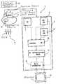
- FIG. 1is a schematic illustration of a medical examination and/or treatment device constructed and operating in accordance with the invention.
- FIG. 2is a schematic illustration for explaining the updating in accordance with the invention, using a 3D ultrasound image dataset.
- FIG. 3is a schematic illustration for explaining the updating in accordance with the invention, using of a 2D ultrasound image.
- FIG. 1shows an inventive examination and/or treatment device 1 having an ultrasound image acquisition device 2 as well as a control and processing device 3 that controls the operation of the ultrasound image acquisition device 2 and also undertakes the processing, editing and analysis of the image data.
- a set of 2D ultrasound images of an examination region—the heart of a patient 4 in this case—that are forwarded to the control and processing device 3are acquired with the ultrasound device 2 .
- the acquisition of the 2D ultrasound imagesensues with triggering by an ECG 6 that is recorded in parallel, since the examination region 15 is a rhythmically moving organ, namely the heart.
- the ECG dataare likewise forwarded to the control and processing device 3 .
- a suitable position detection system 16is used for this purpose.
- the position dataare likewise stored together with the 2D ultrasound images 5 .
- a preoperatively acquired 3D image dataset 7 of the examination region 15is also present in the control and processing device 3 .
- Thiscan be a computed tomography dataset, a magnetic resonance dataset or a 3D angiography image dataset. Since this dataset was acquired preoperatively, i.e. at an arbitrary time before the current treatment, there is the possibility that it does not show the examination region in conformity with the current anatomical conditions. In order nonetheless to be able to employ this high-resolution 3D image dataset for producing a 3D reconstruction image in the context of a subsequent examination or treatment, it is necessary that it be updated, i.e. to adapt it to the current anatomical conditions.
- the updating of the 3D image datasetcan ensue in two ways.
- a first wayis to directly employ the 2D ultrasound images 5 , that are registered with a known spatial position with respect to the coordinate system of the 3D ultrasound image dataset.
- a 3D ultrasound image dataset 9can be generated on the basis of the 2D ultrasound images 5 and utilized for the updating. This shall be discussed with reference to FIGS. 2 and 3.
- step 11After the 3D image dataset has been updated in step 10 , the production of a 3D reconstruction image ensues in step 11 . As illustrated by step 12 (only shown with broken lines), there is also the possibility of mixing an instrument introduced into the examination region 3 into this 3D reconstruction image. This can ensue using the 2D ultrasound images that may possibly show this instrument. Its position is detected; as a result of the registration of the 2D ultrasound images 5 relative to the 3D image dataset, and the detected position and orientation are mixed into the 3D volume image with accurate position and orientation.
- FIG. 2shows the updating using a 3D ultrasound image dataset.
- This 3D ultrasound image dataset 9like the 3D image dataset 7 —is presented in the form of a volume.
- the respective volumesare subdivided into a number of small partial volumes, referred to as voxels.
- Four voxels 7 a , 7 b , 7 c and 7 dare shown in the 3D image dataset 7 ; four corresponding voxels 9 a, 9 b, 9 c and 9 d are shown in the 3D ultrasound image dataset 9 .
- the individual voxelsare compared to one another and a determination is made as to whether the voxels of the 3D image dataset 7 agree with the corresponding voxels of the 3D ultrasound image dataset 9 .
- the voxels 7 a , 7 b , 7 c and the voxels 9 a, 9 b, 9 ccoincide, i.e. there is an image data match.
- the voxel 7 dwhich has been selected merely as an example, cannot be mapped onto the voxel 9 d with exact orientation and position.
- FIG. 3shows the updating using a 2D ultrasound image a schematic illustration.
- the 3D image dataset 7also is shown in FIG. 3 in the form of a three-dimensional cube.
- a 2D ultrasound image 5is then mixed into this 3D image dataset 7 with exact position and orientation.
- the exact spatial position of the 2D ultrasound image 5 in the 3D volumeis known because of the acquisition of the spatial lay of a 2D ultrasound image 5 using the position sensor 5 and due to the registration of the 2D ultrasound image 5 with the coordinate system of the 3D image dataset 7 , so that this mixing can ensue.
- a checkis also made, for example via a grayscale analysis, as to how the examination region that is shown in the tomogram plane of the 3D image dataset 7 and the examination region as shown in the 2D ultrasound image 5 coincide relative to one another.
- the tomogram plane from the 3D image dataset 7is shown at the left in the illustrated example, the 2D ultrasound image 5 shown next to it, being mixed in over it or into it.
- the examination regionis shown idealized as a circle in the 3D tomogram plane (at the left), whereas it is oval in the 2D ultrasound image that indicates the current anatomical conditions.
- the determination of the deformation or updating parametersnow ensues such, for example via a suitable grayscale analysis or an edge detection algorithm, which describe how the presentation of the examination region shown in the 3D plane of section image 14 is to be shifted or deformed until it coincides with the presentation shown in the 2D ultrasound image 5 .
- This mixing and determination of the deformation parametersensues until an updating of the complete 3D image dataset 7 is possible.
Landscapes
- Health & Medical Sciences (AREA)
- Life Sciences & Earth Sciences (AREA)
- Engineering & Computer Science (AREA)
- Medical Informatics (AREA)
- Biophysics (AREA)
- Nuclear Medicine, Radiotherapy & Molecular Imaging (AREA)
- Pathology (AREA)
- Radiology & Medical Imaging (AREA)
- Veterinary Medicine (AREA)
- Biomedical Technology (AREA)
- Heart & Thoracic Surgery (AREA)
- Physics & Mathematics (AREA)
- Molecular Biology (AREA)
- Surgery (AREA)
- Animal Behavior & Ethology (AREA)
- General Health & Medical Sciences (AREA)
- Public Health (AREA)
- Physiology (AREA)
- Ultra Sonic Daignosis Equipment (AREA)
Abstract
Description
- 1. Field of the Invention[0001]
- The present invention is directed to a method and apparatus for the three-dimensional presentation of an examination region of a patient in the form of a 3D reconstruction image.[0002]
- 2. Description of the Prior Art[0003]
- The three-dimensional presentation of an examination region of a patient in the form of a 3D reconstruction image is becoming increasingly important in the framework of medical examinations or treatments. Examples are minimally invasive treatments using endoscopes, laparoscopes or catheters that are respectively introduced into the examination region of the patient via a small body opening. On the basis of the 3D reconstruction image, the physician is provided with a three-dimensional view of the examination region, for example of the heart, which is useful to the physician for the navigation of the medical instrument. Such 3D reconstruction images, however, also are useful in instances wherein no instrument is to be introduced and only a presentation that serves for diagnostic purposes is required.[0004]
- Preoperatively acquired 3D image datasets, i.e. datasets that were acquired an arbitrary time before the actual examination or treatment, often are employed for the reconstruction of a 3D reconstruction image. Particularly when the 3D reconstruction image is employed in the context of ongoing intervention, difficulties can occur due to anatomical conditions that have changed since the preoperative exposure, i.e. the 3D reconstruction image that is reconstructed based on the preoperative image data no longer represents the current anatomical or positional conditions. For example, the patient may have gained or lost weight, can lie in a different position on the patient bed, etc. Ambiguities arise therefrom for the attending physician that can make the diagnosis, examination or treatment more difficult, particularly the intervention treatment.[0005]
- An object of the present invention is to provide a method and apparatus for the three-dimensional presentation of an examination region of a patient in the form of a 3D reconstruction image, wherein the aforementioned disadvantages are alleviated.[0006]
- This object is achieved in accordance with the invention in a method and apparatus wherein a preoperatively acquired 3D image dataset of the examination region is employed in a medical procedure, a number of 2D ultrasound images of the examination region are acquired, the preoperative 3D image dataset is updated using the 2D ultrasound images, and a 3D reconstruction image is generated on the basis of the updated 3D image dataset.[0007]
- Thus in accordance with the invention the preoperative 3D image dataset is updated at the time of the procedure (examination or treatment or intervention), so that the 3D reconstruction image subsequently reconstructed on the basis of the updated 3D image dataset reproduces the actual anatomical or positional situation. A number of 2D ultrasound images of the examination region are acquired for this purpose. These show the examination region in its current position. The 2D ultrasound images form the basis for the updating. The ultrasound image acquisition is advantageous from a number of points of view. First, it can ensue without great apparatus outlay; second, no radiation stressing of the patient whatsoever occurs due to the ultrasound acquisition. The images also can be acquired very quickly in the context of the ongoing examination or treatment or intervention, so that an excessively longer time for the overall procedure does not occur.[0008]
- The preoperative 3D image dataset can have been acquired with an arbitrary acquisition modality; it can, thus, be a CT dataset or an MR dataset or a 3D X-ray angiography dataset. All of these datasets allow an exact reconstruction of the examination region. These different datasets also can be updated using image data acquired with a different examination modality, namely an ultrasound device.[0009]
- In a first embodiment of the invention, the updating of the preoperative 3D image dataset ensues directly on the basis of the 2D ultrasound images. In an alternative version a 3D ultrasound image dataset is reconstructed on the basis of the 2D ultrasound images, and the updating ensues on the basis of this 3D ultrasound image dataset. A combined updating mode is also possible, i.e. the updating can ensue both on the basis of the 2D ultrasound images as well as on the basis of the reconstructed 3D ultrasound image dataset.[0010]
- When, for example, the updating ensues on the basis of a 3D ultrasound image dataset, then the 3D image dataset and the 3D ultrasound image dataset can be overlaid on one another in accordance with the invention, and those dataset parts of the 3D image dataset that do not adequately agree with the corresponding parts of the 3D ultrasound image dataset are deformed by translation and/or rotation until an adequate superimposition has been achieved. A conforming technique known as “deformable matching” between the preoperative 3D image dataset and the reconstructed 3D ultrasound image dataset is used. A registration is undertaken that deforms the preoperatively acquired image dataset such that it matches the current image dataset better, which should ensue in as short a time as possible in order to keep the waiting time for the patient as short as possible. For this reason, a rigid matching first ensues between the preoperative 3D image dataset and the quasi intraoperatively acquired 3D ultrasound image dataset (the ultrasound volume). No further registration, i.e. modification or deformation of the dataset, is required in the regions wherein the datasets agree well. Only those regions are reconsidered wherein the datasets do not yet coincide to an adequate extent. The two datasets, thus, are subdivided and every inadequately registered region is registered again. To this end, for example, it is possible to produce a 3D reconstruction image and a 3D ultrasound reconstruction image from each dataset and to subdivide the respective, reconstructed volumes into individual voxels of the same size and to then separately compare these voxels to one another. Only those voxels that are not adequately registered relative to one another, i.e. that do not coincide well enough, are registered again. When an adequate registration has been found for each sub-region of the two datasets, these registrations must still be linked to or operated with one another. As a result of this linkage or operation, it is possible that overlaps or gaps may arise. In order to compensate these, in accordance with the invention the overlap or gap regions within the 3D image dataset generated due to the translational and/or rotational deformation are smoothed by interpolation. The time required for the registration can be considerably shortened since a deformation occurs only once at the end given such a registration process and the sub-regions of the datasets are otherwise only rigidly registered.[0011]
- The determination of the deformation parameters required for the updating of a voxel ensues, for example, by means of a grayscale analysis of the data parts or voxels to be compared. The arrangement relative to one another of the image parts to be compared can be recognized from this grayscale distribution analysis. The actual modification of the image dataset, i.e. the actual updating or deformation, thus ensues when the deformation parameters have been found.[0012]
- For the updating given direct employment of the 2D ultrasound images, in accordance with the invention, the 2D ultrasound images are acquired with an ultrasound exposure device having a position sensor that supplies information about the spatial orientation and position of the acquired 2D ultrasound image, this ultrasound image being registered with respect to the coordinates of the 3D image dataset, so that the spatial position and orientation of the acquired 2D ultrasound image relative to the 3D image dataset is known. The acquired 2D ultrasound image is mixed with exact position and orientation into a corresponding sectional plane image of the 3D image dataset, which is subsequently deformed corresponding to the 2D ultrasound image. Thus the 2D ultrasound images are directly employed for the updating. The aforementioned position sensor can be integrated into the ultrasound head or applicator. The position and orientation of the position sensor are identified via a suitable position acquisition system in a coordinate system associated with the position acquisition system. This coordinate system and thus the position sensor, is registered (brought into registration with) with the coordinates of the preoperative 3D image dataset, so that the position and orientation of every 2D ultrasound image acquired during the image acquisition is known relative to the preoperative 3D image dataset. The current 2D ultrasound image can be mixed in the preoperative 3D image dataset at the appropriate position with the position and orientation obtained by the position sensor, i.e. it is mixed with exact position and orientation into the corresponding sectional plane image of the 3D image dataset. For the registration and thus updating of the 3D image dataset, a deformation and matching of the 3D image dataset now ensues by deforming the displayed surfaces and contours of the sectional plane image of the 3D image dataset until they correspond to the anatomy shown in the 2D ultrasound image that has been mixed in, i.e., surfaces or geometrical structures are matched.[0013]
- An improvement of the registration can be achieved by segmenting a surface of the examination region, for example the heart surface, shown in the 3D image dataset before the mixing, and the 2D ultrasound image dataset is subsequently mixed in. As a result of the segmenting, a three-dimensional envelope figure is obtained that shows the surface of the examination region, for example of the heart. By this means, the 2D ultrasound image is subsequently placed with exact orientation and planarity, and the direction in which the 3D surface envelope is to be deformed so that it matches the illustrated 2D contour of the heart in the respective image plane is subsequently determined for defining the deformation parameters. How the deformation should appear for a surface fit is calculated for the determination of the deformation parameters. The actual updating or deformation of the preoperative 3D image dataset subsequently ensues using the deformation parameters. By means of the clear demarcation of the heart surface in the ultrasound image, it is thus possible to determine the spacing and the direction of the same surface in the ultrasound image from the surface in the preoperatively acquired 3D image dataset and, based thereon, to define or modify the deformation parameters in a suitable way.[0014]
- If the examination region is a rhythmically or arrhythmically moving region, for example the heart, then for an exact presentation the image data from the 3D image dataset to be updated and the acquired 2D ultrasound images must each show the examination region in the same motion phase. In order to enable this, in accordance with the invention the motion phase of a rhythmically or arrhythmically moving examination region is acquired, and only image data from the 3D image dataset that are acquired in the same motion phase as the 2D ultrasound images are employed for the 3D reconstruction. The acquisition of the motion phase is required in the acquisition of the 3D image dataset as well as in the 2D ultrasound image acquisition in order to be able to produce isophase images or volumes. The image data to be updated and thus the reconstruction volume, are expediently based on the phase in which the 2D ultrasound images are acquired. It can also be expedient when, in addition to the motion phase, the respective points in time of the acquisition of the 2D ultrasound images is acquired, and only image data from the 3D image dataset that are also acquired at the same points in time as the 2D ultrasound images are employed for the updating and reconstruction of the 3D reconstruction image.[0015]
- When the examination is the heart, then an ECG is expediently recorded for acquiring the motion phase and, if used, the time, the acquisition of the 2D ultrasound images being triggered dependent thereon. An ECG likewise is allocated to the image data for the production of the 3D reconstruction image when they are acquired.[0016]
- The 2D ultrasound images can be acquired extra-corporeally using a known ultrasound exposure device externally applied to the patient. An intracorporeal ultrasound image acquisition is also possible, using a suitable medical instrument, for example in the form of a catheter with an ultrasound exposure head integrated at its tip.[0017]
- On the basis of the updated 3D image dataset, it is now possible to produce a 3D reconstruction image corresponding to the current anatomical situation. As described, this can be employed for diagnostic purposes; however, it is also possible to use this in the context of an intervention. To this end, for example, the ultrasound images are acquired using an instrument that already has been introduced into the examination region in the context of the interventional procedure, for example a catheter introduced into the heart, and the position of the instrument is determined on the basis of at least one 2D ultrasound image and is displayed in the current 3D reconstruction image. This affords the possibility of online visualization of the catheter with exact position and orientation in the updated 3D reconstruction image. To this end, the 2D ultrasound images that show the catheter can be employed. As an alternative, there is the possibility, for example, of employing and mixing in 2D fluoroscopic images that are acquired with a suitable X-ray device. In this case, a 2D/3D registration or a 2D/3D fusion of the 2D real-time images that show the image of the instrument with the updated, preoperative 3D image dataset must be implemented. When, alternatively, a position sensor is integrated in the catheter, positions and orientation of the instrument can be continuously acquired during the intervention with the assistance of this position sensor and can be mixed into the updated preoperative 3D image dataset. A 3D/3D registration of the coordinates of the position sensor with the coordinates of the updated 3D image dataset is assumed for this purpose. The registration can ensue on the basis of any known 2D/3D or 3D/3D registration methods. Such registration modes are well known to those skilled in the art so a more detailed description is not required.[0018]
- FIG. 1 is a schematic illustration of a medical examination and/or treatment device constructed and operating in accordance with the invention.[0019]
- FIG. 2 is a schematic illustration for explaining the updating in accordance with the invention, using a 3D ultrasound image dataset.[0020]
- FIG. 3 is a schematic illustration for explaining the updating in accordance with the invention, using of a 2D ultrasound image.[0021]
- In a schematic illustration, FIG. 1 shows an inventive examination and/or[0022]
treatment device 1 having an ultrasoundimage acquisition device 2 as well as a control andprocessing device 3 that controls the operation of the ultrasoundimage acquisition device 2 and also undertakes the processing, editing and analysis of the image data. A set of 2D ultrasound images of an examination region—the heart of a patient4 in this case—that are forwarded to the control andprocessing device 3, are acquired with theultrasound device 2. In the illustrated example, the acquisition of the 2D ultrasound images ensues with triggering by anECG 6 that is recorded in parallel, since theexamination region 15 is a rhythmically moving organ, namely the heart. The ECG data are likewise forwarded to the control andprocessing device 3. - A[0023]
position sensor 8 with which the spatial position of theultrasound acquisition device 2, and thus the respective spatial position of each acquired 2D ultrasound image can be identified, is also provided at theultrasound acquisition device 2. A suitableposition detection system 16 is used for this purpose. The position data are likewise stored together with the2D ultrasound images 5. - A preoperatively acquired[0024]
3D image dataset 7 of theexamination region 15 is also present in the control andprocessing device 3. This can be a computed tomography dataset, a magnetic resonance dataset or a 3D angiography image dataset. Since this dataset was acquired preoperatively, i.e. at an arbitrary time before the current treatment, there is the possibility that it does not show the examination region in conformity with the current anatomical conditions. In order nonetheless to be able to employ this high-resolution 3D image dataset for producing a 3D reconstruction image in the context of a subsequent examination or treatment, it is necessary that it be updated, i.e. to adapt it to the current anatomical conditions. - The updating of the 3D image dataset can ensue in two ways. A first way is to directly employ the[0025]
2D ultrasound images 5, that are registered with a known spatial position with respect to the coordinate system of the 3D ultrasound image dataset. As an alternative, a 3Dultrasound image dataset 9 can be generated on the basis of the2D ultrasound images 5 and utilized for the updating. This shall be discussed with reference to FIGS. 2 and 3. - A number of steps in accordance with the invention are schematically indicated as blocks in the control and[0026]
processing device 3. After the 3D image dataset has been updated instep 10, the production of a 3D reconstruction image ensues instep 11. As illustrated by step12 (only shown with broken lines), there is also the possibility of mixing an instrument introduced into theexamination region 3 into this 3D reconstruction image. This can ensue using the 2D ultrasound images that may possibly show this instrument. Its position is detected; as a result of the registration of the2D ultrasound images 5 relative to the 3D image dataset, and the detected position and orientation are mixed into the 3D volume image with accurate position and orientation. Of course, there is also the possibility of employing other two-dimensional images, for example X-ray fluoroscopic images, that show the instrument in the examination volume instead of the 2D ultrasound images. The 3D reconstruction image is subsequently presented at amonitor 13 with a representation of the instrument. - FIG. 2 shows the updating using a 3D ultrasound image dataset. This 3D[0027]
ultrasound image dataset 9—like the3D image dataset 7—is presented in the form of a volume. The respective volumes are subdivided into a number of small partial volumes, referred to as voxels. Fourvoxels 3D image dataset 7; fourcorresponding voxels ultrasound image dataset 9. For the deformation and updating of the3D image dataset 7, the individual voxels are compared to one another and a determination is made as to whether the voxels of the3D image dataset 7 agree with the corresponding voxels of the 3Dultrasound image dataset 9. In the illustrated example, thevoxels voxels voxel 7d, which has been selected merely as an example, cannot be mapped onto thevoxel 9dwith exact orientation and position. For the updating, a rigid registration of this voxel and of course of every other unmatched voxel, now ensues by translation and/or rotation of the respective voxel until it fits with the respective comparison voxel in the 3D ultrasound image dataset. Thevoxel 7dis translationally or rotationally modified until it can be mapped congruently onto thevoxel 9d.A determination of the deformation of updating parameters ensues from this modification. When the corresponding deformation parameters have been identified for every non-matching voxel, then the actual updating of the3D image dataset 7 ensues, i.e. it is modified dependent on the acquired updating requirements. The acquisition of differences, if any, within the voxels ensues by an analysis of the respective grayscale values. - FIG. 3 shows the updating using a 2D ultrasound image a schematic illustration. The[0028]
3D image dataset 7 also is shown in FIG. 3 in the form of a three-dimensional cube. A2D ultrasound image 5 is then mixed into this3D image dataset 7 with exact position and orientation. As described, the exact spatial position of the2D ultrasound image 5 in the 3D volume is known because of the acquisition of the spatial lay of a2D ultrasound image 5 using theposition sensor 5 and due to the registration of the2D ultrasound image 5 with the coordinate system of the3D image dataset 7, so that this mixing can ensue. A check is also made, for example via a grayscale analysis, as to how the examination region that is shown in the tomogram plane of the3D image dataset 7 and the examination region as shown in the2D ultrasound image 5 coincide relative to one another. The tomogram plane from the3D image dataset 7 is shown at the left in the illustrated example, the2D ultrasound image 5 shown next to it, being mixed in over it or into it. The examination region is shown idealized as a circle in the 3D tomogram plane (at the left), whereas it is oval in the 2D ultrasound image that indicates the current anatomical conditions. The determination of the deformation or updating parameters now ensues such, for example via a suitable grayscale analysis or an edge detection algorithm, which describe how the presentation of the examination region shown in the 3D plane ofsection image 14 is to be shifted or deformed until it coincides with the presentation shown in the2D ultrasound image 5. This mixing and determination of the deformation parameters ensues until an updating of the complete3D image dataset 7 is possible. - Although modifications and changes may be suggested by those skilled in the art, it is the intention of the inventors to embody within the patent warranted hereon all changes and modifications as reasonably and properly come within the scope of their contribution to the art.[0029]
Claims (32)
Applications Claiming Priority (2)
| Application Number | Priority Date | Filing Date | Title |
|---|---|---|---|
| DE10210650.9 | 2002-03-11 | ||
| DE10210650ADE10210650B4 (en) | 2002-03-11 | 2002-03-11 | Method for the three-dimensional representation of a study area of a patient in the form of a 3D reconstruction image and medical examination and / or treatment facility |
Publications (2)
| Publication Number | Publication Date |
|---|---|
| US20030199748A1true US20030199748A1 (en) | 2003-10-23 |
| US7302286B2 US7302286B2 (en) | 2007-11-27 |
Family
ID=28050658
Family Applications (1)
| Application Number | Title | Priority Date | Filing Date |
|---|---|---|---|
| US10/385,865Expired - LifetimeUS7302286B2 (en) | 2002-03-11 | 2003-03-11 | Method and apparatus for the three-dimensional presentation of an examination region of a patient in the form of a 3D reconstruction image |
Country Status (2)
| Country | Link |
|---|---|
| US (1) | US7302286B2 (en) |
| DE (1) | DE10210650B4 (en) |
Cited By (23)
| Publication number | Priority date | Publication date | Assignee | Title |
|---|---|---|---|---|
| US20050148853A1 (en)* | 2003-12-17 | 2005-07-07 | Thomas Redel | Method for supporting navigation of a medical instrument, in particular of a catheter |
| US20050203372A1 (en)* | 2004-01-22 | 2005-09-15 | Isabelle Janssen | Method and medical imaging apparatus for determining a slice in an examination volume for data acquisition in the slice |
| EP1717601A2 (en) | 2005-04-26 | 2006-11-02 | Biosense Webster, Inc. | Display of catheter tip with beam direction for ultrasound system |
| US20070015996A1 (en)* | 2005-05-13 | 2007-01-18 | Estelle Camus | Method for generating and displaying examination images and associated ultrasound catheter |
| US20070027390A1 (en)* | 2005-07-13 | 2007-02-01 | Michael Maschke | System for performing and monitoring minimally invasive interventions |
| US20070167706A1 (en)* | 2005-09-06 | 2007-07-19 | Jan Boese | Method and apparatus for visually supporting an electrophysiological catheter application in the heart by means of bidirectional information transfer |
| US20070233739A1 (en)* | 2006-03-23 | 2007-10-04 | Siemens Aktiengesellschaft | Method for reconstructing a three-dimensional target volume in realtime and displaying it |
| US20070248262A1 (en)* | 2006-03-23 | 2007-10-25 | Jan Boese | Device and method for synchronizing an image capture device with a pre-operative image data set |
| EP1720039A3 (en)* | 2005-04-26 | 2008-02-20 | Biosense Webster, Inc. | Display of a two-dimensional fan shaped ultrasound field |
| US20080247506A1 (en)* | 2006-12-22 | 2008-10-09 | Siemens Aktiengesellschaft | System for carrying out and monitoring minimally-invasive interventions |
| US7502642B2 (en)* | 2004-04-27 | 2009-03-10 | Siemens Aktiengesellschaft | Method and device for visually supporting an electrophysiological catheter application |
| US20090093702A1 (en)* | 2007-10-02 | 2009-04-09 | Fritz Vollmer | Determining and identifying changes in the position of parts of a body structure |
| US20100076262A1 (en)* | 2008-09-19 | 2010-03-25 | National Taiwan University | Endoscope inspection system |
| US20100119135A1 (en)* | 2007-04-02 | 2010-05-13 | Pierrick Coupe | Image processing device for matching images of a same portion of a body obtained by magnetic resonance and ultrasounds |
| US20100316278A1 (en)* | 2009-06-12 | 2010-12-16 | Ulrich Hartung | High-resolution three-dimensional medical imaging with dynamic real-time information |
| AU2006201646B2 (en)* | 2005-04-26 | 2011-01-06 | Biosense Webster, Inc. | Display of catheter tip with beam direction for ultrasound system |
| WO2012095784A1 (en)* | 2011-01-13 | 2012-07-19 | Koninklijke Philips Electronics N.V. | Visualization of catheter in three-dimensional ultrasound |
| CN102762151A (en)* | 2010-01-21 | 2012-10-31 | 卡尔斯特里姆保健公司 | Four-dimensional volume imaging system |
| US20130039555A1 (en)* | 2007-10-26 | 2013-02-14 | Koninklijke Philips Electronics N.V. | Closed loop registration control for multi-modality soft tissue imaging |
| AU2012258444B2 (en)* | 2005-04-26 | 2014-01-09 | Biosense Webster, Inc. | Display of two-dimensional ultrasound fan |
| JP2019517879A (en)* | 2016-06-15 | 2019-06-27 | 中慧医学成像有限公司 | Three-dimensional imaging method and system |
| US10354411B2 (en)* | 2016-12-20 | 2019-07-16 | Symbol Technologies, Llc | Methods, systems and apparatus for segmenting objects |
| CN112601496A (en)* | 2018-08-22 | 2021-04-02 | 皇家飞利浦有限公司 | 3D tracking of interventional medical devices |
Families Citing this family (20)
| Publication number | Priority date | Publication date | Assignee | Title |
|---|---|---|---|---|
| EP1646317A1 (en)* | 2003-07-10 | 2006-04-19 | Koninklijke Philips Electronics N.V. | Apparatus and method for navigating an instrument through an anatomical structure |
| DE102004004620A1 (en)* | 2004-01-29 | 2005-08-25 | Siemens Ag | Medical x-ray imaging method for recording an examination area for use in medical navigational procedures, whereby a spatial position of an examination area is recorded just prior to each shot and images then spatially compensated |
| JP5107709B2 (en)* | 2004-08-13 | 2012-12-26 | コーニンクレッカ フィリップス エレクトロニクス エヌ ヴィ | Adapting radiotherapy treatment plans |
| US7734119B2 (en)* | 2004-09-21 | 2010-06-08 | General Electric Company | Method and system for progressive multi-resolution three-dimensional image reconstruction using region of interest information |
| US8229545B2 (en) | 2005-09-15 | 2012-07-24 | St. Jude Medical, Atrial Fibrillation Division, Inc. | System and method for mapping complex fractionated electrogram information |
| US8038625B2 (en)* | 2005-09-15 | 2011-10-18 | St. Jude Medical, Atrial Fibrillation Division, Inc. | System and method for three-dimensional mapping of electrophysiology information |
| DE102006013476B4 (en)* | 2006-03-23 | 2012-11-15 | Siemens Ag | Method for positionally accurate representation of tissue regions of interest |
| DE102007009179A1 (en)* | 2007-02-26 | 2008-06-26 | Siemens Ag | Registering method for data set of creature, involves deforming one of two-dimensional actual representation corresponding to determined deformation so that deformed representation is registered relative to actual representation |
| DE102007023552B4 (en)* | 2007-05-21 | 2015-02-12 | Siemens Aktiengesellschaft | Combined imaging method |
| JP5394620B2 (en)* | 2007-07-23 | 2014-01-22 | ジーイー・メディカル・システムズ・グローバル・テクノロジー・カンパニー・エルエルシー | Ultrasonic imaging apparatus and image processing apparatus |
| US8200466B2 (en) | 2008-07-21 | 2012-06-12 | The Board Of Trustees Of The Leland Stanford Junior University | Method for tuning patient-specific cardiovascular simulations |
| TWI399195B (en)* | 2008-12-16 | 2013-06-21 | Ind Tech Res Inst | Apparatus and method for providing a dynamic 3d ultrasound image |
| US9405886B2 (en) | 2009-03-17 | 2016-08-02 | The Board Of Trustees Of The Leland Stanford Junior University | Method for determining cardiovascular information |
| CN102686278B (en) | 2009-12-28 | 2016-01-13 | 皇家飞利浦电子股份有限公司 | Therapeutic equipment |
| US8315812B2 (en) | 2010-08-12 | 2012-11-20 | Heartflow, Inc. | Method and system for patient-specific modeling of blood flow |
| US8157742B2 (en) | 2010-08-12 | 2012-04-17 | Heartflow, Inc. | Method and system for patient-specific modeling of blood flow |
| US20120084064A1 (en)* | 2010-09-29 | 2012-04-05 | Nutech Ventures, Inc. | Model-based systems and methods for analyzing and predicting outcomes of vascular interventions and reconstructions |
| US20120289830A1 (en)* | 2011-05-10 | 2012-11-15 | General Electric Company | Method and ultrasound imaging system for image-guided procedures |
| US8548778B1 (en) | 2012-05-14 | 2013-10-01 | Heartflow, Inc. | Method and system for providing information from a patient-specific model of blood flow |
| US11308627B1 (en) | 2021-09-17 | 2022-04-19 | King Abdulaziz University | Method for 3D ultrasound reconstruction of supraspinatus (SSP) tendon |
Citations (18)
| Publication number | Priority date | Publication date | Assignee | Title |
|---|---|---|---|---|
| US4945914A (en)* | 1987-11-10 | 1990-08-07 | Allen George S | Method and apparatus for providing related images over time of a portion of the anatomy using at least four fiducial implants |
| US5005578A (en)* | 1986-12-16 | 1991-04-09 | Sam Technology, Inc. | Three-dimensional magnetic resonance image distortion correction method and system |
| US5016642A (en)* | 1990-04-11 | 1991-05-21 | Hewlett-Packard Company | Slow motion cardiac imaging |
| US5526812A (en)* | 1993-06-21 | 1996-06-18 | General Electric Company | Display system for enhancing visualization of body structures during medical procedures |
| US5787889A (en)* | 1996-12-18 | 1998-08-04 | University Of Washington | Ultrasound imaging with real time 3D image reconstruction and visualization |
| US6216029B1 (en)* | 1995-07-16 | 2001-04-10 | Ultraguide Ltd. | Free-hand aiming of a needle guide |
| US6228028B1 (en)* | 1996-11-07 | 2001-05-08 | Tomtec Imaging Systems Gmbh | Method and apparatus for ultrasound image reconstruction |
| US6246898B1 (en)* | 1995-03-28 | 2001-06-12 | Sonometrics Corporation | Method for carrying out a medical procedure using a three-dimensional tracking and imaging system |
| US6263093B1 (en)* | 1998-03-20 | 2001-07-17 | Aloka Co., Ltd. | Three-dimensional ultrasound image processing apparatus and coordinate transformation method used in the apparatus |
| US6285902B1 (en)* | 1999-02-10 | 2001-09-04 | Surgical Insights, Inc. | Computer assisted targeting device for use in orthopaedic surgery |
| US20010035871A1 (en)* | 2000-03-30 | 2001-11-01 | Johannes Bieger | System and method for generating an image |
| US6317621B1 (en)* | 1999-04-30 | 2001-11-13 | Siemens Aktiengesellschaft | Method and device for catheter navigation in three-dimensional vascular tree exposures |
| US6379302B1 (en)* | 1999-10-28 | 2002-04-30 | Surgical Navigation Technologies Inc. | Navigation information overlay onto ultrasound imagery |
| US20030097068A1 (en)* | 1998-06-02 | 2003-05-22 | Acuson Corporation | Medical diagnostic ultrasound system and method for versatile processing |
| US6572547B2 (en)* | 2001-07-31 | 2003-06-03 | Koninklijke Philips Electronics N.V. | Transesophageal and transnasal, transesophageal ultrasound imaging systems |
| US20030117393A1 (en)* | 2001-08-16 | 2003-06-26 | Frank Sauer | System and method for three-dimensional (3D) reconstruction from ultrasound images |
| US6652460B2 (en)* | 2000-05-05 | 2003-11-25 | Esaote, S.P.A. | Method for ultrasonic imaging, particularly of moving bodies, such as surgical utensils, tissues, flows, or the like |
| US20030220561A1 (en)* | 2002-03-11 | 2003-11-27 | Estelle Camus | Method and apparatus for acquiring and displaying a medical instrument introduced into a cavity organ of a patient to be examined or treated |
Family Cites Families (3)
| Publication number | Priority date | Publication date | Assignee | Title |
|---|---|---|---|---|
| DE19542605A1 (en)* | 1994-11-17 | 1996-05-23 | Gen Electric | Interactive display system for showing internal organs of patient structures during medical operations |
| EP1011424A1 (en)* | 1997-03-03 | 2000-06-28 | Schneider Medical Technologies, Inc. | Imaging device and method |
| DE19846687C2 (en)* | 1998-10-09 | 2001-07-26 | Auer Dorothee | Auxiliary surgical device for use in performing medical procedures and methods for generating an image in the context of medical procedures |
- 2002
- 2002-03-11DEDE10210650Apatent/DE10210650B4/ennot_activeExpired - Fee Related
- 2003
- 2003-03-11USUS10/385,865patent/US7302286B2/ennot_activeExpired - Lifetime
Patent Citations (23)
| Publication number | Priority date | Publication date | Assignee | Title |
|---|---|---|---|---|
| US5005578A (en)* | 1986-12-16 | 1991-04-09 | Sam Technology, Inc. | Three-dimensional magnetic resonance image distortion correction method and system |
| US4991579A (en)* | 1987-11-10 | 1991-02-12 | Allen George S | Method and apparatus for providing related images over time of a portion of the anatomy using fiducial implants |
| US4945914A (en)* | 1987-11-10 | 1990-08-07 | Allen George S | Method and apparatus for providing related images over time of a portion of the anatomy using at least four fiducial implants |
| US5016642A (en)* | 1990-04-11 | 1991-05-21 | Hewlett-Packard Company | Slow motion cardiac imaging |
| US5526812A (en)* | 1993-06-21 | 1996-06-18 | General Electric Company | Display system for enhancing visualization of body structures during medical procedures |
| US6246898B1 (en)* | 1995-03-28 | 2001-06-12 | Sonometrics Corporation | Method for carrying out a medical procedure using a three-dimensional tracking and imaging system |
| US6216029B1 (en)* | 1995-07-16 | 2001-04-10 | Ultraguide Ltd. | Free-hand aiming of a needle guide |
| US6228028B1 (en)* | 1996-11-07 | 2001-05-08 | Tomtec Imaging Systems Gmbh | Method and apparatus for ultrasound image reconstruction |
| US5787889A (en)* | 1996-12-18 | 1998-08-04 | University Of Washington | Ultrasound imaging with real time 3D image reconstruction and visualization |
| US6263093B1 (en)* | 1998-03-20 | 2001-07-17 | Aloka Co., Ltd. | Three-dimensional ultrasound image processing apparatus and coordinate transformation method used in the apparatus |
| US20030097068A1 (en)* | 1998-06-02 | 2003-05-22 | Acuson Corporation | Medical diagnostic ultrasound system and method for versatile processing |
| US6285902B1 (en)* | 1999-02-10 | 2001-09-04 | Surgical Insights, Inc. | Computer assisted targeting device for use in orthopaedic surgery |
| US6317621B1 (en)* | 1999-04-30 | 2001-11-13 | Siemens Aktiengesellschaft | Method and device for catheter navigation in three-dimensional vascular tree exposures |
| US6379302B1 (en)* | 1999-10-28 | 2002-04-30 | Surgical Navigation Technologies Inc. | Navigation information overlay onto ultrasound imagery |
| US6669635B2 (en)* | 1999-10-28 | 2003-12-30 | Surgical Navigation Technologies, Inc. | Navigation information overlay onto ultrasound imagery |
| US20040059217A1 (en)* | 1999-10-28 | 2004-03-25 | Paul Kessman | Method of detecting organ matter shift in a patient |
| US6968224B2 (en)* | 1999-10-28 | 2005-11-22 | Surgical Navigation Technologies, Inc. | Method of detecting organ matter shift in a patient |
| US20010035871A1 (en)* | 2000-03-30 | 2001-11-01 | Johannes Bieger | System and method for generating an image |
| US6768496B2 (en)* | 2000-03-30 | 2004-07-27 | Siemens Aktiengesellschaft | System and method for generating an image from an image dataset and a video image |
| US6652460B2 (en)* | 2000-05-05 | 2003-11-25 | Esaote, S.P.A. | Method for ultrasonic imaging, particularly of moving bodies, such as surgical utensils, tissues, flows, or the like |
| US6572547B2 (en)* | 2001-07-31 | 2003-06-03 | Koninklijke Philips Electronics N.V. | Transesophageal and transnasal, transesophageal ultrasound imaging systems |
| US20030117393A1 (en)* | 2001-08-16 | 2003-06-26 | Frank Sauer | System and method for three-dimensional (3D) reconstruction from ultrasound images |
| US20030220561A1 (en)* | 2002-03-11 | 2003-11-27 | Estelle Camus | Method and apparatus for acquiring and displaying a medical instrument introduced into a cavity organ of a patient to be examined or treated |
Cited By (39)
| Publication number | Priority date | Publication date | Assignee | Title |
|---|---|---|---|---|
| US20050148853A1 (en)* | 2003-12-17 | 2005-07-07 | Thomas Redel | Method for supporting navigation of a medical instrument, in particular of a catheter |
| US7340082B2 (en) | 2004-01-22 | 2008-03-04 | Siemens Aktiengesellschaft | Method and medical imaging apparatus for determining a slice in an examination volume for data acquisition in the slice |
| US20050203372A1 (en)* | 2004-01-22 | 2005-09-15 | Isabelle Janssen | Method and medical imaging apparatus for determining a slice in an examination volume for data acquisition in the slice |
| US7502642B2 (en)* | 2004-04-27 | 2009-03-10 | Siemens Aktiengesellschaft | Method and device for visually supporting an electrophysiological catheter application |
| AU2012258444B2 (en)* | 2005-04-26 | 2014-01-09 | Biosense Webster, Inc. | Display of two-dimensional ultrasound fan |
| US20060253032A1 (en)* | 2005-04-26 | 2006-11-09 | Altmann Andres C | Display of catheter tip with beam direction for ultrasound system |
| US8870779B2 (en) | 2005-04-26 | 2014-10-28 | Biosense Webster, Inc. | Display of two-dimensional ultrasound fan |
| EP1717601A2 (en) | 2005-04-26 | 2006-11-02 | Biosense Webster, Inc. | Display of catheter tip with beam direction for ultrasound system |
| AU2006201646B2 (en)* | 2005-04-26 | 2011-01-06 | Biosense Webster, Inc. | Display of catheter tip with beam direction for ultrasound system |
| EP1720039A3 (en)* | 2005-04-26 | 2008-02-20 | Biosense Webster, Inc. | Display of a two-dimensional fan shaped ultrasound field |
| EP1717601A3 (en)* | 2005-04-26 | 2008-02-27 | Biosense Webster, Inc. | Display of catheter tip with beam direction for ultrasound system |
| US7604601B2 (en) | 2005-04-26 | 2009-10-20 | Biosense Webster, Inc. | Display of catheter tip with beam direction for ultrasound system |
| US20070015996A1 (en)* | 2005-05-13 | 2007-01-18 | Estelle Camus | Method for generating and displaying examination images and associated ultrasound catheter |
| DE102005032755B4 (en)* | 2005-07-13 | 2014-09-04 | Siemens Aktiengesellschaft | System for performing and monitoring minimally invasive procedures |
| US8548567B2 (en) | 2005-07-13 | 2013-10-01 | Siemens Aktiengesellschaft | System for performing and monitoring minimally invasive interventions |
| US20070027390A1 (en)* | 2005-07-13 | 2007-02-01 | Michael Maschke | System for performing and monitoring minimally invasive interventions |
| US20070167706A1 (en)* | 2005-09-06 | 2007-07-19 | Jan Boese | Method and apparatus for visually supporting an electrophysiological catheter application in the heart by means of bidirectional information transfer |
| US8077940B2 (en)* | 2006-03-23 | 2011-12-13 | Siemens Aktiengesellschaft | Method for reconstructing a three-dimensional target volume in realtime and displaying it |
| US20070248262A1 (en)* | 2006-03-23 | 2007-10-25 | Jan Boese | Device and method for synchronizing an image capture device with a pre-operative image data set |
| US20070233739A1 (en)* | 2006-03-23 | 2007-10-04 | Siemens Aktiengesellschaft | Method for reconstructing a three-dimensional target volume in realtime and displaying it |
| US8411921B2 (en)* | 2006-03-23 | 2013-04-02 | Siemens Aktiengesellschaft | Device and method for synchronizing an image capture device with a pre-operative image data set |
| US8335557B2 (en) | 2006-12-22 | 2012-12-18 | Siemens Aktiengesellschaft | System for carrying out and monitoring minimally-invasive interventions |
| US20080247506A1 (en)* | 2006-12-22 | 2008-10-09 | Siemens Aktiengesellschaft | System for carrying out and monitoring minimally-invasive interventions |
| US8755581B2 (en)* | 2007-04-02 | 2014-06-17 | Inria Institut National De Recherche En Informatique Et En Automatique | Image processing device for matching images of a same portion of a body obtained by magnetic resonance and ultrasounds |
| US20100119135A1 (en)* | 2007-04-02 | 2010-05-13 | Pierrick Coupe | Image processing device for matching images of a same portion of a body obtained by magnetic resonance and ultrasounds |
| US20090093702A1 (en)* | 2007-10-02 | 2009-04-09 | Fritz Vollmer | Determining and identifying changes in the position of parts of a body structure |
| US20130039555A1 (en)* | 2007-10-26 | 2013-02-14 | Koninklijke Philips Electronics N.V. | Closed loop registration control for multi-modality soft tissue imaging |
| US8885897B2 (en)* | 2007-10-26 | 2014-11-11 | Koninklijke Philips N.V. | Closed loop registration control for multi-modality soft tissue imaging |
| US20100076262A1 (en)* | 2008-09-19 | 2010-03-25 | National Taiwan University | Endoscope inspection system |
| US9036880B2 (en) | 2009-06-12 | 2015-05-19 | Siemens Aktiengesellschaft | High-resolution three-dimensional medical imaging with dynamic real-time information |
| US20100316278A1 (en)* | 2009-06-12 | 2010-12-16 | Ulrich Hartung | High-resolution three-dimensional medical imaging with dynamic real-time information |
| CN102762151A (en)* | 2010-01-21 | 2012-10-31 | 卡尔斯特里姆保健公司 | Four-dimensional volume imaging system |
| WO2012095784A1 (en)* | 2011-01-13 | 2012-07-19 | Koninklijke Philips Electronics N.V. | Visualization of catheter in three-dimensional ultrasound |
| US9993304B2 (en) | 2011-01-13 | 2018-06-12 | Koninklijke Philips N.V. | Visualization of catheter of three-dimensional ultrasound |
| JP2019517879A (en)* | 2016-06-15 | 2019-06-27 | 中慧医学成像有限公司 | Three-dimensional imaging method and system |
| EP3473182A4 (en)* | 2016-06-15 | 2020-01-01 | Telefield Medical Imaging Limited | METHOD AND SYSTEM FOR THREE-DIMENSIONAL IMAGING |
| AU2017285943B2 (en)* | 2016-06-15 | 2020-05-07 | Telefield Medical Imaging Limited | Three-dimensional imaging method and system |
| US10354411B2 (en)* | 2016-12-20 | 2019-07-16 | Symbol Technologies, Llc | Methods, systems and apparatus for segmenting objects |
| CN112601496A (en)* | 2018-08-22 | 2021-04-02 | 皇家飞利浦有限公司 | 3D tracking of interventional medical devices |
Also Published As
| Publication number | Publication date |
|---|---|
| DE10210650A1 (en) | 2003-10-16 |
| US7302286B2 (en) | 2007-11-27 |
| DE10210650B4 (en) | 2005-04-28 |
Similar Documents
| Publication | Publication Date | Title |
|---|---|---|
| US7302286B2 (en) | Method and apparatus for the three-dimensional presentation of an examination region of a patient in the form of a 3D reconstruction image | |
| US7467007B2 (en) | Respiratory gated image fusion of computed tomography 3D images and live fluoroscopy images | |
| EP1685535B1 (en) | Device and method for combining two images | |
| US6628977B2 (en) | Method and system for visualizing an object | |
| US8548567B2 (en) | System for performing and monitoring minimally invasive interventions | |
| EP1365685B1 (en) | 3d planning target volume | |
| US20030181809A1 (en) | 3D imaging for catheter interventions by use of 2D/3D image fusion | |
| US8942455B2 (en) | 2D/3D image registration method | |
| US6923768B2 (en) | Method and apparatus for acquiring and displaying a medical instrument introduced into a cavity organ of a patient to be examined or treated | |
| US7813785B2 (en) | Cardiac imaging system and method for planning minimally invasive direct coronary artery bypass surgery | |
| US7903859B2 (en) | Image acquisition, archiving and rendering system and method for reproducing imaging modality examination parameters used in an initial examination for use in subsequent radiological imaging | |
| US20030220555A1 (en) | Method and apparatus for image presentation of a medical instrument introduced into an examination region of a patent | |
| US8145012B2 (en) | Device and process for multimodal registration of images | |
| JP4490442B2 (en) | Method and system for affine superposition of an intraoperative 2D image and a preoperative 3D image | |
| US8285021B2 (en) | Three-dimensional (3D) reconstruction of the left atrium and pulmonary veins | |
| US7689019B2 (en) | Method and device for registering 2D projection images relative to a 3D image data record | |
| US10045754B2 (en) | Three dimensional (3D) pre-scan based volumetric image data processing | |
| US20050004449A1 (en) | Method for marker-less navigation in preoperative 3D images using an intraoperatively acquired 3D C-arm image | |
| US20050027193A1 (en) | Method for automatically merging a 2D fluoroscopic C-arm image with a preoperative 3D image with one-time use of navigation markers | |
| JP6620252B2 (en) | Correction of probe induced deformation in ultrasonic fusion imaging system | |
| US8280490B2 (en) | Registration aid for medical images | |
| JP2011502687A (en) | Interventional navigation using 3D contrast ultrasound | |
| US20070287905A1 (en) | Method for registering functional MR image data using radioscopy | |
| WO2011039685A1 (en) | Four-dimensional roadmapping usable in x-ray guided minimally invasive cardiac interventions | |
| Dandekar et al. | Image registration accuracy with low-dose CT: How low can we go? |
Legal Events
| Date | Code | Title | Description |
|---|---|---|---|
| AS | Assignment | Owner name:SIEMENS AKTIENGESELLSCHAFT, GERMANY Free format text:ASSIGNMENT OF ASSIGNORS INTEREST;ASSIGNORS:CAMUS, ESTELLE;DITT, HENDRIK;KILLMANN, REINMAR;AND OTHERS;REEL/FRAME:014173/0332;SIGNING DATES FROM 20030317 TO 20030325 | |
| STCF | Information on status: patent grant | Free format text:PATENTED CASE | |
| FPAY | Fee payment | Year of fee payment:4 | |
| FPAY | Fee payment | Year of fee payment:8 | |
| AS | Assignment | Owner name:SIEMENS HEALTHCARE GMBH, GERMANY Free format text:ASSIGNMENT OF ASSIGNORS INTEREST;ASSIGNOR:SIEMENS AKTIENGESELLSCHAFT;REEL/FRAME:039271/0561 Effective date:20160610 | |
| MAFP | Maintenance fee payment | Free format text:PAYMENT OF MAINTENANCE FEE, 12TH YEAR, LARGE ENTITY (ORIGINAL EVENT CODE: M1553); ENTITY STATUS OF PATENT OWNER: LARGE ENTITY Year of fee payment:12 | |
| AS | Assignment | Owner name:SIEMENS HEALTHINEERS AG, GERMANY Free format text:ASSIGNMENT OF ASSIGNORS INTEREST;ASSIGNOR:SIEMENS HEALTHCARE GMBH;REEL/FRAME:066088/0256 Effective date:20231219 | |
| AS | Assignment | Owner name:SIEMENS HEALTHINEERS AG, GERMANY Free format text:CORRECTIVE ASSIGNMENT TO CORRECT THE ASSIGNEE PREVIOUSLY RECORDED AT REEL: 066088 FRAME: 0256. ASSIGNOR(S) HEREBY CONFIRMS THE ASSIGNMENT;ASSIGNOR:SIEMENS HEALTHCARE GMBH;REEL/FRAME:071178/0246 Effective date:20231219 |