US20030138378A1 - Method and apparatus for examining tissue for predefined target cells, particularly cancerous cells, and a probe useful in such method and apparatus - Google Patents
Method and apparatus for examining tissue for predefined target cells, particularly cancerous cells, and a probe useful in such method and apparatusDownload PDFInfo
- Publication number
- US20030138378A1 US20030138378A1US10/298,196US29819602AUS2003138378A1US 20030138378 A1US20030138378 A1US 20030138378A1US 29819602 AUS29819602 AUS 29819602AUS 2003138378 A1US2003138378 A1US 2003138378A1
- Authority
- US
- United States
- Prior art keywords
- probe
- optical
- pulses
- tissue
- examined tissue
- Prior art date
- Legal status (The legal status is an assumption and is not a legal conclusion. Google has not performed a legal analysis and makes no representation as to the accuracy of the status listed.)
- Granted
Links
- 239000000523sampleSubstances0.000titleclaimsabstractdescription202
- 238000000034methodMethods0.000titleclaimsabstractdescription114
- 230000003287optical effectEffects0.000claimsabstractdescription169
- 239000002872contrast mediaSubstances0.000claimsabstractdescription20
- 239000002245particleSubstances0.000claimsabstractdescription19
- 239000004065semiconductorSubstances0.000claimsabstractdescription15
- 230000007423decreaseEffects0.000claimsabstractdescription8
- 239000004020conductorSubstances0.000claimsdescription58
- 239000013307optical fiberSubstances0.000claimsdescription43
- 238000001574biopsyMethods0.000claimsdescription20
- 230000010287polarizationEffects0.000claimsdescription20
- 229910052751metalInorganic materials0.000claimsdescription18
- 239000002184metalSubstances0.000claimsdescription18
- 239000000463materialSubstances0.000claimsdescription16
- 238000001228spectrumMethods0.000claimsdescription13
- 230000005855radiationEffects0.000claimsdescription8
- 239000003990capacitorSubstances0.000claimsdescription6
- 239000003989dielectric materialSubstances0.000claimsdescription6
- 230000031700light absorptionEffects0.000claimsdescription3
- 210000001519tissueAnatomy0.000description197
- 210000004027cellAnatomy0.000description134
- 206010028980NeoplasmDiseases0.000description41
- 239000011859microparticleSubstances0.000description35
- 239000002105nanoparticleSubstances0.000description35
- 201000011510cancerDiseases0.000description23
- 238000005259measurementMethods0.000description18
- 230000008859changeEffects0.000description16
- 108090000623proteins and genesProteins0.000description12
- 239000013078crystalSubstances0.000description11
- 238000002847impedance measurementMethods0.000description11
- XUIMIQQOPSSXEZ-UHFFFAOYSA-NSiliconChemical compound[Si]XUIMIQQOPSSXEZ-UHFFFAOYSA-N0.000description10
- 229910052710siliconInorganic materials0.000description10
- 239000010703siliconSubstances0.000description10
- 238000001514detection methodMethods0.000description9
- 102000004169proteins and genesHuman genes0.000description9
- 239000000427antigenSubstances0.000description8
- 108091007433antigensProteins0.000description8
- 102000036639antigensHuman genes0.000description8
- 238000010276constructionMethods0.000description8
- 239000002159nanocrystalSubstances0.000description8
- 230000008569processEffects0.000description7
- PCHJSUWPFVWCPO-UHFFFAOYSA-NgoldChemical compound[Au]PCHJSUWPFVWCPO-UHFFFAOYSA-N0.000description6
- 239000010931goldSubstances0.000description6
- 229910052737goldInorganic materials0.000description6
- 238000004458analytical methodMethods0.000description5
- 230000005540biological transmissionEffects0.000description5
- 238000006243chemical reactionMethods0.000description5
- 230000021615conjugationEffects0.000description5
- 238000005516engineering processMethods0.000description5
- 230000006870functionEffects0.000description5
- 210000004408hybridomaAnatomy0.000description5
- UHYPYGJEEGLRJD-UHFFFAOYSA-Ncadmium(2+);selenium(2-)Chemical compound[Se-2].[Cd+2]UHYPYGJEEGLRJD-UHFFFAOYSA-N0.000description4
- 238000002591computed tomographyMethods0.000description4
- 239000000835fiberSubstances0.000description4
- 238000000691measurement methodMethods0.000description4
- 210000004881tumor cellAnatomy0.000description4
- ZCYVEMRRCGMTRW-UHFFFAOYSA-N7553-56-2Chemical compound[I]ZCYVEMRRCGMTRW-UHFFFAOYSA-N0.000description3
- 241001529936MurinaeSpecies0.000description3
- VYPSYNLAJGMNEJ-UHFFFAOYSA-NSilicium dioxideChemical groupO=[Si]=OVYPSYNLAJGMNEJ-UHFFFAOYSA-N0.000description3
- BQCADISMDOOEFD-UHFFFAOYSA-NSilverChemical compound[Ag]BQCADISMDOOEFD-UHFFFAOYSA-N0.000description3
- 230000008901benefitEffects0.000description3
- 230000007910cell fusionEffects0.000description3
- 239000011248coating agentSubstances0.000description3
- 238000000576coating methodMethods0.000description3
- 238000003384imaging methodMethods0.000description3
- 229910052740iodineInorganic materials0.000description3
- 239000011630iodineSubstances0.000description3
- 238000004519manufacturing processMethods0.000description3
- 239000011159matrix materialSubstances0.000description3
- 230000010534mechanism of actionEffects0.000description3
- 229910052709silverInorganic materials0.000description3
- 239000004332silverSubstances0.000description3
- 230000001225therapeutic effectEffects0.000description3
- 241000894006BacteriaSpecies0.000description2
- 206010006187Breast cancerDiseases0.000description2
- 208000026310Breast neoplasmDiseases0.000description2
- 206010035226Plasma cell myelomaDiseases0.000description2
- FAPWRFPIFSIZLT-UHFFFAOYSA-MSodium chlorideChemical compound[Na+].[Cl-]FAPWRFPIFSIZLT-UHFFFAOYSA-M0.000description2
- 238000002679ablationMethods0.000description2
- 238000013459approachMethods0.000description2
- 210000003719b-lymphocyteAnatomy0.000description2
- 210000000988bone and boneAnatomy0.000description2
- 210000005013brain tissueAnatomy0.000description2
- 238000004364calculation methodMethods0.000description2
- 238000002512chemotherapyMethods0.000description2
- 238000010586diagramMethods0.000description2
- 229910003460diamondInorganic materials0.000description2
- 239000010432diamondSubstances0.000description2
- 201000010099diseaseDiseases0.000description2
- 208000037265diseases, disorders, signs and symptomsDiseases0.000description2
- 238000012377drug deliveryMethods0.000description2
- 230000000694effectsEffects0.000description2
- 238000001727in vivoMethods0.000description2
- 210000004698lymphocyteAnatomy0.000description2
- 238000013208measuring procedureMethods0.000description2
- 239000013081microcrystalSubstances0.000description2
- 230000004048modificationEffects0.000description2
- 238000012986modificationMethods0.000description2
- 238000012544monitoring processMethods0.000description2
- 210000003205muscleAnatomy0.000description2
- 201000000050myeloid neoplasmDiseases0.000description2
- 239000005543nano-size silicon particleSubstances0.000description2
- 230000002285radioactive effectEffects0.000description2
- 230000035945sensitivityEffects0.000description2
- 239000011780sodium chlorideSubstances0.000description2
- 239000000126substanceSubstances0.000description2
- 230000000007visual effectEffects0.000description2
- 102000012406Carcinoembryonic AntigenHuman genes0.000description1
- 108010022366Carcinoembryonic AntigenProteins0.000description1
- 229910004613CdTeInorganic materials0.000description1
- 208000001333Colorectal NeoplasmsDiseases0.000description1
- 229910001218Gallium arsenideInorganic materials0.000description1
- 206010020751HypersensitivityDiseases0.000description1
- 241001465754MetazoaSpecies0.000description1
- 206010061535Ovarian neoplasmDiseases0.000description1
- 208000000236Prostatic NeoplasmsDiseases0.000description1
- 229910000577Silicon-germaniumInorganic materials0.000description1
- LEVVHYCKPQWKOP-UHFFFAOYSA-N[Si].[Ge]Chemical compound[Si].[Ge]LEVVHYCKPQWKOP-UHFFFAOYSA-N0.000description1
- 238000010521absorption reactionMethods0.000description1
- 230000003213activating effectEffects0.000description1
- 230000004913activationEffects0.000description1
- 229910052782aluminiumInorganic materials0.000description1
- XAGFODPZIPBFFR-UHFFFAOYSA-NaluminiumChemical compound[Al]XAGFODPZIPBFFR-UHFFFAOYSA-N0.000description1
- 210000000628antibody-producing cellAnatomy0.000description1
- 230000000890antigenic effectEffects0.000description1
- 230000027455bindingEffects0.000description1
- 230000015572biosynthetic processEffects0.000description1
- 210000004369bloodAnatomy0.000description1
- 239000008280bloodSubstances0.000description1
- 239000013590bulk materialSubstances0.000description1
- 239000000969carrierSubstances0.000description1
- 230000024245cell differentiationEffects0.000description1
- 230000010261cell growthEffects0.000description1
- 210000000170cell membraneAnatomy0.000description1
- 239000003153chemical reaction reagentSubstances0.000description1
- 230000000295complement effectEffects0.000description1
- 230000006378damageEffects0.000description1
- 230000003247decreasing effectEffects0.000description1
- 238000013461designMethods0.000description1
- 238000011161developmentMethods0.000description1
- 230000018109developmental processEffects0.000description1
- 238000002059diagnostic imagingMethods0.000description1
- 239000003814drugSubstances0.000description1
- 229940079593drugDrugs0.000description1
- -1e.g.Substances0.000description1
- 230000005684electric fieldEffects0.000description1
- 238000000695excitation spectrumMethods0.000description1
- 230000001747exhibiting effectEffects0.000description1
- 239000007850fluorescent dyeSubstances0.000description1
- 238000010353genetic engineeringMethods0.000description1
- 229910052732germaniumInorganic materials0.000description1
- GNPVGFCGXDBREM-UHFFFAOYSA-Ngermanium atomChemical compound[Ge]GNPVGFCGXDBREM-UHFFFAOYSA-N0.000description1
- 239000003102growth factorSubstances0.000description1
- 230000017525heat dissipationEffects0.000description1
- 239000005556hormoneSubstances0.000description1
- 229940088597hormoneDrugs0.000description1
- 230000005965immune activityEffects0.000description1
- 210000002865immune cellAnatomy0.000description1
- 230000006872improvementEffects0.000description1
- 238000002347injectionMethods0.000description1
- 239000007924injectionSubstances0.000description1
- 230000002452interceptive effectEffects0.000description1
- 238000011835investigationMethods0.000description1
- PNDPGZBMCMUPRI-UHFFFAOYSA-NiodineChemical compoundIIPNDPGZBMCMUPRI-UHFFFAOYSA-N0.000description1
- 210000000265leukocyteAnatomy0.000description1
- 230000004807localizationEffects0.000description1
- 210000002540macrophageAnatomy0.000description1
- 239000004005microsphereSubstances0.000description1
- 239000000203mixtureSubstances0.000description1
- 210000001616monocyteAnatomy0.000description1
- 230000002611ovarianEffects0.000description1
- 230000000149penetrating effectEffects0.000description1
- 230000010363phase shiftEffects0.000description1
- INAAIJLSXJJHOZ-UHFFFAOYSA-NpibenzimolChemical compoundC1CN(C)CCN1C1=CC=C(N=C(N2)C=3C=C4NC(=NC4=CC=3)C=3C=CC(O)=CC=3)C2=C1INAAIJLSXJJHOZ-UHFFFAOYSA-N0.000description1
- 229920000642polymerPolymers0.000description1
- 238000011897real-time detectionMethods0.000description1
- 230000004044responseEffects0.000description1
- 230000000717retained effectEffects0.000description1
- 238000012552reviewMethods0.000description1
- SBIBMFFZSBJNJF-UHFFFAOYSA-Nselenium;zincChemical compound[Se]=[Zn]SBIBMFFZSBJNJF-UHFFFAOYSA-N0.000description1
- 239000000377silicon dioxideSubstances0.000description1
- 239000011856silicon-based particleSubstances0.000description1
- 230000005236sound signalEffects0.000description1
- 230000009870specific bindingEffects0.000description1
- 238000004611spectroscopical analysisMethods0.000description1
- 210000000952spleenAnatomy0.000description1
- 230000004083survival effectEffects0.000description1
- 230000002195synergetic effectEffects0.000description1
- 230000008685targetingEffects0.000description1
- 230000004797therapeutic responseEffects0.000description1
- 230000001988toxicityEffects0.000description1
- 231100000419toxicityToxicity0.000description1
- 239000003053toxinSubstances0.000description1
- 231100000765toxinToxicity0.000description1
- 108700012359toxinsProteins0.000description1
- 238000002604ultrasonographyMethods0.000description1
Images


Classifications
- A—HUMAN NECESSITIES
- A61—MEDICAL OR VETERINARY SCIENCE; HYGIENE
- A61K—PREPARATIONS FOR MEDICAL, DENTAL OR TOILETRY PURPOSES
- A61K49/00—Preparations for testing in vivo
- A—HUMAN NECESSITIES
- A61—MEDICAL OR VETERINARY SCIENCE; HYGIENE
- A61B—DIAGNOSIS; SURGERY; IDENTIFICATION
- A61B5/00—Measuring for diagnostic purposes; Identification of persons
- A61B5/0059—Measuring for diagnostic purposes; Identification of persons using light, e.g. diagnosis by transillumination, diascopy, fluorescence
- A61B5/0075—Measuring for diagnostic purposes; Identification of persons using light, e.g. diagnosis by transillumination, diascopy, fluorescence by spectroscopy, i.e. measuring spectra, e.g. Raman spectroscopy, infrared absorption spectroscopy
- A—HUMAN NECESSITIES
- A61—MEDICAL OR VETERINARY SCIENCE; HYGIENE
- A61B—DIAGNOSIS; SURGERY; IDENTIFICATION
- A61B5/00—Measuring for diagnostic purposes; Identification of persons
- A61B5/0059—Measuring for diagnostic purposes; Identification of persons using light, e.g. diagnosis by transillumination, diascopy, fluorescence
- A61B5/0082—Measuring for diagnostic purposes; Identification of persons using light, e.g. diagnosis by transillumination, diascopy, fluorescence adapted for particular medical purposes
- A61B5/0084—Measuring for diagnostic purposes; Identification of persons using light, e.g. diagnosis by transillumination, diascopy, fluorescence adapted for particular medical purposes for introduction into the body, e.g. by catheters
- A—HUMAN NECESSITIES
- A61—MEDICAL OR VETERINARY SCIENCE; HYGIENE
- A61B—DIAGNOSIS; SURGERY; IDENTIFICATION
- A61B5/00—Measuring for diagnostic purposes; Identification of persons
- A61B5/05—Detecting, measuring or recording for diagnosis by means of electric currents or magnetic fields; Measuring using microwaves or radio waves
- A61B5/053—Measuring electrical impedance or conductance of a portion of the body
- A61B5/0538—Measuring electrical impedance or conductance of a portion of the body invasively, e.g. using a catheter
- A—HUMAN NECESSITIES
- A61—MEDICAL OR VETERINARY SCIENCE; HYGIENE
- A61B—DIAGNOSIS; SURGERY; IDENTIFICATION
- A61B5/00—Measuring for diagnostic purposes; Identification of persons
- A61B5/41—Detecting, measuring or recording for evaluating the immune or lymphatic systems
- A61B5/411—Detecting or monitoring allergy or intolerance reactions to an allergenic agent or substance
- A—HUMAN NECESSITIES
- A61—MEDICAL OR VETERINARY SCIENCE; HYGIENE
- A61B—DIAGNOSIS; SURGERY; IDENTIFICATION
- A61B5/00—Measuring for diagnostic purposes; Identification of persons
- A61B5/41—Detecting, measuring or recording for evaluating the immune or lymphatic systems
- A61B5/414—Evaluating particular organs or parts of the immune or lymphatic systems
- A61B5/416—Evaluating particular organs or parts of the immune or lymphatic systems the spleen
- A—HUMAN NECESSITIES
- A61—MEDICAL OR VETERINARY SCIENCE; HYGIENE
- A61B—DIAGNOSIS; SURGERY; IDENTIFICATION
- A61B5/00—Measuring for diagnostic purposes; Identification of persons
- A61B5/0059—Measuring for diagnostic purposes; Identification of persons using light, e.g. diagnosis by transillumination, diascopy, fluorescence
- A61B5/0071—Measuring for diagnostic purposes; Identification of persons using light, e.g. diagnosis by transillumination, diascopy, fluorescence by measuring fluorescence emission
- A—HUMAN NECESSITIES
- A61—MEDICAL OR VETERINARY SCIENCE; HYGIENE
- A61B—DIAGNOSIS; SURGERY; IDENTIFICATION
- A61B5/00—Measuring for diagnostic purposes; Identification of persons
- A61B5/0059—Measuring for diagnostic purposes; Identification of persons using light, e.g. diagnosis by transillumination, diascopy, fluorescence
- A61B5/0082—Measuring for diagnostic purposes; Identification of persons using light, e.g. diagnosis by transillumination, diascopy, fluorescence adapted for particular medical purposes
- A61B5/0091—Measuring for diagnostic purposes; Identification of persons using light, e.g. diagnosis by transillumination, diascopy, fluorescence adapted for particular medical purposes for mammography
- A—HUMAN NECESSITIES
- A61—MEDICAL OR VETERINARY SCIENCE; HYGIENE
- A61B—DIAGNOSIS; SURGERY; IDENTIFICATION
- A61B5/00—Measuring for diagnostic purposes; Identification of persons
- A61B5/06—Devices, other than using radiation, for detecting or locating foreign bodies ; Determining position of diagnostic devices within or on the body of the patient
- A61B5/065—Determining position of the probe employing exclusively positioning means located on or in the probe, e.g. using position sensors arranged on the probe
- Y—GENERAL TAGGING OF NEW TECHNOLOGICAL DEVELOPMENTS; GENERAL TAGGING OF CROSS-SECTIONAL TECHNOLOGIES SPANNING OVER SEVERAL SECTIONS OF THE IPC; TECHNICAL SUBJECTS COVERED BY FORMER USPC CROSS-REFERENCE ART COLLECTIONS [XRACs] AND DIGESTS
- Y10—TECHNICAL SUBJECTS COVERED BY FORMER USPC
- Y10S—TECHNICAL SUBJECTS COVERED BY FORMER USPC CROSS-REFERENCE ART COLLECTIONS [XRACs] AND DIGESTS
- Y10S607/00—Surgery: light, thermal, and electrical application
- Y10S607/901—Cancer detection
Definitions
- the present applicationrelates to a method and apparatus for examining tissue for the presence of predefined target cells therein, and also to a probe for use in such method and apparatus.
- the inventionis particularly useful for detecting cancerous cells in a real-time manner during a surgical operation for removing, e.g., a breast tumor.
- the inventionis therefore described below with respect to such an application, but it will be appreciated that the invention is useful in many other applications.
- CTcomputed tomography
- MRImagnetic resonance imagining
- T-scanelectrical bioimpedance scanning devices
- ultrasoundand other similar instruments
- a method of examining tissue for the presence of predefined target cells thereincomprising: subjecting the tissue to be examined to a contrast agent containing small particles of a physical element conjugated with a biological carrier, e.g., an antibody, selectively bindable to the target cells; applying energy pulses to the examined tissue; detecting changes in electrical properties of the examined tissue produced by the applied energy pulses; and utilizing the detected changes in electrical properties for determining the presence of the target cells in the examined tissue.
- a biological carriere.g., an antibody
- the methodis particularly useful for detecting cancer target cells in a real-time manner during a surgical operation, and therefore the invention is described below particularly with respect to this application.
- the novel methodexploits the known technique for localization of tumors by the injection of physical elements conjugated with a biological carrier such as an antibody selectively bindable to the cancerous cells.
- a biological carriersuch as an antibody selectively bindable to the cancerous cells.
- Such techniqueshave been used with radioisotopes in order to target tumor tissue or cancerous cells when examined with computer tomography.
- Techniques using nano/micro crystal particleshave also been used as novel intravascular probes for both diagnostic (e.g., imaging) purposes and therapeutic (e.g., drug delivery) purposes.
- diagnostice.g., imaging
- therapeutice.g., drug delivery
- the present inventionutilizes such a technique (e.g., with non-radioactive conjugated antibodies) in an electric optical measuring procedure for detecting cancerous cells.
- the applied energy pulsesinclude pulses of optical (e.g., laser) energy
- the physical element conjugated with the antibodyis one which changes in impedance when illuminated by the optical energy.
- opticale.g., laser
- the physical element conjugated with the antibodyis one which changes in impedance when illuminated by the optical energy.
- the physical elementis a light-sensitive semiconductor having an impedance which substantially decreases in the presence of light.
- changes in optical properties of the examined tissue produced by the applied optical pulsesare also detected and utilized in determining the extent of the presence of the target (e.g., cancer) cells in the examined tissue.
- the inventionmay also be implemented in applications wherein the physical element is a metal having good light reflecting characteristics, and wherein changes in an optical characteristic (e.g., frequency, amplitude and/or phase) of the reflected light are detected and utilized for determining the extent of the presence of the target cells in the examined tissue.
- the physical elementmay also be a fluorescent material which emits radiation of a predetermined frequency when illuminated by light, or a light absorption material which absorbs radiation of a particular frequency, in which cases changes in frequency of the reflected light would be detected and utilized for determining the extent of the presence of the target cells in the examined tissue.
- voltage pulsesmay be applied to a probe area of the examined tissue to detect the presence of the target cells in the probe area
- optical pulsesmay be applied to a central region of the probe area of the examined tissue to detect the presence of the target, e.g., cancerous, cells in the central region.
- the target cellse.g., cancerous cells
- the target cellsmay be subjected to optical energy of sufficient intensity to destroy them.
- the target cellsare cancerous cells and the optical energy is laser energy
- the same probe as used for detecting the cancerous cellsmay also be used for destroying such cancerous cells by applying femtosecond pulses at an intensity of 100 nj-1 mj at the targeted cells. Longer pulses can be also used but with more heat dissipation to the surrounding area.
- the optical pulsesmay be applied by means of a flexible probe introduced into a subject's body via a catheter, or incorporated in a biopsy needle.
- a method of examining tissue for the present of cancerous cells thereincomprising: applying laser pulses to the examined tissue; detecting the reflections of the laser pulses from the examined tissue; comparing an optical characteristic of the laser pulses applied to the examined tissue with that of the laser reflections from the examined tissue; and utilizing the comparison of optical characteristics for determining the presence of cancerous cells in the examined tissue.
- a method of examining tissue for the presence of cancerous cells thereincomprising: applying optical energy pulses to the examined tissue in at least two polarization forms; detecting changes in optical properties of the examined tissue in each of the polarization forms; and utilizing the detected changes for determining the presence of cancerous cells in the examined tissue.
- a method of examining tissue for the presence of cancerous cells thereincomprising: applying voltage pulse and laser pulses to the examined tissue; detecting reflections of the voltage pulses from the examined tissue; comparing an electrical characteristic of the voltage pulse reflections from the examined tissue with that of the voltage pulses applied to the examined tissues with the laser pulses; and utilizing the comparison of electrical characteristics for determining the presence of cancerous cells in the examined tissue.
- apparatus for examining tissue for the presence of predefined target (e.g., cancer) cells thereincomprising: a voltage pulse source; an optical pulse source; a probe having an operative end for applying optical pulses and voltage pulses from the sources to the examined tissue, and for detecting the reflections of the voltage pulses produced by the examined tissue; and a data processor system including an electrical measuring sub-system coupled to the probe for detecting changes in the electrical properties of the examined tissue produced by the optical and voltage pulses, and for determining therefrom the presence of the target cells in the examined tissue.
- the probealso detects optical reflections of the optical pulses from the examined tissue.
- the data processor systemalso includes an optical analyzer sub-system utilizing the detected optical reflections from the examined tissue for detecting changes in optical characteristics of the examined tissue produced by the applied pulses in determining the extent of the presence of the target cells in the examined tissue.
- a probe for use in examining tissue for the presence of cancerous cells thereincomprising: an operative end having at least one pair of spaced conductors for applying voltage pulses to the examined tissue; and an optical fiber at the operative end for applying optical pulses to the examined tissue.
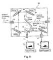
- FIG. 1is a block diagram illustrating one form of apparatus constructed in accordance with the present invention for examining tissue in a real-time manner for the presence of target cells, particularly cancerous cells, therein;
- FIGS. 2 a - 2 eare pictorial illustrations illustrating the mechanism of action involved in the impedance measuring technique of the present invention, when using a contrast agent conjugated with an antibody selectively bindable to the cancerous cells;
- FIG. 3is a block diagram illustrating a second form of apparatus constructed in accordance with the present invention.
- FIG. 4illustrates generally the construction of the electrical properties probe in the apparatus of FIG. 3;
- FIG. 5illustrates a preferred construction of the operative end of the probe of FIG. 3
- FIG. 6 aillustrates the probe of FIG. 9 when examining tissue constituted only of normal cells
- FIG. 6 billustrates the probe of FIG. 5 when examining tissues including cancerous cells
- FIG. 7schematically illustrates the probe in the apparatus of FIG. 3 and its connections to the impedance measuring and optical analyzer sub-systems;
- FIG. 8illustrates the optical splitting box in the optical analyzer sub-system of FIG. 3;
- FIGS. 9 a and 9 billustrate examples of polarization measurements from the two spectrometers in the optical analyzer sub-system of FIGS. 3 and 8;
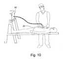
- FIG. 10is a pictorial illustration illustrating the manner of using the apparatus of FIG. 3 for detecting cancer cells in a real-time manner during a surgical operation for removing a tumor;
- FIGS. 11 and 12illustrate possible variations in the construction of the electrical probe
- FIG. 13 aschematically illustrates a possible variation in the probe construction that enables the use of the probe inside a biopsy/needle/tool
- FIG. 14illustrates another probe constructed to include a 3-D orientation sensor to enable determination of the position and orientation of the operative end of the probe
- FIG. 14 bis a plan view schematically illustrating the orientation sensor in the probe of FIG. 14.
- MAbsare extremely selective proteins (antigens or antibodies) that bind to only a single target.
- the specific antigens to which MAbs may bindinclude bacteria, hormones, tumor cell antigens, growth factors, and a variety of other substances.
- the extraordinary specificity of MAbshas tremendous clinical value.
- Mabsappear ideal since they are homogeneous in nature, recognize specific antigenic determinants, can be mass produced, are relatively stable to conjugation methods, and are biocompatible in vivo.
- MAbsare created using cell fusion techniques in a process called hybridoma technology.
- Cell fusionis a form of genetic engineering that merges two types of cells to form a single cell.
- Each hybridomais capable of producing large numbers of identical antibody molecules, and also called MAbs because they are produced by the identical offspring of a single, cloned, antibody-producing cell. Like the lymphocytes from the immunized animal, they produce antibodies targeted at the injected antigen.
- Murine (mouse) MAbshave been the primary focus of MAb creation to date. However, they produce variable results. Because mouse-produced antibodies are not identical to human antibodies, they are eventually recognized as foreign proteins by the human body and cleared from circulation by human antimouse antibodies (HAMA). These reactions are not a serious problem with MAb-based diagnostic and imaging products, where only a single application may be required. However, they are a major obstacle to the therapeutic use of murine antibodies. Most patients produce a HAMA reaction, which significantly reduces therapeutic efficacy and increases toxicity.
- Human MAbsdo not produce a HAMA reaction, so they tend to succeed therapeutically and are less likely to produce allergic reactions. Unfortunately, it is extremely difficult to fuse human B-lymphocytes with myeloma cells. Several biotechnology companies are investigating novel ways to produce human MAbs, but this process appears to be much more expensive than murine-based systems.
- Chimeric MAbsuse recombinant engineering technology and involve the assembly of diverse gene segments not normally found together in nature. With this approach, recombinant genes are constructed that code for the production of specific proteins (MAbs), in which selected segments from the mouse antibody are fused to complementary segments from the human antibody. While the chimeric antibody produced retains its binding specificity, it more closely resembles a natural human antibody. Therefore, it is less likely to produce a HAMA reaction.
- MAbsspecific proteins
- MAbsact as specific probes that can be directed at the protein that induced their formation, they can be used successfully in clinical applications. For instance, MAbs can direct immune system activity by seeking out targeted antigens and attracting immune cells (such as monocytes, macrophages and lymphocytes) to the targeted cell. Additionally, MAbs can be directed at target molecules needed for cellular growth or differentiation. For example, many patients with breast cancer carry a specific protein on the surface of their tumor cells. When a MAb directed at this protein is used in combination with traditional chemotherapy, patients experience a greater degree and duration of therapeutic response. This results in a greater rate of overall survival when compared with treatment with chemotherapy alone.
- Conjugated MAbsare monoclonal antibodies that are combined with some physical element like radioisotopes, toxins, metal, semiconductors or. They are also can be combined with other antibodies or drugs for targeted delivery to specific cells.
- MAbscan be conjugated with a radioisotope, they are well suited for use in diagnosing and monitoring disease. This was one of the earliest uses for biotechnology. The first products using MAbs to diagnose disease were approved by the FDA in 1981, and MAbs have been used in diagnostic imaging since 1992. For example, in cancer diagnostics, MAbs targeted at specific antigens found on cancer cells, such as carcinoembryonic antigen, are conjugated with a radioisotope. These MAbs are then administered to patients, where they target tumor tissue when evaluated with computer tomography. This approach has been successful in diagnosing and monitoring patients with colorectal, ovarian and prostate cancers.
- such a techniqueis used (e.g., with a non-radioactive conjugated antibody) in an impedance measuring procedure for detecting cancer cells.
- FIGS. 2 a - 2 eschematically illustrate the process of producing the conjugated MAbs, namely a physical element conjugated with an antibody selectively bindable to certain defined target cells, such as cancer cells; and the manner in which such MAbs, when included in a contrast agent applied to the tissue to be examined, affect the impedance measurement of such tissue to provide an indication of the extent of the presence of the target (e.g., cancer) cells in such examined tissue.
- the targete.g., cancer
- FIG. 2 aillustrates the conjugation process generally, wherein small particles (of nanometer or micrometer size, and therefore hereinafter termed “nano/micro-particles) of a physical element 2 , such as silicon nano/micro-particles, are conjugated with a specific monoclonal antibody 3 , selectively bindable to the target cells in the tissue to be examined, to produce a conjugation of the antibodies to the silicon nano/micro-particles, as shown at 4 .
- nano/micro-particlessmall particles (of nanometer or micrometer size, and therefore hereinafter termed “nano/micro-particles) of a physical element 2 , such as silicon nano/micro-particles, are conjugated with a specific monoclonal antibody 3 , selectively bindable to the target cells in the tissue to be examined, to produce a conjugation of the antibodies to the silicon nano/micro-particles, as shown at 4 .
- FIG. 2 billustrates a cancer cell 5 containing specific proteins 6 on its cell membrane to which the monoclonal antibodies 3 are selectively bindable or attachable.
- the extent of the presence of cancer cells in the examined tissuewill be indicated by the extent to which the conjugated nano/micro-particles 4 have been bonded to the tissue.
- the extent of the presence of cancer cells in the examined tissuecan thus be determined by measuring the impedance, or changes in impedance, of the examined tissue.
- FIG. 2 dschematically illustrates making such an impedance measurement of the examined tissue 5 in order to determine the extent of the target cells therein.
- the nanoparticles 2 conjugated with the antibodies 3are particles which change in impedance when illuminated by optical energy, particularly laser energy.
- optical energyparticularly laser energy.
- the nano/micro-particles 2are of a semiconductor, such as silicon, silicon absorbs light photons above 1.1 ev and produces a substantial increase in conductivity by transferring electrons from the valance band to the conduction band.
- FIG. 2 dschematically illustrates the situation when the examined tissue 5 is not subjected to the laser, in which case the impedance of the examined tissue would be relatively high; whereas FIG. 2 e illustrates the situation when a light beam is applied via optical fiber 9 to the examined tissue 5 , in which case the impedance of the tissue would be substantially decreased.
- FIGS. 2 d and 2 eillustrate a further important feature of the present invention, namely that the voltage pulses produced by the electrode 7 , 8 are applied to a relatively large probe area of the examined tissue to detect the extent of the presence of cancer cells in the probe area; whereas the optical pulses from optical fiber 9 are applied to a central region of the probe area of the examined tissue to detect the extent-of the presence of cancer cells in the central region.
- This featureenables the probe to detect, with relatively high accuracy, any cancer cells within the probe area, as shown by the following example:
- ⁇is the resistivity
- Lis the sample length
- wis the sample width
- dis the thickness of the layer.
- the change in conductivity of each crystalis of the order of 10 4 . Therefore one cell among 16 normal cells will induce 10 4 ⁇ 10 ⁇ 3 /16 ⁇ 0.5 change in conductivity averaged over the 16 cells area. Even if the noise is of the order of 1000 times greater than the signal, by using a lock-in method, as described more particularly below, the signal is well in the detectable region.
- the apparatus illustrated in FIG. 1includes an electro-optical probe, generally designated 10 , having an operative end 10 a for applying optical pulses and voltage pulses to tissue 5 to be examined.
- the illustrated apparatusfurther includes a voltage pulse source 11 and a laser source 12 coupled to the probe 10 for applying voltage pulses and optical (laser) pulses, respectively, to the examined tissue 5 .
- Probe 10also detects reflections of the voltage pulses and of the optical pulses from the examined tissue 5 .
- the apparatus illustrated in FIG. 1further includes a memory unit 15 for storing pre-prepared data, as well as data produced during the operation of the apparatus.
- the apparatusfurther includes a user interface 16 for producing a display output and/or an audio output during the operation of the apparatus.
- the impedance measuring sub-system 14is preferably that described in my above-cited U.S. application Ser. No. 10/035,428.
- the electro-optical probe 10applies the electrical pulses to the tissue 5 being examined such that the probe generates an electrical fringe field in the examined tissue and produces reflected pulses therefrom with negligible radiation penetrating into neighboring tissues near the examined tissue.
- the reflected electrical pulsesare detected, and their electrical characteristics are compared with respect to the applied electrical pulses, to measure the impedance of the examined tissue relative to the impedance of its neighborhood and by comparison to find similarities to the pre-recorded data.
- Such electrical property measurementsinvolving the complete area of the examined tissue contacted by the operative end of the probe 10 , are capable of distinguishing between fat, muscle, bone, cancerous tissue and other human tissue, and therefore can serve to guide the surgeon on the way to the tumor and to define the tumor margin with a typical accuracy of about 90%.
- the optical beam produced by the laser 12is applied to the center region of the probe area, and is capable of detecting, with much better accuracy, cancerous cells conjugated to the physical particles at the central region.
- the small particlesare nano/micro-particles in size and are of a light-sensitive semiconductor, such as Silicon Germanium or CdTe crystals, having a conductivity that substantially decreases in the presence of light.
- a light-sensitive semiconductorsuch as Silicon Germanium or CdTe crystals
- other materialscan be used for the nano/micro-particles conjugated with the antibody.
- nano/micro-particles of a metal, such as gold, having good reflecting characteristicscan be used, whereupon an optical characteristic (e.g., amplitude, frequency or phase) of the reflected light could also be detected and utilized for determining the extent cancerous cells are present in the examined tissue.
- the nano/micro-particlesmay also be of a fluorescent material, which emits radiation of a predetermined frequency, such as fluorescent dye Hoechst 33258 which emits light at 470 nm when exited at 360 nm, or a semiconducting-fluorescent material like CdSe and Cds could be of a light absorption material which absorbs radiation of a particular frequency, such as diamond nanoparticles which absorbs radiation of from 200 to 700 nm, or CdSe and Cds.
- the emission wavelength of a nanocrystaldepends on its size, and therefore by controlling its size, it is possible to tune the emission wavelength.
- the excitation spectrum of nanocrystalsis very broad.
- Another possible application of the inventionwould be to use nano/micro-particles of a dielectric material, such as Iodine (which has a large dielectric constant (about 120), and to apply only voltage pulses, in which case the optical analyzer sub-system could be omitted or not used, and only the impedance measuring sub-system would be used for measuring the impedance of the examined tissue.
- a dielectric materialsuch as Iodine (which has a large dielectric constant (about 120)
- the optical analyzer sub-systemcould be omitted or not used, and only the impedance measuring sub-system would be used for measuring the impedance of the examined tissue.
- the apparatus of FIGS. 3 - 8also includes an electro-optic probe 20 having an operative end 20 a for applying optical pulses and voltage pulses to tissue 5 to be examined, and for detecting both voltage reflections and optical reflections of such pulses in order to determine the extent cancerous cells are present within tissue 5 .
- an examinationis made after the tissue has been subjected to a contrast agent containing small particles (nano/micro-particles) of a physical element conjugated with an antibody selectively bindable to the cancerous cells.
- the voltage pulsesare applied from a voltage pulse source 30 , and the optical pulses are applied by a laser 40 via an optical chopper 41 and an optical fiber 42 .
- the illustrated apparatusfurther includes a data processor system, generally designated 50 , having an impedance measuring sub-system 60 .
- Data processor system 50further includes an optical analyzer sub-system, generally indicated by the broken-lines 70 , having a light splitting box 80 .
- a user interface 90which includes a visual display as well as an audio output.
- the electro optic probe 20is more particularly illustrated in FIG. 4. It is in the form of an elongated member adapted to be grasped by the surgeon during a surgical operation for the removal of a tumor, as shown in FIG. 10, with its operative end 20 a brought into direct contact with the tissue at the surgical site.
- Probe 20is preferably of a construction similar to the electrical probe described in my above-cited patent application Ser. No. 10/035,428, to include outer and inner coaxial conductors 21 , 22 , insulated from each other by a body of dielectric material 23 , for generating an electrical fringe field in the examined tissue as described in that patent application.
- the outer conductor 21is preferably tapered at the operative end of the probe to provide a tapered tip 21 a . it also extends slightly past the respective end of the inner conductor 22 , as well as of the central optical fiber 24 , to define an open cavity closed by the tissue 5 being examined.
- the inner conductor 22is preferably a silver or other metal coating over the outer surface of the central optical fiber 24 , whereas the outer conductor 21 is preferably in the form a flexible metal braid.
- the outer conductor 21is enclosed by a rigid metal cap 25 .
- the complete outer surface of the outer conductor 2 l, including its cap 25is preferably covered by an insulating jacket 26 .
- the two coaxial conductors 21 , 22apply voltage pulses from the voltage source 30 to the examined tissue 5
- the central optical fiber 24applies optical pulses from the laser 40 to the central region of the probe area.
- the voltage pulsesproduce voltage reflections
- the optical pulsesproduce optical reflections, both of which are detected by the probe 20 .
- FIGS. 6 a and 6 bschematically illustrate the results produced by such an examination with respect to tissue that has been previously subjected to a contrast agent as described above, namely one containing small particles (nano/micro-particles) of a physical element, particularly a light-sensitive semiconductor such as silicon, conjugated with an antibody selectively bindable to cancerous cells.
- a contrast agentas described above, namely one containing small particles (nano/micro-particles) of a physical element, particularly a light-sensitive semiconductor such as silicon, conjugated with an antibody selectively bindable to cancerous cells.
- the examined tissuecontains cancerous cells
- the physical element nano/micro-particlesare bonded to the cells, according to the extent the cancerous cells are present in the examined tissue, as shown in FIG. 6 a ; whereas examined tissue not containing cancerous cells are relatively free of such physical element nano/micro-particles, as shown in FIG. 6 b.
- the inner and outer conductors 21 , 22 of the probeare connected by a connector 27 , e.g., an SMA connector, to the voltage pulse source 30 .
- the central optical fiber 24is passed through an opening in the inner conductor 22 and outer conductor 21 to its external extension 24 ′ which includes a connector 27 , e.g., an SMA connector, for connection to the laser source 40 .
- the coaxial conductors 21 , 22 of the probe 20are connected, via connector 26 and a transmission line 28 , to the voltage pulse source 30 , and to the impedance measuring sub-system 60 .
- the voltage pulse source 30 , and the impedance measuring sub-system 60are basically the same as described in the above-cited patent application Ser. No. 10/035,428, except that the impedance measuring sub-system includes a lock-in amplifier locked-in with chopper 41 of the laser source 40 , to increase the signal-to-noise ratio of the impedance measurement by measuring the mutual optical-electrical effect (mode 3), as will be described more particularly below.
- the laser source 40produces a sequence of femtosecond to nanosecond pulses of viz-nir (Visible-Near Infra-Red) light (the duration of the laser pulse is controlled by the CPU).
- Laser source 40further includes an optical chopper 41 for chopping the train of nanosecond pulses at a desired frequency or modulation.
- the chopping modulationcould be a constant modulation, for example 1000 HZ modulation, or other kind of modulation for example information-like modulation.
- the chopperitself is a standard mechanical chopping device (or an electro optic device) and is mounted inside the laser source box 40 .
- chopper 41could be Model 360C OEM Ultra Miniature Optical Chopper made by Sciatic Instruments Ltd.
- the train of laser pulses exiting from chopper 41are transmitted, via an optical fiber 42 and optical splitting box 80 of the optical analyzer sub-system 70 , to the external extension 24 ′ of optical fiber 24 within the probe 20 .
- the optical analyzer sub-system 70includes two spectrometers 71 , 72 , each coupled by a separate connector 73 , 74 to the optical splitting box 80 .
- the optical splitting box 80is more particularly described below with respect to FIG. 8.
- optical fiber 42includes another connector 75 connecting the chopped laser pulses from laser source 40 to the optical splitting box 80 for transmission therethrough to the optical fiber 24 within probe 20 , via connector 27 and external extension 24 ′ of the optical fiber.
- FIG. 8more particularly illustrates the optical splitting box 80 , including its connector 75 to the laser source 40 , its connector 27 to optical fiber 24 within probe 20 , and its connector 73 and 74 to the two spectrometers 71 , 72 .
- the optical splitting box 80includes a beam splitter 81 which splits the beam of laser pulses from laser 40 and chopper 41 into two beams: a main beam, carrying most of the laser energy, is directed to optical fiber 24 in the probe, via connector 27 and the external extension 24 ′ of the optical fiber; whereas a secondary beam is directed to a polarizing cube 82 .
- Polarizing cube 82splits the secondary beam into two differently polarized beams, one being directed via connector 73 to spectrometer 71 , and the other being directed by mirror 83 and connector 74 to spectrometer 72 .
- this arrangement including polarizing cube 82 and the two spectrometers 71 , 72measures the spectrum of the incident optical (laser) pulses at each polarization separately.
- the light reflected back from the examined tissueis detected by optical fiber 24 and is directed, by its extension 24 ′ and connector 27 , to the backside of beam splitter 81 .
- This reflected lightis in turn reflected by beam splitter 81 , via mirrors 84 , 85 and 86 , to the polarizing cube 82 .
- Cub 82again splits the reflected light according to two polarizations, one being passed via connector 73 to spectrometer 71 , and the other being passed via mirror 83 and connector 74 to the other spectrometer 72 .
- the two spectrometers 71 , 72thus measure the frequency spectrum of both the incident optical pulses and reflected optical pulses at each polarization. As will be described more particularly below, this information is also utilized, in addition to the impedance-measurement information, in determining the extent the cancerous cells are present in the examined tissue.
- Each spectrometer 71 , 72is a fiber optic spectrometer equipped with computer software that allows real-time detection of optical properties of the light directed into the spectrometer, namely a portion of each incident optical pulse, and all of each reflected pulse.
- each spectrometermay be Ocean Optic pc2000 fiber optic spectrometer or Wavestarv750 made by Ophir The main spectrometer reading is the power spectrum of the light (amplitude at each optical frequency). Both spectrometers arc preferably the same.
- FIGS. 9 a , 9 billustrate typical outputs of the two spectrometers 71 , 72 during a typical examination procedure.
- the optical fiber 24may be a commercial Silica core silica clad fiber, optimized for the NIR-VIZ (near infrared, visible light range of the electromagnetic spectrum).
- the outer diameter of the fiberis preferably 45 ⁇ m.
- the metal capfacilitates grasping and moving the probe by the surgeon.
- the metal capmay be a gold-coated aluminum cylinder of 8 mm in length.
- optical fiber 24is split into two portions at the opposite end of the probe.
- One portion coated with the inner conductor 22continues to the opposite end of the probe where it is connected, together with the outer conductor 21 , to connector 26 for connection, via transmission line 28 (FIG. 3), to the voltage pulse source 30 as well as to the impedance measuring sub-system 60 .
- the other part of optical fiber 24is passed through an opening in the outer conductor braid 21 , and serves as the external extension 24 ′ of the probe and carries connector 27 for connection, via the optical splitting box 80 described above, to the laser source 40 .
- Transmission line 28may be a standard coaxial cable such as the RG-174, with the inner conductor constituted of a silver coating applied to the outer surface of the optical fiber 24 .
- the nano/micro-particlesare merely particles of dielectric material, then the extent to which they have become bonded to the examined tissue will merely affect the impedance of the examined tissue.
- the nano/micro-particlesare light-sensitive such that their impedance is of one value in the absence of light, and another value in the presence of light, then the presence of light pulses from the laser 40 will also affect the impedance of the examined tissue. Therefore the light can modulate the impedance of the tissue.
- the ability to modulate the measured impedance, only of cancerous tissue,is what make this mode so powerful.
- the nano/micro-particlesare preferably of a semi-conductor crystal, e.g., silicon, exhibiting a very substantial increase in electrical conductivity when exposed to light.
- laser source 40includes a chopper 41
- the impedance measuring sub-system 60includes a lock-in amplifier.
- a lock-in amplifiercommonly used with most AC indicating instruments, provides a DC output proportional to the AC signal under investigation.
- the DC outputmay be presented as a reading on a digital panel meter or as a digital value communicated over a computer interface, rather than a voltage at an output connector, but the principle remains the same.
- a special rectifiercalled a phase-sensitive detector (PSD), which performs this AC to DC conversion, forms the heart of the instrument. It is special in that it rectifies only the signal of interest, while suppressing the effect of noise or interfering components, which may accompany that signal.
- PSDphase-sensitive detector
- the detectorIn order to function correctly, the detector must be “programmed” to recognize the signal of interest. This is achieved by supplying it with a reference voltage of the same frequency and with a fixed phase relationship to that of the signal. This is most commonly done by ensuring that they are derived from the same source. The use of the reference frequency ensures that only signals at the reference frequency will be measured. In our case the reference frequency is given by the optical modulation frequency so the impedance changes in the tissue are in the reference frequency while all other “noises” are not in that frequency. This inherent tracking ability allows extremely small bandwidths to be defined for the purpose of signal-to-noise ratio improvement since there is no frequency “drift”, as is the case with analog “tuned filter/rectifier” systems. Because of the automatic tracking, lock-in amplifiers can give effective “Q” values (a measure of filter selectivity) in excess of 100,000, whereas a normal band pass filter becomes difficult to use with Q's greater than 50.
- the reference frequencyis the modulation frequency of the optical chopper, where q is a user-adjustable phase-shift introduced within the lock-in amplifier.
- the detection processconsists of multiplying these two components together so that the PSD output voltage is given:
- Modulated at 2 ⁇ ti.e., it contains components at twice the Reference frequency.
- the output from the PSDthen passes to a low-pass filter which removes the 2 ⁇ t component, leaving the output of the lock-in amplifier as the required DC signal.
- the signalwill usually be accompanied by noise, but it can be shown that as long as there is no consistent phase (and therefore by implication frequency) relationship between the noise and the signal, the output of the multiplier due to the noise voltages will not be steady and can therefore be removed by the output filter that integrates the signal over a time interval.
- the out of phase components which contains the noisewill be summed towards zero, while the in phase signal will be summed.
- a condition in the use of the lock-in measurement methodis that the detected signal should be modulated at a reference frequency while all other signals from the surrounding environment should not be modulated. This condition is satisfied in above described method and apparatus by providing modulation to the physical element conjugated to the antibody.
- the lock-in method used hereinis the same as in traditional lock-in measurements.
- a preferred amplifier in the impedance measuring sub-system 60is the commercial Lock-In Model SRS850.
- FIG. 10illustrates a typical examination procedure for examining a patient in order to determine the extent of the presence of cancerous cells in examined tissue during a surgical operation for removing a tumor.
- the contrast agentBefore the surgical operation, the contrast agent is prepared and injected into the patient's bloodstream.
- the contrast agentincludes a tumor specific antibody conjugated to nano/micro-particles of a physical element having a characteristic which affects the impedance measurements of the examined tissue, and preferably also of the optical measurements of the examined tissue.
- the present inventionintegrate synergistically two modalities in the same device: (1) local electrical measurement, and (2) optical detection. Both modalities can work independently to detect the existence of cancerous tissue, or together, in conjunction with a pre-injected contrast agent to detect, with high accuracy the cancerous cells.
- the contrast agentconsists of antibodies conjugated to small particles, or nano/micro-particles, of physical elements.
- the synergetic combination of the two detection technologiesallows the following three detection Modes of operation, depending on the material used for the nano/micro-particles and one cell destruction mode (Mode 4):
- the nano/micro-crystalsare merely non-switchable material like a dielectric or a conductor material, such as Iodine or gold
- the extent of their presence in the examined tissuewould affect the impedance of the examined tissue, in which case merely an impedance measurement may be made of the examined tissue to provide an indication of the extent cancerous cells are present therein.
- an impedance measurementmay be made of the examined tissue to provide an indication of the extent cancerous cells are present therein.
- the measurementwill detect its impedance because of the high dielectric constant of the iodine.
- goldthe measurement will detect the very low conductivity of the gold particles.
- Such an examinationmay be called a Mode 1 examination.
- the nano/micro-particlesare of a light-reflecting material, such as nano/micro-particles of gold or of a light fluorescent material, such as CdSe, Cds, or of a light absorptive material such as diamond nano particles
- a light-reflecting materialsuch as nano/micro-particles of gold or of a light fluorescent material, such as CdSe, Cds, or of a light absorptive material such as diamond nano particles
- an optical examination and analysis of the light detected from the examined tissuewould also provide an indication of the extent cancerous cells are present in the examined tissue. Such an examination may be called a Mode 2 examination.
- the nano/micro-particlesare light sensitive, such as Silicon, Germanium, ZnSe, ZnS or GaAs crystals, which substantially affect the impedance of the examined tissue when irradiated with light (e.g., laser pulses).
- lighte.g., laser pulses
- a Mode 3 type examinationmay be performed. In this mode any sequence of light pulses will induce a similar sequence of impedance-change pulses. Since the light pulses are controllable, the changes in the impedance pulses can be expected. This knowledge regarding the impedance modulation is used to improve the signal-to-noise ratio of the system.
- the third modeis a unique one and can be realized only with a device that combines the 1 and 2 modalities.
- the surgeonmay move the probe 20 over different tissues in order to examine such tissue to determine the extent of cancerous cells therein, and thereby to decide whether such tissue should be considered as cancerous tissue to be removed, or healthy tissue not to be removed.
- the data processor system housed within the user interface unit 90 shown in FIG. 10examines the tissue and provides a visual display and/or audio signals corresponding to the results of the examination of the specific tissue. For example, the tissue type and dielectric properties may displayed on the user interface 90 such that when a cancerous cell is detected, the surgeon hears a sharp tone from the user interface.
- the nano/micro-particlesare of a light-sensitive semi-conductor, such as silicon, which drastically changes in impedance when exposed to light immediately after absorbing the light.
- a light-sensitive semi-conductorsuch as silicon
- both optical (laser) pulses and voltage pulsesare applied together to the tissue being examined. This permits first a Mode 1 examination, and then a Mode 2 and Mode 3 examination to be performed.
- a Mode 1 examinationinvolving merely measuring the impedance of the tissue, can be performed by the application only of the voltage pulses from voltage source 30 (FIG. 3), since such an impedance measurement has an excellent ability of distinguishing between fat, muscle, bone and other human tissues and to define tumor margins in an accuracy scale of about 200 cells.
- Such an impedance measuring modemay be that described in the above-cited patent application Ser. No. 10/035,428.
- the voltage pulse sourcesends a pulse via transmission line 28 to the probe 20 which applies the voltage pulses to the examined tissue, detects the impedance of the examined tissue, and utilizes the detected changes in impedance for determining the extent target cells are present in the examined tissue, thereby distinguishing the type of tissue examined.
- the probegenerates an electrical fringe field in the examined tissue and produces reflected pulses therefrom.
- the reflected electrical pulsesare detected and compared with respect to the applied electrical pulses to provide an indication of the dielectric properties of the examined tissue, and thereby of the type of tissue then contacted by the probe.
- the voltage pulsesare measured by a high-speed digitizer, and are then transformed to the frequency domain mathematically. Since the present invention preferably includes, but does not require, the specific biological tissue impedance measuring technique in that patent application, further details of that biological tissue impedance measuring system and method are not set forth herein.
- the apparatusis switched-over to a Mode 2 and 3 operation in order to more accurately determined the extent of the target cells present in the examined tissue.
- the laser source 40is actuated to produce a train of laser pulses which are applied to the examined tissue via the optical fiber 24 within probe 20 concurrently with the application of the voltage pulses applied by voltage pulse source 30 to the examined tissue via the coaxial conductors 21 , 22 of the probe 20 .
- the laser source 40produces a train of nanosecond pulses of small viz-nir (visible-near infra-red) light via optical chopper 41 which modulates the frequency of the optical pulses in order to produce a more precise indication of the extent target cells are present in the examined tissue.
- the electrical impedance measurementis set to detect impedance changes that are at the same frequency as the optical pulse train modulation frequency of chopper 41 .
- the impedance measurementis set to detect changes in the 500 Hz band.
- the digitizing processis as in Mode 1, e.g., as described in the above-cited patent application Ser. No. 10/035,428, but the system no longer compares pair of electrical voltage pulses (incident and reflected). Instead it looks for the changes in reflected pulse amplitude only at the modulation frequency as described above in the description of the lock-in method.
- This featureproduces a new important advantage: If the probe does not point on cancerous cells, no electrical impedance changes will be detected; but if the probe points on cancerous cells containing the attached nano/micro-particles of light-sensitive material, a substantial change of impedance is induced. Since the detection method looks for changes at the modulation frequency, its sensitivity increases dramatically such that even one cancerous cell will induce a total change in the detected impedance.
- the lock-in method utilizing the lock-in amplifier in the impedance measuring sub-system 60 of FIG. 3utilizes the signal generated by the impedance changes at the examined tissue by a sinus function at the modulation frequency and integrates this signal over a time interval.
- This known lock-in measurement techniquereduces the noise many orders of magnitude since the noise is not modulated and therefore its integral is summed towards zero.
- the change of reflected amplitude at each measurement pointis saved in the memory (e.g., 15 , FIG. 1) and is sent to the analysis program.
- Mode 1 operationi.e., wherein only voltage pulses are applied to the examined tissue
- the Mode 1 operationis capable of detecting changes in impedance of the examined tissue sufficiently for determining the type of tissue sufficient to locate the tumor and to remove it.
- a Mode 2 and Mode 3 examinationis performed to detect changes in the electrical characteristics and in the optical characteristics of the examined tissue, and to utilize such detected characteristics for more accurately determining the presence of cancerous cells in the examined tissue.
- Modes 2 and 3the analysis program looks for different features in order to detect cancerous cells.
- the analysischecks the data array and looks for local, same point, changes of impedance during the light activation. If a change exists, it is due the existence of the physical element nano/micro-particles, and therefore confirms cancer cell presence in the tissue under the tip of the probe. The surgeon thus receives a clear indication of cancerous cell, e.g., by an audio output from the user interface unit 90 .
- the analysisalso compares the light reflection from each tissue point with respect to that from the previous tissue point. It is also looks for a distinct features of light reflection due the existence of the physical elements attached to the cancerous cells; absorption at certain frequency, emission at certain frequency (florescence), a change of polarization as a function of frequency, and total change at reflection (average) of the reflection over the full spectrum.
- absorption at certain frequencyemission at certain frequency (florescence)
- fluorescenceemission at certain frequency
- total change at reflectionaverage
- sub-system 60includes a digitizing unit and a lock-in amplifier for producing a measurement which is characterized by a high signal-to-noise ratio.
- the measurement of changes in the optical characteristics of the examined tissue, according to the Mode 2 examination,is performed by the optical analyzer sub-system 70 in FIG. 3, including the optical splitting box 80 , its polarizing cube 82 , and the two spectrometers 71 and 72 .
- the modulated laser beam from the optical chopper 41is split by beam splitter 81 in the splitting box 80 into a main beam which carries most of the optical energy to the tissue being examined, and a secondary beam to the two spectrometers 71 , 72 , via the polarizing cube 82 which produces two polarizations of the beams. This is done in order to measure the pulse spectrum of the incident optical beam and of the reflected optical beam at each polarization separately.
- Such informationenables an analysis to be made of the changes in the optical characteristics of the examined tissue produced by the nano/micro-particles therein, which changes in optical characteristics may be utilized particularly with the changes in the electrical properties characteristics measured by sub-system 60 , to provide a relatively accurate determination of whether any cancerous cells are present in the examined tissue.
- FIGS. 9 a and 9 bshow how the polarization function is calculated from the readings of the two spectrometers 71 , 72 .
- Each spectrometerdigitizes the power amplitude of light at each frequency.
- the light directed to each of the spectrometersis of a different polarity. Accordingly, at certain frequencies the relation between the amplitudes of the two spectrometers gives the polarity.
- the relation calculationis repeated at all measured frequencies, and the polarization function is measured. This is done for the incident pulse. For the reflected pulse, the change in polarities is again calculated and recorded.
- the apparatuswhen operated as described above, has detected cancerous cells with a relatively high accuracy, the apparatus may now be operated according to a fourth mode (Mode 4) for destroying the detected cells by ablation without the need for aiming it again since it is already in the place.
- Thismay be done automatically when detecting cancerous cells, or on demand, by activating the laser to transmit high-power short duration pulses at the detected spot of the examined tissue.
- the ablation pulsesshould be of femtosecond pulse duration with energy per pulse of about 100 nj up to about 1 mj.
- FIG. 11schematically illustrates a possible modification in the construction of the electro-optic probe, therein generally designated 120 , for use in examining tissue 105 .
- Probe 120includes two capacitor plates 121 , 122 , mounted on a dielectric mounting 123 and connected to a voltage source 130 for applying the voltage pulses to the examined tissue.
- Probe 120further includes an optical fiber 124 received within an opening of the dielectric mounting plate 123 between the two capacitor plates 121 , 122 , and connected to a light source 140 , such as a laser, for applying the optical pulses to the examined tissue.
- Probe 120may be used in apparatus constructed and operating as described above.
- FIG. 12illustrates another variation wherein the probe, therein generally designated 220 , includes an outer conductor 221 and an inner conductor 222 in the form of a metal coating on an optical fiber 223 .
- the outer conductor 221is closed by an end wall 221 a at the operative end of the probe, which end wall is formed with an opening 221 b for receiving the tissue 205 to be examined.
- Probe 220 illustrated in FIG. 12is otherwise the same as described above, including an electrical pulse source 230 connected to the two conductors 221 , 222 for applying voltage pulses to the tissue, and a laser source 240 for applying optical pulses via optical fiber 223 to the tissue.
- FIG. 13 aillustrates the probe, therein generally designated 320 , incorporated in a biopsy needle 300 having a needle cavity 302 .
- a biopsy needlemay be about 2 mm in diameter.
- the probe 320is located within the needle with the operative end 320 a of the probe facing the cavity 302 for examining the biopsy specimen obtained by the needle.
- FIG. 13 billustrates a biopsy needle 400 including a side cavity 402 and a front sharp edge 404 .
- the probe 420is located within the biopsy needle such that the operative 420 a of the probe is in front of the sharp front edge 404 .
- Probe 20 illustrated in FIG. 10is constructed so as to be manually graspable and manipulatable with respect to the tissue to be examined. Such a probe, however, can be constructed so as to be of a size and of sufficient flexibility to enable it to be introduced into the body of a subject via a catheter for examining selected tissue in accordance with the above-described Modes 1, 2 or 3, and/or for ablating selected tissue in order to destroy detected cancerous cells as described above with respect to Mode 4.
- the sensor array 530could all be used for imagining the examined tissue 505 and for displaying its image in a real-time manner during the course of the surgical operation.
- the probecould include a plurality of such pairs, e.g., in the form of micro-electrodes, at the operative end of the probe.
- the apparatuscould be used for targeting other cells, such as bacteria, etc., which are selectively bindable to an antibody conjugated with the nano/micro-particles.
- the apparatus and methodcould be used for examining cells off-line, e.g., in examining pre-operation biopsies or post-operation samples of removed tissue.
- the above selective-bonding techniqueis described above with respect to using antibodies, it will be appreciated that other biological carriers could be used for this purpose instead of antibodies.
- Other electrical-characteristic measuring techniques and/or light-measuring techniquesmay be used. Many other variations and applications of the invention will be apparent.
Landscapes
- Health & Medical Sciences (AREA)
- Life Sciences & Earth Sciences (AREA)
- Physics & Mathematics (AREA)
- Animal Behavior & Ethology (AREA)
- General Health & Medical Sciences (AREA)
- Public Health (AREA)
- Veterinary Medicine (AREA)
- Pathology (AREA)
- Medical Informatics (AREA)
- Biophysics (AREA)
- Surgery (AREA)
- Engineering & Computer Science (AREA)
- Biomedical Technology (AREA)
- Heart & Thoracic Surgery (AREA)
- Molecular Biology (AREA)
- Immunology (AREA)
- Vascular Medicine (AREA)
- Spectroscopy & Molecular Physics (AREA)
- Epidemiology (AREA)
- Nuclear Medicine, Radiotherapy & Molecular Imaging (AREA)
- Radiology & Medical Imaging (AREA)
- Investigating Or Analysing Materials By Optical Means (AREA)
- Investigating Or Analysing Biological Materials (AREA)
Abstract
Description
- The present application is related to, and claims the priority dates of, U.S. Provisional Application No. 60/331,548 filed Nov. 19, 2001, and U.S. Provisional Application No. 60/343,583 filed Jan. 2, 2002: the contents of both applications are incorporated herein by reference. The present application is also related to my prior U.S. application Ser. No. 10/035,428, filed Jan. 4, 2002, the contents of which are also incorporated herein by reference.[0001]
- The present application relates to a method and apparatus for examining tissue for the presence of predefined target cells therein, and also to a probe for use in such method and apparatus. The invention is particularly useful for detecting cancerous cells in a real-time manner during a surgical operation for removing, e.g., a breast tumor. The invention is therefore described below with respect to such an application, but it will be appreciated that the invention is useful in many other applications.[0002]
- During a surgical operation for the removal of a tumor, it would be highly desirable to provide the surgeon with a real-time indication of the nature of the tissue at the surgical site, i.e., whether it is normal tissue or cancerous tissue. In the absence of such a real-time indication, the surgeon may remove more tissue than really necessary in order to provide better assurance that the entire tumor is removed.[0003]
- Existing medical instruments, such as computed tomography (CT) scanners, magnetic resonance imagining (MRI) devices, electrical bioimpedance scanning devices (T-scan), ultrasound, and other similar instruments, are commonly used in pre-operative guided biopsy procedures to obtain samples of tissues in order to delineate the extent of the cancerous tissue. However, the accuracy of such instruments and procedures for the delineation of cancerous tissue depends to a high degree on the accuracy by which the sample was taken, and the expertise of the surgeon in translating such information to the actual conditions at the tumor site.[0004]
- My above-cited U.S. patent application Ser. No. 10/035,428, the contents of which are incorporated herein by reference, briefly reviews various electrical techniques described in the prior art for examining tissue in order to indicate its nature according to the dielectric properties of the examined tissue. That patent application is directed to an improved method of making such an examination, by applying an electrical pulse (or a sequence of pulses) to the tissue to be examined via a probe, which generates an electrical field in the examined tissue and produces a reflected pulse therefrom. The reflected electrical pulse is detected, and its electrical characteristics are compared with those of the applied electrical pulse to provide an indication of the dielectric properties of the examined tissue, and thereby, the extent of the presence of cancerous cells therein.[0005]
- However, because of the critical importance of this information to the surgeon during a surgical operation, efforts are continually being made to provide methods, apparatus and probes, which are capable of more accurately determining the extent of the presence of cancerous cells in a real-time manner.[0006]
- An object of the present invention is to provide a method for examining tissue which is basically an electrical optical measuring technique, but which is capable of more accurately determining the extent of the presence of cancerous cells, as well as other predefined target cells, in an examined tissue. Another object of the invention is to provide a method for destroying the target cells immediately after detection. Another object of the invention is to provide apparatus for use in the novel method, and a further object is to provide a probe particularly useful in such apparatus.[0007]
- According to one aspect of the present invention, there is provided a method of examining tissue for the presence of predefined target cells therein, comprising: subjecting the tissue to be examined to a contrast agent containing small particles of a physical element conjugated with a biological carrier, e.g., an antibody, selectively bindable to the target cells; applying energy pulses to the examined tissue; detecting changes in electrical properties of the examined tissue produced by the applied energy pulses; and utilizing the detected changes in electrical properties for determining the presence of the target cells in the examined tissue.[0008]
- As indicated earlier, the method is particularly useful for detecting cancer target cells in a real-time manner during a surgical operation, and therefore the invention is described below particularly with respect to this application.[0009]
- As will be described more particularly below, the novel method exploits the known technique for localization of tumors by the injection of physical elements conjugated with a biological carrier such as an antibody selectively bindable to the cancerous cells. Such techniques have been used with radioisotopes in order to target tumor tissue or cancerous cells when examined with computer tomography. Techniques using nano/micro crystal particles have also been used as novel intravascular probes for both diagnostic (e.g., imaging) purposes and therapeutic (e.g., drug delivery) purposes. For example, reference is made to the publications Akerman, M. E., et al., Nanocrystal Targetting in vivo, PNAS, Oct. 1, 2002, 12617-12621; and Beard, M. C., et al., Size-Dependent Photoconductivity in CdSe Nanoparticles as Measured by Time-Resolved Terahertz Spectroscopy, American Chemical Society, Aug. 14, 2002, the contents of which publications are incorporated herein by reference.[0010]
- The present invention utilizes such a technique (e.g., with non-radioactive conjugated antibodies) in an electric optical measuring procedure for detecting cancerous cells. Preferably, the applied energy pulses include pulses of optical (e.g., laser) energy, and the physical element conjugated with the antibody is one which changes in impedance when illuminated by the optical energy. As described more particularly below, especially good results are obtainable when the physical element is a light-sensitive semiconductor having an impedance which substantially decreases in the presence of light. In the preferred embodiments described below, changes in optical properties of the examined tissue produced by the applied optical pulses are also detected and utilized in determining the extent of the presence of the target (e.g., cancer) cells in the examined tissue.[0011]
- While particularly good results are obtainable when the physical element conjugated with the antibody is a light-sensitive semiconductor, the invention may also be implemented in applications wherein the physical element is a metal having good light reflecting characteristics, and wherein changes in an optical characteristic (e.g., frequency, amplitude and/or phase) of the reflected light are detected and utilized for determining the extent of the presence of the target cells in the examined tissue. The physical element may also be a fluorescent material which emits radiation of a predetermined frequency when illuminated by light, or a light absorption material which absorbs radiation of a particular frequency, in which cases changes in frequency of the reflected light would be detected and utilized for determining the extent of the presence of the target cells in the examined tissue.[0012]
- According to further features in the described preferred embodiments, voltage pulses may be applied to a probe area of the examined tissue to detect the presence of the target cells in the probe area, and optical pulses may be applied to a central region of the probe area of the examined tissue to detect the presence of the target, e.g., cancerous, cells in the central region. Such an arrangement greatly aids the surgeon in determining, during a surgical operation, the size of a tumor to be removed, and its exact delineation from normal healthy tissue to be retained.[0013]
- According to a further feature of the invention, as described below, the target cells, e.g., cancerous cells, once detected, may be subjected to optical energy of sufficient intensity to destroy them. For example, where the target cells are cancerous cells and the optical energy is laser energy, the same probe as used for detecting the cancerous cells may also be used for destroying such cancerous cells by applying femtosecond pulses at an intensity of 100 nj-1 mj at the targeted cells. Longer pulses can be also used but with more heat dissipation to the surrounding area.[0014]
- According to still further features in the preferred embodiments described below, the optical pulses may be applied by means of a flexible probe introduced into a subject's body via a catheter, or incorporated in a biopsy needle.[0015]
- According to another aspect of the present invention, there is provided a method of examining tissue for the present of cancerous cells therein, comprising: applying laser pulses to the examined tissue; detecting the reflections of the laser pulses from the examined tissue; comparing an optical characteristic of the laser pulses applied to the examined tissue with that of the laser reflections from the examined tissue; and utilizing the comparison of optical characteristics for determining the presence of cancerous cells in the examined tissue.[0016]
- According to a still further aspect of the present invention, there is provided a method of examining tissue for the presence of cancerous cells therein, comprising: applying optical energy pulses to the examined tissue in at least two polarization forms; detecting changes in optical properties of the examined tissue in each of the polarization forms; and utilizing the detected changes for determining the presence of cancerous cells in the examined tissue.[0017]
- According to a still further aspect of the present invention, there is provided a method of examining tissue for the presence of cancerous cells therein, comprising: applying voltage pulse and laser pulses to the examined tissue; detecting reflections of the voltage pulses from the examined tissue; comparing an electrical characteristic of the voltage pulse reflections from the examined tissue with that of the voltage pulses applied to the examined tissues with the laser pulses; and utilizing the comparison of electrical characteristics for determining the presence of cancerous cells in the examined tissue.[0018]
- According to yet another aspect of the present invention, there is provided apparatus for examining tissue for the presence of predefined target (e.g., cancer) cells therein, comprising: a voltage pulse source; an optical pulse source; a probe having an operative end for applying optical pulses and voltage pulses from the sources to the examined tissue, and for detecting the reflections of the voltage pulses produced by the examined tissue; and a data processor system including an electrical measuring sub-system coupled to the probe for detecting changes in the electrical properties of the examined tissue produced by the optical and voltage pulses, and for determining therefrom the presence of the target cells in the examined tissue.[0019]
- According to further features in the described preferred embodiments, the probe also detects optical reflections of the optical pulses from the examined tissue. In such case, the data processor system also includes an optical analyzer sub-system utilizing the detected optical reflections from the examined tissue for detecting changes in optical characteristics of the examined tissue produced by the applied pulses in determining the extent of the presence of the target cells in the examined tissue.[0020]
- According to yet another aspect of the present invention, there is provided a probe for use in examining tissue for the presence of cancerous cells therein, comprising: an operative end having at least one pair of spaced conductors for applying voltage pulses to the examined tissue; and an optical fiber at the operative end for applying optical pulses to the examined tissue.[0021]
- Further features and advantages of the invention will be apparent from the description below.[0022]
- The invention is herein described, by way of example only, with reference to the accompanying drawings, wherein:[0023]
- FIG. 1 is a block diagram illustrating one form of apparatus constructed in accordance with the present invention for examining tissue in a real-time manner for the presence of target cells, particularly cancerous cells, therein;[0024]
- FIGS. 2[0025]a-2eare pictorial illustrations illustrating the mechanism of action involved in the impedance measuring technique of the present invention, when using a contrast agent conjugated with an antibody selectively bindable to the cancerous cells;
- FIG. 3 is a block diagram illustrating a second form of apparatus constructed in accordance with the present invention;[0026]
- FIG. 4 illustrates generally the construction of the electrical properties probe in the apparatus of FIG. 3;[0027]
- FIG. 5 illustrates a preferred construction of the operative end of the probe of FIG. 3;[0028]
- FIG. 6[0029]aillustrates the probe of FIG. 9 when examining tissue constituted only of normal cells; and FIG. 6billustrates the probe of FIG. 5 when examining tissues including cancerous cells;
- FIG. 7 schematically illustrates the probe in the apparatus of FIG. 3 and its connections to the impedance measuring and optical analyzer sub-systems;[0030]
- FIG. 8 illustrates the optical splitting box in the optical analyzer sub-system of FIG. 3;[0031]
- FIGS. 9[0032]aand9billustrate examples of polarization measurements from the two spectrometers in the optical analyzer sub-system of FIGS. 3 and 8;
- FIG. 10 is a pictorial illustration illustrating the manner of using the apparatus of FIG. 3 for detecting cancer cells in a real-time manner during a surgical operation for removing a tumor;[0033]
- FIGS. 11 and 12 illustrate possible variations in the construction of the electrical probe;[0034]
- FIG. 13[0035]aschematically illustrates a possible variation in the probe construction that enables the use of the probe inside a biopsy/needle/tool;
- FIG. 13[0036]bschematically illustrates a possible variation in the probe construction that enables the use of the probe in front of the biopsy/needle/tool;
- FIG. 14 illustrates another probe constructed to include a 3-D orientation sensor to enable determination of the position and orientation of the operative end of the probe;[0037]
- FIG. 14[0038]ais a fragmentary view illustrating the orientation sensor in the probe of FIG. 14; and
- FIG. 14[0039]bis a plan view schematically illustrating the orientation sensor in the probe of FIG. 14.
- It is to be understood that the foregoing drawings, and the description below, are provided primarily for purposes of facilitating understanding the conceptual aspects of the invention and various possible embodiments thereof, including what is presently considered to be a preferred embodiment. In the interest of clarity and brevity, no attempt is made to provide more details than necessary to enable one skilled in the art, using routine skill and design, to understand and practice the described invention. It is to be further understood that the embodiments described are for purposes of example only, and that the invention is capable of being embodied in other forms and applications than described herein.[0040]
- As indicated earlier, the invention of the present application provides an electro optic method and apparatus for examining tissue for the presence of predefined target cells, particularly cancerous cells, by using the known conjugation technique presently used in imaging and drug-delivery procedures. According to the present invention, the tissue to be examined is subjected to a contrast agent containing small particles of a physical element conjugated with an antibody selectively bindable to the target cells. Energy pulses arc applied to the examined tissue; and changes in impedance of the examined tissue produced by the applied energy pulses are detected; and utilizing for determining the extent of the presence of the target cells in the examined tissue.[0041]
- Before describing the particulars of the method and apparatus of the present invention as illustrated in the accompanying drawings, it would be helpful first to provide a technical discussion of the conjugation technique generally and of the mechanism of action, as illustrated in FIGS. 2[0042]a-2e, involved when this technique is used in the impedance-measuring method and apparatus of the present invention for determining the extent of the presence of target (e.g., cancer cells) in an examined tissue.
- One of the most promising developments in the biotechnology field is the creation of MAbs, which are exquisitely selective proteins (antigens or antibodies) that bind to only a single target. Depending on the clinical setting, the specific antigens to which MAbs may bind include bacteria, hormones, tumor cell antigens, growth factors, and a variety of other substances. The extraordinary specificity of MAbs has tremendous clinical value. As reagents, Mabs appear ideal since they are homogeneous in nature, recognize specific antigenic determinants, can be mass produced, are relatively stable to conjugation methods, and are biocompatible in vivo.[0043]
- MAbs are created using cell fusion techniques in a process called hybridoma technology. Cell fusion is a form of genetic engineering that merges two types of cells to form a single cell.[0044]
- First, a mouse is immunized with an antigen that specifically stimulates production of a desired antibody targeted at that antigen. White blood cells that produce antibodies, called B-lymphocytes, are isolated from the mouse's spleen. These cells are then fused to myeloma cells, which are tumor cells that replicate continuously and rapidly. Using cell fusion technology, these two cell types are merged into a single cell, called a hybridoma. The hybridoma contains the DNA of both of the original cells and thus possesses their desirable qualities.[0045]
- Each hybridoma is capable of producing large numbers of identical antibody molecules, and also called MAbs because they are produced by the identical offspring of a single, cloned, antibody-producing cell. Like the lymphocytes from the immunized animal, they produce antibodies targeted at the injected antigen.[0046]
- Efforts in hybridoma technology have created four different types of MAbs. Murine (mouse) MAbs have been the primary focus of MAb creation to date. However, they produce variable results. Because mouse-produced antibodies are not identical to human antibodies, they are eventually recognized as foreign proteins by the human body and cleared from circulation by human antimouse antibodies (HAMA). These reactions are not a serious problem with MAb-based diagnostic and imaging products, where only a single application may be required. However, they are a major obstacle to the therapeutic use of murine antibodies. Most patients produce a HAMA reaction, which significantly reduces therapeutic efficacy and increases toxicity.[0047]
- Human MAbs do not produce a HAMA reaction, so they tend to succeed therapeutically and are less likely to produce allergic reactions. Unfortunately, it is extremely difficult to fuse human B-lymphocytes with myeloma cells. Several biotechnology companies are investigating novel ways to produce human MAbs, but this process appears to be much more expensive than murine-based systems.[0048]
- Chimeric MAbs use recombinant engineering technology and involve the assembly of diverse gene segments not normally found together in nature. With this approach, recombinant genes are constructed that code for the production of specific proteins (MAbs), in which selected segments from the mouse antibody are fused to complementary segments from the human antibody. While the chimeric antibody produced retains its binding specificity, it more closely resembles a natural human antibody. Therefore, it is less likely to produce a HAMA reaction.[0049]
- Humanized MAbs incorporate only the genes for the specific binding sites from the mouse.[0050]
- Because MAbs act as specific probes that can be directed at the protein that induced their formation, they can be used successfully in clinical applications. For instance, MAbs can direct immune system activity by seeking out targeted antigens and attracting immune cells (such as monocytes, macrophages and lymphocytes) to the targeted cell. Additionally, MAbs can be directed at target molecules needed for cellular growth or differentiation. For example, many patients with breast cancer carry a specific protein on the surface of their tumor cells. When a MAb directed at this protein is used in combination with traditional chemotherapy, patients experience a greater degree and duration of therapeutic response. This results in a greater rate of overall survival when compared with treatment with chemotherapy alone.[0051]
- Conjugated MAbs are monoclonal antibodies that are combined with some physical element like radioisotopes, toxins, metal, semiconductors or. They are also can be combined with other antibodies or drugs for targeted delivery to specific cells.[0052]
- Because MAbs can be conjugated with a radioisotope, they are well suited for use in diagnosing and monitoring disease. This was one of the earliest uses for biotechnology. The first products using MAbs to diagnose disease were approved by the FDA in 1981, and MAbs have been used in diagnostic imaging since 1992. For example, in cancer diagnostics, MAbs targeted at specific antigens found on cancer cells, such as carcinoembryonic antigen, are conjugated with a radioisotope. These MAbs are then administered to patients, where they target tumor tissue when evaluated with computer tomography. This approach has been successful in diagnosing and monitoring patients with colorectal, ovarian and prostate cancers.[0053]
- As indicated earlier, in the present invention such a technique is used (e.g., with a non-radioactive conjugated antibody) in an impedance measuring procedure for detecting cancer cells.[0054]
- FIGS. 2[0055]a-2eschematically illustrate the process of producing the conjugated MAbs, namely a physical element conjugated with an antibody selectively bindable to certain defined target cells, such as cancer cells; and the manner in which such MAbs, when included in a contrast agent applied to the tissue to be examined, affect the impedance measurement of such tissue to provide an indication of the extent of the presence of the target (e.g., cancer) cells in such examined tissue.
- FIG. 2[0056]aillustrates the conjugation process generally, wherein small particles (of nanometer or micrometer size, and therefore hereinafter termed “nano/micro-particles) of a
physical element 2, such as silicon nano/micro-particles, are conjugated with a specificmonoclonal antibody 3, selectively bindable to the target cells in the tissue to be examined, to produce a conjugation of the antibodies to the silicon nano/micro-particles, as shown at4. - FIG. 2[0057]billustrates a
cancer cell 5 containingspecific proteins 6 on its cell membrane to which themonoclonal antibodies 3 are selectively bindable or attachable. - FIG. 2[0058]cillustrates the attachment process occurring when the conjugated
physical elements 4 of FIG. 2aare included in a contrast agent injected into the blood stream of thetissue 5 to be examined, so that theconjugated elements 4 attach to thespecific proteins 6 in thecell 5. It will thus be seen that, because of the high bonding selectivity of themonoclonal antibodies 3 of theconjugated silicon particles 4 with respect to thespecific proteins 6 within thecancer cell 5, tissue having such cancer cells will have a concentration of such nano/micro-particles bonded thereto in proportion to the number of cancer cells, whereas tissue not having cancer cells will not have such nano/micro-particles bonded thereto. - Accordingly, the extent of the presence of cancer cells in the examined tissue will be indicated by the extent to which the conjugated nano/[0059]
micro-particles 4 have been bonded to the tissue. The extent of the presence of cancer cells in the examined tissue can thus be determined by measuring the impedance, or changes in impedance, of the examined tissue. - FIG. 2[0060]dschematically illustrates making such an impedance measurement of the examined
tissue 5 in order to determine the extent of the target cells therein. Preferably in accordance with the present invention, thenanoparticles 2 conjugated with theantibodies 3 are particles which change in impedance when illuminated by optical energy, particularly laser energy. Thus, when the nano/micro-particles 2 are of a semiconductor, such as silicon, silicon absorbs light photons above 1.1 ev and produces a substantial increase in conductivity by transferring electrons from the valance band to the conduction band. - In order to exploit this substantial change in the impedance of the examined tissue for purposes of determining the extent of cancer cells therein, an electro-optical probe, generally designated P in FIG. 2[0061]d, applies both optical pulses and voltage pulses to the examined
tissue 5. In a preferred described embodiment, the voltage pulses are applied by a pair ofcoaxial conductors optical fiber 9 extending through theinner conductor 8. - FIG. 2[0062]dschematically illustrates the situation when the examined
tissue 5 is not subjected to the laser, in which case the impedance of the examined tissue would be relatively high; whereas FIG. 2eillustrates the situation when a light beam is applied viaoptical fiber 9 to the examinedtissue 5, in which case the impedance of the tissue would be substantially decreased. - FIGS. 2[0063]dand2eillustrate a further important feature of the present invention, namely that the voltage pulses produced by the
electrode optical fiber 9 are applied to a central region of the probe area of the examined tissue to detect the extent-of the presence of cancer cells in the central region. This feature enables the probe to detect, with relatively high accuracy, any cancer cells within the probe area, as shown by the following example: - Let us assume that the area of the electrical probe P is 160μ×160μ, that silicon conjugated crystals are joined to cancerous cells at that probe area, and that there are about 16 cancerous cells on the surface of that probe area. On each cell there are many thousands of 10 to 100 nm diameter silicon crystals; (in this example we are using the electrical parameter of the bulk material; the exact electrical parameters will be different for the various sizes of the crystals from the nanometer size to the micrometer size). The conductivity [=1/resistivity] of normal cells is, for example, 0.02-0.05 S/m for fat tissue, 0.1 S/m for brain tissue, and 1.3 S/m for saline. Normal tissues of the above dimension will have a resistance of about 10[0064]8Ω for fat, 7.5×10⁵Ω for saline, and 104Ω for brain tissue. The resistance of the probing zone is given by:
- R=ρxL/[w×d].
- where: ρ is the resistivity, L is the sample length, w is the sample width, and d is the thickness of the layer.[0065]
- When light irradiates the sample, the resistivity of the crystal is reduced from about 1KΩcm to about 10[0066]−6Ωcm, and the resultant resistance will be about 1Ω to 0.1Ω?It is clear that the change is dramatically large. This abrupt change will be detected by the impedance measurements, as well as by light reflection, as described below.
- Assuming only one cancerous cell exists between those 16 cells, and the one cell is only partially covered with silicon crystal, even with a covering ratio of 1/1000 the total change can be easily detected using a lock-in method, as also described below.[0067]
- Following is a simplified order of magnitude calculation.[0068]
- 1 cell: 40 μm×40 μm=1600 μm2=1600×106nm2
- 1 average silicon crystal: 40 nm×40 nm=1600 nm2
- Assuming only 10[0069]3crystals on one cell, the covering ratio is 1600 nm2×103/1600×106nm2=10−3; and the probe area is 160 mm×160 mm for about 16 cells. If only one cell is cancerous, the effective covering ratio is 10−3/16. The change in conductivity of each crystal is of the order of 104. Therefore one cell among 16 normal cells will induce 104×10−3/16˜0.5 change in conductivity averaged over the 16 cells area. Even if the noise is of the order of 1000 times greater than the signal, by using a lock-in method, as described more particularly below, the signal is well in the detectable region.
- The Embodiment of FIG. 1[0070]
- The apparatus illustrated in FIG. 1 includes an electro-optical probe, generally designated[0071]10, having an
operative end 10afor applying optical pulses and voltage pulses totissue 5 to be examined. The illustrated apparatus further includes avoltage pulse source 11 and alaser source 12 coupled to theprobe 10 for applying voltage pulses and optical (laser) pulses, respectively, to the examinedtissue 5.Probe 10 also detects reflections of the voltage pulses and of the optical pulses from the examinedtissue 5. - The illustrated apparatus further includes a central[0072]
data processor system 13; animpedance measuring sub-system 14 coupled to the probe for detecting the impedance of the examined tissue and the change of impedance by the optical pulses; and anoptical analyzer sub-system 15 for detecting changes in the optical characteristics of the examined tissue produced by the applied optical pulses. As described above with reference to FIGS. 2a-2e, and as will be described more particularly below, the detected changes in impedance and in the optical characteristics of the examinedtissue 5, as a result of having been subjected to a contrast agent containing small particles of a physical element conjugated with an antibody selectively bindable to cancerous cells, provides an indication of the extent of the presence of cancerous cells in the examined tissue. - The apparatus illustrated in FIG. 1 further includes a[0073]
memory unit 15 for storing pre-prepared data, as well as data produced during the operation of the apparatus. The apparatus further includes auser interface 16 for producing a display output and/or an audio output during the operation of the apparatus. - The[0074]
impedance measuring sub-system 14 is preferably that described in my above-cited U.S. application Ser. No. 10/035,428. In such a system, the electro-optical probe 10 applies the electrical pulses to thetissue 5 being examined such that the probe generates an electrical fringe field in the examined tissue and produces reflected pulses therefrom with negligible radiation penetrating into neighboring tissues near the examined tissue. The reflected electrical pulses are detected, and their electrical characteristics are compared with respect to the applied electrical pulses, to measure the impedance of the examined tissue relative to the impedance of its neighborhood and by comparison to find similarities to the pre-recorded data. - Such electrical property measurements, involving the complete area of the examined tissue contacted by the operative end of the[0075]
probe 10, are capable of distinguishing between fat, muscle, bone, cancerous tissue and other human tissue, and therefore can serve to guide the surgeon on the way to the tumor and to define the tumor margin with a typical accuracy of about 90%. The optical beam produced by thelaser 12 is applied to the center region of the probe area, and is capable of detecting, with much better accuracy, cancerous cells conjugated to the physical particles at the central region. - Preferably, the small particles are nano/micro-particles in size and are of a light-sensitive semiconductor, such as Silicon Germanium or CdTe crystals, having a conductivity that substantially decreases in the presence of light. While the use of a light-sensitive semiconductor is particularly advantageous, as described more particularly below, other materials can be used for the nano/micro-particles conjugated with the antibody. For example, nano/micro-particles of a metal, such as gold, having good reflecting characteristics can be used, whereupon an optical characteristic (e.g., amplitude, frequency or phase) of the reflected light could also be detected and utilized for determining the extent cancerous cells are present in the examined tissue. The nano/micro-particles may also be of a fluorescent material, which emits radiation of a predetermined frequency, such as fluorescent dye Hoechst 33258 which emits light at 470 nm when exited at 360 nm, or a semiconducting-fluorescent material like CdSe and Cds could be of a light absorption material which absorbs radiation of a particular frequency, such as diamond nanoparticles which absorbs radiation of from 200 to 700 nm, or CdSe and Cds. The emission wavelength of a nanocrystal depends on its size, and therefore by controlling its size, it is possible to tune the emission wavelength. The excitation spectrum of nanocrystals is very broad. This has the advantage that nanocrystals can be excited at many wavelengths shorter than the emission peak. It further means that a mixture of nanocrystals with different emission peaks may be excited efficiently by light of a single wavelength. Therefore, the numbers set forth above can be tuned and adjusted.[0076]
- In the above cases, changes in the spectrum of the reflected light would be detected and utilized for determining the extent cancer cells are present in the examined tissue.[0077]
- Another possible application of the invention would be to use nano/micro-particles of a dielectric material, such as Iodine (which has a large dielectric constant (about 120), and to apply only voltage pulses, in which case the optical analyzer sub-system could be omitted or not used, and only the impedance measuring sub-system would be used for measuring the impedance of the examined tissue.[0078]
- The Apparatus of FIGS.[0079]3-8
- The apparatus of FIGS.[0080]3-8 also includes an electro-
optic probe 20 having anoperative end 20afor applying optical pulses and voltage pulses totissue 5 to be examined, and for detecting both voltage reflections and optical reflections of such pulses in order to determine the extent cancerous cells are present withintissue 5. As indicated earlier, such an examination is made after the tissue has been subjected to a contrast agent containing small particles (nano/micro-particles) of a physical element conjugated with an antibody selectively bindable to the cancerous cells. - The voltage pulses are applied from a[0081]
voltage pulse source 30, and the optical pulses are applied by alaser 40 via anoptical chopper 41 and anoptical fiber 42. The illustrated apparatus further includes a data processor system, generally designated50, having animpedance measuring sub-system 60.Data processor system 50 further includes an optical analyzer sub-system, generally indicated by the broken-lines70, having alight splitting box 80. Further illustrated in FIG. 3 is auser interface 90, which includes a visual display as well as an audio output. - The[0082]
electro optic probe 20 is more particularly illustrated in FIG. 4. It is in the form of an elongated member adapted to be grasped by the surgeon during a surgical operation for the removal of a tumor, as shown in FIG. 10, with itsoperative end 20abrought into direct contact with the tissue at the surgical site. - [0083]
Probe 20 is preferably of a construction similar to the electrical probe described in my above-cited patent application Ser. No. 10/035,428, to include outer and innercoaxial conductors dielectric material 23, for generating an electrical fringe field in the examined tissue as described in that patent application.Probe 20 illustrated in FIG. 4, however, further includes anoptical fiber 24 extending centrally through the probe to conduct optical pulses fromlaser 40 to the examined tissue in the central region of the tissue area covered by the operatingend 20aof the probe. As schematically shown in FIG. 5, theouter conductor 21 is preferably tapered at the operative end of the probe to provide a taperedtip 21a. it also extends slightly past the respective end of theinner conductor 22, as well as of the centraloptical fiber 24, to define an open cavity closed by thetissue 5 being examined. - The[0084]
inner conductor 22 is preferably a silver or other metal coating over the outer surface of the centraloptical fiber 24, whereas theouter conductor 21 is preferably in the form a flexible metal braid. At the operative end of the probe, theouter conductor 21 is enclosed by arigid metal cap 25. The complete outer surface of the outer conductor2l, including itscap 25, is preferably covered by an insulatingjacket 26. - As described above, the two[0085]
coaxial conductors voltage source 30 to the examinedtissue 5, whereas the centraloptical fiber 24 applies optical pulses from thelaser 40 to the central region of the probe area. The voltage pulses produce voltage reflections, and the optical pulses produce optical reflections, both of which are detected by theprobe 20. - FIGS. 6[0086]aand6bschematically illustrate the results produced by such an examination with respect to tissue that has been previously subjected to a contrast agent as described above, namely one containing small particles (nano/micro-particles) of a physical element, particularly a light-sensitive semiconductor such as silicon, conjugated with an antibody selectively bindable to cancerous cells. Thus, when the examined tissue contains cancerous cells, the physical element nano/micro-particles are bonded to the cells, according to the extent the cancerous cells are present in the examined tissue, as shown in FIG. 6a; whereas examined tissue not containing cancerous cells are relatively free of such physical element nano/micro-particles, as shown in FIG. 6b.
- As shown in FIG. 7, and also in FIG. 3, the inner and[0087]
outer conductors connector 27, e.g., an SMA connector, to thevoltage pulse source 30. The centraloptical fiber 24 is passed through an opening in theinner conductor 22 andouter conductor 21 to itsexternal extension 24′ which includes aconnector 27, e.g., an SMA connector, for connection to thelaser source 40. - As shown in FIG. 3, the[0088]
coaxial conductors probe 20 are connected, viaconnector 26 and atransmission line 28, to thevoltage pulse source 30, and to theimpedance measuring sub-system 60. Thevoltage pulse source 30, and theimpedance measuring sub-system 60, are basically the same as described in the above-cited patent application Ser. No. 10/035,428, except that the impedance measuring sub-system includes a lock-in amplifier locked-in withchopper 41 of thelaser source 40, to increase the signal-to-noise ratio of the impedance measurement by measuring the mutual optical-electrical effect (mode 3), as will be described more particularly below. - The[0089]
laser source 40 produces a sequence of femtosecond to nanosecond pulses of viz-nir (Visible-Near Infra-Red) light (the duration of the laser pulse is controlled by the CPU).Laser source 40 further includes anoptical chopper 41 for chopping the train of nanosecond pulses at a desired frequency or modulation. The chopping modulation could be a constant modulation, for example 1000 HZ modulation, or other kind of modulation for example information-like modulation. The chopper itself is a standard mechanical chopping device (or an electro optic device) and is mounted inside thelaser source box 40. As one example,chopper 41 could be Model 360C OEM Ultra Miniature Optical Chopper made by Sciatic Instruments Ltd. As seen in FIG. 3, the train of laser pulses exiting fromchopper 41 are transmitted, via anoptical fiber 42 andoptical splitting box 80 of the optical analyzer sub-system70, to theexternal extension 24′ ofoptical fiber 24 within theprobe 20. - As further shown in FIG. 3, the optical analyzer sub-system[0090]70 includes two
spectrometers separate connector optical splitting box 80. Theoptical splitting box 80 is more particularly described below with respect to FIG. 8. As further shown in FIG. 3,optical fiber 42 includes anotherconnector 75 connecting the chopped laser pulses fromlaser source 40 to theoptical splitting box 80 for transmission therethrough to theoptical fiber 24 withinprobe 20, viaconnector 27 andexternal extension 24′ of the optical fiber. - FIG. 8 more particularly illustrates the[0091]
optical splitting box 80, including itsconnector 75 to thelaser source 40, itsconnector 27 tooptical fiber 24 withinprobe 20, and itsconnector spectrometers - As shown in FIG. 8, the[0092]
optical splitting box 80 includes abeam splitter 81 which splits the beam of laser pulses fromlaser 40 andchopper 41 into two beams: a main beam, carrying most of the laser energy, is directed tooptical fiber 24 in the probe, viaconnector 27 and theexternal extension 24′ of the optical fiber; whereas a secondary beam is directed to apolarizing cube 82. Polarizingcube 82 splits the secondary beam into two differently polarized beams, one being directed viaconnector 73 tospectrometer 71, and the other being directed bymirror 83 andconnector 74 tospectrometer 72. As will be described below, this arrangement includingpolarizing cube 82 and the twospectrometers - The light reflected back from the examined tissue is detected by[0093]
optical fiber 24 and is directed, by itsextension 24′ andconnector 27, to the backside ofbeam splitter 81. This reflected light is in turn reflected bybeam splitter 81, viamirrors polarizing cube 82.Cub 82 again splits the reflected light according to two polarizations, one being passed viaconnector 73 tospectrometer 71, and the other being passed viamirror 83 andconnector 74 to theother spectrometer 72. - The two[0094]
spectrometers - Each[0095]
spectrometer spectrometers - The[0096]
optical fiber 24 may be a commercial Silica core silica clad fiber, optimized for the NIR-VIZ (near infrared, visible light range of the electromagnetic spectrum). The outer diameter of the fiber is preferably 45 μm. - Following is one manner of making the electro-optical-[0097]
probe 20. - A length of the commercially available[0098]
optical fiber 24 is coated with a silver layer to define theinner conductor 22, is then covered with a thick dielectric layer, preferably Teflon™, and is inserted into the flexible metal braid serving as theouter conductor 21 of the coaxial cable so produced. A metalrigid cap 25 is then applied at one of the coaxial cable, constituting the operative end of the probe to be produced, and a thin layer of a polymer is then applied over the coaxial cable to serve as theouter jacket 26. - In view of the flexible nature of the probe, the metal cap facilitates grasping and moving the probe by the surgeon. As one example, the metal cap may be a gold-coated aluminum cylinder of 8 mm in length.[0099]
- The[0100]
optical fiber 24 is split into two portions at the opposite end of the probe. One portion coated with theinner conductor 22 continues to the opposite end of the probe where it is connected, together with theouter conductor 21, toconnector 26 for connection, via transmission line28 (FIG. 3), to thevoltage pulse source 30 as well as to theimpedance measuring sub-system 60. The other part ofoptical fiber 24 is passed through an opening in theouter conductor braid 21, and serves as theexternal extension 24′ of the probe and carriesconnector 27 for connection, via theoptical splitting box 80 described above, to thelaser source 40. - [0101]
Transmission line 28, illustrated in FIG. 3, may be a standard coaxial cable such as the RG-174, with the inner conductor constituted of a silver coating applied to the outer surface of theoptical fiber 24. - The impedance-measuring method performed by the[0102]
impedance measuring sub-system 60 in FIG. 3 is preferably basically the same as in my above-cited U.S. patent application Ser. No. 10/035,428, the contents of which are incorporated herein by reference. In this case, however, the impedance measurement is affected by the type of nano/micro-particles selectively bonded to the cancerous cells of the examined tissue, and the extent to which such nano/micro-particles have been bonded. The latter factors thereby provide an indication of the extent of the cancerous cells present in the examined tissue. - For example, if the nano/micro-particles are merely particles of dielectric material, then the extent to which they have become bonded to the examined tissue will merely affect the impedance of the examined tissue. However, if such nano/micro-particles are light-sensitive such that their impedance is of one value in the absence of light, and another value in the presence of light, then the presence of light pulses from the[0103]
laser 40 will also affect the impedance of the examined tissue. Therefore the light can modulate the impedance of the tissue. The ability to modulate the measured impedance, only of cancerous tissue, is what make this mode so powerful. In the preferred embodiment of the invention, the nano/micro-particles are preferably of a semi-conductor crystal, e.g., silicon, exhibiting a very substantial increase in electrical conductivity when exposed to light. - The Lock-In Method[0104]
- As indicated earlier,[0105]
laser source 40 includes achopper 41, and theimpedance measuring sub-system 60 includes a lock-in amplifier. These features enable a lock-in examination to be performed to increase the sensitivity of the measurement particularly with respect to background noise. Thus, the lock-in measurement method can provide high-resolution measurements producing relatively clean signal outputs having a high signal-to-noise ratio over several orders of magnitude and frequency. - A lock-in amplifier, commonly used with most AC indicating instruments, provides a DC output proportional to the AC signal under investigation. In practice, the DC output may be presented as a reading on a digital panel meter or as a digital value communicated over a computer interface, rather than a voltage at an output connector, but the principle remains the same. A special rectifier, called a phase-sensitive detector (PSD), which performs this AC to DC conversion, forms the heart of the instrument. It is special in that it rectifies only the signal of interest, while suppressing the effect of noise or interfering components, which may accompany that signal.[0106]
- The traditional rectifier in a typical AC voltmeter makes no distinction between signal and noise and produces errors due to rectified noise components. The noise at the input to a lock-in amplifier, however, is not rectified but appears at the output as an AC fluctuation. This means that the desired signal response, now a DC level, can be separated from the noise accompanying it in the output by means of a simple low-pass filter. Hence in a lock-in amplifier, the final output is not affected by the presence of noise in the applied signal.[0107]
- In order to function correctly, the detector must be “programmed” to recognize the signal of interest. This is achieved by supplying it with a reference voltage of the same frequency and with a fixed phase relationship to that of the signal. This is most commonly done by ensuring that they are derived from the same source. The use of the reference frequency ensures that only signals at the reference frequency will be measured. In our case the reference frequency is given by the optical modulation frequency so the impedance changes in the tissue are in the reference frequency while all other “noises” are not in that frequency. This inherent tracking ability allows extremely small bandwidths to be defined for the purpose of signal-to-noise ratio improvement since there is no frequency “drift”, as is the case with analog “tuned filter/rectifier” systems. Because of the automatic tracking, lock-in amplifiers can give effective “Q” values (a measure of filter selectivity) in excess of 100,000, whereas a normal band pass filter becomes difficult to use with Q's greater than 50.[0108]
- The following mathematical description of a typical lock-in operation will aid in understanding this measurement method.[0109]
- Consider the case where a noise-free sinusoidal signal voltage Vin is being detected, where Vin=A cos (ωt) ω is the angular frequency of the signal which is related to the frequency, F, in Hertz by the equality: −ω=2πF.[0110]
- The lock-in amplifier is supplied with a reference signal at frequency F derived from the same source as the signal, and uses this to generate an internal reference signal of: −Vref=B cos (ωt+q). The reference frequency is the modulation frequency of the optical chopper, where q is a user-adjustable phase-shift introduced within the lock-in amplifier.[0111]
- The detection process consists of multiplying these two components together so that the PSD output voltage is given:[0112]
- by: −Vpsd=Acos (ωt).Bcos (ωt+q)
- =½ABcos θ½Abcos (2ωt+q)
- If the magnitude, B, of the reference frequency is kept constant, then the output from the phase-sensitive detector is a DC signal which is:[0113]
- Proportional to the magnitude of the input signal A[0114]
- Proportional to the cosine of the angle, q, between it and The reference signal[0115]
- Modulated at 2ωt, i.e., it contains components at twice the Reference frequency.[0116]
- The output from the PSD then passes to a low-pass filter which removes the 2ωt component, leaving the output of the lock-in amplifier as the required DC signal.[0117]
- In a practical situation the signal will usually be accompanied by noise, but it can be shown that as long as there is no consistent phase (and therefore by implication frequency) relationship between the noise and the signal, the output of the multiplier due to the noise voltages will not be steady and can therefore be removed by the output filter that integrates the signal over a time interval. The out of phase components which contains the noise will be summed towards zero, while the in phase signal will be summed.[0118]
- Thus, a condition in the use of the lock-in measurement method is that the detected signal should be modulated at a reference frequency while all other signals from the surrounding environment should not be modulated. This condition is satisfied in above described method and apparatus by providing modulation to the physical element conjugated to the antibody. In all other respects, the lock-in method used herein is the same as in traditional lock-in measurements.[0119]
- A preferred amplifier in the[0120]
impedance measuring sub-system 60 is the commercial Lock-In Model SRS850. - FIG. 10 illustrates a typical examination procedure for examining a patient in order to determine the extent of the presence of cancerous cells in examined tissue during a surgical operation for removing a tumor.[0121]
- Before the surgical operation, the contrast agent is prepared and injected into the patient's bloodstream. As described above, the contrast agent includes a tumor specific antibody conjugated to nano/micro-particles of a physical element having a characteristic which affects the impedance measurements of the examined tissue, and preferably also of the optical measurements of the examined tissue.[0122]
- The present invention integrate synergistically two modalities in the same device: (1) local electrical measurement, and (2) optical detection. Both modalities can work independently to detect the existence of cancerous tissue, or together, in conjunction with a pre-injected contrast agent to detect, with high accuracy the cancerous cells. The contrast agent consists of antibodies conjugated to small particles, or nano/micro-particles, of physical elements. The synergetic combination of the two detection technologies allows the following three detection Modes of operation, depending on the material used for the nano/micro-particles and one cell destruction mode (Mode 4):[0123]
- 1. Electrical measurement only;[0124]
- 2. Light reflection measurement only;[0125]
- 3. Light induced impedance change measurement, preferably, also with light-reflection measurement.[0126]
- Thus, if the nano/micro-crystals are merely non-switchable material like a dielectric or a conductor material, such as Iodine or gold, the extent of their presence in the examined tissue would affect the impedance of the examined tissue, in which case merely an impedance measurement may be made of the examined tissue to provide an indication of the extent cancerous cells are present therein. In the case of iodine, the measurement will detect its impedance because of the high dielectric constant of the iodine. In the case of gold, the measurement will detect the very low conductivity of the gold particles. Such an examination may be called a[0127]
Mode 1 examination. - On the other hand, if the nano/micro-particles are of a light-reflecting material, such as nano/micro-particles of gold or of a light fluorescent material, such as CdSe, Cds, or of a light absorptive material such as diamond nano particles, an optical examination and analysis of the light detected from the examined tissue would also provide an indication of the extent cancerous cells are present in the examined tissue. Such an examination may be called a[0128]
Mode 2 examination. - Preferably, however, the nano/micro-particles are light sensitive, such as Silicon, Germanium, ZnSe, ZnS or GaAs crystals, which substantially affect the impedance of the examined tissue when irradiated with light (e.g., laser pulses). When such materials are used as the nano/micro-particles, a[0129]
Mode 3 type examination may be performed. In this mode any sequence of light pulses will induce a similar sequence of impedance-change pulses. Since the light pulses are controllable, the changes in the impedance pulses can be expected. This knowledge regarding the impedance modulation is used to improve the signal-to-noise ratio of the system. - While the first two modes can be measured, in principle, sequentially by two different devices, the third mode is a unique one and can be realized only with a device that combines the 1 and 2 modalities. The ability to control cells impedance using light, switching on and off, make this mode an enabling tool for ultra-high accuracy tumor cells detection.[0130]
- It will be appreciated that the contrast agent could include just one of the above-type nano/micro-particles conjugated with the tumor-specific antibody, or could include a plurality of such types of nano/micro-particles, to permit more than one of the above mode-type examination to be performed on the tissue. Furthermore a few different types of nanocrystals can be attached together to form a microsphere.[0131]
- Accordingly, during the course of a surgical operation for the removal of a tumor, the surgeon may move the[0132]
probe 20 over different tissues in order to examine such tissue to determine the extent of cancerous cells therein, and thereby to decide whether such tissue should be considered as cancerous tissue to be removed, or healthy tissue not to be removed. As the surgeon movesprobe 20 to different tissues of the patient's body, the data processor system housed within theuser interface unit 90 shown in FIG. 10 examines the tissue and provides a visual display and/or audio signals corresponding to the results of the examination of the specific tissue. For example, the tissue type and dielectric properties may displayed on theuser interface 90 such that when a cancerous cell is detected, the surgeon hears a sharp tone from the user interface. - The broad determination of tissue type by electrical properties measurement, and the accurate detection of cancerous cells by physical element detection, gives the surgeon excellent background knowledge for decision-making during the operation as to the delimitation of the cancerous tissue. Using that knowledge, the surgeon can manipulate the surgical cutting tool to maximizing the removal of cancerous tissue and minimize the removal of healthy, non-cancerous tissue.[0133]
- In the embodiment of the invention described below, the nano/micro-particles are of a light-sensitive semi-conductor, such as silicon, which drastically changes in impedance when exposed to light immediately after absorbing the light. In addition, both optical (laser) pulses and voltage pulses are applied together to the tissue being examined. This permits first a[0134]
Mode 1 examination, and then aMode 2 andMode 3 examination to be performed. - Thus a[0135]
Mode 1 examination, involving merely measuring the impedance of the tissue, can be performed by the application only of the voltage pulses from voltage source30 (FIG. 3), since such an impedance measurement has an excellent ability of distinguishing between fat, muscle, bone and other human tissues and to define tumor margins in an accuracy scale of about 200 cells. Such an impedance measuring mode may be that described in the above-cited patent application Ser. No. 10/035,428. As described therein, the voltage pulse source sends a pulse viatransmission line 28 to theprobe 20 which applies the voltage pulses to the examined tissue, detects the impedance of the examined tissue, and utilizes the detected changes in impedance for determining the extent target cells are present in the examined tissue, thereby distinguishing the type of tissue examined. - As more particularly described in that patent application, the probe generates an electrical fringe field in the examined tissue and produces reflected pulses therefrom. The reflected electrical pulses are detected and compared with respect to the applied electrical pulses to provide an indication of the dielectric properties of the examined tissue, and thereby of the type of tissue then contacted by the probe. The voltage pulses are measured by a high-speed digitizer, and are then transformed to the frequency domain mathematically. Since the present invention preferably includes, but does not require, the specific biological tissue impedance measuring technique in that patent application, further details of that biological tissue impedance measuring system and method are not set forth herein.[0136]
- When the[0137]
probe 20 is determined to be within the area of the tumor site, the apparatus is switched-over to aMode Mode 3 operation, thelaser source 40 is actuated to produce a train of laser pulses which are applied to the examined tissue via theoptical fiber 24 withinprobe 20 concurrently with the application of the voltage pulses applied byvoltage pulse source 30 to the examined tissue via thecoaxial conductors probe 20. In this mode, thelaser source 40 produces a train of nanosecond pulses of small viz-nir (visible-near infra-red) light viaoptical chopper 41 which modulates the frequency of the optical pulses in order to produce a more precise indication of the extent target cells are present in the examined tissue. - Thus during the[0138]
Mode 3 operation, the electrical impedance measurement is set to detect impedance changes that are at the same frequency as the optical pulse train modulation frequency ofchopper 41. For example: if the repetition rate of the electrical pulses is 500 KHz and the repetition of the laser pulse is 2 KHz with modulation of 500 Hz, the impedance measurement is set to detect changes in the 500 Hz band. In this mode, the digitizing process is as inMode 1, e.g., as described in the above-cited patent application Ser. No. 10/035,428, but the system no longer compares pair of electrical voltage pulses (incident and reflected). Instead it looks for the changes in reflected pulse amplitude only at the modulation frequency as described above in the description of the lock-in method. - This feature produces a new important advantage: If the probe does not point on cancerous cells, no electrical impedance changes will be detected; but if the probe points on cancerous cells containing the attached nano/micro-particles of light-sensitive material, a substantial change of impedance is induced. Since the detection method looks for changes at the modulation frequency, its sensitivity increases dramatically such that even one cancerous cell will induce a total change in the detected impedance.[0139]
- Thus, as described above, the lock-in method utilizing the lock-in amplifier in the[0140]
impedance measuring sub-system 60 of FIG. 3 utilizes the signal generated by the impedance changes at the examined tissue by a sinus function at the modulation frequency and integrates this signal over a time interval. This known lock-in measurement technique reduces the noise many orders of magnitude since the noise is not modulated and therefore its integral is summed towards zero. - The change of reflected amplitude at each measurement point is saved in the memory (e.g.,[0141]15, FIG. 1) and is sent to the analysis program.
- The analysis program during the[0142]
Mode 1 operation (i.e., wherein only voltage pulses are applied to the examined tissue), is the same as described in the above-cited patent application Ser. No. 10/035,428. As indicated earlier, theMode 1 operation is capable of detecting changes in impedance of the examined tissue sufficiently for determining the type of tissue sufficient to locate the tumor and to remove it. However, in order to determine whether tissue adjacent to the removed tissue may also be cancerous and should therefore be removed, aMode 2 andMode 3 examination is performed to detect changes in the electrical characteristics and in the optical characteristics of the examined tissue, and to utilize such detected characteristics for more accurately determining the presence of cancerous cells in the examined tissue. - In[0143]
Modes - Thus, the analysis checks the data array and looks for local, same point, changes of impedance during the light activation. If a change exists, it is due the existence of the physical element nano/micro-particles, and therefore confirms cancer cell presence in the tissue under the tip of the probe. The surgeon thus receives a clear indication of cancerous cell, e.g., by an audio output from the[0144]
user interface unit 90. - The analysis also compares the light reflection from each tissue point with respect to that from the previous tissue point. It is also looks for a distinct features of light reflection due the existence of the physical elements attached to the cancerous cells; absorption at certain frequency, emission at certain frequency (florescence), a change of polarization as a function of frequency, and total change at reflection (average) of the reflection over the full spectrum. When the analysis finds a parameter that is a unique for the physical element in the contrast agent, this provides a clear indication of a cancerous cell.[0145]
- The measurement of changes in impedance during the[0146]
Mode 3 examination is performed by the impedance-measuringsub-system 60 of FIG. 3. As described above,sub-system 60 includes a digitizing unit and a lock-in amplifier for producing a measurement which is characterized by a high signal-to-noise ratio. - The measurement of changes in the optical characteristics of the examined tissue, according to the[0147]
Mode 2 examination, is performed by the optical analyzer sub-system70 in FIG. 3, including theoptical splitting box 80, itspolarizing cube 82, and the twospectrometers - As briefly described above, and as more particularly illustrated in FIG. 8, the modulated laser beam from the[0148]
optical chopper 41 is split bybeam splitter 81 in thesplitting box 80 into a main beam which carries most of the optical energy to the tissue being examined, and a secondary beam to the twospectrometers polarizing cube 82 which produces two polarizations of the beams. This is done in order to measure the pulse spectrum of the incident optical beam and of the reflected optical beam at each polarization separately. Such information enables an analysis to be made of the changes in the optical characteristics of the examined tissue produced by the nano/micro-particles therein, which changes in optical characteristics may be utilized particularly with the changes in the electrical properties characteristics measured bysub-system 60, to provide a relatively accurate determination of whether any cancerous cells are present in the examined tissue. - FIGS. 9[0149]aand9bshow how the polarization function is calculated from the readings of the two
spectrometers - Since the apparatus, when operated as described above, has detected cancerous cells with a relatively high accuracy, the apparatus may now be operated according to a fourth mode (Mode 4) for destroying the detected cells by ablation without the need for aiming it again since it is already in the place. This may be done automatically when detecting cancerous cells, or on demand, by activating the laser to transmit high-power short duration pulses at the detected spot of the examined tissue. The ablation pulses should be of femtosecond pulse duration with energy per pulse of about 100 nj up to about 1 mj.[0150]
- Modifications in the Probe Structure[0151]
- FIG. 11 schematically illustrates a possible modification in the construction of the electro-optic probe, therein generally designated[0152]120, for use in examining
tissue 105.Probe 120 includes twocapacitor plates voltage source 130 for applying the voltage pulses to the examined tissue. Probe120 further includes anoptical fiber 124 received within an opening of the dielectric mountingplate 123 between the twocapacitor plates light source 140, such as a laser, for applying the optical pulses to the examined tissue.Probe 120 may be used in apparatus constructed and operating as described above. - FIG. 12 illustrates another variation wherein the probe, therein generally designated[0153]220, includes an
outer conductor 221 and aninner conductor 222 in the form of a metal coating on an optical fiber223. Theouter conductor 221 is closed by anend wall 221aat the operative end of the probe, which end wall is formed with anopening 221bfor receiving thetissue 205 to be examined. - [0154]
Probe 220 illustrated in FIG. 12 is otherwise the same as described above, including anelectrical pulse source 230 connected to the twoconductors laser source 240 for applying optical pulses via optical fiber223 to the tissue. - FIG. 13[0155]aillustrates the probe, therein generally designated320, incorporated in a
biopsy needle 300 having aneedle cavity 302. Such a biopsy needle may be about 2 mm in diameter. As shown in FIG. 13a, theprobe 320 is located within the needle with theoperative end 320aof the probe facing thecavity 302 for examining the biopsy specimen obtained by the needle. - FIG. 13[0156]billustrates a
biopsy needle 400 including aside cavity 402 and a frontsharp edge 404. As shown in FIG. 13b, theprobe 420 is located within the biopsy needle such that the operative420aof the probe is in front of the sharpfront edge 404. - [0157]
Probe 20 illustrated in FIG. 10 is constructed so as to be manually graspable and manipulatable with respect to the tissue to be examined. Such a probe, however, can be constructed so as to be of a size and of sufficient flexibility to enable it to be introduced into the body of a subject via a catheter for examining selected tissue in accordance with the above-describedModes Mode 4. - In addition, the probe could equipped with a 3-D coordinate orientation sensor for determining the location and orientation of the operative end of the probe, and thereby of the tissue being examined. FIG. 14 illustrates such a probe, therein generally designated[0158]500, including an orientation sensor in the form of a rectangular array or matrix of
optical sensor elements 520 at the back end of the probe and at a known location with respect to the operative end500aof the probe. FIG. 14amore particularly illustrates the back end of theprobe 520 carrying the matrix ofoptical sensor elements 530, and FIG. 14bschematically illustrates the matrix of the optical sensor elements. It will be appreciated that any 3-D orientation sensor construction and measuring system could be used with this application of the invention. - It will also be appreciated that the[0159]
sensor array 530 could all be used for imagining the examinedtissue 505 and for displaying its image in a real-time manner during the course of the surgical operation. - Another variation would be to provide the probe with a thimbal-type mounting device for mounting it on the surgeon's fingertip. Further, while the embodiments described above included only one pair of electrodes for applying voltage pulses, the probe could include a plurality of such pairs, e.g., in the form of micro-electrodes, at the operative end of the probe.[0160]
- While the invention has been described with respect to several preferred embodiments, it will be appreciated that these are set forth merely for purposes of example, and that many variations may be made. For example, the apparatus could be used for targeting other cells, such as bacteria, etc., which are selectively bindable to an antibody conjugated with the nano/micro-particles. In addition, the apparatus and method could be used for examining cells off-line, e.g., in examining pre-operation biopsies or post-operation samples of removed tissue. Further, while the above selective-bonding technique is described above with respect to using antibodies, it will be appreciated that other biological carriers could be used for this purpose instead of antibodies. Other electrical-characteristic measuring techniques and/or light-measuring techniques may be used. Many other variations and applications of the invention will be apparent.[0161]
Claims (96)
Priority Applications (7)
| Application Number | Priority Date | Filing Date | Title |
|---|---|---|---|
| US10/298,196US7505811B2 (en) | 2001-11-19 | 2002-11-18 | Method and apparatus for examining tissue for predefined target cells, particularly cancerous cells, and a probe useful in such method and apparatus |
| US11/797,167US8032211B2 (en) | 2002-01-04 | 2007-05-01 | Probes, systems, and methods for examining tissue according to the dielectric properties thereof |
| US11/797,166US20080287750A1 (en) | 2002-01-04 | 2007-05-01 | Ergonomic probes |
| US11/743,028US20070255169A1 (en) | 2001-11-19 | 2007-05-01 | Clean margin assessment tool |
| US11/743,027US8195282B2 (en) | 2001-11-19 | 2007-05-01 | Method and apparatus for examining tissue for predefined target cells, particularly cancerous cells, and a probe useful in such method and apparatus |
| US12/334,565US20090187109A1 (en) | 2001-11-19 | 2008-12-15 | Method and apparatus for examining tissue for predefined target cells, particularly cancerous cells, and a probe useful in such method and apparatus |
| US13/485,412US9226979B2 (en) | 2001-11-19 | 2012-05-31 | Method and apparatus for examining tissue for predefined target cells, particularly cancerous cells, and a probe useful in such method and apparatus |
Applications Claiming Priority (3)
| Application Number | Priority Date | Filing Date | Title |
|---|---|---|---|
| US33154801P | 2001-11-19 | 2001-11-19 | |
| US34358302P | 2002-01-02 | 2002-01-02 | |
| US10/298,196US7505811B2 (en) | 2001-11-19 | 2002-11-18 | Method and apparatus for examining tissue for predefined target cells, particularly cancerous cells, and a probe useful in such method and apparatus |
Related Parent Applications (1)
| Application Number | Title | Priority Date | Filing Date |
|---|---|---|---|
| PCT/IL2007/000071Continuation-In-PartWO2007083310A2 (en) | 2002-01-04 | 2007-01-18 | System and method for analysis and treatment of a body tissue |
Related Child Applications (5)
| Application Number | Title | Priority Date | Filing Date |
|---|---|---|---|
| US10/891,750Continuation-In-PartUS7082325B2 (en) | 2002-01-04 | 2004-07-15 | Method and apparatus for examining a substance, particularly tissue, to characterize its type |
| US11/743,027ContinuationUS8195282B2 (en) | 2001-11-19 | 2007-05-01 | Method and apparatus for examining tissue for predefined target cells, particularly cancerous cells, and a probe useful in such method and apparatus |
| US11/743,028Continuation-In-PartUS20070255169A1 (en) | 2001-11-19 | 2007-05-01 | Clean margin assessment tool |
| US11/797,167Continuation-In-PartUS8032211B2 (en) | 2002-01-04 | 2007-05-01 | Probes, systems, and methods for examining tissue according to the dielectric properties thereof |
| US12/334,565DivisionUS20090187109A1 (en) | 2001-11-19 | 2008-12-15 | Method and apparatus for examining tissue for predefined target cells, particularly cancerous cells, and a probe useful in such method and apparatus |
Publications (2)
| Publication Number | Publication Date |
|---|---|
| US20030138378A1true US20030138378A1 (en) | 2003-07-24 |
| US7505811B2 US7505811B2 (en) | 2009-03-17 |
Family
ID=27404543
Family Applications (4)
| Application Number | Title | Priority Date | Filing Date |
|---|---|---|---|
| US10/298,196Expired - LifetimeUS7505811B2 (en) | 2001-11-19 | 2002-11-18 | Method and apparatus for examining tissue for predefined target cells, particularly cancerous cells, and a probe useful in such method and apparatus |
| US11/743,027Expired - Fee RelatedUS8195282B2 (en) | 2001-11-19 | 2007-05-01 | Method and apparatus for examining tissue for predefined target cells, particularly cancerous cells, and a probe useful in such method and apparatus |
| US12/334,565AbandonedUS20090187109A1 (en) | 2001-11-19 | 2008-12-15 | Method and apparatus for examining tissue for predefined target cells, particularly cancerous cells, and a probe useful in such method and apparatus |
| US13/485,412Expired - Fee RelatedUS9226979B2 (en) | 2001-11-19 | 2012-05-31 | Method and apparatus for examining tissue for predefined target cells, particularly cancerous cells, and a probe useful in such method and apparatus |
Family Applications After (3)
| Application Number | Title | Priority Date | Filing Date |
|---|---|---|---|
| US11/743,027Expired - Fee RelatedUS8195282B2 (en) | 2001-11-19 | 2007-05-01 | Method and apparatus for examining tissue for predefined target cells, particularly cancerous cells, and a probe useful in such method and apparatus |
| US12/334,565AbandonedUS20090187109A1 (en) | 2001-11-19 | 2008-12-15 | Method and apparatus for examining tissue for predefined target cells, particularly cancerous cells, and a probe useful in such method and apparatus |
| US13/485,412Expired - Fee RelatedUS9226979B2 (en) | 2001-11-19 | 2012-05-31 | Method and apparatus for examining tissue for predefined target cells, particularly cancerous cells, and a probe useful in such method and apparatus |
Country Status (1)
| Country | Link |
|---|---|
| US (4) | US7505811B2 (en) |
Cited By (40)
| Publication number | Priority date | Publication date | Assignee | Title |
|---|---|---|---|---|
| US20030236443A1 (en)* | 2002-04-19 | 2003-12-25 | Cespedes Eduardo Ignacio | Methods and apparatus for the identification and stabilization of vulnerable plaque |
| US20050285928A1 (en)* | 2003-12-31 | 2005-12-29 | Broome Barry G | Optical pattern generator using a single rotating component |
| US6993383B2 (en) | 2001-05-24 | 2006-01-31 | Mirabel Medical Systems Ltd. | Anomaly detection based on signal variations |
| US20060119920A1 (en)* | 2003-12-31 | 2006-06-08 | Debenedictis Leonard C | High speed, high efficiency optical pattern generator using rotating optical elements |
| US20060197247A1 (en)* | 1998-02-12 | 2006-09-07 | Moldflow Pty Ltd | Automated Molding Technology For Thermoplastic Injection Molding |
| US20060253107A1 (en)* | 2004-03-23 | 2006-11-09 | Dune Medical Devices Ltd. | Clean margin assessment tool |
| US20060264738A1 (en)* | 2003-07-24 | 2006-11-23 | Dune Medical Devices Ltd. | Method and apparatus for examining a substance, particularly tissue, to characterize its type |
| US20070032739A1 (en)* | 2005-08-04 | 2007-02-08 | Dune Medical Devices Ltd. | Device for forming an effective sensor-to-tissue contact |
| US20070032747A1 (en)* | 2005-08-04 | 2007-02-08 | Dune Medical Devices Ltd. | Tissue-characterization probe with effective sensor-to-tissue contact |
| US20070179397A1 (en)* | 2002-01-04 | 2007-08-02 | Dune Medical Devices Ltd. | Probes, systems, and methods for examining tissue according to the dielectric properties thereof |
| US20070255169A1 (en)* | 2001-11-19 | 2007-11-01 | Dune Medical Devices Ltd. | Clean margin assessment tool |
| US20080021343A1 (en)* | 2002-01-04 | 2008-01-24 | Dune Medical Devices Ltd. | Probes, systems, and methods for examining tissue according to the dielectric properties thereof |
| US20080214953A1 (en)* | 2007-03-01 | 2008-09-04 | Dune Medical Devices Ltd. | Tissue-characterization system and method |
| WO2009010960A2 (en) | 2007-07-16 | 2009-01-22 | Dune Medical Devices Ltd. | Medical device and method for use in tissue characterization and treatment |
| US20090062637A1 (en)* | 2005-03-29 | 2009-03-05 | Dune Medical Devices Ltd. | Electromagnetic Sensors for Tissue Characterization |
| US20090187109A1 (en)* | 2001-11-19 | 2009-07-23 | Dune Medical Devices Ltd. | Method and apparatus for examining tissue for predefined target cells, particularly cancerous cells, and a probe useful in such method and apparatus |
| US20090299196A1 (en)* | 2008-05-27 | 2009-12-03 | Bawendi Moungi G | System and Method for Large Field of View, Single Cell Analysis |
| US20100104513A1 (en)* | 2008-10-28 | 2010-04-29 | General Electric Company | Method and system for dye assessment |
| EP2267422A1 (en)* | 2009-06-26 | 2010-12-29 | Evgeny Pavlovich Germanov | Method and device for malignancy determination of biological materials |
| US7904145B2 (en) | 2004-03-23 | 2011-03-08 | Dune Medical Devices Ltd. | Clean margin assessment tool |
| US20110104071A1 (en)* | 2009-05-27 | 2011-05-05 | Lumicell Diagnostics, Inc. | Methods and systems for spatially identifying abnormal cells |
| EP2418599A2 (en) | 2006-12-12 | 2012-02-15 | Dune Medical Devices Ltd. | Graphical user interfaces (gui), methods and apparatus for data presentation |
| WO2012054560A1 (en)* | 2010-10-21 | 2012-04-26 | Highland Instruments, Inc. | Methods for detecting a condition |
| WO2012100048A1 (en)* | 2011-01-22 | 2012-07-26 | Arcuo Medical, Inc. | Diagnostic and feedback control system for efficacy and safety of laser application for tissue reshaping and regeneration |
| US20120226272A1 (en)* | 2011-03-04 | 2012-09-06 | Tyco Healthcare Group Lp | System and Methods for Identifying Tissue and Vessels |
| WO2014009893A1 (en) | 2012-07-12 | 2014-01-16 | Dune Medical Devices Ltd. | Medical device for use in tissue characterization and treatment |
| US9008756B2 (en) | 2008-06-11 | 2015-04-14 | Dune Medical Devices Ltd. | Mapping system and method for mapping a target containing tissue |
| US9032965B2 (en) | 2010-12-08 | 2015-05-19 | Lumicell, Inc. | Methods and system for image guided cell ablation with microscopic resolution |
| WO2015110938A1 (en) | 2014-01-27 | 2015-07-30 | Ecole Polytechnique Federale De Lausanne (Epfl) | Device and method for testing a material response to a compound |
| WO2016201092A1 (en)* | 2015-06-10 | 2016-12-15 | Boston Scientific Scimed, Inc. | Bodily substance detection by evaluating photoluminescent response to excitation radiation |
| US20160374559A1 (en)* | 2013-09-20 | 2016-12-29 | Regents Of The University Of California | Methods, Systems, and Devices for Imaging Microscopic Tumors |
| US9750425B2 (en) | 2004-03-23 | 2017-09-05 | Dune Medical Devices Ltd. | Graphical user interfaces (GUI), methods and apparatus for data presentation |
| US9757098B2 (en) | 2007-07-16 | 2017-09-12 | Dune Medical Devices Ltd. | Medical device and method for use in tissue characterization and treatment |
| US9763577B2 (en) | 2013-03-14 | 2017-09-19 | Lumicell, Inc. | Imaging agent for detection of diseased cells |
| US9901362B2 (en) | 2007-07-16 | 2018-02-27 | Dune Medical Devices Ltd. | Medical device and method for use in tissue characterization and treatment |
| US9999353B2 (en) | 2007-07-16 | 2018-06-19 | Dune Medical Devices Ltd. | Medical device and method for use in tissue characterization and treatment |
| CN108595448A (en)* | 2017-03-17 | 2018-09-28 | 北京京东尚科信息技术有限公司 | Information-pushing method and device |
| WO2020035860A1 (en) | 2018-08-16 | 2020-02-20 | Dune Medical Devices Ltd. | Tissue properties presentation system and method |
| CN113490451A (en)* | 2019-11-27 | 2021-10-08 | 布鲁瑟姆创新有限公司 | Devices, systems, and methods for tissue analysis, location determination, and tissue ablation |
| US11426075B1 (en) | 2017-08-23 | 2022-08-30 | Lumicell, Inc. | System and method for residual cancer cell detection |
Families Citing this family (32)
| Publication number | Priority date | Publication date | Assignee | Title |
|---|---|---|---|---|
| US20060177852A1 (en)* | 2001-12-12 | 2006-08-10 | Do-Coop Technologies Ltd. | Solid-fluid composition |
| US20080287750A1 (en)* | 2002-01-04 | 2008-11-20 | Dune Medical Devices Ltd. | Ergonomic probes |
| US20080154090A1 (en)* | 2005-01-04 | 2008-06-26 | Dune Medical Devices Ltd. | Endoscopic System for In-Vivo Procedures |
| US8291762B2 (en)* | 2004-01-15 | 2012-10-23 | Robert Akins | Work capacities testing apparatus and method |
| US8029766B2 (en)* | 2004-06-18 | 2011-10-04 | Elmaleh David R | Intravascular imaging device and uses thereof |
| CN101247758B (en)* | 2005-05-11 | 2014-07-02 | 明尼苏达大学评议会 | Methods and apparatus for imaging with magnetic induction |
| US20090311193A1 (en)* | 2006-03-23 | 2009-12-17 | Invitrogen Corporation | Methods and reagents for in vivo imaging of cancer cell lines |
| US20090267703A1 (en)* | 2008-04-23 | 2009-10-29 | Andrzej May | Dynamically adaptable impedance matching circuitry between an electro-optical load and a driving source |
| US9265438B2 (en) | 2008-05-27 | 2016-02-23 | Kyma Medical Technologies Ltd. | Locating features in the heart using radio frequency imaging |
| US8352015B2 (en)* | 2008-05-27 | 2013-01-08 | Kyma Medical Technologies, Ltd. | Location tracking of a metallic object in a living body using a radar detector and guiding an ultrasound probe to direct ultrasound waves at the location |
| US8989837B2 (en)* | 2009-12-01 | 2015-03-24 | Kyma Medical Technologies Ltd. | Methods and systems for determining fluid content of tissue |
| US9242100B2 (en)* | 2012-08-07 | 2016-01-26 | Nuax, Inc. | Optical fiber-fine wire lead for electrostimulation and sensing |
| BRPI0801684A2 (en)* | 2008-06-02 | 2011-01-11 | Dixtal Biomedica Ind E Com Ltda | method and equipment for monitoring the current drawn by the ground electrode in electrical impedance tomography |
| DE102008062547B4 (en)* | 2008-12-16 | 2012-05-24 | Siemens Aktiengesellschaft | magnetic resonance antenna |
| US9844347B2 (en) | 2009-02-13 | 2017-12-19 | The Ohio State University | Electromagnetic system and method |
| US9220420B2 (en) | 2010-07-21 | 2015-12-29 | Kyma Medical Technologies Ltd. | Implantable dielectrometer |
| US9063074B2 (en)* | 2010-12-30 | 2015-06-23 | Empire Technology Development Llc | Analyzing tissue morphology in three dimensions |
| DE102012102870B4 (en)* | 2012-04-02 | 2013-12-24 | Helmholtz-Zentrum Dresden - Rossendorf E.V. | Needle probe for the investigation of multiphase flows and their use |
| US20150157209A1 (en) | 2012-07-10 | 2015-06-11 | Board Of Trustees Of Michigan State University | Biomedical detection apparatus |
| CA2881019C (en)* | 2012-09-27 | 2021-09-21 | Dow Global Technologies Llc | Metallized optical fiber |
| NO336318B1 (en)* | 2013-07-12 | 2015-08-03 | Idex Asa | surface Sensor |
| US10680324B2 (en) | 2013-10-29 | 2020-06-09 | Zoll Medical Israel Ltd. | Antenna systems and devices and methods of manufacture thereof |
| US11013420B2 (en) | 2014-02-05 | 2021-05-25 | Zoll Medical Israel Ltd. | Systems, apparatuses and methods for determining blood pressure |
| DE102014107342B4 (en) | 2014-05-24 | 2023-05-04 | Frank Braun | Device and method for detecting cancerous tumors and other tissue changes |
| US11259715B2 (en) | 2014-09-08 | 2022-03-01 | Zoll Medical Israel Ltd. | Monitoring and diagnostics systems and methods |
| US10548485B2 (en) | 2015-01-12 | 2020-02-04 | Zoll Medical Israel Ltd. | Systems, apparatuses and methods for radio frequency-based attachment sensing |
| US11249068B2 (en) | 2015-11-09 | 2022-02-15 | Ohio State Innovation Foundation | Non-invasive method for detecting a deadly form of malaria |
| US10620150B2 (en)* | 2015-12-05 | 2020-04-14 | Mohammad Abdolahad | Electromechanical approach for cancer detection |
| JP2018094395A (en) | 2016-11-03 | 2018-06-21 | キヤノン ユーエスエイ, インコーポレイテッドCanon U.S.A., Inc | Diagnostic spectrally encoded endoscopy apparatuses and systems, and methods for use with the same |
| US11020002B2 (en) | 2017-08-10 | 2021-06-01 | Zoll Medical Israel Ltd. | Systems, devices and methods for physiological monitoring of patients |
| US12262976B2 (en) | 2019-03-13 | 2025-04-01 | Blossom Innovations Llc | Devices, systems and methods for tissue analysis, location determination and therapy thereof using optical radiation |
| DE102020108957B4 (en) | 2020-03-31 | 2021-10-07 | Otto-Von-Guericke-Universität Magdeburg | Apparatus, method and computer program for measuring fluorescence |
Citations (30)
| Publication number | Priority date | Publication date | Assignee | Title |
|---|---|---|---|---|
| US4291708A (en)* | 1977-11-02 | 1981-09-29 | Yeda Research & Development Co. Ltd. | Apparatus and method for detection of tumors in tissue |
| US4458694A (en)* | 1977-11-02 | 1984-07-10 | Yeda Research & Development Co., Ltd. | Apparatus and method for detection of tumors in tissue |
| US4537203A (en)* | 1980-10-21 | 1985-08-27 | Tokyo Shibaura Denki Kabushiki Kaisha | Abnormal cell detecting device |
| US4539640A (en)* | 1982-01-12 | 1985-09-03 | Tasc Ltd. | Reconstruction system and methods for impedance imaging |
| US4617939A (en)* | 1982-04-30 | 1986-10-21 | The University Of Sheffield | Tomography |
| US4768513A (en)* | 1986-04-21 | 1988-09-06 | Agency Of Industrial Science And Technology | Method and device for measuring and processing light |
| US5143079A (en)* | 1989-08-02 | 1992-09-01 | Yeda Research And Development Company Limited | Apparatus for detection of tumors in tissue |
| US5334941A (en)* | 1992-09-14 | 1994-08-02 | Kdc Technology Corp. | Microwave reflection resonator sensors |
| US5482041A (en)* | 1992-06-05 | 1996-01-09 | Wilk; Peter J. | Medical investigation system and related method |
| US5630426A (en)* | 1995-03-03 | 1997-05-20 | Neovision Corporation | Apparatus and method for characterization and treatment of tumors |
| US5704355A (en)* | 1994-07-01 | 1998-01-06 | Bridges; Jack E. | Non-invasive system for breast cancer detection |
| US5800350A (en)* | 1993-11-01 | 1998-09-01 | Polartechnics, Limited | Apparatus for tissue type recognition |
| US5884239A (en)* | 1995-12-27 | 1999-03-16 | Romanik, Jr.; Carl J. | Optical system for accurate monitoring of the position and orientation of an object |
| US6004263A (en)* | 1996-03-13 | 1999-12-21 | Hihon Kohden Corporation | Endoscope with detachable operation unit and insertion unit |
| US6010455A (en)* | 1995-11-27 | 2000-01-04 | Hill-Rom, Inc. | Skin perfusion evaluation apparatus |
| US6026323A (en)* | 1997-03-20 | 2000-02-15 | Polartechnics Limited | Tissue diagnostic system |
| US6055451A (en)* | 1997-12-12 | 2000-04-25 | Spectrx, Inc. | Apparatus and method for determining tissue characteristics |
| US6061589A (en)* | 1994-07-01 | 2000-05-09 | Interstitial, Inc. | Microwave antenna for cancer detection system |
| US6109270A (en)* | 1997-02-04 | 2000-08-29 | The United States Of America As Represented By The Administrator Of The National Aeronautics And Space Administration | Multimodality instrument for tissue characterization |
| US6167297A (en)* | 1999-05-05 | 2000-12-26 | Benaron; David A. | Detecting, localizing, and targeting internal sites in vivo using optical contrast agents |
| US6173604B1 (en)* | 1996-09-20 | 2001-01-16 | The Regents Of The University Of California | Scanning evanescent electro-magnetic microscope |
| US6321106B1 (en)* | 1996-11-05 | 2001-11-20 | Jerome Lemelson | System and method for treating select tissue in a living being |
| US6377841B1 (en)* | 2000-03-31 | 2002-04-23 | Vanderbilt University | Tumor demarcation using optical spectroscopy |
| US6380747B1 (en)* | 1998-05-12 | 2002-04-30 | Jentek Sensors, Inc. | Methods for processing, optimization, calibration and display of measured dielectrometry signals using property estimation grids |
| US6411103B1 (en)* | 1999-06-03 | 2002-06-25 | Hauni Maschinenbau Ag | Stray-field sensor |
| US6530944B2 (en)* | 2000-02-08 | 2003-03-11 | Rice University | Optically-active nanoparticles for use in therapeutic and diagnostic methods |
| US20030163037A1 (en)* | 1992-08-14 | 2003-08-28 | British Telecommunications Public Limited Company | Surgical navigation |
| US20030187347A1 (en)* | 2001-02-15 | 2003-10-02 | Robin Medical Inc. | Endoscopic examining apparatus particularly useful in MRI, a probe useful in such apparatus, and a method of making such probe |
| US6671540B1 (en)* | 1990-08-10 | 2003-12-30 | Daryl W. Hochman | Methods and systems for detecting abnormal tissue using spectroscopic techniques |
| US6962587B2 (en)* | 2000-07-25 | 2005-11-08 | Rita Medical Systems, Inc. | Method for detecting and treating tumors using localized impedance measurement |
Family Cites Families (129)
| Publication number | Priority date | Publication date | Assignee | Title |
|---|---|---|---|---|
| GB1153980A (en) | 1968-03-18 | 1969-06-04 | Exxon Research Engineering Co | Bitumen Composition of improved Temperature Susceptibility |
| USRE30317E (en)* | 1972-11-15 | 1980-07-01 | Hellige Gmbh | Method and apparatus for determining the perfusion efficiency factor of animal tissue |
| US3830224A (en) | 1972-12-19 | 1974-08-20 | Vanzetti Infrared Computer Sys | Means for detecting changes in the temperature of the skin |
| USRE32000E (en) | 1978-05-22 | 1985-10-08 | B.C.S.I. Laboratories, Inc. | Device for use in early detection of breast cancer |
| US4344440A (en) | 1980-04-01 | 1982-08-17 | Trygve Aaby | Microprobe for monitoring biophysical phenomena associated with cardiac and neural activity |
| US4494548A (en)* | 1982-04-20 | 1985-01-22 | Biosound, Inc. | Ultrasonic sector scanner |
| JPS59148854A (en) | 1983-02-14 | 1984-08-25 | Hitachi Ltd | Testing apparatus using nuclear magnetic resonance |
| US4689567A (en) | 1984-06-01 | 1987-08-25 | Advanced Nmr Systems, Inc. | NMR Fourier imaging from multiple echoes |
| US4682594A (en) | 1985-03-11 | 1987-07-28 | Mcm Laboratories, Inc. | Probe-and-fire lasers |
| DE3637549A1 (en) | 1986-11-04 | 1988-05-11 | Hans Dr Med Rosenberger | Measuring device for testing the dielectric properties of biological tissues |
| US4785806A (en) | 1987-01-08 | 1988-11-22 | Yale University | Laser ablation process and apparatus |
| US4751464A (en) | 1987-05-04 | 1988-06-14 | Advanced Nmr Systems, Inc. | Cavity resonator with improved magnetic field uniformity for high frequency operation and reduced dielectric heating in NMR imaging devices |
| US5277730A (en)* | 1987-12-16 | 1994-01-11 | At&T Bell Laboratories | Methods of recoating spliced lengths of optical fibers |
| SE8900612D0 (en) | 1989-02-22 | 1989-02-22 | Jonas Johansson | TISSUE CHARACTERIZATION USING A BLOOD-FREE FLUORESCENCE CRITERIA |
| US5369496A (en)* | 1989-11-13 | 1994-11-29 | Research Foundation Of City College Of New York | Noninvasive method and apparatus for characterizing biological materials |
| US5922304A (en) | 1989-12-22 | 1999-07-13 | Imarx Pharmaceutical Corp. | Gaseous precursor filled microspheres as magnetic resonance imaging contrast agents |
| DE69132412T2 (en)* | 1990-10-19 | 2001-03-01 | St. Louis University, St. Louis | LOCALIZATION SYSTEM FOR A SURGICAL PROBE FOR USE ON THE HEAD |
| US5574815A (en)* | 1991-01-28 | 1996-11-12 | Kneeland; Foster C. | Combination cable capable of simultaneous transmission of electrical signals in the radio and microwave frequency range and optical communication signals |
| US5442290A (en) | 1992-08-04 | 1995-08-15 | The Regents Of The University Of California | MRI gradient drive current control using all digital controller |
| US5227730A (en) | 1992-09-14 | 1993-07-13 | Kdc Technology Corp. | Microwave needle dielectric sensors |
| US5381795A (en)* | 1993-11-19 | 1995-01-17 | Advanced Technology Laboratories, Inc. | Intraoperative ultrasound probe |
| DE4341063B4 (en)* | 1992-12-09 | 2005-04-21 | Carl Zeiss | Apparatus and method for the optical, spatially resolving determination of density distributions in biological tissue |
| US5678565A (en)* | 1992-12-21 | 1997-10-21 | Artann Corporation | Ultrasonic elasticity imaging method and device |
| US6058324A (en)* | 1993-06-17 | 2000-05-02 | Non-Invasive Technology, Inc. | Examination and imaging of biological tissue |
| US5605673A (en) | 1993-07-30 | 1997-02-25 | Alliance Pharmaceutical Corp. | Stabilized microbubble compositions for ultrasound |
| US6500112B1 (en) | 1994-03-30 | 2002-12-31 | Brava, Llc | Vacuum dome with supporting rim and rim cushion |
| US5829437A (en) | 1994-07-01 | 1998-11-03 | Interstitial, Inc. | Microwave method and system to detect and locate cancers in heterogenous tissues |
| GB9418183D0 (en) | 1994-09-09 | 1994-10-26 | Chan Tsing Y A | Non-destructive method for determination of polar molecules on rigid and semi-rigid substrates |
| US5758646A (en) | 1994-09-12 | 1998-06-02 | U.S. Philips Corporation | Magnetic resonance imaging method with pulse sequence optimization and device for such method |
| US5810742A (en) | 1994-10-24 | 1998-09-22 | Transcan Research & Development Co., Ltd. | Tissue characterization based on impedance images and on impedance measurements |
| US6560480B1 (en) | 1994-10-24 | 2003-05-06 | Transscan Medical Ltd. | Localization of anomalies in tissue and guidance of invasive tools based on impedance imaging |
| EP0799495A4 (en)* | 1994-11-10 | 1999-11-03 | Lawrence Semiconductor Researc | SILICON-GERMANIUM-CARBON COMPOSITIONS AND RELATED PROCESSES |
| US6542772B1 (en)* | 1994-12-02 | 2003-04-01 | Non-Invasive Technology, Inc. | Examination and imaging of biological tissue |
| US6258576B1 (en) | 1996-06-19 | 2001-07-10 | Board Of Regents, The University Of Texas System | Diagnostic method and apparatus for cervical squamous intraepithelial lesions in vitro and in vivo using fluorescence spectroscopy |
| US5558092A (en) | 1995-06-06 | 1996-09-24 | Imarx Pharmaceutical Corp. | Methods and apparatus for performing diagnostic and therapeutic ultrasound simultaneously |
| US5964740A (en)* | 1996-07-09 | 1999-10-12 | Asahi Kogaku Kogyo Kabushiki Kaisha | Treatment accessory for an endoscope |
| US5572132A (en) | 1995-08-15 | 1996-11-05 | Pulyer; Yuly M. | MRI probe for external imaging |
| US5836311A (en) | 1995-09-20 | 1998-11-17 | Medtronic, Inc. | Method and apparatus for temporarily immobilizing a local area of tissue |
| CA2233967A1 (en) | 1995-10-03 | 1997-04-10 | James E. Changus | Lumpectomy facilitating apparatus and methods related thereto |
| US5807272A (en)* | 1995-10-31 | 1998-09-15 | Worcester Polytechnic Institute | Impedance spectroscopy system for ischemia monitoring and detection |
| US5833634A (en)* | 1995-11-09 | 1998-11-10 | Uromed Corporation | Tissue examination |
| US5606971A (en)* | 1995-11-13 | 1997-03-04 | Artann Corporation, A Nj Corp. | Method and device for shear wave elasticity imaging |
| US5727569A (en) | 1996-02-20 | 1998-03-17 | Cardiothoracic Systems, Inc. | Surgical devices for imposing a negative pressure to fix the position of cardiac tissue during surgery |
| DE19734978B4 (en) | 1997-02-12 | 2013-10-17 | Hauni Maschinenbau Ag | Method and arrangement for detecting at least one property of a substance |
| DE19705260B4 (en) | 1996-02-20 | 2008-12-24 | Hauni Maschinenbau Ag | Arrangement for detecting at least one dielectric property of a substance |
| US5735278A (en) | 1996-03-15 | 1998-04-07 | National Research Council Of Canada | Surgical procedure with magnetic resonance imaging |
| GB9610700D0 (en)* | 1996-05-22 | 1996-07-31 | Moor Instr Ltd | Apparatus for imaging microvascular blood flow |
| US5821410A (en) | 1996-09-20 | 1998-10-13 | Regents Of The University Of California | Scanning tip microwave near field microscope |
| GB9703416D0 (en)* | 1997-02-19 | 1997-04-09 | Moor Instr Ltd | Apparatus for imaging blood flow in the microcirculation |
| US6086534A (en)* | 1997-03-07 | 2000-07-11 | Cardiogenesis Corporation | Apparatus and method of myocardial revascularization using ultrasonic pulse-echo distance ranging |
| EP1014858A4 (en)* | 1997-08-19 | 2005-07-13 | John D Mendlein | Ultrasonic transmission films and devices, particularly for hygienic transducer surfaces |
| US5900618A (en) | 1997-08-26 | 1999-05-04 | University Of Maryland | Near-field scanning microwave microscope having a transmission line with an open end |
| US6074382A (en)* | 1997-08-29 | 2000-06-13 | Asah Medico A/S | Apparatus for tissue treatment |
| US6135968A (en) | 1997-09-10 | 2000-10-24 | Scantek Medical, Inc. | Differential temperature measuring device and method |
| DE19841217B4 (en)* | 1997-10-27 | 2005-06-16 | Applied Photonics Worldwide, Inc., Reno | Apparatus and method for the spectroscopic analysis of human or animal tissue or body fluids |
| US6071239A (en)* | 1997-10-27 | 2000-06-06 | Cribbs; Robert W. | Method and apparatus for lipolytic therapy using ultrasound energy |
| US6375634B1 (en) | 1997-11-19 | 2002-04-23 | Oncology Innovations, Inc. | Apparatus and method to encapsulate, kill and remove malignancies, including selectively increasing absorption of x-rays and increasing free-radical damage to residual tumors targeted by ionizing and non-ionizing radiation therapy |
| US6323648B1 (en) | 1997-11-26 | 2001-11-27 | Medrad, Inc. | Peripheral vascular array |
| US6093150A (en)* | 1997-12-31 | 2000-07-25 | Acuson Corporation | Ultrasound otoscope |
| IL122839A0 (en)* | 1997-12-31 | 1998-08-16 | Ultra Guide Ltd | Calibration method and apparatus for calibrating position sensors on scanning transducers |
| US6081738A (en)* | 1998-01-15 | 2000-06-27 | Lumend, Inc. | Method and apparatus for the guided bypass of coronary occlusions |
| US6181414B1 (en)* | 1998-02-06 | 2001-01-30 | Morphometrix Technologies Inc | Infrared spectroscopy for medical imaging |
| US6331166B1 (en) | 1998-03-03 | 2001-12-18 | Senorx, Inc. | Breast biopsy system and method |
| US6585649B1 (en)* | 1998-05-02 | 2003-07-01 | John D. Mendlein | Methods and devices for improving ultrasonic measurements using multiple angle interrogation |
| US6161034A (en)* | 1999-02-02 | 2000-12-12 | Senorx, Inc. | Methods and chemical preparations for time-limited marking of biopsy sites |
| WO1999057529A1 (en)* | 1998-05-04 | 1999-11-11 | The Board Of Regents | Combined fluorescence and reflectance spectroscopy |
| US6233479B1 (en) | 1998-09-15 | 2001-05-15 | The Regents Of The University Of California | Microwave hematoma detector |
| DE19859877A1 (en)* | 1998-12-23 | 2000-06-29 | Robert Magerle | Nanotomography |
| US6179611B1 (en)* | 1999-01-22 | 2001-01-30 | The Regents Of The University Of California | Dental optical coherence domain reflectometry explorer |
| US6090041A (en) | 1999-02-16 | 2000-07-18 | Regents Of The University Of California | vacuum actuated surgical retractor and methods |
| DE19907098A1 (en)* | 1999-02-19 | 2000-08-24 | Schwerionenforsch Gmbh | Ion beam scanning system for radiation therapy e.g. for tumor treatment, uses energy absorption device displaced transverse to ion beam path via linear motor for altering penetration depth |
| US6397095B1 (en) | 1999-03-01 | 2002-05-28 | The Trustees Of The University Of Pennsylvania | Magnetic resonance—electrical impedance tomography |
| US6546787B1 (en) | 1999-03-25 | 2003-04-15 | Regents Of The University Of Minnesota | Means and method for modeling and treating specific tissue structures |
| ATE343963T1 (en) | 1999-04-20 | 2006-11-15 | Nova Technology Corp | METHOD AND DEVICE FOR MEASURING THE WATER CONTENT IN A SUBSTRATE |
| US6190334B1 (en)* | 1999-05-24 | 2001-02-20 | Rbp, Inc. | Method and apparatus for the imaging of tissue |
| US6287302B1 (en) | 1999-06-14 | 2001-09-11 | Fidus Medical Technology Corporation | End-firing microwave ablation instrument with horn reflection device |
| US6546806B1 (en)* | 1999-09-17 | 2003-04-15 | Ut-Battelle | Multi-range force sensors utilizing shape memory alloys |
| US6695782B2 (en) | 1999-10-05 | 2004-02-24 | Omnisonics Medical Technologies, Inc. | Ultrasonic probe device with rapid attachment and detachment means |
| WO2001027605A1 (en)* | 1999-10-12 | 2001-04-19 | Gerald Wiegand | Highly time resolved impedance spectroscopy |
| US6500119B1 (en)* | 1999-12-01 | 2002-12-31 | Medical Tactile, Inc. | Obtaining images of structures in bodily tissue |
| US20020120265A1 (en) | 1999-12-23 | 2002-08-29 | Mayo Foundation For Medical Education And Research | Symmetric conization electrocautery device |
| US6564806B1 (en) | 2000-02-18 | 2003-05-20 | Thomas J. Fogarty | Device for accurately marking tissue |
| WO2001060235A2 (en) | 2000-02-18 | 2001-08-23 | Fogarty Thomas J M D | Improved device for accurately marking tissue |
| US6722371B1 (en) | 2000-02-18 | 2004-04-20 | Thomas J. Fogarty | Device for accurately marking tissue |
| US6728565B2 (en) | 2000-02-25 | 2004-04-27 | Scimed Life Systems, Inc. | Diagnostic catheter using a vacuum for tissue positioning |
| US7499745B2 (en) | 2000-02-28 | 2009-03-03 | Barbara Ann Karmanos Cancer Institute | Multidimensional bioelectrical tissue analyzer |
| GB2360186B (en) | 2000-03-03 | 2003-05-14 | Toshiba Res Europ Ltd | Apparatus and method for investigating a sample |
| GB2360132B (en)* | 2000-03-06 | 2002-04-24 | Marconi Caswell Ltd | Structure with switchable magnetic properties |
| AU2001251114A1 (en)* | 2000-03-28 | 2001-10-08 | Board Of Regents, The University Of Texas System | Enhancing contrast in biological imaging |
| US6595933B2 (en)* | 2000-03-31 | 2003-07-22 | Artann Laboratories | Self-palpation device for examination of breast with 3-D positioning system |
| US20020107514A1 (en)* | 2000-04-27 | 2002-08-08 | Hooven Michael D. | Transmural ablation device with parallel jaws |
| US6549803B1 (en)* | 2000-05-08 | 2003-04-15 | Image-Guided Neurologics Inc. | Method and apparatus for targeting material delivery to tissue |
| US6766185B2 (en) | 2000-05-22 | 2004-07-20 | The Board Of Trustees Of The Leland Stanford Junior University | Transmission line techniques for MRI catheter coil miniaturization and tuning |
| WO2002019594A2 (en)* | 2000-08-02 | 2002-03-07 | Arizona Board Of Regents, Acting On Behalf Of Arizona State University | Scanning fluorescence lifetime microscope: directed evolution |
| US6730080B2 (en)* | 2000-08-23 | 2004-05-04 | Olympus Corporation | Electric operation apparatus |
| JP2002071596A (en) | 2000-08-25 | 2002-03-08 | Yamagata Public Corp For The Development Of Industry | Method and apparatus for measuring electron spin resonance |
| US6909084B2 (en) | 2000-08-31 | 2005-06-21 | Toudai Tlo, Ltd | Optical tactile sensor having a transparent elastic tactile portion |
| US6597185B1 (en) | 2000-09-20 | 2003-07-22 | Neocera, Inc. | Apparatus for localized measurements of complex permittivity of a material |
| US6524246B1 (en)* | 2000-10-13 | 2003-02-25 | Sonocine, Inc. | Ultrasonic cellular tissue screening tool |
| US6544185B2 (en)* | 2000-10-23 | 2003-04-08 | Valentino Montegrande | Ultrasound imaging marker and method of use |
| US7194118B1 (en)* | 2000-11-10 | 2007-03-20 | Lucid, Inc. | System for optically sectioning and mapping surgically excised tissue |
| ATE547045T1 (en)* | 2001-03-06 | 2012-03-15 | Solianis Holding Ag | DEVICE FOR DETERMINING THE CONCENTRATION OF A SUBSTANCE IN BODY FLUID |
| US6432064B1 (en)* | 2001-04-09 | 2002-08-13 | Ethicon Endo-Surgery, Inc. | Biopsy instrument with tissue marking element |
| US6872181B2 (en)* | 2001-04-25 | 2005-03-29 | Siemens Medical Solutions Usa, Inc. | Compound image display system and method |
| CA2455402C (en)* | 2001-07-26 | 2011-10-11 | Medrad, Inc. | Electromagnetic sensors for biological tissue applications and methods for their use |
| US6592520B1 (en)* | 2001-07-31 | 2003-07-15 | Koninklijke Philips Electronics N.V. | Intravascular ultrasound imaging apparatus and method |
| US20030045798A1 (en) | 2001-09-04 | 2003-03-06 | Richard Hular | Multisensor probe for tissue identification |
| US6802957B2 (en)* | 2001-09-28 | 2004-10-12 | Marine Biological Laboratory | Self-referencing enzyme-based microsensor and method of use |
| US6788966B2 (en)* | 2001-10-22 | 2004-09-07 | Transscan Medical Ltd. | Diagnosis probe |
| US7505811B2 (en) | 2001-11-19 | 2009-03-17 | Dune Medical Devices Ltd. | Method and apparatus for examining tissue for predefined target cells, particularly cancerous cells, and a probe useful in such method and apparatus |
| WO2006072947A2 (en) | 2005-01-04 | 2006-07-13 | Dune Medical Devices Ltd. | Endoscopic system for in-vivo procedures |
| US8032211B2 (en)* | 2002-01-04 | 2011-10-04 | Dune Medical Devices Ltd. | Probes, systems, and methods for examining tissue according to the dielectric properties thereof |
| US8721565B2 (en)* | 2005-08-04 | 2014-05-13 | Dune Medical Devices Ltd. | Device for forming an effective sensor-to-tissue contact |
| US20080154090A1 (en)* | 2005-01-04 | 2008-06-26 | Dune Medical Devices Ltd. | Endoscopic System for In-Vivo Procedures |
| US6813515B2 (en) | 2002-01-04 | 2004-11-02 | Dune Medical Devices Ltd. | Method and system for examining tissue according to the dielectric properties thereof |
| US8116845B2 (en)* | 2005-08-04 | 2012-02-14 | Dune Medical Devices Ltd. | Tissue-characterization probe with effective sensor-to-tissue contact |
| US7809425B2 (en) | 2003-07-24 | 2010-10-05 | Dune Medical Devices Ltd. | Method and apparatus for examining a substance, particularly tissue, to characterize its type |
| US7720532B2 (en)* | 2004-03-23 | 2010-05-18 | Dune Medical Ltd. | Clean margin assessment tool |
| US20030199753A1 (en) | 2002-04-23 | 2003-10-23 | Ethicon Endo-Surgery | MRI compatible biopsy device with detachable probe |
| US6840948B2 (en) | 2002-06-06 | 2005-01-11 | Ethicon-Endo Surgery, Inc. | Device for removal of tissue lesions |
| EP1653852A4 (en) | 2003-07-24 | 2010-06-09 | Dune Medical Devices Ltd | Method and apparatus for examining a substance,particularly tissue, to characterize its type |
| JP4461763B2 (en) | 2003-10-02 | 2010-05-12 | オムロンヘルスケア株式会社 | Visceral fat calculation device |
| US8052676B2 (en)* | 2003-12-02 | 2011-11-08 | Boston Scientific Scimed, Inc. | Surgical methods and apparatus for stimulating tissue |
| US7699796B2 (en)* | 2004-01-20 | 2010-04-20 | Chuck Olson | Ergonomic reflexology device |
| US7904145B2 (en)* | 2004-03-23 | 2011-03-08 | Dune Medical Devices Ltd. | Clean margin assessment tool |
| US8449477B2 (en) | 2005-03-02 | 2013-05-28 | Dune Medical Devices Ltd. | Device and method for transporting and handling tissue |
| ES2434851T3 (en) | 2005-03-29 | 2013-12-17 | Dune Medical Devices Ltd. | Electromagnetic sensors for tissue characterization |
| US7742796B2 (en)* | 2005-10-25 | 2010-06-22 | General Electric Company | Breast immobilization device and method of imaging the breast |
| US8147423B2 (en)* | 2007-03-01 | 2012-04-03 | Dune Medical Devices, Ltd. | Tissue-characterization system and method |
- 2002
- 2002-11-18USUS10/298,196patent/US7505811B2/ennot_activeExpired - Lifetime
- 2007
- 2007-05-01USUS11/743,027patent/US8195282B2/ennot_activeExpired - Fee Related
- 2008
- 2008-12-15USUS12/334,565patent/US20090187109A1/ennot_activeAbandoned
- 2012
- 2012-05-31USUS13/485,412patent/US9226979B2/ennot_activeExpired - Fee Related
Patent Citations (31)
| Publication number | Priority date | Publication date | Assignee | Title |
|---|---|---|---|---|
| US4291708A (en)* | 1977-11-02 | 1981-09-29 | Yeda Research & Development Co. Ltd. | Apparatus and method for detection of tumors in tissue |
| US4458694A (en)* | 1977-11-02 | 1984-07-10 | Yeda Research & Development Co., Ltd. | Apparatus and method for detection of tumors in tissue |
| US4537203A (en)* | 1980-10-21 | 1985-08-27 | Tokyo Shibaura Denki Kabushiki Kaisha | Abnormal cell detecting device |
| US4539640A (en)* | 1982-01-12 | 1985-09-03 | Tasc Ltd. | Reconstruction system and methods for impedance imaging |
| US4617939A (en)* | 1982-04-30 | 1986-10-21 | The University Of Sheffield | Tomography |
| US4768513A (en)* | 1986-04-21 | 1988-09-06 | Agency Of Industrial Science And Technology | Method and device for measuring and processing light |
| US5143079A (en)* | 1989-08-02 | 1992-09-01 | Yeda Research And Development Company Limited | Apparatus for detection of tumors in tissue |
| US6671540B1 (en)* | 1990-08-10 | 2003-12-30 | Daryl W. Hochman | Methods and systems for detecting abnormal tissue using spectroscopic techniques |
| US5482041A (en)* | 1992-06-05 | 1996-01-09 | Wilk; Peter J. | Medical investigation system and related method |
| US20030163037A1 (en)* | 1992-08-14 | 2003-08-28 | British Telecommunications Public Limited Company | Surgical navigation |
| US5334941A (en)* | 1992-09-14 | 1994-08-02 | Kdc Technology Corp. | Microwave reflection resonator sensors |
| US5800350A (en)* | 1993-11-01 | 1998-09-01 | Polartechnics, Limited | Apparatus for tissue type recognition |
| US5704355A (en)* | 1994-07-01 | 1998-01-06 | Bridges; Jack E. | Non-invasive system for breast cancer detection |
| US5807257A (en)* | 1994-07-01 | 1998-09-15 | Interstitial, Inc. | Breast cancer detection, imaging and screening by electromagnetic millimeter waves |
| US6061589A (en)* | 1994-07-01 | 2000-05-09 | Interstitial, Inc. | Microwave antenna for cancer detection system |
| US5630426A (en)* | 1995-03-03 | 1997-05-20 | Neovision Corporation | Apparatus and method for characterization and treatment of tumors |
| US6010455A (en)* | 1995-11-27 | 2000-01-04 | Hill-Rom, Inc. | Skin perfusion evaluation apparatus |
| US5884239A (en)* | 1995-12-27 | 1999-03-16 | Romanik, Jr.; Carl J. | Optical system for accurate monitoring of the position and orientation of an object |
| US6004263A (en)* | 1996-03-13 | 1999-12-21 | Hihon Kohden Corporation | Endoscope with detachable operation unit and insertion unit |
| US6173604B1 (en)* | 1996-09-20 | 2001-01-16 | The Regents Of The University Of California | Scanning evanescent electro-magnetic microscope |
| US6321106B1 (en)* | 1996-11-05 | 2001-11-20 | Jerome Lemelson | System and method for treating select tissue in a living being |
| US6109270A (en)* | 1997-02-04 | 2000-08-29 | The United States Of America As Represented By The Administrator Of The National Aeronautics And Space Administration | Multimodality instrument for tissue characterization |
| US6026323A (en)* | 1997-03-20 | 2000-02-15 | Polartechnics Limited | Tissue diagnostic system |
| US6055451A (en)* | 1997-12-12 | 2000-04-25 | Spectrx, Inc. | Apparatus and method for determining tissue characteristics |
| US6380747B1 (en)* | 1998-05-12 | 2002-04-30 | Jentek Sensors, Inc. | Methods for processing, optimization, calibration and display of measured dielectrometry signals using property estimation grids |
| US6167297A (en)* | 1999-05-05 | 2000-12-26 | Benaron; David A. | Detecting, localizing, and targeting internal sites in vivo using optical contrast agents |
| US6411103B1 (en)* | 1999-06-03 | 2002-06-25 | Hauni Maschinenbau Ag | Stray-field sensor |
| US6530944B2 (en)* | 2000-02-08 | 2003-03-11 | Rice University | Optically-active nanoparticles for use in therapeutic and diagnostic methods |
| US6377841B1 (en)* | 2000-03-31 | 2002-04-23 | Vanderbilt University | Tumor demarcation using optical spectroscopy |
| US6962587B2 (en)* | 2000-07-25 | 2005-11-08 | Rita Medical Systems, Inc. | Method for detecting and treating tumors using localized impedance measurement |
| US20030187347A1 (en)* | 2001-02-15 | 2003-10-02 | Robin Medical Inc. | Endoscopic examining apparatus particularly useful in MRI, a probe useful in such apparatus, and a method of making such probe |
Cited By (90)
| Publication number | Priority date | Publication date | Assignee | Title |
|---|---|---|---|---|
| US20060197247A1 (en)* | 1998-02-12 | 2006-09-07 | Moldflow Pty Ltd | Automated Molding Technology For Thermoplastic Injection Molding |
| US6993383B2 (en) | 2001-05-24 | 2006-01-31 | Mirabel Medical Systems Ltd. | Anomaly detection based on signal variations |
| US20070255169A1 (en)* | 2001-11-19 | 2007-11-01 | Dune Medical Devices Ltd. | Clean margin assessment tool |
| US9226979B2 (en) | 2001-11-19 | 2016-01-05 | Dune Medical Devices Ltd. | Method and apparatus for examining tissue for predefined target cells, particularly cancerous cells, and a probe useful in such method and apparatus |
| US8195282B2 (en) | 2001-11-19 | 2012-06-05 | Dune Medical Devices Ltd | Method and apparatus for examining tissue for predefined target cells, particularly cancerous cells, and a probe useful in such method and apparatus |
| US20090187109A1 (en)* | 2001-11-19 | 2009-07-23 | Dune Medical Devices Ltd. | Method and apparatus for examining tissue for predefined target cells, particularly cancerous cells, and a probe useful in such method and apparatus |
| US8032211B2 (en) | 2002-01-04 | 2011-10-04 | Dune Medical Devices Ltd. | Probes, systems, and methods for examining tissue according to the dielectric properties thereof |
| US8019411B2 (en) | 2002-01-04 | 2011-09-13 | Dune Medical Devices Ltd. | Probes, systems, and methods for examining tissue according to the dielectric properties thereof |
| US20080021343A1 (en)* | 2002-01-04 | 2008-01-24 | Dune Medical Devices Ltd. | Probes, systems, and methods for examining tissue according to the dielectric properties thereof |
| US20070179397A1 (en)* | 2002-01-04 | 2007-08-02 | Dune Medical Devices Ltd. | Probes, systems, and methods for examining tissue according to the dielectric properties thereof |
| US20030236443A1 (en)* | 2002-04-19 | 2003-12-25 | Cespedes Eduardo Ignacio | Methods and apparatus for the identification and stabilization of vulnerable plaque |
| US20060264738A1 (en)* | 2003-07-24 | 2006-11-23 | Dune Medical Devices Ltd. | Method and apparatus for examining a substance, particularly tissue, to characterize its type |
| US7809425B2 (en) | 2003-07-24 | 2010-10-05 | Dune Medical Devices Ltd. | Method and apparatus for examining a substance, particularly tissue, to characterize its type |
| US20050285928A1 (en)* | 2003-12-31 | 2005-12-29 | Broome Barry G | Optical pattern generator using a single rotating component |
| US20080043306A1 (en)* | 2003-12-31 | 2008-02-21 | Debenedictis Leonard C | High Speed, High Efficiency Optical Pattern Generator Using Rotating Optical Elements |
| US7372606B2 (en) | 2003-12-31 | 2008-05-13 | Reliant Technologies, Inc. | Optical pattern generator using a single rotating component |
| US7411711B2 (en) | 2003-12-31 | 2008-08-12 | Reliant Technologies, Inc. | High speed, high efficiency optical pattern generator using rotating optical elements |
| US7265884B2 (en) | 2003-12-31 | 2007-09-04 | Reliant Technologies, Inc. | High speed, high efficiency optical pattern generator using rotating optical elements |
| US7480086B2 (en) | 2003-12-31 | 2009-01-20 | Reliant Technologies, Inc. | High speed, high efficiency optical pattern generator using rotating optical elements |
| US20060119920A1 (en)* | 2003-12-31 | 2006-06-08 | Debenedictis Leonard C | High speed, high efficiency optical pattern generator using rotating optical elements |
| US7720532B2 (en) | 2004-03-23 | 2010-05-18 | Dune Medical Ltd. | Clean margin assessment tool |
| US7904145B2 (en) | 2004-03-23 | 2011-03-08 | Dune Medical Devices Ltd. | Clean margin assessment tool |
| US9750425B2 (en) | 2004-03-23 | 2017-09-05 | Dune Medical Devices Ltd. | Graphical user interfaces (GUI), methods and apparatus for data presentation |
| US11179053B2 (en) | 2004-03-23 | 2021-11-23 | Dilon Medical Technologies Ltd. | Graphical user interfaces (GUI), methods and apparatus for data presentation |
| US20060253107A1 (en)* | 2004-03-23 | 2006-11-09 | Dune Medical Devices Ltd. | Clean margin assessment tool |
| US7899515B2 (en) | 2005-03-29 | 2011-03-01 | Dune Medical Devices Ltd. | Electromagnetic sensors for tissue characterization |
| US20090062637A1 (en)* | 2005-03-29 | 2009-03-05 | Dune Medical Devices Ltd. | Electromagnetic Sensors for Tissue Characterization |
| US8116845B2 (en) | 2005-08-04 | 2012-02-14 | Dune Medical Devices Ltd. | Tissue-characterization probe with effective sensor-to-tissue contact |
| US9526460B2 (en) | 2005-08-04 | 2016-12-27 | Dune Medical Devices Ltd. | Tissue-characterization probe with effective sensor-to-tissue contact |
| US20070032739A1 (en)* | 2005-08-04 | 2007-02-08 | Dune Medical Devices Ltd. | Device for forming an effective sensor-to-tissue contact |
| US8721565B2 (en) | 2005-08-04 | 2014-05-13 | Dune Medical Devices Ltd. | Device for forming an effective sensor-to-tissue contact |
| US20070032747A1 (en)* | 2005-08-04 | 2007-02-08 | Dune Medical Devices Ltd. | Tissue-characterization probe with effective sensor-to-tissue contact |
| EP2418599A2 (en) | 2006-12-12 | 2012-02-15 | Dune Medical Devices Ltd. | Graphical user interfaces (gui), methods and apparatus for data presentation |
| US20080214953A1 (en)* | 2007-03-01 | 2008-09-04 | Dune Medical Devices Ltd. | Tissue-characterization system and method |
| US8147423B2 (en) | 2007-03-01 | 2012-04-03 | Dune Medical Devices, Ltd. | Tissue-characterization system and method |
| EP2535006A2 (en) | 2007-07-16 | 2012-12-19 | Dune Medical Devices Ltd. | Medical device for tissue characterization and treatment |
| US20100168611A1 (en)* | 2007-07-16 | 2010-07-01 | Dan Hashimshony | Medical device and method for use in tissue characterization and treatment |
| US9901362B2 (en) | 2007-07-16 | 2018-02-27 | Dune Medical Devices Ltd. | Medical device and method for use in tissue characterization and treatment |
| US9757098B2 (en) | 2007-07-16 | 2017-09-12 | Dune Medical Devices Ltd. | Medical device and method for use in tissue characterization and treatment |
| WO2009010960A2 (en) | 2007-07-16 | 2009-01-22 | Dune Medical Devices Ltd. | Medical device and method for use in tissue characterization and treatment |
| US9999353B2 (en) | 2007-07-16 | 2018-06-19 | Dune Medical Devices Ltd. | Medical device and method for use in tissue characterization and treatment |
| US9301734B2 (en) | 2007-07-16 | 2016-04-05 | Dune Medical Devices Ltd. | Medical device and method for use in tissue characterization and treatment |
| US11266313B2 (en) | 2008-05-27 | 2022-03-08 | Massachusetts Institute Of Technology | System and method for large field of view, single cell analysis |
| US11730371B2 (en) | 2008-05-27 | 2023-08-22 | Massachusetts Institute Of Technology | System and method for large field of view, single cell analysis |
| US20090299196A1 (en)* | 2008-05-27 | 2009-12-03 | Bawendi Moungi G | System and Method for Large Field of View, Single Cell Analysis |
| US8983581B2 (en) | 2008-05-27 | 2015-03-17 | Massachusetts Institute Of Technology | System and method for large field of view, single cell analysis |
| US9008756B2 (en) | 2008-06-11 | 2015-04-14 | Dune Medical Devices Ltd. | Mapping system and method for mapping a target containing tissue |
| US20100104513A1 (en)* | 2008-10-28 | 2010-04-29 | General Electric Company | Method and system for dye assessment |
| US11592396B2 (en) | 2009-05-27 | 2023-02-28 | Lumicell, Inc. | Methods and systems for spatially identifying abnormal cells |
| US9155471B2 (en) | 2009-05-27 | 2015-10-13 | Lumicell, Inc'. | Methods and systems for spatially identifying abnormal cells |
| US20110104071A1 (en)* | 2009-05-27 | 2011-05-05 | Lumicell Diagnostics, Inc. | Methods and systems for spatially identifying abnormal cells |
| US12163887B2 (en) | 2009-05-27 | 2024-12-10 | Lumicell, Inc. | Methods and systems for spatially identifying abnormal cells |
| EP2267422A1 (en)* | 2009-06-26 | 2010-12-29 | Evgeny Pavlovich Germanov | Method and device for malignancy determination of biological materials |
| WO2012054560A1 (en)* | 2010-10-21 | 2012-04-26 | Highland Instruments, Inc. | Methods for detecting a condition |
| WO2012054573A3 (en)* | 2010-10-21 | 2014-04-03 | Highland Instruments | Systems for detecting a condition |
| US9314304B2 (en) | 2010-12-08 | 2016-04-19 | Lumicell, Inc. | Methods and system for image guided cell ablation with microscopic resolution |
| US9532835B2 (en) | 2010-12-08 | 2017-01-03 | Lumicell, Inc. | Methods and system for image guided cell ablation with microscopic resolution |
| US11446088B2 (en) | 2010-12-08 | 2022-09-20 | Lumicell, Inc. | Methods and system for image guided cell ablation |
| US10285759B2 (en) | 2010-12-08 | 2019-05-14 | Lumicell, Inc. | Methods and system for image guided cell ablation with microscopic resolution |
| US9032965B2 (en) | 2010-12-08 | 2015-05-19 | Lumicell, Inc. | Methods and system for image guided cell ablation with microscopic resolution |
| US12318139B2 (en) | 2010-12-08 | 2025-06-03 | Lumicell, Inc. | Methods and system for image guided cell ablation |
| US10039603B2 (en) | 2010-12-08 | 2018-08-07 | Lumicell, Inc. | Methods and system for image guided cell ablation |
| US9125677B2 (en)* | 2011-01-22 | 2015-09-08 | Arcuo Medical, Inc. | Diagnostic and feedback control system for efficacy and safety of laser application for tissue reshaping and regeneration |
| WO2012100048A1 (en)* | 2011-01-22 | 2012-07-26 | Arcuo Medical, Inc. | Diagnostic and feedback control system for efficacy and safety of laser application for tissue reshaping and regeneration |
| US20120226272A1 (en)* | 2011-03-04 | 2012-09-06 | Tyco Healthcare Group Lp | System and Methods for Identifying Tissue and Vessels |
| US10413349B2 (en)* | 2011-03-04 | 2019-09-17 | Covidien Lp | System and methods for identifying tissue and vessels |
| US20190388142A1 (en)* | 2011-03-04 | 2019-12-26 | Covidien Lp | System and methods for identifying tissue and vessels |
| EP3536247A1 (en) | 2012-07-12 | 2019-09-11 | Dune Medical Devices Ltd. | Medical device for use in tissue characterization and treatment |
| WO2014009893A1 (en) | 2012-07-12 | 2014-01-16 | Dune Medical Devices Ltd. | Medical device for use in tissue characterization and treatment |
| US10791937B2 (en) | 2013-03-14 | 2020-10-06 | Lumicell, Inc. | Medical imaging device and methods of use |
| US10813554B2 (en) | 2013-03-14 | 2020-10-27 | Lumicell, Inc. | Medical imaging device and methods of use |
| US11471056B2 (en) | 2013-03-14 | 2022-10-18 | Lumicell, Inc. | Medical imaging device and methods of use |
| US9763577B2 (en) | 2013-03-14 | 2017-09-19 | Lumicell, Inc. | Imaging agent for detection of diseased cells |
| US9820653B2 (en)* | 2013-09-20 | 2017-11-21 | Regents Of The University Of California | Methods, systems, and devices for imaging microscopic tumors |
| US20160374559A1 (en)* | 2013-09-20 | 2016-12-29 | Regents Of The University Of California | Methods, Systems, and Devices for Imaging Microscopic Tumors |
| US11576580B2 (en) | 2013-09-20 | 2023-02-14 | The Regents Of The University Of California | Apparatus, systems and methods for intraoperative imaging |
| WO2015110938A1 (en) | 2014-01-27 | 2015-07-30 | Ecole Polytechnique Federale De Lausanne (Epfl) | Device and method for testing a material response to a compound |
| US10709505B2 (en) | 2015-06-10 | 2020-07-14 | Boston Scientific Corporation | Bodily substance detection by evaluating photoluminescent response to excitation radiation |
| CN107743376A (en)* | 2015-06-10 | 2018-02-27 | 波士顿科学医学有限公司 | Body substance detection by evaluating photoluminescence in response to excitation radiation |
| EP3307138B1 (en)* | 2015-06-10 | 2020-07-22 | Boston Scientific Scimed Inc. | Ablation device |
| US11672600B2 (en) | 2015-06-10 | 2023-06-13 | Boston Scientific Corporation | Bodily substance detection by evaluating photoluminescent response to excitation radiation |
| AU2016274690B2 (en)* | 2015-06-10 | 2020-05-21 | Boston Scientific Scimed, Inc. | Bodily substance detection by evaluating photoluminescent response to excitation radiation |
| WO2016201092A1 (en)* | 2015-06-10 | 2016-12-15 | Boston Scientific Scimed, Inc. | Bodily substance detection by evaluating photoluminescent response to excitation radiation |
| US12295655B2 (en) | 2015-06-10 | 2025-05-13 | Boston Scientific Corporation | Bodily substance detection by evaluating photoluminescent response to excitation radiation |
| CN108595448A (en)* | 2017-03-17 | 2018-09-28 | 北京京东尚科信息技术有限公司 | Information-pushing method and device |
| US11426075B1 (en) | 2017-08-23 | 2022-08-30 | Lumicell, Inc. | System and method for residual cancer cell detection |
| US11771324B1 (en) | 2017-08-23 | 2023-10-03 | Lumicell, Inc. | System and method for residual cancer cell detection |
| US12064271B2 (en) | 2018-08-16 | 2024-08-20 | Dilon Medical Technologies Ltd. | Tissue properties presentation system and method |
| WO2020035860A1 (en) | 2018-08-16 | 2020-02-20 | Dune Medical Devices Ltd. | Tissue properties presentation system and method |
| CN113490451A (en)* | 2019-11-27 | 2021-10-08 | 布鲁瑟姆创新有限公司 | Devices, systems, and methods for tissue analysis, location determination, and tissue ablation |
Also Published As
| Publication number | Publication date |
|---|---|
| US7505811B2 (en) | 2009-03-17 |
| US8195282B2 (en) | 2012-06-05 |
| US20090187109A1 (en) | 2009-07-23 |
| US9226979B2 (en) | 2016-01-05 |
| US20120238867A1 (en) | 2012-09-20 |
| US20070260156A1 (en) | 2007-11-08 |
Similar Documents
| Publication | Publication Date | Title |
|---|---|---|
| US9226979B2 (en) | Method and apparatus for examining tissue for predefined target cells, particularly cancerous cells, and a probe useful in such method and apparatus | |
| CA2206591C (en) | Optical techniques for examination of biological tissue | |
| US6542772B1 (en) | Examination and imaging of biological tissue | |
| US6058324A (en) | Examination and imaging of biological tissue | |
| US5820558A (en) | Optical techniques for examination of biological tissue | |
| KR101793609B1 (en) | The method and apparatus for diagnosing brain tumor in real time based on convergence of multiple optical images | |
| Emelianov et al. | Photoacoustics for molecular imaging and therapy | |
| CN101002670B (en) | Subject information analysis device, endoscope device, subject information analysis method | |
| JP3752693B2 (en) | Laser-induced differential normalization fluorescence cancer diagnosis method and apparatus | |
| US20080221647A1 (en) | System and method for monitoring photodynamic therapy | |
| Wahaia et al. | Terahertz absorption and reflection imaging of carcinoma-affected colon tissues embedded in paraffin | |
| Kim et al. | Measurement of ex vivo and in vivo tissue optical properties: methods and theories | |
| US20100179436A1 (en) | Optical system for detection and characterization of abnormal tissue and cells | |
| US11759184B2 (en) | Apparatus, system and method for diagnostic imaging forceps | |
| WO2006079013A1 (en) | Low coherence interferometry for detecting and characterizing plaques | |
| JP2004500546A (en) | 3D image formation | |
| JP4897007B2 (en) | High-resolution biological image generation apparatus using terahertz electromagnetic waves, high-resolution image generation method, and endoscope apparatus using the same | |
| US8981300B2 (en) | Electromagnetic wave pulse measuring device and method, and application device using the same | |
| KR100963836B1 (en) | Method and apparatus for generating image of human body with high sensitivity via differential detection using terahertz electromagnetic wave and endoscope employing the same | |
| RU2804292C1 (en) | Device for carrying out low-traumatic optical biopsy | |
| Wallace et al. | Biomedical applications of THz imaging | |
| SERITA | Terahertz imaging evaluation of early-stage breast cancer using point terahertz sources | |
| Koutsoupidou et al. | Exotic Imaging Approaches | |
| Karagiannis et al. | Early detection of melanoma with the combined use of acoustic microscopy, infrared reflectance and Raman... |
Legal Events
| Date | Code | Title | Description |
|---|---|---|---|
| AS | Assignment | Owner name:DUNE MEDICAL DEVICES LTD., ISRAEL Free format text:ASSIGNMENT OF ASSIGNORS INTEREST;ASSIGNOR:HASHIMSHONY, DAN;REEL/FRAME:013521/0701 Effective date:20021114 | |
| STCF | Information on status: patent grant | Free format text:PATENTED CASE | |
| FPAY | Fee payment | Year of fee payment:4 | |
| FPAY | Fee payment | Year of fee payment:8 | |
| AS | Assignment | Owner name:OXFORD FINANCE LLC, AS COLLATERAL AGENT AND AS LEN Free format text:SECURITY INTEREST;ASSIGNORS:DUNE MEDICAL DEVICES LTD.;DUNE MEDICAL DEVICES, INC.;REEL/FRAME:043647/0301 Effective date:20170530 | |
| AS | Assignment | Owner name:DUNE MEDICAL DEVICES, INC., PENNSYLVANIA Free format text:RELEASE BY SECURED PARTY;ASSIGNOR:OXFORD FINANCE LLC, AS COLLATERAL AGENT AND LENDER;REEL/FRAME:053376/0194 Effective date:20200430 Owner name:DUNE MEDICAL DEVICES LTD., ISRAEL Free format text:RELEASE BY SECURED PARTY;ASSIGNOR:OXFORD FINANCE LLC, AS COLLATERAL AGENT AND LENDER;REEL/FRAME:053376/0194 Effective date:20200430 | |
| AS | Assignment | Owner name:DUNE MEDICAL DEVICES LTD., VIRGINIA Free format text:ASSIGNMENT OF ASSIGNORS INTEREST;ASSIGNOR:DUNE MEDICAL DEVICES LTD.;REEL/FRAME:053376/0227 Effective date:20200401 | |
| MAFP | Maintenance fee payment | Free format text:PAYMENT OF MAINTENANCE FEE, 12TH YR, SMALL ENTITY (ORIGINAL EVENT CODE: M2553); ENTITY STATUS OF PATENT OWNER: SMALL ENTITY Year of fee payment:12 |