US20020151794A1 - Ultrasonic quantification of valvular regurgitant blood flow - Google Patents
Ultrasonic quantification of valvular regurgitant blood flowDownload PDFInfo
- Publication number
- US20020151794A1 US20020151794A1US09/794,528US79452801AUS2002151794A1US 20020151794 A1US20020151794 A1US 20020151794A1US 79452801 AUS79452801 AUS 79452801AUS 2002151794 A1US2002151794 A1US 2002151794A1
- Authority
- US
- United States
- Prior art keywords
- regurgitant
- doppler
- valve
- velocity
- acquiring
- Prior art date
- Legal status (The legal status is an assumption and is not a legal conclusion. Google has not performed a legal analysis and makes no representation as to the accuracy of the status listed.)
- Granted
Links
- 230000017531blood circulationEffects0.000titledescription3
- 238000011002quantificationMethods0.000title1
- 206010067171RegurgitationDiseases0.000claimsabstractdescription30
- 238000000034methodMethods0.000claimsdescription44
- 238000002604ultrasonographyMethods0.000claimsdescription15
- 238000005259measurementMethods0.000claimsdescription12
- 230000000747cardiac effectEffects0.000claimsdescription6
- 238000003384imaging methodMethods0.000claimsdescription4
- 210000000748cardiovascular systemAnatomy0.000claims4
- 230000004931aggregating effectEffects0.000claims1
- 210000004115mitral valveAnatomy0.000description8
- 230000003595spectral effectEffects0.000description7
- 210000005242cardiac chamberAnatomy0.000description5
- 239000008280bloodSubstances0.000description4
- 210000004369bloodAnatomy0.000description4
- 206010027727Mitral valve incompetenceDiseases0.000description3
- 210000003709heart valveAnatomy0.000description3
- 210000005246left atriumAnatomy0.000description3
- 210000005240left ventricleAnatomy0.000description3
- 239000000523sampleSubstances0.000description3
- 238000013175transesophageal echocardiographyMethods0.000description3
- 230000007704transitionEffects0.000description3
- 238000001514detection methodMethods0.000description2
- 238000002059diagnostic imagingMethods0.000description2
- 238000002405diagnostic procedureMethods0.000description2
- 206010002915Aortic valve incompetenceDiseases0.000description1
- 230000002159abnormal effectEffects0.000description1
- 201000002064aortic valve insufficiencyDiseases0.000description1
- 230000001746atrial effectEffects0.000description1
- 230000036772blood pressureEffects0.000description1
- 230000015556catabolic processEffects0.000description1
- 230000001010compromised effectEffects0.000description1
- 230000008602contractionEffects0.000description1
- 238000006731degradation reactionMethods0.000description1
- 238000003745diagnosisMethods0.000description1
- 238000002592echocardiographyMethods0.000description1
- 210000003238esophagusAnatomy0.000description1
- 230000008338local blood flowEffects0.000description1
- 208000005907mitral valve insufficiencyDiseases0.000description1
- 230000002441reversible effectEffects0.000description1
- 230000035945sensitivityEffects0.000description1
- 238000001228spectrumMethods0.000description1
- 238000001356surgical procedureMethods0.000description1
- 230000001360synchronised effectEffects0.000description1
- 230000002123temporal effectEffects0.000description1
Images


Classifications
- A—HUMAN NECESSITIES
- A61—MEDICAL OR VETERINARY SCIENCE; HYGIENE
- A61B—DIAGNOSIS; SURGERY; IDENTIFICATION
- A61B8/00—Diagnosis using ultrasonic, sonic or infrasonic waves
- A61B8/06—Measuring blood flow
- A61B8/065—Measuring blood flow to determine blood output from the heart
Definitions
- This inventionrelates to ultrasonic diagnostic imaging systems and, in particular, to ultrasonic diagnostic imaging systems which are capable of detecting and quantifying valvular regurgitant blood flow in the heart.
- Valvular regurgitationis a serious and potentially life-threatening heart condition.
- the conditionarises when a valve in the heart does not fully close during a particular phase of the heart cycle.
- Full valve closureis necessary for a complete build-up of the maximum heart chamber blood pressure developed by contraction of the heart. If a valve of the chamber does not close completely, a leak will occur and a jet of blood will escape as the heart contracts. This inefficient operation will cause the heart to expend more effort than it should, can lead to a reduced flow of blood through the body, and in many cases leads to open heart surgery to repair or replace the leaking valve.
- a flow convergence region(FCR) is formed in the proximal region as blood flow velocities in the region instantaneously accelerate toward the regurgitant orifice.
- This flow patternresults in aliasing in the colorflow image as the flow velocities momentarily exceed the velocity range used for the colorflow image.
- a colorflow image at this momentis captured and frozen on the display screen.
- valve tissueproduces large reflections of ultrasound and is moving rapidly as scanning takes place, and can appear as a bulky, blurred or indistinct mass in the image.
- accuracy of the measurement rwill be compromised by the inability to estimate the exact location of the orifice.
- an ultrasonic system and techniqueare described for quantifying valvular regurgitant flow rate and volume.
- the regurgitant valveis imaged by colorflow imaging.
- An M-lineis positioned over the valve in the image by referencing the position of the jet, the FCR, and/or the approximate location of the orifice.
- a sequence of color M-mode measurementsis then captured along the M-line.
- the time-sampled FCR during a cardiac cycleis defined in the color M-mode display by its distinctive aliasing color, and the distance across the FCR and velocities at each end of the distance are recorded.
- the instantaneous flow rateis then calculated from these measurements, and the volume flow during regurgitation is calculated as an integral of the instantaneous flow rates for all the regurgitant M-lines of a cardiac cycle. Since the sample rate of the M-lines can be one or two orders of magnitude greater than the colorflow frame rate, the peak of the jet can be reliably captured, and the inventive technique does not require any precise definition of the orifice location. Moreover, only one image, the color M-mode image, is needed for the diagnosis of a complete heart cycle.
- FIG. 1illustrates a four chamber view of the heart
- FIG. 2is a schematic representation of a mitral valve and regurgitant jet
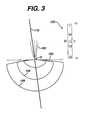
- FIG. 3is a schematic representation of a mitral valve and regurgitant jet with an M-line placed over the jet;
- FIG. 4illustrates a typical color M-mode display acquired along the M-line of FIG. 3;
- FIG. 5illustrates the placement of an adjustable contour graphic over the FCR of the color M-mode display
- FIG. 6illustrates an ultrasound system constructed in accordance with the principles of the present invention.
- FIG. 1a four-chamber ultrasound image of the heart is shown in reverse grayscale.
- the ultrasound image of FIG. 1was acquired transthoracically by an ultrasound probe 10 ′ proximal to the apex of the heart.
- the heartis imaged by a transesophageal echocardiography (TEE) transducer 10 located in the esophagus, which affords high quality images of the heart chambers and valves.
- TEEtransesophageal echocardiography
- An embodiment of the present inventionmay be used to measure regurgitant flow of either aortic or mitral heart valves.
- the embodiment illustrated in the drawingshows mitral regurgitation being measured, as mitral regurgitation is more prevalent than aortic regurgitation and is usually more susceptible to treatment.
- FIG. 1Shown in the image of FIG. 1 is the left ventricle (LV) and the left atrium (LA), separated by the mitral valve 100 . Also depicted in the image is a jet 102 which would be characteristic of regurgitation by the mitral valve 100 . In the past, clinicians would look for the telltale jet 102 as an indication of mitral regurgitation, then infer the size of the leaking valve orifice from the size and shape of the jet 102 . In the illustration of FIG. 1 the jet is shown centered in the LA. However, the orifice of a leaking valve can be eccentrically located so that the jet can be proximal to or directed toward a wall of the atrial chamber. In that case, little about the orifice can be inferred from the eccentric jet, a situation in which the present inventive technique can be performed without degradation.
- the flow convergence region 104 and the jet 102are shown schematically in FIG. 2.
- the jet 102is seen emanating from an orifice 0 in the mitral valve 100 .
- Below the orifice 0 and the mitral valve planeis the FCR 104 .
- the FCRWhen the FCR is imaged as explained below it will exhibit at least two iso-velocity lines 106 and 108 where local blood flow velocities exceed the chosen range of colorflow velocities.
- the iso-velocity linesare located in a colorflow image by looking for color change boundaries.
- the outermost iso-velocity line 106marks a constant velocity boundary radiating approximately spherically around the orifice O, and is located a radial distance of r+d from the orifice.
- the next iso-velocity line 108also marks a constant velocity boundary and is located a radial distance of r from the orifice.
- the flow rate through the orifice Ois measured by acquiring color M-mode data from an M-line location 110 which is aligned with the jet 102 . It is thus not necessary to know the exact location of the orifice O, but only to visualize the jet 102 in the ultrasound image, then place the M-line in alignment with the jet. Such an alignment is illustrated in FIG. 3.
- the resulting color M-mode imageis produce by acquiring Doppler data from along the M-line location at a relatively high rate of acquisition.
- colorflow frame ratesmay be in the range of 10-20 frames per second, depending on image depth and sector width, or slower.
- the M-lineis sampled at a much higher rate in a time-interleaved manner with a concurrently displayed two dimensional (2D) heart image; Doppler data from along the line can be acquired 500-1000 times per second, for instance.
- each heart cycleis sampled at a high temporal resolution each heart cycle, a rate of acquisition which is virtually certain to capture the flow data at the moment of peak regurgitation.
- a typical color M-mode display from such high speed acquisitionis shown in FIG. 4, where each color M-line is oriented vertically in the depth (Z) dimension and successive M-lines are displayed in parallel in the time (t) dimension.
- a systolic phase of the heart cycleis bracketed at the bottom of the display and is seen to contain a distinctively colored region 60 where Doppler aliasing has occurred in the FCR region below the orifice O.
- the flow rate through the orifice Ois computed by measuring the distance d across the aliased region 60 , and the velocities on either side of the region for a particular M-line.
- the opposite sides of the region 60mark iso-velocity boundaries within the FCR.
- the radial distance ris computed from the equation
- Several techniquesmay be used to measure the distance d across and the velocities v 1 and v 2 on either side of the aliasing region 60 .
- Oneis to use a graphic consisting of two contour lines 62 and 64 which can be placed on the FCR region of the color M-mode display as shown in FIG. 5.
- the illustrated graphichas control points located on the lines as delineated by the small boxes in the drawing.
- the graphiccan be moved and reshaped by pulling on the control points with a graphic pointing device such as a mouse until the contour lines 62 and 64 match the outline of the FCR region 60 .
- a second and more preferable techniqueis to use automatic border drawing (ABD) to automatically trace the FCR region 60 , such as one of the techniques discussed in U.S.
- ABSautomatic border drawing
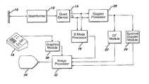
- FIG. 6An ultrasound system constructed in accordance with the principles of the present invention is shown in FIG. 6.
- An array transducer 10 of a TEE probeis operated by a beamformer 12 to transmit ultrasonic beams over a region of the heart and acquires ultrasonic echo information.
- the echoes received by the beamformer for each beam and Doppler ensembleare demodulated by a quadrature demodulator 14 and the I and Q samples are B mode processed by a B mode processor 16 when forming a structural image of the tissue of the heart, and are Doppler processed by a Doppler processor 20 for flow presentation.
- the Doppler signalsare processed for combining with the B mode image in a colorflow display by a CF module 22 , and are processed for a spectral Doppler display by a spectral Doppler module 24 .
- the B mode image data, the colorflow image data, and the spectral Doppler image dataare all coupled to an image processor 32 where they are arranged and formatted for the desired image display.
- the resulting imagesare displayed on a display 34 .
- the image processor 32 and display 34function to display a colorflow ultrasound image, a color M-mode display, and a spectral Doppler display simultaneously. Such a three-image display enables the clinician to monitor all of the critical parameters of the diagnostic procedure at the same time.
- a graphics module 30which is responsive to a user control 18 to locate graphics on the ultrasound images, such as the M-line which is placed over the colorflow image to define the color M-mode acquisition location, and the graphic tracing tool which is used to delineate the FCR region in the color M-mode display.
- the ultrasound system of FIG. 6may be used to quantify mitral valve regurgitation as follows. First, colorflow imaging is performed to image the suspect valve until a regurgitant jet and FCR are seen in the image. The color gain is adjusted so that an appropriate amount of color is seen in the heart chambers; if the gain is set too low, the image will lack necessary sensitivity, and if the gain is set too high there will be too much color blooming. The velocity scale (Doppler Nyquist limit) is adjusted until the FCR region is distinctively seen. This is easily done using the color bar 120 as shown in FIG. 3. In the exemplary color bar shown, the maximum velocity +V in the color display will be shown in yellow (Y).
- Yyellow
- the maximum velocity ⁇ V in the opposite directionis shown in light blue (LB), and flow areas of zero velocity are shown in black (BK). Region of slow flow in the “+” direction are shown in dark red (DR), and regions of slow flow in the “ ⁇ ” direction are shown in dark blue (DB). If the velocity scale is set too high, the aliasing lines in the FCR region will be indistinguishable. If the velocity scale is set too low, the wall filter in the Doppler processor will cut off the Doppler data. When the velocity range is properly scaled, the aliasing lines in this example will appear as follows. Areas outside and below the iso-velocity boundary 106 will appear in dark red, and will transition to yellow at the outermost aliasing line 106 .
- the colorAt the location of the line 106 the color will alias from +V to ⁇ V and will suddenly change from yellow to light blue. Above the iso-velocity boundary 106 the color will transition to dark blue, and at the second aliasing line 108 will undergo a sudden transition to black. This means that at boundary 106 the velocity magnitude is equal to
- the clinicianuses the control 18 to place the color M-mode cursor 110 over the jet in the colorflow image as shown in FIG. 3, and then begins to produce a color M-mode display as shown in FIG. 4.
- the clinicianfreezes the color M-mode display and manipulates the user control to draw or trace the border of the FCR region 60 with the contour lines 62 , 64 as shown in FIG. 5.
- the ultrasound systemcan compute and display the volume flow of the regurgitation calculated as described above from the measured values d, v 1 , and v 2 .
- the volume flowis continuously displayed and updated as the clinician refines the placement of the contour lines 62 , 64 on the color M-mode display.
- the instantaneous volume flow rate measurements Q t(expressed in cm 3 /sec) for each M-line of the color M-mode display during regurgitation can be plotted as a function of time to produce a volume flow rate curve. Integrating the area under the curve produces the total volume flow during regurgitation.
- V t of the flow velocity (expressed in cm/sec) in the orifice areaare produced for the same points in time as Q t , preferably by the CW Doppler technique, and the quotient of Q t /V t at the time of the peak velocity produces a measure of the effective area of the orifice when regurgitation is at its peak and the orifice is generally at its largest.
- Estimation of the EOA over the full interval of regurgitationprovides a profile of the dynamic behavior of the orifice as the event transpires.
- the flow rate curve and spectral Doppler spectrumare displayed simultaneously with synchronized pointers located on the time axis of each display.
- the pointer on the other displaytracks to the same time location.
- a numerical calculation of EOAis displayed for the values of Q t and V t indicated by the instantaneous positions of the two pointers.
- the usercan see and assess the size of the regurgitant orifice for any displayed flow rate or velocity during regurgitation.
Landscapes
- Health & Medical Sciences (AREA)
- Life Sciences & Earth Sciences (AREA)
- Biomedical Technology (AREA)
- Heart & Thoracic Surgery (AREA)
- Hematology (AREA)
- Biophysics (AREA)
- Nuclear Medicine, Radiotherapy & Molecular Imaging (AREA)
- Pathology (AREA)
- Radiology & Medical Imaging (AREA)
- Engineering & Computer Science (AREA)
- Cardiology (AREA)
- Physics & Mathematics (AREA)
- Medical Informatics (AREA)
- Molecular Biology (AREA)
- Surgery (AREA)
- Animal Behavior & Ethology (AREA)
- General Health & Medical Sciences (AREA)
- Public Health (AREA)
- Veterinary Medicine (AREA)
- Ultra Sonic Daignosis Equipment (AREA)
Abstract
Description
- This invention relates to ultrasonic diagnostic imaging systems and, in particular, to ultrasonic diagnostic imaging systems which are capable of detecting and quantifying valvular regurgitant blood flow in the heart.[0001]
- Valvular regurgitation is a serious and potentially life-threatening heart condition. The condition arises when a valve in the heart does not fully close during a particular phase of the heart cycle. Full valve closure is necessary for a complete build-up of the maximum heart chamber blood pressure developed by contraction of the heart. If a valve of the chamber does not close completely, a leak will occur and a jet of blood will escape as the heart contracts. This inefficient operation will cause the heart to expend more effort than it should, can lead to a reduced flow of blood through the body, and in many cases leads to open heart surgery to repair or replace the leaking valve.[0002]
- Ultrasonic detection of valvular regurgitation has traditionally been done by looking for the above-mentioned jet of blood. Over the past fifteen years detection of the jet has been facilitated by colorflow Doppler, in which the high speed and turbulence of the small jet of blood can be detected by careful search for these abnormal local flow velocities near the leaking heart valve. More recently a diagnostic procedure known as the proximal iso-velocity surface area method (PISA) has been endorsed by the cardiology community. In this method the suspect valve and the region inside the heart chamber and proximal to the valve are imaged by colorflow Doppler imaging. At the time of occurrence of the jet a flow convergence region (FCR) is formed in the proximal region as blood flow velocities in the region instantaneously accelerate toward the regurgitant orifice. This flow pattern results in aliasing in the colorflow image as the flow velocities momentarily exceed the velocity range used for the colorflow image. A colorflow image at this moment is captured and frozen on the display screen. A measurement is then made of the velocity v at the first aliasing line of the FCR, and a measurement is made of the distance r from the aliasing line to the center of the valve orifice. These two measurement are then used to compute the flow rate through the orifice using the expression Q[0003]t=2πr2v.
- Several difficulties arise when conducting this procedure. One is that the greatest accuracy is obtained when the jet is captured in the colorflow image at its very peak. The duration of the jet during a heart cycle can be only 300-450 milliseconds, however, while a typical colorflow frame rate may be in the range of 10-20 frames per second. Thus it is probable that the time of acquisition of one of the colorflow image frames will not be the same as the time that the jet is at its peak. The clinician can repeat the colorflow acquisition sequence for additional cardiac cycles, or can settle for the inaccuracy causes by making the measurements at other than the peak of the jet.[0004]
- Another problem is that the center of the valve orifice is not easy to define in the colorflow image. The valve tissue produces large reflections of ultrasound and is moving rapidly as scanning takes place, and can appear as a bulky, blurred or indistinct mass in the image. Thus it is possible that the accuracy of the measurement r will be compromised by the inability to estimate the exact location of the orifice.[0005]
- Yet a third problem is that the PISA method is tedious and exacting, limiting its utility for routine use to measure regurgitant volumes in a clinical setting. The PISA method requires the clinician to make multiple measurements on each image frame of multiple image frames acquired during the regurgitation period of a heart cycle.[0006]
- It is thus desirable to be able to measure the flow rate and volume at the regurgitant valve without such sources of inaccuracy and inconvenience.[0007]
- In accordance with the principles of the present invention an ultrasonic system and technique are described for quantifying valvular regurgitant flow rate and volume. In one embodiment of the inventive technique the regurgitant valve is imaged by colorflow imaging. An M-line is positioned over the valve in the image by referencing the position of the jet, the FCR, and/or the approximate location of the orifice. A sequence of color M-mode measurements is then captured along the M-line. The time-sampled FCR during a cardiac cycle is defined in the color M-mode display by its distinctive aliasing color, and the distance across the FCR and velocities at each end of the distance are recorded. The instantaneous flow rate is then calculated from these measurements, and the volume flow during regurgitation is calculated as an integral of the instantaneous flow rates for all the regurgitant M-lines of a cardiac cycle. Since the sample rate of the M-lines can be one or two orders of magnitude greater than the colorflow frame rate, the peak of the jet can be reliably captured, and the inventive technique does not require any precise definition of the orifice location. Moreover, only one image, the color M-mode image, is needed for the diagnosis of a complete heart cycle.[0008]
- In the drawings:[0009]
- FIG. 1 illustrates a four chamber view of the heart;[0010]
- FIG. 2 is a schematic representation of a mitral valve and regurgitant jet;[0011]
- FIG. 3 is a schematic representation of a mitral valve and regurgitant jet with an M-line placed over the jet;[0012]
- FIG. 4 illustrates a typical color M-mode display acquired along the M-line of FIG. 3;[0013]
- FIG. 5 illustrates the placement of an adjustable contour graphic over the FCR of the color M-mode display; and[0014]
- FIG. 6 illustrates an ultrasound system constructed in accordance with the principles of the present invention.[0015]
- Referring to FIG. 1, a four-chamber ultrasound image of the heart is shown in reverse grayscale. The ultrasound image of FIG. 1 was acquired transthoracically by an[0016]
ultrasound probe 10′ proximal to the apex of the heart. In a preferred embodiment the heart is imaged by a transesophageal echocardiography (TEE)transducer 10 located in the esophagus, which affords high quality images of the heart chambers and valves. An embodiment of the present invention may be used to measure regurgitant flow of either aortic or mitral heart valves. The embodiment illustrated in the drawing shows mitral regurgitation being measured, as mitral regurgitation is more prevalent than aortic regurgitation and is usually more susceptible to treatment. Shown in the image of FIG. 1 is the left ventricle (LV) and the left atrium (LA), separated by themitral valve 100. Also depicted in the image is ajet 102 which would be characteristic of regurgitation by themitral valve 100. In the past, clinicians would look for thetelltale jet 102 as an indication of mitral regurgitation, then infer the size of the leaking valve orifice from the size and shape of thejet 102. In the illustration of FIG. 1 the jet is shown centered in the LA. However, the orifice of a leaking valve can be eccentrically located so that the jet can be proximal to or directed toward a wall of the atrial chamber. In that case, little about the orifice can be inferred from the eccentric jet, a situation in which the present inventive technique can be performed without degradation. - Located in the LV proximal the orifice of the mitral valve is a[0017]
region 104 of flow convergence (FCR). Theflow convergence region 104 and thejet 102 are shown schematically in FIG. 2. Thejet 102 is seen emanating from anorifice 0 in themitral valve 100. Below theorifice 0 and the mitral valve plane is the FCR104. When the FCR is imaged as explained below it will exhibit at least two iso-velocity lines velocity line 106 marks a constant velocity boundary radiating approximately spherically around the orifice O, and is located a radial distance of r+d from the orifice. The next iso-velocity line 108 also marks a constant velocity boundary and is located a radial distance of r from the orifice. - In accordance with the principles of the present invention the flow rate through the orifice O is measured by acquiring color M-mode data from an M-[0018]
line location 110 which is aligned with thejet 102. It is thus not necessary to know the exact location of the orifice O, but only to visualize thejet 102 in the ultrasound image, then place the M-line in alignment with the jet. Such an alignment is illustrated in FIG. 3. The resulting color M-mode image is produce by acquiring Doppler data from along the M-line location at a relatively high rate of acquisition. The time required to acquire a colorflow image of the heart chambers and mitral valve can be quite substantial, as B mode data must be acquired to image the heart structure, then each image line must be sampled numerous times to acquire the colorflow Doppler data. Thus, colorflow frame rates may be in the range of 10-20 frames per second, depending on image depth and sector width, or slower. The M-line is sampled at a much higher rate in a time-interleaved manner with a concurrently displayed two dimensional (2D) heart image; Doppler data from along the line can be acquired 500-1000 times per second, for instance. Thus, the flow through the regurgitant valve is sampled at a high temporal resolution each heart cycle, a rate of acquisition which is virtually certain to capture the flow data at the moment of peak regurgitation. A typical color M-mode display from such high speed acquisition is shown in FIG. 4, where each color M-line is oriented vertically in the depth (Z) dimension and successive M-lines are displayed in parallel in the time (t) dimension. A systolic phase of the heart cycle is bracketed at the bottom of the display and is seen to contain a distinctivelycolored region 60 where Doppler aliasing has occurred in the FCR region below the orifice O. - The flow rate through the orifice O is computed by measuring the distance d across the aliased[0019]
region 60, and the velocities on either side of the region for a particular M-line. The opposite sides of theregion 60 mark iso-velocity boundaries within the FCR. The radial distance r is computed from the equation - Qt=2πr2v2=2π(r+d)2v1
- for a given M-line in the color M-mode display during regurgitation, where Q[0020]tis the instantaneous flow rate, v2is the velocity at the second iso-
velocity line 108 which is a radial distance r from the orifice O, and v1is the velocity at the outermost iso-velocity line 106 which is a radial distance of r+d from the orifice. The integral of these calculations for the systolic M-lines during which regurgitation occurs gives the regurgitant flow volume over the entire regurgitation. The effective orifice area (EOA) is then calculated by measuring the instantaneous velocity Vtat the orifice at corresponding times during regurgitation using spectral Doppler, then computing - EOA=Qt/Vt
- Several techniques may be used to measure the distance d across and the velocities v[0021]1and v2on either side of the
aliasing region 60. One is to use a graphic consisting of twocontour lines contour lines FCR region 60. A second and more preferable technique is to use automatic border drawing (ABD) to automatically trace theFCR region 60, such as one of the techniques discussed in U.S. patent application Ser. No. 09/732,613, filed Dec. 7, 2000. Once the border of the FCR region has been manually or automatically delineated, the ultrasound system can calculate the instantaneous flow rate Qt, total volume flow
- and effective orifice area EOA automatically. An ultrasound system constructed in accordance with the principles of the present invention is shown in FIG. 6. An[0022]
array transducer 10 of a TEE probe is operated by abeamformer 12 to transmit ultrasonic beams over a region of the heart and acquires ultrasonic echo information. The echoes received by the beamformer for each beam and Doppler ensemble are demodulated by aquadrature demodulator 14 and the I and Q samples are B mode processed by aB mode processor 16 when forming a structural image of the tissue of the heart, and are Doppler processed by aDoppler processor 20 for flow presentation. The Doppler signals are processed for combining with the B mode image in a colorflow display by aCF module 22, and are processed for a spectral Doppler display by aspectral Doppler module 24. The B mode image data, the colorflow image data, and the spectral Doppler image data are all coupled to animage processor 32 where they are arranged and formatted for the desired image display. The resulting images are displayed on adisplay 34. Preferably theimage processor 32 anddisplay 34 function to display a colorflow ultrasound image, a color M-mode display, and a spectral Doppler display simultaneously. Such a three-image display enables the clinician to monitor all of the critical parameters of the diagnostic procedure at the same time. Also coupled to theimage processor 32 is agraphics module 30, which is responsive to auser control 18 to locate graphics on the ultrasound images, such as the M-line which is placed over the colorflow image to define the color M-mode acquisition location, and the graphic tracing tool which is used to delineate the FCR region in the color M-mode display. - The ultrasound system of FIG. 6 may be used to quantify mitral valve regurgitation as follows. First, colorflow imaging is performed to image the suspect valve until a regurgitant jet and FCR are seen in the image. The color gain is adjusted so that an appropriate amount of color is seen in the heart chambers; if the gain is set too low, the image will lack necessary sensitivity, and if the gain is set too high there will be too much color blooming. The velocity scale (Doppler Nyquist limit) is adjusted until the FCR region is distinctively seen. This is easily done using the[0023]
color bar 120 as shown in FIG. 3. In the exemplary color bar shown, the maximum velocity +V in the color display will be shown in yellow (Y). The maximum velocity −V in the opposite direction is shown in light blue (LB), and flow areas of zero velocity are shown in black (BK). Region of slow flow in the “+” direction are shown in dark red (DR), and regions of slow flow in the “−” direction are shown in dark blue (DB). If the velocity scale is set too high, the aliasing lines in the FCR region will be indistinguishable. If the velocity scale is set too low, the wall filter in the Doppler processor will cut off the Doppler data. When the velocity range is properly scaled, the aliasing lines in this example will appear as follows. Areas outside and below the iso-velocity boundary 106 will appear in dark red, and will transition to yellow at theoutermost aliasing line 106. At the location of theline 106 the color will alias from +V to −V and will suddenly change from yellow to light blue. Above the iso-velocity boundary 106 the color will transition to dark blue, and at thesecond aliasing line 108 will undergo a sudden transition to black. This means that atboundary 106 the velocity magnitude is equal to |V|, and atboundary 108 the velocity magnitude is equal to |2| when no baseline shift is applied. - When these boundaries are separate and distinct in the image the clinician uses the[0024]
control 18 to place the color M-mode cursor 110 over the jet in the colorflow image as shown in FIG. 3, and then begins to produce a color M-mode display as shown in FIG. 4. With the FCR region distinctly seen in both images, the clinician freezes the color M-mode display and manipulates the user control to draw or trace the border of theFCR region 60 with thecontour lines region 60, the ultrasound system can compute and display the volume flow of the regurgitation calculated as described above from the measured values d, v1, and v2. Preferably the volume flow is continuously displayed and updated as the clinician refines the placement of thecontour lines

Claims (28)
1. A method of ultrasonically assessing valve regurgitation in the cardiovascular system comprising:
identifying a suspected regurgitant valve;
acquiring velocity information from at least two points on a line extending through the suspected regurgitant valve; and
utilizing the velocity information to estimate regurgitant flow through the suspected valve.
2. The method ofclaim 1 , wherein acquiring comprises acquiring velocity information by the Doppler technique from points on the line.
3. The method ofclaim 2 , wherein the line comprises an M-line, and wherein acquiring comprises acquiring color Doppler M-mode information from along the M-line.
4. The method ofclaim 1 , wherein identifying comprises identifying a regurgitant jet in the vicinity of the suspected valve in an ultrasound image.
5. The method ofclaim 4 , wherein acquiring further comprises aligning a cursor with the regurgitant jet in the ultrasound image.
6. The method ofclaim 5 , wherein the cursor comprises an M-line, and wherein acquiring comprises acquiring color Doppler M-mode information from along the M-line location.
7. The method ofclaim 1 , wherein the suspected regurgitant valve is characterized by a flow convergence region in the vicinity of the valve; and wherein acquiring comprises acquiring velocity information from two iso-velocity boundaries of the flow convergence region.
8. The method ofclaim 7 , wherein utilizing comprises utilizing the velocity information from the two iso-velocity boundaries to calculate flow rate during valve regurgitation.
9. The method ofclaim 8 , further comprising measuring the Doppler velocity of flow in the vicinity of the suspected valve during regurgitation; and wherein utilizing further comprises utilizing the calculated flow rate and measured Doppler velocity to estimate the size of a regurgitant orifice.
10. The method ofclaim 7 , wherein acquiring further comprises imaging the flow convergence region by the Doppler M-mode technique; and tracing iso-velocity boundaries in imaged flow convergence region.
11. The method ofclaim 10 , wherein tracing comprises tracing the iso-velocity boundaries with a manually controlled graphic tracing tool.
12. The method ofclaim 10 , wherein tracing comprises tracing the iso-velocity boundaries by automatic border drawing.
13. The method ofclaim 1 , wherein identifying comprises visualizing the suspected valve in a two dimensional ultrasound image; and wherein acquiring comprises acquiring Doppler velocity information from along a line extending through the region of regurgitant flow in the two dimensional image.
14. The method ofclaim 13 , wherein the line comprises an M-line, and wherein acquiring comprises acquiring color Doppler M-mode information from along the M-line location.
15. The method ofclaim 14 , wherein the two dimensional image exhibits a given frame rate of display, and wherein the rate of acquisition of Doppler M-mode information from along the M-line location is greater than the given frame rate.
16. The method ofclaim 14 , wherein the two dimensional image is a colorflow Doppler image.
17. A method of ultrasonically assessing valve regurgitation in the cardiovascular system comprising:
displaying a colorflow Doppler image of a suspected regurgitant valve; and
simultaneously displaying a Doppler M-mode display acquired from an M-line located in the colorflow Doppler image.
18. The method ofclaim 17 , wherein the M-line is located in the region of suspected regurgitation in the colorflow Doppler image.
19. The method ofclaim 18 , further comprising utilizing the data of the Doppler M-mode display to estimate regurgitant flow rate; and displaying an estimate of regurgitant flow rate.
20. A method of ultrasonically assessing valve regurgitation in the cardiovascular system comprising:
acquiring velocity information from the vicinity of a suspected regurgitant valve by the Doppler M-mode technique;
utilizing the velocity information to display regurgitant volume flow rate as a function of time during a given cardiac cycle; and
displaying regurgitant flow velocity as a function of time during the given cardiac cycle.
21. The method ofclaim 20 , further comprising utilizing time-aligned regurgitant volume flow rate data and regurgitant flow velocity data to estimate the size of a regurgitant orifice.
22. The method ofclaim 20 , further comprising aggregating regurgitant volume flow rate data over a cardiac cycle to estimate total volume flow during a regurgitation event.
23. A method of ultrasonically assessing valve regurgitation in the cardiovascular system comprising:
identifying a suspected regurgitant valve in a colorflow Doppler image;
acquiring Doppler M-mode information from the region of the suspected regurgitant valve; and
utilizing the Doppler M-mode information to estimate regurgitant flow through the suspected valve.
24. The method ofclaim 23 , wherein identifying further comprises adjusting a colorflow Doppler parameter so that a flow convergence region in the vicinity of the suspected regurgitant valve is delineated in the image.
25. The method ofclaim 24 , wherein the colorflow Doppler parameter is the velocity scale.
26. The method ofclaim 25 wherein adjusting comprises adjusting the velocity scale so that at least two aliasing boundaries are seen in the flow convergence region.
27. The method ofclaim 26 wherein acquiring further comprises acquiring velocity measurements at the aliasing boundaries; and wherein utilizing comprises utilizing the velocity measurements to estimate volume flow through the valve during regurgitation.
28. The method ofclaim 27 , wherein utilizing further comprises utilizing estimated volume flow to estimate the size of a valve orifice during regurgitation.
Priority Applications (1)
| Application Number | Priority Date | Filing Date | Title |
|---|---|---|---|
| US09/794,528US6719697B2 (en) | 2001-02-27 | 2001-02-27 | Ultrasonic quantification of valvular regurgitant blood flow |
Applications Claiming Priority (1)
| Application Number | Priority Date | Filing Date | Title |
|---|---|---|---|
| US09/794,528US6719697B2 (en) | 2001-02-27 | 2001-02-27 | Ultrasonic quantification of valvular regurgitant blood flow |
Publications (2)
| Publication Number | Publication Date |
|---|---|
| US20020151794A1true US20020151794A1 (en) | 2002-10-17 |
| US6719697B2 US6719697B2 (en) | 2004-04-13 |
Family
ID=25162899
Family Applications (1)
| Application Number | Title | Priority Date | Filing Date |
|---|---|---|---|
| US09/794,528Expired - Fee RelatedUS6719697B2 (en) | 2001-02-27 | 2001-02-27 | Ultrasonic quantification of valvular regurgitant blood flow |
Country Status (1)
| Country | Link |
|---|---|
| US (1) | US6719697B2 (en) |
Cited By (15)
| Publication number | Priority date | Publication date | Assignee | Title |
|---|---|---|---|---|
| US20050107701A1 (en)* | 2003-11-19 | 2005-05-19 | Dubberstein David T. | Automatic color gain adjustments |
| WO2007107925A3 (en)* | 2006-03-21 | 2008-01-03 | Koninkl Philips Electronics Nv | Optimization of velocity scale for color tissue doppler imaging |
| US20080177279A1 (en)* | 2007-01-09 | 2008-07-24 | Cyberheart, Inc. | Depositing radiation in heart muscle under ultrasound guidance |
| US20080177280A1 (en)* | 2007-01-09 | 2008-07-24 | Cyberheart, Inc. | Method for Depositing Radiation in Heart Muscle |
| US20090187100A1 (en)* | 2006-05-15 | 2009-07-23 | Sengupta Partho P | Method for Imaging Intracavitary Blood Flow Patterns |
| WO2010046330A1 (en)* | 2008-10-24 | 2010-04-29 | Tomtec Imaging Systems Gmbh | Three-dimensional derivation of a proximal isokinetic shell of a proximal flow convergence zone and three-dimensional pisa flow measurement |
| US20110040188A1 (en)* | 2009-08-11 | 2011-02-17 | Tadashi Tamura | Methods and apparatus for ultrasound imaging |
| US20110166407A1 (en)* | 2009-07-17 | 2011-07-07 | Cyberheart, Inc. | Heart Treatment Kit, System, and Method For Radiosurgically Alleviating Arrhythmia |
| WO2012085798A1 (en)* | 2010-12-23 | 2012-06-28 | Koninklijke Philips Electronics N.V. | Automated identification of the location of a regurgitant orifice of a mitral valve in an ultrasound image |
| WO2012085797A1 (en)* | 2010-12-23 | 2012-06-28 | Koninklijke Philips Electronics N.V. | Analysis of mitral regurgitation by ultrasonic imaging |
| WO2012085778A1 (en)* | 2010-12-23 | 2012-06-28 | Koninklijke Philips Electronics N.V. | Analysis of mitral regurgitation from slit orifices by ultrasonic imaging |
| US20120215110A1 (en)* | 2011-02-23 | 2012-08-23 | Siemens Medical Solutions Usa, Inc. | Multiple Beam Spectral Doppler in Medical Diagnostic Ultrasound Imaging |
| CN103379864A (en)* | 2010-12-23 | 2013-10-30 | 皇家飞利浦电子股份有限公司 | Wall filter for ultrasonic mitral regurgitation analysis |
| WO2014001955A1 (en)* | 2012-06-27 | 2014-01-03 | Koninklijke Philips N.V. | Ultrasonic color flow map for analysis of mitral regurgitation |
| US11786212B1 (en)* | 2022-12-15 | 2023-10-17 | UAB Ligence | Echocardiogram classification with machine learning |
Families Citing this family (7)
| Publication number | Priority date | Publication date | Assignee | Title |
|---|---|---|---|---|
| US7270635B2 (en)* | 2002-02-27 | 2007-09-18 | Esaote S.p.A | Flow-rate conservative doppler estimate |
| CN101141920B (en)* | 2005-03-15 | 2011-12-14 | 株式会社东芝 | Ultrasonic diagnostic equipment and its controlling program |
| JP5034054B2 (en)* | 2006-03-31 | 2012-09-26 | 国立大学法人京都工芸繊維大学 | Image processing apparatus, ultrasonic imaging apparatus including the same, and image processing method |
| US20090043208A1 (en)* | 2007-08-10 | 2009-02-12 | Norwegian University Of Science And Technology | Methods and devices for estimating blood flow characteristics |
| US9320496B2 (en) | 2010-02-25 | 2016-04-26 | Siemens Medical Solutions Usa, Inc. | Volumetric is quantification for ultrasound diagnostic imaging |
| US9179892B2 (en)* | 2010-11-08 | 2015-11-10 | General Electric Company | System and method for ultrasound imaging |
| EP2709059B1 (en) | 2012-09-17 | 2014-11-05 | Pie Medical Imaging BV | Method and apparatus for quantitative measurements on sequences of images, particularly angiographic images |
Citations (10)
| Publication number | Priority date | Publication date | Assignee | Title |
|---|---|---|---|---|
| US4608993A (en)* | 1984-07-31 | 1986-09-02 | Quinton Instrument Company | Blood flow measurement device and method |
| US4790322A (en)* | 1985-07-24 | 1988-12-13 | Kabushiki Kaisha Toshiba | Ultrasonic type blood flow amount measuring apparatus |
| US4913159A (en)* | 1989-03-17 | 1990-04-03 | Hitachi Medial Corp. | Method for determining blood flow through a narrowed orifice using color doppler echocardiography |
| US4926872A (en)* | 1988-03-28 | 1990-05-22 | Hewlett-Packard Company | Ultrasonic transducer system and method for the operation thereof |
| US5010528A (en)* | 1988-06-30 | 1991-04-23 | Shigeo Ohtsuki | Doppler flow velocity distribution measuring apparatus |
| US5105816A (en)* | 1989-05-20 | 1992-04-21 | Fujitsu Limited | Method and system for making blood flow visible |
| US5425365A (en)* | 1992-03-26 | 1995-06-20 | Kabushiki Kaisha Toshiba | Ultrasonic diagnosis apparatus utilizing Doppler technique |
| US5551434A (en)* | 1994-06-22 | 1996-09-03 | Kabushiki Kaisha Toshiba | Ultrasonic imaging diagnosis apparatus |
| US5622174A (en)* | 1992-10-02 | 1997-04-22 | Kabushiki Kaisha Toshiba | Ultrasonic diagnosis apparatus and image displaying system |
| US6149595A (en)* | 1998-07-02 | 2000-11-21 | Seitz; Walter S. | Noninvasive apparatus and method for the determination of cardiac valve function |
- 2001
- 2001-02-27USUS09/794,528patent/US6719697B2/ennot_activeExpired - Fee Related
Patent Citations (10)
| Publication number | Priority date | Publication date | Assignee | Title |
|---|---|---|---|---|
| US4608993A (en)* | 1984-07-31 | 1986-09-02 | Quinton Instrument Company | Blood flow measurement device and method |
| US4790322A (en)* | 1985-07-24 | 1988-12-13 | Kabushiki Kaisha Toshiba | Ultrasonic type blood flow amount measuring apparatus |
| US4926872A (en)* | 1988-03-28 | 1990-05-22 | Hewlett-Packard Company | Ultrasonic transducer system and method for the operation thereof |
| US5010528A (en)* | 1988-06-30 | 1991-04-23 | Shigeo Ohtsuki | Doppler flow velocity distribution measuring apparatus |
| US4913159A (en)* | 1989-03-17 | 1990-04-03 | Hitachi Medial Corp. | Method for determining blood flow through a narrowed orifice using color doppler echocardiography |
| US5105816A (en)* | 1989-05-20 | 1992-04-21 | Fujitsu Limited | Method and system for making blood flow visible |
| US5425365A (en)* | 1992-03-26 | 1995-06-20 | Kabushiki Kaisha Toshiba | Ultrasonic diagnosis apparatus utilizing Doppler technique |
| US5622174A (en)* | 1992-10-02 | 1997-04-22 | Kabushiki Kaisha Toshiba | Ultrasonic diagnosis apparatus and image displaying system |
| US5551434A (en)* | 1994-06-22 | 1996-09-03 | Kabushiki Kaisha Toshiba | Ultrasonic imaging diagnosis apparatus |
| US6149595A (en)* | 1998-07-02 | 2000-11-21 | Seitz; Walter S. | Noninvasive apparatus and method for the determination of cardiac valve function |
Cited By (37)
| Publication number | Priority date | Publication date | Assignee | Title |
|---|---|---|---|---|
| US6979295B2 (en) | 2003-11-19 | 2005-12-27 | Ge Medical Systems Global Technology Company, Llc | Automatic color gain adjustments |
| US20050107701A1 (en)* | 2003-11-19 | 2005-05-19 | Dubberstein David T. | Automatic color gain adjustments |
| WO2007107925A3 (en)* | 2006-03-21 | 2008-01-03 | Koninkl Philips Electronics Nv | Optimization of velocity scale for color tissue doppler imaging |
| US20090067699A1 (en)* | 2006-03-21 | 2009-03-12 | Koninklijke Philips Electronics, N.V. | Optimization of velocity scale for color tissue doppler imaging |
| US20090187100A1 (en)* | 2006-05-15 | 2009-07-23 | Sengupta Partho P | Method for Imaging Intracavitary Blood Flow Patterns |
| US8328724B2 (en)* | 2006-05-15 | 2012-12-11 | Mayo Foundation For Medical Education And Research | Method for imaging intracavitary blood flow patterns |
| US20080177279A1 (en)* | 2007-01-09 | 2008-07-24 | Cyberheart, Inc. | Depositing radiation in heart muscle under ultrasound guidance |
| US20080177280A1 (en)* | 2007-01-09 | 2008-07-24 | Cyberheart, Inc. | Method for Depositing Radiation in Heart Muscle |
| JP2012506271A (en)* | 2008-10-24 | 2012-03-15 | トムテック イメージング システムズ ゲゼルシャフト ミット ベシュレンクテル ハフツング | 3D derivation and 3D PISA flow measurement of the proximal constant velocity shell in the proximal flow convergence region |
| WO2010046330A1 (en)* | 2008-10-24 | 2010-04-29 | Tomtec Imaging Systems Gmbh | Three-dimensional derivation of a proximal isokinetic shell of a proximal flow convergence zone and three-dimensional pisa flow measurement |
| US8911375B2 (en) | 2008-10-24 | 2014-12-16 | Tomtec Imaging Systems Gmbh | Three-dimensional derivation of a proximal isokinetic shell of a proximal flow convergence zone and three-dimensional PISA flow measurement |
| US9320916B2 (en) | 2009-07-17 | 2016-04-26 | Cyberheart, Inc. | Heart treatment kit, system, and method for radiosurgically alleviating arrhythmia |
| US20110166407A1 (en)* | 2009-07-17 | 2011-07-07 | Cyberheart, Inc. | Heart Treatment Kit, System, and Method For Radiosurgically Alleviating Arrhythmia |
| US8784290B2 (en) | 2009-07-17 | 2014-07-22 | Cyberheart, Inc. | Heart treatment kit, system, and method for radiosurgically alleviating arrhythmia |
| US8480590B2 (en)* | 2009-08-11 | 2013-07-09 | Hitachi Aloka Medical, Ltd. | Methods and apparatus for ultrasound imaging |
| WO2011019853A3 (en)* | 2009-08-11 | 2011-06-03 | Aloka Co., Ltd. | Methods and apparatus for ultrasound imaging |
| US20110040188A1 (en)* | 2009-08-11 | 2011-02-17 | Tadashi Tamura | Methods and apparatus for ultrasound imaging |
| CN103269644A (en)* | 2010-12-23 | 2013-08-28 | 皇家飞利浦电子股份有限公司 | Automated identification of the location of a regurgitant orifice of a mitral valve in an ultrasound image |
| WO2012085778A1 (en)* | 2010-12-23 | 2012-06-28 | Koninklijke Philips Electronics N.V. | Analysis of mitral regurgitation from slit orifices by ultrasonic imaging |
| US20130261458A1 (en)* | 2010-12-23 | 2013-10-03 | Koninklijke Philips Electronics N.V. | Analysis of mitral regurgitation from slit orifices by ultrasonic imaging |
| CN103379864A (en)* | 2010-12-23 | 2013-10-30 | 皇家飞利浦电子股份有限公司 | Wall filter for ultrasonic mitral regurgitation analysis |
| CN103391748A (en)* | 2010-12-23 | 2013-11-13 | 皇家飞利浦电子股份有限公司 | Analysis of Mitral Regurgitation from the Slit Hole by Ultrasound Imaging |
| US10463341B2 (en)* | 2010-12-23 | 2019-11-05 | Koninklijke Philips N.V. | Analysis of mitral regurgitation from slit orifices by ultrasonic imaging |
| JP2014500118A (en)* | 2010-12-23 | 2014-01-09 | コーニンクレッカ フィリップス エヌ ヴェ | Wall filter for backflow ultrasonic analysis of mitral valve |
| US10231693B2 (en) | 2010-12-23 | 2019-03-19 | Koninklijke Philips N.V. | Automated identification of the location of a regurgitant orifice of a mitral valve in an ultrasound image |
| WO2012085797A1 (en)* | 2010-12-23 | 2012-06-28 | Koninklijke Philips Electronics N.V. | Analysis of mitral regurgitation by ultrasonic imaging |
| US10674993B2 (en) | 2010-12-23 | 2020-06-09 | Koninklijke Philips N.V. | Analysis of mitral regurgitation by ultrasonic imaging |
| CN103391748B (en)* | 2010-12-23 | 2016-03-02 | 皇家飞利浦电子股份有限公司 | Analysis of Mitral Regurgitation from the Slit Hole by Ultrasound Imaging |
| WO2012085798A1 (en)* | 2010-12-23 | 2012-06-28 | Koninklijke Philips Electronics N.V. | Automated identification of the location of a regurgitant orifice of a mitral valve in an ultrasound image |
| RU2596722C2 (en)* | 2010-12-23 | 2016-09-10 | Конинклейке Филипс Электроникс Н.В. | Analysis of mitral regurgitation from slit orifices by ultrasonic imaging |
| US9398898B2 (en)* | 2011-02-23 | 2016-07-26 | Siemens Medical Solutions Usa, Inc. | Multiple beam spectral doppler in medical diagnostic ultrasound imaging |
| US20120215110A1 (en)* | 2011-02-23 | 2012-08-23 | Siemens Medical Solutions Usa, Inc. | Multiple Beam Spectral Doppler in Medical Diagnostic Ultrasound Imaging |
| WO2014001955A1 (en)* | 2012-06-27 | 2014-01-03 | Koninklijke Philips N.V. | Ultrasonic color flow map for analysis of mitral regurgitation |
| RU2652257C2 (en)* | 2012-06-27 | 2018-04-25 | Конинклейке Филипс Н.В. | Ultrasonic flow color map for study of mitral regurgitation |
| US10512444B2 (en)* | 2012-06-27 | 2019-12-24 | Koninklijke Philips N.V. | Ultrasonic color flow map for analysis of mitral regurgitation |
| US20150148679A1 (en)* | 2012-06-27 | 2015-05-28 | Koninklijke Philips N.V. | Ultrasonic color flow map for analysis of mitral regurgitation |
| US11786212B1 (en)* | 2022-12-15 | 2023-10-17 | UAB Ligence | Echocardiogram classification with machine learning |
Also Published As
| Publication number | Publication date |
|---|---|
| US6719697B2 (en) | 2004-04-13 |
Similar Documents
| Publication | Publication Date | Title |
|---|---|---|
| US6719697B2 (en) | Ultrasonic quantification of valvular regurgitant blood flow | |
| US10874373B2 (en) | Method and system for measuring flow through a heart valve | |
| US5785655A (en) | Two-dimensional ultrasound display system | |
| US5471990A (en) | Ultrasonic doppler power measurement and display system | |
| Lima et al. | Noninvasive prediction of transvalvular pressure gradient in patients with pulmonary stenosis by quantitative two-dimensional echocardiographic Doppler studies. | |
| US4257278A (en) | Quantitative volume blood flow measurement by an ultrasound imaging system featuring a Doppler modality | |
| EP0747010B1 (en) | Continuous display of cardiac blood flow information | |
| US7044913B2 (en) | Ultrasonic diagnosis apparatus | |
| US6068598A (en) | Method and apparatus for automatic Doppler angle estimation in ultrasound imaging | |
| CN100430025C (en) | Ultrasonic diagnostic device and image processing device | |
| EP0842638A2 (en) | Ultrasonic diagnostic imaging system with real time volume flow calculation | |
| US10512444B2 (en) | Ultrasonic color flow map for analysis of mitral regurgitation | |
| US8882675B2 (en) | Methods and apparatus for ultrasound imaging | |
| US20140046606A1 (en) | Methods and apparatus for multibeam doppler ultrasound display | |
| EP2654571B1 (en) | Analysis of mitral regurgitation from slit orifices by ultrasonic imaging | |
| US10674993B2 (en) | Analysis of mitral regurgitation by ultrasonic imaging | |
| US8480590B2 (en) | Methods and apparatus for ultrasound imaging | |
| EP1021129B1 (en) | Ultrasound imaging for displaying strain | |
| US10231693B2 (en) | Automated identification of the location of a regurgitant orifice of a mitral valve in an ultrasound image | |
| US10398407B2 (en) | Wall filter for ultrasonic mitral regurgitation analysis | |
| JP3443189B2 (en) | Ultrasound diagnostic equipment | |
| JP3182419B2 (en) | Blood flow measurement and display device | |
| US20230143880A1 (en) | Three dimensional color doppler for ultrasonic volume flow measurement | |
| US6048317A (en) | Method and apparatus for assisting a user in positioning an ultrasonic transducer | |
| Waggoner et al. | Principles and physics of Doppler |
Legal Events
| Date | Code | Title | Description |
|---|---|---|---|
| AS | Assignment | Owner name:KONINKLIJKE PHILIPS ELECTRONICS N.V., NETHERLANDS Free format text:ASSIGNMENT OF ASSIGNORS INTEREST;ASSIGNOR:LI, XIANG-NING;REEL/FRAME:011598/0382 Effective date:20010226 | |
| FPAY | Fee payment | Year of fee payment:4 | |
| REMI | Maintenance fee reminder mailed | ||
| LAPS | Lapse for failure to pay maintenance fees | ||
| STCH | Information on status: patent discontinuation | Free format text:PATENT EXPIRED DUE TO NONPAYMENT OF MAINTENANCE FEES UNDER 37 CFR 1.362 | |
| FP | Lapsed due to failure to pay maintenance fee | Effective date:20120413 |