US20020028006A1 - Interactive computer-aided diagnosis method and system for assisting diagnosis of lung nodules in digital volumetric medical images - Google Patents
Interactive computer-aided diagnosis method and system for assisting diagnosis of lung nodules in digital volumetric medical imagesDownload PDFInfo
- Publication number
- US20020028006A1 US20020028006A1US09/840,267US84026701AUS2002028006A1US 20020028006 A1US20020028006 A1US 20020028006A1US 84026701 AUS84026701 AUS 84026701AUS 2002028006 A1US2002028006 A1US 2002028006A1
- Authority
- US
- United States
- Prior art keywords
- interest
- anatomical structure
- real
- time
- user interface
- Prior art date
- Legal status (The legal status is an assumption and is not a legal conclusion. Google has not performed a legal analysis and makes no representation as to the accuracy of the status listed.)
- Granted
Links
Images


Classifications
- G—PHYSICS
- G06—COMPUTING OR CALCULATING; COUNTING
- G06T—IMAGE DATA PROCESSING OR GENERATION, IN GENERAL
- G06T7/00—Image analysis
- G06T7/0002—Inspection of images, e.g. flaw detection
- G06T7/0012—Biomedical image inspection
- G—PHYSICS
- G06—COMPUTING OR CALCULATING; COUNTING
- G06T—IMAGE DATA PROCESSING OR GENERATION, IN GENERAL
- G06T7/00—Image analysis
- G06T7/10—Segmentation; Edge detection
- G06T7/11—Region-based segmentation
- G—PHYSICS
- G06—COMPUTING OR CALCULATING; COUNTING
- G06T—IMAGE DATA PROCESSING OR GENERATION, IN GENERAL
- G06T7/00—Image analysis
- G06T7/10—Segmentation; Edge detection
- G06T7/136—Segmentation; Edge detection involving thresholding
- G—PHYSICS
- G06—COMPUTING OR CALCULATING; COUNTING
- G06V—IMAGE OR VIDEO RECOGNITION OR UNDERSTANDING
- G06V20/00—Scenes; Scene-specific elements
- G06V20/60—Type of objects
- G06V20/69—Microscopic objects, e.g. biological cells or cellular parts
- G—PHYSICS
- G16—INFORMATION AND COMMUNICATION TECHNOLOGY [ICT] SPECIALLY ADAPTED FOR SPECIFIC APPLICATION FIELDS
- G16H—HEALTHCARE INFORMATICS, i.e. INFORMATION AND COMMUNICATION TECHNOLOGY [ICT] SPECIALLY ADAPTED FOR THE HANDLING OR PROCESSING OF MEDICAL OR HEALTHCARE DATA
- G16H15/00—ICT specially adapted for medical reports, e.g. generation or transmission thereof
- G—PHYSICS
- G16—INFORMATION AND COMMUNICATION TECHNOLOGY [ICT] SPECIALLY ADAPTED FOR SPECIFIC APPLICATION FIELDS
- G16H—HEALTHCARE INFORMATICS, i.e. INFORMATION AND COMMUNICATION TECHNOLOGY [ICT] SPECIALLY ADAPTED FOR THE HANDLING OR PROCESSING OF MEDICAL OR HEALTHCARE DATA
- G16H30/00—ICT specially adapted for the handling or processing of medical images
- G16H30/40—ICT specially adapted for the handling or processing of medical images for processing medical images, e.g. editing
- G—PHYSICS
- G16—INFORMATION AND COMMUNICATION TECHNOLOGY [ICT] SPECIALLY ADAPTED FOR SPECIFIC APPLICATION FIELDS
- G16H—HEALTHCARE INFORMATICS, i.e. INFORMATION AND COMMUNICATION TECHNOLOGY [ICT] SPECIALLY ADAPTED FOR THE HANDLING OR PROCESSING OF MEDICAL OR HEALTHCARE DATA
- G16H50/00—ICT specially adapted for medical diagnosis, medical simulation or medical data mining; ICT specially adapted for detecting, monitoring or modelling epidemics or pandemics
- G16H50/50—ICT specially adapted for medical diagnosis, medical simulation or medical data mining; ICT specially adapted for detecting, monitoring or modelling epidemics or pandemics for simulation or modelling of medical disorders
- G—PHYSICS
- G16—INFORMATION AND COMMUNICATION TECHNOLOGY [ICT] SPECIALLY ADAPTED FOR SPECIFIC APPLICATION FIELDS
- G16H—HEALTHCARE INFORMATICS, i.e. INFORMATION AND COMMUNICATION TECHNOLOGY [ICT] SPECIALLY ADAPTED FOR THE HANDLING OR PROCESSING OF MEDICAL OR HEALTHCARE DATA
- G16H70/00—ICT specially adapted for the handling or processing of medical references
- G16H70/60—ICT specially adapted for the handling or processing of medical references relating to pathologies
- G—PHYSICS
- G06—COMPUTING OR CALCULATING; COUNTING
- G06T—IMAGE DATA PROCESSING OR GENERATION, IN GENERAL
- G06T2207/00—Indexing scheme for image analysis or image enhancement
- G06T2207/10—Image acquisition modality
- G06T2207/10072—Tomographic images
- G06T2207/10081—Computed x-ray tomography [CT]
- G—PHYSICS
- G06—COMPUTING OR CALCULATING; COUNTING
- G06T—IMAGE DATA PROCESSING OR GENERATION, IN GENERAL
- G06T2207/00—Indexing scheme for image analysis or image enhancement
- G06T2207/20—Special algorithmic details
- G06T2207/20004—Adaptive image processing
- G06T2207/20012—Locally adaptive
- G—PHYSICS
- G06—COMPUTING OR CALCULATING; COUNTING
- G06T—IMAGE DATA PROCESSING OR GENERATION, IN GENERAL
- G06T2207/00—Indexing scheme for image analysis or image enhancement
- G06T2207/20—Special algorithmic details
- G06T2207/20112—Image segmentation details
- G06T2207/20132—Image cropping
- G—PHYSICS
- G06—COMPUTING OR CALCULATING; COUNTING
- G06T—IMAGE DATA PROCESSING OR GENERATION, IN GENERAL
- G06T2207/00—Indexing scheme for image analysis or image enhancement
- G06T2207/20—Special algorithmic details
- G06T2207/20112—Image segmentation details
- G06T2207/20156—Automatic seed setting
- G—PHYSICS
- G06—COMPUTING OR CALCULATING; COUNTING
- G06T—IMAGE DATA PROCESSING OR GENERATION, IN GENERAL
- G06T2207/00—Indexing scheme for image analysis or image enhancement
- G06T2207/30—Subject of image; Context of image processing
- G06T2207/30004—Biomedical image processing
- G06T2207/30061—Lung
- G06T2207/30064—Lung nodule
- G—PHYSICS
- G16—INFORMATION AND COMMUNICATION TECHNOLOGY [ICT] SPECIALLY ADAPTED FOR SPECIFIC APPLICATION FIELDS
- G16H—HEALTHCARE INFORMATICS, i.e. INFORMATION AND COMMUNICATION TECHNOLOGY [ICT] SPECIALLY ADAPTED FOR THE HANDLING OR PROCESSING OF MEDICAL OR HEALTHCARE DATA
- G16H10/00—ICT specially adapted for the handling or processing of patient-related medical or healthcare data
- G16H10/60—ICT specially adapted for the handling or processing of patient-related medical or healthcare data for patient-specific data, e.g. for electronic patient records
- G—PHYSICS
- G16—INFORMATION AND COMMUNICATION TECHNOLOGY [ICT] SPECIALLY ADAPTED FOR SPECIFIC APPLICATION FIELDS
- G16H—HEALTHCARE INFORMATICS, i.e. INFORMATION AND COMMUNICATION TECHNOLOGY [ICT] SPECIALLY ADAPTED FOR THE HANDLING OR PROCESSING OF MEDICAL OR HEALTHCARE DATA
- G16H40/00—ICT specially adapted for the management or administration of healthcare resources or facilities; ICT specially adapted for the management or operation of medical equipment or devices
- G16H40/60—ICT specially adapted for the management or administration of healthcare resources or facilities; ICT specially adapted for the management or operation of medical equipment or devices for the operation of medical equipment or devices
- G16H40/63—ICT specially adapted for the management or administration of healthcare resources or facilities; ICT specially adapted for the management or operation of medical equipment or devices for the operation of medical equipment or devices for local operation
Definitions
- the present inventiongenerally relates to computer-assisted diagnosis (CADX) and, in particular, to an interactive computer-aided diagnosis (ICAD) method and system for assisting diagnosis of lung nodules in digital volumetric medical images.
- CADXcomputer-assisted diagnosis
- ICADinteractive computer-aided diagnosis
- CADxComputer-Aided diagnosis
- Current clinical practicecancer or other diseases may be missed during a physician's un-aided examination of medical image data, in part because of the large volume of data. This is particularly a problem for screening applications, since there is generally little time to devote to the examination of each patient's data, and the entire range of the data must be examined to make sure it is free from disease.
- Computer analysis that is performed silently in the backgroundcan greatly aid physicians in their work.
- ICADinteractive computer-aided diagnosis
- the method and systemmesh seamlessly with physicians in their current work practices.
- the inventiontakes advantage of their knowledge, augmenting it with the strengths of the computer in rapid computation of numeric values.
- Such an inventionis an “open box”, increasing its acceptance among physicians.
- the inventionprovides results in “real-time” so that it does not add a time delay to the physician's examination process.
- a computer-assisted diagnosis methodfor assisting diagnosis of anatomical structures in a digital volumetric medical image of at least one lung.
- An anatomical structure of interestis identified in the volumetric digital medical image.
- the anatomical structure of interestis automatically segmented, in real-time, in a predefined volume of interest (VOI).
- Quantitative measurements of the anatomical structure of interestare automatically computed, in real-time.
- a result of the segmenting step and a result of the computing stepare displayed, in real-time.
- a likelihood that the anatomical structure of interest corresponds to a disease or an area warranting further investigationis estimating, in real-time, based on predefined criteria and the quantitative measurements.
- a warningis generated, in real-time, when the likelihood is above a predefined threshold.
- the methodfurther includes the step of generating a graphical user interface having a main window for displaying at least one view corresponding to the at least one lung.
- the at least one viewis at least one of an axial view and a maximum intensity projection view.
- the methodfurther includes the step of displaying at least one of the result of the segmenting step and the result of the computing step in a supplemental window or a pop-up window of the graphical user interface.
- the methodfurther includes the step of alternately displaying at least one of at least two different sets of display parameters in a supplemental window of the graphical user interface to view an extent of calcification of the anatomical structure of interest.
- the methodfurther includes the step of determining a local spinning plane for the anatomical structure of interest.
- the local spinning planeis centered at a centroid and a local spinning axis of the anatomical structure of interest.
- the local spinning planeis rotated at least a portion of 360 degrees.
- a viewis created of the anatomical structure of interest at predefined increments of rotation, so as to result in a plurality of views of the anatomical structure of interest.
- the plurality of views of the anatomical structure of interestare displayed in a supplemental window of the graphical user interface.
- the methodfurther includes the step of conducting a tour of the more than one anatomical structure of interest.
- the conducting stepincludes the steps of displaying results of the segmenting and computing steps in at least one supplemental window or at least one pop-up window of the graphical user interface, and receiving indicia for selecting a previous anatomical structure of interest, a next anatomical structure of interest, and a particular anatomical structure of interest from among the more than one anatomical structure of interest.
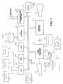
- FIG. 1is a block diagram of an interactive computer-assisted diagnosis (ICAD) system 100 for assisting diagnosis of lung nodules in digital volumetric medical images, according to an illustrative embodiment of the invention
- FIG. 2is a flow diagram illustrating an interactive computer-aided diagnosis (ICAD) method 200 for assisting the detection of lung nodules in digital volumetric medical images, according to an illustrative embodiment of the present invention
- FIG. 3is a flow diagram illustrating an interactive computer-aided diagnosis (ICAD) method 300 for assisting diagnosis of previously detected lung nodules in digital volumetric medical images, according to an illustrative embodiment of the present invention
- FIG. 4is a diagram illustrating visualization tools used for supporting user decisions with respect to the methods of FIGS. 2 and 3, according to an illustrative embodiment of the present invention
- FIG. 5is a diagram illustrating a main window of the ICAD system 100 , represented by a graphical user interface, according to an illustrative embodiment of the present invention.
- FIG. 6is a diagram of a “pop-up” window, corresponding to the main window 500 of FIG. 5, for displaying a real-time, three-dimensional segmentation of an object of interest and quantitative measurements and a confidence level corresponding thereto, according to an illustrative embodiment of the present invention.
- the present inventionis directed to an interactive computer-aided diagnosis (ICAD) method and system for assisting diagnosis of lung nodules in digital volumetric medical images.
- ICADinteractive computer-aided diagnosis
- the present inventionmay be implemented in various forms of hardware, software, firmware, special purpose processors, or a combination thereof.
- the present inventionis implemented as a combination of hardware and software.
- the softwareis preferably implemented as an application program tangibly embodied on a program storage device.
- the application programmay be uploaded to, and executed by, a machine comprising any suitable architecture.
- the machineis implemented on a computer platform having hardware such as one or more central processing units (CPU), a random access memory (RAM), and input/output (I/O) interface(s).
- CPUcentral processing units
- RAMrandom access memory
- I/Oinput/output
- the computer platformalso includes an operating system and microinstruction code.
- various processes and functions described hereinmay either be part of the microinstruction code or part of the application program (or a combination thereof) which is executed via the operating system.
- various other peripheral devicesmay be connected to the computer platform such as an additional data storage device and a printing device.
- FIG. 1is a block diagram of an interactive computer-assisted diagnosis (ICAD) system 100 for assisting diagnosis of lung nodules in digital volumetric medical images, according to an illustrative embodiment of the invention.
- the ICAD system 100includes at least one processor (CPU) 102 operatively coupled to other components via a system bus 104 .
- a read only memory (ROM) 106 , a random access memory (RAM) 108 , a display adapter 110 , an I/O adapter 112 , a user interface adapter 114 , and an audio adapter 130are operatively coupled to the system bus 104 .
- a display device 116is operatively coupled to the system bus 104 by the display adapter 110 .
- a disk storage device (e.g., a magnetic or optical disk storage device) 118is operatively coupled to the system bus 104 by the I/O adapter 112 .
- a mouse 120 , a keyboard 122 , an eye tracking device 124 , and a joystick 142are operatively coupled to the system bus 104 by the user interface adapter 114 .
- the mouse 120 , keyboard 122 , eye tracking device 124 , and joystick 142are used to aid in the selection of suspicious regions in a digital medical image. Any of the mouse 120 , keyboard 122 , eye tracking device 124 , and joystick 142 may be referred to herein as a selection device.
- a user override device 145is also operatively coupled to the system bus 104 by the user interface adapter 114 .
- the user override device 145may be a device similar to a selection device as described above, or may be any type of device which allows a user to input a user rendered decision to the system 100 .
- At least one speaker or other audio signal generator 132(hereinafter “speaker” 132 ) is coupled to the audio adapter 130 for providing a warning or other audio information to the user.
- the speaker 132may also be referred to herein as a “warning generator”.
- a nodule determination device 150 , a segmentation device 170 , a measurement device 180 , and a likelihood estimator 190are also included in the ICAD system 100 . While the nodule determination device 150 , the segmentation device 170 , the measurement device 180 , and the likelihood estimator 190 are illustrated as part of the at least one processor (CPU) 102 , these components are preferably embodied in computer program code stored in at least one of the memories and executed by the at least one processor 102 . Of course, other arrangements are possible, including embodying some or all of the computer program code in registers located on the processor chip.
- nodule determination device 150the segmentation device 170 , the measurement device 180 , and the likelihood estimator 190 , as well as the other elements of the ICAD system 100 , while maintaining the spirit and scope of the present invention.
- the ICAD system 100may also include a digitizer 126 operatively coupled to system bus 104 by user interface adapter 114 for digitizing an MRI or CT image of the lungs.
- digitizer 126may be omitted, in which case a digital MRI or CT image may be input to ICAD system 100 from a network via a communications adapter 128 operatively coupled to system bus 104 .
- FIG. 5is a diagram illustrating a main window 500 of the ICAD system 100 , represented by a graphical user interface, according to an illustrative embodiment of the present invention.
- the main window 500includes a main menu 505 , a “Candidate Tour” menu 510 , an axial image window 515 , an intensity window center slider 520 , an intensity window size slider 525 , a show candidates selector 530 , a candidates manipulation selector 535 , a slice slider 540 , a current position information region 545 , XZ 550 and YZ 555 spinning plane illustrations (elements 520 - 555 correspond to the axial image window 515 ), a Maximum Intensity Projection (MIP) 560 , a “spinning”/“flicker” window 565 , an overlay selector 570 , a flicker selector 575 , a spinning slider 580 (elements 570 - 580 correspond to the “spinning”/“MIP
- FIG. 6is a diagram of a “pop-up” window 600 , corresponding to the main window 500 of FIG. 5, for displaying a real-time, three-dimensional segmentation of an object of interest and quantitative measurements and a confidence level corresponding thereto, according to an illustrative embodiment of the present invention.
- the pop-up window 600includes a three-dimensional rendering window 610 , a quantitative measurements region 615 ,and a confidence level bar plot 620 .
- candidate objectsmay be automatically located by an integrated automatic detection program, or manually selected by the user.
- a candidate objectis an anatomical structure that either the user or the computer has deemed sufficiently suspicious as to warrant further examination.
- candidate objectsare also referred to herein as “objects of interest”, “structures of interest”, or simply candidates.
- a “volume of interest” (VOI)is a 3D volume of the data which contains an “object of interest”, although it may contain other objects as well that are not of interest. In general, the idea is to identify and classify nodules versus non-nodules.
- the useris taken on a “Candidate Tour” to sequentially examine and validate all the candidate objects detected by the computer.
- the “Candidate Tour” menu 510is invoked. Also, the total number of candidate objects is displayed, as well as the number of the candidate object currently being shown, with a “n of m” label 590 included in the “Candidate Tour” menu 510 .
- the usermay go to the “next” candidate, return to examine a “previous” candidate, or “jump to” a particular candidate by actuating corresponding buttons of the “Candidate Tour” menu 510 .
- Interactive tools and automatic measurements as described hereinare made available to the user for aid in validation of the automatically or manually detected candidates. While illustrative interactive tools and measurements are described herein to aid in the comprehension of the present invention, it is to be appreciated that other interactive tools and/or measurement may be employed while maintaining the spirit and scope of the present invention.
- the automatic segmentation methodautomatically segments any object in the lungs at that position.
- the object of interestmay be a lung nodule, or may correspond to an airway wall, vessel, or other anatomical structure, which appears as a bright opacity in CT images.
- the ICAD system 100automatically performs an adaptive threshold operation based upon automatic local histogram analysis to segment the object of interest (see the above referenced application, Attorney Docket Number 00P7879US, entitled “Method and System for Automatically Detecting Lung Nodules from High Resolution Computed Tomography (HRCT) Images”), and then measures and characterizes the object. Such measurements and characterizations are shown in the pop-up window 600 of FIG. 6.
- properties of the candidate objectare measured. These measurements include, for example, the object's centroid, diameter, volume, circularity, sphericity, and average intensity, as shown in the pop-up window of FIG. 6.
- Anatomical knowledgeis used to reason about the likelihood that the object of interest corresponds to a nodule.
- the CAD system 100computes a confidence measurement indicating the CAD system's estimate of the likelihood that the object is a nodule.
- Axial slicesare presented to the user in the axial image window 515 .
- the usermay scroll back and forth through the axial slices by sliding the slice slider 540 .
- a volume of interestmay be selected by moving the positioning device, such as a mouse or a joystick to navigate in the 3-D volumetric image data to a particular point in the current slice, or by invoking the “Candidate Tour”.
- the usermay use several visualization tools to make a decision about whether or not the volume of interest is a nodule. These visualization tools include “fly-around”, “flicker”, “slice scrolling”, and interaction with the three-dimensional surface rendering. Flicker, for example, may be enabled by the flicker selector 575 .
- “Fly-around”is a cine loop, which gives a quick, very natural visualization of the volume surrounding an object of interest.
- Each frame of the cineis a small slice of the data, taken at slightly different angles, centered at the point of interest.
- the cinegives an effect somewhat like flying around the object.
- This visualizationallows the physician to very quickly discover the three-dimensional shape of the object and whether the object has any connecting vessels, without requiring any additional decisions such as segmentation thresholds or viewing angles. In a very short period of time, the user can determine whether an area can be safely dismissed from concern, or warrants further investigation as a lung nodule. Fly-around is described in the above referenced application, U.S. Ser. No. 09/606,564, entitled “Computer-Aided Diagnosis Method for Aiding Diagnosis of Three Dimensional Digital Image Data”.
- “Slice scrolling”lets the user move up and down in a narrow region around the suspect area to visualize the shape of an object. This action is similar to “fly-around” but instead of going around the object, the motion is more of a back-and-forth type motion.
- the result of the automatic segmentationis shown in a separate window (the pop-up window 60 of FIG. 6) as a shaded surface display or a volume rendering.
- Significant adjacent anatomical structuressuch as blood vessels and the chest wall, are also shown, giving an intuitive understanding of the shape and position of the object of interest.
- the segmented objectis shown in one color, whereas the surrounding tissues are shown in a contrasting color. This allows the physician to see the nodule in conjunction with its surroundings, and to verify that that the system's automatic segmentation is valid.
- the usercan easily interact with the three-dimensional rendering to see the object on all sides.
- the measurements of the segmented objectincluding diameter, volume, sphericity, and so forth are instantly displayed for the user (as shown in FIG. 6).
- these measurementsare stored in a table for reporting, and for comparison with earlier or subsequent measurements. In this way, the growth of nodules over time may be closely monitored.
- the real-time segmentationallows the user to manipulate the selecting device as an interactive “volume probe”.
- the physicianplaces the device, using his or her experience and training to determine which areas merit examination.
- the ICAD system 100instantaneously segments whatever object is at that position using the adaptive thresholding technique. As the object is segmented, the system measures the object and computes the confidence value. If the confidence measurement (shown in FIG. 6) indicates that the object is likely to correspond to a lung nodule, the system will generate an audible warning through speaker 132 . Of course, other types of warnings may be provided, such as for example, visual warnings.
- FIG. 2is a flow diagram illustrating an interactive computer-aided diagnosis (ICAD) method 200 for assisting the detection of lung nodules in digital volumetric medical images, according to an illustrative embodiment of the present invention.
- ICADinteractive computer-aided diagnosis
- back-endrefers to a portion of either a system (ICAD system 100 ) or method (methods 200 and 300 ), wherein the portion, operations performed thereby, and/or results obtained therefrom are not visible to the user.
- front-endrefers to a portion of either a system (ICAD system 100 ) or method (methods 200 and 300 ), wherein the portion, operations performed thereby, and/or results obtained therefrom are visible to the user.
- FIGS. 2 and 3which are arranged on the left side thereof, correspond to the front-end.
- FIGS. 2 and 3which are arranged on the right side thereof, correspond to the back-end.
- Volumetric data(e.g., three-dimensional data) for a set of the lungs is loaded (step 105 ).
- a userscans the data using a selection device (step 110 ).
- Step 110may be performed, for example, using the mouse 120 or the joystick 142 as follows: (1) click on a sliding bar (e.g., the slice slider 540 ) to move up and down or to jump to a certain slice; and (2) navigate the scrolling by the movement of the selection device.
- a sliding bare.g., the slice slider 540
- the first approachshows axial slices discretely
- the second approachshows axial slices continuously.
- other approaches and devicesmay be used to scan the data, while maintaining the spirit and scope of the present invention.
- the userlocates/selects an anatomical structure of interest (hereinafter “structure of interest”) to be examined by simply pointing to the structure of interest using the selection device (step 115 ).
- structure of interestan anatomical structure of interest
- the usercan perform the steps illustrated with respect to FIG. 3 below, if the user decides to examine previously detected structures of interest.
- Step 120provides quantitative measurements and intuitive display of the structure of interest.
- the segmentation device 170automatically segments the structure of interest based on local histogram analysis (step 121 ). This segmentation may be performed, for example, as described in the above referenced application, Attorney Docket Number 00P7879US, entitled “Method and System for Automatically Detecting Lung Nodules from High Resolution Computed Tomography (HRCT) Images. Also, at step 121 , measurements of the object are computed by the measurement device 180 based on a segmentation result from the segmentation device 170 . These measurements include the centroid, diameter, volume, sphericitiy, average and standard deviation of intensity. The back-end communicates the results to the front-end.
- HRCTHigh Resolution Computed Tomography
- the front-endreceives messages from the back-end and displays measurement results (step 122 ). While the segmentation results and the quantitative measurements have been thus far described herein as being displayed in a pop-up window 600 , other arrangements are possible which maintain the spirit and scope of the invention. For example, the segmentation results and the quantitative measurements may be displayed in the main GUI (e.g., main window 500 ).
- the confidence that the anatomical structure corresponds to a lung nodule or other suspicious growthis automatically estimated by the likelihood estimator 190 , based on the measurements computed at step 121 by the measurement device 180 (step 123 ).
- the confidence levelis passed to the front-end at step 123 .
- the front-endreceives the confidence level for display, e.g., in the pop-up window 600 of FIG. 6 (step 124 ).
- a colored baris plotted to illustrate the confidence level.
- other visual enhancementsmay be used to display the confidence level as well as any other information provided/displayed by the front-end, while maintaining the spirit and scope of the present invention.
- the structure of interestis automatically classified as a nodule or a non-nodule by the nodule determination device 150 , based on the confidence level computed at step 123 by the likelihood estimator 190 (step 125 ). If the structure of interest is considered to be a nodule (step 125 a ), a message is sent to the front-end.
- the front-endprovides three-dimensional rendering, such as shaded surface rendering, volume rendering, and so forth, of the structure of interest, as well as its neighboring structures, e.g., in the pop-up window 600 of FIG. 6 (step 126 ).
- three-dimensional renderingsuch as shaded surface rendering, volume rendering, and so forth
- colorsare used to differentiate the structure of interest from nearby structures.
- a usercan rotate the rendering in three-dimensions as desired, to have an intuitive understanding of the shape and position of the structure.
- an audible alarm signal(e.g., a beep) is generated by speaker 132 to alert the user of the same (step 128 ).
- the usermakes the final decision to classify the structure of interest as a nodule or non-nodule, based on the provided information (measurements and three-dimensional rendering), as well as the user's visual examination of the original images (step 130 ).
- the final decisionis input by the user to the user override device 145 .
- step 130 aIf the final decision is that the structure is a nodule or other structure (step 130 a ) that should be recorded for future reference, then the user enables the “candidate manipulation” mode and clicks on the nodule or other structure to confirm recordation (step 135 ). Such recordation may be with respect to, e.g., the disk storage device 118 and/or the random access memory 108 .
- the location of the nodule, along with its measurements,is automatically added to the nodule candidate list (step 140 ).
- step 137If user wants to quit the examination process or wants to begin a new study (step 137 ), then the candidate list is automatically saved to a file (step 145 ).
- Any results recorded at steps 140 and 145can be further used for report generation or follow-up study (step 150 ).
- FIG. 3is a flow diagram illustrating an interactive computer-aided diagnosis (ICAD) method 300 for assisting diagnosis of previously detected lung nodules in digital volumetric medical images, according to an illustrative embodiment of the present invention.
- ICADinteractive computer-aided diagnosis
- Volumetric data(e.g., three-dimensional data) for a set of the lungs is loaded (step 205 ).
- Previously saved candidate objectsare loaded (step 210 ).
- These candidate objectscan be obtained, for example, using two illustrative approaches: (1) manual detection, where the user examines the data and manually picks out the candidate objects as described in FIG. 2; and (2) automatic detection, wherein the computer detects candidate objects within the entire lung volume.
- the second approachmay be implemented, for example, using a method such as the one described in the above referenced application, Attorney Docket Number 00P7879US, entitled “Method and System for Automatically Detecting Lung Nodules from High Resolution Computed Tomography (HRCT) Images”.
- HRCTHigh Resolution Computed Tomography
- the computerlaunches a “Candidate Tour”, which activates the “Candidate Tour” menu 510 (step 215 ).
- the “Candidate Tour”is a function that automatically navigates the user to each of the candidate objects for examination and confirmation. All the interactive measurement and display functions shown in steps 122 - 128 of FIG. 2 are accessible to the user.
- the “Candidate Tour” menu bar 510provides functions that enable the user to select candidates easily, such as “Previous”, “Next”, and “Jump to”.
- the userselects a candidate object to examine using the “Candidate Tour” menu bar (step 220 ).
- the usercan perform the steps illustrated with respect to FIG. 2, if the user decides to examine new candidate objects.
- Real-time segmentation and measurementis launched (via segmentation device 170 and measurement device 180 ) to provide the user quantitative measurements and intuitive displaying of the structure of interest, e.g., as shown in the pop-up window 600 of FIG. 6, via the display device 116 (step 225 ).
- Step 225is described in further detail with respect to step 120 of FIG. 2.
- the usermakes the final decision to classify the structure of interest as a nodule or a non-nodule, based on the provided measurements and three-dimensional rendering, as well as the user's manual study of the original images (step 230 ).
- the final decisionis input by the user to the user override device 145 . This step is identical to step 130 of FIG. 2.
- step 230 aIf the final decision considers the structure as a non-nodule (step 230 a ), then the user enables the “candidate manipulation” mode and clicks on the structure (or an index or visual mark corresponding thereto) to discard the structure from the list (step 235 ).
- the non-nodule and the quantitative measurements for the non-noduleare automatically removed from the candidate list (step 240 ).
- step 237If user wants to quit the re-examination process or wants to begin a new study (step 237 ), then the candidate list is automatically saved to a file (step 245 ). This step is identical to step 145 of FIG. 2.
- step 250Any results recorded at steps 240 and 245 can be further used for report generation or follow-up study (step 250 ). This step is identical to step 150 of FIG. 2.
- FIG. 4is a diagram illustrating visualization tools used for supporting user decisions with respect to the methods of FIGS. 2 and 3, according to an illustrative embodiment of the present invention. It is to be appreciated that the user may select one or more of the visualization tools.
- the userselects “Fly Around” to view a short movie of two-dimensional slices of the nodule (step 410 ).
- the usermay select individual frames of the movie by means of the spinning slider described above.
- the userselects “Flicker” to have the computer alternate between two sets of display parameters, to view the extent of calcification of a candidate object (step 420 ).
- the userinteracts with a three-dimensional rendering of a candidate object (shown in step 126 of FIG. 2) by means of the mouse 120 , the joystick 142 , and so forth, to view different sides of the candidate object (step 430 ).
- the userscrolls up and down within slices adjacent to a candidate object (slice scrolling) (step 440 ).
Landscapes
- Engineering & Computer Science (AREA)
- Health & Medical Sciences (AREA)
- Medical Informatics (AREA)
- General Health & Medical Sciences (AREA)
- Public Health (AREA)
- Physics & Mathematics (AREA)
- General Physics & Mathematics (AREA)
- Primary Health Care (AREA)
- Theoretical Computer Science (AREA)
- Epidemiology (AREA)
- Computer Vision & Pattern Recognition (AREA)
- Nuclear Medicine, Radiotherapy & Molecular Imaging (AREA)
- Biomedical Technology (AREA)
- Radiology & Medical Imaging (AREA)
- Multimedia (AREA)
- Molecular Biology (AREA)
- Life Sciences & Earth Sciences (AREA)
- Quality & Reliability (AREA)
- Data Mining & Analysis (AREA)
- Databases & Information Systems (AREA)
- Pathology (AREA)
- Apparatus For Radiation Diagnosis (AREA)
- Measuring And Recording Apparatus For Diagnosis (AREA)
Abstract
Description
- This application is related to the application Ser. No. 60/230,772 filed on Sep. 7, 2000, Attorney Docket Number 2000P07879US01, entitled “Automatic Detection of Lung Nodules from High Resolution CT Images”, which is commonly assigned and the perfection of the provisional is concurrently filed herewith, and the disclosure of which is incorporated herein by reference. This application is also related to U.S. Ser. No. 09/606,564, entitled “Computer-aided Diagnosis of Three Dimensional digital image data”, filed on Jun. 29, 2000, and U.S. Ser. No. 09/271,471, entitled “Mark-Free Computer-Assisted Diagnosis Method and System for Assisting Diagnosis of Abnormalities in Digital Medical Images Using Diagnosis Based Image Enhancement”, filed on Mar. 17, 1999, which are commonly assigned herewith, and the disclosures of which are incorporated herein by reference.[0001]
- 1. Technical Field[0002]
- The present invention generally relates to computer-assisted diagnosis (CADX) and, in particular, to an interactive computer-aided diagnosis (ICAD) method and system for assisting diagnosis of lung nodules in digital volumetric medical images.[0003]
- 2. Background Description[0004]
- Computer-Aided diagnosis (CADx) is an important technology in many clinical applications, such as the detection of lung cancer. In current clinical practice, cancer or other diseases may be missed during a physician's un-aided examination of medical image data, in part because of the large volume of data. This is particularly a problem for screening applications, since there is generally little time to devote to the examination of each patient's data, and the entire range of the data must be examined to make sure it is free from disease. Computer analysis that is performed silently in the background can greatly aid physicians in their work.[0005]
- New technologies that offer three-dimensional (3-D) scans of the human body, such as Magnetic Resonance Imaging (MRI) and Computed Tomography (CT), offer tremendous opportunities for improved detection of disease. However the change from two dimensions to three, especially to large volume 3-D data (such as image volumes produced by multi-detector high resolution CT scanners), results in a much larger amount of data for the physician to examine. Furthermore, the low-dose imaging for cancer screening poses additional challenges to the traditional manual clinical reading. Thus, the assistance of computer analysis becomes even more important.[0006]
- Unfortunately, many current CADx systems are not readily accepted by physicians, because their aid is seen as more of a distraction than a help. Many such systems present the results of the computer's diagnosis to the physicians by marks, such as a red circle or arrow on the softcopy, which some physicians believe can create a bias in their interpretation of the data. Furthermore, too many systems are perceived as a “black box”, where physicians feel they do not have any understanding of how such systems work and how they generate their diagnoses.[0007]
- Accordingly, it would be desirable and highly advantageous to have a CADx system that is an “open box”, increasing its acceptance among physicians.[0008]
- The problems stated above, as well as other related problems of the prior art, are solved by the present invention, an interactive computer-aided diagnosis (ICAD) method and system for assisting diagnosis of lung nodules in digital volumetric medical images.[0009]
- The method and system mesh seamlessly with physicians in their current work practices. By incorporating the physicians' years of training into the present invention, the invention takes advantage of their knowledge, augmenting it with the strengths of the computer in rapid computation of numeric values. Such an invention is an “open box”, increasing its acceptance among physicians. The invention provides results in “real-time” so that it does not add a time delay to the physician's examination process.[0010]
- According to an aspect of the invention, there is provided a computer-assisted diagnosis method for assisting diagnosis of anatomical structures in a digital volumetric medical image of at least one lung. An anatomical structure of interest is identified in the volumetric digital medical image. The anatomical structure of interest is automatically segmented, in real-time, in a predefined volume of interest (VOI). Quantitative measurements of the anatomical structure of interest are automatically computed, in real-time. A result of the segmenting step and a result of the computing step are displayed, in real-time. A likelihood that the anatomical structure of interest corresponds to a disease or an area warranting further investigation is estimating, in real-time, based on predefined criteria and the quantitative measurements. A warning is generated, in real-time, when the likelihood is above a predefined threshold.[0011]
- According to another aspect of the invention, the method further includes the step of generating a graphical user interface having a main window for displaying at least one view corresponding to the at least one lung.[0012]
- According to yet another aspect of the invention, the at least one view is at least one of an axial view and a maximum intensity projection view.[0013]
- According to still yet another aspect of the invention, the method further includes the step of displaying at least one of the result of the segmenting step and the result of the computing step in a supplemental window or a pop-up window of the graphical user interface.[0014]
- According to a further aspect of the invention, the method further includes the step of alternately displaying at least one of at least two different sets of display parameters in a supplemental window of the graphical user interface to view an extent of calcification of the anatomical structure of interest.[0015]
- According to a yet further aspect of the invention, the method further includes the step of determining a local spinning plane for the anatomical structure of interest. The local spinning plane is centered at a centroid and a local spinning axis of the anatomical structure of interest. The local spinning plane is rotated at least a portion of 360 degrees. A view is created of the anatomical structure of interest at predefined increments of rotation, so as to result in a plurality of views of the anatomical structure of interest. The plurality of views of the anatomical structure of interest are displayed in a supplemental window of the graphical user interface.[0016]
- According to an additional aspect of the invention, there is more than one anatomical structure of interest, and the method further includes the step of conducting a tour of the more than one anatomical structure of interest. The conducting step includes the steps of displaying results of the segmenting and computing steps in at least one supplemental window or at least one pop-up window of the graphical user interface, and receiving indicia for selecting a previous anatomical structure of interest, a next anatomical structure of interest, and a particular anatomical structure of interest from among the more than one anatomical structure of interest.[0017]
- These and other aspects, features and advantages of the present invention will become apparent from the following detailed description of preferred embodiments, which is to be read in connection with the accompanying drawings.[0018]
- FIG. 1 is a block diagram of an interactive computer-assisted diagnosis (ICAD)[0019]
system 100 for assisting diagnosis of lung nodules in digital volumetric medical images, according to an illustrative embodiment of the invention; - FIG. 2 is a flow diagram illustrating an interactive computer-aided diagnosis (ICAD)[0020]
method 200 for assisting the detection of lung nodules in digital volumetric medical images, according to an illustrative embodiment of the present invention; - FIG. 3 is a flow diagram illustrating an interactive computer-aided diagnosis (ICAD)[0021]
method 300 for assisting diagnosis of previously detected lung nodules in digital volumetric medical images, according to an illustrative embodiment of the present invention; - FIG. 4 is a diagram illustrating visualization tools used for supporting user decisions with respect to the methods of FIGS. 2 and 3, according to an illustrative embodiment of the present invention;[0022]
- FIG. 5 is a diagram illustrating a main window of the[0023]
ICAD system 100, represented by a graphical user interface, according to an illustrative embodiment of the present invention; and - FIG. 6 is a diagram of a “pop-up” window, corresponding to the main window[0024]500 of FIG. 5, for displaying a real-time, three-dimensional segmentation of an object of interest and quantitative measurements and a confidence level corresponding thereto, according to an illustrative embodiment of the present invention.
- The present invention is directed to an interactive computer-aided diagnosis (ICAD) method and system for assisting diagnosis of lung nodules in digital volumetric medical images.[0025]
- It is to be understood that the present invention may be implemented in various forms of hardware, software, firmware, special purpose processors, or a combination thereof. Preferably, the present invention is implemented as a combination of hardware and software. Moreover, the software is preferably implemented as an application program tangibly embodied on a program storage device. The application program may be uploaded to, and executed by, a machine comprising any suitable architecture. Preferably, the machine is implemented on a computer platform having hardware such as one or more central processing units (CPU), a random access memory (RAM), and input/output (I/O) interface(s). The computer platform also includes an operating system and microinstruction code. The various processes and functions described herein may either be part of the microinstruction code or part of the application program (or a combination thereof) which is executed via the operating system. In addition, various other peripheral devices may be connected to the computer platform such as an additional data storage device and a printing device.[0026]
- It is to be further understood that, because some of the constituent system components and method steps depicted in the accompanying Figures are preferably implemented in software, the actual connections between the system components (or the process steps) may differ depending upon the manner in which the present invention is programmed. Given the teachings herein, one of ordinary skill in the related art will be able to contemplate these and similar implementations or configurations of the present invention.[0027]
- FIG. 1 is a block diagram of an interactive computer-assisted diagnosis (ICAD)[0028]
system 100 for assisting diagnosis of lung nodules in digital volumetric medical images, according to an illustrative embodiment of the invention. TheICAD system 100 includes at least one processor (CPU)102 operatively coupled to other components via asystem bus 104. A read only memory (ROM)106, a random access memory (RAM)108, adisplay adapter 110, an I/O adapter 112, auser interface adapter 114, and anaudio adapter 130 are operatively coupled to thesystem bus 104. - A[0029]
display device 116 is operatively coupled to thesystem bus 104 by thedisplay adapter 110. A disk storage device (e.g., a magnetic or optical disk storage device)118 is operatively coupled to thesystem bus 104 by the I/O adapter 112. - A[0030]
mouse 120, akeyboard 122, aneye tracking device 124, and ajoystick 142 are operatively coupled to thesystem bus 104 by theuser interface adapter 114. Themouse 120,keyboard 122,eye tracking device 124, andjoystick 142 are used to aid in the selection of suspicious regions in a digital medical image. Any of themouse 120,keyboard 122,eye tracking device 124, andjoystick 142 may be referred to herein as a selection device. Auser override device 145 is also operatively coupled to thesystem bus 104 by theuser interface adapter 114. Theuser override device 145 may be a device similar to a selection device as described above, or may be any type of device which allows a user to input a user rendered decision to thesystem 100. - At least one speaker or other audio signal generator[0031]132 (hereinafter “speaker”132) is coupled to the
audio adapter 130 for providing a warning or other audio information to the user. In the case when thespeaker 132 is providing a warning, thespeaker 132 may also be referred to herein as a “warning generator”. - A[0032]
nodule determination device 150, asegmentation device 170, a measurement device180, and alikelihood estimator 190 are also included in theICAD system 100. While thenodule determination device 150, thesegmentation device 170, the measurement device180, and thelikelihood estimator 190 are illustrated as part of the at least one processor (CPU)102, these components are preferably embodied in computer program code stored in at least one of the memories and executed by the at least oneprocessor 102. Of course, other arrangements are possible, including embodying some or all of the computer program code in registers located on the processor chip. Given the teachings of the invention provided herein, one of ordinary skill in the related art will contemplate these and various other configurations and implementations of thenodule determination device 150, thesegmentation device 170, the measurement device180, and thelikelihood estimator 190, as well as the other elements of theICAD system 100, while maintaining the spirit and scope of the present invention. - The[0033]
ICAD system 100 may also include adigitizer 126 operatively coupled tosystem bus 104 byuser interface adapter 114 for digitizing an MRI or CT image of the lungs. Alternatively,digitizer 126 may be omitted, in which case a digital MRI or CT image may be input toICAD system 100 from a network via acommunications adapter 128 operatively coupled tosystem bus 104. - FIG. 5 is a diagram illustrating a main window[0034]500 of the
ICAD system 100, represented by a graphical user interface, according to an illustrative embodiment of the present invention. The main window500 includes amain menu 505, a “Candidate Tour” menu510, anaxial image window 515, an intensity window center slider520, an intensity window size slider525, ashow candidates selector 530, acandidates manipulation selector 535, aslice slider 540, a currentposition information region 545, XZ550 and YZ555 spinning plane illustrations (elements520-555 correspond to the axial image window515), a Maximum Intensity Projection (MIP)560, a “spinning”/“flicker”window 565, anoverlay selector 570, aflicker selector 575, a spinning slider580 (elements570-580 correspond to the “spinning”/“flicker” window565), acircularity plot 585, and apatient information region 590. - FIG. 6 is a diagram of a “pop-up” window[0035]600, corresponding to the main window500 of FIG. 5, for displaying a real-time, three-dimensional segmentation of an object of interest and quantitative measurements and a confidence level corresponding thereto, according to an illustrative embodiment of the present invention. The pop-up window600 includes a three-
dimensional rendering window 610, aquantitative measurements region 615,and a confidencelevel bar plot 620. - In the[0036]
ICAD system 100, “candidate objects” may be automatically located by an integrated automatic detection program, or manually selected by the user. A candidate object is an anatomical structure that either the user or the computer has deemed sufficiently suspicious as to warrant further examination. Candidate objects are also referred to herein as “objects of interest”, “structures of interest”, or simply candidates. A “volume of interest” (VOI) is a 3D volume of the data which contains an “object of interest”, although it may contain other objects as well that are not of interest. In general, the idea is to identify and classify nodules versus non-nodules. - If the automatic detection program is chosen, the user is taken on a “Candidate Tour” to sequentially examine and validate all the candidate objects detected by the computer. During a “Candidate Tour”, the “Candidate Tour” menu[0037]510 is invoked. Also, the total number of candidate objects is displayed, as well as the number of the candidate object currently being shown, with a “n of m”
label 590 included in the “Candidate Tour” menu510. The user may go to the “next” candidate, return to examine a “previous” candidate, or “jump to” a particular candidate by actuating corresponding buttons of the “Candidate Tour” menu510. Interactive tools and automatic measurements as described herein are made available to the user for aid in validation of the automatically or manually detected candidates. While illustrative interactive tools and measurements are described herein to aid in the comprehension of the present invention, it is to be appreciated that other interactive tools and/or measurement may be employed while maintaining the spirit and scope of the present invention. - Whether the volume of interest to be examined is determined by the user or by the automatic detection method, the automatic segmentation method automatically segments any object in the lungs at that position. The object of interest may be a lung nodule, or may correspond to an airway wall, vessel, or other anatomical structure, which appears as a bright opacity in CT images. The[0038]
ICAD system 100 automatically performs an adaptive threshold operation based upon automatic local histogram analysis to segment the object of interest (see the above referenced application, Attorney Docket Number 00P7879US, entitled “Method and System for Automatically Detecting Lung Nodules from High Resolution Computed Tomography (HRCT) Images”), and then measures and characterizes the object. Such measurements and characterizations are shown in the pop-up window600 of FIG. 6. - Once the candidate object has been segmented, properties of the candidate object are measured. These measurements include, for example, the object's centroid, diameter, volume, circularity, sphericity, and average intensity, as shown in the pop-up window of FIG. 6. Anatomical knowledge is used to reason about the likelihood that the object of interest corresponds to a nodule. The[0039]
CAD system 100 computes a confidence measurement indicating the CAD system's estimate of the likelihood that the object is a nodule. These measurements are described in the above referenced application, Attorney Docket Number 00P7879US, entitled “Method and System for Automatically Detecting Lung Nodules from High Resolution Computed Tomography (HRCT) Images”. - Axial slices are presented to the user in the[0040]
axial image window 515. The user may scroll back and forth through the axial slices by sliding theslice slider 540. A volume of interest may be selected by moving the positioning device, such as a mouse or a joystick to navigate in the 3-D volumetric image data to a particular point in the current slice, or by invoking the “Candidate Tour”. Once a volume of interest has been selected, the user may use several visualization tools to make a decision about whether or not the volume of interest is a nodule. These visualization tools include “fly-around”, “flicker”, “slice scrolling”, and interaction with the three-dimensional surface rendering. Flicker, for example, may be enabled by theflicker selector 575. - “Fly-around” is a cine loop, which gives a quick, very natural visualization of the volume surrounding an object of interest. Each frame of the cine is a small slice of the data, taken at slightly different angles, centered at the point of interest. The cine gives an effect somewhat like flying around the object. This visualization allows the physician to very quickly discover the three-dimensional shape of the object and whether the object has any connecting vessels, without requiring any additional decisions such as segmentation thresholds or viewing angles. In a very short period of time, the user can determine whether an area can be safely dismissed from concern, or warrants further investigation as a lung nodule. Fly-around is described in the above referenced application, U.S. Ser. No. 09/606,564, entitled “Computer-Aided Diagnosis Method for Aiding Diagnosis of Three Dimensional Digital Image Data”.[0041]
- “Slice scrolling” lets the user move up and down in a narrow region around the suspect area to visualize the shape of an object. This action is similar to “fly-around” but instead of going around the object, the motion is more of a back-and-forth type motion.[0042]
- If an area is deemed suspicious after “fly-around”, another click of a button (the flicker selector[0043]575) launches the “flicker” mode, giving a useful visualization of the calcification pattern, which is an important sign of malignancy. The system alternates between two sets of display parameters, one set optimized for lung tissue and the other set optimized for mediastinal tissue. This allows the user to see which areas of the suspicious area have been calcified and which have not been calcified.
- The result of the automatic segmentation is shown in a separate window (the pop-up window[0044]60 of FIG. 6) as a shaded surface display or a volume rendering. Significant adjacent anatomical structures, such as blood vessels and the chest wall, are also shown, giving an intuitive understanding of the shape and position of the object of interest. The segmented object is shown in one color, whereas the surrounding tissues are shown in a contrasting color. This allows the physician to see the nodule in conjunction with its surroundings, and to verify that that the system's automatic segmentation is valid.
- If the object warrants further examination, the user can easily interact with the three-dimensional rendering to see the object on all sides. The measurements of the segmented object, including diameter, volume, sphericity, and so forth are instantly displayed for the user (as shown in FIG. 6). Upon a click of a button, these measurements (or a subset thereof) are stored in a table for reporting, and for comparison with earlier or subsequent measurements. In this way, the growth of nodules over time may be closely monitored.[0045]
- The real-time segmentation allows the user to manipulate the selecting device as an interactive “volume probe”. The physician places the device, using his or her experience and training to determine which areas merit examination. The[0046]
ICAD system 100 instantaneously segments whatever object is at that position using the adaptive thresholding technique. As the object is segmented, the system measures the object and computes the confidence value. If the confidence measurement (shown in FIG. 6) indicates that the object is likely to correspond to a lung nodule, the system will generate an audible warning throughspeaker 132. Of course, other types of warnings may be provided, such as for example, visual warnings. - Taken together, these real-time automatic visualization and measurement techniques provide powerful interactive tools for physicians examining volumetric images of the body. The fast and natural presentation does not add a burden to the user, but instead presents useful and timely information which greatly enhances a physician's ability to make decisions about diagnosis and treatment. The nearly instantaneous segmentation and measurement of three-dimensional objects fulfills an important need for physicians. Objects of typical size can be segmented in less than 1 second on an 833 MHz PC with 1 GB of memory. Very large objects may take slightly longer. The interactive reporting of the object's characteristic measurements and the confidence value make the system an “open box” so that the user can easily understand why the automatic detection system made the decisions that it did. In this way, the[0047]
ICAD system 100 will be more acceptable to users as they gain confidence in understanding how it works. - FIG. 2 is a flow diagram illustrating an interactive computer-aided diagnosis (ICAD)[0048]
method 200 for assisting the detection of lung nodules in digital volumetric medical images, according to an illustrative embodiment of the present invention. - As used herein, the term “back-end” refers to a portion of either a system (ICAD system[0049]100) or method (
methods 200 and300), wherein the portion, operations performed thereby, and/or results obtained therefrom are not visible to the user. The term “front-end” refers to a portion of either a system (ICAD system100) or method (methods 200 and300), wherein the portion, operations performed thereby, and/or results obtained therefrom are visible to the user. The steps depicted in FIGS. 2 and 3, which are arranged on the left side thereof, correspond to the front-end. The steps depicted in FIGS. 2 and 3, which are arranged on the right side thereof, correspond to the back-end. - Volumetric data (e.g., three-dimensional data) for a set of the lungs is loaded (step[0050]105). A user scans the data using a selection device (step110). Step110 may be performed, for example, using the
mouse 120 or thejoystick 142 as follows: (1) click on a sliding bar (e.g., the slice slider540) to move up and down or to jump to a certain slice; and (2) navigate the scrolling by the movement of the selection device. As is evident, the first approach shows axial slices discretely, while the second approach shows axial slices continuously. Of course, other approaches and devices may be used to scan the data, while maintaining the spirit and scope of the present invention. - The user locates/selects an anatomical structure of interest (hereinafter “structure of interest”) to be examined by simply pointing to the structure of interest using the selection device (step[0051]115). Alternatively, the user can perform the steps illustrated with respect to FIG. 3 below, if the user decides to examine previously detected structures of interest.
- Continuing with the method of FIG. 1, real-time segmentation and measurements are computed in the back-end, and the results displayed in the front-end, e.g., in the pop-up window[0052]600 of FIG. 6 (step120). Step120 provides quantitative measurements and intuitive display of the structure of interest.
- In the back-end, the[0053]
segmentation device 170 automatically segments the structure of interest based on local histogram analysis (step121). This segmentation may be performed, for example, as described in the above referenced application, Attorney Docket Number 00P7879US, entitled “Method and System for Automatically Detecting Lung Nodules from High Resolution Computed Tomography (HRCT) Images. Also, atstep 121, measurements of the object are computed by the measurement device180 based on a segmentation result from thesegmentation device 170. These measurements include the centroid, diameter, volume, sphericitiy, average and standard deviation of intensity. The back-end communicates the results to the front-end. - The front-end receives messages from the back-end and displays measurement results (step[0054]122). While the segmentation results and the quantitative measurements have been thus far described herein as being displayed in a pop-up window600, other arrangements are possible which maintain the spirit and scope of the invention. For example, the segmentation results and the quantitative measurements may be displayed in the main GUI (e.g., main window500).
- In the back-end, the confidence that the anatomical structure corresponds to a lung nodule or other suspicious growth is automatically estimated by the[0055]
likelihood estimator 190, based on the measurements computed atstep 121 by the measurement device180 (step123). The confidence level is passed to the front-end atstep 123. - The front-end receives the confidence level for display, e.g., in the pop-up window[0056]600 of FIG.6 (step124). In one illustrative embodiment of the invention, a colored bar is plotted to illustrate the confidence level. Of course, other visual enhancements may be used to display the confidence level as well as any other information provided/displayed by the front-end, while maintaining the spirit and scope of the present invention.
- In the back-end, the structure of interest is automatically classified as a nodule or a non-nodule by the[0057]
nodule determination device 150, based on the confidence level computed atstep 123 by the likelihood estimator190 (step125). If the structure of interest is considered to be a nodule (step125a), a message is sent to the front-end. - The front-end provides three-dimensional rendering, such as shaded surface rendering, volume rendering, and so forth, of the structure of interest, as well as its neighboring structures, e.g., in the pop-up window[0058]600 of FIG. 6 (step126). Preferably, but not necessarily, colors are used to differentiate the structure of interest from nearby structures. A user can rotate the rendering in three-dimensions as desired, to have an intuitive understanding of the shape and position of the structure.
- If the front-end is informed that the structure under examination is likely to correspond to a nodule, then an audible alarm signal (e.g., a beep) is generated by[0059]
speaker 132 to alert the user of the same (step128). - The user makes the final decision to classify the structure of interest as a nodule or non-nodule, based on the provided information (measurements and three-dimensional rendering), as well as the user's visual examination of the original images (step[0060]130). The final decision is input by the user to the
user override device 145. - If the final decision is that the structure is a nodule or other structure (step[0061]130a) that should be recorded for future reference, then the user enables the “candidate manipulation” mode and clicks on the nodule or other structure to confirm recordation (step135). Such recordation may be with respect to, e.g., the
disk storage device 118 and/or therandom access memory 108. - In the back-end, the location of the nodule, along with its measurements, is automatically added to the nodule candidate list (step[0062]140).
- If user wants to quit the examination process or wants to begin a new study (step[0063]137), then the candidate list is automatically saved to a file (step145).
- Any results recorded at[0064]
steps - FIG. 3 is a flow diagram illustrating an interactive computer-aided diagnosis (ICAD)[0065]
method 300 for assisting diagnosis of previously detected lung nodules in digital volumetric medical images, according to an illustrative embodiment of the present invention. - Volumetric data (e.g., three-dimensional data) for a set of the lungs is loaded (step[0066]205). Previously saved candidate objects are loaded (step210). These candidate objects can be obtained, for example, using two illustrative approaches: (1) manual detection, where the user examines the data and manually picks out the candidate objects as described in FIG. 2; and (2) automatic detection, wherein the computer detects candidate objects within the entire lung volume. The second approach may be implemented, for example, using a method such as the one described in the above referenced application, Attorney Docket Number 00P7879US, entitled “Method and System for Automatically Detecting Lung Nodules from High Resolution Computed Tomography (HRCT) Images”.
- In the back-end, the computer launches a “Candidate Tour”, which activates the “Candidate Tour” menu[0067]510 (step215). The “Candidate Tour” is a function that automatically navigates the user to each of the candidate objects for examination and confirmation. All the interactive measurement and display functions shown in steps122-128 of FIG. 2 are accessible to the user. The “Candidate Tour” menu bar510 provides functions that enable the user to select candidates easily, such as “Previous”, “Next”, and “Jump to”.
- The user selects a candidate object to examine using the “Candidate Tour” menu bar (step[0068]220). Alternatively, the user can perform the steps illustrated with respect to FIG. 2, if the user decides to examine new candidate objects.
- Real-time segmentation and measurement is launched (via[0069]
segmentation device 170 and measurement device180) to provide the user quantitative measurements and intuitive displaying of the structure of interest, e.g., as shown in the pop-up window600 of FIG. 6, via the display device116 (step225). Step225 is described in further detail with respect to step120 of FIG. 2. - The user makes the final decision to classify the structure of interest as a nodule or a non-nodule, based on the provided measurements and three-dimensional rendering, as well as the user's manual study of the original images (step[0070]230). The final decision is input by the user to the
user override device 145. This step is identical to step130 of FIG. 2. - If the final decision considers the structure as a non-nodule (step[0071]230a), then the user enables the “candidate manipulation” mode and clicks on the structure (or an index or visual mark corresponding thereto) to discard the structure from the list (step235).
- In the back-end, the non-nodule and the quantitative measurements for the non-nodule are automatically removed from the candidate list (step[0072]240).
- If user wants to quit the re-examination process or wants to begin a new study (step[0073]237), then the candidate list is automatically saved to a file (step245). This step is identical to step145 of FIG. 2.
- Any results recorded at[0074]
steps - FIG. 4 is a diagram illustrating visualization tools used for supporting user decisions with respect to the methods of FIGS. 2 and 3, according to an illustrative embodiment of the present invention. It is to be appreciated that the user may select one or more of the visualization tools.[0075]
- The user selects “Fly Around” to view a short movie of two-dimensional slices of the nodule (step[0076]410). The user may select individual frames of the movie by means of the spinning slider described above.
- The user selects “Flicker” to have the computer alternate between two sets of display parameters, to view the extent of calcification of a candidate object (step[0077]420).
- The user interacts with a three-dimensional rendering of a candidate object (shown in[0078]
step 126 of FIG. 2) by means of themouse 120, thejoystick 142, and so forth, to view different sides of the candidate object (step430). - The user scrolls up and down within slices adjacent to a candidate object (slice scrolling) (step[0079]440).
- Although the illustrative embodiments have been described herein with reference to the accompanying drawings, it is to be understood that the present invention is not limited to those precise embodiments, and that various other changes and modifications may be affected therein by one of ordinary skill in the related art without departing from the scope or spirit of the invention. All such changes and modifications are intended to be included within the scope of the invention as defined by the appended claims.[0080]
Claims (34)
Priority Applications (5)
| Application Number | Priority Date | Filing Date | Title |
|---|---|---|---|
| US09/840,267US6944330B2 (en) | 2000-09-07 | 2001-04-23 | Interactive computer-aided diagnosis method and system for assisting diagnosis of lung nodules in digital volumetric medical images |
| JP2002582795AJP2004531315A (en) | 2001-04-23 | 2002-04-01 | Computer-aided diagnosis method and system for assisting diagnosis of pulmonary nodules in digital volumetric medical images |
| PCT/US2002/010406WO2002085211A2 (en) | 2001-04-23 | 2002-04-01 | Method and system for automatically detecting lung nodules from multi-slice high resolution computed tomography (mshr ct) images |
| EP02717760AEP1381997A2 (en) | 2001-04-23 | 2002-04-01 | Method and system for automatically detecting lung nodules from multi-slice high resolution computed tomography (mshr ct) images |
| CNA028123395ACN1518719A (en) | 2001-04-23 | 2002-04-01 | Method and system for automatically detecting lung nodules from multi-slice high resolution computed tomography (MSHR CT) images |
Applications Claiming Priority (2)
| Application Number | Priority Date | Filing Date | Title |
|---|---|---|---|
| US23077200P | 2000-09-07 | 2000-09-07 | |
| US09/840,267US6944330B2 (en) | 2000-09-07 | 2001-04-23 | Interactive computer-aided diagnosis method and system for assisting diagnosis of lung nodules in digital volumetric medical images |
Publications (2)
| Publication Number | Publication Date |
|---|---|
| US20020028006A1true US20020028006A1 (en) | 2002-03-07 |
| US6944330B2 US6944330B2 (en) | 2005-09-13 |
Family
ID=25281897
Family Applications (1)
| Application Number | Title | Priority Date | Filing Date |
|---|---|---|---|
| US09/840,267Expired - Fee RelatedUS6944330B2 (en) | 2000-09-07 | 2001-04-23 | Interactive computer-aided diagnosis method and system for assisting diagnosis of lung nodules in digital volumetric medical images |
Country Status (5)
| Country | Link |
|---|---|
| US (1) | US6944330B2 (en) |
| EP (1) | EP1381997A2 (en) |
| JP (1) | JP2004531315A (en) |
| CN (1) | CN1518719A (en) |
| WO (1) | WO2002085211A2 (en) |
Cited By (54)
| Publication number | Priority date | Publication date | Assignee | Title |
|---|---|---|---|---|
| WO2003024319A3 (en)* | 2001-09-19 | 2003-10-30 | Imp College Innovations Ltd | Manipulation of image data |
| WO2003049040A3 (en)* | 2001-12-07 | 2004-03-11 | Koninkl Philips Electronics Nv | Method and device for forming an isolated visualization of body structures |
| US20040086161A1 (en)* | 2002-11-05 | 2004-05-06 | Radhika Sivaramakrishna | Automated detection of lung nodules from multi-slice CT image data |
| US20040146190A1 (en)* | 2003-01-23 | 2004-07-29 | Konica Minolta Holdings, Inc. | Medical image processing system |
| US20040227756A1 (en)* | 2003-03-06 | 2004-11-18 | Volker Dicken | Method of volume visualisation |
| US20040267149A1 (en)* | 2003-06-02 | 2004-12-30 | Igal Kushnir | Method and system for analyzing cardiovascular sounds |
| US20050069185A1 (en)* | 2003-09-26 | 2005-03-31 | Helmut Barfuss | Method for automatic object marking in medical imaging |
| US6887208B2 (en) | 2002-01-10 | 2005-05-03 | Deepbreeze Ltd. | Method and system for analyzing respiratory tract sounds |
| US20050096530A1 (en)* | 2003-10-29 | 2005-05-05 | Confirma, Inc. | Apparatus and method for customized report viewer |
| US20050137907A1 (en)* | 2003-12-23 | 2005-06-23 | Conrad Barski | Naturally expressed medial procedure descriptions generated via synchronized diagrams and menus |
| US20050238253A1 (en)* | 2002-11-29 | 2005-10-27 | Mirada Solutions Limited British Body Corporate | Image registration |
| US20060025674A1 (en)* | 2004-08-02 | 2006-02-02 | Kiraly Atilla P | System and method for tree projection for detection of pulmonary embolism |
| US20060030768A1 (en)* | 2004-06-18 | 2006-02-09 | Ramamurthy Venkat R | System and method for monitoring disease progression or response to therapy using multi-modal visualization |
| US20060064396A1 (en)* | 2004-04-14 | 2006-03-23 | Guo-Qing Wei | Liver disease diagnosis system, method and graphical user interface |
| EP1514226A4 (en)* | 2002-05-13 | 2006-04-19 | Magnolia Medical Technologies | System and method for analysis of data |
| US20060133659A1 (en)* | 2004-12-21 | 2006-06-22 | Hammond Christopher R | Method and system for viewing image data |
| US20060242146A1 (en)* | 2005-03-14 | 2006-10-26 | General Electric Company | Methods and systems for monitoring tumor burden |
| US20070008317A1 (en)* | 2005-05-25 | 2007-01-11 | Sectra Ab | Automated medical image visualization using volume rendering with local histograms |
| US20070055455A1 (en)* | 2004-04-14 | 2007-03-08 | Guo-Qing Wei | Methods for interactive liver disease diagnosis |
| US20070071301A1 (en)* | 2005-09-16 | 2007-03-29 | Siemens Corporate Research Inc | System and method for visualizing airways for assessment |
| WO2006085166A3 (en)* | 2005-01-10 | 2007-04-19 | Koninkl Philips Electronics Nv | Lung lesion detection during tomographic heart screening |
| US20070101295A1 (en)* | 2005-10-27 | 2007-05-03 | Wei Ding | Method and apparatus for diagnostic imaging assistance |
| US20070129625A1 (en)* | 2005-11-21 | 2007-06-07 | Boston Scientific Scimed Systems, Inc. | Systems and methods for detecting the presence of abnormalities in a medical image |
| EP1833021A1 (en)* | 2006-03-09 | 2007-09-12 | Medicsight PLC | Digital medical image processing |
| US20070255304A1 (en)* | 2002-02-21 | 2007-11-01 | Roschak Edmund J | Devices for creating passages and sensing for blood vessels |
| US20080009760A1 (en)* | 2006-06-30 | 2008-01-10 | Broncus Technologies, Inc. | Airway bypass site selection and treatment planning |
| US20080069477A1 (en)* | 2006-09-19 | 2008-03-20 | Heinz-Peter Engels | Result filter and method for selecting the result data of an application for automatic pattern recognition |
| US20080074427A1 (en)* | 2006-09-26 | 2008-03-27 | Karl Barth | Method for display of medical 3d image data on a monitor |
| US20080294591A1 (en)* | 2004-03-05 | 2008-11-27 | Koninklijke Philips Electronic, N.V. | Interactive Computer-Aided Diagnosis |
| WO2009070212A1 (en)* | 2007-11-21 | 2009-06-04 | Siemens Medical Solutions Usa, Inc. | System and method for additive spatial/intensity decomposition of medical images |
| US20100123715A1 (en)* | 2008-11-14 | 2010-05-20 | General Electric Company | Method and system for navigating volumetric images |
| US20100260383A1 (en)* | 2007-09-17 | 2010-10-14 | Koninklijke Philips Electronics N.V. | caliper for measuring objects in an image |
| US20100305788A1 (en)* | 2007-09-19 | 2010-12-02 | Knorr-Bremse Systeme Fur Schienenfahrzeuge Gmbh | Method for adapting at least one parameter in a controlled system of a vehicle |
| US20120130223A1 (en)* | 2010-11-19 | 2012-05-24 | Dr Systems, Inc. | Annotation and assessment of images |
| US8608724B2 (en) | 2004-07-19 | 2013-12-17 | Broncus Medical Inc. | Devices for delivering substances through an extra-anatomic opening created in an airway |
| US8709034B2 (en) | 2011-05-13 | 2014-04-29 | Broncus Medical Inc. | Methods and devices for diagnosing, monitoring, or treating medical conditions through an opening through an airway wall |
| EP1531425B1 (en)* | 2003-11-14 | 2014-05-14 | GE Medical Systems Global Technology Company LLC | Computer aided image acquisition and diagnosis system |
| US9196046B2 (en) | 2009-03-27 | 2015-11-24 | Koninklijke Philips N.V. | Medical imaging |
| US9265458B2 (en) | 2012-12-04 | 2016-02-23 | Sync-Think, Inc. | Application of smooth pursuit cognitive testing paradigms to clinical drug development |
| US20160110160A1 (en)* | 2014-10-16 | 2016-04-21 | Siemens Medical Solutions Usa, Inc. | Context-sensitive identification of regions of interest in a medical image |
| US20160133028A1 (en)* | 2014-11-07 | 2016-05-12 | Samsung Electronics Co., Ltd. | Apparatus and method for avoiding region of interest re-detection |
| US9345532B2 (en) | 2011-05-13 | 2016-05-24 | Broncus Medical Inc. | Methods and devices for ablation of tissue |
| US20160171158A1 (en)* | 2014-12-12 | 2016-06-16 | Samsung Electronics Co., Ltd. | Medical imaging apparatus and method using comparison image |
| US9380976B2 (en) | 2013-03-11 | 2016-07-05 | Sync-Think, Inc. | Optical neuroinformatics |
| US9533128B2 (en) | 2003-07-18 | 2017-01-03 | Broncus Medical Inc. | Devices for maintaining patency of surgically created channels in tissue |
| US9606584B1 (en)* | 2014-07-01 | 2017-03-28 | D.R. Systems, Inc. | Systems and user interfaces for dynamic interaction with two- and three-dimensional medical image data using hand gestures |
| US20170161456A1 (en)* | 2013-03-15 | 2017-06-08 | Covidien Lp | Pathway planning system and method |
| EP2427858B1 (en)* | 2009-05-08 | 2018-07-11 | Edda Technology, Inc. | Method, system, apparatus, and computer program product for interactive hepatic vascular and biliary system assessment |
| US10127662B1 (en) | 2014-08-11 | 2018-11-13 | D.R. Systems, Inc. | Systems and user interfaces for automated generation of matching 2D series of medical images and efficient annotation of matching 2D medical images |
| US20180374234A1 (en)* | 2017-06-27 | 2018-12-27 | International Business Machines Corporation | Dynamic image and image marker tracking |
| US10272260B2 (en) | 2011-11-23 | 2019-04-30 | Broncus Medical Inc. | Methods and devices for diagnosing, monitoring, or treating medical conditions through an opening through an airway wall |
| CN111160812A (en)* | 2020-02-17 | 2020-05-15 | 杭州依图医疗技术有限公司 | Diagnostic information evaluation method, display method, and storage medium |
| CN111951293A (en)* | 2020-06-30 | 2020-11-17 | 杭州依图医疗技术有限公司 | Method and computing device for displaying nodules according to confidence degrees |
| EP2710957B1 (en)* | 2004-01-21 | 2023-06-14 | Edda Technology, Inc. | Method and system for intelligent qualitative and quantitative analysis of digital radiography softcopy reading |
Families Citing this family (42)
| Publication number | Priority date | Publication date | Assignee | Title |
|---|---|---|---|---|
| JP2004208858A (en)* | 2002-12-27 | 2004-07-29 | Toshiba Corp | Ultrasonic diagnostic device and ultrasonic image processing device |
| US20040150672A1 (en)* | 2003-01-31 | 2004-08-05 | Bozidar Janko | Picture analyzer with a window interface |
| ATE401630T1 (en) | 2003-04-04 | 2008-08-15 | Koninkl Philips Electronics Nv | VOLUME MEASUREMENTS IN THREE-DIMENSIONAL DATA SETS |
| JP4475625B2 (en)* | 2003-05-27 | 2010-06-09 | キヤノン株式会社 | Image processing apparatus and image processing method |
| US7356173B2 (en) | 2003-06-11 | 2008-04-08 | Koninklijke Philips Electronics N.V. | Analysis of pulmonary CT data |
| US20050154286A1 (en)* | 2004-01-02 | 2005-07-14 | Neason Curtis G. | System and method for receiving and displaying information pertaining to a patient |
| US7616794B2 (en)* | 2004-01-26 | 2009-11-10 | Siemens Medical Solutions Usa, Inc. | System and method for automatic bone extraction from a medical image |
| GB2414357A (en) | 2004-05-18 | 2005-11-23 | Medicsight Plc | Nodule boundary detection |
| GB2451367B (en)* | 2004-05-20 | 2009-05-27 | Medicsight Plc | Nodule Detection |
| US20080071160A1 (en)* | 2004-06-22 | 2008-03-20 | Koninklijke Philips Electronics N.V. | Displaying A Tracheobronchial Tree |
| GB2461199B (en) | 2004-06-23 | 2010-04-28 | Medicsight Plc | Lesion extent determination in a CT scan image |
| EP1815430A1 (en)* | 2004-11-19 | 2007-08-08 | Koninklijke Philips Electronics N.V. | System and method for automated detection and segmentation of tumor boundaries within medical imaging data |
| US20060135865A1 (en)* | 2004-11-23 | 2006-06-22 | General Electric Company | Method and apparatus for synching of images using regions of interest mapped by a user |
| JP4748991B2 (en)* | 2005-01-05 | 2011-08-17 | 株式会社日立メディコ | Medical image diagnosis support device |
| US7995809B2 (en) | 2005-04-18 | 2011-08-09 | Siemens Medical Solutions Usa, Inc. | Refined segmentation of nodules for computer assisted diagnosis |
| JP5009918B2 (en)* | 2005-09-13 | 2012-08-29 | コーニンクレッカ フィリップス エレクトロニクス エヌ ヴィ | Automatic generation of optimal views for computed tomography chest diagnosis |
| US20080008371A1 (en)* | 2006-06-13 | 2008-01-10 | Kevin Woods | Considerations when colon segmentation differs between CAD processing and visualization |
| CN101536001B (en)* | 2006-08-11 | 2014-09-10 | 皇家飞利浦电子股份有限公司 | Anatomy-related image-context-dependent applications for efficient diagnosis |
| US8923577B2 (en)* | 2006-09-28 | 2014-12-30 | General Electric Company | Method and system for identifying regions in an image |
| US20110029322A1 (en)* | 2007-04-11 | 2011-02-03 | Dr. Walid Hindo | Health care system |
| JP5390080B2 (en)* | 2007-07-25 | 2014-01-15 | 株式会社東芝 | Medical image display device |
| US20090154782A1 (en)* | 2007-12-17 | 2009-06-18 | Three Palm Software | Dual-magnify-glass visualization for soft-copy mammography viewing |
| CN101344519B (en)* | 2008-08-21 | 2012-08-22 | 上海交通大学 | Tumour token article multi-break point setting system used for optimizing rectal cancer diagnosis |
| CN102436675B (en)* | 2011-09-23 | 2014-04-30 | 清华大学深圳研究生院 | Method for reestablishing three-dimensional form of inner wall of cavity body |
| JP5632446B2 (en)* | 2012-12-03 | 2014-11-26 | 株式会社東芝 | X-ray diagnostic equipment |
| CN103345638A (en)* | 2013-06-24 | 2013-10-09 | 清华大学深圳研究生院 | Cavity focus computer-assisted detecting method based on medical image |
| KR20150120774A (en) | 2014-04-18 | 2015-10-28 | 삼성전자주식회사 | System and method for detecting region of interest |
| WO2015183753A1 (en)* | 2014-05-30 | 2015-12-03 | Zonare Medical Systems, Inc. | Systems and methods for contextual imaging workflow |
| DE102014018107B4 (en) | 2014-12-09 | 2022-03-10 | Drägerwerk AG & Co. KGaA | Device for processing tomographic data to display a course of therapy |
| CN109844865B (en) | 2016-10-27 | 2021-03-30 | 普罗热尼奇制药公司 | Networks, decision support systems and related graphical user interface (GUI) applications for medical image analysis |
| RU2656761C1 (en)* | 2017-02-09 | 2018-06-06 | Общество С Ограниченной Ответственностью "Сибнейро" | Method and system of segmentation of lung foci images |
| CN107392893A (en)* | 2017-06-30 | 2017-11-24 | 上海联影医疗科技有限公司 | Tissue density's analysis method and system |
| WO2019041538A1 (en)* | 2017-11-02 | 2019-03-07 | 西安大医集团有限公司 | Tumour tracking method and apparatus, radiotherapy system, and storage medium |
| CN110335281A (en)* | 2018-03-28 | 2019-10-15 | 北京连心医疗科技有限公司 | A kind of tumor boundaries determine method, equipment and storage medium |
| EP3909014B1 (en) | 2019-01-07 | 2025-04-02 | Exini Diagnostics AB | Systems and methods for platform agnostic whole body image segmentation |
| CN109903278B (en)* | 2019-02-25 | 2020-10-27 | 南京工程学院 | Ultrasonic breast tumor morphological quantitative feature extraction method based on shape histogram |
| BR112021021011A2 (en) | 2019-04-24 | 2021-12-14 | Exini Diagnostics Ab | Systems and methods for automated and interactive analysis of bone scan images for metastasis detection |
| CN110378868B (en)* | 2019-05-29 | 2021-04-20 | 心医国际数字医疗系统(大连)有限公司 | Pericardium segmentation method based on CT sequence and method for roughly positioning pericardium area from pericardium sequence center layer slice |
| JP7373335B2 (en) | 2019-09-18 | 2023-11-02 | 富士フイルム株式会社 | Medical image processing device, processor device, endoscope system, operating method of medical image processing device, and program |
| US12417533B2 (en) | 2019-09-27 | 2025-09-16 | Progenics Pharmaceuticals, Inc. | Systems and methods for artificial intelligence-based image analysis for cancer assessment |
| US11721428B2 (en) | 2020-07-06 | 2023-08-08 | Exini Diagnostics Ab | Systems and methods for artificial intelligence-based image analysis for detection and characterization of lesions |
| CN114783591A (en)* | 2022-04-22 | 2022-07-22 | 郑州大学 | A three-dimensional visualization auxiliary diagnosis system for brain tumors |
Citations (16)
| Publication number | Priority date | Publication date | Assignee | Title |
|---|---|---|---|---|
| US4907156A (en)* | 1987-06-30 | 1990-03-06 | University Of Chicago | Method and system for enhancement and detection of abnormal anatomic regions in a digital image |
| US5459536A (en)* | 1993-12-27 | 1995-10-17 | Alcon Laboratories, Inc. | Apparatus and method for automated perimetry |
| US5491627A (en)* | 1993-05-13 | 1996-02-13 | Arch Development Corporation | Method and system for the detection of microcalcifications in digital mammograms |
| US5579360A (en)* | 1994-12-30 | 1996-11-26 | Philips Electronics North America Corporation | Mass detection by computer using digital mammograms of the same breast taken from different viewing directions |
| US5699082A (en)* | 1993-12-30 | 1997-12-16 | International Business Machines Corporation | Enhanced program access in a graphical user interface |
| US5768406A (en)* | 1994-07-14 | 1998-06-16 | Philips Electronics North America | Mass detection in digital X-ray images using multiple threshold levels to discriminate spots |
| US5815591A (en)* | 1996-07-10 | 1998-09-29 | R2 Technology, Inc. | Method and apparatus for fast detection of spiculated lesions in digital mammograms |
| US5838815A (en)* | 1994-12-01 | 1998-11-17 | University Of Pittsburgh | Method and system to enhance robust identification of abnormal regions in radiographs |
| US5970164A (en)* | 1994-08-11 | 1999-10-19 | Sophisview Technologies, Ltd. | System and method for diagnosis of living tissue diseases |
| US5970115A (en)* | 1996-11-29 | 1999-10-19 | Varian Medical Systems, Inc. | Multiple mode digital X-ray imaging system |
| US6075878A (en)* | 1997-11-28 | 2000-06-13 | Arch Development Corporation | Method for determining an optimally weighted wavelet transform based on supervised training for detection of microcalcifications in digital mammograms |
| US6119033A (en)* | 1997-03-04 | 2000-09-12 | Biotrack, Inc. | Method of monitoring a location of an area of interest within a patient during a medical procedure |
| US6246782B1 (en)* | 1997-06-06 | 2001-06-12 | Lockheed Martin Corporation | System for automated detection of cancerous masses in mammograms |
| US20020006216A1 (en)* | 2000-01-18 | 2002-01-17 | Arch Development Corporation | Method, system and computer readable medium for the two-dimensional and three-dimensional detection of lesions in computed tomography scans |
| US6556696B1 (en)* | 1997-08-19 | 2003-04-29 | The United States Of America As Represented By The Department Of Health And Human Services | Method for segmenting medical images and detecting surface anomalies in anatomical structures |
| US6630937B2 (en)* | 1997-10-30 | 2003-10-07 | University Of South Florida | Workstation interface for use in digital mammography and associated methods |
Family Cites Families (2)
| Publication number | Priority date | Publication date | Assignee | Title |
|---|---|---|---|---|
| US5873824A (en)* | 1996-11-29 | 1999-02-23 | Arch Development Corporation | Apparatus and method for computerized analysis of interstitial infiltrates in chest images using artificial neural networks |
| US6058322A (en)* | 1997-07-25 | 2000-05-02 | Arch Development Corporation | Methods for improving the accuracy in differential diagnosis on radiologic examinations |
- 2001
- 2001-04-23USUS09/840,267patent/US6944330B2/ennot_activeExpired - Fee Related
- 2002
- 2002-04-01WOPCT/US2002/010406patent/WO2002085211A2/enactiveApplication Filing
- 2002-04-01EPEP02717760Apatent/EP1381997A2/ennot_activeWithdrawn
- 2002-04-01CNCNA028123395Apatent/CN1518719A/enactivePending
- 2002-04-01JPJP2002582795Apatent/JP2004531315A/enactivePending
Patent Citations (16)
| Publication number | Priority date | Publication date | Assignee | Title |
|---|---|---|---|---|
| US4907156A (en)* | 1987-06-30 | 1990-03-06 | University Of Chicago | Method and system for enhancement and detection of abnormal anatomic regions in a digital image |
| US5491627A (en)* | 1993-05-13 | 1996-02-13 | Arch Development Corporation | Method and system for the detection of microcalcifications in digital mammograms |
| US5459536A (en)* | 1993-12-27 | 1995-10-17 | Alcon Laboratories, Inc. | Apparatus and method for automated perimetry |
| US5699082A (en)* | 1993-12-30 | 1997-12-16 | International Business Machines Corporation | Enhanced program access in a graphical user interface |
| US5768406A (en)* | 1994-07-14 | 1998-06-16 | Philips Electronics North America | Mass detection in digital X-ray images using multiple threshold levels to discriminate spots |
| US5970164A (en)* | 1994-08-11 | 1999-10-19 | Sophisview Technologies, Ltd. | System and method for diagnosis of living tissue diseases |
| US5838815A (en)* | 1994-12-01 | 1998-11-17 | University Of Pittsburgh | Method and system to enhance robust identification of abnormal regions in radiographs |
| US5579360A (en)* | 1994-12-30 | 1996-11-26 | Philips Electronics North America Corporation | Mass detection by computer using digital mammograms of the same breast taken from different viewing directions |
| US5815591A (en)* | 1996-07-10 | 1998-09-29 | R2 Technology, Inc. | Method and apparatus for fast detection of spiculated lesions in digital mammograms |
| US5970115A (en)* | 1996-11-29 | 1999-10-19 | Varian Medical Systems, Inc. | Multiple mode digital X-ray imaging system |
| US6119033A (en)* | 1997-03-04 | 2000-09-12 | Biotrack, Inc. | Method of monitoring a location of an area of interest within a patient during a medical procedure |
| US6246782B1 (en)* | 1997-06-06 | 2001-06-12 | Lockheed Martin Corporation | System for automated detection of cancerous masses in mammograms |
| US6556696B1 (en)* | 1997-08-19 | 2003-04-29 | The United States Of America As Represented By The Department Of Health And Human Services | Method for segmenting medical images and detecting surface anomalies in anatomical structures |
| US6630937B2 (en)* | 1997-10-30 | 2003-10-07 | University Of South Florida | Workstation interface for use in digital mammography and associated methods |
| US6075878A (en)* | 1997-11-28 | 2000-06-13 | Arch Development Corporation | Method for determining an optimally weighted wavelet transform based on supervised training for detection of microcalcifications in digital mammograms |
| US20020006216A1 (en)* | 2000-01-18 | 2002-01-17 | Arch Development Corporation | Method, system and computer readable medium for the two-dimensional and three-dimensional detection of lesions in computed tomography scans |
Cited By (99)
| Publication number | Priority date | Publication date | Assignee | Title |
|---|---|---|---|---|
| US20050105768A1 (en)* | 2001-09-19 | 2005-05-19 | Guang-Zhong Yang | Manipulation of image data |
| WO2003024319A3 (en)* | 2001-09-19 | 2003-10-30 | Imp College Innovations Ltd | Manipulation of image data |
| WO2003049040A3 (en)* | 2001-12-07 | 2004-03-11 | Koninkl Philips Electronics Nv | Method and device for forming an isolated visualization of body structures |
| US7349569B2 (en) | 2001-12-07 | 2008-03-25 | Koninklijke Philips Electronics N.V. | Method and device for forming an isolated visualization of body structures |
| US6887208B2 (en) | 2002-01-10 | 2005-05-03 | Deepbreeze Ltd. | Method and system for analyzing respiratory tract sounds |
| US20070255304A1 (en)* | 2002-02-21 | 2007-11-01 | Roschak Edmund J | Devices for creating passages and sensing for blood vessels |
| EP1514226A4 (en)* | 2002-05-13 | 2006-04-19 | Magnolia Medical Technologies | System and method for analysis of data |
| US20040086161A1 (en)* | 2002-11-05 | 2004-05-06 | Radhika Sivaramakrishna | Automated detection of lung nodules from multi-slice CT image data |
| US20050238253A1 (en)* | 2002-11-29 | 2005-10-27 | Mirada Solutions Limited British Body Corporate | Image registration |
| US20040146190A1 (en)* | 2003-01-23 | 2004-07-29 | Konica Minolta Holdings, Inc. | Medical image processing system |
| US7576740B2 (en)* | 2003-03-06 | 2009-08-18 | Fraunhofer-Institut für Bildgestützte Medizin Mevis | Method of volume visualization |
| US20040227756A1 (en)* | 2003-03-06 | 2004-11-18 | Volker Dicken | Method of volume visualisation |
| US7517319B2 (en) | 2003-06-02 | 2009-04-14 | Deepbreeze Ltd. | Method and system for analyzing cardiovascular sounds |
| US20040267149A1 (en)* | 2003-06-02 | 2004-12-30 | Igal Kushnir | Method and system for analyzing cardiovascular sounds |
| US9533128B2 (en) | 2003-07-18 | 2017-01-03 | Broncus Medical Inc. | Devices for maintaining patency of surgically created channels in tissue |
| US7447343B2 (en)* | 2003-09-26 | 2008-11-04 | Siemens Aktiengesellschaft | Method for automatic object marking in medical imaging |
| US20050069185A1 (en)* | 2003-09-26 | 2005-03-31 | Helmut Barfuss | Method for automatic object marking in medical imaging |
| DE10344805A1 (en)* | 2003-09-26 | 2005-04-28 | Siemens Ag | Method for automatic object marking in medical imaging |
| US20050096530A1 (en)* | 2003-10-29 | 2005-05-05 | Confirma, Inc. | Apparatus and method for customized report viewer |
| EP1531425B1 (en)* | 2003-11-14 | 2014-05-14 | GE Medical Systems Global Technology Company LLC | Computer aided image acquisition and diagnosis system |
| US20050137907A1 (en)* | 2003-12-23 | 2005-06-23 | Conrad Barski | Naturally expressed medial procedure descriptions generated via synchronized diagrams and menus |
| US7840416B2 (en) | 2003-12-23 | 2010-11-23 | ProVation Medical Inc. | Naturally expressed medical procedure descriptions generated via synchronized diagrams and menus |
| EP2710957B1 (en)* | 2004-01-21 | 2023-06-14 | Edda Technology, Inc. | Method and system for intelligent qualitative and quantitative analysis of digital radiography softcopy reading |
| US8965070B2 (en)* | 2004-03-05 | 2015-02-24 | Koninklijke Philips N.V. | Interactive computer-aided diagnosis |
| US20080294591A1 (en)* | 2004-03-05 | 2008-11-27 | Koninklijke Philips Electronic, N.V. | Interactive Computer-Aided Diagnosis |
| US20060064396A1 (en)* | 2004-04-14 | 2006-03-23 | Guo-Qing Wei | Liver disease diagnosis system, method and graphical user interface |
| US9984456B2 (en) | 2004-04-14 | 2018-05-29 | Edda Technology, Inc. | Method and system for labeling hepatic vascular structure in interactive liver disease diagnosis |
| US20070055455A1 (en)* | 2004-04-14 | 2007-03-08 | Guo-Qing Wei | Methods for interactive liver disease diagnosis |
| US7616799B2 (en)* | 2004-06-18 | 2009-11-10 | Siemens Medical Solutions Usa, Inc. | System and method for monitoring disease progression or response to therapy using multi-modal visualization |
| US20060030768A1 (en)* | 2004-06-18 | 2006-02-09 | Ramamurthy Venkat R | System and method for monitoring disease progression or response to therapy using multi-modal visualization |
| US8784400B2 (en) | 2004-07-19 | 2014-07-22 | Broncus Medical Inc. | Devices for delivering substances through an extra-anatomic opening created in an airway |
| US10369339B2 (en) | 2004-07-19 | 2019-08-06 | Broncus Medical Inc. | Devices for delivering substances through an extra-anatomic opening created in an airway |
| US11357960B2 (en) | 2004-07-19 | 2022-06-14 | Broncus Medical Inc. | Devices for delivering substances through an extra-anatomic opening created in an airway |
| US8608724B2 (en) | 2004-07-19 | 2013-12-17 | Broncus Medical Inc. | Devices for delivering substances through an extra-anatomic opening created in an airway |
| US20060025674A1 (en)* | 2004-08-02 | 2006-02-02 | Kiraly Atilla P | System and method for tree projection for detection of pulmonary embolism |
| US8170640B2 (en)* | 2004-08-02 | 2012-05-01 | Siemens Medical Solutions Usa, Inc. | System and method for tree projection for detection of pulmonary embolism |
| US20060133659A1 (en)* | 2004-12-21 | 2006-06-22 | Hammond Christopher R | Method and system for viewing image data |
| US7729523B2 (en) | 2004-12-21 | 2010-06-01 | General Electric Company | Method and system for viewing image data |
| WO2006085166A3 (en)* | 2005-01-10 | 2007-04-19 | Koninkl Philips Electronics Nv | Lung lesion detection during tomographic heart screening |
| US20080310583A1 (en)* | 2005-01-10 | 2008-12-18 | Koninklijke Philips Electronics N.V. | Cardiopulmonary Screening with Feedback From Analysis to Acquisition |
| US8229200B2 (en)* | 2005-03-14 | 2012-07-24 | General Electric Company | Methods and systems for monitoring tumor burden |
| US20060242146A1 (en)* | 2005-03-14 | 2006-10-26 | General Electric Company | Methods and systems for monitoring tumor burden |
| US20070008317A1 (en)* | 2005-05-25 | 2007-01-11 | Sectra Ab | Automated medical image visualization using volume rendering with local histograms |
| US7532214B2 (en)* | 2005-05-25 | 2009-05-12 | Spectra Ab | Automated medical image visualization using volume rendering with local histograms |
| US8233964B2 (en)* | 2005-09-16 | 2012-07-31 | Siemens Medical Solutions Usa, Inc. | System and method for color-coding segmented chest image airways for assessment |
| US20070071301A1 (en)* | 2005-09-16 | 2007-03-29 | Siemens Corporate Research Inc | System and method for visualizing airways for assessment |
| US20070101295A1 (en)* | 2005-10-27 | 2007-05-03 | Wei Ding | Method and apparatus for diagnostic imaging assistance |
| US20070129625A1 (en)* | 2005-11-21 | 2007-06-07 | Boston Scientific Scimed Systems, Inc. | Systems and methods for detecting the presence of abnormalities in a medical image |
| EP1833021A1 (en)* | 2006-03-09 | 2007-09-12 | Medicsight PLC | Digital medical image processing |
| US7985187B2 (en) | 2006-06-30 | 2011-07-26 | Broncus Technologies, Inc. | Airway bypass site selection and treatment planning |
| US20090124883A1 (en)* | 2006-06-30 | 2009-05-14 | Broncus Technologies, Inc. | Airway bypass site selection and treatment planning |
| US20080009760A1 (en)* | 2006-06-30 | 2008-01-10 | Broncus Technologies, Inc. | Airway bypass site selection and treatment planning |
| US20100305463A1 (en)* | 2006-06-30 | 2010-12-02 | Macklem Peter J | Airway bypass site selection and treatment planning |
| WO2008005953A3 (en)* | 2006-06-30 | 2008-12-04 | Broncus Tech Inc | Airway bypass site selection and treatment planning |
| US8668652B2 (en) | 2006-06-30 | 2014-03-11 | Broncus Medical Inc. | Airway bypass site selection and treatment planning |
| US7517320B2 (en)* | 2006-06-30 | 2009-04-14 | Broncus Technologies, Inc. | Airway bypass site selection and treatment planning |
| US20080069477A1 (en)* | 2006-09-19 | 2008-03-20 | Heinz-Peter Engels | Result filter and method for selecting the result data of an application for automatic pattern recognition |
| US8116574B2 (en)* | 2006-09-19 | 2012-02-14 | Siemens Aktiengesellschaft | Result filter and method for selecting the result data of an application for automatic pattern recognition |
| US20080074427A1 (en)* | 2006-09-26 | 2008-03-27 | Karl Barth | Method for display of medical 3d image data on a monitor |
| US9913969B2 (en) | 2006-10-05 | 2018-03-13 | Broncus Medical Inc. | Devices for delivering substances through an extra-anatomic opening created in an airway |
| US9014441B2 (en) | 2007-09-17 | 2015-04-21 | Koninklijke Philips N.V. | Caliper for measuring objects in an image |
| US20100260383A1 (en)* | 2007-09-17 | 2010-10-14 | Koninklijke Philips Electronics N.V. | caliper for measuring objects in an image |
| US20100305788A1 (en)* | 2007-09-19 | 2010-12-02 | Knorr-Bremse Systeme Fur Schienenfahrzeuge Gmbh | Method for adapting at least one parameter in a controlled system of a vehicle |
| WO2009070212A1 (en)* | 2007-11-21 | 2009-06-04 | Siemens Medical Solutions Usa, Inc. | System and method for additive spatial/intensity decomposition of medical images |
| US8175363B2 (en) | 2007-11-21 | 2012-05-08 | Siemens Medical Solutions Usa, Inc. | System and method for additive spatial/intensity decomposition of medical images |
| US20100123715A1 (en)* | 2008-11-14 | 2010-05-20 | General Electric Company | Method and system for navigating volumetric images |
| US9196046B2 (en) | 2009-03-27 | 2015-11-24 | Koninklijke Philips N.V. | Medical imaging |
| EP2427858B1 (en)* | 2009-05-08 | 2018-07-11 | Edda Technology, Inc. | Method, system, apparatus, and computer program product for interactive hepatic vascular and biliary system assessment |
| US9378331B2 (en)* | 2010-11-19 | 2016-06-28 | D.R. Systems, Inc. | Annotation and assessment of images |
| US20120130223A1 (en)* | 2010-11-19 | 2012-05-24 | Dr Systems, Inc. | Annotation and assessment of images |
| US11205515B2 (en) | 2010-11-19 | 2021-12-21 | International Business Machines Corporation | Annotation and assessment of images |
| US12016640B2 (en) | 2011-05-13 | 2024-06-25 | Broncus Medical Inc. | Methods and devices for diagnosing, monitoring, or treating medical conditions through an opening through an airway wall |
| US9421070B2 (en) | 2011-05-13 | 2016-08-23 | Broncus Medical Inc. | Methods and devices for diagnosing, monitoring, or treating medical conditions through an opening through an airway wall |
| US9486229B2 (en) | 2011-05-13 | 2016-11-08 | Broncus Medical Inc. | Methods and devices for excision of tissue |
| US9345532B2 (en) | 2011-05-13 | 2016-05-24 | Broncus Medical Inc. | Methods and devices for ablation of tissue |
| US8709034B2 (en) | 2011-05-13 | 2014-04-29 | Broncus Medical Inc. | Methods and devices for diagnosing, monitoring, or treating medical conditions through an opening through an airway wall |
| US8932316B2 (en) | 2011-05-13 | 2015-01-13 | Broncus Medical Inc. | Methods and devices for diagnosing, monitoring, or treating medical conditions through an opening through an airway wall |
| US10631938B2 (en) | 2011-05-13 | 2020-04-28 | Broncus Medical Inc. | Methods and devices for diagnosing, monitoring, or treating medical conditions through an opening through an airway wall |
| US9993306B2 (en) | 2011-05-13 | 2018-06-12 | Broncus Medical Inc. | Methods and devices for diagnosing, monitoring, or treating medical conditions through an opening through an airway wall |
| US10272260B2 (en) | 2011-11-23 | 2019-04-30 | Broncus Medical Inc. | Methods and devices for diagnosing, monitoring, or treating medical conditions through an opening through an airway wall |
| US9265458B2 (en) | 2012-12-04 | 2016-02-23 | Sync-Think, Inc. | Application of smooth pursuit cognitive testing paradigms to clinical drug development |
| US9380976B2 (en) | 2013-03-11 | 2016-07-05 | Sync-Think, Inc. | Optical neuroinformatics |
| US20170161456A1 (en)* | 2013-03-15 | 2017-06-08 | Covidien Lp | Pathway planning system and method |
| US11200983B2 (en)* | 2013-03-15 | 2021-12-14 | Covidien Lp | Pathway planning system and method |
| US11804308B2 (en) | 2013-03-15 | 2023-10-31 | Covidien Lp | Pathway planning system and method |
| US9606584B1 (en)* | 2014-07-01 | 2017-03-28 | D.R. Systems, Inc. | Systems and user interfaces for dynamic interaction with two- and three-dimensional medical image data using hand gestures |
| US10229753B2 (en)* | 2014-07-01 | 2019-03-12 | Dr Systems, Inc. | Systems and user interfaces for dynamic interaction with two-and three-dimensional medical image data using hand gestures |
| US10127662B1 (en) | 2014-08-11 | 2018-11-13 | D.R. Systems, Inc. | Systems and user interfaces for automated generation of matching 2D series of medical images and efficient annotation of matching 2D medical images |
| GB2531512B (en)* | 2014-10-16 | 2017-11-15 | Siemens Medical Solutions Usa Inc | Context-sensitive identification of regions of interest |
| US20160110160A1 (en)* | 2014-10-16 | 2016-04-21 | Siemens Medical Solutions Usa, Inc. | Context-sensitive identification of regions of interest in a medical image |
| US9983848B2 (en)* | 2014-10-16 | 2018-05-29 | Siemens Medical Solutions Usa, Inc. | Context-sensitive identification of regions of interest in a medical image |
| GB2531512A (en)* | 2014-10-16 | 2016-04-27 | Siemens Medical Solutions Usa Inc | Context-sensitive Identification of regions of interest |
| US20160133028A1 (en)* | 2014-11-07 | 2016-05-12 | Samsung Electronics Co., Ltd. | Apparatus and method for avoiding region of interest re-detection |
| US10186030B2 (en)* | 2014-11-07 | 2019-01-22 | Samsung Electronics Co., Ltd. | Apparatus and method for avoiding region of interest re-detection |
| US20160171158A1 (en)* | 2014-12-12 | 2016-06-16 | Samsung Electronics Co., Ltd. | Medical imaging apparatus and method using comparison image |
| US10552978B2 (en)* | 2017-06-27 | 2020-02-04 | International Business Machines Corporation | Dynamic image and image marker tracking |
| US20180374234A1 (en)* | 2017-06-27 | 2018-12-27 | International Business Machines Corporation | Dynamic image and image marker tracking |
| CN111160812A (en)* | 2020-02-17 | 2020-05-15 | 杭州依图医疗技术有限公司 | Diagnostic information evaluation method, display method, and storage medium |
| CN111951293A (en)* | 2020-06-30 | 2020-11-17 | 杭州依图医疗技术有限公司 | Method and computing device for displaying nodules according to confidence degrees |
Also Published As
| Publication number | Publication date |
|---|---|
| WO2002085211A3 (en) | 2003-11-20 |
| EP1381997A2 (en) | 2004-01-21 |
| JP2004531315A (en) | 2004-10-14 |
| CN1518719A (en) | 2004-08-04 |
| WO2002085211A2 (en) | 2002-10-31 |
| US6944330B2 (en) | 2005-09-13 |
Similar Documents
| Publication | Publication Date | Title |
|---|---|---|
| US6944330B2 (en) | Interactive computer-aided diagnosis method and system for assisting diagnosis of lung nodules in digital volumetric medical images | |
| US7072501B2 (en) | Graphical user interface for display of anatomical information | |
| US6925200B2 (en) | Graphical user interface for display of anatomical information | |
| EP2896369B1 (en) | Device and method for displaying three-dimensional image, and program | |
| US6983063B1 (en) | Computer-aided diagnosis method for aiding diagnosis of three dimensional digital image data | |
| US9478022B2 (en) | Method and system for integrated radiological and pathological information for diagnosis, therapy selection, and monitoring | |
| EP1356419B1 (en) | Graphical user interface for display of anatomical information | |
| CN1935100B (en) | Clinical review and analysis work flow for lung nodule assessment | |
| US7349563B2 (en) | System and method for polyp visualization | |
| EP2048621B1 (en) | Method and apparatus for volume rendering of medical data sets | |
| US20070276214A1 (en) | Systems and Methods for Automated Segmentation, Visualization and Analysis of Medical Images | |
| US20090063118A1 (en) | Systems and methods for interactive navigation and visualization of medical images | |
| JP6302934B2 (en) | Computer-aided identification of interested organizations | |
| US20090279755A1 (en) | Statistics collection for lesion segmentation | |
| Kohlmann et al. | Livesync: Deformed viewing spheres for knowledge-based navigation | |
| US9589204B2 (en) | Quantification of a characteristic of a lumen of a tubular structure | |
| Jadhav et al. | 3D virtual pancreatography | |
| US20110285695A1 (en) | Pictorial Representation in Virtual Endoscopy | |
| CN117836805A (en) | Coronary artery disease analysis system | |
| Novak et al. | An interactive system for CT lung nodule identification and examination | |
| JP2002330950A (en) | Abnormal shadow candidate detection device | |
| US20230419602A1 (en) | Rendering and displaying a 3d representation of an anatomical structure | |
| WO2005002432A2 (en) | System and method for polyp visualization |
Legal Events
| Date | Code | Title | Description |
|---|---|---|---|
| AS | Assignment | Owner name:SIEMENS CORPORATE RESEARCH, INC., NEW JERSEY Free format text:ASSIGNMENT OF ASSIGNORS INTEREST;ASSIGNORS:NOVAK, CAROL L.;FAN, LI;QIAN, JIANZHONG;REEL/FRAME:012035/0967 Effective date:20010712 | |
| FPAY | Fee payment | Year of fee payment:4 | |
| AS | Assignment | Owner name:SIEMENS CORPORATION,NEW JERSEY Free format text:MERGER;ASSIGNOR:SIEMENS CORPORATE RESEARCH, INC.;REEL/FRAME:024185/0042 Effective date:20090902 | |
| FPAY | Fee payment | Year of fee payment:8 | |
| AS | Assignment | Owner name:SIEMENS MEDICAL SOLUTIONS USA, INC., PENNSYLVANIA Free format text:ASSIGNMENT OF ASSIGNORS INTEREST;ASSIGNOR:SIEMENS CORPORATION;REEL/FRAME:037974/0022 Effective date:20150801 | |
| REMI | Maintenance fee reminder mailed | ||
| LAPS | Lapse for failure to pay maintenance fees | Free format text:PATENT EXPIRED FOR FAILURE TO PAY MAINTENANCE FEES (ORIGINAL EVENT CODE: EXP.) | |
| STCH | Information on status: patent discontinuation | Free format text:PATENT EXPIRED DUE TO NONPAYMENT OF MAINTENANCE FEES UNDER 37 CFR 1.362 | |
| FP | Lapsed due to failure to pay maintenance fee | Effective date:20170913 |