US12349865B2 - Endoscope hood - Google Patents
Endoscope hoodDownload PDFInfo
- Publication number
- US12349865B2 US12349865B2US16/635,641US201816635641AUS12349865B2US 12349865 B2US12349865 B2US 12349865B2US 201816635641 AUS201816635641 AUS 201816635641AUS 12349865 B2US12349865 B2US 12349865B2
- Authority
- US
- United States
- Prior art keywords
- endoscope
- endoscope hood
- disc portion
- hood
- exterior portion
- Prior art date
- Legal status (The legal status is an assumption and is not a legal conclusion. Google has not performed a legal analysis and makes no representation as to the accuracy of the status listed.)
- Active, expires
Links
Images


Classifications
- A—HUMAN NECESSITIES
- A61—MEDICAL OR VETERINARY SCIENCE; HYGIENE
- A61B—DIAGNOSIS; SURGERY; IDENTIFICATION
- A61B1/00—Instruments for performing medical examinations of the interior of cavities or tubes of the body by visual or photographical inspection, e.g. endoscopes; Illuminating arrangements therefor
- A61B1/12—Instruments for performing medical examinations of the interior of cavities or tubes of the body by visual or photographical inspection, e.g. endoscopes; Illuminating arrangements therefor with cooling or rinsing arrangements
- A61B1/126—Instruments for performing medical examinations of the interior of cavities or tubes of the body by visual or photographical inspection, e.g. endoscopes; Illuminating arrangements therefor with cooling or rinsing arrangements provided with means for cleaning in-use
- A—HUMAN NECESSITIES
- A61—MEDICAL OR VETERINARY SCIENCE; HYGIENE
- A61B—DIAGNOSIS; SURGERY; IDENTIFICATION
- A61B1/00—Instruments for performing medical examinations of the interior of cavities or tubes of the body by visual or photographical inspection, e.g. endoscopes; Illuminating arrangements therefor
- A61B1/12—Instruments for performing medical examinations of the interior of cavities or tubes of the body by visual or photographical inspection, e.g. endoscopes; Illuminating arrangements therefor with cooling or rinsing arrangements
- A—HUMAN NECESSITIES
- A61—MEDICAL OR VETERINARY SCIENCE; HYGIENE
- A61B—DIAGNOSIS; SURGERY; IDENTIFICATION
- A61B1/00—Instruments for performing medical examinations of the interior of cavities or tubes of the body by visual or photographical inspection, e.g. endoscopes; Illuminating arrangements therefor
- A61B1/00064—Constructional details of the endoscope body
- A61B1/00071—Insertion part of the endoscope body
- A61B1/0008—Insertion part of the endoscope body characterised by distal tip features
- A61B1/00089—Hoods
- A—HUMAN NECESSITIES
- A61—MEDICAL OR VETERINARY SCIENCE; HYGIENE
- A61B—DIAGNOSIS; SURGERY; IDENTIFICATION
- A61B1/00—Instruments for performing medical examinations of the interior of cavities or tubes of the body by visual or photographical inspection, e.g. endoscopes; Illuminating arrangements therefor
- A61B1/12—Instruments for performing medical examinations of the interior of cavities or tubes of the body by visual or photographical inspection, e.g. endoscopes; Illuminating arrangements therefor with cooling or rinsing arrangements
- A61B1/127—Instruments for performing medical examinations of the interior of cavities or tubes of the body by visual or photographical inspection, e.g. endoscopes; Illuminating arrangements therefor with cooling or rinsing arrangements with means for preventing fogging
- G—PHYSICS
- G02—OPTICS
- G02B—OPTICAL ELEMENTS, SYSTEMS OR APPARATUS
- G02B23/00—Telescopes, e.g. binoculars; Periscopes; Instruments for viewing the inside of hollow bodies; Viewfinders; Optical aiming or sighting devices
- G02B23/24—Instruments or systems for viewing the inside of hollow bodies, e.g. fibrescopes
- G02B23/2476—Non-optical details, e.g. housings, mountings, supports
- A—HUMAN NECESSITIES
- A61—MEDICAL OR VETERINARY SCIENCE; HYGIENE
- A61B—DIAGNOSIS; SURGERY; IDENTIFICATION
- A61B1/00—Instruments for performing medical examinations of the interior of cavities or tubes of the body by visual or photographical inspection, e.g. endoscopes; Illuminating arrangements therefor
- A61B1/00064—Constructional details of the endoscope body
- A61B1/00071—Insertion part of the endoscope body
- A61B1/0008—Insertion part of the endoscope body characterised by distal tip features
- A61B1/00091—Nozzles
Definitions
- the present inventionrelates to an endoscope hood to be attached to an insertion side end of an endoscope.
- a transparent hoodis attached to a tip end of the endoscope so as to check the optimal distance from the tip end (insertion side end) of the endoscope to the living tissue and secure the visual field.
- a hoodis made of acrylonitrile butadiene styrene (ABS), polycarbonate, vinyl chloride, or the like.
- ABSacrylonitrile butadiene styrene
- the hoodhas been made of a silicone rubber or the like so as to prevent damage to living tissues.
- the above-described lens partalso forms a coatable hood, and a hood subjected to antifogging treatment (JP H11-047081 A and JP 2004-267583 A), a hood provided with a hydrophilic coating (see JP 2007-151685 A and JP 2009-213631 A), and the like are disclosed. Furthermore, a technique of providing a liquid spraying mechanism to an endoscope and cleaning a lens part (see JP 2009-240596 A) is also disclosed.
- the endoscope hood or the lensis made of a material having high lipophilicity, there is a problem that the affinity with body fluid or fat and oil is extremely high and it is impossible to completely remove attached body fluid or fat and oil.
- a first featureis an endoscope hood to be attached to an insertion side end of an endoscope, the endoscope hood including: a substantially cylindrical exterior portion; and a disc portion in circumferential contact with an inner surface of the endoscope, wherein the exterior portion and the disc portion are formed integrally and are made of a hydrogel.
- a second featureis an endoscope hood to be attached to an insertion side end of an endoscope, the endoscope hood including: a substantially cylindrical exterior portion; and a disc portion in circumferential contact with an inner surface of the endoscope, wherein the exterior portion and the disc portion are respectively formed separately and are made of a hydrogel.
- FIG. 1 Ais an example of a cross-sectional view and FIG. 1 B is a plan view of an endoscope hood 1 according to a first embodiment.
- FIG. 3 Ais an example of a cross-sectional view and FIG. 3 B is a plan view of an endoscope hood 1 according to a second embodiment.
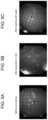
- FIG. 4 A , FIG. 4 B and FIG. 4 Care diagrams for describing an example.
- FIG. 7 A , FIG. 7 B and FIG. 7 Care diagrams for describing an example.
- the endoscope hood 1includes an exterior portion 2 and a disc portion 3 .
- the endoscope hood 1is configured to be attached to an insertion side end (tip end) 100 E of the endoscope 100 .
- the length b of the mounting portion 5is not particularly limited as long as it is a length sufficient to fix the endoscope hood 1 to the endoscope 100 , and the length b of the mounting portion 5 is preferably 5 mm to 20 mm, and more particularly 10 mm to 15 mm.
- the thickness d of the mounting portion 5is preferably 0.1 mm to 1.0 mm, and more preferably 0.3 mm to 0.5 mm.
- the disc portion 3has a disc shape and is provided in circumferential contact with the inner surface of the exterior portion 2 .
- the disc portion 3is formed in a substantially flat surface, and the thickness e of the disc portion 3 is preferably 0.01 mm to 1.0 mm, more preferably 0.05 mm to 0.7 mm, and most preferably 0.1 mm to 0.5 mm.
- the thickness e of the disc portion 3exceeds 1.0 mm, the visual field obtained through the camera lens is adversely affected.
- the thickness e of the disc portion 3is less than 0.01 mm, the strength of the disc portion 3 is lowered, which is not preferable because it is feared that damage occurs during use.
- the exterior portion 2 and the disc portion 3are integrally formed.
- the exterior portion 2 and the disc portion 3may be made of the same material.
- the endoscope hood 1is made of a hydrogel.
- the hydrogelinclude a hydrogel formed by using only a hydrophilic monomer, a hydrogel formed by adding a hydrophobic monomer, a crosslinkable monomer, or both to a hydrophilic monomer, and the like.
- the hydrophilic monomercontributes to the moisture content of the hydrogel, and the hydrophobic monomer contributes to the adjustment action of the moisture content or the swelling ratio of the hydrogel, thereby affecting the wettability or flexibility of the obtained endoscope hood 1 .
- hydrophobic monomerexamples include siloxanyl(meth)acrylate, trifluoroethyl(meth)acrylate, methacrylamide, cyclohexyl(meth)acrylate, and normal butyl(meth)acrylate. Two or more kinds of the hydrophobic monomers may be used in combination.
- crosslinkable monomerexamples include ethylene glycol di(meth)acrylate, methylene bisacrylamide, 2-hydroxy-1,3-dimethacryloxypropane, and trimethylolpropane triacrylate. Two or more of these crosslinkable monomers may be used in combination.
- the blending amount of the crosslinkable monomeris preferably 0.1 wt % to 10 wt % with respect to the total amount of the monomer from the viewpoint of the shape adjustment effect of the obtained endoscope hood 1 .
- the blending amount of the crosslinkable monomeris less than 0.1 wt %, the network structure of the endoscope hood 1 is insufficient, and when the blending amount of the crosslinkable monomer exceeds 10 wt %, the network structure becomes excessive, the endoscope hood 1 becomes fragile, and the flexibility deteriorates.
- Examples of a polymerization initiator used for polymerizing the above-mentioned mixtures of the monomersinclude peroxide such as lauroyl peroxide, cumene hydroperoxide, or benzoyl peroxide, which is a general radical polymerization initiator, azobisvaleronitrile, and azobisisobutyronitrile.
- the addition amount of the polymerization initiatoris preferably about 10 ppm to about 3,500 ppm with respect to the total amount of the monomer.
Landscapes
- Health & Medical Sciences (AREA)
- Life Sciences & Earth Sciences (AREA)
- Physics & Mathematics (AREA)
- Surgery (AREA)
- Optics & Photonics (AREA)
- Biomedical Technology (AREA)
- Public Health (AREA)
- Nuclear Medicine, Radiotherapy & Molecular Imaging (AREA)
- Pathology (AREA)
- Radiology & Medical Imaging (AREA)
- Veterinary Medicine (AREA)
- Engineering & Computer Science (AREA)
- Molecular Biology (AREA)
- Biophysics (AREA)
- Medical Informatics (AREA)
- Heart & Thoracic Surgery (AREA)
- Animal Behavior & Ethology (AREA)
- General Health & Medical Sciences (AREA)
- General Physics & Mathematics (AREA)
- Astronomy & Astrophysics (AREA)
- Endoscopes (AREA)
- Instruments For Viewing The Inside Of Hollow Bodies (AREA)
Abstract
Description
- ⊚: No lard is attached to the lens part.
- ◯: The attachment of the lard to the lens part is observed, but it does not interfere with visibility.
- x: The lard is attached to the lens part, and the visibility is poor.
| TABLE 1 | ||||||
| Com- | ||||||
| Ex- | Ex- | Ex- | Ex- | parative | ||
| ample | ample | ample | ample | Example | ||
| 1 | 2 | 3 | 4 | 4 | ||
| Moisture content (%) | 38.0 | 67.0 | 38.0 | 38.0 | — |
| of exterior portion | |||||
| Moisture content (%) | 60.0 | 67.0 | — | ||
| of disc portion |
| Tip end | Length | 14.0 | 14.0 | 4.0 | 4.0 | 13.0 |
| (mm) | ||||||
| Thickness | 1.0 | 1.0 | 1.0 | 1.0 | 1.3 | |
| (mm) | ||||||
| Mounting | Length | 14.0 | 14.0 | 14.0 | 14.0 | 3.3 |
| portion | (mm) | |||||
| Thickness | 1.0 | 1.0 | 1.0 | 1.0 | 0.6 | |
| (mm) | ||||||
| Disc | Thickness | |||||
| portion | (mm) | 0.5 | 0.5 | 0.5 | 0.5 | — |
| Evaluation results of | ◯ | ⊚ | ⊚ | ⊚ | X |
| antifogging and | |||||
| antifouling properties | |||||
Claims (4)
Applications Claiming Priority (3)
| Application Number | Priority Date | Filing Date | Title |
|---|---|---|---|
| JP2017149219 | 2017-08-01 | ||
| JP2017-149219 | 2017-08-01 | ||
| PCT/JP2018/023998WO2019026473A1 (en) | 2017-08-01 | 2018-06-25 | Hood for endoscope |
Publications (2)
| Publication Number | Publication Date |
|---|---|
| US20210127952A1 US20210127952A1 (en) | 2021-05-06 |
| US12349865B2true US12349865B2 (en) | 2025-07-08 |
Family
ID=65233826
Family Applications (1)
| Application Number | Title | Priority Date | Filing Date |
|---|---|---|---|
| US16/635,641Active2040-01-17US12349865B2 (en) | 2017-08-01 | 2018-06-25 | Endoscope hood |
Country Status (11)
| Country | Link |
|---|---|
| US (1) | US12349865B2 (en) |
| EP (1) | EP3662809B1 (en) |
| JP (1) | JP6725763B2 (en) |
| KR (1) | KR20200026294A (en) |
| CN (1) | CN111194177B (en) |
| AR (1) | AR112523A1 (en) |
| CL (1) | CL2020000257A1 (en) |
| CO (1) | CO2020001504A2 (en) |
| PE (1) | PE20201452A1 (en) |
| TW (1) | TWI766076B (en) |
| WO (1) | WO2019026473A1 (en) |
Families Citing this family (2)
| Publication number | Priority date | Publication date | Assignee | Title |
|---|---|---|---|---|
| EP4012478A4 (en)* | 2019-09-11 | 2023-08-16 | Seed Co., Ltd. | FORMING TOOL FOR ENDOSCOPE CAP |
| CN120417822A (en)* | 2022-12-27 | 2025-08-01 | 株式会社实瞳 | Endoscope cover |
Citations (40)
| Publication number | Priority date | Publication date | Assignee | Title |
|---|---|---|---|---|
| US4943618A (en)* | 1987-12-18 | 1990-07-24 | Kingston Technologies Limited Partnership | Method for preparing polyacrylonitrile copolymers by heterogeneous reaction of polyacrylonitrile aquagel |
| US5252392A (en)* | 1991-07-05 | 1993-10-12 | Konica Corporation | Magnetic recording medium comprising multiple magnetic layers with specified composition and properties |
| EP0595226A2 (en) | 1992-10-30 | 1994-05-04 | Hoya Corporation | Active substance-containing polymer gel |
| JPH06306250A (en) | 1993-04-27 | 1994-11-01 | Hoya Corp | Polymer gel containing working substance |
| US5443781A (en)* | 1992-10-29 | 1995-08-22 | Saab; Mark A. | Method of preparing disposable sheath with optically transparent windows formed continuously integral therewith |
| JPH1147081A (en) | 1997-08-05 | 1999-02-23 | Olympus Optical Co Ltd | Sheath for endoscope |
| US20010053909A1 (en)* | 2000-05-26 | 2001-12-20 | Olympus Optical Co., Ltd. | Endoscope hood for mucous membrane resection |
| US6428839B1 (en)* | 2000-06-02 | 2002-08-06 | Bausch & Lomb Incorporated | Surface treatment of medical device |
| US6530881B1 (en)* | 1999-01-21 | 2003-03-11 | Vision Sciences, Inc. | Sheath apparatus for endoscopes and methods for forming same |
| US20040114105A1 (en)* | 2000-05-10 | 2004-06-17 | Naoki Shimoyama | Surface-treated plastic article and method of surface treatment |
| JP2004267583A (en) | 2003-03-11 | 2004-09-30 | Takeshi Ohira | Laparoscope defogging device, member for defogging laparoscope, optical transmission member for defogging laparoscope and defogging method for laparoscope |
| US20050147685A1 (en)* | 2002-05-01 | 2005-07-07 | Hokkaido Technology Licensing Office Co., Ltd. | Hydrogel of (semi) interpenetrating network structure and process for producing the same |
| US20060069312A1 (en)* | 2004-09-30 | 2006-03-30 | Scimed Life Systems, Inc. | System for retaining optical clarity in a medical imaging system |
| US20060136064A1 (en)* | 2003-06-02 | 2006-06-22 | Sherman Michael C | Intervertebral disc implants and methods for manufacturing and using the same |
| US20060258908A1 (en) | 2005-05-13 | 2006-11-16 | David Stefanchik | Sheath for use with an endoscope |
| JP2007020759A (en) | 2005-07-14 | 2007-02-01 | Pentax Corp | Endoscope hood |
| EP1759627A1 (en) | 2005-08-31 | 2007-03-07 | Fujinon Corporation | Hood for endoscope, endoscope and method of fixing balloon for endoscope |
| JP2007151685A (en) | 2005-12-01 | 2007-06-21 | Olympus Medical Systems Corp | Endoscope and hydrophilic cap |
| US20090196845A1 (en)* | 2008-01-31 | 2009-08-06 | Erning Xia | Opthalmic compositions with an amphoteric surfactant and an anionic biopolymer |
| JP2009213631A (en) | 2008-03-10 | 2009-09-24 | Fujifilm Corp | Tip hood for endoscope and endoscope unit having the same |
| US20090247831A1 (en) | 2008-03-31 | 2009-10-01 | Shinichi Miyamoto | Endoscope, distal end cap-equipped endoscope and endoscope cleaning sheath |
| US20110189420A1 (en)* | 2007-08-27 | 2011-08-04 | Shoichi Masuda | Polymer gel structure and method for producing the same |
| US20110237766A1 (en)* | 2010-03-18 | 2011-09-29 | Maggio Thomas L | Silicone (meth)acrylamide monomer, polymer, ophthalmic lens, and contact lens |
| US20120026457A1 (en) | 2010-07-30 | 2012-02-02 | Yongxing Qiu | Silicone hydrogel lens with a crosslinked hydrophilic coating |
| US20120109272A1 (en)* | 2010-10-29 | 2012-05-03 | Medtronic, Inc. | Implantable medical device with compressible fixation member |
| US20120209074A1 (en) | 2011-02-16 | 2012-08-16 | James Sidney Titus | Optical coupler for an endoscope |
| US20120283381A1 (en)* | 2011-05-04 | 2012-11-08 | Ryuta Tamiya | Macroinitiator containing hydrophobic segment |
| US20120318772A1 (en)* | 2010-03-31 | 2012-12-20 | Sharp Kabushiki Kaisha | Die, process for producing die, and process for producing antireflection film |
| US20130046138A1 (en)* | 2011-08-19 | 2013-02-21 | Cook Medical Technologies Llc | Cap for Attachment to an Endoscope |
| US20130090527A1 (en)* | 2010-05-25 | 2013-04-11 | Arc Medical Design Limited | Covering for a medical scoping device |
| JP2014200974A (en)* | 2013-04-03 | 2014-10-27 | テルモ株式会社 | Structure and endoscope using the same |
| JP2014212835A (en) | 2013-04-23 | 2014-11-17 | ショーダテクトロン株式会社 | Hood for endoscope, and endoscope with same hood for endoscope |
| US20150133566A1 (en)* | 2012-05-12 | 2015-05-14 | National University Corporation Hokkaido University | Aqueous gel |
| JP2016055103A (en) | 2014-09-12 | 2016-04-21 | 富士フイルム株式会社 | Hood for endoscope and endoscope system |
| JP3204800U (en) | 2016-04-05 | 2016-06-16 | ショーダテクトロン株式会社 | Endoscope hood and endoscope provided with the endoscope hood |
| US20170066207A1 (en)* | 2015-04-30 | 2017-03-09 | Sharp Kabushiki Kaisha | Method for producing optical film, and optical film |
| US20170204213A1 (en)* | 2014-06-27 | 2017-07-20 | Toray Industries, Inc. | Silicone hydrogel, medical device, lens for eye and contact lens (as amended) |
| US20180037690A1 (en)* | 2016-08-05 | 2018-02-08 | Johnson & Johnson Vision Care, Inc. | Polymer compositions containing grafted polymeric networks and processes for their preparation and use |
| US20190016900A1 (en)* | 2016-07-12 | 2019-01-17 | Sharp Kabushiki Kaisha | Antifouling film |
| US20190099511A1 (en)* | 2016-03-31 | 2019-04-04 | Toray Industries, Inc. | Copolymer, wetting agent, medical device, and method for producing same |
Family Cites Families (2)
| Publication number | Priority date | Publication date | Assignee | Title |
|---|---|---|---|---|
| JP3639561B2 (en)* | 2002-04-08 | 2005-04-20 | オリンパス株式会社 | Endoscope hood |
| JP6601261B2 (en) | 2016-02-23 | 2019-11-06 | 株式会社Soken | Road surface condition estimation device |
- 2018
- 2018-06-25PEPE2020000120Apatent/PE20201452A1/enunknown
- 2018-06-25JPJP2019533968Apatent/JP6725763B2/enactiveActive
- 2018-06-25KRKR1020207003752Apatent/KR20200026294A/ennot_activeCeased
- 2018-06-25CNCN201880049533.5Apatent/CN111194177B/enactiveActive
- 2018-06-25WOPCT/JP2018/023998patent/WO2019026473A1/ennot_activeCeased
- 2018-06-25EPEP18841503.8Apatent/EP3662809B1/enactiveActive
- 2018-06-25USUS16/635,641patent/US12349865B2/enactiveActive
- 2018-07-30ARARP180102144patent/AR112523A1/enactiveIP Right Grant
- 2018-07-30TWTW107126364Apatent/TWI766076B/enactive
- 2020
- 2020-01-30CLCL2020000257Apatent/CL2020000257A1/enunknown
- 2020-02-11COCONC2020/0001504Apatent/CO2020001504A2/enunknown
Patent Citations (45)
| Publication number | Priority date | Publication date | Assignee | Title |
|---|---|---|---|---|
| US4943618A (en)* | 1987-12-18 | 1990-07-24 | Kingston Technologies Limited Partnership | Method for preparing polyacrylonitrile copolymers by heterogeneous reaction of polyacrylonitrile aquagel |
| US5252392A (en)* | 1991-07-05 | 1993-10-12 | Konica Corporation | Magnetic recording medium comprising multiple magnetic layers with specified composition and properties |
| US5443781A (en)* | 1992-10-29 | 1995-08-22 | Saab; Mark A. | Method of preparing disposable sheath with optically transparent windows formed continuously integral therewith |
| EP0595226A2 (en) | 1992-10-30 | 1994-05-04 | Hoya Corporation | Active substance-containing polymer gel |
| JPH06306250A (en) | 1993-04-27 | 1994-11-01 | Hoya Corp | Polymer gel containing working substance |
| JPH1147081A (en) | 1997-08-05 | 1999-02-23 | Olympus Optical Co Ltd | Sheath for endoscope |
| US6530881B1 (en)* | 1999-01-21 | 2003-03-11 | Vision Sciences, Inc. | Sheath apparatus for endoscopes and methods for forming same |
| US20040114105A1 (en)* | 2000-05-10 | 2004-06-17 | Naoki Shimoyama | Surface-treated plastic article and method of surface treatment |
| US20010053909A1 (en)* | 2000-05-26 | 2001-12-20 | Olympus Optical Co., Ltd. | Endoscope hood for mucous membrane resection |
| US6428839B1 (en)* | 2000-06-02 | 2002-08-06 | Bausch & Lomb Incorporated | Surface treatment of medical device |
| US20050147685A1 (en)* | 2002-05-01 | 2005-07-07 | Hokkaido Technology Licensing Office Co., Ltd. | Hydrogel of (semi) interpenetrating network structure and process for producing the same |
| JP2004267583A (en) | 2003-03-11 | 2004-09-30 | Takeshi Ohira | Laparoscope defogging device, member for defogging laparoscope, optical transmission member for defogging laparoscope and defogging method for laparoscope |
| US20060136064A1 (en)* | 2003-06-02 | 2006-06-22 | Sherman Michael C | Intervertebral disc implants and methods for manufacturing and using the same |
| US20060069312A1 (en)* | 2004-09-30 | 2006-03-30 | Scimed Life Systems, Inc. | System for retaining optical clarity in a medical imaging system |
| US20060258908A1 (en) | 2005-05-13 | 2006-11-16 | David Stefanchik | Sheath for use with an endoscope |
| JP2007020759A (en) | 2005-07-14 | 2007-02-01 | Pentax Corp | Endoscope hood |
| EP1759627A1 (en) | 2005-08-31 | 2007-03-07 | Fujinon Corporation | Hood for endoscope, endoscope and method of fixing balloon for endoscope |
| JP2007151685A (en) | 2005-12-01 | 2007-06-21 | Olympus Medical Systems Corp | Endoscope and hydrophilic cap |
| US20080228035A1 (en) | 2005-12-01 | 2008-09-18 | Olympus Medical Systems Corp. | Endoscope and hydrophilic cap |
| US20110189420A1 (en)* | 2007-08-27 | 2011-08-04 | Shoichi Masuda | Polymer gel structure and method for producing the same |
| US20090196845A1 (en)* | 2008-01-31 | 2009-08-06 | Erning Xia | Opthalmic compositions with an amphoteric surfactant and an anionic biopolymer |
| JP2009213631A (en) | 2008-03-10 | 2009-09-24 | Fujifilm Corp | Tip hood for endoscope and endoscope unit having the same |
| JP2009240596A (en) | 2008-03-31 | 2009-10-22 | Olympus Medical Systems Corp | Endoscope, endoscope with distal end cap and endoscope washing sheath |
| US20090247831A1 (en) | 2008-03-31 | 2009-10-01 | Shinichi Miyamoto | Endoscope, distal end cap-equipped endoscope and endoscope cleaning sheath |
| US20110237766A1 (en)* | 2010-03-18 | 2011-09-29 | Maggio Thomas L | Silicone (meth)acrylamide monomer, polymer, ophthalmic lens, and contact lens |
| US20120318772A1 (en)* | 2010-03-31 | 2012-12-20 | Sharp Kabushiki Kaisha | Die, process for producing die, and process for producing antireflection film |
| US20130090527A1 (en)* | 2010-05-25 | 2013-04-11 | Arc Medical Design Limited | Covering for a medical scoping device |
| US20120026457A1 (en) | 2010-07-30 | 2012-02-02 | Yongxing Qiu | Silicone hydrogel lens with a crosslinked hydrophilic coating |
| US20120109272A1 (en)* | 2010-10-29 | 2012-05-03 | Medtronic, Inc. | Implantable medical device with compressible fixation member |
| JP2014510577A (en) | 2011-02-16 | 2014-05-01 | ザ ジェネラル ホスピタル コーポレイション | Endoscope optical coupler |
| WO2012112755A2 (en) | 2011-02-16 | 2012-08-23 | The General Hospital Corporation | Optical coupler for an endoscope |
| US20120209074A1 (en) | 2011-02-16 | 2012-08-16 | James Sidney Titus | Optical coupler for an endoscope |
| US20200281448A1 (en)* | 2011-02-16 | 2020-09-10 | The General Hospital Corporation | Optical Coupler For An Endoscope |
| US20120283381A1 (en)* | 2011-05-04 | 2012-11-08 | Ryuta Tamiya | Macroinitiator containing hydrophobic segment |
| US20130046138A1 (en)* | 2011-08-19 | 2013-02-21 | Cook Medical Technologies Llc | Cap for Attachment to an Endoscope |
| US20150133566A1 (en)* | 2012-05-12 | 2015-05-14 | National University Corporation Hokkaido University | Aqueous gel |
| JP2014200974A (en)* | 2013-04-03 | 2014-10-27 | テルモ株式会社 | Structure and endoscope using the same |
| JP2014212835A (en) | 2013-04-23 | 2014-11-17 | ショーダテクトロン株式会社 | Hood for endoscope, and endoscope with same hood for endoscope |
| US20170204213A1 (en)* | 2014-06-27 | 2017-07-20 | Toray Industries, Inc. | Silicone hydrogel, medical device, lens for eye and contact lens (as amended) |
| JP2016055103A (en) | 2014-09-12 | 2016-04-21 | 富士フイルム株式会社 | Hood for endoscope and endoscope system |
| US20170066207A1 (en)* | 2015-04-30 | 2017-03-09 | Sharp Kabushiki Kaisha | Method for producing optical film, and optical film |
| US20190099511A1 (en)* | 2016-03-31 | 2019-04-04 | Toray Industries, Inc. | Copolymer, wetting agent, medical device, and method for producing same |
| JP3204800U (en) | 2016-04-05 | 2016-06-16 | ショーダテクトロン株式会社 | Endoscope hood and endoscope provided with the endoscope hood |
| US20190016900A1 (en)* | 2016-07-12 | 2019-01-17 | Sharp Kabushiki Kaisha | Antifouling film |
| US20180037690A1 (en)* | 2016-08-05 | 2018-02-08 | Johnson & Johnson Vision Care, Inc. | Polymer compositions containing grafted polymeric networks and processes for their preparation and use |
Non-Patent Citations (3)
| Title |
|---|
| Chilean Office Action issued in corresponding Chilean Patent Application No. 257-2020 dated Nov. 23, 2020, submitted with Chilean Attorney comments dated Dec. 17, 2020. |
| International Search Report issued in corresponding International Application No. PCT/JP2018/023998 dated Sep. 25, 2018, with English translation. |
| Supplementary European Search Report issued in corresponding European Patent Application No. 18 84 1503 dated Mar. 16, 2021. |
Also Published As
| Publication number | Publication date |
|---|---|
| TW201914516A (en) | 2019-04-16 |
| KR20200026294A (en) | 2020-03-10 |
| PE20201452A1 (en) | 2020-12-15 |
| US20210127952A1 (en) | 2021-05-06 |
| AR112523A1 (en) | 2019-11-06 |
| BR112020001337A2 (en) | 2020-08-11 |
| WO2019026473A1 (en) | 2019-02-07 |
| CL2020000257A1 (en) | 2020-08-14 |
| JPWO2019026473A1 (en) | 2020-01-16 |
| EP3662809A4 (en) | 2021-04-14 |
| CN111194177B (en) | 2022-12-09 |
| TWI766076B (en) | 2022-06-01 |
| CO2020001504A2 (en) | 2020-02-28 |
| JP6725763B2 (en) | 2020-07-22 |
| EP3662809B1 (en) | 2023-05-03 |
| CN111194177A (en) | 2020-05-22 |
| EP3662809A1 (en) | 2020-06-10 |
Similar Documents
| Publication | Publication Date | Title |
|---|---|---|
| JP6159768B2 (en) | Low hydrous soft contact lens and method for producing low hydrous soft contact lens | |
| ES2355773T3 (en) | SURFACE TREATMENT OF MEDICAL DEVICES. | |
| JP6036299B2 (en) | Medical device and manufacturing method thereof | |
| EP3141943B1 (en) | Hydrogel contact lens having wet surface, and manufacturing method therefor | |
| US12349865B2 (en) | Endoscope hood | |
| US20100168356A1 (en) | Biomedical Devices | |
| JP7591507B2 (en) | Endoscope hood molding die | |
| JP7120840B2 (en) | Endoscope hood mold | |
| JP7338477B2 (en) | Medical device and manufacturing method thereof | |
| HK40021782B (en) | Hood for endoscope | |
| HK40021782A (en) | Hood for endoscope | |
| BR112020001337B1 (en) | ENDOSCOPE COVER | |
| EP4585132A1 (en) | Endoscope hood | |
| KR20130023358A (en) | Medical device for controlled release of drug |
Legal Events
| Date | Code | Title | Description |
|---|---|---|---|
| FEPP | Fee payment procedure | Free format text:ENTITY STATUS SET TO UNDISCOUNTED (ORIGINAL EVENT CODE: BIG.); ENTITY STATUS OF PATENT OWNER: LARGE ENTITY | |
| AS | Assignment | Owner name:SEED CO., LTD., JAPAN Free format text:ASSIGNMENT OF ASSIGNORS INTEREST;ASSIGNORS:MORITA, YOSHINORI;TAKAMATSU, TOSHIHIRO;OKINO, AKITOSHI;AND OTHERS;SIGNING DATES FROM 20200106 TO 20200129;REEL/FRAME:051723/0767 | |
| STPP | Information on status: patent application and granting procedure in general | Free format text:APPLICATION DISPATCHED FROM PREEXAM, NOT YET DOCKETED | |
| STPP | Information on status: patent application and granting procedure in general | Free format text:DOCKETED NEW CASE - READY FOR EXAMINATION | |
| STPP | Information on status: patent application and granting procedure in general | Free format text:NON FINAL ACTION MAILED | |
| STPP | Information on status: patent application and granting procedure in general | Free format text:FINAL REJECTION MAILED | |
| STPP | Information on status: patent application and granting procedure in general | Free format text:ADVISORY ACTION MAILED | |
| STPP | Information on status: patent application and granting procedure in general | Free format text:DOCKETED NEW CASE - READY FOR EXAMINATION | |
| STPP | Information on status: patent application and granting procedure in general | Free format text:FINAL REJECTION MAILED | |
| STPP | Information on status: patent application and granting procedure in general | Free format text:DOCKETED NEW CASE - READY FOR EXAMINATION | |
| STPP | Information on status: patent application and granting procedure in general | Free format text:NON FINAL ACTION MAILED | |
| STPP | Information on status: patent application and granting procedure in general | Free format text:RESPONSE TO NON-FINAL OFFICE ACTION ENTERED AND FORWARDED TO EXAMINER | |
| STPP | Information on status: patent application and granting procedure in general | Free format text:FINAL REJECTION MAILED | |
| STPP | Information on status: patent application and granting procedure in general | Free format text:DOCKETED NEW CASE - READY FOR EXAMINATION | |
| STCF | Information on status: patent grant | Free format text:PATENTED CASE |