US12268358B2 - Portable endoscope with side-mountable disposable portion - Google Patents
Portable endoscope with side-mountable disposable portionDownload PDFInfo
- Publication number
- US12268358B2 US12268358B2US17/349,674US202117349674AUS12268358B2US 12268358 B2US12268358 B2US 12268358B2US 202117349674 AUS202117349674 AUS 202117349674AUS 12268358 B2US12268358 B2US 12268358B2
- Authority
- US
- United States
- Prior art keywords
- handle
- endoscope
- cannula
- slot
- housing
- Prior art date
- Legal status (The legal status is an assumption and is not a legal conclusion. Google has not performed a legal analysis and makes no representation as to the accuracy of the status listed.)
- Active, expires
Links
Images


Classifications
- A—HUMAN NECESSITIES
- A61—MEDICAL OR VETERINARY SCIENCE; HYGIENE
- A61B—DIAGNOSIS; SURGERY; IDENTIFICATION
- A61B1/00—Instruments for performing medical examinations of the interior of cavities or tubes of the body by visual or photographical inspection, e.g. endoscopes; Illuminating arrangements therefor
- A61B1/00064—Constructional details of the endoscope body
- A61B1/00103—Constructional details of the endoscope body designed for single use
- A—HUMAN NECESSITIES
- A61—MEDICAL OR VETERINARY SCIENCE; HYGIENE
- A61B—DIAGNOSIS; SURGERY; IDENTIFICATION
- A61B1/00—Instruments for performing medical examinations of the interior of cavities or tubes of the body by visual or photographical inspection, e.g. endoscopes; Illuminating arrangements therefor
- A61B1/00064—Constructional details of the endoscope body
- A61B1/00071—Insertion part of the endoscope body
- A—HUMAN NECESSITIES
- A61—MEDICAL OR VETERINARY SCIENCE; HYGIENE
- A61B—DIAGNOSIS; SURGERY; IDENTIFICATION
- A61B1/00—Instruments for performing medical examinations of the interior of cavities or tubes of the body by visual or photographical inspection, e.g. endoscopes; Illuminating arrangements therefor
- A61B1/00002—Operational features of endoscopes
- A61B1/00011—Operational features of endoscopes characterised by signal transmission
- A61B1/00016—Operational features of endoscopes characterised by signal transmission using wireless means
- A—HUMAN NECESSITIES
- A61—MEDICAL OR VETERINARY SCIENCE; HYGIENE
- A61B—DIAGNOSIS; SURGERY; IDENTIFICATION
- A61B1/00—Instruments for performing medical examinations of the interior of cavities or tubes of the body by visual or photographical inspection, e.g. endoscopes; Illuminating arrangements therefor
- A61B1/00002—Operational features of endoscopes
- A61B1/00043—Operational features of endoscopes provided with output arrangements
- A61B1/00045—Display arrangement
- A61B1/00048—Constructional features of the display
- A—HUMAN NECESSITIES
- A61—MEDICAL OR VETERINARY SCIENCE; HYGIENE
- A61B—DIAGNOSIS; SURGERY; IDENTIFICATION
- A61B1/00—Instruments for performing medical examinations of the interior of cavities or tubes of the body by visual or photographical inspection, e.g. endoscopes; Illuminating arrangements therefor
- A61B1/00002—Operational features of endoscopes
- A61B1/00043—Operational features of endoscopes provided with output arrangements
- A61B1/00045—Display arrangement
- A61B1/00052—Display arrangement positioned at proximal end of the endoscope body
- A—HUMAN NECESSITIES
- A61—MEDICAL OR VETERINARY SCIENCE; HYGIENE
- A61B—DIAGNOSIS; SURGERY; IDENTIFICATION
- A61B1/00—Instruments for performing medical examinations of the interior of cavities or tubes of the body by visual or photographical inspection, e.g. endoscopes; Illuminating arrangements therefor
- A61B1/00064—Constructional details of the endoscope body
- A61B1/00066—Proximal part of endoscope body, e.g. handles
- A—HUMAN NECESSITIES
- A61—MEDICAL OR VETERINARY SCIENCE; HYGIENE
- A61B—DIAGNOSIS; SURGERY; IDENTIFICATION
- A61B1/00—Instruments for performing medical examinations of the interior of cavities or tubes of the body by visual or photographical inspection, e.g. endoscopes; Illuminating arrangements therefor
- A61B1/00064—Constructional details of the endoscope body
- A61B1/00105—Constructional details of the endoscope body characterised by modular construction
- A—HUMAN NECESSITIES
- A61—MEDICAL OR VETERINARY SCIENCE; HYGIENE
- A61B—DIAGNOSIS; SURGERY; IDENTIFICATION
- A61B1/00—Instruments for performing medical examinations of the interior of cavities or tubes of the body by visual or photographical inspection, e.g. endoscopes; Illuminating arrangements therefor
- A61B1/00112—Connection or coupling means
- A61B1/00121—Connectors, fasteners and adapters, e.g. on the endoscope handle
- A61B1/00124—Connectors, fasteners and adapters, e.g. on the endoscope handle electrical, e.g. electrical plug-and-socket connection
- A—HUMAN NECESSITIES
- A61—MEDICAL OR VETERINARY SCIENCE; HYGIENE
- A61B—DIAGNOSIS; SURGERY; IDENTIFICATION
- A61B1/00—Instruments for performing medical examinations of the interior of cavities or tubes of the body by visual or photographical inspection, e.g. endoscopes; Illuminating arrangements therefor
- A61B1/012—Instruments for performing medical examinations of the interior of cavities or tubes of the body by visual or photographical inspection, e.g. endoscopes; Illuminating arrangements therefor characterised by internal passages or accessories therefor
- A61B1/018—Instruments for performing medical examinations of the interior of cavities or tubes of the body by visual or photographical inspection, e.g. endoscopes; Illuminating arrangements therefor characterised by internal passages or accessories therefor for receiving instruments
- A—HUMAN NECESSITIES
- A61—MEDICAL OR VETERINARY SCIENCE; HYGIENE
- A61B—DIAGNOSIS; SURGERY; IDENTIFICATION
- A61B1/00—Instruments for performing medical examinations of the interior of cavities or tubes of the body by visual or photographical inspection, e.g. endoscopes; Illuminating arrangements therefor
- A61B1/04—Instruments for performing medical examinations of the interior of cavities or tubes of the body by visual or photographical inspection, e.g. endoscopes; Illuminating arrangements therefor combined with photographic or television appliances
- A61B1/05—Instruments for performing medical examinations of the interior of cavities or tubes of the body by visual or photographical inspection, e.g. endoscopes; Illuminating arrangements therefor combined with photographic or television appliances characterised by the image sensor, e.g. camera, being in the distal end portion
- A—HUMAN NECESSITIES
- A61—MEDICAL OR VETERINARY SCIENCE; HYGIENE
- A61B—DIAGNOSIS; SURGERY; IDENTIFICATION
- A61B1/00—Instruments for performing medical examinations of the interior of cavities or tubes of the body by visual or photographical inspection, e.g. endoscopes; Illuminating arrangements therefor
- A61B1/06—Instruments for performing medical examinations of the interior of cavities or tubes of the body by visual or photographical inspection, e.g. endoscopes; Illuminating arrangements therefor with illuminating arrangements
- A61B1/0661—Endoscope light sources
- A61B1/0684—Endoscope light sources using light emitting diodes [LED]
- A—HUMAN NECESSITIES
- A61—MEDICAL OR VETERINARY SCIENCE; HYGIENE
- A61B—DIAGNOSIS; SURGERY; IDENTIFICATION
- A61B1/00—Instruments for performing medical examinations of the interior of cavities or tubes of the body by visual or photographical inspection, e.g. endoscopes; Illuminating arrangements therefor
- A61B1/303—Instruments for performing medical examinations of the interior of cavities or tubes of the body by visual or photographical inspection, e.g. endoscopes; Illuminating arrangements therefor for the vagina, i.e. vaginoscopes
- A—HUMAN NECESSITIES
- A61—MEDICAL OR VETERINARY SCIENCE; HYGIENE
- A61B—DIAGNOSIS; SURGERY; IDENTIFICATION
- A61B10/00—Instruments for taking body samples for diagnostic purposes; Other methods or instruments for diagnosis, e.g. for vaccination diagnosis, sex determination or ovulation-period determination; Throat striking implements
- A61B10/02—Instruments for taking cell samples or for biopsy
- A61B10/0291—Instruments for taking cell samples or for biopsy for uterus
- A—HUMAN NECESSITIES
- A61—MEDICAL OR VETERINARY SCIENCE; HYGIENE
- A61B—DIAGNOSIS; SURGERY; IDENTIFICATION
- A61B10/00—Instruments for taking body samples for diagnostic purposes; Other methods or instruments for diagnosis, e.g. for vaccination diagnosis, sex determination or ovulation-period determination; Throat striking implements
- A61B10/02—Instruments for taking cell samples or for biopsy
- A61B10/04—Endoscopic instruments, e.g. catheter-type instruments
Definitions
- This patent specificationgenerally relates to a medical device for use in tissue examinations and endoscopic surgery such as in urology and gynecology. More particularly, some embodiments relate to a portable, handheld, low-cost surgical endoscope device having a single-use portion and a multiple-use portion.
- the lens systemis typically a relay lens system in the case of rigid endoscopes or a bundle of fiber optics or an objective lens system in the case of flexible endoscopes.
- the lens or fiber optic systemis relatively expensive and is intended to be re-used many times. Therefore, stringent decontamination and disinfection procedures need to be carried out after each use.
- an endoscopecomprises: (a) a single-use portion that includes: (i) an insert housing that has a distal end and a proximal end and has a proximal port at said proximal end; (ii) a cannula that extends distally from the distal end of the insert housing, is configured for rotation about said longitudinal axis relative to said insert housing, and has a distal port at a distal portion of the cannula; (iii) an essentially straight lumen extending along said longitudinal axis, from said proximal port at the proximal end of the insert housing to the distal port at the distal portion of the cannula; (iv) an imaging and illumination module at said distal portion of the cannula; and a proximally facing electrical connector that is affixed to said insert housing and is operatively coupled with said imaging and illumination module; and (b) a multiple-use portion that includes: (i) a handle that has a proximal
- a single-use, disposable portion of an endoscopecomprises: (a) an insert housing that is elongated along a longitudinal axis and has an electrical connector fixedly mounted thereon; (b) a cannula extending distally from said insert housing along said axis; (c) wherein said insert housing is configured to releasably snap into and release from an elongated slot of a reusable handle that is open in a direction transverse to said axis; (d) wherein said cannula is mounted to said insert housing for rotation about said axis relative to said insert housing; (e) a lumen that extends along said axis, from a proximal port that starts at a location that is proximal from a proximal end of the insert housing and ends at a distal portion of the cannula; (f) an imaging and illumination module that is at the distal portion of the cannula and is operatively coupled to said electrical connector; and (g) a cable
- the endoscopic apparatus described in the immediately preceding paragraphscan include a lumen that extends essentially straight in said insert housing and cannula along said longitudinal axis and has a distal port extending proximally from a proximal end of the insert housing.
- a method of imaging an internal site in a patientcomprises: (a) providing a single-use portion that includes a cannula extending distally from an insert housing elongated along a longitudinal axis of the cannula; (b) providing a multiple-use portion that has a handle with a slot elongated along said longitudinal axis and open in a direction transverse to the longitudinal axis, said slot extending from a proximal end to a distal end of the handle, and a display module mounted on the handle to rotate and/or pivot relative to the handle about one or more axes; (c) snap-fitting said insert housing in said slot of the handle such that the housing extends from the proximal to the distal ends of the handle, by relative motion of the insert housing relative to the handle that includes motion in a direction transverse to the longitudinal axis; (d) electrically coupling an imaging module at a distal end of the cannula to electronic circuits in the handle that are electronically coupled
- the method described in the immediately preceding paragraphscan include one or more the following additional or more specific steps: (a) making the cable sufficiently long and flexible to enable rotation of the cannula relative to the housing and the handle of at least 180 degrees; (a) sealing said handle sufficiently to allow repeatedly sterilizing said handle in steam; (b) said providing said handle can comprise providing plural portions of an exterior wall of the handle and sealing them against each other using o-rings and/or gaskets to enable said sterilizing in steam; and (c) said providing of said single use portion and said reusable portion can further comprise providing a fluid port at a location proximal to both the handle and the insert housing and providing a lumen that extends along said longitudinal axis from said fluid port to a distal portion of said cannula.
- surgicalor “surgery” refer to any physical intervention on a patient's tissues, and does not necessarily involve cutting a patient's tissues or closure of a previously sustained wound.
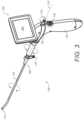
- FIG. 3is a perspective view of a portable endoscope having a disposable side-mountable portion, according to some embodiments
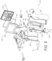
- FIG. 5is an exploded perspective illustrating sealing and other aspects of a portable endoscope having a side-mountable disposable portion and a fluid-resistant handle portion, according to some embodiments;
- FIG. 6is a perspective view illustrating sealing and other aspects of a portable endoscope having a side-mountable disposable portion, according to some embodiments
- FIGS. 8 A and 8 Bare cross section and frontal view illustrating cannula rotation and other aspects of a portable endoscope having a side-mountable disposable portion, according to some embodiments;
- FIG. 9is a side view illustrating a WiFi antenna arrangement and other aspects of a portable endoscope having a side-mountable disposable portion, according to some embodiments.
- FIG. 10is a perspective view illustrating aspects of a touch sensitive video display screen, according to some embodiments.
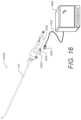
- FIG. 12is a perspective view illustrating a single-use portion of an endoscope, according to some embodiments.
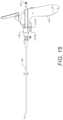
- FIG. 13is a perspective view illustrating a multiple-use portion of an endoscope, according to some embodiments.
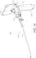
- FIG. 14is a perspective view of an endoscope assembled with the single-use portion and multiple-use portion shown in FIGS. 12 and 13 , according to some embodiments.
- FIG. 15is a perspective view of an endoscope like that of FIGS. 1 and 2 but with a different arrangement of electrical contacts, according to some embodiments.
- FIG. 16is a perspective view of a single-use endoscope portion configured to interact with a remote image processor and display, according to some embodiments.
- FIGS. 1 and 2are right side and top views, respectively, of a portable endoscope having a disposable portion, according to some embodiments.
- the portable endoscope 100includes an elongated cannula 120 with a distal tip 110 for inserting into a hollow organ or cavity of the body.
- distal tip 110is formed as a separate distal tip sub-assembly that is attached to the cannula 120 .
- the distal tip 110is about than 4.55 mm in diameter.
- the cannula 120is rigid, flexible or semi flexible and includes one or more fluid channels which are fluidly connected to port 132 .
- Port 132includes a Luer fitting to facilitate leak-free connection of port 132 with various medical fluid components (not shown).
- the one or more fluid channels or lumens in cannula 120are also connected to one or more distal facing fluid ports of distal tip 110 .
- one or more of the fluid channels or lumens in cannula 120are also connected to proximal ports 130 and 430 .
- Proximal ports 130 and 430also include Luer fittings to facilitate leak-free connection with various medial fluid components (not shown).
- proximal port 130is substantially in-line with the main longitudinal axis 124 of cannula 120 and thereby provides a substantially straight working channel through which rigid or semi rigid tools can pass through. According to some embodiments, only one proximal port 130 is provided and the second proximal port 430 is omitted. Providing a device channel that has straight proximal portion (through port 130 ), allows for improved ease in device insertion and manipulation. It has been found that providing the device port(s) (e.g. ports 130 and/or 430 ) in close proximity to the display module 150 , and vertically in line with a center of display 150 , provides significant ergonomic benefits.
- the device port(s)e.g. ports 130 and/or 430
- the distance d between the proximal port (in this case port 130 ) and the center of the screen 350 of display module 150is less than about 15 cm. According to some embodiments, the distance d is less than about 12 cm. According to some embodiments, the distance dis less than about 10 cm.
- Proximal to the fluid port 132are a housing 134 and insert housing 138 .
- the insert housing 138is configured to slidably mate with, e.g., snap-fit in, a side-slot formed in handle 140 (see side-slot or socket 440 in FIG. 4 A ).
- handle 140is similar to handle 140 shown and described in the '048 patent, the '331 application, and the '880 application in that all are generally pistol-grip handles and may contain electronics and are coupled with displays, but handle 140 in the subject application differs in significant ways, including in the way it releasable couples and interacts with disposable portion 400 .
- FIG. 3is a perspective view of a portable endoscope having a disposable side-mountable portion, according to some embodiments.
- the version of endoscope 100 shownincludes only a single proximal port 130 .
- different versions of the disposable single-use portion 400can be made available to users depending upon their particular needs.
- Some examplesare disposable portions in which an injection needle can protrude from the cannula tip, in a manner similar to that described in U.S. Pat. No. 10,278,563, or disposable portions with cannulas that have bendable distal ends, for example as described in U.S. patent application Ser. No. 16/447,251 filed Jun.
- FIG. 4 Bshows further details of the physical and electrical connections between the single use portion and reusable portion of portable endoscope 100 .
- the electrical connectors 136 and 144use a standard electrical connection scheme such as mini-display port (type DP20), which is also used for versions 1 and 2 of Thunderbolt connectors.
- mini-display porttype DP20
- a male connector 410is shown that can mate with a female connector 144 on handle 140 .
- the insert housing 138can include positioning aids such as positioning balls 460 , 462 and 464 which are spring-loaded and can engage with grooves 442 and 444 formed within slot 440 .
- posts 450 and 452are formed on insert housing 138 and mate with holes 470 and 472 on the inside of slot 440 of handle 140 .
- FIG. 5is an exploded perspective view illustrating sealing and other aspects of a portable endoscope having a side-mountable disposable portion and a fluid-resistant handle portion, according to some embodiments.
- Many of the components of the reusable portion 402are sealed using o-rings and/or gaskets, including left and right handle cover pieces 542 and 540 , respectively, which are sealed to each other using an o-ring or gasket 532 .
- the front cover 550 and rear cover 552 of the display module 150is sealed using an o-ring 534 .
- the display module 150are sealed to the left handle cover piece 542 using o-ring 551 .
- Various other openingsare sealed with o-rings such as o-rings 530 .
- Front cover 581 and bottom cover or piece 583similarly may be sealed against right and left handle cover pieces 540 and 542 by o-rings and/or gaskets.
- FIG. 6is a perspective view illustrating sealing and other aspects of a portable endoscope having a side-mountable disposable portion, according to some embodiments.
- the o-ring seals 532 and 534are shown in this view.
- the o-ring seals shown and described in FIGS. 5 and 6enable the reusable portion 402 of the portable endoscope to be highly water resistant.
- handle 140can be configured to comply with the IXP 7 standard and can withstand being submerged for up to 30 minutes 0.15 meters to 1 meter below a fluid surface.
- endoscope components that will be re-usedcan be disinfected and/or sterilized prior to re-use to prevent cross contamination.
- FIG. 7is an exploded perspective illustrating cannula rotation and other aspects of a portable endoscope having a side-mountable disposable portion, according to some embodiments.
- distal tip 110is shown to include tip housing 710 , two or more LEDs of which only LEDs 720 and 722 are shown as an example, and camera module 730 .
- the proximal end of cannula 120is inserted into the distal end of fluid hub body 732 .
- Hub body 732is fixed at its proximal end to housing 134 , which in turn is fixed at its proximal end to base 750 .
- Within housing 134is insert tube connector 746 that is shaped at its distal end to accept the proximal end of cannula 120 .
- FIGS. 8 A and 8 Bare cross section and frontal view illustrating cannula rotation and other aspects of a portable endoscope having a side-mountable disposable portion, according to some embodiments.
- slot 810is shaped to limit the rotation of the cannula 120 and other components by limiting the movement of tab 752 (shown in FIG. 7 ) of base 750 .
- the working channel in the cannulamay be slightly off center or off axis 124 due to the cannula containing multiple lumens.
- the connector piece 744can be shaped to bring the working channel more towards the central longitudinal axis 124 .
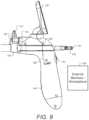
- FIG. 9is a side view illustrating a WiFi antenna arrangement and other aspects of a portable endoscope having a side-mountable disposable portion, according to some embodiments.
- WiFi board 582 and the location of WiFi antenna 584is shown.
- WiFi antenna 584can extend to and along the back of the touch screen of display module 150 to reduce the effects of screening by metal portions of handle 140 .
- the WiFi module on board 582 and the antenna 584can transmit video to WiFi receivers which can be used for other external monitors or to workstations 585 , for example.
- FIG. 10is a perspective view illustrating aspects of a touch sensitive video display screen, according to some embodiments.
- electronics modules 146 in handle 140include an electronic imaging system that supports automated features such as exposure control (AEC), gain control (AGC) and white balance (AWB).
- AECexposure control
- ALCgain control
- ALCwhite balance
- the camera and illumination modules in distal tip 110 and the imaging and control in electronics modules 146are together configured as a “well-tuned” imaging system that provides a useful “Auto Mode.” However, in certain cases, especially in endoscopy, it is sometimes advantageous to use a “Manual Mode”.
- a handheld portable endoscope 100when the distal tip 110 is inside a cavity, Auto Mode works well in most situations. But in certain situations, for example when the camera on distal tip 110 is proximal to a wall that is tangential with the direction of the view of the camera module, part of the image field (the wall due to its proximity to the illuminating LEDs) is very bright compared to other areas which are further away from the LEDs. In order to better visualize the wall (or the portion that is close up to the camera tip), a “Manual Mode” can be used in which all the automated imaging functions are turned off. In FIG.
- Auto Modeis configured to provide full automatic AGC, AEC and ALC, while Manual Mode turns off AGC, AEC and ALC, and sets the LED brightness at 50% (or some other predetermined level such as 75%) of its default brightness
- Screen 350can be configured for non-contact or non-touch operation such that clinical operators need not remove their gloves in order to push and activate a button on the screen 150 .
- buttons 1020 , 1022 , 1024 and 1026are virtual touch buttons that are floating above the display screen 350 . They can be located near the corners or edges of the screen as shown. User can use cross motion (sliding “X” or “+” shape) at location 1030 on the screen 350 to close and hide the floating buttons. User can use circular motion (not shown) to bring back up the floating buttons.
- FIGS. 11 A and 11 Bare cross sections showing further detail of a cannula and a distal tip that form part of a disposable portion of a portable endoscope, according to some embodiments.
- FIG. 11 Ashows some example dimensions for the cannula 120 .
- Cannula 120has an O.D. of 4.1 mm.
- the working channel lumenhas an I.D. of 2.2. mm and the cable lumen 1102 is oval with dimensions shown. Cable lumen 1102 can be used to carry the electrical wires leading to the camera module and LEDs on distal tip 110 .
- FIG. 11 Bthe dimensions and placement of camera module 730 , LEDS 720 and 722 are shown.
- Camera module 730can have a square area 1.05 mm per side, and each of LEDs 720 and 722 can be a rectangle with a larger side approximately the same length as a side of camera 730 .
- FIG. 4 Abut preferably further includes a ridge 1241 at its upper surface that extends proximally, preferably to the proximal end of the insert housing, and may further include a like ridge at its underside.
- Cannula 120 of FIG. 12is rotatable about its longitudinal axis, as described in connection with FIG. 3 , for example.
- the multiple use portion 12402 seen in FIG. 13is otherwise like the multiple-use portion 402 of FIG. 4 A but has a distally facing electrical connector 1244 in place of electrical connector 144 seen in FIG. 4 A .
- multiple-use portion 12402preferably includes a proximally extending groove 1242 at its bottom surface and may include a matching groove at its upper surface.
- FIG. 14illustrates the single-use portion 12400 and the multiple-use portion 12402 assembled into endoscope 1400 .
- Single-use portion 12400typically comes sealed in a sterile pouch.
- the userremoves it from the package and slides a proximal part of insert housing 138 into slot 1240 , in the left-to-right direction as seen in FIGS. 12 and 13 , preferably until ridges 1241 snap into grooves 1242 .
- the userthen snaps insert housing 138 in the proximal direction so electrical connector 1236 engages connector 1244 and the proximal face of post 1201 presses against the distal face of handle 140 .
- the contact between post 1201 and the distal face of handle 140should be sufficient to keep fluids from entering handle 140 or reaching connectors 1236 and 1244 but for extra security the distal face of handle 140 can be further provided with a peripheral seal that would be squeezed when post 1201 presses against handle 140 .
- display unit 150is mounted on handle 140 for tilting relative to the handle about at least one axis, and cannula 120 is mounted for rotation about its longitudinal axis relative to insert housing 138 and handle 140 .
- Assembling the endoscope of FIG. 14automatically aligns the two electrical connectors, 1236 and 1244 .
- the action of snapping insert housing 138 into slot 1240 of handle 140 in a direction transverse to the longitudinal axis of cannula 120automatically aligns connectors 1236 and 1244 . Therefore, moving insert housing 138 proximally relative to handle 140 automatically correctly mates connectors 1236 and 1244 into electrical contact, without the user having to look if they are aligned.
- slot 1240can be at the side of handle 140 opposite the side seen in FIG. 13 .
- Slot 140alternatively can be at another portion of handle 140 such as closer to or at the top of handle 140 , in which case display unit 150 can be mounted to allow insert housing 138 to reach slot 1240 .
- a simplified handlecan be provided that is otherwise like handle 140 but contains no electronics or minimal electronics for users who may prefer a pistol-grip handle.
- the distal end of the simplified handlemay be shortened to allow cable access to connector 1238 but need not be shortened if WiFi communication is used.
- FIG. 15illustrates an endoscope according to some embodiments that is otherwise like the endoscope of FIGS. 1 and 2 but has its electrical connector 15136 mounted to mate with an electrical connector 15144 fixedly mounted to the underside of a distal portion of handle 140 .
- connector 15136can be like connector 136 and extend from insert housing on a flexible cable like cable 414 in FIG. 4 B
- connector 15144can be like connector 144 in FIGS. 1 and 2 .
- unit 16is that a user can hold single unit 16400 in different ways—for example can gasp insert housing 1638 as a stick or can hold single-use portion 16400 as pencil. Placing electrical connector 1636 below insert housing 1638 places cable 1602 below single-use portion 16400 , which has been found to be more ergonomic than having it above the single-use portion, Another is that unit 1606 can be conveniently observed by several people and that it can be an otherwise standard workstation of hospital tower that has been programmed to interact with single-use portion 16400 .
Landscapes
- Health & Medical Sciences (AREA)
- Life Sciences & Earth Sciences (AREA)
- Surgery (AREA)
- Engineering & Computer Science (AREA)
- General Health & Medical Sciences (AREA)
- Veterinary Medicine (AREA)
- Pathology (AREA)
- Public Health (AREA)
- Animal Behavior & Ethology (AREA)
- Molecular Biology (AREA)
- Biomedical Technology (AREA)
- Heart & Thoracic Surgery (AREA)
- Medical Informatics (AREA)
- Nuclear Medicine, Radiotherapy & Molecular Imaging (AREA)
- Physics & Mathematics (AREA)
- Radiology & Medical Imaging (AREA)
- Optics & Photonics (AREA)
- Biophysics (AREA)
- Gynecology & Obstetrics (AREA)
- Reproductive Health (AREA)
- Computer Networks & Wireless Communication (AREA)
- Microelectronics & Electronic Packaging (AREA)
- Endoscopes (AREA)
Abstract
Description
- U.S. Prov. Ser. No. 63/178,136 filed Apr. 22, 2021;
- U.S. Prov. Ser. No. 63/112,765 filed Nov. 12, 2020; and
- U.S. Prov. Ser. No. 63/077,635 filed Sep. 13, 2020.
- U.S. Prov. Ser. No. 62/901,393 filed Sep. 17, 2019;
- U.S. Prov. Ser. No. 62/897,352 filed Sep. 8, 2019
- U.S. Prov. Ser. No. 62/884,688 filed Aug. 9, 2019
- U.S. Prov. Ser. No. 62/880,677 filed Jul. 31, 2019
- U.S. Prov. Ser. No. 62/873,861 filed Jul. 13, 2019
- U.S. Prov. Ser. No. 62/870,748 filed Jul. 4, 2019
- U.S. Prov. Ser. No. 62/878,384 filed Jul. 25, 2019
- U.S. Prov. Ser. No. 62/842,297 filed May 2, 2019;
- U.S. Prov. Ser. No. 62/825,948 filed Mar. 29, 2019;
- U.S. Prov. Ser. No. 62/821,536 filed Mar. 21, 2019;
- U.S. Prov. Ser. No. 62/821,430 filed Mar. 20, 2019;
- U.S. Prov. Ser. No. 62/797,235 filed Jan. 26, 2019;
- U.S. Prov. Ser. No. 62/796,346 filed Jan. 24, 2019;
- U.S. Prov. Ser. No. 62/795,042 filed Jan. 22, 2019; and
- U.S. Prov. Ser. No. 62/791,045 filed Jan. 11, 2019.
- U.S. Pat. No. 9,895,048 issued Feb. 20, 2018;
- U.S. Pat. No. 9,895,858 issued Feb. 20, 2018;
- U.S. Pat. No. 10,278,563 issued May 7, 2019;
- U.S. Pat. No. 10,292,571 issued May 21, 2019;
- U.S. Ser. No. 15/856,077 filed Dec. 28, 2017;
- U.S. Ser. No. 17/145,466 filed Jan. 11, 2021;
- U.S. Ser. No. 16/407,028 filed May 8, 2019;
- U.S. Ser. No. 16/413,160 filed May 15, 2019;
- U.S. Ser. No. 15/462,331 filed Mar. 17, 2017;
- U.S. Ser. No. 14/913,867 filed Feb. 23, 2016;
- Int'l. Pat. App. No. PCT/US20/38349 filed Jun. 18, 2020;
- Int'l. Pat. App. No. PCT/US18/14880 filed Jan. 23, 2018;
- Int'l. Pat. App. No. PCT/US16/65396 filed Dec. 7, 2016;
- Int'l. Pat. App. No. PCT/US16/18670 filed Feb. 19, 2016;
- U.S. Prov. Ser. No. 62/647,454 filed Mar. 23, 2018;
- U.S. Prov. Ser. No. 62/634,854 filed Feb. 24, 2018;
- U.S. Prov. Ser. No. 62/587,038 filed Nov. 16, 2017;
- U.S. Prov. Ser. No. 62/551,264 filed Aug. 29, 2017;
- U.S. Prov. Ser. No. 62/452,883 filed Jan. 31, 2017;
- U.S. Prov. Ser. No. 62/449,257 filed Jan. 23, 2017;
- U.S. Prov. Ser. No. 62/443,769 filed Jan. 8, 2017;
- U.S. Prov. Ser. No. 62/416,403 filed Nov. 2, 2016;
- U.S. Prov. Ser. No. 62/405,930 filed Oct. 9, 2016;
- U.S. Prov. Ser. No. 62/375,814 filed Aug. 16, 2016;
- U.S. Prov. Ser. No. 62/362,643 filed Jul. 15, 2016;
- U.S. Prov. Ser. No. 62/339,810 filed May 21, 2016;
- U.S. Prov. Ser. No. 62/299,453 filed Feb. 24, 2016
- U.S. Prov. Ser. No. 62/287,901 filed Jan. 28, 2016;
- U.S. Prov. Ser. No. 62/279,784 filed Jan. 17, 2016;
- U.S. Prov. Ser. No. 62/275,241 filed Jan. 6, 2016;
- U.S. Prov. Ser. No. 62/275,222 filed Jan. 5, 2016;
- U.S. Prov. Ser. No. 62/259,991 filed Nov. 25, 2015;
- U.S. Prov. Ser. No. 62/254,718 filed Nov. 13, 2015;
- U.S. Prov. Ser. No. 62/139,754 filed Mar. 29, 2015;
- U.S. Prov. Ser. No. 62/120,316 filed Feb. 24, 2015; and
- U.S. Prov. Ser. No. 62/119,521 filed Feb. 23, 2015.
Claims (9)
Priority Applications (6)
| Application Number | Priority Date | Filing Date | Title |
|---|---|---|---|
| US17/349,674US12268358B2 (en) | 2019-12-05 | 2021-06-16 | Portable endoscope with side-mountable disposable portion |
| CN202221054835.1UCN217525062U (en) | 2021-06-16 | 2021-08-10 | The single-use portion of the endoscope |
| CN202121863636.0UCN217090686U (en) | 2021-06-16 | 2021-08-10 | Portable endoscope with side attached disposable section |
| CN202110916243.XACN113558559A (en) | 2021-06-16 | 2021-08-10 | Portable endoscope with side-attached disposable section |
| US18/083,209US20230128303A1 (en) | 2017-09-25 | 2022-12-16 | Compact Robotic Endoscope |
| US19/095,283US20250235084A1 (en) | 2019-12-05 | 2025-03-31 | Portable endoscope with side-mountable disposable portion |
Applications Claiming Priority (2)
| Application Number | Priority Date | Filing Date | Title |
|---|---|---|---|
| US16/664,082US11071442B2 (en) | 2019-01-11 | 2019-12-05 | Portable endoscope with side-mountable disposable portion |
| US17/349,674US12268358B2 (en) | 2019-12-05 | 2021-06-16 | Portable endoscope with side-mountable disposable portion |
Related Parent Applications (2)
| Application Number | Title | Priority Date | Filing Date |
|---|---|---|---|
| US16/664,082Continuation-In-PartUS11071442B2 (en) | 2017-09-25 | 2019-12-05 | Portable endoscope with side-mountable disposable portion |
| US63417340Continuation-In-Part | 2022-10-19 |
Related Child Applications (2)
| Application Number | Title | Priority Date | Filing Date |
|---|---|---|---|
| US17/843,217Continuation-In-PartUS20220313072A1 (en) | 2016-09-25 | 2022-06-17 | Endoscopic fluorescence imaging |
| US19/095,283DivisionUS20250235084A1 (en) | 2019-12-05 | 2025-03-31 | Portable endoscope with side-mountable disposable portion |
Publications (2)
| Publication Number | Publication Date |
|---|---|
| US20210307591A1 US20210307591A1 (en) | 2021-10-07 |
| US12268358B2true US12268358B2 (en) | 2025-04-08 |
Family
ID=77922699
Family Applications (1)
| Application Number | Title | Priority Date | Filing Date |
|---|---|---|---|
| US17/349,674Active2041-11-01US12268358B2 (en) | 2017-09-25 | 2021-06-16 | Portable endoscope with side-mountable disposable portion |
Country Status (2)
| Country | Link |
|---|---|
| US (1) | US12268358B2 (en) |
| CN (3) | CN113558559A (en) |
Families Citing this family (2)
| Publication number | Priority date | Publication date | Assignee | Title |
|---|---|---|---|---|
| US12268358B2 (en)* | 2019-12-05 | 2025-04-08 | Uroviu Corp. | Portable endoscope with side-mountable disposable portion |
| CN116849599A (en)* | 2023-08-10 | 2023-10-10 | 管伟东 | Disposable uterus endoscope |
Citations (248)
| Publication number | Priority date | Publication date | Assignee | Title |
|---|---|---|---|---|
| US4854302A (en) | 1987-11-12 | 1989-08-08 | Welch Allyn, Inc. | Video equipped endoscope with needle probe |
| US4979497A (en) | 1989-06-06 | 1990-12-25 | Olympus Optical Co., Ltd. | Endoscope |
| US5010876A (en) | 1986-06-02 | 1991-04-30 | Smith & Nephew Dyonics, Inc. | Arthroscopic surgical practice |
| US5188093A (en) | 1991-02-04 | 1993-02-23 | Citation Medical Corporation | Portable arthroscope with periscope optics |
| US5237984A (en) | 1991-06-24 | 1993-08-24 | Xomed-Treace Inc. | Sheath for endoscope |
| US5281214A (en) | 1992-04-21 | 1994-01-25 | Laserscope | Disposable surgical probe having fiber diverter |
| US5323767A (en) | 1991-02-04 | 1994-06-28 | Citation Medical Corporation | Portable arthroscope with periscope optics |
| US5329936A (en) | 1991-02-04 | 1994-07-19 | Citation Medical Corporation | Portable arthroscope with periscope optics |
| US5474057A (en) | 1993-02-22 | 1995-12-12 | Valleylab Inc. | Laparoscopic dissection tension retractor device and method |
| US5486155A (en) | 1994-07-15 | 1996-01-23 | Circon Corporation | Rotatable endoscope sheath |
| US5527332A (en) | 1994-11-02 | 1996-06-18 | Mectra Labs, Inc. | Tissue cutter for surgery |
| US5549547A (en) | 1992-10-09 | 1996-08-27 | Symbiosis Corporation | Flexible tube having a tapered diameter portion for use with endoscopic irrigation instruments |
| US5569163A (en) | 1994-09-14 | 1996-10-29 | United States Surgical Corporation | Disposable surgical instrument |
| US5578030A (en) | 1994-11-04 | 1996-11-26 | Levin; John M. | Biopsy needle with cauterization feature |
| US5666561A (en) | 1995-04-10 | 1997-09-09 | Eastman Kodak Company | One-time-use camera with heat disabling mechanism |
| US5667476A (en) | 1995-06-05 | 1997-09-16 | Vision-Sciences, Inc. | Endoscope articulation system to reduce effort during articulation of an endoscope |
| US5667472A (en) | 1994-03-18 | 1997-09-16 | Clarus Medical Systems, Inc. | Surgical instrument and method for use with a viewing system |
| US5785644A (en) | 1996-07-12 | 1998-07-28 | Circon Corporation | Pivotal handle assembly for a video operating laparoscope |
| US5860953A (en) | 1995-11-21 | 1999-01-19 | Catheter Imaging Systems, Inc. | Steerable catheter having disposable module and sterilizable handle and method of connecting same |
| US5873814A (en) | 1996-07-12 | 1999-02-23 | Adair; Edwin L. | Sterile encapsulated endoscopic video monitor and method |
| US5895361A (en) | 1997-02-14 | 1999-04-20 | Symbiosis Corporation | Esophageal biopsy jaw assembly and endoscopic instrument incorporating the same |
| US5928137A (en) | 1996-05-03 | 1999-07-27 | Green; Philip S. | System and method for endoscopic imaging and endosurgery |
| US5935141A (en) | 1997-10-30 | 1999-08-10 | Partisan Management Group | Interventional cardiology instrument controlled from an intracoronary reference |
| US5957947A (en) | 1997-07-18 | 1999-09-28 | Wattiez; Arnaud | Single use trocar assembly |
| US6007531A (en) | 1995-11-21 | 1999-12-28 | Catheter Imaging Systems, Inc. | Steerable catheter having disposable module and sterilizable handle and method of connecting same |
| US6007546A (en) | 1998-10-26 | 1999-12-28 | Boston Scientific Ltd. | Injection snare |
| US6033378A (en) | 1990-02-02 | 2000-03-07 | Ep Technologies, Inc. | Catheter steering mechanism |
| US6059719A (en) | 1997-08-06 | 2000-05-09 | Olympus Optical Co., Ltd. | Endoscope system |
| US6095970A (en) | 1997-02-19 | 2000-08-01 | Asahi Kogaku Kogyo Kabushiki Kaisha | Endoscope |
| US6174307B1 (en) | 1996-03-29 | 2001-01-16 | Eclipse Surgical Technologies, Inc. | Viewing surgical scope for minimally invasive procedures |
| US6211904B1 (en) | 1997-09-11 | 2001-04-03 | Edwin L. Adair | Surgical devices incorporating reduced area imaging devices |
| US6210416B1 (en) | 1998-02-18 | 2001-04-03 | Michael S. H. Chu | Coaxial needle and severing snare |
| US6221070B1 (en) | 1996-10-18 | 2001-04-24 | Irvine Biomedical, Inc. | Steerable ablation catheter system having disposable shaft |
| US6221007B1 (en) | 1996-05-03 | 2001-04-24 | Philip S. Green | System and method for endoscopic imaging and endosurgery |
| US20010007051A1 (en) | 1999-12-03 | 2001-07-05 | Asahi Kogaku Kogyo Kabushiki Kaisha | Electronic endoscope |
| US6261226B1 (en) | 1994-03-30 | 2001-07-17 | Medical Media Systems | Electronically Steerable Endoscope |
| US6280386B1 (en) | 1997-06-16 | 2001-08-28 | The Research Foundation Of The City University Of New York | Apparatus for enhancing the visibility of a luminous object inside tissue and methods for same |
| US20010049509A1 (en) | 2000-02-29 | 2001-12-06 | Olympus Optical Co., Ltd. | Endoscopic treatment system |
| US6331174B1 (en) | 1994-10-27 | 2001-12-18 | Schott Glaswerke | Prefilled, low particle, sterile disposable syringe for injecting preparations and procedure for manufacture |
| US6387043B1 (en) | 1998-05-13 | 2002-05-14 | Inbae Yoon | Penetrating endoscope and endoscopic surgical instrument with CMOS image sensor and display |
| US6398743B1 (en) | 2000-07-28 | 2002-06-04 | Mdc Investment Holdings, Inc. | Medical device for inserting a guide wire having a retractable needle |
| US6507699B2 (en) | 2000-05-15 | 2003-01-14 | Eastman Kodak Company | Photographic process and one-time use camera to prevent unauthorized recycling and/or reuse of the camera |
| US20030016284A1 (en) | 2001-02-28 | 2003-01-23 | Eastman Kodak Company | Intra-oral camera with touch screen integral display and contamination control |
| US20030023142A1 (en) | 1999-10-26 | 2003-01-30 | Grabover Edward A. | Flexible ureteropyeloscope |
| US6518823B1 (en) | 1999-08-31 | 2003-02-11 | Sony Computer Entertainment Inc. | One-time programmable logic device |
| US20030078502A1 (en) | 2001-10-23 | 2003-04-24 | Olympus Optical Co., Ltd. | Device for examining a subject capable of marking a boundary range for insertion/retraction of an insertion/retraction member that is inserted in and retracted from the subject |
| US20030078476A1 (en) | 2001-07-24 | 2003-04-24 | Hill Stephen D. | Apparatus for intubation |
| US20030151680A1 (en) | 2002-02-11 | 2003-08-14 | Eastman Kodak Company | Programmable non-volatile memory for CMOS sensors |
| US20030199735A1 (en) | 2002-04-17 | 2003-10-23 | Olympus Winter & Ibe Gmbh | Disposable implement inserted into an endoscope |
| US6673087B1 (en) | 2000-12-15 | 2004-01-06 | Origin Medsystems | Elongated surgical scissors |
| US20040054259A1 (en) | 2000-03-15 | 2004-03-18 | Olympus Optical Co., Ltd. | Endoscope apparatus with drum part to wind insertion part therearound |
| US20040054254A1 (en) | 2002-09-13 | 2004-03-18 | Kiyoshi Miyake | Endoscope apparatus |
| US20040138558A1 (en) | 2002-11-14 | 2004-07-15 | Dunki-Jacobs Robert J | Methods and devices for detecting tissue cells |
| US20040162572A1 (en) | 2003-02-13 | 2004-08-19 | Sauer Jude S. | Instrument for surgically cutting tissue and method of use |
| US6793882B1 (en) | 1999-05-14 | 2004-09-21 | Fairmont Medical Products Pty Ltd | Sterilization container |
| US20050010178A1 (en) | 2003-07-07 | 2005-01-13 | Victor Katz | Expandable penetrating needle and method of use |
| US20050049459A1 (en) | 2003-06-20 | 2005-03-03 | Soren Hern | Endoscopic attachment device |
| US20050065397A1 (en) | 2003-01-15 | 2005-03-24 | Usgi Medical Inc. | Endoluminal tool deployment system |
| US20050085695A1 (en) | 2003-10-16 | 2005-04-21 | Cemal Shener | Endoscopic device |
| US6917380B1 (en) | 2000-10-05 | 2005-07-12 | Ess Technology, Inc. | One time programmable solid-state device |
| US20050154262A1 (en) | 2003-04-01 | 2005-07-14 | Banik Michael S. | Imaging system for video endoscope |
| US20050159646A1 (en) | 1997-01-13 | 2005-07-21 | Medispectra, Inc. | Optical probe accessory device for use in in vivo diagnostic procedures |
| US20050177027A1 (en) | 2004-02-09 | 2005-08-11 | Olympus Corporation | Endoscope apparatus |
| US20050264687A1 (en) | 2004-05-26 | 2005-12-01 | Fuji Photo Film Co., Ltd. | Endoscope |
| US20050277875A1 (en) | 2004-06-15 | 2005-12-15 | Selkee Thomas V | Steering mechanism for bi-directional catheter |
| US20050277874A1 (en) | 2004-06-14 | 2005-12-15 | Selkee Thomas V | Steering mechanism for bi-directional catheter |
| US20060052710A1 (en) | 2004-09-08 | 2006-03-09 | Olympus Corporation | Endoscope apparatus and fluorescence detection method using endoscope apparatus |
| US20060063976A1 (en) | 2004-09-03 | 2006-03-23 | Sightline Technologies Ltd. | Optical head for endoscope |
| US20060114986A1 (en) | 2004-09-30 | 2006-06-01 | Knapp Keith N Ii | Adapter for use with digital imaging medical device |
| US20060152601A1 (en) | 2005-01-13 | 2006-07-13 | Micron Technology, Inc. | Low cost digital camera with one-time programmable memory |
| US20060167340A1 (en) | 2005-01-10 | 2006-07-27 | Pease Alfred A | Optical snake |
| US20060171693A1 (en) | 2005-01-28 | 2006-08-03 | Stryker Corporation | Endoscope with integrated light source |
| US20060173245A1 (en) | 2005-01-28 | 2006-08-03 | Stryker Corporation | Disposable attachable light source unit for an endoscope |
| EP1690512A1 (en) | 2005-02-11 | 2006-08-16 | Medtronic Vascular, Inc. | Stent-graft delivery system and assembly method |
| US20060259124A1 (en) | 2005-03-28 | 2006-11-16 | Terumo Kabushiki Kaisha | Stent delivery device |
| US20060287576A1 (en) | 2004-03-02 | 2006-12-21 | Olympus Corporation | Endoscope |
| US20070060789A1 (en) | 2004-05-14 | 2007-03-15 | Olympus Corporation | Endoscope |
| US20070081920A1 (en) | 2005-10-12 | 2007-04-12 | Murphy R S | Semi-disposable optoelectronic rapid diagnostic test system |
| US20070117437A1 (en) | 2005-01-10 | 2007-05-24 | Perceptron, Inc. | Detachable coupling for a remote inspection device |
| US20070129604A1 (en) | 2005-12-07 | 2007-06-07 | Siemens Power Generation, Inc. | Remote viewing apparatus |
| US20070162095A1 (en) | 2006-01-06 | 2007-07-12 | Ezc Medical Llc | Modular visualization stylet apparatus and methods of use |
| US20070167868A1 (en) | 2006-01-18 | 2007-07-19 | Lsi Solutions, Inc. | Ergonomic needle tissue harvesting instrument not requiring a stylet |
| US20070167678A1 (en) | 2004-05-14 | 2007-07-19 | Nathan Moskowitz | Tools for implantation and extraction of posteriorly placed lumbar articial discs including: a totally wireless electronically embedded action-ended endoscope utilizing differential directional illumination with digitally controlled mirrors and/or prisms, and a disc ball inserter , a plate extractor, and rescue disc plates |
| US20070173693A1 (en) | 2003-09-09 | 2007-07-26 | Image In Ltd. | Endoscope |
| US7256446B2 (en) | 2005-05-05 | 2007-08-14 | Alpha And Omega Semiconductor, Ltd. | One time programmable memory cell |
| US20070187875A1 (en) | 2005-09-06 | 2007-08-16 | Canon Kabushiki Kaisha | Mold, imprint method, and process for producing chip |
| US20070188604A1 (en) | 2004-02-16 | 2007-08-16 | Olympus Corporation | Endoscope system |
| US20070197875A1 (en) | 2003-11-14 | 2007-08-23 | Osaka Shoji | Endoscope device and imaging method using the same |
| US20070210162A1 (en) | 2003-12-08 | 2007-09-13 | Keen Ian J | Data storage devices |
| US20070225556A1 (en) | 2006-03-23 | 2007-09-27 | Ethicon Endo-Surgery, Inc. | Disposable endoscope devices |
| US20070238927A1 (en) | 2004-12-03 | 2007-10-11 | Olympus Corporation | Power driven bending endoscope with detachable insertion portion |
| US20070249904A1 (en) | 2006-03-09 | 2007-10-25 | Olympus Medical Systems Corp. | Endoscope device and display device |
| US20080004642A1 (en) | 2006-05-31 | 2008-01-03 | Allergan, Inc. | Method for locating an implanted fluid access port |
| US20080071144A1 (en) | 2006-09-15 | 2008-03-20 | William Fein | Novel enhanced higher definition endoscope |
| US20080097550A1 (en) | 2006-10-24 | 2008-04-24 | Kent Dicks | Systems and methods for remote patient monitoring and command execution |
| US20080108869A1 (en) | 2006-10-20 | 2008-05-08 | Femsuite Llc | Optical surgical device and methods of use |
| US20080195125A1 (en) | 2007-02-12 | 2008-08-14 | Hoffman Grant T | Device for heart bypass surgery and anastomosis |
| US20080195128A1 (en) | 2007-02-09 | 2008-08-14 | Skeletal Dynamics, Inc. | Endo-surgical device and method |
| US20080225410A1 (en) | 2006-05-08 | 2008-09-18 | Boston Scientific Scimed, Inc. | Optical assembly for medical imaging devices |
| US7428378B1 (en) | 2005-07-29 | 2008-09-23 | Pure Digital Technologies, Inc. | Controlling an exposure time for digital cameras |
| US20080234547A1 (en) | 2007-03-20 | 2008-09-25 | Irion Klaus M | Deflectable Autoclavable Endoscope |
| US20080255416A1 (en) | 2005-01-27 | 2008-10-16 | Super Dimension, Ltd. | Endoscope with Miniature Imaging Arrangement |
| US20080262306A1 (en) | 2006-01-13 | 2008-10-23 | Olympus Medical Systems Corp. | Electric bending endoscope |
| US20080300456A1 (en) | 2007-05-31 | 2008-12-04 | Irion Klaus M | Video Endoscope |
| US20090027489A1 (en) | 2005-03-29 | 2009-01-29 | Takashi Takemura | Signal Processing Apparatus for Electronic Endoscope and Electronic Endoscope Apparatus |
| US20090065565A1 (en) | 2007-09-12 | 2009-03-12 | Vascular Technologies, Inc. | System, method and apparatus for preventing reuse of medical instruments |
| US20090076328A1 (en) | 2007-09-14 | 2009-03-19 | Root Thomas V | Endoscope with internal light source and power supply |
| US20090076321A1 (en) | 2005-12-26 | 2009-03-19 | Kanagawa Furniture Co., Ltd. | Digital camera for taking image inside oral cavity |
| US7507205B2 (en) | 2004-04-07 | 2009-03-24 | St. Jude Medical, Atrial Fibrillation Division, Inc. | Steerable ultrasound catheter |
| US20090080214A1 (en) | 2007-09-21 | 2009-03-26 | Katsushi Watanabe | Illumination apparatus and endoscope |
| US20090105538A1 (en) | 2007-07-26 | 2009-04-23 | Jacques Van Dam | Endoscope System |
| US20090118580A1 (en) | 2004-07-02 | 2009-05-07 | Wei-Zen Sun | Image-type intubation-aiding device |
| US20090118641A1 (en) | 2007-11-02 | 2009-05-07 | Jacques Van Dam | Devices, Methods, and Kits for a Biopsy Device |
| US20090149713A1 (en) | 2006-08-24 | 2009-06-11 | Olympus Medical Systems Corp. | Endoscope apparatus |
| JP2009148420A (en) | 2007-12-20 | 2009-07-09 | Olympus Medical Systems Corp | Remote endoscope |
| US20090225159A1 (en) | 2008-03-07 | 2009-09-10 | Scott Schneider | Visual inspection device |
| US20090227897A1 (en) | 2004-12-09 | 2009-09-10 | Koninklijke Philips Electronics N.V. | Patient identification for point of care diagnostics |
| US7606609B2 (en) | 2007-12-21 | 2009-10-20 | Irvine Biomedical, Inc. | Devices and methods for cardiac mapping of an annular region |
| US20090287663A1 (en) | 2007-01-19 | 2009-11-19 | Fujitsu Limited | Disease name input support program, method and apparatus |
| US20090286412A1 (en) | 2008-05-16 | 2009-11-19 | Fujifilm Corporation | Connector for endoscope |
| US20100026201A1 (en) | 2008-07-29 | 2010-02-04 | Sang-Chul Byun | Display apparatus having reduced waterfall noise |
| US20100069834A1 (en) | 2008-09-16 | 2010-03-18 | Jeffrey William Schultz | Catheter with adjustable deflection sensitivity |
| US20100094216A1 (en) | 2008-10-10 | 2010-04-15 | Laborie Medical Technologies,Inc. | Adjustable Tip Needle Apparatus |
| US20100095969A1 (en) | 2008-10-17 | 2010-04-22 | Ai Medical Devices, Inc. | Endotracheal intubation device |
| US20100101569A1 (en) | 2007-03-26 | 2010-04-29 | Korea Research Institute Of Chemical Technology | Automatic video instillator |
| US20100121142A1 (en) | 2008-11-12 | 2010-05-13 | Ouyang Xiaolong | Minimally Invasive Imaging Device |
| US20100160914A1 (en) | 2006-03-06 | 2010-06-24 | Howmedica Osteonics Corp. | Single use resection guide |
| US20100157039A1 (en) | 2008-12-22 | 2010-06-24 | Hoya Corporation | Endoscope system with scanning function |
| US20100168827A1 (en) | 2008-12-30 | 2010-07-01 | Schultz Jeffrey W | Deflectable sheath introducer |
| US20100191053A1 (en) | 2009-01-28 | 2010-07-29 | Cani Optical Systems, Llc | Portable Endoscope For Diverse Medical Disciplines |
| US20100191051A1 (en) | 2007-10-29 | 2010-07-29 | Kiyoshi Miyake | Medical apparatus |
| US7780650B2 (en) | 2005-05-04 | 2010-08-24 | Spirus Medical, Inc. | Rotate-to-advance catheterization system |
| US20100234736A1 (en) | 2009-03-11 | 2010-09-16 | Volcano Corporation | Rotational intravascular ultrasound probe with an active spinning element |
| US20110009694A1 (en)* | 2009-07-10 | 2011-01-13 | Schultz Eric E | Hand-held minimally dimensioned diagnostic device having integrated distal end visualization |
| US20110034769A1 (en) | 1997-10-06 | 2011-02-10 | Micro-Imaging Solutions Llc | Reduced area imaging device incorporated within wireless endoscopic devices |
| US20110037876A1 (en) | 2009-08-13 | 2011-02-17 | Olive Medical Corp. | System, apparatus and methods for providing a single use imaging device for sterile environments |
| US20110054446A1 (en) | 2009-08-28 | 2011-03-03 | Jeffrey William Schultz | Catheter with multi-functional control handle having linear mechanism |
| US20110092775A1 (en) | 2006-08-31 | 2011-04-21 | Catholic Healthcare West, | Inflatable surgical retractor |
| US7931616B2 (en) | 2006-10-31 | 2011-04-26 | Biosense Webster, Inc. | Insert molded catheter puller member connectors and method of making |
| US20110105839A1 (en) | 2005-05-13 | 2011-05-05 | Boston Scientific Scimed, Inc. | Endoscopic apparatus with integrated multiple biopsy device |
| US20110112622A1 (en) | 2009-05-29 | 2011-05-12 | Xlumena, Inc. | Apparatus and method for deploying stent across adjacent tissue layers |
| US7946981B1 (en) | 2003-10-23 | 2011-05-24 | Anthony Cubb | Two-piece video laryngoscope |
| US20110124961A1 (en) | 2006-05-16 | 2011-05-26 | David Zimmon | Automated actuator for spring based multiple purpose medical instruments |
| US20110130627A1 (en) | 2009-11-30 | 2011-06-02 | King Systems Corporation | Visualization Instrument |
| US20110211115A1 (en) | 2008-10-29 | 2011-09-01 | Leonard Tsai | Modular television input |
| US20110213206A1 (en) | 2004-08-09 | 2011-09-01 | Boston Scientific Scimed, Inc. | Fiber optic imaging catheter |
| US20110245602A1 (en) | 2010-03-30 | 2011-10-06 | Brannon James K | Flexible endoscope with full-length lumen |
| US20110264191A1 (en) | 2010-04-23 | 2011-10-27 | Medtronic, Inc. | Delivery Systems and Methods of Implantation for Prosthetic Heart Valves |
| US8052609B2 (en) | 2005-04-15 | 2011-11-08 | Imacor Inc. | Connectorized probe with serial engagement mechanism |
| US8057464B2 (en) | 2006-05-03 | 2011-11-15 | Light Sciences Oncology, Inc. | Light transmission system for photoreactive therapy |
| US20110288482A1 (en) | 2010-05-19 | 2011-11-24 | Nathan Farrell | Safety needle system operable with a medical device |
| US20110313245A1 (en) | 2009-02-11 | 2011-12-22 | Scholly Fiberoptic Gmbh | Modular endoscope |
| US20120016191A1 (en) | 2009-03-25 | 2012-01-19 | Olympus Corporation | Cover-adapted treatment endoscope and endoscope cover |
| US20120040305A1 (en) | 2008-09-24 | 2012-02-16 | Denstply International Inc. | Imaging device for dental instruments and methods for intra-oral viewing |
| US20120041533A1 (en) | 2010-08-10 | 2012-02-16 | Boston Scientific Scimed, Inc. | Stent delivery device |
| US20120053515A1 (en) | 2009-07-20 | 2012-03-01 | Crank Justin M | Devices, systems, and methods for delivering fluid to tissue |
| US20120100729A1 (en) | 2010-10-21 | 2012-04-26 | Avram Allan Edidin | Imaging system having a quick connect coupling interface |
| WO2012060932A2 (en) | 2010-10-25 | 2012-05-10 | Endosee Corporation | Method and apparatus for hysteroscopy and endometrial biopsy |
| US8187171B2 (en) | 2007-02-16 | 2012-05-29 | Karl Storz Gmbh & Co. Kg | Video endoscope |
| US8197398B2 (en) | 2007-07-11 | 2012-06-12 | Schölly Fiberoptic GmbH | Endoscope |
| US20120165916A1 (en) | 2010-12-22 | 2012-06-28 | Boston Scientific Scimed, Inc. | Stent deployment system with integrated digital camera and dilation balloon |
| US20120178991A1 (en) | 2011-01-10 | 2012-07-12 | QuickLook, Inc. | Camera, camera system, and methods of using the same |
| US20120226103A1 (en) | 2011-03-01 | 2012-09-06 | Gunday Erhan H | Steerable Catheter |
| US20120236138A1 (en) | 2011-03-18 | 2012-09-20 | Da-Ming Liu | Monitoring apparatus having a detachable display and a power failure protection |
| US20120245242A1 (en) | 2009-12-09 | 2012-09-27 | Bayer Intellectual Property Gmbh | Polyurethane prepolymers |
| US20120245418A1 (en) | 2005-08-30 | 2012-09-27 | Boston Scientific Scimed, Inc. | Method for forming an endoscope articulation joint |
| US20120253116A1 (en) | 2011-03-29 | 2012-10-04 | Tyco Healthcare Group Lp | System and Method for Performing Surgical Procedures with a Modular Surgical System |
| US20120259203A1 (en) | 2011-04-08 | 2012-10-11 | Paul David Devereux | Sheath Retractable Flexible Injection Needle |
| WO2012151073A2 (en) | 2011-05-03 | 2012-11-08 | Endosee Corporation | Method and apparatus for hysteroscopy and endometrial biopsy |
| US20120286020A1 (en) | 2005-07-26 | 2012-11-15 | Smith Kevin W | Surgical Stapling and Cutting Device |
| US20130006145A1 (en) | 2011-06-28 | 2013-01-03 | Toomey Ciaran | Flexible biopsy needle |
| US8361775B2 (en) | 2008-05-14 | 2013-01-29 | Novadaq Technologies Inc. | Imaging methods and compositions comprising fluorescent dyes associated with viral components for nerve imaging |
| US20130035553A1 (en) | 2011-08-01 | 2013-02-07 | Gyrus Acmi, Inc. | Endoscope with multiple working channel ports |
| US20130046142A1 (en) | 1999-09-13 | 2013-02-21 | Paul Remijan | Miniature endoscope system |
| US20130057667A1 (en) | 2010-05-13 | 2013-03-07 | Aircraft Medical Limited | Video laryngoscope |
| US20130096378A1 (en) | 2011-10-18 | 2013-04-18 | Ian Joseph Alexander | Systems And Methods For Controlling Balloon Catheters |
| US20130150672A1 (en) | 2011-06-01 | 2013-06-13 | Olympus Medical Systems Corp. | Endoscope |
| US20130172676A1 (en) | 2010-09-20 | 2013-07-04 | Peer Medical Ltd. | Multi-camera endoscope having fluid channels |
| US20130190561A1 (en) | 2012-01-10 | 2013-07-25 | Boston Scientific Scimed, Inc. | Steerable medical device having an imaging system |
| US20130225921A1 (en) | 2012-02-23 | 2013-08-29 | Jung-Tung Liu | Combined endoscope and surgical instrument guide device |
| US8523808B2 (en) | 2011-11-18 | 2013-09-03 | Biosense Webster (Israel), Ltd. | Medical device control handle with independent self holding puller wire actuators |
| US20130253402A1 (en) | 2012-03-20 | 2013-09-26 | Sight Sciences, Inc. | Ocular delivery systems and methods |
| US20130289559A1 (en) | 2012-04-30 | 2013-10-31 | Vivant Medical, Inc. | Limited Reuse Ablation Needles and Ablation Devices for Use Therewith |
| US20130324973A1 (en) | 2012-06-04 | 2013-12-05 | Justin A. Reed | Handle extension for an elongate medical device |
| US20130345514A1 (en) | 2012-06-22 | 2013-12-26 | Empire Technology Development Llc | Proprioceptive endoscope and virtual dynamic tomography |
| US20140022649A1 (en) | 2012-01-09 | 2014-01-23 | Eyesee360, Inc. | Panoramic optical systems |
| WO2014031192A1 (en) | 2012-03-15 | 2014-02-27 | Endosee Corporation | Method and apparatus for hysteroscopy and combined hysteroscopy and endometrial biopsy |
| US8696552B2 (en) | 2002-09-30 | 2014-04-15 | Covidien Lp | Self-contained sterilizable surgical system |
| US20140107416A1 (en) | 2012-10-17 | 2014-04-17 | Dashiell A. Birnkrant | Detachable Shaft Flexible Endoscope |
| US20140111634A1 (en) | 2012-10-19 | 2014-04-24 | Milwaukee Electric Tool Corporation | Visual inspection device |
| WO2014065901A1 (en) | 2012-05-14 | 2014-05-01 | Endosee Corporation | Low-cost instrument for endoscopically guided operative procedures |
| US20140154399A1 (en) | 2012-11-30 | 2014-06-05 | Sio2 Medical Products, Inc. | Controlling the uniformity of pecvd deposition |
| US20140180007A1 (en) | 2012-12-21 | 2014-06-26 | Avram Allan Edidin | Soft enclosing membrane for camera |
| EP2749258A1 (en) | 2012-12-28 | 2014-07-02 | Cook Medical Technologies LLC | Endoluminal prosthesis introducer |
| US20140213848A1 (en) | 2012-10-10 | 2014-07-31 | Mosheh T. MOSKOWITZ | Endoscopic surgical system |
| US8803960B2 (en) | 2009-09-16 | 2014-08-12 | Medigus Ltd. | Small diameter video camera heads and visualization probes and medical devices containing them |
| US20140228635A1 (en) | 2013-02-14 | 2014-08-14 | Samsung Electronics Co., Ltd. | Endoscope apparatus and control method thereof |
| US8834357B2 (en) | 2008-11-12 | 2014-09-16 | Boston Scientific Scimed, Inc. | Steering mechanism |
| US20140275763A1 (en) | 2013-03-15 | 2014-09-18 | Lucent Medical Systems, Inc. | Partially disposable endoscopic device |
| US8845522B2 (en) | 2007-09-10 | 2014-09-30 | Boston Scientific Scimed, Inc. | Medical instrument with a deflectable distal portion |
| US20140296866A1 (en) | 2009-06-18 | 2014-10-02 | Endochoice, Inc. | Multiple Viewing Elements Endoscope Having Two Front Service Channels |
| US20140323991A1 (en) | 2012-01-18 | 2014-10-30 | Micro-Tech (Nanjing) Co., Ltd. | Disposable remote injection syringe for use under endoscope |
| US20150005575A1 (en) | 2013-06-27 | 2015-01-01 | Olympus Corporation | Endoscope apparatus, method for operating endoscope apparatus, and information storage device |
| US20150011830A1 (en) | 2010-08-27 | 2015-01-08 | Massachusetts Institute Of Technology | Tip actuated disposable endoscope |
| US20150018710A1 (en) | 2011-12-02 | 2015-01-15 | Interscope, Inc. | Endoscope including an torque generation component or torque delivery component disposed within an insertable portion of the endoscope and a surgical cutting assembly insertable within the endoscope |
| US20150018622A1 (en) | 2013-03-13 | 2015-01-15 | Camplex, Inc. | Surgical visualization systems |
| US8952312B2 (en) | 2011-05-12 | 2015-02-10 | Olive Medical Corporation | Image sensor for endoscopic use |
| US20150196197A1 (en) | 2014-01-13 | 2015-07-16 | Trice Medical Technologies, Inc. | Fully integrated, disposable tissue visualization device |
| US20150238175A1 (en) | 2009-09-02 | 2015-08-27 | C. R. Bard, Inc. | Biopsy apparatus including a biopsy device having a sample receiving notch with a tissue anchor |
| US20150238251A1 (en) | 2009-09-22 | 2015-08-27 | Mederi Therapeutics, Inc. | Systems and methods for treating tissue with radiofrequency energy |
| US20150297311A1 (en) | 2013-12-23 | 2015-10-22 | Camplex, Inc. | Surgical visualization systems |
| WO2016032729A1 (en) | 2014-08-29 | 2016-03-03 | Reinroth Gmbh | Endoscope system with concurrent imaging in visible and infrared wavelengths |
| US20160077008A1 (en) | 2013-04-22 | 2016-03-17 | Rohm Co., Ltd. | Cancer diagnostic device, diagnostic system, and diagnostic device |
| US20160073853A1 (en) | 2013-05-15 | 2016-03-17 | Koninklijke Philips N.V. | Imaging a patient's interior |
| WO2016040131A1 (en) | 2014-09-03 | 2016-03-17 | Visionscope Technologies Llc | Devices and methods for minimally invasive surgery |
| CN105636621A (en) | 2013-10-16 | 2016-06-01 | 国立癌症中心 | Endoscopic Injection Device |
| US20160174819A1 (en) | 2012-06-25 | 2016-06-23 | Endosee Corporation | Low-Cost Instrument for Endoscopically Guided Operative Procedures |
| WO2016137838A1 (en) | 2015-02-23 | 2016-09-01 | Xiaolong Ouyang | Handheld surgical endoscope |
| CN106132273A (en) | 2014-08-25 | 2016-11-16 | 奥林巴斯株式会社 | Transmission mechanism, uplifting device and insertion equipment |
| US20160334694A1 (en) | 2015-05-11 | 2016-11-17 | Shenzhen Yonghengfeng Intelligent Device Co., Ltd | Endoscopic device with cleaning function |
| US20170086651A1 (en) | 2014-06-20 | 2017-03-30 | Olympus Corporation | Endoscope apparatus |
| US9649014B2 (en) | 2010-04-28 | 2017-05-16 | Xiaolong OuYang | Single use medical devices |
| US20170188795A1 (en) | 2016-01-05 | 2017-07-06 | UroSee Corporation | Handheld endoscope |
| US20170188793A1 (en) | 2015-02-23 | 2017-07-06 | Xiaolong OuYang | Handheld surgical endoscope |
| US20170215699A1 (en) | 2010-04-28 | 2017-08-03 | Xiaolong OuYang | Single use medical devices |
| US20180132700A1 (en)* | 2015-02-23 | 2018-05-17 | Uro Viu Corporation | Handheld surgical endoscope with wide field of view (fov) and illumination brightness adjusted by area within the fov |
| US20180184892A1 (en)* | 2017-01-04 | 2018-07-05 | Meditrina, Inc. | Endoscope and method of use |
| WO2018136950A1 (en) | 2017-01-23 | 2018-07-26 | Uroviu Corporation | Handheld surgical endoscope |
| US20180235441A1 (en) | 2014-09-29 | 2018-08-23 | Clearmind Biomedical, Inc. | Endocranial endoscope |
| US20180256009A1 (en) | 2015-02-23 | 2018-09-13 | Xiaolong OuYang | Handheld surgical endoscope |
| US20180289241A1 (en) | 2015-04-29 | 2018-10-11 | Shanghai Anqing Medical Instrument Co., Ltd. | Hard-tube endoscope |
| US20190029497A1 (en) | 2015-03-18 | 2019-01-31 | A.M. Surgical, Inc. | Wireless viewing device and method of use thereof |
| US20190142262A1 (en) | 2017-11-15 | 2019-05-16 | Aircraft Medical Ltd. | Multifunctional visualization instrument |
| US20190200845A1 (en)* | 2016-09-29 | 2019-07-04 | 270 Surgical Ltd. | A medical surgery imaging device |
| US20190216325A1 (en) | 2016-09-25 | 2019-07-18 | Micronvision Corp | Endoscopic fluorescence imaging |
| US20190223691A1 (en) | 2016-12-26 | 2019-07-25 | Olympus Corporation | Endoscope |
| US20190246873A1 (en) | 2018-02-14 | 2019-08-15 | Suzhou Acuvu Medical Technology Co, Ltd. | Endoscopy devices and methods of use |
| US20190282073A1 (en)* | 2018-03-13 | 2019-09-19 | Meditrina, Inc. | Endoscope and method of use |
| US20190282071A1 (en) | 2015-02-23 | 2019-09-19 | Uroviu Corp. | Handheld surgical endoscope |
| US20190320879A1 (en) | 2014-07-02 | 2019-10-24 | Xenocor, Inc. | Medical borescopes and related tip assemblies |
| US20190374095A1 (en) | 2018-06-08 | 2019-12-12 | Pristine Surgical, Llc | Endoscope with Disposable Camera Shaft and Reuseable Handle |
| WO2019237003A1 (en) | 2018-06-07 | 2019-12-12 | Micronvision Corp | Placing and removing surgical stents |
| US10595710B2 (en) | 2001-10-19 | 2020-03-24 | Visionscope Technologies Llc | Portable imaging system employing a miniature endoscope |
| US20200204776A1 (en) | 2018-12-21 | 2020-06-25 | Leica Instruments (Singapore) Pte. Ltd. | Image-processing device, fluorescence observation device and method for emulating a first type of fluorescence observation device on a second type of fluorescence observation device |
| US20200214739A1 (en) | 2017-09-20 | 2020-07-09 | LanXian BoLi Medical Technology (Shanghai) CO., LTD. | Medical device having visual puncture apparatus |
| US20200275827A1 (en) | 2016-05-25 | 2020-09-03 | avateramedical GmBH | Arrangement for the sterile handling of non-sterile units in a sterile environment |
| US20210228806A1 (en) | 2018-06-01 | 2021-07-29 | Presage Biosciences, Inc. | Low-profile multi-agent injection system and methods |
| US20210401277A1 (en) | 2017-09-25 | 2021-12-30 | Micronvision Corp. | Portable and ergonomic endoscope with disposable cannula |
Family Cites Families (2)
| Publication number | Priority date | Publication date | Assignee | Title |
|---|---|---|---|---|
| US12268358B2 (en)* | 2019-12-05 | 2025-04-08 | Uroviu Corp. | Portable endoscope with side-mountable disposable portion |
| CN112656345B (en)* | 2019-12-05 | 2025-02-18 | 易诺威公司 | Portable endoscope with side-connected disposable components |
- 2021
- 2021-06-16USUS17/349,674patent/US12268358B2/enactiveActive
- 2021-08-10CNCN202110916243.XApatent/CN113558559A/enactivePending
- 2021-08-10CNCN202221054835.1Upatent/CN217525062U/enactiveActive
- 2021-08-10CNCN202121863636.0Upatent/CN217090686U/enactiveActive
Patent Citations (276)
| Publication number | Priority date | Publication date | Assignee | Title |
|---|---|---|---|---|
| US5010876A (en) | 1986-06-02 | 1991-04-30 | Smith & Nephew Dyonics, Inc. | Arthroscopic surgical practice |
| US4854302A (en) | 1987-11-12 | 1989-08-08 | Welch Allyn, Inc. | Video equipped endoscope with needle probe |
| US4979497A (en) | 1989-06-06 | 1990-12-25 | Olympus Optical Co., Ltd. | Endoscope |
| US6033378A (en) | 1990-02-02 | 2000-03-07 | Ep Technologies, Inc. | Catheter steering mechanism |
| US5188093A (en) | 1991-02-04 | 1993-02-23 | Citation Medical Corporation | Portable arthroscope with periscope optics |
| US5323767A (en) | 1991-02-04 | 1994-06-28 | Citation Medical Corporation | Portable arthroscope with periscope optics |
| US5329936A (en) | 1991-02-04 | 1994-07-19 | Citation Medical Corporation | Portable arthroscope with periscope optics |
| US5237984A (en) | 1991-06-24 | 1993-08-24 | Xomed-Treace Inc. | Sheath for endoscope |
| US5281214A (en) | 1992-04-21 | 1994-01-25 | Laserscope | Disposable surgical probe having fiber diverter |
| US5549547A (en) | 1992-10-09 | 1996-08-27 | Symbiosis Corporation | Flexible tube having a tapered diameter portion for use with endoscopic irrigation instruments |
| US5474057A (en) | 1993-02-22 | 1995-12-12 | Valleylab Inc. | Laparoscopic dissection tension retractor device and method |
| US5667472A (en) | 1994-03-18 | 1997-09-16 | Clarus Medical Systems, Inc. | Surgical instrument and method for use with a viewing system |
| US6261226B1 (en) | 1994-03-30 | 2001-07-17 | Medical Media Systems | Electronically Steerable Endoscope |
| US5486155A (en) | 1994-07-15 | 1996-01-23 | Circon Corporation | Rotatable endoscope sheath |
| US5569163A (en) | 1994-09-14 | 1996-10-29 | United States Surgical Corporation | Disposable surgical instrument |
| US6331174B1 (en) | 1994-10-27 | 2001-12-18 | Schott Glaswerke | Prefilled, low particle, sterile disposable syringe for injecting preparations and procedure for manufacture |
| US5527332A (en) | 1994-11-02 | 1996-06-18 | Mectra Labs, Inc. | Tissue cutter for surgery |
| US5578030A (en) | 1994-11-04 | 1996-11-26 | Levin; John M. | Biopsy needle with cauterization feature |
| US5666561A (en) | 1995-04-10 | 1997-09-09 | Eastman Kodak Company | One-time-use camera with heat disabling mechanism |
| US5667476A (en) | 1995-06-05 | 1997-09-16 | Vision-Sciences, Inc. | Endoscope articulation system to reduce effort during articulation of an endoscope |
| US5860953A (en) | 1995-11-21 | 1999-01-19 | Catheter Imaging Systems, Inc. | Steerable catheter having disposable module and sterilizable handle and method of connecting same |
| US6007531A (en) | 1995-11-21 | 1999-12-28 | Catheter Imaging Systems, Inc. | Steerable catheter having disposable module and sterilizable handle and method of connecting same |
| US6017322A (en) | 1995-11-21 | 2000-01-25 | Catheter Imaging Systems, Inc. | Steerable catheter having disposable module and sterilizable handle and method of connecting same |
| US6174307B1 (en) | 1996-03-29 | 2001-01-16 | Eclipse Surgical Technologies, Inc. | Viewing surgical scope for minimally invasive procedures |
| US5928137A (en) | 1996-05-03 | 1999-07-27 | Green; Philip S. | System and method for endoscopic imaging and endosurgery |
| US6221007B1 (en) | 1996-05-03 | 2001-04-24 | Philip S. Green | System and method for endoscopic imaging and endosurgery |
| US5873814A (en) | 1996-07-12 | 1999-02-23 | Adair; Edwin L. | Sterile encapsulated endoscopic video monitor and method |
| US5785644A (en) | 1996-07-12 | 1998-07-28 | Circon Corporation | Pivotal handle assembly for a video operating laparoscope |
| US6221070B1 (en) | 1996-10-18 | 2001-04-24 | Irvine Biomedical, Inc. | Steerable ablation catheter system having disposable shaft |
| US20050159646A1 (en) | 1997-01-13 | 2005-07-21 | Medispectra, Inc. | Optical probe accessory device for use in in vivo diagnostic procedures |
| US5895361A (en) | 1997-02-14 | 1999-04-20 | Symbiosis Corporation | Esophageal biopsy jaw assembly and endoscopic instrument incorporating the same |
| US6095970A (en) | 1997-02-19 | 2000-08-01 | Asahi Kogaku Kogyo Kabushiki Kaisha | Endoscope |
| US6280386B1 (en) | 1997-06-16 | 2001-08-28 | The Research Foundation Of The City University Of New York | Apparatus for enhancing the visibility of a luminous object inside tissue and methods for same |
| US5957947A (en) | 1997-07-18 | 1999-09-28 | Wattiez; Arnaud | Single use trocar assembly |
| US6059719A (en) | 1997-08-06 | 2000-05-09 | Olympus Optical Co., Ltd. | Endoscope system |
| US6211904B1 (en) | 1997-09-11 | 2001-04-03 | Edwin L. Adair | Surgical devices incorporating reduced area imaging devices |
| US20110034769A1 (en) | 1997-10-06 | 2011-02-10 | Micro-Imaging Solutions Llc | Reduced area imaging device incorporated within wireless endoscopic devices |
| US5935141A (en) | 1997-10-30 | 1999-08-10 | Partisan Management Group | Interventional cardiology instrument controlled from an intracoronary reference |
| US6210416B1 (en) | 1998-02-18 | 2001-04-03 | Michael S. H. Chu | Coaxial needle and severing snare |
| US6387043B1 (en) | 1998-05-13 | 2002-05-14 | Inbae Yoon | Penetrating endoscope and endoscopic surgical instrument with CMOS image sensor and display |
| US6007546A (en) | 1998-10-26 | 1999-12-28 | Boston Scientific Ltd. | Injection snare |
| US6793882B1 (en) | 1999-05-14 | 2004-09-21 | Fairmont Medical Products Pty Ltd | Sterilization container |
| US6518823B1 (en) | 1999-08-31 | 2003-02-11 | Sony Computer Entertainment Inc. | One-time programmable logic device |
| US20130046142A1 (en) | 1999-09-13 | 2013-02-21 | Paul Remijan | Miniature endoscope system |
| US20030023142A1 (en) | 1999-10-26 | 2003-01-30 | Grabover Edward A. | Flexible ureteropyeloscope |
| US20010007051A1 (en) | 1999-12-03 | 2001-07-05 | Asahi Kogaku Kogyo Kabushiki Kaisha | Electronic endoscope |
| US20010049509A1 (en) | 2000-02-29 | 2001-12-06 | Olympus Optical Co., Ltd. | Endoscopic treatment system |
| US20040054259A1 (en) | 2000-03-15 | 2004-03-18 | Olympus Optical Co., Ltd. | Endoscope apparatus with drum part to wind insertion part therearound |
| US6507699B2 (en) | 2000-05-15 | 2003-01-14 | Eastman Kodak Company | Photographic process and one-time use camera to prevent unauthorized recycling and/or reuse of the camera |
| US6398743B1 (en) | 2000-07-28 | 2002-06-04 | Mdc Investment Holdings, Inc. | Medical device for inserting a guide wire having a retractable needle |
| US6917380B1 (en) | 2000-10-05 | 2005-07-12 | Ess Technology, Inc. | One time programmable solid-state device |
| US6673087B1 (en) | 2000-12-15 | 2004-01-06 | Origin Medsystems | Elongated surgical scissors |
| US20030016284A1 (en) | 2001-02-28 | 2003-01-23 | Eastman Kodak Company | Intra-oral camera with touch screen integral display and contamination control |
| US20030078476A1 (en) | 2001-07-24 | 2003-04-24 | Hill Stephen D. | Apparatus for intubation |
| US10595710B2 (en) | 2001-10-19 | 2020-03-24 | Visionscope Technologies Llc | Portable imaging system employing a miniature endoscope |
| US20030078502A1 (en) | 2001-10-23 | 2003-04-24 | Olympus Optical Co., Ltd. | Device for examining a subject capable of marking a boundary range for insertion/retraction of an insertion/retraction member that is inserted in and retracted from the subject |
| US20030151680A1 (en) | 2002-02-11 | 2003-08-14 | Eastman Kodak Company | Programmable non-volatile memory for CMOS sensors |
| US20030199735A1 (en) | 2002-04-17 | 2003-10-23 | Olympus Winter & Ibe Gmbh | Disposable implement inserted into an endoscope |
| US20040054254A1 (en) | 2002-09-13 | 2004-03-18 | Kiyoshi Miyake | Endoscope apparatus |
| US8696552B2 (en) | 2002-09-30 | 2014-04-15 | Covidien Lp | Self-contained sterilizable surgical system |
| US20040138558A1 (en) | 2002-11-14 | 2004-07-15 | Dunki-Jacobs Robert J | Methods and devices for detecting tissue cells |
| US20050065397A1 (en) | 2003-01-15 | 2005-03-24 | Usgi Medical Inc. | Endoluminal tool deployment system |
| US20040162572A1 (en) | 2003-02-13 | 2004-08-19 | Sauer Jude S. | Instrument for surgically cutting tissue and method of use |
| US20050154262A1 (en) | 2003-04-01 | 2005-07-14 | Banik Michael S. | Imaging system for video endoscope |
| US20050049459A1 (en) | 2003-06-20 | 2005-03-03 | Soren Hern | Endoscopic attachment device |
| US20050010178A1 (en) | 2003-07-07 | 2005-01-13 | Victor Katz | Expandable penetrating needle and method of use |
| US20070173693A1 (en) | 2003-09-09 | 2007-07-26 | Image In Ltd. | Endoscope |
| US20050085695A1 (en) | 2003-10-16 | 2005-04-21 | Cemal Shener | Endoscopic device |
| US7946981B1 (en) | 2003-10-23 | 2011-05-24 | Anthony Cubb | Two-piece video laryngoscope |
| US20070197875A1 (en) | 2003-11-14 | 2007-08-23 | Osaka Shoji | Endoscope device and imaging method using the same |
| US20070210162A1 (en) | 2003-12-08 | 2007-09-13 | Keen Ian J | Data storage devices |
| US20050177027A1 (en) | 2004-02-09 | 2005-08-11 | Olympus Corporation | Endoscope apparatus |
| US20070188604A1 (en) | 2004-02-16 | 2007-08-16 | Olympus Corporation | Endoscope system |
| US20060287576A1 (en) | 2004-03-02 | 2006-12-21 | Olympus Corporation | Endoscope |
| US7507205B2 (en) | 2004-04-07 | 2009-03-24 | St. Jude Medical, Atrial Fibrillation Division, Inc. | Steerable ultrasound catheter |
| US20070060789A1 (en) | 2004-05-14 | 2007-03-15 | Olympus Corporation | Endoscope |
| US20070167678A1 (en) | 2004-05-14 | 2007-07-19 | Nathan Moskowitz | Tools for implantation and extraction of posteriorly placed lumbar articial discs including: a totally wireless electronically embedded action-ended endoscope utilizing differential directional illumination with digitally controlled mirrors and/or prisms, and a disc ball inserter , a plate extractor, and rescue disc plates |
| US20050264687A1 (en) | 2004-05-26 | 2005-12-01 | Fuji Photo Film Co., Ltd. | Endoscope |
| US20050277874A1 (en) | 2004-06-14 | 2005-12-15 | Selkee Thomas V | Steering mechanism for bi-directional catheter |
| US7591799B2 (en) | 2004-06-14 | 2009-09-22 | Biosense Webster, Inc. | Steering mechanism for bi-directional catheter |
| US20050277875A1 (en) | 2004-06-15 | 2005-12-15 | Selkee Thomas V | Steering mechanism for bi-directional catheter |
| US20090118580A1 (en) | 2004-07-02 | 2009-05-07 | Wei-Zen Sun | Image-type intubation-aiding device |
| US20110213206A1 (en) | 2004-08-09 | 2011-09-01 | Boston Scientific Scimed, Inc. | Fiber optic imaging catheter |
| US20060063976A1 (en) | 2004-09-03 | 2006-03-23 | Sightline Technologies Ltd. | Optical head for endoscope |
| US20060052710A1 (en) | 2004-09-08 | 2006-03-09 | Olympus Corporation | Endoscope apparatus and fluorescence detection method using endoscope apparatus |
| US20060114986A1 (en) | 2004-09-30 | 2006-06-01 | Knapp Keith N Ii | Adapter for use with digital imaging medical device |
| US20070238927A1 (en) | 2004-12-03 | 2007-10-11 | Olympus Corporation | Power driven bending endoscope with detachable insertion portion |
| US20090227897A1 (en) | 2004-12-09 | 2009-09-10 | Koninklijke Philips Electronics N.V. | Patient identification for point of care diagnostics |
| US20070117437A1 (en) | 2005-01-10 | 2007-05-24 | Perceptron, Inc. | Detachable coupling for a remote inspection device |
| US20060167340A1 (en) | 2005-01-10 | 2006-07-27 | Pease Alfred A | Optical snake |
| US20060152601A1 (en) | 2005-01-13 | 2006-07-13 | Micron Technology, Inc. | Low cost digital camera with one-time programmable memory |
| US20080255416A1 (en) | 2005-01-27 | 2008-10-16 | Super Dimension, Ltd. | Endoscope with Miniature Imaging Arrangement |
| US20060173245A1 (en) | 2005-01-28 | 2006-08-03 | Stryker Corporation | Disposable attachable light source unit for an endoscope |
| US20060171693A1 (en) | 2005-01-28 | 2006-08-03 | Stryker Corporation | Endoscope with integrated light source |
| EP1690512A1 (en) | 2005-02-11 | 2006-08-16 | Medtronic Vascular, Inc. | Stent-graft delivery system and assembly method |
| US20060184227A1 (en) | 2005-02-11 | 2006-08-17 | Medtronic Vascular, Inc. | Increased friction inner member for stent-graft deployment |
| US20060259124A1 (en) | 2005-03-28 | 2006-11-16 | Terumo Kabushiki Kaisha | Stent delivery device |
| US20090027489A1 (en) | 2005-03-29 | 2009-01-29 | Takashi Takemura | Signal Processing Apparatus for Electronic Endoscope and Electronic Endoscope Apparatus |
| US8052609B2 (en) | 2005-04-15 | 2011-11-08 | Imacor Inc. | Connectorized probe with serial engagement mechanism |
| US7780650B2 (en) | 2005-05-04 | 2010-08-24 | Spirus Medical, Inc. | Rotate-to-advance catheterization system |
| US7256446B2 (en) | 2005-05-05 | 2007-08-14 | Alpha And Omega Semiconductor, Ltd. | One time programmable memory cell |
| US20110105839A1 (en) | 2005-05-13 | 2011-05-05 | Boston Scientific Scimed, Inc. | Endoscopic apparatus with integrated multiple biopsy device |
| US20120286020A1 (en) | 2005-07-26 | 2012-11-15 | Smith Kevin W | Surgical Stapling and Cutting Device |
| US7428378B1 (en) | 2005-07-29 | 2008-09-23 | Pure Digital Technologies, Inc. | Controlling an exposure time for digital cameras |
| US20120245418A1 (en) | 2005-08-30 | 2012-09-27 | Boston Scientific Scimed, Inc. | Method for forming an endoscope articulation joint |
| US20070187875A1 (en) | 2005-09-06 | 2007-08-16 | Canon Kabushiki Kaisha | Mold, imprint method, and process for producing chip |
| US20070081920A1 (en) | 2005-10-12 | 2007-04-12 | Murphy R S | Semi-disposable optoelectronic rapid diagnostic test system |
| US20070129604A1 (en) | 2005-12-07 | 2007-06-07 | Siemens Power Generation, Inc. | Remote viewing apparatus |
| US20090076321A1 (en) | 2005-12-26 | 2009-03-19 | Kanagawa Furniture Co., Ltd. | Digital camera for taking image inside oral cavity |
| US20070162095A1 (en) | 2006-01-06 | 2007-07-12 | Ezc Medical Llc | Modular visualization stylet apparatus and methods of use |
| US20080262306A1 (en) | 2006-01-13 | 2008-10-23 | Olympus Medical Systems Corp. | Electric bending endoscope |
| US20070167868A1 (en) | 2006-01-18 | 2007-07-19 | Lsi Solutions, Inc. | Ergonomic needle tissue harvesting instrument not requiring a stylet |
| US20100160914A1 (en) | 2006-03-06 | 2010-06-24 | Howmedica Osteonics Corp. | Single use resection guide |
| US20070249904A1 (en) | 2006-03-09 | 2007-10-25 | Olympus Medical Systems Corp. | Endoscope device and display device |
| US20070225556A1 (en) | 2006-03-23 | 2007-09-27 | Ethicon Endo-Surgery, Inc. | Disposable endoscope devices |
| US8057464B2 (en) | 2006-05-03 | 2011-11-15 | Light Sciences Oncology, Inc. | Light transmission system for photoreactive therapy |
| US8235975B2 (en) | 2006-05-03 | 2012-08-07 | Light Sciences Oncology, Inc. | Light transmission system for photoreactive therapy |
| US20080225410A1 (en) | 2006-05-08 | 2008-09-18 | Boston Scientific Scimed, Inc. | Optical assembly for medical imaging devices |
| US20110124961A1 (en) | 2006-05-16 | 2011-05-26 | David Zimmon | Automated actuator for spring based multiple purpose medical instruments |
| US20080004642A1 (en) | 2006-05-31 | 2008-01-03 | Allergan, Inc. | Method for locating an implanted fluid access port |
| US20090149713A1 (en) | 2006-08-24 | 2009-06-11 | Olympus Medical Systems Corp. | Endoscope apparatus |
| US20110092775A1 (en) | 2006-08-31 | 2011-04-21 | Catholic Healthcare West, | Inflatable surgical retractor |
| US20080071144A1 (en) | 2006-09-15 | 2008-03-20 | William Fein | Novel enhanced higher definition endoscope |
| US20080108869A1 (en) | 2006-10-20 | 2008-05-08 | Femsuite Llc | Optical surgical device and methods of use |
| US20080097550A1 (en) | 2006-10-24 | 2008-04-24 | Kent Dicks | Systems and methods for remote patient monitoring and command execution |
| US7931616B2 (en) | 2006-10-31 | 2011-04-26 | Biosense Webster, Inc. | Insert molded catheter puller member connectors and method of making |
| US20090287663A1 (en) | 2007-01-19 | 2009-11-19 | Fujitsu Limited | Disease name input support program, method and apparatus |
| US20080195128A1 (en) | 2007-02-09 | 2008-08-14 | Skeletal Dynamics, Inc. | Endo-surgical device and method |
| US20080195125A1 (en) | 2007-02-12 | 2008-08-14 | Hoffman Grant T | Device for heart bypass surgery and anastomosis |
| US8187171B2 (en) | 2007-02-16 | 2012-05-29 | Karl Storz Gmbh & Co. Kg | Video endoscope |
| US20080234547A1 (en) | 2007-03-20 | 2008-09-25 | Irion Klaus M | Deflectable Autoclavable Endoscope |
| US20100101569A1 (en) | 2007-03-26 | 2010-04-29 | Korea Research Institute Of Chemical Technology | Automatic video instillator |
| US20080300456A1 (en) | 2007-05-31 | 2008-12-04 | Irion Klaus M | Video Endoscope |
| US8197398B2 (en) | 2007-07-11 | 2012-06-12 | Schölly Fiberoptic GmbH | Endoscope |
| US20090105538A1 (en) | 2007-07-26 | 2009-04-23 | Jacques Van Dam | Endoscope System |
| US8845522B2 (en) | 2007-09-10 | 2014-09-30 | Boston Scientific Scimed, Inc. | Medical instrument with a deflectable distal portion |
| US20090065565A1 (en) | 2007-09-12 | 2009-03-12 | Vascular Technologies, Inc. | System, method and apparatus for preventing reuse of medical instruments |
| US20090076328A1 (en) | 2007-09-14 | 2009-03-19 | Root Thomas V | Endoscope with internal light source and power supply |
| US20090080214A1 (en) | 2007-09-21 | 2009-03-26 | Katsushi Watanabe | Illumination apparatus and endoscope |
| US20100191051A1 (en) | 2007-10-29 | 2010-07-29 | Kiyoshi Miyake | Medical apparatus |
| US20090118641A1 (en) | 2007-11-02 | 2009-05-07 | Jacques Van Dam | Devices, Methods, and Kits for a Biopsy Device |
| JP2009148420A (en) | 2007-12-20 | 2009-07-09 | Olympus Medical Systems Corp | Remote endoscope |
| US7606609B2 (en) | 2007-12-21 | 2009-10-20 | Irvine Biomedical, Inc. | Devices and methods for cardiac mapping of an annular region |
| US20170295347A1 (en) | 2008-03-07 | 2017-10-12 | Milwaukee Electric Tool Corporation | Visual inspection device |
| US20090225159A1 (en) | 2008-03-07 | 2009-09-10 | Scott Schneider | Visual inspection device |
| US8361775B2 (en) | 2008-05-14 | 2013-01-29 | Novadaq Technologies Inc. | Imaging methods and compositions comprising fluorescent dyes associated with viral components for nerve imaging |
| US20090286412A1 (en) | 2008-05-16 | 2009-11-19 | Fujifilm Corporation | Connector for endoscope |
| US20100026201A1 (en) | 2008-07-29 | 2010-02-04 | Sang-Chul Byun | Display apparatus having reduced waterfall noise |
| US20100069834A1 (en) | 2008-09-16 | 2010-03-18 | Jeffrey William Schultz | Catheter with adjustable deflection sensitivity |
| US20120040305A1 (en) | 2008-09-24 | 2012-02-16 | Denstply International Inc. | Imaging device for dental instruments and methods for intra-oral viewing |
| US20100094216A1 (en) | 2008-10-10 | 2010-04-15 | Laborie Medical Technologies,Inc. | Adjustable Tip Needle Apparatus |
| US7798995B2 (en) | 2008-10-10 | 2010-09-21 | Laborie Medical Technologies, Inc. | Adjustable tip needle apparatus |
| US20100095969A1 (en) | 2008-10-17 | 2010-04-22 | Ai Medical Devices, Inc. | Endotracheal intubation device |
| US20110211115A1 (en) | 2008-10-29 | 2011-09-01 | Leonard Tsai | Modular television input |
| US20100121142A1 (en) | 2008-11-12 | 2010-05-13 | Ouyang Xiaolong | Minimally Invasive Imaging Device |
| US8834357B2 (en) | 2008-11-12 | 2014-09-16 | Boston Scientific Scimed, Inc. | Steering mechanism |
| US20100157039A1 (en) | 2008-12-22 | 2010-06-24 | Hoya Corporation | Endoscope system with scanning function |
| US20100168827A1 (en) | 2008-12-30 | 2010-07-01 | Schultz Jeffrey W | Deflectable sheath introducer |
| US20100191053A1 (en) | 2009-01-28 | 2010-07-29 | Cani Optical Systems, Llc | Portable Endoscope For Diverse Medical Disciplines |
| US20110313245A1 (en) | 2009-02-11 | 2011-12-22 | Scholly Fiberoptic Gmbh | Modular endoscope |
| US20100234736A1 (en) | 2009-03-11 | 2010-09-16 | Volcano Corporation | Rotational intravascular ultrasound probe with an active spinning element |
| US20120016191A1 (en) | 2009-03-25 | 2012-01-19 | Olympus Corporation | Cover-adapted treatment endoscope and endoscope cover |
| US20110112622A1 (en) | 2009-05-29 | 2011-05-12 | Xlumena, Inc. | Apparatus and method for deploying stent across adjacent tissue layers |
| US20140296866A1 (en) | 2009-06-18 | 2014-10-02 | Endochoice, Inc. | Multiple Viewing Elements Endoscope Having Two Front Service Channels |
| US20110009694A1 (en)* | 2009-07-10 | 2011-01-13 | Schultz Eric E | Hand-held minimally dimensioned diagnostic device having integrated distal end visualization |
| US20120053515A1 (en) | 2009-07-20 | 2012-03-01 | Crank Justin M | Devices, systems, and methods for delivering fluid to tissue |
| US20110037876A1 (en) | 2009-08-13 | 2011-02-17 | Olive Medical Corp. | System, apparatus and methods for providing a single use imaging device for sterile environments |
| US20110054446A1 (en) | 2009-08-28 | 2011-03-03 | Jeffrey William Schultz | Catheter with multi-functional control handle having linear mechanism |
| US20150238175A1 (en) | 2009-09-02 | 2015-08-27 | C. R. Bard, Inc. | Biopsy apparatus including a biopsy device having a sample receiving notch with a tissue anchor |
| US8803960B2 (en) | 2009-09-16 | 2014-08-12 | Medigus Ltd. | Small diameter video camera heads and visualization probes and medical devices containing them |
| US20150238251A1 (en) | 2009-09-22 | 2015-08-27 | Mederi Therapeutics, Inc. | Systems and methods for treating tissue with radiofrequency energy |
| US20110130627A1 (en) | 2009-11-30 | 2011-06-02 | King Systems Corporation | Visualization Instrument |
| US20120245242A1 (en) | 2009-12-09 | 2012-09-27 | Bayer Intellectual Property Gmbh | Polyurethane prepolymers |
| US20110245602A1 (en) | 2010-03-30 | 2011-10-06 | Brannon James K | Flexible endoscope with full-length lumen |
| US20170181853A1 (en) | 2010-04-23 | 2017-06-29 | Medtronic, Inc. | Delivery systems and methods of implantation for prosthetic heart valves |
| EP2560589A1 (en) | 2010-04-23 | 2013-02-27 | Medtronic, Inc. | Delivery systems and methods of implantation for prosthetic heart valves |
| US20110264191A1 (en) | 2010-04-23 | 2011-10-27 | Medtronic, Inc. | Delivery Systems and Methods of Implantation for Prosthetic Heart Valves |
| CN102858275A (en) | 2010-04-23 | 2013-01-02 | 美敦力公司 | Delivery systems and methods of implantation for prosthetic heart valves |
| EP3384879A1 (en) | 2010-04-23 | 2018-10-10 | Medtronic, Inc. | Delivery systems for prosthetic heart valves |
| WO2011133792A1 (en) | 2010-04-23 | 2011-10-27 | Medtronic Inc. | Delivery systems and methods of implantation for prosthetic heart valves |
| US20210052383A1 (en) | 2010-04-23 | 2021-02-25 | Medtronic, Inc. | Delivery systems and methods of implantation for prosthetic heart valves |
| US9649014B2 (en) | 2010-04-28 | 2017-05-16 | Xiaolong OuYang | Single use medical devices |
| US20170215699A1 (en) | 2010-04-28 | 2017-08-03 | Xiaolong OuYang | Single use medical devices |
| US20130057667A1 (en) | 2010-05-13 | 2013-03-07 | Aircraft Medical Limited | Video laryngoscope |
| US20110288482A1 (en) | 2010-05-19 | 2011-11-24 | Nathan Farrell | Safety needle system operable with a medical device |
| US20120041533A1 (en) | 2010-08-10 | 2012-02-16 | Boston Scientific Scimed, Inc. | Stent delivery device |
| US20150011830A1 (en) | 2010-08-27 | 2015-01-08 | Massachusetts Institute Of Technology | Tip actuated disposable endoscope |
| US20130172676A1 (en) | 2010-09-20 | 2013-07-04 | Peer Medical Ltd. | Multi-camera endoscope having fluid channels |
| US20120100729A1 (en) | 2010-10-21 | 2012-04-26 | Avram Allan Edidin | Imaging system having a quick connect coupling interface |
| US8460182B2 (en) | 2010-10-25 | 2013-06-11 | EndoSee Corporaton | Method and apparatus for hysteroscopy and endometrial biopsy |
| US20120289858A1 (en) | 2010-10-25 | 2012-11-15 | Ouyang Xiaolong | Method and apparatus for hysteroscopy and endometrial biopsy |
| WO2012060932A2 (en) | 2010-10-25 | 2012-05-10 | Endosee Corporation | Method and apparatus for hysteroscopy and endometrial biopsy |
| US20120165916A1 (en) | 2010-12-22 | 2012-06-28 | Boston Scientific Scimed, Inc. | Stent deployment system with integrated digital camera and dilation balloon |
| US20120178991A1 (en) | 2011-01-10 | 2012-07-12 | QuickLook, Inc. | Camera, camera system, and methods of using the same |
| US20120226103A1 (en) | 2011-03-01 | 2012-09-06 | Gunday Erhan H | Steerable Catheter |
| US20120236138A1 (en) | 2011-03-18 | 2012-09-20 | Da-Ming Liu | Monitoring apparatus having a detachable display and a power failure protection |
| US20120253116A1 (en) | 2011-03-29 | 2012-10-04 | Tyco Healthcare Group Lp | System and Method for Performing Surgical Procedures with a Modular Surgical System |
| US20120259203A1 (en) | 2011-04-08 | 2012-10-11 | Paul David Devereux | Sheath Retractable Flexible Injection Needle |
| WO2012151073A2 (en) | 2011-05-03 | 2012-11-08 | Endosee Corporation | Method and apparatus for hysteroscopy and endometrial biopsy |
| US8952312B2 (en) | 2011-05-12 | 2015-02-10 | Olive Medical Corporation | Image sensor for endoscopic use |
| US20130150672A1 (en) | 2011-06-01 | 2013-06-13 | Olympus Medical Systems Corp. | Endoscope |
| US20130006145A1 (en) | 2011-06-28 | 2013-01-03 | Toomey Ciaran | Flexible biopsy needle |
| US20130035553A1 (en) | 2011-08-01 | 2013-02-07 | Gyrus Acmi, Inc. | Endoscope with multiple working channel ports |
| US20130096378A1 (en) | 2011-10-18 | 2013-04-18 | Ian Joseph Alexander | Systems And Methods For Controlling Balloon Catheters |
| US8523808B2 (en) | 2011-11-18 | 2013-09-03 | Biosense Webster (Israel), Ltd. | Medical device control handle with independent self holding puller wire actuators |
| US20150018710A1 (en) | 2011-12-02 | 2015-01-15 | Interscope, Inc. | Endoscope including an torque generation component or torque delivery component disposed within an insertable portion of the endoscope and a surgical cutting assembly insertable within the endoscope |
| US20140022649A1 (en) | 2012-01-09 | 2014-01-23 | Eyesee360, Inc. | Panoramic optical systems |
| US20130190561A1 (en) | 2012-01-10 | 2013-07-25 | Boston Scientific Scimed, Inc. | Steerable medical device having an imaging system |
| US20140323991A1 (en) | 2012-01-18 | 2014-10-30 | Micro-Tech (Nanjing) Co., Ltd. | Disposable remote injection syringe for use under endoscope |
| US20130225921A1 (en) | 2012-02-23 | 2013-08-29 | Jung-Tung Liu | Combined endoscope and surgical instrument guide device |
| WO2014031192A1 (en) | 2012-03-15 | 2014-02-27 | Endosee Corporation | Method and apparatus for hysteroscopy and combined hysteroscopy and endometrial biopsy |
| US20130253402A1 (en) | 2012-03-20 | 2013-09-26 | Sight Sciences, Inc. | Ocular delivery systems and methods |
| US20130289559A1 (en) | 2012-04-30 | 2013-10-31 | Vivant Medical, Inc. | Limited Reuse Ablation Needles and Ablation Devices for Use Therewith |
| US20150150441A1 (en) | 2012-05-14 | 2015-06-04 | Endosee Corporation | Method and Apparatuses for Hysteroscopy and Combined Hysteroscopy and Endometrial Biopsy |
| WO2014065901A1 (en) | 2012-05-14 | 2014-05-01 | Endosee Corporation | Low-cost instrument for endoscopically guided operative procedures |
| US20150164313A1 (en) | 2012-05-14 | 2015-06-18 | Endosee Corporation | Method and Apparatus for Hysteroscopy and Combined Hysteroscopy and Endometrial Biopsy |
| US8998844B2 (en) | 2012-06-04 | 2015-04-07 | St. Jude Medical, Atrial Fibrillation Division, Inc. | Handle extension for an elongate medical device |
| US20130324973A1 (en) | 2012-06-04 | 2013-12-05 | Justin A. Reed | Handle extension for an elongate medical device |
| US20130345514A1 (en) | 2012-06-22 | 2013-12-26 | Empire Technology Development Llc | Proprioceptive endoscope and virtual dynamic tomography |
| US20160174819A1 (en) | 2012-06-25 | 2016-06-23 | Endosee Corporation | Low-Cost Instrument for Endoscopically Guided Operative Procedures |
| US20140213848A1 (en) | 2012-10-10 | 2014-07-31 | Mosheh T. MOSKOWITZ | Endoscopic surgical system |
| EP2721992A1 (en) | 2012-10-17 | 2014-04-23 | Karl Storz Endovision, Inc. | Detachable shaft flexible endoscope |
| US20140107416A1 (en) | 2012-10-17 | 2014-04-17 | Dashiell A. Birnkrant | Detachable Shaft Flexible Endoscope |
| US20170310858A1 (en) | 2012-10-19 | 2017-10-26 | Milwaukee Electric Tool Corporation | Visual inspection device |
| US9736342B2 (en) | 2012-10-19 | 2017-08-15 | Milwaukee Electric Tool Corporation | Visual inspection device |
| US20140111634A1 (en) | 2012-10-19 | 2014-04-24 | Milwaukee Electric Tool Corporation | Visual inspection device |
| US20140154399A1 (en) | 2012-11-30 | 2014-06-05 | Sio2 Medical Products, Inc. | Controlling the uniformity of pecvd deposition |
| US20140180007A1 (en) | 2012-12-21 | 2014-06-26 | Avram Allan Edidin | Soft enclosing membrane for camera |
| EP2749258A1 (en) | 2012-12-28 | 2014-07-02 | Cook Medical Technologies LLC | Endoluminal prosthesis introducer |
| US20140188211A1 (en) | 2012-12-28 | 2014-07-03 | Cook Medical Technologies Llc | Endoluminal prosthesis introducer |
| EP3078354A1 (en) | 2012-12-28 | 2016-10-12 | Cook Medical Technologies LLC | Endoluminal prosthesis introducer |
| US20140228635A1 (en) | 2013-02-14 | 2014-08-14 | Samsung Electronics Co., Ltd. | Endoscope apparatus and control method thereof |
| US20150018622A1 (en) | 2013-03-13 | 2015-01-15 | Camplex, Inc. | Surgical visualization systems |
| US20140275763A1 (en) | 2013-03-15 | 2014-09-18 | Lucent Medical Systems, Inc. | Partially disposable endoscopic device |
| US20160077008A1 (en) | 2013-04-22 | 2016-03-17 | Rohm Co., Ltd. | Cancer diagnostic device, diagnostic system, and diagnostic device |
| US20160073853A1 (en) | 2013-05-15 | 2016-03-17 | Koninklijke Philips N.V. | Imaging a patient's interior |
| US20150005575A1 (en) | 2013-06-27 | 2015-01-01 | Olympus Corporation | Endoscope apparatus, method for operating endoscope apparatus, and information storage device |
| CN105636621A (en) | 2013-10-16 | 2016-06-01 | 国立癌症中心 | Endoscopic Injection Device |
| US20150297311A1 (en) | 2013-12-23 | 2015-10-22 | Camplex, Inc. | Surgical visualization systems |
| US20150196197A1 (en) | 2014-01-13 | 2015-07-16 | Trice Medical Technologies, Inc. | Fully integrated, disposable tissue visualization device |
| US20170086651A1 (en) | 2014-06-20 | 2017-03-30 | Olympus Corporation | Endoscope apparatus |
| US20190320879A1 (en) | 2014-07-02 | 2019-10-24 | Xenocor, Inc. | Medical borescopes and related tip assemblies |
| CN106132273A (en) | 2014-08-25 | 2016-11-16 | 奥林巴斯株式会社 | Transmission mechanism, uplifting device and insertion equipment |
| WO2016032729A1 (en) | 2014-08-29 | 2016-03-03 | Reinroth Gmbh | Endoscope system with concurrent imaging in visible and infrared wavelengths |
| WO2016040131A1 (en) | 2014-09-03 | 2016-03-17 | Visionscope Technologies Llc | Devices and methods for minimally invasive surgery |
| US20180235441A1 (en) | 2014-09-29 | 2018-08-23 | Clearmind Biomedical, Inc. | Endocranial endoscope |
| US20190282071A1 (en) | 2015-02-23 | 2019-09-19 | Uroviu Corp. | Handheld surgical endoscope |
| US10292571B2 (en) | 2015-02-23 | 2019-05-21 | Uroviu Corporation | Handheld surgical endoscope with wide field of view (FOV) and illumination brightness adjusted by area within the FOV |
| US20170188793A1 (en) | 2015-02-23 | 2017-07-06 | Xiaolong OuYang | Handheld surgical endoscope |
| US20160367119A1 (en) | 2015-02-23 | 2016-12-22 | Xiaolong OuYang | Handheld surgical endoscope |
| US20180132700A1 (en)* | 2015-02-23 | 2018-05-17 | Uro Viu Corporation | Handheld surgical endoscope with wide field of view (fov) and illumination brightness adjusted by area within the fov |
| US20180256009A1 (en) | 2015-02-23 | 2018-09-13 | Xiaolong OuYang | Handheld surgical endoscope |
| WO2016137838A1 (en) | 2015-02-23 | 2016-09-01 | Xiaolong Ouyang | Handheld surgical endoscope |
| US10278563B2 (en) | 2015-02-23 | 2019-05-07 | Uroviu Corp. | Handheld surgical endoscope with detachable cannula |
| US20190029497A1 (en) | 2015-03-18 | 2019-01-31 | A.M. Surgical, Inc. | Wireless viewing device and method of use thereof |
| US20180289241A1 (en) | 2015-04-29 | 2018-10-11 | Shanghai Anqing Medical Instrument Co., Ltd. | Hard-tube endoscope |
| US20160334694A1 (en) | 2015-05-11 | 2016-11-17 | Shenzhen Yonghengfeng Intelligent Device Co., Ltd | Endoscopic device with cleaning function |
| US20170188795A1 (en) | 2016-01-05 | 2017-07-06 | UroSee Corporation | Handheld endoscope |
| US9895048B2 (en) | 2016-01-05 | 2018-02-20 | Urosee Corp. | Handheld endoscope |
| US20200275827A1 (en) | 2016-05-25 | 2020-09-03 | avateramedical GmBH | Arrangement for the sterile handling of non-sterile units in a sterile environment |
| US20190216325A1 (en) | 2016-09-25 | 2019-07-18 | Micronvision Corp | Endoscopic fluorescence imaging |
| US20190200845A1 (en)* | 2016-09-29 | 2019-07-04 | 270 Surgical Ltd. | A medical surgery imaging device |
| US20190223691A1 (en) | 2016-12-26 | 2019-07-25 | Olympus Corporation | Endoscope |
| US20180184892A1 (en)* | 2017-01-04 | 2018-07-05 | Meditrina, Inc. | Endoscope and method of use |
| CN110234265A (en) | 2017-01-23 | 2019-09-13 | 乌罗维乌公司 | Hand-held surgical endoscope |
| WO2018136950A1 (en) | 2017-01-23 | 2018-07-26 | Uroviu Corporation | Handheld surgical endoscope |
| US20200214739A1 (en) | 2017-09-20 | 2020-07-09 | LanXian BoLi Medical Technology (Shanghai) CO., LTD. | Medical device having visual puncture apparatus |
| US20210401277A1 (en) | 2017-09-25 | 2021-12-30 | Micronvision Corp. | Portable and ergonomic endoscope with disposable cannula |
| US20190142262A1 (en) | 2017-11-15 | 2019-05-16 | Aircraft Medical Ltd. | Multifunctional visualization instrument |
| US20190246873A1 (en) | 2018-02-14 | 2019-08-15 | Suzhou Acuvu Medical Technology Co, Ltd. | Endoscopy devices and methods of use |
| US20190246884A1 (en) | 2018-02-14 | 2019-08-15 | Suzhou Acuvu Medical Technology Co. Ltd | Endoscopy system with off-center direction of view |
| US20190282073A1 (en)* | 2018-03-13 | 2019-09-19 | Meditrina, Inc. | Endoscope and method of use |
| US20210228806A1 (en) | 2018-06-01 | 2021-07-29 | Presage Biosciences, Inc. | Low-profile multi-agent injection system and methods |
| WO2019237003A1 (en) | 2018-06-07 | 2019-12-12 | Micronvision Corp | Placing and removing surgical stents |
| US20190374095A1 (en) | 2018-06-08 | 2019-12-12 | Pristine Surgical, Llc | Endoscope with Disposable Camera Shaft and Reuseable Handle |
| US20200204776A1 (en) | 2018-12-21 | 2020-06-25 | Leica Instruments (Singapore) Pte. Ltd. | Image-processing device, fluorescence observation device and method for emulating a first type of fluorescence observation device on a second type of fluorescence observation device |
Non-Patent Citations (10)
| Title |
|---|
| Chinese Office Action for application No. 202011219896.4, dated Jan. 30, 2024. |
| Extended European Search Report of European Patent Application No. EP19816177 completed Feb. 2, 2022. |
| International Preliminary Report on Patentability of PCT/US2017/053171 completed on Jul. 1, 2019. |
| International Search Report and Written Opinion for PCT/US20/046018, mailed Oct. 29, 2020. |
| International Search Report and Written Opinion of PCT/US2016/18670, dated Jul. 12, 2016. |
| International Search Report and Written Opinion of PCT/US2017/053171 mailed Dec. 5, 2017. |
| International Search Report and Written Opinion of PCT/US2018/014880, dated Jun. 6, 2018. |
| International Search Report and Written Opinion of PCT/US2018/065396, dated Feb. 24, 2017. |
| International Search Report and Written Opinion of PCT/US2019/036060 dated Aug. 27, 2019. |
| International Search Report and Written Opinion of PCT/US2021/050095 dated Dec. 17, 2021. |
Also Published As
| Publication number | Publication date |
|---|---|
| US20210307591A1 (en) | 2021-10-07 |
| CN217525062U (en) | 2022-10-04 |
| CN217090686U (en) | 2022-08-02 |
| CN113558559A (en) | 2021-10-29 |
Similar Documents
| Publication | Publication Date | Title |
|---|---|---|
| US11071442B2 (en) | Portable endoscope with side-mountable disposable portion | |
| US11253141B2 (en) | Handheld surgical endoscope | |
| US10292571B2 (en) | Handheld surgical endoscope with wide field of view (FOV) and illumination brightness adjusted by area within the FOV | |
| US20230320573A1 (en) | Portable endoscope with steerable cannula | |
| US9895048B2 (en) | Handheld endoscope | |
| CN112656345B (en) | Portable endoscope with side-connected disposable components | |
| US10278563B2 (en) | Handheld surgical endoscope with detachable cannula | |
| US20190282071A1 (en) | Handheld surgical endoscope | |
| EP3659491A1 (en) | Removable tip endoscope | |
| CN114246539A (en) | Handheld Surgical Endoscope | |
| US12268358B2 (en) | Portable endoscope with side-mountable disposable portion | |
| US20220240760A1 (en) | Single use endoscopes, cannulas, and obturators with integrated vision and illumination | |
| US20250235084A1 (en) | Portable endoscope with side-mountable disposable portion | |
| US20230329525A1 (en) | Minimally invasive endoscope | |
| CN218832722U (en) | Minimally invasive endoscope | |
| US20200384215A1 (en) | Device for examining a body cavity | |
| JPWO2018016605A1 (en) | Endoscope apparatus and endoscope system | |
| US20240252031A1 (en) | Ergonomic ureteroscope having single-use and reusable portions | |
| US11980342B2 (en) | Minimally invasive endoscope | |
| CN220275563U (en) | Inspection device | |
| US20250221608A1 (en) | Ergonomic ureteroscope with power-assisted controlled cannula rotation and concurrently operable cannula tip deflection control | |
| CN118319225A (en) | Ergonomic ureteroscope with disposable and reusable portions | |
| CN114391791A (en) | Minimally invasive endoscope | |
| CN119138819A (en) | Inspection device |
Legal Events
| Date | Code | Title | Description |
|---|---|---|---|
| AS | Assignment | Owner name:UROVIU CORP., WASHINGTON Free format text:ASSIGNMENT OF ASSIGNORS INTEREST;ASSIGNOR:OUYANG, XIAOLONG;REEL/FRAME:056568/0131 Effective date:20210614 | |
| FEPP | Fee payment procedure | Free format text:ENTITY STATUS SET TO UNDISCOUNTED (ORIGINAL EVENT CODE: BIG.); ENTITY STATUS OF PATENT OWNER: SMALL ENTITY | |
| FEPP | Fee payment procedure | Free format text:ENTITY STATUS SET TO SMALL (ORIGINAL EVENT CODE: SMAL); ENTITY STATUS OF PATENT OWNER: SMALL ENTITY | |
| STPP | Information on status: patent application and granting procedure in general | Free format text:DOCKETED NEW CASE - READY FOR EXAMINATION | |
| STPP | Information on status: patent application and granting procedure in general | Free format text:NON FINAL ACTION MAILED | |
| STPP | Information on status: patent application and granting procedure in general | Free format text:NON FINAL ACTION MAILED | |
| STPP | Information on status: patent application and granting procedure in general | Free format text:NON FINAL ACTION MAILED | |
| STPP | Information on status: patent application and granting procedure in general | Free format text:NON FINAL ACTION MAILED | |
| STPP | Information on status: patent application and granting procedure in general | Free format text:RESPONSE TO NON-FINAL OFFICE ACTION ENTERED AND FORWARDED TO EXAMINER | |
| STPP | Information on status: patent application and granting procedure in general | Free format text:NOTICE OF ALLOWANCE MAILED -- APPLICATION RECEIVED IN OFFICE OF PUBLICATIONS | |
| STPP | Information on status: patent application and granting procedure in general | Free format text:AWAITING TC RESP., ISSUE FEE NOT PAID | |
| STPP | Information on status: patent application and granting procedure in general | Free format text:AWAITING TC RESP, ISSUE FEE PAYMENT VERIFIED | |
| STPP | Information on status: patent application and granting procedure in general | Free format text:PUBLICATIONS -- ISSUE FEE PAYMENT VERIFIED | |
| STCF | Information on status: patent grant | Free format text:PATENTED CASE |