US12264316B2 - Methods and systems for droplet-based single cell barcoding - Google Patents
Methods and systems for droplet-based single cell barcodingDownload PDFInfo
- Publication number
- US12264316B2 US12264316B2US16/419,428US201916419428AUS12264316B2US 12264316 B2US12264316 B2US 12264316B2US 201916419428 AUS201916419428 AUS 201916419428AUS 12264316 B2US12264316 B2US 12264316B2
- Authority
- US
- United States
- Prior art keywords
- cell
- bead
- nucleic acid
- barcode
- beads
- Prior art date
- Legal status (The legal status is an assumption and is not a legal conclusion. Google has not performed a legal analysis and makes no representation as to the accuracy of the status listed.)
- Active, expires
Links
Images


Classifications
- C—CHEMISTRY; METALLURGY
- C12—BIOCHEMISTRY; BEER; SPIRITS; WINE; VINEGAR; MICROBIOLOGY; ENZYMOLOGY; MUTATION OR GENETIC ENGINEERING
- C12N—MICROORGANISMS OR ENZYMES; COMPOSITIONS THEREOF; PROPAGATING, PRESERVING, OR MAINTAINING MICROORGANISMS; MUTATION OR GENETIC ENGINEERING; CULTURE MEDIA
- C12N15/00—Mutation or genetic engineering; DNA or RNA concerning genetic engineering, vectors, e.g. plasmids, or their isolation, preparation or purification; Use of hosts therefor
- C12N15/09—Recombinant DNA-technology
- C12N15/10—Processes for the isolation, preparation or purification of DNA or RNA
- C12N15/1034—Isolating an individual clone by screening libraries
- C12N15/1065—Preparation or screening of tagged libraries, e.g. tagged microorganisms by STM-mutagenesis, tagged polynucleotides, gene tags
- C—CHEMISTRY; METALLURGY
- C12—BIOCHEMISTRY; BEER; SPIRITS; WINE; VINEGAR; MICROBIOLOGY; ENZYMOLOGY; MUTATION OR GENETIC ENGINEERING
- C12N—MICROORGANISMS OR ENZYMES; COMPOSITIONS THEREOF; PROPAGATING, PRESERVING, OR MAINTAINING MICROORGANISMS; MUTATION OR GENETIC ENGINEERING; CULTURE MEDIA
- C12N15/00—Mutation or genetic engineering; DNA or RNA concerning genetic engineering, vectors, e.g. plasmids, or their isolation, preparation or purification; Use of hosts therefor
- C12N15/09—Recombinant DNA-technology
- C12N15/10—Processes for the isolation, preparation or purification of DNA or RNA
- C12N15/1003—Extracting or separating nucleic acids from biological samples, e.g. pure separation or isolation methods; Conditions, buffers or apparatuses therefor
- C12N15/1006—Extracting or separating nucleic acids from biological samples, e.g. pure separation or isolation methods; Conditions, buffers or apparatuses therefor by means of a solid support carrier, e.g. particles, polymers
- C—CHEMISTRY; METALLURGY
- C12—BIOCHEMISTRY; BEER; SPIRITS; WINE; VINEGAR; MICROBIOLOGY; ENZYMOLOGY; MUTATION OR GENETIC ENGINEERING
- C12Q—MEASURING OR TESTING PROCESSES INVOLVING ENZYMES, NUCLEIC ACIDS OR MICROORGANISMS; COMPOSITIONS OR TEST PAPERS THEREFOR; PROCESSES OF PREPARING SUCH COMPOSITIONS; CONDITION-RESPONSIVE CONTROL IN MICROBIOLOGICAL OR ENZYMOLOGICAL PROCESSES
- C12Q1/00—Measuring or testing processes involving enzymes, nucleic acids or microorganisms; Compositions therefor; Processes of preparing such compositions
- C12Q1/68—Measuring or testing processes involving enzymes, nucleic acids or microorganisms; Compositions therefor; Processes of preparing such compositions involving nucleic acids
- C12Q1/6806—Preparing nucleic acids for analysis, e.g. for polymerase chain reaction [PCR] assay
- C—CHEMISTRY; METALLURGY
- C12—BIOCHEMISTRY; BEER; SPIRITS; WINE; VINEGAR; MICROBIOLOGY; ENZYMOLOGY; MUTATION OR GENETIC ENGINEERING
- C12Q—MEASURING OR TESTING PROCESSES INVOLVING ENZYMES, NUCLEIC ACIDS OR MICROORGANISMS; COMPOSITIONS OR TEST PAPERS THEREFOR; PROCESSES OF PREPARING SUCH COMPOSITIONS; CONDITION-RESPONSIVE CONTROL IN MICROBIOLOGICAL OR ENZYMOLOGICAL PROCESSES
- C12Q1/00—Measuring or testing processes involving enzymes, nucleic acids or microorganisms; Compositions therefor; Processes of preparing such compositions
- C12Q1/68—Measuring or testing processes involving enzymes, nucleic acids or microorganisms; Compositions therefor; Processes of preparing such compositions involving nucleic acids
- C12Q1/6869—Methods for sequencing
- C—CHEMISTRY; METALLURGY
- C12—BIOCHEMISTRY; BEER; SPIRITS; WINE; VINEGAR; MICROBIOLOGY; ENZYMOLOGY; MUTATION OR GENETIC ENGINEERING
- C12Q—MEASURING OR TESTING PROCESSES INVOLVING ENZYMES, NUCLEIC ACIDS OR MICROORGANISMS; COMPOSITIONS OR TEST PAPERS THEREFOR; PROCESSES OF PREPARING SUCH COMPOSITIONS; CONDITION-RESPONSIVE CONTROL IN MICROBIOLOGICAL OR ENZYMOLOGICAL PROCESSES
- C12Q2531/00—Reactions of nucleic acids characterised by
- C12Q2531/10—Reactions of nucleic acids characterised by the purpose being amplify/increase the copy number of target nucleic acid
- C12Q2531/113—PCR
- C—CHEMISTRY; METALLURGY
- C12—BIOCHEMISTRY; BEER; SPIRITS; WINE; VINEGAR; MICROBIOLOGY; ENZYMOLOGY; MUTATION OR GENETIC ENGINEERING
- C12Q—MEASURING OR TESTING PROCESSES INVOLVING ENZYMES, NUCLEIC ACIDS OR MICROORGANISMS; COMPOSITIONS OR TEST PAPERS THEREFOR; PROCESSES OF PREPARING SUCH COMPOSITIONS; CONDITION-RESPONSIVE CONTROL IN MICROBIOLOGICAL OR ENZYMOLOGICAL PROCESSES
- C12Q2535/00—Reactions characterised by the assay type for determining the identity of a nucleotide base or a sequence of oligonucleotides
- C12Q2535/122—Massive parallel sequencing
- C—CHEMISTRY; METALLURGY
- C12—BIOCHEMISTRY; BEER; SPIRITS; WINE; VINEGAR; MICROBIOLOGY; ENZYMOLOGY; MUTATION OR GENETIC ENGINEERING
- C12Q—MEASURING OR TESTING PROCESSES INVOLVING ENZYMES, NUCLEIC ACIDS OR MICROORGANISMS; COMPOSITIONS OR TEST PAPERS THEREFOR; PROCESSES OF PREPARING SUCH COMPOSITIONS; CONDITION-RESPONSIVE CONTROL IN MICROBIOLOGICAL OR ENZYMOLOGICAL PROCESSES
- C12Q2563/00—Nucleic acid detection characterized by the use of physical, structural and functional properties
- C12Q2563/149—Particles, e.g. beads
- C—CHEMISTRY; METALLURGY
- C12—BIOCHEMISTRY; BEER; SPIRITS; WINE; VINEGAR; MICROBIOLOGY; ENZYMOLOGY; MUTATION OR GENETIC ENGINEERING
- C12Q—MEASURING OR TESTING PROCESSES INVOLVING ENZYMES, NUCLEIC ACIDS OR MICROORGANISMS; COMPOSITIONS OR TEST PAPERS THEREFOR; PROCESSES OF PREPARING SUCH COMPOSITIONS; CONDITION-RESPONSIVE CONTROL IN MICROBIOLOGICAL OR ENZYMOLOGICAL PROCESSES
- C12Q2563/00—Nucleic acid detection characterized by the use of physical, structural and functional properties
- C12Q2563/159—Microreactors, e.g. emulsion PCR or sequencing, droplet PCR, microcapsules, i.e. non-liquid containers with a range of different permeability's for different reaction components
- C—CHEMISTRY; METALLURGY
- C12—BIOCHEMISTRY; BEER; SPIRITS; WINE; VINEGAR; MICROBIOLOGY; ENZYMOLOGY; MUTATION OR GENETIC ENGINEERING
- C12Q—MEASURING OR TESTING PROCESSES INVOLVING ENZYMES, NUCLEIC ACIDS OR MICROORGANISMS; COMPOSITIONS OR TEST PAPERS THEREFOR; PROCESSES OF PREPARING SUCH COMPOSITIONS; CONDITION-RESPONSIVE CONTROL IN MICROBIOLOGICAL OR ENZYMOLOGICAL PROCESSES
- C12Q2563/00—Nucleic acid detection characterized by the use of physical, structural and functional properties
- C12Q2563/179—Nucleic acid detection characterized by the use of physical, structural and functional properties the label being a nucleic acid
- C—CHEMISTRY; METALLURGY
- C12—BIOCHEMISTRY; BEER; SPIRITS; WINE; VINEGAR; MICROBIOLOGY; ENZYMOLOGY; MUTATION OR GENETIC ENGINEERING
- C12Q—MEASURING OR TESTING PROCESSES INVOLVING ENZYMES, NUCLEIC ACIDS OR MICROORGANISMS; COMPOSITIONS OR TEST PAPERS THEREFOR; PROCESSES OF PREPARING SUCH COMPOSITIONS; CONDITION-RESPONSIVE CONTROL IN MICROBIOLOGICAL OR ENZYMOLOGICAL PROCESSES
- C12Q2565/00—Nucleic acid analysis characterised by mode or means of detection
- C12Q2565/60—Detection means characterised by use of a special device
- C12Q2565/625—Detection means characterised by use of a special device being a nucleic acid test strip device, e.g. dipsticks, strips, tapes, CD plates
- B—PERFORMING OPERATIONS; TRANSPORTING
- B01—PHYSICAL OR CHEMICAL PROCESSES OR APPARATUS IN GENERAL
- B01L—CHEMICAL OR PHYSICAL LABORATORY APPARATUS FOR GENERAL USE
- B01L2200/00—Solutions for specific problems relating to chemical or physical laboratory apparatus
- B01L2200/06—Fluid handling related problems
- B01L2200/0647—Handling flowable solids, e.g. microscopic beads, cells, particles
- B01L2200/0652—Sorting or classification of particles or molecules
- B—PERFORMING OPERATIONS; TRANSPORTING
- B01—PHYSICAL OR CHEMICAL PROCESSES OR APPARATUS IN GENERAL
- B01L—CHEMICAL OR PHYSICAL LABORATORY APPARATUS FOR GENERAL USE
- B01L2200/00—Solutions for specific problems relating to chemical or physical laboratory apparatus
- B01L2200/06—Fluid handling related problems
- B01L2200/0647—Handling flowable solids, e.g. microscopic beads, cells, particles
- B01L2200/0668—Trapping microscopic beads
- B—PERFORMING OPERATIONS; TRANSPORTING
- B01—PHYSICAL OR CHEMICAL PROCESSES OR APPARATUS IN GENERAL
- B01L—CHEMICAL OR PHYSICAL LABORATORY APPARATUS FOR GENERAL USE
- B01L2200/00—Solutions for specific problems relating to chemical or physical laboratory apparatus
- B01L2200/06—Fluid handling related problems
- B01L2200/0673—Handling of plugs of fluid surrounded by immiscible fluid
- B—PERFORMING OPERATIONS; TRANSPORTING
- B01—PHYSICAL OR CHEMICAL PROCESSES OR APPARATUS IN GENERAL
- B01L—CHEMICAL OR PHYSICAL LABORATORY APPARATUS FOR GENERAL USE
- B01L2300/00—Additional constructional details
- B01L2300/02—Identification, exchange or storage of information
- B01L2300/021—Identification, e.g. bar codes
- B—PERFORMING OPERATIONS; TRANSPORTING
- B01—PHYSICAL OR CHEMICAL PROCESSES OR APPARATUS IN GENERAL
- B01L—CHEMICAL OR PHYSICAL LABORATORY APPARATUS FOR GENERAL USE
- B01L3/00—Containers or dishes for laboratory use, e.g. laboratory glassware; Droppers
- B01L3/50—Containers for the purpose of retaining a material to be analysed, e.g. test tubes
- B01L3/502—Containers for the purpose of retaining a material to be analysed, e.g. test tubes with fluid transport, e.g. in multi-compartment structures
- B01L3/5027—Containers for the purpose of retaining a material to be analysed, e.g. test tubes with fluid transport, e.g. in multi-compartment structures by integrated microfluidic structures, i.e. dimensions of channels and chambers are such that surface tension forces are important, e.g. lab-on-a-chip
- B01L3/502761—Containers for the purpose of retaining a material to be analysed, e.g. test tubes with fluid transport, e.g. in multi-compartment structures by integrated microfluidic structures, i.e. dimensions of channels and chambers are such that surface tension forces are important, e.g. lab-on-a-chip specially adapted for handling suspended solids or molecules independently from the bulk fluid flow, e.g. for trapping or sorting beads, for physically stretching molecules
- B—PERFORMING OPERATIONS; TRANSPORTING
- B01—PHYSICAL OR CHEMICAL PROCESSES OR APPARATUS IN GENERAL
- B01L—CHEMICAL OR PHYSICAL LABORATORY APPARATUS FOR GENERAL USE
- B01L3/00—Containers or dishes for laboratory use, e.g. laboratory glassware; Droppers
- B01L3/50—Containers for the purpose of retaining a material to be analysed, e.g. test tubes
- B01L3/502—Containers for the purpose of retaining a material to be analysed, e.g. test tubes with fluid transport, e.g. in multi-compartment structures
- B01L3/5027—Containers for the purpose of retaining a material to be analysed, e.g. test tubes with fluid transport, e.g. in multi-compartment structures by integrated microfluidic structures, i.e. dimensions of channels and chambers are such that surface tension forces are important, e.g. lab-on-a-chip
- B01L3/502769—Containers for the purpose of retaining a material to be analysed, e.g. test tubes with fluid transport, e.g. in multi-compartment structures by integrated microfluidic structures, i.e. dimensions of channels and chambers are such that surface tension forces are important, e.g. lab-on-a-chip characterised by multiphase flow arrangements
- B01L3/502784—Containers for the purpose of retaining a material to be analysed, e.g. test tubes with fluid transport, e.g. in multi-compartment structures by integrated microfluidic structures, i.e. dimensions of channels and chambers are such that surface tension forces are important, e.g. lab-on-a-chip characterised by multiphase flow arrangements specially adapted for droplet or plug flow, e.g. digital microfluidics
Definitions
- the methods and systemsgenerally operate by bringing together a first liquid phase comprising a plurality of biological particles (e.g., particles comprising a cell or a cell component(s)), a second liquid phase comprising gel beads, and a third immiscible phase.
- the liquid phasesmay interact to form partitions (e.g., droplets).
- partitionse.g., droplets.
- Some of the partitionsmay contain a single biological particle or a plurality of biological particles and one or more gel beads.
- the methods and systemsmay be configured to allow the implementation of a single operation or multi-operation chemical and/or biochemical processing within the partitions.
- Methods and systems of the present disclosuremay allow particular biochemical operations to occur in a droplet prior to allowing other biochemical operations to occur in the droplet.
- the dropletmay contain a gel bead which may contain a tag (such as a barcode) that may be used to barcode macromolecular constituents (e.g., nucleic acid molecules) of a single biological particle.
- a tagsuch as a barcode
- Methods and systems of the present disclosuremay be used to generate target sequence or sequencing reads (“reads”) specific to macromolecular constituents of interest at a higher rate than non-target specific reads.
- readstarget sequence or sequencing reads
- the methods and systemsare characterized by their suppression of no template control (NTC) effects.
- NTCno template control
- the present disclosureprovides a method for analysis of a single biological particle, comprising (a) providing a first liquid phase comprising a plurality of biological particles; (b) providing a second liquid phase comprising a plurality of beads each including a tag to barcode one or more macromolecular constituents of each of the plurality of biological particles; (c) bringing the plurality of biological particles from the first liquid phase and the plurality of beads from the second liquid phase in contact with a third liquid phase that is immiscible with the first or second liquid phase, to partition each of the plurality of biological particles and the plurality of beads into a plurality of partitions (e.g., droplets), wherein upon partitioning, a given partition of the plurality of partitions includes a single biological particle from the plurality of biological particles and a single bead from the plurality of beads; (d) in the given partition (e.g., droplet), using the tag from the single bead to barcode the one or more macromolecular constituents of the single biological particle
- the sequencingis nucleic acid sequencing. In some embodiments, the nucleic acid sequencing is massively parallel sequencing. In some embodiments, the nucleic acid sequencing is digital polymerase chain reaction (PCR).
- PCRdigital polymerase chain reaction
- the specific target read(s) to non-target specific read(s) ratiois greater than 100. In some embodiments, the specific target read(s) to non-target specific read(s) ratio is greater than 1,000. In some embodiments, the specific target read(s) to non-target specific read(s) ratio is greater than 10,000. In some embodiments, the specific target read(s) to non-target specific read(s) ratio is greater than 100,000. In some embodiments, the specific target read(s) to non-target specific read(s) ratio is greater than 1,000,000. In some embodiments, the specific target read(s) to non-target specific read(s) ratio is greater than 10,000,000. In some embodiments, the specific target read(s) to non-target specific read(s) ratio is greater than 100,000,000. In some embodiments, the specific target read(s) to non-target specific read(s) ratio is greater than 1,000,000,000.
- the specific target read(s)correspond to one or more nucleic acid sequences from the single biological particle.
- the non-target specific read(s)corresponds to one or more exogenous nucleic acid sequences.
- the plurality of partitionsis a plurality of droplets. In some embodiments, the plurality of partitions is a plurality of wells.
- a given bead of the plurality of beadsincludes one or more tags coupled to a surface thereof and/or enclosed within the given bead.
- the plurality of partitionsis part of a population of partitions that includes one or more partitions that are unoccupied by biological particles and/or beads.
- the present disclosureprovides a method for analysis of a single biological particle, comprising (a) providing a first liquid phase comprising a plurality of biological particles; (b) providing a second liquid phase comprising a plurality of beads each including a tag to barcode one or more macromolecular constituents of each of the plurality of biological particles; and (c) bringing the plurality of biological particles from the first liquid phase and the plurality of beads from the second liquid phase in contact with a third liquid phase that is immiscible with the first or second liquid phase, to partition each of the plurality of biological particles and the plurality of beads into a plurality of partitions, wherein upon partitioning, a given partition of the plurality of partitions includes a single biological particle from the plurality of biological particles and a single bead from the plurality of beads, wherein the single biological particle includes or is enclosed within a polymer or gel matrix.
- the first liquid phasefurther comprises precursors that are capable of being polymerized or gelled.
- the methodcomprises subjecting the first liquid phase to conditions sufficient to polymerize or gel the precursors so as to encapsulate the single biological particle in the polymer or gel matrix.
- the polymer or gel matrixis diffusively permeable to reagents while retaining the one or more macromolecular constituents.
- the methodcomprises subjecting the single biological particle to conditions sufficient to lyse the single biological particle to provide a lysed single biological particle. In some embodiments, the method comprises subjecting the lysed single biological particle to conditions sufficient to denature the one or more macromolecular constituents released from the lysed single biological particle. In some embodiments, the method comprises subjecting the lysed single biological particle to conditions sufficient to release the one or more macromolecular constituents from the polymer or gel matrix.
- the methodcomprises using the tag from the single bead to barcode the one or more macromolecular constituents, forming one or more barcoded macromolecules. In some embodiments, the method comprises subjecting the barcoded macromolecules to sequencing.
- the polymer or gel matrixincludes one or more of disulfide crosslinked polyacrylamide, agarose, alginate, polyvinyl alcohol, PEG-diacrylate, PEG-acrylate/thiol, PEG-azide/alkyne, other acrylates, chitosan, hyaluronic acid, collagen, fibrin, gelatin, and elastin.
- the conditions sufficient to lyse the single biological particlecomprises exposure to sodium hydroxide (NaOH). In some embodiments, the conditions sufficient to denature the one or more macromolecular constituents comprises exposure to sodium hydroxide (NaOH). In some embodiments, the conditions sufficient to release the one or more macromolecular constituents comprises exposure to dithiothreitol (DTT). In some embodiments, the one or more macromolecular constituents released from the lysed single biological particle are denatured prior to (c).
- the sequencingis nucleic acid sequencing. In some embodiments, the nucleic acid sequencing is massively parallel sequencing. In some embodiments, the nucleic acid sequencing is digital polymerase chain reaction (PCR).
- PCRdigital polymerase chain reaction
- the third liquid phaseincludes an oil.
- the oilincludes a fluorinated hydrocarbon.
- the first liquid phase and the second liquid phaseare the same phase.
- the first liquid phase and the second liquid phaseare mixed to provide a mixed phase, and the mixed phase is brought in contact with the oil phase.
- the single biological particlecomprises an organelle. In some embodiments, the single biological particle comprises a virus. In some embodiments, the single biological particle comprises a cell. In some embodiments, the cell comprises a rare cell from a population of cells.
- the rare cellis present in a sample at a concentration of at least about 1 in 10 2 cells of the population of cells. In some embodiments, the rare cell is present in a sample at a concentration of at least about 1 in 10 3 cells of the population of cells. In some embodiments, the rare cell is present in a sample at a concentration of at least about 1 in 10 4 cells of the population of cells. In some embodiments, the rare cell is present in a sample at a concentration of at least about 1 in 10 5 cells of the population of cells. In some embodiments, the rare cell is present in a sample at a concentration of at least about 1 in 10 6 cells of the population of cells.
- the rare cellis present in a sample at a concentration of at least about 1 in 10 7 cells of the population of cells. In some embodiments, the rare cell is present in a sample at a concentration of at least about 1 in 10 8 cells of the population of cells. In some embodiments, the rare cell is present in a sample at a concentration of at least about 1 in 10 9 cells of the population of cells. In some embodiments, the rare cell is present in a sample at a concentration of at least about 1 in 10 10 cells of the population of cells. In some embodiments, the rare cell is present in a sample at a concentration of at least about 1 in 10 11 cells of the population of cells.
- the rare cellis present in a sample at a concentration of at least about 1 in 10 12 cells of the population of cells. In some embodiments, the rare cell is present in a sample at a concentration of at least about 1 in 10 13 cells of the population of cells. In some embodiments, the rare cell is present in a sample at a concentration of at least about 1 in 10 14 cells of the population of cells. In some embodiments, the rare cell is present in a sample at a concentration of at least about 1 in 10 15 cells of the population of cells.
- the rare cellis a cancerous cell. In some embodiments, the cancer cell is a circulating tumor cell. In some embodiments, the rare cell is a cell obtained from an in vitro fertilization procedure. In some embodiments, the rare cell is a cell obtained from an individual displaying genetic mosaicism. In some embodiments, the rare cell is a cell obtained from an organism produced using synthetic biology techniques. In some embodiments, the population of cells is a heterogeneous population of cells.
- the methodcomprises obtaining the plurality of biological particles.
- the plurality of biological particlesis obtained from a blood of a subject.
- the plurality of biological particlesincludes cells.
- the cellsare cancerous cells.
- the plurality of biological particlesis obtained from a tissue of a subject.
- the one or more macromolecular constituentscomprise deoxyribonucleic acid (DNA). In some embodiments, the one or more macromolecular constituents comprise ribonucleic acid (RNA). In some embodiments, the one or more macromolecular constituents comprise peptides or proteins.
- the tagis a primer.
- (d)further comprises subjecting single biological particle to conditions sufficient for nucleic acid amplification.
- the conditions sufficient for nucleic acid amplificationcomprise priming free amplification.
- the priming free amplificationcomprises priming free amplification by polymerization at nick sites.
- the methodfurther comprises using the tag to identify the one or more macromolecular constituents of the single biological particle from the plurality of biological particles. In some embodiments, the method further comprises subjecting the barcoded macromolecules to nucleic acid sequencing to identify the one or more macromolecular constituents. In some embodiments, the nucleic acid sequencing is untargeted sequencing. In some embodiments, the nucleic acid sequencing is targeted sequencing.
- the plurality of partitionsis a plurality of droplets. In some embodiments, the plurality of partitions is a plurality of wells.
- a given bead of the plurality of beadsincludes one or more tags coupled to a surface thereof and/or enclosed within the given bead.
- the plurality of partitionsis part of a population of partitions that includes one or more partitions that are unoccupied by biological particles and/or beads.
- the present disclosureprovides a method for analysis of a single biological particle, comprising (a) providing a plurality of biological particles, and a plurality of beads each including a tag to barcode one or more macromolecular constituents of each of the plurality of biological particles; and (b) partitioning the plurality of biological particles and the plurality of beads into a plurality of partitions, wherein upon partitioning, a given partition of the plurality of partitions includes a single biological particle from the plurality of biological particles and a single bead from the plurality of beads, wherein the single biological particle includes or is enclosed within a gel or polymer matrix within the given partition.
- the plurality of partitionsis a plurality of droplets. In some embodiments, the plurality of partitions is a plurality of wells.
- a given bead of the plurality of beadsincludes one or more tags coupled to a surface thereof and/or enclosed within the given bead.
- the plurality of partitionsis part of a population of partitions that includes one or more partitions that are unoccupied by biological particles and/or beads.
- the present disclosureprovides a system for analysis of a single biological particle, comprising a partition generator comprising (i) a first source of a first liquid phase comprising a plurality of biological particles, (ii) a second source of a second liquid phase comprising a plurality of beads each including a tag to barcode one or more macromolecular constituents of each of the plurality of biological particles, and (iii) a third source of a third liquid phase that is immiscible with the first or second liquid phase; and a controller operatively coupled to the partition generator, wherein the controller is programmed to (i) bring the first liquid phase from the first source and the second liquid phase from the second source in contact with the third liquid phase from the third source along a first channel to partition each of the plurality of biological particles and the plurality of beads into a plurality of partitions that flow along a second channel, wherein upon partitioning, a given partition of the plurality of partitions includes a single biological particle from the plurality of biological particles and a single
- the present disclosureprovides a system for analysis of a single biological particle, comprising a partition generator comprising (i) a first source of a first liquid phase comprising a plurality of biological particles, (ii) a second source of a second liquid phase comprising a plurality of beads each including a tag to barcode one or more macromolecular constituents of each of the plurality of biological particles, and (iii) a third source of a third liquid phase that is immiscible with the first or second liquid phase, wherein the first liquid phase further comprises precursors that are capable of being polymerized or gelled; and a controller operatively coupled to the partition generator, wherein the controller is programmed to bring the plurality of biological particles from the first liquid phase and the plurality of beads from the second liquid phase in contact with the third liquid phase that is immiscible with the first or second liquid phase, to partition each of the plurality of biological particles and the plurality of beads into a plurality of partitions, wherein upon partitioning, a given partition of
- the third liquid phaseincludes an oil. In some embodiments, the first liquid phase and the second liquid phase are the same phase.
- the plurality of biological particlesincludes cells. In some embodiments, the plurality of biological particles is obtained from a tissue of a subject.
- the one or more macromolecular constituentscomprise deoxyribonucleic acid (DNA). In some embodiments, the one or more macromolecular constituents comprise ribonucleic acid (RNA). In some embodiments, the tag is a primer.
- the controllersubjects the single biological particle to conditions sufficient for nucleic acid amplification. In some embodiments, the controller is programmed to subject the single biological particle to conditions sufficient to barcode at least one macromolecular constituent from the single biological particle with at least one tag from the single bead.
- the present disclosureprovides a non-transitory computer-readable medium comprising machine-executable code that, upon execution by one or more computer processors, implements a method for analysis of a single biological particle, the method comprising (a) providing a first liquid phase comprising a plurality of biological particles; (b) providing a second liquid phase comprising a plurality of beads each including a tag to barcode one or more macromolecular constituents of each of the plurality of biological particles; (c) bringing the plurality of biological particles from the first liquid phase and the plurality of beads from the second liquid phase in contact with a third liquid phase that is immiscible with the first or second liquid phase, to partition each of the plurality of biological particles and the plurality of beads into a plurality of partitions, wherein upon partitioning, a given partition of the plurality of partitions includes a single biological particle from the plurality of biological particles and a single bead from the plurality of beads, wherein the single biological particle includes or is enclosed within a polymer or
- the present disclosureprovides a method for cellular analysis, comprising
- the one or more macromolecular constituentsinclude deoxyribonucleic acid. In some embodiments, the one or more macromolecular constituents include ribonucleic acid.
- the plurality of partitionsare a plurality of droplets. In some embodiments, the plurality of partitions are a plurality of wells. In some embodiments, the set of tags is coupled to a bead in the given partition.
- the methodfurther comprises releasing the one or more barcoded macromolecules or derivatives thereof from the given partition prior to analyzing.
- the methodfurther comprises processing the single cell to include or be enclosed within the gel or polymer matrix prior to partitioning the plurality of cells into the plurality of partitions. In some embodiments, the method further comprises processing the single cell to include or be enclosed within the gel or polymer matrix after partitioning the plurality of cells into the plurality of partitions. In some embodiments, the cells are live cells.
- the live cellsare capable of being cultured. In some embodiments, the live cells are capable of being cultured upon enclosure in or when comprising a gel or polymer matrix.
- Tagsmay be enclosed within the plurality of beads.
- tagsmay be coupled to surfaces of the plurality of beads.
- a given beadmay include a plurality of tags.
- the disclosureprovides a method for processing or analyzing one or more components from a cell, comprising: (a) providing a plurality of cell beads and a plurality of barcode beads, wherein (i) a cell bead of the plurality of cell beads comprises the one or more components of the cell, which one or more components comprise a nucleic acid molecule, and (ii) a barcode bead of the plurality of barcode beads comprises a plurality of nucleic acid barcode molecules for barcoding the nucleic acid molecule; and (b) partitioning the plurality of cell beads and the plurality of barcode beads into a plurality of partitions, wherein upon partitioning, a partition of the plurality of partitions comprises the cell bead and the barcode bead.
- the methodfurther comprises performing one or more reactions on the nucleic acid molecule.
- the one or more reactionscomprise nucleic acid modification, nucleic acid amplification, nucleic acid insertion, nucleic acid cleavage, reverse transcription, or any combination thereof.
- the nucleic acid modificationcomprises ligation, digestion, methylation, random mutagenesis, bisulfite conversion, uracil hydrolysis, nucleic acid repair, capping, decapping, or any combination thereof.
- the nucleic acid amplificationcomprises isothermal amplification or polymerase chain reaction.
- the nucleic acid insertioncomprises transposon-mediated insertion, CRISPR/Cas9-mediated insertion, or any combination thereof.
- the nucleic acid cleavagecomprises transposon-mediated cleavage, CRISPR/Cas9-mediated cleavage, or any combination thereof.
- the one or more reactionsare performed in the partition. In some embodiments, the one or more reactions are performed outside the partition. In some embodiments, the one or more reactions are performed prior to (a). In some embodiments, the one or more reactions are performed subsequent to (a).
- the methodfurther comprises using the plurality of nucleic acid barcode molecules to generate a barcoded nucleic acid molecule from the nucleic acid molecule.
- generating the barcoded nucleic acid moleculecomprises nucleic acid amplification.
- generating the barcoded nucleic acid moleculecomprises ligation.
- the methodfurther comprises releasing the barcoded nucleic acid molecule from the partition.
- the methodfurther comprises subjecting the barcoded nucleic acid molecule or derivative thereof to sequencing.
- the methodfurther comprises, prior to the sequencing, subjecting the barcoded nucleic acid molecule or derivative thereof to nucleic acid amplification.
- the nucleic acid amplificationis isothermal amplification or polymerase chain reaction.
- the polymerase chain reactionis digital polymerase chain reaction.
- the cell beadcomprises the cell, and the cell bead comprising the cell is subjected to conditions sufficient to lyse the cell to generate the one or more components. In some embodiments, the cell bead is subject to the conditions sufficient to lyse the cell in the partition. In some embodiments, the conditions sufficient to lyse the cell comprise exposing the cell beads to a lysis agent. In some embodiments, the conditions sufficient to lyse the cell comprise exposing the cell beads to sodium hydroxide, potassium hydroxide, sodium dodecyl sulfate, a non-ionic surfactant, a saponin, a proteinase, a lytic enzyme, freeze thawing, ultraviolet light, heat, or any combination thereof. In some embodiments, the non-ionic surfactant is 4-(1,1,3,3-Tetramethylbutyl)phenyl-polyethylene glycol (TRITON X-100).
- the cell beadincludes or is enclosed within a gel or polymer matrix within the partition.
- the barcode beadincludes or is enclosed within a gel or polymer matrix within the partition.
- the polymer or gel matrixincludes one or more members selected from the group consisting of disulfide crosslinked polyacrylamide, agarose, alginate, polyvinyl alcohol, PEG-diacrylate, PEG-acrylate/thiol, PEG-azide/alkyne, other acrylates, chitosan, hyaluronic acid, collagen, fibrin, gelatin, and elastin.
- the plurality of partitionsis a plurality of droplets. In some embodiments, the plurality of partitions is a plurality of wells. In some embodiments, one or more nucleic acid barcode molecules of the plurality of nucleic acid barcode molecules are coupled to a surface of the barcode bead and/or enclosed within the barcode bead.
- the cell beadfurther comprises additional reagents.
- the partitionfurther comprises additional reagents.
- the additional reagentscomprise primers, reverse transcriptase enzymes, polymerases, nucleotides, proteases, transposons, endonucleases, switch oligonucleotides, lysis reagents, or any combination thereof.
- the nucleic acid moleculeis a deoxyribonucleic acid molecule.
- the deoxyribonucleic acid moleculeis genomic deoxyribonucleic acid.
- the deoxyribonucleic acid moleculeis complementary deoxyribonucleic acid.
- the nucleic acid moleculeis a ribonucleic acid molecule. In some embodiments, the ribonucleic acid molecule is messenger ribonucleic acid. In some embodiments, the method further comprises recovering the nucleic acid molecule or a derivative thereof from the partition.
- the barcode beadis degradable upon application of a stimulus.
- the methodfurther comprises releasing the plurality of nucleic acid barcode molecules upon application of the stimulus.
- the stimulusis a chemical stimulus, a biological stimulus, a temperature change, exposure to light, a pH change, or any combination thereof.
- the chemical stimulusis a reducing agent.
- the reducing agentis dithiothreitol, ⁇ -mercaptoethanol, (2S)-2-amino-1,4-dimercaptobutane, tris(2-carboxyethyl) phosphine, or any combination thereof.
- the stimulusis a chemical or biological stimulus
- the partitioncomprises the stimulus.
- the cell beadis degradable upon application of a stimulus.
- the stimulusis a chemical stimulus, a biological stimulus, a temperature change, exposure to light, a pH change, or any combination thereof.
- the chemical stimulusis a reducing agent.
- the reducing agentis dithiothreitol, ⁇ -mercaptoethanol, (2S)-2-amino-1,4-dimercaptobutane, tris(2-carboxyethyl) phosphine, or any combination thereof.
- the stimulusis a chemical or biological stimulus
- the partitioncomprises the stimulus.
- the plurality of partitionsis part of a population of partitions that includes one or more partitions that are unoccupied by a cell bead and/or a barcode bead.
- the disclosureprovides a system for processing or analyzing one or more components from a cell, comprising: a first channel in fluid communication with a first source comprising a plurality of cell beads, wherein a cell bead of the plurality of cell beads comprises the one or more components of the cell, which one or more components comprise a nucleic acid molecule; a second channel in fluid communication with a second source comprising a plurality of barcode beads, wherein a barcode bead of the plurality of barcode beads comprises a plurality of nucleic acid barcode molecules for barcoding the nucleic acid molecule; and a junction that brings a first phase comprising the plurality of cell beads from the first channel and the plurality of barcode beads from the second channel in contact with a second phase that is immiscible with the first phase, to yield a plurality of droplets comprising the plurality of cell beads and the plurality of barcode beads, wherein a droplet of the plurality of droplets comprises the cell bead and the barcode
- the first channel and the second channelare the same channel.
- the systemfurther comprises a third channel in fluid communication with a third source comprising additional reagents, wherein the first phase comprises the additional reagents.
- the systemfurther comprises a fourth channel in fluid communication with a fourth source comprising additional reagents, wherein the first phase comprises the additional reagents.
- the additional reagentsare reagents for nucleic acid amplification, reagents that can degrade or dissolve cell beads and/or barcode beads, reagents that degrade linkages between barcodes and barcode beads, or any combination thereof.
- compositionscomprising a cell bead of a plurality of cell beads and a barcode bead of a plurality of barcode beads, wherein the cell bead comprises one or more components from a cell, which one or more components comprise a nucleic acid molecule, and wherein the barcode bead comprises a plurality of nucleic acid barcode molecules for barcoding the nucleic acid molecule.
- the cell beadfurther comprises additional reagents.
- the additional reagentscomprise primers, reverse transcriptase enzymes, polymerases, nucleotides, proteases, transposons, endonucleases, switch oligonucleotides, or any combination thereof.
- the nucleic acid moleculeis a deoxyribonucleic acid molecule. In some embodiments, the deoxyribonucleic acid molecule is genomic deoxyribonucleic acid. In some embodiments, the deoxyribonucleic acid molecule is complementary deoxyribonucleic acid. In some embodiments, the nucleic acid molecule is a ribonucleic acid molecule. In some embodiments, the ribonucleic acid molecule is messenger ribonucleic acid.
- the disclosureprovides a method for generating a cell bead, comprising: (a) providing a plurality of cells and a plurality of polymeric or gel precursors; (b) partitioning the plurality of cells and the plurality of polymeric or gel precursors into a plurality of partitions, wherein upon partitioning, a partition of the plurality of partitions comprises a cell of the plurality of cells and at least a portion of the polymeric or gel precursors; and (c) subjecting the partitions to conditions suitable for cross-linking or polymerizing the polymeric or gel precursors to generate the cell bead, wherein the cell bead encapsulates the cell.
- the methodfurther comprises, subsequent to (b), subjecting the cell bead to conditions sufficient to lyse the cell.
- the conditions sufficient to lyse the cellcomprise exposing the cell beads to a lysis agent.
- the conditions sufficient to lyse the cellcomprise exposing the cell beads to sodium hydroxide, potassium hydroxide, sodium dodecyl sulfate, a non-ionic surfactant, a saponin, a proteinase, a lytic enzyme, freeze thawing, ultraviolet light, heat, or any combination thereof.
- the non-ionic surfactantis 4-(1,1,3,3-Tetramethylbutyl)phenyl-polyethylene glycol (TRITON X-100).
- the partitioncomprises a bead.
- the beadis a magnetic bead.
- the magnetic beadis a paramagnetic particle.
- Another aspect of the present disclosureprovides a method for processing one or more nucleic acid molecules from a cell, comprising (a) providing a plurality of cells and a plurality of polymeric or gel precursors; (b) partitioning the plurality of cells and the plurality of polymeric or gel precursors into a plurality of partitions, wherein upon partitioning, a partition of the plurality of partitions comprises (i) a nucleic acid molecule, (ii) a cell of the plurality of cells and (iii) at least a portion of the polymeric or gel precursors; (c) subjecting the plurality of partitions to conditions sufficient to cross-link or polymerize the polymeric or gel precursors to form a plurality of cell beads; and (d) partitioning the plurality of cell beads and a plurality of barcode beads comprising a plurality of nucleic acid barcode molecules into an additional plurality of partitions, wherein upon partitioning, a partition of the additional plurality of partitions comprises the cell bead and the barcode bea
- the methodfurther comprises, subsequent to (a), subjecting the plurality of partitions to conditions sufficient to lyse the plurality of cells, releasing the nucleic acid molecule from the cell into the partition.
- the nucleic acid moleculeis a deoxyribonucleic acid molecule.
- the nucleic acid moleculeis a ribonucleic acid molecule.
- the partitioncomprises a bead.
- the beadis a magnetic bead.
- the magnetic beadis a paramagnetic particle.
- the methodfurther comprises performing one or more reactions on the nucleic acid molecule.
- the methodfurther comprises barcoding the nucleic acid molecule to generate a barcoded nucleic acid molecule. In some embodiments, the method further comprises, subsequent to (d), releasing the barcoded nucleic acid molecule from the partition. In some embodiments, the method further comprises subjecting the barcoded nucleic acid molecule or derivative thereof to sequencing.
- Another aspect of the present disclosureprovides a non-transitory computer readable medium comprising machine executable code that, upon execution by one or more computer processors, implements any of the methods above or elsewhere herein.
- Another aspect of the present disclosureprovides a system comprising one or more computer processors and computer memory coupled thereto.
- the computer memorycomprises machine executable code that, upon execution by the one or more computer processors, implements any of the methods above or elsewhere herein.
- FIG. 1A schematically illustrates an example method for generating droplets comprising a barcoded bead and a cell bead (e.g., comprising a cell or a cell component(s));
- FIG. 1 Bphotographically illustrates an example microfluidic architecture for generating cell beads
- FIG. 1 Cphotographically illustrates an example microfluidic architecture for generating droplets comprising barcoded beads and cell beads
- FIG. 1 Dphotographically illustrates droplets comprising barcoded beads and cell beads generated with the architecture shown in FIG. 1 C ;
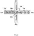
- FIG. 2schematically illustrates a microfluidic channel structure for partitioning individual or small groups of cells or cell beads
- FIG. 3panels A-F schematically illustrate an example process for amplification and barcoding of cell's nucleic acids
- FIG. 4provides a schematic illustration of use of barcoding of a cell's nucleic acids in attributing sequence data to individual cells or groups of cells for use in their characterization;
- FIG. 5provides a schematic illustration of cells associated with labeled cell-binding ligands
- FIG. 6shows an example computer control system that is programmed or otherwise configured to implement methods provided herein;
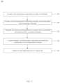
- FIG. 7shows a flowchart for a method of producing droplets containing a cell bead and a barcode bead and generating sequence reads from macromolecular components of the cell bead;
- FIG. 8shows a droplet containing a cell bead and a barcode bead produced using the method of FIG. 7 ;
- FIG. 9shows a flowchart for a method of producing droplets containing a cell and a barcode bead and generating sequence reads from macromolecular components of the cell
- FIG. 10shows a flowchart for a method of producing droplets containing a cell and a barcode bead and generating sequence reads from macromolecular components of the cell;
- FIG. 11shows a flowchart for a method of producing droplets containing a cross-linked cell and a barcode bead and generating sequence reads from macromolecular components the cross-linked cell;
- FIG. 12shows a droplet containing a cross-linked cell and a barcode bead produced using the method of FIG. 11 ;
- FIG. 13shows a flowchart for a method of producing droplets containing a cell bead and a barcode bead and generating sequence reads from macromolecular components of the cell bead;
- FIG. 14shows a droplet containing a cell bead and a barcode bead produced using the method of FIG. 13 ;
- FIG. 15shows a flowchart for a method of producing droplets containing a cell bead, a barcode bead and generating sequence reads from macromolecular components of the cell bead;
- FIG. 16shows a droplet containing a cell bead in its own droplet and a barcode bead produced using the method of FIG. 15 ;
- FIG. 17shows a flowchart for a method of producing droplets containing a coated cell and a barcode bead and generating sequence reads from macromolecular components the coated cell;
- FIG. 18shows a droplet containing a coated cell and a barcode bead produced using the method of FIG. 17 ;
- FIG. 19shows a flowchart for a method of producing droplets containing a cell and barcode bead and generating sequence reads from macromolecular components of the cell;
- FIG. 20shows a droplet containing a cell and a barcode bead produced using the method of FIG. 19 ;
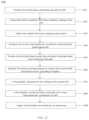
- FIG. 21panels (i)-(v) illustrate an example process of library preparation using priming free amplification of templates
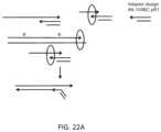
- FIG. 22 Ashows an example method of barcoding amplified templates generated by priming free amplification using an extension barcoding approach
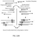
- FIG. 22 Bshows an example method of barcoding amplified templates generated by priming free amplification using a single stranded or double stranded template to barcode ligation approach
- FIG. 22 Cshows an example method of barcoding amplified templates generated by the priming free amplification by attaching a single strand DNA molecule (with barcode or primer sequence) to a bead from the 3′ end;
- FIG. 23shows a schematic of an example method for retaining long nucleic acid segments and removing short nucleic acid segments
- FIG. 24shows a schematic of an example method for the amplification and barcoding of nucleic acid loci from a cell bead
- FIG. 25shows a flowchart for an example method of producing droplets containing cell beads
- FIG. 26 Aschematically depicts an example droplet comprising a cell bead
- FIG. 26 Bschematically depicts an example first cell bead comprising a second cell bead
- FIG. 27schematically depicts an example method for generating a cell bead in cell bead
- FIGS. 28 A and 28 Bare photographs showing example generation of a cell bead in cell bead
- FIG. 29depicts example sequencing data obtained from samples prepared in a cell bead in cell bead approach
- FIG. 30depicts example data depicting centering of a cell in a cell bead in cell bead using different orbital shaking conditions.
- FIG. 31shows an example of a microfluidic channel structure for delivering cell beads and barcoded beads to droplets.
- barcodegenerally refers to a label, or identifier, that conveys or is capable of conveying information about the analyte.
- a barcodecan be part of an analyte.
- a barcodecan be a tag attached to an analyte (e.g., nucleic acid molecule) or a combination of the tag in addition to an endogenous characteristic of the analyte (e.g., size of the analyte or end sequence(s)).
- a barcodemay be unique.
- Barcodescan have a variety of different formats, for example, barcodes can include: polynucleotide barcodes; random nucleic acid and/or amino acid sequences; and synthetic nucleic acid and/or amino acid sequences.
- a barcodecan be attached to an analyte in a reversible or irreversible manner.
- a barcodecan be added to, for example, a fragment of a deoxyribonucleic acid (DNA) or ribonucleic acid (RNA) sample before, during, and/or after sequencing of the sample. Barcodes can allow for identification and/or quantification of individual sequencing-reads in real time.
- subjectgenerally refers to an animal, such as a mammalian species (e.g., human) or avian (e.g., bird) species, or other organism, such as a plant.
- the subjectcan be a vertebrate, a mammal, a mouse, a primate, a simian or a human. Animals may include, but are not limited to, farm animals, sport animals, and pets.
- a subjectcan be a healthy or asymptomatic individual, an individual that has or is suspected of having a disease (e.g., cancer) or a pre-disposition to the disease, or an individual that is in need of therapy or suspected of needing therapy.
- a subjectcan be a patient.
- a genomegenerally refers to an entirety of a subject's hereditary information.
- a genomecan be encoded either in DNA or in RNA.
- a genomecan comprise coding regions that code for proteins as well as non-coding regions.
- a genomecan include the sequence of all chromosomes together in an organism. For example, the human genome has a total of 46 chromosomes. The sequence of all of these together may constitute a human genome.
- adaptor(s)can be used synonymously.
- An adaptor or tagcan be coupled to a polynucleotide sequence to be “tagged” by any approach including ligation, hybridization, or other approaches.
- sequence of nucleotide bases in one or more polynucleotidesgenerally refers to methods and technologies for determining the sequence of nucleotide bases in one or more polynucleotides.
- the polynucleotidescan be, for example, deoxyribonucleic acid (DNA) or ribonucleic acid (RNA), including variants or derivatives thereof (e.g., single stranded DNA).
- Sequencingcan be performed by various systems currently available, such as, with limitation, a sequencing system by Illumina, Pacific Biosciences, Oxford Nanopore, or Life Technologies (Ion Torrent).
- Such devicesmay provide a plurality of raw genetic data corresponding to the genetic information of a subject (e.g., human), as generated by the device from a sample provided by the subject. In some situations, systems and methods provided herein may be used with proteomic information.
- variantgenerally refers to a genetic variant, such as a nucleic acid molecule comprising a polymorphism.
- a variantcan be a structural variant or copy number variant, which can be genomic variants that are larger than single nucleotide variants or short indels.
- a variantcan be an alteration or polymorphism in a nucleic acid sample or genome of a subject.
- Single nucleotide polymorphismsSNPs
- Polymorphismscan include single nucleotide variations (SNVs), insertions, deletions, repeats, small insertions, small deletions, small repeats, structural variant junctions, variable length tandem repeats, and/or flanking sequences.
- Copy number variants (CNVs)transversions and other rearrangements are also forms of genetic variation.
- a genomic alternationmay be a base change, insertion, deletion, repeat, copy number variation, or transversion.
- the term “bead,” as used herein,generally refers to a particle.
- the beadmay be a solid or semi-solid particle.
- the beadmay be a gel.
- the beadmay be formed of a polymeric material.
- the beadmay be magnetic or non-magnetic.
- samplegenerally refers to a biological sample of a subject.
- the biological samplemay be a nucleic acid sample or protein sample.
- the biological samplemay be derived from another sample.
- the samplemay be a tissue sample, such as a biopsy, core biopsy, needle aspirate, or fine needle aspirate.
- the samplemay be a fluid sample, such as a blood sample, urine sample, or saliva sample.
- the samplemay be a skin sample.
- the samplemay be a cheek swap.
- the samplemay be a plasma or serum sample.
- the samplemay be a cell-free or cell free sample.
- a cell-free samplemay include extracellular polynucleotides. Extracellular polynucleotides may be isolated from a bodily sample that may be selected from the group consisting of blood, plasma, serum, urine, saliva, mucosal excretions, sputum, stool and tears.
- cell beadgenerally refers to a particulate material that comprises (e.g., encapsulates, contains, etc.) a cell (e.g., a cell, a fixed cell, a cross-linked cell), a virus, components of, or macromolecular constituents derived from a cell or virus.
- a cell beadmay comprise a virus and/or a cell.
- a cell beadcomprises a single cell.
- a cell beadmay comprise multiple cells adhered together.
- a cell beadmay include any type of cell, including without limitation prokaryotic cells, eukaryotic cells, bacterial, fungal, plant, mammalian, or other animal cell types, mycoplasmas, normal tissue cells, tumor cells, a T-cell (e.g., CD4 T-cell, CD4 T-cell that comprises a dormant copy of human immunodeficiency virus (HIV)), a fixed cell, a cross-linked cell, a rare cell from a population of cells, or any other cell type, whether derived from single cell or multicellular organisms.
- a cell beadmay comprise a live cell, such as, for example, a cell may be capable of being cultured.
- a cell beadmay comprise a derivative of a cell, such as one or more components of the cell (e.g., an organelle, a cell protein, a cellular nucleic acid, genomic nucleic acid, messenger ribonucleic acid, a ribosome, a cellular enzyme, etc.).
- a cell beadmay comprise material obtained from a biological tissue, such as, for example, obtained from a subject.
- cells, viruses or macromolecular constituents thereofare encapsulated within a cell bead. Encapsulation can be within a polymer or gel matrix that forms a structural component of the cell bead.
- a cell beadis generated by fixing a cell in a fixation medium or by cross-linking elements of the cell, such as the cell membrane, the cell cytoskeleton, etc.
- beadsmay or may not be generated without encapsulation within a larger cell bead.
- the rare cellmay be a cancerous cell.
- the cancerous cellmay be a circulating tumor cell.
- the rare cellmay be obtained from an in vitro fertilization (IVF) procedure.
- the rare cellmay be obtained from an individual displaying genetic mosaicism.
- the rare cellmay be obtained from an organism produced using synthetic biology techniques.
- the rare cellmay be present at a concentration of at most about 1 in 10 2 , 1 in 10 3 , 1 in 10 4 , 1 in 10 5 , 1 in 10 6 , 1 in 10 7 , 1 in 10 8 , 1 in 10 9 , 1 in 10 10 , 1 in 10 11 , 1 in 10 12 , 1 in 10 13 , 1 in 10 14 , or 1 in 10 15 cells of the population of cells.
- the rare cellmay be present at a concentration lying in a range defined by any two of the preceding values.
- the term “macromolecular constituent,” as used herein,generally refers to a macromolecule that is a component of or is derived from a biological material (e.g., a cell, a fixed cell, a cross-linked cell, a virus, etc.).
- the macromolecular constituentmay comprise a nucleic acid. Such a macromolecule can be encapsulated within a cell bead.
- the macromolecular constituentmay comprise a nucleic acid.
- the macromolecular constituentmay comprise deoxyribonucleic acid (DNA) or a variant or derivative thereof.
- the macromolecular constituentmay comprise ribonucleic acid (RNA) or a variant or derivative thereof.
- the RNAmay be coding or non-coding.
- the RNAmay be messenger RNA (mRNA), ribosomal RNA (rRNA) or transfer RNA (tRNA), for example.
- the RNAmay be a transcript.
- the RNAmay be small RNA that are less than 200 nucleic acid bases in length, or large RNA that are greater than 200 nucleic acid bases in length.
- Small RNAsmay include 5.8 S ribosomal RNA (rRNA), 5 S rRNA, transfer RNA (tRNA), microRNA (miRNA), small interfering RNA (siRNA), small nucleolar RNA (snoRNAs), Piwi-interacting RNA (piRNA), tRNA-derived small RNA (tsRNA) and small rDNA-derived RNA (srRNA).
- the RNAmay be double-stranded RNA or single-stranded RNA.
- the RNAmay be circular RNA.
- the macromolecular constituentmay comprise a protein or a variant or derivative thereof.
- the macromolecular constituentmay comprise a polynucleotide.
- the macromolecular constituentmay comprise multiple polynucleotides.
- the macromolecular constituentmay compromise chromatin or functional equivalents.
- the macromolecular constituentmay comprise a peptide.
- the macromolecular constituentmay comprise a polypeptide.
- the macromolecular constituentmay comprise a polynucleotide/polypeptide complex.
- the term “tag,” as used herein,generally refers to a material capable of binding to a macromolecular constituent (e.g., DNA, RNA or protein).
- the tagmay bind to the macromolecular constituent with high affinity.
- the tagmay bind to the macromolecular constituent with high specificity.
- the tagmay comprise a nucleotide sequence.
- the tagmay comprise an oligonucleotide or polypeptide sequence.
- the tagmay comprise a DNA aptamer.
- the tagmay be or comprise a primer.
- the tagmay be or comprise a protein.
- the tagmay comprise a polypeptide.
- the tagmay be or include a barcode, such as a barcode sequence.
- the tagmay be a molecular species or atomic species (e.g., atomic particle, collection of atomic particles, or quantum dot).
- microfluidic devicegenerally refers to a device configured for fluid transport and having a fluidic channel through which fluid can flow with at least one dimension of no greater than about 10 millimeters (mm).
- the dimensioncan be any of length, width or height.
- a microfluidic devicecomprises a fluidic channel having multiple dimensions of no greater than about 10 mm.
- a microfluidic devicecan also include a plurality of fluidic channels each having a dimension of no greater than about 10 mm.
- the dimension(s) of a given fluidic channel of a microfluidic devicemay vary depending, for example, on the particular configuration of the channel and/or channels and other features also included in the device.
- a dimension of a fluidic channel of a microfluidic devicemay be at most about 10 mm, at most about 9 mm, at most about 8 mm, at most about 7 mm, at most about 6 mm, at most about 5 mm, at most about 4 mm, at most about 3 mm, at most about 2 mm, at most about 1 mm, at most about 900 micrometers ( ⁇ m), at most about 800 ⁇ m, at most 700 ⁇ m, at most about 600 ⁇ m, at most about 500 ⁇ m, at most about 400 ⁇ m, at most about 300 ⁇ m, at most about 200 ⁇ m, at most about 100 ⁇ m, at most about 90 ⁇ m, at most about 70 ⁇ m, at most about 60 ⁇ m, at most about 50 ⁇ m, at most about 40 ⁇ m, at most about 30 ⁇ m, at most about 20 ⁇ m, at most about 10 ⁇ m, at most about 8 ⁇ m, at most about 6 ⁇ m, at most about 4 ⁇ m, at most
- a dimension of a fluidic channel of a microfluidic devicemay be at least about 1 ⁇ m, at least about 2 ⁇ m, at least about 4 ⁇ m, at least about 6 ⁇ m, at least about 8 ⁇ m, at least about 10 ⁇ m, at least about 20 ⁇ m, at least about 30 ⁇ m, at least about 40 ⁇ m, at least about 50 ⁇ m, at least about 60 ⁇ m, at least about 70 ⁇ m, at least about 80 ⁇ m, at least about 90 ⁇ m, at least about 100 ⁇ m, at least about 200 ⁇ m, at least about 300 ⁇ m, at least about 400 ⁇ m, at least about 500 ⁇ m, at least about 600 ⁇ m, at least about 700 ⁇ m, at least about 800 ⁇ m, at least about 900 ⁇ m, at least about 1 mm, at least about 2 mm, at least about 3 mm, at least about 4 mm, at least about 5 mm, at least about 6 mm, at least about 7 mm
- Microfluidic devices described hereincan also include any additional components that can, for example, aid in regulating fluid flow, such as a fluid flow regulator (e.g., a pump, a source of pressure, etc.), features that aid in preventing clogging of fluidic channels (e.g., funnel features in channels; reservoirs positioned between channels, reservoirs that provide fluids to fluidic channels, etc.) and/or removing debris from fluid streams, such as, for example, filters.
- a fluid flow regulatore.g., a pump, a source of pressure, etc.
- features that aid in preventing clogging of fluidic channelse.g., funnel features in channels; reservoirs positioned between channels, reservoirs that provide fluids to fluidic channels, etc.
- debris from fluid streamssuch as, for example, filters.
- microfluidic devicesmay be configured as a fluidic chip that includes one or more reservoirs that supply fluids to an arrangement of microfluidic channels and also includes one or more reservoirs that receive fluids that have passed through the microfluidic device.
- microfluidic devicesmay be constructed of any suitable material(s), including polymer species and glass.
- Nucleic acid sequencing technologieshave yielded substantial results in sequencing biological materials, including providing substantial sequence information on individual organisms, and relatively pure biological samples. However, these systems have traditionally not been effective at being able to identify and characterize cells at the single cell level.
- nucleic acid sequencing technologiesderive the nucleic acids that they sequence from collections of cells obtained from tissue or other samples, such as biological fluids (e.g., blood, plasma, etc).
- the cellscan be processed (e.g., all together) to extract the genetic material that represents an average of the population of cells, which can then be processed into sequencing ready DNA libraries that are configured for a given sequencing technology.
- the nucleic acids derived from the cellsmay include DNA, or RNA, including, e.g., mRNA, total RNA, or the like, that may be processed to produce cDNA for sequencing. Following processing, absent a cell specific marker, attribution of genetic material as being contributed by a subset of cells or an individual cell may not be possible in such an ensemble approach.
- ensemble sample preparation methodscan be, from the outset, predisposed to primarily identifying and characterizing the majority constituents in the sample of cells, and may not be designed to pick out the minority constituents, e.g., genetic material contributed by one cell, a few cells, or a small percentage of total cells in the sample.
- an ensemble approachcan be predisposed to presenting potentially inaccurate data from cell populations that are non-homogeneous in terms of expression levels. In some cases, where expression is high in a small minority of the cells in an analyzed population, and absent in the majority of the cells of the population, an ensemble method may indicate low level expression for the entire population.
- next generation sequencing technologiesmay rely upon the geometric amplification of nucleic acid fragments, such as via polymerase chain reaction, in order to produce sufficient DNA for the sequencing library.
- amplificationcan be biased toward amplification of majority constituents in a sample, and may not preserve the starting ratios of such minority and majority components.
- the single molecule systemscan also have large input DNA requirements.
- Some single molecule sequencing systemscan have sample input DNA requirements of from 500 nanograms (ng) to upwards of 10 micrograms ( ⁇ g), which may not be obtainable from individual cells or even small subpopulations of cells.
- other NGS systemscan be optimized for starting amounts of sample DNA in the sample of from approximately 50 ng to about 1 ⁇ g, for example.
- the methods described hereinmay compartmentalize the analysis of individual cells or small populations of cells, including e.g., nucleic acids from individual cells or small groups of cells, and then allow that analysis to be attributed back to the individual cell or small group of cells from which the nucleic acids were derived. This can be accomplished regardless of whether the cell population represents a 50/50 mix of cell types, a 90/10 mix of cell types, or virtually any ratio of cell types, as well as a complete heterogeneous mix of different cell types, or any mixture between these.
- Differing cell typesmay include cells from different tissue types of an individual or the same tissue type from different individuals, or biological organisms such as microorganisms from differing genera, species, strains, variants, or any combination of any or all of the foregoing.
- differing cell typesmay include normal and tumor tissue from an individual, various cell types obtained from a human subject such as a variety of immune cells (e.g., B cells, T cells, and the like), multiple different bacterial species, strains and/or variants from environmental, forensic, microbiome or other samples, or any of a variety of other mixtures of cell types.
- the methods and systems described hereinprovide for the compartmentalization, depositing or partitioning of a cell or virus (e.g., a cell) or the macromolecular constituent(s) of the cell or virus from a sample into discrete compartments or partitions (referred to interchangeably herein as partitions), where each partition maintains separation of its own contents from the contents of other partitions.
- partitionsmay themselves be partitioned into additional partitions, such as, for example, droplets or wells.
- Unique identifierse.g., barcodes
- Barcodesmay be delivered, for example on an oligonucleotide, to a partition via any suitable mechanism.
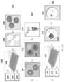
- Method 100comprises three different phases 110 , 120 and 130 that correspond to generation of cell beads comprising a cell or virus or its macromolecular constituent(s) ( 110 ); solvent exchange to bring generated partitions into an aqueous phase, cell or virus lysis and denaturation of the cell or virus or macromolecular constituent(s) of the cell or virus ( 120 ); and generation of partitions comprising the generated cell beads and barcodes and subsequent tagging (e.g., barcoding) ( 130 ).
- phases 110 , 120 and 130correspond to generation of cell beads comprising a cell or virus or its macromolecular constituent(s) ( 110 ); solvent exchange to bring generated partitions into an aqueous phase, cell or virus lysis and denaturation of the cell or virus or macromolecular constituent(s) of the cell or virus ( 120 ); and generation of partitions comprising the generated cell beads and barcodes and subsequent tagging (e.g., barcoding) ( 130 ).
- an oil 101 , polymeric or gel precursors 102 and cells 103are provided to a microfluidic chip 104 .
- a photograph of an example microfluidic chip 104is shown in FIG. 1 B .
- the microfluidic chip 104comprises a plurality of reservoirs for the oil 101 , polymeric or gel precursors 102 and cell or virus reagents 103 .
- Polymeric or gel precursors 102 and cell or virus reagents 103are flowed (e.g., via the action of an applied force, such as negative pressure via a vacuum or positive pressure via a pump) from their reservoirs to a first channel junction at which point they combine to form an aqueous stream.
- This aqueous streamis then flowed to a second channel junction, to which oil 101 is also provided.
- the aqueous stream provided from the first channel junctionis immiscible with the oil 101 resulting in the generation of a suspension of aqueous droplets in the oil which then flow to reservoir 105 and represent the product 105 from the microfluidic process.
- Flowcan be controlled within the microfluidic chip 104 via any suitable strategy, including the use of one or more flow regulators in a channel or various channels, dimensioning of microfluidic channels, etc.
- the productcomprises droplets 105 comprising a cell from the cells 103 and polymeric or gel precursors 102 .
- the droplets 105are then subjected to conditions suitable to polymerize or gel the polymeric or gel precursors 102 in the droplets 105 , which generates cell beads 106 that encapsulate the cell or virus reagents 103 (e.g., a cell, a fixed cell, a cross-linked cell, component(s) or a cell) in the droplets 105 .
- the cell or virus reagents 103e.g., a cell, a fixed cell, a cross-linked cell, component(s) or a cell
- phase 120is initiated which includes solvent exchange 111 to resuspend the cell beads 106 in an aqueous phase. Additional details and examples regarding solvent exchange are provided elsewhere herein.
- the resuspended cell beads 106can then, in bulk 112 , be subjected conditions suitable to lyse cells or viruses associated with the cell beads 106 and, separately or contemporaneously, also subjected, in bulk, to conditions to denature nucleic acids derived from the cells or viruses associated with the cell beads 106 .
- the polymeric matrix of the cell beads 106effectively hinders or prohibits diffusion of larger molecules, such as nucleic acids, from the cell beads 106 .
- the cell beads 106are sufficiently porous to denaturation agents that permit denaturation of trapped nucleic acids within the cell beads 106 .
- the cell beadscan then be subjected, in bulk, to conditions suitable for performing one or more reactions on nucleic acids derived from the cells or viruses associated with the cell beads 106 . Additional details and examples regarding reactions on nucleic acids are provided elsewhere herein.
- the resulting cell beads 113are then collected 114 and can be stored prior to initiation of phase 130 .
- dropletscomprising the cell beads 113 and barcode beads (e.g., gel beads) 122 comprising barcode sequences are generated.
- an oil 121 , the cell beads 113 and barcode beads 122 each comprising a barcode sequence (e.g., each bead comprising a unique barcode sequence)are provided to a microfluidic chip 123 .
- a photograph of an example microfluidic chip 123is shown in FIG. 1 C .
- the microfluidic chip 123comprises a plurality of reservoirs for the oil 121 , cell beads 113 and barcode beads 122 .
- the chipalso includes additional reservoirs 127 and 128 that may be used to supply additional reagents (e.g., reagents for nucleic acid amplification, reagents that can degrade or dissolve cell beads 113 and/or barcode beads 122 , reagents that degrade linkages between barcodes and barcode beads 122 , etc.) to phase 130 .
- Cell beads 113 and barcode beads 122are flowed (e.g., via the action of an applied force, such as negative pressure via a vacuum or positive pressure via a pump) from their reservoirs to a first channel junction at which point they combine to form an aqueous mixture. Materials from reservoirs 127 and 128 can also be provided to the mixture at the first channel junction.
- cell beads and barcode beadscan be mixed before introduction into the microfluidic chip.
- a single reservoir of the microfluidic chip 123comprises a mixture of cell beads and barcode beads.
- the ratio of cell beads to barcode beads in the mixturecan be varied to alter the number of droplets generated that comprise a single cell bead and a single barcode bead.
- the mixture of cell beads and barcode beadsmay be flowed (e.g., via the action of an applied force, such as negative pressure via a vacuum or positive pressure via a pump) from the reservoir to a first channel junction, in some cases together with materials from reservoirs 127 and/or 128 .
- cellsmay be mixed with barcode beads.
- a collection of cells and cell beadsmay be mixed with barcode beads, or a collection of cells may be mixed with barcode beads.
- the mixture comprising cell beads (or cells), barcode beads, and in some cases additional reagentsis then flowed to a second channel junction, to which oil 121 is also provided.
- the aqueous mixture provided from the first channel junctionis immiscible with the oil 121 resulting in the generation of a suspension of aqueous droplets 125 in the oil 124 which then flow to a reservoir and represent the product from the microfluidic process.
- the microfluidic chipcan also include a reservoir 129 that can accept excess oil from the stream emerging from the second channel. Flow can be controlled within the microfluidic chip 123 via any suitable strategy, including the use of one or more flow regulators (see FIGS.
- the productcomprises droplets 125 comprising a cell bead 113 and a barcode bead 122 , in addition to any other reagents provided by reservoirs 127 and 128 .
- a given droplet of the droplets 125comprises a single cell bead and a single barcode bead.
- these reagentscan release the nucleic acids trapped in the cell beads 113 from the cell beads 113 and release the barcodes from the barcode beads 122 .
- the released barcodescan then interact with the released nucleic acids to generate barcoded constructs for nucleic acid sequencing as described elsewhere herein.
- a given sequencing construct generated from the given droplet 125can be associated with the cell or virus of the given cell bead via its barcode sequence.
- FIG. 1 Dphotographically depicts two example runs demonstrating the generation of droplets 125 comprising cell beads and barcode beads using the example method shown in FIG. 1 A and microfluidic devices depicted in FIGS. 1 B and 1 C .
- FIG. 1 Dpanel A
- droplets comprising cell beads and barcode beadsare shown
- FIG. 1 Dpanel B
- droplets comprising cell beads comprising magnetic materials and barcode beadsare shown.
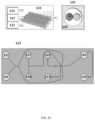
- FIG. 31shows an example of a microfluidic channel structure 3100 for delivering barcode carrying beads to droplets.
- the channel structure 3100can include channel segments 3101 , 3102 , 3104 , 3106 and 3108 communicating at a channel junction 3110 .
- the channel segment 3101may transport an aqueous fluid 3112 that includes a plurality of beads 3114 (e.g., with nucleic acid molecules, oligonucleotides, molecular tags) along the channel segment 3101 into junction 3110 .
- the plurality of beads 3114may be sourced from a suspension of beads.
- the channel segment 3101may be connected to a reservoir comprising an aqueous suspension of beads 3114 .
- the channel segment 3102may transport the aqueous fluid 3112 that includes a plurality of cell beads 3116 along the channel segment 3102 into junction 3110 .
- the plurality of cell beads 3116may be sourced from a suspension of cell beads.
- the channel segment 3102may be connected to a reservoir comprising an aqueous suspension of cell beads 3116 .
- the aqueous fluid 3112 in either the first channel segment 3101 or the second channel segment 3102 , or in both segments,can include one or more reagents, as further described below.
- a second fluid 3118 that is immiscible with the aqueous fluid 3112e.g., oil
- the aqueous fluid 3112can be partitioned as discrete droplets 3120 in the second fluid 3118 and flow away from the junction 3110 along channel segment 3108 .
- the channel segment 3108may deliver the discrete droplets to an outlet reservoir fluidly coupled to the channel segment 3108 , where they may be harvested.
- the channel segments 3101 and 3102may meet at another junction upstream of the junction 3110 .
- beads and cell beadsmay form a mixture that is directed along another channel to the junction 3110 to yield droplets 3120 .
- the mixturemay provide the beads and cell beads in an alternating fashion, such that, for example, a droplet comprises a single bead and a single cell bead.
- Beads, cell beads and dropletsmay flow along channels at substantially regular flow profiles (e.g., at regular flow rates). Such regular flow profiles may permit a droplet to include a single bead and a single cell bead. Such regular flow profiles may permit the droplets to have an occupancy (e.g., droplets having beads and cell beads) greater than 5%, 10%, 20%, 30%, 40%, 50%, 60%, 70%, 80%, 90%, or 95%.
- occupancye.g., droplets having beads and cell beads
- the second fluid 3118can comprise an oil, such as a fluorinated oil, that includes a fluorosurfactant for stabilizing the resulting droplets, for example, inhibiting subsequent coalescence of the resulting droplets 3120 .
- an oilsuch as a fluorinated oil, that includes a fluorosurfactant for stabilizing the resulting droplets, for example, inhibiting subsequent coalescence of the resulting droplets 3120 .
- a discrete droplet that is generatedmay include an individual cell bead 3116 .
- a discrete droplet that is generatedmay include a barcode or other reagent carrying bead 3114 .
- a discrete droplet generatedmay include both an individual cell bead and a barcode carrying bead, such as droplets 3120 .
- a discrete dropletmay include more than one individual cell bead or no cell bead.
- a discrete dropletmay include more than one bead or no bead.
- a discrete dropletmay be unoccupied (e.g., no beads, no cell beads).
- a discrete droplet partitioning a cell bead and a barcode carrying beadmay effectively allow the attribution of the barcode to macromolecular constituents of the cell bead within the partition.
- the contents of a partitionmay remain discrete from the contents of other partitions.
- the channel segments described hereinmay be coupled to any of a variety of different fluid sources or receiving components, including reservoirs, tubing, manifolds, or fluidic components of other systems.
- the microfluidic channel structure 3100may have other geometries.
- a microfluidic channel structurecan have more than one channel junctions.
- a microfluidic channel structurecan have 2, 3, 4, or 5 channel segments each carrying beads that meet at a channel junction.
- Fluidmay be directed flow along one or more channels or reservoirs via one or more fluid flow units.
- a fluid flow unitcan comprise compressors (e.g., providing positive pressure), pumps (e.g., providing negative pressure), actuators, and the like to control flow of the fluid. Fluid may also or otherwise be controlled via applied pressure differentials, centrifugal force, electrokinetic pumping, vacuum, capillary or gravity flow, or the like.
- a partitionmay be a droplet.
- the dropletmay be formed by bringing a first phase in contact with a second phase that is immiscible with the first phase.
- the partitionmay be a well as part of a plurality of wells.
- the partitionmay be a chamber as part of a plurality of chambers. Partitions may be fluidically isolated from one another.
- barcoded oligonucleotidesare delivered to a partition via a microcapsule, such as a bead (e.g., gel bead) or a droplet.
- a microcapsulesuch as a bead (e.g., gel bead) or a droplet.
- barcoded oligonucleotidesare initially associated with the microcapsule and then released from the microcapsule upon application of a stimulus which allows the oligonucleotides to dissociate or to be released from the microcapsule.
- a microcapsulein some embodiments, comprises a bead, such as a droplet comprising the bead.
- the microcapsulecan be a bead (e.g., gel bead).
- a beadmay be porous, non-porous, solid, semi-solid, semi-fluidic, or fluidic.
- a beadmay be dissolvable, disruptable, or degradable. In some cases, a bead may not be degradable.
- the beadmay be a solid or semi-solid particle.
- the beadmay be a gel bead.
- a gel beadmay be a hydrogel bead.
- a gel beadmay be formed from molecular precursors, such as a polymeric or monomeric species.
- a semi-solid beadmay be a liposomal bead.
- Solid beadsmay comprise metals including iron oxide, gold, and silver.
- the beadsare silica beads.
- the beadsare rigid.
- the beadsmay be flexible and/or compressible.
- the beadmay contain molecular precursors (e.g., monomers or polymers), which may form a polymer network via polymerization of the precursors.
- a precursormay be an already polymerized species capable of undergoing further polymerization via, for example, a chemical cross-linkage.
- a precursorcomprises one or more of an acrylamide or a methacrylamide monomer, oligomer, or polymer.
- the beadmay comprise prepolymers, which are oligomers capable of further polymerization.
- polyurethane beadsmay be prepared using prepolymers.
- the beadmay contain individual polymers that may be further polymerized together.
- beadsmay be generated via polymerization of different precursors, such that they comprise mixed polymers, co-polymers, and/or block co-polymers.
- a beadmay comprise natural and/or synthetic materials.
- a polymercan be a natural polymer or a synthetic polymer.
- a beadcomprises both natural and synthetic polymers.
- natural polymersinclude proteins and sugars such as deoxyribonucleic acid, rubber, cellulose, starch (e.g., amylose, amylopectin), proteins, enzymes, polysaccharides, silks, polyhydroxyalkanoates, chitosan, dextran, collagen, carrageenan, ispaghula, acacia, agar, gelatin, shellac, sterculia gum, xanthan gum, Corn sugar gum, guar gum, gum karaya, agarose, alginic acid, alginate, or natural polymers thereof.
- proteins and sugarssuch as deoxyribonucleic acid, rubber, cellulose, starch (e.g., amylose, amylopectin), proteins, enzymes, polysaccharides, silk
- Examples of synthetic polymersinclude acrylics, nylons, silicones, spandex, viscose rayon, polycarboxylic acids, polyvinyl acetate, polyacrylamide, polyacrylate, polyethylene glycol, polyurethanes, polylactic acid, silica, polystyrene, polyacrylonitrile, polybutadiene, polycarbonate, polyethylene, polyethylene terephthalate, poly(chlorotrifluoroethylene), poly(ethylene oxide), poly(ethylene terephthalate), polyethylene, polyisobutylene, poly(methyl methacrylate), poly(oxymethylene), polyformaldehyde, polypropylene, polystyrene, poly(tetrafluoroethylene), poly(vinyl acetate), poly(vinyl alcohol), poly(vinyl chloride), poly(vinylidene dichloride), poly(vinylidene difluoride), poly(vinyl fluoride) and combinations (e.g., co-polymers) thereof. Bea
- a chemical cross-linkermay be a precursor used to cross-link monomers during polymerization of the monomers and/or may be used to attach oligonucleotides (e.g., barcoded oligonucleotides) to the bead.
- polymersmay be further polymerized with a cross-linker species or other type of monomer to generate a further polymeric network.
- Non-limiting examples of chemical cross-linkersinclude cystamine, gluteraldehyde, dimethyl suberimidate, N-Hydroxysuccinimide crosslinker BS3, formaldehyde, carbodiimide (EDC), SMCC, Sulfo-SMCC, vinylsilane, N,N′diallyltartardiamide (DATD), N,N′-Bis(acryloyl)cystamine (BAC), or homologs thereof.
- the crosslinker used in the present disclosurecontains cystamine.
- Crosslinkingmay be permanent or reversible, depending upon the particular crosslinker used. Reversible crosslinking may allow for the polymer to linearize or dissociate under appropriate conditions. In some cases, reversible cross-linking may also allow for reversible attachment of a material bound to the surface of a bead. In some cases, a cross-linker may form disulfide linkages. In some cases, the chemical cross-linker forming disulfide linkages may be cystamine or a modified cystamine.
- disulfide linkagescan be formed between molecular precursor units (e.g., monomers, oligomers, or linear polymers) or precursors incorporated into a bead and oligonucleotides.
- Cystamine(including modified cystamines), for example, is an organic agent comprising a disulfide bond that may be used as a crosslinker agent between individual monomeric or polymeric precursors of a bead.
- Polyacrylamidemay be polymerized in the presence of cystamine or a species comprising cystamine (e.g., a modified cystamine) to generate polyacrylamide gel beads comprising disulfide linkages (e.g., chemically degradable beads comprising chemically-reducible cross-linkers).
- the disulfide linkagesmay permit the bead to be degraded (or dissolved) upon exposure of the bead to a reducing agent.
- chitosana linear polysaccharide polymer
- glutaraldehydevia hydrophilic chains to form a bead.
- Crosslinking of chitosan polymersmay be achieved by chemical reactions that are initiated by heat, pressure, change in pH, and/or radiation.
- the beadmay comprise covalent or ionic bonds between polymeric precursors (e.g., monomers, oligomers, linear polymers), oligonucleotides, primers, and other entities.
- polymeric precursorse.g., monomers, oligomers, linear polymers
- oligonucleotidese.g., primers, and other entities.
- the covalent bondscomprise carbon-carbon bonds or thioether bonds.
- a beadmay comprise an acrydite moiety, which in certain aspects may be used to attach one or more oligonucleotides (e.g., barcode sequence, barcoded oligonucleotide, primer, or other oligonucleotide) to the bead.
- an acrydite moietycan refer to an acrydite analogue generated from the reaction of acrydite with one or more species, such as, the reaction of acrydite with other monomers and cross-linkers during a polymerization reaction.
- Acrydite moietiesmay be modified to form chemical bonds with a species to be attached, such as an oligonucleotide (e.g., barcode sequence, barcoded oligonucleotide, primer, or other oligonucleotide).
- Acrydite moietiesmay be modified with thiol groups capable of forming a disulfide bond or may be modified with groups already comprising a disulfide bond.
- the thiol or disulfidevia disulfide exchange) may be used as an anchor point for a species to be attached or another part of the acrydite moiety may be used for attachment.
- attachmentis reversible, such that when the disulfide bond is broken (e.g., in the presence of a reducing agent), the attached species is released from the bead.
- an acrydite moietycomprises a reactive hydroxyl group that may be used for attachment.
- Functionalization of beads for attachment of oligonucleotidesmay be achieved through a wide range of different approaches, including activation of chemical groups within a polymer, incorporation of active or activatable functional groups in the polymer structure, or attachment at the pre-polymer or monomer stage in bead production.
- precursorse.g., monomers, cross-linkers
- precursorsthat are polymerized to form a bead may comprise acrydite moieties, such that when a bead is generated, the bead also comprises acrydite moieties.
- the acrydite moietiescan be attached to an oligonucleotide, such as a primer (e.g., a primer for amplifying target nucleic acids, barcoded oligonucleotide, etc) to be incorporated into the bead.
- the primercomprises a P5 sequence for attachment to a sequencing flow cell for Illumina sequencing.
- the primercomprises a P7 sequence for attachment to a sequencing flow cell for Illumina sequencing.
- the primercomprises a barcode sequence. In some cases, the primer further comprises a unique molecular identifier (UMI). In some cases, the primer comprises an R1 primer sequence for Illumina sequencing. In some cases, the primer comprises an R2 primer sequence for Illumina sequencing.
- UMIunique molecular identifier
- precursors comprising a functional group that is reactive or capable of being activated such that it becomes reactivecan be polymerized with other precursors to generate gel beads comprising the activated or activatable functional group.
- the functional groupmay then be used to attach additional species (e.g., disulfide linkers, primers, other oligonucleotides, etc.) to the gel beads.
- additional speciese.g., disulfide linkers, primers, other oligonucleotides, etc.
- some precursors comprising a carboxylic acid (COOH) groupcan co-polymerize with other precursors to form a gel bead that also comprises a COOH functional group.
- acrylic acida species comprising free COOH groups
- acrylamideacrylamide
- bis(acryloyl)cystaminecan be co-polymerized together to generate a gel bead comprising free COOH groups.
- the COOH groups of the gel beadcan be activated (e.g., via 1-Ethyl-3-(3-dimethylaminopropyl)carbodiimide (EDC) and N-Hydroxysuccinimide (NHS) or 4-(4,6-Dimethoxy-1,3,5-triazin-2-yl)-4-methylmorpholinium chloride (DMTMM)) such that they are reactive (e.g., reactive to amine functional groups where EDC/NHS or DMTMM are used for activation).
- EDC1-Ethyl-3-(3-dimethylaminopropyl)carbodiimide
- NHSN-Hydroxysuccinimide
- DTMM4-(4,6-Dimethoxy
- the activated COOH groupscan then react with an appropriate species (e.g., a species comprising an amine functional group where the carboxylic acid groups are activated to be reactive with an amine functional group) comprising a moiety to be linked to the bead.
- an appropriate speciese.g., a species comprising an amine functional group where the carboxylic acid groups are activated to be reactive with an amine functional group
- Beads comprising disulfide linkages in their polymeric networkmay be functionalized with additional species via reduction of some of the disulfide linkages to free thiols.
- the disulfide linkagesmay be reduced via, for example, the action of a reducing agent (e.g., DTT, TCEP, etc.) to generate free thiol groups, without dissolution of the bead.
- Free thiols of the beadscan then react with free thiols of a species or a species comprising another disulfide bond (e.g., via thiol-disulfide exchange) such that the species can be linked to the beads (e.g., via a generated disulfide bond).
- free thiols of the beadsmay react with any other suitable group.
- free thiols of the beadsmay react with species comprising an acrydite moiety.
- the free thiol groups of the beadscan react with the acrydite via Michael addition chemistry, such that the species comprising the acrydite is linked to the bead.
- uncontrolled reactionscan be prevented by inclusion of a thiol capping agent such as N-ethylmalieamide or iodoacetate.
- Activation of disulfide linkages within a beadcan be controlled such that a small number of disulfide linkages are activated. Control may be exerted, for example, by controlling the concentration of a reducing agent used to generate free thiol groups and/or concentration of reagents used to form disulfide bonds in bead polymerization. In some cases, a low concentration (e.g., molecules of reducing agent:gel bead ratios of less than about 10,000, less than about 100,000, less than about 1,000,000, less than about 10,000,000, less than about 100,000,000, less than about 1,000,000,000, less than about 10,000,000,000, or less than about 100,000,000,000) of reducing agent may be used for reduction.
- a low concentratione.g., molecules of reducing agent:gel bead ratios of less than about 10,000, less than about 100,000, less than about 1,000,000, less than about 10,000,000, less than about 100,000,000, less than about 1,000,000,000, less than about 10,000,000,000, or less than about 100,000,000,000
- Controlling the number of disulfide linkages that are reduced to free thiolsmay be useful in ensuring bead structural integrity during functionalization.
- optically-active agentssuch as fluorescent dyes may be may be coupled to beads via free thiol groups of the beads and used to quantify the number of free thiols present in a bead and/or track a bead.
- addition of moieties to a gel bead after gel bead formationmay be advantageous.
- addition of an oligonucleotide (e.g., barcoded oligonucleotide) after gel bead formationmay avoid loss of the species during chain transfer termination that can occur during polymerization.
- smaller precursorse.g., monomers or cross linkers that do not comprise side chain groups and linked moieties
- functionalization after gel bead synthesiscan minimize exposure of species (e.g., oligonucleotides) to be loaded with potentially damaging agents (e.g., free radicals) and/or chemical environments.
- the generated gelmay possess an upper critical solution temperature (UCST) that can permit temperature driven swelling and collapse of a bead.
- UCTupper critical solution temperature
- Such functionalitymay aid in oligonucleotide (e.g., a primer) infiltration into the bead during subsequent functionalization of the bead with the oligonucleotide.
- Post-production functionalizationmay also be useful in controlling loading ratios of species in beads, such that, for example, the variability in loading ratio is minimized.
- Species loadingmay also be performed in a batch process such that a plurality of beads can be functionalized with the species in a single batch.
- beadscan be non-covalently loaded with one or more reagents.
- the beadscan be non-covalently loaded by, for instance, subjecting the beads to conditions sufficient to swell the beads, allowing sufficient time for the reagents to diffuse into the interiors of the beads, and subjecting the beads to conditions sufficient to de-swell the beads.
- the swelling of the beadsmay be accomplished, for instance, by placing the beads in a thermodynamically favorable solvent, subjecting the beads to a higher or lower temperature or temperature change, subjecting the beads to a higher or lower ion concentration, and/or subjecting the beads to an electric field.
- the de-swelling of the beadsmay be accomplished, for instance, by transferring the beads in a thermodynamically unfavorable solvent, subjecting the beads to a lower or high temperature or temperature change different from that use to swell the beads, subjecting the beads to a lower or higher ion concentration different from that used to swell the beads, and/or removing the electric field.
- Transferring the beadsmay cause pores in the beads to shrink. Such shrinking may then hinder reagents within the beads from diffusing out of the interiors of the beads. The hindrance may be due to steric interactions between the reagents and the interiors of the beads.
- the transfermay be accomplished microfluidically. For instance, the transfer may be achieved by moving the beads from one co-flowing solvent stream to a different co-flowing solvent stream. The swellability and/or pore size of the beads may be adjusted by changing the polymer composition of the bead.
- an acrydite moiety linked to precursor, another species linked to a precursor, or a precursor itselfcomprises a labile bond, such as chemically, thermally, or photo-sensitive bonds e.g., disulfide bonds, UV sensitive bonds, or the like.
- acrydite moieties or other moieties comprising a labile bondare incorporated into a bead, the bead may also comprise the labile bond.
- the labile bondmay be, for example, useful in reversibly linking (e.g., covalently linking) species (e.g., barcodes, primers, etc.) to a bead.
- a thermally labile bondmay include a nucleic acid hybridization based attachment, e.g., where an oligonucleotide is hybridized to a complementary sequence that is attached to the bead, such that thermal melting of the hybrid releases the oligonucleotide, e.g., a barcode containing sequence, from the bead or microcapsule.
- a nucleic acid hybridization based attachmente.g., where an oligonucleotide is hybridized to a complementary sequence that is attached to the bead, such that thermal melting of the hybrid releases the oligonucleotide, e.g., a barcode containing sequence, from the bead or microcapsule.
- labile bondsmay result in the generation of a bead capable of responding to varied stimuli.
- Each type of labile bondmay be sensitive to an associated stimulus (e.g., chemical stimulus, light, temperature, etc.) such that release of species attached to a bead via each labile bond may be controlled by the application of the appropriate stimulus.
- Such functionalitymay be useful in controlled release of species from a gel bead.
- another species comprising a labile bondmay be linked to a gel bead after gel bead formation via, for example, an activated functional group of the gel bead as described above.
- Barcodes that are releasably, cleavably or reversibly attached to the beads described hereininclude barcodes that are released or releasable through cleavage of a linkage between the barcode molecule and the bead, or that are released through degradation of the underlying bead itself, allowing the barcodes to be accessed or accessible by other reagents, or both.
- the barcodes that are releasable as described hereinmay sometimes be referred to as being activatable, in that they are available for reaction once released.
- an activatable barcodemay be activated by releasing the barcode from a bead (or other suitable type of partition described herein).
- Other activatable configurationsare also envisioned in the context of the described methods and systems.
- labile bondsthat may be coupled to a precursor or bead include an ester linkage (e.g., cleavable with an acid, a base, or hydroxylamine), a vicinal diol linkage (e.g., cleavable via sodium periodate), a Diels-Alder linkage (e.g., cleavable via heat), a sulfone linkage (e.g., cleavable via a base), a silyl ether linkage (e.g., cleavable via an acid), a glycosidic linkage (e.g., cleavable via an amylase), a peptide linkage (e.g., cleavable via a protease), or a phosphodiester linkage (e.g., cleavable via a nuclease (
- Species that do not participate in polymerizationmay also be encapsulated in beads during bead generation (e.g., during polymerization of precursors). Such species may be entered into polymerization reaction mixtures such that generated beads comprise the species upon bead formation. In some cases, such species may be added to the gel beads after formation.
- speciesmay include, for example, oligonucleotides, reagents for a nucleic acid amplification reaction (e.g., primers (e.g. random primers, primers specific for a given DNA loci), polymerases, nucleotides (e.g.
- co-factorse.g., ionic co-factors
- reagents for enzymatic reactionse.g., enzymes, co-factors, substrates
- reagents for reverse transcriptione.g. oligonucleotide primers or reverse transcriptase
- nucleic acid modification reactionssuch as polymerization, ligation, digestion, methylation, random mutagenesis, bisulfite conversion, uracil hydrolysis, nucleic acid repair, nucleic acid insertion or cleavage (e.g.
- CRISPR/Cas9-mediated or transposon-mediated insertion or cleavagevia CRISPR/Cas9-mediated or transposon-mediated insertion or cleavage), capping, or decapping.
- Trapping of such speciesmay be controlled by the polymer network density generated during polymerization of precursors, control of ionic charge within the gel bead (e.g., via ionic species linked to polymerized species), or by the release of other species.
- Encapsulated speciesmay be released from a bead upon bead degradation and/or by application of a stimulus capable of releasing the species from the bead.
- barcode sequencesmay also be encapsulated within a bead and, in some cases, can be released from a bead via bead degradation and/or by application of a stimulus capable of releasing the species from the bead.
- Beadsmay be of uniform size or heterogeneous size.
- the diameter of a beadmay be about 1 ⁇ m, 5 ⁇ m, 10 ⁇ m, 20 ⁇ m, 30 ⁇ m, 40 ⁇ m, 50 ⁇ m, 60 ⁇ m, 70 ⁇ m, 80 ⁇ m, 90 ⁇ m, 100 ⁇ m, 250 ⁇ m, 500 ⁇ m, or 1 mm.
- a beadmay have a diameter of at least about 1 ⁇ m, 5 ⁇ m, 10 ⁇ m, 20 ⁇ m, 30 ⁇ m, 40 ⁇ m, 50 ⁇ m, 60 ⁇ m, 70 ⁇ m, 80 ⁇ m, 90 ⁇ m, 100 ⁇ m, 250 ⁇ m, 500 ⁇ m, 1 mm, or more. In some cases, a bead may have a diameter of less than about 1 ⁇ m, 5 ⁇ m, 10 ⁇ m, 20 ⁇ m, 30 ⁇ m, 40 ⁇ m, 50 ⁇ m, 60 ⁇ m, 70 ⁇ m, 80 ⁇ m, 90 ⁇ m, 100 ⁇ m, 250 ⁇ m, 500 ⁇ m, or 1 mm.
- a beadmay have a diameter in the range of about 40-75 ⁇ m, 30-75 ⁇ m, 20-75 ⁇ m, 40-85 ⁇ m, 40-95 ⁇ m, 20-100 ⁇ m, 10-100 ⁇ m, 1-100 ⁇ m, 20-250 ⁇ m, or 20-500 ⁇ m.
- beadsare provided as a population or plurality of beads having a relatively monodisperse size distribution.
- maintaining relatively consistent bead characteristics, such as sizecan contribute to the overall consistency.
- the beads described hereinmay have size distributions that have a coefficient of variation in their cross-sectional dimensions of less than 50%, less than 40%, less than 30%, less than 20%, and in some cases less than 15%, less than 10%, or less than 5%.
- Beadsmay be of any suitable shape. Examples of bead shapes include, but are not limited to, spherical, non-spherical, oval, oblong, amorphous, circular, cylindrical, and variations thereof.
- the beadsmay be degradable, disruptable, or dissolvable spontaneously or upon exposure to one or more stimuli (e.g., temperature changes, pH changes, exposure to particular chemical species or phase, exposure to light, reducing agent, etc.).
- a beadmay be dissolvable, such that material components of the beads are solubilized when exposed to a particular chemical species or an environmental change, such as a change temperature or a change in pH.
- a gel beadis degraded or dissolved at elevated temperature and/or in basic conditions.
- a beadmay be thermally degradable such that when the bead is exposed to an appropriate change in temperature (e.g., heat), the bead degrades.
- Degradation or dissolution of a bead bound to a speciese.g., a oligonucleotide, e.g., barcoded oligonucleotide
- a speciese.g., a oligonucleotide, e.g., barcoded oligonucleotide
- a degradable beadmay comprise one or more species with a labile bond such that, when the bead/species is exposed to the appropriate stimuli, the bond is broken and the bead degrades.
- the labile bondmay be a chemical bond (e.g., covalent bond, ionic bond) or may be another type of physical interaction (e.g., van der Waals interactions, dipole-dipole interactions, etc.).
- a crosslinker used to generate a beadmay comprise a labile bond.
- the labile bondcan be broken and the bead degraded. For example, upon exposure of a polyacrylamide gel bead comprising cystamine crosslinkers to a reducing agent, the disulfide bonds of the cystamine can be broken and the bead degraded.
- a degradable beadmay be useful in more quickly releasing an attached species (e.g., an oligonucleotide, a barcode sequence, a primer, etc) from the bead when the appropriate stimulus is applied to the bead as compared to a bead that does not degrade.
- an attached speciese.g., an oligonucleotide, a barcode sequence, a primer, etc
- the speciesmay have greater mobility and accessibility to other species in solution upon degradation of the bead.
- a speciesmay also be attached to a degradable bead via a degradable linker (e.g., disulfide linker).
- the degradable linkermay respond to the same stimuli as the degradable bead or the two degradable species may respond to different stimuli.
- a barcode sequencemay be attached, via a disulfide bond, to a polyacrylamide bead comprising cystamine.
- the beadUpon exposure of the barcoded-bead to a reducing agent, the bead degrades and the barcode sequence is released upon breakage of both the disulfide linkage between the barcode sequence and the bead and the disulfide linkages of the cystamine in the bead.
- a degradable beadmay be introduced into a partition, such as a droplet of an emulsion or a well, such that the bead degrades within the partition and any associated species (e.g., oligonucleotides) are released within the droplet when the appropriate stimulus is applied.
- the free speciese.g., oligonucleotides
- a polyacrylamide beadcomprising cystamine and linked, via a disulfide bond, to a barcode sequence, may be combined with a reducing agent within a droplet of a water-in-oil emulsion.
- the reducing agentbreaks the various disulfide bonds resulting in bead degradation and release of the barcode sequence into the aqueous, inner environment of the droplet.
- heating of a droplet comprising a bead-bound barcode sequence in basic solutionmay also result in bead degradation and release of the attached barcode sequence into the aqueous, inner environment of the droplet.
- degradationmay refer to the disassociation of a bound or entrained species from a bead, both with and without structurally degrading the physical bead itself.
- entrained speciesmay be released from beads through osmotic pressure differences due to, for example, changing chemical environments.
- alteration of bead pore sizes due to osmotic pressure differencescan generally occur without structural degradation of the bead itself.
- an increase in pore size due to osmotic swelling of a beadcan permit the release of entrained species within the bead.
- osmotic shrinking of a beadmay cause a bead to better retain an entrained species due to pore size contraction.
- degradable beadsare provided, it may helpful to avoid exposing such beads to the stimulus or stimuli that cause such degradation prior to the requisite time, in order to avoid premature bead degradation and issues that arise from such degradation, including for example poor flow characteristics and aggregation.
- beadscomprise reducible cross-linking groups, such as disulfide groups
- reducing agentse.g., DTT or other disulfide cleaving reagents.
- treatment to the beads described hereinwill, in some cases be provided free of reducing agents, such as DTT.
- reducing agent free (or DTT free) enzyme preparationsin treating the beads described herein.
- examples of such enzymesinclude, e.g., polymerase enzyme preparations, reverse transcriptase enzyme preparations, ligase enzyme preparations, as well as many other enzyme preparations that may be used to treat the beads described herein.
- the terms “reducing agent free” or “DTT free” preparationscan refer to a preparation having less than 1/10th, less than 1/50th, and even less than 1/100th of the lower ranges for such materials used in degrading the beads.
- the reducing agent free preparationwill typically have less than 0.01 mM, 0.005 mM, 0.001 mM DTT, 0.0005 mM DTT, or even less than 0.0001 mM DTT. In many cases, the amount of DTT will be undetectable.
- Numerous chemical triggersmay be used to trigger the degradation of beads. Examples of these chemical changes may include, but are not limited to pH-mediated changes to the integrity of a component within the bead, degradation of a component of a bead via cleavage of cross-linked bonds, and depolymerization of a component of a bead.
- a beadmay be formed from materials that comprise degradable chemical crosslinkers, such as BAC or cystamine. Degradation of such degradable crosslinkers may be accomplished through a number of mechanisms.
- a beadmay be contacted with a chemical degrading agent that may induce oxidation, reduction or other chemical changes.
- a chemical degrading agentmay be a reducing agent, such as dithiothreitol (DTT).
- reducing agentsmay include ⁇ -mercaptoethanol, (2S)-2-amino-1,4-dimercaptobutane (dithiobutylamine or DTBA), tris(2-carboxyethyl) phosphine (TCEP), or combinations thereof.
- a reducing agentmay degrade the disulfide bonds formed between gel precursors forming the bead, and thus, degrade the bead.
- a change in pH of a solutionsuch as an increase in pH, may trigger degradation of a bead.
- exposure to an aqueous solution, such as watermay trigger hydrolytic degradation, and thus degradation of the bead.
- Beadsmay also be induced to release their contents upon the application of a thermal stimulus.
- a change in temperaturecan cause a variety of changes to a bead. For example, heat can cause a solid bead to liquefy. A change in heat may cause melting of a bead such that a portion of the bead degrades. In other cases, heat may increase the internal pressure of the bead components such that the bead ruptures or explodes. Heat may also act upon heat-sensitive polymers used as materials to construct beads.
- a chemical degrading agentmay be a reducing agent, such as DTT, wherein DTT may degrade the disulfide bonds formed between a crosslinker and gel precursors, thus degrading the bead.
- a reducing agentmay be added to degrade the bead, which may or may not cause the bead to release its contents.
- reducing agentsmay include dithiothreitol (DTT), ⁇ -mercaptoethanol, (2S)-2-amino-1,4-dimercaptobutane (dithiobutylamine or DTBA), tris(2-carboxyethyl) phosphine (TCEP), or combinations thereof.
- the reducing agentmay be present at a concentration of about 0.1 mM, 0.5 mM, 1 mM, 5 mM, or 10 mM.
- the reducing agentmay be present at a concentration of at least about 0.1 mM, 0.5 mM, 1 mM, 5 mM, 10 mM, or greater than 10 mM.
- the reducing agentmay be present at a concentration of at most about 0.1 mM, 0.5 mM, 1 mM, 5 mM, or 10 mM.
- barcode moleculese.g., primer, e.g., barcoded oligonucleotide
- primere.g., barcoded oligonucleotide
- the barcode moleculese.g., primer, e.g., barcoded oligonucleotide
- pre-defined concentrationmay be selected to facilitate certain reactions for generating a sequencing library, e.g., amplification, within the partition.
- the pre-defined concentration of the primeris limited by the process of producing oligonucleotide bearing beads.
- the compartments or partitionscan comprise partitions that are flowable within fluid streams. These partitions may comprise, e.g., micro-vesicles that have an outer barrier surrounding an inner fluid center or core, or, in some cases, they may comprise a porous matrix that is capable of entraining and/or retaining materials within its matrix. Partitions can comprise droplets of aqueous fluid within a non-aqueous continuous phase, e.g., an oil phase.
- a variety of different vesselsare described in, for example, U.S. Patent Application Publication No. 2014/0155295, which is entirely incorporated herein by reference for all purposes.
- Emulsion systems for creating stable droplets in non-aqueous or oil continuous phasesare described in detail in, e.g., U.S. Patent Application Publication No. 2010/0105112, which is entirely incorporated herein by reference for all purposes.
- allocating individual cell beads to discrete partitionsmay generally be accomplished by introducing a flowing stream of cell beads in an aqueous fluid into a flowing stream of a non-aqueous fluid, such that droplets are generated at the junction of the two streams.
- the occupancy of the resulting partitionse.g., number of cell beads per partition
- the relative flow rates of the fluidscan be selected such that, on average, the partitions contain less than one cell bead per partition, in order to ensure that those partitions that are occupied, are primarily singly occupied.
- the relative flow rates of the fluidscan be selected such that a majority of partitions are occupied, e.g., allowing for a small percentage of unoccupied partitions.
- the flows and channel architecturescan be controlled as to ensure a requisite number of singly occupied partitions, less than a certain level of unoccupied partitions and less than a certain level of multiply occupied partitions.
- the systems and methods described hereincan be operated such that a majority of occupied partitions include no more than one cell bead per occupied partition.
- the partitioning processis conducted such that fewer than 40% of the occupied partitions contain more than one cell bead, fewer than 35% of the occupied partitions contain more than one cell bead, fewer than 30% of the occupied partitions contain more than one cell bead, fewer than 25% of the occupied partitions contain more than one cell bead, fewer than 20% of the occupied partitions contain more than one cell bead, fewer than 15% of the occupied partitions contain more than one cell bead, fewer than 10% of the occupied partitions contain more than one cell bead, or fewer than 5% of the occupied partitions include more than one cell bead per partition.
- the Poissonian distributionmay expectedly increase the number of partitions that may include multiple cell beads.
- the flow of one or more of the cell beads, or other fluids directed into a partitioning zonecan be manipulated to control occupancy of partitions with cell beads such that no more than 60% of the generated partitions are unoccupied, no more than 50% of the generated partitions are unoccupied, no more than 45% of the generated partitions are unoccupied, no more than 40% of the generated partitions are unoccupied, no more than 35% of the generated partitions are unoccupied, no more than 30% of the generated partitions are unoccupied, no more than 25% of the generated partitions are unoccupied, no more than 20% of the generated partitions are unoccupied, or no more than 10% of the generated partitions are unoccupied.
- These flowscan be controlled so as to present non-Poissonian distribution of single occupied partitions while providing lower levels of unoccupied partitions.
- unoccupied partitionscan be achieved while still providing any of the single occupancy rates described above.
- the use of the systems and methods described hereincreates resulting partitions (e.g., droplets comprising cell beads) that have multiple occupancy rates of less than 50%, less than 45%, less than 40%, less than 35%, less than 30%, less than 25%, less than 20%, less than 15%, less than 10%, less than 5% or less than 1%.
- the above-described occupancy ratesare also applicable to partitions that include both cell beads and additional reagents, including, but not limited to, microcapsules or particles (e.g., beads, gel beads) carrying barcoded oligonucleotides.
- the occupied partitionse.g., at least about 10%, 20%, 30%, 40%, 50%, 60%, 70%, 80%, 90%, 95%, or 99% of the occupied partitions
- multiply occupied partitionse.g., containing two, three, four or more cell beads and/or microcapsules (e.g., beads, gel beads) comprising barcoded oligonucleotides within a single partition. Accordingly, as noted above, the flow characteristics of the cell bead and/or bead containing fluids and partitioning fluids may be controlled to provide for such multiply occupied partitions.
- the flow parametersmay be controlled to provide a requisite occupancy rate at greater than 50% of the partitions, greater than 55% of the partitions, greater than 60% of the partitions, greater than 65% of the partitions, greater than 70% of the partitions, greater than 75% of the partitions, greater than 80% of the partitions, greater than 85% of the partitions, greater than 90% of the partitions, greater than 95% of the partitions, or higher.
- additional microcapsulesare used to deliver additional reagents to a partition.
- the flow and frequency of the different beads into the channel or junctionmay be controlled to provide for the requisite ratio of microcapsules from each source, while ensuring the requisite pairing or combination of such beads into a partition with the requisite number of cell beads.
- the partitions described hereinmay comprise small volumes, e.g., less than 10 ⁇ L, less than 5 ⁇ L, less than 1 ⁇ L, less than 900 picoliters (pL), less than 800 pL, less than 700 pL, less than 600 pL, less than 500 pL, less than 400 pL, less than 300 pL, less than 200 pL, less than 100 pL, less than 50 pL, less than 20 pL, less than 10 pL, less than 1 pL, less than 500 nanoliters (nL), or even less than 100 nL, 50 nL, or even less.
- small volumese.g., less than 10 ⁇ L, less than 5 ⁇ L, less than 1 ⁇ L, less than 900 picoliters (pL), less than 800 pL, less than 700 pL, less than 600 pL, less than 500 pL, less than 400 pL, less than 300 pL, less than 200 pL,
- the dropletsmay have overall volumes that are less than 1000 pL, less than 900 pL, less than 800 pL, less than 700 pL, less than 600 pL, less than 500 pL, less than 400 pL, less than 300 pL, less than 200 pL, less than 100 pL, less than 50 pL, less than 20 pL, less than 10 pL, or even less than 1 pL.
- the sample fluid volume, e.g., including co-partitioned cell beads, within the partitionsmay be less than 90% of the above described volumes, less than 80%, less than 70%, less than 60%, less than 50%, less than 40%, less than 30%, less than 20%, or even less than 10% the above described volumes.
- partitioning speciesmay generate a population or plurality of partitions.
- any suitable number of partitionscan be generated to generate the plurality of partitions.
- a plurality of partitionsmay be generated that comprises at least about 1,000 partitions, at least about 5,000 partitions, at least about 10,000 partitions, at least about 50,000 partitions, at least about 100,000 partitions, at least about 500,000 partitions, at least about 1,000,000 partitions, at least about 5,000,000 partitions at least about 10,000,000 partitions, at least about 50,000,000 partitions, at least about 100,000,000 partitions, at least about 500,000,000 partitions or at least about 1,000,000,000 partitions.
- the plurality of partitionsmay comprise both unoccupied partitions (e.g., empty partitions) and occupied partitions.
- FIG. 2shows an example of a simplified microfluidic channel structure for partitioning individual cell beads (e.g., a fixed cell, a cross-linked cell, a polymer particle comprising a cell).
- individual cell beadse.g., a fixed cell, a cross-linked cell, a polymer particle comprising a cell.
- the majority of occupied partitionsinclude no more than one cell bead per occupied partition and, in some cases, some of the generated partitions are unoccupied. In some cases, though, some of the occupied partitions may include more than one cell bead.
- the partitioning processmay be controlled such that fewer than 25% of the occupied partitions contain more than one cell bead, fewer than 20% of the occupied partitions have more than one cell bead, while in some cases, fewer than 10% or even fewer than 5% of the occupied partitions include more than one cell bead per partition.
- the channel structurecan include channel segments 232 , 234 , 236 and 238 communicating at a channel junction 240 .
- a first aqueous fluid 242 that includes suspended cell bead 244may be transported along channel segment 232 into junction 240 , while a second fluid 246 that is immiscible with the aqueous fluid 242 is delivered to the junction 240 from channel segments 234 and 236 to create discrete droplets 118 of the aqueous fluid including individual cell bead 244 , flowing into channel segment 238 .
- This second fluid 246can comprise an oil, such as a fluorinated oil, that includes a fluorosurfactant for stabilizing the resulting droplets, e.g., inhibiting subsequent coalescence of the resulting droplets.
- an oilsuch as a fluorinated oil
- fluorosurfactantfor stabilizing the resulting droplets, e.g., inhibiting subsequent coalescence of the resulting droplets.
- a cell, virus, components thereof, or macromolecular constituents thereofmay be encapsulated within a cell bead. Encapsulation of a cell, virus, components thereof, or macromolecular constituents thereof may be performed by a variety of processes. Such processes combine an aqueous fluid containing the a cell, virus, components thereof, or macromolecular constituents thereof to be analyzed with a polymeric precursor material that may be capable of being formed into a gel or other solid or semi-solid matrix upon application of a particular stimulus to the polymer precursor.
- Such stimuliinclude, e.g., thermal stimuli (either heating or cooling), photo-stimuli (e.g., through photo-curing), chemical stimuli (e.g., through crosslinking, polymerization initiation of the precursor (e.g., through added initiators), or the like.
- Preparation of cell beads comprising a cell, virus, components thereof, or macromolecular constituents thereofmay be performed by a variety of methods.
- air knife droplet or aerosol generatorsmay be used to dispense droplets of precursor fluids into gelling solutions in order to form cell beads that include individual a cell, virus, components thereof, or macromolecular constituents thereof.
- membrane based encapsulation systemsmay be used to generate cell beads comprising encapsulated a cell, virus, components thereof, or macromolecular constituents thereof as described herein.
- Microfluidic systems of the present disclosuresuch as that shown in FIG. 2 , may be readily used in encapsulating cells as described herein.
- FIG. 2may be readily used in encapsulating cells as described herein.
- non-aqueous fluid 246may also include an initiator to cause polymerization and/or crosslinking of the polymer precursor to form the microcapsule that includes the entrained cells.
- initiatorto cause polymerization and/or crosslinking of the polymer precursor to form the microcapsule that includes the entrained cells.
- the activation agentmay comprise a cross-linking agent, or a chemical that activates a cross-linking agent within the formed droplets.
- the activation agentmay comprise a polymerization initiator.
- the polymer precursorcomprises a mixture of acrylamide monomer with a N,N′-bis-(acryloyl)cystamine (BAC) comonomer
- an agentsuch as tetraethylmethylenediamine (TEMED) may be provided within the second fluid streams in channel segments 234 and 236 , which initiates the copolymerization of the acrylamide and BAC into a cross-linked polymer network or, hydrogel.
- TEMEDtetraethylmethylenediamine
- the TEMEDmay diffuse from the second fluid 246 into the aqueous first fluid 242 comprising the linear polyacrylamide, which will activate the crosslinking of the polyacrylamide within the droplets, resulting in the formation of the gel, e.g., hydrogel, microcapsules 248 , as solid or semi-solid beads or particles entraining the cells 244 .
- alginate dropletsfollowed by exposure to divalent metal ions, e.g., Ca2+, can be used as an encapsulation process using the described processes.
- divalent metal ionse.g., Ca2+
- agarose dropletsmay also be transformed into capsules through temperature based gelling, e.g., upon cooling, or the like.
- an encapsulated cell, virus, components thereof, or macromolecular constituents thereofcan be selectively releasable from the microcapsule, e.g., through passage of time, or upon application of a particular stimulus, that degrades the microcapsule sufficiently to allow the cell, or its contents to be released from the microcapsule, e.g., into a partition, such as a droplet.
- a partitionsuch as a droplet.
- degradation of the microcapsulemay be accomplished through the introduction of an appropriate reducing agent, such as DTT or the like, to cleave disulfide bonds that cross link the polymer matrix (see, e.g., U.S. Patent Application Publication No. 2014/0378345, which is entirely incorporated herein by reference for all purposes).
- the cell beadsmay be contacted with lysis reagents in order to release the contents of cells or viruses associated with the cell bead.
- the lysis agentscan be contacted with a cell bead suspension in bulk after cell bead formation.
- lysis agentsinclude bioactive reagents, such as lysis enzymes that are used for lysis of different cell types, e.g., gram positive or negative bacteria, plants, yeast, mammalian, etc., such as lysozymes, achromopeptidase, lysostaphin, labiase, kitalase, lyticase, and a variety of other lysis enzymes available from, e.g., Sigma-Aldrich, Inc.
- a surfactant based lysis solutione.g., TRITON X-100 (4-(1,1,3,3-Tetramethylbutyl)phenyl-polyethylene glycol), Tween 20, sodium dodecyl sulfate (SDS)
- SDSsodium dodecyl sulfate
- Electroporation, thermal, acoustic or mechanical cellular disruptionmay also be used in certain cases. In some cases, such methods give rise to a pore size that is sufficiently small to retain nucleic acid fragments of a particular size, following cellular disruption.
- reagentscan also be contacted with the cell beads, including, for example, DNase and RNase inactivating agents or inhibitors, such as proteinase K, chelating agents, such as EDTA, and other reagents employed in removing or otherwise reducing negative activity or impact of different cell lysate components on subsequent processing of nucleic acids.
- the cell beadsmay be exposed to an appropriate stimulus to release the cell beads or their contents from a co-partitioned microcapsule.
- a chemical stimulusmay be co-partitioned along with an encapsulated cell bead to allow for the degradation of the microcapsule and release of the cell or its contents into the larger partition.
- this stimulusmay be the same as the stimulus described elsewhere herein for release of oligonucleotides from their respective microcapsule (e.g., bead). In alternative aspects, this may be a different and non-overlapping stimulus, in order to allow an encapsulated cell bead release its contents into a partition at a different time from the release of oligonucleotides into the same partition.
- Additional reagentsmay also be co-partitioned with the cell beads.
- reagentsmay be encapsulated within the cell beads.
- reagentsmay be outside the cell beads.
- Reagentsmay be those useful in modification of a cell bead's nucleic acid (e.g., DNA, RNA, etc.), where such modification may include ligation, digestion, methylation, random mutagenesis, bisulfite conversion, uracil hydrolysis, nucleic acid repair, capping, or decapping.
- Additional reagentsmay also include reagents useful in amplification of a cell bead's nucleic acid, including primers (e.g.
- Additional reagentsmay also include proteases to remove proteins bound to a cell bead's nucleic acids and transposons to fragment or insert a known sequence into a cell bead's DNA. Additional reagents may also include a nucleic acid, a Cas9 nuclease and a guide RNA to mediate editing of a cell bead's DNA.
- Additional reagentsmay also include endonucleases to fragment a cell bead's DNA, DNA polymerase enzymes and nucleotides used to amplify the cell bead's nucleic acid fragments and to attach the barcodes to the amplified fragments. Additional reagents may also include reverse transcriptase enzymes, including enzymes with terminal transferase activity, primers and oligonucleotides, and switch oligonucleotides (also referred to herein as “switch oligos” or “template switching oligonucleotides”) which can be used for template switching. In some cases, template switching can be used to increase the length of a cDNA.
- template switchingcan be used to append a predefined nucleic acid sequence to the cDNA.
- cDNAcan be generated from reverse transcription of a template, e.g., cellular mRNA, where a reverse transcriptase with terminal transferase activity can add additional nucleotides, e.g., polyC, to the cDNA in a template independent manner.
- Switch oligoscan include sequences complementary to the additional nucleotides, e.g., polyG.
- the additional nucleotides (e.g., polyC) on the cDNAcan hybridize to the additional nucleotides (e.g., polyG) on the switch oligo, whereby the switch oligo can be used by the reverse transcriptase as template to further extend the cDNA.
- Template switching oligonucleotidesmay comprise a hybridization region and a template region.
- the hybridization regioncan comprise any sequence capable of hybridizing to the target.
- the hybridization regioncomprises a series of G bases to complement the overhanging C bases at the 3′ end of a cDNA molecule.
- the series of G basesmay comprise 1 G base, 2 G bases, 3 G bases, 4 G bases, 5 G bases or more than 5 G bases.
- the template sequencecan comprise any sequence to be incorporated into the cDNA.
- the template regioncomprises at least 1 (e.g., at least 2, 3, 4, 5 or more) tag sequences and/or functional sequences.
- Switch oligosmay comprise deoxyribonucleic acids; ribonucleic acids; modified nucleic acids including 2-Aminopurine, 2,6-Diaminopurine (2-Amino-dA), inverted dT, 5-Methyl dC, 2′-deoxyInosine, Super T (5-hydroxybutynl-2′-deoxyuridine), Super G (8-aza-7-deazaguanosine), locked nucleic acids (LNAs), unlocked nucleic acids (UNAs, e.g., UNA-A, UNA-U, UNA-C, UNA-G), Iso-dG, Iso-dC, 2′ Fluoro bases (e.g., Fluoro C, Fluoro U, Fluoro A, and Fluoro
- Macromolecular componentsmay be processed (e.g., subjected to nucleic acid amplification) prior to generation of cell beads.
- macromolecular components contained within the cell beadsmay be further processed. Further processing may, in some instances, occur prior to partitioning of the cell beads into discrete partitions. Further processing may also occur following partitioning of the cell beads into discrete partitions and prior to release of the contents of the cell beads into their respective partitions. Alternatively or additionally, further processing may occur once the contents of the cell beads are released into their respective partitions.
- Further processingmay include, for example, nucleic acid modification, where such modification may include ligation, digestion, methylation, random mutagenesis, bisulfite conversion, uracil hydrolysis, nucleic acid repair, capping, or decapping. Further processing may also include nucleic acid amplification, including isothermal amplification (e.g., loop mediated isothermal amplification or multiple displacement amplification) or PCR (e.g., DOP-PCR), where amplification may incorporate unmodified bases, modified bases, or non-canonical bases. Additional processing may also include nucleic acid insertion or cleavage (e.g., via CRISPR/Cas9-mediated or transposon-mediated insertion or cleavage). Additional processing may also include reverse transcription, where reverse transcription may incorporate unmodified bases, modified bases, or non-canonical bases.
- nucleic acid modificationmay include ligation, digestion, methylation, random mutagenesis, bisulfite conversion, uracil hydrolysis,
- Nucleic acid amplificationmay include performing one or more extension reactions. Such one or more extension reactions may be performed using a primer or multiple primers. Nucleic acid amplification may generate one or more copies of a starting molecule. In some examples, nucleic acid amplification includes a single extension reaction without any additional extension reactions. In such a case, for example, nucleic acid amplification may generate a larger molecule from a smaller starting molecule without generating a copy of the smaller starting molecule or the larger molecule. However, in some cases, nucleic acid amplification may include generating the larger molecule and subsequently generating one or more copies of the larger molecule. Nucleic acid amplification may be exponential amplification. Alternatively, nucleic acid amplification may not be exponential amplification (e.g., may be linear amplification).
- nucleic acid amplificationexamples include isothermal amplification, PCR (e.g., DOP-PCR) or PHASE, for example. In some cases, nucleic acid amplification may not be PCR.
- a cell bead comprising a nucleic acid moleculemay be provided in a partition (e.g., droplet), the nucleic acid molecule may be released from the cell bead in the partition, and the nucleic acid molecule may be recovered from the partition without any processing. The nucleic acid molecule may then be processed once recovered from the partition. For example, the nucleic acid molecule may be subjected to nucleic acid amplification and/or sequencing.
- the macromolecular component contents of individual cell beadscan be provided with unique identifiers such that, upon characterization of those macromolecular components they may be attributed as having been derived from the same cell bead or particles (and, thus, cell or virus originally associated with the cell bead).
- unique identifierssuch that, upon characterization of those macromolecular components they may be attributed as having been derived from the same cell bead or particles (and, thus, cell or virus originally associated with the cell bead).
- the ability to attribute characteristics to a cell, virus, components thereof, or macromolecular constituents thereof of individual cell beads or groups of cell beadsis provided by the assignment of unique identifiers specifically to an individual cell bead or groups of cell beads.
- Unique identifierse.g., in the form of nucleic acid barcodes can be assigned or associated with individual cell beads or populations of cell bead, in order to tag or label the cell bead's macromolecular components (and as a result, its characteristics) with the unique identifiers. These unique identifiers can then be used to attribute the cell bead's components and characteristics to the original cell or virus(s) associated with the cell bead. In some aspects, this is performed by co-partitioning the individual cell bead or groups of cell beads with the unique identifiers.
- the unique identifiersare provided in the form of oligonucleotides that comprise nucleic acid barcode sequences that may be attached to or otherwise associated with the nucleic acid contents of individual cell bead, or to other components of the cell bead, and particularly to fragments of those nucleic acids.
- the oligonucleotidesare partitioned such that as between oligonucleotides in a given partition, the nucleic acid barcode sequences contained therein are the same, but as between different partitions, the oligonucleotides can, and do have differing barcode sequences, or at least represent a large number of different barcode sequences across all of the partitions in a given analysis.
- only one nucleic acid barcode sequencecan be associated with a given partition, although in some cases, two or more different barcode sequences may be present.
- the nucleic acid barcode sequencescan include from 6 to about 20 or more nucleotides within the sequence of the oligonucleotides.
- the length of a barcode sequencemay be 6, 7, 8, 9, 10, 11, 12, 13, 14, 15, 16, 17, 18, 19, 20 nucleotides or longer.
- the length of a barcode sequencemay be at least 6, 7, 8, 9, 10, 11, 12, 13, 14, 15, 16, 17, 18, 19, 20 nucleotides or longer.
- the length of a barcode sequencemay be at most 6, 7, 8, 9, 10, 11, 12, 13, 14, 15, 16, 17, 18, 19, 20 nucleotides or shorter.
- nucleotidesmay be completely contiguous, i.e., in a single stretch of adjacent nucleotides, or they may be separated into two or more separate subsequences that are separated by 1 or more nucleotides.
- separated barcode subsequencescan be from about 4 to about 16 nucleotides in length.
- the barcode subsequencemay be 4, 5, 6, 7, 8, 9, 10, 11, 12, 13, 14, 15, 16 nucleotides or longer.
- the barcode subsequencemay be at least 4, 5, 6, 7, 8, 9, 10, 11, 12, 13, 14, 15, 16 nucleotides or longer.
- the barcode subsequencemay be at most 4, 5, 6, 7, 8, 9, 10, 11, 12, 13, 14, 15, 16 nucleotides or shorter.
- the co-partitioned oligonucleotidescan also comprise other functional sequences useful in the processing of the nucleic acids from the co-partitioned cell beads.
- These sequencesinclude, e.g., targeted or random/universal amplification primer sequences for amplifying the genomic DNA from the individual cell beads within the partitions while attaching the associated barcode sequences, sequencing primers or primer recognition sites, hybridization or probing sequences, e.g., for identification of presence of the sequences or for pulling down barcoded nucleic acids, or any of a number of other potential functional sequences.
- oligonucleotidesmay also be employed, including, e.g., coalescence of two or more droplets, where one droplet contains oligonucleotides, or microdispensing of oligonucleotides into partitions, e.g., droplets within microfluidic systems.
- microcapsulessuch as beads
- eachincludes large numbers of the above described barcoded oligonucleotides releasably attached to the beads, where all of the oligonucleotides attached to a particular bead will include the same nucleic acid barcode sequence, but where a large number of diverse barcode sequences are represented across the population of beads used.
- hydrogel beadse.g., comprising polyacrylamide polymer matrices
- hydrogel beadsare used as a solid support and delivery vehicle for the oligonucleotides into the partitions, as they are capable of carrying large numbers of oligonucleotide molecules, and may be configured to release those oligonucleotides upon exposure to a particular stimulus, as described elsewhere herein.
- the population of beadswill provide a diverse barcode sequence library that includes at least 100 different barcode sequences, at least 500 different barcode sequences, at least 1,000 different barcode sequences, at least 5,000 different barcode sequences, at least 10,000 different barcode sequences, at least at least 50,000 different barcode sequences, at least 100,000 different barcode sequences, at least 1,000,000 different barcode sequences, at least 5,000,000 different barcode sequences, or at least 10,000,000 different barcode sequences. Additionally, each bead can be provided with large numbers of oligonucleotide molecules attached.
- the number of molecules of oligonucleotides including the barcode sequence on an individual beadcan be at least 100 oligonucleotide molecules, at least 500 oligonucleotide molecules, at least 1,000 oligonucleotide molecules, at least 5,000 oligonucleotide molecules, at least 10,000 oligonucleotide molecules, at least 50,000 oligonucleotide molecules, at least 100,000 oligonucleotide molecules, at least 500,000 oligonucleotides, at least 1,000,000 oligonucleotide molecules, at least 5,000,000 oligonucleotide molecules, at least 10,000,000 oligonucleotide molecules, at least 50,000,000 oligonucleotide molecules, at least 100,000,000 oligonucleotide molecules, and in some cases at least 1 billion oligonucleotide molecules.
- the resulting population of partitionscan also include a diverse barcode library that includes at least 100 different barcode sequences, at least 500 different barcode sequences, at least 1,000 different barcode sequences, at least 5,000 different barcode sequences, at least 10,000 different barcode sequences, at least at least 50,000 different barcode sequences, at least 100,000 different barcode sequences, at least 1,000,000 different barcode sequences, at least 5,000,000 different barcode sequences, or at least 10,000,000 different barcode sequences.
- each partition of the populationcan include at least 100 oligonucleotide molecules, at least 500 oligonucleotide molecules, at least 1,000 oligonucleotide molecules, at least 5,000 oligonucleotide molecules, at least 10,000 oligonucleotide molecules, at least 50,000 oligonucleotide molecules, at least 100,000 oligonucleotide molecules, at least 500,000 oligonucleotides, at least 1,000,000 oligonucleotide molecules, at least 5,000,000 oligonucleotide molecules, at least 10,000,000 oligonucleotide molecules, at least 50,000,000 oligonucleotide molecules, at least 100,000,000 oligonucleotide molecules, and in some cases at least 1 billion oligonucleotide molecules.
- a mixed, but known barcode sequences setmay provide greater assurance of identification in the subsequent processing, e.g., by providing a stronger address or attribution of the barcodes to a given partition, as a duplicate or independent confirmation of the output from a given partition.
- the oligonucleotidesare releasable from the beads upon the application of a particular stimulus to the beads.
- the stimulusmay be a photo-stimulus, e.g., through cleavage of a photo-labile linkage that releases the oligonucleotides.
- a thermal stimulusmay be used, where elevation of the temperature of the beads environment will result in cleavage of a linkage or other release of the oligonucleotides form the beads.
- a chemical stimulusis used that cleaves a linkage of the oligonucleotides to the beads, or otherwise results in release of the oligonucleotides from the beads.
- compositionsinclude the polyacrylamide matrices described above for encapsulation of a cell, virus, components thereof, or macromolecular constituents thereof, and may be degraded for release of the attached oligonucleotides through exposure to a reducing agent, such as DTT.
- a reducing agentsuch as DTT
- the cell or virus of a cell beadmay include any nucleic acids within including, for example, the cell or virus's DNA, e.g., genomic DNA, RNA, e.g., messenger RNA, and the like.
- the methods and systems described hereinare used in characterizing expressed mRNA, including, e.g., the presence and quantification of such mRNA, and may include RNA sequencing processes as the characterization process.
- the reagents partitioned along with the cell beadmay include reagents for the conversion of mRNA into cDNA, e.g., reverse transcriptase enzymes and reagents, to facilitate sequencing processes where DNA sequencing is employed.
- Reagentsmay be comprised in the cell bead. Reagents may be used (e.g., used for the conversion of mRNA into cDNA) prior to partitioning. Alternatively or additionally, reagents may be used following partitioning. In some cases, where the nucleic acids to be characterized comprise DNA, e.g., gDNA, a schematic illustration of an example of this is shown in FIG. 3 .
- oligonucleotides that include a barcode sequenceare co-partitioned in, e.g., a droplet 302 in an emulsion, along with a sample nucleic acid 304 .
- a sample nucleic acidmay be from a cell bead.
- the oligonucleotides 308may be provided on a bead 306 that is co-partitioned with the sample nucleic acid 304 , which oligonucleotides are releasable from the bead 306 , as shown in panel A.
- the oligonucleotides 308include a barcode sequence 312 , in addition to one or more functional sequences, e.g., sequences 310 , 314 and 316 .
- oligonucleotide 308is shown as comprising barcode sequence 312 , as well as sequence 310 that may function as an attachment or immobilization sequence for a given sequencing system, e.g., a P5 sequence used for attachment in flow cells of an Illumina Hiseq® or Miseq® system.
- the oligonucleotidesalso include a primer sequence 316 , which may include a random or targeted N-mer for priming replication of portions of the sample nucleic acid 304 .
- oligonucleotide 308is also included within oligonucleotide 308 .
- sequence 314may provide a sequencing priming region, such as a “read1” or R1 priming region, that is used to prime polymerase mediated, template directed sequencing by synthesis reactions in sequencing systems.
- the functional sequencesmay be selected to be compatible with a variety of different sequencing systems, e.g., 454 Sequencing, Ion Torrent Proton or PGM, Illumina X10, etc., and the requirements thereof.
- the barcode sequence 312 , immobilization sequence 310 and R1 sequence 314may be common to all of the oligonucleotides attached to a given bead.
- the primer sequence 316may vary for random N-mer primers, or may be common to the oligonucleotides on a given bead for certain targeted applications.
- the functional sequencesmay include primer sequences useful for RNA-seq applications.
- the oligonucleotidesmay include poly-T primers for priming reverse transcription of RNA for RNA-seq.
- oligonucleotides in a given partitione.g., included on an individual bead, may include multiple types of primer sequences in addition to the common barcode sequences, such as DNA-sequencing or RNA sequencing primers, e.g., poly-T primer sequences included within the oligonucleotides coupled to the bead.
- materials derived from a single partitioned cell beadmay be subjected to DNA or RNA sequencing processes.
- the oligonucleotidescan prime the sample nucleic acid as shown in panel B, which allows for extension of the oligonucleotides 308 and 308 a using polymerase enzymes and other extension reagents also co-partitioned with the bead 306 and sample nucleic acid 304 .
- panel Cfollowing extension of the oligonucleotides that, for random N-mer primers, may anneal to multiple different regions of the sample nucleic acid 304 ; multiple overlapping complements or fragments of the nucleic acid are created, e.g., fragments 318 and 320 .
- sequence portions that are complementary to portions of sample nucleic acide.g., sequences 322 and 324
- these constructsare generally referred to herein as comprising fragments of the sample nucleic acid 304 , having the attached barcode sequences.
- the barcoded nucleic acid fragmentsmay then be subjected to characterization, e.g., through sequence analysis, or they may be further amplified in the process, as shown in panel D.
- additional oligonucleotidese.g., oligonucleotide 308 b , also released from bead 306 , may prime the fragments 318 and 320 . This is shown for fragment 318 .
- the oligonucleotideanneals with the fragment 318 , and is extended to create a complement 326 to at least a portion of fragment 318 which includes sequence 328 , that comprises a duplicate of a portion of the sample nucleic acid sequence. Extension of the oligonucleotide 308 b continues until it has replicated through the oligonucleotide portion 308 of fragment 318 .
- the oligonucleotidesmay be configured to prompt a stop in the replication by the polymerase at a particular point, e.g., after replicating through sequences 316 and 314 of oligonucleotide 308 that is included within fragment 318 .
- thismay be accomplished by different methods, including, for example, the incorporation of different nucleotides and/or nucleotide analogues that are not capable of being processed by the polymerase enzyme used.
- thismay include the inclusion of uracil containing nucleotides within the sequence region 312 to prevent a non-uracil tolerant polymerase to cease replication of that region.
- a fragment 326is created that includes the full-length oligonucleotide 308 b at one end, including the barcode sequence 312 , the attachment sequence 310 , the R1 primer region 314 , and the random N-mer sequence 316 b .
- the complement 316 ′ to the random N-mer of the first oligonucleotide 308may be included the complement 316 ′ to the random N-mer of the first oligonucleotide 308 , as well as a complement to all or a portion of the R1 sequence, shown as sequence 314 ′.
- the R1 sequence 314 and its complement 314 ′are then able to hybridize together to form a partial hairpin structure 328 .
- sequence 316 ′which is the complement to random N-mer 316
- sequence 316 bmay not be expected to be complementary to random N-mer sequence 316 b . This may not be the case for other applications, e.g., targeted primers, where the N-mers may be common among oligonucleotides within a given partition.
- partial hairpin structuresBy forming these partial hairpin structures, it allows for the removal of first level duplicates of the sample sequence from further replication, e.g., preventing iterative copying of copies.
- the partial hairpin structurealso provides a useful structure for subsequent processing of the created fragments, e.g., fragment 326 .
- the amplification of the nucleic acids of the cell beadmay be performed until the barcoded overlapping fragments within the partition constitute at least 1 ⁇ coverage of the particular portion or all of the associated cell or virus' genome, at least 2 ⁇ , at least 3 ⁇ , at least 4 ⁇ , at least 5 ⁇ , at least 10 ⁇ , at least 20 ⁇ , at least 40 ⁇ or more coverage of the genome or its relevant portion of interest.
- barcoded fragmentsmay be directly sequenced on an appropriate sequencing system, e.g., an Illumina Hiseq®, Miseq® or X10 system, or they may be subjected to additional processing, such as further amplification, attachment of other functional sequences, e.g., second sequencing primers, for reverse reads, sample index sequences, and the like.
- an appropriate sequencing systeme.g., an Illumina Hiseq®, Miseq® or X10 system
- additional processingsuch as further amplification, attachment of other functional sequences, e.g., second sequencing primers, for reverse reads, sample index sequences, and the like.
- All of the fragments from multiple different partitionsmay then be pooled for sequencing on high throughput sequencers as described herein, where the pooled fragments comprise a large number of fragments derived from the nucleic acids of different cell beads or small cell bead populations, but where the fragments from the nucleic acids of a given cell bead will share the same barcode sequence.
- each fragmentis coded as to its partition of origin, and consequently its single cell bead or small population of cell beads
- the sequence of that fragmentmay be attributed back to that cell bead or those cell beads (and, thus, the original cell or population of cells or viruses) based upon the presence of the barcode, which will also aid in applying the various sequence fragments from multiple partitions to assembly of individual genomes for different cell beads.
- FIG. 4This is schematically illustrated in FIG. 4 .
- a first nucleic acid 404 from a first cell bead 400and a second nucleic acid 406 from a second cell bead 402 are each partitioned along with their own sets of barcode oligonucleotides as described above.
- the nucleic acidsmay comprise a chromosome, entire genome, transcript or other nucleic acid from the cell bead.
- each cell bead's nucleic acids 404 and 406is then processed to separately provide overlapping set of second fragments of the first fragment(s), e.g., second fragment sets 408 and 410 .
- This processingalso provides the second fragments with a barcode sequence that is the same for each of the second fragments derived from a particular first fragment.
- the barcode sequence for second fragment set 408is denoted by “1” while the barcode sequence for fragment set 410 is denoted by “2”.
- a diverse library of barcodesmay be used to differentially barcode large numbers of different fragment sets. However, it is not necessary for every second fragment set from a different first fragment to be barcoded with different barcode sequences. In fact, in many cases, multiple different first fragments may be processed concurrently to include the same barcode sequence. Diverse barcode libraries are described in detail elsewhere herein.
- the barcoded fragmentsmay then be pooled for sequencing using, for example, sequence by synthesis technologies available from Illumina or Ion Torrent division of Thermo-Fisher, Inc.
- sequence reads 412can be attributed to their respective fragment set, e.g., as shown in aggregated reads 414 and 416 , at least in part based upon the included barcodes, and in some cases, in part based upon the sequence of the fragment itself.
- the attributed sequence reads for each fragment setare then assembled to provide the assembled sequence for each cell bead's nucleic acids, e.g., sequences 418 and 420 , which in turn, may be attributed to individual cell beads and cell or virus (e.g., cells) encapsulated within the cell beads.
- the methods and systems described hereinmay have much broader applicability, including the ability to characterize other aspects of individual cells or viruses or cell or virus populations, by allowing for the allocation of reagents to individual cells or viruses, and providing for the attributable analysis or characterization of those cells or viruses in response to those reagents.
- These methods and systemsare particularly valuable in being able to characterize a cell, virus, components thereof, or macromolecular constituents thereof for, e.g., research, diagnostic, pathogen identification, and many other purposes.
- a particularly valuable application of the cell bead processes described hereinis in the sequencing and characterization of a diseased cell that is associated with the cell bead.
- a diseased cellcan have altered metabolic properties, gene expression, and/or morphologic features.
- Exemplary diseasesinclude inflammatory disorders, metabolic disorders, nervous system disorders, and cancer.
- Cancer cellsmay be derived from solid tumors, hematological malignancies, cell lines, or obtained as circulating tumor cells, and subjected to the partitioning processes described above. Upon analysis, one can identify individual cell sequences as deriving from a single cell or small group of cells, and distinguish those over normal tissue cell sequences.
- Non-limiting examples of cancer cellsinclude cells of cancers such as Acanthoma, Acinic cell carcinoma, Acoustic neuroma, Acral lentiginous melanoma, Acrospiroma, Acute eosinophilic leukemia, Acute lymphoblastic leukemia, Acute megakaryoblastic leukemia, Acute monocytic leukemia, Acute myeloblastic leukemia with maturation, Acute myeloid dendritic cell leukemia, Acute myeloid leukemia, Acute promyelocytic leukemia, Adamantinoma, Adenocarcinoma, Adenoid cystic carcinoma, Adenoma, Adenomatoid odontogenic tumor, Adrenocortical carcinoma, Adult T-cell leukemia, Aggressive NK-cell leukemia, AIDS-Related Cancers, AIDS-related lymphoma, Alveolar soft part sarcoma, Ameloblastic fibroma, Anal cancer, Anaplastic large cell
- fetal health or abnormality through the analysis of fetal cellsis a difficult task using conventional techniques.
- relatively invasive proceduressuch as amniocentesis obtaining fetal cell samples can employ harvesting those cells from the maternal circulation.
- Such circulating fetal cellsmake up an extremely small fraction of the overall cellular population of that circulation.
- complex analysesare performed in order to characterize what of the obtained data is likely derived from fetal cells as opposed to maternal cells.
- the single cell characterization methods and systems described hereinone can attribute genetic make up to individual cells, and categorize those cells as maternal or fetal based upon their respective genetic make-up.
- the genetic sequence of fetal cellsmay be used to identify any of a number of genetic disorders, including, e.g., aneuploidy such as Down syndrome, Edwards syndrome, and Patau syndrome.
- immune cellsAlso of interest are immune cells. Methods and compositions disclosed herein can be utilized for sequence analysis of the immune repertoire. Analysis of sequence information underlying the immune repertoire can provide a significant improvement in understanding the status and function of the immune system.
- Non-limiting examples of immune cellswhich can be analyzed utilizing the methods described herein include B cells, T cells (e.g., cytotoxic T cells, natural killer T cells, regulatory T cells, and T helper cells), natural killer cells, cytokine induced killer (CIK) cells; myeloid cells, such as granulocytes (basophil granulocytes, eosinophil granulocytes, neutrophil granulocytes/hypersegmented neutrophils), monocytes/macrophages, mast cell, thrombocytes/megakaryocytes, and dendritic cells.
- T cellse.g., cytotoxic T cells, natural killer T cells, regulatory T cells, and T helper cells
- CIKcytokine induced killer
- myeloid cellssuch as granulocytes (basophil granulocytes, eosinophil granulocytes, neutrophil granulocytes/hypersegmented neutrophils), monocytes/macrophages,
- a single immune cellcan be analyzed together with any associated pathogen (e.g., microbe) which may be adhered to the immune cell (e.g., via an immune receptor).
- individual T cellsare analyzed using the methods disclosed herein.
- individual B cellsare analyzed using the methods disclosed herein.
- Immune cellsexpress various adaptive immunological receptors relating to immune function, such as T cell receptors and B cell receptors.
- T cell receptors and B cells receptorsplay a part in the immune response by specifically recognizing and binding to antigens and aiding in their destruction.
- the T cell receptoris a molecule found on the surface of T cells that is generally responsible for recognizing fragments of antigen as peptides bound to major histocompatibility complex (MHC) molecules.
- MHCmajor histocompatibility complex
- the TCRis generally a heterodimer of two chains, each of which is a member of the immunoglobulin superfamily, possessing an N-terminal variable (V) domain, and a C terminal constant domain.
- VN-terminal variable
- VN-terminal variable
- C terminal constant domainIn humans, in 95% of T cells the TCR consists of an alpha ( ⁇ ) and beta ( ⁇ ) chain, whereas in 5% of T cells the TCR consists of gamma and delta ( ⁇ / ⁇ ) chains. This ratio can change during ontogeny and in diseased states as well as in different species.
- Each of the two chains of a TCRcontains multiple copies of gene segments—a variable ‘V’ gene segment, a diversity ‘D’ gene segment, and a joining “J” gene segment.
- the TCR alpha chainis generated by recombination of V and J segments, while the beta chain is generated by recombination of V, D, and J segments.
- generation of the TCR gamma chaininvolves recombination of V and J gene segments, while generation of the TCR delta chain occurs by recombination of V, D, and J gene segments.
- the intersection of these specific regionscorresponds to the CDR3 region that is important for antigen-MHC recognition.
- Complementarity determining regionsare sequences in the variable domains of antigen receptors (e.g., T cell receptor and immunoglobulin) that can complement an antigen.
- antigen receptorse.g., T cell receptor and immunoglobulin
- Most of the diversity of CDRsis found in CDR3, with the diversity being generated by somatic recombination events during the development of T lymphocytes.
- a unique nucleotide sequence that arises during the gene arrangement processcan be referred to as a clonotype.
- the B cell receptoris a molecule found on the surface of B cells.
- the antigen binding portion of a BCRis composed of a membrane-bound antibody that, like most antibodies (e.g., immunoglobulins), has a unique and randomly determined antigen-binding site.
- the antigen binding portion of a BCRincludes membrane-bound immunoglobulin molecule of one isotype (e.g., IgD, IgM, IgA, IgG, or IgE).
- the BCRis composed of two genes IgH and IgK (or IgL) coding for antibody heavy and light chains. Immunoglobulins are formed by recombination among gene segments, sequence diversification at the junctions of these segments, and point mutations throughout the gene. Each heavy chain gene contains multiple copies of three different gene segments—a variable ‘V’ gene segment, a diversity ‘D’ gene segment, and a joining “J” gene segment. Each light chain gene contains multiple copies of two different gene segments for the variable region of the protein—a variable ‘V’ gene segment and a joining ‘J’ gene segment. The recombination can generate a molecule with one of each of the V, D, and J segments.
- N and P nucleotidesmay be deleted and others added (called N and P nucleotides) at each of the two junctions, thereby generating further diversity.
- B cell activationa process of affinity maturation through somatic hypermutation occurs. In this process progeny cells of the activated B cells accumulate distinct somatic mutations throughout the gene with higher mutation concentration in the CDR regions leading to the generation of antibodies with higher affinity to the antigens.
- activated B cellsundergo the process of isotype switching.
- Antibodies with the same variable segmentscan have different forms (isotypes) depending on the constant segment. Whereas all na ⁇ ve B cells express IgM (or IgD), activated B cells mostly express IgG but also IgM, IgA and IgE.
- IgMand/or IgD
- IgGIgG
- IgAIgA
- IgEIgE
- a unique nucleotide sequence that arises during the gene arrangement processcan similarly be referred to as a clonotype.
- the methods, compositions and systems disclosed hereinare utilized to analyze the various sequences of TCRs and BCRs from immune cells, for example various clonotypes.
- methods, compositions and systems disclosed hereinare used to analyze the sequence of a TCR alpha chain, a TCR beta chain, a TCR delta chain, a TCR gamma chain, or any fragment thereof (e.g., variable regions including VDJ or VJ regions, constant regions, transmembrane regions, fragments thereof, combinations thereof, and combinations of fragments thereof).
- methods, compositions and systems disclosed hereinare used to analyze the sequence of a B cell receptor heavy chain, B cell receptor light chain, or any fragment thereof (e.g., variable regions including VDJ or VJ regions, constant regions, transmembrane regions, fragments thereof, combinations thereof, and combinations of fragments thereof).
- primer sequences useful in any of the various operations for attaching barcode sequences and/or amplification reactionsmay comprise gene specific sequences which target genes or regions of genes of immune cell proteins, for example immune receptors.
- gene sequencesinclude, but are not limited to, sequences of various T cell receptor alpha variable genes (TRAV genes), T cell receptor alpha joining genes (TRAJ genes), T cell receptor alpha constant genes (TRAC genes), T cell receptor beta variable genes (TRBV genes), T cell receptor beta diversity genes (TRBD genes), T cell receptor beta joining genes (TRBJ genes), T cell receptor beta constant genes (TRBC genes), T cell receptor gamma variable genes (TRGV genes), T cell receptor gamma joining genes (TRGJ genes), T cell receptor gamma constant genes (TRGC genes), T cell receptor delta variable genes (TRDV genes), T cell receptor delta diversity genes (TRDD genes), T cell receptor delta joining genes (TRDJ genes), and T cell receptor delta constant genes (TRDC genes).
- TRAV genesT cell receptor alpha variable genes
- TRAJ genes
- samplesmay, by their nature, be made up of diverse populations of cells or viruses and other material that “contaminate” the sample, relative to the cell(s) or virus(es) for which the sample is being tested, e.g., environmental indicator organisms, toxic organisms, and the like for, e.g., environmental and food safety testing, victim and/or perpetrator cells in forensic analysis for sexual assault, and other violent crimes, and the like.
- neural cellscan include long interspersed nuclear elements (LINEs), or ‘jumping’ genes that can move around the genome, which cause each neuron to differ from its neighbor cells.
- LINEslong interspersed nuclear elements
- researchhas shown that the number of LINEs in human brain exceeds that of other tissues, e.g., heart and liver tissue, with between 80 and 300 unique insertions (See, e.g., Coufal, N. G. et al. Nature 460, 1127-1131 (2009), which is entirely incorporated herein by reference for all purposes).
- the cell bead analysis methods described hereinare also useful in the analysis of gene expression, as noted above, both in terms of identification of RNA transcripts and their quantitation.
- using the single cell level analysis methods described hereinone can isolate and analyze the RNA transcripts present in individual cells or viruses, populations of cells or viruses, or subsets of populations of cells or viruses.
- the barcode oligonucleotidesmay be configured to prime, replicate and consequently yield barcoded fragments of RNA from individual cells or viruses.
- the barcode oligonucleotidesmay include mRNA specific priming sequences, e.g., poly-T primer segments that allow priming and replication of mRNA in a reverse transcription reaction or other targeted priming sequences.
- mRNA specific priming sequencese.g., poly-T primer segments that allow priming and replication of mRNA in a reverse transcription reaction or other targeted priming sequences.
- random RNA primingmay be performed using random N-mer primer segments of the barcode oligonucleotides.
- amplificationmay be performed using the Partial Hairpin Amplification for Sequencing (PHASE) method.
- PHASEPartial Hairpin Amplification for Sequencing
- a random N-mer sequencemay be used to randomly prime a sample, such as genomic DNA (gDNA).
- the random N-mermay comprise a primer.
- the random N-mermay prime a sample.
- the random N-mermay prime genomic DNA.
- the random N-mermay prime DNA fragments.
- An example PHASE methodis shown schematically in FIG. 3 . Additional examples of PHASE are provided in U.S. Patent Publication No. 2014/0378345, which is entirely incorporated herein by reference.
- a random N-mer sequencemay also be attached to another oligonucleotide.
- This oligonucleotidemay be a universal sequence and/or may contain one or more primer read sequences that may be compatible with a sequencing device (e.g. Read 1 primer site, Read 2 primer site, Index primer site), one or more barcode sequences, and one or more adaptor segments that may be compatible with a sequencing device (e.g. P5, P7).
- the oligonucleotidemay comprise none of these and may include another sequence.
- priming of a sample nucleic acid with a random N-mermay be used to attach an oligonucleotide sequence (e.g., an oligonucleotide sequence comprising a barcode sequence) linked to a random N-mer to the sample nucleic acid, including a sample nucleic acid to be sequenced.
- an oligonucleotide sequencee.g., an oligonucleotide sequence comprising a barcode sequence
- At least a subsection of an oligonucleotide sequence(e.g., an oligonucleotide comprising a primer) used for PHASE amplification may be substituted with uracil-containing nucleotides in place of thymine containing nucleotides, respectively.
- substitutionmay be complete (e.g., all thymine containing nucleotides are substituted with uracil containing nucleotides), or may be partial such that a portion of an oligonucleotide's thymine containing nucleotides are substituted with uracil containing nucleotides.
- thymine containing nucleotides in all but the last about 10 to 20, last about 10 to 30, last about 10 to 40, or last about 5 to 40 nucleotides of an oligonucleotide sequence adjacent to a random N-mer sequenceare substituted with uracil containing nucleotides, or functional equivalents thereof.
- a polymerase that does not accept or process uracil-containing templatesmay be used for amplification of the sample nucleic acid.
- the non-uracil containing portion of about 10 to about 20 nucleotidesmay be amplified and the remaining portion containing uracil containing nucleotides may not be amplified.
- the portion of an oligonucleotide sequence comprising uracil containing nucleotidesmay be adjacent to the N-mer sequence. In some cases, the portion of an oligonucleotide sequence comprising uracil containing nucleotides may be adjacent to the barcode sequence. Any portion of an oligonucleotide sequence, including an adaptor segment, barcode, or read primer sequence may comprise uracil containing nucleotides (e.g., substituted for thymine containing nucleotides), depending upon the configuration of the oligonucleotide sequence.
- uracil containing nucleotidescan be introduced to oligonucleotides during PHASE amplification with the inclusion of dUTP nucleotides in place of or in combination with dTTPs in amplification reactions.
- the dUTP concentrationmay be increased over time.
- the dUTP concentrationmay be increased at a controlled rate by the inclusion of dCTP deaminase in an amplification reaction mixture.
- the dUTP concentrationmay be increased over time by the dCTP-mediated conversion of dCTP into dUTP. This may result in an increased incorporation of dUTP into daughter DNA fragments.
- the uracil basesmay be excised. As the dUTP concentration increases over the course of a reaction, the reaction products may become shorter and thus available for barcoding.
- the dCTP aminase activitymay be modified by adjusting the reaction parameters.
- the dCTP aminase activitymay be modified by altering the reaction temperature, pH, dCTP concentration, inorganic phosphate concentration, and/or dTTP concentration.
- the dUTP concentrationmay also be modified by the production of dUTP in the reaction mixture.
- the reactionmay be supplied with deoxycytidine monophosphate (dCMP) or deoxycytidine diphosphate (dCDP).
- dCMPdeoxycytidine monophosphate
- dCDPdeoxycytidine diphosphate
- a deaminase and/or kinasemay then act upon the dCMP or dCDP to produce dUTP.
- a plurality of targeted constructscomprising a barcode sequence and a targeted N-mer comprising a poly-T sequence may be coupled to a bead (e.g., a gel bead).
- the plurality of constructsmay comprise an identical barcode sequence.
- the beadsmay be partitioned (e.g., in fluidic droplets) with sample nucleic acid comprising RNA, the bead(s) in each partition degraded to release the coupled constructs into the partition, and the sample RNA captured via the targeted N-mer of the constructs.
- Partitionsmay also comprise barcode constructs (e.g., with barcode sequences identical to the targeted constructs) that comprise a random N-mer.
- extension of the targeted constructscan occur via reverse transcription within each partition, to generate extension products comprising the targeted construct.
- the extension products in each partitioncan then be primed with the barcode constructs comprising the random N-mer to generate partial hairpin amplicons as described above.
- Post processinge.g., addition of additional sequences (e.g., P7, R2), addition of a sample index, etc.
- post processinge.g., addition of additional sequences (e.g., P7, R2), addition of a sample index, etc.) of the generated amplicons may be achieved with any method described herein, including bulk amplification methods (e.g., bulk PCR) and bulk ligation.
- reverse transcription of RNA in a samplemay also be used without the use of a targeted barcode construct.
- sample nucleic acid comprising RNAmay be first subject to a reverse transcription reaction with other types of reverse transcription primers such that cDNA is generated from the RNA.
- the cDNA that is generatedmay then undergo targeted or non-targeted amplification as described herein.
- sample nucleic acid comprising RNAmay be subject to a reverse transcription reaction such that cDNA is generated from the RNA.
- the cDNAmay then enter a PHASE amplification reaction, using a barcode construct with a random N-mer as described above, to generate partial hairpin amplicons comprising the construct's barcode sequence.
- Post processinge.g., addition of additional sequences (e.g., P7, R2), addition of a sample index, etc.
- Post processinge.g., addition of additional sequences (e.g., P7, R2), addition of a sample index, etc.) of the generated partial hairpin amplicons may be achieved with any method described herein, including bulk amplification methods (e.g., bulk PCR) and bulk ligation.
- Targeted barcode constructsmay also be generated toward specific sequences (e.g., gene sequences) on specific strands of a nucleic acid such that strandedness information is retained for sequencer-ready products generated for each strand.
- a sample nucleicmay comprise double stranded nucleic acid (e.g., double-stranded DNA), such that each strand of nucleic acid comprises one or more different target gene sequences.
- Complementary DNA strandscan comprise different gene sequences due to the opposite 5′ to 3′ directionalities and/or base composition of each strand.
- Targeted barcode constructscan be generated for each strand (based on 5′ to 3′ directionality of the strand) based on the targeted N-mer and configuration of the barcode construct.
- a first and second set of targeted barcode constructsmay be targeted to either of a forward strand and reverse strand of a double-stranded sample nucleic acid.
- the first setcan comprise targeted barcode constructs comprising a P5 sequence, a barcode sequence, and a targeted N-mer to either of a first target sequence or a second target sequence.
- the second setcan comprise targeted barcode constructs comprising a P5 sequence, a barcode sequence, and a targeted N-mer to either of the first target sequence and the second target sequence.
- Each constructcan also comprise any additional sequences between the barcode and the targeted N-mer.
- the barcode constructs in the first setcan be configured to prime their respective target sequences on the forward strand of the double-stranded sample nucleic acid.
- the barcode constructs of the second setcan be configured to prime their respective target sequences on the reverse strand of the double-stranded sample nucleic acid.
- the targeted barcode constructs in each setcan be configured in opposite directionality corresponding to the opposite directionality of forward and reverse strands of the double-stranded sample nucleic acid.
- Each barcode constructcan prime its respective target sequence on its respective strand of sample nucleic acid to generate barcoded amplicons via an amplification reaction, such as any amplification reaction described herein.
- Additional sequencescan be added to barcoded amplicons using amplification methods described herein, including bulk amplification, bulk ligation, or a combination thereof.
- a first primer setcorresponds to the first targeted barcode construct set and a second primer set corresponds to the second targeted barcode construct set.
- Each primercan prime its respective target sequence on its respective strand and bulk amplification (e.g., bulk PCR) initiated to generate sequencer-ready constructs that include the P7 and sample index sequences in analogous fashion to bulk amplification methods described elsewhere herein.
- bulk amplificatione.g., bulk PCR
- a random primere.g., a random N-mer sequence
- the random primercan be a component of a larger oligonucleotide that may also include a universal nucleic acid sequence (including any type of universal nucleic acid sequence described herein) and a nucleic acid barcode sequence.
- the universal nucleic acid sequencemay comprise one or more uracil containing nucleotides.
- the universal nucleic acid sequencemay comprise a segment of at least 1, 2, 3, 4, 5, 6, 7, 8, 9, 10, 11, 12, 13, 14, 15, 16, 17, 18, 19, 20, or more nucleotides that do not comprise uracil.
- the random primercan be extended (e.g., in a primer extension reaction or any other suitable type of nucleic acid amplification reaction) to form an amplified product.
- a genomic componente.g., a chromosome, genomic nucleic acid such as genomic DNA, a whole genome of an organism, or any other type of genomic component described herein
- the first fragmentscan be co-partitioned into a plurality of partitions with a plurality of oligonucleotides.
- the oligonucleotides in each of the partitionsmay comprise a primer sequence (including a type of primer sequence described elsewhere herein) and a common sequence (e.g., a barcode sequence). Primer sequences in each partition can then be annealed to a plurality of different regions of the first fragments within each partition.
- the primer sequencescan then be extended along the first fragments to produce amplified first fragments within each partition of the plurality of partitions.
- the amplified first fragments within the partitionsmay comprise any suitable coverage (as described elsewhere herein) of the genomic component. In some cases, the amplified first fragments within the partitions may comprise at least 1 ⁇ coverage, at least 2 ⁇ coverage, at least 5 ⁇ coverage, at least 10 ⁇ coverage, at least 20 ⁇ coverage, at least 40 ⁇ coverage, or greater coverage of the genomic component.
- amplificationis performed using methods disclosed in U.S. Patent Application Publication No. 2016/0257984, which is entirely incorporated herein by reference for all purposes.
- amplificationmay be performed using a priming free amplification by polymerization at nick sites (such as the priming free polymerization methods disclosed in U.S. Patent Application Publication No. 2016/0257984, which is entirely incorporated herein by reference for all purposes).
- Sequencing libraries produced via priming free amplificationmay provide superior sequencing results when compared to conventional primer-based amplification library preparation approaches.
- the priming free amplification approachmay result in more even sequencing coverage across a broad range of GC base content when compared to primer-based amplification results.
- Improved sequencing coverage evennessmay be achieved in priming free amplification, resulting in a more Poissonian distribution when compared to the distributions achieved using primer-based amplification.
- FIG. 21illustrates the process of library preparation using priming free amplification of templates. Although illustrated as a series of panels in FIG. 21 , the reaction processes illustrated may be performed simultaneously with all the reagents present together in the reaction mixture during the priming free amplification by polymerization process. This process may be contrasted with a standard primed amplification process for preparing a sequencing library.
- a DNA polymerasesuch as phi29 DNA Polymerase (New England Biolabs® Inc. (NEB), Ipswich, MA) may be used to perform isothermal amplification.
- the isothermal amplificationmay comprise initiation using a hexamer (short arrow) and phi29 DNA polymerase (oval) which has very high processivity and fidelity that may result in even coverage and low error rates.
- a copied DNA templateis produced.
- the polymerase based incorporation of dUTPresults in a growing template strand (long arrow) at (ii) in FIG. 21 .
- the reactionmay include an enzyme (oval with bolt) capable of excising dUTP and creating nicks in the copied template DNA strand, but not in the original target sequence.
- the nicking by the enzyme capable of excising dUTPmay result in the production of a plurality of amplified strands (short arrows), each of which may be shorter than the original template strand.
- phi29 DNA polymerasemay engage at the nick sites for additional amplification in a priming independent amplification process.
- the original target sequencemay be recycled as a template upon strand displacement of released amplified fragments owing to the highly processive nature of the phi29 DNA polymerase. Subsequent amplifications may mirror the previously described process to produce additional released amplified fragments.
- the priming free amplification methodsmay be extended to provide a barcoding capability, for instance as shown in FIGS. 22 A-C .
- FIG. 22 Ashows a method of barcoding amplified templates generated by the priming free amplification using an extension barcoding approach.
- Strand displacement and the high processivity of phi29 DNA polymerasemay allow the release of amplified fragments, thereby enabling recycling of the template for further amplification.
- the single strand fragments that are generated during stand displacementmay be converted to dsDNA by the hexamer or by the Nmer part of the same polymerase.
- FIG. 22 Bshows a method of barcoding amplified templates generated by the priming free amplification using a single stranded or double stranded template to barcode ligation approach.
- the template DNA moleculesmay be converted to either single stranded (using, for instance, changes in temperature or an enzyme) or double stranded (using, for instance, an enzyme).
- the molecular barcodes,(such as oligonucleotides) may be attached through a ligation process using a ssDNA ligase, dsDNA ligase, or another nucleic acid modifying enzyme. Additional oligonucleotides serving as molecular handles may be added to the first barcode tag in subsequent ligations.
- FIG. 22 Cshows a method of barcoding amplified templates generated by the priming free amplification by attaching a single strand DNA molecule (with barcode or primer sequence) to a bead from the 3′ end.
- the 5′ end of the oligomay be pre-adenylated (either chemically or enzymatically).
- the oligomay be sequestered using Hotstart-IT binding protein which may be released using heat.
- APP DNA/RNA ligasemay ligate 5′ pre-adenylated oligo with 3′ end of the library molecule.
- the APP DNA/RNA ligasemay be a thermostable 5′ App DNA/RNA Ligase including a point mutant of catalytic lysine of RNA ligase from Methanobacterium thermoautotrophicum . This enzyme may be ATP independent. It may require a 5′ pre-adenylated linker for ligation to the 3′-OH end of either RNA or single stranded DNA (ssDNA).
- topoisomerase Ifrom Vaccinia virus may bind to duplex DNA at specific sites and cleave the phosphodiester backbone after 5′-CCCTT in one strand.
- Molecular barcodingmay be achieved when an adapter sequence (such as an oligonucleotide) is pre-bound to a topoisomerase enzyme.
- the amplified templatesmay be prepared for blunt end ligation using, for instance, the Klenow fragment of DNA polymerase.
- amplificationmay be performed using the degenerate oligonucleotide primed-polymerase chain reaction (DOP-PCR) method.
- DOP-PCRuses a partially degenerate sequence in a PCR protocol with two different annealing temperatures. The first PCR cycles are performed using a low annealing temperature. These cycles are then followed by a large number of PCR cycles with a higher annealing temperature. The use of the lower first annealing temperature may ensure that fragments that are specifically tagged in the first PCR cycles are amplified at the higher second annealing temperature.
- the DOP-PCR methodmay allow random amplification of DNA from any source.
- DOP-PCRis characterized by the use of modified PCR primers.
- the DOP-PCR primerconsists of three regions.
- the 5′-endcarries a recognition sequence for XhoI (C ⁇ TCGAG), a restriction endonuclease that cuts rarely within the human genome.
- the six specific nucleotides included in the 3′-end of the degenerate oligonucleotidewill anneal to the genomic strand allowing the primer to initiate PCR.
- the PCR fragmentsare then generated which contain the full length of the oligoprimer at one end and its complementary sequence at the other end. Subsequently, the temperature is increased to the level required for the full length of the degenerate primer to anneal.
- DOP-PCRcomprises two different cycling stages. In the first low stringency phase, low-temperature annealing and extension in the first five to eight cycles occurs at many binding sites in the genome. The 3′-end of the primer binds at sites in the genome complementary to the 6-bp well-defined sequence at the 3′-end of the primer ( ⁇ 10(6) sites in the human genome).
- the adjacent random hexamer sequence(displaying all possible combinations of the nucleotides A, G, C, and T) can then anneal and tags these sequences with the DOP primer.
- the PCR annealing temperatureis raised, which increases priming specificity during amplification of the tagged sequence.
- DOP-PCRWhole-genome amplification by degenerate oligonucleotide primed PCR (DOP-PCR), CSH Protoc DOI: 10.1101/pdb.prot4919 (Published Jan. 1, 2008), which is entirely incorporated herein by reference for all purposes.
- the processes and systems described hereinmay also be used to characterize individual cells or viruses as a way to provide an overall profile of a cellular, or other organismal population.
- a variety of applicationsrequire the evaluation of the presence and quantification of different cells or viruses or organism types within a population of cells or viruses, including, for example, microbiome analysis and characterization, environmental testing, food safety testing, epidemiological analysis, e.g., in tracing contamination or the like.
- the analysis processes described abovemay be used to individually characterize, sequence and/or identify large numbers of individual cells or viruses within a population. This characterization may then be used to assemble an overall profile of the originating population, which can provide important prognostic and diagnostic information.
- shifts in human microbiomesincluding, e.g., gut, buccal, epidermal microbiomes, etc.
- Using the cell bead analysis methods and systems described hereinone can again, characterize, sequence and identify individual cells in an overall population, and identify shifts within that population that may be indicative of diagnostic ally relevant factors.
- sequencing of bacterial 16S ribosomal RNA geneshas been used as a highly accurate method for taxonomic classification of bacteria.
- Using the targeted amplification and sequencing processes described abovecan provide identification of individual cells within a population of cells. One may further quantify the numbers of different cells within a population to identify current states or shifts in states over time.
- identification and diagnosis of infection or potential infectionmay also benefit from the cell bead analyses described herein, e.g., to identify microbial species present in large mixes of other cells and/or nucleic acids, from any diagnostically relevant environment, e.g., cerebrospinal fluid, blood, fecal or intestinal samples, or the like.
- the foregoing analysesmay also be particularly useful in the characterization of potential drug resistance of different cells or pathogens, e.g., cancer cells, bacterial pathogens, etc., through the analysis of distribution and profiling of different resistance markers/mutations across cell populations in a given sample. Additionally, characterization of shifts in these markers/mutations across populations of cells over time can provide valuable insight into the progression, alteration, prevention, and treatment of a variety of diseases characterized by such drug resistance issues.
- pathogense.g., cancer cells, bacterial pathogens, etc.
- analysis of different environmental samples to profile the microbial organisms, viruses, or other biological contaminants that are present within such samplescan provide important information about disease epidemiology, and potentially aid in forecasting disease outbreaks, epidemics an pandemics.
- a sampleis provided that contains cells associated with cell beads that are to be analyzed and characterized as to their cell surface proteins.
- a library of antibodies, antibody fragments, or other molecules having a binding affinity to the cell surface proteins or antigens (or other cell features) for which the cell is to be characterizedalso referred to herein as cell surface feature binding groups.
- binding groupscan include a reporter molecule that is indicative of the cell surface feature to which the binding group binds.
- a binding group type that is specific to one type of cell surface featurewill comprise a first reporter molecule, while a binding group type that is specific to a different cell surface feature will have a different reporter molecule associated with it.
- these reporter moleculeswill comprise oligonucleotide sequences. Oligonucleotide based reporter molecules can provide advantages of being able to generate significant diversity in terms of sequence, while also being readily attachable to most biomolecules, e.g., antibodies, etc., as well as being readily detected, e.g., using sequencing or array technologies.
- the binding groupsinclude oligonucleotides attached to them.
- a first binding group typee.g., antibodies to a first type of cell surface feature
- a reporter oligonucleotidethat has a first nucleotide sequence.
- Different binding group typese.g., antibodies having binding affinity for other, different cell surface features
- reporter oligonucleotidesthat comprise different nucleotide sequences, e.g., having a partially or completely different nucleotide sequence.
- the reporter oligonucleotide sequencemay be known and readily identifiable as being associated with the known cell surface feature binding group.
- oligonucleotidesmay be directly coupled to the binding group, or they may be attached to a bead, molecular lattice, e.g., a linear, globular, cross-slinked, or other polymer, or other framework that is attached or otherwise associated with the binding group, which allows attachment of multiple reporter oligonucleotides to a single binding group.
- molecular latticee.g., a linear, globular, cross-slinked, or other polymer, or other framework that is attached or otherwise associated with the binding group, which allows attachment of multiple reporter oligonucleotides to a single binding group.
- reporter moleculescan comprise the same sequence, or a particular binding group will include a known set of reporter oligonucleotide sequences.
- the reporter moleculescan be different and attributable to the particular binding group.
- Attachment of the reporter groups to the binding groupsmay be achieved through any of a variety of direct or indirect, covalent or non-covalent associations or attachments.
- oligonucleotide reporter groups associated with antibody based binding groupssuch oligonucleotides may be covalently attached to a portion of an antibody or antibody fragment using chemical conjugation techniques (e.g., Lightning-Link® antibody labeling kits available from Innova Biosciences), as well as other non-covalent attachment mechanisms, e.g., using biotinylated antibodies and oligonucleotides (or beads that include one or more biotinylated linker, coupled to oligonucleotides) with an avidin or streptavidin linker.
- chemical conjugation techniquese.g., Lightning-Link® antibody labeling kits available from Innova Biosciences
- other non-covalent attachment mechanismse.g., using biotinylated antibodies and oligonucleotides (or beads that include one or more biotinylated
- Antibody and oligonucleotide biotinylation techniquesare available (See, e.g., Fang, et al., Fluoride - Cleavable Biotinylation Phosphoramidite for 5′- end - Labeling and Affinity Purification of Synthetic Oligonucleotides , Nucleic Acids Res. Jan. 15, 2003; 31(2):708-715, DNA 3′ End Biotinylation Kit, available from Thermo Scientific, which is entirely incorporated herein by reference for all purposes).
- protein and peptide biotinylation techniqueshave been developed and are readily available (See, e.g., U.S. Pat. No. 6,265,552, which is entirely incorporated herein by reference for all purposes).
- the reporter oligonucleotidesmay be provided having any of a range of different lengths, depending upon the diversity of reporter molecules or a given analysis, the sequence detection scheme employed, and the like. In some cases, these reporter sequences can be greater than about 5 nucleotides in length, greater than or equal to about 10 nucleotides in length, greater than or equal to about 20, 30, 40, 50, 60, 70, 80, 90, 100, 120, 150 or 200 nucleotides in length. In some cases, these reporter nucleotides may be less than about 250 nucleotides in length, less than or equal to about 200, 180, 150, 120 100, 90, 80, 70, 60, 50, 40, or 30 nucleotides in length.
- the reporter oligonucleotidesmay be selected to provide barcoded products that are already sized, and otherwise configured to be analyzed on a sequencing system. For example, these sequences may be provided at a length that ideally creates sequenceable products of a length for particular sequencing systems. Likewise, these reporter oligonucleotides may include additional sequence elements, in addition to the reporter sequence, such as sequencer attachment sequences, sequencing primer sequences, amplification primer sequences, or the complements to any of these.
- a cell-containing sampleis incubated with the binding molecules and their associated reporter oligonucleotides, for any of the cell surface features to be analyzed. Following incubation, the cells are washed to remove unbound binding groups. Following washing, the cells (or components) are encapsulated into cell beads and the cell beads partitioned into separate partitions, e.g., droplets, along with the barcode carrying beads described above, where each partition includes a limited number of cells, e.g., in some cases, a single cell. Upon releasing the barcodes from the beads and the cell or cell components from the cell beads, they will prime the amplification and barcoding of the reporter oligonucleotides. As noted above, the barcoded replicates of the reporter molecules may additionally include functional sequences, such as primer sequences, attachment sequences or the like.
- the barcoded reporter oligonucleotidesare then subjected to sequence analysis to identify which reporter oligonucleotides bound to the cells within the partitions. Further, by also sequencing the associated barcode sequence, one can identify that a given cell surface feature likely came from the same cell as other, different cell surface features, whose reporter sequences include the same barcode sequence, i.e., they were derived from the same partition.
- a cell surface profile of individual cellsBased upon the reporter molecules that emanate from an individual partition based upon the presence of the barcode sequence, one may then create a cell surface profile of individual cells from a population of cells. Profiles of individual cells or populations of cells may be compared to profiles from other cells, e.g., ‘normal’ cells, to identify variations in cell surface features, which may provide diagnostically relevant information. In particular, these profiles may be particularly useful in the diagnosis of a variety of disorders that are characterized by variations in cell surface receptors, such as cancer and other disorders.
- the methods and systems described hereinmay be used to characterize cell or virus features, such as cell surface features, e.g., proteins, receptors, etc.
- the methods described hereinmay be used to attach reporter molecules to these cell features, that when partitioned as described above, may be barcoded and analyzed, e.g., using DNA sequencing technologies, to ascertain the presence, and in some cases, relative abundance or quantity of such cell or virus features within an individual cell or virus or population of cells or viruses.
- a library of potential cell binding ligandse.g., antibodies, antibody fragments, cell surface receptor binding molecules, or the like, maybe provided associated with a first set of nucleic acid reporter molecules, e.g., where a different reporter oligonucleotide sequence is associated with a specific ligand, and therefore capable of binding to a specific cell surface feature.
- different members of the librarymay be characterized by the presence of a different oligonucleotide sequence label, e.g., an antibody to a first type of cell surface protein or receptor may have associated with it a first known reporter oligonucleotide sequence, while an antibody to a second receptor protein may have a different known reporter oligonucleotide sequence associated with it.
- the cellsPrior to co-partitioning, the cells may be incubated with the library of ligands, that may represent antibodies to a broad panel of different cell surface features, e.g., receptors, proteins, etc., and which include their associated reporter oligonucleotides.
- Unbound ligandsare washed from the cells, and the cells are then co-partitioned along with the barcode oligonucleotides described above.
- the partitionswill include the cell or cells, as well as the bound ligands and their known, associated reporter oligonucleotides.
- FIG. 5This example is schematically illustrated in FIG. 5 .
- a population of cellsrepresented by cells or cell components 502 and 504 are incubated with a library of cell surface associated reagents, e.g., antibodies, cell surface binding proteins, ligands or the like, where each different type of binding group includes an associated nucleic acid reporter molecule associated with it, shown as ligands and associated reporter molecules 506 , 508 , 510 and 512 (with the reporter molecules being indicated by the differently shaded circles).
- ligands and their associated reporter moleculescan become associated or coupled with the cell surface.
- Individual cellsare encapsulated into cell beads, in some cases subject to lysis and/or denaturing conditions, and the resulting cell beads are then partitioned into separate partitions, e.g., droplets 514 and 516 , along with their associated ligand/reporter molecules, as well as an individual barcode oligonucleotide bead as described elsewhere herein, e.g., beads 522 and 524 , respectively.
- the cellular materialis released from the cell beads and the barcoded oligonucleotides are released from the beads and used to attach the barcode sequence the reporter molecules present within each partition with a barcode that is common to a given partition, but which varies widely among different partitions. For example, as shown in FIG.
- the reporter molecules that associate with cell or cell components 502 in partition 514are barcoded with barcode sequence 518
- the reporter molecules associated with cell or cell components 504 in partition 516are barcoded with barcode 520 .
- a library of oligonucleotidesthat reflects the surface ligands of the cell, as reflected by the reporter molecule, but which is substantially attributable to an individual cell by virtue of a common barcode sequence, allowing a single cell level profiling of the surface characteristics of the cell. This process is not limited to cell surface receptors but may be used to identify the presence of a wide variety of specific cell structures, chemistries or other characteristics.
- Cell bead processing and analysis methods and systems described hereincan be utilized for a wide variety of applications, including analysis of specific individual cells, analysis of different cell types within populations of differing cell types, analysis and characterization of large populations of cells for environmental, human health, epidemiological forensic, or any of a wide variety of different applications.
- Cellsmay be treated with cell surface associated reagents prior to being processed such that the cells or components of the cells are encapsulated within cell beads.
- barcodes from the barcode beadscan be used to generate barcoded constructs derived from reporter molecules associated with cell surface associated reagents.
- kits for analyzing individual cells or viruses or small populations of cells or virusesmay include one, two, three, four, five or more, up to all of partitioning fluids, including both aqueous buffers and non-aqueous partitioning fluids or oils, nucleic acid barcode libraries that are releasably associated with beads, as described herein, microfluidic devices, reagents for disrupting cells amplifying nucleic acids, and providing additional functional sequences on fragments of cellular nucleic acids or replicates thereof, as well as instructions for using any of the foregoing in the methods described herein.
- encapsulating single cell beads and single barcode beads within a dropletit may be useful to utilize methods and systems which allow one or more chemical or biochemical operations enacted on the encapsulated material of the single cell bead to proceed to completion prior to allowing the encapsulated material to interact with the barcodes of the barcode bead.
- chemicals used in preparing a cell for barcodingmay be chemically incompatible with the beads or barcodes themselves.
- lysis agentswhich, may, for example, degrade barcodes
- sodium hydroxide (NaOH)sodium hydroxide
- reagentsmay be used to perform one or more additional chemical or biochemical operation following lysis of a cell encapsulated in a cell bead.
- Reagentsmay include any reagents useful in performing an operation (e.g., a reaction), such as, for example, nucleic acid modification (e.g., ligation, digestion, methylation, random mutagenesis, bisulfite conversion, uracil hydrolysis, nucleic acid repair, capping, or decapping), nucleic acid amplification (e.g., isothermal amplification or PCR), nucleic acid insertion or cleavage (e.g., via CRISPR/Cas9-mediated or transposon-mediated insertion or cleavage), or reverse transcription.
- nucleic acid modificatione.g., ligation, digestion, methylation, random mutagenesis, bisulfite conversion, uracil hydrolysis, nucleic acid repair, capping, or decapping
- NTCno template control
- the systems and methods described hereinmay allow for the production of one or more droplets containing a single cell bead and a single barcode bead.
- the systems and methodsmay also allow for the production of one or more droplets containing a single cell bead and more than barcode one bead, one or more droplets containing more than one cell bead and a single barcode bead, or one or more droplets containing more than one cell bead and more than one barcode bead.
- FIG. 7shows a flowchart for a method 700 of producing droplets containing a cell bead and a barcode bead (e.g., gel bead) comprising a barcode sequence and generating sequence reads from macromolecular components of the cell bead.
- a barcode beade.g., gel bead
- a first liquid phasecomprising a plurality of cell beads.
- the first liquid phasemay be aqueous.
- the first liquid phasemay comprise a cellular growth medium.
- the first liquid phasemay comprise a minimal growth medium.
- a second liquid phasecomprising a plurality of barcode beads
- the second liquid phasemay be aqueous.
- the second liquid phasemay comprise a cellular growth medium.
- the second liquid phasemay comprise a minimal growth medium.
- the barcode beadseach contain a barcode to barcode one or more macromolecular constituents of the plurality of cell beads.
- the first liquid phase and the second liquid phaseare the same phase.
- the first liquid phase and the second liquid phaseare mixed to provide a mixed phase.
- the first liquid phase and the second liquid phasecan be brought together with a third liquid phase that is immiscible with the first and second liquid phase.
- the third liquid phasemay interact with the first and second liquid phases in such a manner as to partition each of the plurality of cell beads and the plurality of barcode beads into a plurality of droplets.
- the third liquid phasemay comprise an oil.
- the third liquid phasemay comprise a fluorinated hydrocarbon.
- a given dropletmay include a single cell bead and a single barcode bead.
- At least 80%, at least 90%, at least 95%, at least 99%, at least 99.5%, at least 99.9%, at least 99.95%, or at least 99.99% of the dropletsmay contain a single cell bead.
- other types of partitionscan be implemented at operation 730 , including those described elsewhere herein, such as a well.
- the barcodecan be used to barcode one or more macromolecular constituents of a given cell bead in a given droplet.
- the macromolecular constituents of the cell beadare subjected to conditions sufficient for nucleic acid amplification for barcoding.
- a barcodecan function as a primer in such amplification.
- ligationcan be used for barcoding.
- the macromolecular constituentsare released from the cell bead prior to amplification.
- the barcodeis used to identify one or more macromolecular constituents of the cell bead.
- a barcoded macromoleculeis subjected to nucleic acid sequencing to identify one or more macromolecular components.
- the sequencingis untargeted sequencing.
- the sequencingis targeted sequencing.
- dropletscomprise an agent that can release the macromolecular constituents from the cell bead during or prior to barcoding.
- a given barcoded sequencing readcan be used to identify the cell (which may have been encapsulated in a cell bead) from which the barcoded sequencing read was generated. Such capability can link particular sequences to particular cells.
- the barcoded macromolecules(or derivatives thereof) can be subjected to sequencing to generate reads.
- the sequencingmay be performed within a droplet (or partition).
- the sequencingmay be performed outside of a droplet.
- the sequencingmay be performed by releasing the barcoded macromolecules from a droplet (e.g., by breaking an emulsion comprising the droplets) and sequencing the barcoded macromolecules using a sequencer, such as an Illumina sequencer or any other sequencer described herein.
- the barcoded macromoleculesmay be further processed prior to sequencing.
- the barcoded macromoleculesare subjected to nucleic acid amplification (e.g., PCR) prior to sequencing.
- additional sequencesare ligated to barcoded macromolecules.
- Such further processingmay be performed in a droplet or external to the droplet, such as by releasing the barcoded macromolecules from the droplets.
- the sequencingis nucleic acid sequencing. In some cases, the nucleic acid sequencing is massively parallel sequencing. In some cases, the nucleic acid sequencing is digital polymerase chain reaction (PCR) sequencing.
- the sequencingmay produce target specific reads from macromolecular constituents of interest from a cell bead and non-target specific reads of other macromolecular sequences.
- the target specific readsmay correspond to one or more nucleic acid sequences from a cell bead.
- the non-target specific readsmay arise from macromolecules external to the cell bead. For instance, the non-target specific reads may correspond to one or more exogenous nucleic acid sequences. As another example, the non-target specific reads may arise from no-template control effects.
- the readsmay be characterized by a target specific read to non-target specific read ratio.
- the target specific read to non-target specific read ratiomay be greater than 5, greater than 10, greater than 100, greater than 1,000, greater than 10,000, greater than greater than 1,000,000, greater than greater than 10,000,000, greater than 100,000,000, or greater than 1,000,000,000.
- FIG. 8shows a droplet containing a cell bead and a barcode bead produced using the method 700 .
- a droplet 800 of aqueous liquidis formed inside a volume 805 of a liquid that is immiscible with the aqueous liquid.
- the dropletcontains a barcode bead 820 .
- the dropletalso contains a cell bead 810 having an outer surface 830 c and containing one or more macromolecular constituents 815 .
- FIG. 9shows a flowchart depicting an example method 900 of producing droplets containing a cell and a barcode bead (e.g., gel bead) comprising a barcode sequence and generating sequence reads from macromolecular components of the cell using the PHASE amplification technique described elsewhere herein.
- the method 900comprises the following operations.
- a first liquid phasecomprising a plurality of cells.
- the first liquid phasemay be aqueous.
- the first liquid phasemay comprise a cellular growth medium.
- the first liquid phasemay comprise a minimal growth medium.
- a second liquid phasecomprising a plurality of barcode beads
- the second liquid phasemay be aqueous.
- the second liquid phasemay comprise a cellular growth medium.
- the second liquid phasemay comprise a minimal growth medium.
- the barcode beadseach contain a barcode to barcode one or more macromolecular constituents of the plurality of cells.
- the first liquid phase and the second liquid phaseare the same phase.
- the first liquid phase and the second liquid phaseare mixed to provide a mixed phase.
- the first liquid phase and the second liquid phasecan be brought together with a third liquid phase that is immiscible with the first and second liquid phase.
- the third liquid phasemay interact with the first and second liquid phases in such a manner as to partition each of the plurality of cells and the plurality of barcode beads into a plurality of droplets.
- the third liquid phasemay comprise an oil and may also comprise a surfactant.
- the third liquid phasemay comprise a fluorinated hydrocarbon.
- a given dropletmay include a single cell and a single barcode bead.
- the dropletsmay contain a single cell.
- the first liquid phase and the second liquid phaseare brought together with a third liquid phase that is immiscible with the first and second liquid phase.
- the third liquid phasemay interact with the first and second liquid phases in such a manner as to partition each of the plurality of cells and the plurality of barcode beads into a plurality of droplets.
- the third liquid phasemay comprise an oil.
- the third liquid phasemay comprise a fluorinated hydrocarbon.
- a given dropletmay include a single cell and a single barcode bead.
- At least 80%, at least 90%, at least 95%, at least 99%, at least 99.5%, at least 99.9%, at least 99.95%, or at least 99.99% of the dropletsmay contain a single cell.
- other types of partitionscan be implemented at operation 930 , including those described elsewhere herein, such as a well.
- the cellcan be subject to lysis. Lysis may be completed as described elsewhere herein, including with a lysis agent.
- a lysis agentmay be included within a droplet such that lysis occurs within the droplet. Lysis of the cell within the droplet can release macromolecular constituents from the cell for additional processing, such as barcoding.
- the barcodecan be used to barcode one or more macromolecular constituents of a given cell in a given droplet. Barcoding can be completed via PHASE amplification as described elsewhere herein. Barcode beads can comprise oligonucleotides having a barcode sequence and a primer sequence that hybridizes with macromolecular constituents released from cells. These oligonucleotides may be released from barcode beads, including within droplets.
- the cellis subjected to conditions sufficient for nucleic acid amplification.
- the barcodeis used to identify one or more macromolecular constituents of the cell.
- the barcodeis subjected to nucleic acid sequencing to identify one or more macromolecular components. In some cases, the sequencing is untargeted sequencing. In some cases, the sequencing is targeted sequencing.
- the barcoded macromolecules(or derivatives thereof) can be subjected to sequencing to generate reads.
- the sequencingmay be performed within a droplet.
- the sequencingmay be performed outside of a droplet.
- the sequencingmay be performed by releasing the barcoded macromolecules from a droplet and sequencing the barcoded macromolecules using a sequencer, such as an Illumina sequencer or any other sequencer described herein.
- a given barcoded sequencing readcan be used to identify the cell from which the barcoded sequencing read was generated. Such capability can link particular sequences to particular cells. Additional details and examples regarding nucleic acid sequencing methods and the use of barcodes for identification are described elsewhere herein.
- the barcoded macromoleculesmay be further processed prior to sequencing.
- the barcoded macromoleculesare subjected to nucleic acid amplification (e.g., PCR) prior to sequencing.
- additional sequencesare ligated to barcoded macromolecules.
- Such further processingmay be performed in a droplet or external to the droplet, such as by releasing the barcoded macromolecules from the droplets.
- FIG. 10shows a flowchart depicting an example method 1000 of producing droplets containing a cell and a barcode bead (e.g., gel bead) comprising a barcode sequence and generating sequence reads from macromolecular components of the cell using the degenerate-oligonucleotide-primed PCR (DOP-PCR) amplification technique described elsewhere herein.
- the method 1000comprises the following operations.
- a first liquid phasecomprising a plurality of cells.
- the first liquid phasemay be aqueous.
- the first liquid phasemay comprise a cellular growth medium.
- the first liquid phasemay comprise a minimal growth medium.
- a second liquid phasecomprising a plurality of barcode beads
- the second liquid phasemay be aqueous.
- the second liquid phasemay comprise a cellular growth medium.
- the second liquid phasemay comprise a minimal growth medium.
- the barcode beadseach contain a barcode to barcode one or more macromolecular constituents of the plurality of cells.
- the first liquid phase and the second liquid phaseare the same phase.
- the first liquid phase and the second liquid phaseare mixed to provide a mixed phase.
- the first liquid phase and the second liquid phasecan be brought together with a third liquid phase that is immiscible with the first and second liquid phase.
- the third liquid phasemay interact with the first and second liquid phases in such a manner as to partition each of the plurality of cells and the plurality of barcode beads into a plurality of droplets.
- the third liquid phasemay comprise an oil and may also comprise a surfactant.
- the third liquid phasemay comprise a fluorinated hydrocarbon.
- a given dropletmay include a single cell and a single barcode bead.
- the dropletsmay contain a single cell.
- the first liquid phase and the second liquid phaseare brought together with a third liquid phase that is immiscible with the first and second liquid phase.
- the third liquid phasemay interact with the first and second liquid phases in such a manner as to partition each of the plurality of cells and the plurality of barcode beads into a plurality of droplets.
- the third liquid phasemay comprise an oil.
- the third liquid phasemay comprise a fluorinated hydrocarbon.
- a given dropletmay include a single cell and a single barcode bead.
- At least 80%, at least 90%, at least 95%, at least 99%, at least 99.5%, at least 99.9%, at least 99.95%, or at least 99.99% of the dropletsmay contain a single cell.
- the cellsare partitioned into droplets in this example, other types of partitions can be implemented at operation 1030 , including those described elsewhere herein, such as a well.
- the cellcan be subjected to lysis. Lysis may be completed as described elsewhere herein, including with a lysis agent.
- a lysis agentmay be included within a droplet such that lysis occurs within the droplet. Lysis of the cell within the droplet can release macromolecular constituents from the cell for additional process, such as barcoding.
- the barcodecan be used to barcode one or more macromolecular constituents of a given cell in a given droplet.
- Barcodingcan be completed via DOP-PCR amplification.
- Barcode beadscan comprise oligonucleotides having a barcode sequence and a primer sequence that hybridizes with macromolecular constituents released from cells. These oligonucleotides may be released from beads, including within droplets.
- the macromolecular constituents of the cellare subjected to conditions sufficient for nucleic acid amplification.
- the barcodeis used to identify one or more macromolecular constituents of the cell.
- the barcodeis subjected to nucleic acid sequencing to identify one or more macromolecular components.
- the sequencingis untargeted sequencing.
- the sequencingis targeted sequencing.
- the barcoded macromolecules(or derivatives thereof) can be subjected to sequencing to generate reads.
- the sequencingmay be performed within a droplet.
- the sequencingmay be performed outside of a droplet.
- the sequencingmay be performed by releasing the barcoded macromolecules from a droplet and sequencing the barcoded macromolecules using a sequencer, such as an Illumina sequencer or any other sequencer described herein.
- a given barcoded sequencing readcan be used to identify the cell from which the barcoded sequencing read was generated. Such capability can link particular sequences to particular cells. Additional details and examples regarding nucleic acid sequencing methods and the use of barcodes for identification are described elsewhere herein.
- the barcoded macromoleculesmay be further processed prior to sequencing.
- the barcoded macromoleculesare subjected to nucleic acid amplification (e.g., PCR) prior to sequencing.
- additional sequencesare ligated to barcoded macromolecules.
- Such further processingmay be performed in a droplet or external to the droplet, such as by releasing the barcoded macromolecules from the droplets.
- FIG. 11shows a flowchart depicting an example method 1100 of producing droplets containing a cell bead and a barcode bead (e.g., a gel bead) comprising a barcode sequence and generating sequence reads from macromolecular components of a the cell bead.
- the cell beadis generated by cross-linking of at least a portion of a cell.
- the method 1100may comprise the following operations.
- a first liquid phasecomprising a plurality of cells.
- the first liquid phasemay be aqueous.
- the first liquid phasemay comprise a cellular growth medium.
- the first liquid phasemay comprise a minimal growth medium.
- the cellscan be subjected to conditions sufficient to cross-link at least a portion of the cells. In some cases, the cells are subjected to conditions sufficient to cross-link at least a portion of a membrane. In some cases, the cells are subjected to conditions sufficient to cross-link the entirety of a membrane.
- the cross-linkingmay be achieved by exposing the cells to diothiobis(succinimidylpropionate) (DSP). The cross-linking may be achieved by exposing the cells to any cross-linking agent.
- the cross-linked portion of the cellsmay be diffusively permeable to chemical or biochemical reagents.
- the cross-linked portionmay be diffusively impermeable to macromolecular constituents of the cells. In this manner, the cross-linked portion may act to allow the cells to be subjected to chemical or biochemical operations while spatially confining the macromolecular constituents to a region of the droplet defined by the cross-linked portion.
- the cross-linked cellscan be subjected to conditions sufficient to lyse the cross-linked cells.
- lysismay be completed in a droplet, such as, for example, via a lysis agent in a droplet.
- the lysis of the cross-linked cellsmay occur subsequent to subjecting the cross-linked cells to conditions sufficient to cross-link the cells.
- the lysis of the cross-linked cellsmay occur contemporaneously with subjecting the cells to conditions sufficient to cross-link the cells.
- lysismay be completed in bulk with multiple cross-linked cells treated in one pot. The lysis may disrupt components of the cross-linked cell that aid in containing macromolecular constituents of the cells.
- the cross-linking of the cellmay provide a barrier such that the “released” materials are still retained within the cross-linked cell.
- the lysismay be achieved by exposing the cross-linked cells to sodium hydroxide (NaOH), potassium hydroxide (KOH), or any other alkaline agent.
- the lysismay be achieved by exposing the cross-linked cells to a detergent, such as sodium dodecyl sulfate (SDS), 4-(1,1,3,3-Tetramethylbutyl)phenyl-polyethylene glycol (TRITON X-100) or any non-ionic surfactant, or a saponin.
- SDSsodium dodecyl sulfate
- TRITON X-1004-(1,1,3,3-Tetramethylbutyl)phenyl-polyethylene glycol
- any non-ionic surfactantor a saponin.
- the lysismay be achieved by exposing the cross-linked cells to an enzyme, such as a proteinase or a lytic enzyme (such as a lysozyme, cellulose, or zymolase).
- an enzymesuch as a proteinase or a lytic enzyme (such as a lysozyme, cellulose, or zymolase).
- the lysismay be achieved by exposing the cross-linked cells to freeze thawing.
- the lysismay be achieved by exposing the cross-linked cells to electromagnetic radiation, such as ultraviolet (UV) light.
- UVultraviolet
- the lysismay be achieved by exposing the cross-linked cells to heat.
- the lysismay be achieved by exposing the cross-linked cells to any other lysis agent.
- the lysed, cross-linked cellscan be subjected to conditions sufficient to denature one or more macromolecular constituents of the lysed, cross-linked cells.
- denaturationis achieved in bulk, where more than one cross-linked cell is subjected to denaturation conditions in a single pot.
- the denaturingmay be achieved by exposing the cross-linked cells to sodium hydroxide (NaOH).
- NaOHsodium hydroxide
- the denaturingmay be achieved by exposing the cross-linked cells to any other denaturing agent.
- operation 1140is completed contemporaneous to operation 1130 .
- a denaturing agentcan both denature macromolecular constituents and lyse the cross-linked cells.
- a second liquid phasecomprising a plurality of barcode beads
- the second liquid phasemay be aqueous.
- the second liquid phasemay comprise a cellular growth medium.
- the second liquid phasemay comprise a minimal growth medium.
- the barcode beadseach contain a barcode to barcode one or more macromolecular constituents of the plurality of cross-linked cells.
- the first liquid phase and the second liquid phaseare the same phase.
- the first liquid phase and the second liquid phaseare mixed to provide a mixed phase.
- the first liquid phase and the second liquid phasecan be brought together with a third liquid phase that is immiscible with the first and second liquid phase.
- the third liquid phasemay interact with the first and second liquid phases in such a manner as to partition each of the plurality of cross-linked cells and the plurality of barcode beads into a plurality of droplets.
- the third liquid phasemay comprise an oil and may also comprise a surfactant.
- the third liquid phasemay comprise a fluorinated hydrocarbon.
- a given dropletmay include a single cross-linked cell and a single barcode bead.
- At least 80%, at least 90%, at least 95%, at least 99%, at least 99.5%, at least 99.9%, at least 99.95%, or at least 99.99% of the dropletsmay contain a single cross-linked cell.
- cross-linked cellsare partitioned into droplets in this example, other types of partitions can be implemented at operation 1160 , including those described elsewhere herein, such as a well.
- the cross-linked cellscan be subjected to conditions sufficient to reverse the cross-linking.
- the reversal of the cross-linkingmay be achieved by exposing the cross-linked cells to a reducing agent (e.g., dithiothreitol (DTT)), which may be present in a droplet.
- DTTdithiothreitol
- the reversal of the cross-linkingmay be achieved by exposing the cross-linked cells to any substance capable of reversing cross-linking. Reversal of cross-linking can release the macromolecular constituents of the cross-linked cells to the interiors of the droplets.
- operation 1170also includes releasing barcodes from the barcode beads, which may be achieved with the same stimulus, such as, for example used to reverse cross-linking of the cells. In some cases, the stimuli are different. Released barcodes can then participate in barcoding as in operation 1180 .
- the barcodecan be used to barcode one or more macromolecular constituents of a given cross-linked cell in a given droplet.
- the macromolecular constituentsare subjected to conditions sufficient for nucleic acid amplification for barcoding.
- the barcodes released from the barcode beadscan function as primers in such amplification.
- ligationis used for barcoding.
- the barcodeis used to identify one or more macromolecular constituents of the cross-linked cell.
- the barcodeis subjected to nucleic acid sequencing to identify one or more macromolecular components.
- the sequencingis untargeted sequencing.
- the sequencingis targeted sequencing.
- the barcoded macromolecules(or derivatives thereof) can be subjected to sequencing to generate reads.
- the sequencingmay be performed within a droplet.
- the sequencingmay be performed outside of a droplet.
- the sequencingmay be performed by releasing the barcoded macromolecules from a droplet and sequencing the barcoded macromolecules using a sequencer, such as an Illumina sequencer or any other sequencer described herein.
- a given barcoded sequencing readcan be used to identify the cell (which may have been a cross-linked cell) from which the barcoded sequencing read was generated. Such capability can link particular sequences to particular cells. Additional details and examples regarding nucleic acid sequencing methods and the use of barcodes for identification are described elsewhere herein.
- the barcoded macromoleculesmay be further processed prior to sequencing.
- the barcoded macromoleculesare subjected to nucleic acid amplification (e.g., PCR) prior to sequencing.
- additional sequencesare ligated to barcoded macromolecules.
- Such further processingmay be performed in a droplet or external to the droplet, such as by releasing the barcoded macromolecules from the droplets.
- FIG. 12shows a droplet containing a cross-linked cell and a barcode bead produced using the method 1100 .
- a droplet 1200 of aqueous liquidis formed inside a volume 1205 of a liquid that is immiscible with the aqueous liquid.
- the dropletcontains a single gel bead 1220 .
- the dropletalso contains a single cross-linked cell 1210 containing one or more macromolecular constituents 1215 .
- a portion of the cross-linked cellis crosslinked to form a crosslinked outer portion 1230 c.
- FIG. 13shows a flowchart that depicts an example method 1300 of producing droplets containing a cell bead (e.g., comprising a cell or components of a cell) and a barcode bead (e.g., gel bead) comprising barcode sequences and generating sequence reads from macromolecular components of a cell of which cell or components have been encapsulated by a polymer or gel.
- the method 1300may comprise the following operations.
- a first liquid phasecomprising a plurality of cells.
- the first liquid phasemay be aqueous.
- the first liquid phasemay comprise a cellular growth medium.
- the first liquid phasemay comprise a minimal growth medium.
- the first liquid phasemay further comprise precursors that are capable of being polymerized or gelled.
- the precursors that are capable of being polymerized or gelledmay comprise poly(acrylamide-co-acrylic acid).
- the first liquid phasemay further comprise a first agent that is completely or partially capable of polymerizing or gelling the precursors, such as an acylating agent.
- the acylating agentmay comprise 4-(4,6-dimethoxy-1,3,5-triazin-2-yl)-4-methylmorpholinium chloride (DMTMM).
- the first liquid phasemay comprise other salts of 4-(4,6-dimethoxy-1,3,5-triazin-2-yl)-4-methylmorpholinium.
- precursorsinclude polyacrylamide, species comprising a disulfide bond (e.g., cystamine (2,2′-dithiobis(ethylamine), disulfide cross-linked polyacrylamide, agarose, alginate, polyvinyl alcohol, polyethylene glycol (PEG)-diacrylate, PEG-acrylate, PEG-thiol, PEG-azide, PEG-alkyne, other acrylates, chitosan, hyaluronic acid, collagen, fibrin, gelatin, or elastin.
- precursorsare pre-formed polymer chains that can be crosslinked (e.g., via gelation) to form larger structures such as beads.
- precursorsmay be monomeric species that are polymerized to form larger structures such as beads.
- the first liquid phasemay further comprise one or more of a magnetic particle, reagents for reverse transcription (e.g., oligonucleotide primers or reverse transcriptase), reagents for nucleic acid amplification (e.g., primers (e.g. random primers, primers specific for given DNA loci), polymerases, nucleotides (e.g.
- nucleic acid modificationincluding ligation, digestion, methylation, random mutagenesis, bisulfite conversion, uracil hydrolysis, nucleic acid repair, nucleic acid insertion or cleavage (e.g. via CRISPR/Cas9-mediated or transposon-mediated insertion or cleavage), capping and decapping.
- co-factorse.g., ionic co-factors
- reagents for nucleic acid modificationincluding ligation, digestion, methylation, random mutagenesis, bisulfite conversion, uracil hydrolysis, nucleic acid repair, nucleic acid insertion or cleavage (e.g. via CRISPR/Cas9-mediated or transposon-mediated insertion or cleavage), capping and decapping.
- the first liquid phasecan be brought into contact with an immiscible second liquid phase to form a plurality of droplets.
- the third liquid phasemay comprise an oil and may also comprise a surfactant.
- the third liquid phasemay comprise a fluorinated hydrocarbon.
- a given dropletmay include a single cell and precursors that are capable of being polymerized or gelled. In some cases, at least 80%, at least 90%, at least 95%, at least 99%, at least 99.5%, at least 99.9%, at least 99.95%, or at least 99.99% of the droplets may contain a single cell.
- the dropletscan be subjected to conditions sufficient to polymerize or gel the precursors.
- the conditions sufficient to polymerize or gel the precursorsmay comprise exposure to heating, cooling, electromagnetic radiation, or light.
- the conditions sufficient to polymerize or gel the precursorsmay comprise any conditions sufficient to polymerize or gel the precursors.
- a polymer or gelmay be formed around the cells or cell components, such that they are encapsulated in cell beads.
- the polymer or gelmay be diffusively permeable to chemical or biochemical reagents.
- the polymer or gelmay be diffusively impermeable to macromolecular constituents of the cells or cell components. In this manner, the polymer or gel may act to allow the cell beads to be subjected to chemical or biochemical operations while spatially confining the contents of the cells beads to a region defined by the polymer or gel.
- the cell beadsmay be functionalized to bind to targeted analytes, such as nucleic acids, proteins, or other analytes.
- the polymer or gel of the cell beadsmay be polymerized or gelled via a passive mechanism.
- the polymer or gelmay be stable in alkaline conditions or at elevated temperature.
- the polymer or gelmay have mechanical properties similar to the mechanical properties (e.g., tensile strength) of a bead.
- the polymer or gelmay be of a lower density than an oil.
- the cell beadsmay be of a density that is roughly similar to that of a buffer.
- the cell beadsmay have a tunable pore size.
- the pore sizemay be chosen to, for instance, retain denatured nucleic acids.
- the pore sizemay be chosen to maintain diffusive permeability to exogenous chemicals such as sodium hydroxide (NaOH) and/or endogenous chemicals such as inhibitors.
- the cell beadsmay be biocompatible.
- the polymer or gel of the cell beadsmay maintain or enhance cell viability.
- the cell beadsmay be biochemically compatible.
- the polymer or gel of the cell beadsmay be polymerized and/or depolymerized thermally, chemically, enzymatically, and/or optically.
- the resulting cell beadsmay comprise poly(acrylamide-co-acrylic acid) crosslinked with disulfide linkages.
- the preparation of these cell beadsmay comprise a two-operation reaction.
- poly(acrylamide-co-acrylic acid)may be exposed to an acylating agent to convert carboxylic acids to esters.
- the poly(acrylamide-co-acrylic acid)may be exposed to 4-(4,6-dimethoxy-1,3,5-triazin-2-yl)-4-methylmorpholinium chloride (DMTMM).
- DTMM4-(4,6-dimethoxy-1,3,5-triazin-2-yl)-4-methylmorpholinium chloride
- the polyacrylamide-co-acrylic acidmay be exposed to other salts of 4-(4,6-dimethoxy-1,3,5-triazin-2-yl)-4-methylmorpholinium.
- the ester formed in the first operationmay be exposed to a disulfide crosslinking agent.
- the estermay be exposed to cystamine (2,2′-dithiobis(ethylamine)).
- cystamine2,2′-dithiobis(ethylamine)
- an encapsulated cell or its componentsare surrounded by polymeric strands, such as polyacrylamide strands linked together by disulfide bridges thereby resulting in a cell bead.
- the cellmay be encased inside of the cell bead.
- one or more magnetic (e.g., paramagnetic) particlesmay be encapsulated within a cell bead such, as for example, by also including such particles within a droplet along with polymeric precursors.
- Cell beadsmay be or include a cell, cell derivative, cellular material and/or material derived from the cell in, within, or encased in a matrix, such as a polymeric matrix.
- a cell encapsulated by a beadmay be a live cell.
- cell beads generated from precursors in dropletsare suspended in the second liquid phase may be resuspended into an aqueous environment by a solvent exchange process.
- the solvent exchange processmay comprise the operations of collecting cell beads in droplets (for instance, in an Eppendorf tube or other collection vessel), removing excess oil (for instance, by pipetting), adding a ligation buffer (such as a 3 ⁇ ligation buffer), vortexing, adding a buffer (such as a 1 ⁇ 1H,1H,2H,2H-perfluoro-1-octanol (PFO) buffer), vortexing, centrifugation, and separation.
- a ligation buffersuch as a 3 ⁇ ligation buffer
- vortexingsuch as a 1 ⁇ 1H,1H,2H,2H-perfluoro-1-octanol (PFO) buffer
- PFOperfluoro-1-octanol
- the separation operationmay comprise magnetic separation via attraction of encapsulated magnetic particles.
- the magnetic separationmay be accomplished by using a magnetic separating apparatus to pull cell beads containing magnetic particles away from unwanted remaining oil and solvents.
- the magnetic separation apparatusmay be used to pull cell beads containing magnetic particles away from the ligation buffer and PFO to allow removal of the ligation buffer and PFO (for instance by pipetting).
- the cell beads containing magnetic particlesmay then be suspended in a ligation buffer and vortexed.
- the cell beads containing paramagnetic particlesmay again be separated magnetically and the ligation buffer may be removed.
- This cycle of re-suspension, vortexing, and magnetic separationmay be repeated until the cell beads are free or substantially free of oil phase and suspended in aqueous medium. For instance, the cycle may be repeated 1, 2, 3, 4, 5, 6, 7, 8, 9, 10, or more than 10 times.
- the cell beadsmay then be processed in aqueous phases and with additional materials.
- the cell beadsmay be further treated. For instance, the cell beads in aqueous solution may be filtered (for instance, using a 70 ⁇ m filter) to remove clumps and/or large cell beads from the solution. In some cases, additional reagents may be added to and/or removed from the aqueous medium to further process the cell beads. Further processing can include, without limitation, reverse transcription, nucleic acid amplification, and nucleic acid modification of macromolecular constituents within the cell beads.
- the cell beadscan be subjected to conditions sufficient to lyse the cells encapsulated in the cell beads.
- lysisis completed via a lysis agent present in a droplet.
- lysisis completed in bulk, for example with the aid of a lysis agent that contacts a plurality of cell beads in one pot.
- the lysis of the cellsoccurs subsequent to subjecting the cells to conditions sufficient to encapsulate the cells in the polymer or gel.
- the lysismay release macromolecular constituents of the lysed cells.
- the lysismay be achieved by exposing the cell beads to sodium hydroxide (NaOH), potassium hydroxide (KOH), or any other alkaline agent.
- the lysismay be achieved by exposing the cell beads to a detergent, such as sodium dodecyl sulfate (SDS), 4-(1,1,3,3-Tetramethylbutyl)phenyl-polyethylene glycol (TRITON X-100) or any non-ionic surfactant, or a saponin.
- a detergentsuch as sodium dodecyl sulfate (SDS), 4-(1,1,3,3-Tetramethylbutyl)phenyl-polyethylene glycol (TRITON X-100) or any non-ionic surfactant, or a saponin.
- the lysismay be achieved by exposing the cell beads to an enzyme, such as a proteinase or a lytic enzyme (such as a lysozyme, cellulose, or zymolase).
- the lysismay be achieved by exposing the cell beads to freeze thawing.
- the lysismay be achieved by exposing the cell beads to electromagnetic radiation, such as ultraviolet (UV)
- the lysismay be achieved by exposing the cell beads to heat.
- the lysismay be achieved by exposing the cell beads to any other lysis agent.
- a cell beadmay retain species released from lysed cells within the cell bead, such as, for example, via its polymeric or gel structure.
- the cell beadscan be subjected to conditions sufficient to denature one or more macromolecular constituents released by the lysed cells.
- denaturationoccurs in bulk where more than one cell bead is subjected to denaturation conditions in a single pot.
- denaturationis achieved via a denaturation agent present in a droplet.
- the denaturingmay be achieved by exposing the cell beads to sodium hydroxide (NaOH).
- NaOHsodium hydroxide
- the denaturingmay be achieved by exposing the cell beads to any other denaturing agent.
- operation 1360is completed contemporaneously with operation 1350 .
- a denaturing agentcan both denature macromolecular constituents and lyse the cells within the cell beads.
- a fourth liquid phasecomprising a plurality of barcode beads
- the fourth liquid phasemay be aqueous.
- the fourth liquid phasemay comprise a cellular growth medium.
- the fourth liquid phasemay comprise a minimal growth medium.
- the barcode beadseach contain a barcode to barcode one or more macromolecular constituents of the plurality of cell beads.
- the third liquid phase and the fourth liquid phaseare the same phase.
- the third liquid phase and the fourth liquid phaseare mixed to provide a mixed phase.
- the third liquid phase and the fourth liquid phasecan be brought together with a fifth liquid phase that is immiscible with the third and fourth liquid phases.
- the fifth liquid phasemay interact with the third and fourth liquid phases in such a manner as to partition cells beads encapsulating cellular material and the plurality of barcode beads into a plurality of droplets.
- the fifth liquid phasemay comprise an oil and may also comprise a surfactant.
- the fifth liquid phasemay comprise a fluorinated hydrocarbon.
- a given dropletmay include a single cell bead and a single barcode bead.
- At least 80%, at least 90%, at least 95%, at least 99%, at least 99.5%, at least 99.9%, at least 99.95%, or at least 99.99% of the dropletsmay contain a single cell bead.
- the cell beads and barcode beadsare partitioned into droplets in this example, other types of partitions can be implemented in operation 1380 , including those described elsewhere herein, such as a well.
- the cell beadsare subjected to conditions sufficient to release the macromolecular constituents from cell beads.
- the release of the macromolecular constituentsmay be achieved by exposing cell beads to a reducing agent (e.g., dithiothreitol (DTT)), which may be present in a droplet.
- DTTdithiothreitol
- the release of the macromolecular constituentsmay be achieved by exposing the cell beads to any substance capable of releasing the macromolecular constituents.
- operation 1390also includes releasing barcodes from the barcode beads, which may be achieved with the same stimulus, such as, for example, that used to release macromolecular constituents from cell beads. In some cases, the stimuli are different. Released barcodes can then participate in barcoding as in operation 1392 .
- the barcodeis used to barcode one or more macromolecular constituents of a given cell bead in a given droplet.
- the macromolecular constituents of the cell beadare subjected to conditions sufficient for nucleic acid amplification for barcoding.
- the barcodemay function as a primer during such amplification.
- ligationcan be used for barcoding.
- the barcodeis used to identify one or more macromolecular constituents of the cell bead.
- the barcodeis subjected to nucleic acid sequencing to identify one or more macromolecular components.
- the sequencingis untargeted sequencing.
- the sequencingis targeted sequencing.
- barcoded macromoleculesare subjected to sequencing to generate reads.
- the sequencingmay be performed within a droplet.
- the sequencingmay be performed outside of a droplet.
- the sequencingmay be performed by releasing the barcoded macromolecules from a droplet and sequencing the barcoded macromolecules using a sequencer, such as an Illumina sequencer or any other sequencer described herein.
- a given barcoded sequencing readcan be used to identify the cell (which may have been encapsulated in a cell bead) from which the barcoded sequencing read was generated. Such capability can link particular sequences to particular cells. Additional details and examples regarding nucleic acid sequencing methods are described elsewhere herein.
- the barcoded macromoleculesmay be further processed prior to sequencing.
- the barcoded macromoleculesare subjected to nucleic acid amplification (e.g., PCR) prior to sequencing.
- additional sequencesare ligated to barcoded macromolecules.
- Such further processingmay be performed in a droplet or external to the droplet, such as by releasing the barcoded macromolecules from the droplets.
- FIG. 14shows a droplet containing a single cell bead and a single barcode bead produced using the method 1300 .
- a droplet 1400 of aqueous liquidis formed inside a volume 1405 of a liquid that is immiscible with the aqueous liquid.
- the dropletcontains a single barcode bead 1420 .
- the dropletalso contains a cell 1410 containing one or more macromolecular constituents 1415 .
- the cellmay be surrounded by a gel or polymer 1430 d and is encapsulated within a cell bead 1430 d.
- FIG. 25shows a flowchart that depicts an example method 2500 of producing droplets containing a cell bead comprising a cell and a barcode bead (e.g., gel bead) comprising barcode sequences and generating sequence reads from macromolecular components of the cell.
- the method 2500comprises the following operations.
- a first liquid phasecomprising a plurality of cells.
- the first liquid phasemay be aqueous.
- the first liquid phasemay comprise a cellular growth medium.
- the first liquid phasemay comprise a minimal growth medium.
- the first liquid phasemay further comprise precursors that are capable of being polymerized or gelled.
- the precursors that are capable of being polymerized or gelledmay comprise poly(acrylamide-co-acrylic acid).
- the first liquid phasemay further comprise a first agent that is completely or partially capable of polymerizing or gelling the precursors, such as an acylating agent.
- the acylating agentmay comprise 4-(4,6-dimethoxy-1,3,5-triazin-2-yl)-4-methylmorpholinium chloride (DMTMM).
- the first liquid phasemay comprise other salts of 4-(4,6-dimethoxy-1,3,5-triazin-2-yl)-4-methylmorpholinium.
- precursorsinclude polyacrylamide, species comprising a disulfide bond (e.g., cystamine (2,2′-dithiobis(ethylamine), disulfide cross-linked polyacrylamide, agarose, alginate, polyvinyl alcohol, polyethylene glycol (PEG)-diacrylate, PEG-acrylate, PEG-thiol, PEG-azide, PEG-alkyne, other acrylates, chitosan, hyaluronic acid, collagen, fibrin, gelatin, or elastin.
- precursorsare pre-formed polymer chains that can be crosslinked (e.g., via gelation) to form larger structures such as beads.
- precursorsmay be monomeric species that are polymerized to form larger structures such as beads.
- the first liquid phasemay further comprise one or more of a magnetic particle, reagents for reverse transcription (e.g., oligonucleotide primers or reverse transcriptase), reagents for nucleic acid amplification (e.g., primers (e.g. random primers, primers specific for given DNA loci), polymerases, nucleotides (e.g.
- nucleic acid modificationincluding ligation, digestion, methylation, random mutagenesis, bisulfite conversion, uracil hydrolysis, nucleic acid repair, nucleic acid insertion or cleavage (e.g. via CRISPR/Cas9-mediated or transposon-mediated insertion or cleavage), capping and decapping.
- co-factorse.g., ionic co-factors
- reagents for nucleic acid modificationincluding ligation, digestion, methylation, random mutagenesis, bisulfite conversion, uracil hydrolysis, nucleic acid repair, nucleic acid insertion or cleavage (e.g. via CRISPR/Cas9-mediated or transposon-mediated insertion or cleavage), capping and decapping.
- a second liquid phasecomprising a plurality of barcode beads
- the second liquid phasemay be aqueous.
- the second liquid phasemay comprise a cellular growth medium.
- the second liquid phasemay comprise a minimal growth medium.
- the barcode beadseach contain a barcode to barcode one or more macromolecular constituents of the plurality of cell beads.
- the first liquid phase and the third liquid phaseare the same phase.
- the first liquid phase and the second liquid phaseare mixed to provide a mixed phase.
- the first liquid phase and second liquid phasecan be brought together, if not already mixed, and two are brought into contact with an immiscible second liquid phase to form a plurality of droplets.
- the third liquid phasemay comprise an oil and may also comprise a surfactant.
- the third liquid phasemay comprise a fluorinated hydrocarbon.
- a given dropletmay include a single cell and precursors that are capable of being polymerized or gelled. In some cases, at least 80%, at least 90%, at least 95%, at least 99%, at least 99.5%, at least 99.9%, at least 99.95%, or at least 99.99% of the droplets may contain a single cell.
- the dropletsare subjected to conditions sufficient to polymerize or gel the precursors.
- the conditions sufficient to polymerize or gel the precursorsmay comprise exposure to heating, cooling, electromagnetic radiation, or light.
- the conditions sufficient to polymerize or gel the precursorsmay comprise any conditions sufficient to polymerize or gel the precursors.
- a polymer or gelmay be formed around the cells and barcode beads, such that the cells and barcode beads are encapsulated in cell beads.
- the polymer or gel of the cell beadsmay be diffusively permeable to chemical or biochemical reagents.
- the polymer or gel of the cell beadsmay be diffusively impermeable to macromolecular constituents of the cells. In this manner, the polymer or gel may act to allow the cells to be subjected to chemical or biochemical operations while spatially confining the macromolecular constituents to a region of the droplet defined by the polymer or gel.
- the polymer or gel of the cell beadsmay be functionalized to bind to targeted analytes, such as nucleic acids, proteins, or other analytes.
- the polymer or gel of the cell beadsmay be polymerized or gelled via a passive mechanism.
- the polymer or gel of the cell beadsmay be stable in alkaline conditions or at elevated temperature.
- the polymer or gel of the cell beadsmay be of a lower density than an oil.
- the polymer or gel of the cell beadsmay be of a density that is roughly similar to that of a buffer.
- the polymer or gel of the cell beadsmay have a tunable pore size.
- the pore sizemay be chosen to, for instance, retain denatured nucleic acids.
- the pore sizemay be chosen to maintain diffusive permeability to exogenous chemicals such as sodium hydroxide (NaOH) and/or endogenous chemicals such as inhibitors.
- the polymer or gel of the cell beadsmay be biocompatible.
- the polymer or gel of the cell beadsmay maintain or enhance cell viability.
- the polymer or gel of the cell beadsmay be biochemically compatible.
- the polymer or gel of the cell beadsmay be polymerized and/or depolymerized thermally, chemically, enzymatically, and/or optically.
- the resulting cell beadsmay comprise poly(acrylamide-co-acrylic acid) crosslinked with disulfide linkages.
- the preparation of this polymermay comprise a two-operation reaction.
- poly(acrylamide-co-acrylic acid)may be exposed to an acylating agent to convert carboxylic acids to esters.
- the poly(acrylamide-co-acrylic acid)may be exposed to 4-(4,6-dimethoxy-1,3,5-triazin-2-yl)-4-methylmorpholinium chloride (DMTMM).
- DTMM4-(4,6-dimethoxy-1,3,5-triazin-2-yl)-4-methylmorpholinium chloride
- the polyacrylamide-co-acrylic acidmay be exposed to other salts of 4-(4,6-dimethoxy-1,3,5-triazin-2-yl)-4-methylmorpholinium.
- the ester formed in the first operationmay be exposed to a disulfide crosslinking agent.
- the estermay be exposed to cystamine (2,2′-dithiobis(ethylamine)).
- cystamine2,2′-dithiobis(ethylamine)
- an encapsulated cell and barcode beadis surrounded by polymeric strands, such as polyacrylamide strands linked together by disulfide bridges thereby resulting in a cell bead comprising the cell and barcode bead.
- the cell and barcode beadmay be encased inside of the cell bead.
- one or more magnetic (e.g., paramagnetic) particlesmay be encapsulated within the cell bead such, as for example, by also including such particles within a droplet along with polymeric precursors.
- Cell beadsmay be or include a cell, cell derivative, cellular material and/or material derived from the cell in, within, or encased in a matrix, such as a polymeric matrix.
- a cell encapsulated by a beadmay be a live cell.
- cell beads generated from precursors, cells and barcode beads in dropletsare suspended in the third liquid phase and may be resuspended into a fourth liquid phase (e.g., an aqueous phase) by a solvent exchange process.
- a fourth liquid phasee.g., an aqueous phase
- Such processingcan promote the processing of cell beads with additional aqueous phase materials.
- the solvent exchange processmay comprise the operations of collecting cell beads in droplets (for instance, in an Eppendorf tube or other collection vessel), removing excess oil (for instance, by pipetting), adding a ligation buffer (such as a 3 ⁇ ligation buffer), vortexing, adding a buffer (such as a 1 ⁇ 1H,1H,2H,2H-perfluoro-1-octanol (PFO) buffer), vortexing, centrifugation, and separation.
- the separation operationmay comprise magnetic separation via attraction of encapsulated magnetic particles. The magnetic separation may be accomplished by using a magnetic separating apparatus to pull cell beads containing magnetic particles away from unwanted remaining oil and solvents.
- the magnetic separation apparatusmay be used to pull cell beads containing magnetic particles away from the ligation buffer and PFO to allow removal of the ligation buffer and PFO (for instance by pipetting).
- the cell beads containing magnetic particlesmay then be suspended in a ligation buffer and vortexed.
- the cell beads containing paramagnetic particlesmay again be separated magnetically and the ligation buffer may be removed.
- This cycle of re-suspension, vortexing, and magnetic separationmay be repeated until the cell beads are free or substantially free of oil phase and suspended in aqueous medium. For instance, the cycle may be repeated 1, 2, 3, 4, 5, 6, 7, 8, 9, 10, or more than 10 times.
- the cell beadsmay then be processed in aqueous phases and with additional materials in aqueous phases.
- the cell beadsmay be further treated. For instance, the cell beads in aqueous solution may be filtered (for instance, using a 70 ⁇ m filter) to remove clumps and/or large cell beads from the solution. In some cases, additional reagents may be added to and/or removed from the aqueous medium to further process the cell beads. Further processing can include, without limitation, reverse transcription, nucleic acid amplification, and nucleic acid modification of macromolecular constituents within the cell beads.
- the cell beadscan be subjected to conditions sufficient to lyse the cells encapsulated in the cell beads.
- lysisis completed via a lysis agent present in a droplet.
- lysisis completed in bulk, for example with the aid of a lysis agent that contacts a plurality of cell beads in one pot.
- the lysis of cells of the cell beadsoccurs subsequent to subjecting the cells to conditions sufficient to encapsulate the cells in the polymer or gel.
- the lysismay release macromolecular constituents of the lysed cells of the cell beads.
- the lysismay be achieved by exposing the cell beads to sodium hydroxide (NaOH), potassium hydroxide (KOH), or any other alkaline agent.
- the lysismay be achieved by exposing the cell beads to a detergent, such as sodium dodecyl sulfate (SDS), 4-(1,1,3,3-Tetramethylbutyl)phenyl-polyethylene glycol (TRITON X-100) or any non-ionic surfactant, or a saponin.
- a detergentsuch as sodium dodecyl sulfate (SDS), 4-(1,1,3,3-Tetramethylbutyl)phenyl-polyethylene glycol (TRITON X-100) or any non-ionic surfactant, or a saponin.
- the lysismay be achieved by exposing the cell beads to an enzyme, such as a proteinase or a lytic enzyme (such as a lysozyme, cellulose, or zymolase).
- the lysismay be achieved by exposing the cell beads to freeze thawing.
- the lysismay be achieved by exposing the cell beads to electromagnetic radiation, such as ultraviolet (UV)
- the lysismay be achieved by exposing the cell beads to heat.
- the lysismay be achieved by exposing the cell beads to any other lysis agent.
- a cell beadmay retain species released from lysed cells within the cell bead, such as, for example, via its polymeric or gel structure.
- the cell beadscan be subjected to conditions sufficient to denature one or more macromolecular constituents released by the lysed cells within the cell beads.
- denaturationoccurs in bulk where more than one cell bead is subjected to denaturation conditions in a single pot.
- denaturationis achieved via a denaturation agent present in a droplet.
- the denaturingmay be achieved by exposing the cell beads to sodium hydroxide (NaOH).
- NaOHsodium hydroxide
- the denaturingmay be achieved by exposing the cell beads to any other denaturing agent.
- operation 2570is completed contemporaneously with operation 2560 .
- a denaturing agentcan both denature macromolecular constituents and lyse the cells of the cell beads.
- the fourth liquid phasehaving the cell beads, is brought into contact with a fifth liquid phase that is immiscible with the fourth liquid phase.
- the fifth liquid phasemay interact with the fourth liquid phase in such a manner as to partition cell beads into a plurality of droplets.
- the fifth liquid phasemay comprise an oil and may also comprise a surfactant.
- the fifth liquid phasemay comprise a fluorinated hydrocarbon.
- a given dropletmay include a single cell bead. In some cases, at least 80%, at least 90%, at least 95%, at least 99%, at least 99.5%, at least 99.9%, at least 99.95%, or at least 99.99% of the droplets may contain a single cell bead.
- additional precursorsare added to the fourth liquid phase, droplets generated and the precursors polymerized or gelled (including as described herein) to generate even larger cell beads comprising the cell bead.
- the larger cell beadscan be stored for future use.
- the cell beadsare partitioned into droplets in this example, other types of partitions can be implemented at operation 2580 , including those described elsewhere herein, such as a well.
- the cell beadscan then be subjected to conditions sufficient to release the barcode beads and macromolecular constituents of cells from the cell beads.
- the release of the macromolecular constituentsmay be achieved by exposing the cell beads to a reducing agent (e.g., dithiothreitol (DTT)), which may be present in the droplet.
- DTTdithiothreitol
- the release of the macromolecular constituentsmay be achieved by exposing the cell beads to any substance capable of releasing the macromolecular constituents.
- operation 2590also includes releasing barcodes from the barcode beads, which may be achieved with the same stimulus, such as, for example, used to reverse cross-linking of the cell bead. In some cases, the stimuli are different. Released barcodes can then participate in barcoding as in operation 2592 .
- the barcodesis used to barcode one or more macromolecular constituents of a given cell bead in a given droplet.
- the macromolecular constituents of the cell beadare subjected to conditions sufficient for nucleic acid amplification for barcoding.
- a barcodemay function a primer during such amplification.
- ligationis used for barcoding.
- the barcodeis used to identify one or more macromolecular constituents of the cell bead.
- the barcodeis subjected to nucleic acid sequencing to identify one or more macromolecular components.
- the sequencingis untargeted sequencing.
- the sequencingis targeted sequencing.
- barcoded macromoleculesare subjected to sequencing to generate reads.
- the sequencingmay be performed within a droplet.
- the sequencingmay be performed outside of a droplet.
- the sequencingmay be performed by releasing the barcoded macromolecules from a droplet and sequencing the barcoded macromolecules using a sequencer, such as an Illumina sequencer or any other sequencer described herein.
- a given barcoded sequencing readcan be used to identify the cell from which the barcoded sequencing read was generated. Such capability can link particular sequences to particular cells. Additional details and examples regarding nucleic acid sequencing methods are described elsewhere herein.
- the barcoded macromoleculesmay be further processed prior to sequencing.
- the barcoded macromoleculesare subjected to nucleic acid amplification (e.g., PCR) prior to sequencing.
- additional sequencesare ligated to barcoded macromolecules.
- Such further processingmay be performed in a droplet or external to the droplet, such as by releasing the barcoded macromolecules from the droplets.
- FIG. 26 Ashows a droplet 2610 containing a cell bead 2620 that encapsulates a cell 2630 and a single gel bead 2640 comprising a barcode sequence produced using the method 2500 .
- FIG. 26 Bshows a larger cell bead 2650 comprising the elements of droplet 2610 in FIG. 26 A , where the larger cell bead 2650 has been generated from precursors present in a droplet and subsequently polymerized or gelled.
- FIG. 15shows a flowchart depicting an example method 1500 of producing droplets containing a droplet containing a cell bead, a barcode bead (e.g., a gel bead), and generating sequence reads from macromolecular components a cell associated with the cell bead.
- the method 1500may comprise the following operations.
- a first liquid phasecomprising a plurality of cells, precursors capable of being polymerized or gelled and a denaturant.
- the first liquid phasemay be aqueous.
- the first liquid phasemay comprise a cellular growth medium.
- the first liquid phasemay comprise a minimal growth medium.
- the first liquid phasecan be brought together with a second liquid phase that is immiscible with the first liquid phase.
- the first liquid phasemay interact with the second liquid phase in such a manner as to partition each of the plurality of cells into a plurality of first droplets that also include polymer or gel precursors and denaturant.
- the second liquid phasemay comprise an oil.
- the second liquid phasemay comprise a fluorinated hydrocarbon.
- a given first dropletmay include a single cell. In some cases, at least 80%, at least 90%, at least 95%, at least 99%, at least 99.5%, at least 99.9%, at least 99.95%, or at least 99.99% of the first droplets may contain a single cell.
- the second liquid phasecan be brought into contact with an immiscible third phase comprising a plurality of barcode beads comprising barcodes and a denaturant neutralization agent.
- the third liquid phasemay be aqueous.
- the barcode beadseach contain a barcode to barcode one or more macromolecular constituents of the plurality of cells.
- the first liquid phase and the third liquid phaseare the same phase. The bringing together of the second liquid phase and the third liquid phase can generate a mixture comprising the barcode beads and the first droplets.
- the mixture generated in operation 1530can be brought into contact with an immiscible fourth liquid phase to form second droplets having the first droplets and beads (e.g., a droplet within a droplet configuration).
- the fourth liquid phasemay interact with the mixture in such a manner as to partition each of the first droplets and the plurality of barcode beads into a plurality of second droplets.
- the fourth liquid phasemay comprise an oil and may also include a surfactant.
- the fourth liquid phasemay comprise a fluorinated hydrocarbon.
- a given second dropletmay include a single first droplet and a single barcode bead. In some cases, at least 80%, at least 90%, at least 95%, at least 99%, at least 99.5%, at least 99.9%, at least 99.95%, or at least 99.99% of the second droplets may contain a single first droplet.
- the cells in the first dropletscan be subjected to conditions sufficient to lyse the cells.
- lysisis completed with the aid of a lysis agent in a droplet.
- the lysismay release macromolecular constituents of the lysed cell bead into the first droplet.
- the lysismay be achieved via the action of the denaturant (e.g., sodium hydroxide (NaOH), potassium hydroxide (KOH), or any other alkaline agent) also present in the first droplet.
- the denaturante.g., sodium hydroxide (NaOH), potassium hydroxide (KOH), or any other alkaline agent
- the lysismay be achieved with a detergent present in the first droplet, such as sodium dodecyl sulfate (SDS), 4-(1,1,3,3-Tetramethylbutyl)phenyl-polyethylene glycol (TRITON X-100) or any non-ionic surfactant, or a saponin.
- the lysismay be achieved with an enzyme in the first droplet, such as a proteinase or a lytic enzyme (such as a lysozyme, cellulose, or zymolase).
- the lysismay be achieved by exposing first droplet to freeze thawing.
- the lysismay be achieved by exposing the first droplet to electromagnetic radiation, such as ultraviolet (UV) light.
- the lysismay be achieved by exposing the first droplet to heat.
- the lysismay be achieved by exposing the contents of the first droplet to any other lysis agent.
- the lysed cellscan then be subjected to conditions sufficient to denature one or more macromolecular constituents released by the lysed cells.
- lysisis completed with the aid of a denaturation agent present in the first droplet, such as, for example sodium hydroxide (NaOH).
- a denaturation agentpresent in the first droplet, such as, for example sodium hydroxide (NaOH).
- NaOHsodium hydroxide
- the denaturation agentis present outside the first droplet.
- the same denaturing agentcan both denature macromolecular constituents and lyse the cells.
- the droplets generated in operation 1540can be subjected to conditions sufficient to polymerize or gel the precursors within first droplets. Polymerization or gelling of the precursors can generate cell beads that encapsulate released/denatured macromolecular components from the lysed cells. In cases where a first droplet generated in operation 1520 comprises a single cell, the resulting generated from that droplet will also comprise macromolecular constituents of the single cell.
- the conditions sufficient to polymerize or gel the precursorsmay comprise exposing the first droplets to heating, cooling, electromagnetic radiation, or light.
- the conditions sufficient to polymerize or gel the precursorsmay comprise any exposing the first droplets conditions sufficient to polymerize or gel the precursors.
- a polymer or gelmay be formed around the material released from cell lysis to generate a cell bead.
- the cell beadmay be diffusively permeable to chemical or biochemical reagents.
- the cell beadmay be diffusively impermeable to macromolecular constituents of the cell bead.
- the polymer or gelmay act to allow the cell bead to be subjected to chemical or biochemical operations while spatially confining the macromolecular constituents to a region of the droplet defined by the polymer or gel.
- the polymer or gel of the cell beadmay include one or more of disulfide cross-linked polyacrylamide, agarose, alginate, polyvinyl alcohol, polyethylene glycol (PEG)-diacrylate, PEG-acrylate, PEG-thiol, PEG-azide, PEG-alkyne, other acrylates, chitosan, hyaluronic acid, collagen, fibrin, gelatin, or elastin.
- the polymer or gel of the cell beadmay comprise any other polymer or gel.
- polymerization of the precursors in the first dropletsgenerates cell beads comprising the macromolecular constituents of cells and also releases the cells beads from the first droplets and into the interiors of the second droplets.
- denaturant neutralization agent present in interiors of the second dropletsneutralizes the denaturant that is also released with the cell beads.
- Polymerizationmay also be coupled to or precede a solvent exchange process that aids in releasing cell beads from the first droplets and into the interiors of the second droplets.
- the cell beadscan be subjected to conditions sufficient to release the macromolecular constituents from cell beads.
- the release of the macromolecular constituentsmay be achieved by exposing the cell beads to a reducing agent (e.g., dithiothreitol (DTT)), which may be present in a droplet.
- DTTdithiothreitol
- the release of the macromolecular constituentsmay be achieved by exposing the cell beads to any substance capable of releasing the macromolecular constituents.
- operation 1580also includes releasing barcodes from the barcode beads in the second droplets which may be achieved with the same stimulus, such as, for example, used to reverse cross-linking of the cell bead. In some cases, the stimuli are different. Released barcodes can then participate in barcoding as in operation 1590 .
- barcodescan be used to barcode one or more macromolecular constituents of a given single cell bead in a given second droplet.
- the macromolecular constituents of the cell beadare subjected to conditions sufficient for nucleic acid amplification for barcoding.
- a barcodecan function as a primer in such amplification.
- ligationmay be used for barcoding.
- the barcodeis used to identify one or more macromolecular constituents of the cell bead.
- the barcodeis subjected to nucleic acid sequencing to identify one or more macromolecular components.
- the sequencingis untargeted sequencing.
- the sequencingis targeted sequencing.
- the barcoded macromoleculesare subjected to sequencing to generate reads.
- the sequencingmay be performed within a second droplet.
- the sequencingmay be performed outside of a second droplet.
- the sequencingmay be performed by releasing the barcoded macromolecules from a second droplet and sequencing the barcoded macromolecules using a sequencer, such as an Illumina sequencer or any other sequencer described herein.
- a given barcoded sequencing readcan be used to identify the cell from which the barcoded sequencing read was generated. Such capability can link particular sequences to particular cells. Additional details and examples regarding nucleic acid sequencing methods are described elsewhere herein.
- the barcoded macromoleculesmay be further processed prior to sequencing.
- the barcoded macromoleculesare subjected to nucleic acid amplification (e.g., PCR) prior to sequencing.
- additional sequencesare ligated to barcoded macromolecules.
- Such further processingmay be performed in a droplet or external to the droplet, such as by releasing the barcoded macromolecules from the droplets.
- FIG. 16shows a droplet containing a single cell bead and a single barcode bead produced using the method 1500 .
- An external droplet 1600 of aqueous liquidis formed inside a first volume 1605 of a liquid that is immiscible with the aqueous liquid.
- the external dropletcontains a single barcode bead 1620 .
- Within the external dropletis an internal droplet 1640 of aqueous liquid that comprises a cell bead.
- the internal dropletis partitioned from the external droplet by a second volume 1630 e of a liquid that is immiscible with the external droplet and internal droplet.
- the internal dropletcontains a single cell 1610 , encapsulated within the cell bead, containing one or more macromolecular constituents 1615 .
- FIG. 17shows a flowchart depicting an example method 1700 of producing droplets containing a cell bead, in the form of a polymer-coated cell, and a barcode bead (e.g., a gel bead) and generating sequence reads from macromolecular components of a cell associated with the cell bead.
- the method 1700may comprise the following operations.
- a first liquid phasecomprising a plurality of cells.
- the first liquid phasemay be aqueous.
- the first liquid phasemay comprise a cellular growth medium.
- the first liquid phasemay comprise a minimal growth medium.
- the cellscan be exposed to a polymer that selectively associates with the cells to form a coating on the cells.
- the polymermay be electrically charged.
- the polymermay comprise a cation.
- the polymermay comprise a polycation.
- the coatingmay be formed by electrostatic interactions between the cells and the charged polymer.
- the polymermay be cholesterol.
- the polymermay be a lipid-modified copolymer.
- the coatingmay be formed by hydrophobic interactions between the cells and the polymer.
- the polymermay be a protein-modified copolymer.
- the coatingmay be formed by protein interactions between surface antigens of the cells and the protein-modified copolymer.
- the coatingmay comprise one or more layers of coating.
- the coatingmay be diffusively permeable to chemical or biochemical reagents.
- the coatingmay be diffusively impermeable to macromolecular constituents of the cells. In this manner, the coating may act to allow the coated cells to be subjected to chemical or biochemical operations while spatially confining the macromolecular constituents to a region encapsulated by the coating.
- the coatingmay comprise any other polymer capable of interacting with the cells.
- the coated cellscan be subjected to conditions sufficient to lyse the cells.
- lysisis completed with the aid of a lysis agent in a droplet.
- lysis of coated cellsis completed in bulk.
- the lysis of the cellsmay occur subsequent to subjecting the cells to conditions sufficient to encapsulate the cells in the polymer coating.
- the lysismay release macromolecular constituents of the lysed coated cells.
- the coating of the cellsmay retain the macromolecular constituents released from the cells within the confines of the coating.
- the lysismay be achieved by exposing the coated cells to sodium hydroxide (NaOH), potassium hydroxide (KOH), or any other alkaline agent.
- the lysismay be achieved by exposing the coated cells to a detergent, such as sodium dodecyl sulfate (SDS), 4-(1,1,3,3-Tetramethylbutyl)phenyl-polyethylene glycol (TRITON X-100) or any non-ionic surfactant, or a saponin.
- the lysismay be achieved by exposing the coated cells to an enzyme, such as a proteinase or a lytic enzyme (such as a lysozyme, cellulose, or zymolase).
- the lysismay be achieved by exposing the coated cells to freeze thawing.
- the lysismay be achieved by exposing the coated cells to electromagnetic radiation, such as ultraviolet (UV) light.
- the lysismay be achieved by exposing the coated cells to heat.
- the lysismay be achieved by exposing the coated cells to any other lysis agent. The lysis may occur subsequent to forming a coating of the coated cells.
- the lysed coated cellscan be subjected to conditions sufficient to denature one or more macromolecular constituents released by the lysed coated cells.
- denaturationis completed with the aid of a denaturation agent in a droplet.
- denaturationis completed in bulk.
- the denaturingmay be achieved by exposing the coated cells to sodium hydroxide (NaOH).
- NaOHsodium hydroxide
- the denaturingmay be achieved by exposing the coated cells to any other denaturing agent.
- operation 1740is completed contemporaneous to operation 1730 .
- a denaturing agentcan both denature macromolecular constituents and lyse the coated cells.
- a second liquid phasecomprising a plurality of barcode beads
- the second liquid phasemay be aqueous.
- the second liquid phasemay comprise a cellular growth medium.
- the second liquid phasemay comprise a minimal growth medium.
- the barcode beadseach contain a barcode to barcode one or more macromolecular constituents of the plurality of coated cells.
- the first liquid phase and the second liquid phaseare the same phase.
- the first liquid phase and the second liquid phaseare mixed to provide a mixed phase.
- the first liquid phase and the second liquid phaseare brought together with a third liquid phase that is immiscible with the first and second liquid phase.
- the third liquid phasemay interact with the first and second liquid phases in such a manner as to partition each of the plurality of coated cells and the plurality of barcode beads into a plurality of droplets.
- the third liquid phasemay comprise an oil and may also comprise a surfactant.
- the third liquid phasemay comprise a fluorinated hydrocarbon.
- a given dropletmay include a single coated cell and a single barcode bead. In some cases, at least 80%, at least 90%, at least 95%, at least 99%, at least 99.5%, at least 99.9%, at least 99.95%, or at least 99.99% of the droplets may contain a single coated cell.
- the coated cellsare subjected to conditions sufficient to depolymerize the coating.
- the depolymerization of the coatingmay be achieved by exposing the coated cells to a reducing agent (e.g., dithiothreitol (DTT)), which may be in a partition.
- DTTdithiothreitol
- the depolymerization of the coatingmay be achieved by exposing the coated cells to any substance capable of depolymerizing the coating.
- operation 1770also includes releasing barcodes from the barcode beads, which may be achieved with the same stimulus, such as, for example, used to reverse cross-linking of the coated cell. In some cases, the stimuli are different. Released barcodes can then participate in barcoding as in operation 1780 .
- the barcodeis used to barcode one or more macromolecular constituents of a given cell in a given droplet.
- the macromolecular constituents of the cellis subjected to conditions sufficient for nucleic acid amplification for barcoding.
- a barcodecan function as a primer in such amplification.
- ligationcan be used for barcoding.
- the barcodeis used to identify one or more macromolecular constituents of the cell.
- the barcodeis subjected to nucleic acid sequencing to identify one or more macromolecular components.
- the sequencingis untargeted sequencing.
- the sequencingis targeted sequencing.
- the barcoded macromoleculesare subjected to sequencing to generate reads.
- the sequencingmay be performed within a droplet.
- the sequencingmay be performed outside of a droplet.
- the sequencingmay be performed by releasing the barcoded macromolecules from a droplet and sequencing the barcoded macromolecules using a sequencer, such as an Illumina sequencer or any other sequencer described herein.
- a given barcoded sequencing readcan be used to identify the cell from which the barcoded sequencing read was generated. Such capability can link particular sequences to particular cells. Additional details and examples regarding nucleic acid sequencing methods are described elsewhere herein.
- the barcoded macromoleculesmay be further processed prior to sequencing.
- the barcoded macromoleculesare subjected to nucleic acid amplification (e.g., PCR) prior to sequencing.
- additional sequencesare ligated to barcoded macromolecules.
- Such further processingmay be performed in a droplet or external to the droplet, such as by releasing the barcoded macromolecules from the droplets.
- FIG. 18shows a droplet containing a single coated cell and a single barcode bead produced using the method 1700 .
- a droplet 1800 of aqueous liquidis formed inside a volume 1805 of a liquid that is immiscible with the aqueous liquid.
- the dropletcontains a single barcode bead 1820 .
- the dropletalso contains a single coated cell 1810 containing one or more macromolecular constituents 1815 .
- the coated cellis surrounded by a coating 1830 f.
- FIG. 19shows a flowchart that depicts an example method 1900 of producing droplets containing cell and a single barcode bead and generating sequence reads from macromolecular components of the cell.
- a droplet comprising aqueous fluids having different viscositiescan segregate an included cell to particular regions within the droplet.
- two miscible phasesare provided in the droplet, but which two miscible phases are of sufficiently different physical properties (e.g., have substantially different viscosities) that diffusion between the two phases is limited.
- the two miscible phasesare phases of an aqueous two phase system (ATPS).
- miscible phasesexamples include an aqueous phase and an aqueous phase comprising one or more of glycerol, ficoll, dextran and polyethylene glycol (PEG).
- aqueous phaseexamples include glycerol, ficoll, dextran and polyethylene glycol (PEG).
- PEGpolyethylene glycol
- incompatible chemical or biochemical reagentsmay be sequestered into the different phases.
- the slowed diffusionmay allow for the timed exposure of the cell in the droplet or barcode bead to chemical or biochemical reagents.
- the method 1900may comprise the following operations.
- a first liquid phasecomprising a plurality of cells.
- the first liquid phasemay be aqueous.
- the first liquid phasemay comprise a cellular growth medium.
- the first liquid phasemay comprise a minimal growth medium.
- the first liquid phasemay comprise one of two miscible liquid phases between which two liquid phases diffusion of molecules from one phase to the other is limited.
- the first liquid phasemay comprise one component of an ATPS or may comprise one or more viscosity enhancing agents, such as glycerol, ficoll, dextran or polyethylene glycol (PEG).
- a second liquid phasecomprising a plurality of barcode beads
- the second liquid phasemay be aqueous.
- the second liquid phasemay be the other component of the ATPS described above or may not include a viscosity enhancing agent, such as glycerol, ficoll, dextran and polyethylene glycol (PEG).
- the second liquid phasemay comprise a cellular growth medium.
- the second liquid phasemay comprise a minimal growth medium.
- the barcode beadseach contain a barcode to barcode one or more macromolecular constituents of the plurality of cells.
- the first liquid phase and the second liquid phaseare brought together with a third liquid phase that is immiscible with the first and second liquid phase.
- the third liquid phasemay interact with the first and second liquid phases in such a manner as to partition each of the plurality of cells and the plurality of barcode beads into a plurality of droplets.
- the third liquid phasemay comprise an oil and may comprise a surfactant.
- the third liquid phasemay comprise a fluorinated hydrocarbon.
- a given dropletmay include a single cells and a single barcode bead. In some cases, at least 80%, at least 90%, at least 95%, at least 99%, at least 99.5%, at least 99.9%, at least 99.95%, or at least 99.99% of the droplets may contain a single cell.
- the cellsare subjected to conditions sufficient to lyse the cells.
- lysisis achieved via the aid of a lysis agent present in the first liquid phase within a droplet.
- the lysismay release macromolecular constituents of the lysed cells.
- diffusion of these macromolecular constituentsmay be limited.
- the lysismay be achieved by exposing the cells to sodium hydroxide (NaOH), potassium hydroxide (KOH), or any other alkaline agent, which may be in the droplet.
- the lysismay be achieved by exposing the cells to a detergent, such as sodium dodecyl sulfate (SDS), 4-(1,1,3,3-Tetramethylbutyl)phenyl-polyethylene glycol (TRITON X-100) or any non-ionic surfactant, or a saponin, which may be in the droplet.
- a detergentsuch as sodium dodecyl sulfate (SDS), 4-(1,1,3,3-Tetramethylbutyl)phenyl-polyethylene glycol (TRITON X-100) or any non-ionic surfactant, or a saponin, which may be in the droplet.
- the lysismay be achieved by exposing the cells to an enzyme, such as a proteinase or a lytic enzyme (such as a lysozyme, cellulose, or zymolase), which may be in the droplet.
- the lysismay be achieved by exposing the cells to freeze thawing.
- the lysed cellscan be subjected to conditions sufficient to denature one or more macromolecular constituents released by the lysed cells.
- denaturationis completed via the aid of a denaturation agent present in the first liquid phase within a droplet.
- the denaturingmay be achieved by exposing the cells to sodium hydroxide (NaOH), which may be in the droplet.
- NaOHsodium hydroxide
- the denaturingmay be achieved by exposing the cells to any other denaturing agent, which may be in the droplet.
- operation 1950is completed contemporaneous to operation 1940 .
- a denaturing agentcan both denature macromolecular constituents and lyse the cells.
- the barcodesare used to barcode one or more macromolecular constituents of a given cell in a given droplet. Barcoding can be timed by the limited diffusion of the macromolecular constituents between the two phases within the droplet. After sufficient time has passed for mixing of the macromolecular constituents with barcode beads, barcoding can proceed. In some cases, the macromolecular constituents are subjected to conditions sufficient for nucleic acid amplification for barcoding. In such cases, a barcode can function as a primer in such amplification. In other cases, ligation may be used for barcoding. In some cases, barcodes are used to identify one or more macromolecular constituents of the cells.
- barcodesare subjected to nucleic acid sequencing to identify one or more macromolecular components.
- the sequencingis untargeted sequencing.
- the sequencingis targeted sequencing.
- operation 1960also includes releasing barcodes from the barcode beads, which may be achieved with a stimulus such as a reducing agent (e.g. DTT). Released barcodes can then participate in barcoding.
- a stimulussuch as a reducing agent (e.g. DTT). Released barcodes can then participate in barcoding.
- the barcoded macromoleculesare subjected to sequencing to generate reads.
- the sequencingmay be performed within a droplet.
- the sequencingmay be performed outside of a droplet.
- the sequencingmay be performed by releasing the barcoded macromolecules from a droplet and sequencing the barcoded macromolecules using a sequencer, such as an Illumina sequencer or any other sequencer described herein.
- a given barcoded sequencing readcan be used to identify the cell from which the barcoded sequencing read was generated. Such capability can link particular sequences to particular cells. Additional details and examples regarding nucleic acid sequencing methods are described elsewhere herein.
- the barcoded macromoleculesmay be further processed prior to sequencing.
- the barcoded macromoleculesare subjected to nucleic acid amplification (e.g., PCR) prior to sequencing.
- additional sequencesare ligated to barcoded macromolecules.
- Such further processingmay be performed in a droplet or external to the droplet, such as by releasing the barcoded macromolecules from the droplets.
- FIG. 20shows a droplet containing a single cell and a single barcode bead produced using the method 2000 .
- a droplet 2000 of aqueous liquidis formed inside a volume 2005 of a liquid that is immiscible with the aqueous liquid.
- the dropletcontains a single gel bead 2020 .
- the dropletalso contains a single cell 2010 containing one or more macromolecular constituents 2015 .
- the dropletalso contains two different aqueous phases that separately confine the barcode bead 2020 and the cell 2010 .
- Phase 2030 gcomprises the cell 2010
- phase 2030 acomprises the barcode bead 2020 .
- compositions, systems and methods for generating cell beads in cell beadscan be useful for positioning cells encapsulated in cell beads at the center or substantially at the center of the cells beads.
- centering of a cellcan prevent the contents of the cell beads (e.g., cells, components of cells, biomolecules derived from cells, nucleic acids from cells) from diffusing or leaking out of the cell bead. Loss of these materials can lead to partial or complete loss of the sequencing information for the contents of a given cell bead. For example, leakage of nucleic acids from cells at the edges of cell beads can lead to noisy profiles derived from sequencing and/or potential false positive calls.
- cell beads in cell beadscan be generated by a similar process used to generate single gel beads, as described elsewhere herein. First order cell beads can be generated as described herein, and then subjected to the same process for cell bead generation again to generate cell beads in cell beads.
- FIG. 27An example method and microfluidic device architecture for generating cell beads in cell beads are schematically depicted in FIG. 27 .
- cell beads 2701which contain cells 2702 may be generated in any suitable manner, including in a manner described herein, are provided in an aqueous phase.
- the cell beads 2701are then provided 2703 to a microfluidic device 2704 .
- the devicecomprises microfluidic channels arranged in a double-cross configuration.
- the cell beads 2701are provided to the microfluidic device where they flow in a first channel 2705 of the microfluidic device 2704 to a first channel intersection with second and third channels 2706 and 2707 .
- the second and third channels 2706 and 2707provide polymeric or gel precursors that come together with the stream of cell beads 2701 from the first microfluidic channel 2705 .
- the stream comprising the cell beads 2701 and polymeric or gel precursorsthen flows through a fourth microfluidic channel 2708 to a second channel intersection with fifth and sixth channels 2709 and 2710 .
- the fifth and sixth channelsprovide a phase immiscible with the aqueous phase of cell beads 2701 and polymeric or gel precursors flowing in channel 2708 .
- the stream comprising the cell beads 2701 and polymeric or gel precursors from the fourth channel 2708flows into the immiscible stream such that droplets 2711 comprising cell beads and polymeric or gel precursors are generated and flow away from the second intersection in a seventh channel 2712 .
- the droplets 2711can then be subject to conditions suitable for polymerizing or gelling the precursors in the droplets 2711 and subject to solvent exchange as is described elsewhere herein and the resulting cell beads in cell beads recovered.
- FIG. 28 AA photograph showing generation of droplets comprising cell beads and polymeric or gel precursors using a microfluidic device, similar to that shown schematically in FIG. 27 , is shown in FIG. 28 A .
- an aqueous phasecomprising cell beads 2701 provided from channel 2705 is provided to a first channel junction, into which aqueous phase polymeric or gel precursors flow from channel 2706 .
- the resulting aqueous mixturecomprising both cell beads 2701 and polymeric or gel precursors, flows through channel 2708 into a second channel junction, into which oil provided by channel 2709 flows.
- the interaction between oil and aqueous phasesgenerates droplets 2711 that comprise a cell bead 2701 and polymeric or gel precursors that flow away from the second channel junction in channel 2712 .
- FIG. 28 Bshows a photograph of a cell bead in cell bead generated from droplets generated in FIG. 28 A .
- the cell bead in cell beadcomprises a larger cell bead 2800 that encapsulates a smaller cell bead 2801 .
- the smaller cell bead 2801encapsulates a cell 2802 .
- the cell 2802is substantially centered within the larger cell bead 2800 .
- FIG. 29shows a histogram of Depth Positional Coefficient of Variation (DPCV) values across individual cells, whose nucleic acids were sequenced using a cell bead in cell bead sample preparation approach.
- DPCVDepth Positional Coefficient of Variation
- cellsmay be centered in droplets without the generation of a cell bead comprising a cell bead.
- droplets comprising polymeric or gel precursors and cellsmay be subjected to shearing prior to cell bead generation. Shearing may be achieved, for example, via orbital shaking or in a microfluidic channel.
- the kinetics of polymerization or gelation of the precursorscan be controlled such that polymerization or gelation is sufficiently slow or delayed.
- Slower or delayed polymerization or gellingcan permit internal circulation of droplet contents that can center a cell within a droplet, such that it can then be fixed in place at the center of a cell bead upon precursor polymerization or gelling.
- FIG. 30depicts a bar plot showing different categories of cell beads as a function of example conditions used to make the cell beads.
- the cell beadswere classified into three categories (edge, off-center, center) depending on the location of a cells with respect to either the edge or the center of a given cell bead.
- the cell beadswere generated with varying time and speed of shaking on an orbital shaker, as is discussed above.
- cellsmay also be centered in droplets by forming core-shell beads, with cells suspended in the solution that forms the core.
- Cellsmay be formed by viscosity-mismatched flowing streams such that cells are suspended in a core fluid having a different viscosity than a shell fluid.
- the shell fluidmay be liquid and/or formed from a cross-linked matrix such as a cross-linked polymer. Examples of such core-shell beads are described in Rossow et al., J. Am. Chem. Soc. 2012, 134, 4983-4989, which is incorporated herein by reference.
- Core-shell beads having cells suspended in the coresmay also be formed through the generation of aqueous-in-aqueous droplets made from aqueous two-phase systems.
- the cellsare suspended in a core solution (e.g., a polymer core solution, a polyethylene glycol (PEG) core solution) that is then surrounded by a cross-linked shell (e.g., cross-linked dextran shell).
- a core solutione.g., a polymer core solution, a polyethylene glycol (PEG) core solution
- PEGpolyethylene glycol
- This beadmay be generated from aqueous-in-aqueous droplets with one aqueous phase comprising cross-link precursors and another aqueous phase comprising cells. Additional details regarding the formation of core-shell beads from aqueous two-phase systems are provided in Mytnyk et al., RSC Adv., 2017, 7, 11331-11337, which is incorporated herein by reference.
- the order of the operations of one or more of the example methods 700 , 900 , 1000 , 1100 , 1300 , 1500 , 1700 , 1900 , and 2500 discussed abovecan be changed, some of the operations removed, some of the operations duplicated, and additional operations added as appropriate.
- Some of the operationscan be performed in succession. Some of the operations can be performed in parallel. Some of the operations can be performed once. Some of the operations can be performed more than once. Some of the operations can comprise sub-operations. Some of the operations can be automated and some of the operations can be manual.
- the processor as described hereincan comprise one or more instructions to perform at least a portion of one or more operations of one or more of the methods.
- these examplesare described above with respect to cell analysis, the same procedures can be extended to other biological species containing macromolecular constituents that can be barcoded, including viruses.
- the systemsmay utilize a droplet generator (e.g., a microfluidic device, droplet generators having a T-junction, droplet generators that generate droplets with cross-channel flow focusing, droplet generators that generate droplets with step/edge emulsification, droplet generations that generate droplets with gradient generation, droplet generators that use piezo/acoustics for droplet generation).
- a droplet generatore.g., a microfluidic device, droplet generators having a T-junction, droplet generators that generate droplets with cross-channel flow focusing, droplet generators that generate droplets with step/edge emulsification, droplet generations that generate droplets with gradient generation, droplet generators that use piezo/acoustics for droplet generation.
- a droplet generatoris a microfluidic device which includes mixing of immiscible fluids at channel junctions of one or more channels to form droplets.
- the channelsmay be microchannels.
- the microchannelsmay be implemented on microfluidic devices. Examples of such microfluidic devices and their operation are provided in FIG. 1 B , FIG. 1 C , FIG. 1 D , and FIG. 31 and are described elsewhere herein.
- Such systemsmay also include a controller programmed to implement a method described herein, including one of the example methods 100 , 700 , 900 , 1000 , 1100 , 1300 , 1500 , 1700 , 1900 , and 2500 described herein.
- FIG. 6shows a computer system 601 that is programmed or otherwise configured to implement methods or parts of methods described herein, including example methods 100 , 700 , 900 , 1000 , 1100 , 1300 , 1500 , 1700 , 1900 , and 2500 .
- the computer system 601can regulate various aspects of the present disclosure, such as, for example, sample preparation of cellular materials in cell beads, barcoding of these materials and/or analysis of barcoded molecules.
- the computer system 601can be an electronic device of a user or a computer system that is remotely located with respect to the electronic device.
- the electronic devicecan be a mobile electronic device.
- the computer system 601includes a central processing unit (CPU, also “processor” and “computer processor” herein) 605 , which can be a single core or multi core processor, or a plurality of processors for parallel processing.
- the computer system 601also includes memory or memory location 610 (e.g., random-access memory, read-only memory, flash memory), electronic storage unit 615 (e.g., hard disk), communication interface 620 (e.g., network adapter) for communicating with one or more other systems, and peripheral devices 625 , such as cache, other memory, data storage and/or electronic display adapters.
- the memory 610 , storage unit 615 , interface 620 and peripheral devices 625are in communication with the CPU 605 through a communication bus (solid lines), such as a motherboard.
- the storage unit 615can be a data storage unit (or data repository) for storing data.
- the computer system 601can be operatively coupled to a computer network (“network”) 630 with the aid of the communication interface 620 .
- the network 630can be the Internet, an internet and/or extranet, or an intranet and/or extranet that is in communication with the Internet.
- the network 630in some cases is a telecommunication and/or data network.
- the network 630can include one or more computer servers, which can enable distributed computing, such as cloud computing.
- the network 630in some cases with the aid of the computer system 601 , can implement a peer-to-peer network, which may enable devices coupled to the computer system 601 to behave as a client or a server.
- the CPU 605can execute a sequence of machine-readable instructions, which can be embodied in a program or software.
- the instructionsmay be stored in a memory location, such as the memory 610 .
- the instructionscan be directed to the CPU 605 , which can subsequently program or otherwise configure the CPU 605 to implement methods of the present disclosure. Examples of operations performed by the CPU 605 can include fetch, decode, execute, and writeback.
- the CPU 605can be part of a circuit, such as an integrated circuit.
- a circuitsuch as an integrated circuit.
- One or more other components of the system 601can be included in the circuit.
- the circuitis an application specific integrated circuit (ASIC).
- the storage unit 615can store files, such as drivers, libraries and saved programs.
- the storage unit 615can store user data, e.g., user preferences and user programs.
- the computer system 601in some cases can include one or more additional data storage units that are external to the computer system 601 , such as located on a remote server that is in communication with the computer system 601 through an intranet or the Internet.
- the computer system 601can communicate with one or more remote computer systems through the network 630 .
- the computer system 601can communicate with a remote computer system of a user.
- remote computer systemsinclude personal computers (e.g., portable PC), slate or tablet PC's (e.g., Apple® iPad, Samsung® Galaxy Tab), telephones, Smart phones (e.g., Apple® iPhone, Android-enabled device, Blackberry®), or personal digital assistants.
- the usercan access the computer system 601 via the network 630 .
- Methods as described hereincan be implemented by way of machine (e.g., computer processor) executable code stored on an electronic storage location of the computer system 601 , such as, for example, on the memory 610 or electronic storage unit 615 .
- the machine executable or machine readable codecan be provided in the form of software.
- the codecan be executed by the processor 605 .
- the codecan be retrieved from the storage unit 615 and stored on the memory 610 for ready access by the processor 605 .
- the electronic storage unit 615can be precluded, and machine-executable instructions are stored on memory 610 .
- the codecan be pre-compiled and configured for use with a machine having a processer adapted to execute the code, or can be compiled during runtime.
- the codecan be supplied in a programming language that can be selected to enable the code to execute in a pre-compiled or as-compiled fashion.
- aspects of the systems and methods provided hereincan be embodied in programming.
- Various aspects of the technologymay be thought of as “products” or “articles of manufacture” typically in the form of machine (or processor) executable code and/or associated data that is carried on or embodied in a type of machine readable medium.
- Machine-executable codecan be stored on an electronic storage unit, such as memory (e.g., read-only memory, random-access memory, flash memory) or a hard disk.
- “Storage” type mediacan include any or all of the tangible memory of the computers, processors or the like, or associated modules thereof, such as various semiconductor memories, tape drives, disk drives and the like, which may provide non-transitory storage at any time for the software programming. All or portions of the software may at times be communicated through the Internet or various other telecommunication networks. Such communications, for example, may enable loading of the software from one computer or processor into another, for example, from a management server or host computer into the computer platform of an application server.
- another type of media that may bear the software elementsincludes optical, electrical and electromagnetic waves, such as used across physical interfaces between local devices, through wired and optical landline networks and over various air-links.
- a machine readable mediumsuch as computer-executable code
- a tangible storage mediumsuch as computer-executable code
- Non-volatile storage mediainclude, for example, optical or magnetic disks, such as any of the storage devices in any computer(s) or the like, such as may be used to implement the databases, etc. shown in the drawings.
- Volatile storage mediainclude dynamic memory, such as main memory of such a computer platform.
- Tangible transmission mediainclude coaxial cables; copper wire and fiber optics, including the wires that comprise a bus within a computer system.
- Carrier-wave transmission mediamay take the form of electric or electromagnetic signals, or acoustic or light waves such as those generated during radio frequency (RF) and infrared (IR) data communications.
- RFradio frequency
- IRinfrared
- Common forms of computer-readable mediatherefore include for example: a floppy disk, a flexible disk, hard disk, magnetic tape, any other magnetic medium, a CD-ROM, DVD or DVD-ROM, any other optical medium, punch cards paper tape, any other physical storage medium with patterns of holes, a RAM, a ROM, a PROM and EPROM, a FLASH-EPROM, any other memory chip or cartridge, a carrier wave transporting data or instructions, cables or links transporting such a carrier wave, or any other medium from which a computer may read programming code and/or data.
- Many of these forms of computer readable mediamay be involved in carrying one or more sequences of one or more instructions to a processor for execution.
- the computer system 601can include or be in communication with an electronic display 635 that comprises a user interface (UI) 640 .
- UIuser interface
- Examples of UI'sinclude, without limitation, a graphical user interface (GUI) and web-based user interface.
- Methods and systems of the present disclosurecan be implemented by way of one or more algorithms.
- An algorithmcan be implemented by way of software upon execution by the central processing unit 605 .
- the algorithmcan, for example, implement methods or parts of methods described herein, including example methods 100 , 700 , 900 , 1000 , 1100 , 1300 , 1500 , 1700 , 1900 , and 2500 .
- the systems and methods described hereinmay be used to detect infectious agents in cells.
- the systems and methodsmay be used to collect CD4 T-cells in droplets and subject the collected CD4 T-cells to nucleic acid sequencing.
- the nucleic acid sequencingmay reveal the presence of HIV-derived nucleic acids in the cells.
- the extent to which the HIV infection has spread in an HIV-infected subjectmay be measured by collecting and performing nucleic acid sequencing on all CD4 T-cells obtained from the HIV-infected subject.
- the systems and methodsmay be used to detect any infections agents in cells.
- the systems and methodsmay be used to detect co-infections by two or more infectious agents in cells.
- a subject's cellsmay be collected in droplets and subjected to nucleic acid sequencing.
- the nucleic acid sequencingmay reveal the presence of two or more infectious agent-derived nucleic acids in the cells.
- the cells collected in dropletsmay be subjected to an antibody-based multiple assay.
- the multiple assaymay reveal the presence of two or more infectious agents.
- the systems and methods described hereinmay be utilized to retain long nucleic acid segments for producing long sequencing reads while removing short nucleic acid segments.
- the retention of long nucleic acid segments and removal of short nucleic acid segmentsmay enhance the accuracy or speed of nucleic acid sequencing technologies, such as those nucleic acid sequencing technologies described herein.
- FIG. 23shows a schematic depicting an example method 2300 for retaining long nucleic acid segments and removing short nucleic acid segments.
- a mixture of short and long DNA segmentsis collected.
- the mixture of long and short DNA segmentsis loaded into cell beads.
- the mixturemay be loaded into the cell beads by any of the systems and methods described herein.
- the mixturemay be loaded into the cell beads such that some cell beads enclose a mixture of short nucleic acid segments and long nucleic acid segments.
- the cell beadsare washed. During washing, the short nucleic acid segments are washed out of the cell beads, such that the cell beads retain the long nucleic acid segments.
- Cell beadscan be tailored to have porosity that traps longer nucleic acid segments within cell beads but allows shorter nucleic acid segments to diffuse or flow out of the cell beads.
- the cell beads containing long nucleic acid segmentsare combined with gel beads to form droplets containing one or more cell beads and one or more gel beads.
- a fifth operation 2350the DNA segments are subjected to nucleic acid sequencing, as described herein.
- the method 2300may be used to generate droplets containing long segments of any macromolecules described herein.
- the method 2300may be used to generate droplets containing long protein segments.
- nucleic acid locimay be derived from nucleic acids that are associated with or encapsulated within a cell bead.
- amplificationmay be performed in an individual partition among a plurality of partitions, such as a droplet among a plurality of droplets. Where partitions are implemented, an individual partition may comprise a cell bead having the nucleic acid(s) to be amplified. In some cases, amplification of nucleic acid(s) of a cell bead may be completed prior to partitioning.
- FIG. 24shows an example process for amplifying and barcoding targeted nucleic acid sequences.
- the forward primershybridize with their respective loci where present and are extended via the action of the polymerase and, in some cases, with the aid of thermal cycling.
- the resulting constructs(not shown in FIG. 24 ) comprise both the universal nucleic acid sequence and complementary sequences of target loci present.
- the reverse primershybridize to the complementary sequences of the target loci generated in the first stage and are extended to generate constructs (not shown in FIG. 24 ) comprising the original loci sequences and a complementary sequence of the universal nucleic acid sequence.
- the constructs generated in the second stageare shorter in length than those generated in the first round, such that the sequences derived from the nucleic acids analyzed that are present in these constructs are the target loci sequences.
- barcoded nucleic acid moleculesshown in FIG. 24 , comprising an R1 primer sequence (e.g., primer for sequencing), a barcode sequence (BC), and the universal nucleic acid sequence are provided.
- the barcoded nucleic acid moleculesmay be coupled to beads and/or may be releasable from the beads. In some cases, these beads are partitioned with cell beads in which amplification of target loci has already been completed prior to partitioning. In other cases, these beads are partitioned with cell beads prior to such amplification. Moreover, where releasable from beads, the barcoded nucleic acid molecules can be released from the beads prior to participating in further downstream reactions.
- the barcoded nucleic acid moleculescan be contacted with the amplified nucleic acids generated above and corresponding to the various loci present.
- the universal nucleic acid sequence of the barcoded nucleic acid moleculescan hybridize with complementary sequences generated in second-stage constructs discussed above.
- the hybridized barcoded nucleic acid moleculesare then extended via the action of a polymerase, such as with the aid of thermal cycling, to generate barcoded constructs comprising the sequences of the barcoded nucleic acid molecules and sequences complementary to the second stage constructs discussed above and corresponding to the original loci sequences analyzed.
- the resulting barcoded constructscan then be further processed to add additional sequences and then subject to sequencing.
- amplification schemes described abovecan generate barcoded, target-specific constructs for sequencing analysis.
Landscapes
- Chemical & Material Sciences (AREA)
- Life Sciences & Earth Sciences (AREA)
- Health & Medical Sciences (AREA)
- Organic Chemistry (AREA)
- Engineering & Computer Science (AREA)
- Zoology (AREA)
- Wood Science & Technology (AREA)
- Genetics & Genomics (AREA)
- Proteomics, Peptides & Aminoacids (AREA)
- Bioinformatics & Cheminformatics (AREA)
- General Engineering & Computer Science (AREA)
- Biotechnology (AREA)
- Molecular Biology (AREA)
- Analytical Chemistry (AREA)
- Microbiology (AREA)
- General Health & Medical Sciences (AREA)
- Biochemistry (AREA)
- Biophysics (AREA)
- Physics & Mathematics (AREA)
- Biomedical Technology (AREA)
- Immunology (AREA)
- Plant Pathology (AREA)
- Chemical Kinetics & Catalysis (AREA)
- Crystallography & Structural Chemistry (AREA)
- Bioinformatics & Computational Biology (AREA)
- Measuring Or Testing Involving Enzymes Or Micro-Organisms (AREA)
- Virology (AREA)
Abstract
Description
- (a) partitioning a plurality of cells or derivatives thereof into a plurality of partitions, wherein upon partitioning, a given partition of the plurality of partitions includes a single cell or derivative thereof from the plurality of cells or derivatives thereof and a set of tags that are capable of barcoding one or more macromolecular constituents of the single cell or derivative thereof, wherein the single cell or derivative thereof includes or is enclosed within a gel or polymer matrix within the given partition;
- (b) using the set of tags to barcode the one or more macromolecular constituents from the single cell, thereby providing one or more barcoded macromolecules; and (c) analyzing the one or more barcoded macromolecules or derivatives thereof.
Claims (19)
Priority Applications (1)
| Application Number | Priority Date | Filing Date | Title |
|---|---|---|---|
| US16/419,428US12264316B2 (en) | 2017-01-30 | 2019-05-22 | Methods and systems for droplet-based single cell barcoding |
Applications Claiming Priority (6)
| Application Number | Priority Date | Filing Date | Title |
|---|---|---|---|
| US201762452261P | 2017-01-30 | 2017-01-30 | |
| US201762500943P | 2017-05-03 | 2017-05-03 | |
| US201762570783P | 2017-10-11 | 2017-10-11 | |
| PCT/US2018/016019WO2018140966A1 (en) | 2017-01-30 | 2018-01-30 | Methods and systems for droplet-based single cell barcoding |
| US15/887,947US10428326B2 (en) | 2017-01-30 | 2018-02-02 | Methods and systems for droplet-based single cell barcoding |
| US16/419,428US12264316B2 (en) | 2017-01-30 | 2019-05-22 | Methods and systems for droplet-based single cell barcoding |
Related Parent Applications (1)
| Application Number | Title | Priority Date | Filing Date |
|---|---|---|---|
| US15/887,947ContinuationUS10428326B2 (en) | 2017-01-30 | 2018-02-02 | Methods and systems for droplet-based single cell barcoding |
Publications (2)
| Publication Number | Publication Date |
|---|---|
| US20200024596A1 US20200024596A1 (en) | 2020-01-23 |
| US12264316B2true US12264316B2 (en) | 2025-04-01 |
Family
ID=62978297
Family Applications (5)
| Application Number | Title | Priority Date | Filing Date |
|---|---|---|---|
| US15/887,947ActiveUS10428326B2 (en) | 2017-01-30 | 2018-02-02 | Methods and systems for droplet-based single cell barcoding |
| US16/419,428Active2042-05-14US12264316B2 (en) | 2017-01-30 | 2019-05-22 | Methods and systems for droplet-based single cell barcoding |
| US16/419,461AbandonedUS20200165603A1 (en) | 2017-01-30 | 2019-05-22 | Methods and systems for droplet-based single cell barcoding |
| US17/208,369ActiveUS11193122B2 (en) | 2017-01-30 | 2021-03-22 | Methods and systems for droplet-based single cell barcoding |
| US17/517,282PendingUS20220195420A1 (en) | 2017-01-30 | 2021-11-02 | Methods and systems for droplet-based single cell barcoding |
Family Applications Before (1)
| Application Number | Title | Priority Date | Filing Date |
|---|---|---|---|
| US15/887,947ActiveUS10428326B2 (en) | 2017-01-30 | 2018-02-02 | Methods and systems for droplet-based single cell barcoding |
Family Applications After (3)
| Application Number | Title | Priority Date | Filing Date |
|---|---|---|---|
| US16/419,461AbandonedUS20200165603A1 (en) | 2017-01-30 | 2019-05-22 | Methods and systems for droplet-based single cell barcoding |
| US17/208,369ActiveUS11193122B2 (en) | 2017-01-30 | 2021-03-22 | Methods and systems for droplet-based single cell barcoding |
| US17/517,282PendingUS20220195420A1 (en) | 2017-01-30 | 2021-11-02 | Methods and systems for droplet-based single cell barcoding |
Country Status (4)
| Country | Link |
|---|---|
| US (5) | US10428326B2 (en) |
| EP (4) | EP4029939B1 (en) |
| CN (2) | CN117512066A (en) |
| WO (1) | WO2018140966A1 (en) |
Families Citing this family (268)
| Publication number | Priority date | Publication date | Assignee | Title |
|---|---|---|---|---|
| US8835358B2 (en) | 2009-12-15 | 2014-09-16 | Cellular Research, Inc. | Digital counting of individual molecules by stochastic attachment of diverse labels |
| US10787701B2 (en) | 2010-04-05 | 2020-09-29 | Prognosys Biosciences, Inc. | Spatially encoded biological assays |
| US20190300945A1 (en) | 2010-04-05 | 2019-10-03 | Prognosys Biosciences, Inc. | Spatially Encoded Biological Assays |
| GB201106254D0 (en) | 2011-04-13 | 2011-05-25 | Frisen Jonas | Method and product |
| US20130034884A1 (en)* | 2011-06-22 | 2013-02-07 | Genomatica, Inc. | Microorganisms for producing 1,4-butanediol and methods related thereto |
| US11435358B2 (en) | 2011-06-23 | 2022-09-06 | Board Of Regents, The University Of Texas System | Single molecule peptide sequencing |
| CN104364392B (en) | 2012-02-27 | 2018-05-25 | 赛卢拉研究公司 | For the composition and kit of numerator counts |
| US9951386B2 (en) | 2014-06-26 | 2018-04-24 | 10X Genomics, Inc. | Methods and systems for processing polynucleotides |
| US10273541B2 (en) | 2012-08-14 | 2019-04-30 | 10X Genomics, Inc. | Methods and systems for processing polynucleotides |
| CN113528634A (en) | 2012-08-14 | 2021-10-22 | 10X基因组学有限公司 | Microcapsule compositions and methods |
| US10323279B2 (en) | 2012-08-14 | 2019-06-18 | 10X Genomics, Inc. | Methods and systems for processing polynucleotides |
| US10221442B2 (en) | 2012-08-14 | 2019-03-05 | 10X Genomics, Inc. | Compositions and methods for sample processing |
| US11591637B2 (en) | 2012-08-14 | 2023-02-28 | 10X Genomics, Inc. | Compositions and methods for sample processing |
| US9701998B2 (en) | 2012-12-14 | 2017-07-11 | 10X Genomics, Inc. | Methods and systems for processing polynucleotides |
| US10752949B2 (en) | 2012-08-14 | 2020-08-25 | 10X Genomics, Inc. | Methods and systems for processing polynucleotides |
| US10400280B2 (en) | 2012-08-14 | 2019-09-03 | 10X Genomics, Inc. | Methods and systems for processing polynucleotides |
| WO2014060483A1 (en) | 2012-10-17 | 2014-04-24 | Spatial Transcriptomics Ab | Methods and product for optimising localised or spatial detection of gene expression in a tissue sample |
| US10533221B2 (en) | 2012-12-14 | 2020-01-14 | 10X Genomics, Inc. | Methods and systems for processing polynucleotides |
| EP2931919B1 (en) | 2012-12-14 | 2019-02-20 | 10X Genomics, Inc. | Methods and systems for processing polynucleotides |
| AU2014214682B2 (en) | 2013-02-08 | 2018-07-26 | 10X Genomics, Inc. | Polynucleotide barcode generation |
| CN105849275B (en) | 2013-06-25 | 2020-03-17 | 普罗格诺西斯生物科学公司 | Method and system for detecting spatial distribution of biological targets in a sample |
| GB2525104B (en) | 2013-08-28 | 2016-09-28 | Cellular Res Inc | Massively Parallel Single Cell Nucleic Acid Analysis |
| US10395758B2 (en) | 2013-08-30 | 2019-08-27 | 10X Genomics, Inc. | Sequencing methods |
| US9824068B2 (en) | 2013-12-16 | 2017-11-21 | 10X Genomics, Inc. | Methods and apparatus for sorting data |
| CN114534806B (en) | 2014-04-10 | 2024-03-29 | 10X基因组学有限公司 | Fluidic devices, systems and methods for packaging and partitioning reagents and uses thereof |
| KR102755843B1 (en) | 2014-06-26 | 2025-01-15 | 10엑스 제노믹스, 인크. | Methods of analyzing nucleic acids from individual cells or cell populations |
| US12312640B2 (en) | 2014-06-26 | 2025-05-27 | 10X Genomics, Inc. | Analysis of nucleic acid sequences |
| AU2015279617A1 (en) | 2014-06-26 | 2017-01-12 | 10X Genomics, Inc. | Analysis of nucleic acid sequences |
| CA2964472A1 (en) | 2014-10-29 | 2016-05-06 | 10X Genomics, Inc. | Methods and compositions for targeted nucleic acid sequencing |
| US9975122B2 (en) | 2014-11-05 | 2018-05-22 | 10X Genomics, Inc. | Instrument systems for integrated sample processing |
| US10221436B2 (en) | 2015-01-12 | 2019-03-05 | 10X Genomics, Inc. | Processes and systems for preparation of nucleic acid sequencing libraries and libraries prepared using same |
| US10697000B2 (en) | 2015-02-24 | 2020-06-30 | 10X Genomics, Inc. | Partition processing methods and systems |
| EP3936619A1 (en) | 2015-02-24 | 2022-01-12 | 10X Genomics, Inc. | Methods for targeted nucleic acid sequence coverage |
| EP3262192B1 (en) | 2015-02-27 | 2020-09-16 | Becton, Dickinson and Company | Spatially addressable molecular barcoding |
| JP7508191B2 (en) | 2015-03-30 | 2024-07-01 | ベクトン・ディキンソン・アンド・カンパニー | Methods and compositions for combinatorial barcoding |
| CA2982146A1 (en) | 2015-04-10 | 2016-10-13 | Spatial Transcriptomics Ab | Spatially distinguished, multiplex nucleic acid analysis of biological specimens |
| CN107580632B (en) | 2015-04-23 | 2021-12-28 | 贝克顿迪金森公司 | Methods and compositions for whole transcriptome amplification |
| KR102395450B1 (en) | 2015-09-11 | 2022-05-09 | 셀룰러 리서치, 인크. | Methods and Compositions for Normalizing Nucleic Acid Libraries |
| US11371094B2 (en) | 2015-11-19 | 2022-06-28 | 10X Genomics, Inc. | Systems and methods for nucleic acid processing using degenerate nucleotides |
| PT3882357T (en) | 2015-12-04 | 2022-09-05 | 10X Genomics Inc | Methods and compositions for nucleic acid analysis |
| EP3414341A4 (en) | 2016-02-11 | 2019-10-09 | 10X Genomics, Inc. | SYSTEMS, METHODS, AND MEDIA FOR ASSEMBLING NOVO OF GENOME SEQUENCE DATA OVERALL |
| KR102379048B1 (en) | 2016-05-02 | 2022-03-28 | 엔코디아, 인코포레이티드 | Macromolecular Analysis Using Encoding Nucleic Acids |
| WO2017197343A2 (en) | 2016-05-12 | 2017-11-16 | 10X Genomics, Inc. | Microfluidic on-chip filters |
| WO2017197338A1 (en) | 2016-05-13 | 2017-11-16 | 10X Genomics, Inc. | Microfluidic systems and methods of use |
| US10301677B2 (en) | 2016-05-25 | 2019-05-28 | Cellular Research, Inc. | Normalization of nucleic acid libraries |
| US10640763B2 (en) | 2016-05-31 | 2020-05-05 | Cellular Research, Inc. | Molecular indexing of internal sequences |
| US10202641B2 (en) | 2016-05-31 | 2019-02-12 | Cellular Research, Inc. | Error correction in amplification of samples |
| AU2017331459B2 (en) | 2016-09-26 | 2023-04-13 | Becton, Dickinson And Company | Measurement of protein expression using reagents with barcoded oligonucleotide sequences |
| US10550429B2 (en) | 2016-12-22 | 2020-02-04 | 10X Genomics, Inc. | Methods and systems for processing polynucleotides |
| US10815525B2 (en) | 2016-12-22 | 2020-10-27 | 10X Genomics, Inc. | Methods and systems for processing polynucleotides |
| US12264411B2 (en) | 2017-01-30 | 2025-04-01 | 10X Genomics, Inc. | Methods and systems for analysis |
| CN117512066A (en) | 2017-01-30 | 2024-02-06 | 10X基因组学有限公司 | Methods and systems for droplet-based single cell barcoding |
| CN110382708A (en) | 2017-02-01 | 2019-10-25 | 赛卢拉研究公司 | Selective amplification using blocking oligonucleotides |
| US10995333B2 (en) | 2017-02-06 | 2021-05-04 | 10X Genomics, Inc. | Systems and methods for nucleic acid preparation |
| EP4218738B1 (en) | 2017-02-24 | 2024-10-16 | The Regents of The University of California | Particle-drop structures and methods for making and using the same |
| US10844372B2 (en) | 2017-05-26 | 2020-11-24 | 10X Genomics, Inc. | Single cell analysis of transposase accessible chromatin |
| CN116064732A (en) | 2017-05-26 | 2023-05-05 | 10X基因组学有限公司 | Single-cell analysis of transposase-accessible chromatin |
| US10676779B2 (en) | 2017-06-05 | 2020-06-09 | Becton, Dickinson And Company | Sample indexing for single cells |
| EP4488382A3 (en)* | 2017-08-01 | 2025-01-22 | Illumina, Inc. | Hydrogel beads for nucleotide sequencing |
| US10837047B2 (en) | 2017-10-04 | 2020-11-17 | 10X Genomics, Inc. | Compositions, methods, and systems for bead formation using improved polymers |
| US10590244B2 (en) | 2017-10-04 | 2020-03-17 | 10X Genomics, Inc. | Compositions, methods, and systems for bead formation using improved polymers |
| EP3691703A1 (en) | 2017-10-04 | 2020-08-12 | 10X Genomics, Inc. | Compositions, methods, and systems for bead formation using improved polymers |
| WO2019084043A1 (en) | 2017-10-26 | 2019-05-02 | 10X Genomics, Inc. | Methods and systems for nuclecic acid preparation and chromatin analysis |
| CN114525273B (en) | 2017-10-27 | 2025-01-28 | 10X基因组学有限公司 | Methods and systems for sample preparation and analysis |
| MX2020004559A (en) | 2017-10-31 | 2020-10-05 | Encodia Inc | Kits for analysis using nucleic acid encoding and/or label. |
| JP7253833B2 (en) | 2017-10-31 | 2023-04-07 | エンコディア, インコーポレイテッド | Methods and Kits Using Nucleic Acid Encoding and/or Labeling |
| EP3954782A1 (en) | 2017-11-15 | 2022-02-16 | 10X Genomics, Inc. | Functionalized gel beads |
| US10829815B2 (en) | 2017-11-17 | 2020-11-10 | 10X Genomics, Inc. | Methods and systems for associating physical and genetic properties of biological particles |
| WO2019108851A1 (en) | 2017-11-30 | 2019-06-06 | 10X Genomics, Inc. | Systems and methods for nucleic acid preparation and analysis |
| CN118818037A (en) | 2017-12-12 | 2024-10-22 | 10X基因组学有限公司 | Systems and methods for single cell processing |
| WO2019126789A1 (en) | 2017-12-22 | 2019-06-27 | 10X Genomics, Inc. | Systems and methods for processing nucleic acid molecules from one or more cells |
| WO2019148042A1 (en)* | 2018-01-26 | 2019-08-01 | 10X Genomics, Inc. | Compositions and methods for sample processing |
| WO2019157529A1 (en) | 2018-02-12 | 2019-08-15 | 10X Genomics, Inc. | Methods characterizing multiple analytes from individual cells or cell populations |
| EP4083225A1 (en) | 2018-02-13 | 2022-11-02 | Illumina, Inc. | Dna sequencing using hydrogel beads |
| EP3755812A1 (en) | 2018-02-22 | 2020-12-30 | 10X Genomics, Inc. | Ligation mediated analysis of nucleic acids |
| US11639928B2 (en) | 2018-02-22 | 2023-05-02 | 10X Genomics, Inc. | Methods and systems for characterizing analytes from individual cells or cell populations |
| WO2019169028A1 (en) | 2018-02-28 | 2019-09-06 | 10X Genomics, Inc. | Transcriptome sequencing through random ligation |
| WO2019191321A1 (en)* | 2018-03-28 | 2019-10-03 | 10X Genomics, Inc. | Nucleic acid enrichment within partitions |
| SG11202009889VA (en) | 2018-04-06 | 2020-11-27 | 10X Genomics Inc | Systems and methods for quality control in single cell processing |
| KR20240052875A (en) | 2018-04-20 | 2024-04-23 | 일루미나, 인코포레이티드 | Methods of encapsulating single cells, the encapsulated cells and uses thereof |
| EP4234717A3 (en) | 2018-05-03 | 2023-11-01 | Becton, Dickinson and Company | High throughput multiomics sample analysis |
| ES3014208T3 (en) | 2018-05-03 | 2025-04-21 | Becton Dickinson Co | Molecular barcoding on opposite transcript ends |
| WO2019217758A1 (en) | 2018-05-10 | 2019-11-14 | 10X Genomics, Inc. | Methods and systems for molecular library generation |
| US11932899B2 (en)* | 2018-06-07 | 2024-03-19 | 10X Genomics, Inc. | Methods and systems for characterizing nucleic acid molecules |
| US11703427B2 (en) | 2018-06-25 | 2023-07-18 | 10X Genomics, Inc. | Methods and systems for cell and bead processing |
| WO2020006183A1 (en) | 2018-06-28 | 2020-01-02 | 10X Genomics, Inc. | Systems and methods for visualization of single-cell resolution characteristics |
| WO2020014586A1 (en) | 2018-07-12 | 2020-01-16 | Board Of Regents, The University Of Texas System | Molecular neighborhood detection by oligonucleotides |
| US12188014B1 (en) | 2018-07-25 | 2025-01-07 | 10X Genomics, Inc. | Compositions and methods for nucleic acid processing using blocking agents |
| US20200032335A1 (en) | 2018-07-27 | 2020-01-30 | 10X Genomics, Inc. | Systems and methods for metabolome analysis |
| SG11202101164TA (en) | 2018-08-03 | 2021-03-30 | 10X Genomics Inc | Methods and systems to minimize barcode exchange |
| US12065688B2 (en) | 2018-08-20 | 2024-08-20 | 10X Genomics, Inc. | Compositions and methods for cellular processing |
| WO2020041148A1 (en) | 2018-08-20 | 2020-02-27 | 10X Genomics, Inc. | Methods and systems for detection of protein-dna interactions using proximity ligation |
| US11519033B2 (en) | 2018-08-28 | 2022-12-06 | 10X Genomics, Inc. | Method for transposase-mediated spatial tagging and analyzing genomic DNA in a biological sample |
| ES2992135T3 (en) | 2018-10-01 | 2024-12-09 | Becton Dickinson Co | Determine 5 transcription sequences |
| US12331320B2 (en) | 2018-10-10 | 2025-06-17 | The Research Foundation For The State University Of New York | Genome edited cancer cell vaccines |
| KR20250134701A (en) | 2018-10-26 | 2025-09-11 | 일루미나, 인코포레이티드 | Modulating polymer beads for dna processing |
| JP7618548B2 (en) | 2018-11-08 | 2025-01-21 | ベクトン・ディキンソン・アンド・カンパニー | Whole-transcriptome analysis of single cells using random priming |
| US11512356B2 (en) | 2018-11-08 | 2022-11-29 | Tokitae Llc | Systems and methods for particle multiplexing in droplets |
| US11459607B1 (en) | 2018-12-10 | 2022-10-04 | 10X Genomics, Inc. | Systems and methods for processing-nucleic acid molecules from a single cell using sequential co-partitioning and composite barcodes |
| WO2020123319A2 (en) | 2018-12-10 | 2020-06-18 | 10X Genomics, Inc. | Methods of using master / copy arrays for spatial detection |
| EP3894552A1 (en) | 2018-12-13 | 2021-10-20 | Becton, Dickinson and Company | Selective extension in single cell whole transcriptome analysis |
| MX2021007292A (en)* | 2018-12-18 | 2021-09-21 | Mbl Int Corp | COMPOSITIONS OF STREPTAVIDIN-OLIGO CONJUGATES OF VARYING pMHC OCCUPANCY. |
| EP3906318A1 (en) | 2019-01-06 | 2021-11-10 | 10X Genomics, Inc. | Methods and systems for enrichment of barcodes |
| US11649485B2 (en) | 2019-01-06 | 2023-05-16 | 10X Genomics, Inc. | Generating capture probes for spatial analysis |
| US11926867B2 (en) | 2019-01-06 | 2024-03-12 | 10X Genomics, Inc. | Generating capture probes for spatial analysis |
| US12169198B2 (en) | 2019-01-08 | 2024-12-17 | 10X Genomics, Inc. | Systems and methods for sample analysis |
| US11845983B1 (en) | 2019-01-09 | 2023-12-19 | 10X Genomics, Inc. | Methods and systems for multiplexing of droplet based assays |
| WO2020154247A1 (en) | 2019-01-23 | 2020-07-30 | Cellular Research, Inc. | Oligonucleotides associated with antibodies |
| SG11202012938YA (en) | 2019-02-04 | 2021-01-28 | Illumina Inc | Microfluidic droplet generators |
| WO2020167862A1 (en) | 2019-02-12 | 2020-08-20 | 10X Genomics, Inc. | Systems and methods for transfer of reagents between droplets |
| US11467153B2 (en) | 2019-02-12 | 2022-10-11 | 10X Genomics, Inc. | Methods for processing nucleic acid molecules |
| US11851683B1 (en) | 2019-02-12 | 2023-12-26 | 10X Genomics, Inc. | Methods and systems for selective analysis of cellular samples |
| EP3924505A1 (en) | 2019-02-12 | 2021-12-22 | 10X Genomics, Inc. | Methods for processing nucleic acid molecules |
| WO2020167866A1 (en) | 2019-02-12 | 2020-08-20 | 10X Genomics, Inc. | Systems and methods for transposon loading |
| US12275993B2 (en) | 2019-02-12 | 2025-04-15 | 10X Genomics, Inc. | Analysis of nucleic acid sequences |
| US12305239B2 (en) | 2019-02-12 | 2025-05-20 | 10X Genomics, Inc. | Analysis of nucleic acid sequences |
| CN113454234B (en) | 2019-02-14 | 2025-03-18 | 贝克顿迪金森公司 | Heterozygote targeted and whole transcriptome amplification |
| US11655499B1 (en) | 2019-02-25 | 2023-05-23 | 10X Genomics, Inc. | Detection of sequence elements in nucleic acid molecules |
| EP3938537A1 (en) | 2019-03-11 | 2022-01-19 | 10X Genomics, Inc. | Systems and methods for processing optically tagged beads |
| CN113811619A (en) | 2019-03-27 | 2021-12-17 | 10X基因组学有限公司 | Systems and methods for processing RNA from cells |
| WO2020206174A1 (en) | 2019-04-03 | 2020-10-08 | 10X Genomics, Inc. | Compositions, methods, and systems for bead formation using improved polymers |
| WO2020218549A1 (en)* | 2019-04-26 | 2020-10-29 | bitBiome株式会社 | Method for screening sample using heredity information pertaining to single organism or other information pertaining to biomolecules |
| CA3138367A1 (en) | 2019-04-30 | 2020-11-05 | Encodia, Inc. | Methods for preparing analytes and related kits |
| US11365441B2 (en)* | 2019-05-22 | 2022-06-21 | Mission Bio, Inc. | Method and apparatus for simultaneous targeted sequencing of DNA, RNA and protein |
| EP3976820A1 (en) | 2019-05-30 | 2022-04-06 | 10X Genomics, Inc. | Methods of detecting spatial heterogeneity of a biological sample |
| WO2020247950A1 (en)* | 2019-06-07 | 2020-12-10 | Bio-Rad Laboratories, Inc. | Multiple beads per droplet resolution |
| EP3995563A4 (en) | 2019-07-01 | 2022-10-05 | Shenyi Biotech (Hangzhou) Co., Ltd. | Novel method for performing pcr reaction using comprehensive pcr reaction system |
| CN112301097B (en)* | 2019-07-26 | 2022-10-21 | 申翌生物科技(杭州)有限公司 | Sample lysis and PCR reaction composition |
| WO2021016239A1 (en) | 2019-07-22 | 2021-01-28 | Becton, Dickinson And Company | Single cell chromatin immunoprecipitation sequencing assay |
| US12235262B1 (en) | 2019-09-09 | 2025-02-25 | 10X Genomics, Inc. | Methods and systems for single cell protein analysis |
| EP4194855A1 (en) | 2019-10-11 | 2023-06-14 | 10X Genomics, Inc. | Methods for analyte detection and analysis |
| US12157124B2 (en) | 2019-11-06 | 2024-12-03 | 10X Genomics, Inc. | Imaging system hardware |
| WO2021092433A2 (en) | 2019-11-08 | 2021-05-14 | 10X Genomics, Inc. | Enhancing specificity of analyte binding |
| CN114729350A (en) | 2019-11-08 | 2022-07-08 | 贝克顿迪金森公司 | Obtaining full-length V (D) J information for immunohistorian sequencing using random priming |
| WO2021091611A1 (en) | 2019-11-08 | 2021-05-14 | 10X Genomics, Inc. | Spatially-tagged analyte capture agents for analyte multiplexing |
| LU101494B1 (en)* | 2019-11-27 | 2021-05-28 | Cytena Gmbh | Method for dispensing a liquid sample by means of a dispensing device |
| CN112844500A (en)* | 2019-11-28 | 2021-05-28 | 中国科学院大连化学物理研究所 | Polyelectrolyte microcapsule one-step preparation method based on aqueous two-phase system |
| WO2021133842A1 (en) | 2019-12-23 | 2021-07-01 | 10X Genomics, Inc. | Compositions and methods for using fixed biological samples in partition-based assays |
| AU2020412766C1 (en) | 2019-12-23 | 2025-01-02 | 10X Genomics, Inc. | Methods for spatial analysis using RNA-templated ligation |
| WO2021133845A1 (en) | 2019-12-23 | 2021-07-01 | 10X Genomics, Inc. | Reversible fixing reagents and methods of use thereof |
| US12365942B2 (en) | 2020-01-13 | 2025-07-22 | 10X Genomics, Inc. | Methods of decreasing background on a spatial array |
| US11649497B2 (en) | 2020-01-13 | 2023-05-16 | Becton, Dickinson And Company | Methods and compositions for quantitation of proteins and RNA |
| CN115698282A (en) | 2020-01-13 | 2023-02-03 | 福路伦特生物科学公司 | Single cell sequencing |
| EP4090328A4 (en)* | 2020-01-13 | 2024-02-14 | Fluent Biosciences Inc. | EMULSION-BASED ACTIVE SCREENING |
| EP4090466A4 (en)* | 2020-01-13 | 2024-01-17 | Fluent Biosciences Inc. | METHODS AND SYSTEMS FOR SINGLE CELL GENETIC PROFILGAE |
| US12405264B2 (en) | 2020-01-17 | 2025-09-02 | 10X Genomics, Inc. | Electrophoretic system and method for analyte capture |
| US11732299B2 (en) | 2020-01-21 | 2023-08-22 | 10X Genomics, Inc. | Spatial assays with perturbed cells |
| US11702693B2 (en) | 2020-01-21 | 2023-07-18 | 10X Genomics, Inc. | Methods for printing cells and generating arrays of barcoded cells |
| US20210230681A1 (en) | 2020-01-24 | 2021-07-29 | 10X Genomics, Inc. | Methods for spatial analysis using proximity ligation |
| EP4097228B1 (en) | 2020-01-29 | 2024-08-14 | Becton, Dickinson and Company | Barcoded wells for spatial mapping of single cells through sequencing |
| US11821035B1 (en) | 2020-01-29 | 2023-11-21 | 10X Genomics, Inc. | Compositions and methods of making gene expression libraries |
| US12076701B2 (en) | 2020-01-31 | 2024-09-03 | 10X Genomics, Inc. | Capturing oligonucleotides in spatial transcriptomics |
| US12110541B2 (en) | 2020-02-03 | 2024-10-08 | 10X Genomics, Inc. | Methods for preparing high-resolution spatial arrays |
| US11898205B2 (en) | 2020-02-03 | 2024-02-13 | 10X Genomics, Inc. | Increasing capture efficiency of spatial assays |
| US12112833B2 (en) | 2020-02-04 | 2024-10-08 | 10X Genomics, Inc. | Systems and methods for index hopping filtering |
| US11732300B2 (en) | 2020-02-05 | 2023-08-22 | 10X Genomics, Inc. | Increasing efficiency of spatial analysis in a biological sample |
| WO2021158925A1 (en) | 2020-02-07 | 2021-08-12 | 10X Genomics, Inc. | Quantitative and automated permeabilization performance evaluation for spatial transcriptomics |
| US11835462B2 (en) | 2020-02-11 | 2023-12-05 | 10X Genomics, Inc. | Methods and compositions for partitioning a biological sample |
| AU2021221158A1 (en) | 2020-02-13 | 2022-09-22 | 10X Genomics, Inc. | Methods for characterizing cells using gene expression and chromatin accessibility |
| US12399123B1 (en) | 2020-02-14 | 2025-08-26 | 10X Genomics, Inc. | Spatial targeting of analytes |
| US12281357B1 (en) | 2020-02-14 | 2025-04-22 | 10X Genomics, Inc. | In situ spatial barcoding |
| US11891654B2 (en) | 2020-02-24 | 2024-02-06 | 10X Genomics, Inc. | Methods of making gene expression libraries |
| US12153043B2 (en) | 2020-02-25 | 2024-11-26 | Becton, Dickinson And Company | Bi-specific probes to enable the use of single-cell samples as single color compensation control |
| US11926863B1 (en) | 2020-02-27 | 2024-03-12 | 10X Genomics, Inc. | Solid state single cell method for analyzing fixed biological cells |
| US11768175B1 (en) | 2020-03-04 | 2023-09-26 | 10X Genomics, Inc. | Electrophoretic methods for spatial analysis |
| US11866782B2 (en) | 2020-03-16 | 2024-01-09 | Fluent Biosciences Inc. | Multi-omic analysis in monodisperse droplets |
| CA3153296A1 (en)* | 2020-03-24 | 2021-09-30 | Alex Chenchik | Single cell genetic analysis |
| US12428685B2 (en) | 2020-03-24 | 2025-09-30 | Illumina, Inc. | Viral detection using template emulsification |
| WO2021207610A1 (en) | 2020-04-10 | 2021-10-14 | 10X Genomics, Inc. | Cold protease treatment method for preparing biological samples |
| EP4136227A1 (en) | 2020-04-16 | 2023-02-22 | 10X Genomics, Inc. | Compositions and methods for use with fixed samples |
| ES3014967T3 (en) | 2020-04-22 | 2025-04-28 | 10X Genomics Inc | Methods for spatial analysis using targeted rna depletion |
| WO2021222302A1 (en) | 2020-04-27 | 2021-11-04 | 10X Genomics, Inc. | Methods and systems for increasing cell recovery efficiency |
| WO2021222301A1 (en) | 2020-04-27 | 2021-11-04 | 10X Genomics, Inc. | Methods and systems for analysis and identification of barcode multiplets |
| WO2021226290A1 (en) | 2020-05-05 | 2021-11-11 | 10X Genomics, Inc. | Methods for identification of antigen-binding molecules |
| US11851700B1 (en) | 2020-05-13 | 2023-12-26 | 10X Genomics, Inc. | Methods, kits, and compositions for processing extracellular molecules |
| WO2021231779A1 (en) | 2020-05-14 | 2021-11-18 | Becton, Dickinson And Company | Primers for immune repertoire profiling |
| WO2021236625A1 (en) | 2020-05-19 | 2021-11-25 | 10X Genomics, Inc. | Electrophoresis cassettes and instrumentation |
| AU2021275906A1 (en) | 2020-05-22 | 2022-12-22 | 10X Genomics, Inc. | Spatial analysis to detect sequence variants |
| EP4414459B1 (en) | 2020-05-22 | 2025-09-03 | 10X Genomics, Inc. | Simultaneous spatio-temporal measurement of gene expression and cellular activity |
| JP7423101B2 (en)* | 2020-05-29 | 2024-01-29 | 国立研究開発法人理化学研究所 | Methods for processing cell populations and methods for analyzing genes contained in cell populations |
| EP4025692A2 (en) | 2020-06-02 | 2022-07-13 | 10X Genomics, Inc. | Nucleic acid library methods |
| EP4158054B1 (en) | 2020-06-02 | 2025-04-16 | 10X Genomics, Inc. | Spatial transcriptomics for antigen-receptors |
| ES2987035T3 (en) | 2020-06-02 | 2024-11-13 | Becton Dickinson Co | Oligonucleotides and beads for gene expression assay 5 |
| US12265079B1 (en) | 2020-06-02 | 2025-04-01 | 10X Genomics, Inc. | Systems and methods for detecting analytes from captured single biological particles |
| US12031177B1 (en) | 2020-06-04 | 2024-07-09 | 10X Genomics, Inc. | Methods of enhancing spatial resolution of transcripts |
| EP4162074B1 (en) | 2020-06-08 | 2024-04-24 | 10X Genomics, Inc. | Methods of determining a surgical margin and methods of use thereof |
| US12435363B1 (en) | 2020-06-10 | 2025-10-07 | 10X Genomics, Inc. | Materials and methods for spatial transcriptomics |
| AU2021294334A1 (en) | 2020-06-25 | 2023-02-02 | 10X Genomics, Inc. | Spatial analysis of DNA methylation |
| US12168801B1 (en) | 2020-07-02 | 2024-12-17 | 10X Genomics, Inc. | Hybrid/capture probe designs for full-length cDNA |
| US11761038B1 (en) | 2020-07-06 | 2023-09-19 | 10X Genomics, Inc. | Methods for identifying a location of an RNA in a biological sample |
| US12209280B1 (en) | 2020-07-06 | 2025-01-28 | 10X Genomics, Inc. | Methods of identifying abundance and location of an analyte in a biological sample using second strand synthesis |
| US11981960B1 (en) | 2020-07-06 | 2024-05-14 | 10X Genomics, Inc. | Spatial analysis utilizing degradable hydrogels |
| US11932901B2 (en) | 2020-07-13 | 2024-03-19 | Becton, Dickinson And Company | Target enrichment using nucleic acid probes for scRNAseq |
| JP2023534028A (en) | 2020-07-15 | 2023-08-07 | フルーエント バイオサイエンシーズ インコーポレイテッド | Oligos for stepwise ligation |
| US12391940B2 (en) | 2020-07-31 | 2025-08-19 | Becton, Dickinson And Company | Single cell assay for transposase-accessible chromatin |
| CN115803460A (en)* | 2020-08-10 | 2023-03-14 | 德迈逊科技有限公司 | Apparatus and method for multidimensional genomic analysis |
| US11981958B1 (en) | 2020-08-20 | 2024-05-14 | 10X Genomics, Inc. | Methods for spatial analysis using DNA capture |
| WO2022051522A1 (en) | 2020-09-02 | 2022-03-10 | 10X Genomics, Inc. | Flow focusing devices, systems, and methods for high throughput droplet formation |
| EP4208291A1 (en) | 2020-09-02 | 2023-07-12 | 10X Genomics, Inc. | Devices, systems, and methods for high throughput droplet formation |
| EP4491742A3 (en) | 2020-09-18 | 2025-05-21 | 10x Genomics, Inc. | Sample handling apparatus and image registration methods |
| WO2022066760A1 (en) | 2020-09-23 | 2022-03-31 | 10X Genomics, Inc. | Selective enzymatic gelation |
| US11926822B1 (en) | 2020-09-23 | 2024-03-12 | 10X Genomics, Inc. | Three-dimensional spatial analysis |
| EP4225934B1 (en) | 2020-10-09 | 2024-07-24 | 10X Genomics, Inc. | Methods and compositions for analyzing antigen binding molecules |
| US12298314B2 (en) | 2020-10-09 | 2025-05-13 | 10X Genomics, Inc. | Methods and compositions for analyzing target binding of molecules |
| WO2022076914A1 (en) | 2020-10-09 | 2022-04-14 | 10X Genomics, Inc. | Methods and compositions for profiling immune repertoire |
| WO2022081643A2 (en) | 2020-10-13 | 2022-04-21 | 10X Genomics, Inc. | Compositions and methods for generating recombinant antigen binding molecules from single cells |
| EP4240870A4 (en)* | 2020-11-03 | 2024-08-07 | Fluent Biosciences Inc. | SYSTEMS AND METHODS FOR MANUFACTURING SEQUENCING LIBRARIES |
| US12084715B1 (en) | 2020-11-05 | 2024-09-10 | 10X Genomics, Inc. | Methods and systems for reducing artifactual antisense products |
| EP4244379A1 (en) | 2020-11-13 | 2023-09-20 | 10X Genomics, Inc. | Nano-partitions (encapsulated nucleic acid processing enzymes) for cell-lysis and multiple reactions in partition-based assays |
| US11827935B1 (en) | 2020-11-19 | 2023-11-28 | 10X Genomics, Inc. | Methods for spatial analysis using rolling circle amplification and detection probes |
| WO2022109343A1 (en) | 2020-11-20 | 2022-05-27 | Becton, Dickinson And Company | Profiling of highly expressed and lowly expressed proteins |
| US12392771B2 (en) | 2020-12-15 | 2025-08-19 | Becton, Dickinson And Company | Single cell secretome analysis |
| AU2021409136A1 (en) | 2020-12-21 | 2023-06-29 | 10X Genomics, Inc. | Methods, compositions, and systems for capturing probes and/or barcodes |
| WO2022150662A1 (en) | 2021-01-08 | 2022-07-14 | 10X Genomics, Inc. | Methods for generating antigen-binding molecules from single cells |
| US12398262B1 (en) | 2021-01-22 | 2025-08-26 | 10X Genomics, Inc. | Triblock copolymer-based cell stabilization and fixation system and methods of use thereof |
| WO2022178267A2 (en) | 2021-02-19 | 2022-08-25 | 10X Genomics, Inc. | Modular assay support devices |
| WO2022178304A1 (en) | 2021-02-19 | 2022-08-25 | 10X Genomics, Inc. | High-throughput methods for analyzing and affinity-maturing an antigen-binding molecule |
| EP4298237B1 (en) | 2021-02-23 | 2025-06-25 | 10X Genomics, Inc. | Single cell glycan profiling |
| WO2022182785A1 (en) | 2021-02-23 | 2022-09-01 | 10X Genomics, Inc. | Drug screening methods |
| WO2022182664A1 (en) | 2021-02-23 | 2022-09-01 | 10X Genomics, Inc. | A method for epitope binning of novel monoclonal antibodies |
| EP4298244A1 (en) | 2021-02-23 | 2024-01-03 | 10X Genomics, Inc. | Probe-based analysis of nucleic acids and proteins |
| WO2022182662A1 (en) | 2021-02-23 | 2022-09-01 | 10X Genomics, Inc. | Compositions and methods for mapping antigen-binding molecule affinity to antigen regions of interest |
| EP4524261A3 (en) | 2021-03-18 | 2025-05-14 | 10X Genomics, Inc. | Multiplex capture of gene and protein expression from a biological sample |
| CA3210998A1 (en)* | 2021-03-19 | 2022-09-22 | Paris Sciences Et Lettres | Methods for the selective analysis of cells or organelles |
| EP4313412A1 (en) | 2021-03-26 | 2024-02-07 | 10X Genomics, Inc. | Devices, methods, and systems for improved droplet recovery |
| EP4305196B1 (en) | 2021-04-14 | 2025-04-02 | 10X Genomics, Inc. | Methods of measuring mislocalization of an analyte |
| WO2022221428A1 (en) | 2021-04-14 | 2022-10-20 | 10X Genomics, Inc. | Compositions and methods for single cell analyte detection and analysis |
| EP4320271B1 (en) | 2021-05-06 | 2025-03-19 | 10X Genomics, Inc. | Methods for increasing resolution of spatial analysis |
| US20240254549A1 (en)* | 2021-05-27 | 2024-08-01 | Chan Zuckerberg Biohub, Inc. | Partitioning cells for high throughput single-cell sequencing |
| WO2022256345A1 (en) | 2021-06-01 | 2022-12-08 | 10X Genomics, Inc. | Methods and systems for engineering antibodies, and antigen-binding fragments thereof, to have altered characteristics |
| EP4347879B1 (en) | 2021-06-03 | 2025-02-19 | 10X Genomics, Inc. | Methods, compositions, kits, and systems for enhancing analyte capture for spatial analysis |
| CN117794646A (en) | 2021-06-04 | 2024-03-29 | 伊努梅里斯公司 | Compositions, methods and systems for single cell barcoding and sequencing |
| EP4359755A1 (en) | 2021-06-23 | 2024-05-01 | 10X Genomics, Inc. | Chop-fix method and chopping device for preparing biological samples |
| WO2023004019A1 (en)* | 2021-07-21 | 2023-01-26 | President And Fellows Of Harvard College | Methods of crosslinking polymers and hydrogel microparticles and of encapsulating biologically active compounds, compositions made therefrom and devices |
| CN117980500A (en) | 2021-07-26 | 2024-05-03 | 10X基因组学有限公司 | Nucleic acid treatment via circularization |
| EP4388123B1 (en) | 2021-08-17 | 2025-06-18 | 10X Genomics, Inc. | Compositions, systems and methods for enzyme optimization |
| EP4196605B1 (en) | 2021-09-01 | 2024-12-04 | 10X Genomics, Inc. | Methods for blocking a capture probe on a spatial array |
| WO2023060110A1 (en) | 2021-10-05 | 2023-04-13 | 10X Genomics, Inc. | Methods of immune cell analysis |
| EP4419707A1 (en) | 2021-11-10 | 2024-08-28 | 10X Genomics, Inc. | Methods, compositions, and kits for determining the location of an analyte in a biological sample |
| WO2023086847A1 (en) | 2021-11-10 | 2023-05-19 | Encodia, Inc. | Methods for barcoding macromolecules in individual cells |
| EP4305195A2 (en) | 2021-12-01 | 2024-01-17 | 10X Genomics, Inc. | Methods, compositions, and systems for improved in situ detection of analytes and spatial analysis |
| WO2023114310A1 (en) | 2021-12-15 | 2023-06-22 | 10X Genomics, Inc. | Methods for improving sensitivity of immune profiling using oligo-tagged antigens |
| EP4441711A1 (en) | 2021-12-20 | 2024-10-09 | 10X Genomics, Inc. | Self-test for pathology/histology slide imaging device |
| WO2023215861A1 (en) | 2022-05-06 | 2023-11-09 | 10X Genomics, Inc. | Reagents for characterizing antigen-binding molecules from immune cells |
| WO2023225294A1 (en) | 2022-05-20 | 2023-11-23 | 10X Genomics, Inc. | Improved major histocompatibility complex molecules |
| WO2023225259A1 (en) | 2022-05-20 | 2023-11-23 | 10X Genomics, Inc. | Compositions and methods for characterizing antigen binding molecules from single cells |
| WO2023225201A1 (en) | 2022-05-20 | 2023-11-23 | 10X Genomics, Inc. | Compositions and methods for characterizing t cell, or t cell-like, receptors from single cells |
| WO2023235570A1 (en) | 2022-06-03 | 2023-12-07 | 10X Genomics, Inc. | Methods and compositions for the identification of antigen binding molecules using lipoparticle-based antigen mapping |
| WO2023235596A1 (en) | 2022-06-03 | 2023-12-07 | 10X Genomics, Inc. | Systems and methods for determining antigen binding specificity of antigen binding molecules |
| CN119895048A (en) | 2022-06-06 | 2025-04-25 | 基因泰克公司 | Combinatorial indexing for single cell nucleic acid sequencing |
| EP4540607A1 (en) | 2022-06-17 | 2025-04-23 | 10X Genomics, Inc. | Catalytic de-crosslinking of samples for in situ analysis |
| WO2023250422A1 (en) | 2022-06-23 | 2023-12-28 | 10X Genomics, Inc. | Compositions and methods for characterizing multispecific antigen binding molecules from single cells |
| WO2024006734A1 (en) | 2022-06-27 | 2024-01-04 | 10X Genomics, Inc. | Methods for preparing and using mhc multimer reagents compositions |
| WO2024006392A1 (en) | 2022-06-29 | 2024-01-04 | 10X Genomics, Inc. | Probe-based analysis of nucleic acids and proteins |
| WO2024015378A1 (en) | 2022-07-13 | 2024-01-18 | 10X Genomics, Inc. | Methods and systems for characterizing antigen-binding molecules expressed by immune cells |
| WO2024015856A1 (en) | 2022-07-13 | 2024-01-18 | 10X Genomics, Inc. | Compositions and methods for characterizing binding characteristics of antigen binding molecules from single cells |
| WO2024015733A1 (en) | 2022-07-13 | 2024-01-18 | 10X Genomics, Inc. | Improved methods and systems for identification and characterization of antigen-binding molecules from single cells |
| DE102022119712A1 (en)* | 2022-08-05 | 2024-02-08 | Endress+Hauser BioSense GmbH | Method for introducing a biological sample containing biological material to be concentrated into a centrifugal-microfluidic cartridge |
| WO2024050299A1 (en) | 2022-08-29 | 2024-03-07 | 10X Genomics, Inc. | Improved methods and compositions for characterization of antigen-binding molecules from single cells |
| WO2024076908A1 (en) | 2022-10-03 | 2024-04-11 | 10X Genomics, Inc. | Compositions and methods for analyzing genomic insertion sites of exogenous nucleic acids |
| CN116656787A (en)* | 2023-04-04 | 2023-08-29 | 墨卓生物科技(浙江)有限公司 | Method for amplifying and marking nucleic acid sequence |
| WO2024243444A1 (en) | 2023-05-23 | 2024-11-28 | 10X Genomics, Inc. | Methods and systems for characterizing analytes from individual cells or cell populations |
| WO2025054494A1 (en)* | 2023-09-08 | 2025-03-13 | Rasyn, Llc | Multi-omic analysis using capture particle aggregation |
| WO2025064703A1 (en) | 2023-09-21 | 2025-03-27 | 10X Genomics, Inc. | Compositions and methods for particle hashing |
| WO2025096581A1 (en) | 2023-10-31 | 2025-05-08 | 10X Genomics, Inc. | Methods of spatial analysis and single nuclei sequencing |
| WO2025111369A1 (en) | 2023-11-21 | 2025-05-30 | 10X Genomics, Inc. | Probe-based analysis of polynucleotides and proteins |
| WO2025166072A1 (en) | 2024-02-01 | 2025-08-07 | 10X Genomics, Inc. | Compositions and methods for sample analysis |
| WO2025166269A1 (en) | 2024-02-02 | 2025-08-07 | 10X Genomics, Inc. | Methods of crispr guide detection in single cell workflows via rna-templated ligation |
Citations (744)
| Publication number | Priority date | Publication date | Assignee | Title |
|---|---|---|---|---|
| US2797149A (en) | 1953-01-08 | 1957-06-25 | Technicon International Ltd | Methods of and apparatus for analyzing liquids containing crystalloid and non-crystalloid constituents |
| US3047367A (en) | 1959-12-01 | 1962-07-31 | Technicon Instr | Automatic analysis with fluid segmentation |
| US3479141A (en) | 1967-05-17 | 1969-11-18 | Technicon Corp | Method and apparatus for analysis |
| US4124638A (en) | 1977-09-12 | 1978-11-07 | Hansen John N | Solubilizable polyacrylamide gels containing disulfide cross-linkages |
| US4253846A (en) | 1979-11-21 | 1981-03-03 | Technicon Instruments Corporation | Method and apparatus for automated analysis of fluid samples |
| JPS5949832A (en) | 1982-08-14 | 1984-03-22 | バイエル・アクチエンゲゼルシヤフト | Dispersion manufacturing method and device |
| WO1984002000A1 (en) | 1981-01-10 | 1984-05-24 | Shaw Stewart P D | Chemical droplet reactor |
| JPS60227826A (en) | 1984-04-27 | 1985-11-13 | Sogo Yatsukou Kk | Graft capsule responding to ph |
| US4582802A (en) | 1983-09-30 | 1986-04-15 | The United States Of America As Represented By The Department Of Health And Human Services | Stimulation of enzymatic ligation of DNA by high concentrations of nonspecific polymers |
| EP0249007A2 (en) | 1986-04-14 | 1987-12-16 | The General Hospital Corporation | A method of screening hybridomas |
| EP0271281A2 (en) | 1986-12-11 | 1988-06-15 | AT&T Corp. | Method for fabricating articles which include high silica glass bodies and articles formed thereby |
| US5137829A (en) | 1987-10-05 | 1992-08-11 | Washington University | DNA transposon TN5SEQ1 |
| US5149625A (en) | 1987-08-11 | 1992-09-22 | President And Fellows Of Harvard College | Multiplex analysis of DNA |
| WO1993001498A1 (en) | 1991-07-04 | 1993-01-21 | Immunodex K/S | Water-soluble, polymer-based reagents and conjugates comprising moieties derived from divinyl sulfone |
| US5185099A (en) | 1988-04-20 | 1993-02-09 | Institut National De Recherche Chimique Appliquee | Visco-elastic, isotropic materials based on water, fluorinate sufactants and fluorinated oils, process for their preparation, and their use in various fields, such as optics, pharmacology and electrodynamics |
| US5202231A (en) | 1987-04-01 | 1993-04-13 | Drmanac Radoje T | Method of sequencing of genomes by hybridization of oligonucleotide probes |
| US5270183A (en) | 1991-02-08 | 1993-12-14 | Beckman Research Institute Of The City Of Hope | Device and method for the automated cycling of solutions between two or more temperatures |
| WO1994018218A1 (en) | 1993-02-01 | 1994-08-18 | Seq, Ltd. | Methods and apparatus for dna sequencing |
| WO1994019101A1 (en) | 1993-02-16 | 1994-09-01 | Alliance Pharmaceutical Corp. | Method of microemulsifying fluorinated oils |
| WO1994023699A1 (en) | 1993-04-19 | 1994-10-27 | Medisorb Technologies International L.P. | Long-acting treatment by slow-release delivery of antisense oligodeoxyribonucleotides from biodegradable microparticles |
| US5413924A (en) | 1992-02-13 | 1995-05-09 | Kosak; Kenneth M. | Preparation of wax beads containing a reagent for release by heating |
| US5418149A (en) | 1990-07-24 | 1995-05-23 | Hoffmann-La Roche Inc. | Reduction of non-specific amplification glycosylase using DUTP and DNA uracil |
| US5436130A (en) | 1992-03-19 | 1995-07-25 | The Regents Of The University Of California | Multiple tag labeling method for DNA sequencing |
| WO1995030782A1 (en) | 1994-05-10 | 1995-11-16 | Soane Biosciences | Separation media for use in gel electrophoresis |
| US5478893A (en) | 1989-01-05 | 1995-12-26 | Siska Diagnostics Inc. | End-attachment of oligonucleotides to polyacrylamide solid supports for capture and detection of nucleic acids |
| US5489523A (en) | 1990-12-03 | 1996-02-06 | Stratagene | Exonuclease-deficient thermostable Pyrococcus furiosus DNA polymerase I |
| US5512131A (en) | 1993-10-04 | 1996-04-30 | President And Fellows Of Harvard College | Formation of microstamped patterns on surfaces and derivative articles |
| US5558071A (en) | 1994-03-07 | 1996-09-24 | Combustion Electromagnetics, Inc. | Ignition system power converter and controller |
| WO1996029629A2 (en) | 1995-03-01 | 1996-09-26 | President And Fellows Of Harvard College | Microcontact printing on surfaces and derivative articles |
| US5585069A (en) | 1994-11-10 | 1996-12-17 | David Sarnoff Research Center, Inc. | Partitioned microelectronic and fluidic device array for clinical diagnostics and chemical synthesis |
| WO1996041011A1 (en) | 1995-06-07 | 1996-12-19 | Lynx Therapeutics, Inc. | Oligonucleotide tags for sorting and identification |
| US5587128A (en) | 1992-05-01 | 1996-12-24 | The Trustees Of The University Of Pennsylvania | Mesoscale polynucleotide amplification devices |
| US5605793A (en) | 1994-02-17 | 1997-02-25 | Affymax Technologies N.V. | Methods for in vitro recombination |
| US5618711A (en) | 1986-08-22 | 1997-04-08 | Hoffmann-La Roche Inc. | Recombinant expression vectors and purification methods for Thermus thermophilus DNA polymerase |
| EP0637996B1 (en) | 1992-05-01 | 1997-07-23 | The Trustees Of The University Of Pennsylvania | Microfabricated detection structures |
| US5695940A (en) | 1987-04-01 | 1997-12-09 | Hyseq, Inc. | Method of sequencing by hybridization of oligonucleotide probes |
| US5700642A (en) | 1995-05-22 | 1997-12-23 | Sri International | Oligonucleotide sizing using immobilized cleavable primers |
| US5705628A (en) | 1994-09-20 | 1998-01-06 | Whitehead Institute For Biomedical Research | DNA purification and isolation using magnetic particles |
| US5708153A (en) | 1991-09-18 | 1998-01-13 | Affymax Technologies N.V. | Method of synthesizing diverse collections of tagged compounds |
| WO1998002237A1 (en) | 1996-07-15 | 1998-01-22 | Kemgas Limited | Production of powders |
| US5736330A (en) | 1995-10-11 | 1998-04-07 | Luminex Corporation | Method and compositions for flow cytometric determination of DNA sequences |
| US5739036A (en) | 1996-04-15 | 1998-04-14 | Dade International Inc. | Method for analysis |
| US5744311A (en) | 1994-04-18 | 1998-04-28 | Becton, Dickinson And Company | Strand displacement amplification using thermophilic enzymes |
| US5756334A (en) | 1990-04-26 | 1998-05-26 | New England Biolabs, Inc. | Thermostable DNA polymerase from 9°N-7 and methods for producing the same |
| US5830663A (en) | 1991-08-10 | 1998-11-03 | Medical Research Council | In situ recombinant PCR within single cells |
| US5834197A (en) | 1994-05-11 | 1998-11-10 | Genera Technologies Limited | Methods of capturing species from liquids and assay procedures |
| WO1998052691A1 (en) | 1997-05-16 | 1998-11-26 | Alberta Research Council | Microfluidic system and methods of use |
| US5842787A (en) | 1997-10-09 | 1998-12-01 | Caliper Technologies Corporation | Microfluidic systems incorporating varied channel dimensions |
| US5846719A (en) | 1994-10-13 | 1998-12-08 | Lynx Therapeutics, Inc. | Oligonucleotide tags for sorting and identification |
| US5846727A (en) | 1996-06-06 | 1998-12-08 | Board Of Supervisors Of Louisiana State University And Agricultural & Mechanical College | Microsystem for rapid DNA sequencing |
| US5851769A (en) | 1995-09-27 | 1998-12-22 | The Regents Of The University Of California | Quantitative DNA fiber mapping |
| US5856174A (en) | 1995-06-29 | 1999-01-05 | Affymetrix, Inc. | Integrated nucleic acid diagnostic device |
| US5872010A (en) | 1995-07-21 | 1999-02-16 | Northeastern University | Microscale fluid handling system |
| WO1999009217A1 (en) | 1997-08-15 | 1999-02-25 | Hyseq, Inc. | Methods and compositions for detection or quantification of nucleic acid species |
| US5897783A (en) | 1992-09-24 | 1999-04-27 | Amersham International Plc | Magnetic separation method |
| US5900481A (en) | 1996-11-06 | 1999-05-04 | Sequenom, Inc. | Bead linkers for immobilizing nucleic acids to solid supports |
| US5942609A (en) | 1998-11-12 | 1999-08-24 | The Porkin-Elmer Corporation | Ligation assembly and detection of polynucleotides on solid-support |
| WO1999042597A1 (en) | 1998-02-19 | 1999-08-26 | President And Fellows Of Harvard College | Monovalent, multivalent, and multimeric mhc binding domain fusion proteins and conjugates, and uses therefor |
| US5958703A (en) | 1996-12-03 | 1999-09-28 | Glaxo Group Limited | Use of modified tethers in screening compound libraries |
| US5965443A (en) | 1996-09-09 | 1999-10-12 | Wisconsin Alumni Research Foundation | System for in vitro transposition |
| WO1999052708A1 (en) | 1998-04-13 | 1999-10-21 | Luminex Corporation | Liquid labeling with fluorescent microparticles |
| US5994056A (en) | 1991-05-02 | 1999-11-30 | Roche Molecular Systems, Inc. | Homogeneous methods for nucleic acid amplification and detection |
| US5997636A (en) | 1998-05-01 | 1999-12-07 | Instrumentation Technology Associates, Inc. | Method and apparatus for growing crystals |
| WO2000008212A1 (en) | 1998-08-07 | 2000-02-17 | Cellay, Llc | Gel microdrops in genetic analysis |
| US6033880A (en) | 1993-07-28 | 2000-03-07 | The Perkin-Elmer Corporation | Nucleic acid amplification reaction apparatus and method |
| US6046003A (en) | 1995-11-30 | 2000-04-04 | Pharmaseq, Inc. | Method of determining the sequence of nucleic acids employing solid-phase particles carrying transponders |
| US6051377A (en) | 1995-11-30 | 2000-04-18 | Pharmaseq, Inc. | Multiplex assay for nucleic acids employing transponders |
| WO2000023181A1 (en) | 1998-10-19 | 2000-04-27 | Lingna Wang | Method and apparatus for production of small particles of micrometer or nanometer size |
| US6057149A (en) | 1995-09-15 | 2000-05-02 | The University Of Michigan | Microscale devices and reactions in microscale devices |
| WO2000026412A1 (en) | 1998-11-02 | 2000-05-11 | Kenneth Loren Beattie | Nucleic acid analysis using sequence-targeted tandem hybridization |
| WO2000034527A2 (en) | 1998-12-11 | 2000-06-15 | The Regents Of The University Of California | Targeted molecular bar codes |
| WO2000043766A1 (en) | 1999-01-21 | 2000-07-27 | Caliper Technologies Corp. | Method and apparatus for continuous liquid flow in microscale channels using pressure injection, wicking, and electrokinetic injection |
| US6103537A (en) | 1997-10-02 | 2000-08-15 | Aclara Biosciences, Inc. | Capillary assays involving separation of free and bound species |
| US6110678A (en) | 1997-05-02 | 2000-08-29 | Gen-Probe Incorporated | Two-step hybridization and capture of a polynucleotide |
| US6123798A (en) | 1998-05-06 | 2000-09-26 | Caliper Technologies Corp. | Methods of fabricating polymeric structures incorporating microscale fluidic elements |
| US6133436A (en) | 1996-11-06 | 2000-10-17 | Sequenom, Inc. | Beads bound to a solid support and to nucleic acids |
| US6143496A (en) | 1997-04-17 | 2000-11-07 | Cytonix Corporation | Method of sampling, amplifying and quantifying segment of nucleic acid, polymerase chain reaction assembly having nanoliter-sized sample chambers, and method of filling assembly |
| WO2000070095A2 (en) | 1999-05-17 | 2000-11-23 | Dade Behring Inc. | Homogeneous isothermal amplification and detection of nucleic acids using a template switch oligonucleotide |
| US6159717A (en) | 1992-09-14 | 2000-12-12 | Institute For Molecular Biology And Biotechnology/Forth | Eukaryotic transposable element |
| US6171850B1 (en) | 1999-03-08 | 2001-01-09 | Caliper Technologies Corp. | Integrated devices and systems for performing temperature controlled reactions and analyses |
| WO2001002850A1 (en) | 1999-07-06 | 2001-01-11 | Caliper Technologies Corp. | Microfluidic systems and methods for determining modulator kinetics |
| US6176962B1 (en) | 1990-02-28 | 2001-01-23 | Aclara Biosciences, Inc. | Methods for fabricating enclosed microchannel structures |
| WO2001014589A2 (en) | 1999-08-20 | 2001-03-01 | Luminex Corporation | Liquid array technology |
| US6207384B1 (en) | 1998-03-27 | 2001-03-27 | The General Hospital Corporation | Systematic identification of essential genes by in vitro transposon mutagenesis |
| WO2001027610A2 (en) | 1999-10-13 | 2001-04-19 | Signature Bioscience, Inc. | System and method for detecting and identifying molecular events in a test sample |
| US6258571B1 (en) | 1998-04-10 | 2001-07-10 | Genset | High throughput DNA sequencing vector |
| US6265552B1 (en) | 1993-07-30 | 2001-07-24 | Affymax Technologies N.V. | Biotinylation of proteins |
| US20010020588A1 (en) | 1997-09-15 | 2001-09-13 | Whitehead Institute For Biomedical Research | Methods and apparatus for processing a sample of biomolecular analyte using a microfabricated device |
| US6291243B1 (en) | 1999-04-28 | 2001-09-18 | The Board Of Trustees Of The Leland Stanford Jr. University | P element derived vector and methods for its use |
| US6294385B1 (en) | 1998-09-23 | 2001-09-25 | Wisconsin Alumni Research Foundation | Method for making insertional mutations using a Tn5 synaptic complex |
| US6297006B1 (en) | 1997-01-16 | 2001-10-02 | Hyseq, Inc. | Methods for sequencing repetitive sequences and for determining the order of sequence subfragments |
| US6297017B1 (en) | 1997-07-11 | 2001-10-02 | Brax Group Limited | Categorising nucleic acids |
| US6296020B1 (en) | 1998-10-13 | 2001-10-02 | Biomicro Systems, Inc. | Fluid circuit components based upon passive fluid dynamics |
| US6303343B1 (en) | 1999-04-06 | 2001-10-16 | Caliper Technologies Corp. | Inefficient fast PCR |
| US6306590B1 (en) | 1998-06-08 | 2001-10-23 | Caliper Technologies Corp. | Microfluidic matrix localization apparatus and methods |
| US20010036669A1 (en) | 2000-02-23 | 2001-11-01 | Paul Jedrzejewski | Microfluidic devices and methods |
| US20010041357A1 (en) | 1999-07-28 | 2001-11-15 | Yves Fouillet | Method for carrying out a biochemical protocol in continuous flow in a microreactor |
| WO2001089787A2 (en) | 2000-05-25 | 2001-11-29 | President And Fellows Of Harvard College | Microfluidic systems including three-dimensionally arrayed channel networks |
| WO2001090418A1 (en) | 2000-05-22 | 2001-11-29 | The Regents Of The University Of California | Rapid haplotyping by single molecule detection |
| US6327410B1 (en) | 1997-03-14 | 2001-12-04 | The Trustees Of Tufts College | Target analyte sensors utilizing Microspheres |
| US20010048900A1 (en) | 2000-05-24 | 2001-12-06 | Bardell Ronald L. | Jet vortex mixer |
| US20010053519A1 (en) | 1990-12-06 | 2001-12-20 | Fodor Stephen P.A. | Oligonucleotides |
| US20020001856A1 (en) | 2000-04-06 | 2002-01-03 | Chow Andrea W. | Methods and devices for achieving long incubation times in high-throughput systems |
| US20020005354A1 (en) | 1997-09-23 | 2002-01-17 | California Institute Of Technology | Microfabricated cell sorter |
| WO2002018949A2 (en) | 2000-08-31 | 2002-03-07 | The Regents Of The University Of California | Capillary array and related methods |
| US6355198B1 (en) | 1996-03-15 | 2002-03-12 | President And Fellows Of Harvard College | Method of forming articles including waveguides via capillary micromolding and microtransfer molding |
| US20020034737A1 (en) | 1997-03-04 | 2002-03-21 | Hyseq, Inc. | Methods and compositions for detection or quantification of nucleic acid species |
| US6361950B1 (en) | 1995-11-30 | 2002-03-26 | Pharmaseq, Inc. | Multiplex assay for nucleic acids employing transponders |
| US6372813B1 (en) | 1999-06-25 | 2002-04-16 | Motorola | Methods and compositions for attachment of biomolecules to solid supports, hydrogels, and hydrogel arrays |
| WO2002031203A2 (en) | 2000-10-10 | 2002-04-18 | Diversa Corporation | High throughput or capillary-based screening for a bioactivity or biomolecule |
| US20020043463A1 (en) | 2000-08-31 | 2002-04-18 | Alexander Shenderov | Electrostatic actuators for microfluidics and methods for using same |
| US6379929B1 (en) | 1996-11-20 | 2002-04-30 | The Regents Of The University Of Michigan | Chip-based isothermal amplification devices and methods |
| US20020051992A1 (en) | 1997-05-23 | 2002-05-02 | Lynx Therapeutics, Inc. | System and apparatus for sequential processing of analytes |
| US20020051971A1 (en) | 1999-05-21 | 2002-05-02 | John R. Stuelpnagel | Use of microfluidic systems in the detection of target analytes using microsphere arrays |
| US20020058332A1 (en) | 2000-09-15 | 2002-05-16 | California Institute Of Technology | Microfabricated crossflow devices and methods |
| US20020065609A1 (en) | 2000-04-10 | 2002-05-30 | Matthew Ashby | Methods for the survey and genetic analysis of populations |
| US20020068278A1 (en) | 1997-12-31 | 2002-06-06 | Klaus Giese | Metastatic breast and colon cancer regulated genes |
| US6409832B2 (en) | 2000-03-31 | 2002-06-25 | Micronics, Inc. | Protein crystallization in microfluidic structures |
| US20020089100A1 (en) | 2000-11-14 | 2002-07-11 | Akira Kawasaki | Production apparatus of monodisperse particle and production process of monodisperse particle and monodisperse particle produced by the process |
| US20020092767A1 (en) | 1997-09-19 | 2002-07-18 | Aclara Biosciences, Inc. | Multiple array microfluidic device units |
| US6432290B1 (en) | 1999-11-26 | 2002-08-13 | The Governors Of The University Of Alberta | Apparatus and method for trapping bead based reagents within microfluidic analysis systems |
| US6432360B1 (en) | 1997-10-10 | 2002-08-13 | President And Fellows Of Harvard College | Replica amplification of nucleic acid arrays |
| US20020113009A1 (en) | 2000-08-07 | 2002-08-22 | Nanostream, Inc. | Microfluidic separators |
| US20020119455A1 (en) | 1997-02-12 | 2002-08-29 | Chan Eugene Y. | Methods and products for analyzing polymers |
| US20020119536A1 (en) | 2000-09-14 | 2002-08-29 | Caliper Technologies Corp. | Microfluidic devices and methods for performing temperature mediated reactions |
| US20020127736A1 (en) | 2000-10-03 | 2002-09-12 | California Institute Of Technology | Microfluidic devices and methods of use |
| US20020131147A1 (en) | 1998-08-27 | 2002-09-19 | Paolini Richard J. | Electrophoretic medium and process for the production thereof |
| WO2002086148A1 (en) | 2001-04-18 | 2002-10-31 | Ambrigen, Llc | Particle based assay system |
| US20020160518A1 (en) | 2001-04-03 | 2002-10-31 | Hayenga Jon W. | Microfluidic sedimentation |
| US20020166582A1 (en) | 2000-04-14 | 2002-11-14 | Nanostream, Inc. | Microfluidic branch metering systems and methods |
| US20020172965A1 (en) | 1996-12-13 | 2002-11-21 | Arcaris, Inc. | Methods for measuring relative amounts of nucleic acids in a complex mixture and retrieval of specific sequences therefrom |
| US6485944B1 (en) | 1997-10-10 | 2002-11-26 | President And Fellows Of Harvard College | Replica amplification of nucleic acid arrays |
| US20020175079A1 (en) | 1997-08-13 | 2002-11-28 | Cepheid | Device and method for the manipulation of a fluid sample |
| US20020179849A1 (en) | 1999-05-12 | 2002-12-05 | Kevin Maher | Multiplexed fluorescent detection in microfluidic devices |
| US6492118B1 (en) | 1999-08-27 | 2002-12-10 | Matrix Technologies Corporation | Methods of immobilizing ligands on solid supports |
| US20030008285A1 (en) | 2001-06-29 | 2003-01-09 | Fischer Steven M. | Method of DNA sequencing using cleavable tags |
| US20030005967A1 (en) | 2001-07-07 | 2003-01-09 | Nanostream, Inc. | Microfluidic metering systems and methods |
| US20030008323A1 (en) | 1999-04-15 | 2003-01-09 | Ilya Ravkin | Chemical-library composition and method |
| US20030007898A1 (en) | 2001-06-20 | 2003-01-09 | Coventor, Inc. | Microfluidic system including a virtual wall fluid interface port for interfacing fluids with the microfluidic system |
| US6511803B1 (en) | 1997-10-10 | 2003-01-28 | President And Fellows Of Harvard College | Replica amplification of nucleic acid arrays |
| US20030022231A1 (en) | 1999-08-13 | 2003-01-30 | Brandeis University | Detection of nucleic acids |
| US20030027221A1 (en) | 2001-04-06 | 2003-02-06 | Scott Melissa E. | High-throughput screening assays by encapsulation |
| US20030027214A1 (en) | 1999-02-17 | 2003-02-06 | Kamb Carl Alexander | Methods for substrate-ligand interaction screening |
| US20030028981A1 (en) | 1997-10-14 | 2003-02-13 | Chandler Don J. | Precision fluorescently dyed particles and methods of making and using same |
| US20030032141A1 (en) | 2001-07-20 | 2003-02-13 | Nguyen Hiep-Hoa T. | Protein and nucleic acid expression systems |
| US20030036206A1 (en) | 2001-02-15 | 2003-02-20 | Caliper Technologies Corp. | Microfluidic systems with enhanced detection systems |
| US6524456B1 (en) | 1999-08-12 | 2003-02-25 | Ut-Battelle, Llc | Microfluidic devices for the controlled manipulation of small volumes |
| US20030039978A1 (en) | 2001-08-27 | 2003-02-27 | Hannah Eric C. | Electron induced fluorescent method for nucleic acid sequencing |
| US20030044777A1 (en) | 1993-10-28 | 2003-03-06 | Kenneth L. Beattie | Flowthrough devices for multiple discrete binding reactions |
| US20030044836A1 (en) | 1998-10-15 | 2003-03-06 | Princeton University, Office Of Technology & Trademark Licensing | Quantitative analysis of hybridization patterns and intensities in oligonucleotide arrays |
| US20030075446A1 (en) | 2001-10-19 | 2003-04-24 | Culbertson Christopher T. | Microfluidic systems and methods of transport and lysis of cells and analysis of cell lysate |
| US20030082587A1 (en) | 2001-12-28 | 2003-05-01 | Michael Seul | Arrays of microparticles and methods of preparation thereof |
| US20030089605A1 (en) | 2001-10-19 | 2003-05-15 | West Virginia University Research Corporation | Microfluidic system for proteome analysis |
| US6569631B1 (en) | 1998-11-12 | 2003-05-27 | 3-Dimensional Pharmaceuticals, Inc. | Microplate thermal shift assay for ligand development using 5-(4″dimethylaminophenyl)-2-(4′-phenyl)oxazole derivative fluorescent dyes |
| US20030104466A1 (en) | 1997-04-04 | 2003-06-05 | Caliper Technologies Corporation | Microfluidic sequencing systems |
| US6579851B2 (en) | 2000-03-14 | 2003-06-17 | Amylin Pharmaceuticals, Inc. | Effects of glucagon-like peptide-1 (7-36) on antro-pyloro-duodenal motility |
| US20030124509A1 (en) | 1999-06-03 | 2003-07-03 | Kenis Paul J.A. | Laminar flow patterning and articles made thereby |
| US6593113B1 (en) | 1997-07-14 | 2003-07-15 | Finnzymes Oy | In vitro method for providing templates for DNA sequencing |
| WO2003062462A2 (en) | 2002-01-16 | 2003-07-31 | Dynal Biotech Asa | Method for isolating nucleic acids and protein from a single sample |
| US20030149307A1 (en) | 2001-10-24 | 2003-08-07 | Baxter International Inc. | Process for the preparation of polyethylene glycol bis amine |
| US6613752B2 (en) | 1999-10-28 | 2003-09-02 | The Board Of Trustees Of The Leland Stanford Junior University | Methods of in vivo gene transfer using a sleeping beauty transposon system |
| US20030170698A1 (en) | 2002-01-04 | 2003-09-11 | Peter Gascoyne | Droplet-based microfluidic oligonucleotide synthesis engine |
| US20030182068A1 (en) | 2001-10-30 | 2003-09-25 | Battersby Bronwyn J. | Device and methods for directed synthesis of chemical libraries |
| US6632655B1 (en) | 1999-02-23 | 2003-10-14 | Caliper Technologies Corp. | Manipulation of microparticles in microfluidic systems |
| US6632606B1 (en) | 2000-06-12 | 2003-10-14 | Aclara Biosciences, Inc. | Methods for single nucleotide polymorphism detection |
| WO2004002627A2 (en) | 2002-06-28 | 2004-01-08 | President And Fellows Of Harvard College | Method and apparatus for fluid dispersion |
| WO2004010106A2 (en) | 2002-07-24 | 2004-01-29 | Ptc Therapeutics, Inc. | METHODS FOR IDENTIFYING SMALL MOLEDULES THAT MODULATE PREMATURE TRANSLATION TERMINATION AND NONSENSE MEDIATED mRNA DECAY |
| US20040063138A1 (en) | 1999-02-16 | 2004-04-01 | Mcginnis Malcolm D. | Polynucleotide sequencing method |
| US6723513B2 (en) | 1998-12-23 | 2004-04-20 | Lingvitae As | Sequencing method using magnifying tags |
| US20040081962A1 (en) | 2002-10-23 | 2004-04-29 | Caifu Chen | Methods for synthesizing complementary DNA |
| US20040101880A1 (en) | 2001-02-08 | 2004-05-27 | Rozwadowski Kevin L | Replicative in vivo gene targeting |
| US20040101680A1 (en) | 2002-11-25 | 2004-05-27 | 3M Innovative Properties Company | Curable compositions and abrasive articles therefrom |
| US20040132122A1 (en) | 2000-06-21 | 2004-07-08 | Sukanta Banerjee | Multianalyte molecular analysis using application-specific random particle arrays |
| WO2004061083A2 (en) | 2002-12-27 | 2004-07-22 | Amgen Inc. | Rna interference |
| WO2004065617A2 (en) | 2003-01-17 | 2004-08-05 | The Trustees Of Boston University | Haplotype analysis |
| WO2004069849A2 (en) | 2003-01-29 | 2004-08-19 | 454 Corporation | Bead emulsion nucleic acid amplification |
| EP1019496B1 (en) | 1997-07-07 | 2004-09-29 | Medical Research Council | In vitro sorting method |
| US6800298B1 (en) | 2000-05-11 | 2004-10-05 | Clemson University | Biological lubricant composition and method of applying lubricant composition |
| US6806058B2 (en) | 2001-05-26 | 2004-10-19 | One Cell Systems, Inc. | Secretions of proteins by encapsulated cells |
| WO2004091763A2 (en) | 2003-04-10 | 2004-10-28 | President And Fellows Of Harvard College | Formation and control of fluidic species |
| WO2004102204A1 (en) | 2003-05-16 | 2004-11-25 | Global Technologies (Nz) Ltd | Method and apparatus for mixing sample and reagent in a suspension fluid |
| WO2004103565A2 (en) | 2003-05-19 | 2004-12-02 | Hans-Knöll-Institut für Naturstoff-Forschung e.V. | Device and method for structuring liquids and for dosing reaction liquids into liquid compartments immersed in a separation medium |
| WO2004105734A1 (en) | 2003-05-28 | 2004-12-09 | Valorisation Recherche, Societe En Commandite | Method of preparing microcapsules |
| US20040258701A1 (en) | 2003-04-04 | 2004-12-23 | Pfizer Inc. | Microfluidized oil-in-water emulsions and vaccine compositions |
| WO2005002730A1 (en) | 2003-07-02 | 2005-01-13 | The University Of Manchester | Microfluidic method and device |
| US20050042625A1 (en) | 1997-01-15 | 2005-02-24 | Xzillion Gmbh & Co. | Mass label linked hybridisation probes |
| WO2005021151A1 (en) | 2003-08-27 | 2005-03-10 | President And Fellows Of Harvard College | Electronic control of fluidic species |
| WO2005023331A2 (en) | 2003-09-04 | 2005-03-17 | The United States Of America As Represented By The Department Of Veterans Affairs | Hydrogel nanocompsites for ophthalmic applications |
| US6880576B2 (en) | 2001-06-07 | 2005-04-19 | Nanostream, Inc. | Microfluidic devices for methods development |
| US6884788B2 (en) | 2001-02-22 | 2005-04-26 | Anika Therapeutics, Inc. | Thiol-modified hyaluronan |
| WO2005040406A1 (en) | 2003-10-17 | 2005-05-06 | Diversa Corporation | High throughput screening of antibody libraries |
| WO2005049787A2 (en) | 2003-11-24 | 2005-06-02 | Yeda Research And Development Co.Ltd. | Compositions and methods for in vitro sorting of molecular and cellular libraries |
| US20050130188A1 (en) | 1997-03-14 | 2005-06-16 | The Trustees Of Tufts College | Methods for detecting target analytes and enzymatic reactions |
| US6913935B1 (en) | 1997-12-04 | 2005-07-05 | Amersham Biosciences Uk Limited | Multiple assay method |
| US6915679B2 (en) | 2000-02-23 | 2005-07-12 | Caliper Life Sciences, Inc. | Multi-reservoir pressure control system |
| US20050181379A1 (en) | 2004-02-18 | 2005-08-18 | Intel Corporation | Method and device for isolating and positioning single nucleic acid molecules |
| WO2005082098A2 (en) | 2004-02-27 | 2005-09-09 | President And Fellows Of Harvard College | Polony fluorescent in situ sequencing beads |
| US20050202489A1 (en) | 2004-03-12 | 2005-09-15 | Cho Yoon-Kyoung | Method and apparatus for amplifying nucleic acids |
| US20050202429A1 (en) | 2002-03-20 | 2005-09-15 | Innovativebio.Biz | Microcapsules with controlable permeability encapsulating a nucleic acid amplification reaction mixture and their use as reaction compartment for parallels reactions |
| US20050221339A1 (en) | 2004-03-31 | 2005-10-06 | Medical Research Council Harvard University | Compartmentalised screening by microfluidic control |
| US20050250147A1 (en) | 2004-05-10 | 2005-11-10 | Macevicz Stephen C | Digital profiling of polynucleotide populations |
| US6969488B2 (en) | 1998-05-22 | 2005-11-29 | Solexa, Inc. | System and apparatus for sequential processing of analytes |
| US20050266582A1 (en) | 2002-12-16 | 2005-12-01 | Modlin Douglas N | Microfluidic system with integrated permeable membrane |
| US20050272159A1 (en) | 2002-05-09 | 2005-12-08 | Ismagilov Rustem F | Device and method for pressure-driven plug transport and reaction |
| US6974669B2 (en) | 2000-03-28 | 2005-12-13 | Nanosphere, Inc. | Bio-barcodes based on oligonucleotide-modified nanoparticles |
| US20050287572A1 (en) | 2004-06-01 | 2005-12-29 | The Regents Of The University Of California | Microfabricated integrated DNA analysis system |
| US20060002890A1 (en) | 2004-07-05 | 2006-01-05 | Ulrich Hersel | Hydrogel formulations |
| US20060020371A1 (en) | 2004-04-13 | 2006-01-26 | President And Fellows Of Harvard College | Methods and apparatus for manipulation and/or detection of biological samples and other objects |
| US20060040382A1 (en) | 1999-05-26 | 2006-02-23 | Oregon Health & Science University | Tagged epitope protein transposable element |
| WO2006030993A1 (en) | 2004-09-14 | 2006-03-23 | Jin-Ho Choy | Information code system using dna sequences |
| US20060073487A1 (en) | 2004-10-01 | 2006-04-06 | Oliver Kerry G | System and method for inhibiting the decryption of a nucleic acid probe sequence used for the detection of a specific nucleic acid |
| US20060078888A1 (en) | 2004-10-08 | 2006-04-13 | Medical Research Council Harvard University | In vitro evolution in microfluidic systems |
| US7041481B2 (en) | 2003-03-14 | 2006-05-09 | The Regents Of The University Of California | Chemical amplification based on fluid partitioning |
| EP1672064A1 (en) | 2003-09-22 | 2006-06-21 | Riken | Efficient method of preparing dna inverted repeat structure |
| WO2006071770A2 (en) | 2004-12-23 | 2006-07-06 | I-Stat Corporation | Molecular diagnostics system and methods |
| US20060153924A1 (en) | 2003-03-31 | 2006-07-13 | Medical Research Council | Selection by compartmentalised screening |
| WO2006078841A1 (en) | 2005-01-21 | 2006-07-27 | President And Fellows Of Harvard College | Systems and methods for forming fluidic droplets encapsulated in particles such as colloidal particles |
| US20060177832A1 (en) | 2005-02-10 | 2006-08-10 | Sydney Brenner | Genetic analysis by sequence-specific sorting |
| US20060177833A1 (en) | 2005-02-10 | 2006-08-10 | Sydney Brenner | Methods and compositions for tagging and identifying polynucleotides |
| US20060199193A1 (en) | 2005-03-04 | 2006-09-07 | Tae-Woong Koo | Sensor arrays and nucleic acid sequencing applications |
| WO2006096571A2 (en) | 2005-03-04 | 2006-09-14 | President And Fellows Of Harvard College | Method and apparatus for forming multiple emulsions |
| US7115400B1 (en) | 1998-09-30 | 2006-10-03 | Solexa Ltd. | Methods of nucleic acid amplification and sequencing |
| JP2006289250A (en) | 2005-04-08 | 2006-10-26 | Kao Corp | Micromixer and fluid mixing method using the same |
| US20060240506A1 (en) | 2002-09-09 | 2006-10-26 | Ariel Kushmaro | Method for isolating and culturing unculturable microorganisms |
| US20060257893A1 (en) | 2005-02-18 | 2006-11-16 | Toru Takahashi | Devices and methods for monitoring genomic DNA of organisms |
| US7138267B1 (en) | 2001-04-04 | 2006-11-21 | Epicentre Technologies Corporation | Methods and compositions for amplifying DNA clone copy number |
| US20060263888A1 (en) | 2000-06-02 | 2006-11-23 | Honeywell International Inc. | Differential white blood count on a disposable card |
| WO2006125458A1 (en) | 2005-05-27 | 2006-11-30 | ETH Zürich | Parallel sequencing of transformed nucleic acids in encapsulated cells |
| US20060275782A1 (en) | 1999-04-20 | 2006-12-07 | Illumina, Inc. | Detection of nucleic acid reactions on bead arrays |
| US20060286570A1 (en) | 2003-09-09 | 2006-12-21 | Rowlen Kathy L | Use of photopolymerization for amplification and detection of a molecular recognition event |
| US20060292583A1 (en) | 1999-08-30 | 2006-12-28 | The Government of the U.S.A as represented by the Secretary of Dept. of Health and Human Services | High speed parallel molecular nucleic acid sequencing |
| WO2007002490A2 (en) | 2005-06-22 | 2007-01-04 | The Research Foundation Of State University Of New York | Massively parallel 2-dimensional capillary electrophoresis |
| WO2007001448A2 (en) | 2004-11-04 | 2007-01-04 | Massachusetts Institute Of Technology | Coated controlled release polymer particles as efficient oral delivery vehicles for biopharmaceuticals |
| US20070020640A1 (en) | 2005-07-21 | 2007-01-25 | Mccloskey Megan L | Molecular encoding of nucleic acid templates for PCR and other forms of sequence analysis |
| JP2007015990A (en) | 2005-07-08 | 2007-01-25 | National Institute Of Advanced Industrial & Technology | Inorganic microcapsule encapsulating macro-biomaterial and method for producing the same |
| WO2007012638A1 (en) | 2005-07-25 | 2007-02-01 | Commissariat A L'energie Atomique | Method for controlling communication between two electrowetting zones, device comprising zones capable of being isolated from one another and method for making such a device |
| US20070026401A1 (en) | 2003-06-09 | 2007-02-01 | Qinetiq Limited | Method and apparatus for spore disruption and/or detection |
| US20070031829A1 (en) | 2002-09-30 | 2007-02-08 | Hideyuki Yasuno | Oligonucleotides for genotyping thymidylate synthase gene |
| WO2007018601A1 (en) | 2005-08-02 | 2007-02-15 | Rubicon Genomics, Inc. | Compositions and methods for processing and amplification of dna, including using multiple enzymes in a single reaction |
| US20070042419A1 (en) | 1996-05-29 | 2007-02-22 | Cornell Research Foundation, Inc. | Detection of nucleic acid sequence differences using coupled ligase detection and polymerase chain reactions |
| US20070042400A1 (en) | 2003-11-10 | 2007-02-22 | Choi K Y | Methods of preparing nucleic acid for detection |
| WO2007024840A2 (en) | 2005-08-22 | 2007-03-01 | Critical Therapeutics, Inc. | Method of quantitating nucleic acids by flow cytometry microparticle-based array |
| US20070054119A1 (en) | 2005-03-04 | 2007-03-08 | Piotr Garstecki | Systems and methods of forming particles |
| US20070072208A1 (en) | 2005-06-15 | 2007-03-29 | Radoje Drmanac | Nucleic acid analysis by random mixtures of non-overlapping fragments |
| US7211654B2 (en) | 2001-03-14 | 2007-05-01 | Regents Of The University Of Michigan | Linkers and co-coupling agents for optimization of oligonucleotide synthesis and purification on solid supports |
| US20070134277A1 (en) | 2005-12-09 | 2007-06-14 | Children's Medical Center Corporation | Pharmaceutical formulation for sulfur-containing drugs in liquid dosage forms |
| US20070141584A1 (en) | 2005-12-20 | 2007-06-21 | Roberts Douglas N | Methods for assessment of native chromatin on microarrays |
| US20070154903A1 (en) | 2005-06-23 | 2007-07-05 | Nanosphere, Inc. | Selective isolation and concentration of nucleic acids from complex samples |
| US20070160503A1 (en) | 2003-06-13 | 2007-07-12 | Palaniappan Sethu | Microfluidic systems for size based removal of red blood cells and platelets from blood |
| WO2007081385A2 (en) | 2006-01-11 | 2007-07-19 | Raindance Technologies, Inc. | Microfluidic devices and methods of use in the formation and control of nanoreactors |
| US20070172873A1 (en) | 2006-01-23 | 2007-07-26 | Sydney Brenner | Molecular counting |
| WO2007084192A2 (en) | 2005-09-16 | 2007-07-26 | The Regents Of The University Of California | A colorimetric bio-barcode amplification assay for analyte detection |
| WO2007089541A2 (en) | 2006-01-27 | 2007-08-09 | President And Fellows Of Harvard College | Fluidic droplet coalescence |
| US20070190543A1 (en) | 2005-11-14 | 2007-08-16 | Applera Corporation | Coded Molecules for Detecting Target Analytes |
| WO2007093819A2 (en) | 2006-02-17 | 2007-08-23 | Isis Innovation Limited | Dna conformation (loop structures) in normal and abnormal gene expression |
| US20070196397A1 (en) | 2004-03-23 | 2007-08-23 | Japan Science And Technology Agency | Method And Device For Producing Micro-Droplets |
| US7262056B2 (en) | 2001-11-08 | 2007-08-28 | Mirus Bio Corporation | Enhancing intermolecular integration of nucleic acids using integrator complexes |
| US20070207060A1 (en) | 2004-07-21 | 2007-09-06 | Chengdu Kuachang Medical Industrial Limited | Testing method of analytic chip of multiple reactors, the analytic chip, and the testing device |
| US7268167B2 (en) | 2001-02-23 | 2007-09-11 | Japan Science And Technology Agency | Process for producing emulsion and microcapsules and apparatus therefor |
| WO2007111937A1 (en) | 2006-03-23 | 2007-10-04 | Applera Corporation | Directed enrichment of genomic dna for high-throughput sequencing |
| US20070228588A1 (en) | 2006-03-30 | 2007-10-04 | Yasuko Noritomi | Apparatus for producing particles, emulsifier holding member, method for producing particles, and method for producing molecular membrane |
| EP1841879A2 (en) | 2005-01-25 | 2007-10-10 | Compass Genetics, Llc | Isothermal dna amplification |
| US20070238113A1 (en) | 1999-11-08 | 2007-10-11 | Eiken Kagaku Kabushiki Kaisha | Method for detecting mutations and/or polymorphisms |
| WO2007114794A1 (en) | 2006-03-31 | 2007-10-11 | Nam Trung Nguyen | Active control for droplet-based microfluidics |
| WO2007121489A2 (en) | 2006-04-19 | 2007-10-25 | Applera Corporation | Reagents, methods, and libraries for gel-free bead-based sequencing |
| US20070259357A1 (en) | 2006-01-23 | 2007-11-08 | Sydney Brenner | Nucleic acid analysis using sequence tokens |
| US20070264320A1 (en) | 2006-05-09 | 2007-11-15 | The Regents Of The University Of California | Microfluidic device for forming monodisperse lipoplexes |
| US7297485B2 (en) | 2001-10-15 | 2007-11-20 | Qiagen Gmbh | Method for nucleic acid amplification that results in low amplification bias |
| WO2007133710A2 (en) | 2006-05-11 | 2007-11-22 | Raindance Technologies, Inc. | Microfluidic devices and methods of use thereof |
| WO2007140015A2 (en) | 2006-05-26 | 2007-12-06 | Althea Technologies, Inc | Biochemical analysis of partitioned cells |
| WO2007138178A2 (en) | 2006-05-30 | 2007-12-06 | Centre National De La Recherche Scientifique | Method for treating drops in a microfluid circuit |
| WO2007139766A2 (en) | 2006-05-22 | 2007-12-06 | Nanostring Technologies, Inc. | Systems and methods for analyzing nanoreporters |
| WO2007147079A2 (en) | 2006-06-14 | 2007-12-21 | Living Microsystems, Inc. | Rare cell analysis using sample splitting and dna tags |
| WO2007149432A2 (en) | 2006-06-19 | 2007-12-27 | The Johns Hopkins University | Single-molecule pcr on microparticles in water-in-oil emulsions |
| US20080004436A1 (en) | 2004-11-15 | 2008-01-03 | Yeda Research And Development Co. Ltd. At The Weizmann Institute Of Science | Directed Evolution and Selection Using in Vitro Compartmentalization |
| US7316903B2 (en) | 2003-03-28 | 2008-01-08 | United States Of America As Represented By The Department Of Health And Human Services | Detection of nucleic acid sequence variations using phase Mu transposase |
| US7329493B2 (en) | 2004-12-22 | 2008-02-12 | Asiagen Corporation | One-tube nested PCR for detecting Mycobacterium tuberculosis |
| WO2008021123A1 (en) | 2006-08-07 | 2008-02-21 | President And Fellows Of Harvard College | Fluorocarbon emulsion stabilizing surfactants |
| US20080056948A1 (en) | 2006-09-06 | 2008-03-06 | Canon U.S. Life Sciences, Inc. | Chip and cartridge design configuration for performing micro-fluidic assays |
| WO2008061193A2 (en) | 2006-11-15 | 2008-05-22 | Biospherex Llc | Multitag sequencing and ecogenomics analysis |
| US20080138878A1 (en) | 2004-06-30 | 2008-06-12 | Usb Corporation | Novel hot start nucleic acid amplification |
| US20080166720A1 (en) | 2006-10-06 | 2008-07-10 | The Regents Of The University Of California | Method and apparatus for rapid nucleic acid analysis |
| EP1944368A1 (en) | 2007-01-15 | 2008-07-16 | Konica Minolta Medical & Graphic, Inc. | Nucleic acid isolation method by heating on magnetic support |
| WO2008091792A2 (en) | 2007-01-23 | 2008-07-31 | Honeywell International Inc. | Hydrogel microarray with embedded metal nanoparticles |
| WO2008102057A1 (en) | 2007-02-21 | 2008-08-28 | Valtion Teknillinen Tutkimuskeskus | Method and test kit for determining the amounts of target sequences and nucleotide variations therein |
| WO2008109176A2 (en) | 2007-03-07 | 2008-09-12 | President And Fellows Of Harvard College | Assays and other reactions involving droplets |
| US20080228268A1 (en) | 2007-03-15 | 2008-09-18 | Uluru, Inc. | Method of Formation of Viscous, Shape Conforming Gels and Their Uses as Medical Prosthesis |
| US20080242560A1 (en) | 2006-11-21 | 2008-10-02 | Gunderson Kevin L | Methods for generating amplified nucleic acid arrays |
| US20080241820A1 (en) | 2007-02-16 | 2008-10-02 | Krutzik Peter O | Multiplex cellular assays using detectable cell barcodes |
| WO2008121342A2 (en) | 2007-03-28 | 2008-10-09 | President And Fellows Of Harvard College | Emulsions and techniques for formation |
| US20080268507A1 (en) | 2004-05-25 | 2008-10-30 | Airbus Deutschland Gmbh | Recombinant Dna Nicking Endonuclease and Uses Thereof |
| WO2008134153A1 (en) | 2007-04-23 | 2008-11-06 | Advanced Liquid Logic, Inc. | Bead-based multiplexed analytical methods and instrumentation |
| WO2008135512A2 (en) | 2007-05-02 | 2008-11-13 | Jerzy Paszkowski | Dna amplification method |
| WO2008150432A1 (en) | 2007-06-01 | 2008-12-11 | 454 Life Sciences Corporation | System and meth0d for identification of individual samples from a multiplex mixture |
| US20090005252A1 (en) | 2006-02-24 | 2009-01-01 | Complete Genomics, Inc. | High throughput genome sequencing on DNA arrays |
| WO2009005680A1 (en) | 2007-06-29 | 2009-01-08 | President And Fellows Of Harvard College | Methods and apparatus for manipulation of fluidic species |
| US20090011943A1 (en) | 2005-06-15 | 2009-01-08 | Complete Genomics, Inc. | High throughput genome sequencing on DNA arrays |
| WO2009011808A1 (en) | 2007-07-13 | 2009-01-22 | President And Fellows Of Harvard College | Droplet-based selection |
| WO2009015296A1 (en) | 2007-07-24 | 2009-01-29 | The Regents Of The University Of California | Microfabricated dropley generator |
| US20090025277A1 (en) | 2005-02-21 | 2009-01-29 | Kagoshima University | Method for purifying biodiesel fuel |
| US20090035770A1 (en) | 2006-10-25 | 2009-02-05 | The Regents Of The University Of California | Inline-injection microdevice and microfabricated integrated DNA analysis system using same |
| US20090047713A1 (en) | 2006-11-14 | 2009-02-19 | Kalyan Handique | Microfluidic Cartridge and Method of Making Same |
| US20090053169A1 (en) | 2007-08-20 | 2009-02-26 | Pharmain Corporation | Oligonucleotide Core Carrier Compositions for Delivery of Nucleic Acid-Containing Therapeutic Agents, Methods of Making and Using the Same |
| WO2009048532A2 (en) | 2007-10-05 | 2009-04-16 | President And Fellows Of Harvard College | Formation of particles for ultrasound application, drug release, and other uses, and microfluidic methods of preparation |
| US20090098555A1 (en) | 2007-09-26 | 2009-04-16 | President And Fellows Of Harvard College | Methods and applications for stitched dna barcodes |
| US20090099041A1 (en) | 2006-02-07 | 2009-04-16 | President And Fellows Of Harvard College | Methods for making nucleotide probes for sequencing and synthesis |
| WO2009061372A1 (en) | 2007-11-02 | 2009-05-14 | President And Fellows Of Harvard College | Systems and methods for creating multi-phase entities, including particles and/or fluids |
| US7536928B2 (en) | 2005-06-16 | 2009-05-26 | Ntn Corporation | Ball screw |
| US20090148961A1 (en) | 2006-09-27 | 2009-06-11 | Alessandra Luchini | Smart hydrogel particles for biomarker harvesting |
| US20090155563A1 (en) | 2007-11-02 | 2009-06-18 | Petsev Dimiter N | Mesoporous Metal Oxide Microspheres and Method for Forming Same |
| US20090155780A1 (en) | 2007-12-14 | 2009-06-18 | Board Of Trustees Of The University Of Illinois | Methods for determining genetic haplotypes and DNA mapping |
| WO2009085215A1 (en) | 2007-12-21 | 2009-07-09 | President And Fellows Of Harvard College | Systems and methods for nucleic acid sequencing |
| US20090197772A1 (en) | 2004-03-31 | 2009-08-06 | Andrew Griffiths | Compartmentalised combinatorial chemistry by microfluidic control |
| US20090203531A1 (en) | 2008-02-12 | 2009-08-13 | Nurith Kurn | Method for Archiving and Clonal Expansion |
| US20090202984A1 (en) | 2008-01-17 | 2009-08-13 | Sequenom, Inc. | Single molecule nucleic acid sequence analysis processes and compositions |
| JP2009208074A (en) | 2008-02-08 | 2009-09-17 | Kao Corp | Manufacturing method of fine particle dispersion liquid |
| US20090235990A1 (en) | 2008-03-21 | 2009-09-24 | Neil Reginald Beer | Monodisperse Microdroplet Generation and Stopping Without Coalescence |
| US7608434B2 (en) | 2004-08-04 | 2009-10-27 | Wisconsin Alumni Research Foundation | Mutated Tn5 transposase proteins and the use thereof |
| US7608451B2 (en) | 2002-06-26 | 2009-10-27 | Transgen Rx, Inc. | Gene regulation in transgenic animals using a transposon-based vector |
| US20090269248A1 (en) | 2008-04-23 | 2009-10-29 | Bioscale, Inc. | Method and apparatus for analyte processing |
| US20090286687A1 (en) | 2003-07-05 | 2009-11-19 | The Johns Hopkins University | Method and Compositions for Detection and Enumeration of Genetic Variations |
| US7622076B2 (en) | 2003-09-05 | 2009-11-24 | Stokes Bio Limited | Microfluidic analysis system |
| US7622280B2 (en) | 2001-11-16 | 2009-11-24 | 454 Life Sciences Corporation | Emulsion compositions |
| WO2009147386A1 (en) | 2008-06-02 | 2009-12-10 | Oxford Biodynamics Limited | Methods of detecting long range chromosomal interactions |
| WO2009152928A2 (en) | 2008-05-28 | 2009-12-23 | Genxpro Gmbh | Method for the quantitative analysis of nucleic acids, markers therefor and their use |
| US20090325260A1 (en) | 2008-02-07 | 2009-12-31 | Pacific Biosciences Of California, Inc. | Cis reactive oxygen quenchers integrated into linkers |
| US7645596B2 (en) | 1998-05-01 | 2010-01-12 | Arizona Board Of Regents | Method of determining the nucleotide sequence of oligonucleotides and DNA molecules |
| WO2010004018A2 (en) | 2008-07-11 | 2010-01-14 | Eth Zurich | Degradable microcapsules |
| US20100022414A1 (en) | 2008-07-18 | 2010-01-28 | Raindance Technologies, Inc. | Droplet Libraries |
| US20100021984A1 (en) | 2008-05-23 | 2010-01-28 | Edd Jon F | Microfluidic Droplet Encapsulation |
| WO2010009735A2 (en) | 2008-07-23 | 2010-01-28 | Dako Denmark A/S | Combinatorial analysis and repair |
| US20100035254A1 (en) | 2003-04-08 | 2010-02-11 | Pacific Biosciences Of California, Inc. | Composition and method for nucleic acid sequencing |
| US7666664B2 (en) | 2006-07-14 | 2010-02-23 | Roche Molecular Systems, Inc. | Instrument for heating and cooling |
| US20100062494A1 (en) | 2008-08-08 | 2010-03-11 | President And Fellows Of Harvard College | Enzymatic oligonucleotide pre-adenylation |
| US20100069263A1 (en) | 2008-09-12 | 2010-03-18 | Washington, University Of | Sequence tag directed subassembly of short sequencing reads into long sequencing reads |
| WO2010033200A2 (en) | 2008-09-19 | 2010-03-25 | President And Fellows Of Harvard College | Creation of libraries of droplets and related species |
| US20100086914A1 (en) | 2008-10-03 | 2010-04-08 | Roche Molecular Systems, Inc. | High resolution, high throughput hla genotyping by clonal sequencing |
| EP1967592B1 (en) | 1995-06-07 | 2010-04-28 | Solexa, Inc. | Method of improving the efficiency of polynucleotide sequencing |
| US20100105866A1 (en) | 2005-09-08 | 2010-04-29 | President And Fellows Of Harvard College | Microfluidic manipulation of fluids and reactions |
| WO2010048605A1 (en) | 2008-10-24 | 2010-04-29 | Epicentre Technologies Corporation | Transposon end compositions and methods for modifying nucleic acids |
| US20100113296A1 (en) | 2008-11-05 | 2010-05-06 | Joel Myerson | Methods And Kits For Nucleic Acid Analysis |
| US20100120098A1 (en) | 2008-10-24 | 2010-05-13 | Epicentre Technologies Corporation | Transposon end compositions and methods for modifying nucleic acids |
| US7745178B2 (en) | 1999-10-27 | 2010-06-29 | Affymetrix, Inc. | Complexity management of genomic DNA |
| US7745218B2 (en) | 2002-02-22 | 2010-06-29 | Korea Advanced Institute Of Science And Technology | Genome minimization by tn5-coupled cre/loxP excision system |
| US20100173394A1 (en) | 2008-09-23 | 2010-07-08 | Colston Jr Billy Wayne | Droplet-based assay system |
| US20100184928A1 (en) | 2007-06-05 | 2010-07-22 | Eugenia Kumacheva | Multiple continuous microfluidic reactors for the scaled up synthesis of gel or polymer particles |
| US20100187705A1 (en) | 2009-01-23 | 2010-07-29 | The Industry & Academic Cooperation In Chungnam National University (Iac) | Preparation method for micro-capsule using a microfluidic chip system |
| US20100210479A1 (en) | 2003-03-31 | 2010-08-19 | Medical Research Council | Method of synthesis and testing of cominatorial libraries using microcapsules |
| US20100216153A1 (en) | 2004-02-27 | 2010-08-26 | Helicos Biosciences Corporation | Methods for detecting fetal nucleic acids and diagnosing fetal abnormalities |
| WO2010104604A1 (en) | 2009-03-13 | 2010-09-16 | President And Fellows Of Harvard College | Method for the controlled creation of emulsions, including multiple emulsions |
| US20100248237A1 (en) | 2009-02-25 | 2010-09-30 | Thomas Froehlich | Miniaturized, high-throughput nucleic acid analysis |
| US20100248991A1 (en) | 2009-02-25 | 2010-09-30 | Angelika Roesler | Solid support for high-throughput nucleic acid analysis |
| WO2010115154A1 (en) | 2009-04-02 | 2010-10-07 | Fluidigm Corporation | Multi-primer amplification method for barcoding of target nucleic acids |
| WO2010117620A2 (en) | 2009-03-30 | 2010-10-14 | Illumina, Inc. | Gene expression analysis in single cells |
| US20100304982A1 (en) | 2009-05-29 | 2010-12-02 | Ion Torrent Systems, Inc. | Scaffolded nucleic acid polymer particles and methods of making and using |
| WO2010148039A2 (en) | 2009-06-15 | 2010-12-23 | Complete Genomics, Inc. | Methods and compositions for long fragment read sequencing |
| WO2010151776A2 (en) | 2009-06-26 | 2010-12-29 | President And Fellows Of Harvard College | Fluid injection |
| US20110000560A1 (en) | 2009-03-23 | 2011-01-06 | Raindance Technologies, Inc. | Manipulation of Microfluidic Droplets |
| US20110008775A1 (en) | 2007-12-10 | 2011-01-13 | Xiaolian Gao | Sequencing of nucleic acids |
| US20110028412A1 (en) | 2009-08-03 | 2011-02-03 | Cappellos, Inc. | Herbal enhanced analgesic formulations |
| US20110033548A1 (en) | 2009-08-05 | 2011-02-10 | E.I. Du Pont De Nemours And Company | Degradable crosslinked aminated dextran microspheres and methods of use |
| US20110033854A1 (en) | 2007-12-05 | 2011-02-10 | Complete Genomics, Inc. | Methods and compositions for long fragment read sequencing |
| US20110053798A1 (en) | 2009-09-02 | 2011-03-03 | Quantalife, Inc. | System for mixing fluids by coalescence of multiple emulsions |
| US20110059556A1 (en) | 2009-09-04 | 2011-03-10 | The Research Foundation Of State University Of New York | Rapid and Continuous Analyte Processing in Droplet Microfluidic Devices |
| US7910354B2 (en) | 2006-10-27 | 2011-03-22 | Complete Genomics, Inc. | Efficient arrays of amplified polynucleotides |
| US7927797B2 (en) | 2004-01-28 | 2011-04-19 | 454 Life Sciences Corporation | Nucleic acid amplification with continuous flow emulsion |
| WO2011047870A1 (en) | 2009-10-22 | 2011-04-28 | Plasticell Ltd | Nested cell encapsulation |
| WO2011056546A1 (en) | 2009-10-27 | 2011-05-12 | President And Fellows Of Harvard College | Droplet creation techniques |
| US7947477B2 (en) | 2006-06-30 | 2011-05-24 | Applied Biosystems, Llc | Emulsion PCR and amplicon capture |
| WO2011066476A1 (en) | 2009-11-25 | 2011-06-03 | Quantalife, Inc. | Methods and compositions for detecting genetic material |
| US7960104B2 (en) | 2005-10-07 | 2011-06-14 | Callida Genomics, Inc. | Self-assembled single molecule arrays and uses thereof |
| WO2011074960A1 (en) | 2009-12-17 | 2011-06-23 | Keygene N.V. | Restriction enzyme based whole genome sequencing |
| US20110160078A1 (en) | 2009-12-15 | 2011-06-30 | Affymetrix, Inc. | Digital Counting of Individual Molecules by Stochastic Attachment of Diverse Labels |
| US20110166034A1 (en) | 2006-08-02 | 2011-07-07 | Kwong Gabriel A | Capture agents and related methods and systems for detecting and/or sorting targets |
| US20110195496A1 (en) | 2003-09-25 | 2011-08-11 | Atsushi Muraguchi | Microwell array chip and method of manufacturing same |
| US8008018B2 (en) | 2006-02-02 | 2011-08-30 | The Board Of Trustees Of The Leland Stanford Junior University | Determination of fetal aneuploidies by massively parallel DNA sequencing |
| WO2011106314A2 (en) | 2010-02-25 | 2011-09-01 | Advanced Liquid Logic, Inc. | Method of making nucleic acid libraries |
| US20110217736A1 (en) | 2010-03-02 | 2011-09-08 | Quantalife, Inc. | System for hot-start amplification via a multiple emulsion |
| US8053192B2 (en) | 2007-02-02 | 2011-11-08 | Illumina Cambridge Ltd. | Methods for indexing samples and sequencing multiple polynucleotide templates |
| WO2011140510A2 (en) | 2010-05-06 | 2011-11-10 | Bioo Scientific Corporation | Oligonucleotide ligation, barcoding and methods and compositions for improving data quality and throughput using massively parallel sequencing |
| WO2011140627A1 (en) | 2009-11-04 | 2011-11-17 | The University Of British Columbia | Nucleic acid-containing lipid particles and related methods |
| US20110281736A1 (en) | 2009-11-30 | 2011-11-17 | Complete Genomics, Inc. | Nucleic Acid Sequencing and Process |
| WO2011156529A2 (en) | 2010-06-08 | 2011-12-15 | Nugen Technologies, Inc. | Methods and composition for multiplex sequencing |
| US20110306141A1 (en) | 2010-06-15 | 2011-12-15 | Src, Inc. | Location Analysis Using Fire Retardant-Protected Nucleic Acid-Labeled Tags |
| US20110305761A1 (en) | 2008-06-05 | 2011-12-15 | President And Fellows Of Harvard College | Polymersomes, colloidosomes, liposomes, and other species associated with fluidic droplets |
| CN102292455A (en) | 2008-12-02 | 2011-12-21 | 伯乐实验室公司 | Chromatin structure detection |
| US20120000777A1 (en) | 2010-06-04 | 2012-01-05 | The Regents Of The University Of California | Devices and methods for forming double emulsion droplet compositions and polymer particles |
| US20120003657A1 (en) | 2010-07-02 | 2012-01-05 | Samuel Myllykangas | Targeted sequencing library preparation by genomic dna circularization |
| US20120015822A1 (en) | 2008-12-19 | 2012-01-19 | President And Fellows Of Harvard College | Particle-assisted nucleic acid sequencing |
| US20120014977A1 (en) | 2010-02-19 | 2012-01-19 | National University Corporation Chiba University | NOVEL ALTERNATIVE SPLICING VARIANT OF OATP1B3 mRNA |
| US8101346B2 (en) | 2005-09-13 | 2012-01-24 | Canon Kabushiki Kaisha | Identifier and nucleic acid amplification method of verification using the same |
| WO2012012037A1 (en) | 2010-07-19 | 2012-01-26 | New England Biolabs, Inc. | Oligonucleotide adaptors: compositions and methods of use |
| US8124404B2 (en) | 2003-07-01 | 2012-02-28 | Oxitec Limited | Stable integrands |
| US8137563B2 (en) | 2007-04-25 | 2012-03-20 | Ge Healthcare Bio-Sciences Ab | Preparation of polysaccharide beads |
| US20120071331A1 (en) | 2010-09-21 | 2012-03-22 | James Casbon | Increasing confidence of allele calls with molecular counting |
| WO2012047889A2 (en) | 2010-10-04 | 2012-04-12 | Genapsys Inc. | Systems and methods for automated reusable parallel biological reactions |
| WO2012048341A1 (en) | 2010-10-08 | 2012-04-12 | President And Fellows Of Harvard College | High-throughput single cell barcoding |
| WO2012048340A2 (en) | 2010-10-08 | 2012-04-12 | President And Fellows Of Harvard College | High-throughput immune sequencing |
| WO2012061832A1 (en) | 2010-11-05 | 2012-05-10 | Illumina, Inc. | Linking sequence reads using paired code tags |
| US20120121481A1 (en) | 2009-03-13 | 2012-05-17 | President And Fellows Of Harvard College | Scale-up of flow-focusing microfluidic devices |
| GB2485850A (en) | 2009-11-25 | 2012-05-30 | Bio Rad Laboratories | DNA copy number and Chromosome aneuploidy detection by amplification wherein the ligated products are partitioned into oil droplets prior to amplification |
| WO2012083225A2 (en) | 2010-12-16 | 2012-06-21 | Gigagen, Inc. | System and methods for massively parallel analysis of nycleic acids in single cells |
| US20120165219A1 (en) | 2009-09-01 | 2012-06-28 | Koninklijke Philips Electronics N.V. | Devices and methods for microarray selection |
| WO2012087736A1 (en) | 2010-12-23 | 2012-06-28 | Good Start Genetics, Inc. | Methods for maintaining the integrity and identification of a nucleic acid template in a multiplex sequencing reaction |
| US20120172259A1 (en) | 2008-07-02 | 2012-07-05 | Illumina Cambridge Limited | Using populations of beads for the fabrication of arrays on surfaces |
| US20120184449A1 (en) | 2010-12-23 | 2012-07-19 | Sequenom, Inc. | Fetal genetic variation detection |
| US20120190037A1 (en) | 2009-05-19 | 2012-07-26 | Commissariat A L'energie Atomique Et Aux Energies Alternatives | Device and Method for Isolating Biological or Chemical Targets |
| US20120190032A1 (en) | 2010-03-25 | 2012-07-26 | Ness Kevin D | Droplet generation for droplet-based assays |
| US20120196755A1 (en) | 2011-02-01 | 2012-08-02 | University Of Washington Through Its Center For Commercializtion | Compositions and methods for genome-wide mapping of chromosome breakage and other methods for manipulation of cells embedded in matrix |
| US20120196288A1 (en) | 2011-01-27 | 2012-08-02 | Lawrence Livermore National Security, Llc | Chip-Based Droplet Sorting |
| EP1905828B1 (en) | 1999-01-07 | 2012-08-08 | Medical Research Council | Optical sorting method |
| WO2012106546A2 (en) | 2011-02-02 | 2012-08-09 | University Of Washington Through Its Center For Commercialization | Massively parallel continguity mapping |
| US20120208724A1 (en) | 2011-02-10 | 2012-08-16 | Steemers Frank J | Linking sequence reads using paired code tags |
| US20120208705A1 (en) | 2011-02-10 | 2012-08-16 | Steemers Frank J | Linking sequence reads using paired code tags |
| US20120211084A1 (en) | 2009-09-02 | 2012-08-23 | President And Fellows Of Harvard College | Multiple emulsions created using jetting and other techniques |
| WO2012112970A2 (en) | 2011-02-18 | 2012-08-23 | Bio-Rad Laboratories, Inc. | Methods and compositions for detecting genetic material |
| WO2012112804A1 (en) | 2011-02-18 | 2012-08-23 | Raindance Technoligies, Inc. | Compositions and methods for molecular labeling |
| WO2012116331A2 (en) | 2011-02-25 | 2012-08-30 | Illumina, Inc. | Methods and systems for haplotype determination |
| US20120220497A1 (en) | 2009-11-03 | 2012-08-30 | Gen 9, Inc. | Methods and Microfluidic Devices for the Manipulation of Droplets in High Fidelity Polynucleotide Assembly |
| US20120219947A1 (en) | 2011-02-11 | 2012-08-30 | Raindance Technologies, Inc. | Methods for forming mixed droplets |
| US20120231972A1 (en) | 2009-03-19 | 2012-09-13 | Golyshin Peter N | Probe Compound for Detecting and Isolating Enzymes and Means and Methods Using the Same |
| US8273573B2 (en) | 2002-05-09 | 2012-09-25 | The University Of Chicago | Method for obtaining a collection of plugs comprising biological molecules |
| US20120253689A1 (en) | 2005-06-07 | 2012-10-04 | Rogan Peter K | Ab initio generation of single copy genomic probes |
| US20120252012A1 (en) | 2009-05-14 | 2012-10-04 | Assistance Publique - Hopitaux De Marseille | Method for detecting prokaryotic dna from a feces sample |
| WO2012136734A1 (en) | 2011-04-05 | 2012-10-11 | Tracesa Ltd. | Fluid identification system and production and use thereof |
| WO2012142531A2 (en) | 2011-04-14 | 2012-10-18 | Complete Genomics, Inc. | Processing and analysis of complex nucleic acid sequence data |
| WO2012142611A2 (en) | 2011-04-14 | 2012-10-18 | Complete Genomics, Inc. | Sequencing small amounts of complex nucleic acids |
| US8298767B2 (en) | 2009-08-20 | 2012-10-30 | Population Genetics Technologies Ltd | Compositions and methods for intramolecular nucleic acid rearrangement |
| WO2012149042A2 (en) | 2011-04-25 | 2012-11-01 | Bio-Rad Laboratories, Inc. | Methods and compositions for nucleic acid analysis |
| WO2012148497A2 (en) | 2011-04-28 | 2012-11-01 | The Board Of Trustees Of The Leland Stanford Junior University | Identification of polynucleotides associated with a sample |
| WO2012150317A1 (en) | 2011-05-05 | 2012-11-08 | Institut National De La Sante Et De La Recherche Medicale (Inserm) | Linear dna amplification |
| US20120289428A1 (en) | 2010-03-01 | 2012-11-15 | Quanterix Corporation | Ultra-sensitive detection of molecules or particles using beads or other capture objects |
| US20120297493A1 (en) | 2003-12-24 | 2012-11-22 | Transgenrx, Inc. | Gene Therapy Using Transposon-Based Vectors |
| WO2012166425A2 (en) | 2011-05-27 | 2012-12-06 | President And Fellows Of Harvard College | Methods of amplifying whole genome of a single cell |
| WO2012167142A2 (en) | 2011-06-02 | 2012-12-06 | Raindance Technolgies, Inc. | Enzyme quantification |
| US20120309002A1 (en) | 2011-06-02 | 2012-12-06 | Raindance Technologies, Inc. | Sample multiplexing |
| US20130017978A1 (en) | 2011-07-11 | 2013-01-17 | Finnzymes Oy | Methods and transposon nucleic acids for generating a dna library |
| US20130018970A1 (en) | 2007-01-22 | 2013-01-17 | Comcast Cable Holdings, Llc | System and Method for Providing an Application to a Device |
| US20130022682A1 (en) | 2011-07-20 | 2013-01-24 | Thomas Ming Hung Lee | Ultra-Stable Oligonucleotide-Gold And-Silver Nanoparticle Conjugates And Method Of Their Preparation |
| US8361299B2 (en) | 2008-10-08 | 2013-01-29 | Sage Science, Inc. | Multichannel preparative electrophoresis system |
| US20130028812A1 (en) | 2010-10-07 | 2013-01-31 | The Regents Of The University Of California | Methods and systems for on demand droplet generation and impedance based detection |
| WO2013019751A1 (en) | 2011-07-29 | 2013-02-07 | Bio-Rad Laboratories, Inc., | Library characterization by digital assay |
| US20130041004A1 (en) | 2008-09-25 | 2013-02-14 | Anthony S. Drager | Liquid Formulations Of Bendamustine |
| US20130046030A1 (en) | 2011-05-23 | 2013-02-21 | Basf Se | Control of emulsions, including multiple emulsions |
| WO2013036929A1 (en) | 2011-09-09 | 2013-03-14 | The Board Of Trustees Of The Leland Stanford Junior | Methods for obtaining a sequence |
| US20130079251A1 (en) | 2011-08-04 | 2013-03-28 | Sage Science, Inc. | Systems and Methods for Processing Fluids |
| US20130084243A1 (en) | 2010-01-27 | 2013-04-04 | Liliane Goetsch | Igf-1r specific antibodies useful in the detection and diagnosis of cellular proliferative disorders |
| US8420386B2 (en) | 2003-02-10 | 2013-04-16 | Max-Delbruck-Centrum Fur Molekulare Medizin (Mdc) | Transposon-based targeting system |
| WO2013055955A1 (en) | 2011-10-12 | 2013-04-18 | Complete Genomics, Inc. | Identification of dna fragments and structural variations |
| US20130096073A1 (en) | 2000-03-01 | 2013-04-18 | Zvi Sidelman | Casein Derived Peptides And Uses Thereof |
| US20130109575A1 (en) | 2009-12-23 | 2013-05-02 | Raindance Technologies, Inc. | Microfluidic systems and methods for reducing the exchange of molecules between droplets |
| US20130109596A1 (en) | 2011-09-26 | 2013-05-02 | Life Technologies Corporation | High efficiency, small volume nucleic acid synthesis |
| US20130109576A1 (en) | 2011-10-28 | 2013-05-02 | Anthony P. Shuber | Methods for detecting mutations |
| US20130121893A1 (en) | 2011-11-16 | 2013-05-16 | Coris Bioconcept | Microfluidic device with deformable valve |
| US20130130919A1 (en) | 2011-10-18 | 2013-05-23 | The Regents Of The University Of California | Long-Range Barcode Labeling-Sequencing |
| US8461129B2 (en) | 2006-09-25 | 2013-06-11 | Archer Daniels Midland Company | Superabsorbent surface-treated carboxyalkylated polysaccharides and process for producing same |
| US20130157899A1 (en) | 2007-12-05 | 2013-06-20 | Perkinelmer Health Sciences, Inc. | Reagents and methods relating to dna assays using amplicon probes on encoded particles |
| WO2013096643A1 (en) | 2011-12-23 | 2013-06-27 | Gigagen | Methods and apparatuses for droplet mixing |
| CN103202812A (en) | 2010-08-09 | 2013-07-17 | 南京大学 | Method for preparing protein nanoparticles for delivering pharmacological active substances inside human bodies |
| US20130189700A1 (en) | 2011-07-25 | 2013-07-25 | Bio-Rad Laboratories, Inc. | Breakage of an emulsion containing nucleic acid |
| US20130203675A1 (en) | 2010-09-16 | 2013-08-08 | Joseph M. DeSimone | Asymmetric biofunctional silyl monomers and particles thereof as prodrugs and delivery vehicles for pharmaceutical, chemical and biological agents |
| US20130210991A1 (en) | 2012-02-09 | 2013-08-15 | Life Technologies Corporation | Hydrophilic Polymeric Particles and Methods for Making and Using Same |
| US20130211055A1 (en) | 2012-02-15 | 2013-08-15 | Wisconsin Alumni Research Foundation | Dithioamine reducing agents |
| WO2013123125A1 (en) | 2012-02-17 | 2013-08-22 | President And Fellows Of Harvard College | Assembly of nucleic acid sequences in emulsions |
| WO2013122996A1 (en) | 2012-02-14 | 2013-08-22 | The Johns Hopkins University | Mirna analysis methods |
| US20130225418A1 (en) | 2012-02-24 | 2013-08-29 | Andrew Watson | Labeling and sample preparation for sequencing |
| US20130225623A1 (en) | 2010-10-27 | 2013-08-29 | Mount Sinai School Of Medicine | Methods of Treating Psychiatric or Neurological Disorders with MGLUR Antagonists |
| WO2013134261A1 (en) | 2012-03-05 | 2013-09-12 | President And Fellows Of Harvard College | Systems and methods for epigenetic sequencing |
| WO2013150083A1 (en) | 2012-04-03 | 2013-10-10 | MAX-PLANCK-Gesellschaft zur Förderung der Wissenschaften e.V. | Analysis of nucleic acid molecules distributed on a surface or within a layer by sequencing with position identification |
| US20130273640A1 (en) | 2012-04-16 | 2013-10-17 | Biological Dynamics, Inc. | Nucleic acid sample preparation |
| US20130296173A1 (en) | 2012-04-23 | 2013-11-07 | Complete Genomics, Inc. | Pre-anchor wash |
| WO2013177220A1 (en) | 2012-05-21 | 2013-11-28 | The Scripps Research Institute | Methods of sample preparation |
| US8598328B2 (en) | 2006-12-13 | 2013-12-03 | National University Corporation Nagoya University | Tol1 factor transposase and DNA introduction system using the same |
| WO2013188872A1 (en) | 2012-06-15 | 2013-12-19 | The Board Of Regents Of The University Of Texas System | High throughput sequencing of multiple transcripts of a single cell |
| US20130344508A1 (en) | 2007-04-18 | 2013-12-26 | The University Of Chicago | Methods and/or Use of Oligonucleotide Conjugates for Suppressing Background Due to Cross-Hybridization |
| US20130343317A1 (en) | 2011-11-04 | 2013-12-26 | Kamran Etemad | Signaling for configuration of downlink coordinated multipoint communications |
| US20140030350A1 (en) | 2011-01-27 | 2014-01-30 | Biocompatibles Uk Limited | Drug Delivery Systems |
| WO2014018460A1 (en) | 2012-07-24 | 2014-01-30 | Sequenta, Inc. | Single cell analysis using sequence tags |
| WO2014028537A1 (en) | 2012-08-14 | 2014-02-20 | 10X Technologies, Inc. | Microcapsule compositions and methods |
| WO2014028378A2 (en) | 2012-08-13 | 2014-02-20 | The Regents Of The University Of California | Methods and systems for detecting biological components |
| US8658430B2 (en) | 2011-07-20 | 2014-02-25 | Raindance Technologies, Inc. | Manipulating droplet size |
| US20140080717A1 (en) | 2010-12-17 | 2014-03-20 | Life Technologies Corporation | Nucleic acid amplification |
| US20140093916A1 (en) | 2012-10-01 | 2014-04-03 | Agilent Technologies, Inc. | Immobilized transposase complexes for dna fragmentation and tagging |
| WO2014053854A1 (en) | 2012-10-04 | 2014-04-10 | Base4 Innovation Ltd | Sequencing method |
| US20140120529A1 (en) | 2012-10-15 | 2014-05-01 | Life Technologies Corporation | Compositions, methods, systems and kits for target nucleic acid enrichment |
| WO2014071361A1 (en) | 2012-11-05 | 2014-05-08 | Rubicon Genomics | Barcoding nucleic acids |
| WO2014072703A1 (en) | 2012-11-06 | 2014-05-15 | Oxford Nanopore Technologies Limited | Quadruplex method |
| WO2014074611A1 (en) | 2012-11-07 | 2014-05-15 | Good Start Genetics, Inc. | Methods and systems for identifying contamination in samples |
| US20140155274A1 (en) | 2011-03-24 | 2014-06-05 | President And Fellows Of Harvard College | Single Cell Nucleic Acid Detection and Analysis |
| WO2014093676A1 (en) | 2012-12-14 | 2014-06-19 | 10X Technologies, Inc. | Methods and systems for processing polynucleotides |
| EP2752664A1 (en) | 2013-01-07 | 2014-07-09 | Max-Planck-Gesellschaft zur Förderung der Wissenschaften e.V. | Label-free method for the detection of analytes |
| WO2014108810A2 (en) | 2013-01-09 | 2014-07-17 | Lumina Cambridge Limited | Sample preparation on a solid support |
| US20140221239A1 (en) | 2013-02-01 | 2014-08-07 | Bio-Rad Laboratories, Inc. | System for detection of spaced droplets |
| US20140228255A1 (en) | 2013-02-08 | 2014-08-14 | 10X Technologies, Inc. | Polynucleotide barcode generation |
| US20140227706A1 (en) | 2011-05-16 | 2014-08-14 | Dna Chip Research Inc. | Method for assessing progression of clinical state of malignant neoplasm by quantitative detection of DNA in blood |
| US20140242664A1 (en) | 2012-12-12 | 2014-08-28 | The Broad Institute, Inc. | Engineering of systems, methods and optimized guide compositions for sequence manipulation |
| US20140272996A1 (en) | 2013-03-15 | 2014-09-18 | Bio-Rad Laboratories, Inc. | Droplet generator with collection tube |
| WO2014140309A1 (en) | 2013-03-15 | 2014-09-18 | Sygnis Biotech S.L.U. | Methods for amplification and sequencing using thermostable tthprimpol |
| WO2014144495A1 (en) | 2013-03-15 | 2014-09-18 | Abvitro, Inc. | Single cell bar-coding for antibody discovery |
| US20140274740A1 (en) | 2013-03-15 | 2014-09-18 | Verinata Health, Inc. | Generating cell-free dna libraries directly from blood |
| WO2014145047A1 (en) | 2013-03-15 | 2014-09-18 | Prognosys Biosciences, Inc. | Methods for detecting peptide/mhc/tcr binding |
| WO2014150931A1 (en) | 2013-03-15 | 2014-09-25 | Nugen Technologies, Inc. | Methods, compositions and kits for generation of stranded rna or dna libraries |
| US20140302503A1 (en) | 2013-03-08 | 2014-10-09 | Bio-Rad Laboratories, Inc. | Compositions, methods and systems for polymerase chain reaction assays |
| WO2014165559A2 (en) | 2013-04-02 | 2014-10-09 | Raindance Technologies, Inc. | Systems and methods for handling microfluidic droplets |
| US20140315755A1 (en) | 2013-12-26 | 2014-10-23 | Tao Chen | Genome-wide Antisense Oligonucleotide and RNAi |
| US20140315725A1 (en) | 2008-11-07 | 2014-10-23 | Sequenta, Inc. | Sequence analysis of complex amplicons |
| WO2014182835A1 (en) | 2013-05-09 | 2014-11-13 | Bio-Rad Laboratories, Inc. | Magnetic immuno digital pcr assay |
| US20140338753A1 (en) | 2011-09-28 | 2014-11-20 | President And Fellows Of Harvard College | Systems and methods for droplet production and/or fluidic manipulation |
| WO2014189957A2 (en) | 2013-05-23 | 2014-11-27 | The Board Of Trustees Of The Leland Stanford Junior University | Transposition into native chromatin for personal epigenomics |
| WO2014200767A1 (en) | 2013-06-12 | 2014-12-18 | The General Hospital Corporation | Methods, kits, and systems for multiplexed detection of target molecules and uses thereof |
| US20140378345A1 (en) | 2012-08-14 | 2014-12-25 | 10X Technologies, Inc. | Compositions and methods for sample processing |
| US20140378322A1 (en) | 2012-08-14 | 2014-12-25 | 10X Technologies, Inc. | Compositions and methods for sample processing |
| US20140378349A1 (en) | 2012-08-14 | 2014-12-25 | 10X Technologies, Inc. | Compositions and methods for sample processing |
| WO2014210353A2 (en) | 2013-06-27 | 2014-12-31 | 10X Technologies, Inc. | Compositions and methods for sample processing |
| US20150005188A1 (en) | 2011-12-22 | 2015-01-01 | President And Fellows Of Harvard College | Compositions and methods for analyte detection |
| US20150005200A1 (en) | 2012-08-14 | 2015-01-01 | 10X Technologies, Inc. | Compositions and methods for sample processing |
| US20150005199A1 (en) | 2012-08-14 | 2015-01-01 | 10X Technologies, Inc. | Compositions and methods for sample processing |
| US8927218B2 (en) | 2011-06-27 | 2015-01-06 | Flir Systems, Inc. | Methods and compositions for segregating target nucleic acid from mixed nucleic acid samples |
| US20150031037A1 (en) | 2013-07-25 | 2015-01-29 | Capitalbio Corporation | Micro-reactor device |
| WO2015015199A2 (en) | 2013-07-30 | 2015-02-05 | Sphere Fluidics Limited | Microfluidic devices and systems |
| WO2015031691A1 (en) | 2013-08-28 | 2015-03-05 | Cellular Research, Inc. | Massively parallel single cell analysis |
| US8975302B2 (en) | 2011-07-07 | 2015-03-10 | Life Technologies Corporation | Polymer particles, nucleic acid polymer particles and methods of making and using the same |
| US20150072899A1 (en) | 2007-10-15 | 2015-03-12 | Sigma-Aldrich Co. Llc | Degenerate oligonucleotides and their uses |
| US8986286B2 (en) | 2001-06-29 | 2015-03-24 | Coloplast A/S | Catheter device |
| WO2015044428A1 (en) | 2013-09-30 | 2015-04-02 | Sten Linnarsson | Method for capturing and encoding nucleic acid from a plurality of single cells |
| US9005935B2 (en) | 2011-05-23 | 2015-04-14 | Agilent Technologies, Inc. | Methods and compositions for DNA fragmentation and tagging by transposases |
| US20150111788A1 (en) | 2011-11-22 | 2015-04-23 | Active Motif | Multiplex isolation of protein-associated nucleic acids |
| US20150119280A1 (en) | 2013-10-28 | 2015-04-30 | Massachusetts Institute Of Technology | Hydrogel Microstructures with Immiscible Fluid Isolation for Small Reaction Volumes |
| US9085798B2 (en) | 2009-04-30 | 2015-07-21 | Prognosys Biosciences, Inc. | Nucleic acid constructs and methods of use |
| US9089844B2 (en) | 2010-11-01 | 2015-07-28 | Bio-Rad Laboratories, Inc. | System for forming emulsions |
| US20150211056A1 (en) | 2014-01-29 | 2015-07-30 | Research & Business Foundation Sungkyunkwan University | Fusion nano liposome-fluorescence labeled nucleic acid for in vivo application, uses thereof and preparation method thereof |
| US20150225786A1 (en) | 2014-02-13 | 2015-08-13 | Bio-Rad Laboratories, Inc. | Chromosome conformation capture in partitions |
| US20150267191A1 (en) | 2012-09-21 | 2015-09-24 | The Broad Institute, Inc. | Compositions and methods for labeling of agents |
| US9150916B2 (en) | 2011-06-24 | 2015-10-06 | Beat Christen | Compositions and methods for identifying the essential genome of an organism |
| US20150291942A1 (en) | 2014-04-15 | 2015-10-15 | Illumina, Inc. | Modified transposases for improved insertion sequence bias and increased dna input tolerance |
| US20150299772A1 (en) | 2012-12-03 | 2015-10-22 | Elim Biopharmaceuticals, Inc. | Single-stranded polynucleotide amplification methods |
| US20150298091A1 (en) | 2014-04-21 | 2015-10-22 | President And Fellows Of Harvard College | Systems and methods for barcoding nucleic acids |
| WO2015164212A1 (en) | 2014-04-21 | 2015-10-29 | President And Fellows Of Harvard College | Systems and methods for barcoding nucleic acids |
| US9175295B2 (en) | 2005-07-05 | 2015-11-03 | The Chemo-Sero-Therapeutic Research Institute | Modified transposon vector and its use |
| US20150329617A1 (en) | 2001-03-14 | 2015-11-19 | Dynal Biotech Asa | Novel MHC molecule constructs, and methods of employing these constructs for diagnosis and therapy, and uses of MHC molecules |
| US20150329891A1 (en) | 2013-12-30 | 2015-11-19 | Atreca, Inc. | Analysis of nucleic acids associated with single cells using nucleic acid barcodes |
| US20150329852A1 (en) | 2012-08-08 | 2015-11-19 | Garry P. Nolan | Increasing dynamic range for identifying multiple epitopes in cells |
| US20150337298A1 (en) | 2014-05-23 | 2015-11-26 | Fluidigm Corporation | Haploidome determination by digitized transposons |
| WO2015185067A1 (en) | 2014-06-06 | 2015-12-10 | Herlev Hospital | Determining antigen recognition through barcoding of mhc multimers |
| US20150361418A1 (en) | 2014-06-11 | 2015-12-17 | Life Technologies Corporation | Systems and Methods for Substrate Enrichment |
| WO2015188839A2 (en) | 2014-06-13 | 2015-12-17 | Immudex Aps | General detection and isolation of specific cells by binding of labeled molecules |
| US20150368694A1 (en) | 2014-06-23 | 2015-12-24 | Yale University | Methods for closed chromatin mapping and dna methylation analysis for single cells |
| US20150368638A1 (en) | 2013-03-13 | 2015-12-24 | Illumina, Inc. | Methods and compositions for nucleic acid sequencing |
| WO2015200541A1 (en) | 2014-06-24 | 2015-12-30 | Bio-Rad Laboratories, Inc. | Digital pcr barcoding |
| WO2015200893A2 (en) | 2014-06-26 | 2015-12-30 | 10X Genomics, Inc. | Methods of analyzing nucleic acids from individual cells or cell populations |
| US20150379196A1 (en) | 2014-06-26 | 2015-12-31 | 10X Technologies, Inc. | Processes and systems for nucleic acid sequence assembly |
| US20150376605A1 (en) | 2014-06-26 | 2015-12-31 | 10X Genomics, Inc. | Methods and Compositions for Sample Analysis |
| US20150376608A1 (en) | 2014-06-26 | 2015-12-31 | IIIumina, Inc. | Library preparation of tagged nucleic acid using single tube add-on protocol |
| US20150376700A1 (en) | 2014-06-26 | 2015-12-31 | 10X Genomics, Inc. | Analysis of nucleic acid sequences |
| US20160008778A1 (en) | 2013-03-06 | 2016-01-14 | President And Fellows Of Harvard College | Devices and methods for forming relatively monodisperse droplets |
| US9238671B2 (en) | 2011-01-28 | 2016-01-19 | Illumina, Inc. | Oligonucleotide replacement for di-tagged and directional libraries |
| US20160024558A1 (en) | 2014-07-23 | 2016-01-28 | 10X Genomics, Inc. | Nucleic acid binding proteins and uses thereof |
| US20160024572A1 (en) | 2013-03-14 | 2016-01-28 | The Broad Institute, Inc. | Massively Multiplexed RNA Sequencing |
| US9266104B2 (en) | 2011-02-11 | 2016-02-23 | Raindance Technologies, Inc. | Thermocycling device for nucleic acid amplification and methods of use |
| US20160053253A1 (en) | 2014-04-29 | 2016-02-25 | Illumina, Inc. | Nucleic acid sequence analysis from single cells |
| US9273349B2 (en) | 2013-03-14 | 2016-03-01 | Affymetrix, Inc. | Detection of nucleic acids |
| WO2016033251A2 (en) | 2014-08-26 | 2016-03-03 | Nugen Technologies, Inc. | Compositions and methods for targeted nucleic acid sequence enrichment and high efficiency library generation |
| WO2016040476A1 (en) | 2014-09-09 | 2016-03-17 | The Broad Institute, Inc. | A droplet-based method and apparatus for composite single-cell nucleic acid analysis |
| WO2016061517A2 (en) | 2014-10-17 | 2016-04-21 | Illumina Cambridge Limited | Contiguity preserving transposition |
| US20160115474A1 (en) | 2011-11-22 | 2016-04-28 | Active Motif, Inc. | Targeted transposition for use in epigenetic studies |
| US9328382B2 (en) | 2013-03-15 | 2016-05-03 | Complete Genomics, Inc. | Multiple tagging of individual long DNA fragments |
| US20160122753A1 (en) | 2013-06-12 | 2016-05-05 | Tarjei Mikkelsen | High-throughput rna-seq |
| US20160122817A1 (en) | 2014-10-29 | 2016-05-05 | 10X Genomics, Inc. | Methods and compositions for targeted nucleic acid sequencing |
| US20160153005A1 (en) | 2013-06-17 | 2016-06-02 | The Broad Institute Inc. | Delivery and use of the crispr-cas systems, vectors and compositions for hepatic targeting and therapy |
| US20160160235A1 (en) | 2013-07-12 | 2016-06-09 | University Of South Alabama | Minimal piggybac vectors for genome integration |
| US9371598B2 (en) | 2010-04-05 | 2016-06-21 | Prognosys Biosciences, Inc. | Spatially encoded biological assays |
| US20160177359A1 (en) | 2014-02-03 | 2016-06-23 | Thermo Fisher Scientific Baltics Uab | Method for controlled dna fragmentation |
| WO2016100976A2 (en) | 2014-12-19 | 2016-06-23 | Apprise Bio, Inc. | Methods for identifying multiple epitopes in selected sub-populations of cells |
| US20160203196A1 (en) | 2015-01-13 | 2016-07-14 | 10X Genomics, Inc. | Systems and methods for visualizing structural variation and phasing information |
| US20160208323A1 (en) | 2013-06-21 | 2016-07-21 | The Broad Institute, Inc. | Methods for Shearing and Tagging DNA for Chromatin Immunoprecipitation and Sequencing |
| WO2016114970A1 (en) | 2015-01-12 | 2016-07-21 | 10X Genomics, Inc. | Processes and systems for preparing nucleic acid sequencing libraries and libraries prepared using same |
| US20160232291A1 (en) | 2015-02-09 | 2016-08-11 | 10X Genomics, Inc. | Systems and methods for determining structural variation and phasing using variant call data |
| US20160231324A1 (en) | 2013-09-24 | 2016-08-11 | The Regents Of The University Of California | Encapsulated sensors and sensing systems for bioassays and diagnostics and methods for making and using them |
| WO2016126871A2 (en) | 2015-02-04 | 2016-08-11 | The Regents Of The University Of California | Sequencing of nucleic acids via barcoding in discrete entities |
| US9417190B2 (en) | 2008-09-23 | 2016-08-16 | Bio-Rad Laboratories, Inc. | Calibrations and controls for droplet-based assays |
| US20160244809A1 (en) | 2015-02-24 | 2016-08-25 | 10X Genomics, Inc. | Partition Processing Methods and Systems |
| US20160244811A1 (en) | 2013-10-09 | 2016-08-25 | Stc.Unm | Synthetic long read dna sequencing |
| US20160244825A1 (en) | 2014-09-15 | 2016-08-25 | Abvitro, Inc. | High-throughput nucleotide library sequencing |
| US20160251697A1 (en) | 2011-01-31 | 2016-09-01 | Garry P. Nolan | Methods of identifying multiple epitopes in cells |
| WO2016138496A1 (en) | 2015-02-27 | 2016-09-01 | Cellular Research, Inc. | Spatially addressable molecular barcoding |
| US9436088B2 (en) | 2001-06-21 | 2016-09-06 | Bioarray Solutions, Ltd. | Un-supported polymeric film with embedded microbeads |
| WO2016145409A1 (en) | 2015-03-11 | 2016-09-15 | The Broad Institute, Inc. | Genotype and phenotype coupling |
| WO2016149661A1 (en) | 2015-03-18 | 2016-09-22 | The Broad Institute, Inc. | Massively parallel on-chip coalescence of microemulsions |
| US20160281160A1 (en) | 2015-02-24 | 2016-09-29 | 10X Genomics, Inc. | Methods and compositions for targeted nucleic acid sequence coverage |
| US20160289769A1 (en) | 2015-03-30 | 2016-10-06 | Verily Life Sciences Llc | Methods for Combining Single Cell Profiling with Combinatorial Nanoparticle Conjugate Library Screening and In Vivo Diagnostic System |
| WO2016168584A1 (en) | 2015-04-17 | 2016-10-20 | President And Fellows Of Harvard College | Barcoding systems and methods for gene sequencing and other applications |
| US20160314242A1 (en) | 2015-04-23 | 2016-10-27 | 10X Genomics, Inc. | Sample indexing methods and compositions for sequencing applications |
| WO2016176322A1 (en) | 2015-04-27 | 2016-11-03 | Abvitro Llc | Methods of sequencing, determining, pairing, and validating therapeutic agents and disease specific antigens |
| WO2016187256A2 (en) | 2015-05-18 | 2016-11-24 | 10X Genomics, Inc. | Mobile solid phase compositions for use in biochemical reactions and analyses |
| WO2016191618A1 (en) | 2015-05-27 | 2016-12-01 | Jianbiao Zheng | Methods of inserting molecular barcodes |
| WO2016187717A1 (en) | 2015-05-26 | 2016-12-01 | Exerkine Corporation | Exosomes useful for genome editing |
| WO2016207661A1 (en) | 2015-06-24 | 2016-12-29 | Oxford Biodynamics Limited | Detection processes using sites of chromosome interaction |
| US20160376663A1 (en) | 2014-02-27 | 2016-12-29 | Igenomx International Genomics Corporation | Methods for analysis of somatic mobile elements, and uses thereof |
| US20170016041A1 (en) | 2015-05-18 | 2017-01-19 | 10X Genomics, Inc. | Stabilized reducing agents and methods using same |
| WO2017015075A1 (en) | 2015-07-17 | 2017-01-26 | President And Fellows Of Harvard College | Methods of amplifying nucleic acid sequences |
| WO2017025594A1 (en) | 2015-08-12 | 2017-02-16 | Cemm Forschungszentrum Für Molekulare Medizin Gmbh | Methods for studying nucleic acids |
| US9574226B2 (en) | 2012-03-06 | 2017-02-21 | Illumina, Inc. | Methods of nucleic acid sequencing |
| WO2017034970A1 (en) | 2015-08-21 | 2017-03-02 | The General Hospital Corporation | Combinatorial single molecule analysis of chromatin |
| US9593365B2 (en) | 2012-10-17 | 2017-03-14 | Spatial Transcriptions Ab | Methods and product for optimising localised or spatial detection of gene expression in a tissue sample |
| WO2017053902A1 (en) | 2015-09-25 | 2017-03-30 | Abvitro Llc | High throughput process for t cell receptor target identification of natively-paired t cell receptor sequences |
| WO2017053905A1 (en) | 2015-09-24 | 2017-03-30 | Abvitro Llc | Affinity-oligonucleotide conjugates and uses thereof |
| WO2017053903A1 (en) | 2015-09-24 | 2017-03-30 | Abvitro Llc | Single amplicon activated exclusion pcr |
| WO2017066231A1 (en) | 2015-10-13 | 2017-04-20 | President And Fellows Of Harvard College | Systems and methods for making and using gel microspheres |
| US20170114390A1 (en) | 2012-12-14 | 2017-04-27 | 10X Genomics, Inc. | Methods and systems for processing polynucleotides |
| WO2017075294A1 (en) | 2015-10-28 | 2017-05-04 | The Board Institute Inc. | Assays for massively combinatorial perturbation profiling and cellular circuit reconstruction |
| WO2017075265A1 (en) | 2015-10-28 | 2017-05-04 | The Broad Institute, Inc. | Multiplex analysis of single cell constituents |
| WO2017079593A1 (en) | 2015-11-04 | 2017-05-11 | Atreca, Inc. | Combinatorial sets of nucleic acid barcodes for analysis of nucleic acids associated with single cells |
| US20170128937A1 (en) | 2013-01-25 | 2017-05-11 | Bio-Rad Laboratories, Inc. | System and method for performing droplet inflation |
| US20170145476A1 (en) | 2015-11-19 | 2017-05-25 | 10X Genomics, Inc. | Transformable tagging compositions, methods, and processes incorporating same |
| WO2017096158A1 (en) | 2015-12-04 | 2017-06-08 | 10X Genomics, Inc. | Methods and compositions for nucleic acid analysis |
| US9694361B2 (en) | 2014-04-10 | 2017-07-04 | 10X Genomics, Inc. | Fluidic devices, systems, and methods for encapsulating and partitioning reagents, and applications of same |
| WO2017117358A1 (en) | 2015-12-30 | 2017-07-06 | Bio-Rad Laboratories, Inc. | Digital protein quantification |
| US20170211127A1 (en) | 2014-06-11 | 2017-07-27 | Samplix S.A.R.L. | Nucleotide sequence exclusion enrichment by droplet sorting (needls) |
| US20170235876A1 (en) | 2016-02-11 | 2017-08-17 | 10X Genomics, Inc. | Systems, methods, and media for de novo assembly of whole genome sequence data |
| WO2017151828A1 (en) | 2016-03-01 | 2017-09-08 | Universal Sequencing Technology Corporation | Methods and kits for tracking nucleic acid target origin for nucleic acid sequencing |
| WO2017156336A1 (en) | 2016-03-10 | 2017-09-14 | The Board Of Trustees Of The Leland Stanford Junior University | Transposase-mediated imaging of the accessible genome |
| US20170260584A1 (en) | 2016-02-11 | 2017-09-14 | 10X Genomics, Inc. | Cell population analysis using single nucleotide polymorphisms from single cell transcriptomes |
| US9764322B2 (en) | 2008-09-23 | 2017-09-19 | Bio-Rad Laboratories, Inc. | System for generating droplets with pressure monitoring |
| WO2017180949A1 (en) | 2016-04-15 | 2017-10-19 | President And Fellows Of Harvard College | Systems and methods for the collection of droplets and/or other entities |
| WO2017180420A1 (en) | 2016-04-11 | 2017-10-19 | Board Of Regents, The University Of Texas System | Methods and compositions for detecting single t cell receptor affinity and sequence |
| WO2017184707A1 (en) | 2016-04-19 | 2017-10-26 | President And Fellows Of Harvard College | Immobilization-based systems and methods for genetic analysis and other applications |
| WO2017197343A2 (en) | 2016-05-12 | 2017-11-16 | 10X Genomics, Inc. | Microfluidic on-chip filters |
| US9824068B2 (en) | 2013-12-16 | 2017-11-21 | 10X Genomics, Inc. | Methods and apparatus for sorting data |
| US9868979B2 (en) | 2013-06-25 | 2018-01-16 | Prognosys Biosciences, Inc. | Spatially encoded biological assays using a microfluidic device |
| US20180016634A1 (en) | 2014-06-26 | 2018-01-18 | 10X Genomics, Inc. | Methods and systems for processing polynucleotides |
| WO2018031631A1 (en) | 2016-08-10 | 2018-02-15 | President And Fellows Of Harvard College | Methods of de novo assembly of barcoded genomic dna fragments |
| US20180051321A1 (en) | 2012-08-14 | 2018-02-22 | 10X Genomics, Inc. | Compositions and methods for sample processing |
| WO2018039338A1 (en) | 2016-08-23 | 2018-03-01 | 10X Genomics, Inc. | Microfluidic surface-mediated emulsion stability control |
| US20180057868A1 (en) | 2016-08-30 | 2018-03-01 | Integrated Dna Technologies, Inc. | Cleavable hairpin primers |
| WO2018039969A1 (en) | 2016-08-31 | 2018-03-08 | President And Fellows Of Harvard College | Methods of whole genome digital amplification |
| WO2018045186A1 (en) | 2016-08-31 | 2018-03-08 | President And Fellows Of Harvard College | Methods of combining the detection of biomolecules into a single assay using fluorescent in situ sequencing |
| US20180080021A1 (en) | 2016-09-17 | 2018-03-22 | The Board Of Trustees Of The Leland Stanford Junior University | Simultaneous sequencing of rna and dna from the same sample |
| WO2018058073A2 (en) | 2016-09-26 | 2018-03-29 | Cellular Research, Inc. | Measurement of protein expression using reagents with barcoded oligonucleotide sequences |
| WO2018064640A1 (en) | 2016-10-01 | 2018-04-05 | Berkeley Lights, Inc. | Dna barcode compositions and methods of in situ identification in a microfluidic device |
| US9946577B1 (en) | 2017-08-14 | 2018-04-17 | 10X Genomics, Inc. | Systems and methods for distributed resource management |
| US20180105808A1 (en) | 2016-10-19 | 2018-04-19 | 10X Genomics, Inc. | Methods and systems for barcoding nucleic acid molecules from individual cells or cell populations |
| US9957558B2 (en) | 2011-04-28 | 2018-05-01 | Life Technologies Corporation | Methods and compositions for multiplex PCR |
| US9975122B2 (en) | 2014-11-05 | 2018-05-22 | 10X Genomics, Inc. | Instrument systems for integrated sample processing |
| WO2018091676A1 (en) | 2016-11-17 | 2018-05-24 | Spatial Transcriptomics Ab | Method for spatial tagging and analysing nucleic acids in a biological specimen |
| WO2018103025A1 (en) | 2016-12-07 | 2018-06-14 | 深圳华大智造科技有限公司 | Method for constructing single cell sequencing library and use thereof |
| WO2018119301A1 (en) | 2016-12-21 | 2018-06-28 | The Regents Of The University Of California | Single cell genomic sequencing using hydrogel based droplets |
| WO2018119447A2 (en) | 2016-12-22 | 2018-06-28 | 10X Genomics, Inc. | Methods and systems for processing polynucleotides |
| WO2018125982A1 (en) | 2016-12-29 | 2018-07-05 | Illumina, Inc. | Analysis system for orthogonal access to and tagging of biomolecules in cellular compartments |
| WO2018129368A2 (en) | 2017-01-06 | 2018-07-12 | Editas Medicine, Inc. | Methods of assessing nuclease cleavage |
| US20180195060A1 (en) | 2015-04-20 | 2018-07-12 | Bgi Shenzhen | Method for constructing long fragment dna library |
| US20180195112A1 (en) | 2016-12-19 | 2018-07-12 | Bio-Rad Laboratories, Inc. | Droplet tagging contiguity preserved tagmented dna |
| WO2018132635A1 (en) | 2017-01-12 | 2018-07-19 | Massachusetts Institute Of Technology | Methods for analyzing t cell receptors and b cell receptors |
| US10030261B2 (en) | 2011-04-13 | 2018-07-24 | Spatial Transcriptomics Ab | Method and product for localized or spatial detection of nucleic acid in a tissue sample |
| US20180216162A1 (en) | 2017-01-30 | 2018-08-02 | 10X Genomics, Inc. | Methods and systems for droplet-based single cell barcoding |
| WO2018156935A1 (en) | 2017-02-24 | 2018-08-30 | The Regents Of The University Of California | Particle-drop structures and methods for making and using the same |
| WO2018174827A1 (en) | 2017-03-24 | 2018-09-27 | National University Of Singapore | Methods for multiplex detection of molecules |
| US20180273933A1 (en) | 2015-02-10 | 2018-09-27 | Illumina, Inc. | Methods and compositions for analyzing cellular components |
| WO2018172726A1 (en) | 2017-03-20 | 2018-09-27 | Blacktrace Holdings Limited | Single cell dna sequencing |
| WO2018191701A1 (en) | 2017-04-14 | 2018-10-18 | The Broad Institute, Inc. | High-throughput screens for exploring biological functions of microscale biological systems |
| US20180305685A1 (en) | 2015-10-19 | 2018-10-25 | Nanjing Annoroad Gene Technology Co., Ltd | Method for constructing high-resolution single cell hi-c library with a lot of information |
| US20180312873A1 (en) | 2015-10-20 | 2018-11-01 | 10X Genomics, Inc. | Method and systems for high throughput single cell genetic manipulation |
| US20180312822A1 (en) | 2017-04-26 | 2018-11-01 | 10X Genomics, Inc. | Mmlv reverse transcriptase variants |
| US10119167B2 (en) | 2014-07-18 | 2018-11-06 | Illumina, Inc. | Non-invasive prenatal diagnosis of fetal genetic condition using cellular DNA and cell free DNA |
| US20180335424A1 (en) | 2017-05-22 | 2018-11-22 | The Trustees Of Princeton University | Methods for detecting protein binding sequences and tagging nucleic acids |
| WO2018213643A1 (en) | 2017-05-18 | 2018-11-22 | 10X Genomics, Inc. | Methods and systems for sorting droplets and beads |
| US20180340170A1 (en) | 2017-05-26 | 2018-11-29 | 10X Genomics, Inc. | Single cell analysis of transposase accessible chromatin |
| US20180346979A1 (en) | 2012-08-14 | 2018-12-06 | 10X Genomics, Inc. | Methods and systems for processing polynucleotides |
| WO2018226546A1 (en) | 2017-06-05 | 2018-12-13 | 10X Genomics, Inc. | Gaskets for the distribution of pressures in a microfluidic system |
| US20180363029A1 (en) | 2012-08-14 | 2018-12-20 | 10X Genomics, Inc. | Compositions and methods for sample processing |
| US20180371545A1 (en) | 2017-05-19 | 2018-12-27 | 10X Genomics, Inc. | Methods for clonotype screening |
| US20180371538A1 (en) | 2017-04-12 | 2018-12-27 | Karius, Inc. | Sample preparation methods, systems and compositions |
| WO2018236615A1 (en) | 2017-06-20 | 2018-12-27 | 10X Genomics, Inc. | METHODS AND SYSTEMS FOR ENHANCED STABILIZATION OF DROPLETS |
| US20180371540A1 (en) | 2012-08-14 | 2018-12-27 | 10X Genomics, Inc. | Methods and systems for sample processing polynucleotides |
| US20180376609A1 (en) | 2017-06-23 | 2018-12-27 | Lotes Co., Ltd | Electrical connector |
| WO2019028166A1 (en) | 2017-08-01 | 2019-02-07 | Illumina, Inc. | HYDROGEL BALLS FOR NUCLEOTIDE SEQUENCING |
| WO2019040637A1 (en) | 2017-08-22 | 2019-02-28 | 10X Genomics, Inc. | Methods and systems for generating droplets |
| US20190085391A1 (en) | 2012-12-14 | 2019-03-21 | 10X Genomics, Inc. | Methods and Systems for Processing Polynucleotides |
| WO2019084043A1 (en) | 2017-10-26 | 2019-05-02 | 10X Genomics, Inc. | Methods and systems for nuclecic acid preparation and chromatin analysis |
| US20190127731A1 (en) | 2017-10-26 | 2019-05-02 | 10X Genomics, Inc. | Methods for preparing nucleic acid molecules |
| WO2019084328A1 (en) | 2017-10-26 | 2019-05-02 | 10X Genomics, Inc. | Methods for preparing nucleic acid molecules |
| WO2019084165A1 (en) | 2017-10-27 | 2019-05-02 | 10X Genomics, Inc. | Methods and systems for sample preparation and analysis |
| WO2019083852A1 (en) | 2017-10-26 | 2019-05-02 | 10X Genomics, Inc. | Microfluidic channel networks for partitioning |
| US20190136316A1 (en) | 2012-08-14 | 2019-05-09 | 10X Genomics, Inc. | Methods and systems for processing polynucleotides |
| US20190136317A1 (en) | 2012-08-14 | 2019-05-09 | 10X Genomics, Inc. | Methods and systems for processing polynucleotides |
| US20190134633A1 (en) | 2016-05-13 | 2019-05-09 | 10X Genomics,Inc. | Microfluidic systems and methods of use |
| US20190145982A1 (en) | 2016-05-02 | 2019-05-16 | Encodia, Inc. | Macromolecule analysis employing nucleic acid encoding |
| WO2019099751A1 (en) | 2017-11-15 | 2019-05-23 | 10X Genomics, Inc. | Functionalized gel beads |
| US20190153532A1 (en) | 2017-11-17 | 2019-05-23 | 10X Genomics, Inc. | Methods and systems for associating physical and genetic properties of biological particles |
| WO2019108851A1 (en) | 2017-11-30 | 2019-06-06 | 10X Genomics, Inc. | Systems and methods for nucleic acid preparation and analysis |
| WO2019113235A1 (en) | 2017-12-06 | 2019-06-13 | 10X Genomics, Inc. | Methods and systems for processing nucleic acid molecules |
| WO2019118355A1 (en) | 2017-12-12 | 2019-06-20 | 10X Genomics, Inc. | Systems and methods for single cell processing |
| WO2019126789A1 (en) | 2017-12-22 | 2019-06-27 | 10X Genomics, Inc. | Systems and methods for processing nucleic acid molecules from one or more cells |
| US10347365B2 (en) | 2017-02-08 | 2019-07-09 | 10X Genomics, Inc. | Systems and methods for visualizing a pattern in a dataset |
| WO2019148042A1 (en) | 2018-01-26 | 2019-08-01 | 10X Genomics, Inc. | Compositions and methods for sample processing |
| WO2019152108A1 (en) | 2018-02-05 | 2019-08-08 | The Board Of Trustees Of The Leland Stanford Junior University | Systems and methods for multiplexed measurements in single and ensemble cells |
| WO2019157529A1 (en) | 2018-02-12 | 2019-08-15 | 10X Genomics, Inc. | Methods characterizing multiple analytes from individual cells or cell populations |
| US10395758B2 (en) | 2013-08-30 | 2019-08-27 | 10X Genomics, Inc. | Sequencing methods |
| WO2019165318A1 (en) | 2018-02-22 | 2019-08-29 | 10X Genomics, Inc. | Ligation mediated analysis of nucleic acids |
| WO2019169028A1 (en) | 2018-02-28 | 2019-09-06 | 10X Genomics, Inc. | Transcriptome sequencing through random ligation |
| WO2019169347A1 (en) | 2018-03-02 | 2019-09-06 | 10X Genomics, Inc. | Systems and apparatus for holding plates |
| WO2019191321A1 (en) | 2018-03-28 | 2019-10-03 | 10X Genomics, Inc. | Nucleic acid enrichment within partitions |
| US20190323088A1 (en) | 2017-12-08 | 2019-10-24 | 10X Genomics, Inc. | Methods and compositions for labeling cells |
| US20190345636A1 (en) | 2018-05-10 | 2019-11-14 | 10X Genomics, Inc. | Methods and systems for molecular library generation |
| WO2019217758A1 (en) | 2018-05-10 | 2019-11-14 | 10X Genomics, Inc. | Methods and systems for molecular library generation |
| US20190352717A1 (en) | 2018-05-18 | 2019-11-21 | 10X Genomics, Inc. | Targeted non-invasive prenatal testing |
| US20190367997A1 (en) | 2018-04-06 | 2019-12-05 | 10X Genomics, Inc. | Systems and methods for quality control in single cell processing |
| US20190376118A1 (en) | 2018-06-07 | 2019-12-12 | 10X Genomics, Inc. | Methods and systems for characterizing nucleic acid molecules |
| US20200002763A1 (en) | 2016-12-22 | 2020-01-02 | 10X Genomics, Inc. | Methods and systems for processing polynucleotides |
| US20200005902A1 (en) | 2018-06-28 | 2020-01-02 | 10X Genomics, Inc. | Systems and methods for visualization of single-cell resolution characteristics |
| US10544413B2 (en) | 2017-05-18 | 2020-01-28 | 10X Genomics, Inc. | Methods and systems for sorting droplets and beads |
| US20200032335A1 (en) | 2018-07-27 | 2020-01-30 | 10X Genomics, Inc. | Systems and methods for metabolome analysis |
| US20200033237A1 (en) | 2018-06-25 | 2020-01-30 | 10X Genomics, Inc. | Methods and systems for cell and bead processing |
| US20200033366A1 (en) | 2018-07-27 | 2020-01-30 | 10X Genomics, Inc. | Systems and methods for metabolome analysis |
| WO2020028882A1 (en) | 2018-08-03 | 2020-02-06 | 10X Genomics, Inc. | Methods and systems to minimize barcode exchange |
| US20200056223A1 (en) | 2018-08-20 | 2020-02-20 | 10X Genomics, Inc. | Compositions and methods for cellular processing |
| WO2020041148A1 (en) | 2018-08-20 | 2020-02-27 | 10X Genomics, Inc. | Methods and systems for detection of protein-dna interactions using proximity ligation |
| US10590244B2 (en) | 2017-10-04 | 2020-03-17 | 10X Genomics, Inc. | Compositions, methods, and systems for bead formation using improved polymers |
| US20200105373A1 (en) | 2018-09-28 | 2020-04-02 | 10X Genomics, Inc. | Systems and methods for cellular analysis using nucleic acid sequencing |
| WO2020142779A1 (en) | 2019-01-06 | 2020-07-09 | 10X Genomics, Inc. | Methods and systems for enrichment of barcodes |
| WO2020168013A1 (en) | 2019-02-12 | 2020-08-20 | 10X Genomics, Inc. | Methods for processing nucleic acid molecules |
| US10774374B2 (en) | 2015-04-10 | 2020-09-15 | Spatial Transcriptomics AB and Illumina, Inc. | Spatially distinguished, multiplex nucleic acid analysis of biological specimens |
| WO2020198532A1 (en) | 2019-03-27 | 2020-10-01 | 10X Genomics, Inc. | Systems and methods for processing rna from cells |
| US10837047B2 (en) | 2017-10-04 | 2020-11-17 | 10X Genomics, Inc. | Compositions, methods, and systems for bead formation using improved polymers |
| WO2021046475A1 (en) | 2019-09-06 | 2021-03-11 | 10X Genomics, Inc. | Systems and methods for barcoding cells and cell beads |
| US10995333B2 (en) | 2017-02-06 | 2021-05-04 | 10X Genomics, Inc. | Systems and methods for nucleic acid preparation |
| US20210190770A1 (en) | 2019-12-23 | 2021-06-24 | 10X Genomics, Inc. | Compositions and methods for using fixed biological samples in partition-based assays |
| WO2021133845A1 (en) | 2019-12-23 | 2021-07-01 | 10X Genomics, Inc. | Reversible fixing reagents and methods of use thereof |
| US20210270703A1 (en) | 2020-02-28 | 2021-09-02 | 10X Genomics, Inc. | Method for isolating nuclei and cells from tissues |
| WO2021207610A1 (en) | 2020-04-10 | 2021-10-14 | 10X Genomics, Inc. | Cold protease treatment method for preparing biological samples |
| WO2021212042A1 (en) | 2020-04-16 | 2021-10-21 | 10X Genomics, Inc. | Compositions and methods for use with fixed samples |
| WO2021222301A1 (en) | 2020-04-27 | 2021-11-04 | 10X Genomics, Inc. | Methods and systems for analysis and identification of barcode multiplets |
| WO2021222302A1 (en) | 2020-04-27 | 2021-11-04 | 10X Genomics, Inc. | Methods and systems for increasing cell recovery efficiency |
| WO2022103712A1 (en) | 2020-11-13 | 2022-05-19 | 10X Genomics, Inc. | Nano-partitions (encapsulated nucleic acid processing enzymes) for cell-lysis and multiple reactions in partition-based assays |
| US20220162671A1 (en) | 2020-10-15 | 2022-05-26 | 10X Genomics, Inc. | Methods and systems for barcoding multiple nucleic acid analytes |
| WO2022182682A1 (en) | 2021-02-23 | 2022-09-01 | 10X Genomics, Inc. | Probe-based analysis of nucleic acids and proteins |
| WO2022182785A1 (en) | 2021-02-23 | 2022-09-01 | 10X Genomics, Inc. | Drug screening methods |
| US11459607B1 (en) | 2018-12-10 | 2022-10-04 | 10X Genomics, Inc. | Systems and methods for processing-nucleic acid molecules from a single cell using sequential co-partitioning and composite barcodes |
| US11467153B2 (en) | 2019-02-12 | 2022-10-11 | 10X Genomics, Inc. | Methods for processing nucleic acid molecules |
| US20220403375A1 (en) | 2021-06-09 | 2022-12-22 | 10X Genomics, Inc. | Methods for enriching nucleic acid libraries for target molecules that do not produce artefactual antisense reads |
| WO2022271908A1 (en) | 2021-06-23 | 2022-12-29 | 10X Genomics, Inc. | Chop-fix method and chopping device for preparing biological samples |
| WO2023076528A2 (en) | 2021-10-28 | 2023-05-04 | 10X Genomics, Inc. | Methods for sample preparation |
| US11655499B1 (en) | 2019-02-25 | 2023-05-23 | 10X Genomics, Inc. | Detection of sequence elements in nucleic acid molecules |
| US20230167496A1 (en) | 2021-10-01 | 2023-06-01 | 10X Genomics, Inc. | Compositions and methods for isolating nucleic acid molecules |
| US20240002914A1 (en) | 2022-06-29 | 2024-01-04 | 10X Genomics, Inc. | Probe-based analysis of nucleic acids and proteins |
Family Cites Families (8)
| Publication number | Priority date | Publication date | Assignee | Title |
|---|---|---|---|---|
| US1014495A (en) | 1910-01-28 | 1912-01-09 | Purd J Lincoln | Reciprocating electric motor for drills. |
| US1011916A (en) | 1911-03-18 | 1911-12-19 | Carl H Carlson | Butcher's-block scraper. |
| US1025336A (en) | 1911-06-01 | 1912-05-07 | Thomas Townson | Automatic spark-timing mechanism for internal-combustion engines. |
| US1017431A (en) | 1911-07-19 | 1912-02-13 | Frederick Lillich | Car-fender. |
| US1027354A (en) | 1912-03-27 | 1912-05-21 | Jakob Emil Noeggerath | Railway system. |
| WO2020167862A1 (en) | 2019-02-12 | 2020-08-20 | 10X Genomics, Inc. | Systems and methods for transfer of reagents between droplets |
| WO2020167866A1 (en) | 2019-02-12 | 2020-08-20 | 10X Genomics, Inc. | Systems and methods for transposon loading |
| EP3938537A1 (en) | 2019-03-11 | 2022-01-19 | 10X Genomics, Inc. | Systems and methods for processing optically tagged beads |
- 2018
- 2018-01-30CNCN202311497245.5Apatent/CN117512066A/enactivePending
- 2018-01-30EPEP22151565.3Apatent/EP4029939B1/enactiveActive
- 2018-01-30EPEP18744153.0Apatent/EP3545089B1/enactiveActive
- 2018-01-30EPEP25180524.8Apatent/EP4628582A2/enactivePending
- 2018-01-30WOPCT/US2018/016019patent/WO2018140966A1/ennot_activeCeased
- 2018-01-30CNCN201880008233.2Apatent/CN110214186B/enactiveActive
- 2018-01-30EPEP23174169.5Apatent/EP4310183B1/enactiveActive
- 2018-02-02USUS15/887,947patent/US10428326B2/enactiveActive
- 2019
- 2019-05-22USUS16/419,428patent/US12264316B2/enactiveActive
- 2019-05-22USUS16/419,461patent/US20200165603A1/ennot_activeAbandoned
- 2021
- 2021-03-22USUS17/208,369patent/US11193122B2/enactiveActive
- 2021-11-02USUS17/517,282patent/US20220195420A1/enactivePending
Patent Citations (1058)
| Publication number | Priority date | Publication date | Assignee | Title |
|---|---|---|---|---|
| US2797149A (en) | 1953-01-08 | 1957-06-25 | Technicon International Ltd | Methods of and apparatus for analyzing liquids containing crystalloid and non-crystalloid constituents |
| US3047367A (en) | 1959-12-01 | 1962-07-31 | Technicon Instr | Automatic analysis with fluid segmentation |
| US3479141A (en) | 1967-05-17 | 1969-11-18 | Technicon Corp | Method and apparatus for analysis |
| US4124638A (en) | 1977-09-12 | 1978-11-07 | Hansen John N | Solubilizable polyacrylamide gels containing disulfide cross-linkages |
| US4253846A (en) | 1979-11-21 | 1981-03-03 | Technicon Instruments Corporation | Method and apparatus for automated analysis of fluid samples |
| WO1984002000A1 (en) | 1981-01-10 | 1984-05-24 | Shaw Stewart P D | Chemical droplet reactor |
| GB2097692B (en) | 1981-01-10 | 1985-05-22 | Shaw Stewart P D | Combining chemical reagents |
| JPS5949832A (en) | 1982-08-14 | 1984-03-22 | バイエル・アクチエンゲゼルシヤフト | Dispersion manufacturing method and device |
| US4582802A (en) | 1983-09-30 | 1986-04-15 | The United States Of America As Represented By The Department Of Health And Human Services | Stimulation of enzymatic ligation of DNA by high concentrations of nonspecific polymers |
| JPS60227826A (en) | 1984-04-27 | 1985-11-13 | Sogo Yatsukou Kk | Graft capsule responding to ph |
| EP0249007A2 (en) | 1986-04-14 | 1987-12-16 | The General Hospital Corporation | A method of screening hybridomas |
| US5618711A (en) | 1986-08-22 | 1997-04-08 | Hoffmann-La Roche Inc. | Recombinant expression vectors and purification methods for Thermus thermophilus DNA polymerase |
| EP0271281A2 (en) | 1986-12-11 | 1988-06-15 | AT&T Corp. | Method for fabricating articles which include high silica glass bodies and articles formed thereby |
| US5695940A (en) | 1987-04-01 | 1997-12-09 | Hyseq, Inc. | Method of sequencing by hybridization of oligonucleotide probes |
| US5202231A (en) | 1987-04-01 | 1993-04-13 | Drmanac Radoje T | Method of sequencing of genomes by hybridization of oligonucleotide probes |
| US5149625A (en) | 1987-08-11 | 1992-09-22 | President And Fellows Of Harvard College | Multiplex analysis of DNA |
| US5137829A (en) | 1987-10-05 | 1992-08-11 | Washington University | DNA transposon TN5SEQ1 |
| US5185099A (en) | 1988-04-20 | 1993-02-09 | Institut National De Recherche Chimique Appliquee | Visco-elastic, isotropic materials based on water, fluorinate sufactants and fluorinated oils, process for their preparation, and their use in various fields, such as optics, pharmacology and electrodynamics |
| US5478893A (en) | 1989-01-05 | 1995-12-26 | Siska Diagnostics Inc. | End-attachment of oligonucleotides to polyacrylamide solid supports for capture and detection of nucleic acids |
| US6176962B1 (en) | 1990-02-28 | 2001-01-23 | Aclara Biosciences, Inc. | Methods for fabricating enclosed microchannel structures |
| US5756334A (en) | 1990-04-26 | 1998-05-26 | New England Biolabs, Inc. | Thermostable DNA polymerase from 9°N-7 and methods for producing the same |
| US5418149A (en) | 1990-07-24 | 1995-05-23 | Hoffmann-La Roche Inc. | Reduction of non-specific amplification glycosylase using DUTP and DNA uracil |
| US5489523A (en) | 1990-12-03 | 1996-02-06 | Stratagene | Exonuclease-deficient thermostable Pyrococcus furiosus DNA polymerase I |
| US20010053519A1 (en) | 1990-12-06 | 2001-12-20 | Fodor Stephen P.A. | Oligonucleotides |
| US5270183A (en) | 1991-02-08 | 1993-12-14 | Beckman Research Institute Of The City Of Hope | Device and method for the automated cycling of solutions between two or more temperatures |
| US5994056A (en) | 1991-05-02 | 1999-11-30 | Roche Molecular Systems, Inc. | Homogeneous methods for nucleic acid amplification and detection |
| WO1993001498A1 (en) | 1991-07-04 | 1993-01-21 | Immunodex K/S | Water-soluble, polymer-based reagents and conjugates comprising moieties derived from divinyl sulfone |
| US5830663A (en) | 1991-08-10 | 1998-11-03 | Medical Research Council | In situ recombinant PCR within single cells |
| US5708153A (en) | 1991-09-18 | 1998-01-13 | Affymax Technologies N.V. | Method of synthesizing diverse collections of tagged compounds |
| US5413924A (en) | 1992-02-13 | 1995-05-09 | Kosak; Kenneth M. | Preparation of wax beads containing a reagent for release by heating |
| US5436130A (en) | 1992-03-19 | 1995-07-25 | The Regents Of The University Of California | Multiple tag labeling method for DNA sequencing |
| US5587128A (en) | 1992-05-01 | 1996-12-24 | The Trustees Of The University Of Pennsylvania | Mesoscale polynucleotide amplification devices |
| EP0637996B1 (en) | 1992-05-01 | 1997-07-23 | The Trustees Of The University Of Pennsylvania | Microfabricated detection structures |
| US6159717A (en) | 1992-09-14 | 2000-12-12 | Institute For Molecular Biology And Biotechnology/Forth | Eukaryotic transposable element |
| US5897783A (en) | 1992-09-24 | 1999-04-27 | Amersham International Plc | Magnetic separation method |
| WO1994018218A1 (en) | 1993-02-01 | 1994-08-18 | Seq, Ltd. | Methods and apparatus for dna sequencing |
| WO1994019101A1 (en) | 1993-02-16 | 1994-09-01 | Alliance Pharmaceutical Corp. | Method of microemulsifying fluorinated oils |
| WO1994023699A1 (en) | 1993-04-19 | 1994-10-27 | Medisorb Technologies International L.P. | Long-acting treatment by slow-release delivery of antisense oligodeoxyribonucleotides from biodegradable microparticles |
| US6033880A (en) | 1993-07-28 | 2000-03-07 | The Perkin-Elmer Corporation | Nucleic acid amplification reaction apparatus and method |
| US6265552B1 (en) | 1993-07-30 | 2001-07-24 | Affymax Technologies N.V. | Biotinylation of proteins |
| US5512131A (en) | 1993-10-04 | 1996-04-30 | President And Fellows Of Harvard College | Formation of microstamped patterns on surfaces and derivative articles |
| US20030044777A1 (en) | 1993-10-28 | 2003-03-06 | Kenneth L. Beattie | Flowthrough devices for multiple discrete binding reactions |
| US5605793A (en) | 1994-02-17 | 1997-02-25 | Affymax Technologies N.V. | Methods for in vitro recombination |
| US5558071A (en) | 1994-03-07 | 1996-09-24 | Combustion Electromagnetics, Inc. | Ignition system power converter and controller |
| US5744311A (en) | 1994-04-18 | 1998-04-28 | Becton, Dickinson And Company | Strand displacement amplification using thermophilic enzymes |
| WO1995030782A1 (en) | 1994-05-10 | 1995-11-16 | Soane Biosciences | Separation media for use in gel electrophoresis |
| US5834197A (en) | 1994-05-11 | 1998-11-10 | Genera Technologies Limited | Methods of capturing species from liquids and assay procedures |
| US5705628A (en) | 1994-09-20 | 1998-01-06 | Whitehead Institute For Biomedical Research | DNA purification and isolation using magnetic particles |
| US6172218B1 (en) | 1994-10-13 | 2001-01-09 | Lynx Therapeutics, Inc. | Oligonucleotide tags for sorting and identification |
| US5846719A (en) | 1994-10-13 | 1998-12-08 | Lynx Therapeutics, Inc. | Oligonucleotide tags for sorting and identification |
| US5585069A (en) | 1994-11-10 | 1996-12-17 | David Sarnoff Research Center, Inc. | Partitioned microelectronic and fluidic device array for clinical diagnostics and chemical synthesis |
| WO1996029629A2 (en) | 1995-03-01 | 1996-09-26 | President And Fellows Of Harvard College | Microcontact printing on surfaces and derivative articles |
| US5700642A (en) | 1995-05-22 | 1997-12-23 | Sri International | Oligonucleotide sizing using immobilized cleavable primers |
| WO1996041011A1 (en) | 1995-06-07 | 1996-12-19 | Lynx Therapeutics, Inc. | Oligonucleotide tags for sorting and identification |
| EP1967592B1 (en) | 1995-06-07 | 2010-04-28 | Solexa, Inc. | Method of improving the efficiency of polynucleotide sequencing |
| US5856174A (en) | 1995-06-29 | 1999-01-05 | Affymetrix, Inc. | Integrated nucleic acid diagnostic device |
| US5872010A (en) | 1995-07-21 | 1999-02-16 | Northeastern University | Microscale fluid handling system |
| US6057149A (en) | 1995-09-15 | 2000-05-02 | The University Of Michigan | Microscale devices and reactions in microscale devices |
| US5851769A (en) | 1995-09-27 | 1998-12-22 | The Regents Of The University Of California | Quantitative DNA fiber mapping |
| US5736330A (en) | 1995-10-11 | 1998-04-07 | Luminex Corporation | Method and compositions for flow cytometric determination of DNA sequences |
| US6057107A (en) | 1995-10-11 | 2000-05-02 | Luminex Corporation | Methods and compositions for flow cytometric determination of DNA sequences |
| US6361950B1 (en) | 1995-11-30 | 2002-03-26 | Pharmaseq, Inc. | Multiplex assay for nucleic acids employing transponders |
| US6046003A (en) | 1995-11-30 | 2000-04-04 | Pharmaseq, Inc. | Method of determining the sequence of nucleic acids employing solid-phase particles carrying transponders |
| US6051377A (en) | 1995-11-30 | 2000-04-18 | Pharmaseq, Inc. | Multiplex assay for nucleic acids employing transponders |
| US20010044109A1 (en) | 1995-11-30 | 2001-11-22 | Pharmaseq, Inc. | Method of determining the sequence of nucleic acids employing solid-phase particles carrying transponders |
| US6355198B1 (en) | 1996-03-15 | 2002-03-12 | President And Fellows Of Harvard College | Method of forming articles including waveguides via capillary micromolding and microtransfer molding |
| US5739036A (en) | 1996-04-15 | 1998-04-14 | Dade International Inc. | Method for analysis |
| US20070042419A1 (en) | 1996-05-29 | 2007-02-22 | Cornell Research Foundation, Inc. | Detection of nucleic acid sequence differences using coupled ligase detection and polymerase chain reactions |
| US5846727A (en) | 1996-06-06 | 1998-12-08 | Board Of Supervisors Of Louisiana State University And Agricultural & Mechanical College | Microsystem for rapid DNA sequencing |
| WO1998002237A1 (en) | 1996-07-15 | 1998-01-22 | Kemgas Limited | Production of powders |
| US5965443A (en) | 1996-09-09 | 1999-10-12 | Wisconsin Alumni Research Foundation | System for in vitro transposition |
| US5900481A (en) | 1996-11-06 | 1999-05-04 | Sequenom, Inc. | Bead linkers for immobilizing nucleic acids to solid supports |
| US6133436A (en) | 1996-11-06 | 2000-10-17 | Sequenom, Inc. | Beads bound to a solid support and to nucleic acids |
| US6379929B1 (en) | 1996-11-20 | 2002-04-30 | The Regents Of The University Of Michigan | Chip-based isothermal amplification devices and methods |
| US5958703A (en) | 1996-12-03 | 1999-09-28 | Glaxo Group Limited | Use of modified tethers in screening compound libraries |
| US20020172965A1 (en) | 1996-12-13 | 2002-11-21 | Arcaris, Inc. | Methods for measuring relative amounts of nucleic acids in a complex mixture and retrieval of specific sequences therefrom |
| US20050042625A1 (en) | 1997-01-15 | 2005-02-24 | Xzillion Gmbh & Co. | Mass label linked hybridisation probes |
| US20030108897A1 (en) | 1997-01-16 | 2003-06-12 | Drmanac Radoje T. | Methods and compositions for detection or quantification of nucleic acid species |
| US6297006B1 (en) | 1997-01-16 | 2001-10-02 | Hyseq, Inc. | Methods for sequencing repetitive sequences and for determining the order of sequence subfragments |
| US20020119455A1 (en) | 1997-02-12 | 2002-08-29 | Chan Eugene Y. | Methods and products for analyzing polymers |
| US20020034737A1 (en) | 1997-03-04 | 2002-03-21 | Hyseq, Inc. | Methods and compositions for detection or quantification of nucleic acid species |
| US20050130188A1 (en) | 1997-03-14 | 2005-06-16 | The Trustees Of Tufts College | Methods for detecting target analytes and enzymatic reactions |
| US6327410B1 (en) | 1997-03-14 | 2001-12-04 | The Trustees Of Tufts College | Target analyte sensors utilizing Microspheres |
| US6859570B2 (en) | 1997-03-14 | 2005-02-22 | Trustees Of Tufts College, Tufts University | Target analyte sensors utilizing microspheres |
| US20030104466A1 (en) | 1997-04-04 | 2003-06-05 | Caliper Technologies Corporation | Microfluidic sequencing systems |
| US6670133B2 (en) | 1997-04-04 | 2003-12-30 | Caliper Technologies Corp. | Microfluidic device for sequencing by hybridization |
| US8278071B2 (en) | 1997-04-17 | 2012-10-02 | Applied Biosystems, Llc | Method for detecting the presence of a single target nucleic acid in a sample |
| US20020164820A1 (en) | 1997-04-17 | 2002-11-07 | Brown James F. | Method of sampling, amplifying and quantifying segment of nucleic acid, polymerase chain reaction assembly having nanoliter-sized sample chambers, and method of filling assembly |
| US7972778B2 (en) | 1997-04-17 | 2011-07-05 | Applied Biosystems, Llc | Method for detecting the presence of a single target nucleic acid in a sample |
| US8067159B2 (en) | 1997-04-17 | 2011-11-29 | Applied Biosystems, Llc | Methods of detecting amplified product |
| US6143496A (en) | 1997-04-17 | 2000-11-07 | Cytonix Corporation | Method of sampling, amplifying and quantifying segment of nucleic acid, polymerase chain reaction assembly having nanoliter-sized sample chambers, and method of filling assembly |
| US20080213766A1 (en) | 1997-04-17 | 2008-09-04 | Cytonix | Method and device for detecting the presence of a single target nucleic acid in samples |
| US6110678A (en) | 1997-05-02 | 2000-08-29 | Gen-Probe Incorporated | Two-step hybridization and capture of a polynucleotide |
| WO1998052691A1 (en) | 1997-05-16 | 1998-11-26 | Alberta Research Council | Microfluidic system and methods of use |
| US20090143244A1 (en) | 1997-05-23 | 2009-06-04 | Solexa, Inc. | System and apparatus for sequential processing of analytes |
| US6406848B1 (en) | 1997-05-23 | 2002-06-18 | Lynx Therapeutics, Inc. | Planar arrays of microparticle-bound polynucleotides |
| US20020051992A1 (en) | 1997-05-23 | 2002-05-02 | Lynx Therapeutics, Inc. | System and apparatus for sequential processing of analytes |
| US6806052B2 (en) | 1997-05-23 | 2004-10-19 | Lynx Therapeutics, Inc. | Planar arrays of microparticle-bound polynucleotides |
| US7282370B2 (en) | 1997-05-23 | 2007-10-16 | Solexa, Inc. | System and apparatus for sequential processing of analytes |
| EP2258846A2 (en) | 1997-07-07 | 2010-12-08 | Medical Research Council | A method for increasing the concentration of a nucleic acid molecule |
| EP1908832B1 (en) | 1997-07-07 | 2012-12-26 | Medical Research Council | A method for increasing the concentration of a nucleic acid molecule |
| EP1019496B1 (en) | 1997-07-07 | 2004-09-29 | Medical Research Council | In vitro sorting method |
| EP1482036B1 (en) | 1997-07-07 | 2007-10-03 | Medical Research Council | A method for increasing the concentration of a nucleic acid molecule |
| US7638276B2 (en) | 1997-07-07 | 2009-12-29 | 454 Life Sciences Corporation | In vitro sorting method |
| US6297017B1 (en) | 1997-07-11 | 2001-10-02 | Brax Group Limited | Categorising nucleic acids |
| US6593113B1 (en) | 1997-07-14 | 2003-07-15 | Finnzymes Oy | In vitro method for providing templates for DNA sequencing |
| US20020175079A1 (en) | 1997-08-13 | 2002-11-28 | Cepheid | Device and method for the manipulation of a fluid sample |
| WO1999009217A1 (en) | 1997-08-15 | 1999-02-25 | Hyseq, Inc. | Methods and compositions for detection or quantification of nucleic acid species |
| US20010020588A1 (en) | 1997-09-15 | 2001-09-13 | Whitehead Institute For Biomedical Research | Methods and apparatus for processing a sample of biomolecular analyte using a microfabricated device |
| US20020092767A1 (en) | 1997-09-19 | 2002-07-18 | Aclara Biosciences, Inc. | Multiple array microfluidic device units |
| US20020005354A1 (en) | 1997-09-23 | 2002-01-17 | California Institute Of Technology | Microfabricated cell sorter |
| US6103537A (en) | 1997-10-02 | 2000-08-15 | Aclara Biosciences, Inc. | Capillary assays involving separation of free and bound species |
| US5842787A (en) | 1997-10-09 | 1998-12-01 | Caliper Technologies Corporation | Microfluidic systems incorporating varied channel dimensions |
| US6511803B1 (en) | 1997-10-10 | 2003-01-28 | President And Fellows Of Harvard College | Replica amplification of nucleic acid arrays |
| US6485944B1 (en) | 1997-10-10 | 2002-11-26 | President And Fellows Of Harvard College | Replica amplification of nucleic acid arrays |
| US6432360B1 (en) | 1997-10-10 | 2002-08-13 | President And Fellows Of Harvard College | Replica amplification of nucleic acid arrays |
| US6929859B2 (en) | 1997-10-14 | 2005-08-16 | Don J. Chandler | Precision fluorescently dyed particles and methods of making and using same |
| US20030028981A1 (en) | 1997-10-14 | 2003-02-13 | Chandler Don J. | Precision fluorescently dyed particles and methods of making and using same |
| US6913935B1 (en) | 1997-12-04 | 2005-07-05 | Amersham Biosciences Uk Limited | Multiple assay method |
| JP2012131798A (en) | 1997-12-31 | 2012-07-12 | Novartis Vaccines & Diagnostics Inc | Metastatic breast and colon cancer regulated gene |
| US20020068278A1 (en) | 1997-12-31 | 2002-06-06 | Klaus Giese | Metastatic breast and colon cancer regulated genes |
| WO1999042597A1 (en) | 1998-02-19 | 1999-08-26 | President And Fellows Of Harvard College | Monovalent, multivalent, and multimeric mhc binding domain fusion proteins and conjugates, and uses therefor |
| US6207384B1 (en) | 1998-03-27 | 2001-03-27 | The General Hospital Corporation | Systematic identification of essential genes by in vitro transposon mutagenesis |
| US6258571B1 (en) | 1998-04-10 | 2001-07-10 | Genset | High throughput DNA sequencing vector |
| WO1999052708A1 (en) | 1998-04-13 | 1999-10-21 | Luminex Corporation | Liquid labeling with fluorescent microparticles |
| US5997636A (en) | 1998-05-01 | 1999-12-07 | Instrumentation Technology Associates, Inc. | Method and apparatus for growing crystals |
| US7645596B2 (en) | 1998-05-01 | 2010-01-12 | Arizona Board Of Regents | Method of determining the nucleotide sequence of oligonucleotides and DNA molecules |
| US6123798A (en) | 1998-05-06 | 2000-09-26 | Caliper Technologies Corp. | Methods of fabricating polymeric structures incorporating microscale fluidic elements |
| US6969488B2 (en) | 1998-05-22 | 2005-11-29 | Solexa, Inc. | System and apparatus for sequential processing of analytes |
| US6306590B1 (en) | 1998-06-08 | 2001-10-23 | Caliper Technologies Corp. | Microfluidic matrix localization apparatus and methods |
| US20030207260A1 (en) | 1998-08-07 | 2003-11-06 | Cellay, Llc C/O One Cell Systems, Inc. | Gel microdroplets in genetic analysis |
| WO2000008212A1 (en) | 1998-08-07 | 2000-02-17 | Cellay, Llc | Gel microdrops in genetic analysis |
| US6586176B1 (en) | 1998-08-07 | 2003-07-01 | Cellay, Llc | Gel microdrops in genetic analysis |
| US20070020617A1 (en) | 1998-08-07 | 2007-01-25 | Cellay, Llc C/O One Cell Systems, Inc. | Gel microdrops in genetic analysis |
| US20020131147A1 (en) | 1998-08-27 | 2002-09-19 | Paolini Richard J. | Electrophoretic medium and process for the production thereof |
| US6294385B1 (en) | 1998-09-23 | 2001-09-25 | Wisconsin Alumni Research Foundation | Method for making insertional mutations using a Tn5 synaptic complex |
| US7115400B1 (en) | 1998-09-30 | 2006-10-03 | Solexa Ltd. | Methods of nucleic acid amplification and sequencing |
| US6296020B1 (en) | 1998-10-13 | 2001-10-02 | Biomicro Systems, Inc. | Fluid circuit components based upon passive fluid dynamics |
| US20030044836A1 (en) | 1998-10-15 | 2003-03-06 | Princeton University, Office Of Technology & Trademark Licensing | Quantitative analysis of hybridization patterns and intensities in oligonucleotide arrays |
| WO2000023181A1 (en) | 1998-10-19 | 2000-04-27 | Lingna Wang | Method and apparatus for production of small particles of micrometer or nanometer size |
| WO2000026412A1 (en) | 1998-11-02 | 2000-05-11 | Kenneth Loren Beattie | Nucleic acid analysis using sequence-targeted tandem hybridization |
| US5942609A (en) | 1998-11-12 | 1999-08-24 | The Porkin-Elmer Corporation | Ligation assembly and detection of polynucleotides on solid-support |
| US6569631B1 (en) | 1998-11-12 | 2003-05-27 | 3-Dimensional Pharmaceuticals, Inc. | Microplate thermal shift assay for ligand development using 5-(4″dimethylaminophenyl)-2-(4′-phenyl)oxazole derivative fluorescent dyes |
| WO2000034527A2 (en) | 1998-12-11 | 2000-06-15 | The Regents Of The University Of California | Targeted molecular bar codes |
| US6723513B2 (en) | 1998-12-23 | 2004-04-20 | Lingvitae As | Sequencing method using magnifying tags |
| EP1905828B1 (en) | 1999-01-07 | 2012-08-08 | Medical Research Council | Optical sorting method |
| WO2000043766A1 (en) | 1999-01-21 | 2000-07-27 | Caliper Technologies Corp. | Method and apparatus for continuous liquid flow in microscale channels using pressure injection, wicking, and electrokinetic injection |
| US20040063138A1 (en) | 1999-02-16 | 2004-04-01 | Mcginnis Malcolm D. | Polynucleotide sequencing method |
| US20030027214A1 (en) | 1999-02-17 | 2003-02-06 | Kamb Carl Alexander | Methods for substrate-ligand interaction screening |
| US20030215862A1 (en) | 1999-02-23 | 2003-11-20 | Caliper Technologies Corp. | Sequencing by incorporation |
| US6632655B1 (en) | 1999-02-23 | 2003-10-14 | Caliper Technologies Corp. | Manipulation of microparticles in microfluidic systems |
| US6171850B1 (en) | 1999-03-08 | 2001-01-09 | Caliper Technologies Corp. | Integrated devices and systems for performing temperature controlled reactions and analyses |
| US6303343B1 (en) | 1999-04-06 | 2001-10-16 | Caliper Technologies Corp. | Inefficient fast PCR |
| US20030008323A1 (en) | 1999-04-15 | 2003-01-09 | Ilya Ravkin | Chemical-library composition and method |
| US20060275782A1 (en) | 1999-04-20 | 2006-12-07 | Illumina, Inc. | Detection of nucleic acid reactions on bead arrays |
| US6291243B1 (en) | 1999-04-28 | 2001-09-18 | The Board Of Trustees Of The Leland Stanford Jr. University | P element derived vector and methods for its use |
| US20020179849A1 (en) | 1999-05-12 | 2002-12-05 | Kevin Maher | Multiplexed fluorescent detection in microfluidic devices |
| WO2000070095A2 (en) | 1999-05-17 | 2000-11-23 | Dade Behring Inc. | Homogeneous isothermal amplification and detection of nucleic acids using a template switch oligonucleotide |
| US20020051971A1 (en) | 1999-05-21 | 2002-05-02 | John R. Stuelpnagel | Use of microfluidic systems in the detection of target analytes using microsphere arrays |
| US20060040382A1 (en) | 1999-05-26 | 2006-02-23 | Oregon Health & Science University | Tagged epitope protein transposable element |
| US20030124509A1 (en) | 1999-06-03 | 2003-07-03 | Kenis Paul J.A. | Laminar flow patterning and articles made thereby |
| US6372813B1 (en) | 1999-06-25 | 2002-04-16 | Motorola | Methods and compositions for attachment of biomolecules to solid supports, hydrogels, and hydrogel arrays |
| WO2001002850A1 (en) | 1999-07-06 | 2001-01-11 | Caliper Technologies Corp. | Microfluidic systems and methods for determining modulator kinetics |
| US20010041357A1 (en) | 1999-07-28 | 2001-11-15 | Yves Fouillet | Method for carrying out a biochemical protocol in continuous flow in a microreactor |
| US6524456B1 (en) | 1999-08-12 | 2003-02-25 | Ut-Battelle, Llc | Microfluidic devices for the controlled manipulation of small volumes |
| US20030022231A1 (en) | 1999-08-13 | 2003-01-30 | Brandeis University | Detection of nucleic acids |
| WO2001014589A2 (en) | 1999-08-20 | 2001-03-01 | Luminex Corporation | Liquid array technology |
| US6492118B1 (en) | 1999-08-27 | 2002-12-10 | Matrix Technologies Corporation | Methods of immobilizing ligands on solid supports |
| US20060292583A1 (en) | 1999-08-30 | 2006-12-28 | The Government of the U.S.A as represented by the Secretary of Dept. of Health and Human Services | High speed parallel molecular nucleic acid sequencing |
| WO2001027610A3 (en) | 1999-10-13 | 2002-03-28 | Signature Bioscience Inc | System and method for detecting and identifying molecular events in a test sample |
| WO2001027610A2 (en) | 1999-10-13 | 2001-04-19 | Signature Bioscience, Inc. | System and method for detecting and identifying molecular events in a test sample |
| US7745178B2 (en) | 1999-10-27 | 2010-06-29 | Affymetrix, Inc. | Complexity management of genomic DNA |
| US6613752B2 (en) | 1999-10-28 | 2003-09-02 | The Board Of Trustees Of The Leland Stanford Junior University | Methods of in vivo gene transfer using a sleeping beauty transposon system |
| US20070238113A1 (en) | 1999-11-08 | 2007-10-11 | Eiken Kagaku Kabushiki Kaisha | Method for detecting mutations and/or polymorphisms |
| US6432290B1 (en) | 1999-11-26 | 2002-08-13 | The Governors Of The University Of Alberta | Apparatus and method for trapping bead based reagents within microfluidic analysis systems |
| US20010036669A1 (en) | 2000-02-23 | 2001-11-01 | Paul Jedrzejewski | Microfluidic devices and methods |
| US6915679B2 (en) | 2000-02-23 | 2005-07-12 | Caliper Life Sciences, Inc. | Multi-reservoir pressure control system |
| US20130096073A1 (en) | 2000-03-01 | 2013-04-18 | Zvi Sidelman | Casein Derived Peptides And Uses Thereof |
| US6579851B2 (en) | 2000-03-14 | 2003-06-17 | Amylin Pharmaceuticals, Inc. | Effects of glucagon-like peptide-1 (7-36) on antro-pyloro-duodenal motility |
| US6974669B2 (en) | 2000-03-28 | 2005-12-13 | Nanosphere, Inc. | Bio-barcodes based on oligonucleotide-modified nanoparticles |
| US6409832B2 (en) | 2000-03-31 | 2002-06-25 | Micronics, Inc. | Protein crystallization in microfluidic structures |
| US20020001856A1 (en) | 2000-04-06 | 2002-01-03 | Chow Andrea W. | Methods and devices for achieving long incubation times in high-throughput systems |
| US20020065609A1 (en) | 2000-04-10 | 2002-05-30 | Matthew Ashby | Methods for the survey and genetic analysis of populations |
| US20020166582A1 (en) | 2000-04-14 | 2002-11-14 | Nanostream, Inc. | Microfluidic branch metering systems and methods |
| US6800298B1 (en) | 2000-05-11 | 2004-10-05 | Clemson University | Biological lubricant composition and method of applying lubricant composition |
| WO2001090418A1 (en) | 2000-05-22 | 2001-11-29 | The Regents Of The University Of California | Rapid haplotyping by single molecule detection |
| US20060008799A1 (en) | 2000-05-22 | 2006-01-12 | Hong Cai | Rapid haplotyping by single molecule detection |
| US20010048900A1 (en) | 2000-05-24 | 2001-12-06 | Bardell Ronald L. | Jet vortex mixer |
| WO2001089787A2 (en) | 2000-05-25 | 2001-11-29 | President And Fellows Of Harvard College | Microfluidic systems including three-dimensionally arrayed channel networks |
| US20060263888A1 (en) | 2000-06-02 | 2006-11-23 | Honeywell International Inc. | Differential white blood count on a disposable card |
| US6632606B1 (en) | 2000-06-12 | 2003-10-14 | Aclara Biosciences, Inc. | Methods for single nucleotide polymorphism detection |
| US20050244850A1 (en) | 2000-06-21 | 2005-11-03 | Hiu Huang | Assembly of arrays on chips segmented from wafers |
| US20040132122A1 (en) | 2000-06-21 | 2004-07-08 | Sukanta Banerjee | Multianalyte molecular analysis using application-specific random particle arrays |
| US20020113009A1 (en) | 2000-08-07 | 2002-08-22 | Nanostream, Inc. | Microfluidic separators |
| WO2002018949A2 (en) | 2000-08-31 | 2002-03-07 | The Regents Of The University Of California | Capillary array and related methods |
| US20020043463A1 (en) | 2000-08-31 | 2002-04-18 | Alexander Shenderov | Electrostatic actuators for microfluidics and methods for using same |
| WO2002018949A3 (en) | 2000-08-31 | 2003-01-16 | Univ California | Capillary array and related methods |
| US20020119536A1 (en) | 2000-09-14 | 2002-08-29 | Caliper Technologies Corp. | Microfluidic devices and methods for performing temperature mediated reactions |
| US8252539B2 (en) | 2000-09-15 | 2012-08-28 | California Institute Of Technology | Microfabricated crossflow devices and methods |
| US7294503B2 (en) | 2000-09-15 | 2007-11-13 | California Institute Of Technology | Microfabricated crossflow devices and methods |
| US20020058332A1 (en) | 2000-09-15 | 2002-05-16 | California Institute Of Technology | Microfabricated crossflow devices and methods |
| US20020127736A1 (en) | 2000-10-03 | 2002-09-12 | California Institute Of Technology | Microfluidic devices and methods of use |
| WO2002031203A2 (en) | 2000-10-10 | 2002-04-18 | Diversa Corporation | High throughput or capillary-based screening for a bioactivity or biomolecule |
| US20020089100A1 (en) | 2000-11-14 | 2002-07-11 | Akira Kawasaki | Production apparatus of monodisperse particle and production process of monodisperse particle and monodisperse particle produced by the process |
| US20040101880A1 (en) | 2001-02-08 | 2004-05-27 | Rozwadowski Kevin L | Replicative in vivo gene targeting |
| US20030036206A1 (en) | 2001-02-15 | 2003-02-20 | Caliper Technologies Corp. | Microfluidic systems with enhanced detection systems |
| US6884788B2 (en) | 2001-02-22 | 2005-04-26 | Anika Therapeutics, Inc. | Thiol-modified hyaluronan |
| US7268167B2 (en) | 2001-02-23 | 2007-09-11 | Japan Science And Technology Agency | Process for producing emulsion and microcapsules and apparatus therefor |
| US7772287B2 (en) | 2001-02-23 | 2010-08-10 | Japan Science & Technology Agency | Process and apparatus for producing emulsion and microcapsules |
| US20150329617A1 (en) | 2001-03-14 | 2015-11-19 | Dynal Biotech Asa | Novel MHC molecule constructs, and methods of employing these constructs for diagnosis and therapy, and uses of MHC molecules |
| US7211654B2 (en) | 2001-03-14 | 2007-05-01 | Regents Of The University Of Michigan | Linkers and co-coupling agents for optimization of oligonucleotide synthesis and purification on solid supports |
| US20020160518A1 (en) | 2001-04-03 | 2002-10-31 | Hayenga Jon W. | Microfluidic sedimentation |
| US7138267B1 (en) | 2001-04-04 | 2006-11-21 | Epicentre Technologies Corporation | Methods and compositions for amplifying DNA clone copy number |
| US20030027221A1 (en) | 2001-04-06 | 2003-02-06 | Scott Melissa E. | High-throughput screening assays by encapsulation |
| WO2002086148A1 (en) | 2001-04-18 | 2002-10-31 | Ambrigen, Llc | Particle based assay system |
| US6806058B2 (en) | 2001-05-26 | 2004-10-19 | One Cell Systems, Inc. | Secretions of proteins by encapsulated cells |
| US20050019839A1 (en) | 2001-05-26 | 2005-01-27 | Once Cell Systems, Inc. | Secretions of proteins by encapsulated cells |
| US6880576B2 (en) | 2001-06-07 | 2005-04-19 | Nanostream, Inc. | Microfluidic devices for methods development |
| US20030007898A1 (en) | 2001-06-20 | 2003-01-09 | Coventor, Inc. | Microfluidic system including a virtual wall fluid interface port for interfacing fluids with the microfluidic system |
| US9436088B2 (en) | 2001-06-21 | 2016-09-06 | Bioarray Solutions, Ltd. | Un-supported polymeric film with embedded microbeads |
| US20030008285A1 (en) | 2001-06-29 | 2003-01-09 | Fischer Steven M. | Method of DNA sequencing using cleavable tags |
| US8986286B2 (en) | 2001-06-29 | 2015-03-24 | Coloplast A/S | Catheter device |
| US20030005967A1 (en) | 2001-07-07 | 2003-01-09 | Nanostream, Inc. | Microfluidic metering systems and methods |
| US20030032141A1 (en) | 2001-07-20 | 2003-02-13 | Nguyen Hiep-Hoa T. | Protein and nucleic acid expression systems |
| US6767731B2 (en) | 2001-08-27 | 2004-07-27 | Intel Corporation | Electron induced fluorescent method for nucleic acid sequencing |
| US20030039978A1 (en) | 2001-08-27 | 2003-02-27 | Hannah Eric C. | Electron induced fluorescent method for nucleic acid sequencing |
| US7297485B2 (en) | 2001-10-15 | 2007-11-20 | Qiagen Gmbh | Method for nucleic acid amplification that results in low amplification bias |
| US20030089605A1 (en) | 2001-10-19 | 2003-05-15 | West Virginia University Research Corporation | Microfluidic system for proteome analysis |
| US20030075446A1 (en) | 2001-10-19 | 2003-04-24 | Culbertson Christopher T. | Microfluidic systems and methods of transport and lysis of cells and analysis of cell lysate |
| US20030149307A1 (en) | 2001-10-24 | 2003-08-07 | Baxter International Inc. | Process for the preparation of polyethylene glycol bis amine |
| US20030182068A1 (en) | 2001-10-30 | 2003-09-25 | Battersby Bronwyn J. | Device and methods for directed synthesis of chemical libraries |
| US7262056B2 (en) | 2001-11-08 | 2007-08-28 | Mirus Bio Corporation | Enhancing intermolecular integration of nucleic acids using integrator complexes |
| US7622280B2 (en) | 2001-11-16 | 2009-11-24 | 454 Life Sciences Corporation | Emulsion compositions |
| US20030082587A1 (en) | 2001-12-28 | 2003-05-01 | Michael Seul | Arrays of microparticles and methods of preparation thereof |
| US20030170698A1 (en) | 2002-01-04 | 2003-09-11 | Peter Gascoyne | Droplet-based microfluidic oligonucleotide synthesis engine |
| WO2003062462A2 (en) | 2002-01-16 | 2003-07-31 | Dynal Biotech Asa | Method for isolating nucleic acids and protein from a single sample |
| US7745218B2 (en) | 2002-02-22 | 2010-06-29 | Korea Advanced Institute Of Science And Technology | Genome minimization by tn5-coupled cre/loxP excision system |
| US20050202429A1 (en) | 2002-03-20 | 2005-09-15 | Innovativebio.Biz | Microcapsules with controlable permeability encapsulating a nucleic acid amplification reaction mixture and their use as reaction compartment for parallels reactions |
| US20050272159A1 (en) | 2002-05-09 | 2005-12-08 | Ismagilov Rustem F | Device and method for pressure-driven plug transport and reaction |
| US7129091B2 (en) | 2002-05-09 | 2006-10-31 | University Of Chicago | Device and method for pressure-driven plug transport and reaction |
| US8304193B2 (en) | 2002-05-09 | 2012-11-06 | The University Of Chicago | Method for conducting an autocatalytic reaction in plugs in a microfluidic system |
| US8822148B2 (en) | 2002-05-09 | 2014-09-02 | The University Of Chicago | Method of performing PCR reaction in continuously flowing microfluidic plugs |
| US8329407B2 (en) | 2002-05-09 | 2012-12-11 | The University Of Chicago | Method for conducting reactions involving biological molecules in plugs in a microfluidic system |
| US8273573B2 (en) | 2002-05-09 | 2012-09-25 | The University Of Chicago | Method for obtaining a collection of plugs comprising biological molecules |
| US8889083B2 (en) | 2002-05-09 | 2014-11-18 | The University Of Chicago | Device and method for pressure-driven plug transport and reaction |
| US7608451B2 (en) | 2002-06-26 | 2009-10-27 | Transgen Rx, Inc. | Gene regulation in transgenic animals using a transposon-based vector |
| US8986628B2 (en) | 2002-06-28 | 2015-03-24 | President And Fellows Of Harvard College | Method and apparatus for fluid dispersion |
| JP2006507921A (en) | 2002-06-28 | 2006-03-09 | プレジデント・アンド・フェロウズ・オブ・ハーバード・カレッジ | Method and apparatus for fluid dispersion |
| US8337778B2 (en) | 2002-06-28 | 2012-12-25 | President And Fellows Of Harvard College | Method and apparatus for fluid dispersion |
| US20050172476A1 (en) | 2002-06-28 | 2005-08-11 | President And Fellows Of Havard College | Method and apparatus for fluid dispersion |
| WO2004002627A2 (en) | 2002-06-28 | 2004-01-08 | President And Fellows Of Harvard College | Method and apparatus for fluid dispersion |
| US7708949B2 (en) | 2002-06-28 | 2010-05-04 | President And Fellows Of Harvard College | Method and apparatus for fluid dispersion |
| WO2004010106A2 (en) | 2002-07-24 | 2004-01-29 | Ptc Therapeutics, Inc. | METHODS FOR IDENTIFYING SMALL MOLEDULES THAT MODULATE PREMATURE TRANSLATION TERMINATION AND NONSENSE MEDIATED mRNA DECAY |
| US20060240506A1 (en) | 2002-09-09 | 2006-10-26 | Ariel Kushmaro | Method for isolating and culturing unculturable microorganisms |
| US20070031829A1 (en) | 2002-09-30 | 2007-02-08 | Hideyuki Yasuno | Oligonucleotides for genotyping thymidylate synthase gene |
| US20040081962A1 (en) | 2002-10-23 | 2004-04-29 | Caifu Chen | Methods for synthesizing complementary DNA |
| US20040101680A1 (en) | 2002-11-25 | 2004-05-27 | 3M Innovative Properties Company | Curable compositions and abrasive articles therefrom |
| US20050266582A1 (en) | 2002-12-16 | 2005-12-01 | Modlin Douglas N | Microfluidic system with integrated permeable membrane |
| WO2004061083A2 (en) | 2002-12-27 | 2004-07-22 | Amgen Inc. | Rna interference |
| US7700325B2 (en) | 2003-01-17 | 2010-04-20 | Trustees Of Boston University | Haplotype analysis |
| US20040224331A1 (en) | 2003-01-17 | 2004-11-11 | The Trustees Of Boston University | Haplotype analysis |
| US8318460B2 (en) | 2003-01-17 | 2012-11-27 | Trustees Of Boston University | Haplotype analysis |
| WO2004065617A2 (en) | 2003-01-17 | 2004-08-05 | The Trustees Of Boston University | Haplotype analysis |
| US7842457B2 (en) | 2003-01-29 | 2010-11-30 | 454 Life Sciences Corporation | Bead emulsion nucleic acid amplification |
| US7323305B2 (en) | 2003-01-29 | 2008-01-29 | 454 Life Sciences Corporation | Methods of amplifying and sequencing nucleic acids |
| US20110201526A1 (en) | 2003-01-29 | 2011-08-18 | Jan Berka | Bead emulsion nucleic acid amplification |
| US8765380B2 (en) | 2003-01-29 | 2014-07-01 | 454 Life Sciences Corporation | Bead emulsion nucleic acid amplification |
| US20130078638A1 (en) | 2003-01-29 | 2013-03-28 | Jan Berka | Bead Emulsion Nucleic Acid Amplification |
| US20090048124A1 (en) | 2003-01-29 | 2009-02-19 | Leamon John H | Methods of amplifying and sequencing nucleic acids |
| WO2004069849A2 (en) | 2003-01-29 | 2004-08-19 | 454 Corporation | Bead emulsion nucleic acid amplification |
| EP1594980B1 (en) | 2003-01-29 | 2009-11-11 | 454 Corporation | Bead emulsion nucleic acid amplification |
| EP2145955B1 (en) | 2003-01-29 | 2012-02-22 | 454 Life Sciences Corporation | Bead emulsion nucleic acid amplification |
| US8748102B2 (en) | 2003-01-29 | 2014-06-10 | 454 Life Sciences Corporation | Bead emulsion nucleic acid amplification |
| US20050079510A1 (en) | 2003-01-29 | 2005-04-14 | Jan Berka | Bead emulsion nucleic acid amplification |
| US8420386B2 (en) | 2003-02-10 | 2013-04-16 | Max-Delbruck-Centrum Fur Molekulare Medizin (Mdc) | Transposon-based targeting system |
| USRE41780E1 (en) | 2003-03-14 | 2010-09-28 | Lawrence Livermore National Security, Llc | Chemical amplification based on fluid partitioning in an immiscible liquid |
| US7041481B2 (en) | 2003-03-14 | 2006-05-09 | The Regents Of The University Of California | Chemical amplification based on fluid partitioning |
| US7316903B2 (en) | 2003-03-28 | 2008-01-08 | United States Of America As Represented By The Department Of Health And Human Services | Detection of nucleic acid sequence variations using phase Mu transposase |
| US20120010107A1 (en) | 2003-03-31 | 2012-01-12 | Medical Research Council | Selection by compartmentalised screening |
| US20120010098A1 (en) | 2003-03-31 | 2012-01-12 | Medical Research Council | Selection by compartmentalised screening |
| US20060153924A1 (en) | 2003-03-31 | 2006-07-13 | Medical Research Council | Selection by compartmentalised screening |
| US20100210479A1 (en) | 2003-03-31 | 2010-08-19 | Medical Research Council | Method of synthesis and testing of cominatorial libraries using microcapsules |
| EP2540389A1 (en) | 2003-03-31 | 2013-01-02 | Medical Research Council | Method of encapsulating a molecule and a microbead |
| US20040258701A1 (en) | 2003-04-04 | 2004-12-23 | Pfizer Inc. | Microfluidized oil-in-water emulsions and vaccine compositions |
| US20100035254A1 (en) | 2003-04-08 | 2010-02-11 | Pacific Biosciences Of California, Inc. | Composition and method for nucleic acid sequencing |
| US20060163385A1 (en) | 2003-04-10 | 2006-07-27 | Link Darren R | Formation and control of fluidic species |
| WO2004091763A2 (en) | 2003-04-10 | 2004-10-28 | President And Fellows Of Harvard College | Formation and control of fluidic species |
| WO2004102204A1 (en) | 2003-05-16 | 2004-11-25 | Global Technologies (Nz) Ltd | Method and apparatus for mixing sample and reagent in a suspension fluid |
| WO2004103565A2 (en) | 2003-05-19 | 2004-12-02 | Hans-Knöll-Institut für Naturstoff-Forschung e.V. | Device and method for structuring liquids and for dosing reaction liquids into liquid compartments immersed in a separation medium |
| WO2004105734A1 (en) | 2003-05-28 | 2004-12-09 | Valorisation Recherche, Societe En Commandite | Method of preparing microcapsules |
| US20070026401A1 (en) | 2003-06-09 | 2007-02-01 | Qinetiq Limited | Method and apparatus for spore disruption and/or detection |
| US20070160503A1 (en) | 2003-06-13 | 2007-07-12 | Palaniappan Sethu | Microfluidic systems for size based removal of red blood cells and platelets from blood |
| US8124404B2 (en) | 2003-07-01 | 2012-02-28 | Oxitec Limited | Stable integrands |
| WO2005002730A1 (en) | 2003-07-02 | 2005-01-13 | The University Of Manchester | Microfluidic method and device |
| US20090286687A1 (en) | 2003-07-05 | 2009-11-19 | The Johns Hopkins University | Method and Compositions for Detection and Enumeration of Genetic Variations |
| US20070003442A1 (en) | 2003-08-27 | 2007-01-04 | President And Fellows Of Harvard College | Electronic control of fluidic species |
| WO2005021151A1 (en) | 2003-08-27 | 2005-03-10 | President And Fellows Of Harvard College | Electronic control of fluidic species |
| WO2005023331A2 (en) | 2003-09-04 | 2005-03-17 | The United States Of America As Represented By The Department Of Veterans Affairs | Hydrogel nanocompsites for ophthalmic applications |
| US7622076B2 (en) | 2003-09-05 | 2009-11-24 | Stokes Bio Limited | Microfluidic analysis system |
| US20060286570A1 (en) | 2003-09-09 | 2006-12-21 | Rowlen Kathy L | Use of photopolymerization for amplification and detection of a molecular recognition event |
| EP1672064A1 (en) | 2003-09-22 | 2006-06-21 | Riken | Efficient method of preparing dna inverted repeat structure |
| US20110195496A1 (en) | 2003-09-25 | 2011-08-11 | Atsushi Muraguchi | Microwell array chip and method of manufacturing same |
| WO2005040406A1 (en) | 2003-10-17 | 2005-05-06 | Diversa Corporation | High throughput screening of antibody libraries |
| US20070042400A1 (en) | 2003-11-10 | 2007-02-22 | Choi K Y | Methods of preparing nucleic acid for detection |
| WO2005049787A2 (en) | 2003-11-24 | 2005-06-02 | Yeda Research And Development Co.Ltd. | Compositions and methods for in vitro sorting of molecular and cellular libraries |
| US20070077572A1 (en) | 2003-11-24 | 2007-04-05 | Yeda Research And Development Co. Ltd. | Compositions and methods for in vitro sorting of molecular and cellular libraries |
| US20120297493A1 (en) | 2003-12-24 | 2012-11-22 | Transgenrx, Inc. | Gene Therapy Using Transposon-Based Vectors |
| US7927797B2 (en) | 2004-01-28 | 2011-04-19 | 454 Life Sciences Corporation | Nucleic acid amplification with continuous flow emulsion |
| US20050181379A1 (en) | 2004-02-18 | 2005-08-18 | Intel Corporation | Method and device for isolating and positioning single nucleic acid molecules |
| WO2005082098A2 (en) | 2004-02-27 | 2005-09-09 | President And Fellows Of Harvard College | Polony fluorescent in situ sequencing beads |
| US20100216153A1 (en) | 2004-02-27 | 2010-08-26 | Helicos Biosciences Corporation | Methods for detecting fetal nucleic acids and diagnosing fetal abnormalities |
| US7425431B2 (en) | 2004-02-27 | 2008-09-16 | President And Fellows Of Harvard College | Polony fluorescent in situ sequencing beads |
| US20050202489A1 (en) | 2004-03-12 | 2005-09-15 | Cho Yoon-Kyoung | Method and apparatus for amplifying nucleic acids |
| US20070196397A1 (en) | 2004-03-23 | 2007-08-23 | Japan Science And Technology Agency | Method And Device For Producing Micro-Droplets |
| US20070092914A1 (en) | 2004-03-31 | 2007-04-26 | Medical Research Council, Harvard University | Compartmentalised screening by microfluidic control |
| US20050221339A1 (en) | 2004-03-31 | 2005-10-06 | Medical Research Council Harvard University | Compartmentalised screening by microfluidic control |
| US20090197772A1 (en) | 2004-03-31 | 2009-08-06 | Andrew Griffiths | Compartmentalised combinatorial chemistry by microfluidic control |
| US20060020371A1 (en) | 2004-04-13 | 2006-01-26 | President And Fellows Of Harvard College | Methods and apparatus for manipulation and/or detection of biological samples and other objects |
| US20050250147A1 (en) | 2004-05-10 | 2005-11-10 | Macevicz Stephen C | Digital profiling of polynucleotide populations |
| US20080268507A1 (en) | 2004-05-25 | 2008-10-30 | Airbus Deutschland Gmbh | Recombinant Dna Nicking Endonuclease and Uses Thereof |
| US20050287572A1 (en) | 2004-06-01 | 2005-12-29 | The Regents Of The University Of California | Microfabricated integrated DNA analysis system |
| US7799553B2 (en) | 2004-06-01 | 2010-09-21 | The Regents Of The University Of California | Microfabricated integrated DNA analysis system |
| US20080138878A1 (en) | 2004-06-30 | 2008-06-12 | Usb Corporation | Novel hot start nucleic acid amplification |
| US20060002890A1 (en) | 2004-07-05 | 2006-01-05 | Ulrich Hersel | Hydrogel formulations |
| US20070207060A1 (en) | 2004-07-21 | 2007-09-06 | Chengdu Kuachang Medical Industrial Limited | Testing method of analytic chip of multiple reactors, the analytic chip, and the testing device |
| US7608434B2 (en) | 2004-08-04 | 2009-10-27 | Wisconsin Alumni Research Foundation | Mutated Tn5 transposase proteins and the use thereof |
| WO2006030993A1 (en) | 2004-09-14 | 2006-03-23 | Jin-Ho Choy | Information code system using dna sequences |
| US20060073487A1 (en) | 2004-10-01 | 2006-04-06 | Oliver Kerry G | System and method for inhibiting the decryption of a nucleic acid probe sequence used for the detection of a specific nucleic acid |
| US20130178368A1 (en) | 2004-10-08 | 2013-07-11 | Andrew David Griffiths | In vitro evolution in microfluidic systems |
| US20060078888A1 (en) | 2004-10-08 | 2006-04-13 | Medical Research Council Harvard University | In vitro evolution in microfluidic systems |
| US7968287B2 (en) | 2004-10-08 | 2011-06-28 | Medical Research Council Harvard University | In vitro evolution in microfluidic systems |
| US9029083B2 (en) | 2004-10-08 | 2015-05-12 | Medical Research Council | Vitro evolution in microfluidic systems |
| US20090197248A1 (en) | 2004-10-08 | 2009-08-06 | President And Fellows Of Harvard College | Vitro evolution in microfluidic systems |
| US8871444B2 (en) | 2004-10-08 | 2014-10-28 | Medical Research Council | In vitro evolution in microfluidic systems |
| WO2006040551A2 (en) | 2004-10-12 | 2006-04-20 | Medical Research Council | Compartmentalised screening by microfluidic control |
| WO2007001448A2 (en) | 2004-11-04 | 2007-01-04 | Massachusetts Institute Of Technology | Coated controlled release polymer particles as efficient oral delivery vehicles for biopharmaceuticals |
| US20080004436A1 (en) | 2004-11-15 | 2008-01-03 | Yeda Research And Development Co. Ltd. At The Weizmann Institute Of Science | Directed Evolution and Selection Using in Vitro Compartmentalization |
| US7329493B2 (en) | 2004-12-22 | 2008-02-12 | Asiagen Corporation | One-tube nested PCR for detecting Mycobacterium tuberculosis |
| WO2006071770A2 (en) | 2004-12-23 | 2006-07-06 | I-Stat Corporation | Molecular diagnostics system and methods |
| WO2006078841A1 (en) | 2005-01-21 | 2006-07-27 | President And Fellows Of Harvard College | Systems and methods for forming fluidic droplets encapsulated in particles such as colloidal particles |
| EP1841879A2 (en) | 2005-01-25 | 2007-10-10 | Compass Genetics, Llc | Isothermal dna amplification |
| US20060177832A1 (en) | 2005-02-10 | 2006-08-10 | Sydney Brenner | Genetic analysis by sequence-specific sorting |
| US20060177833A1 (en) | 2005-02-10 | 2006-08-10 | Sydney Brenner | Methods and compositions for tagging and identifying polynucleotides |
| US8318433B2 (en) | 2005-02-10 | 2012-11-27 | Population Genetics Technologies Ltd. | Methods and compositions for tagging and identifying polynucleotides |
| WO2006086210A2 (en) | 2005-02-10 | 2006-08-17 | Compass Genetics, Llc | Methods and compositions for tagging and identifying polynucleotides |
| US8168385B2 (en) | 2005-02-10 | 2012-05-01 | Population Genetics Technologies Ltd | Methods and compositions for tagging and identifying polynucleotides |
| US7604938B2 (en) | 2005-02-18 | 2009-10-20 | Canon U.S. Life Sciences, Inc. | Devices and methods for monitoring genomic DNA of organisms |
| US20060257893A1 (en) | 2005-02-18 | 2006-11-16 | Toru Takahashi | Devices and methods for monitoring genomic DNA of organisms |
| US20090025277A1 (en) | 2005-02-21 | 2009-01-29 | Kagoshima University | Method for purifying biodiesel fuel |
| US20060199193A1 (en) | 2005-03-04 | 2006-09-07 | Tae-Woong Koo | Sensor arrays and nucleic acid sequencing applications |
| US20070054119A1 (en) | 2005-03-04 | 2007-03-08 | Piotr Garstecki | Systems and methods of forming particles |
| WO2006096571A2 (en) | 2005-03-04 | 2006-09-14 | President And Fellows Of Harvard College | Method and apparatus for forming multiple emulsions |
| US20090131543A1 (en) | 2005-03-04 | 2009-05-21 | Weitz David A | Method and Apparatus for Forming Multiple Emulsions |
| JP2006289250A (en) | 2005-04-08 | 2006-10-26 | Kao Corp | Micromixer and fluid mixing method using the same |
| WO2006125458A1 (en) | 2005-05-27 | 2006-11-30 | ETH Zürich | Parallel sequencing of transformed nucleic acids in encapsulated cells |
| US20120253689A1 (en) | 2005-06-07 | 2012-10-04 | Rogan Peter K | Ab initio generation of single copy genomic probes |
| US8133719B2 (en) | 2005-06-15 | 2012-03-13 | Callida Genomics, Inc. | Methods for making single molecule arrays |
| US20090137404A1 (en) | 2005-06-15 | 2009-05-28 | Complete Genomics, Inc. | Single molecule arrays for genetic and chemical analysis |
| US20110319281A1 (en) | 2005-06-15 | 2011-12-29 | Callida Genomics, Inc. | Nucleic Acid Analysis by Random Mixtures of Non-Overlapping Fragments |
| US20110071053A1 (en) | 2005-06-15 | 2011-03-24 | Callida Genomics, Inc. | Single Molecule Arrays for Genetic and Chemical Analysis |
| US20090011943A1 (en) | 2005-06-15 | 2009-01-08 | Complete Genomics, Inc. | High throughput genome sequencing on DNA arrays |
| US7709197B2 (en) | 2005-06-15 | 2010-05-04 | Callida Genomics, Inc. | Nucleic acid analysis by random mixtures of non-overlapping fragments |
| US7901891B2 (en) | 2005-06-15 | 2011-03-08 | Callida Genomics, Inc. | Nucleic acid analysis by random mixtures of non-overlapping fragments |
| US20070099208A1 (en) | 2005-06-15 | 2007-05-03 | Radoje Drmanac | Single molecule arrays for genetic and chemical analysis |
| US20070072208A1 (en) | 2005-06-15 | 2007-03-29 | Radoje Drmanac | Nucleic acid analysis by random mixtures of non-overlapping fragments |
| US20090137414A1 (en) | 2005-06-15 | 2009-05-28 | Complete Genomics, Inc. | Single molecule arrays for genetic and chemical analysis |
| US7536928B2 (en) | 2005-06-16 | 2009-05-26 | Ntn Corporation | Ball screw |
| WO2007002490A2 (en) | 2005-06-22 | 2007-01-04 | The Research Foundation Of State University Of New York | Massively parallel 2-dimensional capillary electrophoresis |
| US20070154903A1 (en) | 2005-06-23 | 2007-07-05 | Nanosphere, Inc. | Selective isolation and concentration of nucleic acids from complex samples |
| US9175295B2 (en) | 2005-07-05 | 2015-11-03 | The Chemo-Sero-Therapeutic Research Institute | Modified transposon vector and its use |
| JP2007015990A (en) | 2005-07-08 | 2007-01-25 | National Institute Of Advanced Industrial & Technology | Inorganic microcapsule encapsulating macro-biomaterial and method for producing the same |
| US20070020640A1 (en) | 2005-07-21 | 2007-01-25 | Mccloskey Megan L | Molecular encoding of nucleic acid templates for PCR and other forms of sequence analysis |
| WO2007012638A1 (en) | 2005-07-25 | 2007-02-01 | Commissariat A L'energie Atomique | Method for controlling communication between two electrowetting zones, device comprising zones capable of being isolated from one another and method for making such a device |
| US20090134027A1 (en) | 2005-07-25 | 2009-05-28 | Commissariat A L'energie Atomique | Method for Controlling a Communication Between Two Areas By Electrowetting, a Device Including Areas Isolatable From Each Other and Method for making Such a Device |
| US20100021973A1 (en) | 2005-08-02 | 2010-01-28 | Makarov Vladimir L | Compositions and methods for processing and amplification of dna, including using multiple enzymes in a single reaction |
| WO2007018601A1 (en) | 2005-08-02 | 2007-02-15 | Rubicon Genomics, Inc. | Compositions and methods for processing and amplification of dna, including using multiple enzymes in a single reaction |
| WO2007024840A2 (en) | 2005-08-22 | 2007-03-01 | Critical Therapeutics, Inc. | Method of quantitating nucleic acids by flow cytometry microparticle-based array |
| US20100105866A1 (en) | 2005-09-08 | 2010-04-29 | President And Fellows Of Harvard College | Microfluidic manipulation of fluids and reactions |
| US8101346B2 (en) | 2005-09-13 | 2012-01-24 | Canon Kabushiki Kaisha | Identifier and nucleic acid amplification method of verification using the same |
| WO2007084192A2 (en) | 2005-09-16 | 2007-07-26 | The Regents Of The University Of California | A colorimetric bio-barcode amplification assay for analyte detection |
| US20080268450A1 (en) | 2005-09-16 | 2008-10-30 | The Regents Of The University Of California | Amplification assay for analyte detection |
| JP2009513948A (en) | 2005-09-16 | 2009-04-02 | ザ リージェンツ オブ ザ ユニバーシティ オブ カリフォルニア | Colorimetric bio barcode amplification assay for analyte detection |
| US7960104B2 (en) | 2005-10-07 | 2011-06-14 | Callida Genomics, Inc. | Self-assembled single molecule arrays and uses thereof |
| US20110281738A1 (en) | 2005-10-07 | 2011-11-17 | Callida Genomics, Inc. | Self-assembled single molecule arrays and uses thereof |
| US20070190543A1 (en) | 2005-11-14 | 2007-08-16 | Applera Corporation | Coded Molecules for Detecting Target Analytes |
| US20070134277A1 (en) | 2005-12-09 | 2007-06-14 | Children's Medical Center Corporation | Pharmaceutical formulation for sulfur-containing drugs in liquid dosage forms |
| US20070141584A1 (en) | 2005-12-20 | 2007-06-21 | Roberts Douglas N | Methods for assessment of native chromatin on microarrays |
| WO2007081387A1 (en) | 2006-01-11 | 2007-07-19 | Raindance Technologies, Inc. | Microfluidic devices, methods of use, and kits for performing diagnostics |
| US20100137163A1 (en) | 2006-01-11 | 2010-06-03 | Link Darren R | Microfluidic Devices and Methods of Use in The Formation and Control of Nanoreactors |
| WO2007081385A2 (en) | 2006-01-11 | 2007-07-19 | Raindance Technologies, Inc. | Microfluidic devices and methods of use in the formation and control of nanoreactors |
| US20070259357A1 (en) | 2006-01-23 | 2007-11-08 | Sydney Brenner | Nucleic acid analysis using sequence tokens |
| US20070172873A1 (en) | 2006-01-23 | 2007-07-26 | Sydney Brenner | Molecular counting |
| US7544473B2 (en) | 2006-01-23 | 2009-06-09 | Population Genetics Technologies Ltd. | Nucleic acid analysis using sequence tokens |
| US20070195127A1 (en) | 2006-01-27 | 2007-08-23 | President And Fellows Of Harvard College | Fluidic droplet coalescence |
| WO2007089541A2 (en) | 2006-01-27 | 2007-08-09 | President And Fellows Of Harvard College | Fluidic droplet coalescence |
| US8008018B2 (en) | 2006-02-02 | 2011-08-30 | The Board Of Trustees Of The Leland Stanford Junior University | Determination of fetal aneuploidies by massively parallel DNA sequencing |
| US20090099041A1 (en) | 2006-02-07 | 2009-04-16 | President And Fellows Of Harvard College | Methods for making nucleotide probes for sequencing and synthesis |
| WO2007093819A2 (en) | 2006-02-17 | 2007-08-23 | Isis Innovation Limited | Dna conformation (loop structures) in normal and abnormal gene expression |
| US20090264299A1 (en) | 2006-02-24 | 2009-10-22 | Complete Genomics, Inc. | High throughput genome sequencing on DNA arrays |
| US20090118488A1 (en) | 2006-02-24 | 2009-05-07 | Complete Genomics, Inc. | High throughput genome sequencing on DNA arrays |
| US20120135893A1 (en) | 2006-02-24 | 2012-05-31 | Complete Genomics, Inc. | High throughput genome sequencing on DNA arrays |
| US20090155781A1 (en) | 2006-02-24 | 2009-06-18 | Complete Genomics, Inc. | High throughput genome sequencing on DNA arrays |
| US20090005252A1 (en) | 2006-02-24 | 2009-01-01 | Complete Genomics, Inc. | High throughput genome sequencing on DNA arrays |
| US20070231823A1 (en) | 2006-03-23 | 2007-10-04 | Mckernan Kevin J | Directed enrichment of genomic DNA for high-throughput sequencing |
| WO2007111937A1 (en) | 2006-03-23 | 2007-10-04 | Applera Corporation | Directed enrichment of genomic dna for high-throughput sequencing |
| US20070228588A1 (en) | 2006-03-30 | 2007-10-04 | Yasuko Noritomi | Apparatus for producing particles, emulsifier holding member, method for producing particles, and method for producing molecular membrane |
| JP2007268350A (en) | 2006-03-30 | 2007-10-18 | Toshiba Corp | Fine particle production apparatus, emulsifier holding part, fine particle production method, and molecular film production method |
| WO2007114794A1 (en) | 2006-03-31 | 2007-10-11 | Nam Trung Nguyen | Active control for droplet-based microfluidics |
| WO2007121489A2 (en) | 2006-04-19 | 2007-10-25 | Applera Corporation | Reagents, methods, and libraries for gel-free bead-based sequencing |
| US20090062129A1 (en) | 2006-04-19 | 2009-03-05 | Agencourt Personal Genomics, Inc. | Reagents, methods, and libraries for gel-free bead-based sequencing |
| US20070264320A1 (en) | 2006-05-09 | 2007-11-15 | The Regents Of The University Of California | Microfluidic device for forming monodisperse lipoplexes |
| US20080003142A1 (en) | 2006-05-11 | 2008-01-03 | Link Darren R | Microfluidic devices |
| US20130210639A1 (en) | 2006-05-11 | 2013-08-15 | Darren R. Link | Microfluidic devices |
| US20080014589A1 (en) | 2006-05-11 | 2008-01-17 | Link Darren R | Microfluidic devices and methods of use thereof |
| WO2007133710A2 (en) | 2006-05-11 | 2007-11-22 | Raindance Technologies, Inc. | Microfluidic devices and methods of use thereof |
| WO2007139766A2 (en) | 2006-05-22 | 2007-12-06 | Nanostring Technologies, Inc. | Systems and methods for analyzing nanoreporters |
| US20080124726A1 (en) | 2006-05-26 | 2008-05-29 | Althea Technologies, Inc. | Biochemical analysis of partitioned cells |
| WO2007140015A2 (en) | 2006-05-26 | 2007-12-06 | Althea Technologies, Inc | Biochemical analysis of partitioned cells |
| WO2007138178A2 (en) | 2006-05-30 | 2007-12-06 | Centre National De La Recherche Scientifique | Method for treating drops in a microfluid circuit |
| WO2007147079A2 (en) | 2006-06-14 | 2007-12-21 | Living Microsystems, Inc. | Rare cell analysis using sample splitting and dna tags |
| WO2007149432A2 (en) | 2006-06-19 | 2007-12-27 | The Johns Hopkins University | Single-molecule pcr on microparticles in water-in-oil emulsions |
| US7947477B2 (en) | 2006-06-30 | 2011-05-24 | Applied Biosystems, Llc | Emulsion PCR and amplicon capture |
| US7666664B2 (en) | 2006-07-14 | 2010-02-23 | Roche Molecular Systems, Inc. | Instrument for heating and cooling |
| US20110166034A1 (en) | 2006-08-02 | 2011-07-07 | Kwong Gabriel A | Capture agents and related methods and systems for detecting and/or sorting targets |
| WO2008021123A1 (en) | 2006-08-07 | 2008-02-21 | President And Fellows Of Harvard College | Fluorocarbon emulsion stabilizing surfactants |
| US20100105112A1 (en) | 2006-08-07 | 2010-04-29 | Christian Holtze | Fluorocarbon emulsion stabilizing surfactants |
| US9498761B2 (en) | 2006-08-07 | 2016-11-22 | Raindance Technologies, Inc. | Fluorocarbon emulsion stabilizing surfactants |
| US9012390B2 (en) | 2006-08-07 | 2015-04-21 | Raindance Technologies, Inc. | Fluorocarbon emulsion stabilizing surfactants |
| US20080056948A1 (en) | 2006-09-06 | 2008-03-06 | Canon U.S. Life Sciences, Inc. | Chip and cartridge design configuration for performing micro-fluidic assays |
| US8461129B2 (en) | 2006-09-25 | 2013-06-11 | Archer Daniels Midland Company | Superabsorbent surface-treated carboxyalkylated polysaccharides and process for producing same |
| US20090148961A1 (en) | 2006-09-27 | 2009-06-11 | Alessandra Luchini | Smart hydrogel particles for biomarker harvesting |
| US20080166720A1 (en) | 2006-10-06 | 2008-07-10 | The Regents Of The University Of California | Method and apparatus for rapid nucleic acid analysis |
| US20090035770A1 (en) | 2006-10-25 | 2009-02-05 | The Regents Of The University Of California | Inline-injection microdevice and microfabricated integrated DNA analysis system using same |
| US7910354B2 (en) | 2006-10-27 | 2011-03-22 | Complete Genomics, Inc. | Efficient arrays of amplified polynucleotides |
| US20090047713A1 (en) | 2006-11-14 | 2009-02-19 | Kalyan Handique | Microfluidic Cartridge and Method of Making Same |
| WO2008061193A3 (en) | 2006-11-15 | 2008-11-13 | Biospherex Llc | Multitag sequencing and ecogenomics analysis |
| WO2008061193A2 (en) | 2006-11-15 | 2008-05-22 | Biospherex Llc | Multitag sequencing and ecogenomics analysis |
| US20140194323A1 (en) | 2006-11-15 | 2014-07-10 | BioSpherex, LLC a Limited Liability Company | Multitag sequencing ecogenomics analysis-us |
| US8603749B2 (en) | 2006-11-15 | 2013-12-10 | Biospherex, LLC | Multitag sequencing ecogenomics analysis-US |
| US20080242560A1 (en) | 2006-11-21 | 2008-10-02 | Gunderson Kevin L | Methods for generating amplified nucleic acid arrays |
| US8598328B2 (en) | 2006-12-13 | 2013-12-03 | National University Corporation Nagoya University | Tol1 factor transposase and DNA introduction system using the same |
| EP1944368A1 (en) | 2007-01-15 | 2008-07-16 | Konica Minolta Medical & Graphic, Inc. | Nucleic acid isolation method by heating on magnetic support |
| US20130018970A1 (en) | 2007-01-22 | 2013-01-17 | Comcast Cable Holdings, Llc | System and Method for Providing an Application to a Device |
| WO2008091792A2 (en) | 2007-01-23 | 2008-07-31 | Honeywell International Inc. | Hydrogel microarray with embedded metal nanoparticles |
| US8053192B2 (en) | 2007-02-02 | 2011-11-08 | Illumina Cambridge Ltd. | Methods for indexing samples and sequencing multiple polynucleotide templates |
| US20080241820A1 (en) | 2007-02-16 | 2008-10-02 | Krutzik Peter O | Multiplex cellular assays using detectable cell barcodes |
| US20110263457A1 (en) | 2007-02-16 | 2011-10-27 | Krutzik Peter O | Multiplex Cellular Assays Using Detectable Cell Barcodes |
| US8003312B2 (en) | 2007-02-16 | 2011-08-23 | The Board Of Trustees Of The Leland Stanford Junior University | Multiplex cellular assays using detectable cell barcodes |
| WO2008102057A1 (en) | 2007-02-21 | 2008-08-28 | Valtion Teknillinen Tutkimuskeskus | Method and test kit for determining the amounts of target sequences and nucleotide variations therein |
| WO2008109176A2 (en) | 2007-03-07 | 2008-09-12 | President And Fellows Of Harvard College | Assays and other reactions involving droplets |
| US20140199730A1 (en) | 2007-03-07 | 2014-07-17 | President And Fellows Of Harvard College | Assays and other reactions involving droplets |
| US9029085B2 (en) | 2007-03-07 | 2015-05-12 | President And Fellows Of Harvard College | Assays and other reactions involving droplets |
| US20140199731A1 (en) | 2007-03-07 | 2014-07-17 | President And Fellows Of Harvard College | Assay and other reactions involving droplets |
| US20100136544A1 (en) | 2007-03-07 | 2010-06-03 | Jeremy Agresti | Assays and other reactions involving droplets |
| US20150353999A1 (en) | 2007-03-07 | 2015-12-10 | President And Fellows Of Harvard College | Assay and other reactions involving droplets |
| US9017948B2 (en) | 2007-03-07 | 2015-04-28 | President And Fellows Of Harvard College | Assays and other reactions involving droplets |
| US9068210B2 (en) | 2007-03-07 | 2015-06-30 | President And Fellows Of Harvard College | Assay and other reactions involving droplets |
| US20170183701A1 (en) | 2007-03-07 | 2017-06-29 | President And Fellows Of Harvard College | Assays and other reactions involving droplets |
| US20080228268A1 (en) | 2007-03-15 | 2008-09-18 | Uluru, Inc. | Method of Formation of Viscous, Shape Conforming Gels and Their Uses as Medical Prosthesis |
| US20090012187A1 (en) | 2007-03-28 | 2009-01-08 | President And Fellows Of Harvard College | Emulsions and Techniques for Formation |
| EP2136786B1 (en) | 2007-03-28 | 2012-10-10 | The President and Fellows of Harvard College | Apparatus for forming droplets |
| WO2008121342A2 (en) | 2007-03-28 | 2008-10-09 | President And Fellows Of Harvard College | Emulsions and techniques for formation |
| US7776927B2 (en) | 2007-03-28 | 2010-08-17 | President And Fellows Of Harvard College | Emulsions and techniques for formation |
| US20130344508A1 (en) | 2007-04-18 | 2013-12-26 | The University Of Chicago | Methods and/or Use of Oligonucleotide Conjugates for Suppressing Background Due to Cross-Hybridization |
| US20100130369A1 (en) | 2007-04-23 | 2010-05-27 | Advanced Liquid Logic, Inc. | Bead-Based Multiplexed Analytical Methods and Instrumentation |
| WO2008134153A1 (en) | 2007-04-23 | 2008-11-06 | Advanced Liquid Logic, Inc. | Bead-based multiplexed analytical methods and instrumentation |
| US8137563B2 (en) | 2007-04-25 | 2012-03-20 | Ge Healthcare Bio-Sciences Ab | Preparation of polysaccharide beads |
| WO2008135512A2 (en) | 2007-05-02 | 2008-11-13 | Jerzy Paszkowski | Dna amplification method |
| US20090105959A1 (en) | 2007-06-01 | 2009-04-23 | Braverman Michael S | System and method for identification of individual samples from a multiplex mixture |
| WO2008150432A1 (en) | 2007-06-01 | 2008-12-11 | 454 Life Sciences Corporation | System and meth0d for identification of individual samples from a multiplex mixture |
| US20100184928A1 (en) | 2007-06-05 | 2010-07-22 | Eugenia Kumacheva | Multiple continuous microfluidic reactors for the scaled up synthesis of gel or polymer particles |
| WO2009005680A1 (en) | 2007-06-29 | 2009-01-08 | President And Fellows Of Harvard College | Methods and apparatus for manipulation of fluidic species |
| US20120015382A1 (en) | 2007-07-13 | 2012-01-19 | President And Fellows Of Harvard College | Droplet-based selection |
| WO2009011808A1 (en) | 2007-07-13 | 2009-01-22 | President And Fellows Of Harvard College | Droplet-based selection |
| US20090068170A1 (en) | 2007-07-13 | 2009-03-12 | President And Fellows Of Harvard College | Droplet-based selection |
| WO2009015296A1 (en) | 2007-07-24 | 2009-01-29 | The Regents Of The University Of California | Microfabricated dropley generator |
| US20090053169A1 (en) | 2007-08-20 | 2009-02-26 | Pharmain Corporation | Oligonucleotide Core Carrier Compositions for Delivery of Nucleic Acid-Containing Therapeutic Agents, Methods of Making and Using the Same |
| US20090098555A1 (en) | 2007-09-26 | 2009-04-16 | President And Fellows Of Harvard College | Methods and applications for stitched dna barcodes |
| US8268564B2 (en) | 2007-09-26 | 2012-09-18 | President And Fellows Of Harvard College | Methods and applications for stitched DNA barcodes |
| WO2009048532A2 (en) | 2007-10-05 | 2009-04-16 | President And Fellows Of Harvard College | Formation of particles for ultrasound application, drug release, and other uses, and microfluidic methods of preparation |
| US20150072899A1 (en) | 2007-10-15 | 2015-03-12 | Sigma-Aldrich Co. Llc | Degenerate oligonucleotides and their uses |
| US20090155563A1 (en) | 2007-11-02 | 2009-06-18 | Petsev Dimiter N | Mesoporous Metal Oxide Microspheres and Method for Forming Same |
| WO2009061372A1 (en) | 2007-11-02 | 2009-05-14 | President And Fellows Of Harvard College | Systems and methods for creating multi-phase entities, including particles and/or fluids |
| US8592150B2 (en) | 2007-12-05 | 2013-11-26 | Complete Genomics, Inc. | Methods and compositions for long fragment read sequencing |
| US20130157899A1 (en) | 2007-12-05 | 2013-06-20 | Perkinelmer Health Sciences, Inc. | Reagents and methods relating to dna assays using amplicon probes on encoded particles |
| US20110033854A1 (en) | 2007-12-05 | 2011-02-10 | Complete Genomics, Inc. | Methods and compositions for long fragment read sequencing |
| US20110008775A1 (en) | 2007-12-10 | 2011-01-13 | Xiaolian Gao | Sequencing of nucleic acids |
| US20090155780A1 (en) | 2007-12-14 | 2009-06-18 | Board Of Trustees Of The University Of Illinois | Methods for determining genetic haplotypes and DNA mapping |
| WO2009085215A1 (en) | 2007-12-21 | 2009-07-09 | President And Fellows Of Harvard College | Systems and methods for nucleic acid sequencing |
| US20110267457A1 (en) | 2007-12-21 | 2011-11-03 | David A Weitz | Systems and methods for nucleic acid sequencing |
| US20090202984A1 (en) | 2008-01-17 | 2009-08-13 | Sequenom, Inc. | Single molecule nucleic acid sequence analysis processes and compositions |
| US20090325260A1 (en) | 2008-02-07 | 2009-12-31 | Pacific Biosciences Of California, Inc. | Cis reactive oxygen quenchers integrated into linkers |
| US20140038178A1 (en) | 2008-02-07 | 2014-02-06 | Pacific Biosciences Of California, Inc. | CIS Reactive Oxygen Quenchers Integrated into Linkers |
| JP2009208074A (en) | 2008-02-08 | 2009-09-17 | Kao Corp | Manufacturing method of fine particle dispersion liquid |
| US20090203531A1 (en) | 2008-02-12 | 2009-08-13 | Nurith Kurn | Method for Archiving and Clonal Expansion |
| US20090235990A1 (en) | 2008-03-21 | 2009-09-24 | Neil Reginald Beer | Monodisperse Microdroplet Generation and Stopping Without Coalescence |
| US20090269248A1 (en) | 2008-04-23 | 2009-10-29 | Bioscale, Inc. | Method and apparatus for analyte processing |
| US20100021984A1 (en) | 2008-05-23 | 2010-01-28 | Edd Jon F | Microfluidic Droplet Encapsulation |
| WO2009152928A2 (en) | 2008-05-28 | 2009-12-23 | Genxpro Gmbh | Method for the quantitative analysis of nucleic acids, markers therefor and their use |
| WO2009147386A1 (en) | 2008-06-02 | 2009-12-10 | Oxford Biodynamics Limited | Methods of detecting long range chromosomal interactions |
| US20110305761A1 (en) | 2008-06-05 | 2011-12-15 | President And Fellows Of Harvard College | Polymersomes, colloidosomes, liposomes, and other species associated with fluidic droplets |
| US20140065234A1 (en) | 2008-06-05 | 2014-03-06 | President And Fellows Of Harvard College | Polymersomes, liposomes, and other species associated with fluidic droplets |
| US20120172259A1 (en) | 2008-07-02 | 2012-07-05 | Illumina Cambridge Limited | Using populations of beads for the fabrication of arrays on surfaces |
| WO2010004018A2 (en) | 2008-07-11 | 2010-01-14 | Eth Zurich | Degradable microcapsules |
| US20100022414A1 (en) | 2008-07-18 | 2010-01-28 | Raindance Technologies, Inc. | Droplet Libraries |
| WO2010009735A2 (en) | 2008-07-23 | 2010-01-28 | Dako Denmark A/S | Combinatorial analysis and repair |
| US20110212090A1 (en) | 2008-07-23 | 2011-09-01 | Dako Denmark A/S | Combinatorial Analysis and Repair |
| US20100062494A1 (en) | 2008-08-08 | 2010-03-11 | President And Fellows Of Harvard College | Enzymatic oligonucleotide pre-adenylation |
| US20100069263A1 (en) | 2008-09-12 | 2010-03-18 | Washington, University Of | Sequence tag directed subassembly of short sequencing reads into long sequencing reads |
| WO2010033200A2 (en) | 2008-09-19 | 2010-03-25 | President And Fellows Of Harvard College | Creation of libraries of droplets and related species |
| US20110218123A1 (en) | 2008-09-19 | 2011-09-08 | President And Fellows Of Harvard College | Creation of libraries of droplets and related species |
| US9417190B2 (en) | 2008-09-23 | 2016-08-16 | Bio-Rad Laboratories, Inc. | Calibrations and controls for droplet-based assays |
| US9623384B2 (en) | 2008-09-23 | 2017-04-18 | Bio-Rad Laboratories, Inc. | System for transporting emulsions from an array to a detector |
| US9764322B2 (en) | 2008-09-23 | 2017-09-19 | Bio-Rad Laboratories, Inc. | System for generating droplets with pressure monitoring |
| US20170144161A1 (en) | 2008-09-23 | 2017-05-25 | Bio-Rad Laboratories, Inc. | Method of generating droplets |
| US20110092376A1 (en) | 2008-09-23 | 2011-04-21 | Quantalife, Inc. | System for droplet-based assays using an array of emulsions |
| US20100173394A1 (en) | 2008-09-23 | 2010-07-08 | Colston Jr Billy Wayne | Droplet-based assay system |
| US9126160B2 (en) | 2008-09-23 | 2015-09-08 | Bio-Rad Laboratories, Inc. | System for forming an array of emulsions |
| US20110092392A1 (en) | 2008-09-23 | 2011-04-21 | Quantalife, Inc. | System for forming an array of emulsions |
| US9156010B2 (en) | 2008-09-23 | 2015-10-13 | Bio-Rad Laboratories, Inc. | Droplet-based assay system |
| US20110086780A1 (en) | 2008-09-23 | 2011-04-14 | Quantalife, Inc. | System for forming an array of emulsions |
| US9216392B2 (en) | 2008-09-23 | 2015-12-22 | Bio-Rad Laboratories, Inc. | System for forming an array of emulsions |
| US20130041004A1 (en) | 2008-09-25 | 2013-02-14 | Anthony S. Drager | Liquid Formulations Of Bendamustine |
| US20100086914A1 (en) | 2008-10-03 | 2010-04-08 | Roche Molecular Systems, Inc. | High resolution, high throughput hla genotyping by clonal sequencing |
| US8361299B2 (en) | 2008-10-08 | 2013-01-29 | Sage Science, Inc. | Multichannel preparative electrophoresis system |
| US9080211B2 (en) | 2008-10-24 | 2015-07-14 | Epicentre Technologies Corporation | Transposon end compositions and methods for modifying nucleic acids |
| US9040256B2 (en) | 2008-10-24 | 2015-05-26 | Epicentre Technologies Corporation | Transposon end compositions and methods for modifying nucleic acids |
| WO2010048605A1 (en) | 2008-10-24 | 2010-04-29 | Epicentre Technologies Corporation | Transposon end compositions and methods for modifying nucleic acids |
| US20110287435A1 (en) | 2008-10-24 | 2011-11-24 | Epicentre Technologies Corporation | Transposon end compositions and methods for modifying nucleic acids |
| US20100120098A1 (en) | 2008-10-24 | 2010-05-13 | Epicentre Technologies Corporation | Transposon end compositions and methods for modifying nucleic acids |
| US20100113296A1 (en) | 2008-11-05 | 2010-05-06 | Joel Myerson | Methods And Kits For Nucleic Acid Analysis |
| US20140315725A1 (en) | 2008-11-07 | 2014-10-23 | Sequenta, Inc. | Sequence analysis of complex amplicons |
| CN102292455A (en) | 2008-12-02 | 2011-12-21 | 伯乐实验室公司 | Chromatin structure detection |
| US20120015822A1 (en) | 2008-12-19 | 2012-01-19 | President And Fellows Of Harvard College | Particle-assisted nucleic acid sequencing |
| US8748094B2 (en) | 2008-12-19 | 2014-06-10 | President And Fellows Of Harvard College | Particle-assisted nucleic acid sequencing |
| US20100187705A1 (en) | 2009-01-23 | 2010-07-29 | The Industry & Academic Cooperation In Chungnam National University (Iac) | Preparation method for micro-capsule using a microfluidic chip system |
| US20100248991A1 (en) | 2009-02-25 | 2010-09-30 | Angelika Roesler | Solid support for high-throughput nucleic acid analysis |
| US20100248237A1 (en) | 2009-02-25 | 2010-09-30 | Thomas Froehlich | Miniaturized, high-throughput nucleic acid analysis |
| US9486757B2 (en) | 2009-03-13 | 2016-11-08 | President And Fellows Of Harvard College | Scale-up of microfluidic devices |
| WO2010104604A1 (en) | 2009-03-13 | 2010-09-16 | President And Fellows Of Harvard College | Method for the controlled creation of emulsions, including multiple emulsions |
| US20120121481A1 (en) | 2009-03-13 | 2012-05-17 | President And Fellows Of Harvard College | Scale-up of flow-focusing microfluidic devices |
| US20120231972A1 (en) | 2009-03-19 | 2012-09-13 | Golyshin Peter N | Probe Compound for Detecting and Isolating Enzymes and Means and Methods Using the Same |
| US20110000560A1 (en) | 2009-03-23 | 2011-01-06 | Raindance Technologies, Inc. | Manipulation of Microfluidic Droplets |
| US20120010091A1 (en) | 2009-03-30 | 2012-01-12 | Illumina, Inc. | Gene expression analysis in single cells |
| WO2010117620A3 (en) | 2009-03-30 | 2011-02-17 | Illumina, Inc. | Gene expression analysis in single cells |
| WO2010117620A2 (en) | 2009-03-30 | 2010-10-14 | Illumina, Inc. | Gene expression analysis in single cells |
| JP2012522517A (en) | 2009-04-02 | 2012-09-27 | フリューダイム・コーポレイション | Multi-primer amplification method for adding barcode to target nucleic acid |
| WO2010115154A1 (en) | 2009-04-02 | 2010-10-07 | Fluidigm Corporation | Multi-primer amplification method for barcoding of target nucleic acids |
| US9085798B2 (en) | 2009-04-30 | 2015-07-21 | Prognosys Biosciences, Inc. | Nucleic acid constructs and methods of use |
| US20120252012A1 (en) | 2009-05-14 | 2012-10-04 | Assistance Publique - Hopitaux De Marseille | Method for detecting prokaryotic dna from a feces sample |
| US20120190037A1 (en) | 2009-05-19 | 2012-07-26 | Commissariat A L'energie Atomique Et Aux Energies Alternatives | Device and Method for Isolating Biological or Chemical Targets |
| US20100304982A1 (en) | 2009-05-29 | 2010-12-02 | Ion Torrent Systems, Inc. | Scaffolded nucleic acid polymer particles and methods of making and using |
| WO2010148039A2 (en) | 2009-06-15 | 2010-12-23 | Complete Genomics, Inc. | Methods and compositions for long fragment read sequencing |
| US20120132288A1 (en) | 2009-06-26 | 2012-05-31 | President And Fellows Of Harvard College | Fluid injection |
| WO2010151776A2 (en) | 2009-06-26 | 2010-12-29 | President And Fellows Of Harvard College | Fluid injection |
| US20110028412A1 (en) | 2009-08-03 | 2011-02-03 | Cappellos, Inc. | Herbal enhanced analgesic formulations |
| US20110033548A1 (en) | 2009-08-05 | 2011-02-10 | E.I. Du Pont De Nemours And Company | Degradable crosslinked aminated dextran microspheres and methods of use |
| US20130059310A1 (en) | 2009-08-20 | 2013-03-07 | Population Genetics Technologies Ltd | Compositions and Methods for Intramolecular Nucleic Acid Rearrangement |
| US20180080075A1 (en) | 2009-08-20 | 2018-03-22 | 10X Genomics, Inc. | Methods for analyzing nucleic acids from single cells |
| US9102980B2 (en) | 2009-08-20 | 2015-08-11 | Population Genetics Technologies Ltd. | Compositions and methods for intramolecular nucleic acid rearrangement |
| US8563274B2 (en) | 2009-08-20 | 2013-10-22 | Population Genetics Technologies Ltd | Compositions and methods for intramolecular nucleic acid rearrangement |
| US8679756B1 (en) | 2009-08-20 | 2014-03-25 | Population Genetics Technologies Ltd | Compositions and methods for intramolecular nucleic acid rearrangement |
| US8298767B2 (en) | 2009-08-20 | 2012-10-30 | Population Genetics Technologies Ltd | Compositions and methods for intramolecular nucleic acid rearrangement |
| US20120165219A1 (en) | 2009-09-01 | 2012-06-28 | Koninklijke Philips Electronics N.V. | Devices and methods for microarray selection |
| US20160059204A1 (en) | 2009-09-02 | 2016-03-03 | Bio-Rad Laboratories, Inc. | System for mixing fluids by coalescence of multiple emulsions |
| US10874997B2 (en) | 2009-09-02 | 2020-12-29 | President And Fellows Of Harvard College | Multiple emulsions created using jetting and other techniques |
| US9194861B2 (en) | 2009-09-02 | 2015-11-24 | Bio-Rad Laboratories, Inc. | Method of mixing fluids by coalescence of multiple emulsions |
| US20110053798A1 (en) | 2009-09-02 | 2011-03-03 | Quantalife, Inc. | System for mixing fluids by coalescence of multiple emulsions |
| WO2011028539A1 (en) | 2009-09-02 | 2011-03-10 | Quantalife, Inc. | System for mixing fluids by coalescence of multiple emulsions |
| US20180071695A1 (en) | 2009-09-02 | 2018-03-15 | President And Fellows Of Harvard College | Multiple emulsions created using jetting and other techniques |
| US20120211084A1 (en) | 2009-09-02 | 2012-08-23 | President And Fellows Of Harvard College | Multiple emulsions created using jetting and other techniques |
| US20110059556A1 (en) | 2009-09-04 | 2011-03-10 | The Research Foundation Of State University Of New York | Rapid and Continuous Analyte Processing in Droplet Microfluidic Devices |
| WO2011047870A1 (en) | 2009-10-22 | 2011-04-28 | Plasticell Ltd | Nested cell encapsulation |
| US20120222748A1 (en) | 2009-10-27 | 2012-09-06 | President And Fellows Of Harvard College | Droplet creation techniques |
| WO2011056546A1 (en) | 2009-10-27 | 2011-05-12 | President And Fellows Of Harvard College | Droplet creation techniques |
| US20120220497A1 (en) | 2009-11-03 | 2012-08-30 | Gen 9, Inc. | Methods and Microfluidic Devices for the Manipulation of Droplets in High Fidelity Polynucleotide Assembly |
| WO2011140627A1 (en) | 2009-11-04 | 2011-11-17 | The University Of British Columbia | Nucleic acid-containing lipid particles and related methods |
| GB2485850A (en) | 2009-11-25 | 2012-05-30 | Bio Rad Laboratories | DNA copy number and Chromosome aneuploidy detection by amplification wherein the ligated products are partitioned into oil droplets prior to amplification |
| WO2011066476A1 (en) | 2009-11-25 | 2011-06-03 | Quantalife, Inc. | Methods and compositions for detecting genetic material |
| US20110281736A1 (en) | 2009-11-30 | 2011-11-17 | Complete Genomics, Inc. | Nucleic Acid Sequencing and Process |
| US9290808B2 (en) | 2009-12-15 | 2016-03-22 | Cellular Research, Inc. | Digital counting of individual molecules by stochastic attachment of diverse labels |
| US20110160078A1 (en) | 2009-12-15 | 2011-06-30 | Affymetrix, Inc. | Digital Counting of Individual Molecules by Stochastic Attachment of Diverse Labels |
| US8835358B2 (en) | 2009-12-15 | 2014-09-16 | Cellular Research, Inc. | Digital counting of individual molecules by stochastic attachment of diverse labels |
| WO2011074960A1 (en) | 2009-12-17 | 2011-06-23 | Keygene N.V. | Restriction enzyme based whole genome sequencing |
| US20130109575A1 (en) | 2009-12-23 | 2013-05-02 | Raindance Technologies, Inc. | Microfluidic systems and methods for reducing the exchange of molecules between droplets |
| US20130084243A1 (en) | 2010-01-27 | 2013-04-04 | Liliane Goetsch | Igf-1r specific antibodies useful in the detection and diagnosis of cellular proliferative disorders |
| US20120014977A1 (en) | 2010-02-19 | 2012-01-19 | National University Corporation Chiba University | NOVEL ALTERNATIVE SPLICING VARIANT OF OATP1B3 mRNA |
| WO2011106314A2 (en) | 2010-02-25 | 2011-09-01 | Advanced Liquid Logic, Inc. | Method of making nucleic acid libraries |
| US20120289428A1 (en) | 2010-03-01 | 2012-11-15 | Quanterix Corporation | Ultra-sensitive detection of molecules or particles using beads or other capture objects |
| US20110217736A1 (en) | 2010-03-02 | 2011-09-08 | Quantalife, Inc. | System for hot-start amplification via a multiple emulsion |
| US20120190032A1 (en) | 2010-03-25 | 2012-07-26 | Ness Kevin D | Droplet generation for droplet-based assays |
| US9500664B2 (en) | 2010-03-25 | 2016-11-22 | Bio-Rad Laboratories, Inc. | Droplet generation for droplet-based assays |
| US9371598B2 (en) | 2010-04-05 | 2016-06-21 | Prognosys Biosciences, Inc. | Spatially encoded biological assays |
| WO2011140510A3 (en) | 2010-05-06 | 2012-03-29 | Bioo Scientific Corporation | Oligonucleotide ligation, barcoding and methods and compositions for improving data quality and throughput using massively parallel sequencing |
| WO2011140510A2 (en) | 2010-05-06 | 2011-11-10 | Bioo Scientific Corporation | Oligonucleotide ligation, barcoding and methods and compositions for improving data quality and throughput using massively parallel sequencing |
| US20120000777A1 (en) | 2010-06-04 | 2012-01-05 | The Regents Of The University Of California | Devices and methods for forming double emulsion droplet compositions and polymer particles |
| WO2011156529A2 (en) | 2010-06-08 | 2011-12-15 | Nugen Technologies, Inc. | Methods and composition for multiplex sequencing |
| US20110306141A1 (en) | 2010-06-15 | 2011-12-15 | Src, Inc. | Location Analysis Using Fire Retardant-Protected Nucleic Acid-Labeled Tags |
| US20120003657A1 (en) | 2010-07-02 | 2012-01-05 | Samuel Myllykangas | Targeted sequencing library preparation by genomic dna circularization |
| WO2012012037A1 (en) | 2010-07-19 | 2012-01-26 | New England Biolabs, Inc. | Oligonucleotide adaptors: compositions and methods of use |
| CN103202812A (en) | 2010-08-09 | 2013-07-17 | 南京大学 | Method for preparing protein nanoparticles for delivering pharmacological active substances inside human bodies |
| US20130203675A1 (en) | 2010-09-16 | 2013-08-08 | Joseph M. DeSimone | Asymmetric biofunctional silyl monomers and particles thereof as prodrugs and delivery vehicles for pharmaceutical, chemical and biological agents |
| US20120071331A1 (en) | 2010-09-21 | 2012-03-22 | James Casbon | Increasing confidence of allele calls with molecular counting |
| WO2012047889A2 (en) | 2010-10-04 | 2012-04-12 | Genapsys Inc. | Systems and methods for automated reusable parallel biological reactions |
| US20130028812A1 (en) | 2010-10-07 | 2013-01-31 | The Regents Of The University Of California | Methods and systems for on demand droplet generation and impedance based detection |
| WO2012048341A1 (en) | 2010-10-08 | 2012-04-12 | President And Fellows Of Harvard College | High-throughput single cell barcoding |
| US20130274117A1 (en) | 2010-10-08 | 2013-10-17 | President And Fellows Of Harvard College | High-Throughput Single Cell Barcoding |
| WO2012048340A2 (en) | 2010-10-08 | 2012-04-12 | President And Fellows Of Harvard College | High-throughput immune sequencing |
| US20130225623A1 (en) | 2010-10-27 | 2013-08-29 | Mount Sinai School Of Medicine | Methods of Treating Psychiatric or Neurological Disorders with MGLUR Antagonists |
| US9089844B2 (en) | 2010-11-01 | 2015-07-28 | Bio-Rad Laboratories, Inc. | System for forming emulsions |
| EP2635679B1 (en) | 2010-11-05 | 2017-04-19 | Illumina, Inc. | Linking sequence reads using paired code tags |
| WO2012061832A1 (en) | 2010-11-05 | 2012-05-10 | Illumina, Inc. | Linking sequence reads using paired code tags |
| EP2635679A1 (en) | 2010-11-05 | 2013-09-11 | Illumina, Inc. | Linking sequence reads using paired code tags |
| WO2012083225A2 (en) | 2010-12-16 | 2012-06-21 | Gigagen, Inc. | System and methods for massively parallel analysis of nycleic acids in single cells |
| US20160326583A1 (en) | 2010-12-16 | 2016-11-10 | Gigagen, Inc. | System and Methods for Massively Parallel Analysis of Nucleic Acids in Single Cells |
| US20140057799A1 (en) | 2010-12-16 | 2014-02-27 | Gigagen | System and Methods for Massively Parallel Analysis of Nucleic Acids in Single Cells |
| US20140080717A1 (en) | 2010-12-17 | 2014-03-20 | Life Technologies Corporation | Nucleic acid amplification |
| WO2012087736A1 (en) | 2010-12-23 | 2012-06-28 | Good Start Genetics, Inc. | Methods for maintaining the integrity and identification of a nucleic acid template in a multiplex sequencing reaction |
| US20120184449A1 (en) | 2010-12-23 | 2012-07-19 | Sequenom, Inc. | Fetal genetic variation detection |
| US20140030350A1 (en) | 2011-01-27 | 2014-01-30 | Biocompatibles Uk Limited | Drug Delivery Systems |
| US20120196288A1 (en) | 2011-01-27 | 2012-08-02 | Lawrence Livermore National Security, Llc | Chip-Based Droplet Sorting |
| US9238671B2 (en) | 2011-01-28 | 2016-01-19 | Illumina, Inc. | Oligonucleotide replacement for di-tagged and directional libraries |
| US10144950B2 (en) | 2011-01-31 | 2018-12-04 | Roche Sequencing Solutions, Inc. | Methods of identifying multiple epitopes in cells |
| US20160251697A1 (en) | 2011-01-31 | 2016-09-01 | Garry P. Nolan | Methods of identifying multiple epitopes in cells |
| US20120196755A1 (en) | 2011-02-01 | 2012-08-02 | University Of Washington Through Its Center For Commercializtion | Compositions and methods for genome-wide mapping of chromosome breakage and other methods for manipulation of cells embedded in matrix |
| US20130203605A1 (en) | 2011-02-02 | 2013-08-08 | University Of Washington Through Its Center For Commercialization | Massively parallel contiguity mapping |
| WO2012106546A2 (en) | 2011-02-02 | 2012-08-09 | University Of Washington Through Its Center For Commercialization | Massively parallel continguity mapping |
| US20120208724A1 (en) | 2011-02-10 | 2012-08-16 | Steemers Frank J | Linking sequence reads using paired code tags |
| US8829171B2 (en) | 2011-02-10 | 2014-09-09 | Illumina, Inc. | Linking sequence reads using paired code tags |
| US20120208705A1 (en) | 2011-02-10 | 2012-08-16 | Steemers Frank J | Linking sequence reads using paired code tags |
| US20150259736A1 (en) | 2011-02-10 | 2015-09-17 | Illumina, Inc. | Linking sequence reads using paired code tags |
| US9074251B2 (en) | 2011-02-10 | 2015-07-07 | Illumina, Inc. | Linking sequence reads using paired code tags |
| US9266104B2 (en) | 2011-02-11 | 2016-02-23 | Raindance Technologies, Inc. | Thermocycling device for nucleic acid amplification and methods of use |
| US20120219947A1 (en) | 2011-02-11 | 2012-08-30 | Raindance Technologies, Inc. | Methods for forming mixed droplets |
| WO2012112804A1 (en) | 2011-02-18 | 2012-08-23 | Raindance Technoligies, Inc. | Compositions and methods for molecular labeling |
| WO2012112970A2 (en) | 2011-02-18 | 2012-08-23 | Bio-Rad Laboratories, Inc. | Methods and compositions for detecting genetic material |
| US20160201125A1 (en) | 2011-02-18 | 2016-07-14 | Raindance Technologies, Inc. | Compositions and methods for molecular labeling |
| US20120220494A1 (en) | 2011-02-18 | 2012-08-30 | Raindance Technolgies, Inc. | Compositions and methods for molecular labeling |
| WO2012116331A2 (en) | 2011-02-25 | 2012-08-30 | Illumina, Inc. | Methods and systems for haplotype determination |
| US20140155274A1 (en) | 2011-03-24 | 2014-06-05 | President And Fellows Of Harvard College | Single Cell Nucleic Acid Detection and Analysis |
| WO2012136734A1 (en) | 2011-04-05 | 2012-10-11 | Tracesa Ltd. | Fluid identification system and production and use thereof |
| US10030261B2 (en) | 2011-04-13 | 2018-07-24 | Spatial Transcriptomics Ab | Method and product for localized or spatial detection of nucleic acid in a tissue sample |
| WO2012142611A2 (en) | 2011-04-14 | 2012-10-18 | Complete Genomics, Inc. | Sequencing small amounts of complex nucleic acids |
| WO2012142531A2 (en) | 2011-04-14 | 2012-10-18 | Complete Genomics, Inc. | Processing and analysis of complex nucleic acid sequence data |
| US20150011430A1 (en) | 2011-04-25 | 2015-01-08 | Bio-Rad Laboratories, Inc. | Methods and compositions for nucleic acid analysis |
| US20150011432A1 (en) | 2011-04-25 | 2015-01-08 | Bio-Rad Laboratories, Inc. | Methods and compositions for nucleic acid analysis |
| US9347059B2 (en) | 2011-04-25 | 2016-05-24 | Bio-Rad Laboratories, Inc. | Methods and compositions for nucleic acid analysis |
| US20120316074A1 (en) | 2011-04-25 | 2012-12-13 | Bio-Rad Laboratories, Inc. | Methods and compositions for nucleic acid analysis |
| WO2012149042A2 (en) | 2011-04-25 | 2012-11-01 | Bio-Rad Laboratories, Inc. | Methods and compositions for nucleic acid analysis |
| US9957558B2 (en) | 2011-04-28 | 2018-05-01 | Life Technologies Corporation | Methods and compositions for multiplex PCR |
| WO2012148497A2 (en) | 2011-04-28 | 2012-11-01 | The Board Of Trustees Of The Leland Stanford Junior University | Identification of polynucleotides associated with a sample |
| WO2012150317A1 (en) | 2011-05-05 | 2012-11-08 | Institut National De La Sante Et De La Recherche Medicale (Inserm) | Linear dna amplification |
| US20140227706A1 (en) | 2011-05-16 | 2014-08-14 | Dna Chip Research Inc. | Method for assessing progression of clinical state of malignant neoplasm by quantitative detection of DNA in blood |
| US20130046030A1 (en) | 2011-05-23 | 2013-02-21 | Basf Se | Control of emulsions, including multiple emulsions |
| US9005935B2 (en) | 2011-05-23 | 2015-04-14 | Agilent Technologies, Inc. | Methods and compositions for DNA fragmentation and tagging by transposases |
| US9238206B2 (en) | 2011-05-23 | 2016-01-19 | President And Fellows Of Harvard College | Control of emulsions, including multiple emulsions |
| WO2012166425A2 (en) | 2011-05-27 | 2012-12-06 | President And Fellows Of Harvard College | Methods of amplifying whole genome of a single cell |
| US20120309002A1 (en) | 2011-06-02 | 2012-12-06 | Raindance Technologies, Inc. | Sample multiplexing |
| WO2012167142A2 (en) | 2011-06-02 | 2012-12-06 | Raindance Technolgies, Inc. | Enzyme quantification |
| US9150916B2 (en) | 2011-06-24 | 2015-10-06 | Beat Christen | Compositions and methods for identifying the essential genome of an organism |
| US8927218B2 (en) | 2011-06-27 | 2015-01-06 | Flir Systems, Inc. | Methods and compositions for segregating target nucleic acid from mixed nucleic acid samples |
| US8975302B2 (en) | 2011-07-07 | 2015-03-10 | Life Technologies Corporation | Polymer particles, nucleic acid polymer particles and methods of making and using the same |
| US20130017978A1 (en) | 2011-07-11 | 2013-01-17 | Finnzymes Oy | Methods and transposon nucleic acids for generating a dna library |
| US20130022682A1 (en) | 2011-07-20 | 2013-01-24 | Thomas Ming Hung Lee | Ultra-Stable Oligonucleotide-Gold And-Silver Nanoparticle Conjugates And Method Of Their Preparation |
| US8658430B2 (en) | 2011-07-20 | 2014-02-25 | Raindance Technologies, Inc. | Manipulating droplet size |
| US20130189700A1 (en) | 2011-07-25 | 2013-07-25 | Bio-Rad Laboratories, Inc. | Breakage of an emulsion containing nucleic acid |
| WO2013019751A1 (en) | 2011-07-29 | 2013-02-07 | Bio-Rad Laboratories, Inc., | Library characterization by digital assay |
| US20130079251A1 (en) | 2011-08-04 | 2013-03-28 | Sage Science, Inc. | Systems and Methods for Processing Fluids |
| US9249460B2 (en) | 2011-09-09 | 2016-02-02 | The Board Of Trustees Of The Leland Stanford Junior University | Methods for obtaining a sequence |
| US20130157870A1 (en) | 2011-09-09 | 2013-06-20 | The Board Of Trustees Of The Leland Stanford Junior University | Methods for obtaining a sequence |
| US20130079231A1 (en) | 2011-09-09 | 2013-03-28 | The Board Of Trustees Of The Leland Stanford Junior University | Methods for obtaining a sequence |
| WO2013036929A1 (en) | 2011-09-09 | 2013-03-14 | The Board Of Trustees Of The Leland Stanford Junior | Methods for obtaining a sequence |
| US20130109596A1 (en) | 2011-09-26 | 2013-05-02 | Life Technologies Corporation | High efficiency, small volume nucleic acid synthesis |
| US20140338753A1 (en) | 2011-09-28 | 2014-11-20 | President And Fellows Of Harvard College | Systems and methods for droplet production and/or fluidic manipulation |
| WO2013055955A1 (en) | 2011-10-12 | 2013-04-18 | Complete Genomics, Inc. | Identification of dna fragments and structural variations |
| US20130130919A1 (en) | 2011-10-18 | 2013-05-23 | The Regents Of The University Of California | Long-Range Barcode Labeling-Sequencing |
| US20130109576A1 (en) | 2011-10-28 | 2013-05-02 | Anthony P. Shuber | Methods for detecting mutations |
| US20130343317A1 (en) | 2011-11-04 | 2013-12-26 | Kamran Etemad | Signaling for configuration of downlink coordinated multipoint communications |
| US20130121893A1 (en) | 2011-11-16 | 2013-05-16 | Coris Bioconcept | Microfluidic device with deformable valve |
| US20150111788A1 (en) | 2011-11-22 | 2015-04-23 | Active Motif | Multiplex isolation of protein-associated nucleic acids |
| US20160115474A1 (en) | 2011-11-22 | 2016-04-28 | Active Motif, Inc. | Targeted transposition for use in epigenetic studies |
| US20150005188A1 (en) | 2011-12-22 | 2015-01-01 | President And Fellows Of Harvard College | Compositions and methods for analyte detection |
| WO2013096643A1 (en) | 2011-12-23 | 2013-06-27 | Gigagen | Methods and apparatuses for droplet mixing |
| US20130210991A1 (en) | 2012-02-09 | 2013-08-15 | Life Technologies Corporation | Hydrophilic Polymeric Particles and Methods for Making and Using Same |
| WO2013122996A1 (en) | 2012-02-14 | 2013-08-22 | The Johns Hopkins University | Mirna analysis methods |
| US20130211055A1 (en) | 2012-02-15 | 2013-08-15 | Wisconsin Alumni Research Foundation | Dithioamine reducing agents |
| WO2013123125A1 (en) | 2012-02-17 | 2013-08-22 | President And Fellows Of Harvard College | Assembly of nucleic acid sequences in emulsions |
| US20150111256A1 (en) | 2012-02-17 | 2015-04-23 | President And Fellows Of Harvard College | Assembly of Nucleic Acid Sequences in Emulsions |
| US20130225418A1 (en) | 2012-02-24 | 2013-08-29 | Andrew Watson | Labeling and sample preparation for sequencing |
| WO2013126741A1 (en) | 2012-02-24 | 2013-08-29 | Raindance Technologies, Inc. | Labeling and sample preparation for sequencing |
| US20150057163A1 (en) | 2012-03-05 | 2015-02-26 | The General Hospital Corporation | Systems and methods for epigenetic sequencing |
| WO2013134261A1 (en) | 2012-03-05 | 2013-09-12 | President And Fellows Of Harvard College | Systems and methods for epigenetic sequencing |
| US9574226B2 (en) | 2012-03-06 | 2017-02-21 | Illumina, Inc. | Methods of nucleic acid sequencing |
| WO2013150083A1 (en) | 2012-04-03 | 2013-10-10 | MAX-PLANCK-Gesellschaft zur Förderung der Wissenschaften e.V. | Analysis of nucleic acid molecules distributed on a surface or within a layer by sequencing with position identification |
| US20130273640A1 (en) | 2012-04-16 | 2013-10-17 | Biological Dynamics, Inc. | Nucleic acid sample preparation |
| US20130296173A1 (en) | 2012-04-23 | 2013-11-07 | Complete Genomics, Inc. | Pre-anchor wash |
| WO2013177220A1 (en) | 2012-05-21 | 2013-11-28 | The Scripps Research Institute | Methods of sample preparation |
| WO2013188872A1 (en) | 2012-06-15 | 2013-12-19 | The Board Of Regents Of The University Of Texas System | High throughput sequencing of multiple transcripts of a single cell |
| US20150247182A1 (en) | 2012-07-24 | 2015-09-03 | Adaptive Biotechnologies Corp. | Single cell analysis using sequence tags |
| WO2014018460A1 (en) | 2012-07-24 | 2014-01-30 | Sequenta, Inc. | Single cell analysis using sequence tags |
| US20150329852A1 (en) | 2012-08-08 | 2015-11-19 | Garry P. Nolan | Increasing dynamic range for identifying multiple epitopes in cells |
| US10174310B2 (en) | 2012-08-08 | 2019-01-08 | Roche Sequencing Solutions, Inc. | Increasing dynamic range for identifying multiple epitopes in cells |
| WO2014028378A2 (en) | 2012-08-13 | 2014-02-20 | The Regents Of The University Of California | Methods and systems for detecting biological components |
| US20140378322A1 (en) | 2012-08-14 | 2014-12-25 | 10X Technologies, Inc. | Compositions and methods for sample processing |
| US9695468B2 (en) | 2012-08-14 | 2017-07-04 | 10X Genomics, Inc. | Methods for droplet-based sample preparation |
| US20190136317A1 (en) | 2012-08-14 | 2019-05-09 | 10X Genomics, Inc. | Methods and systems for processing polynucleotides |
| US20150225777A1 (en) | 2012-08-14 | 2015-08-13 | 10X Genomics, Inc. | Capsule array devices and methods of use |
| US20150224466A1 (en) | 2012-08-14 | 2015-08-13 | 10X Genomics, Inc. | Capsule array devices and methods of use |
| US20180094298A1 (en) | 2012-08-14 | 2018-04-05 | 10X Genomics, Inc. | Compositions and methods for sample processing |
| US20180051321A1 (en) | 2012-08-14 | 2018-02-22 | 10X Genomics, Inc. | Compositions and methods for sample processing |
| US20180030512A1 (en) | 2012-08-14 | 2018-02-01 | 10X Genomics, Inc. | Capsule array devices and methods of use |
| US20140378349A1 (en) | 2012-08-14 | 2014-12-25 | 10X Technologies, Inc. | Compositions and methods for sample processing |
| US20190136316A1 (en) | 2012-08-14 | 2019-05-09 | 10X Genomics, Inc. | Methods and systems for processing polynucleotides |
| US20190177789A1 (en) | 2012-08-14 | 2019-06-13 | 10X Genomics, Inc. | Methods and systems for sample processing polynucleotides |
| US9689024B2 (en) | 2012-08-14 | 2017-06-27 | 10X Genomics, Inc. | Methods for droplet-based sample preparation |
| US20180112253A1 (en) | 2012-08-14 | 2018-04-26 | 10X Genomics, Inc. | Capsule array devices and methods of use |
| US10323279B2 (en) | 2012-08-14 | 2019-06-18 | 10X Genomics, Inc. | Methods and systems for processing polynucleotides |
| US20150005200A1 (en) | 2012-08-14 | 2015-01-01 | 10X Technologies, Inc. | Compositions and methods for sample processing |
| US20170321252A1 (en) | 2012-08-14 | 2017-11-09 | 10X Genomics, Inc. | Capsule array devices and methods of use |
| US20180179580A1 (en) | 2012-08-14 | 2018-06-28 | 10X Genomics, Inc. | Compositions and methods for sample processing |
| US20140378345A1 (en) | 2012-08-14 | 2014-12-25 | 10X Technologies, Inc. | Compositions and methods for sample processing |
| US10053723B2 (en) | 2012-08-14 | 2018-08-21 | 10X Genomics, Inc. | Capsule array devices and methods of use |
| US20150005199A1 (en) | 2012-08-14 | 2015-01-01 | 10X Technologies, Inc. | Compositions and methods for sample processing |
| WO2014028537A1 (en) | 2012-08-14 | 2014-02-20 | 10X Technologies, Inc. | Microcapsule compositions and methods |
| US10273541B2 (en) | 2012-08-14 | 2019-04-30 | 10X Genomics, Inc. | Methods and systems for processing polynucleotides |
| US20180258466A1 (en) | 2012-08-14 | 2018-09-13 | 10X Genomics, Inc. | Capsule array devices and methods of use |
| US20190203262A1 (en) | 2012-08-14 | 2019-07-04 | 10X Genomics, Inc. | Compositions and methods for sample processing |
| US20140287963A1 (en) | 2012-08-14 | 2014-09-25 | 10X Technologies, Inc. | Capsule Array Devices and Methods of Use |
| US10400280B2 (en) | 2012-08-14 | 2019-09-03 | 10X Genomics, Inc. | Methods and systems for processing polynucleotides |
| US10221442B2 (en) | 2012-08-14 | 2019-03-05 | 10X Genomics, Inc. | Compositions and methods for sample processing |
| US20180346979A1 (en) | 2012-08-14 | 2018-12-06 | 10X Genomics, Inc. | Methods and systems for processing polynucleotides |
| US20180363029A1 (en) | 2012-08-14 | 2018-12-20 | 10X Genomics, Inc. | Compositions and methods for sample processing |
| US20180371540A1 (en) | 2012-08-14 | 2018-12-27 | 10X Genomics, Inc. | Methods and systems for sample processing polynucleotides |
| US20170356027A1 (en) | 2012-08-14 | 2017-12-14 | 10X Genomics, Inc. | Capsule array devices and methods of use |
| US20140155295A1 (en) | 2012-08-14 | 2014-06-05 | 10X Technologies, Inc. | Capsule array devices and methods of use |
| US10752949B2 (en) | 2012-08-14 | 2020-08-25 | 10X Genomics, Inc. | Methods and systems for processing polynucleotides |
| US20150267191A1 (en) | 2012-09-21 | 2015-09-24 | The Broad Institute, Inc. | Compositions and methods for labeling of agents |
| US20140093916A1 (en) | 2012-10-01 | 2014-04-03 | Agilent Technologies, Inc. | Immobilized transposase complexes for dna fragmentation and tagging |
| WO2014053854A1 (en) | 2012-10-04 | 2014-04-10 | Base4 Innovation Ltd | Sequencing method |
| US20140120529A1 (en) | 2012-10-15 | 2014-05-01 | Life Technologies Corporation | Compositions, methods, systems and kits for target nucleic acid enrichment |
| US9593365B2 (en) | 2012-10-17 | 2017-03-14 | Spatial Transcriptions Ab | Methods and product for optimising localised or spatial detection of gene expression in a tissue sample |
| WO2014071361A1 (en) | 2012-11-05 | 2014-05-08 | Rubicon Genomics | Barcoding nucleic acids |
| WO2014072703A1 (en) | 2012-11-06 | 2014-05-15 | Oxford Nanopore Technologies Limited | Quadruplex method |
| WO2014074611A1 (en) | 2012-11-07 | 2014-05-15 | Good Start Genetics, Inc. | Methods and systems for identifying contamination in samples |
| US20150299772A1 (en) | 2012-12-03 | 2015-10-22 | Elim Biopharmaceuticals, Inc. | Single-stranded polynucleotide amplification methods |
| US20140242664A1 (en) | 2012-12-12 | 2014-08-28 | The Broad Institute, Inc. | Engineering of systems, methods and optimized guide compositions for sequence manipulation |
| US20180258482A1 (en) | 2012-12-14 | 2018-09-13 | 10X Genomics, Inc. | Methods and systems for processing polynucleotides |
| US20190136319A1 (en) | 2012-12-14 | 2019-05-09 | 10X Genomics, Inc. | Methods and systems for processing polynucleotides |
| US9567631B2 (en) | 2012-12-14 | 2017-02-14 | 10X Genomics, Inc. | Methods and systems for processing polynucleotides |
| US20170335385A1 (en) | 2012-12-14 | 2017-11-23 | 10X Genomics, Inc. | Methods and systems for processing polynucleotides |
| US20190085391A1 (en) | 2012-12-14 | 2019-03-21 | 10X Genomics, Inc. | Methods and Systems for Processing Polynucleotides |
| US20180274027A1 (en) | 2012-12-14 | 2018-09-27 | 10X Genomics, Inc. | Methods and systems for processing polynucleotides |
| US20180282804A1 (en) | 2012-12-14 | 2018-10-04 | 10X Genomics, Inc. | Methods and systems for processing polynucleotides |
| US20140206554A1 (en) | 2012-12-14 | 2014-07-24 | 10X Technologies, Inc. | Methods and Systems for Processing Polynucleotides |
| US20150218633A1 (en) | 2012-12-14 | 2015-08-06 | 10X Genomics, Inc. | Methods and Systems for Processing Polynucleotides |
| US20170114390A1 (en) | 2012-12-14 | 2017-04-27 | 10X Genomics, Inc. | Methods and systems for processing polynucleotides |
| US10227648B2 (en) | 2012-12-14 | 2019-03-12 | 10X Genomics, Inc. | Methods and systems for processing polynucleotides |
| US10253364B2 (en) | 2012-12-14 | 2019-04-09 | 10X Genomics, Inc. | Method and systems for processing polynucleotides |
| WO2014093676A1 (en) | 2012-12-14 | 2014-06-19 | 10X Technologies, Inc. | Methods and systems for processing polynucleotides |
| US9701998B2 (en) | 2012-12-14 | 2017-07-11 | 10X Genomics, Inc. | Methods and systems for processing polynucleotides |
| US9856530B2 (en) | 2012-12-14 | 2018-01-02 | 10X Genomics, Inc. | Methods and systems for processing polynucleotides |
| US10533221B2 (en) | 2012-12-14 | 2020-01-14 | 10X Genomics, Inc. | Methods and systems for processing polynucleotides |
| US9410201B2 (en) | 2012-12-14 | 2016-08-09 | 10X Genomics, Inc. | Methods and systems for processing polynucleotides |
| EP2752664A1 (en) | 2013-01-07 | 2014-07-09 | Max-Planck-Gesellschaft zur Förderung der Wissenschaften e.V. | Label-free method for the detection of analytes |
| WO2014108810A2 (en) | 2013-01-09 | 2014-07-17 | Lumina Cambridge Limited | Sample preparation on a solid support |
| US20170128937A1 (en) | 2013-01-25 | 2017-05-11 | Bio-Rad Laboratories, Inc. | System and method for performing droplet inflation |
| US20140221239A1 (en) | 2013-02-01 | 2014-08-07 | Bio-Rad Laboratories, Inc. | System for detection of spaced droplets |
| US9644204B2 (en) | 2013-02-08 | 2017-05-09 | 10X Genomics, Inc. | Partitioning and processing of analytes and other species |
| US9388465B2 (en) | 2013-02-08 | 2016-07-12 | 10X Genomics, Inc. | Polynucleotide barcode generation |
| US20140235506A1 (en) | 2013-02-08 | 2014-08-21 | 10X Technologies, Inc. | Polynucleotide barcode generation |
| US20140227684A1 (en) | 2013-02-08 | 2014-08-14 | 10X Technologies, Inc. | Partitioning and processing of analytes and other species |
| US20160304860A1 (en) | 2013-02-08 | 2016-10-20 | 10X Genomics, Inc. | Polynucleotide barcode generation |
| US10150964B2 (en) | 2013-02-08 | 2018-12-11 | 10X Genomics, Inc. | Partitioning and processing of analytes and other species |
| US10150963B2 (en) | 2013-02-08 | 2018-12-11 | 10X Genomics, Inc. | Partitioning and processing of analytes and other species |
| US20140228255A1 (en) | 2013-02-08 | 2014-08-14 | 10X Technologies, Inc. | Polynucleotide barcode generation |
| US20160008778A1 (en) | 2013-03-06 | 2016-01-14 | President And Fellows Of Harvard College | Devices and methods for forming relatively monodisperse droplets |
| US20140302503A1 (en) | 2013-03-08 | 2014-10-09 | Bio-Rad Laboratories, Inc. | Compositions, methods and systems for polymerase chain reaction assays |
| US20150368638A1 (en) | 2013-03-13 | 2015-12-24 | Illumina, Inc. | Methods and compositions for nucleic acid sequencing |
| US20160024572A1 (en) | 2013-03-14 | 2016-01-28 | The Broad Institute, Inc. | Massively Multiplexed RNA Sequencing |
| US9273349B2 (en) | 2013-03-14 | 2016-03-01 | Affymetrix, Inc. | Detection of nucleic acids |
| US20140272996A1 (en) | 2013-03-15 | 2014-09-18 | Bio-Rad Laboratories, Inc. | Droplet generator with collection tube |
| US20160025726A1 (en) | 2013-03-15 | 2016-01-28 | Prognosys Biosciences, Inc. | Methods for detecting peptide/mhc/tcr binding |
| WO2014145047A1 (en) | 2013-03-15 | 2014-09-18 | Prognosys Biosciences, Inc. | Methods for detecting peptide/mhc/tcr binding |
| US20140274740A1 (en) | 2013-03-15 | 2014-09-18 | Verinata Health, Inc. | Generating cell-free dna libraries directly from blood |
| WO2014140309A1 (en) | 2013-03-15 | 2014-09-18 | Sygnis Biotech S.L.U. | Methods for amplification and sequencing using thermostable tthprimpol |
| WO2014144495A1 (en) | 2013-03-15 | 2014-09-18 | Abvitro, Inc. | Single cell bar-coding for antibody discovery |
| WO2014150931A1 (en) | 2013-03-15 | 2014-09-25 | Nugen Technologies, Inc. | Methods, compositions and kits for generation of stranded rna or dna libraries |
| US20140357500A1 (en) | 2013-03-15 | 2014-12-04 | Abvitro, Inc. | Single cell bar-coding for antibody discovery |
| US20160032282A1 (en) | 2013-03-15 | 2016-02-04 | Abvitro, Inc. | Single cell bar-coding for antibody discovery |
| US9328382B2 (en) | 2013-03-15 | 2016-05-03 | Complete Genomics, Inc. | Multiple tagging of individual long DNA fragments |
| WO2014165559A2 (en) | 2013-04-02 | 2014-10-09 | Raindance Technologies, Inc. | Systems and methods for handling microfluidic droplets |
| WO2014182835A1 (en) | 2013-05-09 | 2014-11-13 | Bio-Rad Laboratories, Inc. | Magnetic immuno digital pcr assay |
| US10150995B1 (en) | 2013-05-23 | 2018-12-11 | The Board Of Trustees Of The Leland Stanford Junior University | Transposition of native chromatin for personal epigenomics |
| US20190040464A1 (en) | 2013-05-23 | 2019-02-07 | The Board Of Trustees Of The Leland Stanford Junior University | Transposition of Native Chromatin for Personal Epigenomics |
| WO2014189957A2 (en) | 2013-05-23 | 2014-11-27 | The Board Of Trustees Of The Leland Stanford Junior University | Transposition into native chromatin for personal epigenomics |
| US10059989B2 (en) | 2013-05-23 | 2018-08-28 | The Board Of Trustees Of The Leland Stanford Junior University | Transposition of native chromatin for personal epigenomics |
| US20190032130A1 (en) | 2013-05-23 | 2019-01-31 | The Board Of Trustees Of The Leland Stanford Junior University | Transposition of Native Chromatin for Personal Epigenomics |
| US20160060691A1 (en) | 2013-05-23 | 2016-03-03 | The Board Of Trustees Of The Leland Stanford Junior University | Transposition of Native Chromatin for Personal Epigenomics |
| WO2014200767A1 (en) | 2013-06-12 | 2014-12-18 | The General Hospital Corporation | Methods, kits, and systems for multiplexed detection of target molecules and uses thereof |
| US20160122753A1 (en) | 2013-06-12 | 2016-05-05 | Tarjei Mikkelsen | High-throughput rna-seq |
| US20160153005A1 (en) | 2013-06-17 | 2016-06-02 | The Broad Institute Inc. | Delivery and use of the crispr-cas systems, vectors and compositions for hepatic targeting and therapy |
| US20160208323A1 (en) | 2013-06-21 | 2016-07-21 | The Broad Institute, Inc. | Methods for Shearing and Tagging DNA for Chromatin Immunoprecipitation and Sequencing |
| US9868979B2 (en) | 2013-06-25 | 2018-01-16 | Prognosys Biosciences, Inc. | Spatially encoded biological assays using a microfluidic device |
| US9879313B2 (en) | 2013-06-25 | 2018-01-30 | Prognosys Biosciences, Inc. | Methods and systems for determining spatial patterns of biological targets in a sample |
| WO2014210353A2 (en) | 2013-06-27 | 2014-12-31 | 10X Technologies, Inc. | Compositions and methods for sample processing |
| US20160160235A1 (en) | 2013-07-12 | 2016-06-09 | University Of South Alabama | Minimal piggybac vectors for genome integration |
| US20150031037A1 (en) | 2013-07-25 | 2015-01-29 | Capitalbio Corporation | Micro-reactor device |
| WO2015015199A2 (en) | 2013-07-30 | 2015-02-05 | Sphere Fluidics Limited | Microfluidic devices and systems |
| US9637799B2 (en) | 2013-08-28 | 2017-05-02 | Cellular Research, Inc. | Massively parallel single cell analysis |
| US20160145683A1 (en) | 2013-08-28 | 2016-05-26 | Cellular Research, Inc. | Massively parallel single cell analysis |
| WO2015031691A1 (en) | 2013-08-28 | 2015-03-05 | Cellular Research, Inc. | Massively parallel single cell analysis |
| US20150299784A1 (en) | 2013-08-28 | 2015-10-22 | Cellular Research, Inc. | Massively parallel single cell analysis |
| US10395758B2 (en) | 2013-08-30 | 2019-08-27 | 10X Genomics, Inc. | Sequencing methods |
| US20160231324A1 (en) | 2013-09-24 | 2016-08-11 | The Regents Of The University Of California | Encapsulated sensors and sensing systems for bioassays and diagnostics and methods for making and using them |
| US20160244742A1 (en) | 2013-09-30 | 2016-08-25 | Sten Linnarsson | Method for capturing and encoding nucleic acid from a plurality of single cells |
| WO2015044428A1 (en) | 2013-09-30 | 2015-04-02 | Sten Linnarsson | Method for capturing and encoding nucleic acid from a plurality of single cells |
| US20160244811A1 (en) | 2013-10-09 | 2016-08-25 | Stc.Unm | Synthetic long read dna sequencing |
| US20150119280A1 (en) | 2013-10-28 | 2015-04-30 | Massachusetts Institute Of Technology | Hydrogel Microstructures with Immiscible Fluid Isolation for Small Reaction Volumes |
| US9824068B2 (en) | 2013-12-16 | 2017-11-21 | 10X Genomics, Inc. | Methods and apparatus for sorting data |
| US20140315755A1 (en) | 2013-12-26 | 2014-10-23 | Tao Chen | Genome-wide Antisense Oligonucleotide and RNAi |
| US20150329891A1 (en) | 2013-12-30 | 2015-11-19 | Atreca, Inc. | Analysis of nucleic acids associated with single cells using nucleic acid barcodes |
| US20150211056A1 (en) | 2014-01-29 | 2015-07-30 | Research & Business Foundation Sungkyunkwan University | Fusion nano liposome-fluorescence labeled nucleic acid for in vivo application, uses thereof and preparation method thereof |
| US20160177359A1 (en) | 2014-02-03 | 2016-06-23 | Thermo Fisher Scientific Baltics Uab | Method for controlled dna fragmentation |
| US20150225786A1 (en) | 2014-02-13 | 2015-08-13 | Bio-Rad Laboratories, Inc. | Chromosome conformation capture in partitions |
| US20160376663A1 (en) | 2014-02-27 | 2016-12-29 | Igenomx International Genomics Corporation | Methods for analysis of somatic mobile elements, and uses thereof |
| US20180015473A1 (en) | 2014-04-10 | 2018-01-18 | 10X Genomics, Inc. | Fluidic devices, systems, and methods for encapsulating and partitioning reagents, and applications of same |
| US10150117B2 (en) | 2014-04-10 | 2018-12-11 | 10X Genomics, Inc. | Fluidic devices, systems, and methods for encapsulating and partitioning reagents, and applications of same |
| US20180008984A1 (en) | 2014-04-10 | 2018-01-11 | 10X Genomics, Inc. | Fluidic devices, systems, and methods for encapsulating and partitioning reagents, and applications of same |
| US20170348691A1 (en) | 2014-04-10 | 2017-12-07 | 10X Genomics, Inc. | Fluidic devices, systems, and methods for encapsulating and partitioning reagents, and applications of same |
| US10137449B2 (en) | 2014-04-10 | 2018-11-27 | 10X Genomics, Inc. | Fluidic devices, systems, and methods for encapsulating and partitioning reagents, and applications of same |
| US20180015472A1 (en) | 2014-04-10 | 2018-01-18 | 10X Genomics, Inc. | Fluidic devices, systems, and methods for encapsulating and partitioning reagents, and applications of same |
| US10071377B2 (en) | 2014-04-10 | 2018-09-11 | 10X Genomics, Inc. | Fluidic devices, systems, and methods for encapsulating and partitioning reagents, and applications of same |
| US9694361B2 (en) | 2014-04-10 | 2017-07-04 | 10X Genomics, Inc. | Fluidic devices, systems, and methods for encapsulating and partitioning reagents, and applications of same |
| US20150291942A1 (en) | 2014-04-15 | 2015-10-15 | Illumina, Inc. | Modified transposases for improved insertion sequence bias and increased dna input tolerance |
| US20150298091A1 (en) | 2014-04-21 | 2015-10-22 | President And Fellows Of Harvard College | Systems and methods for barcoding nucleic acids |
| WO2015164212A1 (en) | 2014-04-21 | 2015-10-29 | President And Fellows Of Harvard College | Systems and methods for barcoding nucleic acids |
| US20160053253A1 (en) | 2014-04-29 | 2016-02-25 | Illumina, Inc. | Nucleic acid sequence analysis from single cells |
| US20150337298A1 (en) | 2014-05-23 | 2015-11-26 | Fluidigm Corporation | Haploidome determination by digitized transposons |
| US20170343545A1 (en) | 2014-06-06 | 2017-11-30 | Herlev Hospital | Determining Antigen Recognition through Barcoding of MHC Multimers |
| WO2015185067A1 (en) | 2014-06-06 | 2015-12-10 | Herlev Hospital | Determining antigen recognition through barcoding of mhc multimers |
| US20170211127A1 (en) | 2014-06-11 | 2017-07-27 | Samplix S.A.R.L. | Nucleotide sequence exclusion enrichment by droplet sorting (needls) |
| US20150361418A1 (en) | 2014-06-11 | 2015-12-17 | Life Technologies Corporation | Systems and Methods for Substrate Enrichment |
| WO2015188839A2 (en) | 2014-06-13 | 2015-12-17 | Immudex Aps | General detection and isolation of specific cells by binding of labeled molecules |
| US20180180601A1 (en) | 2014-06-13 | 2018-06-28 | Immudex Aps | General Detection and Isolation of Specific Cells by Binding of Labeled Molecules |
| US20150368694A1 (en) | 2014-06-23 | 2015-12-24 | Yale University | Methods for closed chromatin mapping and dna methylation analysis for single cells |
| WO2015200541A1 (en) | 2014-06-24 | 2015-12-30 | Bio-Rad Laboratories, Inc. | Digital pcr barcoding |
| US20160060621A1 (en) | 2014-06-24 | 2016-03-03 | Bio-Rad Laboratories, Inc. | Digital pcr barcoding |
| US20190024166A1 (en) | 2014-06-26 | 2019-01-24 | 10X Genomics, Inc. | Methods and Systems for Processing Polynucleotides |
| US20180094312A1 (en) | 2014-06-26 | 2018-04-05 | 10X Genomics, Inc. | Methods and systems for processing polynucleotides |
| US20190032129A1 (en) | 2014-06-26 | 2019-01-31 | 10X Genomics, Inc. | Methods and Systems for Processing Polynucleotides |
| US20150376608A1 (en) | 2014-06-26 | 2015-12-31 | IIIumina, Inc. | Library preparation of tagged nucleic acid using single tube add-on protocol |
| US10017759B2 (en) | 2014-06-26 | 2018-07-10 | Illumina, Inc. | Library preparation of tagged nucleic acid |
| US20150379196A1 (en) | 2014-06-26 | 2015-12-31 | 10X Technologies, Inc. | Processes and systems for nucleic acid sequence assembly |
| US20150376609A1 (en) | 2014-06-26 | 2015-12-31 | 10X Genomics, Inc. | Methods of Analyzing Nucleic Acids from Individual Cells or Cell Populations |
| US20150376700A1 (en) | 2014-06-26 | 2015-12-31 | 10X Genomics, Inc. | Analysis of nucleic acid sequences |
| US10208343B2 (en) | 2014-06-26 | 2019-02-19 | 10X Genomics, Inc. | Methods and systems for processing polynucleotides |
| US20180112266A1 (en) | 2014-06-26 | 2018-04-26 | 10X Genomics, Inc. | Methods and systems for processing polynucleotides |
| US10030267B2 (en) | 2014-06-26 | 2018-07-24 | 10X Genomics, Inc. | Methods and systems for processing polynucleotides |
| US20180016634A1 (en) | 2014-06-26 | 2018-01-18 | 10X Genomics, Inc. | Methods and systems for processing polynucleotides |
| US9951386B2 (en) | 2014-06-26 | 2018-04-24 | 10X Genomics, Inc. | Methods and systems for processing polynucleotides |
| US20180327839A1 (en) | 2014-06-26 | 2018-11-15 | 10X Genomics, Inc. | Methods and systems for processing polynucleotides |
| WO2015200893A2 (en) | 2014-06-26 | 2015-12-30 | 10X Genomics, Inc. | Methods of analyzing nucleic acids from individual cells or cell populations |
| US10041116B2 (en) | 2014-06-26 | 2018-08-07 | 10X Genomics, Inc. | Methods and systems for processing polynucleotides |
| US20180094315A1 (en) | 2014-06-26 | 2018-04-05 | 10X Genomics, Inc. | Methods and Systems for Processing Polynucleotides |
| US20180094313A1 (en) | 2014-06-26 | 2018-04-05 | 10X Genomics, Inc. | Methods and systems for processing polynucleotides |
| US20180094314A1 (en) | 2014-06-26 | 2018-04-05 | 10X Genomics, Inc. | Methods and Systems for Processing Polynucleotides |
| US20150376605A1 (en) | 2014-06-26 | 2015-12-31 | 10X Genomics, Inc. | Methods and Compositions for Sample Analysis |
| US20180265928A1 (en) | 2014-06-26 | 2018-09-20 | 10X Genomics, Inc. | Analysis of nucleic acid sequences |
| US10119167B2 (en) | 2014-07-18 | 2018-11-06 | Illumina, Inc. | Non-invasive prenatal diagnosis of fetal genetic condition using cellular DNA and cell free DNA |
| US20160024558A1 (en) | 2014-07-23 | 2016-01-28 | 10X Genomics, Inc. | Nucleic acid binding proteins and uses thereof |
| WO2016033251A2 (en) | 2014-08-26 | 2016-03-03 | Nugen Technologies, Inc. | Compositions and methods for targeted nucleic acid sequence enrichment and high efficiency library generation |
| WO2016033251A3 (en) | 2014-08-26 | 2016-04-21 | Nugen Technologies, Inc. | Compositions and methods for targeted nucleic acid sequence enrichment and high efficiency library generation |
| WO2016040476A1 (en) | 2014-09-09 | 2016-03-17 | The Broad Institute, Inc. | A droplet-based method and apparatus for composite single-cell nucleic acid analysis |
| US20180030515A1 (en) | 2014-09-09 | 2018-02-01 | The Broad Institute Inc. | Droplet-Based Method And Apparatus For Composite Single-Cell Nucleic Acid Analysis |
| US20160244825A1 (en) | 2014-09-15 | 2016-08-25 | Abvitro, Inc. | High-throughput nucleotide library sequencing |
| WO2016061517A2 (en) | 2014-10-17 | 2016-04-21 | Illumina Cambridge Limited | Contiguity preserving transposition |
| US20190040382A1 (en) | 2014-10-17 | 2019-02-07 | Illumina Cambridge Limited | Contiguity preserving transposition |
| US20160122817A1 (en) | 2014-10-29 | 2016-05-05 | 10X Genomics, Inc. | Methods and compositions for targeted nucleic acid sequencing |
| US9975122B2 (en) | 2014-11-05 | 2018-05-22 | 10X Genomics, Inc. | Instrument systems for integrated sample processing |
| WO2016100976A2 (en) | 2014-12-19 | 2016-06-23 | Apprise Bio, Inc. | Methods for identifying multiple epitopes in selected sub-populations of cells |
| US10221436B2 (en) | 2015-01-12 | 2019-03-05 | 10X Genomics, Inc. | Processes and systems for preparation of nucleic acid sequencing libraries and libraries prepared using same |
| WO2016114970A1 (en) | 2015-01-12 | 2016-07-21 | 10X Genomics, Inc. | Processes and systems for preparing nucleic acid sequencing libraries and libraries prepared using same |
| US10557158B2 (en) | 2015-01-12 | 2020-02-11 | 10X Genomics, Inc. | Processes and systems for preparation of nucleic acid sequencing libraries and libraries prepared using same |
| US20160203196A1 (en) | 2015-01-13 | 2016-07-14 | 10X Genomics, Inc. | Systems and methods for visualizing structural variation and phasing information |
| US20170009274A1 (en) | 2015-02-04 | 2017-01-12 | The Regents Of The University Of California | Sequencing of nucleic acids via barcoding in discrete entities |
| WO2016126871A2 (en) | 2015-02-04 | 2016-08-11 | The Regents Of The University Of California | Sequencing of nucleic acids via barcoding in discrete entities |
| US20160232291A1 (en) | 2015-02-09 | 2016-08-11 | 10X Genomics, Inc. | Systems and methods for determining structural variation and phasing using variant call data |
| US20180273933A1 (en) | 2015-02-10 | 2018-09-27 | Illumina, Inc. | Methods and compositions for analyzing cellular components |
| US20160281160A1 (en) | 2015-02-24 | 2016-09-29 | 10X Genomics, Inc. | Methods and compositions for targeted nucleic acid sequence coverage |
| US20160244809A1 (en) | 2015-02-24 | 2016-08-25 | 10X Genomics, Inc. | Partition Processing Methods and Systems |
| WO2016138496A1 (en) | 2015-02-27 | 2016-09-01 | Cellular Research, Inc. | Spatially addressable molecular barcoding |
| WO2016145409A1 (en) | 2015-03-11 | 2016-09-15 | The Broad Institute, Inc. | Genotype and phenotype coupling |
| WO2016149661A1 (en) | 2015-03-18 | 2016-09-22 | The Broad Institute, Inc. | Massively parallel on-chip coalescence of microemulsions |
| US20160289769A1 (en) | 2015-03-30 | 2016-10-06 | Verily Life Sciences Llc | Methods for Combining Single Cell Profiling with Combinatorial Nanoparticle Conjugate Library Screening and In Vivo Diagnostic System |
| US10774374B2 (en) | 2015-04-10 | 2020-09-15 | Spatial Transcriptomics AB and Illumina, Inc. | Spatially distinguished, multiplex nucleic acid analysis of biological specimens |
| WO2016168584A1 (en) | 2015-04-17 | 2016-10-20 | President And Fellows Of Harvard College | Barcoding systems and methods for gene sequencing and other applications |
| US20180195060A1 (en) | 2015-04-20 | 2018-07-12 | Bgi Shenzhen | Method for constructing long fragment dna library |
| US20160314242A1 (en) | 2015-04-23 | 2016-10-27 | 10X Genomics, Inc. | Sample indexing methods and compositions for sequencing applications |
| WO2016176322A1 (en) | 2015-04-27 | 2016-11-03 | Abvitro Llc | Methods of sequencing, determining, pairing, and validating therapeutic agents and disease specific antigens |
| US20170016041A1 (en) | 2015-05-18 | 2017-01-19 | 10X Genomics, Inc. | Stabilized reducing agents and methods using same |
| US20160348093A1 (en) | 2015-05-18 | 2016-12-01 | 10X Genomics, Inc. | Mobile Solid Phase Compositions for Use in Biochemical Reactions and Analyses |
| WO2016187256A2 (en) | 2015-05-18 | 2016-11-24 | 10X Genomics, Inc. | Mobile solid phase compositions for use in biochemical reactions and analyses |
| WO2016187717A1 (en) | 2015-05-26 | 2016-12-01 | Exerkine Corporation | Exosomes useful for genome editing |
| WO2016191618A1 (en) | 2015-05-27 | 2016-12-01 | Jianbiao Zheng | Methods of inserting molecular barcodes |
| US20180087050A1 (en) | 2015-05-27 | 2018-03-29 | Jianbiao Zheng | Methods of inserting molecular barcodes |
| WO2016207653A1 (en) | 2015-06-24 | 2016-12-29 | Oxford Biodynamics Limited | Detection of chromosome interactions |
| WO2016207647A1 (en) | 2015-06-24 | 2016-12-29 | Oxford Biodynamics Limited | Epigenetic chromosome interactions |
| WO2016207661A1 (en) | 2015-06-24 | 2016-12-29 | Oxford Biodynamics Limited | Detection processes using sites of chromosome interaction |
| WO2017015075A1 (en) | 2015-07-17 | 2017-01-26 | President And Fellows Of Harvard College | Methods of amplifying nucleic acid sequences |
| US20190002967A1 (en) | 2015-07-17 | 2019-01-03 | President And Fellows Of Harvard College | Methods of Amplifying Nucleic Acid Sequences |
| US20180237951A1 (en) | 2015-08-12 | 2018-08-23 | Cemm - Forschungszentrum Für Molekulare Medizin Gmbh | Methods for studying nucleic acids |
| WO2017025594A1 (en) | 2015-08-12 | 2017-02-16 | Cemm Forschungszentrum Für Molekulare Medizin Gmbh | Methods for studying nucleic acids |
| WO2017034970A1 (en) | 2015-08-21 | 2017-03-02 | The General Hospital Corporation | Combinatorial single molecule analysis of chromatin |
| US20170268056A1 (en) | 2015-09-24 | 2017-09-21 | Abvitro Llc | Affinity-oligonucleotide conjugates and uses thereof |
| WO2017053905A1 (en) | 2015-09-24 | 2017-03-30 | Abvitro Llc | Affinity-oligonucleotide conjugates and uses thereof |
| WO2017053903A1 (en) | 2015-09-24 | 2017-03-30 | Abvitro Llc | Single amplicon activated exclusion pcr |
| WO2017053902A1 (en) | 2015-09-25 | 2017-03-30 | Abvitro Llc | High throughput process for t cell receptor target identification of natively-paired t cell receptor sequences |
| WO2017066231A1 (en) | 2015-10-13 | 2017-04-20 | President And Fellows Of Harvard College | Systems and methods for making and using gel microspheres |
| US20180305685A1 (en) | 2015-10-19 | 2018-10-25 | Nanjing Annoroad Gene Technology Co., Ltd | Method for constructing high-resolution single cell hi-c library with a lot of information |
| US20180312873A1 (en) | 2015-10-20 | 2018-11-01 | 10X Genomics, Inc. | Method and systems for high throughput single cell genetic manipulation |
| US20180340939A1 (en) | 2015-10-28 | 2018-11-29 | The Broad Institute, Inc. | Multiplex analysis of single cell constituents |
| WO2017075265A1 (en) | 2015-10-28 | 2017-05-04 | The Broad Institute, Inc. | Multiplex analysis of single cell constituents |
| WO2017075294A1 (en) | 2015-10-28 | 2017-05-04 | The Board Institute Inc. | Assays for massively combinatorial perturbation profiling and cellular circuit reconstruction |
| WO2017079593A1 (en) | 2015-11-04 | 2017-05-11 | Atreca, Inc. | Combinatorial sets of nucleic acid barcodes for analysis of nucleic acids associated with single cells |
| US20170145476A1 (en) | 2015-11-19 | 2017-05-25 | 10X Genomics, Inc. | Transformable tagging compositions, methods, and processes incorporating same |
| WO2017096158A1 (en) | 2015-12-04 | 2017-06-08 | 10X Genomics, Inc. | Methods and compositions for nucleic acid analysis |
| US20170159109A1 (en) | 2015-12-04 | 2017-06-08 | 10X Genomics, Inc. | Methods and compositions for nucleic acid analysis |
| WO2017117358A1 (en) | 2015-12-30 | 2017-07-06 | Bio-Rad Laboratories, Inc. | Digital protein quantification |
| US20170260584A1 (en) | 2016-02-11 | 2017-09-14 | 10X Genomics, Inc. | Cell population analysis using single nucleotide polymorphisms from single cell transcriptomes |
| US20170235876A1 (en) | 2016-02-11 | 2017-08-17 | 10X Genomics, Inc. | Systems, methods, and media for de novo assembly of whole genome sequence data |
| WO2017151828A1 (en) | 2016-03-01 | 2017-09-08 | Universal Sequencing Technology Corporation | Methods and kits for tracking nucleic acid target origin for nucleic acid sequencing |
| US20190078150A1 (en) | 2016-03-01 | 2019-03-14 | Universal Sequencing Technology Corporation | Methods and Kits for Tracking Nucleic Acid Target Origin for Nucleic Acid Sequencing |
| WO2017156336A1 (en) | 2016-03-10 | 2017-09-14 | The Board Of Trustees Of The Leland Stanford Junior University | Transposase-mediated imaging of the accessible genome |
| US20190071656A1 (en) | 2016-03-10 | 2019-03-07 | The Board Of Trustees Of The Leland Stanford Junior University | Transposase-mediated imaging of the accessible genome |
| WO2017180420A1 (en) | 2016-04-11 | 2017-10-19 | Board Of Regents, The University Of Texas System | Methods and compositions for detecting single t cell receptor affinity and sequence |
| WO2017180949A1 (en) | 2016-04-15 | 2017-10-19 | President And Fellows Of Harvard College | Systems and methods for the collection of droplets and/or other entities |
| WO2017184707A1 (en) | 2016-04-19 | 2017-10-26 | President And Fellows Of Harvard College | Immobilization-based systems and methods for genetic analysis and other applications |
| US20190145982A1 (en) | 2016-05-02 | 2019-05-16 | Encodia, Inc. | Macromolecule analysis employing nucleic acid encoding |
| WO2017197343A2 (en) | 2016-05-12 | 2017-11-16 | 10X Genomics, Inc. | Microfluidic on-chip filters |
| US20190134633A1 (en) | 2016-05-13 | 2019-05-09 | 10X Genomics,Inc. | Microfluidic systems and methods of use |
| WO2018031631A1 (en) | 2016-08-10 | 2018-02-15 | President And Fellows Of Harvard College | Methods of de novo assembly of barcoded genomic dna fragments |
| US20190176152A1 (en) | 2016-08-23 | 2019-06-13 | 10X Genomics, Inc. | Microfluidic Surface-Mediated Emulsion Stability Control |
| WO2018039338A1 (en) | 2016-08-23 | 2018-03-01 | 10X Genomics, Inc. | Microfluidic surface-mediated emulsion stability control |
| US20180057868A1 (en) | 2016-08-30 | 2018-03-01 | Integrated Dna Technologies, Inc. | Cleavable hairpin primers |
| WO2018039969A1 (en) | 2016-08-31 | 2018-03-08 | President And Fellows Of Harvard College | Methods of whole genome digital amplification |
| WO2018045186A1 (en) | 2016-08-31 | 2018-03-08 | President And Fellows Of Harvard College | Methods of combining the detection of biomolecules into a single assay using fluorescent in situ sequencing |
| US20180080021A1 (en) | 2016-09-17 | 2018-03-22 | The Board Of Trustees Of The Leland Stanford Junior University | Simultaneous sequencing of rna and dna from the same sample |
| US20180088112A1 (en) | 2016-09-26 | 2018-03-29 | Cellular Research, Inc. | Measurement of protein expression using reagents with barcoded oligonucleotide sequences |
| US20180267036A1 (en) | 2016-09-26 | 2018-09-20 | Cellular Research, Inc. | Measurement of protein expression using reagents with barcoded oligonucleotide sequences |
| WO2018058073A2 (en) | 2016-09-26 | 2018-03-29 | Cellular Research, Inc. | Measurement of protein expression using reagents with barcoded oligonucleotide sequences |
| WO2018064640A1 (en) | 2016-10-01 | 2018-04-05 | Berkeley Lights, Inc. | Dna barcode compositions and methods of in situ identification in a microfluidic device |
| US20180105808A1 (en) | 2016-10-19 | 2018-04-19 | 10X Genomics, Inc. | Methods and systems for barcoding nucleic acid molecules from individual cells or cell populations |
| WO2018091676A1 (en) | 2016-11-17 | 2018-05-24 | Spatial Transcriptomics Ab | Method for spatial tagging and analysing nucleic acids in a biological specimen |
| WO2018103025A1 (en) | 2016-12-07 | 2018-06-14 | 深圳华大智造科技有限公司 | Method for constructing single cell sequencing library and use thereof |
| US20180195112A1 (en) | 2016-12-19 | 2018-07-12 | Bio-Rad Laboratories, Inc. | Droplet tagging contiguity preserved tagmented dna |
| WO2018119301A1 (en) | 2016-12-21 | 2018-06-28 | The Regents Of The University Of California | Single cell genomic sequencing using hydrogel based droplets |
| US10011872B1 (en) | 2016-12-22 | 2018-07-03 | 10X Genomics, Inc. | Methods and systems for processing polynucleotides |
| US20180179591A1 (en) | 2016-12-22 | 2018-06-28 | 10X Genomics, Inc. | Methods and systems for processing polynucleotides |
| US20200002763A1 (en) | 2016-12-22 | 2020-01-02 | 10X Genomics, Inc. | Methods and systems for processing polynucleotides |
| US10815525B2 (en) | 2016-12-22 | 2020-10-27 | 10X Genomics, Inc. | Methods and systems for processing polynucleotides |
| WO2018119447A2 (en) | 2016-12-22 | 2018-06-28 | 10X Genomics, Inc. | Methods and systems for processing polynucleotides |
| WO2018125982A1 (en) | 2016-12-29 | 2018-07-05 | Illumina, Inc. | Analysis system for orthogonal access to and tagging of biomolecules in cellular compartments |
| WO2018129368A2 (en) | 2017-01-06 | 2018-07-12 | Editas Medicine, Inc. | Methods of assessing nuclease cleavage |
| WO2018132635A1 (en) | 2017-01-12 | 2018-07-19 | Massachusetts Institute Of Technology | Methods for analyzing t cell receptors and b cell receptors |
| US20180216162A1 (en) | 2017-01-30 | 2018-08-02 | 10X Genomics, Inc. | Methods and systems for droplet-based single cell barcoding |
| WO2018140966A1 (en) | 2017-01-30 | 2018-08-02 | 10X Genomics, Inc. | Methods and systems for droplet-based single cell barcoding |
| US11193122B2 (en) | 2017-01-30 | 2021-12-07 | 10X Genomics, Inc. | Methods and systems for droplet-based single cell barcoding |
| US20220195420A1 (en) | 2017-01-30 | 2022-06-23 | 10X Genomics, Inc. | Methods and systems for droplet-based single cell barcoding |
| US10428326B2 (en) | 2017-01-30 | 2019-10-01 | 10X Genomics, Inc. | Methods and systems for droplet-based single cell barcoding |
| US10995333B2 (en) | 2017-02-06 | 2021-05-04 | 10X Genomics, Inc. | Systems and methods for nucleic acid preparation |
| US10347365B2 (en) | 2017-02-08 | 2019-07-09 | 10X Genomics, Inc. | Systems and methods for visualizing a pattern in a dataset |
| WO2018156935A1 (en) | 2017-02-24 | 2018-08-30 | The Regents Of The University Of California | Particle-drop structures and methods for making and using the same |
| WO2018172726A1 (en) | 2017-03-20 | 2018-09-27 | Blacktrace Holdings Limited | Single cell dna sequencing |
| WO2018174827A1 (en) | 2017-03-24 | 2018-09-27 | National University Of Singapore | Methods for multiplex detection of molecules |
| US20180371538A1 (en) | 2017-04-12 | 2018-12-27 | Karius, Inc. | Sample preparation methods, systems and compositions |
| WO2018191701A1 (en) | 2017-04-14 | 2018-10-18 | The Broad Institute, Inc. | High-throughput screens for exploring biological functions of microscale biological systems |
| US20180312822A1 (en) | 2017-04-26 | 2018-11-01 | 10X Genomics, Inc. | Mmlv reverse transcriptase variants |
| WO2018213643A1 (en) | 2017-05-18 | 2018-11-22 | 10X Genomics, Inc. | Methods and systems for sorting droplets and beads |
| US10544413B2 (en) | 2017-05-18 | 2020-01-28 | 10X Genomics, Inc. | Methods and systems for sorting droplets and beads |
| US20180371545A1 (en) | 2017-05-19 | 2018-12-27 | 10X Genomics, Inc. | Methods for clonotype screening |
| US20180335424A1 (en) | 2017-05-22 | 2018-11-22 | The Trustees Of Princeton University | Methods for detecting protein binding sequences and tagging nucleic acids |
| US20180340171A1 (en) | 2017-05-26 | 2018-11-29 | 10X Genomics, Inc. | Single cell analysis of transposase accessible chromatin |
| US20180340169A1 (en) | 2017-05-26 | 2018-11-29 | 10X Genomics, Inc. | Single cell analysis of transposase accessible chromatin |
| US20190153436A1 (en) | 2017-05-26 | 2019-05-23 | 10X Genomics, Inc. | Single cell analysis of transposase accessible chromatin |
| US20180340172A1 (en) | 2017-05-26 | 2018-11-29 | 10X Genomics, Inc. | Single cell analysis of transposase accessible chromatin |
| US20180340170A1 (en) | 2017-05-26 | 2018-11-29 | 10X Genomics, Inc. | Single cell analysis of transposase accessible chromatin |
| WO2018226546A1 (en) | 2017-06-05 | 2018-12-13 | 10X Genomics, Inc. | Gaskets for the distribution of pressures in a microfluidic system |
| WO2018236615A1 (en) | 2017-06-20 | 2018-12-27 | 10X Genomics, Inc. | METHODS AND SYSTEMS FOR ENHANCED STABILIZATION OF DROPLETS |
| US20180376609A1 (en) | 2017-06-23 | 2018-12-27 | Lotes Co., Ltd | Electrical connector |
| WO2019028166A1 (en) | 2017-08-01 | 2019-02-07 | Illumina, Inc. | HYDROGEL BALLS FOR NUCLEOTIDE SEQUENCING |
| US9946577B1 (en) | 2017-08-14 | 2018-04-17 | 10X Genomics, Inc. | Systems and methods for distributed resource management |
| US10549279B2 (en) | 2017-08-22 | 2020-02-04 | 10X Genomics, Inc. | Devices having a plurality of droplet formation regions |
| WO2019040637A1 (en) | 2017-08-22 | 2019-02-28 | 10X Genomics, Inc. | Methods and systems for generating droplets |
| US20190064173A1 (en) | 2017-08-22 | 2019-02-28 | 10X Genomics, Inc. | Methods of producing droplets including a particle and an analyte |
| US10357771B2 (en) | 2017-08-22 | 2019-07-23 | 10X Genomics, Inc. | Method of producing emulsions |
| US20190060905A1 (en) | 2017-08-22 | 2019-02-28 | 10X Genomics, Inc. | Droplet forming devices and system with differential surface properties |
| US20190060890A1 (en) | 2017-08-22 | 2019-02-28 | 10X Genomics, Inc. | Devices, systems, and kits for forming droplets |
| US10590244B2 (en) | 2017-10-04 | 2020-03-17 | 10X Genomics, Inc. | Compositions, methods, and systems for bead formation using improved polymers |
| US10837047B2 (en) | 2017-10-04 | 2020-11-17 | 10X Genomics, Inc. | Compositions, methods, and systems for bead formation using improved polymers |
| WO2019084328A1 (en) | 2017-10-26 | 2019-05-02 | 10X Genomics, Inc. | Methods for preparing nucleic acid molecules |
| WO2019083852A1 (en) | 2017-10-26 | 2019-05-02 | 10X Genomics, Inc. | Microfluidic channel networks for partitioning |
| US20190127731A1 (en) | 2017-10-26 | 2019-05-02 | 10X Genomics, Inc. | Methods for preparing nucleic acid molecules |
| WO2019084043A1 (en) | 2017-10-26 | 2019-05-02 | 10X Genomics, Inc. | Methods and systems for nuclecic acid preparation and chromatin analysis |
| WO2019084165A1 (en) | 2017-10-27 | 2019-05-02 | 10X Genomics, Inc. | Methods and systems for sample preparation and analysis |
| US10745742B2 (en) | 2017-11-15 | 2020-08-18 | 10X Genomics, Inc. | Functionalized gel beads |
| WO2019099751A1 (en) | 2017-11-15 | 2019-05-23 | 10X Genomics, Inc. | Functionalized gel beads |
| US10829815B2 (en) | 2017-11-17 | 2020-11-10 | 10X Genomics, Inc. | Methods and systems for associating physical and genetic properties of biological particles |
| US20190153532A1 (en) | 2017-11-17 | 2019-05-23 | 10X Genomics, Inc. | Methods and systems for associating physical and genetic properties of biological particles |
| US20200340034A1 (en)* | 2017-11-30 | 2020-10-29 | 10X Genomics, Inc. | Systems and methods for nucleic acid preparation and analysis |
| WO2019108851A1 (en) | 2017-11-30 | 2019-06-06 | 10X Genomics, Inc. | Systems and methods for nucleic acid preparation and analysis |
| WO2019113235A1 (en) | 2017-12-06 | 2019-06-13 | 10X Genomics, Inc. | Methods and systems for processing nucleic acid molecules |
| US20190323088A1 (en) | 2017-12-08 | 2019-10-24 | 10X Genomics, Inc. | Methods and compositions for labeling cells |
| WO2019118355A1 (en) | 2017-12-12 | 2019-06-20 | 10X Genomics, Inc. | Systems and methods for single cell processing |
| WO2019126789A1 (en) | 2017-12-22 | 2019-06-27 | 10X Genomics, Inc. | Systems and methods for processing nucleic acid molecules from one or more cells |
| WO2019148042A1 (en) | 2018-01-26 | 2019-08-01 | 10X Genomics, Inc. | Compositions and methods for sample processing |
| WO2019152108A1 (en) | 2018-02-05 | 2019-08-08 | The Board Of Trustees Of The Leland Stanford Junior University | Systems and methods for multiplexed measurements in single and ensemble cells |
| WO2019157529A1 (en) | 2018-02-12 | 2019-08-15 | 10X Genomics, Inc. | Methods characterizing multiple analytes from individual cells or cell populations |
| WO2019165318A1 (en) | 2018-02-22 | 2019-08-29 | 10X Genomics, Inc. | Ligation mediated analysis of nucleic acids |
| WO2019169028A1 (en) | 2018-02-28 | 2019-09-06 | 10X Genomics, Inc. | Transcriptome sequencing through random ligation |
| WO2019169347A1 (en) | 2018-03-02 | 2019-09-06 | 10X Genomics, Inc. | Systems and apparatus for holding plates |
| WO2019191321A1 (en) | 2018-03-28 | 2019-10-03 | 10X Genomics, Inc. | Nucleic acid enrichment within partitions |
| US20190367997A1 (en) | 2018-04-06 | 2019-12-05 | 10X Genomics, Inc. | Systems and methods for quality control in single cell processing |
| US20190345636A1 (en) | 2018-05-10 | 2019-11-14 | 10X Genomics, Inc. | Methods and systems for molecular library generation |
| WO2019217758A1 (en) | 2018-05-10 | 2019-11-14 | 10X Genomics, Inc. | Methods and systems for molecular library generation |
| US20190352717A1 (en) | 2018-05-18 | 2019-11-21 | 10X Genomics, Inc. | Targeted non-invasive prenatal testing |
| US20190376118A1 (en) | 2018-06-07 | 2019-12-12 | 10X Genomics, Inc. | Methods and systems for characterizing nucleic acid molecules |
| US20200033237A1 (en) | 2018-06-25 | 2020-01-30 | 10X Genomics, Inc. | Methods and systems for cell and bead processing |
| US20200005902A1 (en) | 2018-06-28 | 2020-01-02 | 10X Genomics, Inc. | Systems and methods for visualization of single-cell resolution characteristics |
| US20200033366A1 (en) | 2018-07-27 | 2020-01-30 | 10X Genomics, Inc. | Systems and methods for metabolome analysis |
| US20200032335A1 (en) | 2018-07-27 | 2020-01-30 | 10X Genomics, Inc. | Systems and methods for metabolome analysis |
| WO2020028882A1 (en) | 2018-08-03 | 2020-02-06 | 10X Genomics, Inc. | Methods and systems to minimize barcode exchange |
| US20200056223A1 (en) | 2018-08-20 | 2020-02-20 | 10X Genomics, Inc. | Compositions and methods for cellular processing |
| WO2020041148A1 (en) | 2018-08-20 | 2020-02-27 | 10X Genomics, Inc. | Methods and systems for detection of protein-dna interactions using proximity ligation |
| US20200105373A1 (en) | 2018-09-28 | 2020-04-02 | 10X Genomics, Inc. | Systems and methods for cellular analysis using nucleic acid sequencing |
| US11459607B1 (en) | 2018-12-10 | 2022-10-04 | 10X Genomics, Inc. | Systems and methods for processing-nucleic acid molecules from a single cell using sequential co-partitioning and composite barcodes |
| WO2020142779A1 (en) | 2019-01-06 | 2020-07-09 | 10X Genomics, Inc. | Methods and systems for enrichment of barcodes |
| WO2020168013A1 (en) | 2019-02-12 | 2020-08-20 | 10X Genomics, Inc. | Methods for processing nucleic acid molecules |
| US11467153B2 (en) | 2019-02-12 | 2022-10-11 | 10X Genomics, Inc. | Methods for processing nucleic acid molecules |
| US11655499B1 (en) | 2019-02-25 | 2023-05-23 | 10X Genomics, Inc. | Detection of sequence elements in nucleic acid molecules |
| WO2020198532A1 (en) | 2019-03-27 | 2020-10-01 | 10X Genomics, Inc. | Systems and methods for processing rna from cells |
| WO2021046475A1 (en) | 2019-09-06 | 2021-03-11 | 10X Genomics, Inc. | Systems and methods for barcoding cells and cell beads |
| US20220403452A1 (en) | 2019-09-06 | 2022-12-22 | 10X Genomics, Inc. | Systems and methods for barcoding cells and cell beads |
| US20210190770A1 (en) | 2019-12-23 | 2021-06-24 | 10X Genomics, Inc. | Compositions and methods for using fixed biological samples in partition-based assays |
| WO2021133845A1 (en) | 2019-12-23 | 2021-07-01 | 10X Genomics, Inc. | Reversible fixing reagents and methods of use thereof |
| US20210270703A1 (en) | 2020-02-28 | 2021-09-02 | 10X Genomics, Inc. | Method for isolating nuclei and cells from tissues |
| WO2021207610A1 (en) | 2020-04-10 | 2021-10-14 | 10X Genomics, Inc. | Cold protease treatment method for preparing biological samples |
| WO2021212042A1 (en) | 2020-04-16 | 2021-10-21 | 10X Genomics, Inc. | Compositions and methods for use with fixed samples |
| WO2021222302A1 (en) | 2020-04-27 | 2021-11-04 | 10X Genomics, Inc. | Methods and systems for increasing cell recovery efficiency |
| WO2021222301A1 (en) | 2020-04-27 | 2021-11-04 | 10X Genomics, Inc. | Methods and systems for analysis and identification of barcode multiplets |
| US20220162671A1 (en) | 2020-10-15 | 2022-05-26 | 10X Genomics, Inc. | Methods and systems for barcoding multiple nucleic acid analytes |
| WO2022103712A1 (en) | 2020-11-13 | 2022-05-19 | 10X Genomics, Inc. | Nano-partitions (encapsulated nucleic acid processing enzymes) for cell-lysis and multiple reactions in partition-based assays |
| WO2022182682A1 (en) | 2021-02-23 | 2022-09-01 | 10X Genomics, Inc. | Probe-based analysis of nucleic acids and proteins |
| WO2022182785A1 (en) | 2021-02-23 | 2022-09-01 | 10X Genomics, Inc. | Drug screening methods |
| US20220403375A1 (en) | 2021-06-09 | 2022-12-22 | 10X Genomics, Inc. | Methods for enriching nucleic acid libraries for target molecules that do not produce artefactual antisense reads |
| WO2022271908A1 (en) | 2021-06-23 | 2022-12-29 | 10X Genomics, Inc. | Chop-fix method and chopping device for preparing biological samples |
| US20230167496A1 (en) | 2021-10-01 | 2023-06-01 | 10X Genomics, Inc. | Compositions and methods for isolating nucleic acid molecules |
| WO2023076528A2 (en) | 2021-10-28 | 2023-05-04 | 10X Genomics, Inc. | Methods for sample preparation |
| US20240002914A1 (en) | 2022-06-29 | 2024-01-04 | 10X Genomics, Inc. | Probe-based analysis of nucleic acids and proteins |
Non-Patent Citations (469)
| Title |
|---|
| "How many species of bacteria are there" (wisegeek.com; accessed Jan. 21, 2014). |
| "Lennon et al. A scalable, fully automated process for construction of sequence-ready barcoded libraries for 454. Genome Biology 11:R15 (2010)." |
| "List of sequenced bacterial genomes" (Wikipedia.com; accessed Jan. 24, 2014). |
| "Meyer, et al., From micrograms to picograms: quantitative PCR reduces the material demands of high-throughput sequencing, Nucleic Acids Research, 2008, vol. 36, No. 1, 6 pages". |
| "Portable Water Filters" (http://www.portablewaterfilters.org/water-filter-guide/particle-contaminant-size-chart-microns/) 2015, accessed Oct. 19, 2017. |
| "U.S. Appl. No. 61/982,001, filed Apr. 21, 2014 (Year:2014)". |
| 10X Genomics, Inc. CG000153 Rev A. Chromium Single Cell DNA Reagent Kits User Guide. 2018. |
| 10X Genomics, Inc. CG000184 Rev A. Chromium Single Cell 3′ Reagent Kits v3 User Guide with Feature Barcoding Technology for CRISPR Screening. 2018. |
| 10X Genomics, Inc. CG000185 Rev B. Chromium Single Cell 3′ Reagent Kits User Guide with Feature Barcoding Technology for Cell Surface Protein. 2018. |
| 10X Genomics, Inc. CG000208 Rev E. Chromium Next GEM Single Cell V(D)J reagent Kits v1.1 User Guide with Feature Barcode Technology for Cell Surface Protein. 2020. |
| 10X Genomics, Inc. CG000209 Rev D. Chromium Next GEM Single Cell ATAC Reagent Kits v1.1 User Guide. 2020. |
| 10X Genomics, Inc. CG000239 Rev B. Visium Spatial Gene Expression Reagent Kits User Guide. 2020. |
| 10X Genomics, Inc. CG00026. Chromium Single Cell 3′ Reagent Kit User Guide. 2016. |
| 10X Genomics, Inc. LIT00003 Rev B Chromium Genome Solution Application Note. 2017. |
| 10X Genomics. 10x Genomics Chromium™ Single Cell 3′ Solution Utilized for Perturb-seq Approach. Press Release. Dec. 19, 2016. Retrieved from https://www.10xgenomics.com/news/10x-genomics-chromium-single-cell-3-solution-utilized-perturb-seq-approach/. |
| Abate et al., Valve-based flow focusing for drop formation. Appl Phys Lett. 2009;94. 3 pages. |
| Abate, et al. Beating Poisson encapsulation statistics using close-packed ordering. Lab Chip. Sep. 21, 2009;9(18):2628-31. doi: 10.1039/b909386a. Epub Jul. 28, 2009. |
| Abate, et al. High-throughput injection with microfluidics using picoinjectors. Proc Natl Acad Sci U S A. Nov. 9, 2010;107(45):19163-6. doi: 10.1073/pNas.1006888107. Epub Oct. 20, 2010. |
| Adamson et al., "Production of arrays of chemically distinct nanolitre plugs via repeated splitting in microfluidic devices", Lab Chip 6(9): 1178-1186 (Sep. 2006). |
| Adamson, et al. A Multiplexed Single-Cell CRISPR Screening Platform Enables Systematic Dissection of the Unfolded Protein Response. Cell. Dec. 15, 2016;167(7):1867-1882.e21. doi: 10.1016/j.cell.2016.11.048. |
| Adey, et al. Rapid, low-input, low-bias construction of shotgun fragment libraries by high-density in vitro transposition. Genome Biology 11:R119 (2010). |
| Adey, et al., "Ultra-low-input, tagmentation-based whole-genome bisulfite sequencing", Genome Research, 2012, 22 ;6): 1139-1143. |
| Agasti, et al. Photocleavable DNA barcode-antibody conjugates allow sensitive and multiplexed protein analysis in single cells. J Amer Chem Soc ePub, Nov. 2, 2012, vol. 134, No. 45, pp. 18499-18502. |
| Agresti, et al. Selection of ribozymes that catalyse multiple-turnover Diels-Alder cycloadditions by using in vitro compartmentalization. Proc Natl Acad Sci U S A. Nov. 8, 2005;102(45):16170-5. Epub Oct. 31, 2005. |
| AH006633.3 (Homo sapiens clone P1 and PAC max interactor 1 (MXI1) gene, complete cds, NCBI Reference Sequence, priority to Jun. 10, 2016, 5 pages) (Year:2016). |
| Ahern, "Biochemical, Reagents Kits Offer Scientists Good Return on Investment" The Scientist (1995) 9(15):1-7. |
| Ahern, H. The Scientist, vol. 20, pp. 20 and 22. July (Year: 1995). |
| Ailenberg, et al. (2000) Controlled Hot Start and Improved Specificity in Carrying Out PCR Utilizing Touch-Up and Loop Incorporated Primers (TULIPS). BioTechniques, 29:1018-1024. (Year: 2000). |
| Aitman, et al. Copy number polymorphism in Fcgr3 predisposes to glomerulonephritis in rats and humans. Nature. Feb. 16, 2006;439(7078):851-5. |
| Akselband, "Enrichment of slow-growing marine microorganisms from mixed cultures using gel microdrop (GMD) growth assay and fluorescence-activated cell sorting", J. Exp. Marine Bioi., 329: 196-205 (2006). |
| Akselband, "Rapid mycobacteria drug susceptibility testing using gel microdrop (GMD) growth assay and flow cytometry", J. Microbiol. Methods, 62:181-197 (2005). |
| Altemos et al., "Genomic Characterization of Large Heterochromatic Gaps in the Human Genome Assembly," PLOS Computational Biology, May 15, 2014, vol. 10, Issue 5, 14 pages. |
| Amini, S. et al. "Haplotype-resolved whole-genome sequencing by contiguity-preserving transposition and combinatorial indexing" Nature Genetics (2014) 46:1343-1349 doi:10.1038/ng.3119. |
| Anna, S.L., et al., "Formation of dispersions using "flow focusing" in microchannels," Applied Physics Letters, vol. 82, No. 3, pp. 364-366 (2003). |
| Anonymous, "Oligo(dT)25 cellulose beads" NEB (2012) Retrieved from the Internet:https://www.neb.com/˜/media/Catalog/All-Products/286CA51268E24DE1B06F1CB288698B54/Datacards%20or%Manuals/S1408Datasheet-Lot0011205.pdf. |
| Anonymous, "Oligotex Handbook" Qiagen (2012) XP055314680, Retrieved from the Internet: URL:http://www.qiagen.com/de/resources/download.apsx?id=f9fald98-d54d-47e7-a20b-8b0cb8975009&lang=en. |
| Anonymous: "Dynal MPC(TM)-S", Oct. 13, 2008 (Oct. 13, 2008), XP055603532, Retrieved from the Internet on Jul. 9, 2019; URL:<https://www.veritastk.co.jp/products/pdf/120%2020D.Dynal_MPC-S%28rev005%29.pdf>. |
| Anonymous: "TCEP=HCI" Thermo Scientific, Dec. 31, 2013 (Dec. 31, 2013), XP055508461, Retrieved from the Internet: URL:https://assets.thermofisher.com/TFS-Assets/LSG/manuals/MAN0011306_TCEP_HCI_UG.pdf. |
| Anonymous: "Three Ways to Get Intimate with Epigenetic Marks". Oct. 24, 2012. Retrieved from Internet: https://epigenie.com/three-ways-to-get-intimate-with-epigenetic-marks/. |
| Anonymous: "Viscosity-Basic concepts" (2004) XP055314117, Retrieved from the Internet: URL:http://lhtc.epfl.ch/webdav/site/lhtc/shared/import/migration/2 VISCOSITY.pdf. |
| Arneson et al., Whole-Genome Amplification by Degenerate Oligonucleotide Primed PCR (DOP-PCR), CSH Protocols; vol. 3, Issue 1, Jan. 2008. |
| Ason et al. DNA sequence bias during Tn5 transposition. Journal of molecular biology 335.5 (2004): 1213-1225. |
| Attia, et al. Micro-injection moulding of polymer microfluidic devices. Microfluidics and nanofluidics. 2009; 7(1):1-28. |
| Balikova, et al. Autosomal-dominant microtia linked to five tandem copies of a copy-number-variable region at chromosome 4p16. Am J Hum Genet. Jan. 2008;82(1):181-7. doi: 10.1016/j.ajhg.2007.08.001. |
| Banchelli, et al. Phospholipid membranes decorated by cholesterol-based oligonucleotides as soft hybrid nanostructures. J Phys Chem B. Sep. 4, 2008;112(35):10942-52. doi: 10.1021/jp802415t. Epub Aug. 9, 2008. |
| Baret, "Surfactants in droplet-based microfluidics" Lab Chip (12(3):422-433 (2012). |
| Baret, et al. Fluorescence-activated droplet sorting (FADS): efficient microfluidic cell sorting based on enzymatic activity. Lab Chip. Jul. 7, 2009;9(13):1850-8. doi: 10.1039/b902504a. Epub Apr. 23, 2009. |
| BD. BD Rhapsody™ Single-Cell Analysis System: Analyze hundreds of genes across tens of thousands of single cells in parallel. BD, Becton, Dickinson and Company. BDGM1012 Rev. 1. 2017. 8 pages. |
| Beer et al. On-Chip, Real-Time, Single-Copy Polymerase Chain Reaction in Picoliter Droplets. Anal Chem 79:8471-8475 (2007). |
| Bentley, et al. 2008. Supplementary Information. pp 1-55 Nature. Nov. 6, 2008; 456(7218):53- 9. |
| Bentolila, et al. Single-step multicolor fluorescence in situ hybridization using semiconductor quantum dot-DNA conjugates. Cell Biochem Biophys. 2006;45(1):59-70. |
| Bentzen, et al. Large-scale detection of antigen-specific T cells using peptide-MHC-I multimers labeled with DNA barcodes. Nat Biotechnol. Oct. 2016;34(10):1037-1045. doi: 10.1038/nbt.3662. Epub Aug. 29, 2016. |
| Berkum, et al. Hi-C: a method to study the three-dimensional architecture of genomes. J Vis Exp. May 6, 2010;(39). pii: 1869. doi: 10.3791/1869. |
| Biles et al., Low-fidelity Pyrococcus furiosis DNA polymerase mutants useful in error-prone PCR. Nucl. Acids Res. 32(22):e176 2004. |
| Bjornsson et al., Intra-individual change over time in DNA methylation with familial clustering, JAMA, Jun. 25, 2008, vol. 299 No. 24, pp. 2877-2883. |
| Bodi, K. et al. "Comparison of Commercially Available Target Enrichment Methods for Next-Generation Sequencing" J Biomolecular Techniques (2013) 24:73-86. |
| Boone, et al. Plastic advances microfluidic devices. The devices debuted in silicon and glass, but plastic fabrication may make them hugely successful in biotechnology application. Analytical Chemistry. Feb. 2002; 78A-86A. |
| Bouhadir et al. Degradation of partially oxidized alginate and its potential application for tissue engineering. Biotechnology Progress, vol. 17, No. 5, pp. 945-950, 2001. (Year: 2001).* |
| Boulanger, et al., "Massively parallel haplotyping on microscopic beads for the high-throughput phase analysis of single molecules", PLoS One, vol. 7:1-10, 2012. |
| Boyle, et al. "High-resolution genome-wide in vivo footprinting of diverse transcription factors in human cells", Genome Res. Mar. 2011;21(3):456-64. |
| Braeckmans et al., Scanning the Code. Modern Drug Discovery. 2003:28-32. |
| Bransky, et al. A microfluidic droplet generator based on a piezoelectric actuator. Lab Chip. Feb. 21, 2009;9(4):516-20. doi: 10.1039/b814810d. Epub Nov. 20, 2008. |
| Brenner, et al. In vitro cloning of complex mixtures of DNA on microbeads: physical separation of differentially expressed cDNAs. Proc Natl Acad Sci U S A. Feb. 15, 2000;97(4):1665-70. |
| Briggs, et al. "Tumor-infiltrating immune repertoires captures by single-cell barcoding in emulsion" with Supplementary material. bioRxiv 134841; doi: https://doi.org/10.1101/134841. Posted May 5, 2017. |
| Brouzes, et al. Droplet microfluidic technology for single-cell high-throughput screening. Proc Natl Acad Sci U S A. Aug. 25, 2009;106(34):14195-200. doi: 10.1073/pnas.0903542106. Epub Jul. 15, 2009. |
| Brown, K., Targeted Sequencing Using Droplet-Based Microfluidics, RainDance Technologies, 2009, 1-18. |
| Browning, et al. Haplotype phasing: existing methods and new developments. Nat Rev Genet. Sep. 16, 2011;12(10):703-14. doi: 10.1038/nrg3054. Review. |
| Buchman GW, et al. Selective RNA amplification: a novel method using dUMP-containing primers and uracil DNA glycosylase. PCR Methods Appl. Aug. 1993; 3(1):28-31. |
| Buenrostro, et al. "Transposition of native chromatin for fast and sensitive epigenomic profiling of open chromatin, DNA-binding proteins and nucleosome position." Nat Methods. Dec. 2013;10(12):1213-8. doi: 10.1038/nmeth.2688. Epub Oct. 6, 2013. |
| Buenrostro, et al. ATAC-seq: A Method for Assaying Chromatin Accessibility Genome-Wide. Curr Protoc Mol Biol.; 109: 21.29.1-21.29.9. doi:10.1002/0471142727.mb2129s109. Jan. 2015. |
| Buenrostro, et al. Single-cell chromatin accessibility reveals principles of regulatory variation. Nature. Jul. 23, 2015;523(7561):486-90. doi: 10.1038/nature14590. Epub Jun. 17, 2015. |
| Buenrostro, et al., "Tranposition of native chromatin for fast and sensitive epigenomic profiling of open chromatin, DNA-binding proteins and nucleosome position", Nature Methods, 2013, 10(12): 1213-1218. |
| Burns, et al. An Integrated Nanoliter DNA Analysis Device. Science. Oct. 16, 1998;282(5388):484-7. |
| Burns, et al. Microfabricated structures for integrated DNA analysis. Proc Natl Acad Sci U S A. May 28, 1996; 93(11): 5556-5561. |
| Burns, et al. The intensification of rapid reactions in multiphase systems using slug flow in capillaries. Lab Chip. Sep. 2001;1(1):10-5. Epub Aug. 9, 2001. |
| Bystrykh, et al. Generalized DNA barcode design based on Hamming codes. PLoS One. 2012;7(5):e36852. doi: 10.1371/journal.pone.0036852. Epub May 17, 2012. |
| Cappuzzo, et al. Increased HER2 gene copy number is associated with response to gefitinib therapy in epidermal growth factor receptor-positive non-small-cell lung cancer patients. J Clin Oncol. Aug. 1, 2005;23(22):5007-18. |
| Carroll, "The selection of high-producing cell lines using flow cytometry and cell sorting", Exp. Op. Bioi. Therp., 4:11 1821-1829 (2004). |
| Caruccio N., Preparation of Next-Generation Sequencing Libraries Using Nextera Technology: Simultaneous DNA Fragmentation and Adaptor Tagging by In Vitro Transposition. Ch. 17 Methods in Microbiology 733:241 (2011). |
| Caruccio, et al. Nextera Technology for NGS DNA Library Preparation: Simultaneous Fragmentation and Tagging by In Vitro Transposition, Nextera Technology, 2009, 16-3, 1-3. (Year: 2009). |
| Casbon, et al., "Reflex: intramolecular barcoding of long-range PCR products for sequencing multiple pooled DNAs", Nucleic Acids Res., pp. 1-6, 2013. |
| Cejas, P. et al. "Chromatin immunoprecipitation from fixed clinical tissues reveals tumor- specific enhancer profiles" Nature Med (2016) 22(6):685-691. |
| Chang et al. Droplet-based microfluidic platform for heterogeneous enzymatic assays, 2013, Lab Chip, 13, 1817-1822 (Year: 2013). |
| Chaudhary "A rapid method of cloning functional variable-region antibody genes in Escherichia coli as single-chain immunotoxins" Proc. Natl. Acad. Sci USA 87: 1066-1070 (Feb. 1990). |
| Chechetkin et al., Sequencing by hybridization with the generic 6-mer oligonucleotide microarray: an advanced scheme for data processing. J Biomol Struct Dyn. Aug. 2000;I8(1):83-101. |
| Chen, et al. Chemical transfection of cells in picoliter aqueous droplets in fluorocarbon oil. Anal Chem. Nov. 15, 2011;83(22):8816-20. doi: 10.1021/ac2022794. Epub Oct. 17, 2011. |
| Chien et al. "Multiport flow-control system for lab-on-a-chip microfluidic devices", Fresenius J. Anal Chem, 371:106-111 (Jul. 27, 2001). |
| Choi, et al. Identification of novel isoforms of the EML4-ALK transforming gene in non-small cell lung cancer. Cancer Res. Jul. 1, 2008;68(13):4971-6. doi: 10.1158/0008-5472.CAN-07-6158. |
| Chokkalingam, et al. Probing cellular heterogeneity in cytokine-secreting immune cells using droplet-based microfluidics. Lab Chip. Dec. 21, 2013;13(24):4740-4. doi: 10.1039/c3lc50945a. |
| Chou, et al. Disposable Microdevices for DNA Analysis and Cell Sorting. Proc. Solid-State Sensor and Actuator Workshop, Hilton Head, SC. Jun. 8-11, 1998; 11-14. |
| Christian, et al. Targeting DNA double-strand breaks with TAL effector nucleases. Genetics.186 (2010): 757-761. |
| Christiansen et al. "The Covalent Eukaryotic Topoisomerase I-DNA Intermediate Catalyzes pH-dependent Hydrolysis and Alcoholysis" J Biol Chem (Apr. 14, 1994) 269(15):11367-11373. |
| Chu, et al. Controllable monodisperse multiple emulsions. Angew Chem Int Ed Engl. 2007;46(47):8970-4. |
| Chung, et al. Structural and molecular interrogation of intact biological systems. Nature. May 16, 2013;497(7449):332-7. doi: 10.1038/nature12107. Epub Apr. 10, 2013. |
| Clark, et al. Single-cell epigenomics: powerful new methods for understanding gene regulation and cell identity. Genome Biol. Apr. 18, 2016;17:72. doi: 10.1186/s13059-016-0944-x. |
| Clausell-Tormos et al., "Droplet-based microfluidic platforms for the encapsulation and screening of mammalian cells and multicellular organisms", Chem. Biol. 15:427-437 (2008). |
| Cong, et al. Multiplex genome engineering using CRISPR/Cas systems. Science. 339.6121 (Feb. 15, 2013): 819-23. doi: 10.1126/science.1231143. Epub Jan. 3, 2013. |
| Cook, et al. Copy-number variations associated with neuropsychiatric conditions. Nature. Oct. 16, 2008;455(7215):919-23. doi: 10.1038/nature07458. |
| Co-pending U.S. Appl. No. 15/440,772, filed Feb. 23, 2017. |
| Co-pending U.S. Appl. No. 15/449,741, filed Mar. 3, 2017. |
| Co-pending U.S. Appl. No. 16/033,065, filed Jul. 11, 2018. |
| Co-pending U.S. Appl. No. 16/044,374, filed Jul. 24, 2018. |
| Co-pending U.S. Appl. No. 16/107,685, filed Aug. 21, 2018. |
| Co-pending U.S. Appl. No. 16/170,980, filed Oct. 25, 2018. |
| Co-pending U.S. Appl. No. 16/206,168, filed Nov. 30, 2018. |
| Co-pending U.S. Appl. No. 16/228,261, filed Dec. 20, 2018. |
| Co-pending U.S. Appl. No. 16/228,362, filed Dec. 20, 2018. |
| Co-pending U.S. Appl. No. 16/230,936, filed Dec. 21, 2018. |
| Co-pending U.S. Appl. No. 16/231,185, filed Dec. 21, 2018. |
| Co-pending U.S. Appl. No. 16/242,962, filed Jan. 8, 2019. |
| Co-pending U.S. Appl. No. 16/246,322, filed Jan. 11, 2019. |
| Co-pending U.S. Appl. No. 16/294,769, filed Mar. 6, 2019. |
| Co-pending U.S. Appl. No. 16/395,090, filed Apr. 25, 2019. |
| Co-pending U.S. Appl. No. 16/419,461, filed May 22, 2019. |
| Co-pending U.S. Appl. No. 16/419,555, filed May 22, 2019. |
| Co-pending U.S. Appl. No. 16/419,630, filed May 22, 2019. |
| Co-pending U.S. Appl. No. 16/419,820, filed May 22, 2019. |
| Co-pending U.S. Appl. No. 16/434,076, filed Jun. 6, 2019. |
| Co-pending U.S. Appl. No. 16/434,084, filed Jun. 6, 2019. |
| Co-pending U.S. Appl. No. 16/434,102, filed Jun. 6, 2019. |
| Co-pending U.S. Appl. No. 16/435,362, filed Jun. 7, 2019. |
| Co-pending U.S. Appl. No. 16/435,417, filed Jun. 7, 2019. |
| Co-pending U.S. Appl. No. 16/530,930, filed Aug. 2, 2019. |
| Co-pending U.S. Appl. No. 16/708,214, filed Dec. 9, 2019. |
| Co-pending U.S. Appl. No. 16/737,762, filed Jan. 8, 2020. |
| Co-pending U.S. Appl. No. 16/737,770, filed Jan. 8, 2020. |
| Co-pending U.S. Appl. No. 16/789,273, filed Feb. 12, 2020. |
| Co-pending U.S. Appl. No. 16/789,287, filed Feb. 12, 2020. |
| Co-pending U.S. Appl. No. 16/800,450, filed Feb. 25, 2020. |
| Co-pending U.S. Appl. No. 16/814,908, filed Mar. 10, 2020. |
| Co-pending U.S. Appl. No. 17/014,909, inventor Giresi; Paul, filed on Sep. 8, 2020. |
| Co-pending U.S. Appl. No. 17/148,942, inventors McDermott; Geoffrey et al., filed on Jan. 14, 2021. |
| Co-pending U.S. Appl. No. 17/166,982, inventors McDermott; Geoffrey et al., filed on Feb. 3, 2021. |
| Co-pending U.S. Appl. No. 17/175,542, inventors Maheshwari; Arundhati Shamoni et al., filed on Feb. 12, 2021. |
| Co-pending U.S. Appl. No. 17/208,369, inventors Belhocine; Zahra Kamila et al., filed on Mar. 22, 2021. |
| Co-pending U.S. Appl. No. 17/220,303, inventor Walter; Dagmar, filed on Apr. 1, 2021. |
| Co-pending U.S. Appl. No. 17/318,364, inventors Bava; Felice Alessio et al., filed on May 12, 2021. |
| Co-pending U.S. Appl. No. 17/381,612, inventor Martinez; Luigi Jhon Alvarado, filed on Jul. 21, 2021. |
| Co-pending U.S. Appl. No. 17/480,724, inventors Martinez; Luigi Jhon Alvarado et al., filed on Sep. 21, 2021. |
| Co-pending U.S. Appl. No. 17/512,241, inventors Hill; Andrew John et al., filed on Oct. 27, 2021. |
| Co-pending U.S. Appl. No. 17/517,408, inventors Salmanzadeh; Alireza et al., filed on Nov. 2, 2021. |
| Co-pending U.S. Appl. No. 17/518,213, inventor Lund; Paul Eugene, filed on Nov. 3, 2021. |
| Co-pending U.S. Appl. No. 17/522,741, inventors Zheng; Xinying et al., filed on Nov. 9, 2021. |
| Co-pending U.S. Appl. No. 17/538,783, inventors Hindson; Benjamin et al., filed on Nov. 30, 2021. |
| Co-pending U.S. Appl. No. 17/545,862, inventor Katherine; Pfeiffer, filed on Dec. 8, 2021. |
| Co-pending U.S. Appl. No. 17/573,350, inventor Corey; M. Nemec, filed on Jan. 11, 2022. |
| Co-pending U.S. Appl. No. 17/580,947, inventor Gibbons; Michael, filed on Jan. 21, 2022. |
| Co-pending U.S. Appl. No. 18/046,843, inventor Toh; Mckenzi, filed on Oct. 14, 2022. |
| Co-pending U.S. Appl. No. 18/152,650, inventor Shastry; Shankar, filed on Jan. 10, 2023. |
| Coufal, et al. L1 retrotransposition in human neural progenitor cells. Nature. Aug. 27, 2009;460(7259):1127-31. doi: 10.1038/nature08248. Epub Aug. 5, 2009. |
| Curcio. Improved Techniques for High-Throughput Molecular Diagnostics. PhD Thesis. 2002. |
| Cusanovich, et al. Supplementary materials for Multiplex single-cell profiling of chromatin accessibility by combinatorial cellular indexing. Science. May 22, 2015;348(6237):910-4. doi: 10.1126/science.aab1601. Epub May 7, 2015. |
| Cusanovich; et al., "Multiplex single-cell profiling of chromatin accessibility by combinatorial cellular indexing. Sciencexpress, May 7, 2014, p. 1-9." |
| Damean, et al. Simultaneous measurement of reactions in microdroplets filled by concentration gradients. Lab Chip. Jun. 21, 2009;9(12):1707-13. doi: 10.1039/b821021g. Epub Mar. 19, 2009. |
| De Bruin et al., UBS Investment Research. Q-Series®: DNA Sequencing. UBS Securities LLC. Jul. 12, 2007. 15 pages. |
| Definition of "corresponding", Merriam-Webster Online, downloaded from http://www.merriam-webster.com/dictionary/corresponding (Year: 2019). |
| Dekker, et al. Capturing chromosome conformation. Science. Feb. 15, 2002;295(5558):1306-11. |
| Delehanty, et al. Peptides for specific intracellular delivery and targeting of nanoparticles: implications for developing nanoparticle-mediated drug delivery. Ther Deliv. Sep. 2010;1(3):411-33. |
| Demirci, et al. Single cell epitaxy by acoustic picolitre droplets. Lab Chip. Sep. 2007;7(9):1139-45. Epub Jul. 10, 2007. |
| Depristo et al. A framework for variation discovery and genotyping using next-generation DNA sequencing data. Nature Genet 43:491-498 (2011). |
| Devor, et at. Strategies for attaching oligonucleotides to solid supports. IDT DNA Rep (2005): 1-24. |
| Dey, et al. Integrated genome and transcriptome sequencing of the same cell. Dey, Siddharth S. et al. "Integrated Genome and Transcriptome Sequencing from the Same Cell." Nature biotechnology 33.3 (2015): 285-289. PMC. Web. Dec. 18, 2017. |
| Dhingra, et al. A complete solution for high throughput single cell targeted multiomic DNA and RNA sequencing for cancer research. Poster. AACR 2019. |
| Dixit, et al. Perturb-Seq: Dissecting Molecular Circuits with Scalable Single-Cell RNA Profiling of Pooled Genetic Screens. Cell. Dec. 15, 2016;167(7):1853-1866.e17. doi: 10.1016/j.cell.2016.11.038. |
| Doerr, "The smallest bioreactor", Nature Methods, 2:5 326 (2005). |
| Doshi, et al. Red blood cell-mimicking synthetic biomaterial particles. Proceedings of the National Academy of Sciences 106.51 (2009): 21495-21499. |
| Dowding, et al. Oil core/polymer shell microcapsules by internal phase separation from emulsion droplets. II: controlling the release profile of active molecules. Langmuir. Jun. 7, 2005;21(12):5278-84. |
| Draper, et al. Compartmentalization of electrophoretically separated analytes in a multiphase microfluidic platform. Anal Chem. Jul. 3, 2012;84(13):5801-8. doi: 10.1021/ac301141x. Epub Jun. 13, 2012. |
| Dressler, et al. Droplet-based microfluidics enabling impact on drug discovery. J Biomol Screen. Apr. 2014;19(4):483-96. doi: 10.1177/1087057113510401. Epub Nov. 15, 2013. |
| Dressman et al. Supplementary Information pp. 1-2 of article published 2003, PNAS 100(15:8817-22). |
| Dressman et al. Transforming single DNA molecules into fluorescent magnetic particles for detection and enumeration of genetic variations. Proc. Natl. Acad. Sci. 2003. 100(15):8817-8822. |
| Drmanac et al., Sequencing by hybridization (SBH): advantages, achievements, and opportunities. Adv Biochem Eng Biotechnol. 2002;77 :75-101. |
| Droplet Based Sequencing (slides) dated (Mar. 12, 2008). |
| Duffy et al., Rapid Protyping of Microfluidic Systems and Polydimethylsiloxane, Anal Chem 70:4974-4984 (1998). |
| Eastburn, et al. Ultrahigh-throughput mammalian single-cell reverse-transcriptase polymerase chain reaction in microfluidic droplets. Anal Chem. Aug. 20, 2013;85(16):8016-21. doi: 10.1021/ac402057q. Epub Aug. 8, 2013. |
| Epicenter, EZ-Tn5 Transposase, Epicenter, 2012, 1-5. (Year: 2012). |
| Epicentre., "EZ-Tn5TM Custom Transposome Construction Kits", http://www.epicentre.com, pp. 1-17, 2012. |
| Esser-Kahn, et al. Triggered release from polymer capsules. Macromolecules. 2011; 44:5539-5553. |
| Fabi, et al. Correlation of efficacy between EGFR gene copy No. and lapatinib/capecitabine therapy in HER2-positive metastatic breast cancer. J. Clin. Oncol. 2010; 28:15S. 2010 ASCO Meeting abstract Jun. 14, 2010:1059. |
| Fan, et al. Noninvasive diagnosis of fetal aneuploidy by shotgun sequencing DNA from maternal blood. Proc Natl Acad Sci U S A. Oct. 21, 2008;105(42):16266-71. Epub Oct. 6, 2008. |
| Fan, et al. Whole-genome molecular haplotyping of single cells. Nature Biotechnology, vol. 29, No. 1. Jan. 1, 2011. pp. 51-57. |
| Fang, et al. Fluoride-cleavable biotinylation phosphoramidite for 5′-end-labeling and affinity purification of synthetic oligonucleotides. Nucleic Acids Res. Jan. 15, 2003;31(2):708-15. |
| Fanielli, M. et al. "Pathology tissue-chromatin immunoprecipitation, coupled with high-throughput sequencing, allows the epigenetic profiling of patient samples" PNAS (2010) 107(50):21535-21540. |
| Fisher, et al. A scalable, fully automated process for construction of sequence-ready human exome targeted capture libraries. Genome Biol. 2011;12(1):R1. doi: 10.1186/GB-2011-12-1-r1. Epub Jan. 4, 2011. |
| Frampton, G.M. et al. "Development and validation of a clinical cancer genomic profiling test based on massively parallel DNA sequencing" Nature Biotechnology (2013) 31(11):1023-1031. doi:10.1038/nbr.2696. |
| Fredrickson, et al. Macro-to-micro interfaces for microfluidic devices. Lab Chip. Dec. 2004;4(6):526-33. Epub Nov. 10, 2004. |
| Freiberg, et al. Polymer microspheres for controlled drug release. Int J Pharm. Sep. 10, 2004;282(1-2):1-18. |
| Fu, et al. A Microfabricated Fluorescence-Activated Cell Sorter. Nature Biotechnology. 1999; 17:1109-1111. |
| Fulton, et al. Advanced multiplexed analysis with the FlowMetrix system. Clin Chem. Sep. 1997;43(9):1749-56. |
| Gangadharan et al., DNA transposon Hermes insert into DNA in nucleosome-free regions in vivo, Proc nat Ad Sci, Dec. 21, 2010, vol. 107, No. 51, pp. 1966-1972. |
| Gao et al., Toehold of dsDNA Exchange Affects the Hydrogel Swelling Kinetic of a Polymer-dsDNA Hybrid Hydrogel, Royal Soc. Chem. 7:1741-1746 (Dec. 20, 2010). |
| Garstecki, et al. Formation of monodisperse bubbles in a microfluidic flow-focusing device. Applied Physics Letters. 2004; 85(13):2649-2651. DOI: 10.1063/1.1796526. |
| Gartner, et al. The Microfluidic Toolbox—examples for fluidic interfaces and standardization concepts. Proc. SPIE 4982, Microfluidics, BioMEMS, and Medical Microsystems, (Jan. 17, 2003); doi: 10.1117/12.479566. |
| Gericke, et al. Functional cellulose beads: preparation, characterization, and applications. Chemical reviews 113.7 (2013): 4812-4836. |
| Ghadessy, et al. Directed evolution of polymerase function by compartmentalized self-replication. Proc Natl Acad Sci USA. 2001;98:4552-4557. |
| Gonzalez, et al. The influence of CCL3L1 gene-containing segmental duplications on HIV-1/AIDS susceptibility. Science. Mar. 4, 2005;307(5714):1434-40. Epub Jan. 6, 2005. |
| Goodwin et al. "Coming of age: ten years of next generation sequencing technologies" Nature Reviews 17:333-351, 2016, doi: 10.1038/nrg.2016.49 (Year: 2016). |
| Granieri, Lucia. Droplet-based microfluidics and engineering of tissue plasminogen activator for biomedical applications. Ph.D. Thesis, Nov. 13, 2009 (131 pages). |
| Grasland-Mongrain, et al. Droplet coalescence in microfluidic devices. Jan.-Jul. 2003. 31 pages. http://www.eleves.ens.fr/home/grasland/rapports/stage4.pdf. |
| Green et al. Insertion site preference of Mu, Tn5, and Tn7 transposons. Mobile DNA 3.1 (2012): 3. |
| Greenleaf, et al. Assaying the epigenome in limited Nos. of cells. Methods. Jan. 15, 2015;72:51-6. doi: 10.1016/j.ymeth.2014.10.010. Epub Oct. 22, 2014. |
| Gunderson et al., Decoding randomly ordered DNA arrays. Genome Research. 14(5):870-877 (2004). |
| Guo, et al. Droplet microfluidics for high-throughput biological assays. Lab Chip. Jun. 21, 2012;12(12):2146-55. doi: 10.1039/c2lc21147e. Epub Feb. 9, 2012. |
| Gyarmati, et al. Reversible disulphide formation in polymer networks: a versatile functional group from synthesis to applications. European Polymer Journal. 2013; 49:1268-1286. |
| Hamilton, A.J. "microRNA in erythrocytes" Biochem. Soc. Trans. (2010) 38, 229-231. |
| Han, SW et al. "Targeted Sequencing of Cancer-Related Genes in Colorectal Cancer Using Next-Generation Sequencing" PLOS One (2013) 8(5):e64271. |
| Han, X. et al. "CRISPR-Cas9 delivery to hard-to-transfect cells via membrane deformation" Science Advances (2015) 1(7): E1500454 (8 pages). |
| Haring, et al. Chromatin immunoprecipitation: optimization, quantitative analysis and data normalization. Plant Methods. 2007; 3: 11. |
| Hashimshony, et al. CEL-Seq: Single-Cell RNA-Seq by Multiplexed Linear Amplification. Cell Rep. Sep. 27, 2012;2(3):666-73. doi: 10.1016/j.celrep.2012.08.003. Epub Aug. 30, 2012. |
| He, "Selective Encapsulation of Single Cells and Subcellular Organelles into Picoliter- and Femtoliter-Volume Droplets" Anal. Chem 77: 1539-1544 (2005). |
| He, J. et al. "Genotyping-by-sequencing (GBS), an ultimate marker-assisted selections (MAS) tool to accelerate plant breeding" Frontiers in Plant Sci (Sep. 30, 2014) 5:1-8. |
| Hebenstreit. Methods, Challenges and Potentials of Single Cell RNA-seq. Biology (Basel). Nov. 16, 2012;1(3):658-67. doi: 10.3390/biology1030658. |
| Hiatt, et al. Parallel, tag-directed assembly of locally derived short sequence reads. Nat Methods. Feb. 2010;7(2):119-22. Epub Jan. 17, 2010. |
| Hirsch et al. (2002) "Easily reversible desthiobiotin binding to streptavidin, avidin, and other biotin-binding proteins: uses for protein labeling, detection, and isolation." Analytical of Biochemistry 308(2):343-357. |
| Hjerten, et al. General methods to render macroporous stationary phases nonporous and deformable, exemplified with agarose and silica beads and their use in high-performance ion-exchange and hydrophobic-interaction chromatography of proteins. Chromatographia 31.1-2 (1991): 85-94. |
| Holmberg, et al. The biotin-streptavidin interaction can be reversibly broken using water at elevated temperatures. Feb. 2, 2005. Electrophoresis, 26:501-510. |
| Holtze, et al. Biocompatible surfactants for water-in-fluorocarbon emulsions. Lab Chip. Oct. 2008;8(10):1632-9. doi: 10.1039/b806706f. Epub Sep. 2, 2008. |
| Hosokawa, et al. Massively parallel whole genome amplification for single-cell sequencing using droplet microfluidics. Scientific Reports 7, Article No. 5199 (2017). |
| Hosono S, et al. Unbiased whole-genome amplification directly from clinical samples. Genome Res. May 2003; 13(5):954-64. Epub Apr. 14, 2003. |
| Hu et al., Shape Controllable Microgel Particles Prepared by Microfluidic Combining External Crosslinking, Biomicrofluidics 6:26502 (May 18, 2012). |
| Huebner, "Quantitative detection of protein expression in single cells using droplet microfluidics", Chem. Commun. 1218-1220 (2007). |
| Hug, et al. Measurement of the number of molecules of a single mRNA species in a complex mRNA preparation. J Theor Biol. Apr. 21, 2003;221(4):615-24. |
| Illumina Nextera Enrichment Sample Preparation Guide. Feb. 2013. |
| Illumina TruSeq Custom Enrichment Kit Data Sheet. (c) 2014. |
| Imburgio, et al., "Studies of promoter recognition and start site selection by T7 RNA polymerase using a comprehensive collection of promoter variants", Biochemistry., 39:10419-30, 2000. |
| Invitrogen Dynal. Dynabeads M-280 Streptavidin 2006 product sheet. |
| Ioannidis, N. Manufacturing of agarose-based chromatographic adsorbents with controlled pore and particle sizes. A thesis submitted to The University of Birmingham for the degree of Doctor of Philosophy. 2009. |
| Islam, et al. Highly multiplexed and strand-specific single-cell RNA 5′ end sequencing. Nat Protoc. Apr. 5, 2012;7(5):813-28. doi: 10.1038/nprot.2012.022. |
| Jaitin, et al. Massively parallel single-cell RNA-seq for marker-free decomposition of tissues into cell types. Science. Feb. 14, 2014;343(6172):776-9. doi: 10.1126/science.1247651. |
| Jarosz, M et al. "Using 1ng of DNA to detect haplotype phasing and gene fusions from whole exome sequencing of cancer cell lines" Cancer Res (2015) 75(supp15):4742. |
| Jena, et al. Cyclic olefin copolymer based microfluidic devices for biochip applications: Ultraviolet surface grafting using 2-methacryloyloxyethyl phosphorylcholine. Biomicrofluidics. Mar. 2012;6(1):12822-1282212. doi: 10.1063/1.3682098. Epub Mar. 15, 2012. |
| Jin, et al. Genome-wide detection of DNase I hypersensitive sites in single cells and FFPE tissue samples. Nature. Dec. 3, 2015;528(7580):142-6. doi: 10.1038/nature15740. |
| Joneja, et al. Linear nicking endonuclease-mediated strand-displacement DNA amplification. Anal Biochem. Jul. 1, 2011;414(1):58-69. doi: 10.1016/j.ab.2011.02.025. Epub Feb. 20, 2011. |
| JPK "Determining the elastic modulus of biological samples using atomic force microscopy" (https://www.jpk.com/ app-technotes-img/AFM/pdf/jpk-app-elastic-modulus.14-1.pdf) 2009, pp. 1-9 (Year: 2009). |
| Jung, et al. Micro machining of injection mold inserts for fluidic channel of polymeric biochips. Sensors. 2007; 7(8):1643-1654. |
| Kamperman, et al. Centering Single Cells in Microgels via Delayed Crosslinking Supports Long-Term 3D Culture by Preventing Cell Escape. Small. Jun. 2017; 13(22). doi: 10.1002/smll.201603711. Epub Apr. 28, 2017. |
| Kaper, et al. Supporting Information for "Whole-genome haplotyping by dilution, amplification, and sequencing." Proc Natl Acad Sci U S A. Apr. 2, 2013;110(14):5552-7. doi: 10.1073/pnas.1218696110. Epub Mar. 18, 2013. |
| Kaper, et al. Whole-genome haplotyping by dilution, amplification, and sequencing. Proc Natl Acad Sci U S A. Apr. 2, 2013;110(14):5552-7. doi: 10.1073/pnas.1218696110. Epub Mar. 18, 2013. |
| Karmakar, et al. Organocatalytic removal of formaldehyde adducts from RNA and DNA bases. Nat Chem. Sep. 2015;7(9):752-8. doi: 10.1038/nchem.2307. Epub Aug. 3, 2015. |
| Katsura, et al. Indirect micromanipulation of single molecules in water-in-oil emulsion. Electrophoresis. Jan. 2001;22(2):289-93. |
| Kebschull, et al. High-Throughput Mapping of Single-Neuron Projections by Sequencing of Barcoded RNA. Neuron. Sep. 7, 2016;91(5):975-87. doi: 10.1016/j.neuron.2016.07.036. Epub Aug. 18, 2016. |
| Kenis, et al. Microfabrication Inside Capillaries Using Multiphase Laminar Flow Patterning. Science. 1999; 285:83-85. |
| Khomiakova et al., Analysis of perfect and mismatched DNA duplexes by a generic hexanucleotide microchip. Mol Biol(Mosk). Jul.-Aug. 2003;37(4):726-41. Russian. Abstract only. |
| Kim et al., Albumin loaded microsphere of amphiphilic poly( ethylene glycol)/poly(a-ester) multiblock copolymer. Eu. J. Pharm. Sci. 2004;23:245-51. Available online Sep. 27, 2004. |
| Kim, et al. Fabrication of monodisperse gel shells and functional microgels in microfluidic devices. Angew Chem Int Ed Engl. 2007;46(11):1819-22. |
| Kim, et al. Rapid prototyping of microfluidic systems using a PDMS/polymer tape composite. Lab Chip. May 7, 2009;9(9):1290-3. doi: 10.1039/b818389a. Epub Feb. 10, 2009. |
| Kirkness et al. "Sequencing of isolated sperm cells for direct haplotyping of a human genome," Genome Res (2013) 23:826-832. |
| Kitzman et al. "Haplotype-resolved genome sequencing of a Gujarati Indian individual." Nat Biotechnol (2011) 29:59-63. |
| Kitzman, et al. Noninvasive whole-genome sequencing of a human fetus. Sci Transl Med. Jun. 6, 2012;4(137):137ra76. doi: 10.1126/scitranslmed.3004323. |
| Kivioja, et al. Counting absolute numbers of molecules using unique molecular identifiers. Nat Methods. Nov. 20, 2011;9(1):72-4. |
| Klein, et al. Droplet barcoding for single-cell transcriptomics applied to embryonic stem cells. Cell. May 21, 2015;161(5):1187-201. doi: 10.1016/j.cell.2015.04.044. |
| Knapp, et al. Generating barcoded libraries for multiplex high-throughput sequencing. Methods Mol Biol. 2012;840:155-70. doi: 10.1007/978-1-61779-516-9_19. |
| Knight, et al. Subtle chromosomal rearrangements in children with unexplained mental retardation. Lancet. Nov. 13, 1999;354(9191):1676-81. |
| Kolodeziejczyk et al., "The technology and biology of single-cell RNA sequencing", Molecular Cell, vol. 58 (May 21, 2015). |
| Korlach et al., Methods in Enzymology, Real-Time DNA Sequencing from Single Polymerase Molecules, (2010) 472:431-455. |
| Koster et al., "Drop-based microfluidic devices for encapsulation of single cells", Lab on a Chip The Royal Soc. of Chern. 8: 1110-1115 (2008). |
| Kozarewa, et al. "Amplification-free Illumina sequencing-library preparation facilitates improved mapping and assembly of GC-biased genomes", Nat Methods., 6: 291-5, 2009. |
| Kozarewa, et al., "96-plex molecular barcoding for the Illumina Genome Analyzer", Methods Mol Biol., 733:279-98, 2011. |
| Kutyavin, et al. Oligonucleotides containing 2-aminoadenine and 2-thiothymine act as selectively binding complementary agents. Biochemistry. Aug. 27, 1996;35(34):11170-6. |
| Kwok, et al, "Single-molecule analysis for molecular haplotyping", Hum Mutat., 23:442-6, 2004. |
| Lagally, et al. Single-Molecular DNA Amplification and Analysis in an Integrated Microfluidic Device. Anal Chem. Feb. 1, 2001;73(3):565-70. |
| Lagus, et al. A review of the theory, methods and recent applications of high-throughput single-cell droplet microfluidics. J. Phys. D: Appl. Phys. (2013) 46:114005. (21 pages). |
| Lai; et al., ""Characterization and Use of Laser-Based Lysis for Cell Analysis On-Chip", Journal of the Royal Society, Interface, vol. 5, Supplement 2, pp. S113-S121, Oct. 2008, (Year:2008)", Journal of the Royal Society, Interface, Oct. 2008, vol. 5, Supplement 2, S113-S121. |
| Laird et al, Hairpin-bisulfite PCR: Assessing epigenetic methylation patterns on complementary strands of individual DNA molecules, 2004, PNAS, 101, 204-209. |
| Lake, et al. "Integrative Single-Cell Analysis by Transcriptional and Epigenetic States in Human Adult Brain". Apr. 19, 2017. doi: https://doi.org/10.1101/128520. |
| Lan, et al. "Single-cell genome sequencing at ultra-high-throughput with microfluidic droplet barcoding" with Supplementary Material. Nat Biotechnol. May 29, 2017. doi: 10.1038/nbt.3880. [Epub ahead of print]. |
| Lander, et al. Initial sequencing and analysis of the human genome. Nature, 409 (Feb. 15, 2001): 860-921. |
| Lasken, et al. (1996) Archaebacterial DNA Polymerases Tightly Bind Uracil-containing DNA. The Journal of Biological Chemistry, 271(30):17692-17696 (Year: 1996). |
| Lebedev, A. et al. "Hot Start PCR with heat-activatable primers: a novel approach for improved PCR performance" NAR (2008) 36(20):E131-1. |
| Lee et al. Alginate: Properties and biomedical applications. Prog Polym Sci 37(1):106-126 (2012). |
| Lee, et al. ACT-PRESTO: Rapid and consistent tissue clearing and labeling method for 3-dimensional (3D) imaging. Sci Rep. Jan. 11, 2016;6:18631. doi: 10.1038/srep18631. |
| Lee, et al., "Highly multiplexed subcellular RNA sequencing in situ. Science. Mar. 21, 2014;343(6177):1360-3. doi: 10.1126/science.1250212. Epub Feb. 27, 2014." |
| Lee, J-H. et al. "Fluorescent in situ sequencing (FISSEQ) of RNA for gene expression profiling in intact cells and tissues" Nature Protocols (Feb. 12, 2015) 10(3):442-458. |
| Li, et al. A single-cell-based platform for copy No. variation profiling through digital counting of amplified genomic DNA fragments. ACS Appl Mater Interfaces. Mar. 24, 2017. doi: 10.1021/acsami.7b03146. [Epub ahead of print]. |
| Li, Y., et al., "PEGylated PLGA Nanoparticles as protein carriers: synthesis, preparation and biodistribution in rats," Journal of Controlled Release, vol. 71, pp. 203-211 (2001). |
| Lienemann, et al. Single cell-laden protease-sensitive microniches for long-term culture in 3D. Lab Chip. Feb. 14, 2017;17(4):727-737. doi: 10.1039/c6lc01444e. |
| Linch, et al. Bone marrow processing and cryopreservation. Journal of Clinical Pathology; Feb. 1982, vol. 35, No. 2; pp. 186-190. |
| Liu, et al. Preparation of uniform-sized PLA microcapsules by combining Shirasu porous glass membrane emulsification technique and multiple emulsion-solvent evaporation method. J Control Release. Mar. 2, 2005;103(1):31-43. Epub Dec. 21, 2004. |
| Liu, et al. Smart thermo-triggered squirting capsules for Nanoparticle delivery. Soft Matter. 2010; 6(16):3759-3763. |
| Lo, et al. On the design of clone-based haplotyping. Genome Biol. 2013;14(9):R100. |
| Loscertales, I.G., et al., "Micro/Nano Encapsulation via Electrified Coaxial Liquid Jets," Science, vol. 295, pp. 1695-1698 (2002). |
| Love, "A microengraving method for rapid selection of single cells producing antigen-specific antibodies", Nature Biotech, 24:6 703 (Jun. 2006). |
| Lowe, Adam J. Norbornenes and [n]polynorbornanes as molecular scaffolds for anion recognition. Ph.D. Thesis (May 2010). (361 pages). |
| Lundin, et al, "Hierarchical molecular tagging to resolve long continuous sequences by massively parallel sequencing", Sci Rep., 3:1186, 2003. |
| Lupski. Genomic rearrangements and sporadic disease. Nat Genet. Jul. 2007;39(7 Suppl):S43-7. |
| Macaulay, et al. Single-Cell Multiomics: Multiple Measurements from Single Cells. Trends in Genetics 33.2 (2017): 155-168. PMC. Web. Dec. 18, 2017. |
| Macaulay; et al., "G&T-seq: parallel sequencing of single-cell genomes and transcriptomes. Nature Methods, 2015, p. 1-7." |
| Macosko, et al. Highly Parallel Genome-wide Expression Profiling of Individual Cells Using Nanoliter Droplets. Cell. May 21, 2015;161(5):1202-14. doi: 10.1016/j.cell.2015.05.002. |
| Madl, et al. "Bioorthogonal Strategies for Engineering Extracellular matrices", Madal, Chritopher, Adv. Funct. Master. Jan. 19, 2018, vol. 28, 1706046, pp. 1-21. |
| Maeda, et al. Development of a DNA barcode tagging method for monitoring dynamic changes in gene expression by using an ultra high-throughput sequencer. Biotechniques. Jul. 2008;45(1):95-7. doi: 10.2144/000112814. |
| Mair, et al. Injection molded microfluidic chips featuring integrated interconnects. Lab Chip. Oct. 2006;6(10):1346-54. Epub Jul. 31, 2006. |
| Makino, et al. Preparation of hydrogel microcapsules: Effects of preparation conditions upon membrane properties. Colloids and Surfaces B: Biointerfaces. Nov. 1998; 12(2), 97-104. |
| Mali, et al. Barcoding cells using cell-surface programmable DNA-binding domains. Nat Methods. May 2013;10(5):403-6. doi: 10.1038/nmeth.2407. Epub Mar. 17, 2013. |
| Mamedov, I.Z., et al. (2013), Preparing unbiased T-cell receptor and antibody cDNA libraries for the deep next generation sequencing profiling, Front Immunol 4: 456. |
| Man. Monolithic Structures for Integrated Microfluidic Analysis. PHD Thesis. 2001. |
| Marcus. Gene method offers diagnostic hope. The Wall Street Journal. Jul. 11, 2012. |
| Margulies 2005 Supplementary methods (Year: 2005). |
| Margulies et al. "Genome sequencing in microfabricated high-density picoliter reactors", Nature (2005) 437:376-380. |
| Maricic T, et al. Optimization of 454 sequencing library preparation from small amounts of DNA permits sequence determination of both DNA strands. Biotechniques. Jan. 2009; 46(1):51-2, 54-7. |
| Matochko, et al. Uniform amplification of phage display libraries in monodisperse emulsions. Methods. Sep. 2012;58(1):18-27. doi: 10.1016/j.ymeth.2012.07.012. Epub Jul. 20, 2012. |
| Mazutis, et al. Selective droplet coalescence using microfluidic systems. Lab Chip. Apr. 24, 2012;12(10):1800-6. doi: 10.1039/c2lc40121e. Epub Mar. 27, 2012. |
| McCoy, R. et al. "Illumina TruSeq Synthetic Long-Reads Empower De Novo Assembly and Resolve Complex, Highly-Repetitive Transposable Elements" PLOS (2014) 9(9):e1016689. |
| Mcginnis, et al. MULTI-seq: Scalable sample multiplexing for single-cell RNA sequencing using lipid-tagged indices. bioRxiv 387241; doi: https://doi.org/10.1101/387241. Aug. 8, 2018. |
| Merriman, et al. Progress in ion torrent semiconductor chip based sequencing. Electrophoresis. Dec. 2012;33(23):3397-417. doi: 10.1002/elps.201200424. |
| Meyer, et al. Targeted high-throughput sequencing of tagged nucleic acid samples. Nucleic Acids Res. 2007;35(15):e97. |
| Microfluidic ChipShop. Microfluidic product catalogue. Mar. 2005. |
| Microfluidic ChipShop. Microfluidic product catalogue. Oct. 2009. |
| Mignardi, M. et al. "Oligonucleotide gap-fill ligation for mutation detection and sequencing in situ" Nucl Acids Res (2015) 43(22):e151. |
| Miller JC, et al. An improved zinc-finger nuclease architecture for highly specific genome editing. Nat. Biotechnol. 2007;25:778-785. |
| Miller-Stephenson Chemicals 157 FS Series catalog, www.miller-stephenon.com. 2016. |
| miRNA (http://www.exiqon.com/what-are-microRNAs) accessed Oct. 19, 2017. |
| Mirzabekov, "DNA Sequencing by Hybridization—a Megasequencing Method and a Diagnostic Tool?" Trends in Biotechnology 12(1): 27-32 (1994). |
| Moore, et al. Behavior of capillary valves in centrifugal microfluidic devices prepared by three-dimensional printing. Microfluidics and Nanofluidics. 2011; 10(4):877-888. |
| Morgan, et al. Chapter 12: Human microbiome analysis. PLoS Comput Biol. 2012;8(12):e1002808. doi: 10.1371/journal.pcbi.1002808. Epub Dec. 27, 2012. |
| Morimoto, et al. Monodisperse semi-permeable microcapsules for continuous observation of cells. 2009. Lab Chip 9(15):2217-2223. |
| Morton. Parameters of the human genome. Apr. 23, 1991. Proceedings of the National Academy of Sciences of the United States of America, 88: 7474-7476. |
| Mouritzen et al., Single nucleotide polymorphism genotyping using locked nucleic acid (LNa). Expert Rev Mol Diagn. Jan. 2003;3(1):27-38. |
| Mozhanova, A.A. et al. "Local elastic properties of biological materials studied by SFM" (2003) XP055314108, Retrieved from the Internet: URL:http://www.ntmdt.com/data/media/files/publications/2003/08.08_a.a.mozhanova_n.i.n_english.pdf. |
| Muotri, et al. L1 retrotransposition in neurons is modulated by MeCP2. Nature. Nov. 18, 2010;468(7322):443-6. doi: 10.1038/nature09544. |
| Myllykangas et al., Targeted Sequencing Library Preparation by Genomic DNA Circularization, BMC Biotechnology, 2011, 11(122), 1-12. |
| Mytnyk et al., Microcapsules with a permeable hydrogel shell and an aqueous core continuously produced in a 3Dmicrodevice by all-aqueous microfluidics, RSC Advances, Feb. 2017, vol. 7, No. 9 ; pp. 11331-11337. |
| Nagano, et al. Single-cell Hi-C reveals cell-to-cell variability in chromosome structure. Nature. Oct. 3, 2013;502(7469):59-64. doi: 10.1038/nature12593. Epub Sep. 25, 2013. |
| Nagashima, et al. Preparation of monodisperse poly (acrylamide-co-acrylic acid) hydrogel microspheres by a membrane emulsification technique and their size-dependent surface properties. Colloids and Surfaces B: Biointerfaces. Jun. 15, 1998; 11(1-2), 47-56. |
| Narayanan, J. et al. "Determination of agarose gel pore size: Absorbance measurements vis a vis other techniques" Journal of Physics: Conference Series 28 (2006) 83-86 (Year: 2006). |
| National Human Genome Research Institute (NHGRI). The Human Genome Project Completion: Frequently Asked Questions. Last Updated: Oct. 30, 2010. |
| Navin. The first five years of single-cell cancer genomics and beyond. Genome Res. Oct. 2015;25(10):1499-507. doi: 10.1101/gr.191098.115. |
| Nguyen, et al. In situ hybridization to chromosomes stabilized in gel microdrops. Cytometry. 1995; 21:111-119. |
| Nisisako, et al. Droplet formation in a microchannel network. Lab Chip. Feb. 2002;2(1):24-6. Epub Jan. 18, 2002. |
| Nisisako, T. et al. "Droplet Formation in a Microchannel on PMMA Plate" Abstract. 2001 Kluwer Academic Publishers. p. 137-138. |
| Nisisako, T. et al., Microfluidics large-scale integration on a chip for mass production of monodisperse droplets and particles, The Royal Society of Chemistry: Lab Chip, (Nov. 23, 2007) 8:287-293. |
| Novak, et al. Single cell multiplex gene detection and sequencing using microfluidicallygenerated agarose emulsions. Angew Chem Int Ed Engl. Jan. 10, 2011;50(2):390-5. doi: 10.1002/anie.201006089. |
| Oberholzer, et al. Polymerase chain reaction in liposomes. Chem Biol. Oct. 1995;2(10):677-82. |
| Ogawa, et al. Production and characterization of O/W emulsions containing cationic droplets stabilized by lecithin-chitosan membranes. J Agric Food Chem. Apr. 23, 2003;51(9):2806-12. |
| Okushima, S., et al.,. "Controlled Production ofMonodisperse Double Emulsions by Two-Step Droplet Breakup in Microfluidic Devices," Langmuir, vol. 20, pp. 9905-9908 (2004). |
| Oligotex Handbook. For purification of poly A+ RNA from total RNA and directly from cultured cells or tissues as well as purification of polyadenylated in vitro transcripts. Jun. 2012. |
| Orakdogen, N. "Novel responsive poly(N,N-dimethylaminoethyl methacrylate) gel beads: preparation, mechanical properties and pH-dependent swelling behavior" J Polym Res (2012) 19:9914. |
| Oyola, et al., "Optimizing Illumina next-generation sequencing library preparation for extremely AT-biased genomes", BMC Genomics., 13:1, 2012. |
| Pantel, et al. Detection methods of circulating tumor cells. J Thorac Dis. Oct. 2012;4(5):446-7. doi: 10.3978/j.issn.2072-1439.2012.08.15. |
| Park. ChIP-seq: advantages and challenges of a maturing technology. Nature Reviews Genetics vol. 10, pp. 669-680 (2009). |
| Patel, et al. Single-cell RNA-seq highlights intratumoral heterogeneity in primary glioblastoma. Science. Jun. 20, 2014;344(6190):1396-401. doi: 10.1126/science.1254257. Epub Jun. 12, 2014. |
| PCT/US2020/017785 Application filed on Feb. 11, 2020 by Ziraldo, Solongo B. et al. |
| PCT/US2020/017789 Application filed on Feb. 11, 2020 by Belhocine, Zahara Kamila et al. |
| Perez, C., et al., "Poly(lactic acid)-poly(ethylene glycol) Nanoparticles as new carriers for the delivery of plasmid DNA," Journal of Controlled Release, vol. 75, pp. 211-224 (2001). |
| Perrott, Jimmy. Optimization and Improvement of Emulsion PCR for the lon Torrent Next-Generation Sequencing Platform. (2011) Thesis. |
| Peters, B.A. et al. Accurate whole-genome sequencing and haplotyping from 10 to 20 human cells. Nature, 487(7406):190-195 (Jul. 11, 2012). |
| Pfeifer, et al. Bivalent cholesterol-based coupling of oligonucleotides to lipid membrane assemblies. J Am Chem Soc. Aug. 25, 2004;126(33):10224-5. |
| Picot, J. et al. "A biomimetic microfluidic chip to study the circulation and mechanical retention of red blood cells in the spleen" Am J Hematology (Jan. 12, 2015) 90(4):339-345. |
| Pinto, et al. Functional impact of global rare copy number variation in autism spectrum disorders. Nature. Jul. 15, 2010;466(7304):368-72. doi: 10.1038/nature09146. Epub Jun. 9, 2010. |
| Plunkett, et al. Chymotrypsin responsive hydrogel: application of a disulfide exchange protocol for the preparation of methacrylamide containing peptides. Biomacromolecules. Mar.-Apr. 2005;6(2):632-7. |
| Porteus MH, Baltimore D. Chimeric nucleases stimulate gene targeting in human cells. Science. 2003;300:763. |
| Pott, et al. Single-cell ATAC-seq: strength in Nos. Genome Biol. Aug. 21, 2015;16:172. doi: 10.1186/s13059-015-0737-7. |
| Preissl, et al. Single nucleus analysis of the chromatin landscape in mouse forebrain development. Posted Jul. 4, 2017. bioRxiv 159137; doi: https://doi.org/10.1101/159137. |
| Priest, et al. Generation of Monodisperse Gel Emulsions in a Microfluidic Device, Applied Physics Letters, 88:024106 (2006). |
| Pushkarev et al. "Single-molecule sequencing of an individual human genome," Nature Biotech (2009) 27:847-850. |
| Qiagen. Omniscript Reverse Transcription Handbook. Oct. 2010. |
| Rakszewska, A. et al. "One drop at a time: toward droplet microfluidics as a versatile tool for single-cell analysis" NPG Asia Materials (2014) 6(10):e133 (12 pages). |
| Ram, et al. Strategy for microbiome analysis using 16S rRNA gene sequence analysis on the Illumina sequencing platform. Syst Biol Reprod Med. Jun. 2011;57(3):162-70. doi: 10.3109/19396368.2011.555598. Epub Mar. 1, 2011. |
| Ramsey, J.M. "The burgeoning power of the shrinking laboratory" Nature Biotech (1999) 17:1061-1062. |
| Ramskold et al. (2012) "Full-length mRNA-Seq from single-cell levels of RNA and individual circulating tumor cells" Nature Biotechnology 30(8):777-782. |
| Ran et al. Genome engineering using the CRISPR-Cas9 system. Nature Protocols 8:2281-2308 (2013). |
| Reis, A. et al. "CRISPR/Cas9 and Targeted Genome Editing: A New Era in Molecular Biology" (2014) XP002766825: URL:https://ww.neb.com/tools-and-resources/feabture-articles/crispr-cas9-and-targeted-genome-editing-a-new-era-in-molecular-biology. |
| Reisner, et al., "Single-molecule denaturation mapping of DNA in nanofluidic channels", Proc Natl Acad Sci U.S.A., 107: 13294-9, 2010. |
| Repp et al. "Genotyping by Multiplex Polymerase Chain Reaction for Detection of Endemic Hepatitis B Virus Transmission" J Clinical Microbiology (1993) 31:1095-1102. |
| Richardson, et al. Novel inhibition of archaeal family-D DNA polymerase by uracil. Nucleic acids research 41.7 (2013): 4207-4218. |
| Roche. Using Multiplex Identifier (MID) Adaptors for the GS FLX Titanium Chemistry Basic MID Set Genome Sequencer FLX System, Technical Bulletin 004-2009, (Apr. 1, 2009) pp. 1-7. URL:http://454.com/downloads/my454/documentation/technical-bulletins/TCB-09004 UsingMultiplexIdentifierAdaptorsForTheGSFLXTitaniumSeriesChemistry-BasicMIDSet.pdf. |
| Roche. Using Multiplex Identifier (MID) Adaptors for the GS FLX Titanium Chemistry Extended MID Set Genome Sequencer FLX System, Technical Bulletin 005-2009, (Apr. 1, 2009) pp. 1-7. URL:http://454.com/downloads/my454/documentation/technical-bulletins/TCB-09005UsingMultiplexIdentifierAdaptorsForTheGSFLXTitaniumChemistry-ExtendedMIDSet.pdf. |
| Rodrigue, S. et al. "Whole genome amplification and de novo assembly of single bacterial cells" PLoS One. Sep. 2, 2009;4(9):e6864. doi: 10.1371/journal.pone.0006864. |
| Rogozin, et al. A highly conserved family of inactivated archaeal B family DNA polymerases. Biol Direct. Aug. 6, 2008;3:32. doi: 10.1186/1745-6150-3-32. |
| Ropers. New perspectives for the elucidation of genetic disorders. Am J Hum Genet. Aug. 2007;81(2):199-207. Epub Jun. 29, 2007. |
| Rossow et al., Controlled Synthesis of Cell-Laden Microgels by Radical-Free Gelation in Droplet Microfluidics, Journal of the American Chemical Society, vol. 134; pp. 4983-4989, Published: Feb. 22, 2012. |
| Rotem, et al. High-Throughput Single-Cell Labeling (Hi-SCL) for RNA-Seq Using Drop-Based Microfluidics. PLoS One. May 22, 2015;10(5):e0116328. doi: 10.1371/journal.pone.0116328. eCollection 2015. |
| Rotem, et al. Single Cell Chip-Seq Using Drop-Based Microfluidics. Abstract #50. Frontiers of Single Cell Analysis, Stanford University Sep. 5-7, 2013. |
| Rotem, et al. Single-cell ChIP-seq reveals cell subpopulations defined by chromatin state. Nat Biotechnol. Nov. 2015;33(11):1165-72. doi: 10.1038/nbt.3383. Epub Oct. 12, 2015. |
| Ryan, "Rapid assay for mycobacterial growth and antibiotic susceptibility using gel microdrop and encapsulation", J. Clinical Microbial., 33:7 1720-1726 (1995). |
| Saikia, et al. Simultaneous multiplexed amplicon sequencing and transcriptome profiling in single cells. Nat Methods. Jan. 2019; 16(1):59-62. doi: 10.1038/s41592-018-0259-9. Epub Dec. 17, 2018. |
| Sakaguchi, et al. (1996) Cautionary Note on the Use of dUMP-Containing PCR Primers with Pfu and VentR. Biotechniques, 21(3): 369-370 (Year: 1996). |
| Sander JD, et al. Selection-free zinc-finger-nuclease engineering by context-dependent assembly (CoDA). Nat. Methods. 2011;8:67-69. |
| Savva, et al. The structural basis of specific base-excision repair by uracil-DNA glycosylase. Nature. Feb. 9, 1995;373(6514):487-93. |
| Schirinzi et al., Combinatorial sequencing-by-hybridization: Analysis of the NF1 gene. Genet Test. 2006 Spring;10(1):8-17. |
| Schmeider, et al. Fast identification and removal of sequence contamination from genomic and metagenomic datasets. PLoS One. Mar. 9, 2011;6(3):e17288. doi: 10.1371/journal.pone.0017288. |
| Schmitt, "Bead-based multiplex genotyping of human papillomaviruses", J. Clinical Microbial., 44:2 504-512 (2006). |
| Schubert, et al. Microemulsifying fluorinated oils with mixtures of fluorinated and hydrogenated surfactants. Colloids and Surfaces A; Physicochemical and Engineering Aspects, 84(1994) 97-106. |
| Schwartz, et al., "Capturing native long-range contiguity by in situ library construction and optical sequencing", PNAS (Nov. 2012), 109(46)18749-18754. |
| Sebat, et al. Strong association of de novo copy number mutations with autism. Science. Apr. 20, 2007;316(5823):445-9. Epub Mar. 15, 2007. |
| Seiffert, et al. Microfluidic fabrication of smart microgels from macromolecular precursors. 2010. Polymer. |
| Seiffert, et al. Smart microgel capsules from macromolecular precursors. J Am Chem Soc. May 12, 2010;132(18):6606-9. doi: 10.1021/ja102156h. |
| Shah, "Fabrication of mono disperse thermosensitive microgels and gel capsules in micro fluidic devices", Soft Matter, 4:2303-2309 (2008). |
| Shahi, et al. Abseq: Ultrahigh-throughput single cell protein profiling with droplet microfluidic barcoding. Sci Rep. 2017; 7: 44447. Published online Mar. 14, 2017. doi: 10.1038/srep44447. |
| Shaikh, et al. A modular microfluidic architecture for integrated biochemical analysis. Proc Natl Acad Sci U S A. Jul. 12, 2005;102(28):9745-50. Epub Jun. 28, 2005. |
| Shendure, et al., Accurate Multiplex Polony Sequencing of an Evolved Bacterial Genome. Science 309.5741 (Sep. 2005): 1728-1732. XP002427180, ISSN: 0036-8075, DOI: 10.1126/SCIENCE.1117839. |
| Shimkus, et al. A chemically cleavable biotinylated nucleotide: usefulness in the recovery of protein-DNA complexes from avidin affinity cols. Proc Natl Acad Sci U S A. May 1985;82(9):2593-7. |
| Shlien, et al. Copy number variations and cancer. Genome Med. Jun. 16, 2009;1(6):62. doi: 10.1186/gm62. |
| Shlien, et al. Excessive genomic DNA copy number variation in the Li-Fraumeni cancer predisposition syndrome. Proc Natl Acad Sci U S A. Aug. 12, 2008;105(32):11264-9. doi: 10.1073/pnas.0802970105. Epub Aug. 6, 2008. |
| Shuttleworth, et al. Recognition of the pro-mutagenic base uracil by family B DNA polymerases from archaea. J Mol Biol. Mar. 26, 2004;337(3):621-34. |
| Sigma. Streptavidin-agarose (S1638) product information sheet. www.sigma-aldrich.com. 3pages, 1999. |
| Simeonov et al., Single nucleotide polymorphism genotyping using short, fluorescently labeled locked nucleic acid (LNA) probes and fluorescence polarization detection. Nucleic Acids Res. Sep. 1, 2002;30(17):e91. |
| Simon, et al., "Using formaldehyde-assisted isolation of regulatory elements (FAIRE) to isolate active regulatory DNA", Nature Protocols, 2012, 7(2): 256-267. |
| Skerra. Phosphorothioate primers improve the amplification of DNA sequences by DNA polymerases with proofreading activity. Nucleic Acids Res. Jul. 25, 1992; 20(14):3551-4. |
| Smith, et al. Highly-multiplexed barcode sequencing: an efficient method for parallel analysis of pooled samples. Nucleic Acids Research, 38(13): e142 (2010). |
| Song, et al. Reactions in droplets in microfluidic channels. Angew Chem Int Ed Engl. Nov. 13, 2006;45(44):7336-56. |
| Song, et al., "DNase-seq: A High-Resolution Technique for Mapping Active Gene Regulatory Elements across the Senome from Mammalian Cells", Cold Spring Harbor Laboratory Press, 2010, 2010(2), doi:10.1101/pdb.prot5384. |
| Sorokin et al., Discrimination between perfect and mismatched duplexes with oligonucleotide gel microchips: role of thermodynamic and kinetic effects during hybridization. J Biomol Struct Dyn. Jun. 2005;22(6):725-34. |
| Spormann Laboratory, Polymerase Chain Reaction (PCR), Alfred Spormann Laboratory, 2009, 1-3. (Year: 2009). |
| Stoeckius, et al. Large-scale simultaneous measurement of epitopes and transcriptomes in single cells. bioRxiv 113068; doi: https://doi.org/10.1101/113068. 35 pages, Jun. 7, 2017. |
| Stoeckius, et al. Simultaneous epitope and transcriptome measurement in single cells. Nature methods. Jul. 31, 2017. Supplemental Materials. vol. 14, No. 9, 15 pages. |
| Su, et al., Microfluidics-Based Biochips: Technology Issues, Implementation Platforms, and Design-Automation Challenges. IEEE Transactions on Computer-Aided Design of Integrated Circuits and Systems. 2006;25(2):211-23. (Feb. 2006). |
| Sun et al., Progress in research and application of liquid-phase chip technology. Chinese Journal Experimental Surgery. May 2005;22(5):639-40. |
| Susaki, et al. Whole-brain imaging with single-cell resolution using chemical cocktails and computational analysis. Cell. Apr. 24, 2014;157(3):726-39. doi: 10.1016/j.cell.2014.03.042. Epub Apr. 17, 2014. |
| Syed, et al. Next-generation sequencing library preparation: simultaneous fragmentation and tagging using in vitro transposition. Nature Methods 2 pgs (Nov. 2009). |
| Tawfik, D.S., et al., "Man-made cell-like compartments for molecular evolution," Nature Biotechnology, vol. 16, pp. 652-656 (1998). |
| Tayyab, S. et al. "Size exclusion chromatography and size exclusion HPLC of proteins" Biochem Ed, Pergamon, (1991) 19(3):149-152. |
| Tewhey et al., Supplementary Materials, Nature Biotechnology, 2009, 27(11), 1-22. |
| Tewhey, et al. Microdroplet-based PCR amplification for large-scale targeted sequencing. Nat Biotechnol. Nov. 2009;27(11):1025-31. doi: 10.1038/nbt.1583. Epub Nov. 1, 2009. |
| Thaxton, C.S. et al. "A Bio-Bar-Code Assay Based Upon Dithiothreitol Oligonucleotide Release" Anal Chem (2005) 77:8174-8178. |
| Theberge, et al. Microdroplets in microfluidics: an evolving platform for discoveries in chemistry and biology. Angew Chem Int Ed Engl. Aug. 9, 2010;49(34):5846-68. doi: 10.1002/anie.200906653. |
| ThermoFisher, Protocols, M-270 Streptavidin, ThermoFisherScientific, 2007, 1-5. (Year: 2007). |
| Thorsen, et al. Dynamic pattern formation in a vesicle-generating microfluidic device. Physical Review Letters. American Physical Society. 2001; 86(18):4163-4166. |
| Tomer, et al. Advanced CLARITY for rapid and high-resolution imaging of intact tissues. Nat Protoc. Jul. 2014;9(7):1682-97. doi: 10.1038/nprot.2014.123. Epub Jun. 19, 2014. |
| Tonelli, et al. Perfluoropolyether functional oligomers: unusual reactivity in organic chemistry. Journal of fluorine chemistry. 2002; 118(1)″107-121. |
| Tubeleviciute, et al. Compartmentalized self-replication (CSR) selection of Thermococcus litoralis Sh1B DNa polymerase for diminished uracil binding. Protein Eng Des Sel. Aug. 2010;23(8):589-97. doi: 10.1093/protein/gzq032. Epub May 31, 2010. |
| Turchinovich, et al. "Capture and Amplification by Tailing and Switching (CATS): An Ultrasensitive Ligation-Independent Method for Generation of DNA Libraries for Deep Sequencing from Picogram Amounts of DNA and RNA." RNA Biology 11.7 (2014): 817-828. PMC. Web. Nov. 13, 2017. |
| Turner, et al. Assaying chromosomal inversions by single-molecule haplotyping. Nat Methods. Jun. 2006;3(6):439-45. |
| Turner, et al. Methods for genomic partitioning. Annu Rev Genomics Hum Genet. 2009;10:263-84. doi: 10.1146/annurev-genom-082908-150112. Review. |
| Turner, et al., "High-throughput haplotype determination over long distances by haplotype fusion PCR and ligation haplotyping", Nat Protoc., 4:1771-83, 2009. |
| Ullal et al. Cancer Cell Profiling by Barcoding Allows Multiplexed Protein Analysis in Fine- Needle Aspirates. Sci Transl Med. Jan. 15, 2014; 6(219): 219ra9. |
| Ushijima et al., Detection and interpretation of altered methylation patterns in cancer cells, 2005, Nature reviews, 5, 223-231. |
| Uttamapinant, et al. Fast, cell-compatible click chemistry with copper-chelating azides for biomolecular labeling.Angew. Chem. Int. End. Engl., Jun. 11, 2012: 51(24) pp. 5852-5856. |
| Van Nieuwerburgh, et al., "Illumina mate-paired DNA sequencing-library preparation using Cre-Lox recombination", Nucleic Acids Res., 40:1-8, 2012. |
| Vilkaitis et al. Bisulfite sequencing protocol displays both 5-methylcytosine and N4-methylcytosine Analytical Biochemistry, vol. 271, pp. 116-119, 1999. (Year: 1999).* |
| Wagner, et al. Biocompatible fluorinated polyglycerols for droplet microfluidics as an alternative to PEG-based copolymer surfactants. Lab Chip. Jan. 7, 2016;16(1):65-9. doi: 10.1039/c5lc00823a. Epub Dec. 2, 2015. |
| Wang et al., "Self-Formed Adaptor PCR: a Simple and Efficient Method for Chromosome Walking", Applied and Environmental Microbiology (Aug. 2007), 73(15):5048-5051. |
| Wang et al., Single nucleotide polymorphism discrimination assisted by improved base stacking hybridization using oligonucleotide microarrays. Biotechniques. 2003;35:300-08. |
| Wang, et al. A novel thermo-induced self-bursting microcapsule with magnetic-targeting property. Chemphyschem. Oct. 5, 2009;10(14):2405-9. |
| Wang, et al. Digital karyotyping. Proc Natl Acad Sci U S A. Dec. 10, 2002;99(25):16156-61. Epub Dec. 2, 2002. |
| Ward, et al. Microfluidic flow focusing: Drop size and scaling in pressure versus flow-rate-driven pumping. Electrophoresis. Oct. 2005;26(19):3716-24. |
| Weaver, "Rapid clonal growth measurements at the single-cell level: gel microdroplets and flow cytometry", Biotechnology, 9:873-877 (1991). |
| Weigl, et al. Microfluidic Diffusion-Based Separation and Detection. Science. 1999; pp. 346-347. |
| Wesolowska, et al. Cost-effective multiplexing before capture allows screening of 25 000 clinically relevant SNPs in childhood acute lymphoblastic leukemia. Leukemia. Jun. 2011;25(6):1001-6. doi: 10.1038/leu.2011.32. Epub Mar. 18, 2011. |
| Whitesides, "Soft lithography in biology and biochemistry", Annual Review of Biomedical Engineering, 3:335-373 (2001). |
| Williams, et al. Amplification of complex gene libraries by emulsion PCR. Nature Methods. 2006;3(7):545-50. |
| Wiseman, R.W. et al. "Major histocompatibility complex genotyping with massively parallel pyrosequencing" Nature Medicine (Oct. 11, 2009) 15(11):1322-1326. |
| Wong, et al. Multiplexed Barcoded CRISPR-Cas9 Screening Enabled By CombiGEM. PNAS. Mar. 1, 2016, vol. 113, pp. 2544-2549. |
| Woo, et al. G/C-modified oligodeoxynucleotides with selective complementarity: synthesis and hybridization properties. Nucleic Acids Res. Jul. 1, 1996;24(13):2470-5. |
| Wood AJ, et al. Targeted genome editing across species using ZFNs and TALENs. Science. 2011;333:307. |
| Xi, et al. New library construction method for single-cell genomes. PLOS One. Jul. 19, 2017;12(7):e0181163. doi: 10.1371/journal.pone.0181163. eCollection 2017. |
| Xia and Whitesides, Soft Lithography, Angew. Chem. Int. Ed. 37:550-575 (1998). |
| Xia and Whitesides, Soft Lithography, Ann. Rev. Mat. Sci. 28:153-184 (1998). |
| Xiao, et al., "Determination of haplotypes from single DNA molecules: a method for single- molecule barcoding", Hum Mutat., 28:913-21, 2007. |
| Yamamoto, et al. Chemical modification of Ce(IV)/EDTA-base artificial restriction DNA cutter for versatile manipulation of double-stranded DNA. Nucleic Acids Research. 2007; 35(7):e53. |
| Yan, Pu et al. "Rapid one-step construction of hairpin RNA" Biochem and Biophys Res Comm (Jun. 12, 2009) 383(4):464-468. |
| Zeng, et al. High-performance single cell genetic analysis using microfluidic emulsion generator arrays. Anal Chem. Apr. 15, 2010;82(8):3183-90. doi: 10.1021/ac902683t. |
| Zentner, et al. Surveying the epigenomic landscape, one base at a time. Genome Biol. Oct. 22, 2012;13(10):250. doi: 10.1186/gb4051. |
| Zhang F, et al. Efficient construction of sequence-specific TAL effectors for modulating mammalian transcription. Nat. Biotechnol. 2011;29:149-153. |
| Zhang, "Combinatorial marking of cells and organelles with reconstituted fluorescent proteins", Cell, 119:137-144 (Oct. 1, 2004). |
| Zhang, et al. Degradable disulfide core-cross-linked micelles as a drug delivery system prepared from vinyl functionalized nucleosides via the RAFT process. Biomacromolecules. Nov. 2008;9(11):3321-31. doi: 10.1021/bm800867n. Epub Oct. 9, 2008. |
| Zhang, et al. One-step fabrication of supramolecular microcapsules from microfluidic droplets. Science. Feb. 10, 2012;335(6069):690-4. doi: 10.1126/science.1215416. |
| Zhang, et al. Reconstruction of DNA sequencing by hybridization. Bioinformatics. Jan. 2003;19(1):14-21. |
| Zhang, H. et al. "Massively Parallel Single-Molecule and Single-Cell Emulsion Reverse Transcription Polymerase Chain Reaction using Agarose Droplet Microfluidics" Anal Chem (2012) 84:3599-3606, Supporting Information. |
| Zhang, H. et al. Massively Parallel Single-Molecule and Single-Cell Emulsion Reverse Transcription Polymerase Chain Reaction using Agarose Droplet Microfluidics. Anal Chem (2012) 84:3599-3606. |
| Zhang. Genomics of inherited bone marrow failure and myelodysplasia. Dissertation [online]. University of Washington. 2015 [Retrieved on May 3, 2017]. |
| Zhao, J., et al., "Preparation of hemoglobin-loaded Nano-sized particles with porous structure as oxygen carriers," Biomaterials, vol. 28, pp. 1414-1422 (2007). |
| Zheng, et al. Massively parallel digital transcriptional profiling of single cells. Nat Commun. Jan. 16, 2017;8:14049. doi: 10.1038/ncomms14049. |
| Zheng, X.Y. et al. "Haplotyping germline and cancer genomes with high-throughput linked- read sequencing" Nature Biotech (Feb. 1, 2016) 34(3):303-311. |
| Zhou, Y. et al. "Development of an enzyme activity screening system for p-glucosidase-displaying yeasts using calcium alginate micro-beads and flow sorting" Appl Microbiol Biotechnol (2009) 84:375-382 (Year: 2009). |
| Zhu et al. Hydrogel Droplet Microfluidics for High-Throughput Single Molecule/Cell Analysis. Accounts of Chemical Research Article ASAP. DOI: 10.1021/acs.accounts.6b00370. |
| Zhu, et al. Reverse transcriptase template switching: a SMART approach for full-length cDNA library construction. Biotechniques. Apr. 2001;30(4):892-7. |
| Zhu, et al. Synthesis and self-assembly of highly incompatible polybutadienepoly(hexafluoropropoylene oxide) diblock copolymers. Journal of Polymer Science Part B: Polymer Physics. 2005; 43(24):3685-3694. |
| Zimmermann et at., Microscale production of hybridomas by hypo-osmolar electrofusion. Hum. Antibodies Hybridomas. Jan. 1992;3(1 ): 14-8. |
| Zong et al. Genome-Wide Detection of Single Nucleotide and Copy number Variations of a Single Human Cell. Science 338(6114):1622-1626 (2012). |
Also Published As
| Publication number | Publication date |
|---|---|
| EP3545089A4 (en) | 2020-08-05 |
| US20210222158A1 (en) | 2021-07-22 |
| US20180216162A1 (en) | 2018-08-02 |
| US10428326B2 (en) | 2019-10-01 |
| CN110214186B (en) | 2023-11-24 |
| EP4310183A2 (en) | 2024-01-24 |
| EP4310183C0 (en) | 2025-07-09 |
| US20200024596A1 (en) | 2020-01-23 |
| EP4029939A1 (en) | 2022-07-20 |
| EP4029939B1 (en) | 2023-06-28 |
| CN110214186A (en) | 2019-09-06 |
| EP4310183B1 (en) | 2025-07-09 |
| US20200165603A1 (en) | 2020-05-28 |
| WO2018140966A1 (en) | 2018-08-02 |
| EP4628582A2 (en) | 2025-10-08 |
| US11193122B2 (en) | 2021-12-07 |
| EP4310183A3 (en) | 2024-02-21 |
| CN117512066A (en) | 2024-02-06 |
| EP3545089A1 (en) | 2019-10-02 |
| EP3545089B1 (en) | 2022-03-09 |
| EP4029939C0 (en) | 2023-06-28 |
| US20220195420A1 (en) | 2022-06-23 |
Similar Documents
| Publication | Publication Date | Title |
|---|---|---|
| US11193122B2 (en) | Methods and systems for droplet-based single cell barcoding | |
| US11732302B2 (en) | Methods and systems for processing polynucleotides | |
| US20240067953A1 (en) | Methods and systems for processing polynucleotides | |
| US10480029B2 (en) | Methods and systems for processing polynucleotides | |
| US12264411B2 (en) | Methods and systems for analysis |
Legal Events
| Date | Code | Title | Description |
|---|---|---|---|
| FEPP | Fee payment procedure | Free format text:ENTITY STATUS SET TO UNDISCOUNTED (ORIGINAL EVENT CODE: BIG.); ENTITY STATUS OF PATENT OWNER: LARGE ENTITY | |
| FEPP | Fee payment procedure | Free format text:ENTITY STATUS SET TO SMALL (ORIGINAL EVENT CODE: SMAL); ENTITY STATUS OF PATENT OWNER: LARGE ENTITY | |
| STPP | Information on status: patent application and granting procedure in general | Free format text:APPLICATION DISPATCHED FROM PREEXAM, NOT YET DOCKETED | |
| AS | Assignment | Owner name:10X GENOMICS, INC., CALIFORNIA Free format text:ASSIGNMENT OF ASSIGNORS INTEREST;ASSIGNORS:BELHOCINE, KAMILA;BHARADWAJ, RAJIV;HINDSON, CHRISTOPHER;AND OTHERS;SIGNING DATES FROM 20180302 TO 20180329;REEL/FRAME:052983/0865 | |
| STPP | Information on status: patent application and granting procedure in general | Free format text:DOCKETED NEW CASE - READY FOR EXAMINATION | |
| STPP | Information on status: patent application and granting procedure in general | Free format text:NON FINAL ACTION MAILED | |
| STPP | Information on status: patent application and granting procedure in general | Free format text:RESPONSE TO NON-FINAL OFFICE ACTION ENTERED AND FORWARDED TO EXAMINER | |
| STPP | Information on status: patent application and granting procedure in general | Free format text:FINAL REJECTION MAILED | |
| STPP | Information on status: patent application and granting procedure in general | Free format text:RESPONSE AFTER FINAL ACTION FORWARDED TO EXAMINER | |
| STPP | Information on status: patent application and granting procedure in general | Free format text:DOCKETED NEW CASE - READY FOR EXAMINATION | |
| STPP | Information on status: patent application and granting procedure in general | Free format text:NOTICE OF ALLOWANCE MAILED -- APPLICATION RECEIVED IN OFFICE OF PUBLICATIONS | |
| AS | Assignment | Owner name:10X GENOMICS, INC., CALIFORNIA Free format text:ASSIGNMENT OF ASSIGNORS INTEREST;ASSIGNORS:BELHOCINE, ZAHRA KAMILA;LIN, BILL KENGLI;SIGNING DATES FROM 20210923 TO 20210926;REEL/FRAME:070224/0795 | |
| FEPP | Fee payment procedure | Free format text:ENTITY STATUS SET TO UNDISCOUNTED (ORIGINAL EVENT CODE: BIG.); ENTITY STATUS OF PATENT OWNER: LARGE ENTITY | |
| STPP | Information on status: patent application and granting procedure in general | Free format text:PUBLICATIONS -- ISSUE FEE PAYMENT VERIFIED | |
| STCF | Information on status: patent grant | Free format text:PATENTED CASE |