US12243441B2 - Hysterectomy model - Google Patents
Hysterectomy modelDownload PDFInfo
- Publication number
- US12243441B2 US12243441B2US18/364,205US202318364205AUS12243441B2US 12243441 B2US12243441 B2US 12243441B2US 202318364205 AUS202318364205 AUS 202318364205AUS 12243441 B2US12243441 B2US 12243441B2
- Authority
- US
- United States
- Prior art keywords
- simulated
- frame
- silicone
- sheet
- surgical
- Prior art date
- Legal status (The legal status is an assumption and is not a legal conclusion. Google has not performed a legal analysis and makes no representation as to the accuracy of the status listed.)
- Active
Links
Images


Classifications
- G—PHYSICS
- G09—EDUCATION; CRYPTOGRAPHY; DISPLAY; ADVERTISING; SEALS
- G09B—EDUCATIONAL OR DEMONSTRATION APPLIANCES; APPLIANCES FOR TEACHING, OR COMMUNICATING WITH, THE BLIND, DEAF OR MUTE; MODELS; PLANETARIA; GLOBES; MAPS; DIAGRAMS
- G09B23/00—Models for scientific, medical, or mathematical purposes, e.g. full-sized devices for demonstration purposes
- G09B23/28—Models for scientific, medical, or mathematical purposes, e.g. full-sized devices for demonstration purposes for medicine
- G09B23/285—Models for scientific, medical, or mathematical purposes, e.g. full-sized devices for demonstration purposes for medicine for injections, endoscopy, bronchoscopy, sigmoidscopy, insertion of contraceptive devices or enemas
- G—PHYSICS
- G09—EDUCATION; CRYPTOGRAPHY; DISPLAY; ADVERTISING; SEALS
- G09B—EDUCATIONAL OR DEMONSTRATION APPLIANCES; APPLIANCES FOR TEACHING, OR COMMUNICATING WITH, THE BLIND, DEAF OR MUTE; MODELS; PLANETARIA; GLOBES; MAPS; DIAGRAMS
- G09B23/00—Models for scientific, medical, or mathematical purposes, e.g. full-sized devices for demonstration purposes
- G09B23/28—Models for scientific, medical, or mathematical purposes, e.g. full-sized devices for demonstration purposes for medicine
- G09B23/30—Anatomical models
- G09B23/32—Anatomical models with moving parts
- G—PHYSICS
- G09—EDUCATION; CRYPTOGRAPHY; DISPLAY; ADVERTISING; SEALS
- G09B—EDUCATIONAL OR DEMONSTRATION APPLIANCES; APPLIANCES FOR TEACHING, OR COMMUNICATING WITH, THE BLIND, DEAF OR MUTE; MODELS; PLANETARIA; GLOBES; MAPS; DIAGRAMS
- G09B23/00—Models for scientific, medical, or mathematical purposes, e.g. full-sized devices for demonstration purposes
- G09B23/28—Models for scientific, medical, or mathematical purposes, e.g. full-sized devices for demonstration purposes for medicine
- G09B23/30—Anatomical models
- G09B23/34—Anatomical models with removable parts
- G—PHYSICS
- G09—EDUCATION; CRYPTOGRAPHY; DISPLAY; ADVERTISING; SEALS
- G09B—EDUCATIONAL OR DEMONSTRATION APPLIANCES; APPLIANCES FOR TEACHING, OR COMMUNICATING WITH, THE BLIND, DEAF OR MUTE; MODELS; PLANETARIA; GLOBES; MAPS; DIAGRAMS
- G09B9/00—Simulators for teaching or training purposes
- A—HUMAN NECESSITIES
- A61—MEDICAL OR VETERINARY SCIENCE; HYGIENE
- A61B—DIAGNOSIS; SURGERY; IDENTIFICATION
- A61B17/00—Surgical instruments, devices or methods
- A61B17/42—Gynaecological or obstetrical instruments or methods
- A—HUMAN NECESSITIES
- A61—MEDICAL OR VETERINARY SCIENCE; HYGIENE
- A61B—DIAGNOSIS; SURGERY; IDENTIFICATION
- A61B17/00—Surgical instruments, devices or methods
- A61B17/42—Gynaecological or obstetrical instruments or methods
- A61B2017/4216—Operations on uterus, e.g. endometrium
Definitions
- This applicationis generally related to surgical training tools, and in particular, to simulated tissue structures and models for teaching and practicing various surgical techniques and procedures related but not limited to laparoscopic, endoscopic and minimally invasive surgery.
- a trocar or cannulais inserted to access a body cavity and to create a channel for the insertion of a camera such as a laparoscope.
- the cameraprovides a live video feed capturing images that are then displayed to the surgeon on one or more monitors.
- At least one additional small incisionis made through which another trocar/cannula is inserted to create a pathway through which surgical instruments can be passed for performing procedures observed on the monitor.
- the targeted tissue locationsuch as the abdomen is typically enlarged by delivering carbon dioxide gas to insufflate the body cavity and create a working space large enough to accommodate the scope and instruments used by the surgeon.
- the insufflation pressure in the tissue cavityis maintained by using specialized trocars.
- Laparoscopic surgeryoffers a number of advantages when compared with an open procedure. These advantages include reduced pain, reduced blood and shorter recovery times due to smaller incisions.
- Laparoscopic or endoscopic minimally invasive surgeryrequires an increased level of skill compared to open surgery because the target tissue is not directly observed by the clinician.
- the target tissueis observed on monitors displaying a portion of the surgical site that is accessed through a small opening. Therefore, clinicians need to practice visually determining tissue planes, three-dimensional depth perception on a two-dimensional viewing screen, hand-to-hand transfer of instruments, suturing, precision cutting and tissue and instrument manipulation.
- models simulating a particular anatomy or procedureare placed in a simulated pelvic trainer where the anatomical model is obscured from direct visualization by the practitioner. Ports in the trainer are employed for passing instruments to practice techniques on the anatomical model hidden from direct visualization.
- Simulated pelvic trainersprovide a functional, inexpensive and practical means to train surgeons and residents the basic skills and typical techniques used in laparoscopic surgery such as grasping, manipulating, cutting, tying knots, suturing, stapling, cauterizing as well as how to perform specific surgical procedures that utilized these basic skills. Simulated pelvic trainers are also effective sales tools for demonstrating medical devices required to perform these laparoscopic procedures.
- One procedureis a hysterectomy in which the uterus is removed.
- the hysterectomymay be performed vaginally extracting the uterus through the vaginal canal or abdominally through a small incision in the abdomen.
- the vaginal hysterectomyis historically hard to train on as the field of view is limited. Unlike laparoscopic procedures, there is no camera that is projecting the surgery onto a screen and unlike open procedures there is not a wide incision that can be viewed by multiple people. As such, the best way to teach a vaginal hysterectomy is through a simulated model. Therefore, there is a need for model for training hysterectomy procedures.
- a surgical simulator for surgical trainingincludes a frame having an inner surface and an outer surface defining a frame wall therebetween.
- the inner surfacedefines a lumen extending along a longitudinal axis of the frame.
- the lumenincludes at least one of a proximal opening and a distal opening.
- the frameis configured to removably receive at least one artificial tissue structure within the lumen such that the at least one artificial tissue structure is at least partially suspended within the lumen and at least partially encompassed by the frame wall along the lumen.
- the at least one artificial tissue structureis suspended with fasteners configured to connect the at least one artificial tissue structure to the frame wall.
- the lumenhas a cross-sectional area taken perpendicular to the longitudinal axis that progressively increases from the proximal end to the distal end.
- a surgical simulator for surgical trainingincludes an inner surface and an outer surface defining a frame wall therebetween.
- the inner surfacedefines a lumen extending along the longitudinal axis.
- the lumenhas at least one of a proximal opening and a distal opening and a top and a bottom.
- the surgical simulatorfurther includes an artificial uterus and an artificial vaginal canal defining an opening at the proximal end and connected to the artificial uterus at the distal end.
- the surgical simulatorfurther includes an artificial rectum having a lumen defining a proximal opening.
- the surgical simulatorfurther includes an artificial bladder.
- the surgical simulatorfurther includes an artificial bladder.
- the surgical simulatorfurther includes a first planar sheet of silicone having a first surface and a second surface defining a substantially uniform thickness therebetween.
- the surgical simulatorfurther includes a second planar sheet of silicone having a first surface and a second surface.
- the artificial uterus, artificial vaginal canal, and artificial bladderare connected to the first surface of the first planar sheet and the first planar sheet is connected to the top of the lumen.
- the artificial uterus and artificial vaginal canalare connected to the artificial rectum by the second planar sheet.
- the artificial rectumis connected to the bottom of the frame.
- an artificial uterus for surgical trainingincludes a bulbous body at a distal end and a simulated cervix at a proximal end.
- the simulated cervixis made of silicone and defines an opening at the proximal end.
- the simulated cervixincludes a reinforcement made of mesh material.
- the mesh materialhas a plurality of interwoven filaments forming a tubular structure having a first end and a second end.
- the tubular structureforms a first layer of mesh material and is folded to create a second layer of mesh material.
- the foldis formed at a proximal end such that the first end and the second end of the tubular structure are distal to the fold.
- the second layer of mesh materialis substantially coaxial with the tubular first layer of mesh material.
- the folded tubular structureis embedded in silicone of the simulated cervix at the proximal end.
- a surgical simulator for surgical trainingincludes a frame defining an enclosure and a simulated tissue model located inside the enclosure.
- the simulated tissue modelis adapted for practicing a number of surgical procedures including but not limited to transanal excisions and transvaginal hysterectomies.
- the simulated tissue modelincludes one more components and is interchangeably connected to the frame with fasteners configured to pass through apertures in the frame to suspend the simulated tissue model within the frame.
- the enclosure of the frameis increasingly laterally constricted along the longitudinal axis to progressively increase the confinement of the components of the simulated tissue model.
- a surgical simulator for surgical trainingincludes a rigid frame having an inner surface and an outer surface defining a frame wall therebetween.
- the inner surfacedefines a passageway extending along a longitudinal axis.
- the passagewayhas at least one of a proximal opening and a distal opening.
- An artificial tissue structure made of siliconeis provided and at least one fastener is connected to the artificial tissue structure.
- the at least one fasteneris configured to removably connect the artificial tissue structure to the frame.
- the frameincludes one or more apertures and the fasteners are configured to pass through the one or more apertures to connect the artificial tissue structure to the frame.
- a surgical simulator for surgical trainingincludes a frame defining an enclosure and a simulated tissue model located inside the enclosure.
- the simulated tissue modelis adapted for practicing a number of surgical procedures including but not limited to transanal excisions, transvaginal hysterectomies, and other laparoscopic, minimally invasive and open procedures.
- the simulated tissue modelincludes one more components.
- the modelis interchangeably connected to the frame with fasteners configured to pass through apertures in the frame to suspend the simulated tissue model within the frame.
- the enclosure of the frameis increasingly laterally constricted along the longitudinal axis to progressively increase the confinement of the components of the simulated tissue model. The increased confinement provides reduced pendulation of the model components.
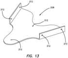
- FIG. 1is a top perspective view of a surgical training device according to the present invention.
- FIG. 2is an antero-cephalad, top perspective view of a model according to the present invention.
- FIG. 3 Ais a top perspective view of a pelvic frame according to the present invention.
- FIG. 3 Bis a top perspective view of a pelvic frame according to the present invention.
- FIG. 3 Cis a top perspective view of a pelvic frame according to the present invention.
- FIG. 3 Dis a top view of a pelvic frame in a flat orientation according to the present invention.
- FIG. 4 Ais a caudal end view of a model inside a surgical training device according to the present invention.
- FIG. 4 Bis a lateral side view of a model inside a surgical training device according to the present invention.
- FIG. 4 Cis a lateral side view of a model inside a surgical training device according to the present invention.
- FIG. 4 Dis an antero-caudal, top perspective view of a model inside a surgical training device according to the present invention.
- FIG. 4 Eis a cephalad end view of a model inside a surgical training device according to the present invention.
- FIG. 19is a top perspective view of a mold according to the present invention.
- FIG. 20is a top perspective view of a mold and a silicone tube placed over a center post of the mold according to the present invention.
- FIG. 21is a top perspective view of a mold, a silicone tube placed over a center post and a silicone outer interface formed in the mold according to the present invention.
- FIG. 22is a cross-sectional view of a mold, a silicone tube placed over a center post and a silicone outer interface formed in the mold according to the present invention.
- FIG. 24is a cross-sectional view of a mold, a silicone tube placed over a center post, a silicone outer interface formed in the mold and a flat plate according to the present invention.
- FIG. 25is a top perspective view of a mold, a silicone tube placed over a center post, a silicone outer interface formed in the mold, a flat plate and a backing mold according to the present invention.
- FIG. 26is a cross-sectional view of a mold, a silicone tube placed over a center post, a silicone outer interface formed in the mold, a flat plate and a backing mold according to the present invention.
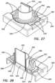
- FIG. 27is a top perspective view of a mold, a silicone tube placed over a center post, a silicone outer interface formed in the mold, a flat plate, a backing mold and a silicone inner interface according to the present invention.
- FIG. 28is a cross-sectional view of a mold, a silicone tube placed over a center post, a silicone outer interface formed in the mold, a flat plate, a backing mold and a silicone inner interface according to the present invention.
- FIG. 29is a side elevational, partial cross-sectional view of a hysterectomy model according to the present invention.
- FIG. 30is a side elevational, exploded view of a hysterectomy model according to the present invention.
- FIG. 31is a top perspective view of a frame according to the present invention.
- FIG. 32is a top perspective view of a mesh inside an open uterine mold and a mandrel according to the present invention.
- FIG. 33is a top perspective view of mesh and silicone inside an open uterine mold according to the present invention.
- FIG. 34is a top perspective view of mesh and silicone inside an open uterine mold according to the present invention.
- FIG. 35is a top perspective view of a mesh, silicone and a mandrel inside an open uterine mold according to the present invention.
- FIG. 36 Ais a top perspective view of mesh, silicone and a mandrel inside a closed uterine mold according to the present invention.
- FIG. 36 Bis a top perspective view of a plurality of closed and clamped uterine molds according to the present invention.
- FIG. 37is a top perspective view of a cervix mold according to the present invention.
- FIG. 38 Ais a top perspective view of a folded fabric sleeve according to the present invention.
- FIG. 38 Bis a top perspective view of a tubular sleeve being folded unto itself to create a folded sleeve having two layers of fabric according to the present invention.
- FIG. 39is a top perspective view of a post inside a fabric sleeve according to the present invention.
- FIG. 40is a top perspective view of a post inside a fabric sleeve inside a well of a cervix mold according to the present invention.
- FIG. 41 Ais a top perspective view of uncured silicone being poured into a cervix mold according to the present invention.
- FIG. 41 Bis a cross-sectional view of a folded sleeve with a post inside a well of a cervix mold according to the present invention.
- FIG. 42is a top perspective view of a post inside a fabric sleeve inside a well of a cervix mold partially filled with uncured silicone and a uterine form being squeezed according to the present invention.
- FIG. 43is a top perspective view of a uterine form being inserted into a well of a cervix mold while being squeezed according to the present invention.
- FIG. 44is a top perspective view of a uterine form inside a well of a cervix mold according to the present invention.
- FIG. 45is a top perspective view of a simulated uterus with a post according to the present invention.
- FIG. 46 Ais a proximal end view of a simulated uterus according to the present invention.
- FIG. 47is a top perspective view of a pair of mesh socks attached to a pair of mandrels according to the present invention.
- FIG. 48is a top perspective view of simulated vaginal canal with an embedded mesh layer according to the present invention.
- FIG. 49is a top perspective view of a distal end of a simulated vaginal canal according to the present invention.
- FIG. 50is a top perspective view of a distal end of a simulated vaginal canal according to the present invention.
- FIG. 51is a top perspective view of an inverted simulated vaginal canal according to the present invention.
- FIG. 52is a proximal end view of simulated vaginal canal according to the present invention.
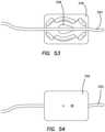
- FIG. 53is a top view of silicone vessel located across an ovary mold according to the present invention.
- FIG. 54is a top view of a silicone vessel located across an ovary mold according to the present invention.
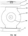
- FIG. 55is a top perspective view of a transvaginal adapter according to the present invention.
- FIG. 56is a top perspective sectional view of a transvaginal adapter according to the present invention.
- FIG. 57is an end view of a transvaginal adapter according to the present invention.
- FIG. 58is a side view of a transvaginal adapter according to the present invention.
- FIG. 59is an end view of a simulated uterus connected to a transvaginal adapter that is connected between a top cover and a base of surgical trainer is a top perspective view of a transvaginal adapter according to the present invention.
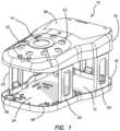
- a surgical training device 10that is configured to mimic the torso of a patient such as the abdominal region is shown in FIG. 1 .
- the surgical training device 10provides a body cavity 12 substantially obscured from the user for receiving simulated or live tissue or model organs or training models of the like described in this invention.
- the body cavity 12is accessed via a tissue simulation region 14 that is penetrated by the user employing devices to practice surgical techniques on the tissue or practice model found located in the body cavity 12 .
- the body cavity 12is shown to be accessible through a tissue simulation region, a hand-assisted access device or single-site port device may be alternatively employed to access the body cavity 12 .
- An exemplary surgical training deviceis described in U.S. patent application Ser. No. 13/248,449 entitled “Portable Laparoscopic Trainer” filed on Sep. 29, 2011 and incorporated herein by reference in its entirety.
- the surgical training device 10is particularly well suited for practicing laparoscopic or other minimally invasive surgical procedures.
- the surgical training device 10includes a top cover 16 connected to and spaced apart from a base 18 by at least one leg 20 .
- FIG. 1shows a plurality of legs 20 .
- the surgical training device 10is configured to mimic the torso of a patient such as the abdominal region.
- the top cover 16is representative of the anterior surface of the patient and the space 12 between the top cover 16 and the base 18 is representative of an interior of the patient or body cavity where organs reside.
- the surgical trainer 10is a useful tool for teaching, practicing and demonstrating various surgical procedures and their related instruments in simulation of a patient undergoing a surgical procedure. Surgical instruments are inserted into the cavity 12 through the tissue simulation region 14 as well as through pre-established apertures 22 in the top cover 16 .
- the base 18includes a model-receiving area 24 or tray for staging or holding a simulated tissue model or live tissue.
- the model-receiving area 24 of the base 18includes frame-like elements for holding the model (not shown) in place.
- a clip attached to a retractable wireis provided at locations 26 . The retractable wire is extended and then clipped to hold the tissue model in position substantially beneath the tissue simulation region 14 .
- tissue modelOther means for retaining the tissue model include a patch of hook-and-loop type fastening material (VELCRO®) affixed to the base 18 in the model receiving area 24 such that it is removably connectable to a complementary piece of hook-and-loop type fastening material (VELCRO®) affixed to the model.
- VELCRO®hook-and-loop type fastening material
- a video display monitor 28 that is hinged to the top cover 16is shown in a closed orientation in FIG. 1 .
- the video monitor 28is connectable to a variety of visual systems for delivering an image to the monitor.
- a laparoscope inserted through one of the pre-established apertures 22 or a webcam located in the cavity and used to observe the simulated procedurecan be connected to the video monitor 28 and/or a mobile computing device to provide an image to the user.
- audio recording or delivery meansmay also be provided and integrated with the trainer 10 to provide audio and visual capabilities.
- Means for connecting a portable memory storage device such as a flash drive, smart phone, digital audio or video player, or other digital mobile deviceis also provided, to record training procedures and/or play back pre-recorded videos on the monitor for demonstration purposes.
- connection means for providing an audio visual output to a screen larger than the monitoris provided.
- the top cover 10does not include a video display but includes means for connecting with a laptop computer, a mobile digital device or tablet and connecting it by wire or wirelessly to the trainer.
- the top cover 16When assembled, the top cover 16 is positioned directly above the base 18 with the legs 20 located substantially around the periphery and interconnected between the top cover 16 and base 18 .
- the top cover 16 and base 18are substantially the same shape and size and have substantially the same peripheral outline.
- the internal cavityis partially or entirely obscured from view.
- the legsinclude openings to allow ambient light to illuminate the internal cavity as much as possible and also to advantageously provide as much weight reduction as possible for convenient portability.
- the top cover 16is removable from the legs 20 which in turn are removable or collapsible via hinges or the like with respect to the base 18 . Therefore, the unassembled trainer 10 has a reduced height that makes for easier portability.
- the simulated uterus 32further includes simulated fallopian tubes 54 connected to ovaries 56 .
- the simulated uterus 32 , fallopian tubes 54 and ovaries 56are made of silicone or other elastomeric material and may include other material such as foam material combined with the silicone.
- the simulated uterus 32is made of silicone or lighter foam such as urethane or silicone foam or a combination of the two.
- the silicone constructionimparts the simulated uterus 32 with a more realistic weight when the attached simulated cervix 50 is being pulled and manipulated.
- the simulated uterus 32 made of foammakes the simulated uterus 32 easier to suspend inside the simulated pelvic cavity.
- 3 Dillustrates the outer perimeter having a top 68 and a bottom 70 interconnected by a first side and a second side 72 , 74 .
- the top 68includes two curved portions 76 a , 76 b interconnected at a first protrusion 78 along a vertical axis.
- the two curved portions 76 a , 76 brepresent the left and right ilium/iliac crest.
- the bottom 70includes a second protrusion 80 along the vertical axis.
- the first protrusion 78represents the sacrum of a human pelvis and the second protrusion 80 represents the coccyx.
- the first side 72includes a first lower lobe 82 having a first aperture 86 and the second side 74 includes a second lower lobe 84 having a second aperture 88 .
- the first and second lower lobes 82 , 84represent the left and right ischium and the first aperture 86 and the second aperture 88 represent the obturator foramen of the human pelvis.
- a piece of foam having a thicknessis cut to have the flat pattern shape shown in FIG. 3 D . Then the piece of foam is curved such that the first lower lobe 82 and second lower lobe 84 join together in a cylinder-like configuration.
- the frame 34is made of soft, compressible, semi-rigid foam that can be die cut and then formed into the correct shape with adhesive. If the frame 34 is made of harder plastic, it could be a thin thermoform that is initially formed into the correct shape or a thicker plastic that is cut into the pelvis shape and then formed into a cylindrical shape with heat. The frame 34 may also be made of deformable metal that holds its shape.
- the frame 34is not a perfect replica of the anatomy and need only include certain features selected to practice certain procedures that require those specific features as anatomical reference points or visual landmarks for the practitioner. For example, for practicing a vaginal hysterectomy, the important features of the pelvis are the restriction of the pelvic inlet and the attachments to the pelvic sidewall.
- the first sheet 36is a thin layer of clear silicone material having a top surface 96 and a bottom surface 98 and a first end 100 and a second end 102 .
- the first sheet 36is transparent and at least one of the top surface 96 and the bottom surface 98 is textured in one variation.
- the first sheet 36is attached to the simulated uterus 32 .
- the bottom surface 98 of the first sheet 36 near the first end 100is attached along at least a portion of the length of simulated uterus 32 to one or more of the bulbous portion 40 and tubular portion 44 as shown in FIG. 2 .
- the first sheet 36is then folded back toward the top of the model 30 and toward the first end 100 of the first sheet 36 creating a fold near the tubular portion 44 of the simulated uterus 32 .
- At least a portion of the first sheet 36 near the second end 102 of the first sheet 36is attached to the frame 34 such that the bottom surface 98 of the first sheet 36 is adhered to the frame 34 in the general location of where the two lobes 82 , 84 are in juxtaposition to create a cylinder-like configuration for the frame 34 .
- the attachment of the first sheet 36may also serve to hold the frame 34 in the cylindrical-like configuration.
- Adhesiveis used to attach the bottom surface 98 of the first sheet 36 to the frame 34 .
- the bottom surface 98 of the first sheet 36is attached to the first surface 62 or inner surface of the frame 34 and then folded around a portion of the first side 72 and second side 74 of the frame 34 .
- the second end 102 of the first sheet 36is also attached with adhesive to the outer surface of the simulated bladder 90 capturing the simulated bladder 90 between the frame 34 and the first sheet 36 .
- a portion of the second end 102 of the first sheet 36is folded around the edge of the frame 34 and attached to the second surface 64 of the frame 34 such that at least part of the second end 102 of the first sheet 36 is resident above the second or outer surface 64 of the frame 34 as visible in FIG. 4 D .
- the first sheet 36is sized and configured to suspend the simulated uterus 32 inside the interior 60 of the frame 34 .
- Simulated vasculature 58may be attached to the top surface 96 or bottom surface 98 of the first sheet 36 .
- the configuration of the first sheet 36forms a pocket-like structure wherein the top surface 96 of the first sheet 36 is folded and at least in part facing itself.
- the first sheet 36creates a webbing of suspension that simulates the peritoneum layer.
- the second sheet 38is a thin layer of clear silicone material having a top surface 104 and a bottom surface 106 and a first end 108 and a second end 110 .
- the second sheet 38is transparent and at least one of the top surface 104 and the bottom surface 106 is textured in one variation.
- the second sheet 38is attached to the simulated uterus 32 .
- the bottom surface 106 of the second sheet 38 near the first end 108is attached along at least a portion of the length of simulated uterus 32 to one or more of the bulbous portion 40 and tubular portion 44 on a side opposite from where the first sheet 36 is attached.
- the first sheet 36is attached to the anterior side of the model 30 which is also the anterior side of the simulated uterus 32 .
- the second sheet 38is attached to the posterior side of the model 30 which is also the posterior side of the simulated uterus 32 . After being attached to the posterior side of the simulated uterus 32 , the second sheet 38 is then folded back toward the top of the model 30 and toward the first end 108 of the second sheet 38 creating a fold near the tubular portion 44 of the simulated uterus 32 . At least a portion of the second sheet 38 near the second end 110 of the second sheet 38 is attached to the frame 34 such that the bottom surface 106 of the second sheet 38 is adhered to the frame 34 in the general location of the second protrusion 80 . Adhesive is used to attach the bottom surface 106 of the second sheet 38 to the frame 34 .
- the bottom surface 106 of the second sheet 38is attached to the first surface 62 or inner surface of the frame 34 and may be folded around the edge of the frame 34 such that at least part of the second end 110 of the second sheet 38 is connected to second or outer surface 64 of the frame 34 . If a simulated colon 92 is employed in the model 30 , then the second end 110 of the second sheet 38 is also attached with adhesive to the outer surface of the simulated colon 92 or at least overlaying and not attached with adhesive such that at least a portion of the simulated colon 92 is captured or located between the frame 34 and the second sheet 38 .
- the second sheet 38is sized and configured to suspend the simulated uterus 32 inside the interior 60 of the frame 34 if the model 30 is turned over.
- Simulated vasculature 58may be attached to the top surface 104 or bottom surface 106 of the second sheet 38 .
- the configuration of the second sheet 38forms a pocket-like structure wherein the top surface 104 of the second sheet 38 is folded and at least in part facing itself.
- the second sheet 38creates a suspended webbing that simulates the peritoneum layer.
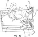
- the model 30is shown placed inside a surgical training device 10 of the like described with respect to FIG. 1 .
- the model 30is shown inside the body cavity 12 and oriented such that the top 68 of the frame 34 is in the cephalad direction of the simulated training device 10 and the vaginal opening 48 of the simulated uterus 32 faces the caudal direction of the simulated training device 10 .
- the model 30can be connected to the surgical training device 10 with the clips 26 attached to the trainer 10 .
- the retractable clips 26can be pulled out and the clips 26 attached to any portion of the model 30 such as to the frame 34 of the model 30 .
- the second or outer surface 64 of the model 30may include a hook-and-loop type fastener configured to attach to a complementary portion of hook-and-loop type fastener connected to the base 18 of the trainer 10 .
- the model 30is securely attached to the trainer 10 such that it can be manipulated in simulated surgery without dislodging the model 30 from the body cavity 12 of the trainer 10 .
- the model 30is further connected to the trainer 10 via a transvaginal adapter 112 that is sized and configured to connect between the top cover 16 and the base 18 as an additional leg 20 positioned at the caudal direction of the surgical training device 10 .
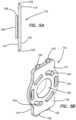
- FIGS. 5 A- 5 B and 6 A- 6 Bthere is shown a transvaginal adapter 112 .
- a top coversupported above the base by five legs 20 .
- a sixth leg 20is provided as shown in FIGS. 4 A- 4 D in the form of the transvaginal adapter 112 .
- the trainer 10may be assembled with an optional sixth support structure or leg which is configured for simulating transvaginal surgery including transvaginal hysterectomies.
- the transvaginal adapter 112includes a flat plate 114 having an inner surface 116 for facing toward the interior of the trainer and an outer surface 118 for facing outwardly towards the user.
- the plate 114has a rectangular shape and includes an aperture 120 passing through the plate 108 from the inner surface 116 to the outer surface 118 .
- the aperture 120is circular in shape.
- the aperture 120is elongate elliptical oval-like in shape and oriented vertically along the longitudinal axis of the adapter 112 .
- the aperture 120is elongate elliptical oval-like in shape and oriented perpendicularly to the longitudinal axis of the adapter. As shown in FIGS.
- the plate 114also includes means such as tabs 122 or a U-shaped channel for inserting to connect the transvaginal adapter 112 to the top cover 16 and to the base 18 to help support and space apart the top cover 16 .
- the transvaginal adapter 112is located between the top cover 16 and the base 18 and provides a side access aperture 16 lateral to the trainer 10 or substantially perpendicular to the top cover 16 and the base 18 .
- the plate 114further includes a plurality of molding apertures 124 surrounding or encompassing the main aperture 120 configured for overmolding a soft simulated vaginal tissue interface made of silicone or the like. In another variation the interface is insertable into the aperture 120 of the transvaginal adapter 112 .
- the tissue interface(not shown) includes an aperture that is substantially coaxial with the plate aperture 120 .
- a tubular extension 126is integrally provided and extends into the simulated body cavity 12 of the trainer 10 .
- the tubular extension 126is longer in FIGS. 6 A- 6 B in comparison to the tubular extension 126 of FIGS. 5 A- 5 B .
- the tubular extension 126is sized and configured such that the tubular portion 44 of the simulated uterus 32 can be stretched around the extension 126 and secured to the transvaginal adapter 112 such that the vaginal canal 46 is supported in an open configuration, coincident with and accessible through the aperture 120 of the adapter 112 as shown in FIGS. 4 A- 4 D .
- the tubular extension 126serves as a connector connecting the model 30 with the trainer 10 in a manner that permits the interior of the uterus to be accessed as in real surgery.
- the tubular extension 126is a cylindrically-shaped extension having a radially-extending distal flange 128 that extends around at least a portion of the extension 128 to help secure and retain the model 30 attached to the trainer 10 .
- the tubular portion 44 of the model 20is attached to the tubular extension 126 by pulling the tubular portion 44 over the distal flange 128 , if one is provided, and over and around the tubular extension 126 the outer diameter of which is the same or slightly larger than the relaxed inner diameter of the tubular portion 126 to keep the tubular portion 44 secured to the transvaginal adapter 112 .
- the transvaginal adapter 112can be made of flexible or rigid material. If the adapter 112 is made of rigid material it will tend to simulate an already retracted vaginal canal 46 . If the adapter 112 is made of flexible material or soft material, the adapter 112 is suited for practicing retraction.
- the transvaginal adapter 112has a tubular extension 126 that is made of soft flexible material and plate 114 made of rigid material or surrounded by rigid material to keep the top cover 16 of the trainer 10 supported which would still allow the practitioner to practice retraction at the opening of the vaginal canal 46 at the adapter 112 .
- the model 30is placed inside the surgical training device 10 and held in place with a hook-and-loop type fastener and/or retracting clips 26 .
- the tubular portion 44is attached to the transvaginal adapter 112 by stretching the vaginal opening 48 over the tubular extension 126 of the adapter 112 .
- a curtainmay be employed that is placed around the sides of the trainer 30 to further conceal the model 30 such that the only visualization is through the simulated vaginal canal 46 .
- the vaginal canal 46is then retracted using a surgical retractor.
- the vaginal canal 46is made of a flexible thermoplastic elastomer (TPE).
- TPEprovides resistance as it is retracted and wants to spring back to its original shape which permits the user to practice realistic retraction.
- the transvaginal adapter 112 of FIGS. 6 A- 6 B having a longer tubular extension 126is used to simulate an already retracted vaginal canal.
- the transvaginal adapter 112permits the practitioner to practice the hysterectomy procedure without needing extra-hands and assistance to perform the retraction.
- the transvaginal adapter 112 of FIGS. 5 A- 5 B having the shorter tubular extension 126is used, the practitioner will practice retracting the vaginal canal 46 with retractors and the help of extra hands during the procedure.
- the transvaginal adapter 112can be made of rigid or flexible material or rigid and flexible material as described above and selected for the purpose of practicing retraction of the vaginal canal 46 or not.
- the simulated cervix 50is grasped and pulled towards the opening 48 of the vaginal canal 46 .
- the simulated cervix 50is made of high durometer silicone relative to the surrounding tubular portion 44 .
- the simulated cervix 50is also made as a solid component which allows it to be grasped with real surgical tools and pulled on without fear of the silicone ripping or tearing.
- the simulated cervix 50is incised circumferentially and the practitioner is able to practice carefully dissecting the vaginal mucosa off of the simulated cervix 50 .
- a sheet of cotton or other webbing-like substancecan be included in the model 30 between the vaginal canal 46 and the simulated bladder 90 .
- the simulated bladder 90is a hollow, air-filled component. If the practitioner cuts to high while dissecting the simulated vaginal mucosa and the simulated bladder 90 is accidentally incised, the simulated bladder 90 could pop and give immediate feedback to the practitioner especially if the simulated bladder 90 contains fluid.
- the model 30advantageously includes a second sheet 38 forming a fold between the simulated uterus 32 and the frame 34 .
- the suspension of the simulated uterus 32 within the frame 34advantageously creates a realistic response when the simulated uterus 32 is being incised and manipulated.
- the simulated uteruswill remain suspended, hang and swing in response to being manipulated with surgical instruments. At least portions of the simulated uterus and simulated vagina are held in suspension inside the enclosure defined by the pelvic frame and connected thereto or directly connected to the enclosure defined by the trainer.
- the suspensionadvantageously permits the fold of the second sheet to be accessed to practice posterior colostomy into the posterior cul-de-sac incision by incising the peritoneum forming the recto-uterine fold.
- the suspended simulated uterus 32allows for the existence of the recto-uterine peritoneum fold.
- the simulated uterus 32is pendent inside the frame 34 made of foam material that mimics a human pelvis.
- the simulated uterus 32is suspended by a folded first sheet of silicone material on the anterior side of the simulated uterus 32 and a folded second sheet of silicone material on the posterior side of the simulated uterus 32 .
- the frame 34can be made of any material such as plastic or harder foam material.
- the frame 34serves as an attachment area for the various simulated portions of the anatomy including the broad ligament, ovaries 56 and fallopian tubes 54 .
- the elasticity of the silicone of these anatomical componentsallows the simulated uterus 32 to be pulled and manipulated and still remain attached to the frame 34 .
- a frame 34 made of semi-rigid foammay also move as the simulated uterus is being manipulated. A more rigid frame 34 would move less.
- the practitionerthen divides the uteralsacral ligaments 59 .
- the practitionerthen performs an anterior colostomy into the anterior cul-de-sac by incising the first sheet 38 simulating the peritoneum forming the vesico-uterine fold.
- the practitionerdivides the tubo ovarian and round ligaments 61 on each side of the simulated uterus 32 . Due to the foam frame 34 , the round and tubo ovarian ligaments 59 remain realistically attached to the frame 34 after they have been divided from the simulated uterus 32 .
- the simulated uterus 32is then freed and removed.
- the practitionerthen practices to suture the vaginal cuff closed by passing a needle and suture through the tubular portion 44 of the model 32 to close the vaginal canal 46 opening. Suturing the vaginal cuff in real surgery is another difficult part of the vaginal hysterectomy due to the space limitations.
- the tubular portion 44 that is made of TPEholds the suture without tearing and limits the space allowed for instruments during the suturing process.
- the model 30allows the practitioner to practice numerous difficult procedures on one model.
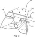
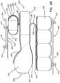
- the model 200includes a plurality of simulated organ structures 202 connected to and located inside a frame 204 .
- the frame 204is configured to simulate a pelvis and serve as a box-like encasement for housing the plurality of simulated organ structures 202 .
- the frame 204includes a top frame portion 206 connected with fasteners 210 to a bottom frame portion 208 .
- the assembled frame 204forms a base and a top interconnected by two upstanding sidewalls and defines a central lumen with an open proximal end and an open distal end.
- the frame 204has a flat base permitting it to be placed and stand on a flat surface.
- FIG. 10illustrates the top frame portion 206 in a flat arrangement.
- the bottom sideincludes curvatures representative of the bony structure of the human pelvis and form the sidewalls and top of the frame 204 .
- the top frame portion 206is folded to form the folded arrangement shown in FIG. 11 .
- the top frame portion 206includes a plurality of apertures 212 configured to receive fasteners for connecting the plurality of simulated organ structures 202 to the frame 204 .
- Other apertures 212are configured to pass the simulated organ structures through the apertures 212 and into the frame 204 for supporting the various simulated organ structures such as simulated vasculature with respect to the frame 204 as will be described in greater detail below.
- FIGS. 12 - 13there is shown a bottom frame portion 208 .
- FIG. 12illustrates the bottom frame portion 208 in a flat arrangement and FIG. 13 illustrates it in a folded arrangement.
- the bottom frame portion 208defines the base of the frame 204 and includes curved ends that simulate the bony anatomy of the human pelvis.
- the bottom frame portion 208also includes a plurality of apertures 212 configured to connect the plurality of simulated organ structures 202 to the frame 204 with fasteners passed through the apertures and/or by passing the simulated organ structures directly through the apertures 212 as will be described in greater detail below.
- the plurality of simulated organ structures 202includes a simulated bladder 214 , a simulated uterus 216 , a simulated vaginal canal 218 , a simulated rectum 220 , a first sheet 222 , a second sheet 224 , a dissecting layer 226 and a plurality of fasteners 210 .
- the plurality of organ structures 202are interconnected as shown in FIG. 14 and in turn connected to the frame 204 .
- Tubular shaped vasculature, ducts, arteries and the like in addition to other simulated organs structures not mentioned hereinmay be included in this model in an anatomically correct or anatomically similar arrangement for the same or different anatomical location of the body.
- Each simulated organ structurewill now be described.
- the simulated bladder 214forms a closed receptacle with an outer membrane made of pink-colored silicone.
- the interior of the simulated bladder 214may be stuffed with polyfil or other material to maintain its shape.
- the simulated bladder 214has a proximal end 240 and a distal end 242 .
- the simulated uterus 216is also made of silicone.
- the simulated uterus 216has a proximal end 260 and a distal end 262 .
- the simulated vaginal canal 218is a tubular structure made of silicone and may optionally contain an embedded mesh layer 230 .
- the simulated vaginal canal 218has a proximal end 256 and a distal end 258 .
- the simulated rectum 220is a tubular structure made of silicone with molded transverse folds.
- the simulated rectum 220has a proximal end 244 and a distal end 246 .
- Each of the first sheet 222 and the second sheet 224comprises a large flat planar layer of silicone material. Both sheets 222 , 224 represent the peritoneum.
- the first sheet 222has a first surface 232 and a second surface 234 and a proximal end 248 and a distal end 250 .
- the second sheet 224has a first surface 236 and a second surface 238 and a proximal end 252 and a distal end 254 .
- the distal end of the bladder 242is attached to the first surface 232 of the first sheet 222 with adhesive approximately midway between the proximal end 248 and the distal end 250 of the first sheet 222 such that the first sheet 222 wraps around the distal end 242 of the simulated bladder 214 from the top of the simulated bladder to the bottom of the simulated bladder 214 .
- the first surface 232is attached to a fastener 210 near the distal end 248 of the first sheet 222 .
- the first sheet 222is folded in an approximate U-shape such that the distal end 250 of the first sheet 222 and, in particular, the first surface 232 of the first sheet 222 , is attached to the simulated uterus 216 and attached to the simulated vaginal canal 218 via the dissecting layer 226 using adhesive.
- the dissecting layer 226is a construct comprising a silicone layer 228 interconnected with a fiber layer 229 . While the silicone layer 228 is uncured, a fiber layer 229 is embedded to form the dissecting layer 226 .
- the dissecting layer 226is attached to the simulated vaginal canal 218 in pieces or strips while the silicone of the simulated vaginal canal 218 is still wet and uncured on a mandrel.
- the dissecting layer 226When the dissecting layer 226 is applied to the uncured simulated vaginal canal 218 , the uncured silicone of the uncured simulated vaginal canal 218 is allowed to cure to attach the dissecting layer 226 , in particular, to attach the fiber layer 229 of the dissecting layer 226 to the simulated vaginal canal 218 sandwiching the fiber layer 229 between two layers of silicone.
- the dissecting layer 226may be sectional around the simulated vaginal canal 218 or completely tubular in shape to surround the circumference of the simulated vaginal canal 218 . Although the dissecting layer 226 is shown with the same reference number, two dissecting layers 226 may be provided on either side of the simulated vaginal canal 218 as shown in the figures.
- the dissecting layer 226is attached to distal end 258 of the simulated vaginal canal 218 .
- the dissecting layer 226is described in detail in co-pending International Patent Application Serial No. PCT/US2016/041852 entitled “Simulated dissectible tissue” filed on Jul. 12, 2016 incorporated herein by reference in its entirety.
- the second sheet 224is attached between the simulated uterus 216 and the simulated rectum 220 .
- the first surface 236 at the distal end 252 of the second sheet 224is attached near the distal end 262 of the simulated uterus 216 .
- the second sheet 224is attached along the length of the simulated uterus 216 toward the proximal end 260 using adhesive.
- the second sheet 224is attached to the dissecting layer 226 .
- the first surface 236 of the second sheet 224is attached to the silicone layer 228 of the dissecting layer 226 using adhesive.
- the second sheet 224is folded to extend back towards the distal end of the simulated rectum 220 and attached along the top side and outer surface of the simulated rectum 220 such that the distal end 254 of the second sheet 224 is near the distal end 246 of the simulated rectum 220 .
- the top side of the simulated bladder 214is connected to a fastener 210 and this fastener 210 is passed through an aperture 212 in the top frame 206 of the frame 204 .
- the proximal end 248 of the first sheet 222is also attached to a fastener 210 which is also passed through an aperture 212 in the top frame 206 of the frame 204 to attach the plurality of the simulated organ structures 202 to the frame 204 in a suspended manner. While suspended from the top frame 204 , the interconnected plurality of simulated organ structures 202 advantageously pendulate and move together in a realistic fashion wherein the point of contact with instruments and the like will move most and simulated organs distal to the point of contact with instruments move to a lesser degree.
- the bottom side of the simulated rectum 220is attached to at least two fasteners 210 as shown in FIGS. 14 - 15 .
- the two fasteners 210are passed through apertures 212 in the bottom frame 208 to secure the plurality of simulated organ structures 202 to the frame 202 .
- the plurality of simulated organs structuresis spanned across the central opening of the frame 202 with the first sheet 222 and second sheet 224 forming an interconnecting webbing.
- the proximal end 260 of the simulated uterus 216is inserted into the distal end 258 of the simulated vaginal canal 218 and joined together with adhesive.
- a simulated cervixis provided made of silicone and located inside the simulated uterus 216 at the proximal end 260 .
- the fastener 210has a dual-pronged, hooked, deflectable end 264 connected to a planar surface end 266 .
- the two prongs of the rivet-like fastener 210extend from the planar surface 266 .
- the two prongsare resiliently deflectable toward and away from each other such that when passed through a smaller aperture, the prongs flex inwardly when ramped against the aperture and then spring back outwardly when the widest portion of the prongs has passed through the aperture, thereby, snapping and hooking into the aperture wall.
- the fastener 210is not limited to having a two-prong arrangement.
- a single prongmay be employed having a bulbous portion for example that is configured to snap through an aperture.
- a looped layer 268 of looped-sided VELCRO hook-and-loop type fasteneris attached to the planar end 266 with cyanoacrylate glue 270 .
- a silicone layer 272is applied while uncured to the looped layer 268 making sure that the wet silicone is spread into the loops of the looped layer 268 . Then, the silicone layer 272 is allowed to dry.
- a layer of silicone adhesivemay be used.
- the fastener 210is easily attached to a silicone organ structure with adhesive or by putting a layer of wet silicone onto the organ structure at a location where the fastener 210 is desired to be located. The fastener 210 is then placed on the patch of wet uncured silicone and the patch is allowed to dry, adhering the silicone embedded in the looped layer 268 to the silicone organ structure.
- the silicone layer 272is part of the silicone organ structure as a patch of wet silicone or part of a cured silicone component of the organ structure and attached with silicone glue.
- the fasteners 210are removable with respect to the frame 204 by pressing the prongs together and/or pushing the fastener 210 out of the apertures 212 making the plurality of simulated organ structures 202 removable and replaceable with a new plurality of simulated organ structures 202 for continued practice and training of surgical procedures.
- the apertures 212 and fasteners 210may be color-coded to make attachment of the plurality of simulated organ structures 202 to the frame 204 quick and easy.
- simulated vasculature 274 , ducts, fallopian tubes, ureters or other anatomical or non-anatomical structure having a tubular/cylindrical form and typically made of siliconeare pulled through appropriately-sized apertures 212 as shown in FIGS. 7 - 8 to further support the connected simulated tissue structures.
- These tubular structureshave a free end and another end that is attached to other simulated tissue structures. The free end is passed through an aperture in the frame and can be secured with adjustable length to adjust the tension on the simulated tissue structures to which it is connected. For example, a loose tension may be created by securing the tubular structure with more slack between the frame and other simulated tissue structure.
- tension on the simulated tissuemay be increased by pulling the tubular structure taunt with respect to the frame to create a relatively less pendulating simulated tissue construct within the frame.
- the tubular rope-like structurecan be tied into a knot along its length to adjust the tension.
- the knot diameteris made larger than the aperture in frame in order to secure the larger tissue structure to the frame.
- the knotsmay be untied to remove the simulated tissue structure or re-tied to provide a different tension level.
- the tubular silicone simulated vasculature 274 , ducts, fallopian tubes, ureters or other anatomical or non-anatomical structure having a tubular/cylindrical formare provided with rivets at their distal end.
- the rivetsinclude a distal end for connection with the frame and a proximal portion embedded or swaged into the ends of the silicone tubular structure to make a mechanical connection.
- the rivet-like fastener 210serves as an interface connection between the soft, pliable silicone of the simulated tissue structures and the rigid plastic frame.
- the simulated tissue structuresare often made from room temperature vulcanized (RTV) silicone elastomers. As a result, the simulated tissue structures are delicate and may tear easily if not reinforced. This makes it difficult to connect such artificial tissue structures to the frame.
- the fastenerhas a rigid portion for connecting with the rigid frame and an interfacing layer located between the rigid portion of the fastener and the attaching simulated tissue structure.
- the interfacing layeris a fiber layer that wet, uncured silicone may interpenetrate and when cured adhered securely thereto along the area of the interfacing layer.
- the uncured silicone layermay be a patch on the artificial tissue structure such that when cured, the patch becomes integrally connected to the artificial tissue structure and to the interfacing layer.
- This type of fasteneradvantageously minimizes stress concentrations that would result in the fastener tearing away from the simulated tissue structure permitting the simulated tissue structures to be manipulated aggressively.
- the fastenersalso permit a quick assembly of the simulated tissue structure inside the frame by simply snapping the fasteners through a plurality of apertures in the frame.
- Disassemblyis also facilitated and the frame is reusable after a simulated tissue structure is consumed with practiced and replaced with another simulated tissue model that is the same or different from the discarded model.
- no additional tools or adhesiveis required for assembly.
- the first, second and/or third sheets 222 , 224 and 225may be attached to the frame directly with adhesive.
- rivetsare described any suitable fastener adapted to secure the simulated tissue structure to the frame is within the scope of the present invention.
- the transvaginal adapter 280is formed as a leg 20 configured to support the top cover of the trainer 10 . It is configured for simulating transvaginal surgery including transvaginal hysterectomies.
- the transvaginal adapter 280includes a flat plate 282 having an inner surface 284 for facing toward the interior of the trainer and an outer surface 286 for facing outwardly towards the user.
- the plate 280has a rectangular shape and includes an aperture 288 passing through the plate 280 from the inner surface 284 to the outer surface 286 . In one variation, the aperture 288 is circular in shape.
- the aperture 288is elongate elliptical, oval-like in shape and oriented vertically along the longitudinal axis of the adapter 280 . In another variation, the aperture 288 is elongate elliptical, oval-like in shape and oriented perpendicularly to the longitudinal axis of the adapter.
- the plate 280also includes means such as tabs 290 or a U-shaped channel for inserting to connect the transvaginal adapter 280 to the top cover 16 and to the base 18 to help support and space apart the top cover 16 .
- the transvaginal adapter 280is located between the top cover 16 and the base 18 and provides a side access aperture 288 lateral to the trainer 10 or substantially perpendicular to the top cover 16 and the base 18 .
- the plate 280further includes a plurality of molding apertures 292 , shown in FIGS. 23 - 28 , surrounding or encompassing the main aperture 288 configured for overmolding a soft simulated vaginal tissue interface 294 made of silicone or the like. The method of forming the overmolded soft simulated vaginal tissue interface 294 will now be described.
- the mold 298includes a well 298 encompassing an elongated center post 300 .
- the center post 300is oval or circular in shape. The circular or oval shape will result in an opening having the same shape and suitable for a TATME application in which the adapter is connectable to a simulated rectum and, thereby, serves as a transanal adapter instead of a transvaginal adapter.
- a pre-made silicone tube 302is placed over the center post 300 as shown in FIG. 20 .
- FIGS. 21 - 22uncured silicone is poured into the well 298 to form the outer interface 304 .
- FIGS. 21 - 22uncured silicone is poured into the well 298 to form the outer interface 304 .
- the transvaginal adapter 280is placed on top of the uncured silicone located inside the well 298 .
- the uncured silicone of the outer interface 304is allowed to cure.
- a backing mold 306is placed around the silicone tube 302 and inside the aperture 288 . Uncured silicone is then poured between the backing mold 306 and the inside of the aperture 288 and into the molding apertures 292 and onto the inner surface 284 and allowed to cure to form the inner interface 308 as shown in FIGS. 27 - 28 .
- the mold 296 and the backing mold 306are removed.
- the resulting transvaginal adapter 280is shown in FIGS. 17 - 18 .
- the transvaginal adapter 280is connected between the top cover 16 and base 18 of the trainer 10 .
- the model 200is placed inside the body cavity 12 of the trainer 10 and connected to the transvaginal adapter 280 such that the silicone tube 302 faces the interior of the cavity 12 and is inserted into the proximal end 256 of the simulated vaginal canal 218 .
- the elongated center post 300 of the mold 296creates an elongated entry way leading into the model 200 .
- a practicing surgeonmay approach the simulated uterus 216 with surgical instruments and retractors through the transvaginal adapter 280 to perform a transvaginal hysterectomy.
- the simulated uterus 216may be approached through the simulated abdominal wall of the top cover 16 of the trainer 10 .
- the userwill practice laparoscopic surgical skills, employing a trocar and scope to examine the anatomy and perform the simulated surgical hysterectomy.
- the procedureinvolves making key incisions to detach the uterus and then remove it.
- the model 200advantageously provides the one or more dissecting layer 226 that includes fibers embedded in silicone that make the incisions and separation of the simulated uterus 216 realistic.
- the model 200is similar to the model 200 described with respect to FIGS. 7 - 28 and like numbers will be used to describe like parts.
- the model 200includes a plurality of simulated organ structures 202 connected to and located inside a frame 204 .
- the frame 204is configured to simulate a pelvis and serve as a box-like encasement for housing the plurality of simulated organ structures 202 .
- the frame 204includes a top frame portion 206 connected with fasteners 210 to a bottom frame portion 208 to form a top planar surface and a bottom planar surface interconnected by two upstanding sidewalls.
- the top planar surface and the bottom planar surfaceare parallel with each other and form corners with the sidewalls that are approximately 90 degrees.
- the sidewallsare angled towards each such that at the proximal end the distance between the sidewalls is close and increases progressively with increasing distance toward the distal end where the sidewalls are farther apart from each other.
- the assembled frame 204having a base and a top interconnected by two upstanding sidewalls defines a central lumen with an open proximal end and an open distal end.
- the area of the central lumen in cross-section taken perpendicular to the longitudinal axisincreases progressively with increasing distance from the proximal end toward the distal end.
- the outer shape of the frame 204can be dissimilar from the shape of the central lumen.
- a tapered framethat has a central lumen with increasing area and that does not have corners is a frame that forms a frusto-conical shape.
- the central lumen of one such variationhas a frusto-conical shape.
- the outer shape of the framemay also match the tapered shape of the central lumen.
- the frame 204has a flat base permitting it to be placed and stand on a flat surface.
- the bottom frame portion 208includes a first level and a raised second floor 209 that raises the level of the model inside the frame 204 to be in line with the transvaginal adapter 280 .
- the frame 204may include apertures 212 for passing of fasteners 210 and/or connecting tissue structures, such as vasculature, by passing them through the apertures and suspending them in the frame 204 .
- the apertures 212include a first set of apertures and a second set of apertures aligned with the first set of apertures to form the box-like shape.
- the frame 204 of FIG. 31is similar to the frame 204 shown in FIGS. 7 - 13 in which the frame 204 is comprised of folded plastic that is transparent and/or translucent.
- the folding of the plastic components of the frame 204results corners that are representative of a pelvis that is not anatomically correct yet provides advantages needed in simulating laparoscopic procedures in exchange for the realism of an anatomically correct pelvis. These advantages include the mechanical constriction of organs located in the tapered proximal end having the smallest luminal cross-sectional area. The physical constriction of organs at the proximal end creates a more rigid response in the organs when manipulated by surgical instruments relative to the distal end where organs located therein are less constricted and freer to pendulate and more fluidly respond to manipulations with surgical instruments.
- the frame 204 of the present inventionis an intentional simplification of the pelvis that combines variable resistance in the organs along the length of the longitudinal axis of the central lumen.
- the smaller opening to the central lumen at the proximal end of the frameis where the opening to the vaginal canal would be positioned when the organs are placed inside the frame.
- the proximal end of the frameis also oriented toward the transvaginal or transanal adapted for connection therewith.
- the distal end of the frame 204is the location of the artificial uterus 216 .
- the central lumen of the frameexpands, widens and angles outwardly towards the distal end. This taper of the box-like frame widens relaxing the organs located therein and the narrow proximal end constricts the organs, limiting the range of motion of the organs relatively more as a result of supporting the organs in closer confines.
- the plurality of simulated organ structures 202includes a simulated bladder 214 , a simulated uterus 216 , a simulated vaginal canal 218 , a simulated rectum 220 , a first sheet 222 , a second sheet 224 , a third sheet 225 and a plurality of fasteners 210 .
- the plurality of organ structures 202are interconnected as shown in FIG. 29 and in turn connected to the frame 204 .
- the first sheet 222 , second sheet 224 and third sheet 225are connected as shown in dotted lines in FIG. 29 .
- the first sheet 22extends proximally along the top of the simulated bladder 214 and around the proximal end of the simulated bladder 214 downwardly and toward the distal end of the simulated bladder 214 .
- the simulated bladder 214is not suspended with a fastener 210 as shown in FIG. 29 .
- the third sheet 225commences at the proximal end of the simulated bladder 214 and extends downwardly and is connected to the first sheet 222 at a location 360 that in this variation comprises a location of adhesive connecting the first sheet 222 and the third sheet 225 .
- the second sheet 224 in this alternative variationfollows approximately the same path but includes slit to pass the simulated uterus 216 through such that the second sheet 224 extends upwardly as shown with the dotted line.
- Tubular shaped vasculature, ducts, arteries and the like in addition to other simulated organs structures not mentioned hereinmay be included in this model in an anatomically correct or anatomically similar arrangement for the same or different anatomical location of the body. Each simulated organ structure will now be described.
- the simulated bladder 214forms a closed receptacle with an outer membrane made of pink-colored silicone.
- the interior of the simulated bladder 214may be stuffed with polyfil or other material to maintain its shape.
- the simulated bladder 214has a proximal end 240 and a distal end 242 .
- the simulated uterus 216is also made of silicone.
- the simulated uterus 216has a proximal end 260 and a distal end 262 .
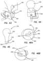
- the simulated uterus 216is made by providing a uterine mold 310 comprising two halves 310 a , 310 b as shown in FIG. 32 .
- a mesh fabric layer 312is placed inside both halves the mold 310 a , 310 b . In one half of the mold 310 a , the mesh fabric layer 312 is placed only in the proximal end of the mold 310 to reinforce that portion that will be subjected to the most force during practice of a surgical technique.
- the mesh fabric layer 312may cover the entire mold in one variation.
- the mesh fabric layer 312includes darts or cutouts to allow the fabric to lay as smoothly as possible in the mold.
- Uncured silicone foam 314is poured over the mesh fabric layer 312 and into each half 310 a , 310 b of the mold 310 as shown in FIGS. 33 - 34 .
- the wet silicone foam 314is spread evenly to contact all of the surfaces to ensure that the silicone foam 314 will expand uniformly.
- a mandrel 316is placed onto half 310 b the mold 310 .
- the two halves 310 a , 310 bare placed together and the mold 310 is clamped as shown in FIGS. 36 A and 36 B and the silicone foam 314 is allowed to expand and cure.
- the mold 310Upon curing, the mold 310 is removed from the cured silicone foam 314 and the mandrel 316 is mounted on a motor and the cured silicone foam is rotated and uncured silicone gel is applied evenly onto the silicone foam 314 to create an outer layer of silicone that encompasses the silicone foam 314 .
- the outer layer of siliconeis allowed to cure and the mandrel 316 is removed leaving behind a uterine-like form 332 shown in FIGS. 29 - 30 and 43 - 45 .
- a simulated cervix 318is formed and inserted into the proximal end 260 of the cured foam and cured outer layer of silicone.
- the simulated cervix 318is formed by first providing a cervix mold 320 having a well 322 as shown in FIG. 37 .
- the cervix mold 320is generally made of two pieces that are clamped together to define the well 322 .
- a sleeve 324 of fabric such as KEVLAR synthetic fiber fabricis provided and inverted to create a fold 326 wherein the thickness of one end of the sleeve 324 is doubled as shown in FIGS. 38 A, 38 B and 39 .
- the KEVLAR synthetic fiber reinforcement of the simulated cervixmakes the portion of the cervix that is pulled by the surgeon strong and allows the surgeon to use a tenaculum with an ability to puncture and pull the simulated cervix as in real surgery without the model tearing.
- the fabric sleeveis compatible with silicone in that it does not inhibit the curing of room temperature vulcanizing (RTV) silicone elastomers that form the rest of the artificial cervix, uterus and vaginal canal.
- RTVroom temperature vulcanizing
- KEVLAR synthetic fiberis porous, it allows for a strong mechanical connection without additional adhesive and can be used with materials other than silicone. Any suitable fiber having a high tensile strength-to-weight ratio may be employed.
- a post 328is inserted into the lumen of the fabric sleeve 324 such that it protrudes outwardly from the proximal end having the fold 326 as shown in FIG. 39 .
- the cross-section of the post 328is elongate and narrow such that a wooden popsicle stick can serve as the post 328 .
- the sleeve 324 with the post 328is placed into the well 322 of the cervix mold 320 with the fold 326 and protruding post 328 being placed into the bottom of the well 322 as shown in FIGS. 40 and 41 B .
- Wet silicone 330is poured into the well 322 of the mold 320 such that approximately three quarters of the well 322 is filled with uncured silicone 330 full as shown in FIGS. 41 A and 42 .
- the proximal end of the form 332comprising the cured silicone foam with a coating of silicone is squeezed to substantially close the hole left by the mandrel 316 .
- the form 332is squeezed to remove as much air as possible from out of the inside of the form 332 as shown in FIG. 42 and while still squeezing the form 332 , the proximal end of the form 332 is inserted into the wet silicone 330 inside the well 322 of the cervix mold 320 as shown in FIG. 43 and released.
- a negative pressureis equalized moving wet silicone 330 into the opening left by the removed mandrel 316 drawing wet silicone up into the mandrel hole and into and around the fabric sleeve 324 .
- the wet silicone 330 inside the cervix mold 320is allowed to cure and adhere to the form 332 as shown in FIG. 44 .
- the cervix mold 320is removed leaving behind the simulated uterus 216 that includes the form 332 comprising the silicone foam 314 and overcoat of silicone and the attached simulated cervix 334 at the proximal end as shown in FIG. 45 .
- the post 328is removed to define a narrow opening 338 at the proximal end 260 of the simulated uterus 216 that is reinforced with the fabric sleeve 324 as clearly seen in FIGS. 46 A and 46 B .
- the fabric sleeve 324advantageously reinforces that portion of the simulated uterus 216 that is grasped strongly by the surgeon in practicing a hysterectomy.
- the fabric sleeve 324remains inside the simulated cervix 334 .
- the fold 326 in the sleeve 324creates a smooth distal end such that individual threads of the fabric sleeve 324 do not protrude from the cured silicone at the proximal end 260 that would increase the chance of the sleeve 324 ripping when pulled during surgical practice. Any flash is trimmed from the simulated cervix 334 .
- the well 322 of the cervix mold 320is provided with a circumferential ledge that forms a ridge 336 on the resulting simulated cervix 334 .
- the ridge 336is visible in FIG. 45 and is useful for connection to the vaginal canal 218 which will be described next.
- the simulated vaginal canal 218is a tubular structure made of silicone and may optionally contain an embedded mesh layer 230 .
- the simulated vaginal canal 218has a proximal end 256 and a distal end 258 .
- the mesh layer 230is formed into a tubular shape having an open proximal end.
- the mesh layer 230 in the form of a sockis placed onto a mandrel 340 and attached with an elastic as shown in FIG. 47 . Uncured silicone is applied onto the sock-like mesh layer 230 as the mandrel 240 is rotated to form a thin layer of silicone that embeds the mesh layer 230 .
- the mesh layer 230 reinforcementadvantageously prevents the propagation of a tear in the silicone/and/or foam and makes the artificial uterus pliable and strong and not cut resistant.
- the mesh layer 230can be made of KEVLAR para-aramid synthetic fiber or poly-paraphenylene terephthalamide or other substantial equivalent known to a person skilled in the art.
- the uncured siliconeis allowed to cure and the simulated vaginal canal 218 is removed from the mandrel 240 as shown in FIG. 48 .
- a hole 342is punched through the domed distal end 258 of the simulated vaginal canal 218 such that the hole 342 is substantially coaxial with the longitudinal axis of the simulated vaginal canal 218 .
- the hole 342is visible in FIG. 49 .
- the simulated vaginal canal 218is attached to the simulated uterus 216 .
- the simulated cervix 334is pushed through the hole 342 of the distal end 258 of the simulated vaginal canal 218 as shown in FIG. 50 .
- some adhesiveis applied circumferentially around the simulated cervix 334 in the location of the ridge 336 and the simulated cervix 334 is pushed through the hole 342 until the ridge 336 just passes through the hole 342 .
- the ridge 336facilitates holding the simulated cervix 334 attached preventing it from easily backing out of the hole 342 .
- the simulated vaginal canal 218is inverted inside out and more adhesive is applied at the interface of the simulated vaginal canal 218 and simulated cervix 334 as shown in FIG. 51 .
- FIG. 52illustrates the resulting attached simulated vaginal canal 218 and proximal end of the simulated cervix 334 with the opening 338 .
- the simulated vaginal canal 218is enlarged to simulate a pre-retracted vaginal canal, allowing the user to practice with additional hands to aid in maintaining the retraction.
- the domed distal end of the simulated vaginal canal 218 that includes the hole 342will invert as the simulated cervix 334 is pulled proximally through the lumen of the simulated vaginal canal 218 due to the dome effect and the adhesive. This feature advantageously closely represents what actually happens anatomically with real tissue.
- the distal end 262 of the simulated uterus 216may be provided with simulated ovaries 344 .
- the method of manufacturing simulated ovaries 344includes the step of providing an ovary mold 346 which is typically a two-piece mold comprising two halves as shown in FIGS. 53 and 54 .
- the mold 346includes a well 348 that is shaped like an ovary.
- a cylindrical silicone vessel 350is provided inside the well 348 and inside channels spanning across the mold 346 .
- the silicone vessel 350may be tied into a knot and the knot placed in the location of the ovary well 348 to provide more structure to the resulting simulated ovary.
- the mold 346is closed and uncured silicone is then injected into the mold 346 .
- the siliconeis allowed to cure in the mold and around the silicone vessel 350 become attached thereto.
- the mold 346is opened and the simulated ovary 344 is removed and one end of the silicone vessel 350 is attached with adhesive to the simulated uterus 216 and other end of the silicone vessel is attached to the frame 204 by pulling the silicone vessel through one of the apertures 212 provided in the frame 204 .
- the simulated rectum 220is a tubular structure made of silicone with molded transverse folds.
- the simulated rectum 220has a proximal end 244 and a distal end 246 .
- Each of the first sheet 222 , second sheet 224 and third sheet 225comprises a flat planar layer of silicone material. Both sheets 222 , 224 represent the peritoneum and the third sheet 225 represents a bladder flap or peritoneal reflection.
- the first sheet 222has a first surface 232 and a second surface 234 and a proximal end 248 and a distal end 250 .
- a cutoutmay be provided in from the distal end 250 such that the first sheet 222 does not overlap a portion of the simulated uterus 216 .
- the second sheet 224has a first surface 236 and a second surface 238 and a proximal end 252 and a distal end 254 .
- a cutoutmay be provided at the proximal end 252 of the second sheet 224 such that a portion of the simulated uterus 216 is not overlapped.
- the third sheet 225has a proximal end 352 and a distal end 354 and a first surface 356 and a second surface 358 .
- the distal end of the bladder 242is attached to the first surface 232 of the first sheet 222 with adhesive approximately midway between the proximal end 248 and the distal end 250 of the first sheet 222 such that the first sheet 222 wraps around the distal end 242 of the simulated bladder 214 from the top of the simulated bladder to the bottom of the simulated bladder 214 .
- the proximal end of the bladder 240is attached to the first surface 356 of the third sheet 225 with adhesive such that the third sheet 225 wraps around the proximal end 240 of the simulated bladder 214 from the top of the simulated bladder to the bottom of the simulated bladder 214 .
- the first sheet 222 and the third sheet 225come together at the bottom of the simulated bladder 214 to contact a silicone webbing 360 .
- silicone glueis used to connect the two sheets 222 , 225 .
- the first surface 232 of the first sheet 222is attached to a fastener 210 near the distal end 248 of the first sheet 222 .
- the first sheet 222is folded in an approximate U-shape such that the distal end 250 of the first sheet 222 and, in particular, the first surface 232 of the first sheet 222 , is attached to the simulated uterus 216 and attached to the simulated vaginal canal 218 using adhesive.
- the third sheet 225turns around at point 360 and backs over itself towards the proximal end creating an overlap. In this location where the third sheet overlaps itself, the sticky silicone of the third sheet 225 will easily stick onto itself.
- this overlapping portionis what a surgeon will practice separating when the surgeon approaches point 360 from the proximal end. In use laparoscopically, the surgeon will practice delicately separating the overlap until point 360 is approached where a silicone membrane is formed by adhesive or other means.
- the surgeonwill dissect the silicone membrane 360 to detach the uterus.
- the surgeonwill approach via the lumen of vaginal canal 218 and make a circumferential incision through the vaginal canal 218 and then detach the membrane 360 .
- the surgeonwill advantageously not notice the third sheet 225 which, in essence, is not anatomically correct, because the third sheet 225 is very thin and is well-adhered to the vaginal canal and bladder.
- the third sheet 225does not include surface texturing in order to create a closer adherence to the vaginal canal and bladder.
- the third sheet 225is omitted.
- the bladder 240is adhered to the vaginal canal 218 directly or indirectly via a fiber layer as shown in FIGS. 14 - 15 .
- the second sheet 224is attached between the simulated uterus 216 and the simulated rectum 220 .
- the first surface 236 at the distal end 252 of the second sheet 224is attached near the distal end 262 of the simulated uterus 216 .
- the second sheet 224is attached along the length of the simulated uterus 216 toward the proximal end 260 using adhesive.
- the second sheet 224is folded to extend back towards the distal end of the simulated rectum 220 and attached along the top side and outer surface of the simulated rectum 220 such that the distal end 254 of the second sheet 224 is near the distal end 246 of the simulated rectum 220 .
- the top side of the simulated bladder 214is connected to a fastener 210 and this fastener 210 is passed through an aperture 212 in the top frame 206 of the frame 204 .
- the proximal end 248 of the first sheet 222is also attached to a fastener 210 which is also passed through an aperture 212 in the top frame 206 of the frame 204 to attach the plurality of the simulated organ structures 202 to the frame 204 in a suspended manner. While suspended from the top frame 204 , the interconnected plurality of simulated organ structures 202 advantageously pendulate and move together in a realistic fashion wherein the point of contact with instruments and the like will move most and simulated organs distal to the point of contact with instruments move to a lesser degree.
- the bottom side of the simulated rectum 220is attached to at least two fasteners 210 as shown in FIGS. 29 - 30 .
- the two fasteners 210are passed through apertures 212 in the bottom frame 208 to secure the plurality of simulated organ structures 202 to the frame 202 .
- the plurality of simulated organs structuresis spanned across the central opening of the frame 202 with the first sheet 222 and second sheet 224 forming an interconnecting webbing.
- the proximal end 260 of the simulated uterus 216is inserted into the distal end 258 of the simulated vaginal canal 218 and joined together with adhesive.
- the simulated cervix 334is provided made of silicone and located inside the simulated uterus 216 at the proximal end 260 as described above.
- the fastener 210is the same fastener 210 as described above with respect to FIG. 16 .
- a transvaginal adapter 280will now be described.
- the transvaginal adapter 280is formed as a leg 20 configured to support the top cover 16 of the trainer 10 . It is configured for simulating transvaginal surgery including transvaginal hysterectomies.
- the transvaginal adapter 280includes a flat plate 282 having an inner surface 284 for facing toward the interior of the trainer and an outer surface 286 for facing outwardly towards the user.
- the plate 280has a rectangular shape and includes an aperture 288 passing through the plate 280 from the inner surface 284 to the outer surface 286 .
- the aperture 288is circular in shape.
- the aperture 288is elongate elliptical, oval-like in shape and oriented vertically along the longitudinal axis of the adapter 280 .
- the aperture 288is elongate elliptical, oval-like in shape and oriented perpendicularly to the longitudinal axis of the adapter.
- the plate 280also includes means such as tabs 290 or a U-shaped channel for inserting to connect the transvaginal adapter 280 to the top cover 16 and to the base 18 to help support and space apart the top cover 16 .
- the transvaginal adapter 280is located between the top cover 16 and the base 18 and provides a side access aperture 288 lateral to the trainer 10 or substantially perpendicular to the top cover 16 and the base 18 .
- the access aperture 288is extra-large to simulate a pre-retracted vaginal canal as described above.
- the proximal end 256 of the simulated vaginal canal 218is stretched over the access aperture 288 at the inner surface 284 to connect the simulated vaginal canal 218 to the adapter 280 .
- the adapteradvantageously secures the model in every axial direction and serves as an interface for the surgeon.
- the adapteris also not anatomically correct but advantageously permits use of real instrumentation.
- a practicing surgeonmay approach the simulated uterus 216 with surgical instruments and retractors through the transvaginal adapter 280 to perform a transvaginal hysterectomy.
- the simulated uterus 216may be approached through the simulated abdominal wall of the top cover 16 of the trainer 10 .
- the userwill practice laparoscopic surgical skills, employing a trocar and scope to examine the anatomy and perform the simulated surgical hysterectomy.
- the procedureinvolves making key incisions to detach the uterus and then remove it.
- the model 200advantageously provides the first sheet 222 and third sheet 225 and silicone webbing 360 that make the incisions and separation of the simulated uterus 216 realistic.
- the KEVLAR synthetic fiber mesh reinforced simulated cervix 334prevents tearing of the silicone when being pulled.
- the usermay further practice suturing the simulated vaginal canal 218 after removal of the simulated uterus 216 .
- the simulated vaginal canal 218is provided with an embedded mesh that makes it possible for the silicone to hold sutures without easily tearing.
- the model 200is removed from the trainer 10 and the plurality of simulated organ structures 202 is removed from the model 200 by releasing the fasteners 212 from the frame 204 . A new plurality of simulated organ structures 202 is then connected to the frame 204 and inserted into the trainer 10 for continued practice.
- Any portion of the modelcan be made of one or more organic base polymer including but not limited to hydrogel, single-polymer hydrogel, multi-polymer hydrogel, rubber, latex, nitrile, protein, gelatin, collagen, soy, non-organic base polymer such as thermo plastic elastomer, Kraton, silicone, foam, silicone-based foam, urethane-based foam and ethylene vinyl acetate foam and the like.
- organic base polymerincluding but not limited to hydrogel, single-polymer hydrogel, multi-polymer hydrogel, rubber, latex, nitrile, protein, gelatin, collagen, soy, non-organic base polymer such as thermo plastic elastomer, Kraton, silicone, foam, silicone-based foam, urethane-based foam and ethylene vinyl acetate foam and the like.
- any base polymerone or more filler may be employed such as a fabric, woven or non-woven fiber, polyester, nylon, cotton and silk, conductive filler material such as graphite, platinum, silver, gold, copper, miscellaneous additives, gels, oil, cornstarch, glass, dolomite, carbonate mineral, alcohol, deadener, silicone oil, pigment, foam, poloxamer, collagen, gelatin and the like.
- the adhesives employedmay include but are not limited to cyanoacrylate, silicone, epoxy, spray adhesive, rubber adhesive and the like.
Landscapes
- Engineering & Computer Science (AREA)
- Physics & Mathematics (AREA)
- General Physics & Mathematics (AREA)
- Health & Medical Sciences (AREA)
- Theoretical Computer Science (AREA)
- Business, Economics & Management (AREA)
- Educational Technology (AREA)
- Educational Administration (AREA)
- General Health & Medical Sciences (AREA)
- Medical Informatics (AREA)
- Computational Mathematics (AREA)
- Mathematical Optimization (AREA)
- Mathematical Physics (AREA)
- Pure & Applied Mathematics (AREA)
- Mathematical Analysis (AREA)
- Algebra (AREA)
- Medicinal Chemistry (AREA)
- Chemical & Material Sciences (AREA)
- Pulmonology (AREA)
- Radiology & Medical Imaging (AREA)
- Surgery (AREA)
- Life Sciences & Earth Sciences (AREA)
- Heart & Thoracic Surgery (AREA)
- Animal Behavior & Ethology (AREA)
- Reproductive Health (AREA)
- Pregnancy & Childbirth (AREA)
- Biomedical Technology (AREA)
- Gynecology & Obstetrics (AREA)
- Molecular Biology (AREA)
- Nuclear Medicine, Radiotherapy & Molecular Imaging (AREA)
- Public Health (AREA)
- Veterinary Medicine (AREA)
- Instructional Devices (AREA)
- Details Of Audible-Bandwidth Transducers (AREA)
- Surgical Instruments (AREA)
- Investigating Or Analyzing Materials By The Use Of Electric Means (AREA)
Abstract
Description
This patent application is a continuation of U.S. patent application Ser. No. 16/934,900 entitled “Hysterectomy model” filed on Jul. 21, 2020 which is a continuation of U.S. patent application Ser. No. 15/705,861 entitled “Hysterectomy model” filed on Sep. 15, 2017, now U.S. Pat. No. 10,720,084, which is a continuation of International Patent Application No. PCT/US2016/055148 entitled “Hysterectomy model” filed on Oct. 3, 2016 which claims priority to and benefit of U.S. Provisional Patent Application Ser. No. 62/236,756 entitled “Hysterectomy model” filed on Oct. 2, 2015 and U.S. Provisional Patent Application Ser. No. 62/254,477 entitled “Hysterectomy model” filed on Nov. 12, 2015 incorporated herein by reference in their entirety.
This application is generally related to surgical training tools, and in particular, to simulated tissue structures and models for teaching and practicing various surgical techniques and procedures related but not limited to laparoscopic, endoscopic and minimally invasive surgery.
Medical students as well as experienced doctors learning new surgical techniques must undergo extensive training before they are qualified to perform surgery on human patients. The training must teach proper techniques employing various medical devices for cutting, penetrating, clamping, grasping, stapling, cauterizing and suturing a variety of tissue types. The range of possibilities that a trainee may encounter is great. For example, different organs and patient anatomies and diseases are presented. The thickness and consistency of the various tissue layers will also vary from one part of the body to the next and from one patient to another. Different procedures demand different skills. Furthermore, the trainee must practice techniques in various anatomical environs that are influenced by factors such as the size and condition of the patient, the adjacent anatomical landscape and the types of targeted tissues and whether they are readily accessible or relatively inaccessible.
Numerous teaching aids, trainers, simulators and model organs are available for one or more aspects of surgical training. However, there is a need for models or simulated tissue elements that are likely to be encountered in and that can be used for practicing endoscopic and laparoscopic, minimally invasive, transluminal surgical procedures. In laparoscopic surgery, a trocar or cannula is inserted to access a body cavity and to create a channel for the insertion of a camera such as a laparoscope. The camera provides a live video feed capturing images that are then displayed to the surgeon on one or more monitors. At least one additional small incision is made through which another trocar/cannula is inserted to create a pathway through which surgical instruments can be passed for performing procedures observed on the monitor. The targeted tissue location such as the abdomen is typically enlarged by delivering carbon dioxide gas to insufflate the body cavity and create a working space large enough to accommodate the scope and instruments used by the surgeon. The insufflation pressure in the tissue cavity is maintained by using specialized trocars. Laparoscopic surgery offers a number of advantages when compared with an open procedure. These advantages include reduced pain, reduced blood and shorter recovery times due to smaller incisions.
Laparoscopic or endoscopic minimally invasive surgery requires an increased level of skill compared to open surgery because the target tissue is not directly observed by the clinician. The target tissue is observed on monitors displaying a portion of the surgical site that is accessed through a small opening. Therefore, clinicians need to practice visually determining tissue planes, three-dimensional depth perception on a two-dimensional viewing screen, hand-to-hand transfer of instruments, suturing, precision cutting and tissue and instrument manipulation. Typically, models simulating a particular anatomy or procedure are placed in a simulated pelvic trainer where the anatomical model is obscured from direct visualization by the practitioner. Ports in the trainer are employed for passing instruments to practice techniques on the anatomical model hidden from direct visualization. Simulated pelvic trainers provide a functional, inexpensive and practical means to train surgeons and residents the basic skills and typical techniques used in laparoscopic surgery such as grasping, manipulating, cutting, tying knots, suturing, stapling, cauterizing as well as how to perform specific surgical procedures that utilized these basic skills. Simulated pelvic trainers are also effective sales tools for demonstrating medical devices required to perform these laparoscopic procedures.
One procedure is a hysterectomy in which the uterus is removed. The hysterectomy may be performed vaginally extracting the uterus through the vaginal canal or abdominally through a small incision in the abdomen. The vaginal hysterectomy is historically hard to train on as the field of view is limited. Unlike laparoscopic procedures, there is no camera that is projecting the surgery onto a screen and unlike open procedures there is not a wide incision that can be viewed by multiple people. As such, the best way to teach a vaginal hysterectomy is through a simulated model. Therefore, there is a need for model for training hysterectomy procedures.
According to one aspect of the invention, a surgical simulator for surgical training is provided. The surgical simulator includes a frame having an inner surface and an outer surface defining a frame wall therebetween. The inner surface defines a lumen extending along a longitudinal axis of the frame. The lumen includes at least one of a proximal opening and a distal opening. The frame is configured to removably receive at least one artificial tissue structure within the lumen such that the at least one artificial tissue structure is at least partially suspended within the lumen and at least partially encompassed by the frame wall along the lumen. The at least one artificial tissue structure is suspended with fasteners configured to connect the at least one artificial tissue structure to the frame wall. The lumen has a cross-sectional area taken perpendicular to the longitudinal axis that progressively increases from the proximal end to the distal end.
According to another aspect of the invention, a surgical simulator for surgical training is provided. The frame includes an inner surface and an outer surface defining a frame wall therebetween. The inner surface defines a lumen extending along the longitudinal axis. The lumen has at least one of a proximal opening and a distal opening and a top and a bottom. The surgical simulator further includes an artificial uterus and an artificial vaginal canal defining an opening at the proximal end and connected to the artificial uterus at the distal end. The surgical simulator further includes an artificial rectum having a lumen defining a proximal opening. The surgical simulator further includes an artificial bladder. The surgical simulator further includes an artificial bladder. The surgical simulator further includes a first planar sheet of silicone having a first surface and a second surface defining a substantially uniform thickness therebetween. The surgical simulator further includes a second planar sheet of silicone having a first surface and a second surface. The artificial uterus, artificial vaginal canal, and artificial bladder are connected to the first surface of the first planar sheet and the first planar sheet is connected to the top of the lumen. The artificial uterus and artificial vaginal canal are connected to the artificial rectum by the second planar sheet. The artificial rectum is connected to the bottom of the frame.
According to another aspect of the invention, an artificial uterus for surgical training is provided. The artificial uterus includes a bulbous body at a distal end and a simulated cervix at a proximal end. The simulated cervix is made of silicone and defines an opening at the proximal end. The simulated cervix includes a reinforcement made of mesh material. The mesh material has a plurality of interwoven filaments forming a tubular structure having a first end and a second end. The tubular structure forms a first layer of mesh material and is folded to create a second layer of mesh material. The fold is formed at a proximal end such that the first end and the second end of the tubular structure are distal to the fold. The second layer of mesh material is substantially coaxial with the tubular first layer of mesh material. The folded tubular structure is embedded in silicone of the simulated cervix at the proximal end.
A surgical simulator for surgical training is provided. The simulator includes a frame defining an enclosure and a simulated tissue model located inside the enclosure. The simulated tissue model is adapted for practicing a number of surgical procedures including but not limited to transanal excisions and transvaginal hysterectomies. The simulated tissue model includes one more components and is interchangeably connected to the frame with fasteners configured to pass through apertures in the frame to suspend the simulated tissue model within the frame. The enclosure of the frame is increasingly laterally constricted along the longitudinal axis to progressively increase the confinement of the components of the simulated tissue model.
According to another aspect of the invention, a surgical simulator for surgical training is provided. The surgical simulator includes a rigid frame having an inner surface and an outer surface defining a frame wall therebetween. The inner surface defines a passageway extending along a longitudinal axis. The passageway has at least one of a proximal opening and a distal opening. An artificial tissue structure made of silicone is provided and at least one fastener is connected to the artificial tissue structure. The at least one fastener is configured to removably connect the artificial tissue structure to the frame. The frame includes one or more apertures and the fasteners are configured to pass through the one or more apertures to connect the artificial tissue structure to the frame.
According to another aspect of the invention, a surgical simulator for surgical training is provided. The simulator includes a frame defining an enclosure and a simulated tissue model located inside the enclosure. The simulated tissue model is adapted for practicing a number of surgical procedures including but not limited to transanal excisions, transvaginal hysterectomies, and other laparoscopic, minimally invasive and open procedures. The simulated tissue model includes one more components. The model is interchangeably connected to the frame with fasteners configured to pass through apertures in the frame to suspend the simulated tissue model within the frame. The enclosure of the frame is increasingly laterally constricted along the longitudinal axis to progressively increase the confinement of the components of the simulated tissue model. The increased confinement provides reduced pendulation of the model components.
Asurgical training device 10 that is configured to mimic the torso of a patient such as the abdominal region is shown inFIG.1 . Thesurgical training device 10 provides abody cavity 12 substantially obscured from the user for receiving simulated or live tissue or model organs or training models of the like described in this invention. Thebody cavity 12 is accessed via atissue simulation region 14 that is penetrated by the user employing devices to practice surgical techniques on the tissue or practice model found located in thebody cavity 12. Although thebody cavity 12 is shown to be accessible through a tissue simulation region, a hand-assisted access device or single-site port device may be alternatively employed to access thebody cavity 12. An exemplary surgical training device is described in U.S. patent application Ser. No. 13/248,449 entitled “Portable Laparoscopic Trainer” filed on Sep. 29, 2011 and incorporated herein by reference in its entirety. Thesurgical training device 10 is particularly well suited for practicing laparoscopic or other minimally invasive surgical procedures.
Still referencingFIG.1 , thesurgical training device 10 includes atop cover 16 connected to and spaced apart from a base18 by at least oneleg 20.FIG.1 shows a plurality oflegs 20. Thesurgical training device 10 is configured to mimic the torso of a patient such as the abdominal region. Thetop cover 16 is representative of the anterior surface of the patient and thespace 12 between thetop cover 16 and thebase 18 is representative of an interior of the patient or body cavity where organs reside. Thesurgical trainer 10 is a useful tool for teaching, practicing and demonstrating various surgical procedures and their related instruments in simulation of a patient undergoing a surgical procedure. Surgical instruments are inserted into thecavity 12 through thetissue simulation region 14 as well as throughpre-established apertures 22 in thetop cover 16. Various tools and techniques may be used to penetrate thetop cover 16 to perform mock procedures on simulated organs or practice models placed between thetop cover 16 and thebase 18. Thebase 18 includes a model-receivingarea 24 or tray for staging or holding a simulated tissue model or live tissue. The model-receivingarea 24 of thebase 18 includes frame-like elements for holding the model (not shown) in place. To help retain a simulated tissue model or live organs on thebase 18, a clip attached to a retractable wire is provided atlocations 26. The retractable wire is extended and then clipped to hold the tissue model in position substantially beneath thetissue simulation region 14. Other means for retaining the tissue model include a patch of hook-and-loop type fastening material (VELCRO®) affixed to the base18 in themodel receiving area 24 such that it is removably connectable to a complementary piece of hook-and-loop type fastening material (VELCRO®) affixed to the model.
A video display monitor28 that is hinged to thetop cover 16 is shown in a closed orientation inFIG.1 . The video monitor28 is connectable to a variety of visual systems for delivering an image to the monitor. For example, a laparoscope inserted through one of thepre-established apertures 22 or a webcam located in the cavity and used to observe the simulated procedure can be connected to thevideo monitor 28 and/or a mobile computing device to provide an image to the user. Also, audio recording or delivery means may also be provided and integrated with thetrainer 10 to provide audio and visual capabilities. Means for connecting a portable memory storage device such as a flash drive, smart phone, digital audio or video player, or other digital mobile device is also provided, to record training procedures and/or play back pre-recorded videos on the monitor for demonstration purposes. Of course, connection means for providing an audio visual output to a screen larger than the monitor is provided. In another variation, thetop cover 10 does not include a video display but includes means for connecting with a laptop computer, a mobile digital device or tablet and connecting it by wire or wirelessly to the trainer.
When assembled, thetop cover 16 is positioned directly above the base18 with thelegs 20 located substantially around the periphery and interconnected between thetop cover 16 andbase 18. Thetop cover 16 andbase 18 are substantially the same shape and size and have substantially the same peripheral outline. The internal cavity is partially or entirely obscured from view. In the variation shown inFIG.1 , the legs include openings to allow ambient light to illuminate the internal cavity as much as possible and also to advantageously provide as much weight reduction as possible for convenient portability. Thetop cover 16 is removable from thelegs 20 which in turn are removable or collapsible via hinges or the like with respect to thebase 18. Therefore, theunassembled trainer 10 has a reduced height that makes for easier portability. In essence, thesurgical trainer 10 provides asimulated body cavity 12 that is obscured from the user. Thebody cavity 12 is configured to receive at least one surgical model accessible via at least onetissue simulation region 14 and/orapertures 22 in thetop cover 16 through which the user may access the models to practice laparoscopic or endoscopic minimally invasive surgical techniques.
Amodel 30 for practicing hysterectomies and, in particular, for practicing vaginal hysterectomies according to the present invention is shown inFIG.2 . Themodel 30 is configured to be placed inside thesurgical training device 10 described above or other similar surgical trainer. Themodel 30 includes asimulated uterus 32 connected to aframe 34 with afirst sheet 36 and asecond sheet 38. Thesimulated uterus 32 includes abulbous portion 40 defining a hollow simulateduterine cavity 42. Thebulbous portion 40 is connected to atubular portion 44 defining avaginal canal 46 having anopening 48. Thesimulated uterus 32 further includes a simulated cervix50 (shown inFIG.4A ) located inside thesimulated uterus 32 in a location substantially between theuterine cavity 42 and thevaginal canal 46. Thesimulated cervix 50 includes a slit52. Thesimulated cervix 50 is made of a solid, high durometer silicone.
Thesimulated uterus 32 further includes simulatedfallopian tubes 54 connected toovaries 56. Thesimulated uterus 32,fallopian tubes 54 andovaries 56 are made of silicone or other elastomeric material and may include other material such as foam material combined with the silicone. Thesimulated uterus 32 is made of silicone or lighter foam such as urethane or silicone foam or a combination of the two. The silicone construction imparts thesimulated uterus 32 with a more realistic weight when the attachedsimulated cervix 50 is being pulled and manipulated. Thesimulated uterus 32 made of foam makes thesimulated uterus 32 easier to suspend inside the simulated pelvic cavity. Also, when removing thesimulated uterus 32 the lightweight foam flexes more easily than asimulated uterus 32 made of higher durometer silicone allowing a largersimulated uterus 32 to be placed into themodel 30 and still be removed. Thefoam uterus 32 would compress and flex as it is being removed through thevaginal opening 48 similar to an actual surgery. Thesimulated uterus 32 is approximately 300-500 grams and thesimulated uterus 32 is composed of a selected durometer foam to accurately represent the size and weight of a real uterus that could normally be removed vaginally without significant morcellation. The use of foam for the artificial uterus provides a realistic resistance during vaginal hysterectomy, proper bulk density and realistic morcellation properties along with an overall muscular-like feel without collapsing during removal. In another variation, thesimulated uterus 32 is a combination of silicone and foam to give a more realistic look to thesimulated uterus 32 while still having the flexibility of the foam. The foam can be cast and then the silicone can be applied over the foam such as, for example, on a rotational mold in an over-molding method. This variation advantageously results in not having to put mesh reinforcement along the entire length of the artificial uterus and allows the artificial uterus to be manipulated while having a smooth realistic finish that permits the color to be changed by using different colored silicone and/or foam. Thesimulated uterus 32 is generally pink in color and thefallopian tubes 54 and ovaries are clear or white in color. Furthermore, thesimulated uterus 32 may include embedded tumors, cysts and/or ectopic pregnancies in thefallopian tubes 54. Themodel 30 may further includesimulated vasculature 58 such as blood vessels. Thesimulated vasculature 58 is made of solid or hollow tubular silicone or other suitable elastomer. Liquid may be included inside the hollow tubing of thesimulated vasculature 58. Thesimulated vasculature 58 that simulates blood vessels may be red in color. Themodel 30 may also includesimulated ligaments 59 such as theuteralsacral ligament 59 and made of silicone material as seen inFIGS.2 and4E . Themodel 30 may further include the round and tuboovarian ligaments 61 attached to theframe 34 shown inFIG.2 .
With additional reference toFIGS.3A-3D , theframe 34 comprises a cylindrical-like shape defining an interior/lumen 60. Theframe 34 includes afirst surface 62 interconnected to asecond surface 64 defining a thickness therebetween. Thefirst surface 62 defines the inner surface of the cylindrical-like shape of theframe 34 and thesecond surface 64 defines an outer surface of the cylindrical-like shape of theframe 34. Theframe 34 is made of flexible foam material that is also slightly compressible. Theframe 34 includes one ormore cutouts 66 extending between thefirst surface 62 and thesecond surface 64 to define an outer perimeter and apertures. In one variation, theframe 34 is made of a sheet of foam material that is cut according to a pattern shown inFIG.3D .FIG.3D illustrates the outer perimeter having a top68 and a bottom70 interconnected by a first side and asecond side curved portions first protrusion 78 along a vertical axis. The twocurved portions second protrusion 80 along the vertical axis. Thefirst protrusion 78 represents the sacrum of a human pelvis and thesecond protrusion 80 represents the coccyx. Thefirst side 72 includes a firstlower lobe 82 having afirst aperture 86 and thesecond side 74 includes a secondlower lobe 84 having asecond aperture 88. The first and secondlower lobes first aperture 86 and thesecond aperture 88 represent the obturator foramen of the human pelvis. A piece of foam having a thickness is cut to have the flat pattern shape shown inFIG.3D . Then the piece of foam is curved such that the firstlower lobe 82 and secondlower lobe 84 join together in a cylinder-like configuration. Where the twolobes lobes lobes frame 34 is bendable and may be made of a material that retains its shape after bending such as aluminum. Also, theclips 26 and wire that are connected to thetrainer 10 may be used to hold the twolobes trainer 10. The anatomy of the pelvis is shown inFIG.7 .
Theframe 34 is made of soft, compressible, semi-rigid foam that can be die cut and then formed into the correct shape with adhesive. If theframe 34 is made of harder plastic, it could be a thin thermoform that is initially formed into the correct shape or a thicker plastic that is cut into the pelvis shape and then formed into a cylindrical shape with heat. Theframe 34 may also be made of deformable metal that holds its shape. Theframe 34 is not a perfect replica of the anatomy and need only include certain features selected to practice certain procedures that require those specific features as anatomical reference points or visual landmarks for the practitioner. For example, for practicing a vaginal hysterectomy, the important features of the pelvis are the restriction of the pelvic inlet and the attachments to the pelvic sidewall. For practicing a transanal total mesolectal excision (taTME), the L-shape of the sacrum is an important landmark. For hernia procedures, the pubic tubercle is an important landmark. Theframe 34 can be made to have all anatomically correct features or only the ones needed for the specific procedure. As such, theframe 34 andmodel 30 can be used for the simulation of a vaginal hysterectomy, abdominal hysterectomy, colectomy, hernia, taTME, and other pelvic procedures. In another variation, theframe 34 forms a conical shape or frusto-conical shape having an open proximal and open distal ends.
With reference back toFIG.2 , themodel 30 may further include asimulated bladder 90. Thesimulated bladder 90 is a hollow, air-filled component typically made of silicone or other elastomeric material. In another variation, the simulated bladder contains liquid. Thesimulated bladder 90 is connected to theframe 34 with adhesive or other means. It is connected to thefirst surface 62 or inner surface of theframe 34. Thesimulated bladder 90 is attached in alignment with the vertical axis in the location of where the twolobes simulated bladder 90 extends into thelumen 60 of theframe 34. Thesimulated bladder 90 may further include a simulated ureter94. In one variation, the simulated ureter94 is connected to thesimulated bladder 90. The simulated ureter is made of solid or hollow tubular silicone.
Still referencingFIG.2 , themodel 30 may further include asimulated colon 92 or bowel portion. Thesimulated colon 92 is a tubular structure that includes a lumen. Thesimulated colon 92 is laid on thefirst surface 62 inside the interior60 of theframe 34 and substantially along the vertical axis and against thesecond protrusion 80 of theframe 34. Adhesive may be used to attach thesimulated colon 92 to theframe 34. Thesimulated colon 92 is made of silicone or other suitable elastomeric material and colored pink or other suitable color and may or may not include simulated tumors.
Thefirst sheet 36 is a thin layer of clear silicone material having atop surface 96 and a bottom surface98 and afirst end 100 and asecond end 102. Thefirst sheet 36 is transparent and at least one of thetop surface 96 and the bottom surface98 is textured in one variation. Thefirst sheet 36 is attached to thesimulated uterus 32. In particular, the bottom surface98 of thefirst sheet 36 near thefirst end 100 is attached along at least a portion of the length ofsimulated uterus 32 to one or more of thebulbous portion 40 andtubular portion 44 as shown inFIG.2 . Thefirst sheet 36 is then folded back toward the top of themodel 30 and toward thefirst end 100 of thefirst sheet 36 creating a fold near thetubular portion 44 of thesimulated uterus 32. At least a portion of thefirst sheet 36 near thesecond end 102 of thefirst sheet 36 is attached to theframe 34 such that the bottom surface98 of thefirst sheet 36 is adhered to theframe 34 in the general location of where the twolobes frame 34. The attachment of thefirst sheet 36 may also serve to hold theframe 34 in the cylindrical-like configuration. Adhesive is used to attach the bottom surface98 of thefirst sheet 36 to theframe 34. The bottom surface98 of thefirst sheet 36 is attached to thefirst surface 62 or inner surface of theframe 34 and then folded around a portion of thefirst side 72 andsecond side 74 of theframe 34. If asimulated bladder 90 is employed in themodel 30, then thesecond end 102 of thefirst sheet 36 is also attached with adhesive to the outer surface of thesimulated bladder 90 capturing thesimulated bladder 90 between theframe 34 and thefirst sheet 36. A portion of thesecond end 102 of thefirst sheet 36 is folded around the edge of theframe 34 and attached to thesecond surface 64 of theframe 34 such that at least part of thesecond end 102 of thefirst sheet 36 is resident above the second orouter surface 64 of theframe 34 as visible inFIG.4D . Thefirst sheet 36 is sized and configured to suspend thesimulated uterus 32 inside the interior60 of theframe 34.Simulated vasculature 58 may be attached to thetop surface 96 or bottom surface98 of thefirst sheet 36. The configuration of thefirst sheet 36 forms a pocket-like structure wherein thetop surface 96 of thefirst sheet 36 is folded and at least in part facing itself. Thefirst sheet 36 creates a webbing of suspension that simulates the peritoneum layer.
Thesecond sheet 38 is a thin layer of clear silicone material having atop surface 104 and abottom surface 106 and a first end108 and a second end110. Thesecond sheet 38 is transparent and at least one of thetop surface 104 and thebottom surface 106 is textured in one variation. Thesecond sheet 38 is attached to thesimulated uterus 32. In particular, thebottom surface 106 of thesecond sheet 38 near the first end108 is attached along at least a portion of the length ofsimulated uterus 32 to one or more of thebulbous portion 40 andtubular portion 44 on a side opposite from where thefirst sheet 36 is attached. Thefirst sheet 36 is attached to the anterior side of themodel 30 which is also the anterior side of thesimulated uterus 32. Thesecond sheet 38 is attached to the posterior side of themodel 30 which is also the posterior side of thesimulated uterus 32. After being attached to the posterior side of thesimulated uterus 32, thesecond sheet 38 is then folded back toward the top of themodel 30 and toward the first end108 of thesecond sheet 38 creating a fold near thetubular portion 44 of thesimulated uterus 32. At least a portion of thesecond sheet 38 near the second end110 of thesecond sheet 38 is attached to theframe 34 such that thebottom surface 106 of thesecond sheet 38 is adhered to theframe 34 in the general location of thesecond protrusion 80. Adhesive is used to attach thebottom surface 106 of thesecond sheet 38 to theframe 34. Thebottom surface 106 of thesecond sheet 38 is attached to thefirst surface 62 or inner surface of theframe 34 and may be folded around the edge of theframe 34 such that at least part of the second end110 of thesecond sheet 38 is connected to second orouter surface 64 of theframe 34. If asimulated colon 92 is employed in themodel 30, then the second end110 of thesecond sheet 38 is also attached with adhesive to the outer surface of thesimulated colon 92 or at least overlaying and not attached with adhesive such that at least a portion of thesimulated colon 92 is captured or located between theframe 34 and thesecond sheet 38. Thesecond sheet 38 is sized and configured to suspend thesimulated uterus 32 inside the interior60 of theframe 34 if themodel 30 is turned over.Simulated vasculature 58 may be attached to thetop surface 104 orbottom surface 106 of thesecond sheet 38. The configuration of thesecond sheet 38 forms a pocket-like structure wherein thetop surface 104 of thesecond sheet 38 is folded and at least in part facing itself. Thesecond sheet 38 creates a suspended webbing that simulates the peritoneum layer.
With reference now toFIGS.4A-4E , themodel 30 is shown placed inside asurgical training device 10 of the like described with respect toFIG.1 . Themodel 30 is shown inside thebody cavity 12 and oriented such that the top68 of theframe 34 is in the cephalad direction of thesimulated training device 10 and thevaginal opening 48 of thesimulated uterus 32 faces the caudal direction of thesimulated training device 10. Themodel 30 can be connected to thesurgical training device 10 with theclips 26 attached to thetrainer 10. Theretractable clips 26 can be pulled out and theclips 26 attached to any portion of themodel 30 such as to theframe 34 of themodel 30. Also, the second orouter surface 64 of themodel 30 may include a hook-and-loop type fastener configured to attach to a complementary portion of hook-and-loop type fastener connected to thebase 18 of thetrainer 10. Together with one or more fasteners such as theclips 26 and/or hook-and-loop type fasteners, themodel 30 is securely attached to thetrainer 10 such that it can be manipulated in simulated surgery without dislodging themodel 30 from thebody cavity 12 of thetrainer 10. Themodel 30 is further connected to thetrainer 10 via atransvaginal adapter 112 that is sized and configured to connect between thetop cover 16 and the base18 as anadditional leg 20 positioned at the caudal direction of thesurgical training device 10.
Turning now toFIGS.5A-5B and6A-6B , there is shown atransvaginal adapter 112. With reference also back toFIG.1 , there is shown a top cover supported above the base by fivelegs 20. In one variation, asixth leg 20 is provided as shown inFIGS.4A-4D in the form of thetransvaginal adapter 112. Thetrainer 10 may be assembled with an optional sixth support structure or leg which is configured for simulating transvaginal surgery including transvaginal hysterectomies.
Thetransvaginal adapter 112 includes aflat plate 114 having aninner surface 116 for facing toward the interior of the trainer and anouter surface 118 for facing outwardly towards the user. Theplate 114 has a rectangular shape and includes anaperture 120 passing through the plate108 from theinner surface 116 to theouter surface 118. In one variation, theaperture 120 is circular in shape. In another variation, theaperture 120 is elongate elliptical oval-like in shape and oriented vertically along the longitudinal axis of theadapter 112. In another variation, theaperture 120 is elongate elliptical oval-like in shape and oriented perpendicularly to the longitudinal axis of the adapter. As shown inFIGS.5A-6B , theplate 114 also includes means such astabs 122 or a U-shaped channel for inserting to connect thetransvaginal adapter 112 to thetop cover 16 and to the base18 to help support and space apart thetop cover 16. Thetransvaginal adapter 112 is located between thetop cover 16 and thebase 18 and provides aside access aperture 16 lateral to thetrainer 10 or substantially perpendicular to thetop cover 16 and thebase 18. Theplate 114 further includes a plurality ofmolding apertures 124 surrounding or encompassing themain aperture 120 configured for overmolding a soft simulated vaginal tissue interface made of silicone or the like. In another variation the interface is insertable into theaperture 120 of thetransvaginal adapter 112. The tissue interface (not shown) includes an aperture that is substantially coaxial with theplate aperture 120. At the inner surface of thetransvaginal adapter 112, atubular extension 126 is integrally provided and extends into thesimulated body cavity 12 of thetrainer 10. Thetubular extension 126 is longer inFIGS.6A-6B in comparison to thetubular extension 126 ofFIGS.5A-5B . Thetubular extension 126 is sized and configured such that thetubular portion 44 of thesimulated uterus 32 can be stretched around theextension 126 and secured to thetransvaginal adapter 112 such that thevaginal canal 46 is supported in an open configuration, coincident with and accessible through theaperture 120 of theadapter 112 as shown inFIGS.4A-4D . Thetubular extension 126 serves as a connector connecting themodel 30 with thetrainer 10 in a manner that permits the interior of the uterus to be accessed as in real surgery. In one variation, thetubular extension 126 is a cylindrically-shaped extension having a radially-extendingdistal flange 128 that extends around at least a portion of theextension 128 to help secure and retain themodel 30 attached to thetrainer 10. Thetubular portion 44 of themodel 20 is attached to thetubular extension 126 by pulling thetubular portion 44 over thedistal flange 128, if one is provided, and over and around thetubular extension 126 the outer diameter of which is the same or slightly larger than the relaxed inner diameter of thetubular portion 126 to keep thetubular portion 44 secured to thetransvaginal adapter 112. Thetransvaginal adapter 112 can be made of flexible or rigid material. If theadapter 112 is made of rigid material it will tend to simulate an already retractedvaginal canal 46. If theadapter 112 is made of flexible material or soft material, theadapter 112 is suited for practicing retraction. In another variation, thetransvaginal adapter 112 has atubular extension 126 that is made of soft flexible material andplate 114 made of rigid material or surrounded by rigid material to keep thetop cover 16 of thetrainer 10 supported which would still allow the practitioner to practice retraction at the opening of thevaginal canal 46 at theadapter 112.
In use, themodel 30 is placed inside thesurgical training device 10 and held in place with a hook-and-loop type fastener and/or retracting clips26. Thetubular portion 44 is attached to thetransvaginal adapter 112 by stretching thevaginal opening 48 over thetubular extension 126 of theadapter 112. A curtain may be employed that is placed around the sides of thetrainer 30 to further conceal themodel 30 such that the only visualization is through the simulatedvaginal canal 46. Thevaginal canal 46 is then retracted using a surgical retractor. Thevaginal canal 46 is made of a flexible thermoplastic elastomer (TPE). The TPE provides resistance as it is retracted and wants to spring back to its original shape which permits the user to practice realistic retraction. Thetransvaginal adapter 112 ofFIGS.6A-6B having a longertubular extension 126 is used to simulate an already retracted vaginal canal. Hence, thetransvaginal adapter 112 permits the practitioner to practice the hysterectomy procedure without needing extra-hands and assistance to perform the retraction. If thetransvaginal adapter 112 ofFIGS.5A-5B having the shortertubular extension 126 is used, the practitioner will practice retracting thevaginal canal 46 with retractors and the help of extra hands during the procedure. Thetransvaginal adapter 112 can be made of rigid or flexible material or rigid and flexible material as described above and selected for the purpose of practicing retraction of thevaginal canal 46 or not. Next, thesimulated cervix 50 is grasped and pulled towards the opening48 of thevaginal canal 46. Thesimulated cervix 50 is made of high durometer silicone relative to the surroundingtubular portion 44. Thesimulated cervix 50 is also made as a solid component which allows it to be grasped with real surgical tools and pulled on without fear of the silicone ripping or tearing. Thesimulated cervix 50 is incised circumferentially and the practitioner is able to practice carefully dissecting the vaginal mucosa off of thesimulated cervix 50. A sheet of cotton or other webbing-like substance can be included in themodel 30 between thevaginal canal 46 and thesimulated bladder 90. As described above, thesimulated bladder 90 is a hollow, air-filled component. If the practitioner cuts to high while dissecting the simulated vaginal mucosa and thesimulated bladder 90 is accidentally incised, thesimulated bladder 90 could pop and give immediate feedback to the practitioner especially if thesimulated bladder 90 contains fluid.
Themodel 30 advantageously includes asecond sheet 38 forming a fold between thesimulated uterus 32 and theframe 34. Also, the suspension of thesimulated uterus 32 within theframe 34 advantageously creates a realistic response when thesimulated uterus 32 is being incised and manipulated. Also, in the variation in which the simulated uterus is made of lighter foam material, the simulated uterus will remain suspended, hang and swing in response to being manipulated with surgical instruments. At least portions of the simulated uterus and simulated vagina are held in suspension inside the enclosure defined by the pelvic frame and connected thereto or directly connected to the enclosure defined by the trainer. The suspension advantageously permits the fold of the second sheet to be accessed to practice posterior colostomy into the posterior cul-de-sac incision by incising the peritoneum forming the recto-uterine fold. The suspendedsimulated uterus 32 allows for the existence of the recto-uterine peritoneum fold. As previously described, thesimulated uterus 32 is pendent inside theframe 34 made of foam material that mimics a human pelvis. Thesimulated uterus 32 is suspended by a folded first sheet of silicone material on the anterior side of thesimulated uterus 32 and a folded second sheet of silicone material on the posterior side of thesimulated uterus 32. Theframe 34 can be made of any material such as plastic or harder foam material. Theframe 34 serves as an attachment area for the various simulated portions of the anatomy including the broad ligament,ovaries 56 andfallopian tubes 54. The elasticity of the silicone of these anatomical components allows thesimulated uterus 32 to be pulled and manipulated and still remain attached to theframe 34. Aframe 34 made of semi-rigid foam may also move as the simulated uterus is being manipulated. A morerigid frame 34 would move less. The practitioner then divides theuteralsacral ligaments 59. The practitioner then performs an anterior colostomy into the anterior cul-de-sac by incising thefirst sheet 38 simulating the peritoneum forming the vesico-uterine fold. The practitioner divides the tubo ovarian andround ligaments 61 on each side of thesimulated uterus 32. Due to thefoam frame 34, the round and tuboovarian ligaments 59 remain realistically attached to theframe 34 after they have been divided from thesimulated uterus 32. Thesimulated uterus 32 is then freed and removed. The practitioner then practices to suture the vaginal cuff closed by passing a needle and suture through thetubular portion 44 of themodel 32 to close thevaginal canal 46 opening. Suturing the vaginal cuff in real surgery is another difficult part of the vaginal hysterectomy due to the space limitations. Thetubular portion 44 that is made of TPE holds the suture without tearing and limits the space allowed for instruments during the suturing process. Themodel 30 allows the practitioner to practice numerous difficult procedures on one model.
Turning now toFIGS.7-8 , ahysterectomy model 200 will now be described. Themodel 200 includes a plurality ofsimulated organ structures 202 connected to and located inside aframe 204.
Turning now toFIG.9 , there is shown aframe 204 according to the present invention. Theframe 204 is configured to simulate a pelvis and serve as a box-like encasement for housing the plurality ofsimulated organ structures 202. Theframe 204 includes atop frame portion 206 connected withfasteners 210 to abottom frame portion 208. The assembledframe 204 forms a base and a top interconnected by two upstanding sidewalls and defines a central lumen with an open proximal end and an open distal end. Theframe 204 has a flat base permitting it to be placed and stand on a flat surface.
Turning now toFIGS.10-11 , there is shown thetop frame portion 206.FIG.10 illustrates thetop frame portion 206 in a flat arrangement. The bottom side includes curvatures representative of the bony structure of the human pelvis and form the sidewalls and top of theframe 204. From the flat arrangement, thetop frame portion 206 is folded to form the folded arrangement shown inFIG.11 . Thetop frame portion 206 includes a plurality ofapertures 212 configured to receive fasteners for connecting the plurality ofsimulated organ structures 202 to theframe 204.Other apertures 212 are configured to pass the simulated organ structures through theapertures 212 and into theframe 204 for supporting the various simulated organ structures such as simulated vasculature with respect to theframe 204 as will be described in greater detail below.
Turning now toFIGS.12-13 , there is shown abottom frame portion 208.FIG.12 illustrates thebottom frame portion 208 in a flat arrangement andFIG.13 illustrates it in a folded arrangement. Thebottom frame portion 208 defines the base of theframe 204 and includes curved ends that simulate the bony anatomy of the human pelvis. Thebottom frame portion 208 also includes a plurality ofapertures 212 configured to connect the plurality ofsimulated organ structures 202 to theframe 204 with fasteners passed through the apertures and/or by passing the simulated organ structures directly through theapertures 212 as will be described in greater detail below.
Turning now toFIGS.14-15 , the plurality ofsimulated organ structures 202 and its connection to theframe 204 will now be described. The plurality ofsimulated organ structures 202 includes asimulated bladder 214, asimulated uterus 216, a simulatedvaginal canal 218, asimulated rectum 220, afirst sheet 222, asecond sheet 224, adissecting layer 226 and a plurality offasteners 210. The plurality oforgan structures 202 are interconnected as shown inFIG.14 and in turn connected to theframe 204. Tubular shaped vasculature, ducts, arteries and the like in addition to other simulated organs structures not mentioned herein may be included in this model in an anatomically correct or anatomically similar arrangement for the same or different anatomical location of the body. Each simulated organ structure will now be described.
Thesimulated bladder 214 forms a closed receptacle with an outer membrane made of pink-colored silicone. The interior of thesimulated bladder 214 may be stuffed with polyfil or other material to maintain its shape. Thesimulated bladder 214 has aproximal end 240 and adistal end 242. Thesimulated uterus 216 is also made of silicone. Thesimulated uterus 216 has aproximal end 260 and adistal end 262. The simulatedvaginal canal 218 is a tubular structure made of silicone and may optionally contain an embeddedmesh layer 230. The simulatedvaginal canal 218 has aproximal end 256 and adistal end 258. Thesimulated rectum 220 is a tubular structure made of silicone with molded transverse folds. Thesimulated rectum 220 has aproximal end 244 and adistal end 246. Each of thefirst sheet 222 and thesecond sheet 224 comprises a large flat planar layer of silicone material. Bothsheets first sheet 222 has afirst surface 232 and asecond surface 234 and aproximal end 248 and adistal end 250. Thesecond sheet 224 has afirst surface 236 and asecond surface 238 and aproximal end 252 and adistal end 254.
With continued reference toFIGS.14-15 , the assembly, configuration and connection of the plurality ofsimulated organ structures 202 will now be described. The distal end of thebladder 242 is attached to thefirst surface 232 of thefirst sheet 222 with adhesive approximately midway between theproximal end 248 and thedistal end 250 of thefirst sheet 222 such that thefirst sheet 222 wraps around thedistal end 242 of thesimulated bladder 214 from the top of the simulated bladder to the bottom of thesimulated bladder 214. Thefirst surface 232 is attached to afastener 210 near thedistal end 248 of thefirst sheet 222. Thefirst sheet 222 is folded in an approximate U-shape such that thedistal end 250 of thefirst sheet 222 and, in particular, thefirst surface 232 of thefirst sheet 222, is attached to thesimulated uterus 216 and attached to the simulatedvaginal canal 218 via thedissecting layer 226 using adhesive.
Thedissecting layer 226 is a construct comprising asilicone layer 228 interconnected with afiber layer 229. While thesilicone layer 228 is uncured, afiber layer 229 is embedded to form thedissecting layer 226. Thedissecting layer 226 is attached to the simulatedvaginal canal 218 in pieces or strips while the silicone of the simulatedvaginal canal 218 is still wet and uncured on a mandrel. When thedissecting layer 226 is applied to the uncured simulatedvaginal canal 218, the uncured silicone of the uncured simulatedvaginal canal 218 is allowed to cure to attach thedissecting layer 226, in particular, to attach thefiber layer 229 of thedissecting layer 226 to the simulatedvaginal canal 218 sandwiching thefiber layer 229 between two layers of silicone. Thedissecting layer 226 may be sectional around the simulatedvaginal canal 218 or completely tubular in shape to surround the circumference of the simulatedvaginal canal 218. Although thedissecting layer 226 is shown with the same reference number, two dissectinglayers 226 may be provided on either side of the simulatedvaginal canal 218 as shown in the figures. Also, as shown inFIGS.14-15 , thedissecting layer 226 is attached todistal end 258 of the simulatedvaginal canal 218. Thedissecting layer 226 is described in detail in co-pending International Patent Application Serial No. PCT/US2016/041852 entitled “Simulated dissectible tissue” filed on Jul. 12, 2016 incorporated herein by reference in its entirety.
Thesecond sheet 224 is attached between thesimulated uterus 216 and thesimulated rectum 220. In particular, thefirst surface 236 at thedistal end 252 of thesecond sheet 224 is attached near thedistal end 262 of thesimulated uterus 216. Thesecond sheet 224 is attached along the length of thesimulated uterus 216 toward theproximal end 260 using adhesive. Thesecond sheet 224 is attached to thedissecting layer 226. In particular, thefirst surface 236 of thesecond sheet 224 is attached to thesilicone layer 228 of thedissecting layer 226 using adhesive. Then, thesecond sheet 224 is folded to extend back towards the distal end of thesimulated rectum 220 and attached along the top side and outer surface of thesimulated rectum 220 such that thedistal end 254 of thesecond sheet 224 is near thedistal end 246 of thesimulated rectum 220. The top side of thesimulated bladder 214 is connected to afastener 210 and thisfastener 210 is passed through anaperture 212 in thetop frame 206 of theframe 204. Theproximal end 248 of thefirst sheet 222 is also attached to afastener 210 which is also passed through anaperture 212 in thetop frame 206 of theframe 204 to attach the plurality of thesimulated organ structures 202 to theframe 204 in a suspended manner. While suspended from thetop frame 204, the interconnected plurality ofsimulated organ structures 202 advantageously pendulate and move together in a realistic fashion wherein the point of contact with instruments and the like will move most and simulated organs distal to the point of contact with instruments move to a lesser degree. The bottom side of thesimulated rectum 220 is attached to at least twofasteners 210 as shown inFIGS.14-15 . The twofasteners 210 are passed throughapertures 212 in thebottom frame 208 to secure the plurality ofsimulated organ structures 202 to theframe 202. Hence, the plurality of simulated organs structures is spanned across the central opening of theframe 202 with thefirst sheet 222 andsecond sheet 224 forming an interconnecting webbing. Theproximal end 260 of thesimulated uterus 216 is inserted into thedistal end 258 of the simulatedvaginal canal 218 and joined together with adhesive. A simulated cervix is provided made of silicone and located inside thesimulated uterus 216 at theproximal end 260.
Turning now toFIG.16 , thefastener 210 will now be described in greater detail. Thefastener 210 has a dual-pronged, hooked,deflectable end 264 connected to aplanar surface end 266. The two prongs of the rivet-like fastener 210 extend from theplanar surface 266. The two prongs are resiliently deflectable toward and away from each other such that when passed through a smaller aperture, the prongs flex inwardly when ramped against the aperture and then spring back outwardly when the widest portion of the prongs has passed through the aperture, thereby, snapping and hooking into the aperture wall. Thefastener 210 is not limited to having a two-prong arrangement. Instead, a single prong may be employed having a bulbous portion for example that is configured to snap through an aperture. A loopedlayer 268 of looped-sided VELCRO hook-and-loop type fastener is attached to theplanar end 266 withcyanoacrylate glue 270. After the layer of looped-side VELCRO hook-and-loop type fastener is attached, asilicone layer 272 is applied while uncured to the loopedlayer 268 making sure that the wet silicone is spread into the loops of the loopedlayer 268. Then, thesilicone layer 272 is allowed to dry. Instead of asilicone layer 272, a layer of silicone adhesive may be used. Thefastener 210 is easily attached to a silicone organ structure with adhesive or by putting a layer of wet silicone onto the organ structure at a location where thefastener 210 is desired to be located. Thefastener 210 is then placed on the patch of wet uncured silicone and the patch is allowed to dry, adhering the silicone embedded in the loopedlayer 268 to the silicone organ structure. In another variation, thesilicone layer 272 is part of the silicone organ structure as a patch of wet silicone or part of a cured silicone component of the organ structure and attached with silicone glue. Thefasteners 210 are removable with respect to theframe 204 by pressing the prongs together and/or pushing thefastener 210 out of theapertures 212 making the plurality ofsimulated organ structures 202 removable and replaceable with a new plurality ofsimulated organ structures 202 for continued practice and training of surgical procedures. Theapertures 212 andfasteners 210 may be color-coded to make attachment of the plurality ofsimulated organ structures 202 to theframe 204 quick and easy.
Upon attachment,simulated vasculature 274, ducts, fallopian tubes, ureters or other anatomical or non-anatomical structure having a tubular/cylindrical form and typically made of silicone are pulled through appropriately-sized apertures 212 as shown inFIGS.7-8 to further support the connected simulated tissue structures. These tubular structures have a free end and another end that is attached to other simulated tissue structures. The free end is passed through an aperture in the frame and can be secured with adjustable length to adjust the tension on the simulated tissue structures to which it is connected. For example, a loose tension may be created by securing the tubular structure with more slack between the frame and other simulated tissue structure. Alternatively, tension on the simulated tissue may be increased by pulling the tubular structure taunt with respect to the frame to create a relatively less pendulating simulated tissue construct within the frame. The tubular rope-like structure can be tied into a knot along its length to adjust the tension. The knot diameter is made larger than the aperture in frame in order to secure the larger tissue structure to the frame. The knots may be untied to remove the simulated tissue structure or re-tied to provide a different tension level. In another variation, the tubular siliconesimulated vasculature 274, ducts, fallopian tubes, ureters or other anatomical or non-anatomical structure having a tubular/cylindrical form are provided with rivets at their distal end. The rivets include a distal end for connection with the frame and a proximal portion embedded or swaged into the ends of the silicone tubular structure to make a mechanical connection. The rivet-like fastener 210 serves as an interface connection between the soft, pliable silicone of the simulated tissue structures and the rigid plastic frame. The simulated tissue structures are often made from room temperature vulcanized (RTV) silicone elastomers. As a result, the simulated tissue structures are delicate and may tear easily if not reinforced. This makes it difficult to connect such artificial tissue structures to the frame. The fastener has a rigid portion for connecting with the rigid frame and an interfacing layer located between the rigid portion of the fastener and the attaching simulated tissue structure. In one variation, the interfacing layer is a fiber layer that wet, uncured silicone may interpenetrate and when cured adhered securely thereto along the area of the interfacing layer. The uncured silicone layer may be a patch on the artificial tissue structure such that when cured, the patch becomes integrally connected to the artificial tissue structure and to the interfacing layer. This type of fastener advantageously minimizes stress concentrations that would result in the fastener tearing away from the simulated tissue structure permitting the simulated tissue structures to be manipulated aggressively. The fasteners also permit a quick assembly of the simulated tissue structure inside the frame by simply snapping the fasteners through a plurality of apertures in the frame. Disassembly is also facilitated and the frame is reusable after a simulated tissue structure is consumed with practiced and replaced with another simulated tissue model that is the same or different from the discarded model. Advantageously, no additional tools or adhesive is required for assembly. As an alternative to the fastener shown inFIG.16 , the first, second and/orthird sheets
Turning now toFIGS.17-18 , atransvaginal adapter 280 will now be described. As described above, thetransvaginal adapter 280 is formed as aleg 20 configured to support the top cover of thetrainer 10. It is configured for simulating transvaginal surgery including transvaginal hysterectomies. Thetransvaginal adapter 280 includes aflat plate 282 having aninner surface 284 for facing toward the interior of the trainer and anouter surface 286 for facing outwardly towards the user. Theplate 280 has a rectangular shape and includes anaperture 288 passing through theplate 280 from theinner surface 284 to theouter surface 286. In one variation, theaperture 288 is circular in shape. In another variation, theaperture 288 is elongate elliptical, oval-like in shape and oriented vertically along the longitudinal axis of theadapter 280. In another variation, theaperture 288 is elongate elliptical, oval-like in shape and oriented perpendicularly to the longitudinal axis of the adapter. Theplate 280 also includes means such astabs 290 or a U-shaped channel for inserting to connect thetransvaginal adapter 280 to thetop cover 16 and to the base18 to help support and space apart thetop cover 16. Thetransvaginal adapter 280 is located between thetop cover 16 and thebase 18 and provides aside access aperture 288 lateral to thetrainer 10 or substantially perpendicular to thetop cover 16 and thebase 18. Theplate 280 further includes a plurality ofmolding apertures 292, shown inFIGS.23-28 , surrounding or encompassing themain aperture 288 configured for overmolding a soft simulatedvaginal tissue interface 294 made of silicone or the like. The method of forming the overmolded soft simulatedvaginal tissue interface 294 will now be described.
Turning now toFIG.19 , amold 298 is provided. Themold 298 includes a well298 encompassing anelongated center post 300. In another variation, thecenter post 300 is oval or circular in shape. The circular or oval shape will result in an opening having the same shape and suitable for a TATME application in which the adapter is connectable to a simulated rectum and, thereby, serves as a transanal adapter instead of a transvaginal adapter. Apre-made silicone tube 302 is placed over thecenter post 300 as shown inFIG.20 . Next, turning toFIGS.21-22 , uncured silicone is poured into the well298 to form theouter interface 304. Next, turning toFIGS.23-24 , thetransvaginal adapter 280 is placed on top of the uncured silicone located inside thewell 298. The uncured silicone of theouter interface 304 is allowed to cure. Turning now toFIGS.25-26 , abacking mold 306 is placed around thesilicone tube 302 and inside theaperture 288. Uncured silicone is then poured between the backingmold 306 and the inside of theaperture 288 and into themolding apertures 292 and onto theinner surface 284 and allowed to cure to form theinner interface 308 as shown inFIGS.27-28 . Themold 296 and thebacking mold 306 are removed. The resultingtransvaginal adapter 280 is shown inFIGS.17-18 . At least part of theflat plate 282 of thetransvaginal adapter 280 is sandwiched between theinner interface 308 and theouter interface 304 as the wet silicone of theinner interface 308 adheres to the cured silicone of theouter interface 304 through theaperture 288 and themolding apertures 292. Theinner interface 308 and theouter interface 304 provide a soft and realistic tissue appearance and feel. Thetransvaginal adapter 280 is connected between thetop cover 16 andbase 18 of thetrainer 10. Themodel 200 is placed inside thebody cavity 12 of thetrainer 10 and connected to thetransvaginal adapter 280 such that thesilicone tube 302 faces the interior of thecavity 12 and is inserted into theproximal end 256 of the simulatedvaginal canal 218. Theelongated center post 300 of themold 296 creates an elongated entry way leading into themodel 200.
In use, a practicing surgeon may approach thesimulated uterus 216 with surgical instruments and retractors through thetransvaginal adapter 280 to perform a transvaginal hysterectomy. Alternatively, thesimulated uterus 216 may be approached through the simulated abdominal wall of thetop cover 16 of thetrainer 10. The user will practice laparoscopic surgical skills, employing a trocar and scope to examine the anatomy and perform the simulated surgical hysterectomy. The procedure involves making key incisions to detach the uterus and then remove it. In particular, themodel 200 advantageously provides the one ormore dissecting layer 226 that includes fibers embedded in silicone that make the incisions and separation of thesimulated uterus 216 realistic. The user may further practice suturing the simulatedvaginal canal 218 after removal of thesimulated uterus 216. For this purpose, the simulatedvaginal canal 218 is provided with an embedded mesh that makes it possible for the silicone to hold sutures without easily tearing. After use, themodel 200 is removed from thetrainer 10 and the plurality ofsimulated organ structures 202 is removed from themodel 200 by releasing thefasteners 212 from theframe 204. A new plurality ofsimulated organ structures 202 is then connected to theframe 204 and inserted into thetrainer 10 for continued practice.
Turning now toFIGS.29-30 , another variation of thehysterectomy model 200 will now be described. Themodel 200 is similar to themodel 200 described with respect toFIGS.7-28 and like numbers will be used to describe like parts. Themodel 200 includes a plurality ofsimulated organ structures 202 connected to and located inside aframe 204.
Turning briefly now toFIG.31 , there is shown aframe 204 according to the present invention. Theframe 204 is configured to simulate a pelvis and serve as a box-like encasement for housing the plurality ofsimulated organ structures 202. Theframe 204 includes atop frame portion 206 connected withfasteners 210 to abottom frame portion 208 to form a top planar surface and a bottom planar surface interconnected by two upstanding sidewalls. The top planar surface and the bottom planar surface are parallel with each other and form corners with the sidewalls that are approximately 90 degrees. The sidewalls are angled towards each such that at the proximal end the distance between the sidewalls is close and increases progressively with increasing distance toward the distal end where the sidewalls are farther apart from each other. The assembledframe 204 having a base and a top interconnected by two upstanding sidewalls defines a central lumen with an open proximal end and an open distal end. The area of the central lumen in cross-section taken perpendicular to the longitudinal axis increases progressively with increasing distance from the proximal end toward the distal end. The outer shape of theframe 204 can be dissimilar from the shape of the central lumen. Another example of a tapered frame that has a central lumen with increasing area and that does not have corners is a frame that forms a frusto-conical shape. The central lumen of one such variation has a frusto-conical shape. The outer shape of the frame may also match the tapered shape of the central lumen. Theframe 204 has a flat base permitting it to be placed and stand on a flat surface. In one variation, thebottom frame portion 208 includes a first level and a raised second floor209 that raises the level of the model inside theframe 204 to be in line with thetransvaginal adapter 280. Theframe 204 may includeapertures 212 for passing offasteners 210 and/or connecting tissue structures, such as vasculature, by passing them through the apertures and suspending them in theframe 204. In various embodiments, theapertures 212 include a first set of apertures and a second set of apertures aligned with the first set of apertures to form the box-like shape. Theframe 204 ofFIG.31 is similar to theframe 204 shown inFIGS.7-13 in which theframe 204 is comprised of folded plastic that is transparent and/or translucent. The folding of the plastic components of theframe 204 results corners that are representative of a pelvis that is not anatomically correct yet provides advantages needed in simulating laparoscopic procedures in exchange for the realism of an anatomically correct pelvis. These advantages include the mechanical constriction of organs located in the tapered proximal end having the smallest luminal cross-sectional area. The physical constriction of organs at the proximal end creates a more rigid response in the organs when manipulated by surgical instruments relative to the distal end where organs located therein are less constricted and freer to pendulate and more fluidly respond to manipulations with surgical instruments. Theframe 204 of the present invention is an intentional simplification of the pelvis that combines variable resistance in the organs along the length of the longitudinal axis of the central lumen. The smaller opening to the central lumen at the proximal end of the frame is where the opening to the vaginal canal would be positioned when the organs are placed inside the frame. The proximal end of the frame is also oriented toward the transvaginal or transanal adapted for connection therewith. The distal end of theframe 204 is the location of theartificial uterus 216. The central lumen of the frame expands, widens and angles outwardly towards the distal end. This taper of the box-like frame widens relaxing the organs located therein and the narrow proximal end constricts the organs, limiting the range of motion of the organs relatively more as a result of supporting the organs in closer confines.
With reference back toFIGS.29-30 , the plurality ofsimulated organ structures 202 and its connection to theframe 204 will now be described. The plurality ofsimulated organ structures 202 includes asimulated bladder 214, asimulated uterus 216, a simulatedvaginal canal 218, asimulated rectum 220, afirst sheet 222, asecond sheet 224, athird sheet 225 and a plurality offasteners 210. The plurality oforgan structures 202 are interconnected as shown inFIG.29 and in turn connected to theframe 204. Alternatively, thefirst sheet 222,second sheet 224 andthird sheet 225 are connected as shown in dotted lines inFIG.29 . In this variation, thefirst sheet 22 extends proximally along the top of thesimulated bladder 214 and around the proximal end of thesimulated bladder 214 downwardly and toward the distal end of thesimulated bladder 214. In this variation, thesimulated bladder 214 is not suspended with afastener 210 as shown inFIG.29 . Thethird sheet 225 commences at the proximal end of thesimulated bladder 214 and extends downwardly and is connected to thefirst sheet 222 at alocation 360 that in this variation comprises a location of adhesive connecting thefirst sheet 222 and thethird sheet 225. Thesecond sheet 224 in this alternative variation follows approximately the same path but includes slit to pass thesimulated uterus 216 through such that thesecond sheet 224 extends upwardly as shown with the dotted line. Tubular shaped vasculature, ducts, arteries and the like in addition to other simulated organs structures not mentioned herein may be included in this model in an anatomically correct or anatomically similar arrangement for the same or different anatomical location of the body. Each simulated organ structure will now be described.
Thesimulated bladder 214 forms a closed receptacle with an outer membrane made of pink-colored silicone. The interior of thesimulated bladder 214 may be stuffed with polyfil or other material to maintain its shape. Thesimulated bladder 214 has aproximal end 240 and adistal end 242.
Thesimulated uterus 216 is also made of silicone. Thesimulated uterus 216 has aproximal end 260 and adistal end 262. Thesimulated uterus 216 is made by providing auterine mold 310 comprising twohalves FIG.32 . Amesh fabric layer 312 is placed inside both halves themold mold 310a, themesh fabric layer 312 is placed only in the proximal end of themold 310 to reinforce that portion that will be subjected to the most force during practice of a surgical technique. Themesh fabric layer 312 may cover the entire mold in one variation. Themesh fabric layer 312 includes darts or cutouts to allow the fabric to lay as smoothly as possible in the mold.Uncured silicone foam 314 is poured over themesh fabric layer 312 and into each half310a,310bof themold 310 as shown in FIGS.33-34. Thewet silicone foam 314 is spread evenly to contact all of the surfaces to ensure that thesilicone foam 314 will expand uniformly. As shown inFIG.35 , amandrel 316 is placed ontohalf 310bthemold 310. The twohalves mold 310 is clamped as shown inFIGS.36A and36B and thesilicone foam 314 is allowed to expand and cure. Upon curing, themold 310 is removed from the curedsilicone foam 314 and themandrel 316 is mounted on a motor and the cured silicone foam is rotated and uncured silicone gel is applied evenly onto thesilicone foam 314 to create an outer layer of silicone that encompasses thesilicone foam 314. The outer layer of silicone is allowed to cure and themandrel 316 is removed leaving behind a uterine-like form 332 shown inFIGS.29-30 and43-45 . Next, asimulated cervix 318 is formed and inserted into theproximal end 260 of the cured foam and cured outer layer of silicone. Thesimulated cervix 318 is formed by first providing acervix mold 320 having a well322 as shown inFIG.37 . Thecervix mold 320 is generally made of two pieces that are clamped together to define thewell 322. Asleeve 324 of fabric such as KEVLAR synthetic fiber fabric is provided and inverted to create afold 326 wherein the thickness of one end of thesleeve 324 is doubled as shown inFIGS.38A,38B and39 . The KEVLAR synthetic fiber reinforcement of the simulated cervix makes the portion of the cervix that is pulled by the surgeon strong and allows the surgeon to use a tenaculum with an ability to puncture and pull the simulated cervix as in real surgery without the model tearing. Also, at the same time, the fabric sleeve is compatible with silicone in that it does not inhibit the curing of room temperature vulcanizing (RTV) silicone elastomers that form the rest of the artificial cervix, uterus and vaginal canal. Because KEVLAR synthetic fiber is porous, it allows for a strong mechanical connection without additional adhesive and can be used with materials other than silicone. Any suitable fiber having a high tensile strength-to-weight ratio may be employed. Apost 328 is inserted into the lumen of thefabric sleeve 324 such that it protrudes outwardly from the proximal end having thefold 326 as shown inFIG.39 . The cross-section of thepost 328 is elongate and narrow such that a wooden popsicle stick can serve as thepost 328. Thesleeve 324 with thepost 328 is placed into the well322 of thecervix mold 320 with thefold 326 and protrudingpost 328 being placed into the bottom of the well322 as shown inFIGS.40 and41B .Wet silicone 330 is poured into the well322 of themold 320 such that approximately three quarters of the well322 is filled withuncured silicone 330 full as shown inFIGS.41A and42 . The proximal end of theform 332 comprising the cured silicone foam with a coating of silicone is squeezed to substantially close the hole left by themandrel 316. Theform 332 is squeezed to remove as much air as possible from out of the inside of theform 332 as shown inFIG.42 and while still squeezing theform 332, the proximal end of theform 332 is inserted into thewet silicone 330 inside the well322 of thecervix mold 320 as shown inFIG.43 and released. When theform 332 is released, a negative pressure is equalized movingwet silicone 330 into the opening left by the removedmandrel 316 drawing wet silicone up into the mandrel hole and into and around thefabric sleeve 324. Thewet silicone 330 inside thecervix mold 320 is allowed to cure and adhere to theform 332 as shown inFIG.44 . Thecervix mold 320 is removed leaving behind thesimulated uterus 216 that includes theform 332 comprising thesilicone foam 314 and overcoat of silicone and the attachedsimulated cervix 334 at the proximal end as shown inFIG.45 . Thepost 328 is removed to define anarrow opening 338 at theproximal end 260 of thesimulated uterus 216 that is reinforced with thefabric sleeve 324 as clearly seen inFIGS.46A and46B . Thefabric sleeve 324 advantageously reinforces that portion of thesimulated uterus 216 that is grasped strongly by the surgeon in practicing a hysterectomy. Thefabric sleeve 324 remains inside thesimulated cervix 334. Thefold 326 in thesleeve 324 creates a smooth distal end such that individual threads of thefabric sleeve 324 do not protrude from the cured silicone at theproximal end 260 that would increase the chance of thesleeve 324 ripping when pulled during surgical practice. Any flash is trimmed from thesimulated cervix 334. In one variation, the well322 of thecervix mold 320 is provided with a circumferential ledge that forms aridge 336 on the resultingsimulated cervix 334. Theridge 336 is visible inFIG.45 and is useful for connection to thevaginal canal 218 which will be described next.
Turning now toFIGS.47-52 , the simulatedvaginal canal 218 will be described. The simulatedvaginal canal 218 is a tubular structure made of silicone and may optionally contain an embeddedmesh layer 230. The simulatedvaginal canal 218 has aproximal end 256 and adistal end 258. Themesh layer 230 is formed into a tubular shape having an open proximal end. Themesh layer 230 in the form of a sock is placed onto amandrel 340 and attached with an elastic as shown inFIG.47 . Uncured silicone is applied onto the sock-like mesh layer 230 as themandrel 240 is rotated to form a thin layer of silicone that embeds themesh layer 230. Themesh layer 230 reinforcement advantageously prevents the propagation of a tear in the silicone/and/or foam and makes the artificial uterus pliable and strong and not cut resistant. In one variation, themesh layer 230 can be made of KEVLAR para-aramid synthetic fiber or poly-paraphenylene terephthalamide or other substantial equivalent known to a person skilled in the art. The uncured silicone is allowed to cure and the simulatedvaginal canal 218 is removed from themandrel 240 as shown inFIG.48 . Ahole 342 is punched through the domeddistal end 258 of the simulatedvaginal canal 218 such that thehole 342 is substantially coaxial with the longitudinal axis of the simulatedvaginal canal 218. Thehole 342 is visible inFIG.49 . The simulatedvaginal canal 218 is attached to thesimulated uterus 216. In particular, thesimulated cervix 334 is pushed through thehole 342 of thedistal end 258 of the simulatedvaginal canal 218 as shown inFIG.50 . In particular, some adhesive is applied circumferentially around thesimulated cervix 334 in the location of theridge 336 and thesimulated cervix 334 is pushed through thehole 342 until theridge 336 just passes through thehole 342. Theridge 336 facilitates holding thesimulated cervix 334 attached preventing it from easily backing out of thehole 342. The simulatedvaginal canal 218 is inverted inside out and more adhesive is applied at the interface of the simulatedvaginal canal 218 andsimulated cervix 334 as shown inFIG.51 .FIG.52 illustrates the resulting attached simulatedvaginal canal 218 and proximal end of thesimulated cervix 334 with theopening 338. The simulatedvaginal canal 218 is enlarged to simulate a pre-retracted vaginal canal, allowing the user to practice with additional hands to aid in maintaining the retraction. Furthermore, the domed distal end of the simulatedvaginal canal 218 that includes thehole 342 will invert as thesimulated cervix 334 is pulled proximally through the lumen of the simulatedvaginal canal 218 due to the dome effect and the adhesive. This feature advantageously closely represents what actually happens anatomically with real tissue.
Thedistal end 262 of thesimulated uterus 216 may be provided with simulated ovaries344. The method of manufacturing simulated ovaries344 includes the step of providing anovary mold 346 which is typically a two-piece mold comprising two halves as shown inFIGS.53 and54 . Themold 346 includes a well348 that is shaped like an ovary. Acylindrical silicone vessel 350 is provided inside the well348 and inside channels spanning across themold 346. Thesilicone vessel 350 may be tied into a knot and the knot placed in the location of the ovary well348 to provide more structure to the resulting simulated ovary. Themold 346 is closed and uncured silicone is then injected into themold 346. The silicone is allowed to cure in the mold and around thesilicone vessel 350 become attached thereto. Themold 346 is opened and the simulated ovary344 is removed and one end of thesilicone vessel 350 is attached with adhesive to thesimulated uterus 216 and other end of the silicone vessel is attached to theframe 204 by pulling the silicone vessel through one of theapertures 212 provided in theframe 204.
With reference back toFIGS.29-30 , thesimulated rectum 220 is a tubular structure made of silicone with molded transverse folds. Thesimulated rectum 220 has aproximal end 244 and adistal end 246. Each of thefirst sheet 222,second sheet 224 andthird sheet 225 comprises a flat planar layer of silicone material. Bothsheets third sheet 225 represents a bladder flap or peritoneal reflection. Thefirst sheet 222 has afirst surface 232 and asecond surface 234 and aproximal end 248 and adistal end 250. A cutout (not shown) may be provided in from thedistal end 250 such that thefirst sheet 222 does not overlap a portion of thesimulated uterus 216. Thesecond sheet 224 has afirst surface 236 and asecond surface 238 and aproximal end 252 and adistal end 254. A cutout (not shown) may be provided at theproximal end 252 of thesecond sheet 224 such that a portion of thesimulated uterus 216 is not overlapped. Thethird sheet 225 has aproximal end 352 and adistal end 354 and afirst surface 356 and asecond surface 358.
With continued reference toFIGS.29-30 , the assembly, configuration and connection of the plurality ofsimulated organ structures 202 will now be described. The distal end of thebladder 242 is attached to thefirst surface 232 of thefirst sheet 222 with adhesive approximately midway between theproximal end 248 and thedistal end 250 of thefirst sheet 222 such that thefirst sheet 222 wraps around thedistal end 242 of thesimulated bladder 214 from the top of the simulated bladder to the bottom of thesimulated bladder 214. The proximal end of thebladder 240 is attached to thefirst surface 356 of thethird sheet 225 with adhesive such that thethird sheet 225 wraps around theproximal end 240 of thesimulated bladder 214 from the top of the simulated bladder to the bottom of thesimulated bladder 214. Thefirst sheet 222 and thethird sheet 225 come together at the bottom of thesimulated bladder 214 to contact asilicone webbing 360. In lieu of asilicone webbing 360, silicone glue is used to connect the twosheets first surface 232 of thefirst sheet 222 is attached to afastener 210 near thedistal end 248 of thefirst sheet 222. Thefirst sheet 222 is folded in an approximate U-shape such that thedistal end 250 of thefirst sheet 222 and, in particular, thefirst surface 232 of thefirst sheet 222, is attached to thesimulated uterus 216 and attached to the simulatedvaginal canal 218 using adhesive. Thethird sheet 225 turns around atpoint 360 and backs over itself towards the proximal end creating an overlap. In this location where the third sheet overlaps itself, the sticky silicone of thethird sheet 225 will easily stick onto itself. Advantageously, this overlapping portion is what a surgeon will practice separating when the surgeon approachespoint 360 from the proximal end. In use laparoscopically, the surgeon will practice delicately separating the overlap untilpoint 360 is approached where a silicone membrane is formed by adhesive or other means. Whenpoint 360 is reached, the surgeon will dissect thesilicone membrane 360 to detach the uterus. In use transvaginally, the surgeon will approach via the lumen ofvaginal canal 218 and make a circumferential incision through thevaginal canal 218 and then detach themembrane 360. In doing so, the surgeon will advantageously not notice thethird sheet 225 which, in essence, is not anatomically correct, because thethird sheet 225 is very thin and is well-adhered to the vaginal canal and bladder. In one variation, thethird sheet 225 does not include surface texturing in order to create a closer adherence to the vaginal canal and bladder. In another variation, thethird sheet 225 is omitted. In such a variation, thebladder 240 is adhered to thevaginal canal 218 directly or indirectly via a fiber layer as shown inFIGS.14-15 .
Thesecond sheet 224 is attached between thesimulated uterus 216 and thesimulated rectum 220. In particular, thefirst surface 236 at thedistal end 252 of thesecond sheet 224 is attached near thedistal end 262 of thesimulated uterus 216. Thesecond sheet 224 is attached along the length of thesimulated uterus 216 toward theproximal end 260 using adhesive. Thesecond sheet 224 is folded to extend back towards the distal end of thesimulated rectum 220 and attached along the top side and outer surface of thesimulated rectum 220 such that thedistal end 254 of thesecond sheet 224 is near thedistal end 246 of thesimulated rectum 220. The top side of thesimulated bladder 214 is connected to afastener 210 and thisfastener 210 is passed through anaperture 212 in thetop frame 206 of theframe 204. Theproximal end 248 of thefirst sheet 222 is also attached to afastener 210 which is also passed through anaperture 212 in thetop frame 206 of theframe 204 to attach the plurality of thesimulated organ structures 202 to theframe 204 in a suspended manner. While suspended from thetop frame 204, the interconnected plurality ofsimulated organ structures 202 advantageously pendulate and move together in a realistic fashion wherein the point of contact with instruments and the like will move most and simulated organs distal to the point of contact with instruments move to a lesser degree. The bottom side of thesimulated rectum 220 is attached to at least twofasteners 210 as shown inFIGS.29-30 . The twofasteners 210 are passed throughapertures 212 in thebottom frame 208 to secure the plurality ofsimulated organ structures 202 to theframe 202. Hence, the plurality of simulated organs structures is spanned across the central opening of theframe 202 with thefirst sheet 222 andsecond sheet 224 forming an interconnecting webbing. Theproximal end 260 of thesimulated uterus 216 is inserted into thedistal end 258 of the simulatedvaginal canal 218 and joined together with adhesive. Thesimulated cervix 334 is provided made of silicone and located inside thesimulated uterus 216 at theproximal end 260 as described above.
Thefastener 210 is thesame fastener 210 as described above with respect toFIG.16 . Turning now toFIGS.55-59 , atransvaginal adapter 280 will now be described. As described above, thetransvaginal adapter 280 is formed as aleg 20 configured to support thetop cover 16 of thetrainer 10. It is configured for simulating transvaginal surgery including transvaginal hysterectomies. Thetransvaginal adapter 280 includes aflat plate 282 having aninner surface 284 for facing toward the interior of the trainer and anouter surface 286 for facing outwardly towards the user. Theplate 280 has a rectangular shape and includes anaperture 288 passing through theplate 280 from theinner surface 284 to theouter surface 286. In one variation, theaperture 288 is circular in shape. In another variation, theaperture 288 is elongate elliptical, oval-like in shape and oriented vertically along the longitudinal axis of theadapter 280. In another variation, theaperture 288 is elongate elliptical, oval-like in shape and oriented perpendicularly to the longitudinal axis of the adapter. Theplate 280 also includes means such astabs 290 or a U-shaped channel for inserting to connect thetransvaginal adapter 280 to thetop cover 16 and to the base18 to help support and space apart thetop cover 16. Thetransvaginal adapter 280 is located between thetop cover 16 and thebase 18 and provides aside access aperture 288 lateral to thetrainer 10 or substantially perpendicular to thetop cover 16 and thebase 18. Theaccess aperture 288 is extra-large to simulate a pre-retracted vaginal canal as described above. Theproximal end 256 of the simulatedvaginal canal 218 is stretched over theaccess aperture 288 at theinner surface 284 to connect the simulatedvaginal canal 218 to theadapter 280. The adapter advantageously secures the model in every axial direction and serves as an interface for the surgeon. The adapter is also not anatomically correct but advantageously permits use of real instrumentation.
In use, a practicing surgeon may approach thesimulated uterus 216 with surgical instruments and retractors through thetransvaginal adapter 280 to perform a transvaginal hysterectomy. Alternatively, thesimulated uterus 216 may be approached through the simulated abdominal wall of thetop cover 16 of thetrainer 10. The user will practice laparoscopic surgical skills, employing a trocar and scope to examine the anatomy and perform the simulated surgical hysterectomy. The procedure involves making key incisions to detach the uterus and then remove it. In particular, themodel 200 advantageously provides thefirst sheet 222 andthird sheet 225 andsilicone webbing 360 that make the incisions and separation of thesimulated uterus 216 realistic. Also, the KEVLAR synthetic fiber mesh reinforcedsimulated cervix 334 prevents tearing of the silicone when being pulled. The user may further practice suturing the simulatedvaginal canal 218 after removal of thesimulated uterus 216. For this purpose, the simulatedvaginal canal 218 is provided with an embedded mesh that makes it possible for the silicone to hold sutures without easily tearing. After use, themodel 200 is removed from thetrainer 10 and the plurality ofsimulated organ structures 202 is removed from themodel 200 by releasing thefasteners 212 from theframe 204. A new plurality ofsimulated organ structures 202 is then connected to theframe 204 and inserted into thetrainer 10 for continued practice.
Any portion of the model can be made of one or more organic base polymer including but not limited to hydrogel, single-polymer hydrogel, multi-polymer hydrogel, rubber, latex, nitrile, protein, gelatin, collagen, soy, non-organic base polymer such as thermo plastic elastomer, Kraton, silicone, foam, silicone-based foam, urethane-based foam and ethylene vinyl acetate foam and the like. Into any base polymer one or more filler may be employed such as a fabric, woven or non-woven fiber, polyester, nylon, cotton and silk, conductive filler material such as graphite, platinum, silver, gold, copper, miscellaneous additives, gels, oil, cornstarch, glass, dolomite, carbonate mineral, alcohol, deadener, silicone oil, pigment, foam, poloxamer, collagen, gelatin and the like. The adhesives employed may include but are not limited to cyanoacrylate, silicone, epoxy, spray adhesive, rubber adhesive and the like.
It is understood that various modifications may be made to the embodiments and variations disclosed herein. Therefore, the above description should not be construed as limiting, but merely as exemplifications of preferred embodiments. Those skilled in the art will envision other modifications within the scope and spirit of the present disclosure.
Claims (17)
1. A surgical simulator for surgical training comprising:
a frame configured to simulate a pelvis having an inner surface and an outer surface defining a frame wall therebetween; the inner surface defines a central lumen extending along a longitudinal axis of the frame; the central lumen having a proximal opening and a distal opening, and wherein the frame comprises a first folded planar sheet forming a top portion of the frame connected to a second folded planar sheet forming a bottom portion of the frame;
wherein the frame is configured to removably receive at least one artificial tissue structure within the central lumen such that the at least one artificial tissue structure is at least partially suspended within the central lumen and at least partially encompassed by the frame wall along the central lumen;
wherein the central lumen has a cross-sectional area taken perpendicular to the longitudinal axis; wherein the cross-sectional area progressively increases from the proximal opening to the distal opening.
2. The surgical simulator ofclaim 1 further comprising a laparoscopic trainer having a top cover, a base, and a cavity defined therebetween, wherein the laparoscopic trainer is configured to house the frame within the cavity.
3. The surgical simulator ofclaim 1 , wherein the first folded planar sheet and the second folded planar sheet each have a plurality of curvatures configured to simulate a bony structure of the simulated pelvis.
4. The surgical simulator ofclaim 3 , wherein the first folded planar sheet and the second folded planar sheet each have a plurality of apertures.
5. The surgical simulator ofclaim 4 , wherein the first folded planar sheet and the second folded planar sheet are joined together by one or more fasteners attached to one or more of the plurality of apertures of the first and second folded planar sheets.
6. A surgical simulator for surgical training comprising:
a box-like frame configured to simulate a pelvis, the box-like frame defining an enclosure having an inner surface, an outer surface, and at least one opening, wherein the box- like frame comprises a first planar sheet attached to a second planar sheet, the first and second planar sheets having folds at pre-determined locations, wherein the first planar sheet has a first set apertures and the second planar sheet has a second set of apertures aligned with the first set of apertures to form a box-like shape; and
a simulated tissue model comprising one or more simulated organs, wherein at least one of the one or more simulated organs is suspended within the enclosure of the box-like frame.
7. The surgical simulator ofclaim 6 , further comprising one or more sheets configured to attach and suspend the at least one of the one or more simulated organs within the enclosure of the box-like frame.
8. The surgical simulator ofclaim 6 , wherein the at least one of the one or more simulated organs are configured to connect to the first planar sheet of the box-like frame.
9. The surgical simulator ofclaim 8 , wherein the simulated tissue model further comprises one or more simulated tissue structures configured to suspend the at least one of the one or more simulated organs within the box-like frame, wherein the one or more simulated tissue structures comprise simulated vasculatures, ducts, or arteries.
10. The surgical simulator ofclaim 6 further comprising a laparoscopic trainer having a top cover, a base, and a cavity defined therebetween, wherein the laparoscopic trainer is configured to house the box-like frame within the cavity.
11. The surgical simulator ofclaim 6 , wherein the first and second planar sheets have curved ends configured to simulate a bony anatomy.
12. A surgical simulator for surgical training comprising:
a frame having a central lumen extending along a longitudinal axis, a proximal opening, a distal opening, and a plurality of apertures; and
at least one simulated organ suspended within the central lumen of the frame via a tubular structure having a first end passed through one of the plurality of apertures of the frame and a second end connected to the at least one simulated organ.
13. The surgical simulator ofclaim 12 , wherein the tubular structure is configured to be secured to the frame with an adjustable length to provide different tensions in suspending the at least one simulated organ.
14. The surgical simulator ofclaim 13 , wherein the first end of the tubular structure is secured to the frame via a knot configured to be larger than a size of the one of the plurality of apertures through which the first end of the tubular structure is passed therethrough.
15. The surgical simulator ofclaim 13 , wherein the first end of the tubular structure comprises a fastener configured to connect the first end of the tubular structure to the frame.
16. The surgical simulator ofclaim 12 , wherein the frame comprises a top frame portion, a bottom frame portion, and upstanding sidewalls interconnecting the top frame portion and the bottom frame portion.
17. The surgical simulator ofclaim 16 , wherein the bottom frame portion comprises a first level and a second level configured to be raised with respect to the first level, wherein the second level is configured to align the at least one simulated organ with an adapter attached to the proximal opening of the frame.
Priority Applications (1)
| Application Number | Priority Date | Filing Date | Title |
|---|---|---|---|
| US18/364,205US12243441B2 (en) | 2015-10-02 | 2023-08-02 | Hysterectomy model |
Applications Claiming Priority (6)
| Application Number | Priority Date | Filing Date | Title |
|---|---|---|---|
| US201562236756P | 2015-10-02 | 2015-10-02 | |
| US201562254477P | 2015-11-12 | 2015-11-12 | |
| PCT/US2016/055148WO2017059417A1 (en) | 2015-10-02 | 2016-10-03 | Hysterectomy model |
| US15/705,861US10720084B2 (en) | 2015-10-02 | 2017-09-15 | Hysterectomy model |
| US16/934,900US11721242B2 (en) | 2015-10-02 | 2020-07-21 | Hysterectomy model |
| US18/364,205US12243441B2 (en) | 2015-10-02 | 2023-08-02 | Hysterectomy model |
Related Parent Applications (1)
| Application Number | Title | Priority Date | Filing Date |
|---|---|---|---|
| US16/934,900ContinuationUS11721242B2 (en) | 2015-10-02 | 2020-07-21 | Hysterectomy model |
Publications (2)
| Publication Number | Publication Date |
|---|---|
| US20230419862A1 US20230419862A1 (en) | 2023-12-28 |
| US12243441B2true US12243441B2 (en) | 2025-03-04 |
Family
ID=57206360
Family Applications (3)
| Application Number | Title | Priority Date | Filing Date |
|---|---|---|---|
| US15/705,861Active2037-08-06US10720084B2 (en) | 2015-10-02 | 2017-09-15 | Hysterectomy model |
| US16/934,900Active2037-12-01US11721242B2 (en) | 2015-10-02 | 2020-07-21 | Hysterectomy model |
| US18/364,205ActiveUS12243441B2 (en) | 2015-10-02 | 2023-08-02 | Hysterectomy model |
Family Applications Before (2)
| Application Number | Title | Priority Date | Filing Date |
|---|---|---|---|
| US15/705,861Active2037-08-06US10720084B2 (en) | 2015-10-02 | 2017-09-15 | Hysterectomy model |
| US16/934,900Active2037-12-01US11721242B2 (en) | 2015-10-02 | 2020-07-21 | Hysterectomy model |
Country Status (8)
| Country | Link |
|---|---|
| US (3) | US10720084B2 (en) |
| EP (2) | EP4300467A3 (en) |
| JP (4) | JP6916781B2 (en) |
| KR (3) | KR20250099424A (en) |
| AU (3) | AU2016329211A1 (en) |
| CA (1) | CA3025540A1 (en) |
| ES (1) | ES2962620T3 (en) |
| WO (1) | WO2017059417A1 (en) |
Families Citing this family (16)
| Publication number | Priority date | Publication date | Assignee | Title |
|---|---|---|---|---|
| AU2016276771B2 (en)* | 2015-06-09 | 2022-02-03 | Applied Medical Resources Corporation | Hysterectomy model |
| KR20250099424A (en)* | 2015-10-02 | 2025-07-01 | 어플라이드 메디컬 리소시스 코포레이션 | Hysterectomy Model |
| JP6886975B2 (en)* | 2015-11-20 | 2021-06-16 | アプライド メディカル リソーシーズ コーポレイション | Simulated incisable tissue |
| AU2018368439B2 (en) | 2017-11-14 | 2024-08-29 | Applied Medical Resources Corporation | Hysterectomy model |
| EP3727172A1 (en)* | 2017-12-19 | 2020-10-28 | Applied Medical Resources Corporation | Total mesorectal excision surgical simulator |
| WO2019234937A1 (en)* | 2018-06-08 | 2019-12-12 | 株式会社ファソテック | Multi-purpose fixture tool for manipulation training |
| FR3095716B1 (en)* | 2019-05-03 | 2021-11-05 | Bone 3D | Surgical intervention training simulator |
| CN110415576A (en)* | 2019-08-31 | 2019-11-05 | 中国人民解放军陆军军医大学第一附属医院 | Laparoscopic simulation training operating device |
| FR3106232B1 (en) | 2020-01-13 | 2022-08-26 | Virtualisurg | DRIVE DEVICE |
| KR20230113600A (en)* | 2020-12-03 | 2023-07-31 | 인튜어티브 서지컬 오퍼레이션즈 인코포레이티드 | Systems and methods for assessing surgical performance |
| US20220198958A1 (en)* | 2020-12-17 | 2022-06-23 | Intuitive Surgical Operations, Inc. | Systems and methods for surgical training |
| CN112634725B (en)* | 2021-01-12 | 2022-05-17 | 中国人民解放军联勤保障部队第九八八医院 | Cervical cerclage simulation teaching medical model |
| AU2022353968A1 (en)* | 2021-09-29 | 2024-03-21 | Applied Medical Resources Corporation | Simulated tissue structure composition and use for surgical training |
| CA3234698A1 (en)* | 2021-10-22 | 2023-04-27 | Jacqueline FOSS | Colpotomy model |
| US20230290280A1 (en)* | 2022-03-09 | 2023-09-14 | Intuitive Surgical Operations, Inc. | Surgical training model including a simulated human uterus and associated methods |
| WO2024103120A1 (en)* | 2022-11-18 | 2024-05-23 | CFI Surgery Pty Ltd | Surgical training device |
Citations (442)
| Publication number | Priority date | Publication date | Assignee | Title |
|---|---|---|---|---|
| US184573A (en) | 1876-11-21 | Improvement in gas-cocks | ||
| US2127774A (en) | 1936-04-27 | 1938-08-23 | Jacobs Julian Bay | Apparatus for teaching obstetrics |
| US2284888A (en) | 1941-04-14 | 1942-06-02 | Arc Diaphragm & Drug Co | Demonstrating device for vaginal diaphragms |
| US2324702A (en) | 1938-11-30 | 1943-07-20 | Karl F Hoffmann | Surgical simulacra and process of preparing same |
| US2345489A (en) | 1943-04-10 | 1944-03-28 | Frederic P Lord | Anatomical model |
| US2495568A (en) | 1948-12-30 | 1950-01-24 | Holland Rantos Company Inc | Clinical model |
| US3766666A (en) | 1971-10-13 | 1973-10-23 | Robins Co Inc A H | Uterine simulator trainer |
| US3775865A (en) | 1972-07-24 | 1973-12-04 | R Rowan | Simulator for teaching suturing techniques |
| US3789518A (en) | 1972-04-12 | 1974-02-05 | Weatherby Nasco Inc | Simulated human limb |
| US3921311A (en) | 1975-01-06 | 1975-11-25 | Pathfinder Fund | Clinical demonstration model |
| US3991490A (en) | 1973-04-09 | 1976-11-16 | Markman H David | Teaching aid for sigmoidoscope and the like |
| US4001951A (en) | 1975-03-25 | 1977-01-11 | Fasse Wolfgang G | Breast cancer detection training device |
| US4001952A (en) | 1975-10-24 | 1977-01-11 | Kleppinger Trygve M | Hysteroscopy teaching aid |
| US4321047A (en) | 1980-06-05 | 1982-03-23 | Bradley Landis | Simulator and process for teaching surgical knot tying techniques |
| US4323350A (en) | 1980-09-22 | 1982-04-06 | Bowden Jr Robert L | Anatomical model |
| US4332569A (en) | 1981-03-16 | 1982-06-01 | University Of Kentucky Research Foundation | Instructional device for use of a bronchoscope |
| US4371345A (en) | 1980-10-17 | 1983-02-01 | National Research Development Corporation | Multi-dimensional display equipment |
| US4386917A (en) | 1981-09-16 | 1983-06-07 | Forrest Leonard E | Suturing training device and method |
| US4459113A (en) | 1981-08-31 | 1984-07-10 | Boscaro Gatti Antonino E | Device for specializing in the use of endoscopes |
| US4481001A (en) | 1983-05-26 | 1984-11-06 | Collagen Corporation | Human skin model for intradermal injection demonstration or training |
| US4596528A (en) | 1984-07-02 | 1986-06-24 | Lewis Leonard A | Simulated skin and method |
| US4726772A (en) | 1986-12-01 | 1988-02-23 | Kurt Amplatz | Medical simulator |
| US4737109A (en) | 1987-03-03 | 1988-04-12 | Abramson Daniel J | Breast cancer detection training device |
| US4789340A (en) | 1987-08-18 | 1988-12-06 | Zikria Bashir A | Surgical student teaching aid |
| US4832978A (en) | 1987-04-24 | 1989-05-23 | Lesser Jary M | Simulated connective tissue for construction of models and prostheses |
| US4867686A (en) | 1989-02-09 | 1989-09-19 | Goldstein Mark K | Breast cancer detection model and method for using same |
| US4907973A (en) | 1988-11-14 | 1990-03-13 | Hon David C | Expert system simulator for modeling realistic internal environments and performance |
| US4938696A (en) | 1989-07-25 | 1990-07-03 | Foster-Pickard International, Inc. | Model demonstrating human organ systems |
| US4940412A (en) | 1987-12-08 | 1990-07-10 | Elscint Ltd. | Method of manufacturing anatomical models |
| DE9102218U1 (en) | 1991-02-14 | 1991-05-16 | Pier, Arnold, Dipl.-Ing. Dr.med., 5138 Heinsberg | Training device for laparoscopic surgical technique |
| US5061187A (en) | 1990-04-12 | 1991-10-29 | Ravinder Jerath | Ultrasound training apparatus |
| US5083962A (en) | 1987-08-24 | 1992-01-28 | Pracas Victor M | Doll capable of bodily functions |
| US5104328A (en) | 1990-04-18 | 1992-04-14 | Lounsbury Katherine L | Anatomical model |
| DE4105892A1 (en) | 1991-02-14 | 1992-08-27 | Arnold Dipl Ing Dr Med Pier | Simulation training appts. for surgery - comprises sealed housing and cover releasably connected to housing, excess pressure being formable in housing, and having flat insert in cover top side opening |
| US5149270A (en) | 1990-10-29 | 1992-09-22 | Mckeown M J | Apparatus for practicing surgical procedures |
| US5180308A (en) | 1992-01-06 | 1993-01-19 | Garito Jon C | Medical demonstration model |
| US5230630A (en) | 1992-07-20 | 1993-07-27 | Richard Burgett | Suture training device |
| FR2691826A1 (en) | 1992-06-01 | 1993-12-03 | Allal Hossein | Coelio-surgery simulator for teaching and training of surgeons - uses semi-rigid envelope in form of human body, with coeloscope connected to video camera and to video screen |
| US5273435A (en) | 1992-07-16 | 1993-12-28 | The Mcw Research Foundation, Inc. | Tumor localization phantom |
| WO1994006109A1 (en) | 1992-09-07 | 1994-03-17 | Diethelm Wallwiener | Medical training apparatus |
| US5295694A (en) | 1992-10-27 | 1994-03-22 | Levin John M | Laparoscopic surgery simulating game |
| US5310348A (en) | 1992-10-09 | 1994-05-10 | United States Surgical Corporation | Suture demonstration portfolio |
| US5318448A (en) | 1992-01-06 | 1994-06-07 | Garito Jon C | Demonstration model for gynecological procedure |
| US5320537A (en) | 1993-03-16 | 1994-06-14 | Triangle Research And Development Corporation | Microsurgical training apparatus |
| DE9320422U1 (en) | 1992-09-07 | 1994-06-30 | Bastert, G., Prof. Dr., 69115 Heidelberg | Medical training device |
| US5358408A (en) | 1993-03-09 | 1994-10-25 | Marelyn Medina | Tissue specimen suspension device |
| US5368487A (en) | 1992-07-31 | 1994-11-29 | Medina; Marelyn | Laparoscopic training device and method of use |
| US5380207A (en) | 1993-12-27 | 1995-01-10 | Siepser; Steven B. | Slip suture practice kit |
| US5403191A (en) | 1991-10-21 | 1995-04-04 | Tuason; Leo B. | Laparoscopic surgery simulator and method of use |
| US5425644A (en) | 1993-05-13 | 1995-06-20 | Gerhard Szinicz | Surgical training apparatus and method |
| US5425731A (en) | 1991-04-05 | 1995-06-20 | Metcal, Inc. | Instrument for cutting, coagulating and ablating tissue |
| DE4414832A1 (en) | 1994-04-28 | 1995-11-02 | Laerdal Asmund S As | Teaching model for practising blood taking or injection of blood vessels |
| US5472345A (en) | 1993-04-14 | 1995-12-05 | Gaumard Scientific Company, Inc. | Gynecological simulator |
| US5518407A (en) | 1993-11-02 | 1996-05-21 | Greenfield; Cathy L. | Anatomically correct artificial organ replicas for use as teaching aids |
| US5518406A (en) | 1993-11-24 | 1996-05-21 | Waters; Tammie C. | Percutaneous endoscopic gastrostomy teaching device |
| US5520633A (en) | 1991-01-03 | 1996-05-28 | Costin; John A. | Computer controlled smart phacoemulsification method and apparatus |
| US5541304A (en) | 1994-05-02 | 1996-07-30 | Hercules Incorporated | Crosslinked hydrogel compositions with improved mechanical performance |
| WO1996042076A1 (en) | 1995-06-09 | 1996-12-27 | Simulab Corporation | Anatomical simulator for videoendoscopic surgical training |
| US5720742A (en) | 1994-10-11 | 1998-02-24 | Zacharias; Jaime | Controller and actuating system for surgical instrument |
| US5722836A (en) | 1996-05-21 | 1998-03-03 | Simulab Corporation | Reflected-image videoendoscopic surgical trainer and method of training |
| US5727948A (en) | 1996-09-05 | 1998-03-17 | Jordan; Lynette S. | Syringe injection practice device |
| US5743730A (en) | 1996-05-07 | 1998-04-28 | Clester; Kenneth E. | Dental porcelain shading guide and method of use therefore |
| US5762458A (en) | 1996-02-20 | 1998-06-09 | Computer Motion, Inc. | Method and apparatus for performing minimally invasive cardiac procedures |
| US5769640A (en) | 1992-12-02 | 1998-06-23 | Cybernet Systems Corporation | Method and system for simulating medical procedures including virtual reality and control method and system for use therein |
| US5775916A (en) | 1992-01-15 | 1998-07-07 | Limbs & Things Limited | Method of making a surgical and/or clinical apparatus |
| US5785531A (en) | 1996-06-06 | 1998-07-28 | Wilson-Cook Medical Incorporated | Cuttable papilla and sphincterotomy training apparatus |
| JPH10211160A (en) | 1997-01-29 | 1998-08-11 | Olympus Optical Co Ltd | Colon endoscope insertion training device |
| US5800178A (en) | 1995-03-29 | 1998-09-01 | Gillio; Robert G. | Virtual surgery input device |
| US5803746A (en) | 1996-01-23 | 1998-09-08 | Medisim Corporation | Body part model and method of making same |
| US5807378A (en) | 1995-06-07 | 1998-09-15 | Sri International | Surgical manipulator for a telerobotic system |
| US5810880A (en) | 1995-06-07 | 1998-09-22 | Sri International | System and method for releasably holding a surgical instrument |
| DE19716341A1 (en) | 1997-03-19 | 1998-10-15 | Erbe Elektromedizin | Model of human torso with simulated organs for surgical training |
| US5850033A (en) | 1994-09-30 | 1998-12-15 | Mirzeabasov; Timur Akhmedbekovich | Target for simulating biological subjects |
| CA2293585A1 (en) | 1997-06-19 | 1998-12-23 | Limbs & Things Limited | Clinical and/or surgical training apparatus |
| US5855583A (en) | 1996-02-20 | 1999-01-05 | Computer Motion, Inc. | Method and apparatus for performing minimally invasive cardiac procedures |
| WO1999001074A1 (en) | 1997-07-03 | 1999-01-14 | Neothermia Corporation | Methods and apparatus for therapeutic cauterization of predetermined volumes of biological tissue |
| US5873732A (en) | 1997-09-26 | 1999-02-23 | Hasson; Harrith M. | Apparatus for training for the performance of a medical procedure |
| US5873863A (en) | 1997-08-29 | 1999-02-23 | United States Surgical Corporation | Vascular surgery demonstration/training kit |
| US5908302A (en) | 1998-06-12 | 1999-06-01 | Goldfarb; Michael A. | Inguinal hernia model |
| US5947743A (en) | 1997-09-26 | 1999-09-07 | Hasson; Harrith M. | Apparatus for training for the performance of a medical procedure |
| WO2000036577A1 (en) | 1998-12-14 | 2000-06-22 | Pharmabotics Limited | Simulated body tissue |
| US6083008A (en) | 1997-09-01 | 2000-07-04 | Agency Of Industrial Science & Technology, Ministry Of International Trade & Industry | Optical phantom of living body and method for producing it |
| EP1024173A1 (en) | 1999-01-14 | 2000-08-02 | Dow Corning Corporation | Silicone gel composition and silicone gel produced therefrom |
| US6113395A (en) | 1998-08-18 | 2000-09-05 | Hon; David C. | Selectable instruments with homing devices for haptic virtual reality medical simulation |
| JP2001005378A (en) | 1999-06-24 | 2001-01-12 | Yasuhisa Koki:Kk | Simulator for training of operation technique |
| CN2421706Y (en) | 2000-04-26 | 2001-02-28 | 佟玉章 | Multifunctional exerciser for operating tying and suturing |
| US6234804B1 (en) | 1999-03-02 | 2001-05-22 | Peter Yong | Thoracic training model for endoscopic cardiac surgery |
| US6271278B1 (en) | 1997-05-13 | 2001-08-07 | Purdue Research Foundation | Hydrogel composites and superporous hydrogel composites having fast swelling, high mechanical strength, and superabsorbent properties |
| WO2002038039A2 (en) | 2000-10-23 | 2002-05-16 | Toly Christopher C | Human surgical trainer and methods for training |
| US6398557B1 (en) | 1999-09-17 | 2002-06-04 | The University Of Iowa Research Foundation | Devices, methods and kits for training in surgical techniques |
| US6474993B1 (en) | 1996-12-04 | 2002-11-05 | Erbe Elektromedizin Gmbh | Artificial tissue |
| US20020168619A1 (en) | 1999-12-29 | 2002-11-14 | Provenza Thadeu Rezende | Simulator device for human feminine mammary gland |
| US6485308B1 (en) | 2001-07-09 | 2002-11-26 | Mark K. Goldstein | Training aid for needle biopsy |
| US6488507B1 (en) | 1999-11-29 | 2002-12-03 | Ethicon, Inc. | Portable surgical trainer |
| US6497902B1 (en) | 1999-12-01 | 2002-12-24 | The Regents Of The University Of Michigan | Ionically crosslinked hydrogels with adjustable gelation time |
| US6511325B1 (en) | 1998-05-04 | 2003-01-28 | Advanced Research & Technology Institute | Aortic stent-graft calibration and training model |
| US6517354B1 (en) | 2000-11-17 | 2003-02-11 | David Levy | Medical simulation apparatus and related method |
| US20030031993A1 (en) | 1999-08-30 | 2003-02-13 | Carla Pugh | Medical examination teaching and measurement system |
| US20030091967A1 (en) | 2000-04-12 | 2003-05-15 | Edna Chosack | Endoscopic tutorial system for urology |
| US6589057B1 (en) | 2000-09-27 | 2003-07-08 | Becton, Dickinson & Company | Incision trainer for ophthalmological surgery |
| US20030176770A1 (en) | 2000-03-16 | 2003-09-18 | Merril Gregory L. | System and method for controlling force applied to and manipulation of medical instruments |
| MXPA02004422A (en) | 2002-05-03 | 2003-11-07 | Univ Mexico Nacional Autonoma | Training device for surgery of minimal invasion. |
| US6654000B2 (en) | 1994-07-14 | 2003-11-25 | Immersion Corporation | Physically realistic computer simulation of medical procedures |
| US6659776B1 (en) | 2000-12-28 | 2003-12-09 | 3-D Technical Services, Inc. | Portable laparoscopic trainer |
| US20040005423A1 (en) | 2000-05-12 | 2004-01-08 | Dalton Paul D. | Method of producing structures using centrifugal forces |
| WO2004032095A1 (en) | 2002-10-07 | 2004-04-15 | Xitact S.A. | Interactive medical training system and method |
| US20040126746A1 (en) | 2000-10-23 | 2004-07-01 | Toly Christopher C. | Medical physiological simulator including a conductive elastomer layer |
| US6773263B2 (en) | 2001-10-09 | 2004-08-10 | Robert J. Nicholls | Medical simulator |
| WO2004082486A1 (en) | 2003-03-18 | 2004-09-30 | Anke Gasche | Apparatus and method for colonoscopic appendectomy |
| US6820025B2 (en) | 2000-10-30 | 2004-11-16 | The United States Of America As Represented By The Secretary Of The Navy | Method and apparatus for motion tracking of an articulated rigid body |
| US20040248072A1 (en) | 2001-10-02 | 2004-12-09 | Gray Roger Leslie | Method and apparatus for simulation of an endoscopy operation |
| US20050008997A1 (en) | 2003-07-08 | 2005-01-13 | Mayo Foundation For Medical Education And Research | Portable endoscopic training and research device and methods of use |
| US20050026125A1 (en) | 2000-10-23 | 2005-02-03 | Toly Christopher C. | Simulated anatomical structures incorporating an embedded image layer |
| US6854976B1 (en) | 2002-11-02 | 2005-02-15 | John S. Suhr | Breast model teaching aid and method |
| US6857878B1 (en) | 1998-01-26 | 2005-02-22 | Simbionix Ltd. | Endoscopic tutorial system |
| US6866514B2 (en) | 2003-01-31 | 2005-03-15 | Von Enterprises, Inc. | Gel electrophoresis training aid and training kit |
| US20050064378A1 (en) | 2003-09-24 | 2005-03-24 | Toly Christopher C. | Laparoscopic and endoscopic trainer including a digital camera |
| US20050084833A1 (en) | 2002-05-10 | 2005-04-21 | Gerard Lacey | Surgical training simulator |
| US6887082B2 (en) | 2000-08-23 | 2005-05-03 | The Royal Alexandra Hospital For Children | Laparoscopic trainer |
| US20050131390A1 (en) | 2002-04-25 | 2005-06-16 | Russell Heinrich | Surgical instruments including mems devices |
| US20050142525A1 (en) | 2003-03-10 | 2005-06-30 | Stephane Cotin | Surgical training system for laparoscopic procedures |
| WO2005071639A1 (en) | 2004-01-09 | 2005-08-04 | Board Of Regents, The University Of Texas System | Models imitating internal organs and the real anatomy |
| US6929481B1 (en) | 1996-09-04 | 2005-08-16 | Immersion Medical, Inc. | Interface device and method for interfacing instruments to medical procedure simulation systems |
| US20050196740A1 (en) | 2004-03-08 | 2005-09-08 | Olympus Corporation | Simulator system and training method for endoscopic manipulation using simulator |
| US20050196739A1 (en) | 2004-03-08 | 2005-09-08 | Olympus Corporation | Endoscopic simulator system and training method for endoscopic manipulation using endoscopic simulator |
| WO2005083653A1 (en) | 2004-02-24 | 2005-09-09 | Cedars-Sinai Medical Center | Laparoscopic surgery training device with adjustable instrument placement |
| US6950025B1 (en) | 2002-05-17 | 2005-09-27 | Li Nguyen | Medical surgery safety device |
| US20050214727A1 (en) | 2004-03-08 | 2005-09-29 | The Johns Hopkins University | Device and method for medical training and evaluation |
| US6960617B2 (en) | 2002-04-22 | 2005-11-01 | Purdue Research Foundation | Hydrogels having enhanced elasticity and mechanical strength properties |
| EP1609431A1 (en) | 2004-06-22 | 2005-12-28 | Simsurgery AS | Kit, operating element and haptic device for use in surgical smulation systems |
| CN2751372Y (en) | 2004-12-15 | 2006-01-11 | 武彪 | Laparoscope simulated training table |
| US6997719B2 (en) | 2002-06-26 | 2006-02-14 | Ethicon, Inc. | Training model for endoscopic vessel harvesting |
| US7008232B2 (en) | 2001-09-29 | 2006-03-07 | Friedhelm Brassel | Method for producing a modeling system for vessel deformations |
| US7018327B1 (en) | 2002-08-27 | 2006-03-28 | Conti James C | Test apparatus providing pulsatile flow service for test samples |
| US7056123B2 (en) | 2001-07-16 | 2006-06-06 | Immersion Corporation | Interface apparatus with cable-driven force feedback and grounded actuators |
| JP2006187566A (en) | 2005-01-05 | 2006-07-20 | Tamotsu Sato | Table for injection and blood collection |
| US7080984B1 (en) | 2002-04-29 | 2006-07-25 | Bonnie Cohen | Simulated disposable foreskin for training surgical procedure of infant circumcision |
| WO2006083963A2 (en) | 2005-02-03 | 2006-08-10 | Christopher Sakezles | Models and methods of using same for testing medical devices |
| US20060232664A1 (en) | 2003-09-24 | 2006-10-19 | Christopher Toly | Laparoscopic and endoscopic trainer including a digital camera with multiple camera angles |
| US20060252019A1 (en) | 2005-05-06 | 2006-11-09 | David Burkitt | Knot tying training apparatus |
| US20060275741A1 (en) | 2005-06-02 | 2006-12-07 | Depuy Spine, Inc. | Spine simulator system |
| US20070078484A1 (en) | 2005-10-03 | 2007-04-05 | Joseph Talarico | Gentle touch surgical instrument and method of using same |
| US20070074584A1 (en) | 2005-10-03 | 2007-04-05 | Joseph Talarico | Gentle touch surgical instrument and method of using same |
| US20070077544A1 (en) | 2005-06-16 | 2007-04-05 | Gottfried Lemperle | Life-like anatomic feature for testing injection of soft tissue fillers |
| CN2909427Y (en) | 2006-03-21 | 2007-06-06 | 南方医科大学珠江医院 | Surgical basic skill training box |
| WO2007068360A1 (en) | 2005-12-13 | 2007-06-21 | Erbe Elektromedizin Gmbh | Training model for the endoscopic investigation and treatment of hollow organs |
| US20070148626A1 (en) | 2003-10-16 | 2007-06-28 | Seiichi Ikeda | Three-dimensional model |
| US20070166682A1 (en) | 2003-01-22 | 2007-07-19 | Realsim Systems, Llc. | Medical training apparatus |
| US7255565B2 (en) | 2004-03-15 | 2007-08-14 | Brian Keegan | Anthropomorphic phantoms and method |
| US20070197895A1 (en) | 2006-02-17 | 2007-08-23 | Sdgi Holdings, Inc. | Surgical instrument to assess tissue characteristics |
| US7269532B2 (en) | 2002-04-05 | 2007-09-11 | Commissariat A L'energie Atomique | Device and method for measuring orientation of a solid with measurement correction means |
| US7272766B2 (en) | 2005-04-04 | 2007-09-18 | Christopher Sakezles | Method of making tissue simulating analog materials and models made from same |
| US20070225734A1 (en) | 2006-03-22 | 2007-09-27 | Minos Medical | Systems and methods for less invasive resolution of maladies of tissue including the appendix, gall bladder, and hemorrhoids |
| US20070238081A1 (en) | 2006-04-11 | 2007-10-11 | Koh Charles H | Surgical training device and method |
| US7300450B2 (en) | 2001-09-03 | 2007-11-27 | Vleugels Holding B.V. | Surgical instrument |
| US20080032272A1 (en) | 2006-08-01 | 2008-02-07 | Palakodeti Ratna K | Surgery Practice Kit |
| US20080032273A1 (en) | 2006-06-21 | 2008-02-07 | Boston Scientific Scimed, Inc. | Anatomical model |
| WO2008021720A2 (en) | 2006-08-14 | 2008-02-21 | Artann Laboratories, Inc. | Human tissue phantoms and methods for manufacturing thereof |
| US20080064017A1 (en) | 2006-08-29 | 2008-03-13 | Grundmeyer Ramond Iii | Suture training device |
| US20080076101A1 (en) | 2006-05-12 | 2008-03-27 | Abbott Laboratories | Forming vascular diseases within anatomical models |
| US20080097501A1 (en) | 2006-06-22 | 2008-04-24 | Tyco Healthcare Group Lp | Ultrasonic probe deflection sensor |
| US7364582B2 (en) | 2003-10-30 | 2008-04-29 | Cambridge Endoscopic Devices, Inc. | Surgical instrument |
| US20080108869A1 (en) | 2006-10-20 | 2008-05-08 | Femsuite Llc | Optical surgical device and methods of use |
| US20080188948A1 (en) | 2007-02-05 | 2008-08-07 | Flatt Terry J | Liner system and liner for prosthetics and method for using and making |
| WO2008103383A1 (en) | 2007-02-20 | 2008-08-28 | Gildenberg Philip L | Videotactic and audiotactic assisted surgical methods and procedures |
| US7427199B2 (en) | 2005-02-03 | 2008-09-23 | Christopher Sakezles | Models and methods of using same for testing medical devices |
| US7431189B2 (en) | 2006-08-02 | 2008-10-07 | Ethicon Endo-Surgery, Inc. | Pneumatically powered surgical cutting and fastening instrument with mechanical linkage coupling end effector and trigger motion |
| CN101313842A (en) | 2007-05-29 | 2008-12-03 | 高永东 | Peritoneoscope vermiform appendix ablation lancing retractor |
| US7465168B2 (en) | 2004-09-03 | 2008-12-16 | Birth Injury Prevention, Llc | Birthing simulator |
| US7467075B2 (en) | 2004-12-23 | 2008-12-16 | Covidien Ag | Three-dimensional finite-element code for electrosurgery and thermal ablation simulations |
| US20080317818A1 (en) | 2005-09-09 | 2008-12-25 | May Griffith | Interpenetrating Networks, and Related Methods and Compositions |
| FR2917876A1 (en) | 2007-06-22 | 2008-12-26 | Michel Bams | Anatomical pedagogical device for use in e.g. healthcare establishment, has abdominal muscles with strap connecting lower part of sternum to pubis to shorten/extend muscles length for inducing hyperpressure and amplitude limitation |
| WO2009000939A1 (en) | 2007-06-22 | 2008-12-31 | Gmv, S.A. | Laparoscopic surgical simulator |
| JP2009063787A (en) | 2007-09-06 | 2009-03-26 | Hakko Co Ltd | Endoscopic surgery training device |
| US20090142741A1 (en) | 2007-11-29 | 2009-06-04 | Cedars-Sinai Medical Center | Medical training methods and devices |
| US20090143642A1 (en) | 2007-11-29 | 2009-06-04 | Kazuhiko Takahashi | Therapeutic device system and manipulator system |
| US20090142739A1 (en) | 2006-10-18 | 2009-06-04 | Shyh-Jen Wang | Laparoscopic trainer and method of training |
| US7544062B1 (en) | 2005-08-02 | 2009-06-09 | Ams Research Corporation | Abdominopelvic region male anatomic model |
| EP2068295A2 (en) | 2007-12-03 | 2009-06-10 | Endosim Limited | Laparoscopic training apparatus |
| US7549866B2 (en) | 2005-12-15 | 2009-06-23 | Kimberly-Clark Worldwide, Inc. | Mannequin with more skin-like properties |
| US7553159B1 (en) | 2006-05-12 | 2009-06-30 | Ams Research Corporation | Abdominopelvic region surgical training model |
| WO2009089614A1 (en) | 2008-01-14 | 2009-07-23 | The University Of Western Ontario | Sensorized medical instrument |
| US20090187079A1 (en) | 2008-01-22 | 2009-07-23 | Applied Medical Resources Corporation | Surgical instrument access device |
| CN101528780A (en) | 2006-09-11 | 2009-09-09 | 费迪亚医药股份公司 | Hyaluronic acid derivatives obtained via 'click chemistry' crosslinking |
| US20090246747A1 (en) | 2008-03-25 | 2009-10-01 | Operative Experience, Inc. | Simulator for major surgical operations |
| JP2009236963A (en) | 2008-03-25 | 2009-10-15 | Panasonic Electric Works Co Ltd | Training device for endoscopic surgery, and skill evaluation method for endoscopic surgery |
| US7621749B2 (en) | 2006-05-05 | 2009-11-24 | Wallcur, Inc. | Kit, injectable object, aids and a method of using them for practicing hypodermic needle insertion techniques |
| US20090298034A1 (en) | 2008-06-03 | 2009-12-03 | Techline Technologies, Inc. Dba Mps Techline Of Pennsylvania, Inc. | Wearable Wound Simulant |
| CN201364679Y (en) | 2008-12-05 | 2009-12-16 | 天津市天堰医教科技开发有限公司 | Genital cutting demonstration model |
| US20090314550A1 (en) | 2008-06-18 | 2009-12-24 | Layton Michael D | Touchpad designed in a planar configuration that can be molded to conform to a non-planar object |
| US7646901B2 (en) | 2001-04-30 | 2010-01-12 | Chase Medical, L.P. | System and method for facilitating cardiac intervention |
| US7648367B1 (en) | 2005-09-23 | 2010-01-19 | Acclarent, Inc. | Anatomical models and methods for training and demonstration of medical procedures |
| US7651332B2 (en) | 2003-08-01 | 2010-01-26 | Centre National De La Recherche Scientifique | Functional and anatomical delivery simulator |
| US20100047752A1 (en) | 2006-12-21 | 2010-02-25 | Koninklijke Philips Electronics N.V. | Anatomically and functionally accurate soft tissue phantoms and method for generating same |
| US20100094312A1 (en) | 2006-10-25 | 2010-04-15 | The European Atomic Energy Community (Euratom), Represented By The European Commission | Force estimation for a minimally invasive robotic surgery system |
| US20100099067A1 (en) | 2008-10-21 | 2010-04-22 | Felice Eugenio Agro' | Mannequin for Medical Training |
| US20100167253A1 (en) | 2008-12-31 | 2010-07-01 | Haptica Ltd. | Surgical training simulator |
| US20100167249A1 (en) | 2008-12-31 | 2010-07-01 | Haptica Ltd. | Surgical training simulator having augmented reality |
| US20100167254A1 (en) | 2008-12-23 | 2010-07-01 | Dermsurg Scientific, Llc | Cutaneous surgical training model of the head, neck and shoulders |
| US20100167250A1 (en) | 2008-12-31 | 2010-07-01 | Haptica Ltd. | Surgical training simulator having multiple tracking systems |
| US20100167248A1 (en) | 2008-12-31 | 2010-07-01 | Haptica Ltd. | Tracking and training system for medical procedures |
| US20100196867A1 (en) | 2007-07-13 | 2010-08-05 | Koninklijke Philips Electronics N.V. | Phantom for ultrasound guided needle insertion and method for making the phantom |
| US20100204713A1 (en) | 2006-02-03 | 2010-08-12 | The European Atomic Energy Community (Euratom) | Medical robotic system |
| US7775916B1 (en) | 2005-08-05 | 2010-08-17 | Thomas Henry Mahoney | Soccer goal structure |
| EP2218570A1 (en) | 2009-01-26 | 2010-08-18 | VKR Holding A/S | Roofing components having vacuum-formed thermoset materials and related manufacturing methods |
| US20100209899A1 (en) | 2009-02-18 | 2010-08-19 | Park Adrian E | Simulated Abdominal Wall |
| US7780451B2 (en) | 2006-11-07 | 2010-08-24 | Arthrex, Inc. | Shoulder model for shoulder arthroscopy |
| JP3162161U (en) | 2009-11-26 | 2010-08-26 | 株式会社ワインレッド | Endoscopic surgery / inspection training organ placement device and pulsation device |
| WO2010094730A1 (en) | 2009-02-20 | 2010-08-26 | Commissariat A L'energie Atomique Et Aux Energies Alternatives | Device for connecting two hollow biological tissues by sutures |
| US7803151B2 (en) | 2001-12-04 | 2010-09-28 | Power Medical Interventions, Llc | System and method for calibrating a surgical instrument |
| US7802990B2 (en) | 2004-01-23 | 2010-09-28 | Korndorffer James R Jr | Laparoscopic camera navigation trainer |
| US20100248200A1 (en) | 2008-09-26 | 2010-09-30 | Ladak Hanif M | System, Method and Computer Program for Virtual Reality Simulation for Medical Procedure Skills Training |
| US7806696B2 (en) | 1998-01-28 | 2010-10-05 | Immersion Corporation | Interface device and method for interfacing instruments to medical procedure simulation systems |
| US20100258611A1 (en) | 2006-05-19 | 2010-10-14 | Smith Kevin W | Electrical Surgical Stapling Instrument with Tissue Compressive Force Control |
| US7819799B2 (en) | 2000-03-16 | 2010-10-26 | Immersion Medical, Inc. | System and method for controlling force applied to and manipulation of medical instruments |
| US20100273136A1 (en) | 2009-04-24 | 2010-10-28 | Sangampalyam Vedanayagam Kandasami | Svk's real time turp simulator |
| US20100279263A1 (en) | 2009-04-29 | 2010-11-04 | Scott Duryea | Polysomnography Training Apparatus |
| US7850456B2 (en) | 2003-07-15 | 2010-12-14 | Simbionix Ltd. | Surgical simulation device, system and method |
| US7854612B2 (en) | 2006-05-19 | 2010-12-21 | Spirus Medical, Inc. | Anatomical model |
| US7866983B2 (en) | 2006-01-13 | 2011-01-11 | East Tennessee State University Research Foundation | Surgical simulator system |
| US20110020779A1 (en) | 2005-04-25 | 2011-01-27 | University Of Washington | Skill evaluation using spherical motion mechanism |
| US20110046659A1 (en) | 2007-07-09 | 2011-02-24 | Immersion Corporation | Minimally Invasive Surgical Tools With Haptic Feedback |
| WO2011035410A1 (en) | 2009-09-22 | 2011-03-31 | The University Of Western Ontario | Surgical training aids and methods of fabrication thereof |
| WO2011046606A1 (en) | 2009-10-15 | 2011-04-21 | Miyazaki Douglas W | Pelvic surgery training model |
| US7931471B2 (en) | 2007-05-24 | 2011-04-26 | Anthony Senagore | Surgical training aid apparatus |
| JP2011113056A (en) | 2009-11-30 | 2011-06-09 | Kagoshima Tlo Co Ltd | Device for simulating operation using mirror |
| US20110137337A1 (en) | 2008-05-30 | 2011-06-09 | Vieugels Holding B.V. | Instrument for Minimally Invasive Surgery |
| US7988992B2 (en) | 2006-07-06 | 2011-08-02 | KOS Life Sciences Abbott Laboratories | Superporous hydrogels for heavy-duty applications |
| US7997903B2 (en) | 2003-01-22 | 2011-08-16 | Realsim Systems, Llc | Medical training apparatus |
| US20110200976A1 (en) | 2010-02-12 | 2011-08-18 | Mari Hou | Method and apparatus for in vitro testing for medical devices |
| US20110207104A1 (en) | 2010-02-19 | 2011-08-25 | Gaumard Scientific Company, Inc. | Breast tissue models, materials, and methods |
| CN201955979U (en) | 2010-11-17 | 2011-08-31 | 天津市医学堂科技有限公司 | Abdominal operation opening-suturing model |
| US20110218550A1 (en) | 2010-03-08 | 2011-09-08 | Tyco Healthcare Group Lp | System and method for determining and adjusting positioning and orientation of a surgical device |
| US8017107B2 (en) | 2005-12-22 | 2011-09-13 | Zimmer, Inc. | Perfluorocyclobutane crosslinked hydrogels |
| US8016818B2 (en) | 2005-09-23 | 2011-09-13 | Mcgill University | Tactile amplification instrument and method of use |
| US8021162B2 (en) | 2004-08-06 | 2011-09-20 | The Chinese University Of Hong Kong | Navigation surgical training model, apparatus having the same and method thereof |
| US20110244436A1 (en) | 2010-04-01 | 2011-10-06 | Campo Theresa M | Incision and drainage simulator |
| WO2011127379A2 (en) | 2010-04-09 | 2011-10-13 | University Of Florida Research Foundation Inc. | Interactive mixed reality system and uses thereof |
| US20110281251A1 (en) | 2009-01-27 | 2011-11-17 | Thierry Mousques | Pedagogical device for incisions and sutures |
| WO2011151304A1 (en) | 2010-05-31 | 2011-12-08 | Laerdal Medical As | Iv training system |
| US8083691B2 (en) | 2008-11-12 | 2011-12-27 | Hansen Medical, Inc. | Apparatus and method for sensing force |
| US20120015337A1 (en) | 2010-07-15 | 2012-01-19 | Hendrickson Dean A | Simulated tissue, body lumens and body wall and methods of making same |
| US20120028231A1 (en) | 2009-02-17 | 2012-02-02 | Terumo Kabushiki Kaisha | Biological model for training and production method of biological model for training |
| US20120034587A1 (en) | 2000-10-23 | 2012-02-09 | Toly Christopher C | Phsysiological simulator for use as a brachial plexus nerve block trainer |
| US8116847B2 (en) | 2006-10-19 | 2012-02-14 | Stryker Corporation | System and method for determining an optimal surgical trajectory |
| US20120045743A1 (en) | 2009-04-28 | 2012-02-23 | Yuugengaisha Seiwadental | Organ model |
| US20120065632A1 (en) | 1998-03-27 | 2012-03-15 | Tsunami Medtech, Llc | Medical instrument and method of use |
| US8137110B2 (en) | 2005-02-03 | 2012-03-20 | Christopher Sakezles | Dielectric properties models and methods of using same |
| US20120082970A1 (en) | 2010-10-01 | 2012-04-05 | Applied Medical Resources Corporation | Portable laparoscopic trainer |
| US8157145B2 (en) | 2007-05-31 | 2012-04-17 | Ethicon Endo-Surgery, Inc. | Pneumatically powered surgical cutting and fastening instrument with electrical feedback |
| US20120100217A1 (en) | 2010-10-22 | 2012-04-26 | Newsouth Innovations Pty Limited | Polymeric material |
| US20120116391A1 (en) | 2010-11-05 | 2012-05-10 | Houser Kevin L | Surgical instrument with sensor and powered control |
| US20120115117A1 (en) | 2010-11-08 | 2012-05-10 | Marshall M Blair | Suture training device |
| US20120115118A1 (en) | 2010-11-08 | 2012-05-10 | Marshall M Blair | Suture training device |
| CN102458496A (en) | 2009-05-15 | 2012-05-16 | 新加坡南洋理工大学 | Composition for manufacturing scaffold for tissue engineering and production method thereof |
| US8197464B2 (en) | 2007-10-19 | 2012-06-12 | Cordis Corporation | Deflecting guide catheter for use in a minimally invasive medical procedure for the treatment of mitral valve regurgitation |
| US20120148994A1 (en) | 2010-12-14 | 2012-06-14 | Sini Inc. | Human body partial manikin |
| US8205779B2 (en) | 2009-07-23 | 2012-06-26 | Tyco Healthcare Group Lp | Surgical stapler with tactile feedback system |
| US20120164616A1 (en) | 2009-09-07 | 2012-06-28 | Koken Co., Ltd. | Exercise Mode For Small Intestine Endoscope |
| US20120165866A1 (en) | 2006-02-03 | 2012-06-28 | Biomet Sports Medicine, Llc | Method and Apparatus for Coupling Soft Tissue to Bone |
| US20120172873A1 (en) | 2010-10-04 | 2012-07-05 | Tyco Healthcare Group Lp | Vessel Sealing Instrument |
| US20120179072A1 (en) | 2009-09-22 | 2012-07-12 | Marc Kegreiss | Surgical device |
| US20120202180A1 (en) | 2008-10-14 | 2012-08-09 | Pyng Medical Corp. | Training Device For Medical Procedures |
| CN202443680U (en) | 2012-01-19 | 2012-09-19 | 德州学院 | Teaching model for abdominal operation |
| GB2488994A (en) | 2011-03-14 | 2012-09-19 | Marek Stefan Cynk | Surgical Training Model |
| US20120264096A1 (en) | 2011-04-15 | 2012-10-18 | Taylor Christopher J | Bph laser ablation simulation |
| US20120264097A1 (en) | 2010-12-15 | 2012-10-18 | Allergan, Inc. | Anatomical model |
| US20120282583A1 (en) | 2011-05-02 | 2012-11-08 | Ofer Thaler | System and method for performing a hybrid simulation of a medical procedure |
| WO2012149606A1 (en) | 2011-05-05 | 2012-11-08 | University Of New England | Artificial bowel model |
| US20120283707A1 (en) | 2011-03-01 | 2012-11-08 | Giordano James R | Surgical instrument with wireless communication between control unit and remote sensor |
| US8308817B2 (en) | 2007-08-02 | 2012-11-13 | Ossur Hf | Liner for prosthetic and orthopedic systems |
| US20120288839A1 (en) | 2010-05-12 | 2012-11-15 | Traves Dean Crabtree | Surgical simulation model and methods of practicing surgical procedures using the same |
| CN202563792U (en) | 2012-05-17 | 2012-11-28 | 北京日正华瑞科技发展有限公司 | Base laparoscope simulator |
| US8323028B2 (en) | 2006-10-05 | 2012-12-04 | Mudit Matanhelia | Endotrainer |
| US20120308977A1 (en) | 2010-08-24 | 2012-12-06 | Angelo Tortola | Apparatus and method for laparoscopic skills training |
| CN202601055U (en) | 2012-05-17 | 2012-12-12 | 谢梅芳 | Perineum cutting and suturing simulation teaching model |
| WO2012168287A1 (en) | 2011-06-06 | 2012-12-13 | Lapskill Medical As | Artificial organs for surgical simulation training and method of producing artificial organs |
| WO2012175993A1 (en) | 2011-06-22 | 2012-12-27 | Royal Brompton & Harefield Nhs Foundation | Simulation apparatus |
| US8342851B1 (en) | 2008-09-19 | 2013-01-01 | Devicor Medical Products, Inc. | Tissue model for testing biopsy needles |
| CN202694651U (en) | 2012-03-15 | 2013-01-23 | 中国人民解放军第二军医大学 | Laparoscopic surgery puncture operation training device |
| KR101231565B1 (en) | 2009-09-04 | 2013-02-08 | 한양대학교 산학협력단 | Preparation method of dna-carbon nanotube hydrogel fiber and dna-carbon nanotube hydrogel fiber thereof |
| US8403676B2 (en) | 2006-05-19 | 2013-03-26 | Olympus Endo Technology America Inc. | Anatomical model |
| US8403674B2 (en) | 2004-03-23 | 2013-03-26 | Laerdal Medical As | Vascular-access simulation system with ergonomic features |
| US8408920B2 (en) | 2006-11-10 | 2013-04-02 | Bayer Healthcare Llc | Training aid |
| WO2013048978A1 (en) | 2011-09-26 | 2013-04-04 | Allergan, Inc. | Silicone implant with imprinted texture |
| US20130087597A1 (en) | 2006-01-31 | 2013-04-11 | Ethicon Endo-Surgery, Inc. | Surgical instrument |
| CN103050040A (en) | 2011-10-11 | 2013-04-17 | 天津艾劢奇科技有限公司 | Surgical planar model for use in simulation teaching of laparoscope gynecological tumor surgery |
| US8425234B2 (en) | 2005-02-03 | 2013-04-23 | Christopher Sakezles | Joint replica models and methods of using same for testing medical devices |
| US20130101973A1 (en) | 2011-10-21 | 2013-04-25 | Applied Medical Resources Corporation | Simulated tissue structure for surgical training |
| US20130105552A1 (en) | 2011-10-26 | 2013-05-02 | Intuitive Surgical Operations, Inc. | Cartridge Status and Presence Detection |
| US8439687B1 (en) | 2006-12-29 | 2013-05-14 | Acclarent, Inc. | Apparatus and method for simulated insertion and positioning of guidewares and other interventional devices |
| US8442621B2 (en) | 2006-05-17 | 2013-05-14 | Nuvasive, Inc. | Surgical trajectory monitoring system and related methods |
| US8459094B2 (en) | 2009-01-30 | 2013-06-11 | Research In Motion Limited | Method for calibrating an accelerometer of an electronic device, an accelerometer, and an electronic device having an accelerometer with improved calibration features |
| US8459520B2 (en) | 2007-01-10 | 2013-06-11 | Ethicon Endo-Surgery, Inc. | Surgical instrument with wireless communication between control unit and remote sensor |
| US8465771B2 (en) | 2005-03-30 | 2013-06-18 | The University Of Western Ontario | Anisotropic hydrogels |
| CN203013103U (en) | 2013-01-16 | 2013-06-19 | 黄磊 | A uterus operation teaching and training model |
| US20130157240A1 (en) | 2011-12-20 | 2013-06-20 | Applied Medical Resources Corporation | Advanced surgical simulation |
| US8469715B2 (en) | 2007-09-26 | 2013-06-25 | Rose Marie Ambrozio | Dynamic human model |
| US8469716B2 (en) | 2010-04-19 | 2013-06-25 | Covidien Lp | Laparoscopic surgery simulator |
| JP2013127496A (en) | 2011-12-16 | 2013-06-27 | Tanac Co Ltd | Simulated internal organ installing base and surgical operation training device |
| CN203038549U (en) | 2012-12-12 | 2013-07-03 | 内蒙古自治区人民医院医学工程处 | Endoscopic surgery operation training device |
| US20130171288A1 (en) | 2011-12-29 | 2013-07-04 | Allergan, Inc. | Device for facilitating molding of breast implant shells |
| US8480408B2 (en) | 2005-02-09 | 2013-07-09 | Koken Co., Ltd. | Medical training model device |
| US8480407B2 (en) | 2008-08-13 | 2013-07-09 | National Research Council Of Canada | Tissue-mimicking phantom for prostate cancer brachytherapy |
| US20130177890A1 (en) | 2011-11-23 | 2013-07-11 | Christopher Sakezles | Artificial anatomic model |
| WO2013103956A1 (en) | 2012-01-05 | 2013-07-11 | President And Fellows Of Harvard College | Interpenetrating networks with covalent and ionic crosslinks |
| US8491309B2 (en) | 2008-06-03 | 2013-07-23 | Techline Technologies, Inc. | Wearable wound simulant |
| US20130192741A1 (en) | 2012-01-27 | 2013-08-01 | Gaumard Scientific Company, Inc. | Human Tissue Models, Materials, and Methods |
| US20130218166A1 (en) | 2012-02-21 | 2013-08-22 | Ranell Elmore | Surgical Angulation Measurement Instrument for Orthopedic Instumentation System |
| US8521252B2 (en) | 2006-01-13 | 2013-08-27 | Siemens Aktiengesellschaft | Method for displaying a hollow space in an object under investigation |
| US20130224709A1 (en) | 2012-02-24 | 2013-08-29 | Arizona Board Of Regents, On Behalf Of The University Of Arizona | Portable Low Cost Computer Assisted Surgical Trainer and Assessment System |
| US20130245681A1 (en) | 2012-03-16 | 2013-09-19 | Ethicon, Inc. | Devices for dispensing surgical fasteners into tissue while simultaneously generating external marks that mirror the number and location of the dispensed surgical fasteners |
| US20130253480A1 (en) | 2012-03-22 | 2013-09-26 | Cory G. Kimball | Surgical instrument usage data management |
| PT106230A (en) | 2012-03-27 | 2013-09-27 | David Serrano Faustino Angelo | SURGICAL TRAINING PLATFORM |
| US20130267876A1 (en) | 2010-12-14 | 2013-10-10 | Aslam Khan | Stylus and treatment head for use with a medical device |
| US20130282038A1 (en) | 2012-04-18 | 2013-10-24 | William D. Dannaher | Surgical instrument with tissue density sensing |
| US20130288216A1 (en) | 2008-06-03 | 2013-10-31 | Techline Technologies, Inc. | Simulant with Vascular Element Mechanically Responsive to a Tourniquet |
| US20130302771A1 (en) | 2012-04-17 | 2013-11-14 | Suzanne Renee Alderete | Three-dimensional muscle and fascial pieces |
| US20130324991A1 (en) | 2012-05-31 | 2013-12-05 | William E. Clem | Surgical instrument with stress sensor |
| US20130324999A1 (en) | 2012-05-31 | 2013-12-05 | Daniel W. Price | Surgical instrument with orientation sensing |
| CN203338651U (en) | 2013-07-09 | 2013-12-11 | 金黑鹰 | Laparoscope exercising machine |
| US20140011172A1 (en) | 2012-01-28 | 2014-01-09 | Gaumard Scientific Company, Inc. | Surgical Simulation Models, Materials, and Methods |
| CN203397593U (en) | 2013-08-22 | 2014-01-15 | 马常兰 | Obstetric perineum cutting and stitching skill training model |
| US20140017651A1 (en) | 2011-03-31 | 2014-01-16 | Fasotec Co., Ltd. | Method for Manufacturing Three-Dimensional Molded Model and Support Tool for Medical Treatment, Medical Training, Research, and Education |
| US8636520B2 (en) | 2008-07-16 | 2014-01-28 | Waseda University | Mold for producing simulated blood vessel, method of producing simulated blood vessel and simulated blood vessel |
| US20140030682A1 (en) | 2012-07-26 | 2014-01-30 | William Jackson THILENIUS | Training device and method for spaying and/or suturing animals |
| US8641423B2 (en) | 2009-08-14 | 2014-02-04 | Covidien Lp | Circumcision testing and training model |
| WO2014022815A1 (en) | 2012-08-03 | 2014-02-06 | Applied Medical Resources Corporation | Simulated stapling and energy based ligation for surgical training |
| USD699297S1 (en) | 2012-03-30 | 2014-02-11 | Ali Nehme Bahsoun | Laparoscopic trainer |
| US8647125B2 (en) | 2007-03-30 | 2014-02-11 | Emory University | Apparatuses and methods for simulating microlaryngeal surgery |
| US20140051049A1 (en) | 2012-08-17 | 2014-02-20 | Intuitive Surgical Operations, Inc. | Anatomical model and method for surgical training |
| US8679279B2 (en) | 2010-11-16 | 2014-03-25 | Allergan, Inc. | Methods for creating foam-like texture |
| US20140087345A1 (en) | 2012-09-26 | 2014-03-27 | Applied Medical Resources Corporation | Surgical training model for laparoscopic procedures |
| US20140088413A1 (en) | 2011-06-10 | 2014-03-27 | Koninklijke Philips N.V. | Optical fiber sensing for determining real time changes in applicator geometry for interventional therapy |
| US20140087347A1 (en) | 2012-09-27 | 2014-03-27 | Applied Medical Resources Corporation | Surgical training model for laparoscopic procedures |
| US20140087348A1 (en) | 2012-09-27 | 2014-03-27 | Applied Medical Resources Corporation | Surgical training model for laparoscopic procedures |
| US20140093854A1 (en) | 2012-09-28 | 2014-04-03 | Applied Medical Resources Corporation | Surgical training model for transluminal laparoscopic procedures |
| US20140093852A1 (en) | 2012-09-28 | 2014-04-03 | Applied Medical Resources Corporation | Surgical training model for laparoscopic procedures |
| US20140099858A1 (en) | 2012-10-05 | 2014-04-10 | Wsm Investment Llc | Model dolls and methods for making the same |
| US20140107471A1 (en) | 2011-06-27 | 2014-04-17 | Hani Haider | On-board tool tracking system and methods of computer assisted surgery |
| US20140106328A1 (en) | 2012-10-17 | 2014-04-17 | The Cleveland Clinic Foundation | Surgical training apparatus |
| CN203562128U (en) | 2013-11-29 | 2014-04-23 | 刘兰峰 | Porous laparoscope simulation teaching aid |
| US8708213B2 (en) | 2006-01-31 | 2014-04-29 | Ethicon Endo-Surgery, Inc. | Surgical instrument having a feedback system |
| CN102596275B (en) | 2009-09-04 | 2014-06-04 | 亚洲大学校产学协力团 | In situ forming hydrogels for tissue adhesives and their biomedical applications |
| CN103845757A (en) | 2013-12-13 | 2014-06-11 | 天津大学 | Artificial articular cartilage material and preparation method thereof |
| US20140162016A1 (en) | 2012-12-06 | 2014-06-12 | Sony Corporation | Molded article producing method and molded article |
| US20140170623A1 (en) | 2012-12-19 | 2014-06-19 | John S. Jarstad | Cataract surgical trainer |
| WO2014093669A1 (en) | 2012-12-13 | 2014-06-19 | Allergan, Inc. | Device and method for making a variable surface breast implant |
| CN103886797A (en) | 2014-03-13 | 2014-06-25 | 西安交通大学 | High-simulation laparoscopic surgery simulated training device |
| US8764449B2 (en) | 2012-10-30 | 2014-07-01 | Trulnject Medical Corp. | System for cosmetic and therapeutic training |
| US20140187855A1 (en) | 2012-12-28 | 2014-07-03 | Boston Scientific Scimed, Inc. | Methods, compositions and kits for surgical repair |
| US20140200561A1 (en) | 2013-01-16 | 2014-07-17 | Covidien Lp | Hand held electromechanical surgical system including battery compartment diagnostic display |
| US20140212861A1 (en) | 2013-01-29 | 2014-07-31 | Peter Joseph Romano | Educational suturing apparatus |
| US20140220527A1 (en) | 2013-02-07 | 2014-08-07 | AZ Board of Regents, a body corporate of the State of AZ, acting for & on behalf of AZ State | Video-Based System for Improving Surgical Training by Providing Corrective Feedback on a Trainee's Movement |
| US20140220530A1 (en) | 2013-02-07 | 2014-08-07 | The Johns Hopkins University | Human Surrogate Neck Model |
| US8800839B2 (en) | 2005-06-03 | 2014-08-12 | Covidien Lp | Surgical instruments employing sensors |
| US8807414B2 (en) | 2006-10-06 | 2014-08-19 | Covidien Lp | System and method for non-contact electronic articulation sensing |
| US20140248596A1 (en)* | 2013-03-01 | 2014-09-04 | Applied Medical Resources Corporation | Advanced surgical simulation constructions and methods |
| US20140246479A1 (en) | 2013-03-01 | 2014-09-04 | Ethicon Endo-Surgery, Inc. | Sensor straightened end effector during removal through trocar |
| US8827988B2 (en) | 2009-02-27 | 2014-09-09 | Modular Surgical, Inc. | Apparatus and methods for hybrid endoscopic and laparoscopic surgery |
| US20140275795A1 (en) | 2013-03-14 | 2014-09-18 | 7-Sigma, Inc. | Access device with variable lumen |
| US20140263538A1 (en) | 2013-03-14 | 2014-09-18 | Ethicon Endo-Surgery, Inc. | Sensor arrangements for absolute positioning system for surgical instruments |
| US20140275981A1 (en) | 2013-03-13 | 2014-09-18 | Sean P. Selover | Methods, systems, and devices for guiding surgical instruments using radio frequency technology |
| US20140272878A1 (en) | 2013-03-15 | 2014-09-18 | Smartummy Llc | Dynamically-changeable abdominal simulator system |
| US20140272879A1 (en) | 2013-03-15 | 2014-09-18 | Smsrtummy Llc | Dynamically-changeable abdominal simulator system |
| US20140303643A1 (en) | 2013-04-08 | 2014-10-09 | Samsung Electronics Co., Ltd. | Surgical robot system |
| US20140303646A1 (en) | 2006-01-31 | 2014-10-09 | Ethicon Endo-Surgery, Inc. | Robotically-controlled end effector |
| US20140303660A1 (en) | 2013-04-04 | 2014-10-09 | Elwha Llc | Active tremor control in surgical instruments |
| US8888498B2 (en) | 2009-06-02 | 2014-11-18 | National Research Council Of Canada | Multilayered tissue phantoms, fabrication methods, and use |
| US20140342334A1 (en) | 2013-05-15 | 2014-11-20 | Applied Medical Resources Corporation | Hernia model |
| US8893946B2 (en) | 2007-03-28 | 2014-11-25 | Ethicon Endo-Surgery, Inc. | Laparoscopic tissue thickness and clamp load measuring devices |
| US20140349266A1 (en) | 2011-12-06 | 2014-11-27 | Ohio University | Active colonoscopy training model and method of using the same |
| US20140357977A1 (en) | 2011-05-12 | 2014-12-04 | William Beaumont Hospital | Catheter Placement Detection System and Method for Surgical Procedures |
| WO2014197793A1 (en) | 2013-06-06 | 2014-12-11 | The Board Of Regents Of The University Of Nebraska | Camera aided simulator for minimally invasive surgical training |
| US8911238B2 (en) | 2011-11-28 | 2014-12-16 | BrachyTech LLC | Prostate brachytherapy simulator |
| US20140371761A1 (en) | 2010-12-17 | 2014-12-18 | Jesus Hernandez Juanpera | Portable stand-alone device, particularly suitable for use in surgery, micro-component handling and the like |
| US20140370477A1 (en) | 2013-06-18 | 2014-12-18 | Applied Medical Resources Corporation | Gallbladder model |
| US20140378995A1 (en) | 2011-05-05 | 2014-12-25 | Intuitive Surgical Operations, Inc. | Method and system for analyzing a task trajectory |
| US20150031008A1 (en) | 2013-07-24 | 2015-01-29 | Applied Medical Resources | First entry model |
| US8945095B2 (en) | 2005-03-30 | 2015-02-03 | Intuitive Surgical Operations, Inc. | Force and torque sensing for surgical instruments |
| US20150037773A1 (en) | 2011-10-06 | 2015-02-05 | Cesar Quirarte Catano | Tissue-Simulation Device for Learning and Training in Basic Techniques of Laparoscopic, Endoscopic or Minimally-Invasive Surgery |
| US8966954B2 (en) | 2011-10-31 | 2015-03-03 | Institute Of Nuclear Energy Research Atomic Energy Council, Executive Yuan | Anthropomorphic phantom for medical imaging systems |
| US20150086955A1 (en) | 2012-05-03 | 2015-03-26 | Lauren H. Poniatowski | Systems and methods for analyzing surgical techniques |
| US9008989B2 (en) | 2012-05-02 | 2015-04-14 | Microsoft Technology Licensing, Llc | Wireless controller |
| US9017080B1 (en) | 2008-08-29 | 2015-04-28 | Otto J. Placik | System and method for teaching injection techniques of the human head and face |
| US9026247B2 (en) | 2011-03-30 | 2015-05-05 | University of Washington through its Center for Communication | Motion and video capture for tracking and evaluating robotic surgery and associated systems and methods |
| US20150132733A1 (en) | 2012-04-30 | 2015-05-14 | Laerdal Global Health As | Postpartum uterus model |
| US20150135832A1 (en) | 2013-11-13 | 2015-05-21 | Intuitive Surgical Operations, Inc. | Integrated fiber bragg grating accelerometer in a surgical instrument |
| US20150148660A1 (en) | 2012-06-28 | 2015-05-28 | Koninklijke Philips N.V. | Dedicated user interface for mr-guided interstitial interventions |
| US9056126B2 (en) | 2004-07-05 | 2015-06-16 | Ascendis Pharma A/S | Hydrogel formulations |
| US9070306B2 (en) | 2012-11-02 | 2015-06-30 | Digital Surgicals Pte. Ltd. | Apparatus, method and system for microsurgical suture training |
| US20150187229A1 (en) | 2013-07-24 | 2015-07-02 | Applied Medical Resources Corporation | Advanced first entry model for surgical simulation |
| CN103396562B (en) | 2013-07-09 | 2015-07-08 | 西安交通大学 | A kind of preparation method based on sodium alginate-polyacrylamide hydrogel |
| US20150202299A1 (en) | 2012-08-14 | 2015-07-23 | The Trustees Of The University Of Pennsylvania | Stabilizing shear-thinning hydrogels |
| US20150209059A1 (en) | 2014-01-28 | 2015-07-30 | Ethicon Endo-Surgery, Inc. | Methods and devices for controlling motorized surgical devices |
| US20150209573A1 (en) | 2014-01-28 | 2015-07-30 | Ethicon Endo-Surgery, Inc. | Surgical devices having controlled tissue cutting and sealing |
| US20150209035A1 (en) | 2007-10-05 | 2015-07-30 | Covidien Lp | Methods to shorten calibration times for powered devices |
| US9119572B2 (en) | 2007-10-24 | 2015-09-01 | Josef Gorek | Monitoring trajectory of surgical instrument during the placement of a pedicle screw |
| US9129054B2 (en) | 2012-09-17 | 2015-09-08 | DePuy Synthes Products, Inc. | Systems and methods for surgical and interventional planning, support, post-operative follow-up, and, functional recovery tracking |
| US20150262511A1 (en) | 2014-03-17 | 2015-09-17 | Henry Lin | Systems and methods for medical device simulator scoring |
| US20150272581A1 (en) | 2014-03-26 | 2015-10-01 | Ethicon Endo-Surgery, Inc. | Sterilization verification circuit |
| US20150272604A1 (en) | 2014-03-31 | 2015-10-01 | Covidien Lp | Apparatus and method for tissue thickness sensing |
| US20150272583A1 (en) | 2014-03-26 | 2015-10-01 | Ethicon Endo-Surgery, Inc. | Surgical stapling instrument system |
| WO2015148817A1 (en) | 2014-03-26 | 2015-10-01 | Applied Medical Resources Corporation | Simulated dissectible tissue |
| US20150332609A1 (en) | 2014-05-16 | 2015-11-19 | Kimberly Jean Alexander | Kit for simulated animal spaying |
| US9196176B2 (en) | 2009-03-20 | 2015-11-24 | The Johns Hopkins University | Systems and methods for training one or more training users |
| CN105194740A (en) | 2015-09-20 | 2015-12-30 | 哈尔滨工业大学 | Postoperation anti-adhesion hydrogel and preparing method thereof |
| US20150374449A1 (en) | 2014-06-26 | 2015-12-31 | Covidien Lp | Adapter assemblies for interconnecting electromechanical handle assemblies and surgical loading units |
| US20150374378A1 (en) | 2007-01-10 | 2015-12-31 | Ethicon Endo-Surgery, Inc. | Surgical instrument with wireless communication between control unit and remote sensor |
| US9226799B2 (en) | 2010-06-23 | 2016-01-05 | Mako Surgical Corp. | Inertially tracked objects |
| US20160022374A1 (en) | 2013-03-15 | 2016-01-28 | Board Of Regents Of The University Of Nebraska | On-board tool tracking system and methods of computer assisted surgery |
| US20160031091A1 (en) | 2013-03-29 | 2016-02-04 | Koninklijke Philips N.V. | Force feedback gripping device with magnetorheological based actuator |
| US20160030240A1 (en) | 2014-07-29 | 2016-02-04 | The Johns Hopkins University | Micromanipulation systems and methods |
| US9265587B2 (en) | 2010-05-03 | 2016-02-23 | General Electric Company | Method for determining an insertion trajectory of a tool in a deformable tissular matrix and robotic system executing the method |
| US20160058534A1 (en) | 2014-08-27 | 2016-03-03 | The Cleveland Clinic Foundation | Biocompatible tissue graft |
| US20160070436A1 (en) | 2013-03-15 | 2016-03-10 | Monroe M. Thomas | Planning, navigation and simulation systems and methods for minimally invasive therapy |
| US20160066909A1 (en) | 2014-09-05 | 2016-03-10 | Ethicon Endo-Surgery, Inc. | Multiple sensors with one sensor affecting a second sensor's output or interpretation |
| US20160073928A1 (en) | 2003-12-12 | 2016-03-17 | University Of Washington | Catheterscope 3d guidance and interface system |
| US20160074103A1 (en) | 2014-09-15 | 2016-03-17 | Covidien Lp | Vessel-sealing device including force-balance interface and electrosurgical system including same |
| US20160104394A1 (en) | 2014-10-09 | 2016-04-14 | Douglas Miyazaki | Pelvic Model |
| CN105504166A (en) | 2016-01-20 | 2016-04-20 | 武汉理工大学 | Sodium alginate-acrylamide composite aquagel, and preparation method and application thereof |
| US20160117956A1 (en) | 2013-06-07 | 2016-04-28 | Surgical Science Sweden Ab | A user interface for a surgical simulation system |
| US20160125762A1 (en) | 2014-11-05 | 2016-05-05 | Illinois Tool Works Inc. | System and method for welding system clamp assembly |
| US20160140876A1 (en) | 2014-11-18 | 2016-05-19 | Ibrahim Ihsan Jabbour | Collapsible Surgical Training Apparatus and Method for Laparoscopic Procedures |
| US9364224B2 (en) | 2010-11-19 | 2016-06-14 | Covidien Lp | Surgical device |
| US9364279B2 (en) | 2010-11-05 | 2016-06-14 | Ethicon Endo-Surgery, Llc | User feedback through handpiece of surgical instrument |
| US9370361B2 (en) | 2005-06-03 | 2016-06-21 | Covidien Lp | Surgical stapler with timer and feedback display |
| US20160194378A1 (en) | 2013-03-18 | 2016-07-07 | Heart Biotech Limited | Peptides and uses thereof |
| US20160225288A1 (en) | 2013-07-18 | 2016-08-04 | Biotras Holdings, Llc | Spinal injection trainer and methods therefor |
| US20160220314A1 (en) | 2014-03-12 | 2016-08-04 | Justin Huelman | Surgical guidance systems, devices, and methods |
| US20160220150A1 (en) | 2013-09-25 | 2016-08-04 | Covidien Lp | Surgical instrument with magnetic sensor |
| US20160232819A1 (en) | 2014-11-13 | 2016-08-11 | Applied Medical Resources Corporation | Simulated tissue models and methods |
| US9427496B2 (en) | 2005-02-18 | 2016-08-30 | Drexel University | Method for creating an internal transport system within tissue scaffolds using computer-aided tissue engineering |
| WO2016138528A1 (en) | 2015-02-27 | 2016-09-01 | Wayne State University | Methods and compositions relating to biocompatible implants |
| US20160256187A1 (en) | 2015-03-06 | 2016-09-08 | Ethicon Endo-Surgery, Llc | Time dependent evaluation of sensor data to determine stability, creep, and viscoelastic elements of measures |
| US20160293055A1 (en) | 2015-02-19 | 2016-10-06 | Applied Medical Resources Corporation | Simulated tissue structures and methods |
| US20160296144A1 (en) | 2014-04-29 | 2016-10-13 | Nxp B.V. | Time and frequency domain based activity tracking system |
| WO2016183412A1 (en) | 2015-05-14 | 2016-11-17 | Applied Medical Resources Corporation | Synthetic tissue structures for electrosurgical training and simulation |
| WO2016198238A1 (en) | 2015-06-11 | 2016-12-15 | Commissariat à l'énergie atomique et aux énergies alternatives | Material comprising a polymer capable of forming a hydrogel and nanoparticles |
| WO2016201085A1 (en) | 2015-06-09 | 2016-12-15 | Applied Medical Resources Corporation | Hysterectomy model |
| WO2017031214A1 (en) | 2015-08-19 | 2017-02-23 | University Of Iowa Research Foundation | Preventative therapy for post-traumatic osteoarthritis |
| WO2017042301A1 (en) | 2015-09-09 | 2017-03-16 | ETH Zürich | Injectable macroporous hydrogels |
Family Cites Families (8)
| Publication number | Priority date | Publication date | Assignee | Title |
|---|---|---|---|---|
| JP2541334Y2 (en)* | 1991-10-02 | 1997-07-16 | 株式会社高研 | Gynecological diagnosis training model |
| JP3092146U (en)* | 2002-08-16 | 2003-02-28 | 社団法人日本家族計画協会 | Educational model of female genitalia |
| JP2012163710A (en) | 2011-02-04 | 2012-08-30 | Fain-Biomedical Inc | Catheter operation simulator, fixture thereof and fixing method |
| JP5984104B2 (en)* | 2011-11-17 | 2016-09-06 | 学校法人関西医科大学 | Prostatic hypertrophy model and prostate surgery simulator |
| JP6133015B2 (en) | 2012-03-14 | 2017-05-24 | テルモ株式会社 | Procedure simulator and simulation method using the same |
| JP2013235040A (en) | 2012-05-07 | 2013-11-21 | Tesshokai | Human body model and practice method for operation |
| JP3197290U (en)* | 2015-02-18 | 2015-04-30 | 株式会社高研 | High-frequency female treatment practice model |
| KR20250099424A (en)* | 2015-10-02 | 2025-07-01 | 어플라이드 메디컬 리소시스 코포레이션 | Hysterectomy Model |
- 2016
- 2016-10-03KRKR1020257021290Apatent/KR20250099424A/enactivePending
- 2016-10-03EPEP23186659.1Apatent/EP4300467A3/enactivePending
- 2016-10-03JPJP2018516684Apatent/JP6916781B2/enactiveActive
- 2016-10-03ESES16787597Tpatent/ES2962620T3/enactiveActive
- 2016-10-03KRKR1020187012587Apatent/KR102649261B1/enactiveActive
- 2016-10-03KRKR1020247008633Apatent/KR102827732B1/enactiveActive
- 2016-10-03CACA3025540Apatent/CA3025540A1/enactivePending
- 2016-10-03AUAU2016329211Apatent/AU2016329211A1/ennot_activeAbandoned
- 2016-10-03WOPCT/US2016/055148patent/WO2017059417A1/ennot_activeCeased
- 2016-10-03EPEP16787597.0Apatent/EP3357054B1/enactiveActive
- 2017
- 2017-09-15USUS15/705,861patent/US10720084B2/enactiveActive
- 2020
- 2020-07-21USUS16/934,900patent/US11721242B2/enactiveActive
- 2021
- 2021-07-16JPJP2021117580Apatent/JP7245293B2/enactiveActive
- 2022
- 2022-08-08AUAU2022215161Apatent/AU2022215161B2/enactiveActive
- 2023
- 2023-03-10JPJP2023037841Apatent/JP7506783B2/enactiveActive
- 2023-08-02USUS18/364,205patent/US12243441B2/enactiveActive
- 2024
- 2024-06-14JPJP2024096730Apatent/JP2024133501A/enactivePending
- 2024-10-10AUAU2024227235Apatent/AU2024227235A1/enactivePending
Patent Citations (571)
| Publication number | Priority date | Publication date | Assignee | Title |
|---|---|---|---|---|
| US184573A (en) | 1876-11-21 | Improvement in gas-cocks | ||
| US2127774A (en) | 1936-04-27 | 1938-08-23 | Jacobs Julian Bay | Apparatus for teaching obstetrics |
| US2324702A (en) | 1938-11-30 | 1943-07-20 | Karl F Hoffmann | Surgical simulacra and process of preparing same |
| US2284888A (en) | 1941-04-14 | 1942-06-02 | Arc Diaphragm & Drug Co | Demonstrating device for vaginal diaphragms |
| US2345489A (en) | 1943-04-10 | 1944-03-28 | Frederic P Lord | Anatomical model |
| US2495568A (en) | 1948-12-30 | 1950-01-24 | Holland Rantos Company Inc | Clinical model |
| US3766666A (en) | 1971-10-13 | 1973-10-23 | Robins Co Inc A H | Uterine simulator trainer |
| US3789518A (en) | 1972-04-12 | 1974-02-05 | Weatherby Nasco Inc | Simulated human limb |
| US3775865A (en) | 1972-07-24 | 1973-12-04 | R Rowan | Simulator for teaching suturing techniques |
| US3991490A (en) | 1973-04-09 | 1976-11-16 | Markman H David | Teaching aid for sigmoidoscope and the like |
| US3921311A (en) | 1975-01-06 | 1975-11-25 | Pathfinder Fund | Clinical demonstration model |
| US4001951A (en) | 1975-03-25 | 1977-01-11 | Fasse Wolfgang G | Breast cancer detection training device |
| US4001952A (en) | 1975-10-24 | 1977-01-11 | Kleppinger Trygve M | Hysteroscopy teaching aid |
| US4321047A (en) | 1980-06-05 | 1982-03-23 | Bradley Landis | Simulator and process for teaching surgical knot tying techniques |
| US4323350A (en) | 1980-09-22 | 1982-04-06 | Bowden Jr Robert L | Anatomical model |
| US4371345A (en) | 1980-10-17 | 1983-02-01 | National Research Development Corporation | Multi-dimensional display equipment |
| US4332569A (en) | 1981-03-16 | 1982-06-01 | University Of Kentucky Research Foundation | Instructional device for use of a bronchoscope |
| US4459113A (en) | 1981-08-31 | 1984-07-10 | Boscaro Gatti Antonino E | Device for specializing in the use of endoscopes |
| US4386917A (en) | 1981-09-16 | 1983-06-07 | Forrest Leonard E | Suturing training device and method |
| US4481001A (en) | 1983-05-26 | 1984-11-06 | Collagen Corporation | Human skin model for intradermal injection demonstration or training |
| US4596528A (en) | 1984-07-02 | 1986-06-24 | Lewis Leonard A | Simulated skin and method |
| US4726772A (en) | 1986-12-01 | 1988-02-23 | Kurt Amplatz | Medical simulator |
| US4737109A (en) | 1987-03-03 | 1988-04-12 | Abramson Daniel J | Breast cancer detection training device |
| US4832978A (en) | 1987-04-24 | 1989-05-23 | Lesser Jary M | Simulated connective tissue for construction of models and prostheses |
| US4789340A (en) | 1987-08-18 | 1988-12-06 | Zikria Bashir A | Surgical student teaching aid |
| US5083962A (en) | 1987-08-24 | 1992-01-28 | Pracas Victor M | Doll capable of bodily functions |
| US4940412A (en) | 1987-12-08 | 1990-07-10 | Elscint Ltd. | Method of manufacturing anatomical models |
| US4907973A (en) | 1988-11-14 | 1990-03-13 | Hon David C | Expert system simulator for modeling realistic internal environments and performance |
| US4867686A (en) | 1989-02-09 | 1989-09-19 | Goldstein Mark K | Breast cancer detection model and method for using same |
| US4938696A (en) | 1989-07-25 | 1990-07-03 | Foster-Pickard International, Inc. | Model demonstrating human organ systems |
| US5061187A (en) | 1990-04-12 | 1991-10-29 | Ravinder Jerath | Ultrasound training apparatus |
| US5104328A (en) | 1990-04-18 | 1992-04-14 | Lounsbury Katherine L | Anatomical model |
| US5149270A (en) | 1990-10-29 | 1992-09-22 | Mckeown M J | Apparatus for practicing surgical procedures |
| US5520633A (en) | 1991-01-03 | 1996-05-28 | Costin; John A. | Computer controlled smart phacoemulsification method and apparatus |
| DE9102218U1 (en) | 1991-02-14 | 1991-05-16 | Pier, Arnold, Dipl.-Ing. Dr.med., 5138 Heinsberg | Training device for laparoscopic surgical technique |
| DE4105892A1 (en) | 1991-02-14 | 1992-08-27 | Arnold Dipl Ing Dr Med Pier | Simulation training appts. for surgery - comprises sealed housing and cover releasably connected to housing, excess pressure being formable in housing, and having flat insert in cover top side opening |
| US5425731A (en) | 1991-04-05 | 1995-06-20 | Metcal, Inc. | Instrument for cutting, coagulating and ablating tissue |
| US5403191A (en) | 1991-10-21 | 1995-04-04 | Tuason; Leo B. | Laparoscopic surgery simulator and method of use |
| US5180308A (en) | 1992-01-06 | 1993-01-19 | Garito Jon C | Medical demonstration model |
| US5318448A (en) | 1992-01-06 | 1994-06-07 | Garito Jon C | Demonstration model for gynecological procedure |
| US5775916A (en) | 1992-01-15 | 1998-07-07 | Limbs & Things Limited | Method of making a surgical and/or clinical apparatus |
| FR2691826A1 (en) | 1992-06-01 | 1993-12-03 | Allal Hossein | Coelio-surgery simulator for teaching and training of surgeons - uses semi-rigid envelope in form of human body, with coeloscope connected to video camera and to video screen |
| US5273435A (en) | 1992-07-16 | 1993-12-28 | The Mcw Research Foundation, Inc. | Tumor localization phantom |
| US5273435B1 (en) | 1992-07-16 | 1995-12-05 | Wisconsin Med College Inc | Tumor localization phantom |
| US5230630A (en) | 1992-07-20 | 1993-07-27 | Richard Burgett | Suture training device |
| US5368487A (en) | 1992-07-31 | 1994-11-29 | Medina; Marelyn | Laparoscopic training device and method of use |
| DE9320422U1 (en) | 1992-09-07 | 1994-06-30 | Bastert, G., Prof. Dr., 69115 Heidelberg | Medical training device |
| WO1994006109A1 (en) | 1992-09-07 | 1994-03-17 | Diethelm Wallwiener | Medical training apparatus |
| US5310348A (en) | 1992-10-09 | 1994-05-10 | United States Surgical Corporation | Suture demonstration portfolio |
| US5295694A (en) | 1992-10-27 | 1994-03-22 | Levin John M | Laparoscopic surgery simulating game |
| US5769640A (en) | 1992-12-02 | 1998-06-23 | Cybernet Systems Corporation | Method and system for simulating medical procedures including virtual reality and control method and system for use therein |
| US5358408A (en) | 1993-03-09 | 1994-10-25 | Marelyn Medina | Tissue specimen suspension device |
| US5320537A (en) | 1993-03-16 | 1994-06-14 | Triangle Research And Development Corporation | Microsurgical training apparatus |
| US5472345A (en) | 1993-04-14 | 1995-12-05 | Gaumard Scientific Company, Inc. | Gynecological simulator |
| US5425644A (en) | 1993-05-13 | 1995-06-20 | Gerhard Szinicz | Surgical training apparatus and method |
| US5518407A (en) | 1993-11-02 | 1996-05-21 | Greenfield; Cathy L. | Anatomically correct artificial organ replicas for use as teaching aids |
| US5518406A (en) | 1993-11-24 | 1996-05-21 | Waters; Tammie C. | Percutaneous endoscopic gastrostomy teaching device |
| US5380207A (en) | 1993-12-27 | 1995-01-10 | Siepser; Steven B. | Slip suture practice kit |
| DE4414832A1 (en) | 1994-04-28 | 1995-11-02 | Laerdal Asmund S As | Teaching model for practising blood taking or injection of blood vessels |
| US5541304A (en) | 1994-05-02 | 1996-07-30 | Hercules Incorporated | Crosslinked hydrogel compositions with improved mechanical performance |
| US6654000B2 (en) | 1994-07-14 | 2003-11-25 | Immersion Corporation | Physically realistic computer simulation of medical procedures |
| US5850033A (en) | 1994-09-30 | 1998-12-15 | Mirzeabasov; Timur Akhmedbekovich | Target for simulating biological subjects |
| US5720742A (en) | 1994-10-11 | 1998-02-24 | Zacharias; Jaime | Controller and actuating system for surgical instrument |
| US5800178A (en) | 1995-03-29 | 1998-09-01 | Gillio; Robert G. | Virtual surgery input device |
| US5810880A (en) | 1995-06-07 | 1998-09-22 | Sri International | System and method for releasably holding a surgical instrument |
| US5807378A (en) | 1995-06-07 | 1998-09-15 | Sri International | Surgical manipulator for a telerobotic system |
| US6620174B2 (en) | 1995-06-07 | 2003-09-16 | Sri International | Surgical manipulator for a telerobotic system |
| US7648513B2 (en) | 1995-06-07 | 2010-01-19 | Sri International | Surgical manipulator for a telerobotic system |
| US6413264B1 (en) | 1995-06-07 | 2002-07-02 | Sri International | Surgical manipulator for a telerobotic system |
| US6080181A (en) | 1995-06-07 | 2000-06-27 | Sri International | System and method for releasably holding a surgical instrument |
| US20050192595A1 (en) | 1995-06-07 | 2005-09-01 | Sri International | Surgical manipulator for a telerobotic system |
| US8048088B2 (en) | 1995-06-07 | 2011-11-01 | Sri International | Surgical manipulator for a telerobotic system |
| US8500753B2 (en) | 1995-06-07 | 2013-08-06 | Sri International | Surgical manipulator for a telerobotic system |
| US5814038A (en) | 1995-06-07 | 1998-09-29 | Sri International | Surgical manipulator for a telerobotic system |
| US8840628B2 (en) | 1995-06-07 | 2014-09-23 | Intuitive Surgical Operations, Inc. | Surgical manipulator for a telerobotic system |
| WO1996042076A1 (en) | 1995-06-09 | 1996-12-27 | Simulab Corporation | Anatomical simulator for videoendoscopic surgical training |
| US5951301A (en) | 1995-06-09 | 1999-09-14 | Simulab Corporation | Anatomical simulator for videoendoscopic surgical training |
| EP0870292B1 (en) | 1995-06-09 | 2008-07-30 | Simulab Corporation | Anatomical simulator for videoendoscopic surgical training |
| US5620326A (en) | 1995-06-09 | 1997-04-15 | Simulab Corporation | Anatomical simulator for videoendoscopic surgical training |
| US5803746A (en) | 1996-01-23 | 1998-09-08 | Medisim Corporation | Body part model and method of making same |
| US5762458A (en) | 1996-02-20 | 1998-06-09 | Computer Motion, Inc. | Method and apparatus for performing minimally invasive cardiac procedures |
| US5855583A (en) | 1996-02-20 | 1999-01-05 | Computer Motion, Inc. | Method and apparatus for performing minimally invasive cardiac procedures |
| US20110087238A1 (en) | 1996-02-20 | 2011-04-14 | Intuitive Surgical Operations, Inc. | Method and apparatus for performing minimally invasive cardiac procedures |
| US7118582B1 (en) | 1996-02-20 | 2006-10-10 | Computer Motion, Inc. | Method and apparatus for performing minimally invasive cardiac procedures |
| US7025064B2 (en) | 1996-02-20 | 2006-04-11 | Intuitive Surgical Inc | Method and apparatus for performing minimally invasive cardiac procedures |
| US5743730A (en) | 1996-05-07 | 1998-04-28 | Clester; Kenneth E. | Dental porcelain shading guide and method of use therefore |
| US5722836A (en) | 1996-05-21 | 1998-03-03 | Simulab Corporation | Reflected-image videoendoscopic surgical trainer and method of training |
| US5785531A (en) | 1996-06-06 | 1998-07-28 | Wilson-Cook Medical Incorporated | Cuttable papilla and sphincterotomy training apparatus |
| US7833018B2 (en) | 1996-09-04 | 2010-11-16 | Immersion Corporation | Interface device and method for interfacing instruments to medical procedure simulation systems |
| US6929481B1 (en) | 1996-09-04 | 2005-08-16 | Immersion Medical, Inc. | Interface device and method for interfacing instruments to medical procedure simulation systems |
| US20060046235A1 (en) | 1996-09-04 | 2006-03-02 | David Alexander | Interface device and method for interfacing instruments to medical procedure simulation systems |
| US7931470B2 (en) | 1996-09-04 | 2011-04-26 | Immersion Medical, Inc. | Interface device and method for interfacing instruments to medical procedure simulation systems |
| US5727948A (en) | 1996-09-05 | 1998-03-17 | Jordan; Lynette S. | Syringe injection practice device |
| US6474993B1 (en) | 1996-12-04 | 2002-11-05 | Erbe Elektromedizin Gmbh | Artificial tissue |
| JPH10211160A (en) | 1997-01-29 | 1998-08-11 | Olympus Optical Co Ltd | Colon endoscope insertion training device |
| DE19716341A1 (en) | 1997-03-19 | 1998-10-15 | Erbe Elektromedizin | Model of human torso with simulated organs for surgical training |
| US6271278B1 (en) | 1997-05-13 | 2001-08-07 | Purdue Research Foundation | Hydrogel composites and superporous hydrogel composites having fast swelling, high mechanical strength, and superabsorbent properties |
| EP0990227B1 (en) | 1997-06-19 | 2002-04-10 | LIMBS & THINGS LIMITED | Clinical and/or surgical training apparatus |
| US6336812B1 (en) | 1997-06-19 | 2002-01-08 | Limbs & Things Limited | Clinical and/or surgical training apparatus |
| CA2293585A1 (en) | 1997-06-19 | 1998-12-23 | Limbs & Things Limited | Clinical and/or surgical training apparatus |
| WO1998058358A1 (en) | 1997-06-19 | 1998-12-23 | Limbs & Things Limited | Clinical and/or surgical training apparatus |
| WO1999001074A1 (en) | 1997-07-03 | 1999-01-14 | Neothermia Corporation | Methods and apparatus for therapeutic cauterization of predetermined volumes of biological tissue |
| US5873863A (en) | 1997-08-29 | 1999-02-23 | United States Surgical Corporation | Vascular surgery demonstration/training kit |
| US6083008A (en) | 1997-09-01 | 2000-07-04 | Agency Of Industrial Science & Technology, Ministry Of International Trade & Industry | Optical phantom of living body and method for producing it |
| US5873732A (en) | 1997-09-26 | 1999-02-23 | Hasson; Harrith M. | Apparatus for training for the performance of a medical procedure |
| US5947743A (en) | 1997-09-26 | 1999-09-07 | Hasson; Harrith M. | Apparatus for training for the performance of a medical procedure |
| US6857878B1 (en) | 1998-01-26 | 2005-02-22 | Simbionix Ltd. | Endoscopic tutorial system |
| US6863536B1 (en) | 1998-01-26 | 2005-03-08 | Simbionix Ltd. | Endoscopic tutorial system with a bleeding complication |
| US7806696B2 (en) | 1998-01-28 | 2010-10-05 | Immersion Corporation | Interface device and method for interfacing instruments to medical procedure simulation systems |
| US20120065632A1 (en) | 1998-03-27 | 2012-03-15 | Tsunami Medtech, Llc | Medical instrument and method of use |
| US6511325B1 (en) | 1998-05-04 | 2003-01-28 | Advanced Research & Technology Institute | Aortic stent-graft calibration and training model |
| US5908302A (en) | 1998-06-12 | 1999-06-01 | Goldfarb; Michael A. | Inguinal hernia model |
| US6113395A (en) | 1998-08-18 | 2000-09-05 | Hon; David C. | Selectable instruments with homing devices for haptic virtual reality medical simulation |
| WO2000036577A1 (en) | 1998-12-14 | 2000-06-22 | Pharmabotics Limited | Simulated body tissue |
| EP1024173A1 (en) | 1999-01-14 | 2000-08-02 | Dow Corning Corporation | Silicone gel composition and silicone gel produced therefrom |
| US6234804B1 (en) | 1999-03-02 | 2001-05-22 | Peter Yong | Thoracic training model for endoscopic cardiac surgery |
| US20010019818A1 (en) | 1999-03-02 | 2001-09-06 | Peter Yong | Method of endoscopic cardiac surgery training |
| JP2001005378A (en) | 1999-06-24 | 2001-01-12 | Yasuhisa Koki:Kk | Simulator for training of operation technique |
| US20030031993A1 (en) | 1999-08-30 | 2003-02-13 | Carla Pugh | Medical examination teaching and measurement system |
| US6398557B1 (en) | 1999-09-17 | 2002-06-04 | The University Of Iowa Research Foundation | Devices, methods and kits for training in surgical techniques |
| US6488507B1 (en) | 1999-11-29 | 2002-12-03 | Ethicon, Inc. | Portable surgical trainer |
| US6497902B1 (en) | 1999-12-01 | 2002-12-24 | The Regents Of The University Of Michigan | Ionically crosslinked hydrogels with adjustable gelation time |
| US20020168619A1 (en) | 1999-12-29 | 2002-11-14 | Provenza Thadeu Rezende | Simulator device for human feminine mammary gland |
| US6817973B2 (en) | 2000-03-16 | 2004-11-16 | Immersion Medical, Inc. | Apparatus for controlling force for manipulation of medical instruments |
| US20030176770A1 (en) | 2000-03-16 | 2003-09-18 | Merril Gregory L. | System and method for controlling force applied to and manipulation of medical instruments |
| US7819799B2 (en) | 2000-03-16 | 2010-10-26 | Immersion Medical, Inc. | System and method for controlling force applied to and manipulation of medical instruments |
| US20030091967A1 (en) | 2000-04-12 | 2003-05-15 | Edna Chosack | Endoscopic tutorial system for urology |
| US6939138B2 (en) | 2000-04-12 | 2005-09-06 | Simbionix Ltd. | Endoscopic tutorial system for urology |
| CN2421706Y (en) | 2000-04-26 | 2001-02-28 | 佟玉章 | Multifunctional exerciser for operating tying and suturing |
| US20040005423A1 (en) | 2000-05-12 | 2004-01-08 | Dalton Paul D. | Method of producing structures using centrifugal forces |
| US6887082B2 (en) | 2000-08-23 | 2005-05-03 | The Royal Alexandra Hospital For Children | Laparoscopic trainer |
| US6589057B1 (en) | 2000-09-27 | 2003-07-08 | Becton, Dickinson & Company | Incision trainer for ophthalmological surgery |
| US20090068627A1 (en) | 2000-10-23 | 2009-03-12 | Toly Christopher C | Medical physiological simulator including a conductive elastomer layer |
| WO2002038039A3 (en) | 2000-10-23 | 2002-10-10 | Christopher C Toly | Human surgical trainer and methods for training |
| US7857626B2 (en) | 2000-10-23 | 2010-12-28 | Toly Christopher C | Medical physiological simulator including a conductive elastomer layer |
| US8323029B2 (en) | 2000-10-23 | 2012-12-04 | Toly Christopher C | Medical physiological simulator including a conductive elastomer layer |
| WO2002038039A2 (en) | 2000-10-23 | 2002-05-16 | Toly Christopher C | Human surgical trainer and methods for training |
| US20120034587A1 (en) | 2000-10-23 | 2012-02-09 | Toly Christopher C | Phsysiological simulator for use as a brachial plexus nerve block trainer |
| US7850454B2 (en) | 2000-10-23 | 2010-12-14 | Toly Christopher C | Simulated anatomical structures incorporating an embedded image layer |
| US20050026125A1 (en) | 2000-10-23 | 2005-02-03 | Toly Christopher C. | Simulated anatomical structures incorporating an embedded image layer |
| US8556635B2 (en) | 2000-10-23 | 2013-10-15 | Christopher C. Toly | Physiological simulator for use as a brachial plexus nerve block trainer |
| US20040126746A1 (en) | 2000-10-23 | 2004-07-01 | Toly Christopher C. | Medical physiological simulator including a conductive elastomer layer |
| US6780016B1 (en) | 2000-10-23 | 2004-08-24 | Christopher C. Toly | Human surgical trainer and methods for training |
| US6820025B2 (en) | 2000-10-30 | 2004-11-16 | The United States Of America As Represented By The Secretary Of The Navy | Method and apparatus for motion tracking of an articulated rigid body |
| US6517354B1 (en) | 2000-11-17 | 2003-02-11 | David Levy | Medical simulation apparatus and related method |
| US6659776B1 (en) | 2000-12-28 | 2003-12-09 | 3-D Technical Services, Inc. | Portable laparoscopic trainer |
| US7646901B2 (en) | 2001-04-30 | 2010-01-12 | Chase Medical, L.P. | System and method for facilitating cardiac intervention |
| US6568941B1 (en) | 2001-07-09 | 2003-05-27 | Mark K. Goldstein | Training aids and methods for needle biopsy |
| US6485308B1 (en) | 2001-07-09 | 2002-11-26 | Mark K. Goldstein | Training aid for needle biopsy |
| US7056123B2 (en) | 2001-07-16 | 2006-06-06 | Immersion Corporation | Interface apparatus with cable-driven force feedback and grounded actuators |
| US7404716B2 (en) | 2001-07-16 | 2008-07-29 | Immersion Corporation | Interface apparatus with cable-driven force feedback and four grounded actuators |
| US8007282B2 (en) | 2001-07-16 | 2011-08-30 | Immersion Corporation | Medical simulation interface apparatus and method |
| US7300450B2 (en) | 2001-09-03 | 2007-11-27 | Vleugels Holding B.V. | Surgical instrument |
| US7008232B2 (en) | 2001-09-29 | 2006-03-07 | Friedhelm Brassel | Method for producing a modeling system for vessel deformations |
| US20040248072A1 (en) | 2001-10-02 | 2004-12-09 | Gray Roger Leslie | Method and apparatus for simulation of an endoscopy operation |
| US8696363B2 (en) | 2001-10-02 | 2014-04-15 | Keymed (Medical & Industrial Equipment) Limited | Method and apparatus for simulation of an endoscopy operation |
| US6773263B2 (en) | 2001-10-09 | 2004-08-10 | Robert J. Nicholls | Medical simulator |
| US7803151B2 (en) | 2001-12-04 | 2010-09-28 | Power Medical Interventions, Llc | System and method for calibrating a surgical instrument |
| US20100324541A1 (en) | 2001-12-04 | 2010-12-23 | Power Medical Interventions, Llc | System and method for calibrating a surgical instrument |
| US20080052034A1 (en) | 2002-04-05 | 2008-02-28 | Commissariat A L'energie Atomique | System and method for rotational motion capture of a solid |
| US7269532B2 (en) | 2002-04-05 | 2007-09-11 | Commissariat A L'energie Atomique | Device and method for measuring orientation of a solid with measurement correction means |
| US6960617B2 (en) | 2002-04-22 | 2005-11-01 | Purdue Research Foundation | Hydrogels having enhanced elasticity and mechanical strength properties |
| US20120016362A1 (en) | 2002-04-25 | 2012-01-19 | Tyco Healthcare Group Lp | Surgical Instrument Including MEMS Devices |
| US9295468B2 (en) | 2002-04-25 | 2016-03-29 | Covidien Lp | Surgical instruments including MEMS devices |
| US20050131390A1 (en) | 2002-04-25 | 2005-06-16 | Russell Heinrich | Surgical instruments including mems devices |
| US8808311B2 (en) | 2002-04-25 | 2014-08-19 | Covidien Lp | Surgical instruments including MEMS devices |
| US7080984B1 (en) | 2002-04-29 | 2006-07-25 | Bonnie Cohen | Simulated disposable foreskin for training surgical procedure of infant circumcision |
| MXPA02004422A (en) | 2002-05-03 | 2003-11-07 | Univ Mexico Nacional Autonoma | Training device for surgery of minimal invasion. |
| US20050084833A1 (en) | 2002-05-10 | 2005-04-21 | Gerard Lacey | Surgical training simulator |
| US6950025B1 (en) | 2002-05-17 | 2005-09-27 | Li Nguyen | Medical surgery safety device |
| US6997719B2 (en) | 2002-06-26 | 2006-02-14 | Ethicon, Inc. | Training model for endoscopic vessel harvesting |
| US7018327B1 (en) | 2002-08-27 | 2006-03-28 | Conti James C | Test apparatus providing pulsatile flow service for test samples |
| WO2004032095A1 (en) | 2002-10-07 | 2004-04-15 | Xitact S.A. | Interactive medical training system and method |
| US6854976B1 (en) | 2002-11-02 | 2005-02-15 | John S. Suhr | Breast model teaching aid and method |
| US7997903B2 (en) | 2003-01-22 | 2011-08-16 | Realsim Systems, Llc | Medical training apparatus |
| US20070166682A1 (en) | 2003-01-22 | 2007-07-19 | Realsim Systems, Llc. | Medical training apparatus |
| US6866514B2 (en) | 2003-01-31 | 2005-03-15 | Von Enterprises, Inc. | Gel electrophoresis training aid and training kit |
| US20050142525A1 (en) | 2003-03-10 | 2005-06-30 | Stephane Cotin | Surgical training system for laparoscopic procedures |
| WO2004082486A1 (en) | 2003-03-18 | 2004-09-30 | Anke Gasche | Apparatus and method for colonoscopic appendectomy |
| US20050008997A1 (en) | 2003-07-08 | 2005-01-13 | Mayo Foundation For Medical Education And Research | Portable endoscopic training and research device and methods of use |
| US7850456B2 (en) | 2003-07-15 | 2010-12-14 | Simbionix Ltd. | Surgical simulation device, system and method |
| US7651332B2 (en) | 2003-08-01 | 2010-01-26 | Centre National De La Recherche Scientifique | Functional and anatomical delivery simulator |
| US20050064378A1 (en) | 2003-09-24 | 2005-03-24 | Toly Christopher C. | Laparoscopic and endoscopic trainer including a digital camera |
| US20060232664A1 (en) | 2003-09-24 | 2006-10-19 | Christopher Toly | Laparoscopic and endoscopic trainer including a digital camera with multiple camera angles |
| US7594815B2 (en) | 2003-09-24 | 2009-09-29 | Toly Christopher C | Laparoscopic and endoscopic trainer including a digital camera |
| US8007281B2 (en) | 2003-09-24 | 2011-08-30 | Toly Christopher C | Laparoscopic and endoscopic trainer including a digital camera with multiple camera angles |
| US20070148626A1 (en) | 2003-10-16 | 2007-06-28 | Seiichi Ikeda | Three-dimensional model |
| US7364582B2 (en) | 2003-10-30 | 2008-04-29 | Cambridge Endoscopic Devices, Inc. | Surgical instrument |
| US20160073928A1 (en) | 2003-12-12 | 2016-03-17 | University Of Washington | Catheterscope 3d guidance and interface system |
| WO2005071639A1 (en) | 2004-01-09 | 2005-08-04 | Board Of Regents, The University Of Texas System | Models imitating internal organs and the real anatomy |
| US7802990B2 (en) | 2004-01-23 | 2010-09-28 | Korndorffer James R Jr | Laparoscopic camera navigation trainer |
| WO2005083653A1 (en) | 2004-02-24 | 2005-09-09 | Cedars-Sinai Medical Center | Laparoscopic surgery training device with adjustable instrument placement |
| US20050214727A1 (en) | 2004-03-08 | 2005-09-29 | The Johns Hopkins University | Device and method for medical training and evaluation |
| US20050196739A1 (en) | 2004-03-08 | 2005-09-08 | Olympus Corporation | Endoscopic simulator system and training method for endoscopic manipulation using endoscopic simulator |
| US8403675B2 (en) | 2004-03-08 | 2013-03-26 | The Johns Hopkins University | Device and method for medical training and evaluation |
| US20050196740A1 (en) | 2004-03-08 | 2005-09-08 | Olympus Corporation | Simulator system and training method for endoscopic manipulation using simulator |
| US7255565B2 (en) | 2004-03-15 | 2007-08-14 | Brian Keegan | Anthropomorphic phantoms and method |
| US8403674B2 (en) | 2004-03-23 | 2013-03-26 | Laerdal Medical As | Vascular-access simulation system with ergonomic features |
| EP1609431A1 (en) | 2004-06-22 | 2005-12-28 | Simsurgery AS | Kit, operating element and haptic device for use in surgical smulation systems |
| US20070275359A1 (en) | 2004-06-22 | 2007-11-29 | Rotnes Jan S | Kit, operating element and haptic device for use in surgical simulation systems |
| US9056126B2 (en) | 2004-07-05 | 2015-06-16 | Ascendis Pharma A/S | Hydrogel formulations |
| US8021162B2 (en) | 2004-08-06 | 2011-09-20 | The Chinese University Of Hong Kong | Navigation surgical training model, apparatus having the same and method thereof |
| US7465168B2 (en) | 2004-09-03 | 2008-12-16 | Birth Injury Prevention, Llc | Birthing simulator |
| CN2751372Y (en) | 2004-12-15 | 2006-01-11 | 武彪 | Laparoscope simulated training table |
| US7467075B2 (en) | 2004-12-23 | 2008-12-16 | Covidien Ag | Three-dimensional finite-element code for electrosurgery and thermal ablation simulations |
| JP2006187566A (en) | 2005-01-05 | 2006-07-20 | Tamotsu Sato | Table for injection and blood collection |
| US7677897B2 (en) | 2005-02-03 | 2010-03-16 | Christopher Sakezles | Models and methods of using same for testing medical devices |
| US20080187895A1 (en) | 2005-02-03 | 2008-08-07 | Christopher Sakezles | Models And Methods Of Using Same For Testing Medical Devices |
| WO2006083963A2 (en) | 2005-02-03 | 2006-08-10 | Christopher Sakezles | Models and methods of using same for testing medical devices |
| US8137110B2 (en) | 2005-02-03 | 2012-03-20 | Christopher Sakezles | Dielectric properties models and methods of using same |
| US8425234B2 (en) | 2005-02-03 | 2013-04-23 | Christopher Sakezles | Joint replica models and methods of using same for testing medical devices |
| US7993140B2 (en) | 2005-02-03 | 2011-08-09 | Christopher Sakezles | Models and methods of using same for testing medical devices |
| US8512044B2 (en) | 2005-02-03 | 2013-08-20 | Christopher Sakezles | Dielectric properties models and methods of using same |
| US7427199B2 (en) | 2005-02-03 | 2008-09-23 | Christopher Sakezles | Models and methods of using same for testing medical devices |
| US8480408B2 (en) | 2005-02-09 | 2013-07-09 | Koken Co., Ltd. | Medical training model device |
| US9427496B2 (en) | 2005-02-18 | 2016-08-30 | Drexel University | Method for creating an internal transport system within tissue scaffolds using computer-aided tissue engineering |
| US8465771B2 (en) | 2005-03-30 | 2013-06-18 | The University Of Western Ontario | Anisotropic hydrogels |
| US20150164598A1 (en) | 2005-03-30 | 2015-06-18 | Intuitive Surgical Operations, Inc. | Force and torque sensing for surgical instruments |
| US9096744B2 (en) | 2005-03-30 | 2015-08-04 | The University Of Western Ontario | Anisotropic hydrogels |
| US8945095B2 (en) | 2005-03-30 | 2015-02-03 | Intuitive Surgical Operations, Inc. | Force and torque sensing for surgical instruments |
| US7272766B2 (en) | 2005-04-04 | 2007-09-18 | Christopher Sakezles | Method of making tissue simulating analog materials and models made from same |
| US20110020779A1 (en) | 2005-04-25 | 2011-01-27 | University Of Washington | Skill evaluation using spherical motion mechanism |
| US20060252019A1 (en) | 2005-05-06 | 2006-11-09 | David Burkitt | Knot tying training apparatus |
| US20060275741A1 (en) | 2005-06-02 | 2006-12-07 | Depuy Spine, Inc. | Spine simulator system |
| US9370361B2 (en) | 2005-06-03 | 2016-06-21 | Covidien Lp | Surgical stapler with timer and feedback display |
| US8800839B2 (en) | 2005-06-03 | 2014-08-12 | Covidien Lp | Surgical instruments employing sensors |
| US20070077544A1 (en) | 2005-06-16 | 2007-04-05 | Gottfried Lemperle | Life-like anatomic feature for testing injection of soft tissue fillers |
| US7544062B1 (en) | 2005-08-02 | 2009-06-09 | Ams Research Corporation | Abdominopelvic region male anatomic model |
| US7775916B1 (en) | 2005-08-05 | 2010-08-17 | Thomas Henry Mahoney | Soccer goal structure |
| US20080317818A1 (en) | 2005-09-09 | 2008-12-25 | May Griffith | Interpenetrating Networks, and Related Methods and Compositions |
| US8016818B2 (en) | 2005-09-23 | 2011-09-13 | Mcgill University | Tactile amplification instrument and method of use |
| US7648367B1 (en) | 2005-09-23 | 2010-01-19 | Acclarent, Inc. | Anatomical models and methods for training and demonstration of medical procedures |
| US20070078484A1 (en) | 2005-10-03 | 2007-04-05 | Joseph Talarico | Gentle touch surgical instrument and method of using same |
| US20070074584A1 (en) | 2005-10-03 | 2007-04-05 | Joseph Talarico | Gentle touch surgical instrument and method of using same |
| US20080299529A1 (en) | 2005-12-13 | 2008-12-04 | Daniel Schaller | Training Model for the Endoscopic Investigation and Treatment of Hollow Organs |
| WO2007068360A1 (en) | 2005-12-13 | 2007-06-21 | Erbe Elektromedizin Gmbh | Training model for the endoscopic investigation and treatment of hollow organs |
| US7549866B2 (en) | 2005-12-15 | 2009-06-23 | Kimberly-Clark Worldwide, Inc. | Mannequin with more skin-like properties |
| US8017107B2 (en) | 2005-12-22 | 2011-09-13 | Zimmer, Inc. | Perfluorocyclobutane crosslinked hydrogels |
| US7866983B2 (en) | 2006-01-13 | 2011-01-11 | East Tennessee State University Research Foundation | Surgical simulator system |
| US8521252B2 (en) | 2006-01-13 | 2013-08-27 | Siemens Aktiengesellschaft | Method for displaying a hollow space in an object under investigation |
| US20160256229A1 (en) | 2006-01-31 | 2016-09-08 | Ethicon Endo-Surgery, Llc | Robotically-controlled end effector |
| US8708213B2 (en) | 2006-01-31 | 2014-04-29 | Ethicon Endo-Surgery, Inc. | Surgical instrument having a feedback system |
| US20130087597A1 (en) | 2006-01-31 | 2013-04-11 | Ethicon Endo-Surgery, Inc. | Surgical instrument |
| US20160262745A1 (en) | 2006-01-31 | 2016-09-15 | Ethicon Endo-Surgery, Llc | Robotically-controlled end effector |
| US20140303646A1 (en) | 2006-01-31 | 2014-10-09 | Ethicon Endo-Surgery, Inc. | Robotically-controlled end effector |
| US20160199059A1 (en) | 2006-01-31 | 2016-07-14 | Ethicon Endo-Surgery, Llc | Surgical instrument having a feedback system |
| US20130116668A1 (en) | 2006-01-31 | 2013-05-09 | Ethicon Endo-Surgery, Inc. | Surgical instrument having force feedback capabilities. |
| US9439649B2 (en) | 2006-01-31 | 2016-09-13 | Ethicon Endo-Surgery, Llc | Surgical instrument having force feedback capabilities |
| US9358682B2 (en) | 2006-02-03 | 2016-06-07 | The European Atomic Energy Community (Euratom), Represented By The European Commission | Medical robotic system |
| US20100204713A1 (en) | 2006-02-03 | 2010-08-12 | The European Atomic Energy Community (Euratom) | Medical robotic system |
| US20120165866A1 (en) | 2006-02-03 | 2012-06-28 | Biomet Sports Medicine, Llc | Method and Apparatus for Coupling Soft Tissue to Bone |
| US20070197895A1 (en) | 2006-02-17 | 2007-08-23 | Sdgi Holdings, Inc. | Surgical instrument to assess tissue characteristics |
| CN2909427Y (en) | 2006-03-21 | 2007-06-06 | 南方医科大学珠江医院 | Surgical basic skill training box |
| US20070225734A1 (en) | 2006-03-22 | 2007-09-27 | Minos Medical | Systems and methods for less invasive resolution of maladies of tissue including the appendix, gall bladder, and hemorrhoids |
| US7837473B2 (en) | 2006-04-11 | 2010-11-23 | Koh Charles H | Surgical training device and method |
| US20070238081A1 (en) | 2006-04-11 | 2007-10-11 | Koh Charles H | Surgical training device and method |
| US7621749B2 (en) | 2006-05-05 | 2009-11-24 | Wallcur, Inc. | Kit, injectable object, aids and a method of using them for practicing hypodermic needle insertion techniques |
| US20080076101A1 (en) | 2006-05-12 | 2008-03-27 | Abbott Laboratories | Forming vascular diseases within anatomical models |
| US7553159B1 (en) | 2006-05-12 | 2009-06-30 | Ams Research Corporation | Abdominopelvic region surgical training model |
| US8442621B2 (en) | 2006-05-17 | 2013-05-14 | Nuvasive, Inc. | Surgical trajectory monitoring system and related methods |
| US7854612B2 (en) | 2006-05-19 | 2010-12-21 | Spirus Medical, Inc. | Anatomical model |
| US20100258611A1 (en) | 2006-05-19 | 2010-10-14 | Smith Kevin W | Electrical Surgical Stapling Instrument with Tissue Compressive Force Control |
| US8403676B2 (en) | 2006-05-19 | 2013-03-26 | Olympus Endo Technology America Inc. | Anatomical model |
| US20080032273A1 (en) | 2006-06-21 | 2008-02-07 | Boston Scientific Scimed, Inc. | Anatomical model |
| US20080097501A1 (en) | 2006-06-22 | 2008-04-24 | Tyco Healthcare Group Lp | Ultrasonic probe deflection sensor |
| US7988992B2 (en) | 2006-07-06 | 2011-08-02 | KOS Life Sciences Abbott Laboratories | Superporous hydrogels for heavy-duty applications |
| US7575434B2 (en) | 2006-08-01 | 2009-08-18 | Palakodeti Ratna K | Surgery practice kit |
| US20080032272A1 (en) | 2006-08-01 | 2008-02-07 | Palakodeti Ratna K | Surgery Practice Kit |
| US7431189B2 (en) | 2006-08-02 | 2008-10-07 | Ethicon Endo-Surgery, Inc. | Pneumatically powered surgical cutting and fastening instrument with mechanical linkage coupling end effector and trigger motion |
| US7441684B2 (en) | 2006-08-02 | 2008-10-28 | Ethicon Endo-Surgery, Inc. | Pneumatically powered surgical cutting and fastening instrument with audible and visual feedback features |
| WO2008021720A2 (en) | 2006-08-14 | 2008-02-21 | Artann Laboratories, Inc. | Human tissue phantoms and methods for manufacturing thereof |
| US7419376B2 (en) | 2006-08-14 | 2008-09-02 | Artahn Laboratories, Inc. | Human tissue phantoms and methods for manufacturing thereof |
| US20080064017A1 (en) | 2006-08-29 | 2008-03-13 | Grundmeyer Ramond Iii | Suture training device |
| CN101528780A (en) | 2006-09-11 | 2009-09-09 | 费迪亚医药股份公司 | Hyaluronic acid derivatives obtained via 'click chemistry' crosslinking |
| US8323028B2 (en) | 2006-10-05 | 2012-12-04 | Mudit Matanhelia | Endotrainer |
| US20140350530A1 (en) | 2006-10-06 | 2014-11-27 | Covidien Lp | System and method for non-contact electronic articulation sensing |
| US20160262736A1 (en) | 2006-10-06 | 2016-09-15 | Covidien Lp | System and method for non-contact electronic articulation sensing |
| US8807414B2 (en) | 2006-10-06 | 2014-08-19 | Covidien Lp | System and method for non-contact electronic articulation sensing |
| US9351714B2 (en) | 2006-10-06 | 2016-05-31 | Covidien Lp | System and method for non-contact electronic articulation sensing |
| US20090142739A1 (en) | 2006-10-18 | 2009-06-04 | Shyh-Jen Wang | Laparoscopic trainer and method of training |
| US8460002B2 (en) | 2006-10-18 | 2013-06-11 | Shyh-Jen Wang | Laparoscopic trainer and method of training |
| US8116847B2 (en) | 2006-10-19 | 2012-02-14 | Stryker Corporation | System and method for determining an optimal surgical trajectory |
| US20080108869A1 (en) | 2006-10-20 | 2008-05-08 | Femsuite Llc | Optical surgical device and methods of use |
| US20100094312A1 (en) | 2006-10-25 | 2010-04-15 | The European Atomic Energy Community (Euratom), Represented By The European Commission | Force estimation for a minimally invasive robotic surgery system |
| US7780451B2 (en) | 2006-11-07 | 2010-08-24 | Arthrex, Inc. | Shoulder model for shoulder arthroscopy |
| US8408920B2 (en) | 2006-11-10 | 2013-04-02 | Bayer Healthcare Llc | Training aid |
| US20100047752A1 (en) | 2006-12-21 | 2010-02-25 | Koninklijke Philips Electronics N.V. | Anatomically and functionally accurate soft tissue phantoms and method for generating same |
| US8439687B1 (en) | 2006-12-29 | 2013-05-14 | Acclarent, Inc. | Apparatus and method for simulated insertion and positioning of guidewares and other interventional devices |
| US8459520B2 (en) | 2007-01-10 | 2013-06-11 | Ethicon Endo-Surgery, Inc. | Surgical instrument with wireless communication between control unit and remote sensor |
| US20160000437A1 (en) | 2007-01-10 | 2016-01-07 | Ethicon Endo-Surgery, Inc. | Surgical instrument with wireless communication between control unit and remote sensor |
| US8517243B2 (en) | 2007-01-10 | 2013-08-27 | Ethicon Endo-Surgery, Inc. | Surgical instrument with wireless communication between control unit and remote sensor |
| US20160235494A1 (en) | 2007-01-10 | 2016-08-18 | Ethicon Endo-Surgery, Llc | Surgical instrument with wireless communication between a control unit of a robotic system and remote sensor |
| US20150374378A1 (en) | 2007-01-10 | 2015-12-31 | Ethicon Endo-Surgery, Inc. | Surgical instrument with wireless communication between control unit and remote sensor |
| US20080188948A1 (en) | 2007-02-05 | 2008-08-07 | Flatt Terry J | Liner system and liner for prosthetics and method for using and making |
| WO2008103383A1 (en) | 2007-02-20 | 2008-08-28 | Gildenberg Philip L | Videotactic and audiotactic assisted surgical methods and procedures |
| US20150076207A1 (en) | 2007-03-28 | 2015-03-19 | Ethicon Endo-Surgery, Inc. | Laparoscopic tissue thickness and clamp load measuring devices |
| US8893946B2 (en) | 2007-03-28 | 2014-11-25 | Ethicon Endo-Surgery, Inc. | Laparoscopic tissue thickness and clamp load measuring devices |
| US8647125B2 (en) | 2007-03-30 | 2014-02-11 | Emory University | Apparatuses and methods for simulating microlaryngeal surgery |
| US7931471B2 (en) | 2007-05-24 | 2011-04-26 | Anthony Senagore | Surgical training aid apparatus |
| CN101313842A (en) | 2007-05-29 | 2008-12-03 | 高永东 | Peritoneoscope vermiform appendix ablation lancing retractor |
| US8157145B2 (en) | 2007-05-31 | 2012-04-17 | Ethicon Endo-Surgery, Inc. | Pneumatically powered surgical cutting and fastening instrument with electrical feedback |
| FR2917876A1 (en) | 2007-06-22 | 2008-12-26 | Michel Bams | Anatomical pedagogical device for use in e.g. healthcare establishment, has abdominal muscles with strap connecting lower part of sternum to pubis to shorten/extend muscles length for inducing hyperpressure and amplitude limitation |
| WO2009000939A1 (en) | 2007-06-22 | 2008-12-31 | Gmv, S.A. | Laparoscopic surgical simulator |
| US20110046659A1 (en) | 2007-07-09 | 2011-02-24 | Immersion Corporation | Minimally Invasive Surgical Tools With Haptic Feedback |
| US20100196867A1 (en) | 2007-07-13 | 2010-08-05 | Koninklijke Philips Electronics N.V. | Phantom for ultrasound guided needle insertion and method for making the phantom |
| US20150265431A1 (en) | 2007-08-02 | 2015-09-24 | Ossur Hf | Liner for prosthetic and orthopedic systems |
| US9050201B2 (en) | 2007-08-02 | 2015-06-09 | Ossur Hf | Liner for prosthetic and orthopedic systems |
| US8308817B2 (en) | 2007-08-02 | 2012-11-13 | Ossur Hf | Liner for prosthetic and orthopedic systems |
| JP2009063787A (en) | 2007-09-06 | 2009-03-26 | Hakko Co Ltd | Endoscopic surgery training device |
| US8469715B2 (en) | 2007-09-26 | 2013-06-25 | Rose Marie Ambrozio | Dynamic human model |
| US20150209035A1 (en) | 2007-10-05 | 2015-07-30 | Covidien Lp | Methods to shorten calibration times for powered devices |
| US8197464B2 (en) | 2007-10-19 | 2012-06-12 | Cordis Corporation | Deflecting guide catheter for use in a minimally invasive medical procedure for the treatment of mitral valve regurgitation |
| US9119572B2 (en) | 2007-10-24 | 2015-09-01 | Josef Gorek | Monitoring trajectory of surgical instrument during the placement of a pedicle screw |
| US20090142741A1 (en) | 2007-11-29 | 2009-06-04 | Cedars-Sinai Medical Center | Medical training methods and devices |
| US20090143642A1 (en) | 2007-11-29 | 2009-06-04 | Kazuhiko Takahashi | Therapeutic device system and manipulator system |
| US8454368B2 (en) | 2007-11-29 | 2013-06-04 | Cedars-Sinai Medical Center | Medical training methods and devices |
| EP2068295A2 (en) | 2007-12-03 | 2009-06-10 | Endosim Limited | Laparoscopic training apparatus |
| US8328560B2 (en) | 2007-12-03 | 2012-12-11 | Endosim Limited | Laparoscopic apparatus |
| US20090176196A1 (en) | 2007-12-03 | 2009-07-09 | Endosim Limited | Laparoscopic apparatus |
| US20110046637A1 (en) | 2008-01-14 | 2011-02-24 | The University Of Western Ontario | Sensorized medical instrument |
| WO2009089614A1 (en) | 2008-01-14 | 2009-07-23 | The University Of Western Ontario | Sensorized medical instrument |
| US20090187079A1 (en) | 2008-01-22 | 2009-07-23 | Applied Medical Resources Corporation | Surgical instrument access device |
| US20090246747A1 (en) | 2008-03-25 | 2009-10-01 | Operative Experience, Inc. | Simulator for major surgical operations |
| JP2009236963A (en) | 2008-03-25 | 2009-10-15 | Panasonic Electric Works Co Ltd | Training device for endoscopic surgery, and skill evaluation method for endoscopic surgery |
| US20110137337A1 (en) | 2008-05-30 | 2011-06-09 | Vieugels Holding B.V. | Instrument for Minimally Invasive Surgery |
| US8221129B2 (en) | 2008-06-03 | 2012-07-17 | Techline Technologies, Inc. | Wearable wound simulant |
| US20130288216A1 (en) | 2008-06-03 | 2013-10-31 | Techline Technologies, Inc. | Simulant with Vascular Element Mechanically Responsive to a Tourniquet |
| US20090298034A1 (en) | 2008-06-03 | 2009-12-03 | Techline Technologies, Inc. Dba Mps Techline Of Pennsylvania, Inc. | Wearable Wound Simulant |
| US8491309B2 (en) | 2008-06-03 | 2013-07-23 | Techline Technologies, Inc. | Wearable wound simulant |
| US20090314550A1 (en) | 2008-06-18 | 2009-12-24 | Layton Michael D | Touchpad designed in a planar configuration that can be molded to conform to a non-planar object |
| US8636520B2 (en) | 2008-07-16 | 2014-01-28 | Waseda University | Mold for producing simulated blood vessel, method of producing simulated blood vessel and simulated blood vessel |
| US8480407B2 (en) | 2008-08-13 | 2013-07-09 | National Research Council Of Canada | Tissue-mimicking phantom for prostate cancer brachytherapy |
| US9017080B1 (en) | 2008-08-29 | 2015-04-28 | Otto J. Placik | System and method for teaching injection techniques of the human head and face |
| US8342851B1 (en) | 2008-09-19 | 2013-01-01 | Devicor Medical Products, Inc. | Tissue model for testing biopsy needles |
| US20100248200A1 (en) | 2008-09-26 | 2010-09-30 | Ladak Hanif M | System, Method and Computer Program for Virtual Reality Simulation for Medical Procedure Skills Training |
| US20120202180A1 (en) | 2008-10-14 | 2012-08-09 | Pyng Medical Corp. | Training Device For Medical Procedures |
| US20100099067A1 (en) | 2008-10-21 | 2010-04-22 | Felice Eugenio Agro' | Mannequin for Medical Training |
| US8083691B2 (en) | 2008-11-12 | 2011-12-27 | Hansen Medical, Inc. | Apparatus and method for sensing force |
| CN201364679Y (en) | 2008-12-05 | 2009-12-16 | 天津市天堰医教科技开发有限公司 | Genital cutting demonstration model |
| US8535062B2 (en) | 2008-12-23 | 2013-09-17 | Simskin, Llc | Cutaneous surgical training model of the head, neck and shoulders |
| US20100167254A1 (en) | 2008-12-23 | 2010-07-01 | Dermsurg Scientific, Llc | Cutaneous surgical training model of the head, neck and shoulders |
| US8814573B2 (en) | 2008-12-23 | 2014-08-26 | Simskin, Llc | Cutaneous surgical training model of the head, neck and shoulders |
| US20100167250A1 (en) | 2008-12-31 | 2010-07-01 | Haptica Ltd. | Surgical training simulator having multiple tracking systems |
| US20100167253A1 (en) | 2008-12-31 | 2010-07-01 | Haptica Ltd. | Surgical training simulator |
| US20100167248A1 (en) | 2008-12-31 | 2010-07-01 | Haptica Ltd. | Tracking and training system for medical procedures |
| US20100167249A1 (en) | 2008-12-31 | 2010-07-01 | Haptica Ltd. | Surgical training simulator having augmented reality |
| EP2218570A1 (en) | 2009-01-26 | 2010-08-18 | VKR Holding A/S | Roofing components having vacuum-formed thermoset materials and related manufacturing methods |
| US20110281251A1 (en) | 2009-01-27 | 2011-11-17 | Thierry Mousques | Pedagogical device for incisions and sutures |
| US8801437B2 (en) | 2009-01-27 | 2014-08-12 | Thierry Mousques | Pedagogical device for incisions and sutures |
| US8459094B2 (en) | 2009-01-30 | 2013-06-11 | Research In Motion Limited | Method for calibrating an accelerometer of an electronic device, an accelerometer, and an electronic device having an accelerometer with improved calibration features |
| US20120028231A1 (en) | 2009-02-17 | 2012-02-02 | Terumo Kabushiki Kaisha | Biological model for training and production method of biological model for training |
| US8808004B2 (en) | 2009-02-17 | 2014-08-19 | Terumo Kabushiki Kaisha | Biological model for training and production method of biological model for training |
| US8297982B2 (en) | 2009-02-18 | 2012-10-30 | University Of Maryland, Baltimore | Simulated abdominal wall |
| US20100209899A1 (en) | 2009-02-18 | 2010-08-19 | Park Adrian E | Simulated Abdominal Wall |
| US20110301620A1 (en) | 2009-02-20 | 2011-12-08 | Commissariat A L'energie Atomique Et Aux Energies Alternatives | Device for suturing two hollow biological tissues |
| WO2010094730A1 (en) | 2009-02-20 | 2010-08-26 | Commissariat A L'energie Atomique Et Aux Energies Alternatives | Device for connecting two hollow biological tissues by sutures |
| US8827988B2 (en) | 2009-02-27 | 2014-09-09 | Modular Surgical, Inc. | Apparatus and methods for hybrid endoscopic and laparoscopic surgery |
| US20160098933A1 (en) | 2009-03-20 | 2016-04-07 | The Johns Hopkins University | Method and system for quantifying technical skill |
| US9196176B2 (en) | 2009-03-20 | 2015-11-24 | The Johns Hopkins University | Systems and methods for training one or more training users |
| US20100273136A1 (en) | 2009-04-24 | 2010-10-28 | Sangampalyam Vedanayagam Kandasami | Svk's real time turp simulator |
| US20120045743A1 (en) | 2009-04-28 | 2012-02-23 | Yuugengaisha Seiwadental | Organ model |
| US20100279263A1 (en) | 2009-04-29 | 2010-11-04 | Scott Duryea | Polysomnography Training Apparatus |
| CN102458496A (en) | 2009-05-15 | 2012-05-16 | 新加坡南洋理工大学 | Composition for manufacturing scaffold for tissue engineering and production method thereof |
| US8888498B2 (en) | 2009-06-02 | 2014-11-18 | National Research Council Of Canada | Multilayered tissue phantoms, fabrication methods, and use |
| US8205779B2 (en) | 2009-07-23 | 2012-06-26 | Tyco Healthcare Group Lp | Surgical stapler with tactile feedback system |
| US8544711B2 (en) | 2009-07-23 | 2013-10-01 | Covidien Lp | Surgical stapler with tactile feedback system |
| US8641423B2 (en) | 2009-08-14 | 2014-02-04 | Covidien Lp | Circumcision testing and training model |
| CN102596275B (en) | 2009-09-04 | 2014-06-04 | 亚洲大学校产学协力团 | In situ forming hydrogels for tissue adhesives and their biomedical applications |
| KR101231565B1 (en) | 2009-09-04 | 2013-02-08 | 한양대학교 산학협력단 | Preparation method of dna-carbon nanotube hydrogel fiber and dna-carbon nanotube hydrogel fiber thereof |
| US20120164616A1 (en) | 2009-09-07 | 2012-06-28 | Koken Co., Ltd. | Exercise Mode For Small Intestine Endoscope |
| US9257055B2 (en) | 2009-09-07 | 2016-02-09 | Showa University | Small intestine endoscope training simulator |
| WO2011035410A1 (en) | 2009-09-22 | 2011-03-31 | The University Of Western Ontario | Surgical training aids and methods of fabrication thereof |
| US20120282584A1 (en) | 2009-09-22 | 2012-11-08 | The University Of Western Ontario | Surgical training aids and methods of fabrication thereof |
| US8870576B2 (en) | 2009-09-22 | 2014-10-28 | The University Of Western Ontario | Surgical training aids and methods of fabrication thereof |
| US20120179072A1 (en) | 2009-09-22 | 2012-07-12 | Marc Kegreiss | Surgical device |
| US20110091855A1 (en) | 2009-10-15 | 2011-04-21 | Evans, Harrison & Hackett, PLLC | Pelvic surgery training model |
| US9373270B2 (en) | 2009-10-15 | 2016-06-21 | Douglas Wayne Miyazaki | Pelvic surgery training model |
| US20110269109A2 (en) | 2009-10-15 | 2011-11-03 | Douglas Miyazaki | Pelvic surgery training model |
| WO2011046606A1 (en) | 2009-10-15 | 2011-04-21 | Miyazaki Douglas W | Pelvic surgery training model |
| JP3162161U (en) | 2009-11-26 | 2010-08-26 | 株式会社ワインレッド | Endoscopic surgery / inspection training organ placement device and pulsation device |
| JP2011113056A (en) | 2009-11-30 | 2011-06-09 | Kagoshima Tlo Co Ltd | Device for simulating operation using mirror |
| US20110200976A1 (en) | 2010-02-12 | 2011-08-18 | Mari Hou | Method and apparatus for in vitro testing for medical devices |
| US20110207104A1 (en) | 2010-02-19 | 2011-08-25 | Gaumard Scientific Company, Inc. | Breast tissue models, materials, and methods |
| US8678831B2 (en) | 2010-02-19 | 2014-03-25 | Gaumard Scientific Company, Inc. | Ultrasound phantom models, materials, and methods |
| US8608483B2 (en) | 2010-02-19 | 2013-12-17 | Gaumard Scientific Company, Inc. | Breast tissue models, materials, and methods |
| US20140308643A1 (en) | 2010-02-19 | 2014-10-16 | Gaumard Scientific Company, Inc. | Ultrasound Phantom Models, Materials, and Methods |
| US20110218550A1 (en) | 2010-03-08 | 2011-09-08 | Tyco Healthcare Group Lp | System and method for determining and adjusting positioning and orientation of a surgical device |
| US20110244436A1 (en) | 2010-04-01 | 2011-10-06 | Campo Theresa M | Incision and drainage simulator |
| WO2011127379A2 (en) | 2010-04-09 | 2011-10-13 | University Of Florida Research Foundation Inc. | Interactive mixed reality system and uses thereof |
| US8469716B2 (en) | 2010-04-19 | 2013-06-25 | Covidien Lp | Laparoscopic surgery simulator |
| US9265587B2 (en) | 2010-05-03 | 2016-02-23 | General Electric Company | Method for determining an insertion trajectory of a tool in a deformable tissular matrix and robotic system executing the method |
| US20120288839A1 (en) | 2010-05-12 | 2012-11-15 | Traves Dean Crabtree | Surgical simulation model and methods of practicing surgical procedures using the same |
| WO2011151304A1 (en) | 2010-05-31 | 2011-12-08 | Laerdal Medical As | Iv training system |
| US9226799B2 (en) | 2010-06-23 | 2016-01-05 | Mako Surgical Corp. | Inertially tracked objects |
| US8968003B2 (en) | 2010-07-15 | 2015-03-03 | Colorado State University Research Foundation | Surgical simulator, simulated organs and methods of making same |
| US20140186809A1 (en) | 2010-07-15 | 2014-07-03 | Colorado State University Research Foundation | Surgical simulator, simulated organs and methods of making same |
| US20120015339A1 (en) | 2010-07-15 | 2012-01-19 | Hendrickson Dean A | Surgical simulator, simulated organs and method of making same |
| US8613621B2 (en) | 2010-07-15 | 2013-12-24 | Colorado State University Research Foundation | Simulated tissue, body lumens and body wall and methods of making same |
| US8708707B2 (en) | 2010-07-15 | 2014-04-29 | Colorado State University Research Foundation | Surgical simulator, simulated organs and method of making same |
| US20120015337A1 (en) | 2010-07-15 | 2012-01-19 | Hendrickson Dean A | Simulated tissue, body lumens and body wall and methods of making same |
| US8915742B2 (en) | 2010-07-15 | 2014-12-23 | Colorado State University Research Foundation | Simulated tissue, body lumens and body wall and methods of making same |
| US20140072941A1 (en) | 2010-07-15 | 2014-03-13 | Colorado State University Research Foundation | Simulated tissue, body lumens and body wall and methods of making same |
| US20120308977A1 (en) | 2010-08-24 | 2012-12-06 | Angelo Tortola | Apparatus and method for laparoscopic skills training |
| US20140242564A1 (en) | 2010-10-01 | 2014-08-28 | Applied Medical Resources Corporation | Portable laparoscopic trainer |
| US20120082970A1 (en) | 2010-10-01 | 2012-04-05 | Applied Medical Resources Corporation | Portable laparoscopic trainer |
| US8764452B2 (en) | 2010-10-01 | 2014-07-01 | Applied Medical Resources Corporation | Portable laparoscopic trainer |
| US20120172873A1 (en) | 2010-10-04 | 2012-07-05 | Tyco Healthcare Group Lp | Vessel Sealing Instrument |
| US20120100217A1 (en) | 2010-10-22 | 2012-04-26 | Newsouth Innovations Pty Limited | Polymeric material |
| US20120116391A1 (en) | 2010-11-05 | 2012-05-10 | Houser Kevin L | Surgical instrument with sensor and powered control |
| US9364279B2 (en) | 2010-11-05 | 2016-06-14 | Ethicon Endo-Surgery, Llc | User feedback through handpiece of surgical instrument |
| US20120115118A1 (en) | 2010-11-08 | 2012-05-10 | Marshall M Blair | Suture training device |
| US20120115117A1 (en) | 2010-11-08 | 2012-05-10 | Marshall M Blair | Suture training device |
| US8679279B2 (en) | 2010-11-16 | 2014-03-25 | Allergan, Inc. | Methods for creating foam-like texture |
| US20140156002A1 (en) | 2010-11-16 | 2014-06-05 | Allergan, Inc. | Methods for creating foam-like texture |
| CN201955979U (en) | 2010-11-17 | 2011-08-31 | 天津市医学堂科技有限公司 | Abdominal operation opening-suturing model |
| US9364224B2 (en) | 2010-11-19 | 2016-06-14 | Covidien Lp | Surgical device |
| US20120148994A1 (en) | 2010-12-14 | 2012-06-14 | Sini Inc. | Human body partial manikin |
| US20130267876A1 (en) | 2010-12-14 | 2013-10-10 | Aslam Khan | Stylus and treatment head for use with a medical device |
| US20120264097A1 (en) | 2010-12-15 | 2012-10-18 | Allergan, Inc. | Anatomical model |
| US20140371761A1 (en) | 2010-12-17 | 2014-12-18 | Jesus Hernandez Juanpera | Portable stand-alone device, particularly suitable for use in surgery, micro-component handling and the like |
| US20120283707A1 (en) | 2011-03-01 | 2012-11-08 | Giordano James R | Surgical instrument with wireless communication between control unit and remote sensor |
| GB2488994A (en) | 2011-03-14 | 2012-09-19 | Marek Stefan Cynk | Surgical Training Model |
| US9026247B2 (en) | 2011-03-30 | 2015-05-05 | University of Washington through its Center for Communication | Motion and video capture for tracking and evaluating robotic surgery and associated systems and methods |
| US20140017651A1 (en) | 2011-03-31 | 2014-01-16 | Fasotec Co., Ltd. | Method for Manufacturing Three-Dimensional Molded Model and Support Tool for Medical Treatment, Medical Training, Research, and Education |
| US20120264096A1 (en) | 2011-04-15 | 2012-10-18 | Taylor Christopher J | Bph laser ablation simulation |
| US20120282583A1 (en) | 2011-05-02 | 2012-11-08 | Ofer Thaler | System and method for performing a hybrid simulation of a medical procedure |
| US20140378995A1 (en) | 2011-05-05 | 2014-12-25 | Intuitive Surgical Operations, Inc. | Method and system for analyzing a task trajectory |
| WO2012149606A1 (en) | 2011-05-05 | 2012-11-08 | University Of New England | Artificial bowel model |
| US20140357977A1 (en) | 2011-05-12 | 2014-12-04 | William Beaumont Hospital | Catheter Placement Detection System and Method for Surgical Procedures |
| WO2012168287A1 (en) | 2011-06-06 | 2012-12-13 | Lapskill Medical As | Artificial organs for surgical simulation training and method of producing artificial organs |
| US20140088413A1 (en) | 2011-06-10 | 2014-03-27 | Koninklijke Philips N.V. | Optical fiber sensing for determining real time changes in applicator geometry for interventional therapy |
| US20140220532A1 (en) | 2011-06-22 | 2014-08-07 | Royal Brompton & Harefield Nhs Foundation | Simulation apparatus |
| WO2012175993A1 (en) | 2011-06-22 | 2012-12-27 | Royal Brompton & Harefield Nhs Foundation | Simulation apparatus |
| US20140107471A1 (en) | 2011-06-27 | 2014-04-17 | Hani Haider | On-board tool tracking system and methods of computer assisted surgery |
| WO2013048978A1 (en) | 2011-09-26 | 2013-04-04 | Allergan, Inc. | Silicone implant with imprinted texture |
| US20150037773A1 (en) | 2011-10-06 | 2015-02-05 | Cesar Quirarte Catano | Tissue-Simulation Device for Learning and Training in Basic Techniques of Laparoscopic, Endoscopic or Minimally-Invasive Surgery |
| CN103050040A (en) | 2011-10-11 | 2013-04-17 | 天津艾劢奇科技有限公司 | Surgical planar model for use in simulation teaching of laparoscope gynecological tumor surgery |
| US20130101973A1 (en) | 2011-10-21 | 2013-04-25 | Applied Medical Resources Corporation | Simulated tissue structure for surgical training |
| US20130105552A1 (en) | 2011-10-26 | 2013-05-02 | Intuitive Surgical Operations, Inc. | Cartridge Status and Presence Detection |
| US8966954B2 (en) | 2011-10-31 | 2015-03-03 | Institute Of Nuclear Energy Research Atomic Energy Council, Executive Yuan | Anthropomorphic phantom for medical imaging systems |
| US8801438B2 (en) | 2011-11-23 | 2014-08-12 | Christopher Sakezles | Artificial anatomic model |
| US20130177890A1 (en) | 2011-11-23 | 2013-07-11 | Christopher Sakezles | Artificial anatomic model |
| US8911238B2 (en) | 2011-11-28 | 2014-12-16 | BrachyTech LLC | Prostate brachytherapy simulator |
| US20140349266A1 (en) | 2011-12-06 | 2014-11-27 | Ohio University | Active colonoscopy training model and method of using the same |
| JP2013127496A (en) | 2011-12-16 | 2013-06-27 | Tanac Co Ltd | Simulated internal organ installing base and surgical operation training device |
| US20150132732A1 (en) | 2011-12-20 | 2015-05-14 | Applied Medical Resources Corporation | Advanced surgical simulation |
| US8961190B2 (en) | 2011-12-20 | 2015-02-24 | Applied Medical Resources Corporation | Advanced surgical simulation |
| US20130157240A1 (en) | 2011-12-20 | 2013-06-20 | Applied Medical Resources Corporation | Advanced surgical simulation |
| US20130171288A1 (en) | 2011-12-29 | 2013-07-04 | Allergan, Inc. | Device for facilitating molding of breast implant shells |
| WO2013103956A1 (en) | 2012-01-05 | 2013-07-11 | President And Fellows Of Harvard College | Interpenetrating networks with covalent and ionic crosslinks |
| US9387276B2 (en) | 2012-01-05 | 2016-07-12 | President And Fellows Of Harvard College | Interpenetrating networks with covalent and Ionic Crosslinks |
| US20150038613A1 (en) | 2012-01-05 | 2015-02-05 | President And Fellows Of Harvard College | Interpenetrating Networks With Covalent and Ionic Crosslinks |
| CN202443680U (en) | 2012-01-19 | 2012-09-19 | 德州学院 | Teaching model for abdominal operation |
| US20130192741A1 (en) | 2012-01-27 | 2013-08-01 | Gaumard Scientific Company, Inc. | Human Tissue Models, Materials, and Methods |
| US20140011172A1 (en) | 2012-01-28 | 2014-01-09 | Gaumard Scientific Company, Inc. | Surgical Simulation Models, Materials, and Methods |
| US20150371560A1 (en) | 2012-01-28 | 2015-12-24 | Gaumard Scientific Company, Inc. | Surgical simulation models, materials, and methods |
| US9123261B2 (en) | 2012-01-28 | 2015-09-01 | Gaumard Scientific Company, Inc. | Surgical simulation models, materials, and methods |
| US20130218166A1 (en) | 2012-02-21 | 2013-08-22 | Ranell Elmore | Surgical Angulation Measurement Instrument for Orthopedic Instumentation System |
| US20130224709A1 (en) | 2012-02-24 | 2013-08-29 | Arizona Board Of Regents, On Behalf Of The University Of Arizona | Portable Low Cost Computer Assisted Surgical Trainer and Assessment System |
| CN202694651U (en) | 2012-03-15 | 2013-01-23 | 中国人民解放军第二军医大学 | Laparoscopic surgery puncture operation training device |
| US20130245681A1 (en) | 2012-03-16 | 2013-09-19 | Ethicon, Inc. | Devices for dispensing surgical fasteners into tissue while simultaneously generating external marks that mirror the number and location of the dispensed surgical fasteners |
| US20150358426A1 (en) | 2012-03-22 | 2015-12-10 | Ethicon Endo-Surgery, Inc. | Surgical instrument usage data management |
| US20130253480A1 (en) | 2012-03-22 | 2013-09-26 | Cory G. Kimball | Surgical instrument usage data management |
| PT106230A (en) | 2012-03-27 | 2013-09-27 | David Serrano Faustino Angelo | SURGICAL TRAINING PLATFORM |
| USD699297S1 (en) | 2012-03-30 | 2014-02-11 | Ali Nehme Bahsoun | Laparoscopic trainer |
| US20130302771A1 (en) | 2012-04-17 | 2013-11-14 | Suzanne Renee Alderete | Three-dimensional muscle and fascial pieces |
| US20130282038A1 (en) | 2012-04-18 | 2013-10-24 | William D. Dannaher | Surgical instrument with tissue density sensing |
| US20150132733A1 (en) | 2012-04-30 | 2015-05-14 | Laerdal Global Health As | Postpartum uterus model |
| US9008989B2 (en) | 2012-05-02 | 2015-04-14 | Microsoft Technology Licensing, Llc | Wireless controller |
| US20150086955A1 (en) | 2012-05-03 | 2015-03-26 | Lauren H. Poniatowski | Systems and methods for analyzing surgical techniques |
| CN202563792U (en) | 2012-05-17 | 2012-11-28 | 北京日正华瑞科技发展有限公司 | Base laparoscope simulator |
| CN202601055U (en) | 2012-05-17 | 2012-12-12 | 谢梅芳 | Perineum cutting and suturing simulation teaching model |
| US20130324999A1 (en) | 2012-05-31 | 2013-12-05 | Daniel W. Price | Surgical instrument with orientation sensing |
| US20130324991A1 (en) | 2012-05-31 | 2013-12-05 | William E. Clem | Surgical instrument with stress sensor |
| US20150148660A1 (en) | 2012-06-28 | 2015-05-28 | Koninklijke Philips N.V. | Dedicated user interface for mr-guided interstitial interventions |
| US20140030682A1 (en) | 2012-07-26 | 2014-01-30 | William Jackson THILENIUS | Training device and method for spaying and/or suturing animals |
| WO2014022815A1 (en) | 2012-08-03 | 2014-02-06 | Applied Medical Resources Corporation | Simulated stapling and energy based ligation for surgical training |
| US20140038151A1 (en) | 2012-08-03 | 2014-02-06 | Applied Medical Resources Corporation | Simulated stapling and energy based ligation for surgical training |
| US20150202299A1 (en) | 2012-08-14 | 2015-07-23 | The Trustees Of The University Of Pennsylvania | Stabilizing shear-thinning hydrogels |
| US20140051049A1 (en) | 2012-08-17 | 2014-02-20 | Intuitive Surgical Operations, Inc. | Anatomical model and method for surgical training |
| US9129054B2 (en) | 2012-09-17 | 2015-09-08 | DePuy Synthes Products, Inc. | Systems and methods for surgical and interventional planning, support, post-operative follow-up, and, functional recovery tracking |
| US20140087346A1 (en) | 2012-09-26 | 2014-03-27 | Applied Medical Resources Corporation | Surgical training model for laparoscopic procedures |
| US20140087345A1 (en) | 2012-09-26 | 2014-03-27 | Applied Medical Resources Corporation | Surgical training model for laparoscopic procedures |
| US20140087348A1 (en) | 2012-09-27 | 2014-03-27 | Applied Medical Resources Corporation | Surgical training model for laparoscopic procedures |
| US20140087347A1 (en) | 2012-09-27 | 2014-03-27 | Applied Medical Resources Corporation | Surgical training model for laparoscopic procedures |
| US20140093854A1 (en) | 2012-09-28 | 2014-04-03 | Applied Medical Resources Corporation | Surgical training model for transluminal laparoscopic procedures |
| US20140093852A1 (en) | 2012-09-28 | 2014-04-03 | Applied Medical Resources Corporation | Surgical training model for laparoscopic procedures |
| US20140099858A1 (en) | 2012-10-05 | 2014-04-10 | Wsm Investment Llc | Model dolls and methods for making the same |
| US20140106328A1 (en) | 2012-10-17 | 2014-04-17 | The Cleveland Clinic Foundation | Surgical training apparatus |
| US8764449B2 (en) | 2012-10-30 | 2014-07-01 | Trulnject Medical Corp. | System for cosmetic and therapeutic training |
| US20150194075A1 (en) | 2012-11-02 | 2015-07-09 | Digital Surgicals Pte. Ltd. | Apparatus, Method and System for Microsurgical Suture Training |
| US9070306B2 (en) | 2012-11-02 | 2015-06-30 | Digital Surgicals Pte. Ltd. | Apparatus, method and system for microsurgical suture training |
| US20140162016A1 (en) | 2012-12-06 | 2014-06-12 | Sony Corporation | Molded article producing method and molded article |
| CN203038549U (en) | 2012-12-12 | 2013-07-03 | 内蒙古自治区人民医院医学工程处 | Endoscopic surgery operation training device |
| WO2014093669A1 (en) | 2012-12-13 | 2014-06-19 | Allergan, Inc. | Device and method for making a variable surface breast implant |
| US20140170623A1 (en) | 2012-12-19 | 2014-06-19 | John S. Jarstad | Cataract surgical trainer |
| US20140187855A1 (en) | 2012-12-28 | 2014-07-03 | Boston Scientific Scimed, Inc. | Methods, compositions and kits for surgical repair |
| CN203013103U (en) | 2013-01-16 | 2013-06-19 | 黄磊 | A uterus operation teaching and training model |
| US20140200561A1 (en) | 2013-01-16 | 2014-07-17 | Covidien Lp | Hand held electromechanical surgical system including battery compartment diagnostic display |
| US20140212861A1 (en) | 2013-01-29 | 2014-07-31 | Peter Joseph Romano | Educational suturing apparatus |
| US20140220527A1 (en) | 2013-02-07 | 2014-08-07 | AZ Board of Regents, a body corporate of the State of AZ, acting for & on behalf of AZ State | Video-Based System for Improving Surgical Training by Providing Corrective Feedback on a Trainee's Movement |
| US20140220530A1 (en) | 2013-02-07 | 2014-08-07 | The Johns Hopkins University | Human Surrogate Neck Model |
| US9468438B2 (en) | 2013-03-01 | 2016-10-18 | Eticon Endo-Surgery, LLC | Sensor straightened end effector during removal through trocar |
| US20140246479A1 (en) | 2013-03-01 | 2014-09-04 | Ethicon Endo-Surgery, Inc. | Sensor straightened end effector during removal through trocar |
| US20140248596A1 (en)* | 2013-03-01 | 2014-09-04 | Applied Medical Resources Corporation | Advanced surgical simulation constructions and methods |
| US20140275981A1 (en) | 2013-03-13 | 2014-09-18 | Sean P. Selover | Methods, systems, and devices for guiding surgical instruments using radio frequency technology |
| US20140275795A1 (en) | 2013-03-14 | 2014-09-18 | 7-Sigma, Inc. | Access device with variable lumen |
| US20140277017A1 (en) | 2013-03-14 | 2014-09-18 | Ethicon Endo-Surgery, Inc. | Method and system for operating a surgical instrument |
| US20140263538A1 (en) | 2013-03-14 | 2014-09-18 | Ethicon Endo-Surgery, Inc. | Sensor arrangements for absolute positioning system for surgical instruments |
| US20160022374A1 (en) | 2013-03-15 | 2016-01-28 | Board Of Regents Of The University Of Nebraska | On-board tool tracking system and methods of computer assisted surgery |
| US20140272879A1 (en) | 2013-03-15 | 2014-09-18 | Smsrtummy Llc | Dynamically-changeable abdominal simulator system |
| US9336694B2 (en) | 2013-03-15 | 2016-05-10 | Smartummy Llc | Dynamically programmable abdominal simulator system |
| US20160070436A1 (en) | 2013-03-15 | 2016-03-10 | Monroe M. Thomas | Planning, navigation and simulation systems and methods for minimally invasive therapy |
| US9117377B2 (en) | 2013-03-15 | 2015-08-25 | SmarTummy, LLC | Dynamically-changeable abdominal simulator system |
| US20150228206A1 (en) | 2013-03-15 | 2015-08-13 | Smartummy Llc | Dynamically programmable abdominal simulator system |
| US9087458B2 (en) | 2013-03-15 | 2015-07-21 | Smartummy Llc | Dynamically-changeable abdominal simulator system |
| US20140272878A1 (en) | 2013-03-15 | 2014-09-18 | Smartummy Llc | Dynamically-changeable abdominal simulator system |
| US20160194378A1 (en) | 2013-03-18 | 2016-07-07 | Heart Biotech Limited | Peptides and uses thereof |
| US20160031091A1 (en) | 2013-03-29 | 2016-02-04 | Koninklijke Philips N.V. | Force feedback gripping device with magnetorheological based actuator |
| US20140303660A1 (en) | 2013-04-04 | 2014-10-09 | Elwha Llc | Active tremor control in surgical instruments |
| US20140303643A1 (en) | 2013-04-08 | 2014-10-09 | Samsung Electronics Co., Ltd. | Surgical robot system |
| US9439733B2 (en) | 2013-04-08 | 2016-09-13 | Samsung Electronics Co., Ltd. | Surgical robot system |
| US20140342334A1 (en) | 2013-05-15 | 2014-11-20 | Applied Medical Resources Corporation | Hernia model |
| US9449532B2 (en) | 2013-05-15 | 2016-09-20 | Applied Medical Resources Corporation | Hernia model |
| US20160133158A1 (en) | 2013-06-06 | 2016-05-12 | The Board Of Regents Of The University Of Nebraska | PORTABLE CAMERA AIDED SIMULATOR (PortCAS) FOR MINIMALLY INVASIVE SURGICAL TRAINING |
| WO2014197793A1 (en) | 2013-06-06 | 2014-12-11 | The Board Of Regents Of The University Of Nebraska | Camera aided simulator for minimally invasive surgical training |
| US20160117956A1 (en) | 2013-06-07 | 2016-04-28 | Surgical Science Sweden Ab | A user interface for a surgical simulation system |
| US20140370477A1 (en) | 2013-06-18 | 2014-12-18 | Applied Medical Resources Corporation | Gallbladder model |
| CN203338651U (en) | 2013-07-09 | 2013-12-11 | 金黑鹰 | Laparoscope exercising machine |
| CN103396562B (en) | 2013-07-09 | 2015-07-08 | 西安交通大学 | A kind of preparation method based on sodium alginate-polyacrylamide hydrogel |
| US20160225288A1 (en) | 2013-07-18 | 2016-08-04 | Biotras Holdings, Llc | Spinal injection trainer and methods therefor |
| US20150187229A1 (en) | 2013-07-24 | 2015-07-02 | Applied Medical Resources Corporation | Advanced first entry model for surgical simulation |
| US20150031008A1 (en) | 2013-07-24 | 2015-01-29 | Applied Medical Resources | First entry model |
| CN203397593U (en) | 2013-08-22 | 2014-01-15 | 马常兰 | Obstetric perineum cutting and stitching skill training model |
| US20160220150A1 (en) | 2013-09-25 | 2016-08-04 | Covidien Lp | Surgical instrument with magnetic sensor |
| US20150135832A1 (en) | 2013-11-13 | 2015-05-21 | Intuitive Surgical Operations, Inc. | Integrated fiber bragg grating accelerometer in a surgical instrument |
| CN203562128U (en) | 2013-11-29 | 2014-04-23 | 刘兰峰 | Porous laparoscope simulation teaching aid |
| CN103845757A (en) | 2013-12-13 | 2014-06-11 | 天津大学 | Artificial articular cartilage material and preparation method thereof |
| US20150209059A1 (en) | 2014-01-28 | 2015-07-30 | Ethicon Endo-Surgery, Inc. | Methods and devices for controlling motorized surgical devices |
| US20150209573A1 (en) | 2014-01-28 | 2015-07-30 | Ethicon Endo-Surgery, Inc. | Surgical devices having controlled tissue cutting and sealing |
| US20160220314A1 (en) | 2014-03-12 | 2016-08-04 | Justin Huelman | Surgical guidance systems, devices, and methods |
| CN103886797A (en) | 2014-03-13 | 2014-06-25 | 西安交通大学 | High-simulation laparoscopic surgery simulated training device |
| US20150262511A1 (en) | 2014-03-17 | 2015-09-17 | Henry Lin | Systems and methods for medical device simulator scoring |
| WO2015148817A1 (en) | 2014-03-26 | 2015-10-01 | Applied Medical Resources Corporation | Simulated dissectible tissue |
| US20150272571A1 (en) | 2014-03-26 | 2015-10-01 | Ethicon Endo-Surgery, Inc. | Surgical instrument utilizing sensor adaptation |
| US20150272581A1 (en) | 2014-03-26 | 2015-10-01 | Ethicon Endo-Surgery, Inc. | Sterilization verification circuit |
| US20150272583A1 (en) | 2014-03-26 | 2015-10-01 | Ethicon Endo-Surgery, Inc. | Surgical stapling instrument system |
| US20150272580A1 (en) | 2014-03-26 | 2015-10-01 | Ethicon Endo-Surgery, Inc. | Verification of number of battery exchanges/procedure count |
| US20150272574A1 (en) | 2014-03-26 | 2015-10-01 | Ethicon Endo-Surgery, Inc. | Power management through sleep options of segmented circuit and wake up control |
| US20150272604A1 (en) | 2014-03-31 | 2015-10-01 | Covidien Lp | Apparatus and method for tissue thickness sensing |
| US20160296144A1 (en) | 2014-04-29 | 2016-10-13 | Nxp B.V. | Time and frequency domain based activity tracking system |
| US20150332609A1 (en) | 2014-05-16 | 2015-11-19 | Kimberly Jean Alexander | Kit for simulated animal spaying |
| US20150374449A1 (en) | 2014-06-26 | 2015-12-31 | Covidien Lp | Adapter assemblies for interconnecting electromechanical handle assemblies and surgical loading units |
| US20160030240A1 (en) | 2014-07-29 | 2016-02-04 | The Johns Hopkins University | Micromanipulation systems and methods |
| US20160058534A1 (en) | 2014-08-27 | 2016-03-03 | The Cleveland Clinic Foundation | Biocompatible tissue graft |
| US20160066909A1 (en) | 2014-09-05 | 2016-03-10 | Ethicon Endo-Surgery, Inc. | Multiple sensors with one sensor affecting a second sensor's output or interpretation |
| US20160074103A1 (en) | 2014-09-15 | 2016-03-17 | Covidien Lp | Vessel-sealing device including force-balance interface and electrosurgical system including same |
| US20160104394A1 (en) | 2014-10-09 | 2016-04-14 | Douglas Miyazaki | Pelvic Model |
| US20160125762A1 (en) | 2014-11-05 | 2016-05-05 | Illinois Tool Works Inc. | System and method for welding system clamp assembly |
| US20160232819A1 (en) | 2014-11-13 | 2016-08-11 | Applied Medical Resources Corporation | Simulated tissue models and methods |
| US20160140876A1 (en) | 2014-11-18 | 2016-05-19 | Ibrahim Ihsan Jabbour | Collapsible Surgical Training Apparatus and Method for Laparoscopic Procedures |
| US20160293055A1 (en) | 2015-02-19 | 2016-10-06 | Applied Medical Resources Corporation | Simulated tissue structures and methods |
| WO2016138528A1 (en) | 2015-02-27 | 2016-09-01 | Wayne State University | Methods and compositions relating to biocompatible implants |
| US20160256187A1 (en) | 2015-03-06 | 2016-09-08 | Ethicon Endo-Surgery, Llc | Time dependent evaluation of sensor data to determine stability, creep, and viscoelastic elements of measures |
| WO2016183412A1 (en) | 2015-05-14 | 2016-11-17 | Applied Medical Resources Corporation | Synthetic tissue structures for electrosurgical training and simulation |
| WO2016201085A1 (en) | 2015-06-09 | 2016-12-15 | Applied Medical Resources Corporation | Hysterectomy model |
| WO2016198238A1 (en) | 2015-06-11 | 2016-12-15 | Commissariat à l'énergie atomique et aux énergies alternatives | Material comprising a polymer capable of forming a hydrogel and nanoparticles |
| WO2017031214A1 (en) | 2015-08-19 | 2017-02-23 | University Of Iowa Research Foundation | Preventative therapy for post-traumatic osteoarthritis |
| WO2017042301A1 (en) | 2015-09-09 | 2017-03-16 | ETH Zürich | Injectable macroporous hydrogels |
| CN105194740A (en) | 2015-09-20 | 2015-12-30 | 哈尔滨工业大学 | Postoperation anti-adhesion hydrogel and preparing method thereof |
| CN105504166A (en) | 2016-01-20 | 2016-04-20 | 武汉理工大学 | Sodium alginate-acrylamide composite aquagel, and preparation method and application thereof |
Non-Patent Citations (99)
| Title |
|---|
| "Surgical Female Pelvic Trainer (SFPT) with Advanced Surgical Uterus," Limbs & Things Limited, Issue 1, Jul. 31, 2003, URL:https://www.accuratesolutions.it/wp-content/uploads/2012/08/ Surgical_Female_Pelvic_Trainer_SFPT_with_Advanced_Uterus_User_Guide.pdf, retrieved Feb. 21, 2020, 2 pgs. |
| 3D-MED Corporation, "Loops and Wire #1," https://www.3-dmed.com/product/loops-and-wire-1 , printed Aug. 23, 2016, 4 pgs. |
| 3D-MED Corporation, "Validated Training Course for Laparoscopic Skills", https://www.3-dmed.com/sites/default/files/product-additional/product-spec/Validated%20Training%20Course%20for%20Laparoscopic%20Skills.docx__3.pdf , printed Aug. 23, 2016, pp. 1-6. |
| Anonymous: Realsim Systems—LTS2000, Sep. 4, 2005, pp. 1-2, XP055096193, Retrieved from the Internet: URL:https://web.archive.org/web/2005090403; 3030/http://www.realsimsystems.com/exersizes.htm (retrieved on Jan. 14, 2014). |
| Anonymous: Silicone rubber-from Wikipedia, the free encyclopedia, pp. 1-6, XP055192375, Retrieved from the Internet: URL:http://en.wikipedia.org/w.index.php?title=Silicone_rubber&oldid=596456058 (retrieved on May 29, 2015). |
| Australian Patent Office, Patent Examination Report No. 1 for Australian Application No. 2012358851, titled "Advanced Surgical Simulation," dated May 26, 2016, 3 pgs. |
| Barrier, et al., "A Novel and Inexpensive Vaginal Hysterectomy Simulatory,"Simulation in Healthcare: The Journal of the Society for Simulation in Healthcare, vol. 7, No. 6, Dec. 1, 2012, pp. 374-379. |
| Condino et al.; "How to build patient-specific synthetic abdominal anatomies. An innovative approach from physical toward hybrid surgical simulators," The International Journal of Medical Robotics and Computer Assisted Surgery, Apr. 27, 2011, vol. 7, No. 2, pp. 202-213. |
| European Patent Office, Examination Report for European Application No. 14733949.3 titled "Gallbladder Model," mailed Dec. 21, 2016, 6 pgs. |
| European Patent Office, Extended European Search Report for European Patent Application No. 21159294.4, titled "Surgical Training Model for Laparoscopic Procedures," dated Apr. 5, 2021, 7 pgs. |
| European Patent Office, Extended European Search Report for European Patent Application No. 22212824.1, titled "Surgical Training Model for Laparoscopic Procedures," dated Feb. 28, 2023, 20 pgs. |
| European Patent Office, Extended European Search Report for European Patent Application No. 22214865.2, titled "Gallbladder Model," dated Feb. 28, 2023, 18 pgs. |
| European Patent Office, Extended European Search Report for European Patent Application No. 23180886.6, titled "Simulated Dissectible Tissue," dated Dec. 21, 2023, 14 pgs. |
| European Patent Office, Extended European Search Report for European Patent Application No. 23186659.1, titled "Hysterectomy Model," dated Mar. 5, 2023, 11 pgs. |
| European Patent Office, Extended European Search Report for European Patent Application No. EP 17202365.7, titled "Gallbladder Model", dated Jan. 31, 2018, 8 pgs. |
| European Patent Office, Extended European Search Report for European Patent Application No. EP 18177751.7, titled "Portable Laparoscopic Trainer," dated Jul. 13, 2018, 8 pgs. |
| European Patent Office, Extended European Search Report for European Patent Application No. EP 18184147.9, titled "First Entry Model," dated Nov. 7, 2018, 7 pgs. |
| European Patent Office, Extended European Search Report for European Patent Application No. EP 18207214.0, titled "Synthetic Tissue Structures for Electrosurgical Training and Simulation," dated Mar. 28, 2019, 6 pgs. |
| European Patent Office, Extended European Search Report for European Patent Application No. EP 18210006.5, titled "Surgical Training Model for Laparoscopic Procedures," dated Jan. 21, 2019, 7 pgs. |
| European Patent Office, Extended European Search Report for European Patent Application No. EP 18216002.8, titled "Surgical Training Model for Laparoscopic Procedures," dated Feb. 4, 2019, 6 pgs. |
| European Patent Office, Extended European Search Report for European Patent Application No. EP 18216005.1, titled "Surgical Training Model for Laparoscopic Procedures," dated Feb. 4, 2019, 7 pgs. |
| European Patent Office, Extended European Search Report for European Patent Application No. EP 19159065.2, titled "Simulated Tissue Structures and Methods," dated May 29, 2019, 8 pgs. |
| European Patent Office, Extended European Search Report for European Patent Application No. EP 19215545.5, titled "Advanced First Entry Model for Surgical Simulation," dated Mar. 26, 2020, 8 pgs. |
| European Patent Office, Extended European Search Report for European Patent Application No. EP 20153338.7, titled "Advanced Surgical Simulation Constructions and Methods," dated Mar. 5, 2020, 7 pgs. |
| European Patent Office, Extended European Search Report for European Patent Application No. EP 20158500.7, titled "Surgical Training Device," dated May 14, 2020, 9 pgs. |
| European Patent Office, Extended European Search Report for European Patent Application No. EP 20186713.2, titled "Simulated Dissectible Tissue," dated Nov. 10, 2020, 12 pgs. |
| European Patent Office, Extended European Search Report for European Patent Application No. EP 21182654.0, titled "Simulated Dissectible Tissue," dated Oct. 22, 2021, 13 pgs. |
| European Patent Office, Extended European Search Report for European Patent Application No. EP 21191452.8, titled "Advanced Surgical Simulation Constructions and Methods," dated Dec. 13, 2021, 8 pgs. |
| European Patent Office, Extended European Search Report for European Patent Application No. EP 22151452.4, titled "Portable Laparoscopic Trainer," dated Apr. 13, 2022, 8 pgs. |
| European Patent Office, International Search Report and Written Opinion for International Application No. PCT/US2013/053497, titled "Simulated Stapling and Energy Based Ligation for Surgical Training," dated Nov. 5, 2013, 8 pgs. |
| European Patent Office, International Search Report and Written Opinion for International Application No. PCT/US2015/059668, entitled "Simulated Tissue Models and Methods," dated Apr. 26, 2016, 20 pgs. |
| European Patent Office, International Search Report and Written Opinion for International Application No. PCT/US2016/018697, entitled "Simulated Tissue Structures and Methods," dated Jul. 14, 2016, 21 pgs. |
| European Patent Office, International Search Report and Written Opinion for International Application No. PCT/US2016/032292, entitled "Synthetic Tissue Structures for Electrosurgical Training and Simulation," dated Jul. 14, 2016, 11 pgs. |
| European Patent Office, International Search Report and Written Opinion for International Application No. PCT/US2016/034591, entitled "Surgical Training Model for Laparoscopic Procedures," dated Aug. 8, 2016, 18 pgs. |
| European Patent Office, International Search Report for International Application No. PCT/US2011/053859 A3, mailed on Apr. 5, 2012, entitled "Portable Laparoscopic Trainer," 8 pgs. |
| European Patent Office, Invitation to Pay Additional Fees for International Application No. PCT/US2016/062669, titled "Simulated Dissectible Tissue", mailed Feb. 10, 2017, 8 pgs. |
| European Patent Office, Partial Extended European Search Report for European Patent Application No. 23180886.6, titled "Simulated Dissectible Tissue," dated Sep. 20, 2023, 16 pgs. |
| European Patent Office, Partial Extended European Search Report for European Patent Application No. 23200455.6, titled "Simulated Training Model for Laparoscopic Procedures," dated Dec. 4, 2023, 7 pgs. |
| European Patent Office, The International Search Report and Written Opinion for International Application No. PCT/US2012/070971, entitled "Advanced Surgical Simulation," mailed Mar. 18, 2013, 10 pgs. |
| European Patent Office, The International Search Report and Written Opinion for International Application No. PCT/US2012/60997, entitled "Simulated Tissue Structure for Surgical Training," mailed Mar. 7, 2013, 8 pgs. |
| European Patent Office, The International Search Report and Written Opinion for International Application No. PCT/US2013/061557, entitled "Surgical Training Model for Laparoscopic Procedures," mailed Feb. 10, 2014, 9 pgs. |
| European Patent Office, The International Search Report and Written Opinion for International Application No. PCT/US2013/061728, entitled "Surgical Training Model for Laparoscopic Procedures," mailed Oct. 18, 2013, 9 pgs. |
| European Patent Office, The International Search Report and Written Opinion for International Application No. PCT/US2013/061949, entitled "Surgical Training Model for Laparoscopic Procedures," mailed Feb. 17, 2014, 7 pgs. |
| European Patent Office, The International Search Report and Written Opinion for International Application No. PCT/US2013/062269, entitled "Surgical Training Model for Transluminal Procedures," mailed Feb. 17, 2014, 8 pgs. |
| European Patent Office, The International Search Report and Written Opinion for International Application No. PCT/US2013/062363, entitled "Surgical Training Model for Laparoscopic Procedures," mailed Jan. 22, 2014, 11 pgs. |
| European Patent Office, The International Search Report and Written Opinion for International Application No. PCT/US2014/019840, entitled "Advanced Surgical Simulation Constructions and Methods," mailed Jul. 4, 2014, 8 pgs. |
| European Patent Office, The International Search Report and Written Opinion for International Application No. PCT/US2014/042998, entitled "Gallbladder Model," mailed Jan. 7, 2015, 20 pgs. |
| European Patent Office, The International Search Report and Written Opinion for International Application No. PCT/US2015/020574, entitled "Advanced First Entry Model for Surgical Simulation," mailed Jun. 1, 2015, 12 pgs. |
| European Patent Office, The International Search Report and Written Opinion for International Application No. PCT/US2015/022774, entitled "Simulated Dissectible Tissue," mailed Jun. 11, 2015, 13 pgs. |
| European Patent Office, The International Search Report and Written Opinion for International Application No. PCT/US2017/039113, entitled "Simulated Abdominal Wall," mailed Aug. 7, 2017, 13 pgs. |
| European Patent Office, The International Search Report and Written Opinion for International Application No. PCT/US2018/018036, entitled "Laparoscopic Training System," mailed Jun. 8, 2018, 13 pgs. |
| European Patent Office, The International Search Report and Written Opinion for International Application No. PCT/US2018/018895, entitled "Synthetic Tissue Structures for Electrosurgical Training and Simulation," mailed May 17, 2018, 12 pgs. |
| European Patent Office, The International Search Report and Written Opinion for International Application No. PCT/US2018/034705, entitled "Laparoscopic Training System," mailed Aug. 20, 2018, 14 pgs. |
| European Patent Office, The International Search Report and Written Opinion of the International Searching Authority for International Application No. PCT/US2014/038195, entitled "Hernia Model", mailed Oct. 15, 2014, 20 pgs. |
| European Patent Office, The International Search Report and Written Opinion of the International Searching Authority for International Application No. PCT/US2014/048027, entitled "First Entry Model", mailed Oct. 17, 2014, 10 pgs. |
| European Patent Office, The International Search Report and Written Opinion of the International Searching Authority for International Application No. PCT/US2016/0043277 titled "Appendectomy Model", mailed Oct. 4, 2016, 12 pgs. |
| European Patent Office, The International Search Report and Written Opinion of the International Searching Authority for International Application No. PCT/US2016/036664, entitled "Hysterectomy Model", mailed Aug. 19, 2016, 15 pgs. |
| European Patent Office, The International Search Report and Written Opinion of the International Searching Authority for International Application No. PCT/US2016/041852 titled "Simulated Dissectible Tissue", mailed Oct. 13, 2016, 12 pgs. |
| European Patent Office, The International Search Report and Written Opinion of the International Searching Authority for International Application No. PCT/US2016/055148 titled "Hysterectomy Model", mailed Feb. 28, 2017, 12 pgs. |
| European Patent Office, The International Search Report and Written Opinion of the International Searching Authority for International Application No. PCT/US2016/062669 titled "Simulated Dissectible Tissue", mailed Apr. 5, 2017, 19 pgs. |
| European Patent Office, The International Search Report and Written Opinion of the International Searching Authority for International Application No. PCT/US2017/020389 titled "Simulated Tissue Cartridge", mailed May 24, 2017, 13 pgs. |
| Human Patient Simulator, Medical Education Technologies, Inc., http://www.meti.com (1999) all, printed Apr. 12, 2013, 24 pgs. |
| Kurashima, et al, "A tool for training and evaluation of Laparoscopic inguinal hernia repair; the Global Operative Assessment of Laparoscopic Skills—Groin Hernia" American Journal of Surgery, Paul Hoeber, New York, NY, US vol. 201, No. 1, Jan. 1, 2011, pp. 54-61 XP027558745. |
| Lamouche, et al., "Review of tissue simulating phantoms with controllable optical, mechanical and structural properties for use in optical coherence tomography," Biomedical Optics Express, Jun. 1, 2012, 18 pgs., vol. 3, No. 6. |
| Limps and Things, EP Guildford MATTU Hernia Trainer, http://limbsandthings.com/us/products/tep-guildford-mattu-hernia-trainer/, printed May 29, 2014, 11 pgs. |
| McGill Laparoscopic Inguinal Hernia Simulator, Novel Low-Cost Simulator for Laparoscopic Inguinal Hernia Repair, Feb. 8, 2011, 1 pg. |
| Miyazaki Enterprises, "Miya Model Pelvic Surgery Training Model and Video," www.miyazakienterprises, printed Jul. 1, 2016, 1 pg. |
| Simulab, Hernia Model, http://www.simulab.com/product/surgery/open/hernia model, printed printed May 29, 2014, 4 pgs. |
| Society of Laparoendoscopic Surgeons, "Future Technology Session: The Edge of Innovation in Surgery, Space, and Business," http://www.laparoscopytoday.com/endourology/page/2/ , Figure 1B: http://laparoscopy.blogs.com/laparoscopy_today/images/6-1/6-1VlaovicPicB.jpg , Sep. 5-8, 2007, 10 pgs. |
| The International Bureau of WIPO, International Preliminary Report on Patentability and Written Opinion for International Application No. PCT/US2011/053859, entitled "Portable Laparoscopic Trainer," dated Apr. 2, 2013, 9 pgs. |
| The International Bureau of WIPO, International Preliminary Report on Patentability for International Application No. PCT/US2012/060997, entitled "Simulated Tissue Structure For Surgical Training" dated Apr. 22, 2014, 6 pgs. |
| The International Bureau of WIPO, International Preliminary Report on Patentability for International Application No. PCT/US2012/070971, entitled "Advanced Surgical Simulation," dated Jun. 24, 2014, 7 pgs. |
| The International Bureau of WIPO, International Preliminary Report on Patentability for International Application No. PCT/US2013/061557, entitled "Surgical Training Model for Laparoscopic Procedures," mailed Apr. 9, 2015, 6 pgs. |
| The International Bureau of WIPO, International Preliminary Report on Patentability for International Application No. PCT/US2013/061728, entitled "Surgical Training Model for Laparoscopic Procedures," mailed Apr. 9, 2015, 7 pgs. |
| The International Bureau of WIPO, International Preliminary Report on Patentability for International Application No. PCT/US2013/061949, entitled "Surgical Training Model for Laparoscopic Procedures," mailed Apr. 9, 2015, 6 pgs. |
| The International Bureau of WIPO, International Preliminary Report on Patentability for International Application No. PCT/US2013/062269, entitled "Surgical Training Model for Laparoscopic Procedures," mailed Apr. 9, 2015, 6 pgs. |
| The International Bureau of WIPO, International Preliminary Report on Patentability for International Application No. PCT/US2013/062363, entitled "Surgical Training Model for Laparoscopic Procedures," mailed Apr. 9, 2015, 9 pgs. |
| The International Bureau of WIPO, International Preliminary Report on Patentability for International Application No. PCT/US2014/019840, entitled "Simulated Tissue Structure For Surgical Training," dated Sep. 11, 2015, 8 pgs. |
| The International Bureau of WIPO, International Preliminary Report on Patentability for International Application No. PCT/US2014/038195, entitled "Hernia Model," mailed Nov. 26, 2015, 16 pgs. |
| The International Bureau of WIPO, International Preliminary Report on Patentability for International Application No. PCT/US2014/042998, entitled "Gallbladder Model," dated Dec. 30, 2015, 15 pgs. |
| The International Bureau of WIPO, International Preliminary Report on Patentability for International Application No. PCT/US2014/048027, entitled "First Entry Model," dated Feb. 4, 2016, 8 pgs. |
| The International Bureau of WIPO, International Preliminary Report on Patentability for International Application No. PCT/US2015/020574, entitled "Advanced First Entry Model for Surgical Simulation," dated Sep. 22, 2016, 9 pgs. |
| The International Bureau of WIPO, International Preliminary Report on Patentability for International Application No. PCT/US2015/022774, titled "Simulated Dissectible Tissue," dated Oct. 6, 2016, 9 pgs. |
| The International Bureau of WIPO, International Preliminary Report on Patentability for International Application No. PCT/US2015/059668, entitled "Simulated Tissue Models and Methods," dated May 26, 2017, 16 pgs. |
| The International Bureau of WIPO, International Preliminary Report on Patentability for International Application No. PCT/US2016/0032292, entitled "Synthetic Tissue Structures for Electrosurgical Training and Simulation," dated Nov. 23, 2017, 8 pgs. |
| The International Bureau of WIPO, International Preliminary Report on Patentability for International Application No. PCT/US2016/018697, entitled "Simulated Tissue Structures and Methods," dated Aug. 31, 2017, 14 pgs. |
| The International Bureau of WIPO, International Preliminary Report on Patentability for International Application No. PCT/US2016/034591, entitled "Surgical Training Model for Laparoscopic Procedures," dated Dec. 7, 2017, 14 pgs. |
| The International Bureau of WIPO, International Preliminary Report on Patentability for International Application No. PCT/US2016/036664, entitled "Hysterectomy Model," dated Dec. 21, 2017, 10 pgs. |
| The International Bureau of WIPO, International Preliminary Report on Patentability for International Application No. PCT/US2016/041852, entitled "Simulated Dissectible Tissue," dated Jan. 25, 2018, 12 pgs. |
| The International Bureau of WIPO, International Preliminary Report on Patentability for International Application No. PCT/US2016/043277, entitled "Appendectomy Model," dated Feb. 1, 2018, 9 pgs. |
| The International Bureau of WIPO, International Preliminary Report on Patentability for International Application No. PCT/US2016/055148, entitled "Hysterectomy Model," dated Apr. 12, 2018, 12 pgs. |
| The International Bureau of WIPO, International Preliminary Report on Patentability for International Application No. PCT/US2017/020389, entitled "Simulated Tissue Cartridge," dated Sep. 13, 2018, 8 pgs. |
| The International Bureau of WIPO, International Preliminary Report on Patentability for International Application No. PCT/US2017/039113, entitled "Simulated Abdominal Wall," dated Jan. 10, 2019, 8 pgs. |
| The International Bureau of WIPO, International Preliminary Report on Patentability for International Application No. PCT/US2018/018036, entitled "Laparoscopic Training System," dated Aug. 29, 2019, 8 pgs. |
| The International Bureau of WIPO, International Preliminary Report on Patentability for International Application No. PCT/US2018/018895, entitled "Synthetic Tissue Structures for Electrosurgical Training and Simulation," dated Sep. 6, 2019, 7 pgs. |
| The International Bureau of WIPO, International Preliminary Report on Patentability, for PCT application No. PCT/US2013/053497, entitled Simulated Stapling and Energy Based Ligation for Surgical Training, mailed Feb. 12, 2015, 6 pgs. |
| The International Bureau of WIPO, International Preliminary Report on Patentabilityfor International Application No. PCT/US2016/062669, entitled "Simulated Dissectible Tissue," dated May 31, 2018, 11 pgs. |
| University of Wisconsin-Madison Biomedical Engineering, Inguinal Hernia Model, http://bmedesign.engr.wisc.edu/projects/s10/hernia_model/, printed May 29, 2014, 62 pgs. |
| Wilkes et al.; "Closed Incision Management with Negative Pressure Wound Therapy (CIM): Biomechanics," Surgical Innovation 19(1), URL:https://journals.sagepub.com/doi/pdf/10.1177/1553350611414920, Jan. 1, 2012, pp. 67-75. |
Also Published As
| Publication number | Publication date |
|---|---|
| EP3357054A1 (en) | 2018-08-08 |
| KR20250099424A (en) | 2025-07-01 |
| KR102649261B1 (en) | 2024-03-20 |
| US20230419862A1 (en) | 2023-12-28 |
| AU2022215161A1 (en) | 2022-09-01 |
| JP2018533757A (en) | 2018-11-15 |
| CA3025540A1 (en) | 2017-04-06 |
| US11721242B2 (en) | 2023-08-08 |
| JP6916781B2 (en) | 2021-08-11 |
| US20210005111A1 (en) | 2021-01-07 |
| JP7245293B2 (en) | 2023-03-23 |
| AU2016329211A1 (en) | 2018-04-26 |
| EP3357054C0 (en) | 2023-08-30 |
| US20180005549A1 (en) | 2018-01-04 |
| US10720084B2 (en) | 2020-07-21 |
| EP4300467A2 (en) | 2024-01-03 |
| WO2017059417A1 (en) | 2017-04-06 |
| AU2022215161B2 (en) | 2024-07-11 |
| JP2024133501A (en) | 2024-10-02 |
| JP2023085299A (en) | 2023-06-20 |
| AU2024227235A1 (en) | 2024-10-31 |
| EP3357054B1 (en) | 2023-08-30 |
| ES2962620T3 (en) | 2024-03-20 |
| JP7506783B2 (en) | 2024-06-26 |
| JP2021170131A (en) | 2021-10-28 |
| KR20180058831A (en) | 2018-06-01 |
| KR102827732B1 (en) | 2025-07-01 |
| EP4300467A3 (en) | 2024-04-03 |
| KR20240037386A (en) | 2024-03-21 |
Similar Documents
| Publication | Publication Date | Title |
|---|---|---|
| US12243441B2 (en) | Hysterectomy model | |
| JP7128304B2 (en) | hysterectomy model | |
| AU2024264674A1 (en) | Hysterectomy model |
Legal Events
| Date | Code | Title | Description |
|---|---|---|---|
| FEPP | Fee payment procedure | Free format text:ENTITY STATUS SET TO UNDISCOUNTED (ORIGINAL EVENT CODE: BIG.); ENTITY STATUS OF PATENT OWNER: LARGE ENTITY | |
| STPP | Information on status: patent application and granting procedure in general | Free format text:DOCKETED NEW CASE - READY FOR EXAMINATION | |
| STPP | Information on status: patent application and granting procedure in general | Free format text:NON FINAL ACTION MAILED | |
| AS | Assignment | Owner name:BMO BANK N.A., AS ADMINISTRATIVE AGENT, CALIFORNIA Free format text:SECURITY INTEREST;ASSIGNOR:APPLIED MEDICAL RESOURCES CORPORATION;REEL/FRAME:066702/0123 Effective date:20240227 | |
| STPP | Information on status: patent application and granting procedure in general | Free format text:RESPONSE TO NON-FINAL OFFICE ACTION ENTERED AND FORWARDED TO EXAMINER | |
| STPP | Information on status: patent application and granting procedure in general | Free format text:FINAL REJECTION MAILED | |
| STPP | Information on status: patent application and granting procedure in general | Free format text:NOTICE OF ALLOWANCE MAILED -- APPLICATION RECEIVED IN OFFICE OF PUBLICATIONS | |
| STPP | Information on status: patent application and granting procedure in general | Free format text:PUBLICATIONS -- ISSUE FEE PAYMENT VERIFIED | |
| STCF | Information on status: patent grant | Free format text:PATENTED CASE |