US12243206B2 - System and methods for qualifying medical images - Google Patents
System and methods for qualifying medical imagesDownload PDFInfo
- Publication number
- US12243206B2 US12243206B2US17/558,016US202117558016AUS12243206B2US 12243206 B2US12243206 B2US 12243206B2US 202117558016 AUS202117558016 AUS 202117558016AUS 12243206 B2US12243206 B2US 12243206B2
- Authority
- US
- United States
- Prior art keywords
- image
- retinal
- images
- conforming
- user
- Prior art date
- Legal status (The legal status is an assumption and is not a legal conclusion. Google has not performed a legal analysis and makes no representation as to the accuracy of the status listed.)
- Active, expires
Links
Images


Classifications
- G—PHYSICS
- G06—COMPUTING OR CALCULATING; COUNTING
- G06T—IMAGE DATA PROCESSING OR GENERATION, IN GENERAL
- G06T7/00—Image analysis
- G06T7/0002—Inspection of images, e.g. flaw detection
- G—PHYSICS
- G16—INFORMATION AND COMMUNICATION TECHNOLOGY [ICT] SPECIALLY ADAPTED FOR SPECIFIC APPLICATION FIELDS
- G16H—HEALTHCARE INFORMATICS, i.e. INFORMATION AND COMMUNICATION TECHNOLOGY [ICT] SPECIALLY ADAPTED FOR THE HANDLING OR PROCESSING OF MEDICAL OR HEALTHCARE DATA
- G16H30/00—ICT specially adapted for the handling or processing of medical images
- G16H30/20—ICT specially adapted for the handling or processing of medical images for handling medical images, e.g. DICOM, HL7 or PACS
- G—PHYSICS
- G16—INFORMATION AND COMMUNICATION TECHNOLOGY [ICT] SPECIALLY ADAPTED FOR SPECIFIC APPLICATION FIELDS
- G16H—HEALTHCARE INFORMATICS, i.e. INFORMATION AND COMMUNICATION TECHNOLOGY [ICT] SPECIALLY ADAPTED FOR THE HANDLING OR PROCESSING OF MEDICAL OR HEALTHCARE DATA
- G16H30/00—ICT specially adapted for the handling or processing of medical images
- G16H30/40—ICT specially adapted for the handling or processing of medical images for processing medical images, e.g. editing
- G—PHYSICS
- G16—INFORMATION AND COMMUNICATION TECHNOLOGY [ICT] SPECIALLY ADAPTED FOR SPECIFIC APPLICATION FIELDS
- G16H—HEALTHCARE INFORMATICS, i.e. INFORMATION AND COMMUNICATION TECHNOLOGY [ICT] SPECIALLY ADAPTED FOR THE HANDLING OR PROCESSING OF MEDICAL OR HEALTHCARE DATA
- G16H50/00—ICT specially adapted for medical diagnosis, medical simulation or medical data mining; ICT specially adapted for detecting, monitoring or modelling epidemics or pandemics
- G16H50/20—ICT specially adapted for medical diagnosis, medical simulation or medical data mining; ICT specially adapted for detecting, monitoring or modelling epidemics or pandemics for computer-aided diagnosis, e.g. based on medical expert systems
- G—PHYSICS
- G06—COMPUTING OR CALCULATING; COUNTING
- G06T—IMAGE DATA PROCESSING OR GENERATION, IN GENERAL
- G06T2207/00—Indexing scheme for image analysis or image enhancement
- G06T2207/10—Image acquisition modality
- G06T2207/10072—Tomographic images
- G06T2207/10101—Optical tomography; Optical coherence tomography [OCT]
- G—PHYSICS
- G06—COMPUTING OR CALCULATING; COUNTING
- G06T—IMAGE DATA PROCESSING OR GENERATION, IN GENERAL
- G06T2207/00—Indexing scheme for image analysis or image enhancement
- G06T2207/30—Subject of image; Context of image processing
- G06T2207/30004—Biomedical image processing
- G06T2207/30041—Eye; Retina; Ophthalmic
- G—PHYSICS
- G06—COMPUTING OR CALCULATING; COUNTING
- G06T—IMAGE DATA PROCESSING OR GENERATION, IN GENERAL
- G06T2207/00—Indexing scheme for image analysis or image enhancement
- G06T2207/30—Subject of image; Context of image processing
- G06T2207/30168—Image quality inspection
Definitions
- the disclosurerelates to systems and methods for image-based medical diagnostics.
- Automated systemscan be useful in determining whether a given set of medical images meet the requirement of a given protocol in terms of quality, field size, and field location. Conformity to these requirements is particularly important when utilizing automated screening tools, which require consistent formatting for downstream processing and diagnostic analysis. Thus, there is a need in the art for an automated system to determine whether user submitted images conform with image parameters required for further diagnostic processing and analysis.
- an image input moduleconfigured to receive one or more image input by a user
- an image protocol conformation moduleconfigured to receive the one or more images from the image input module, and further configured to analyze each of the one or more images for conformity with a predefined protocol and wherein images that do not conform to the predefined protocol are flagged as non-conforming images
- an image output moduleconfigured to identify to the user each of the one or more images flagged as non-conforming; and prompting the user to resubmit a new image for each of the non-conforming images
- an image resubmission moduleconfigured to receive the user resubmitted image and provide the resubmitted image to the image protocol conformation module.
- the predefined protocolis comprised of one or more image parameters and wherein protocol conformation is established when the image conforms with each of the one or more parameters.
- the one or more imageis an ocular image.
- the ocular imageis a retinal image.
- the retinal imageis a fundus camera image or an optical coherence tomography (OCT) image.
- the one or more image parametersare image quality, field size, and/or retinal coverage. According to certain further aspects, the one or more image parameters further comprise foveal location within the image field and/or optic disk location within the image field. In still further aspects, the output module is further configured to guide the user in selecting or generating an image that corrects the non-conformity of the non-conforming image.
- the systemis configured to provide for iterative improvement of image conformity.
- the image protocol conformation moduleis configured to analyze conformity of each of the one or more images with the predefined protocol by way of an algorithm.
- the algorithmis modified by machine learning from example protocols.
- the algorithmis a rule based algorithm.
- the systemfurther comprises a graphic user interface.
- the image protocol conformation moduleis housed in a server.
- FIG. 1is a flowchart representation of the disclosed system, according to certain embodiments.
- FIG. 2shows a screen shot of a graphic user interface of the system, according to certain embodiments.
- FIG. 3shows a screen shot of a graphic user interface of the system, according to certain embodiments.
- FIG. 4shows a screen shot of a graphic user interface of the system, according to certain embodiments.
- FIG. 5shows a screen shot of a graphic user interface of the system, according to certain embodiments.
- a system of one or more computerscan be configured to perform particular operations or actions by virtue of having software, firmware, hardware, or a combination of them installed on the system that in operation causes or cause the system to perform the actions.
- One or more computer programscan be configured to perform particular operations or actions by virtue of including instructions that, when executed by data processing apparatus, cause the apparatus to perform the actions.
- One general aspectincludes an image input module, configured to receive one or more image input by a user.
- the systemalso includes an image protocol conformation module, configured to receive the one or more images from the image input module, and further configured to analyze each of the one or more images for conformity with a predefined protocol and where images that do not conform to the predefined protocol are flagged as non-conforming images.
- the systemalso includes an image output module, configured to identify to the user each of the one or more images flagged as non-conforming and prompting the user to resubmit a new image for each of the non-conforming images.
- the systemalso includes an image resubmission module, configured to receive the user resubmitted image and provide the resubmitted image to the image protocol conformation module.
- Other embodiments of this aspectinclude corresponding computer systems, apparatus, and computer programs recorded on one or more computer storage devices, each configured to perform the actions of the methods.
- the one or more imageis an ocular image, for example, a retinal image.
- ocular imagesmay be acquired through a number of methods know in the art, including but not limited to use of a fundus camera or through optical coherence tomography (OCT).
- OCToptical coherence tomography
- the one or more image parametersmay be image quality, field size, and/or retinal coverage. Further parameters in these embodiments include foveal location within the image field and/or optic disk location within the image field.
- the output moduleis further configured to guide the user in selecting or generating an image that corrects the non-conformity of the non-conforming image.
- the systemis configured to provide for iterative improvement of image conformity through successive rounds of user resubmission, with each round bringing the image closer into conformity.
- the image protocol conformation moduleis configured to analyze conformity of each of the one or more images with the predefined protocol by way of an algorithm.
- the algorithmis modified by machine learning from example protocols.
- the algorithmis a rule based algorithm.
- the systemincludes a graphic user interface to facilitate user image input and provide feedback to the user with respect to non-conforming images. Implementations of the described techniques may include hardware, a method or process, or computer software on a computer-accessible medium.
- the image protocol conformation moduleis housed in a server. In further embodiments, the server is a cloud-based server.
- FIG. 1shows a flowchart of the system according to certain embodiments.
- the userinputs images into the image input module 12 which provides the images to the protocol conformation module 14 .
- the protocol conformation module 14analyzes the images for conformation with the predefined protocol parameters. For images that conform with the protocol parameters, the images are ready for further diagnostic processing 18 . Images that do not conform are sent to the image output module 16 along with information for the user to determine the manner in which the image is non-conforming.
- the userselects or generates a new image to correct the non-conforming attribute(s) of the non-conforming image and inputs the new image into the image resubmission module 20 .
- the image resubmission module 20then directs 22 the resubmitted image to the protocol conformation module 14 to analyze whether the resubmitted image conforms. This process can be repeated iteratively until a conforming image is supplied.
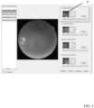
- FIGS. 2 - 5show screen shots of the graphic user interface (GUI), according to certain embodiments.
- FIG. 2shows a screen shot of the GUI for the user input module and shows that the user has input four retinal images 24 for analysis.
- a system progress display 26shows the user the progress of the system in analyzing image conformity.
- FIG. 3shows the GUI of the image output module indicating that one of the images 28 has been flagged as non-conforming
- FIG. 4shows a screenshot of the GUI of the image resubmission module.
- the resubmission moduleindicates to the user that the fovea centered left eye image is non-conforming 30 .
- FIG. 5shows the user input of a new conforming image 32 into the resubmission module.
Landscapes
- Engineering & Computer Science (AREA)
- Health & Medical Sciences (AREA)
- Medical Informatics (AREA)
- Public Health (AREA)
- General Health & Medical Sciences (AREA)
- Epidemiology (AREA)
- Primary Health Care (AREA)
- Radiology & Medical Imaging (AREA)
- Nuclear Medicine, Radiotherapy & Molecular Imaging (AREA)
- General Physics & Mathematics (AREA)
- Computer Vision & Pattern Recognition (AREA)
- Physics & Mathematics (AREA)
- Quality & Reliability (AREA)
- Theoretical Computer Science (AREA)
- Biomedical Technology (AREA)
- Data Mining & Analysis (AREA)
- Databases & Information Systems (AREA)
- Pathology (AREA)
- Eye Examination Apparatus (AREA)
Abstract
Description
Claims (20)
Priority Applications (2)
| Application Number | Priority Date | Filing Date | Title |
|---|---|---|---|
| US17/558,016US12243206B2 (en) | 2016-03-22 | 2021-12-21 | System and methods for qualifying medical images |
| US19/040,642US20250173851A1 (en) | 2016-03-22 | 2025-01-29 | System and Methods for Qualifying Medical Images |
Applications Claiming Priority (3)
| Application Number | Priority Date | Filing Date | Title |
|---|---|---|---|
| US201662311660P | 2016-03-22 | 2016-03-22 | |
| US15/466,636US11232548B2 (en) | 2016-03-22 | 2017-03-22 | System and methods for qualifying medical images |
| US17/558,016US12243206B2 (en) | 2016-03-22 | 2021-12-21 | System and methods for qualifying medical images |
Related Parent Applications (1)
| Application Number | Title | Priority Date | Filing Date |
|---|---|---|---|
| US15/466,636ContinuationUS11232548B2 (en) | 2016-03-22 | 2017-03-22 | System and methods for qualifying medical images |
Related Child Applications (1)
| Application Number | Title | Priority Date | Filing Date |
|---|---|---|---|
| US19/040,642ContinuationUS20250173851A1 (en) | 2016-03-22 | 2025-01-29 | System and Methods for Qualifying Medical Images |
Publications (2)
| Publication Number | Publication Date |
|---|---|
| US20220114719A1 US20220114719A1 (en) | 2022-04-14 |
| US12243206B2true US12243206B2 (en) | 2025-03-04 |
Family
ID=59898985
Family Applications (3)
| Application Number | Title | Priority Date | Filing Date |
|---|---|---|---|
| US15/466,636ActiveUS11232548B2 (en) | 2016-03-22 | 2017-03-22 | System and methods for qualifying medical images |
| US17/558,016Active2038-06-03US12243206B2 (en) | 2016-03-22 | 2021-12-21 | System and methods for qualifying medical images |
| US19/040,642PendingUS20250173851A1 (en) | 2016-03-22 | 2025-01-29 | System and Methods for Qualifying Medical Images |
Family Applications Before (1)
| Application Number | Title | Priority Date | Filing Date |
|---|---|---|---|
| US15/466,636ActiveUS11232548B2 (en) | 2016-03-22 | 2017-03-22 | System and methods for qualifying medical images |
Family Applications After (1)
| Application Number | Title | Priority Date | Filing Date |
|---|---|---|---|
| US19/040,642PendingUS20250173851A1 (en) | 2016-03-22 | 2025-01-29 | System and Methods for Qualifying Medical Images |
Country Status (1)
| Country | Link |
|---|---|
| US (3) | US11232548B2 (en) |
Families Citing this family (6)
| Publication number | Priority date | Publication date | Assignee | Title |
|---|---|---|---|---|
| WO2019166447A1 (en)* | 2018-03-02 | 2019-09-06 | Koninklijke Philips N.V. | Image registration qualification |
| CN108898586A (en)* | 2018-06-19 | 2018-11-27 | 歌尔股份有限公司 | Product test method and system, client terminal device, test of heuristics device |
| US12171496B2 (en)* | 2020-08-19 | 2024-12-24 | Digital Diagnostics Inc. | Using infrared to detect proper eye alignment before capturing retinal images |
| USD975118S1 (en)* | 2021-01-01 | 2023-01-10 | Lensar, Inc. | Display screen with graphical user interface for a laser-phacoemulsification system |
| EP4084010A1 (en)* | 2021-04-30 | 2022-11-02 | Siemens Healthcare GmbH | Method for operating an evaluation system for medical image data sets, evaluation system, computer program and electronically readable storage medium |
| WO2024201381A1 (en)* | 2023-03-31 | 2024-10-03 | Optina Diagnostics, Inc. | Method and system for processing retinal images |
Citations (21)
| Publication number | Priority date | Publication date | Assignee | Title |
|---|---|---|---|---|
| US20040221855A1 (en)* | 2002-10-17 | 2004-11-11 | Paul Ashton | Methods for monitoring treatment of disease |
| US20050157848A1 (en)* | 2003-12-22 | 2005-07-21 | Kabushiki Kaisha Toshiba | Image-quality control system |
| US20060119622A1 (en)* | 2004-11-23 | 2006-06-08 | General Electric Company | Method and apparatus for volume rendering display protocol |
| US20060238546A1 (en)* | 2005-03-08 | 2006-10-26 | Carrie Handley | Comparative image review system and method |
| US20070237308A1 (en)* | 2006-01-30 | 2007-10-11 | Bruce Reiner | Method and apparatus for generating a technologist quality assurance scorecard |
| US20080044069A1 (en)* | 2006-08-15 | 2008-02-21 | Dugal Tiffany Ann | Processes and apparatus for imaging protocols and analysis |
| WO2008050222A2 (en)* | 2006-10-25 | 2008-05-02 | Rcadia Medical Imaging Ltd. | Method and system for automatic quality control used in computerized analysis of ct angiography |
| US20100021027A1 (en)* | 2008-07-25 | 2010-01-28 | Thomas Hartkens | Image data management systems |
| US20100303363A1 (en)* | 2004-12-09 | 2010-12-02 | Fedorovskaya Elena A | Method for automatically determining the acceptability of a digital image |
| US20110054266A1 (en)* | 2007-12-17 | 2011-03-03 | Koninklijke Philips Electronics N.V. | Method for protocol creation in a diagnostic imaging system |
| US20110188718A1 (en)* | 2008-07-25 | 2011-08-04 | Derek Lionel Glendon Hill | Image data management systems |
| US20110242306A1 (en)* | 2008-12-19 | 2011-10-06 | The Johns Hopkins University | System and method for automated detection of age related macular degeneration and other retinal abnormalities |
| US20130182220A1 (en)* | 2012-01-16 | 2013-07-18 | Canon Kabushiki Kaisha | Image forming method and image forming apparatus |
| US20130190600A1 (en)* | 2012-01-25 | 2013-07-25 | General Electric Company | System and Method for Identifying an Optimal Image Frame for Ultrasound Imaging |
| US20130322711A1 (en)* | 2012-06-04 | 2013-12-05 | Verizon Patent And Licesing Inc. | Mobile dermatology collection and analysis system |
| US20140029828A1 (en)* | 2011-03-15 | 2014-01-30 | Method and System for Quality Assurance of Cross Sectional Imaging Scans | Method and Systems for Quality Assurance of Cross Sectional Imaging Scans |
| WO2014140258A2 (en)* | 2013-03-14 | 2014-09-18 | Carl Zeiss Meditec Ag | Multimodal integration of ocular data acquisition and analysis |
| US20150029464A1 (en)* | 2013-07-26 | 2015-01-29 | The Regents Of The University Of Michigan | Automated Measurement of Changes in Retinal, Retinal Pigment Epithelial, or Choroidal Disease |
| US20150104087A1 (en)* | 2013-10-10 | 2015-04-16 | University Of Rochester | Automated Fundus Image Field Detection and Quality Assessment |
| US20160155229A1 (en)* | 2014-11-28 | 2016-06-02 | Kabushiki Kaisha Toshiba | Magnetic resonance imaging apparatus and medical image processing method |
| US10013639B1 (en)* | 2013-12-16 | 2018-07-03 | Amazon Technologies, Inc. | Analyzing digital images based on criteria |
- 2017
- 2017-03-22USUS15/466,636patent/US11232548B2/enactiveActive
- 2021
- 2021-12-21USUS17/558,016patent/US12243206B2/enactiveActive
- 2025
- 2025-01-29USUS19/040,642patent/US20250173851A1/enactivePending
Patent Citations (22)
| Publication number | Priority date | Publication date | Assignee | Title |
|---|---|---|---|---|
| US20040221855A1 (en)* | 2002-10-17 | 2004-11-11 | Paul Ashton | Methods for monitoring treatment of disease |
| US20050157848A1 (en)* | 2003-12-22 | 2005-07-21 | Kabushiki Kaisha Toshiba | Image-quality control system |
| US20060119622A1 (en)* | 2004-11-23 | 2006-06-08 | General Electric Company | Method and apparatus for volume rendering display protocol |
| US20100303363A1 (en)* | 2004-12-09 | 2010-12-02 | Fedorovskaya Elena A | Method for automatically determining the acceptability of a digital image |
| US20060238546A1 (en)* | 2005-03-08 | 2006-10-26 | Carrie Handley | Comparative image review system and method |
| US20070237308A1 (en)* | 2006-01-30 | 2007-10-11 | Bruce Reiner | Method and apparatus for generating a technologist quality assurance scorecard |
| US7532942B2 (en) | 2006-01-30 | 2009-05-12 | Bruce Reiner | Method and apparatus for generating a technologist quality assurance scorecard |
| US20080044069A1 (en)* | 2006-08-15 | 2008-02-21 | Dugal Tiffany Ann | Processes and apparatus for imaging protocols and analysis |
| WO2008050222A2 (en)* | 2006-10-25 | 2008-05-02 | Rcadia Medical Imaging Ltd. | Method and system for automatic quality control used in computerized analysis of ct angiography |
| US20110054266A1 (en)* | 2007-12-17 | 2011-03-03 | Koninklijke Philips Electronics N.V. | Method for protocol creation in a diagnostic imaging system |
| US20100021027A1 (en)* | 2008-07-25 | 2010-01-28 | Thomas Hartkens | Image data management systems |
| US20110188718A1 (en)* | 2008-07-25 | 2011-08-04 | Derek Lionel Glendon Hill | Image data management systems |
| US20110242306A1 (en)* | 2008-12-19 | 2011-10-06 | The Johns Hopkins University | System and method for automated detection of age related macular degeneration and other retinal abnormalities |
| US20140029828A1 (en)* | 2011-03-15 | 2014-01-30 | Method and System for Quality Assurance of Cross Sectional Imaging Scans | Method and Systems for Quality Assurance of Cross Sectional Imaging Scans |
| US20130182220A1 (en)* | 2012-01-16 | 2013-07-18 | Canon Kabushiki Kaisha | Image forming method and image forming apparatus |
| US20130190600A1 (en)* | 2012-01-25 | 2013-07-25 | General Electric Company | System and Method for Identifying an Optimal Image Frame for Ultrasound Imaging |
| US20130322711A1 (en)* | 2012-06-04 | 2013-12-05 | Verizon Patent And Licesing Inc. | Mobile dermatology collection and analysis system |
| WO2014140258A2 (en)* | 2013-03-14 | 2014-09-18 | Carl Zeiss Meditec Ag | Multimodal integration of ocular data acquisition and analysis |
| US20150029464A1 (en)* | 2013-07-26 | 2015-01-29 | The Regents Of The University Of Michigan | Automated Measurement of Changes in Retinal, Retinal Pigment Epithelial, or Choroidal Disease |
| US20150104087A1 (en)* | 2013-10-10 | 2015-04-16 | University Of Rochester | Automated Fundus Image Field Detection and Quality Assessment |
| US10013639B1 (en)* | 2013-12-16 | 2018-07-03 | Amazon Technologies, Inc. | Analyzing digital images based on criteria |
| US20160155229A1 (en)* | 2014-11-28 | 2016-06-02 | Kabushiki Kaisha Toshiba | Magnetic resonance imaging apparatus and medical image processing method |
Non-Patent Citations (8)
| Title |
|---|
| Fairbanks, A. et al. "Ocular Ultrasound: A Quick Reference Guide for the On-Call Physician." EyeRounds.org, Opthalmology and Visual Sciences, Feb. 4, 2016, 5 pages, [Online] [Retrieved Nov. 23, 2020], Retrieved from the Internet <URL:http://www.eyerounds.org/tutorials/ultrasound/>. |
| United States Office Action, U.S. Appl. No. 15/466,636, Apr. 30, 2020, 11 pages. |
| United States Office Action, U.S. Appl. No. 15/466,636, Dec. 10, 2018, 15 pages. |
| United States Office Action, U.S. Appl. No. 15/466,636, Feb. 7, 2020, 10 pages. |
| United States Office Action, U.S. Appl. No. 15/466,636, Jul. 9, 2021, 12 pages. |
| United States Office Action, U.S. Appl. No. 15/466,636, Mar. 2, 2018, 12 pages. |
| United States Office Action, U.S. Appl. No. 15/466,636, Nov. 23, 2020, 10 pages. |
| United States Office Action, U.S. Appl. No. 15/466,636, Sep. 10, 2019, 10 pages. |
Also Published As
| Publication number | Publication date |
|---|---|
| US20170278241A1 (en) | 2017-09-28 |
| US20250173851A1 (en) | 2025-05-29 |
| US11232548B2 (en) | 2022-01-25 |
| US20220114719A1 (en) | 2022-04-14 |
Similar Documents
| Publication | Publication Date | Title |
|---|---|---|
| US12243206B2 (en) | System and methods for qualifying medical images | |
| US11017695B2 (en) | Method for developing a machine learning model of a neural network for classifying medical images | |
| US10339655B2 (en) | Automated image evaluation in x-ray imaging | |
| KR20210140763A (en) | Gaze direction determination method, apparatus, electronic device and storage medium | |
| JP2019528113A (en) | Fundus image processing using machine learning models | |
| EP3564961A1 (en) | Interactive coronary labeling using interventional x-ray images and deep learning | |
| US11836946B2 (en) | Methods and devices for guiding a patient | |
| WO2020202680A1 (en) | Information processing device and information processing method | |
| JP7406901B2 (en) | Information processing device and information processing method | |
| JPWO2019207800A1 (en) | Ophthalmic image processing equipment and ophthalmic image processing program | |
| CN111563910A (en) | Fundus image segmentation method and device | |
| AU2025202817A1 (en) | Image retention and stitching for minimal-flash eye disease diagnosis | |
| WO2019171398A1 (en) | A fundus image analysis system | |
| JP7709047B2 (en) | Medical information processing program and medical information processing device | |
| US20240020839A1 (en) | Medical image processing device, medical image processing program, and medical image processing method | |
| JP2020030695A (en) | Teacher data generator and machine learning system | |
| CN115916030A (en) | Using infrared to detect correct eye alignment before capturing retinal images | |
| JP2021097728A (en) | Information processing device, information processing method and program | |
| Ovens et al. | Automated Grading of Refractive Error from Fundus Images using Deep Learning | |
| JP2016047076A (en) | Image processor, fundus photographing system, image processing method, and program | |
| Muchibwa et al. | Early Cataract Detection with Computer Vision for Accessible Screening in Underserved Communities |
Legal Events
| Date | Code | Title | Description |
|---|---|---|---|
| FEPP | Fee payment procedure | Free format text:ENTITY STATUS SET TO UNDISCOUNTED (ORIGINAL EVENT CODE: BIG.); ENTITY STATUS OF PATENT OWNER: SMALL ENTITY | |
| FEPP | Fee payment procedure | Free format text:ENTITY STATUS SET TO SMALL (ORIGINAL EVENT CODE: SMAL); ENTITY STATUS OF PATENT OWNER: SMALL ENTITY | |
| STPP | Information on status: patent application and granting procedure in general | Free format text:DOCKETED NEW CASE - READY FOR EXAMINATION | |
| AS | Assignment | Owner name:DIGITAL DIAGNOSTICS INC., IOWA Free format text:CHANGE OF NAME;ASSIGNOR:IDX TECHNOLOGIES INC.;REEL/FRAME:063027/0164 Effective date:20200529 Owner name:IDX TECHNOLOGIES INC., IOWA Free format text:CHANGE OF NAME;ASSIGNOR:IDX, LLC;REEL/FRAME:062663/0620 Effective date:20180912 Owner name:IDX, LLC, IOWA Free format text:ASSIGNMENT OF ASSIGNORS INTEREST;ASSIGNORS:ABRAMOFF, MICHAEL D.;CLARK, BEN;TALMAGE, ERIC;AND OTHERS;SIGNING DATES FROM 20170405 TO 20170410;REEL/FRAME:062663/0584 | |
| STPP | Information on status: patent application and granting procedure in general | Free format text:NON FINAL ACTION MAILED | |
| STPP | Information on status: patent application and granting procedure in general | Free format text:RESPONSE TO NON-FINAL OFFICE ACTION ENTERED AND FORWARDED TO EXAMINER | |
| STPP | Information on status: patent application and granting procedure in general | Free format text:FINAL REJECTION MAILED | |
| STPP | Information on status: patent application and granting procedure in general | Free format text:DOCKETED NEW CASE - READY FOR EXAMINATION | |
| STPP | Information on status: patent application and granting procedure in general | Free format text:NOTICE OF ALLOWANCE MAILED -- APPLICATION RECEIVED IN OFFICE OF PUBLICATIONS | |
| STPP | Information on status: patent application and granting procedure in general | Free format text:PUBLICATIONS -- ISSUE FEE PAYMENT VERIFIED | |
| STCF | Information on status: patent grant | Free format text:PATENTED CASE |