US12214189B2 - Fourier analysis spectroscopy for monitoring tissue impedance changes and treatment outcome during electroporation-based-therapies - Google Patents
Fourier analysis spectroscopy for monitoring tissue impedance changes and treatment outcome during electroporation-based-therapiesDownload PDFInfo
- Publication number
- US12214189B2 US12214189B2US16/938,778US202016938778AUS12214189B2US 12214189 B2US12214189 B2US 12214189B2US 202016938778 AUS202016938778 AUS 202016938778AUS 12214189 B2US12214189 B2US 12214189B2
- Authority
- US
- United States
- Prior art keywords
- impedance
- fast
- tissue
- voltage
- burst
- Prior art date
- Legal status (The legal status is an assumption and is not a legal conclusion. Google has not performed a legal analysis and makes no representation as to the accuracy of the status listed.)
- Active, expires
Links
Images


Classifications
- A—HUMAN NECESSITIES
- A61—MEDICAL OR VETERINARY SCIENCE; HYGIENE
- A61N—ELECTROTHERAPY; MAGNETOTHERAPY; RADIATION THERAPY; ULTRASOUND THERAPY
- A61N1/00—Electrotherapy; Circuits therefor
- A61N1/02—Details
- A61N1/08—Arrangements or circuits for monitoring, protecting, controlling or indicating
- A—HUMAN NECESSITIES
- A61—MEDICAL OR VETERINARY SCIENCE; HYGIENE
- A61B—DIAGNOSIS; SURGERY; IDENTIFICATION
- A61B18/00—Surgical instruments, devices or methods for transferring non-mechanical forms of energy to or from the body
- A61B18/04—Surgical instruments, devices or methods for transferring non-mechanical forms of energy to or from the body by heating
- A61B18/12—Surgical instruments, devices or methods for transferring non-mechanical forms of energy to or from the body by heating by passing a current through the tissue to be heated, e.g. high-frequency current
- A61B18/1206—Generators therefor
- A—HUMAN NECESSITIES
- A61—MEDICAL OR VETERINARY SCIENCE; HYGIENE
- A61N—ELECTROTHERAPY; MAGNETOTHERAPY; RADIATION THERAPY; ULTRASOUND THERAPY
- A61N1/00—Electrotherapy; Circuits therefor
- A61N1/18—Applying electric currents by contact electrodes
- A61N1/32—Applying electric currents by contact electrodes alternating or intermittent currents
- A61N1/327—Applying electric currents by contact electrodes alternating or intermittent currents for enhancing the absorption properties of tissue, e.g. by electroporation
- A—HUMAN NECESSITIES
- A61—MEDICAL OR VETERINARY SCIENCE; HYGIENE
- A61B—DIAGNOSIS; SURGERY; IDENTIFICATION
- A61B17/00—Surgical instruments, devices or methods
- A61B2017/00017—Electrical control of surgical instruments
- A61B2017/00022—Sensing or detecting at the treatment site
- A61B2017/00039—Electric or electromagnetic phenomena other than conductivity, e.g. capacity, inductivity, Hall effect
- A—HUMAN NECESSITIES
- A61—MEDICAL OR VETERINARY SCIENCE; HYGIENE
- A61B—DIAGNOSIS; SURGERY; IDENTIFICATION
- A61B17/00—Surgical instruments, devices or methods
- A61B2017/00017—Electrical control of surgical instruments
- A61B2017/00137—Details of operation mode
- A61B2017/00154—Details of operation mode pulsed
- A61B2017/00181—Means for setting or varying the pulse energy
- A61B2017/0019—Means for setting or varying the pulse width
- A—HUMAN NECESSITIES
- A61—MEDICAL OR VETERINARY SCIENCE; HYGIENE
- A61B—DIAGNOSIS; SURGERY; IDENTIFICATION
- A61B18/00—Surgical instruments, devices or methods for transferring non-mechanical forms of energy to or from the body
- A61B2018/00571—Surgical instruments, devices or methods for transferring non-mechanical forms of energy to or from the body for achieving a particular surgical effect
- A61B2018/00613—Irreversible electroporation
- A—HUMAN NECESSITIES
- A61—MEDICAL OR VETERINARY SCIENCE; HYGIENE
- A61B—DIAGNOSIS; SURGERY; IDENTIFICATION
- A61B18/00—Surgical instruments, devices or methods for transferring non-mechanical forms of energy to or from the body
- A61B2018/00636—Sensing and controlling the application of energy
- A61B2018/00642—Sensing and controlling the application of energy with feedback, i.e. closed loop control
- A—HUMAN NECESSITIES
- A61—MEDICAL OR VETERINARY SCIENCE; HYGIENE
- A61B—DIAGNOSIS; SURGERY; IDENTIFICATION
- A61B18/00—Surgical instruments, devices or methods for transferring non-mechanical forms of energy to or from the body
- A61B2018/00636—Sensing and controlling the application of energy
- A61B2018/00696—Controlled or regulated parameters
- A61B2018/00702—Power or energy
- A61B2018/00708—Power or energy switching the power on or off
- A—HUMAN NECESSITIES
- A61—MEDICAL OR VETERINARY SCIENCE; HYGIENE
- A61B—DIAGNOSIS; SURGERY; IDENTIFICATION
- A61B18/00—Surgical instruments, devices or methods for transferring non-mechanical forms of energy to or from the body
- A61B2018/00636—Sensing and controlling the application of energy
- A61B2018/00696—Controlled or regulated parameters
- A61B2018/00761—Duration
- A—HUMAN NECESSITIES
- A61—MEDICAL OR VETERINARY SCIENCE; HYGIENE
- A61B—DIAGNOSIS; SURGERY; IDENTIFICATION
- A61B18/00—Surgical instruments, devices or methods for transferring non-mechanical forms of energy to or from the body
- A61B2018/00636—Sensing and controlling the application of energy
- A61B2018/00773—Sensed parameters
- A61B2018/00791—Temperature
- A—HUMAN NECESSITIES
- A61—MEDICAL OR VETERINARY SCIENCE; HYGIENE
- A61B—DIAGNOSIS; SURGERY; IDENTIFICATION
- A61B18/00—Surgical instruments, devices or methods for transferring non-mechanical forms of energy to or from the body
- A61B2018/00636—Sensing and controlling the application of energy
- A61B2018/00773—Sensed parameters
- A61B2018/00875—Resistance or impedance
- A—HUMAN NECESSITIES
- A61—MEDICAL OR VETERINARY SCIENCE; HYGIENE
- A61B—DIAGNOSIS; SURGERY; IDENTIFICATION
- A61B18/00—Surgical instruments, devices or methods for transferring non-mechanical forms of energy to or from the body
- A61B18/04—Surgical instruments, devices or methods for transferring non-mechanical forms of energy to or from the body by heating
- A61B18/12—Surgical instruments, devices or methods for transferring non-mechanical forms of energy to or from the body by heating by passing a current through the tissue to be heated, e.g. high-frequency current
- A61B18/1206—Generators therefor
- A61B2018/124—Generators therefor switching the output to different electrodes, e.g. sequentially
- A—HUMAN NECESSITIES
- A61—MEDICAL OR VETERINARY SCIENCE; HYGIENE
- A61B—DIAGNOSIS; SURGERY; IDENTIFICATION
- A61B18/00—Surgical instruments, devices or methods for transferring non-mechanical forms of energy to or from the body
- A61B18/04—Surgical instruments, devices or methods for transferring non-mechanical forms of energy to or from the body by heating
- A61B18/12—Surgical instruments, devices or methods for transferring non-mechanical forms of energy to or from the body by heating by passing a current through the tissue to be heated, e.g. high-frequency current
- A61B18/14—Probes or electrodes therefor
- A61B2018/1405—Electrodes having a specific shape
- A61B2018/1425—Needle
- A61B2018/143—Needle multiple needles
- A—HUMAN NECESSITIES
- A61—MEDICAL OR VETERINARY SCIENCE; HYGIENE
- A61N—ELECTROTHERAPY; MAGNETOTHERAPY; RADIATION THERAPY; ULTRASOUND THERAPY
- A61N1/00—Electrotherapy; Circuits therefor
- A61N1/02—Details
- A61N1/08—Arrangements or circuits for monitoring, protecting, controlling or indicating
- A61N2001/083—Monitoring integrity of contacts, e.g. by impedance measurement
Definitions
- Treatment options for nonresectable tumorsare restricted due to multiple clinical factors, most notably the presence of tumors near critical structures (large blood vessels and nerve bundles), extensive tumor volumes, and/or metastases.
- Alternate therapieslike microwave ablation, radiofrequency ablation, and cryoablation are also limited by tumor location, and thermal therapies are indiscriminate and can easily damage these critical structures.
- electroporation-based therapiesEBTs are appealing and have proven advantageous for treating or ablating nonresectable tumors due to their nonthermal mechanisms of cell death, which spare proteinaceous structures including nerves and vasculature.
- EBTsare applied using one or more array of needle electrodes placed in and around the target tissue site.
- High amplitude (up to 3000 V), short duration (50-100 ⁇ s) pulsed electric fields (PEFs)are applied across the electrodes, exposing target tissues to high local field strengths. This results in an increased voltage drop across the cell membrane (transmembrane potential) and gives rise to the creation of defects on the cell membrane. Depending on the pulsing protocol, these defects are either transient or can lead to irrevocable damage.
- electrochemotherapywhich utilizes reversible electroporation (RE) for enhanced drug delivery
- ECTelectrochemotherapy
- REreversible electroporation
- IREirreversible electroporation
- IREcan be utilized as a combinatorial therapy to ablate a central tumor core with peritumoral RE for enhanced delivery of adjuvant molecular agents.
- combinatorial treatment of locally advanced pancreatic cancer with standard-of-care and IREdemonstrated nearly double median overall survival compared to standard-of-care alone.
- H-FIREHigh Frequency IRE
- GBMglioblastoma
- H-FIREforms a predominantly nonthermal ablation within heterogenous tumors such as GBM, regardless of chemotherapeutic resistance.
- prior in vitro studiesdemonstrate lower therapeutic electric fields are required to induce cell death in malignant brain cell lines compared to healthy brain cell lines, suggesting preferential targeting of malignant phenotypes.
- Sub-H-FIRE electric fieldsare known to cause a reversible electroporation (RE) effect where cells are temporarily permeabilized to larger, normally impermeable chemotherapeutics.
- REreversible electroporation
- EBTsface a unique challenge: intraoperative monitoring of treatment progression towards the determination of a clinical endpoint.
- Current approaches to determine extent of ablationrely either on postoperative procedures, including imaging with MRI and ultrasound techniques or intraoperative electrical characterization techniques based on a pre-defined change in tissue resistance/current.
- the inventorsprovide a method which uses a high-frequency reference, which is not susceptible to electroporation effects ( FIGS. 11 F-G ), to decouple the low-frequency impedance measurements. This allows for thermal impedance changes to be removed and then use the electroporation effects to determine a clinical endpoint.
- thermal ablation modalitiesutilize intraoperative temperature measurements coupled with Arrhenius damage models to predict the treatment volume in real-time. (B. D. de Senneville, C. Mougenot, B. Quesson, I. Dragonu, N. Grenier, and C. T. Moonen, “MR thermometry for monitoring tumor ablation,” European radiology, vol. 17, no. 9, pp. 2401-2410, 2007.)
- proposed methods for EBTsrely on detection of electrical impedance changes as an indicator to EP. The rationale behind using impedance measurements is as follows.
- biological tissuecan be represented as a 3-domain system, where a high impedance cell membrane separates the intracellular and extracellular spaces.
- tissue impedance with bursts of bipolar pulsescan be characterized into two categories: 1) intra-burst (in-pulse) impedance measurements, the impedance analysis of the EBT waveform, and 2) inter-burst impedance measurements, the impedance analysis at discrete timepoints between therapeutic PEFs.
- An application for intra-burst impedancehas focused on quantifying changes in tissue conductivity during the EBT pulses (N. Beitel-White, S. Bhonsle, R. Martin, and R. V. Davalos, “Electrical characterization of human biological tissue for irreversible electroporation treatments,” in 2018 40th Annual International Conference of the IEEE Engineering in Medicine and Biology Society (EMBC). IEEE, 2018, pp.
- H-FIREaddresses intricacies associated with current IRE technology (i.e., H-FIRE mitigates muscle excitation, field distortions in heterogenous tissues, and potential cardiac arrythmias)
- challenges in determining a clinical endpoint with all EBTsstill exist.
- Proposed solutions towards determining a clinical endpoint of EBTsinclude monitoring changes in bulk tissue impedance/resistance and changes in tissue conductivity distribution. This includes:
- the present inventorsintroduce Fourier Analysis SpecTroscopy (FAST) as an impedance spectroscopy methodology for monitoring tissue impedance changes during EBTs in real-time.
- FASTFourier Analysis SpecTroscopy
- diagnostic FASTaimed towards monitoring inter-burst impedance
- therapeutic FASTaimed towards inducing tissue EP while simultaneously monitoring high voltage intra-burst tissue impedance
- FIG. 1 Ddiagnostic FAST is achieved by delivering a low-voltage, wideband signal composed of rectangular bipolar pulses; voltage and current capture followed by discrete Fourier transform for analysis in the frequency domain allows for real-time EIS with wide frequency bands.
- Therapeutic FASTis achieved by delivering a high-voltage burst of bipolar PEFs; therapeutic FAST is an adaptation of a traditional high-frequency IRE burst, modified to maximize the frequency bandwidth and resolution to enable intra-burst impedance spectroscopy. Impedance analysis with therapeutic FAST is distinguished from diagnostic FAST as diagnostic FAST measures impedance following significant pore resealing and membrane recovery, while investigations from therapeutic FAST theoretically incorporate impedance change during pore formation and pore expansion.
- Garc ⁇ a-Sánchez et al.proposed utilization of a multi-sine burst for impedance analysis between 5 kHz to 1.313 MHz at 21 discrete frequencies.
- Preference to using rectangular waveforms, over sinusoidal excitation signalsB. Sanchez, G. Vandersteen, R. Bragos, and J.
- FASTintroduces a novel application of high-frequency tissue impedance measurements as an indicator of the extent of Joule heating effects during EP. Although low-frequency impedance measurements are sensitive to both EP effects and Joule heating, the inventors have found that high frequency currents, which short the membrane reactance, are less sensitive to EP effects and can uniquely act to distinguish thermal effects ( FIGS. 1 A-D ).
- the inventors' initial findingsare extended to develop diagnostic FAST for monitoring inter-burst electrical impedance spectra directed towards monitoring treatment outcome with EBTs and therapeutic FAST for monitoring intra-burst impedance directed towards characterizing biological tissue response during EP.
- Their findingssuggest acquisition of an impedance spectrum ( ⁇ 0.1 kHz-100 MHz), such as a high-bandwidth inter-burst impedance spectrum (between 1.8 kHz-4.93 MHz) and intra-burst impedance spectrum (between 18.3 kHz-1.96 MHz) is possible at >100 discrete frequencies per spectrum and at a capture rate ⁇ 1 s.
- FAST technologiescan be used to monitor EBT treatment outcomes (e.g., when an expected endpoint of treatment has been reached and/or the success of a treatment, such as whether the desired ablation effect has been achieved), EBT treatment progress, and/or detect or warn of thermal effects.
- FAST technologiesare especially suited to provide for such monitoring in real-time during an EBT treatment, which can be very helpful in knowing when to alter treatment parameters, or when to halt treatment temporarily (such as to avoid excess temperatures), or when to stop treatment completely.
- Embodiments of the inventioninclude Aspect 1, which is a method for monitoring administration of electrical pulses comprising: obtaining a baseline impedance spectrum; administering a plurality of electrical pulses; obtaining one or more additional impedance spectrum; identifying any impedance spectrum change relative to the baseline; and monitoring the administering to determine if a desired endpoint is reached as indicated by the impedance spectrum change, and i) adjusting one or more parameters of, ii) stopping, iii) halting, and/or iv) continuing the administering based on the monitoring.
- Aspect 2is the embodiment of Aspect 1, wherein the endpoint for irreversible electroporation is a point during the administering where electroporation no longer contributes to any impedance spectrum change as evidenced by the obtaining of a flat impedance spectrum.
- Aspect 3is the method of Aspect 1 or 2, further comprising halting the administering to allow tissue temperature to reach a desired level, then resuming the administering to the desired endpoint.
- Aspect 4is the method of any of Aspects 1-3, comprising: delivering a low-voltage, wideband signal of electrical pulses; and monitoring treatment outcome through monitoring inter-burst impedance by capturing voltage and current and performing discrete Fourier transform analysis.
- Aspect 5is the method of any of Aspects 1-4, wherein impedance is captured within a frequency range of above 0.1 kHz to 100 MHz, such as between 1.8 kHz and 4.93 MHz.
- Aspect 6is the method of any of Aspects 1-5, wherein delivering of the electrical pulses comprises applying one or more low-voltage pulses interleaved between one or more high-voltage pulses.
- Aspect 7is the method of any of Aspects 1-6, wherein delivering of the electrical pulses comprises applying pulses in the range of 0.1 ⁇ s to 10 ms, such as at a high frequency signal of 1-50-1-50 ⁇ s, appended to a low frequency signal of 250-10-250-10 ⁇ s.
- Aspect 8is the method of any of Aspects 1-7, further comprising: delivering one or more high-voltage burst of pulsed electric fields; and monitoring tissue response through monitoring high-voltage intra-burst impedance by capturing voltage and current and performing discrete Fourier transform analysis.
- Aspect 9is the method of any of Aspects 1-8, wherein impedance is captured within a frequency range of above 0.1 kHz to 100 MHz, such as between 18.3 kHz and 1.96 MHz.
- Aspect 10is the method of any of Aspects 1-9, wherein the delivering comprises a high-frequency irreversible electroporation burst scheme of pulse width and intra-phase delay ranging from 0.1 ⁇ s to 10 ms, and inter-pulse delay ranging from 0.1 ⁇ s to 1 s, such as delivering high-frequency irreversible electroporation using a 2-5-2 ⁇ s scheme, followed by a 100 ⁇ s delay.
- Aspect 11is the method of any of Aspects 1-10, wherein the baseline impedance spectrum and/or the additional impedance spectrum is obtained by one or more of: reference to an impedance spectrum based on standard impedance values for a particular material or tissue; measuring impedance of a material or tissue over a selected frequency band; measuring voltage and/or current and calculating impedance therefrom; and/or calculating impedance as a function of frequency using the formula:
- Aspect 12is the method of any of Aspects 1-11, further comprising using the impedance spectrum change measured at high frequencies to predict a temperature change, such as in tissue, relating to the administering of the electrical pulses.
- Aspect 13is the method of any of Aspects 1-12, wherein the impedance spectrum change indicates one or more of: whether irreversible or reversible electroporation of a tissue has, is or will occur; whether chemical cell death and/or decellularization has, is or will occur; whether death and/or decellularization has, is or will occur due to a physical disruption; whether a tissue is healthy or cancerous; whether a tissue has damage from a stroke and/or traumatic brain injury; whether cell lysis has, is or will occur as evidenced by flattening of the impedance spectrum with no recovery following pulse cessation; whether cell necrosis has, is or will occur as evidenced by flattening of the impedance spectrum with minimal recovery following pulse cessation; and/or whether cell apoptosis has, is or will occur as is evidenced by flattening of the impedance spectrum with moderate recovery following pulse cessation.
- Aspect 14is the method of any of Aspects 1-13, wherein the change in the impedance indicates: whether degradation of a coating has, is or will occur; and/or whether a coating of a material has corrosion and/or a level of the corrosion.
- Aspect 15is the method of any of Aspects 1-14, wherein the monitoring comprises: monitoring tissue decellularization and/or cell death; monitoring gene-transfection efficiency and uptake; monitoring thermal and/or non-thermal tissue ablation for cardiac arrythmias; and/or monitoring cell lysis for immunotherapies.
- Aspect 16is a treatment monitoring system for administering electrical pulses comprising: one or more electrical pulse generator(s); one or more probe(s) capable of connection with the electrical pulse generator(s); one or more controller(s) capable of controlling one or more of the electrical pulse generator(s) and/or one or more of the probe(s) to: administer a plurality of electrical pulses; obtain a baseline impedance spectrum; obtain one or more additional impedance spectrum; and identify any impedance spectrum change relative to the baseline.
- Aspect 17is the treatment monitoring system of any of Aspect 16, further comprising a processing module with a processor in combination with memory and computer-executable instructions configured to process the impedance spectra using a Fourier Transform algorithm.
- Aspect 18is the treatment monitoring system of Aspect 16 or 17, wherein: one or more of the pulse generator(s) is capable of delivering high-voltage pulses; and one or more of the pulse generator(s) is capable of delivering low-voltage pulses.
- Aspect 19is a treatment monitoring system (such as that of any of Aspects 16-18), wherein: one or more controller(s) is a microcontroller capable of connection with: a first 5V H-Bridge circuit for connection with a high-voltage pulse generator; a 15V H-Bridge circuit for connection with a low-voltage pulse generator; and a second 5V H-Bridge circuit for connection with two Reed relays on a high-voltage circuit (HVRR) and two Reed relays on a low-voltage circuit (LVRR); wherein the microcontroller is capable of: triggering the Reed relays on the low-voltage circuit to close; triggering the low-voltage generator to deliver pulses; ceasing the LVRR trigger signal to open the LV and HV circuits; triggering the HVRR Reed relays on the high-voltage circuit to close; triggering the high-voltage generator to deliver pulses.
- HVRRhigh-voltage circuit
- LVRRlow-voltage circuit
- Aspect 20is the treatment monitoring system of any of Aspects 16-19, wherein one or more of the controller(s) is capable of i) adjusting one or more parameters of, ii) stopping, iii) halting, and/or iv) continuing the triggering of the low-voltage and/or high-voltage generators based on the impedance spectrum change.
- Aspect 21is a system comprising: one or more probe(s) providing functionality for: delivering a plurality of electrical pulses to a tissue; and measuring electrical impedance relating to the tissue; wherein the functionality is on the same probe or different probes; an impedance analyzer coupled to one or more of the impedance measuring probes; a low voltage power supply and a high voltage power supply coupled to one or more of the probes and configured to deliver a low voltage and/or high voltage energy to the probe(s); one or more waveform generator coupled to one or more of the probes and the low and/or high voltage power supplies; and one or more switch coupled to the low voltage and high voltage power supplies, and configured to perform the delivering of the plurality of electrical pulses in the form of low voltage pulses and/or high voltage pulses, and configured to enable switching between HV and LV.
- FIG. 1 Ais diagram showing low voltage inter-burst current paths (of low and high-frequency) during electroporation (EP), where low-voltages are voltages applied such that minimal electroporation effects and minimal heating effects are incurred (e.g., in the range of 0 V to 100 V).
- FIG. 1 Bis a diagram showing high voltage intra-burst current paths (of low and high frequency) during electroporation (EP), where high voltages are voltages which induce desired electroporation effects (such as in the range of 100 V to 15,000 V).
- FIG. 1 Cis an illustration depicting a circuit model representation of biological cells.
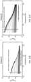
- FIG. 1 Dis a graph depicting the thermal adjustment to the impedance spectrum.
- FIG. 2 Ais a diagram showing representative diagnostic FAST and therapeutic FAST pulse delivery schemes.
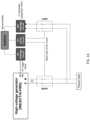
- FIG. 2 Bis an illustration depicting a representative treatment monitoring system according to embodiments of the invention.
- FIG. 2 Cis an illustration depicting an experimental setup with a 2D configuration to investigate changes in bulk tissue impedance following 1, 5, 10, 20, 40, and 80 IRE pulses.
- FIG. 2 Dis an illustration depicting an experimental setup with a 1D configuration aimed towards uniformly heating a potato tissue sample for determination of electroporation and Joule Heating effects.
- FIG. 2 Eis a graph depicting examples of diagnostic FAST, with the computed V(f) showing the differences in impedance extraction possible from each waveform.
- FIGS. 3 A-Bare diagrams of representative diagnostic and therapeutic FAST schemes, including a diagnostic FAST scheme comprising a 1-50-1-50 ⁇ s burst scheme (84 cycles) appended to a 250-10-250-10 ⁇ s (2 cycles) for a total signal duration ⁇ 10 ms ( FIG. 3 A ), and a therapeutic FAST scheme comprising a 2-5-2 ⁇ s H-FIRE scheme, modified to 2-5-2-100 ⁇ s incorporate a 100 ⁇ s extended delay after a set of bipolar pulses ( FIG. 3 B ).
- a diagnostic FAST schemecomprising a 1-50-1-50 ⁇ s burst scheme (84 cycles) appended to a 250-10-250-10 ⁇ s (2 cycles) for a total signal duration ⁇ 10 ms
- a therapeutic FAST schemecomprising a 2-5-2 ⁇ s H-FIRE scheme, modified to 2-5-2-100 ⁇ s incorporate a 100 ⁇ s extended delay after a set of bipolar pulses ( FIG. 3 B ).
- FIGS. 4 A-Bare graphs depicting the power spectral density of the MATLAB developed ideal voltage waveforms.
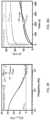
- FIGS. 5 A-Fare graphs depicting the in silico investigation of low voltage diagnostic and therapeutic FAST schemes.
- FIGS. 6 A-Hare graphs depicting an assessment of low voltage diagnostic and therapeutic FAST schemes in 1D potato tissue.
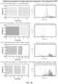
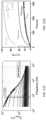
- FIGS. 7 A-Bare graphs comparing diagnostic FAST and a commercial potentiostat to measure bulk potato impedance before ( FIG. 7 A ) and after ( FIG. 7 B ) IRE pulses.
- FIG. 7 Cis a graph showing bulk potato impedance after administration of varying numbers of IRE pulses.
- FIG. 7 Dis a graph showing ablation areas after administration of IRE pulses.
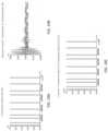
- FIGS. 8 A-Care photographs taken by a FLIR camera of a russet potato placed between two flat plate electrodes.
- FIG. 8 Dis a graph depicting a therapeutic FAST (2-5-2-100 ⁇ s, 400 bursts, 1200 V, 40 ⁇ s energized-time per burst) waveform being applied, for example to delineate thermal and EP effects.
- FIG. 8 Eis a graph showing thermal effects from those of EP, ⁇ T was calculated for the impedance change between pre-treatment impedance Z 0 and 1 st burst intra-burst impedance Z1.
- FIG. 8 Fis a graph showing the impedance changes at temperatures exceeding 25° C. at 18.2 kHz. Analysis of the ⁇ T from Z 0 to the end of the 400 th burst intra-burst impedance (Z400).
- FIG. 8 Gis a graph demonstrating a negligible temperature rise following the 1 st HV therapeutic FAST ( ⁇ T 0.04° C.).
- FIG. 9 Ais a graph depicting high frequency impedance changes to predict temperature changes (electrical impedance vs. number of bursts applied for therapeutic FAST), with a frequency of 1.78 MHz chosen to be sufficiently high to negate impedance changes due to electroporation.
- FIG. 9 Bis a graph showing the application of equation 1, an alpha 2.25%/° C. allows for accurate approximation of temperature changes due to Joule Heating only.
- FIG. 10is a graph showing bursts of bipolar pulses delivered across a rectangular potato tissue section.
- FIGS. 11 A-Care photographs taken by a FLIR thermal camera to record tissue temperature during HFIRE treatment.
- FIGS. 11 D-Eare schematics showing a high voltage 2-5-2-100 ⁇ s FAST pulse protocol intertwined with a low voltage diagnostic FAST protocol (1-50-1-50 ⁇ s+250-10-250-10 ⁇ s).
- FIG. 11 Fis a graph showing a continual decrease in tissue impedance throughout a high-voltage treatment, where the arrow indicates the progression of treatment with high-voltage pulses (H-FIRE), which causes a decrease and flattening of tissue inter-burst impedance.
- H-FIREhigh-voltage pulses
- FIG. 11 Gis a graph showing the relationship between change in temperature and electrical impedance.
- FIGS. 12 A-Dare graphs of a clinical endpoint for irreversible electroporation, which show impedance after 300 bursts are delivered across a tissue ( FIG. 12 A ); a high increase in temperature following delivery of the 300 burst treatment ( FIG. 12 B ); the FAST-controlled pulsing endpoint as the impedance spectrum saturates to a high-frequency reference ( FIG. 12 C ); and the decrease in total change in temperature using a FAST-controlled treatment protocol ( FIG. 12 D ), where the arrow in FIGS. 12 A and 12 C indicates the progression of treatment with high-voltage pulses (H-FIRE), which causes a decrease and flattening of tissue inter-burst impedance.
- H-FIREhigh-voltage pulses
- FIGS. 12 E-Fare graphs demonstrating respective clinical endpoints for patients with different electrical impedance values.
- FIGS. 13 A-Bare graphs showing temperature controlled halting of a pulsing procedure and the corresponding impedance changes associated with administration of electroporation pulses according to a FAST-controlled pulsing method, where the treatment is paused at intervals when a temperature increase of 5° C. is achieved to allow for sufficient cooling before treatment is resumed ( FIG. 13 B ), and the corresponding change in impedance for the treatment at various points in time for the first round of treatment defined by halting point (1) ( FIG. 13 A ).
- FIGS. 13 C-Dare graphs showing continuance of the temperature controlled halting of the FAST-controlled EP protocol from FIGS. 13 A-B , with the temperature-controlled halting intervals shown in FIG. 13 C (halting points 1, 2), and the corresponding impedance changes shown in FIG. 13 D for round two of the treatment.
- FIGS. 13 E-Fare graphs showing continuance of the temperature controlled halting of the FAST-controlled EP protocol from FIGS. 13 A-D , with the temperature-controlled halting intervals shown in FIG. 13 E (halting points 1 and 2 and stopping point 3), and the corresponding impedance changes ( FIG. 13 F ) for the final round of the treatment, where the clinical endpoint is indicated by obtaining a flat impedance profile (as shown by convergence of the curve).
- FIG. 14is a schematic diagram depicting a representative FAST board comprising a microcontroller, a high-voltage pulse generator, 4 Reed relays, an H-Bridge low-voltage pulse generator, and driving circuitry (2 additional H-Bridge circuits).
- FIG. 15is a schematic depicting exemplary microcontroller timing protocols for low voltage and high voltage pulsing.
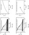
- FIG. 16 Ais a graph showing the diagnostic FAST pulse protocol used during validation of the FAST board.
- FIG. 16 Bis a graph comparing the real part of the impedance of the FAST board and that from a commercial potentiostat.
- FIG. 17is a schematic showing a pulsing protocol in which low-voltage pulses are interleaved between high-voltage pulses.
- FIG. 18is a schematic showing an embodiment of the invention using various FAST waveforms, including pulsed triangular, pulsed sawtooth, pulsed multi-stage, multisine bursts, and square waveforms with voltages ranging from 0 V-100 V and pulse lengths of up to 100 ms.
- FIGS. 19 A-Care graphs showing wide-bandwidth waveform definition for FAST data acquisition, including a defined frequency spectrum ( FIG. 19 A ); an inverse Fourier Transform of the frequency domain signal to obtain the desired time domain waveform ( FIG. 19 B ); return to the defined frequency spectrum by taking the Fourier Transform of the time domain signal ( FIG. 19 C ).
- FIGS. 20 A-Care graphs showing the wide-bandwidth waveform definition for FAST data acquisition, including the defined frequency spectrum ( FIG. 20 A ); the inverse Fourier Transform of the frequency domain signal to obtain the desired time domain waveform ( FIG. 20 B ); and return to the defined frequency spectrum by taking the Fourier Transform of the time domain signal ( FIG. 20 C ).
- FIGS. 21 A-Care graphs depicting zoomed in data for FIG. 20 B , with FIG. 21 A showing 0-0.001 s; FIG. 21 B showing 0-0.0002 s; and FIG. 21 C showing 0-0.0001 s.
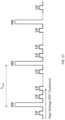
- FIG. 22is a diagram depicting a 4-probe array for a “cycled pulsing” FAST protocol.
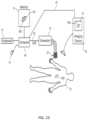
- FIG. 23is a schematic diagram of a representative system of the invention.
- FIG. 24is a diagram of a representative treatment control computer of the invention.
- FIG. 25is diagram illustrating details of the generator shown in the system of FIG. 23 .
- FIG. 26is a diagram of a system for implementing high-frequency, bipolar pulses for tissue electroporation.
- FIG. 27is an illustration showing an example graphical user interface (GUI) for administering and monitoring diagnostic and/or therapeutic FAST protocols.
- GUIgraphical user interface
- pulserefers to an electrical signal with a single phase (monopolar, unipolar) or more than one phase (bi-polar). If bi-polar, there can be a delay between phases or the switch between phases can be immediate (no intra-pulse delay).
- burstrefers to a set of pulses, a group of pulses, or a pulse group.
- introduction-pulse delayrefers to the condition where no energy is applied for a period of time during the bipolar pulses.
- inter-pulse delayrefers to the condition where no energy is applied for a period of time between one bipolar pulse or set of bipolar pulses and another bipolar pulse.
- intra-burst delayrefers to the condition where no energy is applied for a period of time between one or more bursts.
- inter-burst delayrefers to the condition where no energy is applied for a period of time within a burst and in some cases may be the same as an intra- or inter-pulse delay.
- cycled pulsing schemerefers to a pulse scheme in which the total number of pulses are delivered over more than one cycle. The total number of pulses per cycle is calculated by dividing the total number of desired pulses by the number of cycles.
- cycled pulsing protocolcycled pulsing sequence
- cycled pulsingcycled pulsing paradigm
- cycled pulsing embodimentcycled pulse sequencing
- cycled pulse paradigmcycled pulsing pattern
- total on timeor “combined signal duration” or “energized time” refers to the time associated with energizing an electrode. For example, a single 1-50-1-50 ⁇ s burst scheme would have a total on time of 2 ⁇ s for each burst, whereas a 250-10-250-10 ⁇ s burst would have a total on time of 500 ⁇ s for each burst.
- pulse protocolor “pulsing scheme” or “pulsing protocol” or “pulse scheme” refers to a protocol defined by any one or more of or all of the following: the number of pulses; the duration of each pulse; any inter-pulse, intra-pulse, inter-burst, or intra-burst delay; the number of bursts; and the number of cycles, if applicable.
- saturationrefers to an impedance measurement or spectrum matching or converging with another impedance measurement or spectrum.
- low frequency impedancecan be monitored for saturation to the high frequency impedance, meaning a low frequency impedance measurement can be taken and compared with a previous baseline and/or reference high frequency impedance measurement.
- Embodiments of the inventionprovide a Fourier Analysis SpecTroscopy (FAST) technique as an impedance spectroscopy methodology for monitoring impedance changes in a material or tissue, such as during electroporation-based therapies (EBTs) in real-time. Included within embodiments are a diagnostic FAST methodology and a therapeutic FAST methodology.
- FASTFourier Analysis SpecTroscopy
- diagnostic FASTrefers to a pulse scheme designed for monitoring inter-burst impedance of a material or tissue, such as during EBTs, including IRE and/or H-FIRE treatments. Diagnostic FAST can be achieved by delivering a low-voltage (0 V to 100 V), wideband signal (e.g., comprising rectangular bipolar pulses) and performing voltage and current capture followed by discrete Fourier transform for analysis in the frequency domain.
- Diagnostic FASTcan be applied prior to treatment, to determine the impedance spectrum of the tissue in an intact state, during treatment in between high voltage pulses, to continually monitor changes in impedance due to electroporation pulses, immediately after treatment to measure the final state of the tissue, and in the time following treatment to measure the recovery of the cell membrane and recovery of the impedance spectrum. This will be described in more detail below.
- the low-voltage electrical pulses/burstscan be applied using a voltage of above 0 V to 100 V, such as up to 10 V, up to 15 V, up to 20 V, up to 30 V, up to 40 V, up to 50 V, up to 60 V, up to 70 V, up to 80 V, up to 90 V, or from 5-85 V, or from 12-55 V, or from 35-75 V, or from 15-45 V, or any range in between any of these ranges or endpoints, including as endpoints any number encompassed thereby.
- a voltage of above 0 V to 100 Vsuch as up to 10 V, up to 15 V, up to 20 V, up to 30 V, up to 40 V, up to 50 V, up to 60 V, up to 70 V, up to 80 V, up to 90 V, or from 5-85 V, or from 12-55 V, or from 35-75 V, or from 15-45 V, or any range in between any of these ranges or endpoints, including as endpoints any number encompassed thereby.
- the signalcan comprise a waveform with any step, square, sinusoidal, ramp, Gaussian, or sinc function having constant, increasing, or decreasing frequency, or any arbitrary signal designed to achieve a desired frequency spectrum, such as in the range of 0.1 kHz to 100 MHz.
- Diagnostic FASTcomprises a pulse protocol (typically administering at a low-voltage) constructed by concatenating a high-frequency signal to low-frequency signal to form the final waveform: positive phase ⁇ intra-phase delay ⁇ negative phase ⁇ inter-pulse delay+positive phase ⁇ intra-phase delay ⁇ negative phase ⁇ inter-pulse delay.
- the set of high-frequency signal(s)(comprising: positive phase ⁇ intra-phase delay ⁇ negative phase ⁇ inter-pulse delay) can be delivered first or after the low-frequency signal(s) (comprising: positive phase ⁇ intra-phase delay ⁇ negative phase ⁇ inter-pulse delay).
- the patterncan be a set of high-frequency signal(s)+a set of low-frequency signal(s), or low-frequency signal(s)+high frequency signal(s).
- multiple pulsessuch as rectangular bipolar pulse signals, can be concatenated to form a continuous chirp like that in FIG. 18 “Square Chirp Continuous”.
- the signalscan be concatenated in any order, and can be the same or different type, phase, and/or amplitude.
- therapeutic FASTrefers to a pulse scheme designed to induce tissue EP while monitoring high voltage intra-burst impedance changes in real time.
- Therapeutic FASTcan be achieved by delivering a high-voltage (100 V to 15,000 V) burst of bipolar PEFs and is an adaptation of a traditional high-frequency IRE burst, modified to maximize the frequency bandwidth and resolution to enable intra-burst impedance spectroscopy.
- the high-voltage electrical pulses/burstscan be applied using a voltage of 100 V to 15,000 V, such as from 500 V up to 3,000 V, and/or from 1,000 V up to 2,000 V, such as up to 250 V, up to 300 V, up to 350 V, up to 600 V, up to 650 V, up to 800 V, up to 1,200 V, up to 1,500 V, up to 15,000 V, up to 7,500 V, from 4,000 V to 12,000 V, or any range in between any of these ranges or endpoints, including as endpoints any number encompassed thereby.
- a voltage of 100 V to 15,000 Vsuch as from 500 V up to 3,000 V, and/or from 1,000 V up to 2,000 V, such as up to 250 V, up to 300 V, up to 350 V, up to 600 V, up to 650 V, up to 800 V, up to 1,200 V, up to 1,500 V, up to 15,000 V, up to 7,500 V, from 4,000 V to 12,000 V, or any range in between any of these
- the electrical pulsescan be administered using a frequency ranging from 100 Hz to 100 MHz, such as in the Hz range from 100 Hz or 1 Hz up to 100 Hz, or from 2 Hz to 100 Hz, or from 3 Hz to 80 Hz, or from 4 Hz to 75 Hz, or from 15 Hz to 80 Hz, or from 20 Hz to 60 Hz, or from 25 Hz to 33 Hz, or from 30 Hz to 55 Hz, or from 35 Hz to 40 Hz, or from 28 Hz to 52 Hz, or any range in between any of these ranges or endpoints, including as endpoints any number encompassed thereby.
- pulsescan be administered using a frequency in the kHz or MHz range, such as from 1 kHz to 10 kHz, or from 2 kHz to 8 kHz, or from 3 kHz to 5 kHz, or from 4 kHz to 15 kHz, or from 6 kHz to 20 kHz, or from 12 kHz to 30 kHz, or from 25 kHz to 40 kHz, or from 5 kHz to 55 kHz, or from 50 kHz to 2 MHz, including any range in between, such as from 75 kHz to 150 kHz, or from 100 kHz to 175 kHz, or from 200 kHz to 250 kHz, or from 225 kHz to 500 kHz, or from 250 kHz to 750 kHz, or from 500 kHz to 1 MHz, or any range in between any of these ranges or endpoints, including as endpoints any number encompassed thereby.
- a frequency in the kHz or MHz rangesuch as from 1
- FAST schemescan be employed to measure the electrical impedance of testing loads which include, but are not limited to, biological tissues.
- the bandwidth (frequency limits) and the resolution (number of data points within the bandwidth) of the obtained FAST impedance spectrumare dependent on the pulsing schemes applied to the tissue.
- pulsing parameterscan be modified to a user-desired frequency bandwidth.
- a method to verifying frequency contententails taking the Fourier Transform and identifying the high-power signal in the frequency spectrum. The high-power signal is processed, and these frequencies constitute the frequencies at which impedance capture is possible.
- FASTFourier Analysis SpecTroscopy
- diagnostic FASTis the state of the tissue following pore resealing
- therapeutic FASTis a snapshot of the impedance during the pulse, a state when the pores are forming/formed.
- a 1-50-1-50+250-10-250 ⁇ s diagnostic FAST burst schemecan be used to measure frequencies ranging from above 0.1 kHz to 100 MHz, such as between ⁇ 2 kHz-5 MHz ( FIG. 3 A ), which exhibit high frequency content (i.e., high resolution) at low and high frequencies ranging from ⁇ 2 kHz to 5 MHz ( FIG. 4 A ).
- This particular burst scheme, 1-50-1-50+250-10-250 ⁇ sis not meant to be restrictive or meant to limit the signal variations for FAST.
- the pulse width and/or the width of inter-pulse, intra-pulse, inter-burst, and/or intra-burst delayscan be selected/modified to comply with other treatment-specific requirements.
- a diagnostic FAST schemecan be used at any point during treatment, for example, before, during, after high-voltage pulsing, such as immediately after consecutive pulsing.
- a therapeutic FAST/H-FIRE burst schemecan be used to ablate and capture impedance information with FAST, for example simultaneously.
- a 2-5-2-100 ⁇ s pulse protocolcan be used as the therapeutic FAST scheme, with changes in tissue impedance being monitored preferably in real-time ( FIG. 3 B ).
- This particular therapeutic FAST scheme (2-5-2-100 ⁇ s)shows high resolution extending up to ⁇ 2 MHz ( FIG. 4 B ), yet other pulsing protocols can also be used.
- lower resolution spectracan be used in some applications, higher resolution results typically will provide the user/practitioner with a greater confidence level relating to the treatment (e.g., a greater confidence level that a particular outcome or effect has been achieved or is occurring during treatment).
- the pulse widthcan be modified, for example, to include: 1) 1-5-1--100 ⁇ s, 2) 5-5-5--100 ⁇ s, 3) 10-5-10--100 ⁇ s
- the intra-phase delaycan also be modified to include, for example: 1) 2-1-2--100 ⁇ s, 2) 2-10-2--100 ⁇ s, 3) 2-100-2--100 ⁇ s, as well as the inter-pulse delay, which can for example include: 1) 2-5-2--1 ⁇ s, 2) 2-5-2--10 ⁇ s, 3)[-]]2-5-2--100 ⁇ s.
- Pulse widthcan be in the range of for example 0.1 ⁇ s to 10 ms.
- Non-square waveformsi.e. pulsed triangular waveforms, continuous sawtooth waveforms, multisine bursts, and/or multistage waveforms— FIG. 18
- the waveformcan comprise any step, square, sinusoidal, ramp, Gaussian, or sinc function having constant, increasing, or decreasing frequency, or any arbitrary signal designed to achieve a desired frequency spectrum in the range of above 0.1 kHz to 100 MHz.
- diagnostic FASTcan be used for a pulse sequence that does not include an intra-pulse delay (e.g., instant charge reversal, or a pulse with no delay between changing from negative to positive polarity).
- the real impedance datamatches that attained from a commercial potentiostat ( FIGS. 7 A-C ).
- the commercial potentiostatrealized a complete impedance spectrum in >10 s
- FAST impedance measurementsare realized ⁇ 1 s, thereby enabling impedance capture when using electroporation pulsing rates of 1 Hz.
- FASTcan be utilized for monitoring the extent of tissue/cellular recovery or lack thereof during pulsing in real time. As seen in FIG.
- the extent of electroporation from IRE treatmentcan be reliably captured and shows good agreement with the commercial potentiostat (e.g., as shown by the flattening of the impedance curve for 80 pulses as compared with that of 1-20 pulses).
- the use of dedicated hardwaresuch as an H-Bridge low-voltage pulse generator, or other low-voltage generator, which can be incorporated into the system with any required drive circuit as a stand-alone component or integrated as part of the generator
- diagnostic FAST pulsesto monitor the recovery (electroporation and/or temperature) during treatment after pulses and/or bursts (such as after every pulse), providing real-time feedback for electroporation-based therapies.
- This methodologycan be achieved by interlacing diagnostic and therapeutic FAST schemes at a desired repetition rate (see, e.g., FIGS. 15 and 22 ).
- a slow tissue impedance recovery(back to baseline) will signify dead/dying tissue, whereas a quick recovery can suggest reversible or minimal electroporation, with such analyses helpful in determining when a particular treatment endpoint has been reached, and/or when a particular treatment should be modified/adjusted, halted or stopped.
- Diagnostic FASTcan be used to provide a patient specific endpoint for electroporation-based therapies. While the existence of the beta dispersion within an impedance spectrum of a densely populated cell suspension/tissue is known, FAST allows for detecting the magnitude of the high frequency impedance. Once this value is determined, a low frequency impedance measurement can be monitored for its saturation to the high frequency impedance reference; this will signify ablation of cells within a therapeutic field, as this is an indication of cell membrane damage. The magnitude of this impedance will vary patient to patient, though the saturation of the low frequency to the high frequency impedance can serve as a marker for the endpoint of treatment. For example, as shown in FIGS. 12 E-F , different respective clinical endpoints for patients with different electrical impedance values can be expected.
- This relationshipsaturation of the low frequency ( ⁇ 5 kHz) to high frequency ( ⁇ 2 MHz) impedance, is specific to peripheral tumor tissue, calcified tumor tissue, necrotic tissues, healthy/tumor ischemic tissues, traumatic brain injury, fibrotic tissues, tissue states prior to and after chemotherapy, and tissue states prior to and after radiation.
- This techniqueis also applicable to the classification of tissues, such as identifying/determining between various tissue types such as brain, liver, prostate, kidney, pancreas, and tumor tissues arising from unique electrical characteristics of each tissue.
- Diagnostic FASTcan be fine-tuned and utilized to monitor changes in tissue morphology, edema, perfusion, fluid infusion, thermal necrosis, cell death by electrical, mechanical, or thermal means, identifying cell subtypes, identifying intracellular structures and morphology, gauging efficacy of drug delivery, integrity of the blood-brain-barrier, and tissue heterogeneity.
- methods of the inventionare capable of detecting increases in conductivity due to edema influx, which is an indication of a positive immune response to the EP treatment.
- the methods of treating/monitoringcan include treating tissue and/or cells (e.g., a tumor or a cancer) within a target site in a subject by administering a plurality of electrical pulses to the target site which can induce non-thermal electroporation, such as irreversible electroporation (IRE), of the treatment site; measuring a treatment parameter, including, but not limited to, impedance or an impedance spectrum; detecting a change in the measured parameter such as a change in impedance; and performing an additional downstream or secondary treatment as a result of a change in impedance that indicates a positive immune response (such as edema), wherein the downstream or secondary treatment step can include, but is not limited to, tumor resection, thermal ablation, a secondary non-thermal ablation, chemotherapy, radiation therapy, immunotherapy, biologic therapy, genetic therapy (gene editing), and combinations thereof.
- tissue and/or cellse.g., a tumor or a cancer
- IREirreversible electroporation
- diagnostic FASTcan be used to monitor tissue decellularization/cell death as the impedance spectrum/dispersion flattens.
- This techniquecan be used for, but is not limited to, monitoring tissue ablation such as irreversible electroporation, chemical cell death/decellularization (e.g., by surfactants, acids, and/or bases), disruption by physical means (such as by high-intensity focused ultrasound (HIFU), Histotripsy, and/or laser ablation), monitoring gene-transfection efficiency and/or uptake, monitoring tissue ablation (nonthermal, thermal) for cardiac arrythmias, monitoring cell lysis for immunotherapies, and monitoring mechanisms of cell death.
- tissue ablationsuch as irreversible electroporation, chemical cell death/decellularization (e.g., by surfactants, acids, and/or bases), disruption by physical means (such as by high-intensity focused ultrasound (HIFU), Histotripsy, and/or laser ablation), monitoring gene-transfection efficiency and/or uptake, monitoring tissue ablation (
- lysisWith respect to monitoring cell death, lysis will be represented as a flattening of the impedance spectrum/dispersion with no recovery following pulse cessation. As cell lysis involves immediate destruction of the cell membrane, the measurement of impedance recovery, or lack thereof, will be possible on within minutes after treatment. Necrosis will be represented as a flattening of the impedance spectrum/dispersion with minimum recovery following pulse cessation. This assumes pore-formation does not recover and cell is essentially in a lysed state. Apoptosis will be represented as a flattening of the impedance spectrum/dispersion with moderate recovery following pulse cessation. This assumes pore-formation recovery, where the cell dies due to intracellular signaling.
- impedance measurements with FASTcan be conducted to monitor recovery of the cell membrane within minutes following the end of treatment.
- Pro-inflammatory forms of cell deathsuch as necroptosis/necrosis, will aid in determination of administration of immunologic agents.
- High frequency tissue impedance measurementscan be used to approximate changes in tissue temperature and gauge nonthermal ablation.
- Z Tis the conductivity of the tissue at an elevated change in temperature ⁇ T
- Zthe baseline conductivity
- ⁇the temperature coefficient of resistance.
- ⁇the temperature coefficient of resistance.
- the inventorsemploy: 1) numerical methods to examine various FAST schemes in regards to the maximum attainable frequency range and resolution, 2) a flat-plate electrode configuration using potato tissue to validate FAST-measured impedance against a commercial potentiostat using the selected diagnostic and therapeutic FAST from numerical methods, 3) a two-needle configuration in potato tissue to monitor impedance changes during IRE therapy and validate FAST-measured impedance against a commercial potentiostat, 4) a flat-plate electrode configuration to heat potato tissue using therapeutic FAST, with the goal of using the measured impedance to delineate thermal effects from those of EP.
- V(t) and I(t)were analyzed in MATLAB vR2018a (MathWorks Inc., Natick, MA, US) using the Fast Fourier Transform (FFT) algorithm, in which the length of the FFT was defined as the next power of 2 from the length of the voltage signal. This resulted in V(f) and I(f), thereafter the magnitude of V(f), labeled as V FFT , was defined.
- FFTFast Fourier Transform
- MATLAB functions “real( )” and “imag( )”were used to analyze the real and imaginary parts of the impedance, respectively.
- V(t) of amplitude 15 Vwere constructed in MATLAB using a series of concatenated rectangular pulses with 80 ns rise/fall-times. These V(t) waveforms were imported into COMSOL for numerical analysis.
- a 0D circuit modelwas constructed in the “Electrical Circuit” module for the circuit shown in FIG. 1 C and a time-dependent study implemented to attain the current response I(t) from this circuit model. The V(t) and I(t) were then processed to extract the real and imaginary impedance.
- Both diagnostic and therapeutic FASTwere produced to follow a positive phase ⁇ intra-phase delay ⁇ negative phase ⁇ inter-pulse delay pattern ( FIG. 2 A ), where the pulse-widths and intra-phase/inter-pulse delays were varied ranging for example from 0.1 ⁇ s to 1 ms.
- the representative diagnostic FAST scheme shown in FIG. 2 Ashows a high-frequency component appended to low-frequency component.
- the waveformscan be bipolar, square waveforms.
- the therapeutic FAST scheme shown in FIG. 2 Ashows a bipolar square waveform. In other embodiments, the waveforms can be unipolar. With respect to each waveform, the following criteria were imposed:
- PSDfrequency content and power spectral density
- the PSDreveals the frequencies at which data acquisition, the impedance spectrogram, is possible for the 1-50-1-50+250-10-250-10 ⁇ s diagnostic FAST scheme ( FIG. 4 A ) and the 2-5-2-100 ⁇ s therapeutic FAST scheme ( FIG. 4 B ).
- the lowest frequency content with sufficient power for data extractionis located at ⁇ 1.8 kHz using the diagnostic FAST, although lower frequencies could be used (such as above 0.1 kHz up to 5 kHz). Above 100 kHz, there are numerous peaks and extends beyond 4 MHz; the upper limit at which the PSD still meets the inventors' criteria was found at 4.8 MHz.
- the desired diagnostic and therapeutic FASTwhich maximized the frequency range and frequency resolution for impedance capture were identified and selected.
- the desired frequency range for impedance capture and the corresponding selection of pulsing scheme pulse width, intraphase delay, and interpulse delaycan be determined prior to treatment.
- the impedance extracted from the low-voltage pulseis then used to inform treatment endpoint, which can be controlled by personal or software/hardware.
- potato tissueis composed of cells embedded within a fibrous matrix, this system loosely represents the macroscopic composition of biological tissues and similarly undergo electroporation.
- impedance changes due to electroporation and tissue ablation in potatoesresemble that of biological tissues.
- Potato tissue ablationoccurs through an enhanced oxidative mechanism involving the release of intracellular enzymes that results in a rapid black oxidation of the tissue exposed to higher therapeutic fields; this rapidly oxidized tissue area represents the ablative region.
- FIG. 2 Bcomprised a function generator programmed to deliver low voltage FAST prior to and following high voltage pulse delivery, an oscilloscope for recording FAST waveforms, a computer capable of processing the waveforms in MATLAB using a Fast Fourier Transform algorithm for decomposition into the frequency domain, and a Gamry Reference 600 potentiostat used to determine accuracy of the FAST-recorded impedance.
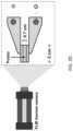
- a russet potatowas sliced to a thickness of 0.7 cm and further sectioned using a cylindrical cutter of diameter 0.8 cm. This tissue sample was placed between two flat plate electrodes ( FIG. 2 D ) with square cross section 2 ⁇ 2 cm (BTX, Harvard Apparatus, Cambridge, MA).
- a baseline impedance spectrumwas measured using a commercial potentiostat Gamry Reference 600 (Gamry, Warminster, PA, US) at a frequency band 1 kHz to 1 MHz at 10 points per decade. The potentiostat impedance spectrum served as a comparison to assess accuracy of the FAST schemes.
- low voltage diagnostic and therapeutic FAST schemeswere delivered using an AFG31000 series function generator (Tektronix Inc., Beaverton, OR, US) at a voltage 15 V.
- the voltage and current waveformswere recorded using a WaveSurfer 3024z Oscilloscope (Teledyne LeCroy, Chestnut Ridge, New York) and a 1 ⁇ current probe (2877, Pearson Electronics, Palo Alto, California).
- V(t) and I(t)were analyzed.
- therapeutic FAST schemesare ultimately intended to be delivered at higher voltages to generate biophysical tissue changes, a direct comparison to the low-voltage (LV) diagnostic FAST was desired and thus a LV therapeutic FAST was examined.
- the impedance spectrum acquired from this experimentwas used to inform the circuit model fitting for numerical analysis.
- FIG. 6 A-BSelect diagnostic and therapeutic FAST schemes were tested in potato tissue in vitro ( FIG. 6 A-B ).
- the diagnostic 1-50-1-50 ⁇ s (84 cycles)+250-10-250-10 ⁇ s (2 cycles) FAST scheme ( FIG. 6 A ) and therapeutic 2-5-2-100 ⁇ s (25 cycles) FAST scheme ( FIG. 6 B )were delivered across a cylindrical potato section using an arbitrary function generator.
- Impedance datawere extracted from each high-power peak in V FFT and are shown for diagnostic and therapeutic FAST schemes, respectively ( FIGS. 6 C, 6 D ) and in comparison to the numerically derived impedance data, the experimental data for V FFT similarly showed an abundance of high-power peaks along the desired frequency range.
- the minimum and maximum frequency at which data was extracted for diagnostic FASTwere 1.78 kHz and 4.69 MHz, respectively, with 216 data points fitting within this range.
- the minimum and maximum frequency for data acquisitionwere 18.3 kHz and 1.82 MHz, respectively, with 170 data points fitting within this range.
- the resultant potato impedancewas calculated using Ohm's law; real (tissue resistance) and imaginary (tissue reactance) impedance are shown in FIGS. 6 E-H .
- Nonlinear regressionwas conducted to test a null hypothesis for a single curve fit being used to describe the potentiostat and FAST generated datasets.
- a 2D configurationwas implemented to simplify characterization of ablation areas. Potatoes were sectioned uniformly to a 0.7 cm thickness with an ellipsoidal cross section ⁇ 15 ⁇ 10 cm using a generic mandolin cutter. Two, 20-gauge cylindrical stainless-steel needle electrodes were inserted at the center of the sample and maintained at a 1 cm spacing; these electrodes were used for both EIS and tissue ablation with IRE ( FIG. 2 C ). Prior to tissue ablation, baseline impedance and diagnostic FAST were implemented. The schematic in FIG. 2 B depicts a representative setup.
- a baseline impedance spectrumwas measured using a Gamry Reference 600 (Gamry, Warminster, PA, US) at a frequency band 1 kHz and 1 MHz at 10 points per decade. Thereafter, the diagnostic FAST scheme was also implemented for comparison to the commercial potentiostat. All low voltage FAST schemes were delivered using an AFG3021C function generator (Tektronix Inc., Beaverton, OR, US). The voltage and current waveforms were recorded using a WaveSurfer 3024z Oscilloscope (Teledyne LeCroy, Chestnut Ridge, New York).
- the voltagewas stepped down using a 1000 ⁇ high voltage probe (Enhancer 3000, BTX, Holliston, MA) while the current was recorded using a 10 ⁇ current probe (2877, Pearson Electronics, Palo Alto, California).
- a 1000 ⁇ high voltage probeEnhancer 3000, BTX, Holliston, MA
- 10 ⁇ current probe2877, Pearson Electronics, Palo Alto, California.
- separate LV and HV generatorscan be used to administer the LV and HV waveforms, respectively.
- IRE pulseswere delivered in sets of 10 where applicable with a 1-minute delay in between each set. The potatoes were covered in plastic wrap and stored overnight to allow for tissue oxidation. The oxidized area in each sample was used to quantify the ablative region and was measured in ImageJ software.
- the baseline impedance spectrum ( FIG. 7 A ) as well as after the 1 st , 5 th , 10 th , 20 th , 40 th , and 80 th pulses( FIG. 7 B-C ) suggest good agreement across the bandwidth 1.8 kHz-1 MHz. While FAST data was acquired for frequencies up to 4.8 MHz, no direct comparisons for frequencies above 1 MHz were made due to limitations with the potentiostat, however, higher frequencies can be used, such as up to 100 MHz.
- IREwas delivered at a pulsing rate 1 Hz; of the 6 treatment groups (Table 1), pulses for groups 4-6 were split into sets of 10 and a 1 minute delay added in between sets to allow for impedance capture with FAST ( ⁇ 1 s) and the potentiostat (>10 s), as well as to allow for heat dissipation. All groups demonstrated an IRE ablation, with 1 pulse (1 P) showing the smallest ablation (1.63 ⁇ 0.26 cm 2 ).
- the ablation areaincreased: 5 P (2.23 ⁇ 0.34 cm 2 ), 10P (3.45 ⁇ 0.49 cm 2 ), 20 P (3.92 ⁇ 0.33 cm 2 ), 40 P (4.44 ⁇ 0.35 cm 2 ), and lastly the 80 P group resolving in the largest measured ablation (4.57 ⁇ 0.29 cm 2 ) ( FIG. 7 D ).
- the low frequency impedance changesare likely attributed to a combination of tissue electroporation as well as Joule Heating; measuring only the low frequency impedance, as is the standard method with the commercial NanoKnife system, will result in a multivariate signal with no means of separation.
- the present inventorstherefore introduce the measurements of high frequency impedance for isolating impedance changes due solely to Joule Heating.
- these temperature effectscan then be subtracted from those of the low frequency, effectively isolating the changes in tissue impedance due solely to electroporation.
- H-FIRE waveformswere delivered using a custom bipolar pulse generator (EPULSUS-FBM1-5, Lisboa, Portugal) to output stainless steel electrodes.
- This generatorconsists of two unipolar Marx generators capable of producing voltage waveforms with pulse risetimes of 100 ns and a maximum voltage/current output of 5 kV/50 A.
- a 1D electroporation setupconsisting of flat plate stainless steel electrodes were utilized to expose the tissue to a uniform electric field.
- a rectangular shape factor of length, width, and thickness dimensions 2 cm ⁇ 2 cm ⁇ 7 mmwas used.
- the H-FIRE treatmentcomprised 400 bursts of bipolar pulses, each energized for 100 ⁇ s at an electric field of 1,500 V/cm.
- a russet potatowas sectioned to a rectangular shape of length, width, and thickness 2 ⁇ 2 ⁇ 0.7 cm.
- an impedance spectrumwas quantified using a low-voltage therapeutic FAST scheme with energized time 100 ⁇ s; this scheme was implemented to match the impedance attained during treatment.
- high voltage therapeutic FASTwas delivered using a custom-built H-FIRE generator; due to pulse-generator limitations, output voltage was restricted to 1200 V with an energized time of 40 ⁇ s per burst and 400 bursts delivered.
- Temperaturewas monitored using a FLIR A325SC thermal camera (FLIR, Wilsonville, OR, USA).
- High voltage waveformswere recorded using a 1000 high voltage probe (Enhancer 3000, BTX, Holliston, MA) and current was captured using a 10 current probe (3792, Pearson Electronics, Palo Alto, California).
- ⁇ ⁇ ⁇ T ⁇ ( t )Z ⁇ ( t ) - Z 0 Z 0 ⁇ 1 ⁇ ( 3 )
- ⁇ T(t)is the calculated increase in temperature
- Z(t)is the impedance at the N th burst
- Z 0is the pre-treatment impedance
- athe temperature coefficient of resistance.
- Nonlinear regression analysiswas performed and a null hypothesis describing “a single curve fit” between the commercial potentiostat and FAST impedance data was tested. Here, a p-value ⁇ 0.05 was considered statistically significant (null hypothesis rejected).
- the R 2 reportedare those relative to the global/shared curve fit between the potentiostat and FAST-measured impedance.
- the real part of the impedance for all data setswas fit to a 4-parameter logistic regression model, an adaptation to the Cole-Cole equations:
- the measured ablationswere compared using a one-way ANOVA with a post-hoc Tukey's multiple comparisons test. A p-value ⁇ 0.05 was considered statistically significant. Statistical analysis was conducted in GraphPad Prism v8.2.
- a FLIR thermal camerawas used to record the increase in temperature at a rate of 10 Hz during treatment ( FIGS. 8 A-C , G).
- the maximum ⁇ Toccurred at the end of treatment, measured as 14.8° C. ( FIG. 8 G ).
- impedance characterization with low voltage therapeutic FAST(2-5-2-100 ⁇ s, 15 V, 25 cycles
- 400 bursts of bipolar pulses(high voltage therapeutic FAST) were delivered (2-5-2-100 ⁇ s, 1200 V, 10 cycles), see FIGS. 8 D-E ).
- Therapeutic FAST schemeswere recorded on the oscilloscope at a rate 0.5 Hz and subsequently transferred to MATAB for analysis.
- equation 1is used to predict the ⁇ T based off the experimentally varied voltage and current waveforms ( FIG. 9 B ). More specifically, a frequency of 1.78 MHz for this analysis in combination with the therapeutic FAST scheme allowed for the measurement of changes in tissue impedance due to temperature.
- the estimated temperature profile, seen in FIG. 9 Bclosely matches that measured using a FLIR thermal camera. Modification of this scheme can yield similar results incorporating higher bandwidth and/or resolution.
- FIG. 9 Adepicts the changes in tissue impedance at a frequency 1.78 MHz; this frequency was chosen as this is the location where the tissue impedance varied the least during pulsing, signifying extent of electroporation played a minimal role for the changes in tissue impedance.
- the intra-burst impedanceis a function of R e R i ; in this experiment, in order to heat the tissue, the applied voltage was seemingly beyond this point and resolved in a relatively flat intra-burst impedance spectrum within the 1 st burst ( FIG. 8 E ). Subsequently, measurements of intra-burst impedance maintained the flat profile with a shift downwards attributed to a decrease in impedance due to thermal effects.
- the predicted temperature starting at the 400 th burstwas calculated and is seen in FIG. 8 G .
- the impedance data attained at 1.21 MHzbest predicts the measured ⁇ T, as this frequency is able to penetrate the cell membrane prior to EP and minimizes the impedance changes from initial EP effects. It is stressed again, in order to heat potato tissue, a voltage much higher than that needed to partially EP the tissue was required. In translation to biological tissue, as demonstrated in Bhonsle et al. 2017, fields that partially EP tissue and inherently heat tissue are within the same magnitude and therefore it is expected that EP effects continually contribute to low-frequency intra-burst impedance change as a function of number of bursts.
- the parameters R e , R m , C m , and R i fitted to the impedance measurementswere determined as 2071.2 ⁇ , 9.71E06 ⁇ , 6.53 nF, and 198.35 ⁇ , respectively. It should be noted that a more accurate fit to the impedance spectrum is attained by replacing the membrane elements R m ⁇ C m with a constant phase element, though the R m ⁇ C m was selected to allow for time dependent circuit analysis using the circuit element nodes in COMSOL.
- the numerical V(t) and I(t) from COMSOLwere processed in MATLAB and a subset of the results are plotted in FIG. 5 A-F .
- bipolar or unipolar pulsescan be used for diagnostic FAST and/or therapeutic FAST, along with any type of waveform (e.g., a waveform with any step, square, sinusoidal, ramp, Gaussian, or sinc function) having constant, increasing, and/or decreasing frequency, or any arbitrary signal designed to achieve a desired frequency spectrum in the range of above 0.1 kHz to 100 MHz.
- any type of waveforme.g., a waveform with any step, square, sinusoidal, ramp, Gaussian, or sinc function
- any arbitrary signaldesigned to achieve a desired frequency spectrum in the range of above 0.1 kHz to 100 MHz.
- the total number of pulses/bursts per diagnostic FAST or therapeutic FAST treatment or total number of pulses/bursts per cyclecan range from 1 to 5,000 pulses/bursts, such as from at least 1 up to 3,000 pulses/bursts, or at least 2 up to 2,000 pulses/bursts, or at least 5 up to 1,000 pulses/bursts, or at least 10 up to 500 pulses/bursts, or from 10 to 100 pulses/bursts, such as from 20 to 75 pulses/bursts, or from 30 to 50 pulses/bursts, such as 1, 5, 10, 15, 20, 25, 30, 35, 40, 45, 50, 60, 70, or 90 pulses/bursts, or any range in between any of these ranges or endpoints, including as endpoints any number encompassed thereby.
- the number of cyclescan be any number of cycles, such as zero cycles, or from 1-100 cycles, or from 2-50 cycles, or from 3-40 cycles, or from 4-30 cycles, or from 5-20 cycles, or from 10-15 cycles, or any range in between any of these ranges or endpoints, including as endpoints any number encompassed thereby.
- the therapeutic FAST schemeis a modified H-FIRE burst scheme to include an extended inter-pulse delay (e.g., 100 ⁇ s): 2-5-2-100 ⁇ s burst scheme (25 cycles) energized for 100 ⁇ s ( FIG. 5 B ).
- An extended inter-pulse delaye.g., 100 ⁇ s
- 2-5-2-100 ⁇ s burst scheme25 cycles energized for 100 ⁇ s
- FIGS. 5 A, 5 C, and 5 EA comparison between diagnostic FAST of 5 ⁇ s and 50 ⁇ s intra-phase and inter-pulse delays is seen in FIGS. 5 A, 5 C, and 5 E .
- an increased intra-phase and inter pulse delayseemingly increase the resolution of the frequency band. Therefore, the 50 ⁇ s intra-phase and inter-pulse delay was selected.
- a comparison between therapeutic FAST and a 2-5-2-5 ⁇ s H-FIRE burst schemeis seen in FIGS. 5 B, 5 D, and 5 F .
- impedancewas extracted for peaks in the V FFT plots containing at least 2% of the maximum power in the signal.
- diagnostic FASTthe real part of the impedance is seen in FIG. 5 E .
- therapeutic FASTthe real part of the impedance is shown in FIG. 5 F .
- the 1-50-1-50+250-10-250-10 ⁇ s diagnostic FAST schemeproduced 204 data points within the frequency range of 1.81 kHz-4.93 MHz and the 2-5-2-100 ⁇ s therapeutic FAST produced 164 data points within a frequency range of 18.3 kHz-1.96 MHz.
- the therapeutic FAST waveformis proposed to permeabilize/ablate biological cells while simultaneously allowing for high-bandwidth intra-burst impedance spectroscopy.
- the selected therapeutic FASTis a high voltage 2-5-2-100 ⁇ s burst scheme, an H-FIRE burst scheme modified to include an extended inter-pulse delay following the negative polarity pulse. This extended inter-pulse delay increases the resolution of the impedance spectrum, allowing for intra-burst impedance spectroscopy between 18.3 kHz to 1.96 MHz ( FIG. 5 ) at 170 discrete frequencies. Since the intra-phase delay remains 5 ⁇ s, minimal impact on the muscle/nerve stimulation thresholds was expected (B. Mercadal, C. B. Arena, R. V.
- Equation 7can be converted into Equation 8.
- FIG. 11 Eshows the output electric current resulting from the LV diagnostic FAST.
- the recorded diagnostic FAST electric currentincreases and is reflected in the frequency domain as a decrease in inter-burst impedance ( FIG. 11 F ).
- the calculated ⁇demonstrates electrical impedance measured at high frequencies best approximates the true ⁇ T.
- a clinical endpoint for electroporationcan be defined when low-frequency impedance measurements, either a single frequency or a range of frequencies between 0.1 kHz to 500 kHz, continually decreases to a value within 20% of the reference or baseline high-frequency impedance measurements, either a single frequency or a range of frequencies between 500 kHz to 100 MHz. This is visually depicted as a flattening of the impedance spectrum during electroporation treatment.
- a clinical endpointcan be defined when the present low-frequency impedance measurements, either a single frequency or a range of frequencies between 0.1 kHz to 500 kHz, is unchanged compared to a prior low-frequency measurement, such as within a window of approximately 1-10%, such as within 5%.
- the low-frequency impedance measurementdoes not decrease to the magnitude of the high-frequency reference and does not satisfy criteria for clinical endpoint defined in above.
- the tissueis still “fully-electroporated.” Pulsing in this scenario is terminated to avoid thermal damage.
- the following equationcan be used to calculate the percent change in the present low-frequency impedance relative to the prior measurement:
- Z 0is the baseline low-frequency impedance measurement
- Z iis the i th pulse/burst in the treatment
- Z i ⁇ 1is the pulse/burst previous to the i th pulse/burst in the treatment.
- treatmentcan be adjusted, stopped, and/or halted to allow for heat dissipation to avoid or prevent excessive tissue heating.
- the adjusting or stopping/halting of the treatmentcan be performed automatically by the system and/or the system can provide prompts to the user through a graphical user interface that provide information and/or options to the user on how to proceed.
- pulsingcan continue. This methodology can be repeated until the pulsing endpoint is met (flat impedance profile).
- FIGS. 13 A-BA theoretical, representative pulsing profile for which multiple pulsing sets might be used is depicted in FIGS. 13 A-B .
- This conditioncan be controlled as a ⁇ T, pre-defined number of pulses, or other timing criteria.
- the halting/pausing of electroporation treatmentcan be defined as delivering a pre-determined number of pulses/bursts (0-1,000) across one or more electrode/probe pair combinations. Once this value is reached, treatment can be paused for a pre-determined time (e.g., 0-600 s), as shown in FIGS. 13 A-B . This process can then be repeated ( FIGS. 13 C-F ) until the clinical endpoint, such as determined from diagnostic FAST impedance measurements, is reached ( FIG. 13 F ).
- a pre-determined timee.g., 0-600 s
- the halting/pausing of electroporation treatmentcan be defined as reaching a pre-determined increase in temperature (e.g., 0-50° C.) across one or more electrode/probe pair combinations.
- a pre-determined increase in temperaturee.g., 0-50° C.
- This increase in temperatureis measured using either a temperature measurement device (fiber optic temperature sensor, thermocouple, etc.) or by using high-frequency impedance measurements to predict temperature change.
- a temperature measurement devicefiber optic temperature sensor, thermocouple, etc.
- a temperature decreasee.g., 0-50° C.
- Embodiments of the present inventioncan include one or more components as illustrated in FIGS. 23 and 24 .
- one or more probescan be used to deliver therapeutic energy and are powered by one or more voltage pulse generator 10 that generates high or low voltage pulses as therapeutic energy, diagnostic FAST or therapeutic FAST, such as pulses capable of electroporating (e.g., irreversibly electroporating) the tissue cells.
- the voltage pulse generator(s)can include any number of receptacles for receiving up to any number of individual probes, which probes are adapted to be plugged into the respective receptacle.
- the voltage pulse generator(s)can have any number of receptacles for receiving for example from 1-20 probes.
- a treatment protocol according to the inventioncould include a plurality of electrodes disposed on any number of probes.
- the plurality of electrodescan be disposed in various positions relative to one another.
- a plurality of electrodescan be disposed in a relatively circular or square or rectangular pattern. Any configuration of electrodes is possible and the arrangement need not be circular, square or rectangular, but any shape periphery can be used, including triangular, depending on the area to be treated, including any regular or irregular polygon shape, including convex or concave polygon shapes.
- Any of the electrodescan be a ground electrode and any of the other electrodes in the plurality can be energized.
- any number of electrodescan be in the plurality such as from about 1 to 20. Indeed, even 3 electrodes can form a plurality of electrodes, or 4 electrodes can be disposed in a manner to provide two electrode pairs (each pair comprising one ground and one electrode capable of being energized).
- methods of treatingcan involve energizing the electrodes in any sequence, such as energizing one or more electrode simultaneously, and/or energizing one or more electrode in a particular sequence, such as sequentially, in an alternating pattern, in a skipping pattern, and/or energizing multiple electrodes but less than all electrodes simultaneously, for example.
- each probeincludes either a monopolar electrode or bipolar electrodes.
- the amount of exposure of an active portion of the electrodecan be adjusted by retracting or advancing an insulating sleeve relative to the electrode. See, for example, U.S. Pat. No. 7,344,533, which is incorporated by reference herein in its entirety.
- the pulse generator(s)are connected to a treatment control computer 40 having input devices such as keyboard 12 and a pointing device 14 , and an output device such as a display device 11 , including a graphical user interface (GUI), for viewing information obtained from the FAST protocol.
- GUIgraphical user interface
- One or more probeis used to treat a lesion, tissue, or area/region of interest (ROI) 300 inside a patient 15 and/or to perform diagnostic and/or therapeutic FAST monitoring in conjunction with such treatment.
- An imaging device 30includes a monitor 31 for viewing the lesion 300 inside the patient 15 in real time and for monitoring the diagnostic and/or therapeutic FAST monitoring. Examples of imaging devices 30 include ultrasonic, CT, MRI and fluoroscopic devices as are known in the art.
- the present inventionincludes computer software (treatment monitoring module 54 ) which assists a user to plan for, execute, and review the results of a medical treatment procedure, as will be discussed in more detail below.
- the treatment monitoring module 54assists a user to plan for a medical treatment procedure by enabling a user to more accurately position each of the probes 22 of the therapeutic energy delivery device 20 in relation to the tissue, ROI, or lesion 300 in a way that will generate the most effective treatment zone, and/or provide options for the practitioner/user concerning whether and how to adjust treatment, and/or halt or stop treatment based on the impedance changes being measured by way of the diagnostic and/or therapeutic FAST monitoring.
- the treatment monitoring module 54can display the progress of the treatment, such as monitoring with FAST, in real time and can display the results of the treatment procedure and/or results of the diagnostic or therapeutic FAST after it is completed. This information can be displayed in a manner such that it can be used for example by a physician to determine whether the treatment was successful and/or whether it is necessary or desirable to re-treat the patient, continue to treat the patient, or stop treatment based on the FAST results.
- Any non-transitory computer-readable mediacan be used to store the software and/or the output of the software for a particular treatment protocol.
- codeFor purposes of this application, the terms “code”, “software”, “program”, “application”, “software code”, “computer readable code”, “software module”, “module” and “software program” are used interchangeably to mean software instructions that are executable by a processor.
- the “user”can be a physician or other medical professional.
- the treatment monitoring module 54 executed by a processoroutputs various data including text and graphical data to the monitor 11 associated with the generator 10 .
- the treatment control computer 40 of the present inventionmanages monitoring of treatment for a patient.
- the computer 40is connected to the communication link 52 through an I/O interface 42 such as a USB (universal serial bus) interface, which receives information from and sends information over the communication link 52 to the voltage generator 10 .
- the computer 40includes memory storage 44 such as RAM, processor (CPU) 46 , program storage 48 such as ROM or EEPROM, and data storage 50 such as a hard disk, all commonly connected to each other through a bus 53 .
- the program storage 48stores, among others, a treatment monitoring module 54 which includes a user interface module that interacts with the user in monitoring, executing and reviewing the result of a treatment. Any of the software program modules in the program storage 48 and data from the data storage 50 can be transferred to the memory 44 as needed and is executed by the CPU 46 .
- the computer 40is built into the voltage generator(s) 10 . In another embodiment, the computer 40 is a separate unit which is connected to the voltage generator(s) through the communications link 52 . In a preferred embodiment, the communication link 52 is a USB link. In one embodiment, the imaging device 30 is a standalone device which is not connected to the computer 40 . In the embodiment as shown in FIG. 23 , the computer 40 is connected to the imaging device 30 through a communications link 53 . As shown, the communication link 53 is a USB link. In this embodiment, the computer can analyze the FAST impedance measurements and/or spectra obtained and the computer 40 can display this information on the monitor 11 . In this embodiment, the FAST information/analysis/results obtained and/or recommendations or options for whether and how to proceed with treatment can be provided on a GUI and presented to the user ( FIG. 27 ).
- the softwarecan be used independently of the pulse generator(s) 10 .
- the usercan monitor the treatment by way of FAST in a different computer as will be explained below and then save the treatment parameters to an external memory device, such as a USB flash drive (not shown).
- the data from the memory device relating to the treatment parameterscan then be downloaded into the computer 40 to be used with the generator 10 for treatment.
- the datacan be evaluated by a human to determine or estimate favorable treatment protocols for a particular patient or programmed into a device for implementing the particular protocol.
- FIG. 25illustrates one embodiment of a circuitry to detect an abnormality in the applied pulses such as a high current, low current, high voltage or low voltage condition.
- This circuitryis located within the generator 10 (see FIG. 23 ).
- a USB connection 52carries instructions from the user computer 40 to a controller 71 .
- the controllercan be a computer similar to the computer 40 as shown in FIG. 24 .
- the controller 71can include a processor, ASIC (application-specific integrated circuit), microcontroller or wired logic.
- the controller 71then sends the instructions to a pulse generation circuit 72 .
- the pulse generation circuit 72generates the pulses and sends electrical energy to the probes. For clarity, only one pair of probes/electrodes are shown.
- the generator 10can accommodate any number of probes/electrodes (e.g., from 1-10, such as 6 probes) and energizing multiple electrodes simultaneously for customizing the shape of the ablation zone.
- the pulsesare applied one pair of electrodes at a time, and then switched to another pair.
- the pulse generation circuit 72includes a switch, preferably an electronic switch, that switches the probe pairs based on the instructions received from the computer 40 .
- a sensor 73such as a sensor can sense the current or voltage between each pair of the probes in real time and communicate such information to the controller 71 , which in turn, communicates the information to the computer 40 .
- the treatment monitoring module 54can further include a feature that tracks the treatment progress and provides the user with an option to automatically retreat for low or missing pulses, or over-current pulses (see discussion below). Also, if the generator stops prematurely for any reason, the treatment monitoring module 54 can restart at the same point where it terminated, and administer the missing treatment pulses as part of the same treatment. In other embodiments, the treatment monitoring module 54 is able to detect certain errors during treatment, which include, but are not limited to, “charge failure”, “hardware failure”, “high current failure”, and “low current failure”.
- an electronic drive system for delivering bipolar electroporation signalsis schematically depicted in FIG. 26 .
- An arbitrary function generatore.g., Tektronix AFG 3011
- associated softwarecan be programmed to output the desired waveform(s).
- the burst width, interval between bursts, and total number of burstscan be externally controlled by a microcontroller (e.g., iOS Duemilanove) through the general purpose input/output (GPIO) pins.
- the output signalcan be simultaneously fed through both positive polarity and negative polarity high voltage MOSFET switches (IXYS Colorado HV 1000).
- the signal into the negative polaritycan be inverted using an amp with a desired slew rate in order to properly sequence the amplification of the positive and negative polarity pulses with or without delay.
- the input power to each HV 1000is maintained by a high voltage sequencer, which can regulate voltage up to +/ ⁇ 15,000 V.
- an external capacitor bankcan be included. The total capacitance of the bank can be adjusted depending on the desired voltage and current output or electrode spacing. This system allows for a flexible treatment program that may be tailored to meet a patient's individual needs.
- the frequency of the pulses administered according to embodiments of the inventionare an order of magnitude greater, which is easily met by the bandwidth of the AFG 3011.
- the MOSFET switchesprovide an excellent means to produce high-frequency pulses for high voltage switching.
- MOSFETsare not the only semiconductor devices that can be utilized to produce a pulse.
- Bipolar Junction TransistorsBJTs
- IGBTsInsulated Gate Bipolar Transistors
- JFETsJunction Field Effect Transistors
- Embodiments of the inventioninclude an electrical energy monitoring system comprising: one or more probes/electrodes; one or more low voltage generator; one or more high voltage generator; wherein the low voltage and high voltage generators are in operable connection with the probes/electrodes; wherein the low and/or high voltage generators are capable of generating a plurality of electrical pulses; wherein the system is configured to cause electroporation of tissue, obtain one or more low-frequency and/or high-frequency impedance measurement and/or impedance spectrum, identify any change in impedance relative to a reference impedance measurement, and based on the change determine whether a desired endpoint of treatment is reached.
- treatment monitoring systems for administering electrical pulsescan comprise: one or more probe configured to deliver electrical energy; a low-voltage pulse generator, and optionally a high-voltage pulse generator; an impedance analyzer in communication with one or more of the probes; wherein the impedance analyzer is configured to: obtain one or more impedance measurement or spectrum; and identify any impedance change relative to a reference impedance measurement; a treatment monitoring module configured to adjust, halt or stop delivering of the electrical energy based on the impedance change.
- the treatment control modulemay control the delivery of electrical energy based on the various functions/outputs of the FAST technology as described above. Also, the module is connected to GUI allowing a user to receive real-time feedback on the FAST outputs and provides user options to adjust/halt/stop/continue delivery of electrical pulses based on this feedback.
- a FAST boardwas developed to interlace high-voltage ECT/IRE/H-FIRE pulses with low-voltage diagnostic FAST pulses.
- This boardcomprises a microcontroller, a high-voltage pulse generator, one or more relays (here, 4 Reed relays), an H-Bridge low-voltage pulse generator, and driving circuitry (with two additional H-Bridge circuits).
- FIG. 14shows a schematic of a representative system/device.
- the devicecan comprise an chicken Uno R3 (Digikey, 1050-1041-ND), four Reed Relays 5503-05-1 (Digikey, 306-1300-ND), three commercial H-Bridge Circuits TB6612FNG (Digikey, 1568-1756-ND), a custom high-voltage pulse generator (EPULSUS-FBM1-5), and two resistors in series (500 Ohm, 50 W—Digikey, MP850-500-F-ND).
- the part numbersare listed as examples and are not intended to limit the invention.
- the FAST boardcan be incorporated into an electrical pulse generator or provided as a separate component.
- the microcontroller(Arduino Uno) is used to synchronize all trigger signals.
- the firmware Unois a logic board and cannot supply the necessary current to sustain a 5V/15V signal. Therefore, commercial H-Bridge circuits were used in combination with power supplied by an external DC power supply to produce the 5 V and 15 V signals needed to drive the Reed relays, high-voltage pulse generator, and low-voltage diagnostic FAST pulse generator.
- the timingis such that there is ⁇ 500 ms delay between the high-voltage and low-voltage pulsing. Any amount of delay between administering high-voltage and low-voltage pulses can be used depending on a particular application.
- the delay between a high-voltage pulse and a low-voltage pulseis preferably at minimum of about 1 ms and up to however long is desired, such as about 10 s.
- a single low-voltage pulsecan be applied between the high-voltage pulses or a plurality of low-voltage pulses, such as 1,000 low-voltage pulses can be applied ( FIGS. 15 and 17 ).
- the Reed relaysform an open circuit unless triggered by the microcontroller.
- the microcontrollercan trigger in the following order:
- Reed Relaysallows for the physical isolation of the low voltage and high voltage circuitry, so as to not pulse high voltage into the low voltage side.
- One H-bridge circuitis used to drive the EPULSUS high-voltage generator with a 5V monopolar trigger signal.
- a 1 kW resistor loadhere, two 500 W resistors placed in series
- a second H-Bridgeis used to drive four reed relays each at 5V.
- the final H-Bridgeis used to deliver 15V diagnostic FAST across the tissue load.
- the microcontrollersynchronizes the timing of these circuits. An exemplary timing protocol is given in FIG. 15 .
- FIG. 16 AA diagnostic FAST comprising a pulsing scheme of 1-50-1-50 ⁇ s ( ⁇ 84)+250-10-250-10 ⁇ s ( ⁇ 2) was used and good agreement between the real part of the impedance of the FAST board and that from a commercial potentiostat was shown ( FIG. 16 B ).
- the FAST boardenables synchronous pulsing as demonstrated in FIG. 17 .
- Multiple (1-1000) low-voltage diagnostic FAST pulsescan be interleaved in between high-voltage pulses. This method of pulsing can be repeated for any portion of or the duration of treatment so as to monitor impedance changes throughout treatment.
- a diagnostic FASTcan be utilized for such an approximation, and therapeutic FAST to verify the intra-burst impedance spectrum saturates to this high frequency impedance, as was the case for the potato tissue tested in FIGS. 8 A-G .
- FASTFourier Analysis SpecTroscopy
- a diagnostic FAST(1-50-1-50+250-10-250-10 ⁇ s), aimed towards monitoring inter-burst impedance, demonstrated impedance capture between 1.8 kHz-4.93 MHz at a resolution 216 data points within this frequency range.
- a therapeutic FAST(2-5-2-100 ⁇ s), aimed towards inducing tissue EP while simultaneously monitoring high voltage intra-burst impedance, demonstrated impedance capture between 18.3 kHz-1.96 MHz at a resolution 170 data points within this frequency range.
- Therapeutic FASTwas used to identify a frequency at which EP effects on tissue impedance are minimized, 1.21 MHz in the case of potatoes.
- the impedance data obtained using FASTmatches that from a commercial potentiostat, and the FAST method can be used to translate impedance information into a waveform used for H-FIRE treatment.
- Another embodiment of the inventionincludes a method to quantity a specific waveform for treatment with known frequency points gathered from the FAST method.
- the Unit Step function in Mathematicawas used to define a magnitude spectrum around frequencies of interest in combination ( FIG. 19 A ) with a phase of zero. Then, the frequency domain signal inverted using the Inverse Fourier Transform to obtain the desired waveform in the time domain ( FIG. 19 B ). This is the diagnostic waveform that should be applied by a function or pulse generator to obtain data around the frequencies of interest. As a test, the Fourier Transform of the desired waveform was applied to show a return to the original magnitude spectrum ( FIG. 19 C ).
- FIG. 21 A-Cillustrates the time domain waveform from FIG. 20 A-C at different “zoom” levels. The oscillation of various sine waves that produces the collection of frequencies defined in the initial plot ( FIG. 20 A ) can be seen.
- multiple electrodese.g., an electrode array
- an electrode arraycan be used to deliver the electroporation pulses and/or the diagnostic FAST pulses.
- one or more of the electrical pulsescan be administered using a “cycled pulsing” protocol. Examples of cycled pulsing protocols are provided in FIG. 22 , with the corresponding pulse/protocol parameters provided in Table 2:
- any number of electrodescan be used, including a single probe comprising any number of electrodes and/or multiple electrodes arranged in an electrode array, such as 2, 3, 4, 5, 6, 7, 8, 9, or 10 electrodes and so on.
- a “cycled pulsing” protocolactive pairs of electrodes are alternated until a total number of desired pulses have been applied.
- a select number of H-FIRE pulsesare administered.
- electrodes 1 and 4Table 2, Group 1—see FIG. 22 for labels
- the number of pulses/burstscan be any number and the number of pulses/bursts can be the same or different for each set of pulses/bursts. Indeed, any of the parameters can be adjusted to meet particular goals/needs, including electrode spacing or arrangement, magnitude of voltage, number of electrodes and/or probes, etc.
- diagnostic FAST pulsescan be administered.
- the voltageis stepped down to a lower voltage, or separate HV and LV generators can be used to administer high-voltage and low-voltage pulses, respectively.
- the low-voltage diagnostic FASTcan be applied either by performing a voltage step-down with the high-voltage generator, where the high-voltage is reduced to a voltage 0-100V, or by integrating a separate low-voltage pulse generator. This sequence of administration of electroporation delivery, followed by diagnostic FAST delivery is repeated until a desired number of therapeutic electrical pulses have been delivered for the treatment.
- This treatmentconstitutes an IRE treatment using a pair (2) of needle electrodes placed into the desired target tissue (tumor).
- the applied voltage, electrode spacing, and electrode exposureare defined by the clinician.
- the pulse widthis also defined by the clinician.
- the number of pulses to be applied and duration of treatmentis dictated by the FAST technique.
- a clinicianwill insert the IRE electrodes into the tumor tissue.
- the clinicianwill initiate the pulse generator software and a low-voltage diagnostic FAST will be automatically implemented to acquire a baseline impedance spectrum of the patient.
- a high-frequency reference, ⁇ 10 MHz,will serve to quantify the clinical endpoint and will be stored in the pulse generator software.
- High-voltage pulsingwill commence. A series of pulses will be applied, where diagnostic FAST is interlaced in between each high voltage pulse.
- the pulse generatoris pre-programmed with a halting criterion defining an increase in 5° C. at the electrode-tissue interface as measured using a fiber optic temperature sensor. Pulsing will continue until either the halting criteria is met, or the clinical endpoint is reached.
- the softwarecontinually verifies if the clinical endpoint criteria is met:
- the low-frequency impedance measurement⁇ 5 kHz, falls within 10% of the reference high-frequency impedance measurement.
- the softwarehalts pulsing for a preprogrammed time such as 10 s to allow for heat dissipation. Following this delay, pulsing will commence.
- Pulsingcontinues intermittently in this way, until one of the two clinical endpoints is met.
- a final diagnostic FASTis delivered to measure the end state of tissue impedance.
- This treatmentconstitutes an IRE treatment using a pair (2) of needle electrodes placed into the desired target tissue (tumor).
- the applied voltage, electrode spacing, and electrode exposureare defined by the clinician.
- the pulse widthis also defined by the clinician.
- the number of pulses to be applied and duration of treatmentis dictated by the FAST technique.
- a clinicianwill insert the IRE electrodes into the tumor tissue.
- the clinicianwill initiate the pulse generator software and a low-voltage diagnostic FAST will be automatically implemented to acquire a baseline impedance spectrum of the patient.
- a high-frequency reference, ⁇ 10 MHz,will serve to quantify the clinical endpoint and will be stored in the pulse generator software.
- High-voltage pulsingwill commence. A series of pulses will be applied, where diagnostic FAST is interlaced in between each high voltage pulse.
- the pulse generatoris pre-programmed with a halting criterion defining application of 25 pulses prior to halting, followed by a 20 seconds delay to allow for heat dissipation. This method of pulsing will continue until the clinical endpoint is reached.
- the softwarecontinually verifies if the clinical endpoint criteria is met:
- the low-frequency impedance measurement⁇ 5 kHz, falls within 10% of the reference high-frequency impedance measurement.
- Pulsingcontinues intermittently in this way, until one of the two clinical endpoints is met.
- a final diagnostic FASTis delivered to measure the end state of tissue impedance.
- the diagnostic FAST pulsescan be applied at any time, for example, before, during and/or after any of the electroporation pulses are delivered.
- diagnostic FASTis used in between and/or following any part of the electroporation treatment, such as between individual pulses and/or between bursts of pulses.
- Diagnostic FASTcan also be administered between a single electrode pair or between several electrode pairs.
Landscapes
- Health & Medical Sciences (AREA)
- Life Sciences & Earth Sciences (AREA)
- Engineering & Computer Science (AREA)
- Animal Behavior & Ethology (AREA)
- Nuclear Medicine, Radiotherapy & Molecular Imaging (AREA)
- Veterinary Medicine (AREA)
- Biomedical Technology (AREA)
- Public Health (AREA)
- General Health & Medical Sciences (AREA)
- Surgery (AREA)
- Radiology & Medical Imaging (AREA)
- Heart & Thoracic Surgery (AREA)
- Otolaryngology (AREA)
- Molecular Biology (AREA)
- Medical Informatics (AREA)
- Physics & Mathematics (AREA)
- Biophysics (AREA)
- Plasma & Fusion (AREA)
- Neurology (AREA)
- Electrotherapy Devices (AREA)
- Surgical Instruments (AREA)
Abstract
Description
- 1) Electrical impedance tomography (EIT) which utilizes an array of current injection electrodes to reconstruct an impedance map following treatment (R. V. Davalos, D. M. Otten, L. M. Mir, and B. Rubinsky, “Electrical impedance tomography for imaging tissue electroporation,” IEEE Transactions on Biomedical Engineering, vol. 51, no. 5, pp. 761-767, 2004; Y. Granot, A. Ivorra, E. Maor, and B. Rubinsky, “In vivo imaging of irreversible electroporation by means of electrical impedance tomography,” Physics in Medicine & Biology, vol. 54, no. 16, p. 4927, 2009). EIT has been proposed to monitor impedance changes before and after EBTs to visualize areas impacted by electroporation.
- 2) Electrical impedance spectroscopy (EIS) either with current carrying electrodes (A. Ivorra and B. Rubinsky, “In vivo electrical impedance measurements during and after electroporation of rat liver,” Bioelectrochemistry, vol. 70, no. 2, pp. 287-295, 2007) or with an impedance sensing array (Smart Probe, M. Bonakdar, E. L. Latouche, R. L. Mahajan, and R. V. Davalos, “The feasibility of a smart surgical probe for verification of ire treatments using electrical impedance spectroscopy,” IEEE Transactions on Biomedical Engineering, vol. 62, no. 11, pp. 2674-2684, 2015) to measure impedance changes along the length of a single insertion device. Impedance measurements along this impedance sensor array before and after treatment allows for a spatial resolution of tissues impacted by electroporation.
- 3) Current Density Imaging in conjunction with Bz-based Magnetic Resonance EIT (MREIT) to solve for the electric field distribution and predict cell kill (M. Kranjc, S. Kranjc, F. Bajd, G. Sersa, I. Sersa, and D. Miklavcic, “Predicting irreversible electroporation-induced tissue damage by means of magnetic resonance electrical impedance tomography,” Scientific reports, vol. 7, no. 1, pp. 1-10, 2017).
- wherein: Z is impedance; V is voltage; and I is current.
fchar=1/(2*pulse width[μs]+intra-phase delay[μs]+interpulse delay[μs]) kHz.
ZT=Z·(1+α·ΔT) (1)
- 1) The diagnostic FAST was constructed to attain an impedance spectrum with a desired frequency range of above 0.1 kHz to 100 MHz, such as ˜2 kHz-5 MHz. As this waveform is to be applied immediately following an EBT therapeutic pulse, only the total duration of the signal (˜10 ms) and the voltage amplitude (15 V) were restricted. The diagnostic FAST was constructed by concatenating a high-frequency signal to low-frequency signal, so two burst schemes are used to describe the final waveform (positive phase−intra-phase delay−negative phase−inter-pulse delay+positive phase−intra-phase delay−negative phase−inter-pulse delay). The inter-burst measurements from this scheme would allow for continuous monitoring of impedance changes during treatment with an EBT. In some embodiments, the energized time for these schemes was not restricted, only to the total duration of the signal (10 ms). A pulsing scheme of a 1-50-1-50 μs (84 cycles) and of 250-10-250-10 μs (2 cycles) was selected (
FIG.3A ). - 2) The therapeutic FAST was constructed to simultaneously ablate cells/tissue while monitoring intra-burst impedance changes in real-time. To maintain nonthermal ablation, the energized time of each voltage waveform was restricted to 100 μs as is typical with H-FIRE. Additionally, to mitigate muscle excitation, the intra-phase delay of this bipolar burst scheme was restricted to a maximum 5 μs, though the inter-pulse delay was not restricted. In one embodiment, the therapeutic FAST scheme consisted of a 2-5-2 μs H-FIRE pulsing scheme (positive phase, intra-phase delay, negative phase), modified to incorporate a 100 μs extended delay after a set of bipolar pulses (e.g., 100 μs delay after the 2-5-2 μs set of pulses); this delay intentionally zero pads the signal, providing higher resolution in the frequency domain. A representative 2-5-2-100 μs waveform is depicted in
FIG.3B .
- 1) The diagnostic FAST was constructed to attain an impedance spectrum with a desired frequency range of above 0.1 kHz to 100 MHz, such as ˜2 kHz-5 MHz. As this waveform is to be applied immediately following an EBT therapeutic pulse, only the total duration of the signal (˜10 ms) and the voltage amplitude (15 V) were restricted. The diagnostic FAST was constructed by concatenating a high-frequency signal to low-frequency signal, so two burst schemes are used to describe the final waveform (positive phase−intra-phase delay−negative phase−inter-pulse delay+positive phase−intra-phase delay−negative phase−inter-pulse delay). The inter-burst measurements from this scheme would allow for continuous monitoring of impedance changes during treatment with an EBT. In some embodiments, the energized time for these schemes was not restricted, only to the total duration of the signal (10 ms). A pulsing scheme of a 1-50-1-50 μs (84 cycles) and of 250-10-250-10 μs (2 cycles) was selected (
| TABLE 1 |
| Monitoring Extent of Electroporation: |
| Diagnostic FAST vs. Potentiostat |
| 2D ablation with | IRE Ablation | Nonlinear p- | |||
| Group | Pulse # × sets | (cm2) | Regression R | 2 | |
| 0 | 0 | 0.216 | 0.994 | |
| 1 | 1 × 1 | 1.63 ± 0.26 | 0.383 | 0.994 |
| 2 | 5 × 1 | 2.23 ± 0.34 | 0.675 | 0.991 |
| 3 | 10 × 1 | 3.45 ± 0.49 | 0.072 | 0.998 |
| 4 | 10 × 2 | 3.92 ± 0.33 | 0.099 | 0.991 |
| 5 | 10 × 4 | 4.43 ± 0.35 | 0.018* | 0.989 |
| 6 | 10 × 8 | 4.57 ± 0.29 | 0.003* | 0.976 |
| Mean ± SD; | ||||
| *p < 0.05, indicates global curve fit is invalid | ||||
Delineating Temperature Effects from EP Using High Frequency Impedance
Zi+1=Zi·(1+α(T−T0)) (6)
Zi+1=Zi·(1+α(θ−θ0)) (7)
- 1) Reed relays on the low-voltage circuit (LVRR), are triggered to close thereby completing the low-voltage circuit;
- 2) the low-voltage pulse generator is triggered to deliver diagnostic FAST;
- 3) the LVRR trigger signal is ceased, forming an open circuit on both LV and HV sides;
- 4) after approximately 500 ms, the Reed relays on the high-voltage circuit (HVRR) are triggered to close thereby completing the high-voltage circuit;
- 5) the high-voltage pulse generator is triggered to deliver electroporation pulses.
| TABLE 2 |
| Example Cycled Pulsing Protocol |
| Probe | Pulse | Burst | Total Number | |||||
| Group | Spacing | Voltage | Length | Energized Time | of Bursts | |||
| (#) | P+ | P− | (cm) | (V) | V/cm | (μs) | (μs) | (#) |
| 1 | 1 | 4 | 1.7 | 3400 | 2000 | 2 | 60 | 100 |
| 1 | 2 | 3 | 1.7 | 3400 | 2000 | 2 | 60 | 100 |
| 2 | 1 | 2 | 1.4 | 2800 | 2000 | 2 | 60 | 100 |
| 2 | 3 | 4 | 1.4 | 2800 | 2000 | 2 | 60 | 100 |
| 3 | 1 | 3 | 1 | 2000 | 2000 | 2 | 60 | 100 |
| 3 | 2 | 4 | 1 | 2000 | 2000 | 2 | 60 | 100 |
Claims (18)
Priority Applications (2)
| Application Number | Priority Date | Filing Date | Title |
|---|---|---|---|
| US16/938,778US12214189B2 (en) | 2019-07-24 | 2020-07-24 | Fourier analysis spectroscopy for monitoring tissue impedance changes and treatment outcome during electroporation-based-therapies |
| US19/044,045US20250177741A1 (en) | 2019-07-24 | 2025-02-03 | Fourier analysis spectroscopy for monitoring tissue impedance changes and treatment outcome during electroporation-based-therapies |
Applications Claiming Priority (3)
| Application Number | Priority Date | Filing Date | Title |
|---|---|---|---|
| US201962878194P | 2019-07-24 | 2019-07-24 | |
| US201962895652P | 2019-09-04 | 2019-09-04 | |
| US16/938,778US12214189B2 (en) | 2019-07-24 | 2020-07-24 | Fourier analysis spectroscopy for monitoring tissue impedance changes and treatment outcome during electroporation-based-therapies |
Related Child Applications (1)
| Application Number | Title | Priority Date | Filing Date |
|---|---|---|---|
| US19/044,045ContinuationUS20250177741A1 (en) | 2019-07-24 | 2025-02-03 | Fourier analysis spectroscopy for monitoring tissue impedance changes and treatment outcome during electroporation-based-therapies |
Publications (2)
| Publication Number | Publication Date |
|---|---|
| US20210023362A1 US20210023362A1 (en) | 2021-01-28 |
| US12214189B2true US12214189B2 (en) | 2025-02-04 |
Family
ID=74190111
Family Applications (2)
| Application Number | Title | Priority Date | Filing Date |
|---|---|---|---|
| US16/938,778Active2043-08-05US12214189B2 (en) | 2019-07-24 | 2020-07-24 | Fourier analysis spectroscopy for monitoring tissue impedance changes and treatment outcome during electroporation-based-therapies |
| US19/044,045PendingUS20250177741A1 (en) | 2019-07-24 | 2025-02-03 | Fourier analysis spectroscopy for monitoring tissue impedance changes and treatment outcome during electroporation-based-therapies |
Family Applications After (1)
| Application Number | Title | Priority Date | Filing Date |
|---|---|---|---|
| US19/044,045PendingUS20250177741A1 (en) | 2019-07-24 | 2025-02-03 | Fourier analysis spectroscopy for monitoring tissue impedance changes and treatment outcome during electroporation-based-therapies |
Country Status (1)
| Country | Link |
|---|---|
| US (2) | US12214189B2 (en) |
Cited By (2)
| Publication number | Priority date | Publication date | Assignee | Title |
|---|---|---|---|---|
| US12390262B2 (en) | 2018-03-13 | 2025-08-19 | Virginia Tech Intellectual Properties, Inc. | Treatment planning system for immunotherapy enhancement via non-thermal ablation |
| US12390268B2 (en) | 2008-04-29 | 2025-08-19 | Virginia Tech Intellectual Properties, Inc. | System and method for estimating tissue heating of a target ablation zone for electrical-energy based therapies |
Families Citing this family (48)
| Publication number | Priority date | Publication date | Assignee | Title |
|---|---|---|---|---|
| US10245098B2 (en) | 2008-04-29 | 2019-04-02 | Virginia Tech Intellectual Properties, Inc. | Acute blood-brain barrier disruption using electrical energy based therapy |
| US9198733B2 (en) | 2008-04-29 | 2015-12-01 | Virginia Tech Intellectual Properties, Inc. | Treatment planning for electroporation-based therapies |
| US8992517B2 (en) | 2008-04-29 | 2015-03-31 | Virginia Tech Intellectual Properties Inc. | Irreversible electroporation to treat aberrant cell masses |
| US10238447B2 (en) | 2008-04-29 | 2019-03-26 | Virginia Tech Intellectual Properties, Inc. | System and method for ablating a tissue site by electroporation with real-time monitoring of treatment progress |
| US10702326B2 (en) | 2011-07-15 | 2020-07-07 | Virginia Tech Intellectual Properties, Inc. | Device and method for electroporation based treatment of stenosis of a tubular body part |
| US9283051B2 (en) | 2008-04-29 | 2016-03-15 | Virginia Tech Intellectual Properties, Inc. | System and method for estimating a treatment volume for administering electrical-energy based therapies |
| US11254926B2 (en) | 2008-04-29 | 2022-02-22 | Virginia Tech Intellectual Properties, Inc. | Devices and methods for high frequency electroporation |
| US9867652B2 (en) | 2008-04-29 | 2018-01-16 | Virginia Tech Intellectual Properties, Inc. | Irreversible electroporation using tissue vasculature to treat aberrant cell masses or create tissue scaffolds |
| US8632534B2 (en) | 2009-04-03 | 2014-01-21 | Angiodynamics, Inc. | Irreversible electroporation (IRE) for congestive obstructive pulmonary disease (COPD) |
| US11382681B2 (en) | 2009-04-09 | 2022-07-12 | Virginia Tech Intellectual Properties, Inc. | Device and methods for delivery of high frequency electrical pulses for non-thermal ablation |
| US11638603B2 (en) | 2009-04-09 | 2023-05-02 | Virginia Tech Intellectual Properties, Inc. | Selective modulation of intracellular effects of cells using pulsed electric fields |
| WO2010138919A2 (en) | 2009-05-28 | 2010-12-02 | Angiodynamics, Inc. | System and method for synchronizing energy delivery to the cardiac rhythm |
| US9895189B2 (en) | 2009-06-19 | 2018-02-20 | Angiodynamics, Inc. | Methods of sterilization and treating infection using irreversible electroporation |
| EP2627274B1 (en) | 2010-10-13 | 2022-12-14 | AngioDynamics, Inc. | System for electrically ablating tissue of a patient |
| US9078665B2 (en) | 2011-09-28 | 2015-07-14 | Angiodynamics, Inc. | Multiple treatment zone ablation probe |
| US9414881B2 (en) | 2012-02-08 | 2016-08-16 | Angiodynamics, Inc. | System and method for increasing a target zone for electrical ablation |
| WO2015175570A1 (en) | 2014-05-12 | 2015-11-19 | Virginia Tech Intellectual Properties, Inc. | Selective modulation of intracellular effects of cells using pulsed electric fields |
| US12114911B2 (en) | 2014-08-28 | 2024-10-15 | Angiodynamics, Inc. | System and method for ablating a tissue site by electroporation with real-time pulse monitoring |
| US10694972B2 (en) | 2014-12-15 | 2020-06-30 | Virginia Tech Intellectual Properties, Inc. | Devices, systems, and methods for real-time monitoring of electrophysical effects during tissue treatment |
| WO2018005511A1 (en) | 2016-06-27 | 2018-01-04 | Gala Therapeutics, Inc. | Generator and a catheter with an electrode and a method for treating a lung passageway |
| US12403305B2 (en) | 2016-06-27 | 2025-09-02 | Galvanize Therapeutics, Inc. | Immunostimulation in the treatment of viral infection |
| US10905492B2 (en) | 2016-11-17 | 2021-02-02 | Angiodynamics, Inc. | Techniques for irreversible electroporation using a single-pole tine-style internal device communicating with an external surface electrode |
| US11432871B2 (en) | 2017-04-10 | 2022-09-06 | St. Jude Medical, Cardiology Division, Inc. | Electroporation system and method of preconditioning tissue for electroporation therapy |
| US11607537B2 (en) | 2017-12-05 | 2023-03-21 | Virginia Tech Intellectual Properties, Inc. | Method for treating neurological disorders, including tumors, with electroporation |
| DK3731771T3 (en) | 2017-12-26 | 2023-08-28 | Galvanize Therapeutics Inc | V462033DK00 |
| US11925405B2 (en) | 2018-03-13 | 2024-03-12 | Virginia Tech Intellectual Properties, Inc. | Treatment planning system for immunotherapy enhancement via non-thermal ablation |
| US11311329B2 (en) | 2018-03-13 | 2022-04-26 | Virginia Tech Intellectual Properties, Inc. | Treatment planning for immunotherapy based treatments using non-thermal ablation techniques |
| US11950835B2 (en) | 2019-06-28 | 2024-04-09 | Virginia Tech Intellectual Properties, Inc. | Cycled pulsing to mitigate thermal damage for multi-electrode irreversible electroporation therapy |
| US20210228260A1 (en)* | 2020-01-28 | 2021-07-29 | Boston Scientific Scimed, Inc. | Customized waveform and control for pulsed electric field ablation systems |
| US11953532B2 (en)* | 2020-03-09 | 2024-04-09 | Arizona Board Of Regents On Behalf Of Arizona State University | Systems and methods to determine electrochemical impedance spectrogram rapidly in real time |
| US11771368B2 (en) | 2020-03-09 | 2023-10-03 | Arizona Board Of Regents On Behalf Of Arizona State University | Rapid assessment of microcirculation in patients to realize closed-loop systems |
| US20220031386A1 (en)* | 2020-07-28 | 2022-02-03 | Biosense Webster (Israel) Ltd. | Controlling irreversible electroporation ablation using a focal catheter having contact-force and temperature sensors |
| US12076071B2 (en) | 2020-08-14 | 2024-09-03 | Kardium Inc. | Systems and methods for treating tissue with pulsed field ablation |
| US20220257297A1 (en)* | 2021-02-12 | 2022-08-18 | Boston Scientific Scimed Inc | Pulse sequence for cardiac ablation by irreversible electroporation with low skeletal muscle stimulation |
| CN117460476A (en)* | 2021-03-25 | 2024-01-26 | 盖能适治疗股份有限公司 | Multi-electrode system and method for inferring therapeutic effects |
| CN113852363A (en)* | 2021-04-15 | 2021-12-28 | 杭州德诺电生理医疗科技有限公司 | Pulse switch signal generating circuit and pulse generating equipment |
| CN113558744A (en)* | 2021-07-26 | 2021-10-29 | 南京工业大学 | Irreversible electroporation equipment for real-time measurement of fused impedance spectrum |
| US20230052114A1 (en)* | 2021-08-12 | 2023-02-16 | St. Jude Medical, Cardiology Division, Inc. | Systems and methods for electroporation using asymmetric waveforms and waveforms with reduced burst duration |
| US20240366290A1 (en)* | 2021-09-15 | 2024-11-07 | Focal Medical Technology Ltd | Tissue ablation guided by electrical impedance spectroscopy |
| CN115015649B (en)* | 2022-05-27 | 2025-06-27 | 国网智能电网研究院有限公司 | Calculation method, control method and equipment for electric field distribution under irregular voltage excitation |
| CN115067998A (en)* | 2022-07-07 | 2022-09-20 | 中国科学院苏州生物医学工程技术研究所 | Impulse response dynamic impedance measurement and regulation device, method and storage medium |
| CN114983551B (en)* | 2022-07-12 | 2022-10-25 | 深圳迈微医疗科技有限公司 | Tissue Ablation Device and Electrochemical Impedance Measurement Device |
| CN115102530B (en)* | 2022-08-29 | 2023-09-29 | 赛福凯尔(绍兴)医疗科技有限公司 | Electric field generating device and system for inhibiting tumor cells |
| IT202200019635A1 (en)* | 2022-09-23 | 2024-03-23 | Igea S P A | IMPROVED ELECTROPORATION DEVICE |
| CN116196081B (en)* | 2023-02-07 | 2024-02-23 | 上海玮启医疗器械有限公司 | Circuit switching system and method for pulse ablation catheter |
| CN117282024B (en)* | 2023-09-22 | 2024-05-14 | 赛福凯尔(绍兴)医疗科技有限公司 | Electric field emission system |
| CN117045340B (en)* | 2023-09-28 | 2025-04-18 | 南京瑞波医学科技有限公司 | Ablation apparatus net power detection method, device and electronic equipment |
| WO2025144615A1 (en)* | 2023-12-29 | 2025-07-03 | Hinge Health, Inc. | Waveform generator for neuromodulation device and method for use |
Citations (741)
| Publication number | Priority date | Publication date | Assignee | Title |
|---|---|---|---|---|
| US1653819A (en) | 1926-08-07 | 1927-12-27 | Northcott Ephraim | Electrotherapeutical apparatus |
| DE863111C (en) | 1951-07-03 | 1953-01-15 | Walter Hallegger | Instrument for transcutaneous and subcutaneous heating and iontophoresis and method of its use |
| US3730238A (en) | 1971-09-21 | 1973-05-01 | R Butler | Friction type screwdriver |
| US3746004A (en) | 1971-06-14 | 1973-07-17 | B Jankelson | Disposable electrodes for electrical stimulation of muscles and nerves of the head |
| US3871359A (en) | 1973-06-25 | 1975-03-18 | Interscience Technology Corp | Impedance measuring system |
| US4016886A (en) | 1974-11-26 | 1977-04-12 | The United States Of America As Represented By The United States Energy Research And Development Administration | Method for localizing heating in tumor tissue |
| US4037341A (en) | 1973-08-13 | 1977-07-26 | Johns-Manville Corporation | Luminaire for lighting a sign and method |
| US4216860A (en) | 1978-12-11 | 1980-08-12 | Electro-Catheter Corporation | Medical device container and method of manufacture |
| US4226246A (en) | 1977-05-27 | 1980-10-07 | Carba Societe Anonyme | Apparatus for maintaining the negative potential of human, animal, and plant cells |
| US4262672A (en) | 1978-01-02 | 1981-04-21 | Horst Kief | Acupuncture instrument |
| US4267047A (en) | 1977-02-11 | 1981-05-12 | Akzo N.V. Of Arnhem/Nederland | Dialyzing membrane with adsorbent layer |
| US4278092A (en) | 1979-07-05 | 1981-07-14 | American Hospital Supply Corporation | Peritoneal catheter |
| US4299217A (en) | 1977-06-03 | 1981-11-10 | Terumo Corporation | Intravascular catheter |
| US4311148A (en) | 1980-05-19 | 1982-01-19 | Mitchell V. Kaminski, Jr. | Micro-jejunostomy feeding tube |
| US4336881A (en) | 1979-06-14 | 1982-06-29 | Diachem, Inc. | Aqueous acid concentrate for hemodialysis dialysate |
| US4344436A (en) | 1980-01-16 | 1982-08-17 | Yukio Kubota | Device for determining location of the tip of catheter |
| US4392855A (en) | 1980-05-08 | 1983-07-12 | Oreopoulos Dimitrios G | Catheter |
| US4406827A (en) | 1979-09-04 | 1983-09-27 | Minnesota Mining And Manufacturing Company | Cohesive nonsticky electrically conductive gel composition |
| US4407943A (en) | 1976-12-16 | 1983-10-04 | Millipore Corporation | Immobilized antibody or antigen for immunoassay |
| US4416276A (en) | 1981-10-26 | 1983-11-22 | Valleylab, Inc. | Adaptive, return electrode monitoring system |
| US4447235A (en) | 1981-05-07 | 1984-05-08 | John M. Clarke | Thoracentesis device |
| US4469098A (en) | 1978-12-18 | 1984-09-04 | Davi Samantha K | Apparatus for and method of utilizing energy to excise pathological tissue |
| US4489535A (en) | 1980-10-02 | 1984-12-25 | Veltman Preston Leonard | Materials and method for preparing dialysis solutions containing bicarbonate ions |
| US4512765A (en) | 1983-06-09 | 1985-04-23 | Rudolph Muto | Selective tracheal bronchial catheter |
| US4580572A (en) | 1983-06-01 | 1986-04-08 | Bio-Stimu Trend Corp. | Garment apparatus for delivering or receiving electric impulses |
| US4636199A (en) | 1984-07-09 | 1987-01-13 | Victor Lyle D | Device for inserting a catheter within the intercostal space |
| EP0218275A1 (en) | 1985-08-30 | 1987-04-15 | Fijneman, Martinus Jacobus Antonius Johannes | Multi-purpose catheter |
| US4672969A (en) | 1983-10-06 | 1987-06-16 | Sonomo Corporation | Laser healing method |
| US4676782A (en) | 1984-09-21 | 1987-06-30 | Vitaphore Corporation | Positionable tissue interfacing device for the management of percutaneous conduits |
| US4676258A (en) | 1983-01-24 | 1987-06-30 | Kureha Kagaku Kogyo Kabushiki Kaisha | Device for hyperthermia |
| US4687471A (en) | 1985-05-01 | 1987-08-18 | Curators Of The University Of Missouri | Peritoneal dialysis catheter |
| US4716896A (en) | 1986-08-01 | 1988-01-05 | Ackrad Laboratories | Bronchial catheter |
| US4723549A (en) | 1986-09-18 | 1988-02-09 | Wholey Mark H | Method and apparatus for dilating blood vessels |
| USD294519S (en) | 1985-07-29 | 1988-03-01 | Peter LaHaye | Instrument for tattooing |
| US4756838A (en) | 1980-02-21 | 1988-07-12 | Veltman Preston Leonard | Preparation of dry dialysate products |
| US4772269A (en) | 1985-05-01 | 1988-09-20 | Curators Of The University Of Missouri | Peritoneal dialysis catheter |
| US4798585A (en) | 1986-06-06 | 1989-01-17 | Asahi Kogaku Kogyo Kabushiki Kaisha | Support for biomedical implant device |
| US4810963A (en) | 1984-04-03 | 1989-03-07 | Public Health Laboratory Service Board | Method for investigating the condition of a bacterial suspension through frequency profile of electrical admittance |
| US4813929A (en) | 1987-02-19 | 1989-03-21 | Neal Semrad | Chest tube device and method of inserting device |
| US4819637A (en) | 1987-09-01 | 1989-04-11 | Interventional Therapeutics Corporation | System for artificial vessel embolization and devices for use therewith |
| US4822470A (en) | 1987-10-09 | 1989-04-18 | Baylor College Of Medicine | Method of and apparatus for cell poration and cell fusion using radiofrequency electrical pulses |
| US4836204A (en) | 1987-07-06 | 1989-06-06 | Landymore Roderick W | Method for effecting closure of a perforation in the septum of the heart |
| US4840172A (en) | 1986-09-04 | 1989-06-20 | Augustine Scott D | Device for positioning an endotracheal tube |
| US4863426A (en) | 1987-08-18 | 1989-09-05 | Ferragamo Michael C | Percutaneous venous catheter |
| EP0339501A2 (en) | 1988-04-26 | 1989-11-02 | Arturo Dr. Muti | Bronchial examination catheter |
| US4885003A (en) | 1988-07-25 | 1989-12-05 | Cordis Corporation | Double mesh balloon catheter device |
| US4886496A (en) | 1988-02-04 | 1989-12-12 | Conoscenti Craig S | Bronchoscopic balloon tipped catheter and method of making the same |
| US4886502A (en) | 1986-12-09 | 1989-12-12 | Thermedics, Inc. | Peritoneal access catheter |
| US4889634A (en) | 1988-10-04 | 1989-12-26 | Gynex, Inc. | Dialysate solution containing hydroxypropyl-beta-cyclodextrin and method of using same |
| US4903707A (en) | 1988-04-22 | 1990-02-27 | Camino Laboratories | Ventricular catheter assembly |
| US4907601A (en) | 1988-06-15 | 1990-03-13 | Etama Ag | Electrotherapy arrangement |
| US4919148A (en) | 1988-06-13 | 1990-04-24 | Muccio Philip E | Apparatus and method for transcutaneous electrical stimulation |
| US4921484A (en) | 1988-07-25 | 1990-05-01 | Cordis Corporation | Mesh balloon catheter device |
| US4920978A (en) | 1988-08-31 | 1990-05-01 | Triangle Research And Development Corporation | Method and apparatus for the endoscopic treatment of deep tumors using RF hyperthermia |
| EP0378132A2 (en) | 1989-01-09 | 1990-07-18 | S.L. Cit Ionofor | A device for the administration of medication by iontopheresis for local - regional treatment. |
| US4946793A (en) | 1986-05-09 | 1990-08-07 | Electropore, Inc. | Impedance matching for instrumentation which electrically alters vesicle membranes |
| US4976709A (en) | 1988-12-15 | 1990-12-11 | Sand Bruce J | Method for collagen treatment |
| US4981477A (en) | 1988-04-16 | 1991-01-01 | Rudolf Schon | Catheter for introduction into the trachea and the bronchial system |
| US4986810A (en) | 1989-09-01 | 1991-01-22 | Neal Semrad | Toggle catheter |
| US4987895A (en) | 1986-10-06 | 1991-01-29 | Heimlich Henry J | Tracheal tube |
| WO1991004014A1 (en) | 1989-09-21 | 1991-04-04 | Synergen, Inc. | Method for transporting compositions across the blood brain barrier |
| US5019034A (en) | 1988-01-21 | 1991-05-28 | Massachusetts Institute Of Technology | Control of transport of molecules across tissue using electroporation |
| US5031775A (en) | 1990-02-14 | 1991-07-16 | Angeion Corporation | Medical instrument holder |
| DE4000893A1 (en) | 1990-01-15 | 1991-07-18 | Bosch Gmbh Robert | Multichannel appts. for electro-simulation - provides several current circuits for patient with electrodes applying pulse signals |
| US5052391A (en) | 1990-10-22 | 1991-10-01 | R.F.P., Inc. | High frequency high intensity transcutaneous electrical nerve stimulator and method of treatment |
| US5053013A (en) | 1990-03-01 | 1991-10-01 | The Regents Of The University Of Michigan | Implantable infusion device |
| US5058605A (en) | 1989-02-22 | 1991-10-22 | Ceske Vysoke Uceni Technicke | Method and device for the controlled local, non-invasive application of dc pulses to human and animal tissues |
| US5071558A (en) | 1989-08-11 | 1991-12-10 | Nikkiso Co., Ltd. | Sodium bicarbonate dialysate |
| US5098843A (en) | 1987-06-04 | 1992-03-24 | Calvin Noel M | Apparatus for the high efficiency transformation of living cells |
| US5122137A (en) | 1990-04-27 | 1992-06-16 | Boston Scientific Corporation | Temperature controlled rf coagulation |
| US5134070A (en) | 1990-06-04 | 1992-07-28 | Casnig Dael R | Method and device for cell cultivation on electrodes |
| US5137517A (en) | 1989-11-28 | 1992-08-11 | Scimed Life Systems, Inc. | Device and method for gripping medical shaft |
| US5141499A (en) | 1991-10-09 | 1992-08-25 | Zappacosta Anthony R | Peritoneal dialysis catheter |
| USD329496S (en) | 1990-02-20 | 1992-09-15 | Celia Clarke | Needle depth gauge |
| US5156597A (en) | 1989-12-30 | 1992-10-20 | B. Braun Melsungen Ag | Transcutaneous implantation catheter |
| US5173158A (en) | 1991-07-22 | 1992-12-22 | Schmukler Robert E | Apparatus and methods for electroporation and electrofusion |
| US5186715A (en) | 1990-12-06 | 1993-02-16 | E-Z-Em, Inc. | Biliary drainage method |
| US5186800A (en) | 1988-04-18 | 1993-02-16 | Bio-Rad Laboratories, Inc. | Electroporation of prokaryotic cells |
| US5188592A (en) | 1991-06-24 | 1993-02-23 | Hakki Sam I | Dynamic pressurized catheter with simultaneous oxygen delivery and suction |
| US5190541A (en) | 1990-10-17 | 1993-03-02 | Boston Scientific Corporation | Surgical instrument and method |
| US5192312A (en) | 1991-03-05 | 1993-03-09 | Colorado State University Research Foundation | Treated tissue for implantation and methods of treatment and use |
| US5193537A (en) | 1990-06-12 | 1993-03-16 | Zmd Corporation | Method and apparatus for transcutaneous electrical cardiac pacing |
| EP0533511A1 (en) | 1991-07-22 | 1993-03-24 | Thomas Schmitz-Rode | Device for maintaining the patency of a bodily duct, and especially of a blood vessel, and uses thereof |
| US5209723A (en) | 1990-01-08 | 1993-05-11 | The Curators Of The University Of Missouri | Multiple lumen catheter for hemodialysis |
| US5215530A (en) | 1991-07-11 | 1993-06-01 | City Of Hope | Sleeved extension and anchoring system for percutaneous catheters |
| US5224933A (en) | 1992-03-23 | 1993-07-06 | C. R. Bard, Inc. | Catheter purge device |
| US5227730A (en) | 1992-09-14 | 1993-07-13 | Kdc Technology Corp. | Microwave needle dielectric sensors |
| US5242415A (en) | 1992-08-14 | 1993-09-07 | L-Vad Technology, Inc. | Percutaneous access device |
| US5273525A (en) | 1992-08-13 | 1993-12-28 | Btx Inc. | Injection and electroporation apparatus for drug and gene delivery |
| US5277201A (en) | 1992-05-01 | 1994-01-11 | Vesta Medical, Inc. | Endometrial ablation apparatus and method |
| US5279564A (en) | 1992-09-11 | 1994-01-18 | Edward Weck Incorporated | Cannula retention device |
| US5281213A (en) | 1992-04-16 | 1994-01-25 | Implemed, Inc. | Catheter for ice mapping and ablation |
| USD343687S (en) | 1992-01-06 | 1994-01-25 | Becton, Dickinson And Company | Biopsy procedure tray |
| US5290263A (en) | 1989-02-02 | 1994-03-01 | Regents Of The University Of Minnesota | Bidirectional check valve catheter |
| US5308338A (en) | 1993-04-22 | 1994-05-03 | Helfrich G Baird | Catheter or the like with medication injector to prevent infection |
| US5308325A (en) | 1991-01-28 | 1994-05-03 | Corpak, Inc. | Retention balloon for percutaneous catheter |
| US5318543A (en) | 1992-10-08 | 1994-06-07 | Abbott Laboratories | Laparoscopic jejunostomy instrumentation kit |
| US5318563A (en) | 1992-06-04 | 1994-06-07 | Valley Forge Scientific Corporation | Bipolar RF generator |
| US5328451A (en) | 1991-08-15 | 1994-07-12 | Board Of Regents, The University Of Texas System | Iontophoretic device and method for killing bacteria and other microbes |
| US5334167A (en) | 1993-11-19 | 1994-08-02 | Cocanower David A | Modified nasogastric tube for use in enteral feeding |
| US5348554A (en) | 1992-12-01 | 1994-09-20 | Cardiac Pathways Corporation | Catheter for RF ablation with cooled electrode |
| USD351661S (en) | 1993-02-16 | 1994-10-18 | Ultradent Products, Inc. | Combined organizer and tray for an endodontic dental kit |
| US5383917A (en) | 1991-07-05 | 1995-01-24 | Jawahar M. Desai | Device and method for multi-phase radio-frequency ablation |
| US5389069A (en) | 1988-01-21 | 1995-02-14 | Massachusetts Institute Of Technology | Method and apparatus for in vivo electroporation of remote cells and tissue |
| US5391158A (en) | 1994-02-24 | 1995-02-21 | Peters; Michael J. | Nasogastric tube |
| US5403311A (en) | 1993-03-29 | 1995-04-04 | Boston Scientific Corporation | Electro-coagulation and ablation and other electrotherapeutic treatments of body tissue |
| US5405320A (en) | 1990-01-08 | 1995-04-11 | The Curators Of The University Of Missouri | Multiple lumen catheter for hemodialysis |
| US5425752A (en) | 1991-11-25 | 1995-06-20 | Vu'nguyen; Dung D. | Method of direct electrical myostimulation using acupuncture needles |
| US5439440A (en) | 1993-04-01 | 1995-08-08 | Genetronics, Inc. | Electroporation system with voltage control feedback for clinical applications |
| US5458625A (en) | 1994-05-04 | 1995-10-17 | Kendall; Donald E. | Transcutaneous nerve stimulation device and method for using same |
| US5484400A (en) | 1992-08-12 | 1996-01-16 | Vidamed, Inc. | Dual channel RF delivery system |
| US5484401A (en) | 1992-11-04 | 1996-01-16 | Denver Biomaterials, Inc. | Treatment method for pleural effusion |
| US5533999A (en) | 1993-08-23 | 1996-07-09 | Refractec, Inc. | Method and apparatus for modifications of visual acuity by thermal means |
| US5536240A (en) | 1992-08-12 | 1996-07-16 | Vidamed, Inc. | Medical probe device and method |
| US5536267A (en) | 1993-11-08 | 1996-07-16 | Zomed International | Multiple electrode ablation apparatus |
| US5540737A (en) | 1991-06-26 | 1996-07-30 | Massachusetts Institute Of Technology | Minimally invasive monopole phased array hyperthermia applicators and method for treating breast carcinomas |
| US5546940A (en) | 1994-01-28 | 1996-08-20 | Ep Technologies, Inc. | System and method for matching electrical characteristics and propagation velocities in cardiac tissue to locate potential ablation sites |
| US5562720A (en) | 1992-05-01 | 1996-10-08 | Vesta Medical, Inc. | Bipolar/monopolar endometrial ablation device and method |
| WO1996034571A1 (en) | 1995-05-04 | 1996-11-07 | Cosman Eric R | Cool-tip electrode thermosurgery system |
| US5575811A (en) | 1993-07-08 | 1996-11-19 | Urologix, Inc. | Benign prostatic hyperplasia treatment catheter with urethral cooling |
| US5582588A (en) | 1993-04-19 | 1996-12-10 | Olympus Optical Co., Ltd. | Ultrasonic therapeutic apparatus |
| WO1996039531A1 (en) | 1995-06-06 | 1996-12-12 | Massachusetts Institute Of Technology | Delivery of nucleotides into organisms by electroporation |
| USD376652S (en) | 1995-07-07 | 1996-12-17 | Hunt Ilyssa A | Medical instrument tray |
| US5586982A (en) | 1992-04-10 | 1996-12-24 | Abela; George S. | Cell transfection apparatus and method |
| US5588424A (en) | 1995-06-28 | 1996-12-31 | The Cleveland Clinic Foundation | Bronchial blocker endotracheal apparatus |
| US5588960A (en) | 1994-12-01 | 1996-12-31 | Vidamed, Inc. | Transurethral needle delivery device with cystoscope and method for treatment of urinary incontinence |
| US5599311A (en) | 1994-07-25 | 1997-02-04 | Raulerson; J. Daniel | Subcutaneous catheter stabilizing devices |
| US5616126A (en) | 1995-03-03 | 1997-04-01 | Malekmehr; Farshad | Low residual bladder catheter |
| US5620479A (en) | 1992-11-13 | 1997-04-15 | The Regents Of The University Of California | Method and apparatus for thermal therapy of tumors |
| US5626146A (en) | 1992-12-18 | 1997-05-06 | British Technology Group Limited | Electrical impedance tomography |
| US5634899A (en) | 1993-08-20 | 1997-06-03 | Cortrak Medical, Inc. | Simultaneous cardiac pacing and local drug delivery method |
| USD380272S (en) | 1995-07-07 | 1997-06-24 | Becton, Dickinson And Company | Skin preparation tray |
| US5643197A (en) | 1993-12-21 | 1997-07-01 | Angeion Corporation | Fluid cooled and perfused tip for a catheter |
| US5645855A (en) | 1996-03-13 | 1997-07-08 | Ridge Scientific Enterprises, Inc. | Adhesive compositions including polyvinylpyrrolidone acrylic acid polymers, and polyamines |
| US5672173A (en) | 1995-08-15 | 1997-09-30 | Rita Medical Systems, Inc. | Multiple antenna ablation apparatus and method |
| US5674267A (en) | 1993-03-30 | 1997-10-07 | Centre National De La Recherche Scientifique | Electric pulse applicator using pairs of needle electrodes for the treatment of biological tissue |
| US5683384A (en) | 1993-11-08 | 1997-11-04 | Zomed | Multiple antenna ablation apparatus |
| US5687723A (en) | 1993-12-03 | 1997-11-18 | Avitall; Boaz | Mapping and ablation catheter system |
| US5690620A (en) | 1996-05-14 | 1997-11-25 | Knott; Michael Mcfarland | Anatomically conforming nasogastric tube with normally-curved tip and method for using same |
| US5697905A (en) | 1995-06-19 | 1997-12-16 | Leo T. d'Ambrosio | Triple-lumen intra-aortic catheter |
| US5700252A (en) | 1995-11-01 | 1997-12-23 | Klingenstein; Ralph James | Lumen-seeking nasogastric tube and method |
| US5702359A (en) | 1995-06-06 | 1997-12-30 | Genetronics, Inc. | Needle electrodes for mediated delivery of drugs and genes |
| US5718246A (en) | 1996-01-03 | 1998-02-17 | Preferential, Inc. | Preferential induction of electrically mediated cell death from applied pulses |
| US5720921A (en) | 1995-03-10 | 1998-02-24 | Entremed, Inc. | Flow electroporation chamber and method |
| WO1998010745A1 (en) | 1996-09-11 | 1998-03-19 | Aksys, Ltd. | Batch quantity dialysate chemical formulations |
| US5735847A (en) | 1995-08-15 | 1998-04-07 | Zomed International, Inc. | Multiple antenna ablation apparatus and method with cooling element |
| WO1998014238A1 (en) | 1996-07-18 | 1998-04-09 | Bertil Persson | A method and an apparatus for treating tumoral diseases (cancer) |
| US5752939A (en) | 1992-12-24 | 1998-05-19 | Kabushiki Kaisha Hayashidera Medinooru | Catheter for continuous ambulatory peritoneal dialysis |
| US5778894A (en) | 1996-04-18 | 1998-07-14 | Elizabeth Arden Co. | Method for reducing human body cellulite by treatment with pulsed electromagnetic energy |
| US5782882A (en) | 1995-11-30 | 1998-07-21 | Hewlett-Packard Company | System and method for administering transcutaneous cardiac pacing with transcutaneous electrical nerve stimulation |
| US5800484A (en) | 1995-08-15 | 1998-09-01 | Rita Medical Systems, Inc. | Multiple antenna ablation apparatus with expanded electrodes |
| US5807306A (en) | 1992-11-09 | 1998-09-15 | Cortrak Medical, Inc. | Polymer matrix drug delivery apparatus |
| US5807395A (en) | 1993-08-27 | 1998-09-15 | Medtronic, Inc. | Method and apparatus for RF ablation and hyperthermia |
| US5807272A (en) | 1995-10-31 | 1998-09-15 | Worcester Polytechnic Institute | Impedance spectroscopy system for ischemia monitoring and detection |
| US5810742A (en) | 1994-10-24 | 1998-09-22 | Transcan Research & Development Co., Ltd. | Tissue characterization based on impedance images and on impedance measurements |
| US5810762A (en) | 1995-04-10 | 1998-09-22 | Genetronics, Inc. | Electroporation system with voltage control feedback for clinical applications |
| US5830184A (en) | 1996-03-06 | 1998-11-03 | Medical Components, Inc. | Composite catheter stabilizing devices, methods of making the same and catheter extracting device |
| US5836905A (en) | 1994-06-20 | 1998-11-17 | Lemelson; Jerome H. | Apparatus and methods for gene therapy |
| US5836897A (en) | 1990-02-02 | 1998-11-17 | Olympus Optical Co., Ltd. | Ultrasonic treatment apparatus |
| US5843182A (en) | 1994-03-14 | 1998-12-01 | Cryolife, Inc. | Treated tissue for implantation and methods of preparation |
| US5843026A (en) | 1992-08-12 | 1998-12-01 | Vidamed, Inc. | BPH ablation method and apparatus |
| WO1999001076A1 (en) | 1997-07-02 | 1999-01-14 | Broncus Technologies, Inc. | Bleb reducer |
| CA2297846A1 (en) | 1997-07-25 | 1999-02-04 | Eric R. Cosman | Cluster ablation electrode system |
| US5868708A (en) | 1997-05-07 | 1999-02-09 | Applied Medical Resources Corporation | Balloon catheter apparatus and method |
| US5873849A (en) | 1997-04-24 | 1999-02-23 | Ichor Medical Systems, Inc. | Electrodes and electrode arrays for generating electroporation inducing electrical fields |
| US5904648A (en) | 1996-06-18 | 1999-05-18 | Cook Incorporated | Guided endobronchial blocker catheter |
| US5919142A (en) | 1995-06-22 | 1999-07-06 | Btg International Limited | Electrical impedance tomography method and apparatus |
| US5919191A (en) | 1995-01-30 | 1999-07-06 | Boston Scientific Corporation | Electro-surgical tissue removal |
| US5921982A (en) | 1993-07-30 | 1999-07-13 | Lesh; Michael D. | Systems and methods for ablating body tissue |
| US5944710A (en) | 1996-06-24 | 1999-08-31 | Genetronics, Inc. | Electroporation-mediated intravascular delivery |
| US5947284A (en) | 1998-02-13 | 1999-09-07 | United States Surgical Corporation | Package with guide for flexible medical instruments |
| US5947889A (en) | 1995-01-17 | 1999-09-07 | Hehrlein; Christoph | Balloon catheter used to prevent re-stenosis after angioplasty and process for producing a balloon catheter |
| US5951546A (en) | 1994-12-13 | 1999-09-14 | Lorentzen; Torben | Electrosurgical instrument for tissue ablation, an apparatus, and a method for providing a lesion in damaged and diseased tissue from a mammal |
| US5954745A (en) | 1997-05-16 | 1999-09-21 | Gertler; Jonathan | Catheter-filter set having a compliant seal |
| US5957963A (en) | 1998-01-23 | 1999-09-28 | Del Mar Medical Technologies, Inc. | Selective organ hypothermia method and apparatus |
| US5968006A (en) | 1997-11-04 | 1999-10-19 | Genetronics, Inc. | Method and apparatus for a combination of electroporation and iontophoresis for the delivery of drugs and genes |
| US5983131A (en) | 1995-08-11 | 1999-11-09 | Massachusetts Institute Of Technology | Apparatus and method for electroporation of tissue |
| US5984896A (en) | 1997-10-28 | 1999-11-16 | Ojp #73, Inc. | Fixated catheter |
| US5991697A (en) | 1996-12-31 | 1999-11-23 | The Regents Of The University Of California | Method and apparatus for optical Doppler tomographic imaging of fluid flow velocity in highly scattering media |
| US5999847A (en) | 1997-10-21 | 1999-12-07 | Elstrom; John A. | Apparatus and method for delivery of surgical and therapeutic agents |
| US6004339A (en) | 1996-11-13 | 1999-12-21 | Angiodynamics Incorporated | Balloon catheter with multiple distensibilities |
| US6009347A (en) | 1998-01-27 | 1999-12-28 | Genetronics, Inc. | Electroporation apparatus with connective electrode template |
| US6009877A (en) | 1994-06-24 | 2000-01-04 | Edwards; Stuart D. | Method for treating a sphincter |
| US6010613A (en) | 1995-12-08 | 2000-01-04 | Cyto Pulse Sciences, Inc. | Method of treating materials with pulsed electrical fields |
| US6016452A (en) | 1996-03-19 | 2000-01-18 | Kasevich; Raymond S. | Dynamic heating method and radio frequency thermal treatment |
| US6029090A (en) | 1997-01-27 | 2000-02-22 | Herbst; Ewa | Multi-functional electrical stimulation system |
| US6041252A (en) | 1995-06-07 | 2000-03-21 | Ichor Medical Systems Inc. | Drug delivery system and method |
| US6043066A (en) | 1997-09-04 | 2000-03-28 | Mangano; Joseph A. | Cell separation using electric fields |
| WO2000020554A1 (en) | 1998-10-08 | 2000-04-13 | Astrazeneca Ab | Microfabricated cell injector |
| US6050994A (en) | 1998-05-05 | 2000-04-18 | Cardiac Pacemakers, Inc. | RF ablation apparatus and method using controllable duty cycle with alternate phasing |
| US6055453A (en) | 1997-08-01 | 2000-04-25 | Genetronics, Inc. | Apparatus for addressing needle array electrodes for electroporation therapy |
| US6059780A (en) | 1995-08-15 | 2000-05-09 | Rita Medical Systems, Inc. | Multiple antenna ablation apparatus and method with cooling element |
| US6066134A (en) | 1992-01-07 | 2000-05-23 | Arthrocare Corporation | Method for electrosurgical cutting and ablation |
| US6068121A (en) | 1998-03-11 | 2000-05-30 | Schneider (Usa) Inc. | Universal catheter tray |
| US6071281A (en) | 1998-05-05 | 2000-06-06 | Ep Technologies, Inc. | Surgical method and apparatus for positioning a diagnostic or therapeutic element within the body and remote power control unit for use with same |
| US6074389A (en) | 1995-03-10 | 2000-06-13 | Seedling Enterprises, Llc | Electrosurgery with cooled electrodes |
| US6074374A (en) | 1998-07-31 | 2000-06-13 | Angiodynamics, Inc. | Catheter with lumen occluding means |
| US6085115A (en) | 1997-05-22 | 2000-07-04 | Massachusetts Institite Of Technology | Biopotential measurement including electroporation of tissue surface |
| EP0528891B1 (en) | 1990-04-23 | 2000-07-05 | Alkermes, Inc. | Method for increasing blood-brain barrier permeability |
| US6090105A (en) | 1995-08-15 | 2000-07-18 | Rita Medical Systems, Inc. | Multiple electrode ablation apparatus and method |
| US6090106A (en) | 1996-01-09 | 2000-07-18 | Gyrus Medical Limited | Electrosurgical instrument |
| US6090016A (en) | 1998-11-18 | 2000-07-18 | Kuo; Hai Pin | Collapsible treader with enhanced stability |
| US6096035A (en) | 1995-08-18 | 2000-08-01 | Sodhi; Chris | Multipolar transmural probe |
| US6102885A (en) | 1996-08-08 | 2000-08-15 | Bass; Lawrence S. | Device for suction-assisted lipectomy and method of using same |
| US6106521A (en) | 1996-08-16 | 2000-08-22 | United States Surgical Corporation | Apparatus for thermal treatment of tissue |
| US6110192A (en) | 1996-09-23 | 2000-08-29 | Boston Scientific Corporation | Catheter balloon having raised radial segments |
| US6109270A (en) | 1997-02-04 | 2000-08-29 | The United States Of America As Represented By The Administrator Of The National Aeronautics And Space Administration | Multimodality instrument for tissue characterization |
| USD430015S (en) | 1998-06-11 | 2000-08-29 | Pharmacia & Upjohn Ab | Blister pack for a syringe |
| US6113593A (en) | 1999-02-01 | 2000-09-05 | Tu; Lily Chen | Ablation apparatus having temperature and force sensing capabilities |
| US6116330A (en) | 1999-06-23 | 2000-09-12 | The University Of Dayton | Heat storage system utilizing phase change materials government rights |
| US6122599A (en) | 1998-02-13 | 2000-09-19 | Mehta; Shailesh | Apparatus and method for analyzing particles |
| US6120493A (en) | 1998-01-27 | 2000-09-19 | Genetronics, Inc. | Method for the introduction of therapeutic agents utilizing an electroporation apparatus |
| US6123701A (en) | 1997-10-09 | 2000-09-26 | Perfect Surgical Techniques, Inc. | Methods and systems for organ resection |
| US6132419A (en) | 1992-05-22 | 2000-10-17 | Genetronics, Inc. | Electroporetic gene and drug therapy |
| US6132397A (en) | 1997-05-01 | 2000-10-17 | Chase Medical Inc. | Integral aortic arch infusion clamp catheter |
| US6134460A (en) | 1988-11-02 | 2000-10-17 | Non-Invasive Technology, Inc. | Spectrophotometers with catheters for measuring internal tissue |
| US6135999A (en) | 1997-02-12 | 2000-10-24 | Oratec Internationals, Inc. | Concave probe for arthroscopic surgery |
| US6139545A (en) | 1998-09-09 | 2000-10-31 | Vidaderm | Systems and methods for ablating discrete motor nerve regions |
| US6150148A (en) | 1998-10-21 | 2000-11-21 | Genetronics, Inc. | Electroporation apparatus for control of temperature during the process |
| US6159163A (en) | 1998-05-07 | 2000-12-12 | Cedars-Sinai Medical Center | System for attenuating pain during bone marrow aspiration and method |
| US6178354B1 (en) | 1998-12-02 | 2001-01-23 | C. R. Bard, Inc. | Internal mechanism for displacing a slidable electrode |
| CA2378110A1 (en) | 1999-07-21 | 2001-02-01 | The Regents Of The University Of California | Cell/tissue analysis via controlled electroporation |
| WO2001007585A1 (en) | 1999-07-21 | 2001-02-01 | The Regents Of The University Of California | Electrical impedance tomography to control electroporation |
| WO2001007584A1 (en) | 1999-07-21 | 2001-02-01 | The Regents Of The University Of California | Controlled electroporation and mass transfer across cell membranes |
| WO2001010319A1 (en) | 1999-08-04 | 2001-02-15 | Eastern Virginia Medical School Of The Medical College Of Hampton Roads | Method and apparatus for intracellular electro-manipulation |
| USD437941S1 (en) | 1999-10-27 | 2001-02-20 | J F Medical L.L.C. | Equipment storage tray |
| US6193715B1 (en) | 1999-03-19 | 2001-02-27 | Medical Scientific, Inc. | Device for converting a mechanical cutting device to an electrosurgical cutting device |
| US6198970B1 (en) | 1995-10-27 | 2001-03-06 | Esd Limited Liability Company | Method and apparatus for treating oropharyngeal respiratory and oral motor neuromuscular disorders with electrical stimulation |
| US6200314B1 (en) | 1998-05-05 | 2001-03-13 | Cardiac Pacemakers, Inc. | RF ablation apparatus and method using unipolar and bipolar techniques |
| US6208893B1 (en) | 1998-01-27 | 2001-03-27 | Genetronics, Inc. | Electroporation apparatus with connective electrode template |
| US6210402B1 (en) | 1995-11-22 | 2001-04-03 | Arthrocare Corporation | Methods for electrosurgical dermatological treatment |
| US6212433B1 (en) | 1998-07-28 | 2001-04-03 | Radiotherapeutics Corporation | Method for treating tumors near the surface of an organ |
| US6216034B1 (en) | 1997-08-01 | 2001-04-10 | Genetronics, Inc. | Method of programming an array of needle electrodes for electroporation therapy of tissue |
| US6219577B1 (en) | 1998-04-14 | 2001-04-17 | Global Vascular Concepts, Inc. | Iontophoresis, electroporation and combination catheters for local drug delivery to arteries and other body tissues |
| AU7656800A (en) | 1999-09-15 | 2001-04-17 | Delaval Holding Ab | Milking arrangement |
| US6233490B1 (en) | 1999-02-09 | 2001-05-15 | Kai Technologies, Inc. | Microwave antennas for medical hyperthermia, thermotherapy and diagnosis |
| US6235023B1 (en) | 1995-08-15 | 2001-05-22 | Rita Medical Systems, Inc. | Cell necrosis apparatus |
| USD442697S1 (en) | 1997-10-27 | 2001-05-22 | Mohammed Ali Hajianpour | Accessory tray for hip replacement surgery |
| USD443360S1 (en) | 2000-03-22 | 2001-06-05 | Dexterity Surgical Inc. | Distal end of obturator for a trocar |
| US6241702B1 (en) | 1992-08-12 | 2001-06-05 | Vidamed, Inc. | Radio frequency ablation device for treatment of the prostate |
| US6241725B1 (en) | 1993-12-15 | 2001-06-05 | Sherwood Services Ag | High frequency thermal ablation of cancerous tumors and functional targets with image data assistance |
| WO2001048153A1 (en) | 1999-12-29 | 2001-07-05 | Children's Medical Center Corporation | Reconstructing organs from decellularized biomaterial scaffold |
| US6258100B1 (en) | 1999-08-24 | 2001-07-10 | Spiration, Inc. | Method of reducing lung size |
| USD445198S1 (en) | 1999-10-27 | 2001-07-17 | J F Medical L.L.C. | Equipment storage tray |
| US6261831B1 (en) | 1999-03-26 | 2001-07-17 | The United States Of America As Represented By The Secretary Of The Air Force | Ultra-wide band RF-enhanced chemotherapy for cancer treatmeat |
| US6277114B1 (en) | 1998-04-03 | 2001-08-21 | Gyrus Medical Limited | Electrode assembly for an electrosurical instrument |
| US6280441B1 (en) | 1997-12-15 | 2001-08-28 | Sherwood Services Ag | Apparatus and method for RF lesioning |
| US6283989B1 (en) | 1997-04-07 | 2001-09-04 | Broncus Technolgies, Inc. | Method of treating a bronchial tube with a bronchial stenter having diametrically adjustable electrodes |
| US6284140B1 (en) | 1992-12-18 | 2001-09-04 | Fresenius Ag | Dialysis solution for peritoneal dialysis |
| US6283988B1 (en) | 1997-04-07 | 2001-09-04 | Broncus Technologies, Inc. | Bronchial stenter having expandable electrodes |
| US6287304B1 (en) | 1999-10-15 | 2001-09-11 | Neothermia Corporation | Interstitial cauterization of tissue volumes with electrosurgically deployed electrodes |
| US6287293B1 (en) | 1999-09-28 | 2001-09-11 | C. R. Bard, Inc. | Method and apparatus for locating the injection point of an implanted medical device |
| WO2001070114A1 (en) | 2000-03-17 | 2001-09-27 | Rita Medical Systems Inc. | Lung treatment apparatus |
| US6296636B1 (en) | 1994-05-10 | 2001-10-02 | Arthrocare Corporation | Power supply and methods for limiting power in electrosurgery |
| US6298726B1 (en) | 1998-06-25 | 2001-10-09 | Olympus Optical Co., Ltd. | Acoustic impedance measuring apparatus using ultrasonic waves |
| US6299633B1 (en) | 1997-04-07 | 2001-10-09 | Broncus Technologies, Inc. | Bronchial stenter |
| WO2001081533A1 (en) | 2000-04-21 | 2001-11-01 | Igea S.R.L. | Electroporation device and method, where amplitude of the electric pulse or pulses is automatically set according to pre-pulse measurement of electric properties of the sample |
| US6312428B1 (en) | 1995-03-03 | 2001-11-06 | Neothermia Corporation | Methods and apparatus for therapeutic cauterization of predetermined volumes of biological tissue |
| US20010039393A1 (en) | 1997-11-05 | 2001-11-08 | Kenji Mori | Apparatus and method for in vivo delivery of therapeutic agents |
| USD450391S1 (en) | 1995-07-07 | 2001-11-13 | Arrow International, Inc. | Medical instrument tray |
| US20010044596A1 (en) | 2000-05-10 | 2001-11-22 | Ali Jaafar | Apparatus and method for treatment of vascular restenosis by electroporation |
| US20010047167A1 (en) | 2000-02-03 | 2001-11-29 | Heggeness Michael H. | Methods and devices for intraosseous nerve ablation |
| US6327505B1 (en) | 1998-05-07 | 2001-12-04 | Medtronic, Inc. | Method and apparatus for rf intraluminal reduction and occlusion |
| US6328689B1 (en) | 2000-03-23 | 2001-12-11 | Spiration, Inc., | Lung constriction apparatus and method |
| US20020002393A1 (en) | 1998-11-16 | 2002-01-03 | James Mitchell | Apparatus for thermal treatment of tissue |
| US20020010491A1 (en) | 1999-08-04 | 2002-01-24 | Schoenbach Karl H. | Method and apparatus for intracellular electro-manipulation |
| US6347247B1 (en) | 1998-05-08 | 2002-02-12 | Genetronics Inc. | Electrically induced vessel vasodilation |
| US6349233B1 (en) | 1993-02-22 | 2002-02-19 | Angeion Corporation | Neuro-stimulation to control pain during cardioversion defibrillation |
| US20020022864A1 (en) | 2000-06-07 | 2002-02-21 | Mahvi David M. | Multipolar electrode system for radiofrequency ablation |
| US6351674B2 (en) | 1998-11-23 | 2002-02-26 | Synaptic Corporation | Method for inducing electroanesthesia using high frequency, high intensity transcutaneous electrical nerve stimulation |
| US20020040204A1 (en) | 1996-06-24 | 2002-04-04 | Dev Nagendu B. | Electroporation-enhanced inhibition of vascular neointimal hyperplasia |
| US6375634B1 (en) | 1997-11-19 | 2002-04-23 | Oncology Innovations, Inc. | Apparatus and method to encapsulate, kill and remove malignancies, including selectively increasing absorption of x-rays and increasing free-radical damage to residual tumors targeted by ionizing and non-ionizing radiation therapy |
| US20020049370A1 (en) | 1999-08-05 | 2002-04-25 | Laufer Michael D. | Devices for creating collateral channels in the lungs |
| US20020052601A1 (en) | 1997-05-30 | 2002-05-02 | Goldberg S. Nahum | System and method for performing plate type radiofrequency ablation |
| US20020055731A1 (en) | 1997-10-24 | 2002-05-09 | Anthony Atala | Methods for promoting cell transfection in vivo |
| US20020065541A1 (en) | 2000-09-07 | 2002-05-30 | Raymond Fredricks | Apparatus and method for treatment of an intervertebral disc |
| US6398779B1 (en) | 1998-10-23 | 2002-06-04 | Sherwood Services Ag | Vessel sealing system |
| US20020072742A1 (en) | 2000-07-06 | 2002-06-13 | Schaefer Dean A. | Tumor ablation needle with independently activated and independently traversing tines |
| US6405732B1 (en) | 1994-06-24 | 2002-06-18 | Curon Medical, Inc. | Method to treat gastric reflux via the detection and ablation of gastro-esophageal nerves and receptors |
| US20020077314A1 (en) | 1991-07-03 | 2002-06-20 | Rudolf E. Falk | Use of hyaluronic acid and forms to prevent arterial restenosis |
| US20020077676A1 (en) | 1999-04-09 | 2002-06-20 | Schroeppel Edward A. | Implantable device and method for the electrical treatment of cancer |
| US6411852B1 (en) | 1997-04-07 | 2002-06-25 | Broncus Technologies, Inc. | Modification of airways by application of energy |
| US20020082543A1 (en) | 2000-12-14 | 2002-06-27 | Jung-Hwan Park | Microneedle devices and production thereof |
| US6419674B1 (en) | 1996-11-27 | 2002-07-16 | Cook Vascular Incorporated | Radio frequency dilator sheath |
| US20020099323A1 (en) | 1998-07-13 | 2002-07-25 | Nagendu B. Dev | Skin and muscle-targeted gene therapy by pulsed electrical field |
| US6428802B1 (en) | 1999-12-29 | 2002-08-06 | Children's Medical Center Corp. | Preparing artificial organs by forming polylayers of different cell populations on a substrate |
| US20020104318A1 (en) | 2001-02-08 | 2002-08-08 | Ali Jaafar | Miniature thermoelectric cooler |
| US20020111615A1 (en) | 1993-12-15 | 2002-08-15 | Eric R. Cosman | Cluster ablation electrode system |
| US20020115208A1 (en) | 2000-08-16 | 2002-08-22 | Shannon Mitchell | Decellularized tissue engineered constructs and tissues |
| US20020112729A1 (en) | 2001-02-21 | 2002-08-22 | Spiration, Inc. | Intra-bronchial obstructing device that controls biological interaction with the patient |
| US20020119437A1 (en) | 2000-09-20 | 2002-08-29 | Grooms Jamie M. | Method of preparing and processing transplant tissue |
| US6443952B1 (en) | 1997-07-29 | 2002-09-03 | Medtronic, Inc. | Tissue sealing electrosurgery device and methods of sealing tissue |
| US20020133324A1 (en) | 2000-11-03 | 2002-09-19 | Weaver James C. | Functional simulation method |
| US20020138117A1 (en) | 2000-06-21 | 2002-09-26 | Son Young Tae | Apparatus and method for selectively removing a body fat mass in human body |
| US20020137121A1 (en) | 1999-07-21 | 2002-09-26 | Boris Rubinsky | Cell viability detection using electrical measurements |
| US6463331B1 (en) | 1999-04-19 | 2002-10-08 | Novasys Medical, Inc. | Application of energy and substances in the treatment of uro-genital disorders |
| WO2002078527A2 (en) | 2001-03-30 | 2002-10-10 | Ethicon Endo-Surgery, Inc. | Endoscopic ablation system with sealed sheath |
| US20020147462A1 (en) | 2000-09-11 | 2002-10-10 | Closure Medical Corporation | Bronchial occlusion method and apparatus |
| US6470211B1 (en) | 1997-06-03 | 2002-10-22 | Uab Research Foundation | Method and apparatus for treating cardiac arrhythmia |
| US20020156472A1 (en) | 2000-06-07 | 2002-10-24 | Lee Fred T. | Radio-frequency ablation system using multiple electrodes |
| US20020161361A1 (en) | 1998-05-05 | 2002-10-31 | Sherman Marshall L. | RF ablation system and method having automatic temperature control |
| CA2445392A1 (en) | 2001-05-10 | 2002-11-14 | Rita Medical Systems, Inc. | Rf tissue ablation apparatus and method |
| US6482221B1 (en) | 2000-08-21 | 2002-11-19 | Counter Clockwise, Inc. | Manipulatable delivery catheter for occlusive devices (II) |
| US6485487B1 (en) | 1998-05-05 | 2002-11-26 | Cardiac Pacemakers, Inc. | RF ablation apparatus having high output impedance drivers |
| US6488673B1 (en) | 1997-04-07 | 2002-12-03 | Broncus Technologies, Inc. | Method of increasing gas exchange of a lung |
| US6488680B1 (en) | 2000-04-27 | 2002-12-03 | Medtronic, Inc. | Variable length electrodes for delivery of irrigated ablation |
| US20020183735A1 (en) | 2000-04-25 | 2002-12-05 | Edwards Stuart D. | Ablation of rectal and other internal body structures |
| US6491706B1 (en) | 2001-07-10 | 2002-12-10 | Spiration, Inc. | Constriction device including fixation structure |
| US6493589B1 (en) | 1998-05-07 | 2002-12-10 | Medtronic, Inc. | Methods and apparatus for treatment of pulmonary conditions |
| US6493592B1 (en) | 1999-12-01 | 2002-12-10 | Vertis Neuroscience, Inc. | Percutaneous electrical therapy system with electrode position maintenance |
| US20020188242A1 (en) | 2001-06-12 | 2002-12-12 | Allan Wu | Method and invention for the treatment of diseases and disorders of the cervix |
| WO2002100459A2 (en) | 2001-06-11 | 2002-12-19 | Endobionics, Inc. | Electroporation microneedle and methods for its use |
| US20020193784A1 (en) | 2001-03-07 | 2002-12-19 | Mchale Anthony Patrick | Ultrasound therapy for selective cell ablation |
| US20020193831A1 (en) | 2001-04-26 | 2002-12-19 | Smith Edward Dewey | Method and apparatus for the treatment of cosmetic skin conditions |
| US6500173B2 (en) | 1992-01-07 | 2002-12-31 | Ronald A. Underwood | Methods for electrosurgical spine surgery |
| US6503248B1 (en) | 2000-10-30 | 2003-01-07 | Seedling Enterprises, Llc | Cooled, non-sticking electrosurgical devices |
| US20030009110A1 (en) | 2001-07-06 | 2003-01-09 | Hosheng Tu | Device for tumor diagnosis and methods thereof |
| US20030016168A1 (en) | 2001-07-18 | 2003-01-23 | Fastlocation.Net, Llc | Method and system for processing positioning signals in a stand-alone mode |
| US6514248B1 (en) | 1999-10-15 | 2003-02-04 | Neothermia Corporation | Accurate cutting about and into tissue volumes with electrosurgically deployed electrodes |
| US6520183B2 (en) | 2001-06-11 | 2003-02-18 | Memorial Sloan-Kettering Cancer Center | Double endobronchial catheter for one lung isolation anesthesia and surgery |
| USD471640S1 (en) | 2002-02-28 | 2003-03-11 | Kimberly-Clark Worldwide, Inc. | Surgical kit for percutaneous endoscopic gastrostomy procedures |
| USD471641S1 (en) | 2002-02-28 | 2003-03-11 | Kimberly-Clark Worldwide, Inc. | Surgical kit tray |
| US6533784B2 (en) | 2001-02-24 | 2003-03-18 | Csaba Truckai | Electrosurgical working end for transecting and sealing tissue |
| US20030055420A1 (en) | 2001-09-18 | 2003-03-20 | Kadhiresan Veerichetty A | System and method for assessing electrode-tissue contact and lesion quality during RF ablation by measurement of conduction time |
| US20030055220A1 (en) | 2001-01-12 | 2003-03-20 | Pierre Legrain | Protein-protein interactions between Shigella flexneri polypeptides and mammalian polypeptides |
| US6537976B1 (en) | 1997-08-07 | 2003-03-25 | Ajay Gupta | Dialysis solutions containing water soluble vitamins and nutrients |
| US20030060856A1 (en) | 2001-08-13 | 2003-03-27 | Victor Chornenky | Apparatus and method for treatment of benign prostatic hyperplasia |
| US20030059945A1 (en) | 2001-02-21 | 2003-03-27 | Dzekunov Sergey M. | Apparatus and method for flow electroporation of biological samples |
| US6540695B1 (en) | 1998-04-08 | 2003-04-01 | Senorx, Inc. | Biopsy anchor device with cutter |
| US20030078490A1 (en) | 1999-05-26 | 2003-04-24 | Damasco Sanford D. | System for providing computer guided ablation of tissue |
| US20030088189A1 (en) | 2001-11-05 | 2003-05-08 | Hosheng Tu | Apparatus and methods for monitoring tissue impedance |
| US20030088199A1 (en) | 1999-10-01 | 2003-05-08 | Toshikuni Kawaji | Analgesic and anti-inflammatory patches for external use containing 4-biphenylylylacetic acid |
| US20030096407A1 (en) | 2001-11-16 | 2003-05-22 | Anthony Atala | Creation of tissue engineered female reproductive organs |
| US6569162B2 (en) | 2001-03-29 | 2003-05-27 | Ding Sheng He | Passively self-cooled electrode design for ablation catheters |
| US20030105454A1 (en) | 1990-12-14 | 2003-06-05 | Cucin Robert L. | Power-assisted liposuction instrument with cauterizing cannula assembly |
| US6575969B1 (en) | 1995-05-04 | 2003-06-10 | Sherwood Services Ag | Cool-tip radiofrequency thermosurgery electrode system for tumor ablation |
| US20030109871A1 (en) | 2000-07-25 | 2003-06-12 | Johnson Theodore C. | Apparatus for detecting and treating tumors using locaIized impedance measurement |
| WO2003047684A2 (en) | 2001-12-04 | 2003-06-12 | University Of Southern California | Method for intracellular modifications within living cells using pulsed electric fields |
| US6589161B2 (en) | 2001-10-18 | 2003-07-08 | Spiration, Inc. | Constriction device including tear resistant structures |
| US20030130711A1 (en) | 2001-09-28 | 2003-07-10 | Pearson Robert M. | Impedance controlled tissue ablation apparatus and method |
| US20030127090A1 (en) | 2001-11-14 | 2003-07-10 | Emphasys Medical, Inc. | Active pump bronchial implant devices and methods of use thereof |
| US6592594B2 (en) | 2001-10-25 | 2003-07-15 | Spiration, Inc. | Bronchial obstruction device deployment system and method |
| US20030135242A1 (en) | 2000-07-27 | 2003-07-17 | Mongeon Luc R. | Forced deceleration algorithm for synchronization of atrial cardioversion shock and technique for the implementation |
| US20030149451A1 (en) | 2001-08-17 | 2003-08-07 | Chomenky Victor I. | Apparatus and method for reducing subcutaneous fat deposits by electroporation with improved comfort of patients |
| US20030153960A1 (en) | 2001-08-17 | 2003-08-14 | Chornenky Victor I. | Apparatus and method for reducing subcutaneous fat deposits by electroporation |
| US6607529B1 (en) | 1995-06-19 | 2003-08-19 | Medtronic Vidamed, Inc. | Electrosurgical device |
| US20030154988A1 (en) | 2002-02-21 | 2003-08-21 | Spiration, Inc. | Intra-bronchial device that provides a medicant intra-bronchially to the patient |
| US6611706B2 (en) | 1998-11-09 | 2003-08-26 | Transpharma Ltd. | Monopolar and bipolar current application for transdermal drug delivery and analyte extraction |
| US6613211B1 (en) | 1999-08-27 | 2003-09-02 | Aclara Biosciences, Inc. | Capillary electrokinesis based cellular assays |
| US6616657B2 (en) | 1998-05-05 | 2003-09-09 | Cardiac Pacemakers, Inc. | RF ablation catheter tip electrode with multiple thermal sensors |
| US6627421B1 (en) | 1999-04-13 | 2003-09-30 | Imarx Therapeutics, Inc. | Methods and systems for applying multi-mode energy to biological samples |
| USD480816S1 (en) | 2002-02-28 | 2003-10-14 | Kimberly-Clark Worldwide, Inc. | Surgical kit for percutaneous endoscopic gastrostomy procedures |
| US20030195406A1 (en) | 1999-11-22 | 2003-10-16 | Jenkins Thomas R. | Loop structures for supporting diagnostic and therapeutic elements in contact with body tissue |
| US20030195385A1 (en) | 2002-04-16 | 2003-10-16 | Spiration, Inc. | Removable anchored lung volume reduction devices and methods |
| US6634363B1 (en) | 1997-04-07 | 2003-10-21 | Broncus Technologies, Inc. | Methods of treating lungs having reversible obstructive pulmonary disease |
| US6638253B2 (en) | 2001-07-17 | 2003-10-28 | Eugene Michael Breznock | Method and apparatus for chest drainage |
| US20030208200A1 (en) | 2002-05-03 | 2003-11-06 | Palanker Daniel V. | Method and apparatus for plasma-mediated thermo-electrical ablation |
| US20030208236A1 (en) | 2002-05-06 | 2003-11-06 | Cardiac Pacemakers, Inc. | System and method for providing temporary stimulation therapy to optimize chronic electrical performance for electrodes used in conjunction with a cardiac rhythm management system |
| US20030212412A1 (en) | 2002-05-09 | 2003-11-13 | Spiration, Inc. | Intra-bronchial obstructing device that permits mucus transport |
| US6653091B1 (en) | 1998-09-30 | 2003-11-25 | Cyngnus, Inc. | Method and device for predicting physiological values |
| WO2003099382A1 (en) | 2002-05-23 | 2003-12-04 | Gendel Limited | Ablation device |
| US20030225360A1 (en) | 2002-03-11 | 2003-12-04 | Jonathan Eppstein | Transdermal drug delivery patch system, method of making same and method of using same |
| US20030228344A1 (en) | 2002-03-08 | 2003-12-11 | Fields Antony J. | Methods and devices for inducing collapse in lung regions fed by collateral pathways |
| US6666858B2 (en) | 2001-04-12 | 2003-12-23 | Scimed Life Systems, Inc. | Cryo balloon for atrial ablation |
| US6669691B1 (en) | 2000-07-18 | 2003-12-30 | Scimed Life Systems, Inc. | Epicardial myocardial revascularization and denervation methods and apparatus |
| US6673070B2 (en) | 1994-06-24 | 2004-01-06 | Curon Medical, Inc. | Sphincter treatment apparatus |
| US6678558B1 (en) | 1999-03-25 | 2004-01-13 | Genetronics, Inc. | Method and apparatus for reducing electroporation-mediated muscle reaction and pain response |
| US20040009459A1 (en) | 2002-05-06 | 2004-01-15 | Anderson James H. | Simulation system for medical procedures |
| US20040019371A1 (en) | 2001-02-08 | 2004-01-29 | Ali Jaafar | Apparatus and method for reducing subcutaneous fat deposits, virtual face lift and body sculpturing by electroporation |
| US6689096B1 (en) | 1997-10-31 | 2004-02-10 | Soprane S.A. | Multipurpose catheter |
| US6692493B2 (en) | 1998-02-11 | 2004-02-17 | Cosman Company, Inc. | Method for performing intraurethral radio-frequency urethral enlargement |
| US6695861B1 (en) | 2002-09-20 | 2004-02-24 | Interrad Medical, Inc. | Sutureless retention device |
| US6694984B2 (en) | 2001-03-27 | 2004-02-24 | Imperial College Innovations Limited | Liver surgery |
| US6694979B2 (en) | 2000-03-04 | 2004-02-24 | Emphasys Medical, Inc. | Methods and devices for use in performing pulmonary procedures |
| US6702808B1 (en) | 2000-09-28 | 2004-03-09 | Syneron Medical Ltd. | Device and method for treating skin |
| US20040059328A1 (en) | 2001-01-11 | 2004-03-25 | Rita Medical Systems, Inc. | Bone-treatment instrument and method |
| US20040055606A1 (en) | 2001-03-02 | 2004-03-25 | Emphasys Medical, Inc. | Bronchial flow control devices with membrane seal |
| US20040059389A1 (en) | 2002-08-13 | 2004-03-25 | Chornenky Victor I. | Apparatus and method for the treatment of benign prostatic hyperplasia |
| US6712811B2 (en) | 1998-02-20 | 2004-03-30 | Arthrocare Corporation | Methods for electrosurgical spine surgery |
| US20040068228A1 (en) | 2002-10-04 | 2004-04-08 | Jon Cunningham | Device and method for stabilizing catheters |
| WO2004037341A2 (en) | 2002-05-07 | 2004-05-06 | Schroeppel Edward A | Method and device for treating concer with electrical therapy in conjunction with chemotherapeutic agents and radiation therapy |
| US6733516B2 (en) | 2001-01-17 | 2004-05-11 | Scimed Life Systems, Inc. | Method and apparatus for limiting revascularization to viable tissue |
| USD489973S1 (en) | 2003-06-02 | 2004-05-18 | Vascular Solutions, Inc. | Medical device package |
| US20040116965A1 (en) | 2002-12-11 | 2004-06-17 | Eric Falkenberg | Atrial fibrillation therapy with pulmonary vein support |
| US6753171B2 (en) | 1998-03-12 | 2004-06-22 | Center For Advanced Science And Technology Incubation, Ltd. | Site-specific cell perforation technique |
| US20040133194A1 (en) | 2003-01-04 | 2004-07-08 | Eum Jay J. | Open system heat exchange catheters and methods of use |
| US20040138715A1 (en) | 2003-01-13 | 2004-07-15 | Van Groeningen Christianus J.J.E. | Synchronized atrial anti-tachy pacing system and method |
| JP2004203224A (en) | 2002-12-25 | 2004-07-22 | Asmo Co Ltd | Actuator |
| US20040146877A1 (en) | 2001-04-12 | 2004-07-29 | Diss James K.J. | Diagnosis and treatment of cancer:I |
| EP1442765A1 (en) | 2001-10-16 | 2004-08-04 | Daiken Iki Kabushiki Kaisha | IMPLEMENT FOR ASSISTING INFLATION OF MEDICAL IMPLEMENT WITH CUFF, AND BRONCHUS CLOSING IMPLEMENT WITH THE IMPLEMENT |
| US20040153057A1 (en) | 1998-11-20 | 2004-08-05 | Arthrocare Corporation | Electrosurgical apparatus and methods for ablating tissue |
| USD495807S1 (en) | 2003-06-23 | 2004-09-07 | Codman & Shurtleff, Inc. | Tray |
| US20040176855A1 (en) | 2003-03-07 | 2004-09-09 | Acell, Inc. | Decellularized liver for repair of tissue and treatment of organ deficiency |
| WO2004080347A2 (en) | 2003-03-12 | 2004-09-23 | Spiration Inc. | Apparatus, method and assembly for delivery of intra-bronchial devices |
| US20040193042A1 (en) | 2003-03-27 | 2004-09-30 | Steven Scampini | Guidance of invasive medical devices by high resolution three dimensional ultrasonic imaging |
| US20040193097A1 (en) | 1999-05-10 | 2004-09-30 | Hofmann Gunter A. | Devices for needle-free injection and electroporation |
| US6801804B2 (en) | 2002-05-03 | 2004-10-05 | Aciont, Inc. | Device and method for monitoring and controlling electrical resistance at a tissue site undergoing iontophoresis |
| US20040199159A1 (en) | 2001-09-12 | 2004-10-07 | Manoa Medical, Inc., A Delaware Corporation | Devices and methods for tissue severing and removal |
| US20040200484A1 (en) | 2003-04-08 | 2004-10-14 | Springmeyer Steven C. | Bronchoscopic lung volume reduction method |
| US20040206349A1 (en) | 2001-09-11 | 2004-10-21 | Alferness Clifton A. | Removable lung reduction devices, systems, and methods |
| JP2004303590A (en) | 2003-03-31 | 2004-10-28 | Sanyo Electric Co Ltd | Laminated battery, and manufacturing method of the same |
| US6812204B1 (en) | 1999-07-23 | 2004-11-02 | Gendel Limited | Delivery of an agent |
| EP1061983B1 (en) | 1998-03-11 | 2004-11-17 | Oldfield Family Holdings PTY Limited | Endotracheal tube for selective bronchial occlusion |
| US20040236376A1 (en) | 2001-06-04 | 2004-11-25 | Damijan Miklavcic | Electroporation device which reduces muscle contraction and pain sensation |
| US20040243107A1 (en) | 2001-10-01 | 2004-12-02 | Macoviak John A | Methods and devices for treating atrial fibrilation |
| US20040267340A1 (en) | 2002-12-12 | 2004-12-30 | Wit Ip Corporation | Modular thermal treatment systems with single-use disposable catheter assemblies and related methods |
| US20040267189A1 (en) | 2001-10-24 | 2004-12-30 | Daniela Mavor | Device and method for controlled delivery of active substance into the skin |
| US6837886B2 (en) | 2000-05-03 | 2005-01-04 | C.R. Bard, Inc. | Apparatus and methods for mapping and ablation in electrophysiology procedures |
| US20050004507A1 (en) | 2000-03-13 | 2005-01-06 | Oncostim. Inc. | Method and device for treating cancer with electrical therapy in conjunction with chemotherapeutic agents and radiation therapy |
| US20050010259A1 (en) | 2002-09-06 | 2005-01-13 | Medtronic, Inc. | Method, system and device for treating disorders of the pelvic floor by delivering drugs to various nerves or tissues |
| US20050010209A1 (en) | 2000-06-07 | 2005-01-13 | Lee Fred T. | Radiofrequency ablation system using multiple prong probes |
| US20050013870A1 (en) | 2003-07-17 | 2005-01-20 | Toby Freyman | Decellularized extracellular matrix of conditioned body tissues and uses thereof |
| US6847848B2 (en) | 2003-01-07 | 2005-01-25 | Mmtc, Inc | Inflatable balloon catheter structural designs and methods for treating diseased tissue of a patient |
| US20050020965A1 (en) | 2003-03-20 | 2005-01-27 | Scimed Life Systems, Inc. | Devices and methods for delivering agents to tissue region while preventing leakage |
| US20050043726A1 (en) | 2001-03-07 | 2005-02-24 | Mchale Anthony Patrick | Device II |
| US6860847B2 (en) | 2001-07-10 | 2005-03-01 | Spiration, Inc. | Constriction device viewable under X ray fluoroscopy |
| US20050049541A1 (en) | 2001-10-12 | 2005-03-03 | Francine Behar | Device for medicine delivery by intraocular iontophoresis or electroporation |
| US20050048651A1 (en) | 1997-11-06 | 2005-03-03 | Frida Ryttsen | Method and apparatus for spatially confined electroporation |
| US20050061322A1 (en) | 2003-01-20 | 2005-03-24 | Pulmonx | Method and arrangement for reducing the volume of a lung |
| US20050066974A1 (en) | 2002-05-28 | 2005-03-31 | Antony Fields | Modification of lung region flow dynamics using flow control devices implanted in bronchial wall channels |
| US6881213B2 (en) | 2002-06-28 | 2005-04-19 | Ethicon, Inc. | Device and method to expand treatment array |
| US6895267B2 (en) | 2001-10-24 | 2005-05-17 | Scimed Life Systems, Inc. | Systems and methods for guiding and locating functional elements on medical devices positioned in a body |
| US20050112141A1 (en) | 2000-08-30 | 2005-05-26 | Terman David S. | Compositions and methods for treatment of neoplastic disease |
| US6905480B2 (en) | 2001-02-28 | 2005-06-14 | Rex Medical, L.P. | Apparatus for delivering ablation fluid to treat lesions |
| US6912417B1 (en) | 2002-04-05 | 2005-06-28 | Ichor Medical Systmes, Inc. | Method and apparatus for delivery of therapeutic agents |
| US20050143817A1 (en) | 2003-11-10 | 2005-06-30 | Angiotech International Ag | Medical implants and anti-scarring agents |
| WO2005065284A2 (en) | 2003-12-24 | 2005-07-21 | The Regents Of The University Of California | Tissue ablation with irreversible electroporation |
| US20050165393A1 (en) | 1996-12-31 | 2005-07-28 | Eppstein Jonathan A. | Microporation of tissue for delivery of bioactive agents |
| US20050171522A1 (en) | 2004-01-30 | 2005-08-04 | Christopherson Mark A. | Transurethral needle ablation system with needle position indicator |
| US20050197619A1 (en) | 2003-04-22 | 2005-09-08 | Rule Peter R. | Ultrasound enhanced central venous catheter |
| US6942681B2 (en) | 2001-02-16 | 2005-09-13 | Cordis Corporation | Method of balloon catheter stent delivery system with ridges |
| US6941950B2 (en) | 2001-10-11 | 2005-09-13 | Emphasys Medical, Inc. | Bronchial flow control devices and methods of use |
| US6958062B1 (en) | 1993-11-08 | 2005-10-25 | Rita Medical Systems, Inc. | Multiple antenna ablation apparatus and method |
| US6960189B2 (en) | 2002-03-29 | 2005-11-01 | Gore Enterprise Holdings | Proximal catheter assembly allowing for natural and suction-assisted aspiration |
| US20050261672A1 (en) | 2004-05-18 | 2005-11-24 | Mark Deem | Systems and methods for selective denervation of heart dysrhythmias |
| US20050267407A1 (en) | 2002-02-01 | 2005-12-01 | Vascular Designs, Inc. | Multi-function catheter and use thereof |
| US6972013B1 (en) | 1998-07-13 | 2005-12-06 | Genetronics, Inc. | Enhanced delivery of naked DNA to skin by non-invasive in vivo electroporation |
| US20050283149A1 (en) | 2004-06-08 | 2005-12-22 | Thorne Jonathan O | Electrosurgical cutting instrument |
| US20050282284A1 (en) | 1999-07-21 | 2005-12-22 | The Regents Of The University Of California | Controlled electroporation and mass transfer across cell membranes in tissue |
| US20050288730A1 (en) | 2002-04-08 | 2005-12-29 | Mark Deem | Methods and apparatus for renal neuromodulation |
| US20050288684A1 (en) | 2004-06-16 | 2005-12-29 | Aronson Nathan A | Method of reducing collateral flow in a portion of a lung |
| US20060004356A1 (en) | 2002-11-26 | 2006-01-05 | Bilski W J | Cooling Element for electrosurgery |
| US20060015147A1 (en) | 1998-03-31 | 2006-01-19 | Aditus Medical Ab. | Apparatus for controlling the generation of electric fields |
| US6989010B2 (en) | 2001-04-26 | 2006-01-24 | Medtronic, Inc. | Ablation system and method of use |
| US20060020347A1 (en) | 2004-03-08 | 2006-01-26 | Michael Barrett | Implanted bronchial isolation devices and methods |
| US20060025760A1 (en) | 2002-05-06 | 2006-02-02 | Podhajsky Ronald J | Blood detector for controlling anesu and method therefor |
| US20060024359A1 (en) | 1995-06-07 | 2006-02-02 | Walker Jeffrey P | Drug delivery system and method |
| US6994689B1 (en) | 1995-06-05 | 2006-02-07 | Medtronic Vascular, Inc. | Occlusion of a vessel |
| US7011094B2 (en) | 2001-03-02 | 2006-03-14 | Emphasys Medical, Inc. | Bronchial flow control devices and methods of use |
| US7012061B1 (en) | 1998-10-19 | 2006-03-14 | New York University | Method for increasing the permeability of the blood brain barrier |
| WO2006031541A1 (en) | 2004-09-09 | 2006-03-23 | Vnus Medical Technologies, Inc. | Methods and apparatus for treatment of hollow anatomical structures |
| US20060074413A1 (en) | 2004-06-28 | 2006-04-06 | Kamran Behzadian | Method and apparatus for substantial and uniform ablation about a linear bipolar array of electrodes |
| US7027869B2 (en) | 1998-01-07 | 2006-04-11 | Asthmatx, Inc. | Method for treating an asthma attack |
| US20060079838A1 (en) | 2004-10-08 | 2006-04-13 | Walker Steven C | Movable Balloon anchor for medical devices |
| US20060079883A1 (en) | 2004-10-13 | 2006-04-13 | Ahmed Elmouelhi | Transurethral needle ablation system |
| US20060079845A1 (en) | 2004-10-08 | 2006-04-13 | Eben Howard And Pamela A. Howard | Movable inflatable anchor for medical devices |
| US20060089635A1 (en) | 2004-10-22 | 2006-04-27 | Scimed Life Systems, Inc. | Methods and apparatus for focused bipolar tissue ablation using an insulated shaft |
| US7036510B2 (en) | 2003-04-28 | 2006-05-02 | Cook Critical Care Incorporated | Percutaneous tracheostomy balloon apparatus |
| US7063698B2 (en) | 2002-06-14 | 2006-06-20 | Ncontact Surgical, Inc. | Vacuum coagulation probes |
| US20060142801A1 (en) | 2002-04-08 | 2006-06-29 | Ardian, Inc. | Methods and apparatus for intravascularly-induced neuromodulation |
| US20060149123A1 (en) | 2000-03-21 | 2006-07-06 | Myocor, Inc. | Splint assembly for improving cardiac function in hearts, and method for implanting the splint assembly |
| US20060173490A1 (en) | 2005-02-01 | 2006-08-03 | Boston Scientific Scimed, Inc. | Filter system and method |
| US7087040B2 (en) | 2001-02-28 | 2006-08-08 | Rex Medical, L.P. | Apparatus for delivering ablation fluid to treat lesions |
| US20060182684A1 (en) | 2003-01-03 | 2006-08-17 | Richard Beliveau | Method for transporting a compound across the blood-brain barrier |
| US7097612B2 (en) | 2003-07-29 | 2006-08-29 | Endoscopic Technologies, Inc. | Tissue positioner |
| US20060195146A1 (en) | 2004-02-11 | 2006-08-31 | Tracey Michael R | System and method for selectively stimulating different body parts |
| US20060212032A1 (en) | 2005-01-18 | 2006-09-21 | Daniel Steven A | Device and method for thermal ablation of biological tissue using spherical ablation patterns |
| US20060212078A1 (en) | 2002-04-08 | 2006-09-21 | Ardian, Inc. | Methods and apparatus for treating congestive heart failure |
| US7113821B1 (en) | 1999-08-25 | 2006-09-26 | Johnson & Johnson Consumer Companies, Inc. | Tissue electroperforation for enhanced drug delivery |
| US20060224188A1 (en) | 2005-04-05 | 2006-10-05 | Cardiac Pacemakers, Inc. | Method and apparatus for synchronizing neural stimulation to cardiac cycles |
| US20060235474A1 (en) | 2002-04-08 | 2006-10-19 | Ardian, Inc. | Methods and apparatus for multi-vessel renal neuromodulation |
| US20060247619A1 (en) | 2004-11-05 | 2006-11-02 | Asthmatx, Inc. | Medical device with procedure improvement features |
| US20060264807A1 (en) | 2002-03-07 | 2006-11-23 | Advisys, Inc. | Electrode assembly for constant-current electroporation and use |
| US20060264752A1 (en) | 2005-04-27 | 2006-11-23 | The Regents Of The University Of California | Electroporation controlled with real time imaging |
| US20060269531A1 (en) | 2003-07-18 | 2006-11-30 | Eastern Virginia Medical School | Apparatus for generating electrical pulses and methods of using the same |
| US20060276710A1 (en) | 2005-03-03 | 2006-12-07 | Krishnan Subramaniam C | Method and apparatus for locating the fossa ovalis, creating a virtual fossa ovalis and performing transseptal puncture |
| WO2006130194A2 (en) | 2005-05-27 | 2006-12-07 | Boston Scientific Limited | Fiber mesh controlled expansion balloon catheter |
| US20060278241A1 (en) | 2004-12-14 | 2006-12-14 | Gualberto Ruano | Physiogenomic method for predicting clinical outcomes of treatments in patients |
| US20060293730A1 (en) | 2005-06-24 | 2006-12-28 | Boris Rubinsky | Methods and systems for treating restenosis sites using electroporation |
| US20060293734A1 (en) | 2005-04-27 | 2006-12-28 | Scott David J | Apparatus and method for providing enhanced heat transfer from a body |
| US20060293713A1 (en) | 2005-06-24 | 2006-12-28 | Boris Rubinsky | Methods and systems for treating BPH using electroporation |
| US20060293725A1 (en) | 2005-06-24 | 2006-12-28 | Boris Rubinsky | Methods and systems for treating fatty tissue sites using electroporation |
| US20060293731A1 (en) | 2005-06-24 | 2006-12-28 | Boris Rubinsky | Methods and systems for treating tumors using electroporation |
| US20070010805A1 (en) | 2005-07-08 | 2007-01-11 | Fedewa Russell J | Method and apparatus for the treatment of tissue |
| US20070016125A1 (en) | 2005-06-29 | 2007-01-18 | National Cheng Kung University | Painless electroporating apparatus |
| US20070016185A1 (en) | 2005-04-29 | 2007-01-18 | Tullis Philip J | Medical Bipolar Electrode Assembly With A Cannula Having A Bipolar Active Tip And A Separate Supply Electrode And Medical Monopolar Electrode Assembly With A Cannula Having A Monopolar Active Tip And A Separate Temperature-Transducer Post |
| US20070016183A1 (en) | 2005-07-01 | 2007-01-18 | Bruce Lee | Radio frequency ablation device for the destruction of tissue masses |
| US20070021803A1 (en) | 2005-07-22 | 2007-01-25 | The Foundry Inc. | Systems and methods for neuromodulation for treatment of pain and other disorders associated with nerve conduction |
| US20070025919A1 (en) | 2005-07-22 | 2007-02-01 | The Foundry Inc. | Systems and methods for delivery of a therapeutic agent |
| US20070060989A1 (en) | 2005-09-07 | 2007-03-15 | Deem Mark E | Apparatus and method for disrupting subcutaneous structures |
| US20070078391A1 (en) | 2005-09-30 | 2007-04-05 | Angiodynamics Inc. | Implantable medical device |
| US20070088347A1 (en) | 2005-10-13 | 2007-04-19 | Boston Scientific Scimed, Inc. | Magnetically augmented radio frequency ablation |
| US20070093789A1 (en) | 2005-09-30 | 2007-04-26 | Transcutaneous Technologies Inc. | Iontophoresis apparatus and method for delivery of angiogenic factors to enhance healing of injured tissue |
| US7211083B2 (en) | 2003-03-17 | 2007-05-01 | Minnesota Medical Physics, Llc | Apparatus and method for hair removal by electroporation |
| US20070096048A1 (en) | 2005-10-14 | 2007-05-03 | Claude Clerc | Bronchoscopic lung volume reduction valve |
| US20070129711A1 (en) | 1999-01-08 | 2007-06-07 | Altshuler Gregory B | Cooling system for a photocosmetic device |
| US20070129760A1 (en) | 2002-04-08 | 2007-06-07 | Ardian, Inc. | Methods and apparatus for intravasculary-induced neuromodulation or denervation |
| US20070129720A1 (en) | 2002-04-08 | 2007-06-07 | Ardian, Inc. | Methods and apparatus for performing a non-continuous circumferential treatment of a body lumen |
| CA2631940A1 (en) | 2005-12-06 | 2007-06-14 | St. Jude Medical, Atrial Fibrillation Division, Inc. | Assessment of electrode coupling for tissue ablation |
| WO2007067940A2 (en) | 2005-12-06 | 2007-06-14 | St. Jude Medical, Atrial Fibrillation Division, Inc. | Assessment of electrode coupling for tissue ablation |
| US7232437B2 (en) | 2003-10-30 | 2007-06-19 | Medical Cv, Inc. | Assessment of lesion transmurality |
| WO2007070361A2 (en) | 2005-12-06 | 2007-06-21 | St. Jude Medical, Atrial Fibrillation Division, Inc. | Assessment of electrode coupling for tissue ablation |
| US20070156135A1 (en) | 2006-01-03 | 2007-07-05 | Boris Rubinsky | System and methods for treating atrial fibrillation using electroporation |
| US20070151848A1 (en) | 2006-01-05 | 2007-07-05 | Nano-Proprietary, Inc. | Capacitance based biosensor |
| US7250048B2 (en) | 2001-04-26 | 2007-07-31 | Medtronic, Inc. | Ablation system and method of use |
| US7257450B2 (en) | 2003-02-13 | 2007-08-14 | Coaptus Medical Corporation | Systems and methods for securing cardiovascular tissue |
| US20070191889A1 (en) | 2006-02-15 | 2007-08-16 | Biotronik Crm Patent Ag | Atrial defibrillator |
| USD549332S1 (en) | 2004-04-09 | 2007-08-21 | Olympus Corporation | Endoscope |
| US20070203486A1 (en) | 2004-10-14 | 2007-08-30 | Boston Scientific Scimed, Inc. | Ablation probe with distal inverted electrode array |
| WO2007100727A2 (en) | 2006-02-24 | 2007-09-07 | Eastern Virginia Medical School | Nanosecond pulsed electric fields cause melanomas to self-destruct |
| US20070230757A1 (en) | 2006-04-04 | 2007-10-04 | John Trachtenberg | System and method of guided treatment within malignant prostate tissue |
| US20070239099A1 (en) | 2006-03-03 | 2007-10-11 | Paul Goldfarb | Method and device for treating microscopic tumors remaining in tissues following surgical resection |
| US20070244521A1 (en) | 2002-02-14 | 2007-10-18 | Pacesetter, Inc. | Systems and methods for preventing, detecting, and terminating pacemaker mediated tachycardia in biventricular implantable cardiac stimulation systems |
| WO2007123690A2 (en) | 2006-03-31 | 2007-11-01 | Spiration, Inc. | Articulable anchor |
| US7291146B2 (en) | 2003-09-12 | 2007-11-06 | Minnow Medical, Inc. | Selectable eccentric remodeling and/or ablation of atherosclerotic material |
| WO2007137303A2 (en) | 2006-05-24 | 2007-11-29 | Myelin Repair Foundation, Inc. | Permeability of blood-brain barrier |
| US20070287950A1 (en) | 2006-02-11 | 2007-12-13 | Rune Kjeken | Device and method for single-needle in vivo electroporation |
| US20070295337A1 (en) | 2006-06-22 | 2007-12-27 | Nelson Donald S | Endotracheal cuff and technique for using the same |
| US20070295336A1 (en) | 2006-06-22 | 2007-12-27 | Nelson Donald S | Endotracheal cuff and technique for using the same |
| US20080027314A1 (en) | 2003-10-23 | 2008-01-31 | Osamu Miyazaki | Image Processor for Medical Treatment Support |
| US20080033417A1 (en) | 2006-08-04 | 2008-02-07 | Nields Morgan W | Apparatus for planning and performing thermal ablation |
| US20080033340A1 (en) | 2000-02-22 | 2008-02-07 | University Of South Florida | Electroporation and Electrophoresis System and Method for Achieving Molecular Penetration into Cells In Vivo |
| US7331949B2 (en) | 2003-02-27 | 2008-02-19 | Margaret Grahn Marisi | Urinary catheter with check valve |
| US7331940B2 (en) | 2003-05-30 | 2008-02-19 | Codman & Shurtleff, Inc. | Percutaneous access device |
| US20080045880A1 (en) | 2006-02-11 | 2008-02-21 | Rune Kjeken | Device and method for single-needle in vivo electroporation |
| US20080052786A1 (en) | 2006-08-24 | 2008-02-28 | Pei-Cheng Lin | Animal Model of Prostate Cancer and Use Thereof |
| US7341558B2 (en) | 2003-09-19 | 2008-03-11 | Medcanica, Llc | Pericardial retractor |
| US20080065062A1 (en) | 2002-03-05 | 2008-03-13 | Baylis Medical Company Inc. | Electrosurgical tissue treatment method |
| WO2008034103A2 (en) | 2006-09-14 | 2008-03-20 | Lazure Technologies, Llc | Device and method for destruction of cancer cells |
| USD565743S1 (en) | 2007-04-12 | 2008-04-01 | Vector Surgical, Inc. | Surgical container with applicators |
| US20080097422A1 (en) | 1998-02-19 | 2008-04-24 | Curon Medical Inc. | Graphical user interface for association with an electrode structure deployed in contact with a tissue region |
| US20080097139A1 (en) | 2006-07-14 | 2008-04-24 | Boston Scientific Scimed, Inc. | Systems and methods for treating lung tissue |
| US20080103529A1 (en) | 2006-10-26 | 2008-05-01 | Old Dominion University | Apparatus and methods for performing cellular electro-manipulations |
| US20080125772A1 (en) | 2004-09-10 | 2008-05-29 | Minnow Medical, Inc | Tuned RF energy and electrical tissue characterization for selective treatment of target tissues |
| US20080121375A1 (en) | 2006-11-27 | 2008-05-29 | Honeywell International Inc. | Systems and methods for passive thermal management using phase change material |
| WO2008063195A1 (en) | 2006-10-12 | 2008-05-29 | St. Jude Medical, Atrial Fibrillation Division, Inc. | Assessment of electrode coupling for tissue ablation |
| US20080132826A1 (en) | 2003-01-18 | 2008-06-05 | Shadduck John H | Medical instruments and techniques for treating pulmonary disorders |
| US20080132884A1 (en) | 2006-12-01 | 2008-06-05 | Boris Rubinsky | Systems for treating tissue sites using electroporation |
| US20080132885A1 (en) | 2006-12-01 | 2008-06-05 | Boris Rubinsky | Methods for treating tissue sites using electroporation |
| EP1406685B1 (en) | 2001-06-01 | 2008-06-11 | Baxter International Inc. | Hemodialyzer having improved dialysate perfusion |
| US20080140064A1 (en) | 2006-12-07 | 2008-06-12 | Cierra, Inc. | Energy delivery apparatus with tissue piercing thermocouple |
| USD571478S1 (en) | 2006-10-25 | 2008-06-17 | American Sterilizer Company | Container for reprocessing medical devices |
| US20080146934A1 (en) | 2006-12-08 | 2008-06-19 | Gerald Czygan | Implantable medical system with acoustic sensor to measure mitral blood flow |
| US20080154259A1 (en) | 1995-08-15 | 2008-06-26 | Angiodynamics, Inc. | Ablation apparatus and method |
| US20080167649A1 (en) | 1994-08-12 | 2008-07-10 | Angiodynamics, Inc. | Ablation apparatus and method |
| US7399747B1 (en) | 1998-11-30 | 2008-07-15 | Synt:Em | Peptides carrying substances across the blood brain barrier |
| US20080171985A1 (en) | 2006-10-16 | 2008-07-17 | Y.K.K. Saglik Hizmetleri Limited Sirketi | Flexible and Rigid Catheter Resector Balloon |
| US20080190434A1 (en) | 2004-05-13 | 2008-08-14 | Anaesthetiq B.V. | Bronchus Blocker and Artificial Respiration System |
| USD575402S1 (en) | 2005-05-23 | 2008-08-19 | Sandor Marilyn C | Disposable prophylaxis |
| US20080200912A1 (en) | 2007-02-15 | 2008-08-21 | Long Gary L | Electroporation ablation apparatus, system, and method |
| US20080200911A1 (en) | 2007-02-15 | 2008-08-21 | Long Gary L | Electrical ablation apparatus, system, and method |
| US20080208052A1 (en) | 2004-04-16 | 2008-08-28 | Lepivert Patrick | Systems and methods for improving image-guided tissue ablation |
| US20080210243A1 (en) | 2007-03-02 | 2008-09-04 | Jessica Clayton | Endotracheal cuff and technique for using the same |
| US20080214986A1 (en) | 2006-10-16 | 2008-09-04 | The Regents Of The University Of California | Gels with predetermined conductivity used in electroporation of tissue |
| US20080236593A1 (en) | 2006-06-22 | 2008-10-02 | Nellcor Puritan Bennett Llc | Endotracheal cuff and technique for using the same |
| US7434578B2 (en) | 2002-05-17 | 2008-10-14 | Spiration, Inc. | Methods of achieving lung volume reduction with removable anchored devices |
| US20080262489A1 (en) | 2007-04-23 | 2008-10-23 | Minnow Medical, Llc | Thrombus removal |
| US20080269838A1 (en) | 2000-02-23 | 2008-10-30 | The Trustees Of The University Of Pennsylvania | Regulation of genes via application of specific and selective electrical and electromagnetic signals |
| US7449019B2 (en) | 1999-01-25 | 2008-11-11 | Smith & Nephew, Inc. | Intervertebral decompression |
| US7451765B2 (en) | 2004-11-18 | 2008-11-18 | Mark Adler | Intra-bronchial apparatus for aspiration and insufflation of lung regions distal to placement or cross communication and deployment and placement system therefor |
| US20080283065A1 (en) | 2007-05-15 | 2008-11-20 | Portaero, Inc. | Methods and devices to maintain patency of a lumen in parenchymal tissue of the lung |
| US7455675B2 (en) | 2002-11-06 | 2008-11-25 | Angiodynamics, Inc. | Device and method for withdrawing a tubular body part |
| US20080306427A1 (en) | 2007-06-05 | 2008-12-11 | Cook Incorporated | Chronic Hemodialysis Catheter with Balloon |
| US20080312599A1 (en) | 2007-06-15 | 2008-12-18 | Interrad Medical, Inc. | Anchor instrumentation and methods |
| US7476203B2 (en) | 2002-09-24 | 2009-01-13 | Spiration, Inc. | Device and method for measuring the diameter of an air passageway |
| US20090018206A1 (en) | 2005-09-06 | 2009-01-15 | Meditor Pharmaceuticals Ltd. | Prevention of hypotension and stabilization of blood pressure in hemodialysis patients |
| US20090029407A1 (en) | 2005-12-19 | 2009-01-29 | Yissum Research Development Company Of The Hebrew University Of Jerusalem | Systems And Methods For Analyzing And Manipulating Biological Samples |
| US20090038752A1 (en) | 2005-02-09 | 2009-02-12 | Adel Weng | Reinforced balloon for a catheter |
| US20090062792A1 (en) | 2007-08-31 | 2009-03-05 | Ethicon Endo-Surgery, Inc. | Electrical ablation surgical instruments |
| US20090062788A1 (en) | 2007-08-31 | 2009-03-05 | Long Gary L | Electrical ablation surgical instruments |
| US20090062795A1 (en) | 2007-08-31 | 2009-03-05 | Ethicon Endo-Surgery, Inc. | Electrical ablation surgical instruments |
| US20090081272A1 (en) | 2007-09-24 | 2009-03-26 | John Clarke | Medical devices having a metal particulate composition for controlled diffusion |
| WO2009046176A1 (en) | 2007-10-02 | 2009-04-09 | C. R. Bard, Inc. | Drainage catheter with one-way valve |
| US20090105703A1 (en) | 2000-12-09 | 2009-04-23 | Shadduck John H | Method for treating tissue |
| US7533671B2 (en) | 2003-08-08 | 2009-05-19 | Spiration, Inc. | Bronchoscopic repair of air leaks in a lung |
| US20090138014A1 (en) | 1998-02-06 | 2009-05-28 | Bonutti Peter M | Apparatus and method for securing bone |
| US7544301B2 (en) | 2004-08-19 | 2009-06-09 | Hhd Llc | Citrate-based dialysate chemical formulations |
| US20090157166A1 (en) | 2007-12-12 | 2009-06-18 | Boston Scientific Scimed, Inc. | Medical Devices Having Porous Component For Controlled Diffusion |
| US20090163904A1 (en) | 2005-12-06 | 2009-06-25 | St. Jude Medical, Atrial Fibrillation Division, Inc. | System and Method for Assessing Coupling Between an Electrode and Tissue |
| USD595422S1 (en) | 2008-06-18 | 2009-06-30 | Wire • Caddy, LLC | Support device for interventional instruments |
| US20090171280A1 (en) | 2006-05-18 | 2009-07-02 | Peter Samuel | Catheter |
| US20090177111A1 (en) | 2006-12-06 | 2009-07-09 | Miller Stephan P | System and method for displaying contact between a catheter and tissue |
| US7565208B2 (en) | 2004-03-25 | 2009-07-21 | Boston Scientific Scimed, Inc. | Catheter with sensor tips, tool and device and methods of use of same |
| US20090186850A1 (en) | 2002-11-21 | 2009-07-23 | Kowa Co., Ltd. | Peritoneal dialysis method |
| US20090198231A1 (en) | 2007-12-06 | 2009-08-06 | Massachusetts Institute Of Technology | Methods to treat unwanted tissue with electric pulses |
| US7571729B2 (en) | 2004-03-09 | 2009-08-11 | Usgi Medical, Inc. | Apparatus and methods for performing mucosectomy |
| US20090228001A1 (en) | 2005-03-10 | 2009-09-10 | Emcision Limited | Device and method for the treatment of diseased tissue such as tumors |
| US20090247933A1 (en) | 2008-03-27 | 2009-10-01 | The Regents Of The University Of California; Angiodynamics, Inc. | Balloon catheter method for reducing restenosis via irreversible electroporation |
| US20090269317A1 (en) | 2008-04-29 | 2009-10-29 | Davalos Rafael V | Irreversible electroporation to create tissue scaffolds |
| WO2009135070A1 (en) | 2008-05-01 | 2009-11-05 | Spiration, Inc. | Direct lung sensor systems, methods, and apparatuses |
| US20090275827A1 (en) | 2005-12-06 | 2009-11-05 | Aiken Robert D | System and method for assessing the proximity of an electrode to tissue in a body |
| WO2009137800A2 (en) | 2008-05-09 | 2009-11-12 | Angiodynamics, Inc. | Electroporation device and method |
| US20090306545A1 (en) | 2008-06-09 | 2009-12-10 | Mamdouh Elsakka | Bronchoalveolar lavage catheter assembly |
| US20090306544A1 (en) | 2008-06-09 | 2009-12-10 | Ho-Kin Ng | Instillation/aspiration device |
| US20090301480A1 (en) | 2008-06-09 | 2009-12-10 | Mamdouh Elsakka | Diagnostic sample collection system and method of use |
| US7632291B2 (en) | 2003-06-13 | 2009-12-15 | Trivascular2, Inc. | Inflatable implant |
| US20090318905A1 (en) | 2008-06-23 | 2009-12-24 | Angiodynamics, Inc. | Treatment Devices and Methods |
| US20090326366A1 (en) | 2008-06-25 | 2009-12-31 | Robert Krieg | Method for visually monitoring an irreversible electroporation treatment, and magnetic resonance imaging apparatus with integrated electroporation treatment device |
| US20090326570A1 (en) | 2008-06-27 | 2009-12-31 | Kenneth Brown | Treated needle holding tube for use in tattooing |
| US20090326436A1 (en) | 2003-12-24 | 2009-12-31 | The Regents Of The University Of California | Electroporation to deliver chemotherapeutics and enhance tumor regression |
| US20100004623A1 (en) | 2008-03-27 | 2010-01-07 | Angiodynamics, Inc. | Method for Treatment of Complications Associated with Arteriovenous Grafts and Fistulas Using Electroporation |
| US20100006441A1 (en) | 2006-02-01 | 2010-01-14 | Ecole Polytechnique Federale De Lausanne | Apparatus for manipulating, modifying and characterizing particles in a micro channel |
| US20100023004A1 (en) | 2008-07-28 | 2010-01-28 | David Francischelli | Systems and methods for cardiac tissue electroporation ablation |
| US20100030211A1 (en) | 2008-04-29 | 2010-02-04 | Rafael Davalos | Irreversible electroporation to treat aberrant cell masses |
| US20100049190A1 (en) | 2008-08-25 | 2010-02-25 | Ethicon Endo-Surgery, Inc. | Electrical ablation devices |
| US20100057074A1 (en) | 2008-09-02 | 2010-03-04 | Roman Ricardo D | Irrigated Ablation Catheter System and Methods |
| US20100069921A1 (en) | 2006-12-06 | 2010-03-18 | Miller Stephan P | System and method for assessing lesions in tissue |
| USD613418S1 (en) | 2008-07-23 | 2010-04-06 | Smiths Medical Asd, Inc. | Tray assembly |
| WO2010064154A1 (en) | 2008-12-03 | 2010-06-10 | Koninklijke Philips Electronics, N.V. | Feedback system for integrating interventional planning and navigation |
| US20100152725A1 (en) | 2008-12-12 | 2010-06-17 | Angiodynamics, Inc. | Method and system for tissue treatment utilizing irreversible electroporation and thermal track coagulation |
| US20100147701A1 (en) | 2008-12-17 | 2010-06-17 | Tennant Company | Method and apparatus for applying electrical charge through a liquid to enhance sanitizing properties |
| US7742795B2 (en) | 2005-03-28 | 2010-06-22 | Minnow Medical, Inc. | Tuned RF energy for selective treatment of atheroma and other target tissues and/or structures |
| US20100168735A1 (en) | 2005-12-06 | 2010-07-01 | Don Curtis Deno | System and method for assessing coupling between an electrode and tissue |
| US20100174282A1 (en) | 2002-04-08 | 2010-07-08 | Ardian, Inc. | Apparatus for thermal modulation of nerves contributing to renal function |
| WO2010080974A1 (en) | 2009-01-12 | 2010-07-15 | Ethicon Endo-Surgery Inc. | Electrical ablation devices |
| US7771401B2 (en) | 2006-06-08 | 2010-08-10 | Angiodynamics, Inc. | Selective renal cannulation and infusion systems and methods |
| US20100204638A1 (en) | 2009-02-10 | 2010-08-12 | Angiodynamics, Inc. | Irreversible electroporation and tissue regeneration |
| US20100204560A1 (en) | 2008-11-11 | 2010-08-12 | Amr Salahieh | Low profile electrode assembly |
| US20100222677A1 (en) | 2009-02-27 | 2010-09-02 | Gynesonics, Inc. | Needle and tine deployment mechanism |
| US20100228234A1 (en) | 2009-02-25 | 2010-09-09 | Searete Llc, A Limited Liability Corporaiton Of The State Of Delaware | Device for actively removing a target cell from blood or lymph of a vertebrate subject |
| US20100250209A1 (en) | 2009-03-31 | 2010-09-30 | Pearson Robert M | System and method for estimating a treatment region for a medical treatment device |
| US20100256628A1 (en) | 2009-04-03 | 2010-10-07 | Angiodynamics, Inc. | Irreversible Electroporation (IRE) for Congestive Obstructive Pulmonary Disease (COPD) |
| US20100256630A1 (en) | 2009-04-07 | 2010-10-07 | Angiodynamics, Inc. | Irreversible electroporation (ire) for esophageal disease |
| US20100255795A1 (en) | 2007-06-18 | 2010-10-07 | The Regents Of The University Of California | Cellular Phone Enabled Medical Imaging System |
| US20100261994A1 (en) | 2009-04-09 | 2010-10-14 | Rafael Davalos | Integration of very short electric pulses for minimally to noninvasive electroporation |
| USRE42016E1 (en) | 2001-08-13 | 2010-12-28 | Angiodynamics, Inc. | Apparatus and method for the treatment of benign prostatic hyperplasia |
| USD630321S1 (en) | 2009-05-08 | 2011-01-04 | Angio Dynamics, Inc. | Probe handle |
| US20110017207A1 (en) | 2001-03-02 | 2011-01-27 | Pulmonx Corporation | Bronchial flow control devices with membrane seal |
| US20110034209A1 (en) | 2007-06-18 | 2011-02-10 | Boris Rubinsky | Wireless technology as a data conduit in three-dimensional ultrasonogray |
| US20110064671A1 (en) | 2008-03-10 | 2011-03-17 | Cornell University | Modulation of blood brain barrier permeability |
| WO2011047387A2 (en) | 2009-10-16 | 2011-04-21 | Virginia Tech Intellectual Properties, Inc. | Treatment planning for electroporation-based therapies |
| US7937143B2 (en) | 2004-11-02 | 2011-05-03 | Ardian, Inc. | Methods and apparatus for inducing controlled renal neuromodulation |
| US20110106221A1 (en) | 2008-04-29 | 2011-05-05 | Neal Ii Robert E | Treatment planning for electroporation-based therapies |
| US20110112531A1 (en) | 2005-02-25 | 2011-05-12 | Boston Scientific Scimed, Inc. | Dual mode lesion formation apparatus, systems and methods |
| US20110118727A1 (en) | 2005-12-06 | 2011-05-19 | Fish Jeffrey M | System and method for assessing the formation of a lesion in tissue |
| US20110118732A1 (en) | 2009-11-19 | 2011-05-19 | The Regents Of The University Of California | Controlled irreversible electroporation |
| US20110144635A1 (en) | 2009-12-16 | 2011-06-16 | Tyco Healthcare Group Lp | System and Method for Tissue Sealing |
| US20110144657A1 (en) | 2009-12-11 | 2011-06-16 | Fish Jeffrey M | Systems and methods for determining the likelihood of endocardial barotrauma in tissue during ablation |
| US20110144524A1 (en) | 2005-12-06 | 2011-06-16 | Fish Jeffrey M | Graphical user interface for real-time rf lesion depth display |
| US20110152678A1 (en) | 2005-01-20 | 2011-06-23 | Pulmonx Corporation | Methods and devices for passive residual lung volume reduction and functional lung volume expansion |
| US20110176037A1 (en) | 2010-01-15 | 2011-07-21 | Benkley Iii Fred G | Electronic Imager Using an Impedance Sensor Grid Array and Method of Making |
| US20110202053A1 (en) | 2010-02-16 | 2011-08-18 | Angiodynamics, Inc. | Ablation Device with Guide Sleeves |
| USD647628S1 (en) | 2008-02-12 | 2011-10-25 | Stryker Leibinger Gmbh & Co. Kg | Storage module for surgical devices, in particular for boneplate templates |
| US20110301587A1 (en) | 2010-04-06 | 2011-12-08 | Innovative Pulmonary Solutions, Inc. | System and method for pulmonary treatment |
| US20120034131A1 (en) | 2009-03-30 | 2012-02-09 | Boris Rubinsky | apparatus, system and method for preventing biological contamination to materials during storage using pulsed electrical energy |
| US20120059255A1 (en) | 2010-09-02 | 2012-03-08 | Saurav Paul | Catheter systems |
| US20120071870A1 (en) | 2008-11-11 | 2012-03-22 | Amr Salahieh | Low Profile Electrode Assembly |
| US20120089009A1 (en) | 2010-10-11 | 2012-04-12 | Omary Reed A | Methods and apparatus to deliver nanoparticles to tissue usingelectronanotherapy |
| US20120085649A1 (en) | 2010-03-09 | 2012-04-12 | Virginia Tech Intellectual Properties, Inc. | Dielectrophoresis devices and methods therefor |
| US20120090646A1 (en) | 2009-09-03 | 2012-04-19 | Arakawa Chemical Industries, Ltd. | Cleaning agent for removal of, removal method for, and cleaning method for water-soluble, lead-free solder flux |
| WO2012051433A2 (en) | 2010-10-13 | 2012-04-19 | Angiodynamics, Inc. | System and method for electrically ablating tissue of a patient |
| US20120109122A1 (en) | 2009-04-09 | 2012-05-03 | Arena Christopher B | High-frequency electroporation for cancer therapy |
| US20120130289A1 (en) | 2002-04-08 | 2012-05-24 | Ardian, Inc. | Methods for renal neuromodulation |
| WO2012071526A2 (en) | 2010-11-23 | 2012-05-31 | Virginia Tech Intellectual Properties, Inc. | Irreversible electroporation using tissue vasculature to treat aberrant cell masses or create tissue scaffolds |
| US20120150172A1 (en) | 2009-08-20 | 2012-06-14 | Mark Ortiz | Multi-Electrode Energy Delivery Device and Method of Using the Same |
| US8240468B2 (en) | 2005-04-22 | 2012-08-14 | Becton, Dickinson And Company | Prepackaged medical device and packaging tray |
| US8251986B2 (en) | 2000-08-17 | 2012-08-28 | Angiodynamics, Inc. | Method of destroying tissue cells by eletroporation |
| US20120226271A1 (en) | 2005-03-25 | 2012-09-06 | Peter Callas | Vacuum Ablation Apparatus and Method |
| US20120226218A1 (en) | 2009-09-04 | 2012-09-06 | Mary Phillips | Extracellular matrix material created using non-thermal irreversible electroporation |
| US8267927B2 (en) | 2007-01-24 | 2012-09-18 | Koninklijke Philips Electronics N.V. | Advanced ablation planning |
| US8267936B2 (en) | 2007-09-28 | 2012-09-18 | Tyco Healthcare Group Lp | Insulating mechanically-interfaced adhesive for electrosurgical forceps |
| US20120265186A1 (en) | 2009-05-20 | 2012-10-18 | Keith Burger | Steerable curvable ablation catheter for vertebroplasty |
| US20130023871A1 (en) | 2011-07-19 | 2013-01-24 | Tyco Healthcare Group Lp | Microwave and rf ablation system and related method for dynamic impedance matching |
| US20130030239A1 (en) | 2010-01-11 | 2013-01-31 | Thomas Weyh | Magnetic stimulation having a freely selectable pulse shape |
| USD677798S1 (en) | 2012-06-25 | 2013-03-12 | Angiodynamics, Inc. | Medical device package tray |
| US20130090646A1 (en) | 2011-09-28 | 2013-04-11 | Angiodynamics, Inc. | Multiple Treatment Zone Ablation Probe |
| US8425455B2 (en) | 2010-03-30 | 2013-04-23 | Angiodynamics, Inc. | Bronchial catheter and method of use |
| US20130110106A1 (en) | 2011-10-28 | 2013-05-02 | Boston Scientific Scimed, Inc. | Expandable structure for off-wall ablation electrode |
| US20130108667A1 (en) | 2011-10-27 | 2013-05-02 | Soiwisa Soikum | Method, apparatus and system for electroporation |
| US8465464B2 (en) | 2010-08-06 | 2013-06-18 | WalkMed Infusion LLC | Infusion pump and slide clamp apparatus and method |
| US20130184702A1 (en) | 2011-07-15 | 2013-07-18 | II Robert E. Neal | Device and Method for Electroporation Based Treatment of Stenosis of a Tubular Body Part |
| US20130196441A1 (en) | 2010-06-03 | 2013-08-01 | The Regents Of The University Of California | Electroporation electrode configuration and methods |
| US20130197425A1 (en) | 2011-12-16 | 2013-08-01 | The Regents Of The University Of California | Current cage for reduction of a non-target tissue exposure to electric fields in electroporation based treatment |
| US20130202766A1 (en) | 2012-02-06 | 2013-08-08 | Elmedtech, LLC | Composition, Methods and Devices for Reduction of Cells in a Volume of Matter Using Low Voltage High Electric Field (LVHEF) Electrical Energy |
| US8506564B2 (en) | 2009-12-18 | 2013-08-13 | Ethicon Endo-Surgery, Inc. | Surgical instrument comprising an electrode |
| US8511317B2 (en) | 2006-05-12 | 2013-08-20 | Vytronus, Inc. | Method for ablating body tissue |
| US20130218157A1 (en) | 2012-02-08 | 2013-08-22 | Angiodynamics, Inc. | System and method for increasing a target zone for electrical ablation |
| US8518031B2 (en) | 2008-10-31 | 2013-08-27 | The Invention Science Fund I, Llc | Systems, devices and methods for making or administering frozen particles |
| US20130253415A1 (en) | 2008-04-29 | 2013-09-26 | Virginia Tech Intellectual Properties, Inc. | Irreversible electroporation using tissue vasculature to treat aberrant cell masses or create tissue scaffolds |
| US20130345779A1 (en) | 2012-01-12 | 2013-12-26 | The Regents Of The University Of California | Two dimensional and one dimensional field electroporation |
| US20130345697A1 (en) | 2008-04-29 | 2013-12-26 | Virginia Tech Intellectual Properties, Inc. | System and method for estimating a treatment volume for administering electrical-energy based therapies |
| US20140017218A1 (en) | 2012-07-12 | 2014-01-16 | Canadian Blood Services | Method for Inducing Immune Tolerance Using Viable Polymer-Modified Allogeneic Leukocytes |
| US20140039489A1 (en) | 2008-04-29 | 2014-02-06 | Virginia Tech Intellectual Properties, Inc. | Acute blood-brain barrier disruption using electrical energy based therapy |
| US20140066913A1 (en) | 2012-09-06 | 2014-03-06 | Medtronic Ablation Frontiers Llc | Ablation device and method for electroporating tissue cells |
| US20140088578A1 (en) | 2011-04-01 | 2014-03-27 | The Regents Of The University Of California | Cryoelectric systems and methods for treatment of biological matter |
| US20140121728A1 (en) | 2012-10-25 | 2014-05-01 | Oncosec Medical Incorporated | Electroporation device |
| US8753335B2 (en) | 2009-01-23 | 2014-06-17 | Angiodynamics, Inc. | Therapeutic energy delivery device with rotational mechanism |
| US20140207133A1 (en) | 2013-01-22 | 2014-07-24 | Angiodynamics, Inc. | Integrated pump and generator device and method of use |
| US20140276748A1 (en) | 2013-03-15 | 2014-09-18 | Medtronic Ardian Luxembourg S.a.r.I. | Helical Push Wire Electrode |
| US20140296844A1 (en) | 2010-02-16 | 2014-10-02 | Angiodynamics, Inc. | Dual bracketed energy delivery probe and method of use |
| US8880195B2 (en) | 2007-09-14 | 2014-11-04 | Lazure Technologies, Llc | Transurethral systems and methods for ablation treatment of prostate tissue |
| US8903488B2 (en) | 2009-05-28 | 2014-12-02 | Angiodynamics, Inc. | System and method for synchronizing energy delivery to the cardiac rhythm |
| US20140378964A1 (en) | 2009-06-19 | 2014-12-25 | Angiodynamics, Inc. | Methods of Sterilization and Treating Infection Using Irreversible Electroporation |
| US8968542B2 (en) | 2009-03-09 | 2015-03-03 | Virginia Tech Intellectual Properties, Inc. | Devices and methods for contactless dielectrophoresis for cell or particle manipulation |
| US20150088120A1 (en) | 2008-04-29 | 2015-03-26 | Virginia Tech Intellectual Properties Inc. | System and method for estimating tissue heating of a target ablation zone for electrical-energy based therapies |
| US20150126922A1 (en) | 2011-06-01 | 2015-05-07 | Angiodynamics, Inc. | Coaxial dual function probe and method of use |
| US20150152504A1 (en) | 2012-05-01 | 2015-06-04 | Board Of Regents, The University Of Texas System | Method for determining complete response to anticancer therapy |
| US9149331B2 (en) | 2007-04-19 | 2015-10-06 | Miramar Labs, Inc. | Methods and apparatus for reducing sweat production |
| US20150289923A1 (en) | 2014-04-14 | 2015-10-15 | Virginia Tech Intellectual Properties, Inc. | Treatment planning for electrical-energy based therapies based on cell characteristics |
| US9173704B2 (en) | 2008-06-20 | 2015-11-03 | Angiodynamics, Inc. | Device and method for the ablation of fibrin sheath formation on a venous catheter |
| US20150320478A1 (en) | 2014-05-06 | 2015-11-12 | Chenes Llc | Electrosurgical generator |
| US20150320999A1 (en) | 2013-06-03 | 2015-11-12 | Nanoblate Corp. | Methods and devices for stimulating an immune response using nanosecond pulsed electric fields |
| WO2015175570A1 (en) | 2014-05-12 | 2015-11-19 | Virginia Tech Intellectual Properties, Inc. | Selective modulation of intracellular effects of cells using pulsed electric fields |
| US20160066977A1 (en)* | 2008-04-29 | 2016-03-10 | Angiodynamics, Inc. | System and Method for Ablating a Tissue Site by Electroporation with Real-Time monitoring of Treatment Progress |
| WO2016100325A1 (en) | 2014-12-15 | 2016-06-23 | Virginia Tech Intellectual Properties, Inc. | Devices, systems, and methods for real-time monitoring of electrophysical effects during tissue treatment |
| WO2016164930A1 (en) | 2015-04-10 | 2016-10-13 | Angiodynamics Inc. | System and method for irreversible electroporation with thermally controlled electrodes |
| US20160367310A1 (en) | 2013-12-05 | 2016-12-22 | Rfemb Holdings, Llc | Cancer immunotherapy by radiofrequency electrical membrane breakdown (rf-emb) |
| US20170189579A1 (en) | 2008-04-29 | 2017-07-06 | Virginia Tech Intellectual Properties Inc. | Irreversible electroporation to create tissue scaffolds |
| WO2017117418A1 (en) | 2015-12-30 | 2017-07-06 | Anthrogenesis Corporation | T lymphocyte production methods and t lymphocytes produced thereby |
| US20170319851A1 (en) | 2016-05-06 | 2017-11-09 | Pulse Biosciences, Inc. | Low-voltage impedance check pulse generator |
| US20170348525A1 (en) | 2016-06-07 | 2017-12-07 | The Board Of Trustees Of The Leland Stanford Junior University | Methods for enhancing and modulating reversible and irreversible electroporation lesions by manipulating pulse waveforms |
| US20180036529A1 (en)* | 2015-03-31 | 2018-02-08 | University Of South Florida | System and method for electroporation controlled by electrical impedance measurements |
| US9943599B2 (en) | 2011-12-22 | 2018-04-17 | Herlev Hospital | Therapeutic applications of calcium electroporation to effectively induce tumor necrosis |
| US20180132922A1 (en)* | 2016-11-17 | 2018-05-17 | Angiodymamics, Inc. | Techniques for irreversible electroporation using a single-pole tine-style internal device communicating with an external surface electrode |
| US20180198218A1 (en) | 2014-11-10 | 2018-07-12 | Rhythmlink International, Llc | Electrode cable system for neurological monitoring and imaging |
| US20190023804A1 (en) | 2016-01-15 | 2019-01-24 | Rfemb Holdings, Llc | Immunologic treatment of cancer |
| US20190029749A1 (en) | 2008-04-29 | 2019-01-31 | Virginia Tech Intellectual Properties Inc. | System and method for estimating tissue heating of a target ablation zone for electrical-energy based therapies |
| US20190076528A1 (en) | 2014-05-08 | 2019-03-14 | University College Cork | Method for treating cancer |
| US20190083169A1 (en) | 2008-05-09 | 2019-03-21 | Angiodynamics, Inc. | Techniques for Controlling an Irreversible Electroporation System |
| US20190223938A1 (en) | 2009-04-09 | 2019-07-25 | Virgnia Tech Intellectual Properties, Inc. | Device and methods for delivery of high frequency electrical pulses for non-thermal ablation |
| US20190232048A1 (en) | 2017-12-05 | 2019-08-01 | Eduardo Latouche | Method for treating neurological disorders, including tumors, with electroporation |
| US20190256839A1 (en) | 2008-04-29 | 2019-08-22 | Virginia Tech Intellectual Properties, Inc. | Devices and methods for high frequency electroporation |
| US20190282294A1 (en) | 2018-03-13 | 2019-09-19 | Virginia Tech Intellectual Properties, Inc. | Treatment planning for immunotherapy based treatments using non-thermal ablation techniques |
| US20190328445A1 (en) | 2009-04-09 | 2019-10-31 | Virginia Tech Intellectual Properties, Inc. | High-frequency electroporation for cancer therapy |
| US20200046967A1 (en) | 2016-10-06 | 2020-02-13 | Virginia Tech Intellectual Properties, Inc. | Induced cell morphology electroporation |
| WO2020061192A1 (en) | 2018-09-18 | 2020-03-26 | Virginia Tech Intellectual Properties, Inc. | Treatment planning system for immunotherapy enhancement via non-thermal ablation |
| US20200405373A1 (en) | 2019-06-28 | 2020-12-31 | Virginia Tech Intellectual Properties, Inc. | Cycled Pulsing to Mitigate Thermal Damage for Multi-Electrode Irreversible Electroporation Therapy |
| US20210052882A1 (en) | 2019-08-21 | 2021-02-25 | Virginia Tech Intellectual Properties, Inc. | Enhanced treatment volume and selective ablation using electroporation with adjuvant calcium |
| US20210113265A1 (en) | 2019-10-21 | 2021-04-22 | DyaMX, Inc. | Devices, systems, and methods for pulsed electric field treatment of the duodenum |
| US20210137410A1 (en) | 2014-12-15 | 2021-05-13 | Virginia Tech Intellectual Properties, Inc. | Intelligent surgical probe for real-time monitoring of electroporation-based therapies |
| US20210393312A1 (en) | 2018-03-13 | 2021-12-23 | Virginia Tech Intellectual Properties, Inc. | Treatment planning system for immunotherapy enhancement via non-thermal ablation |
| WO2022066768A1 (en) | 2020-09-22 | 2022-03-31 | Virginia Tech Intellectual Properties, Inc. | Electroporation-based platform for generation of tumor-activated t cells |
| US20220161027A1 (en) | 2020-11-25 | 2022-05-26 | Virginia Tech Intellectual Properties Inc. | Methods for modulating temporal infrastructure of pulsed electric fields |
| US20220290183A1 (en) | 2019-08-30 | 2022-09-15 | Virginia Tech Intellectual Properties, Inc. | Method for intracellular delivery of compounds using cell force and shape with electric fields |
| US20230157759A1 (en) | 2008-04-29 | 2023-05-25 | Virginia Tech Intellectual Properties Inc. | System and method for estimating a treatment volume for administering electrical-energy based therapies |
| US20230248414A1 (en) | 2009-04-09 | 2023-08-10 | Virginia Tech Intellectual Properties Inc. | Selective modulation of intracellular effects of cells using pulsed electric fields |
| WO2023172773A1 (en) | 2022-03-11 | 2023-09-14 | Virginia Tech Intellectual Properties, Inc. | Systems and methods of using pulsed electric fields to spatially control cell death mechanisms |
| US20240008911A1 (en) | 2008-04-29 | 2024-01-11 | Virginia Tech Intellectual Properties, Inc. | Immunotherapeutic methods using electroporation |
| WO2024081749A2 (en) | 2022-10-11 | 2024-04-18 | Virginia Tech Intellectual Properties, Inc. | Modifying ph of tissue to reverse immunosupression |
| US20240173063A1 (en) | 2008-04-29 | 2024-05-30 | Virginia Tech Intellectual Properties Inc. | System and method for ablating a tissue site by electroporation with real-time monitoring of treatment progress |
| US20240268878A1 (en) | 2018-03-13 | 2024-08-15 | Virginia Tech Intellectual Properties, Inc. | Treatment planning system for immunotherapy enhancement via non-thermal ablation |
- 2020
- 2020-07-24USUS16/938,778patent/US12214189B2/enactiveActive
- 2025
- 2025-02-03USUS19/044,045patent/US20250177741A1/enactivePending
Patent Citations (1082)
| Publication number | Priority date | Publication date | Assignee | Title |
|---|---|---|---|---|
| US1653819A (en) | 1926-08-07 | 1927-12-27 | Northcott Ephraim | Electrotherapeutical apparatus |
| DE863111C (en) | 1951-07-03 | 1953-01-15 | Walter Hallegger | Instrument for transcutaneous and subcutaneous heating and iontophoresis and method of its use |
| US3746004A (en) | 1971-06-14 | 1973-07-17 | B Jankelson | Disposable electrodes for electrical stimulation of muscles and nerves of the head |
| US3730238A (en) | 1971-09-21 | 1973-05-01 | R Butler | Friction type screwdriver |
| US3871359A (en) | 1973-06-25 | 1975-03-18 | Interscience Technology Corp | Impedance measuring system |
| US4037341A (en) | 1973-08-13 | 1977-07-26 | Johns-Manville Corporation | Luminaire for lighting a sign and method |
| US4016886A (en) | 1974-11-26 | 1977-04-12 | The United States Of America As Represented By The United States Energy Research And Development Administration | Method for localizing heating in tumor tissue |
| US4407943A (en) | 1976-12-16 | 1983-10-04 | Millipore Corporation | Immobilized antibody or antigen for immunoassay |
| US4267047A (en) | 1977-02-11 | 1981-05-12 | Akzo N.V. Of Arnhem/Nederland | Dialyzing membrane with adsorbent layer |
| US4226246A (en) | 1977-05-27 | 1980-10-07 | Carba Societe Anonyme | Apparatus for maintaining the negative potential of human, animal, and plant cells |
| US4299217A (en) | 1977-06-03 | 1981-11-10 | Terumo Corporation | Intravascular catheter |
| US4262672A (en) | 1978-01-02 | 1981-04-21 | Horst Kief | Acupuncture instrument |
| US4216860A (en) | 1978-12-11 | 1980-08-12 | Electro-Catheter Corporation | Medical device container and method of manufacture |
| US4469098A (en) | 1978-12-18 | 1984-09-04 | Davi Samantha K | Apparatus for and method of utilizing energy to excise pathological tissue |
| US4336881A (en) | 1979-06-14 | 1982-06-29 | Diachem, Inc. | Aqueous acid concentrate for hemodialysis dialysate |
| US4278092A (en) | 1979-07-05 | 1981-07-14 | American Hospital Supply Corporation | Peritoneal catheter |
| US4406827A (en) | 1979-09-04 | 1983-09-27 | Minnesota Mining And Manufacturing Company | Cohesive nonsticky electrically conductive gel composition |
| US4344436A (en) | 1980-01-16 | 1982-08-17 | Yukio Kubota | Device for determining location of the tip of catheter |
| US4756838A (en) | 1980-02-21 | 1988-07-12 | Veltman Preston Leonard | Preparation of dry dialysate products |
| US4392855A (en) | 1980-05-08 | 1983-07-12 | Oreopoulos Dimitrios G | Catheter |
| US4311148A (en) | 1980-05-19 | 1982-01-19 | Mitchell V. Kaminski, Jr. | Micro-jejunostomy feeding tube |
| US4489535A (en) | 1980-10-02 | 1984-12-25 | Veltman Preston Leonard | Materials and method for preparing dialysis solutions containing bicarbonate ions |
| US4447235A (en) | 1981-05-07 | 1984-05-08 | John M. Clarke | Thoracentesis device |
| US4416276A (en) | 1981-10-26 | 1983-11-22 | Valleylab, Inc. | Adaptive, return electrode monitoring system |
| US4676258A (en) | 1983-01-24 | 1987-06-30 | Kureha Kagaku Kogyo Kabushiki Kaisha | Device for hyperthermia |
| US4580572A (en) | 1983-06-01 | 1986-04-08 | Bio-Stimu Trend Corp. | Garment apparatus for delivering or receiving electric impulses |
| US4512765A (en) | 1983-06-09 | 1985-04-23 | Rudolph Muto | Selective tracheal bronchial catheter |
| US4672969A (en) | 1983-10-06 | 1987-06-16 | Sonomo Corporation | Laser healing method |
| US4810963A (en) | 1984-04-03 | 1989-03-07 | Public Health Laboratory Service Board | Method for investigating the condition of a bacterial suspension through frequency profile of electrical admittance |
| US4636199A (en) | 1984-07-09 | 1987-01-13 | Victor Lyle D | Device for inserting a catheter within the intercostal space |
| US4676782A (en) | 1984-09-21 | 1987-06-30 | Vitaphore Corporation | Positionable tissue interfacing device for the management of percutaneous conduits |
| US4772269B1 (en) | 1985-05-01 | 1992-05-19 | Univ Missouri | |
| US4772269A (en) | 1985-05-01 | 1988-09-20 | Curators Of The University Of Missouri | Peritoneal dialysis catheter |
| US4687471A (en) | 1985-05-01 | 1987-08-18 | Curators Of The University Of Missouri | Peritoneal dialysis catheter |
| USD294519S (en) | 1985-07-29 | 1988-03-01 | Peter LaHaye | Instrument for tattooing |
| EP0218275A1 (en) | 1985-08-30 | 1987-04-15 | Fijneman, Martinus Jacobus Antonius Johannes | Multi-purpose catheter |
| US4946793A (en) | 1986-05-09 | 1990-08-07 | Electropore, Inc. | Impedance matching for instrumentation which electrically alters vesicle membranes |
| US4798585A (en) | 1986-06-06 | 1989-01-17 | Asahi Kogaku Kogyo Kabushiki Kaisha | Support for biomedical implant device |
| US4716896A (en) | 1986-08-01 | 1988-01-05 | Ackrad Laboratories | Bronchial catheter |
| US4840172A (en) | 1986-09-04 | 1989-06-20 | Augustine Scott D | Device for positioning an endotracheal tube |
| US4723549A (en) | 1986-09-18 | 1988-02-09 | Wholey Mark H | Method and apparatus for dilating blood vessels |
| US4987895A (en) | 1986-10-06 | 1991-01-29 | Heimlich Henry J | Tracheal tube |
| US4886502A (en) | 1986-12-09 | 1989-12-12 | Thermedics, Inc. | Peritoneal access catheter |
| US4813929A (en) | 1987-02-19 | 1989-03-21 | Neal Semrad | Chest tube device and method of inserting device |
| US5098843A (en) | 1987-06-04 | 1992-03-24 | Calvin Noel M | Apparatus for the high efficiency transformation of living cells |
| US4836204A (en) | 1987-07-06 | 1989-06-06 | Landymore Roderick W | Method for effecting closure of a perforation in the septum of the heart |
| US4863426A (en) | 1987-08-18 | 1989-09-05 | Ferragamo Michael C | Percutaneous venous catheter |
| US4819637A (en) | 1987-09-01 | 1989-04-11 | Interventional Therapeutics Corporation | System for artificial vessel embolization and devices for use therewith |
| US4822470A (en) | 1987-10-09 | 1989-04-18 | Baylor College Of Medicine | Method of and apparatus for cell poration and cell fusion using radiofrequency electrical pulses |
| US5019034B1 (en) | 1988-01-21 | 1995-08-15 | Massachusetts Inst Technology | Control of transport of molecules across tissue using electroporation |
| US5389069A (en) | 1988-01-21 | 1995-02-14 | Massachusetts Institute Of Technology | Method and apparatus for in vivo electroporation of remote cells and tissue |
| US5019034A (en) | 1988-01-21 | 1991-05-28 | Massachusetts Institute Of Technology | Control of transport of molecules across tissue using electroporation |
| US4886496A (en) | 1988-02-04 | 1989-12-12 | Conoscenti Craig S | Bronchoscopic balloon tipped catheter and method of making the same |
| US4981477A (en) | 1988-04-16 | 1991-01-01 | Rudolf Schon | Catheter for introduction into the trachea and the bronchial system |
| US5186800A (en) | 1988-04-18 | 1993-02-16 | Bio-Rad Laboratories, Inc. | Electroporation of prokaryotic cells |
| US4903707A (en) | 1988-04-22 | 1990-02-27 | Camino Laboratories | Ventricular catheter assembly |
| EP0339501A2 (en) | 1988-04-26 | 1989-11-02 | Arturo Dr. Muti | Bronchial examination catheter |
| US4919148A (en) | 1988-06-13 | 1990-04-24 | Muccio Philip E | Apparatus and method for transcutaneous electrical stimulation |
| US4907601A (en) | 1988-06-15 | 1990-03-13 | Etama Ag | Electrotherapy arrangement |
| US4921484A (en) | 1988-07-25 | 1990-05-01 | Cordis Corporation | Mesh balloon catheter device |
| US4885003A (en) | 1988-07-25 | 1989-12-05 | Cordis Corporation | Double mesh balloon catheter device |
| US4920978A (en) | 1988-08-31 | 1990-05-01 | Triangle Research And Development Corporation | Method and apparatus for the endoscopic treatment of deep tumors using RF hyperthermia |
| US4889634A (en) | 1988-10-04 | 1989-12-26 | Gynex, Inc. | Dialysate solution containing hydroxypropyl-beta-cyclodextrin and method of using same |
| US6134460A (en) | 1988-11-02 | 2000-10-17 | Non-Invasive Technology, Inc. | Spectrophotometers with catheters for measuring internal tissue |
| US4976709A (en) | 1988-12-15 | 1990-12-11 | Sand Bruce J | Method for collagen treatment |
| EP0378132A2 (en) | 1989-01-09 | 1990-07-18 | S.L. Cit Ionofor | A device for the administration of medication by iontopheresis for local - regional treatment. |
| US5290263A (en) | 1989-02-02 | 1994-03-01 | Regents Of The University Of Minnesota | Bidirectional check valve catheter |
| US5058605A (en) | 1989-02-22 | 1991-10-22 | Ceske Vysoke Uceni Technicke | Method and device for the controlled local, non-invasive application of dc pulses to human and animal tissues |
| US5071558A (en) | 1989-08-11 | 1991-12-10 | Nikkiso Co., Ltd. | Sodium bicarbonate dialysate |
| US4986810A (en) | 1989-09-01 | 1991-01-22 | Neal Semrad | Toggle catheter |
| WO1991004014A1 (en) | 1989-09-21 | 1991-04-04 | Synergen, Inc. | Method for transporting compositions across the blood brain barrier |
| US5137517A (en) | 1989-11-28 | 1992-08-11 | Scimed Life Systems, Inc. | Device and method for gripping medical shaft |
| US5156597A (en) | 1989-12-30 | 1992-10-20 | B. Braun Melsungen Ag | Transcutaneous implantation catheter |
| US5405320A (en) | 1990-01-08 | 1995-04-11 | The Curators Of The University Of Missouri | Multiple lumen catheter for hemodialysis |
| US5209723A (en) | 1990-01-08 | 1993-05-11 | The Curators Of The University Of Missouri | Multiple lumen catheter for hemodialysis |
| DE4000893A1 (en) | 1990-01-15 | 1991-07-18 | Bosch Gmbh Robert | Multichannel appts. for electro-simulation - provides several current circuits for patient with electrodes applying pulse signals |
| US5836897A (en) | 1990-02-02 | 1998-11-17 | Olympus Optical Co., Ltd. | Ultrasonic treatment apparatus |
| US5031775A (en) | 1990-02-14 | 1991-07-16 | Angeion Corporation | Medical instrument holder |
| USD329496S (en) | 1990-02-20 | 1992-09-15 | Celia Clarke | Needle depth gauge |
| US5053013A (en) | 1990-03-01 | 1991-10-01 | The Regents Of The University Of Michigan | Implantable infusion device |
| EP0528891B1 (en) | 1990-04-23 | 2000-07-05 | Alkermes, Inc. | Method for increasing blood-brain barrier permeability |
| US5122137A (en) | 1990-04-27 | 1992-06-16 | Boston Scientific Corporation | Temperature controlled rf coagulation |
| US5134070A (en) | 1990-06-04 | 1992-07-28 | Casnig Dael R | Method and device for cell cultivation on electrodes |
| US5193537A (en) | 1990-06-12 | 1993-03-16 | Zmd Corporation | Method and apparatus for transcutaneous electrical cardiac pacing |
| US5190541A (en) | 1990-10-17 | 1993-03-02 | Boston Scientific Corporation | Surgical instrument and method |
| US5052391A (en) | 1990-10-22 | 1991-10-01 | R.F.P., Inc. | High frequency high intensity transcutaneous electrical nerve stimulator and method of treatment |
| US5186715A (en) | 1990-12-06 | 1993-02-16 | E-Z-Em, Inc. | Biliary drainage method |
| US20030105454A1 (en) | 1990-12-14 | 2003-06-05 | Cucin Robert L. | Power-assisted liposuction instrument with cauterizing cannula assembly |
| US5308325A (en) | 1991-01-28 | 1994-05-03 | Corpak, Inc. | Retention balloon for percutaneous catheter |
| US5192312A (en) | 1991-03-05 | 1993-03-09 | Colorado State University Research Foundation | Treated tissue for implantation and methods of treatment and use |
| US5188592A (en) | 1991-06-24 | 1993-02-23 | Hakki Sam I | Dynamic pressurized catheter with simultaneous oxygen delivery and suction |
| US5540737A (en) | 1991-06-26 | 1996-07-30 | Massachusetts Institute Of Technology | Minimally invasive monopole phased array hyperthermia applicators and method for treating breast carcinomas |
| US20020077314A1 (en) | 1991-07-03 | 2002-06-20 | Rudolf E. Falk | Use of hyaluronic acid and forms to prevent arterial restenosis |
| US5383917A (en) | 1991-07-05 | 1995-01-24 | Jawahar M. Desai | Device and method for multi-phase radio-frequency ablation |
| US5215530A (en) | 1991-07-11 | 1993-06-01 | City Of Hope | Sleeved extension and anchoring system for percutaneous catheters |
| EP0533511A1 (en) | 1991-07-22 | 1993-03-24 | Thomas Schmitz-Rode | Device for maintaining the patency of a bodily duct, and especially of a blood vessel, and uses thereof |
| US5283194A (en) | 1991-07-22 | 1994-02-01 | Schmukler Robert E | Apparatus and methods for electroporation and electrofusion |
| US5173158A (en) | 1991-07-22 | 1992-12-22 | Schmukler Robert E | Apparatus and methods for electroporation and electrofusion |
| US5328451A (en) | 1991-08-15 | 1994-07-12 | Board Of Regents, The University Of Texas System | Iontophoretic device and method for killing bacteria and other microbes |
| US5141499A (en) | 1991-10-09 | 1992-08-25 | Zappacosta Anthony R | Peritoneal dialysis catheter |
| US5425752A (en) | 1991-11-25 | 1995-06-20 | Vu'nguyen; Dung D. | Method of direct electrical myostimulation using acupuncture needles |
| USD343687S (en) | 1992-01-06 | 1994-01-25 | Becton, Dickinson And Company | Biopsy procedure tray |
| US6500173B2 (en) | 1992-01-07 | 2002-12-31 | Ronald A. Underwood | Methods for electrosurgical spine surgery |
| US6066134A (en) | 1992-01-07 | 2000-05-23 | Arthrocare Corporation | Method for electrosurgical cutting and ablation |
| US5224933A (en) | 1992-03-23 | 1993-07-06 | C. R. Bard, Inc. | Catheter purge device |
| US5586982A (en) | 1992-04-10 | 1996-12-24 | Abela; George S. | Cell transfection apparatus and method |
| US5281213A (en) | 1992-04-16 | 1994-01-25 | Implemed, Inc. | Catheter for ice mapping and ablation |
| US5562720A (en) | 1992-05-01 | 1996-10-08 | Vesta Medical, Inc. | Bipolar/monopolar endometrial ablation device and method |
| US5277201A (en) | 1992-05-01 | 1994-01-11 | Vesta Medical, Inc. | Endometrial ablation apparatus and method |
| US6132419A (en) | 1992-05-22 | 2000-10-17 | Genetronics, Inc. | Electroporetic gene and drug therapy |
| US5318563A (en) | 1992-06-04 | 1994-06-07 | Valley Forge Scientific Corporation | Bipolar RF generator |
| US6610054B1 (en) | 1992-08-12 | 2003-08-26 | Vidamed, Inc. | Medical probe device and method |
| US5843026A (en) | 1992-08-12 | 1998-12-01 | Vidamed, Inc. | BPH ablation method and apparatus |
| US5484400A (en) | 1992-08-12 | 1996-01-16 | Vidamed, Inc. | Dual channel RF delivery system |
| US5800378A (en) | 1992-08-12 | 1998-09-01 | Vidamed, Inc. | Medical probe device and method |
| US6241702B1 (en) | 1992-08-12 | 2001-06-05 | Vidamed, Inc. | Radio frequency ablation device for treatment of the prostate |
| US5536240A (en) | 1992-08-12 | 1996-07-16 | Vidamed, Inc. | Medical probe device and method |
| US5599294A (en) | 1992-08-12 | 1997-02-04 | Vidamed, Inc. | Microwave probe device and method |
| US7387626B2 (en) | 1992-08-12 | 2008-06-17 | Medtronic Vidamed, Inc. | Medical probe device and method |
| US20020183740A1 (en) | 1992-08-12 | 2002-12-05 | Vidamed, Inc. | Medical probe device and method relationship to copending application |
| US5273525A (en) | 1992-08-13 | 1993-12-28 | Btx Inc. | Injection and electroporation apparatus for drug and gene delivery |
| US5242415A (en) | 1992-08-14 | 1993-09-07 | L-Vad Technology, Inc. | Percutaneous access device |
| US5279564A (en) | 1992-09-11 | 1994-01-18 | Edward Weck Incorporated | Cannula retention device |
| US5227730A (en) | 1992-09-14 | 1993-07-13 | Kdc Technology Corp. | Microwave needle dielectric sensors |
| US5865787A (en) | 1992-10-06 | 1999-02-02 | Cortrak Medical, Inc. | Simultaneous cardiac pacing and local drug delivery |
| US5318543A (en) | 1992-10-08 | 1994-06-07 | Abbott Laboratories | Laparoscopic jejunostomy instrumentation kit |
| US5484401A (en) | 1992-11-04 | 1996-01-16 | Denver Biomaterials, Inc. | Treatment method for pleural effusion |
| US5807306A (en) | 1992-11-09 | 1998-09-15 | Cortrak Medical, Inc. | Polymer matrix drug delivery apparatus |
| US5620479A (en) | 1992-11-13 | 1997-04-15 | The Regents Of The University Of California | Method and apparatus for thermal therapy of tumors |
| US5348554A (en) | 1992-12-01 | 1994-09-20 | Cardiac Pathways Corporation | Catheter for RF ablation with cooled electrode |
| US5626146A (en) | 1992-12-18 | 1997-05-06 | British Technology Group Limited | Electrical impedance tomography |
| US6284140B1 (en) | 1992-12-18 | 2001-09-04 | Fresenius Ag | Dialysis solution for peritoneal dialysis |
| US5752939A (en) | 1992-12-24 | 1998-05-19 | Kabushiki Kaisha Hayashidera Medinooru | Catheter for continuous ambulatory peritoneal dialysis |
| USD351661S (en) | 1993-02-16 | 1994-10-18 | Ultradent Products, Inc. | Combined organizer and tray for an endodontic dental kit |
| US6349233B1 (en) | 1993-02-22 | 2002-02-19 | Angeion Corporation | Neuro-stimulation to control pain during cardioversion defibrillation |
| US5403311A (en) | 1993-03-29 | 1995-04-04 | Boston Scientific Corporation | Electro-coagulation and ablation and other electrotherapeutic treatments of body tissue |
| US5674267A (en) | 1993-03-30 | 1997-10-07 | Centre National De La Recherche Scientifique | Electric pulse applicator using pairs of needle electrodes for the treatment of biological tissue |
| US5439440A (en) | 1993-04-01 | 1995-08-08 | Genetronics, Inc. | Electroporation system with voltage control feedback for clinical applications |
| US5582588A (en) | 1993-04-19 | 1996-12-10 | Olympus Optical Co., Ltd. | Ultrasonic therapeutic apparatus |
| US5308338A (en) | 1993-04-22 | 1994-05-03 | Helfrich G Baird | Catheter or the like with medication injector to prevent infection |
| US5575811A (en) | 1993-07-08 | 1996-11-19 | Urologix, Inc. | Benign prostatic hyperplasia treatment catheter with urethral cooling |
| US5921982A (en) | 1993-07-30 | 1999-07-13 | Lesh; Michael D. | Systems and methods for ablating body tissue |
| US5634899A (en) | 1993-08-20 | 1997-06-03 | Cortrak Medical, Inc. | Simultaneous cardiac pacing and local drug delivery method |
| US5533999A (en) | 1993-08-23 | 1996-07-09 | Refractec, Inc. | Method and apparatus for modifications of visual acuity by thermal means |
| US5807395A (en) | 1993-08-27 | 1998-09-15 | Medtronic, Inc. | Method and apparatus for RF ablation and hyperthermia |
| US6958062B1 (en) | 1993-11-08 | 2005-10-25 | Rita Medical Systems, Inc. | Multiple antenna ablation apparatus and method |
| US5683384A (en) | 1993-11-08 | 1997-11-04 | Zomed | Multiple antenna ablation apparatus |
| US5536267A (en) | 1993-11-08 | 1996-07-16 | Zomed International | Multiple electrode ablation apparatus |
| US5334167A (en) | 1993-11-19 | 1994-08-02 | Cocanower David A | Modified nasogastric tube for use in enteral feeding |
| US5687723A (en) | 1993-12-03 | 1997-11-18 | Avitall; Boaz | Mapping and ablation catheter system |
| US20020111615A1 (en) | 1993-12-15 | 2002-08-15 | Eric R. Cosman | Cluster ablation electrode system |
| US6241725B1 (en) | 1993-12-15 | 2001-06-05 | Sherwood Services Ag | High frequency thermal ablation of cancerous tumors and functional targets with image data assistance |
| US6530922B2 (en) | 1993-12-15 | 2003-03-11 | Sherwood Services Ag | Cluster ablation electrode system |
| US5643197A (en) | 1993-12-21 | 1997-07-01 | Angeion Corporation | Fluid cooled and perfused tip for a catheter |
| US5546940A (en) | 1994-01-28 | 1996-08-20 | Ep Technologies, Inc. | System and method for matching electrical characteristics and propagation velocities in cardiac tissue to locate potential ablation sites |
| US5391158A (en) | 1994-02-24 | 1995-02-21 | Peters; Michael J. | Nasogastric tube |
| US5843182A (en) | 1994-03-14 | 1998-12-01 | Cryolife, Inc. | Treated tissue for implantation and methods of preparation |
| US5458625A (en) | 1994-05-04 | 1995-10-17 | Kendall; Donald E. | Transcutaneous nerve stimulation device and method for using same |
| US6296636B1 (en) | 1994-05-10 | 2001-10-02 | Arthrocare Corporation | Power supply and methods for limiting power in electrosurgery |
| US5836905A (en) | 1994-06-20 | 1998-11-17 | Lemelson; Jerome H. | Apparatus and methods for gene therapy |
| US6405732B1 (en) | 1994-06-24 | 2002-06-18 | Curon Medical, Inc. | Method to treat gastric reflux via the detection and ablation of gastro-esophageal nerves and receptors |
| US6009877A (en) | 1994-06-24 | 2000-01-04 | Edwards; Stuart D. | Method for treating a sphincter |
| US6673070B2 (en) | 1994-06-24 | 2004-01-06 | Curon Medical, Inc. | Sphincter treatment apparatus |
| US5599311A (en) | 1994-07-25 | 1997-02-04 | Raulerson; J. Daniel | Subcutaneous catheter stabilizing devices |
| US20080167649A1 (en) | 1994-08-12 | 2008-07-10 | Angiodynamics, Inc. | Ablation apparatus and method |
| US5810742A (en) | 1994-10-24 | 1998-09-22 | Transcan Research & Development Co., Ltd. | Tissue characterization based on impedance images and on impedance measurements |
| US5588960A (en) | 1994-12-01 | 1996-12-31 | Vidamed, Inc. | Transurethral needle delivery device with cystoscope and method for treatment of urinary incontinence |
| US5951546A (en) | 1994-12-13 | 1999-09-14 | Lorentzen; Torben | Electrosurgical instrument for tissue ablation, an apparatus, and a method for providing a lesion in damaged and diseased tissue from a mammal |
| US5947889A (en) | 1995-01-17 | 1999-09-07 | Hehrlein; Christoph | Balloon catheter used to prevent re-stenosis after angioplasty and process for producing a balloon catheter |
| US5919191A (en) | 1995-01-30 | 1999-07-06 | Boston Scientific Corporation | Electro-surgical tissue removal |
| US6312428B1 (en) | 1995-03-03 | 2001-11-06 | Neothermia Corporation | Methods and apparatus for therapeutic cauterization of predetermined volumes of biological tissue |
| US5616126A (en) | 1995-03-03 | 1997-04-01 | Malekmehr; Farshad | Low residual bladder catheter |
| US6074389A (en) | 1995-03-10 | 2000-06-13 | Seedling Enterprises, Llc | Electrosurgery with cooled electrodes |
| US5720921A (en) | 1995-03-10 | 1998-02-24 | Entremed, Inc. | Flow electroporation chamber and method |
| US5810762A (en) | 1995-04-10 | 1998-09-22 | Genetronics, Inc. | Electroporation system with voltage control feedback for clinical applications |
| EP1462065A2 (en) | 1995-05-04 | 2004-09-29 | Sherwood Services AG | Cool-tip electrode thermosurgery system |
| EP1011495B1 (en) | 1995-05-04 | 2005-11-09 | Sherwood Services AG | Cool-tip electrode thermosurgery system |
| WO1996034571A1 (en) | 1995-05-04 | 1996-11-07 | Cosman Eric R | Cool-tip electrode thermosurgery system |
| US6575969B1 (en) | 1995-05-04 | 2003-06-10 | Sherwood Services Ag | Cool-tip radiofrequency thermosurgery electrode system for tumor ablation |
| US6506189B1 (en) | 1995-05-04 | 2003-01-14 | Sherwood Services Ag | Cool-tip electrode thermosurgery system |
| US6994689B1 (en) | 1995-06-05 | 2006-02-07 | Medtronic Vascular, Inc. | Occlusion of a vessel |
| US5702359A (en) | 1995-06-06 | 1997-12-30 | Genetronics, Inc. | Needle electrodes for mediated delivery of drugs and genes |
| WO1996039531A1 (en) | 1995-06-06 | 1996-12-12 | Massachusetts Institute Of Technology | Delivery of nucleotides into organisms by electroporation |
| US6041252A (en) | 1995-06-07 | 2000-03-21 | Ichor Medical Systems Inc. | Drug delivery system and method |
| US20060024359A1 (en) | 1995-06-07 | 2006-02-02 | Walker Jeffrey P | Drug delivery system and method |
| US5697905A (en) | 1995-06-19 | 1997-12-16 | Leo T. d'Ambrosio | Triple-lumen intra-aortic catheter |
| US6607529B1 (en) | 1995-06-19 | 2003-08-19 | Medtronic Vidamed, Inc. | Electrosurgical device |
| US5919142A (en) | 1995-06-22 | 1999-07-06 | Btg International Limited | Electrical impedance tomography method and apparatus |
| US5588424A (en) | 1995-06-28 | 1996-12-31 | The Cleveland Clinic Foundation | Bronchial blocker endotracheal apparatus |
| USD450391S1 (en) | 1995-07-07 | 2001-11-13 | Arrow International, Inc. | Medical instrument tray |
| USD376652S (en) | 1995-07-07 | 1996-12-17 | Hunt Ilyssa A | Medical instrument tray |
| USD380272S (en) | 1995-07-07 | 1997-06-24 | Becton, Dickinson And Company | Skin preparation tray |
| US5983131A (en) | 1995-08-11 | 1999-11-09 | Massachusetts Institute Of Technology | Apparatus and method for electroporation of tissue |
| US20080154259A1 (en) | 1995-08-15 | 2008-06-26 | Angiodynamics, Inc. | Ablation apparatus and method |
| US5672173A (en) | 1995-08-15 | 1997-09-30 | Rita Medical Systems, Inc. | Multiple antenna ablation apparatus and method |
| US5735847A (en) | 1995-08-15 | 1998-04-07 | Zomed International, Inc. | Multiple antenna ablation apparatus and method with cooling element |
| US6059780A (en) | 1995-08-15 | 2000-05-09 | Rita Medical Systems, Inc. | Multiple antenna ablation apparatus and method with cooling element |
| US6235023B1 (en) | 1995-08-15 | 2001-05-22 | Rita Medical Systems, Inc. | Cell necrosis apparatus |
| US5800484A (en) | 1995-08-15 | 1998-09-01 | Rita Medical Systems, Inc. | Multiple antenna ablation apparatus with expanded electrodes |
| US6090105A (en) | 1995-08-15 | 2000-07-18 | Rita Medical Systems, Inc. | Multiple electrode ablation apparatus and method |
| US6096035A (en) | 1995-08-18 | 2000-08-01 | Sodhi; Chris | Multipolar transmural probe |
| US6198970B1 (en) | 1995-10-27 | 2001-03-06 | Esd Limited Liability Company | Method and apparatus for treating oropharyngeal respiratory and oral motor neuromuscular disorders with electrical stimulation |
| US5807272A (en) | 1995-10-31 | 1998-09-15 | Worcester Polytechnic Institute | Impedance spectroscopy system for ischemia monitoring and detection |
| US5700252A (en) | 1995-11-01 | 1997-12-23 | Klingenstein; Ralph James | Lumen-seeking nasogastric tube and method |
| US6210402B1 (en) | 1995-11-22 | 2001-04-03 | Arthrocare Corporation | Methods for electrosurgical dermatological treatment |
| US5782882A (en) | 1995-11-30 | 1998-07-21 | Hewlett-Packard Company | System and method for administering transcutaneous cardiac pacing with transcutaneous electrical nerve stimulation |
| US6010613A (en) | 1995-12-08 | 2000-01-04 | Cyto Pulse Sciences, Inc. | Method of treating materials with pulsed electrical fields |
| US5718246A (en) | 1996-01-03 | 1998-02-17 | Preferential, Inc. | Preferential induction of electrically mediated cell death from applied pulses |
| US6090106A (en) | 1996-01-09 | 2000-07-18 | Gyrus Medical Limited | Electrosurgical instrument |
| US5830184A (en) | 1996-03-06 | 1998-11-03 | Medical Components, Inc. | Composite catheter stabilizing devices, methods of making the same and catheter extracting device |
| US5645855A (en) | 1996-03-13 | 1997-07-08 | Ridge Scientific Enterprises, Inc. | Adhesive compositions including polyvinylpyrrolidone acrylic acid polymers, and polyamines |
| US6016452A (en) | 1996-03-19 | 2000-01-18 | Kasevich; Raymond S. | Dynamic heating method and radio frequency thermal treatment |
| US5778894A (en) | 1996-04-18 | 1998-07-14 | Elizabeth Arden Co. | Method for reducing human body cellulite by treatment with pulsed electromagnetic energy |
| US5690620A (en) | 1996-05-14 | 1997-11-25 | Knott; Michael Mcfarland | Anatomically conforming nasogastric tube with normally-curved tip and method for using same |
| US5904648A (en) | 1996-06-18 | 1999-05-18 | Cook Incorporated | Guided endobronchial blocker catheter |
| US20020183684A1 (en) | 1996-06-24 | 2002-12-05 | Genetronics | Electroporation-enhanced inhibition of vascular neointimal hyperplasia |
| US5944710A (en) | 1996-06-24 | 1999-08-31 | Genetronics, Inc. | Electroporation-mediated intravascular delivery |
| US20020040204A1 (en) | 1996-06-24 | 2002-04-04 | Dev Nagendu B. | Electroporation-enhanced inhibition of vascular neointimal hyperplasia |
| WO1998014238A1 (en) | 1996-07-18 | 1998-04-09 | Bertil Persson | A method and an apparatus for treating tumoral diseases (cancer) |
| EP0935482B1 (en) | 1996-07-18 | 2005-05-04 | Radinvent AB | An apparatus for treating tumoral diseases (cancer) |
| US6102885A (en) | 1996-08-08 | 2000-08-15 | Bass; Lawrence S. | Device for suction-assisted lipectomy and method of using same |
| US6106521A (en) | 1996-08-16 | 2000-08-22 | United States Surgical Corporation | Apparatus for thermal treatment of tissue |
| WO1998010745A1 (en) | 1996-09-11 | 1998-03-19 | Aksys, Ltd. | Batch quantity dialysate chemical formulations |
| US6110192A (en) | 1996-09-23 | 2000-08-29 | Boston Scientific Corporation | Catheter balloon having raised radial segments |
| US6004339A (en) | 1996-11-13 | 1999-12-21 | Angiodynamics Incorporated | Balloon catheter with multiple distensibilities |
| US6419674B1 (en) | 1996-11-27 | 2002-07-16 | Cook Vascular Incorporated | Radio frequency dilator sheath |
| US20050165393A1 (en) | 1996-12-31 | 2005-07-28 | Eppstein Jonathan A. | Microporation of tissue for delivery of bioactive agents |
| US5991697A (en) | 1996-12-31 | 1999-11-23 | The Regents Of The University Of California | Method and apparatus for optical Doppler tomographic imaging of fluid flow velocity in highly scattering media |
| US20020143365A1 (en) | 1997-01-27 | 2002-10-03 | Ewa Herbst | Multi-functional electrical stimulation system |
| US6029090A (en) | 1997-01-27 | 2000-02-22 | Herbst; Ewa | Multi-functional electrical stimulation system |
| US6109270A (en) | 1997-02-04 | 2000-08-29 | The United States Of America As Represented By The Administrator Of The National Aeronautics And Space Administration | Multimodality instrument for tissue characterization |
| US6135999A (en) | 1997-02-12 | 2000-10-24 | Oratec Internationals, Inc. | Concave probe for arthroscopic surgery |
| US6299633B1 (en) | 1997-04-07 | 2001-10-09 | Broncus Technologies, Inc. | Bronchial stenter |
| US6634363B1 (en) | 1997-04-07 | 2003-10-21 | Broncus Technologies, Inc. | Methods of treating lungs having reversible obstructive pulmonary disease |
| US20030159700A1 (en) | 1997-04-07 | 2003-08-28 | Laufer Michael D. | Method of increasing gas exchange of a lung |
| US20090143705A1 (en) | 1997-04-07 | 2009-06-04 | Asthmatx, Inc. | Modification of airways by application of ultrasound energy |
| US6488673B1 (en) | 1997-04-07 | 2002-12-03 | Broncus Technologies, Inc. | Method of increasing gas exchange of a lung |
| US6283989B1 (en) | 1997-04-07 | 2001-09-04 | Broncus Technolgies, Inc. | Method of treating a bronchial tube with a bronchial stenter having diametrically adjustable electrodes |
| US20090192508A1 (en) | 1997-04-07 | 2009-07-30 | Asthmatx, Inc. | Modification of airways by application of mechanical energy |
| US6283988B1 (en) | 1997-04-07 | 2001-09-04 | Broncus Technologies, Inc. | Bronchial stenter having expandable electrodes |
| US6411852B1 (en) | 1997-04-07 | 2002-06-25 | Broncus Technologies, Inc. | Modification of airways by application of energy |
| US5873849A (en) | 1997-04-24 | 1999-02-23 | Ichor Medical Systems, Inc. | Electrodes and electrode arrays for generating electroporation inducing electrical fields |
| US6278895B1 (en) | 1997-04-24 | 2001-08-21 | Ichor Medical Systems, Inc. | Electrodes and electrode arrays for generating electroporation inducing electrical fields |
| US6132397A (en) | 1997-05-01 | 2000-10-17 | Chase Medical Inc. | Integral aortic arch infusion clamp catheter |
| US5868708A (en) | 1997-05-07 | 1999-02-09 | Applied Medical Resources Corporation | Balloon catheter apparatus and method |
| US5954745A (en) | 1997-05-16 | 1999-09-21 | Gertler; Jonathan | Catheter-filter set having a compliant seal |
| US6085115A (en) | 1997-05-22 | 2000-07-04 | Massachusetts Institite Of Technology | Biopotential measurement including electroporation of tissue surface |
| US20020052601A1 (en) | 1997-05-30 | 2002-05-02 | Goldberg S. Nahum | System and method for performing plate type radiofrequency ablation |
| US6470211B1 (en) | 1997-06-03 | 2002-10-22 | Uab Research Foundation | Method and apparatus for treating cardiac arrhythmia |
| WO1999001076A1 (en) | 1997-07-02 | 1999-01-14 | Broncus Technologies, Inc. | Bleb reducer |
| US5957919A (en) | 1997-07-02 | 1999-09-28 | Laufer; Michael D. | Bleb reducer |
| CA2297846A1 (en) | 1997-07-25 | 1999-02-04 | Eric R. Cosman | Cluster ablation electrode system |
| EP0998235A1 (en) | 1997-07-25 | 2000-05-10 | Eric Richard Cosman | Cluster ablation electrode system |
| WO1999004710A1 (en) | 1997-07-25 | 1999-02-04 | Cosman Eric R | Cluster ablation electrode system |
| EP1493397A1 (en) | 1997-07-25 | 2005-01-05 | Sherwood Services AG | Cluster ablation electrode system |
| JP2001510702A (en) | 1997-07-25 | 2001-08-07 | エリック アール コスマン | Collective electrode system |
| US6443952B1 (en) | 1997-07-29 | 2002-09-03 | Medtronic, Inc. | Tissue sealing electrosurgery device and methods of sealing tissue |
| US6055453A (en) | 1997-08-01 | 2000-04-25 | Genetronics, Inc. | Apparatus for addressing needle array electrodes for electroporation therapy |
| US6216034B1 (en) | 1997-08-01 | 2001-04-10 | Genetronics, Inc. | Method of programming an array of needle electrodes for electroporation therapy of tissue |
| US6068650A (en) | 1997-08-01 | 2000-05-30 | Gentronics Inc. | Method of Selectively applying needle array configurations |
| US6537976B1 (en) | 1997-08-07 | 2003-03-25 | Ajay Gupta | Dialysis solutions containing water soluble vitamins and nutrients |
| US20030199050A1 (en) | 1997-09-04 | 2003-10-23 | Joseph Mangano | Cell separation using electric fields |
| US6043066A (en) | 1997-09-04 | 2000-03-28 | Mangano; Joseph A. | Cell separation using electric fields |
| US6123701A (en) | 1997-10-09 | 2000-09-26 | Perfect Surgical Techniques, Inc. | Methods and systems for organ resection |
| US5999847A (en) | 1997-10-21 | 1999-12-07 | Elstrom; John A. | Apparatus and method for delivery of surgical and therapeutic agents |
| US20020055731A1 (en) | 1997-10-24 | 2002-05-09 | Anthony Atala | Methods for promoting cell transfection in vivo |
| USD442697S1 (en) | 1997-10-27 | 2001-05-22 | Mohammed Ali Hajianpour | Accessory tray for hip replacement surgery |
| US5984896A (en) | 1997-10-28 | 1999-11-16 | Ojp #73, Inc. | Fixated catheter |
| US6689096B1 (en) | 1997-10-31 | 2004-02-10 | Soprane S.A. | Multipurpose catheter |
| US5968006A (en) | 1997-11-04 | 1999-10-19 | Genetronics, Inc. | Method and apparatus for a combination of electroporation and iontophoresis for the delivery of drugs and genes |
| US20010039393A1 (en) | 1997-11-05 | 2001-11-08 | Kenji Mori | Apparatus and method for in vivo delivery of therapeutic agents |
| US20050048651A1 (en) | 1997-11-06 | 2005-03-03 | Frida Ryttsen | Method and apparatus for spatially confined electroporation |
| US6375634B1 (en) | 1997-11-19 | 2002-04-23 | Oncology Innovations, Inc. | Apparatus and method to encapsulate, kill and remove malignancies, including selectively increasing absorption of x-rays and increasing free-radical damage to residual tumors targeted by ionizing and non-ionizing radiation therapy |
| US6280441B1 (en) | 1997-12-15 | 2001-08-28 | Sherwood Services Ag | Apparatus and method for RF lesioning |
| US7027869B2 (en) | 1998-01-07 | 2006-04-11 | Asthmatx, Inc. | Method for treating an asthma attack |
| US5957963A (en) | 1998-01-23 | 1999-09-28 | Del Mar Medical Technologies, Inc. | Selective organ hypothermia method and apparatus |
| US6009347A (en) | 1998-01-27 | 1999-12-28 | Genetronics, Inc. | Electroporation apparatus with connective electrode template |
| US6120493A (en) | 1998-01-27 | 2000-09-19 | Genetronics, Inc. | Method for the introduction of therapeutic agents utilizing an electroporation apparatus |
| US6208893B1 (en) | 1998-01-27 | 2001-03-27 | Genetronics, Inc. | Electroporation apparatus with connective electrode template |
| US20090138014A1 (en) | 1998-02-06 | 2009-05-28 | Bonutti Peter M | Apparatus and method for securing bone |
| US6692493B2 (en) | 1998-02-11 | 2004-02-17 | Cosman Company, Inc. | Method for performing intraurethral radio-frequency urethral enlargement |
| US6122599A (en) | 1998-02-13 | 2000-09-19 | Mehta; Shailesh | Apparatus and method for analyzing particles |
| US5947284A (en) | 1998-02-13 | 1999-09-07 | United States Surgical Corporation | Package with guide for flexible medical instruments |
| US20020138075A1 (en) | 1998-02-19 | 2002-09-26 | Curon Medical, Inc. | Method to treat gastric reflux via the detection and ablation of gastro-esophageal nerves and receptors |
| US20080097422A1 (en) | 1998-02-19 | 2008-04-24 | Curon Medical Inc. | Graphical user interface for association with an electrode structure deployed in contact with a tissue region |
| US6712811B2 (en) | 1998-02-20 | 2004-03-30 | Arthrocare Corporation | Methods for electrosurgical spine surgery |
| EP1061983B1 (en) | 1998-03-11 | 2004-11-17 | Oldfield Family Holdings PTY Limited | Endotracheal tube for selective bronchial occlusion |
| US6068121A (en) | 1998-03-11 | 2000-05-30 | Schneider (Usa) Inc. | Universal catheter tray |
| US6753171B2 (en) | 1998-03-12 | 2004-06-22 | Center For Advanced Science And Technology Incubation, Ltd. | Site-specific cell perforation technique |
| US8187269B2 (en) | 1998-03-27 | 2012-05-29 | Tsunami Medtech, Llc | Medical instruments and techniques for treating pulmonary disorders |
| US20060015147A1 (en) | 1998-03-31 | 2006-01-19 | Aditus Medical Ab. | Apparatus for controlling the generation of electric fields |
| US20070118069A1 (en) | 1998-03-31 | 2007-05-24 | Aditus Medical Ab | Apparatus for controlling the generation of electric fields |
| US6277114B1 (en) | 1998-04-03 | 2001-08-21 | Gyrus Medical Limited | Electrode assembly for an electrosurical instrument |
| US6540695B1 (en) | 1998-04-08 | 2003-04-01 | Senorx, Inc. | Biopsy anchor device with cutter |
| US6219577B1 (en) | 1998-04-14 | 2001-04-17 | Global Vascular Concepts, Inc. | Iontophoresis, electroporation and combination catheters for local drug delivery to arteries and other body tissues |
| US6558378B2 (en) | 1998-05-05 | 2003-05-06 | Cardiac Pacemakers, Inc. | RF ablation system and method having automatic temperature control |
| US6071281A (en) | 1998-05-05 | 2000-06-06 | Ep Technologies, Inc. | Surgical method and apparatus for positioning a diagnostic or therapeutic element within the body and remote power control unit for use with same |
| US6488678B2 (en) | 1998-05-05 | 2002-12-03 | Cardiac Pacemakers, Inc. | RF ablation apparatus and method using unipolar and bipolar techniques |
| US6485487B1 (en) | 1998-05-05 | 2002-11-26 | Cardiac Pacemakers, Inc. | RF ablation apparatus having high output impedance drivers |
| US6050994A (en) | 1998-05-05 | 2000-04-18 | Cardiac Pacemakers, Inc. | RF ablation apparatus and method using controllable duty cycle with alternate phasing |
| US20020161361A1 (en) | 1998-05-05 | 2002-10-31 | Sherman Marshall L. | RF ablation system and method having automatic temperature control |
| US6200314B1 (en) | 1998-05-05 | 2001-03-13 | Cardiac Pacemakers, Inc. | RF ablation apparatus and method using unipolar and bipolar techniques |
| US6616657B2 (en) | 1998-05-05 | 2003-09-09 | Cardiac Pacemakers, Inc. | RF ablation catheter tip electrode with multiple thermal sensors |
| US6159163A (en) | 1998-05-07 | 2000-12-12 | Cedars-Sinai Medical Center | System for attenuating pain during bone marrow aspiration and method |
| US6493589B1 (en) | 1998-05-07 | 2002-12-10 | Medtronic, Inc. | Methods and apparatus for treatment of pulmonary conditions |
| US6327505B1 (en) | 1998-05-07 | 2001-12-04 | Medtronic, Inc. | Method and apparatus for rf intraluminal reduction and occlusion |
| US6347247B1 (en) | 1998-05-08 | 2002-02-12 | Genetronics Inc. | Electrically induced vessel vasodilation |
| US6865416B2 (en) | 1998-05-08 | 2005-03-08 | Genetronics, Inc. | Electrically induced vessel vasodilation |
| US7264002B2 (en) | 1998-06-10 | 2007-09-04 | Asthmatx, Inc. | Methods of treating reversible obstructive pulmonary disease |
| US7273055B2 (en) | 1998-06-10 | 2007-09-25 | Asthmatx, Inc. | Methods of treating asthma |
| USD430015S (en) | 1998-06-11 | 2000-08-29 | Pharmacia & Upjohn Ab | Blister pack for a syringe |
| US6298726B1 (en) | 1998-06-25 | 2001-10-09 | Olympus Optical Co., Ltd. | Acoustic impedance measuring apparatus using ultrasonic waves |
| US6697669B2 (en) | 1998-07-13 | 2004-02-24 | Genetronics, Inc. | Skin and muscle-targeted gene therapy by pulsed electrical field |
| US6972013B1 (en) | 1998-07-13 | 2005-12-06 | Genetronics, Inc. | Enhanced delivery of naked DNA to skin by non-invasive in vivo electroporation |
| US20020099323A1 (en) | 1998-07-13 | 2002-07-25 | Nagendu B. Dev | Skin and muscle-targeted gene therapy by pulsed electrical field |
| US6212433B1 (en) | 1998-07-28 | 2001-04-03 | Radiotherapeutics Corporation | Method for treating tumors near the surface of an organ |
| US6074374A (en) | 1998-07-31 | 2000-06-13 | Angiodynamics, Inc. | Catheter with lumen occluding means |
| US6139545A (en) | 1998-09-09 | 2000-10-31 | Vidaderm | Systems and methods for ablating discrete motor nerve regions |
| US6653091B1 (en) | 1998-09-30 | 2003-11-25 | Cyngnus, Inc. | Method and device for predicting physiological values |
| WO2000020554A1 (en) | 1998-10-08 | 2000-04-13 | Astrazeneca Ab | Microfabricated cell injector |
| US7012061B1 (en) | 1998-10-19 | 2006-03-14 | New York University | Method for increasing the permeability of the blood brain barrier |
| US6150148A (en) | 1998-10-21 | 2000-11-21 | Genetronics, Inc. | Electroporation apparatus for control of temperature during the process |
| US6398779B1 (en) | 1998-10-23 | 2002-06-04 | Sherwood Services Ag | Vessel sealing system |
| US6611706B2 (en) | 1998-11-09 | 2003-08-26 | Transpharma Ltd. | Monopolar and bipolar current application for transdermal drug delivery and analyte extraction |
| US6526320B2 (en) | 1998-11-16 | 2003-02-25 | United States Surgical Corporation | Apparatus for thermal treatment of tissue |
| US20020002393A1 (en) | 1998-11-16 | 2002-01-03 | James Mitchell | Apparatus for thermal treatment of tissue |
| US6090016A (en) | 1998-11-18 | 2000-07-18 | Kuo; Hai Pin | Collapsible treader with enhanced stability |
| US20040153057A1 (en) | 1998-11-20 | 2004-08-05 | Arthrocare Corporation | Electrosurgical apparatus and methods for ablating tissue |
| US6351674B2 (en) | 1998-11-23 | 2002-02-26 | Synaptic Corporation | Method for inducing electroanesthesia using high frequency, high intensity transcutaneous electrical nerve stimulation |
| US7399747B1 (en) | 1998-11-30 | 2008-07-15 | Synt:Em | Peptides carrying substances across the blood brain barrier |
| US6178354B1 (en) | 1998-12-02 | 2001-01-23 | C. R. Bard, Inc. | Internal mechanism for displacing a slidable electrode |
| US20070129711A1 (en) | 1999-01-08 | 2007-06-07 | Altshuler Gregory B | Cooling system for a photocosmetic device |
| US7449019B2 (en) | 1999-01-25 | 2008-11-11 | Smith & Nephew, Inc. | Intervertebral decompression |
| US6113593A (en) | 1999-02-01 | 2000-09-05 | Tu; Lily Chen | Ablation apparatus having temperature and force sensing capabilities |
| US6233490B1 (en) | 1999-02-09 | 2001-05-15 | Kai Technologies, Inc. | Microwave antennas for medical hyperthermia, thermotherapy and diagnosis |
| US6193715B1 (en) | 1999-03-19 | 2001-02-27 | Medical Scientific, Inc. | Device for converting a mechanical cutting device to an electrosurgical cutting device |
| US6678558B1 (en) | 1999-03-25 | 2004-01-13 | Genetronics, Inc. | Method and apparatus for reducing electroporation-mediated muscle reaction and pain response |
| US7054685B2 (en) | 1999-03-25 | 2006-05-30 | Genetronics, Inc. | Method and apparatus for reducing electroporation-mediated muscle reaction and pain response |
| US6261831B1 (en) | 1999-03-26 | 2001-07-17 | The United States Of America As Represented By The Secretary Of The Air Force | Ultra-wide band RF-enhanced chemotherapy for cancer treatmeat |
| US20090024075A1 (en) | 1999-04-09 | 2009-01-22 | Schroeppel Edward A | Method and Device for Treating Abnormal Tissue Growth With Electrical Therapy |
| US20020077676A1 (en) | 1999-04-09 | 2002-06-20 | Schroeppel Edward A. | Implantable device and method for the electrical treatment of cancer |
| US6627421B1 (en) | 1999-04-13 | 2003-09-30 | Imarx Therapeutics, Inc. | Methods and systems for applying multi-mode energy to biological samples |
| US6463331B1 (en) | 1999-04-19 | 2002-10-08 | Novasys Medical, Inc. | Application of energy and substances in the treatment of uro-genital disorders |
| US20040193097A1 (en) | 1999-05-10 | 2004-09-30 | Hofmann Gunter A. | Devices for needle-free injection and electroporation |
| US20030078490A1 (en) | 1999-05-26 | 2003-04-24 | Damasco Sanford D. | System for providing computer guided ablation of tissue |
| US6116330A (en) | 1999-06-23 | 2000-09-12 | The University Of Dayton | Heat storage system utilizing phase change materials government rights |
| US20060121610A1 (en) | 1999-07-21 | 2006-06-08 | The Regents Of The University Of California | Controlled electroporation and mass transfer across cell membranes |
| US6403348B1 (en) | 1999-07-21 | 2002-06-11 | The Regents Of The University Of California | Controlled electroporation and mass transfer across cell membranes |
| US7718409B2 (en) | 1999-07-21 | 2010-05-18 | The Regents Of The University Of California | Controlled electroporation and mass transfer across cell membranes |
| US6300108B1 (en) | 1999-07-21 | 2001-10-09 | The Regents Of The University Of California | Controlled electroporation and mass transfer across cell membranes |
| US7053063B2 (en) | 1999-07-21 | 2006-05-30 | The Regents Of The University Of California | Controlled electroporation and mass transfer across cell membranes in tissue |
| US20050282284A1 (en) | 1999-07-21 | 2005-12-22 | The Regents Of The University Of California | Controlled electroporation and mass transfer across cell membranes in tissue |
| US20100196984A1 (en) | 1999-07-21 | 2010-08-05 | The Regents Of The University Of California | Controlled electroporation and mass transfer across cell membranes |
| CA2378110A1 (en) | 1999-07-21 | 2001-02-01 | The Regents Of The University Of California | Cell/tissue analysis via controlled electroporation |
| US20020137121A1 (en) | 1999-07-21 | 2002-09-26 | Boris Rubinsky | Cell viability detection using electrical measurements |
| US20030166181A1 (en) | 1999-07-21 | 2003-09-04 | The Regents Of The University Of California | Controlled electroporation and mass transfer across cell membranes |
| JP2003505072A (en) | 1999-07-21 | 2003-02-12 | ザ・レジェンツ・オブ・ザ・ユニバーシティー・オブ・カリフォルニア | Cell / tissue analysis by controlled electroporation |
| US6927049B2 (en) | 1999-07-21 | 2005-08-09 | The Regents Of The University Of California | Cell viability detection using electrical measurements |
| US6387671B1 (en) | 1999-07-21 | 2002-05-14 | The Regents Of The University Of California | Electrical impedance tomography to control electroporation |
| WO2001007584A1 (en) | 1999-07-21 | 2001-02-01 | The Regents Of The University Of California | Controlled electroporation and mass transfer across cell membranes |
| WO2001007585A1 (en) | 1999-07-21 | 2001-02-01 | The Regents Of The University Of California | Electrical impedance tomography to control electroporation |
| US7955827B2 (en) | 1999-07-21 | 2011-06-07 | The Regents Of The University Of California | Controlled electroporation and mass transfer across cell membranes |
| US20010046706A1 (en) | 1999-07-21 | 2001-11-29 | Boris Rubinsky | Controlled electroporation and mass transfer across cell membranes |
| EP1196550A2 (en) | 1999-07-21 | 2002-04-17 | The Regents Of The University Of California | Cell/tissue analysis via controlled electroporation |
| WO2001007583A1 (en) | 1999-07-21 | 2001-02-01 | The Regents Of The University Of California | Cell/tissue analysis via controlled electroporation |
| US20030194808A1 (en) | 1999-07-21 | 2003-10-16 | Boris Rubinsky | Controlled electroporation and mass transfer across cell membranes |
| US20010051366A1 (en) | 1999-07-21 | 2001-12-13 | Boris Rubinsky | Controlled electroporation and mass transfer across cell membranes |
| US6482619B1 (en) | 1999-07-21 | 2002-11-19 | The Regents Of The University Of California | Cell/tissue analysis via controlled electroporation |
| US6562604B2 (en) | 1999-07-21 | 2003-05-13 | The Regents Of The University Of California | Controlled electroporation and mass transfer across cell membranes |
| US6812204B1 (en) | 1999-07-23 | 2004-11-02 | Gendel Limited | Delivery of an agent |
| US20020010491A1 (en) | 1999-08-04 | 2002-01-24 | Schoenbach Karl H. | Method and apparatus for intracellular electro-manipulation |
| US6326177B1 (en) | 1999-08-04 | 2001-12-04 | Eastern Virginia Medical School Of The Medical College Of Hampton Roads | Method and apparatus for intracellular electro-manipulation |
| WO2001010319A1 (en) | 1999-08-04 | 2001-02-15 | Eastern Virginia Medical School Of The Medical College Of Hampton Roads | Method and apparatus for intracellular electro-manipulation |
| JP2011137025A (en) | 1999-08-04 | 2011-07-14 | Eastern Virginia Medical School Of The Medical College Of Hampton Roads | Method for intracellular electro-manipulation |
| DE60038026T2 (en) | 1999-08-04 | 2009-02-05 | Eastern Virginia Medical School Of The Medical College Of Hampton Roads | METHOD OF INTRA-CELLULAR ELECTRO-MANIPULATION |
| ES2300272T3 (en) | 1999-08-04 | 2008-06-16 | Eastern Virginia Medical School Of The Medical College Of Hampton Roads | METHOD AND APPARATUS FOR INTRACELLULAR ELECTROMANIPULATION. |
| EP1207797B1 (en) | 1999-08-04 | 2008-02-13 | Eastern Virginia Medical School of the Medical College of Hampton Roads | Method and apparatus for intracellular electro-manipulation |
| JP2003506064A (en) | 1999-08-04 | 2003-02-18 | イースタン バージニア メディカル スクール オブ ザ メディカル カレッジ オブ ハンプトン ローズ | Intracellular electromanipulation method |
| US20020049370A1 (en) | 1999-08-05 | 2002-04-25 | Laufer Michael D. | Devices for creating collateral channels in the lungs |
| US6258100B1 (en) | 1999-08-24 | 2001-07-10 | Spiration, Inc. | Method of reducing lung size |
| US7113821B1 (en) | 1999-08-25 | 2006-09-26 | Johnson & Johnson Consumer Companies, Inc. | Tissue electroperforation for enhanced drug delivery |
| US6613211B1 (en) | 1999-08-27 | 2003-09-02 | Aclara Biosciences, Inc. | Capillary electrokinesis based cellular assays |
| AU7656800A (en) | 1999-09-15 | 2001-04-17 | Delaval Holding Ab | Milking arrangement |
| US6287293B1 (en) | 1999-09-28 | 2001-09-11 | C. R. Bard, Inc. | Method and apparatus for locating the injection point of an implanted medical device |
| US20030088199A1 (en) | 1999-10-01 | 2003-05-08 | Toshikuni Kawaji | Analgesic and anti-inflammatory patches for external use containing 4-biphenylylylacetic acid |
| US6514248B1 (en) | 1999-10-15 | 2003-02-04 | Neothermia Corporation | Accurate cutting about and into tissue volumes with electrosurgically deployed electrodes |
| US6287304B1 (en) | 1999-10-15 | 2001-09-11 | Neothermia Corporation | Interstitial cauterization of tissue volumes with electrosurgically deployed electrodes |
| USD437941S1 (en) | 1999-10-27 | 2001-02-20 | J F Medical L.L.C. | Equipment storage tray |
| USD445198S1 (en) | 1999-10-27 | 2001-07-17 | J F Medical L.L.C. | Equipment storage tray |
| US20030195406A1 (en) | 1999-11-22 | 2003-10-16 | Jenkins Thomas R. | Loop structures for supporting diagnostic and therapeutic elements in contact with body tissue |
| US6493592B1 (en) | 1999-12-01 | 2002-12-10 | Vertis Neuroscience, Inc. | Percutaneous electrical therapy system with electrode position maintenance |
| US6428802B1 (en) | 1999-12-29 | 2002-08-06 | Children's Medical Center Corp. | Preparing artificial organs by forming polylayers of different cell populations on a substrate |
| WO2001048153A1 (en) | 1999-12-29 | 2001-07-05 | Children's Medical Center Corporation | Reconstructing organs from decellularized biomaterial scaffold |
| US20010047167A1 (en) | 2000-02-03 | 2001-11-29 | Heggeness Michael H. | Methods and devices for intraosseous nerve ablation |
| US20080033340A1 (en) | 2000-02-22 | 2008-02-07 | University Of South Florida | Electroporation and Electrophoresis System and Method for Achieving Molecular Penetration into Cells In Vivo |
| US20080269838A1 (en) | 2000-02-23 | 2008-10-30 | The Trustees Of The University Of Pennsylvania | Regulation of genes via application of specific and selective electrical and electromagnetic signals |
| US20090114226A1 (en) | 2000-03-04 | 2009-05-07 | Deem Mark E | Methods and devices for use in performing pulmonary procedures |
| US6694979B2 (en) | 2000-03-04 | 2004-02-24 | Emphasys Medical, Inc. | Methods and devices for use in performing pulmonary procedures |
| US20050004507A1 (en) | 2000-03-13 | 2005-01-06 | Oncostim. Inc. | Method and device for treating cancer with electrical therapy in conjunction with chemotherapeutic agents and radiation therapy |
| WO2001070114A1 (en) | 2000-03-17 | 2001-09-27 | Rita Medical Systems Inc. | Lung treatment apparatus |
| US20060149123A1 (en) | 2000-03-21 | 2006-07-06 | Myocor, Inc. | Splint assembly for improving cardiac function in hearts, and method for implanting the splint assembly |
| USD443360S1 (en) | 2000-03-22 | 2001-06-05 | Dexterity Surgical Inc. | Distal end of obturator for a trocar |
| US6328689B1 (en) | 2000-03-23 | 2001-12-11 | Spiration, Inc., | Lung constriction apparatus and method |
| WO2001081533A1 (en) | 2000-04-21 | 2001-11-01 | Igea S.R.L. | Electroporation device and method, where amplitude of the electric pulse or pulses is automatically set according to pre-pulse measurement of electric properties of the sample |
| US20020183735A1 (en) | 2000-04-25 | 2002-12-05 | Edwards Stuart D. | Ablation of rectal and other internal body structures |
| US6488680B1 (en) | 2000-04-27 | 2002-12-03 | Medtronic, Inc. | Variable length electrodes for delivery of irrigated ablation |
| US6837886B2 (en) | 2000-05-03 | 2005-01-04 | C.R. Bard, Inc. | Apparatus and methods for mapping and ablation in electrophysiology procedures |
| US20010044596A1 (en) | 2000-05-10 | 2001-11-22 | Ali Jaafar | Apparatus and method for treatment of vascular restenosis by electroporation |
| US7520877B2 (en) | 2000-06-07 | 2009-04-21 | Wisconsin Alumni Research Foundation | Radiofrequency ablation system using multiple prong probes |
| US20020022864A1 (en) | 2000-06-07 | 2002-02-21 | Mahvi David M. | Multipolar electrode system for radiofrequency ablation |
| US20020156472A1 (en) | 2000-06-07 | 2002-10-24 | Lee Fred T. | Radio-frequency ablation system using multiple electrodes |
| US20050010209A1 (en) | 2000-06-07 | 2005-01-13 | Lee Fred T. | Radiofrequency ablation system using multiple prong probes |
| US20040230187A1 (en) | 2000-06-07 | 2004-11-18 | Lee Fred T. | Multipolar electrode system for volumetric radiofrequency ablation |
| US20020138117A1 (en) | 2000-06-21 | 2002-09-26 | Son Young Tae | Apparatus and method for selectively removing a body fat mass in human body |
| US20020072742A1 (en) | 2000-07-06 | 2002-06-13 | Schaefer Dean A. | Tumor ablation needle with independently activated and independently traversing tines |
| US6669691B1 (en) | 2000-07-18 | 2003-12-30 | Scimed Life Systems, Inc. | Epicardial myocardial revascularization and denervation methods and apparatus |
| US6962587B2 (en) | 2000-07-25 | 2005-11-08 | Rita Medical Systems, Inc. | Method for detecting and treating tumors using localized impedance measurement |
| US20140081255A1 (en) | 2000-07-25 | 2014-03-20 | Angiodynamics, Inc. | Method and Apparatuses for Tissue Treatment |
| US7419487B2 (en) | 2000-07-25 | 2008-09-02 | Angiodynamics, Inc. | Apparatus for detecting and treating tumors using localized impedance measurement |
| US20030109871A1 (en) | 2000-07-25 | 2003-06-12 | Johnson Theodore C. | Apparatus for detecting and treating tumors using locaIized impedance measurement |
| US20030135242A1 (en) | 2000-07-27 | 2003-07-17 | Mongeon Luc R. | Forced deceleration algorithm for synchronization of atrial cardioversion shock and technique for the implementation |
| US20020115208A1 (en) | 2000-08-16 | 2002-08-22 | Shannon Mitchell | Decellularized tissue engineered constructs and tissues |
| US20050182462A1 (en) | 2000-08-17 | 2005-08-18 | Chornenky Victor I. | Apparatus and method for reducing subcutaneous fat deposits, virtual face lift and body sculpturing by electroporation |
| USRE42277E1 (en) | 2000-08-17 | 2011-04-05 | Angiodynamics, Inc. | Apparatus and method for reducing subcutaneous fat deposits, virtual face lift and body sculpturing by electroporation |
| USRE43009E1 (en) | 2000-08-17 | 2011-12-06 | Angiodynamics, Inc. | Apparatus and method for reducing subcutaneous fat deposits by electroporation |
| USRE42835E1 (en) | 2000-08-17 | 2011-10-11 | Angiodynamics, Inc. | Apparatus and method for reducing subcutaneous fat deposits by electroporation with improved comfort of patients |
| US20160338761A1 (en) | 2000-08-17 | 2016-11-24 | Angiodynamics, Inc. | Method of Destroying Tissue Cells by Electroporation |
| US8251986B2 (en) | 2000-08-17 | 2012-08-28 | Angiodynamics, Inc. | Method of destroying tissue cells by eletroporation |
| US20120303020A1 (en) | 2000-08-17 | 2012-11-29 | Chornenky Victor I | Method of Destroying Tissue Cells by Electroporation |
| US8906006B2 (en) | 2000-08-17 | 2014-12-09 | Angiodynamics, Inc. | Method of destroying tissue cells by electroporation |
| US7938824B2 (en) | 2000-08-17 | 2011-05-10 | Angiodynamics, Inc. | Apparatus and method for reducing subcutaneous fat deposits, virtual face lift and body sculpturing by electroporation |
| US8647338B2 (en) | 2000-08-17 | 2014-02-11 | Angiodynamics, Inc. | Method of destroying tissue cells by electroporation |
| US6482221B1 (en) | 2000-08-21 | 2002-11-19 | Counter Clockwise, Inc. | Manipulatable delivery catheter for occlusive devices (II) |
| US20050112141A1 (en) | 2000-08-30 | 2005-05-26 | Terman David S. | Compositions and methods for treatment of neoplastic disease |
| US20020065541A1 (en) | 2000-09-07 | 2002-05-30 | Raymond Fredricks | Apparatus and method for treatment of an intervertebral disc |
| US20020147462A1 (en) | 2000-09-11 | 2002-10-10 | Closure Medical Corporation | Bronchial occlusion method and apparatus |
| US20020119437A1 (en) | 2000-09-20 | 2002-08-29 | Grooms Jamie M. | Method of preparing and processing transplant tissue |
| US6702808B1 (en) | 2000-09-28 | 2004-03-09 | Syneron Medical Ltd. | Device and method for treating skin |
| US6503248B1 (en) | 2000-10-30 | 2003-01-07 | Seedling Enterprises, Llc | Cooled, non-sticking electrosurgical devices |
| US20020133324A1 (en) | 2000-11-03 | 2002-09-19 | Weaver James C. | Functional simulation method |
| US20090105703A1 (en) | 2000-12-09 | 2009-04-23 | Shadduck John H | Method for treating tissue |
| US20020082543A1 (en) | 2000-12-14 | 2002-06-27 | Jung-Hwan Park | Microneedle devices and production thereof |
| US20040059328A1 (en) | 2001-01-11 | 2004-03-25 | Rita Medical Systems, Inc. | Bone-treatment instrument and method |
| US20030055220A1 (en) | 2001-01-12 | 2003-03-20 | Pierre Legrain | Protein-protein interactions between Shigella flexneri polypeptides and mammalian polypeptides |
| US6733516B2 (en) | 2001-01-17 | 2004-05-11 | Scimed Life Systems, Inc. | Method and apparatus for limiting revascularization to viable tissue |
| US20040019371A1 (en) | 2001-02-08 | 2004-01-29 | Ali Jaafar | Apparatus and method for reducing subcutaneous fat deposits, virtual face lift and body sculpturing by electroporation |
| US6892099B2 (en) | 2001-02-08 | 2005-05-10 | Minnesota Medical Physics, Llc | Apparatus and method for reducing subcutaneous fat deposits, virtual face lift and body sculpturing by electroporation |
| US20020104318A1 (en) | 2001-02-08 | 2002-08-08 | Ali Jaafar | Miniature thermoelectric cooler |
| US6942681B2 (en) | 2001-02-16 | 2005-09-13 | Cordis Corporation | Method of balloon catheter stent delivery system with ridges |
| US20030059945A1 (en) | 2001-02-21 | 2003-03-27 | Dzekunov Sergey M. | Apparatus and method for flow electroporation of biological samples |
| US20020112729A1 (en) | 2001-02-21 | 2002-08-22 | Spiration, Inc. | Intra-bronchial obstructing device that controls biological interaction with the patient |
| US6533784B2 (en) | 2001-02-24 | 2003-03-18 | Csaba Truckai | Electrosurgical working end for transecting and sealing tissue |
| US6905480B2 (en) | 2001-02-28 | 2005-06-14 | Rex Medical, L.P. | Apparatus for delivering ablation fluid to treat lesions |
| US7087040B2 (en) | 2001-02-28 | 2006-08-08 | Rex Medical, L.P. | Apparatus for delivering ablation fluid to treat lesions |
| US20040055606A1 (en) | 2001-03-02 | 2004-03-25 | Emphasys Medical, Inc. | Bronchial flow control devices with membrane seal |
| US7011094B2 (en) | 2001-03-02 | 2006-03-14 | Emphasys Medical, Inc. | Bronchial flow control devices and methods of use |
| US20110017207A1 (en) | 2001-03-02 | 2011-01-27 | Pulmonx Corporation | Bronchial flow control devices with membrane seal |
| US20050043726A1 (en) | 2001-03-07 | 2005-02-24 | Mchale Anthony Patrick | Device II |
| US20020193784A1 (en) | 2001-03-07 | 2002-12-19 | Mchale Anthony Patrick | Ultrasound therapy for selective cell ablation |
| US6694984B2 (en) | 2001-03-27 | 2004-02-24 | Imperial College Innovations Limited | Liver surgery |
| US6569162B2 (en) | 2001-03-29 | 2003-05-27 | Ding Sheng He | Passively self-cooled electrode design for ablation catheters |
| WO2002078527A2 (en) | 2001-03-30 | 2002-10-10 | Ethicon Endo-Surgery, Inc. | Endoscopic ablation system with sealed sheath |
| US6666858B2 (en) | 2001-04-12 | 2003-12-23 | Scimed Life Systems, Inc. | Cryo balloon for atrial ablation |
| US20040146877A1 (en) | 2001-04-12 | 2004-07-29 | Diss James K.J. | Diagnosis and treatment of cancer:I |
| US7250048B2 (en) | 2001-04-26 | 2007-07-31 | Medtronic, Inc. | Ablation system and method of use |
| US20020193831A1 (en) | 2001-04-26 | 2002-12-19 | Smith Edward Dewey | Method and apparatus for the treatment of cosmetic skin conditions |
| US6989010B2 (en) | 2001-04-26 | 2006-01-24 | Medtronic, Inc. | Ablation system and method of use |
| CN1525839A (en) | 2001-05-10 | 2004-09-01 | ҽ������ϵͳ����˾ | RF tissue ablation apparatus and method |
| US20030212394A1 (en) | 2001-05-10 | 2003-11-13 | Rob Pearson | Tissue ablation apparatus and method |
| JP4252316B2 (en) | 2001-05-10 | 2009-04-08 | リタ メディカル システムズ インコーポレイテッド | RF tissue excision apparatus and method |
| JP2004525726A (en) | 2001-05-10 | 2004-08-26 | リタ メディカル システムズ インコーポレイテッド | RF tissue resection device and method |
| CA2445392A1 (en) | 2001-05-10 | 2002-11-14 | Rita Medical Systems, Inc. | Rf tissue ablation apparatus and method |
| WO2002089686A1 (en) | 2001-05-10 | 2002-11-14 | Rita Medical Systems, Inc. | Rf tissue ablation apparatus and method |
| EP1406685B1 (en) | 2001-06-01 | 2008-06-11 | Baxter International Inc. | Hemodialyzer having improved dialysate perfusion |
| US20040236376A1 (en) | 2001-06-04 | 2004-11-25 | Damijan Miklavcic | Electroporation device which reduces muscle contraction and pain sensation |
| US6520183B2 (en) | 2001-06-11 | 2003-02-18 | Memorial Sloan-Kettering Cancer Center | Double endobronchial catheter for one lung isolation anesthesia and surgery |
| WO2002100459A2 (en) | 2001-06-11 | 2002-12-19 | Endobionics, Inc. | Electroporation microneedle and methods for its use |
| AU2002315095A1 (en) | 2001-06-11 | 2002-12-23 | Endobionics, Inc. | Electroporation microneedle and methods for its use |
| US20020188242A1 (en) | 2001-06-12 | 2002-12-12 | Allan Wu | Method and invention for the treatment of diseases and disorders of the cervix |
| US20030009110A1 (en) | 2001-07-06 | 2003-01-09 | Hosheng Tu | Device for tumor diagnosis and methods thereof |
| US6491706B1 (en) | 2001-07-10 | 2002-12-10 | Spiration, Inc. | Constriction device including fixation structure |
| US6860847B2 (en) | 2001-07-10 | 2005-03-01 | Spiration, Inc. | Constriction device viewable under X ray fluoroscopy |
| US6638253B2 (en) | 2001-07-17 | 2003-10-28 | Eugene Michael Breznock | Method and apparatus for chest drainage |
| US20030016168A1 (en) | 2001-07-18 | 2003-01-23 | Fastlocation.Net, Llc | Method and system for processing positioning signals in a stand-alone mode |
| US20150112333A1 (en) | 2001-08-13 | 2015-04-23 | Angiodynamics, Inc. | Method for Treating a Tubular Anatomical Structure |
| US6994706B2 (en) | 2001-08-13 | 2006-02-07 | Minnesota Medical Physics, Llc | Apparatus and method for treatment of benign prostatic hyperplasia |
| USRE42016E1 (en) | 2001-08-13 | 2010-12-28 | Angiodynamics, Inc. | Apparatus and method for the treatment of benign prostatic hyperplasia |
| US20060217703A1 (en) | 2001-08-13 | 2006-09-28 | Chornenky Victor I | Apparatus and method for treatment of benign prostatic hyperplasia |
| US20030060856A1 (en) | 2001-08-13 | 2003-03-27 | Victor Chornenky | Apparatus and method for treatment of benign prostatic hyperplasia |
| US8958888B2 (en) | 2001-08-13 | 2015-02-17 | Angiodynamics, Inc. | Method for relaxing muscle tension on a tubular anatomical structure |
| US8634929B2 (en) | 2001-08-13 | 2014-01-21 | Angiodynamics, Inc. | Method for treatment of neoplastic cells in the prostate of a patient |
| US7765010B2 (en) | 2001-08-13 | 2010-07-27 | Angiodynamics, Inc. | Apparatus and method for treatment of benign prostatic hyperplasia |
| US20170035501A1 (en) | 2001-08-13 | 2017-02-09 | Angiodynamics, Inc. | Method for Treating a Tubular Anatomical Structure |
| US6795728B2 (en) | 2001-08-17 | 2004-09-21 | Minnesota Medical Physics, Llc | Apparatus and method for reducing subcutaneous fat deposits by electroporation |
| US20030149451A1 (en) | 2001-08-17 | 2003-08-07 | Chomenky Victor I. | Apparatus and method for reducing subcutaneous fat deposits by electroporation with improved comfort of patients |
| US20030153960A1 (en) | 2001-08-17 | 2003-08-14 | Chornenky Victor I. | Apparatus and method for reducing subcutaneous fat deposits by electroporation |
| US6697670B2 (en) | 2001-08-17 | 2004-02-24 | Minnesota Medical Physics, Llc | Apparatus and method for reducing subcutaneous fat deposits by electroporation with improved comfort of patients |
| KR101034682B1 (en) | 2001-08-28 | 2011-05-16 | 위스콘신 얼럼나이 리서어치 화운데이션 | Radiofrequency Ablation System Using Multiple Electrodes |
| WO2003020144A1 (en) | 2001-08-28 | 2003-03-13 | Wisconsin Alumni Research Foundation | Radio-frequency ablation system using multiple electrodes |
| CA2458676A1 (en) | 2001-08-28 | 2003-03-13 | Wisconsin Alumni Research Foundation | Radio-frequency ablation system using multiple electrodes |
| EP1439792A1 (en) | 2001-08-28 | 2004-07-28 | Wisconsin Alumni Research Foundation | Radio-frequency ablation system using multiple electrodes |
| JP2005501596A (en) | 2001-08-28 | 2005-01-20 | ウイスコンシン アラムナイ リサーチ フオンデーシヨン | Radio frequency ablation system using multiple electrodes |
| EP1424970B1 (en) | 2001-09-11 | 2008-12-31 | Spiration, Inc. | Removable lung reduction devices and systems |
| US20040206349A1 (en) | 2001-09-11 | 2004-10-21 | Alferness Clifton A. | Removable lung reduction devices, systems, and methods |
| US20040199159A1 (en) | 2001-09-12 | 2004-10-07 | Manoa Medical, Inc., A Delaware Corporation | Devices and methods for tissue severing and removal |
| US6761716B2 (en) | 2001-09-18 | 2004-07-13 | Cardiac Pacemakers, Inc. | System and method for assessing electrode-tissue contact and lesion quality during RF ablation by measurement of conduction time |
| US20030055420A1 (en) | 2001-09-18 | 2003-03-20 | Kadhiresan Veerichetty A | System and method for assessing electrode-tissue contact and lesion quality during RF ablation by measurement of conduction time |
| US20030130711A1 (en) | 2001-09-28 | 2003-07-10 | Pearson Robert M. | Impedance controlled tissue ablation apparatus and method |
| US7344533B2 (en) | 2001-09-28 | 2008-03-18 | Angiodynamics, Inc. | Impedance controlled tissue ablation apparatus and method |
| US20040243107A1 (en) | 2001-10-01 | 2004-12-02 | Macoviak John A | Methods and devices for treating atrial fibrilation |
| US20110130834A1 (en) | 2001-10-11 | 2011-06-02 | Pulmonx Corporation | Bronchial flow control devices and methods of use |
| US6941950B2 (en) | 2001-10-11 | 2005-09-13 | Emphasys Medical, Inc. | Bronchial flow control devices and methods of use |
| US20050049541A1 (en) | 2001-10-12 | 2005-03-03 | Francine Behar | Device for medicine delivery by intraocular iontophoresis or electroporation |
| EP1442765A1 (en) | 2001-10-16 | 2004-08-04 | Daiken Iki Kabushiki Kaisha | IMPLEMENT FOR ASSISTING INFLATION OF MEDICAL IMPLEMENT WITH CUFF, AND BRONCHUS CLOSING IMPLEMENT WITH THE IMPLEMENT |
| US6589161B2 (en) | 2001-10-18 | 2003-07-08 | Spiration, Inc. | Constriction device including tear resistant structures |
| US6895267B2 (en) | 2001-10-24 | 2005-05-17 | Scimed Life Systems, Inc. | Systems and methods for guiding and locating functional elements on medical devices positioned in a body |
| US20040267189A1 (en) | 2001-10-24 | 2004-12-30 | Daniela Mavor | Device and method for controlled delivery of active substance into the skin |
| US6592594B2 (en) | 2001-10-25 | 2003-07-15 | Spiration, Inc. | Bronchial obstruction device deployment system and method |
| US20030088189A1 (en) | 2001-11-05 | 2003-05-08 | Hosheng Tu | Apparatus and methods for monitoring tissue impedance |
| US20030127090A1 (en) | 2001-11-14 | 2003-07-10 | Emphasys Medical, Inc. | Active pump bronchial implant devices and methods of use thereof |
| US20030096407A1 (en) | 2001-11-16 | 2003-05-22 | Anthony Atala | Creation of tissue engineered female reproductive organs |
| WO2003047684A2 (en) | 2001-12-04 | 2003-06-12 | University Of Southern California | Method for intracellular modifications within living cells using pulsed electric fields |
| US20030170898A1 (en) | 2001-12-04 | 2003-09-11 | Gundersen Martin A. | Method for intracellular modifications within living cells using pulsed electric fields |
| US20050267407A1 (en) | 2002-02-01 | 2005-12-01 | Vascular Designs, Inc. | Multi-function catheter and use thereof |
| US20070244521A1 (en) | 2002-02-14 | 2007-10-18 | Pacesetter, Inc. | Systems and methods for preventing, detecting, and terminating pacemaker mediated tachycardia in biventricular implantable cardiac stimulation systems |
| US20030154988A1 (en) | 2002-02-21 | 2003-08-21 | Spiration, Inc. | Intra-bronchial device that provides a medicant intra-bronchially to the patient |
| USD480816S1 (en) | 2002-02-28 | 2003-10-14 | Kimberly-Clark Worldwide, Inc. | Surgical kit for percutaneous endoscopic gastrostomy procedures |
| USD471641S1 (en) | 2002-02-28 | 2003-03-11 | Kimberly-Clark Worldwide, Inc. | Surgical kit tray |
| USD471640S1 (en) | 2002-02-28 | 2003-03-11 | Kimberly-Clark Worldwide, Inc. | Surgical kit for percutaneous endoscopic gastrostomy procedures |
| US20080065062A1 (en) | 2002-03-05 | 2008-03-13 | Baylis Medical Company Inc. | Electrosurgical tissue treatment method |
| US20060264807A1 (en) | 2002-03-07 | 2006-11-23 | Advisys, Inc. | Electrode assembly for constant-current electroporation and use |
| US20080027343A1 (en) | 2002-03-08 | 2008-01-31 | Emphasys Medical, Inc. | Methods and Devices for Lung Treatment |
| US20080249503A1 (en) | 2002-03-08 | 2008-10-09 | Fields Antony J | Methods and devices for lung treatment |
| US20060283462A1 (en) | 2002-03-08 | 2006-12-21 | Fields Antony J | Methods and devices for inducing collapse in lung regions fed by collateral pathways |
| US20030228344A1 (en) | 2002-03-08 | 2003-12-11 | Fields Antony J. | Methods and devices for inducing collapse in lung regions fed by collateral pathways |
| US20030225360A1 (en) | 2002-03-11 | 2003-12-04 | Jonathan Eppstein | Transdermal drug delivery patch system, method of making same and method of using same |
| US6960189B2 (en) | 2002-03-29 | 2005-11-01 | Gore Enterprise Holdings | Proximal catheter assembly allowing for natural and suction-assisted aspiration |
| US6912417B1 (en) | 2002-04-05 | 2005-06-28 | Ichor Medical Systmes, Inc. | Method and apparatus for delivery of therapeutic agents |
| US20070129760A1 (en) | 2002-04-08 | 2007-06-07 | Ardian, Inc. | Methods and apparatus for intravasculary-induced neuromodulation or denervation |
| US20100174282A1 (en) | 2002-04-08 | 2010-07-08 | Ardian, Inc. | Apparatus for thermal modulation of nerves contributing to renal function |
| US20060142801A1 (en) | 2002-04-08 | 2006-06-29 | Ardian, Inc. | Methods and apparatus for intravascularly-induced neuromodulation |
| US20070129720A1 (en) | 2002-04-08 | 2007-06-07 | Ardian, Inc. | Methods and apparatus for performing a non-continuous circumferential treatment of a body lumen |
| US8454594B2 (en) | 2002-04-08 | 2013-06-04 | Medtronic Ardian Luxembourg S.A.R.L. | Apparatus for performing a non-continuous circumferential treatment of a body lumen |
| US20120130289A1 (en) | 2002-04-08 | 2012-05-24 | Ardian, Inc. | Methods for renal neuromodulation |
| US20060235474A1 (en) | 2002-04-08 | 2006-10-19 | Ardian, Inc. | Methods and apparatus for multi-vessel renal neuromodulation |
| US20050288730A1 (en) | 2002-04-08 | 2005-12-29 | Mark Deem | Methods and apparatus for renal neuromodulation |
| US20060212078A1 (en) | 2002-04-08 | 2006-09-21 | Ardian, Inc. | Methods and apparatus for treating congestive heart failure |
| US20030195385A1 (en) | 2002-04-16 | 2003-10-16 | Spiration, Inc. | Removable anchored lung volume reduction devices and methods |
| US20030208200A1 (en) | 2002-05-03 | 2003-11-06 | Palanker Daniel V. | Method and apparatus for plasma-mediated thermo-electrical ablation |
| US6801804B2 (en) | 2002-05-03 | 2004-10-05 | Aciont, Inc. | Device and method for monitoring and controlling electrical resistance at a tissue site undergoing iontophoresis |
| US20060025760A1 (en) | 2002-05-06 | 2006-02-02 | Podhajsky Ronald J | Blood detector for controlling anesu and method therefor |
| US20040009459A1 (en) | 2002-05-06 | 2004-01-15 | Anderson James H. | Simulation system for medical procedures |
| US20030208236A1 (en) | 2002-05-06 | 2003-11-06 | Cardiac Pacemakers, Inc. | System and method for providing temporary stimulation therapy to optimize chronic electrical performance for electrodes used in conjunction with a cardiac rhythm management system |
| WO2004037341A2 (en) | 2002-05-07 | 2004-05-06 | Schroeppel Edward A | Method and device for treating concer with electrical therapy in conjunction with chemotherapeutic agents and radiation therapy |
| US20030212412A1 (en) | 2002-05-09 | 2003-11-13 | Spiration, Inc. | Intra-bronchial obstructing device that permits mucus transport |
| US7434578B2 (en) | 2002-05-17 | 2008-10-14 | Spiration, Inc. | Methods of achieving lung volume reduction with removable anchored devices |
| JP2005526579A (en) | 2002-05-23 | 2005-09-08 | ジェンデル・リミテッド | Shochu device |
| WO2003099382A1 (en) | 2002-05-23 | 2003-12-04 | Gendel Limited | Ablation device |
| EP1506039A1 (en) | 2002-05-23 | 2005-02-16 | Gendel Limited | Ablation device |
| CA2487284A1 (en) | 2002-05-23 | 2003-12-04 | Gendel Limited | Ablation device |
| AU2003227960A1 (en) | 2002-05-23 | 2003-12-12 | Gendel Limited | Ablation device |
| ES2315493T3 (en) | 2002-05-23 | 2009-04-01 | Gendel Limited | ABLATION DEVICE. |
| US20050066974A1 (en) | 2002-05-28 | 2005-03-31 | Antony Fields | Modification of lung region flow dynamics using flow control devices implanted in bronchial wall channels |
| US7063698B2 (en) | 2002-06-14 | 2006-06-20 | Ncontact Surgical, Inc. | Vacuum coagulation probes |
| US6881213B2 (en) | 2002-06-28 | 2005-04-19 | Ethicon, Inc. | Device and method to expand treatment array |
| US20040059389A1 (en) | 2002-08-13 | 2004-03-25 | Chornenky Victor I. | Apparatus and method for the treatment of benign prostatic hyperplasia |
| US7130697B2 (en) | 2002-08-13 | 2006-10-31 | Minnesota Medical Physics Llc | Apparatus and method for the treatment of benign prostatic hyperplasia |
| US20050010259A1 (en) | 2002-09-06 | 2005-01-13 | Medtronic, Inc. | Method, system and device for treating disorders of the pelvic floor by delivering drugs to various nerves or tissues |
| US6695861B1 (en) | 2002-09-20 | 2004-02-24 | Interrad Medical, Inc. | Sutureless retention device |
| US7476203B2 (en) | 2002-09-24 | 2009-01-13 | Spiration, Inc. | Device and method for measuring the diameter of an air passageway |
| US20040068228A1 (en) | 2002-10-04 | 2004-04-08 | Jon Cunningham | Device and method for stabilizing catheters |
| US7455675B2 (en) | 2002-11-06 | 2008-11-25 | Angiodynamics, Inc. | Device and method for withdrawing a tubular body part |
| US20090186850A1 (en) | 2002-11-21 | 2009-07-23 | Kowa Co., Ltd. | Peritoneal dialysis method |
| US20060004356A1 (en) | 2002-11-26 | 2006-01-05 | Bilski W J | Cooling Element for electrosurgery |
| US20040116965A1 (en) | 2002-12-11 | 2004-06-17 | Eric Falkenberg | Atrial fibrillation therapy with pulmonary vein support |
| US20040267340A1 (en) | 2002-12-12 | 2004-12-30 | Wit Ip Corporation | Modular thermal treatment systems with single-use disposable catheter assemblies and related methods |
| JP2004203224A (en) | 2002-12-25 | 2004-07-22 | Asmo Co Ltd | Actuator |
| US20060182684A1 (en) | 2003-01-03 | 2006-08-17 | Richard Beliveau | Method for transporting a compound across the blood-brain barrier |
| US20040133194A1 (en) | 2003-01-04 | 2004-07-08 | Eum Jay J. | Open system heat exchange catheters and methods of use |
| US6972014B2 (en) | 2003-01-04 | 2005-12-06 | Endocare, Inc. | Open system heat exchange catheters and methods of use |
| US6847848B2 (en) | 2003-01-07 | 2005-01-25 | Mmtc, Inc | Inflatable balloon catheter structural designs and methods for treating diseased tissue of a patient |
| US20040138715A1 (en) | 2003-01-13 | 2004-07-15 | Van Groeningen Christianus J.J.E. | Synchronized atrial anti-tachy pacing system and method |
| US20080132826A1 (en) | 2003-01-18 | 2008-06-05 | Shadduck John H | Medical instruments and techniques for treating pulmonary disorders |
| US20050061322A1 (en) | 2003-01-20 | 2005-03-24 | Pulmonx | Method and arrangement for reducing the volume of a lung |
| US7257450B2 (en) | 2003-02-13 | 2007-08-14 | Coaptus Medical Corporation | Systems and methods for securing cardiovascular tissue |
| US7331949B2 (en) | 2003-02-27 | 2008-02-19 | Margaret Grahn Marisi | Urinary catheter with check valve |
| US20040176855A1 (en) | 2003-03-07 | 2004-09-09 | Acell, Inc. | Decellularized liver for repair of tissue and treatment of organ deficiency |
| US20040210248A1 (en) | 2003-03-12 | 2004-10-21 | Spiration, Inc. | Apparatus, method and assembly for delivery of intra-bronchial devices |
| WO2004080347A2 (en) | 2003-03-12 | 2004-09-23 | Spiration Inc. | Apparatus, method and assembly for delivery of intra-bronchial devices |
| US7267676B2 (en) | 2003-03-17 | 2007-09-11 | Minnesota Medical Physics Llc | Method for hair removal by electroporation |
| US7211083B2 (en) | 2003-03-17 | 2007-05-01 | Minnesota Medical Physics, Llc | Apparatus and method for hair removal by electroporation |
| US20050020965A1 (en) | 2003-03-20 | 2005-01-27 | Scimed Life Systems, Inc. | Devices and methods for delivering agents to tissue region while preventing leakage |
| US20040193042A1 (en) | 2003-03-27 | 2004-09-30 | Steven Scampini | Guidance of invasive medical devices by high resolution three dimensional ultrasonic imaging |
| JP2004303590A (en) | 2003-03-31 | 2004-10-28 | Sanyo Electric Co Ltd | Laminated battery, and manufacturing method of the same |
| US20040200484A1 (en) | 2003-04-08 | 2004-10-14 | Springmeyer Steven C. | Bronchoscopic lung volume reduction method |
| US7100616B2 (en) | 2003-04-08 | 2006-09-05 | Spiration, Inc. | Bronchoscopic lung volume reduction method |
| US20050197619A1 (en) | 2003-04-22 | 2005-09-08 | Rule Peter R. | Ultrasound enhanced central venous catheter |
| US7036510B2 (en) | 2003-04-28 | 2006-05-02 | Cook Critical Care Incorporated | Percutaneous tracheostomy balloon apparatus |
| US7331940B2 (en) | 2003-05-30 | 2008-02-19 | Codman & Shurtleff, Inc. | Percutaneous access device |
| USD489973S1 (en) | 2003-06-02 | 2004-05-18 | Vascular Solutions, Inc. | Medical device package |
| US7632291B2 (en) | 2003-06-13 | 2009-12-15 | Trivascular2, Inc. | Inflatable implant |
| USD495807S1 (en) | 2003-06-23 | 2004-09-07 | Codman & Shurtleff, Inc. | Tray |
| US20050013870A1 (en) | 2003-07-17 | 2005-01-20 | Toby Freyman | Decellularized extracellular matrix of conditioned body tissues and uses thereof |
| US20060269531A1 (en) | 2003-07-18 | 2006-11-30 | Eastern Virginia Medical School | Apparatus for generating electrical pulses and methods of using the same |
| US7097612B2 (en) | 2003-07-29 | 2006-08-29 | Endoscopic Technologies, Inc. | Tissue positioner |
| US7533671B2 (en) | 2003-08-08 | 2009-05-19 | Spiration, Inc. | Bronchoscopic repair of air leaks in a lung |
| US7291146B2 (en) | 2003-09-12 | 2007-11-06 | Minnow Medical, Inc. | Selectable eccentric remodeling and/or ablation of atherosclerotic material |
| US7341558B2 (en) | 2003-09-19 | 2008-03-11 | Medcanica, Llc | Pericardial retractor |
| US20080027314A1 (en) | 2003-10-23 | 2008-01-31 | Osamu Miyazaki | Image Processor for Medical Treatment Support |
| US7232437B2 (en) | 2003-10-30 | 2007-06-19 | Medical Cv, Inc. | Assessment of lesion transmurality |
| US20050143817A1 (en) | 2003-11-10 | 2005-06-30 | Angiotech International Ag | Medical implants and anti-scarring agents |
| US20120277741A1 (en) | 2003-12-24 | 2012-11-01 | The Regents Of The University Of California | Tissue ablation with irreversible electroporation |
| US20080269586A1 (en) | 2003-12-24 | 2008-10-30 | The Regents Of The University Of California | Electroporation to interrupt blood flow |
| US20090326436A1 (en) | 2003-12-24 | 2009-12-31 | The Regents Of The University Of California | Electroporation to deliver chemotherapeutics and enhance tumor regression |
| US20150173824A1 (en) | 2003-12-24 | 2015-06-25 | The Regents Of The University Of California | Tissue ablation with irreversible electroporation |
| US20190046255A1 (en) | 2003-12-24 | 2019-02-14 | The Regents Of The University Of California | Tissue ablation with irreversible electroporation |
| US20070043345A1 (en) | 2003-12-24 | 2007-02-22 | Rafael Davalos | Tissue ablation with irreversible electroporation |
| US10117701B2 (en) | 2003-12-24 | 2018-11-06 | The Regents Of The University Of California | Tissue ablation with irreversible electroporation |
| US8048067B2 (en) | 2003-12-24 | 2011-11-01 | The Regents Of The University Of California | Tissue ablation with irreversible electroporation |
| WO2005065284A2 (en) | 2003-12-24 | 2005-07-21 | The Regents Of The University Of California | Tissue ablation with irreversible electroporation |
| US8298222B2 (en) | 2003-12-24 | 2012-10-30 | The Regents Of The University Of California | Electroporation to deliver chemotherapeutics and enhance tumor regression |
| US20120071874A1 (en) | 2003-12-24 | 2012-03-22 | The Regents Of The University Of California | Tissue ablation with irreversible electroporation |
| US9005189B2 (en) | 2003-12-24 | 2015-04-14 | The Regents Of The University Of California | Tissue ablation with irreversible electroporation |
| US20050171523A1 (en) | 2003-12-24 | 2005-08-04 | The Regents Of The University Of California | Irreversible electroporation to control bleeding |
| US20050171574A1 (en) | 2003-12-24 | 2005-08-04 | The Regents Of The University Of California | Electroporation to interrupt blood flow |
| US8282631B2 (en) | 2003-12-24 | 2012-10-09 | The Regents Of The University Of California | Tissue ablation with irreversible electroporation |
| US20050171522A1 (en) | 2004-01-30 | 2005-08-04 | Christopherson Mark A. | Transurethral needle ablation system with needle position indicator |
| US20060195146A1 (en) | 2004-02-11 | 2006-08-31 | Tracey Michael R | System and method for selectively stimulating different body parts |
| US20060020347A1 (en) | 2004-03-08 | 2006-01-26 | Michael Barrett | Implanted bronchial isolation devices and methods |
| US7571729B2 (en) | 2004-03-09 | 2009-08-11 | Usgi Medical, Inc. | Apparatus and methods for performing mucosectomy |
| US7565208B2 (en) | 2004-03-25 | 2009-07-21 | Boston Scientific Scimed, Inc. | Catheter with sensor tips, tool and device and methods of use of same |
| USD549332S1 (en) | 2004-04-09 | 2007-08-21 | Olympus Corporation | Endoscope |
| USD575399S1 (en) | 2004-04-09 | 2008-08-19 | Olympus Corporation | Endoscope |
| US20080208052A1 (en) | 2004-04-16 | 2008-08-28 | Lepivert Patrick | Systems and methods for improving image-guided tissue ablation |
| US20080190434A1 (en) | 2004-05-13 | 2008-08-14 | Anaesthetiq B.V. | Bronchus Blocker and Artificial Respiration System |
| US20050261672A1 (en) | 2004-05-18 | 2005-11-24 | Mark Deem | Systems and methods for selective denervation of heart dysrhythmias |
| US20050283149A1 (en) | 2004-06-08 | 2005-12-22 | Thorne Jonathan O | Electrosurgical cutting instrument |
| US7549984B2 (en) | 2004-06-16 | 2009-06-23 | Pneumrx, Inc. | Method of compressing a portion of a lung |
| US20050288702A1 (en) | 2004-06-16 | 2005-12-29 | Mcgurk Erin | Intra-bronchial lung volume reduction system |
| US20060009748A1 (en) | 2004-06-16 | 2006-01-12 | Mathis Mark L | Method of compressing a portion of a lung |
| US20050288684A1 (en) | 2004-06-16 | 2005-12-29 | Aronson Nathan A | Method of reducing collateral flow in a portion of a lung |
| US20060004400A1 (en) | 2004-06-16 | 2006-01-05 | Mcgurk Erin | Method of treating a lung |
| US20060074413A1 (en) | 2004-06-28 | 2006-04-06 | Kamran Behzadian | Method and apparatus for substantial and uniform ablation about a linear bipolar array of electrodes |
| JP2008508946A (en) | 2004-08-05 | 2008-03-27 | ウイスコンシン アラムナイ リサーチ フオンデーシヨン | High-frequency ablation system using a multi-prong probe |
| WO2006017666A2 (en) | 2004-08-05 | 2006-02-16 | Wisconsin Alumni Research Foundation | Radiofrequency ablation system using multiple-prong probes |
| CA2575792A1 (en) | 2004-08-05 | 2006-02-16 | Wisconsin Alumni Research Foundation | Radiofrequency ablation system using multiple-prong probes |
| AU2005271471A2 (en) | 2004-08-05 | 2006-02-16 | Wisconsin Alumni Research Foundation | Radiofrequency ablation system using multiple-prong probes |
| EP1791485B1 (en) | 2004-08-05 | 2014-12-31 | Wisconsin Alumni Research Foundation | Radiofrequency ablation system using multiple-prong probes |
| US7544301B2 (en) | 2004-08-19 | 2009-06-09 | Hhd Llc | Citrate-based dialysate chemical formulations |
| US20060085054A1 (en) | 2004-09-09 | 2006-04-20 | Zikorus Arthur W | Methods and apparatus for treatment of hollow anatomical structures |
| US8715276B2 (en) | 2004-09-09 | 2014-05-06 | Covidien Lp | Methods and apparatus for treatment of hollow anatomical structures |
| US20090125009A1 (en) | 2004-09-09 | 2009-05-14 | Zikorus Arthur W | Methods and apparatus for treatment of hollow anatomical structures |
| EP1796568A1 (en) | 2004-09-09 | 2007-06-20 | Vnus Medical Technologies, Inc. | Methods and apparatus for treatment of hollow anatomical structures |
| WO2006031541A1 (en) | 2004-09-09 | 2006-03-23 | Vnus Medical Technologies, Inc. | Methods and apparatus for treatment of hollow anatomical structures |
| US20080125772A1 (en) | 2004-09-10 | 2008-05-29 | Minnow Medical, Inc | Tuned RF energy and electrical tissue characterization for selective treatment of target tissues |
| US20060079838A1 (en) | 2004-10-08 | 2006-04-13 | Walker Steven C | Movable Balloon anchor for medical devices |
| US20060079845A1 (en) | 2004-10-08 | 2006-04-13 | Eben Howard And Pamela A. Howard | Movable inflatable anchor for medical devices |
| US20060079883A1 (en) | 2004-10-13 | 2006-04-13 | Ahmed Elmouelhi | Transurethral needle ablation system |
| US20070203486A1 (en) | 2004-10-14 | 2007-08-30 | Boston Scientific Scimed, Inc. | Ablation probe with distal inverted electrode array |
| US20060089635A1 (en) | 2004-10-22 | 2006-04-27 | Scimed Life Systems, Inc. | Methods and apparatus for focused bipolar tissue ablation using an insulated shaft |
| US7937143B2 (en) | 2004-11-02 | 2011-05-03 | Ardian, Inc. | Methods and apparatus for inducing controlled renal neuromodulation |
| US20060247619A1 (en) | 2004-11-05 | 2006-11-02 | Asthmatx, Inc. | Medical device with procedure improvement features |
| US7451765B2 (en) | 2004-11-18 | 2008-11-18 | Mark Adler | Intra-bronchial apparatus for aspiration and insufflation of lung regions distal to placement or cross communication and deployment and placement system therefor |
| US20060278241A1 (en) | 2004-12-14 | 2006-12-14 | Gualberto Ruano | Physiogenomic method for predicting clinical outcomes of treatments in patients |
| US8845635B2 (en) | 2005-01-18 | 2014-09-30 | S.D.M.H. Pty. Ltd. | Device and method for thermal ablation of biological tissue using spherical ablation patterns |
| US20060212032A1 (en) | 2005-01-18 | 2006-09-21 | Daniel Steven A | Device and method for thermal ablation of biological tissue using spherical ablation patterns |
| US20110152678A1 (en) | 2005-01-20 | 2011-06-23 | Pulmonx Corporation | Methods and devices for passive residual lung volume reduction and functional lung volume expansion |
| US20060173490A1 (en) | 2005-02-01 | 2006-08-03 | Boston Scientific Scimed, Inc. | Filter system and method |
| US20090038752A1 (en) | 2005-02-09 | 2009-02-12 | Adel Weng | Reinforced balloon for a catheter |
| US20110112531A1 (en) | 2005-02-25 | 2011-05-12 | Boston Scientific Scimed, Inc. | Dual mode lesion formation apparatus, systems and methods |
| US20060276710A1 (en) | 2005-03-03 | 2006-12-07 | Krishnan Subramaniam C | Method and apparatus for locating the fossa ovalis, creating a virtual fossa ovalis and performing transseptal puncture |
| US20090228001A1 (en) | 2005-03-10 | 2009-09-10 | Emcision Limited | Device and method for the treatment of diseased tissue such as tumors |
| US20120226271A1 (en) | 2005-03-25 | 2012-09-06 | Peter Callas | Vacuum Ablation Apparatus and Method |
| US7742795B2 (en) | 2005-03-28 | 2010-06-22 | Minnow Medical, Inc. | Tuned RF energy for selective treatment of atheroma and other target tissues and/or structures |
| US20060224188A1 (en) | 2005-04-05 | 2006-10-05 | Cardiac Pacemakers, Inc. | Method and apparatus for synchronizing neural stimulation to cardiac cycles |
| US8240468B2 (en) | 2005-04-22 | 2012-08-14 | Becton, Dickinson And Company | Prepackaged medical device and packaging tray |
| US20150201996A1 (en) | 2005-04-27 | 2015-07-23 | The Regents Of The University Of California | Electroporation controlled with real time imaging |
| US20060293734A1 (en) | 2005-04-27 | 2006-12-28 | Scott David J | Apparatus and method for providing enhanced heat transfer from a body |
| US20060264752A1 (en) | 2005-04-27 | 2006-11-23 | The Regents Of The University Of California | Electroporation controlled with real time imaging |
| US20070016185A1 (en) | 2005-04-29 | 2007-01-18 | Tullis Philip J | Medical Bipolar Electrode Assembly With A Cannula Having A Bipolar Active Tip And A Separate Supply Electrode And Medical Monopolar Electrode Assembly With A Cannula Having A Monopolar Active Tip And A Separate Temperature-Transducer Post |
| US7918852B2 (en) | 2005-04-29 | 2011-04-05 | Stryker Corporation | Bipolar cannula for use with an electrode assembly having a separate supply electrode |
| USD575402S1 (en) | 2005-05-23 | 2008-08-19 | Sandor Marilyn C | Disposable prophylaxis |
| WO2006130194A2 (en) | 2005-05-27 | 2006-12-07 | Boston Scientific Limited | Fiber mesh controlled expansion balloon catheter |
| US20060293731A1 (en) | 2005-06-24 | 2006-12-28 | Boris Rubinsky | Methods and systems for treating tumors using electroporation |
| US20060293730A1 (en) | 2005-06-24 | 2006-12-28 | Boris Rubinsky | Methods and systems for treating restenosis sites using electroporation |
| US8114070B2 (en) | 2005-06-24 | 2012-02-14 | Angiodynamics, Inc. | Methods and systems for treating BPH using electroporation |
| US8603087B2 (en) | 2005-06-24 | 2013-12-10 | Angiodynamics, Inc. | Methods and systems for treating restenosis using electroporation |
| US20080015571A1 (en) | 2005-06-24 | 2008-01-17 | Boris Rubinsky | Methods and systems for treating tumors using electroporation |
| US20080021371A1 (en) | 2005-06-24 | 2008-01-24 | Boris Rubinsky | Methods and systems for treating restenosis using electroporation |
| US20090292342A1 (en) | 2005-06-24 | 2009-11-26 | Boris Rubinsky | Methods and Systems for Treating BPH Using Electroporation |
| US20060293725A1 (en) | 2005-06-24 | 2006-12-28 | Boris Rubinsky | Methods and systems for treating fatty tissue sites using electroporation |
| US20060293713A1 (en) | 2005-06-24 | 2006-12-28 | Boris Rubinsky | Methods and systems for treating BPH using electroporation |
| US20070016125A1 (en) | 2005-06-29 | 2007-01-18 | National Cheng Kung University | Painless electroporating apparatus |
| US20070016183A1 (en) | 2005-07-01 | 2007-01-18 | Bruce Lee | Radio frequency ablation device for the destruction of tissue masses |
| US20120165813A1 (en) | 2005-07-01 | 2012-06-28 | Halt Medical Inc. | Radio frequency ablation device for the destruction of tissue masses |
| US20070010805A1 (en) | 2005-07-08 | 2007-01-11 | Fedewa Russell J | Method and apparatus for the treatment of tissue |
| US20070021803A1 (en) | 2005-07-22 | 2007-01-25 | The Foundry Inc. | Systems and methods for neuromodulation for treatment of pain and other disorders associated with nerve conduction |
| US20070025919A1 (en) | 2005-07-22 | 2007-02-01 | The Foundry Inc. | Systems and methods for delivery of a therapeutic agent |
| US20090018206A1 (en) | 2005-09-06 | 2009-01-15 | Meditor Pharmaceuticals Ltd. | Prevention of hypotension and stabilization of blood pressure in hemodialysis patients |
| US20070060989A1 (en) | 2005-09-07 | 2007-03-15 | Deem Mark E | Apparatus and method for disrupting subcutaneous structures |
| US20110166499A1 (en) | 2005-09-20 | 2011-07-07 | Ardian, Inc. | Methods and apparatus for inducing controlled renal neuromodulation |
| US20070078391A1 (en) | 2005-09-30 | 2007-04-05 | Angiodynamics Inc. | Implantable medical device |
| US20070093789A1 (en) | 2005-09-30 | 2007-04-26 | Transcutaneous Technologies Inc. | Iontophoresis apparatus and method for delivery of angiogenic factors to enhance healing of injured tissue |
| US20070088347A1 (en) | 2005-10-13 | 2007-04-19 | Boston Scientific Scimed, Inc. | Magnetically augmented radio frequency ablation |
| US20070096048A1 (en) | 2005-10-14 | 2007-05-03 | Claude Clerc | Bronchoscopic lung volume reduction valve |
| US20080281319A1 (en) | 2005-12-06 | 2008-11-13 | Saurav Paul | Assessment of Electrode Coupling For Tissue Ablation |
| AU2006321918A1 (en) | 2005-12-06 | 2007-06-14 | St. Jude Medical, Atrial Fibrillation Division Inc. | Assessment of electrode coupling for tissue ablation |
| WO2007067939A2 (en) | 2005-12-06 | 2007-06-14 | St. Jude Medical, Arial Fibrillation Division, Inc. | Assessment of electrode coupling for tissue ablation |
| JP2009518151A (en) | 2005-12-06 | 2009-05-07 | セント・ジュード・メディカル・エイトリアル・フィブリレーション・ディヴィジョン・インコーポレーテッド | Evaluation of electrode coupling for tissue ablation |
| US20100241117A1 (en) | 2005-12-06 | 2010-09-23 | Saurav Paul | Assessment of Electrode Coupling for Tissue Ablation |
| WO2007067628A1 (en) | 2005-12-06 | 2007-06-14 | St. Jude Medical, Atrial Fibrillation Division, Inc. | Assessment of electrode coupling for tissue ablation |
| US20090163904A1 (en) | 2005-12-06 | 2009-06-25 | St. Jude Medical, Atrial Fibrillation Division, Inc. | System and Method for Assessing Coupling Between an Electrode and Tissue |
| AU2006321570A1 (en) | 2005-12-06 | 2007-06-14 | St. Jude Medical Atrial Fibrillation Division, Inc. | Method for displaying catheter electrode-tissue contact in electro-anatomic mapping and navigation system |
| US20110144524A1 (en) | 2005-12-06 | 2011-06-16 | Fish Jeffrey M | Graphical user interface for real-time rf lesion depth display |
| US20080275465A1 (en) | 2005-12-06 | 2008-11-06 | Saurav Paul | Design of Handle Set for Ablation Catheter with Indicators of Catheter and Tissue Parameters |
| CA2631946A1 (en) | 2005-12-06 | 2007-06-14 | St. Jude Medical, Atrial Fibrillation Division, Inc. | Assessment of electrode coupling for tissue ablation |
| CA2631940A1 (en) | 2005-12-06 | 2007-06-14 | St. Jude Medical, Atrial Fibrillation Division, Inc. | Assessment of electrode coupling for tissue ablation |
| CA2632604A1 (en) | 2005-12-06 | 2007-06-14 | St. Jude Medical, Atrial Fibrillation Division, Inc. | Method for displaying catheter electrode-tissue contact in electro-anatomic mapping and navigation system |
| US20100168735A1 (en) | 2005-12-06 | 2010-07-01 | Don Curtis Deno | System and method for assessing coupling between an electrode and tissue |
| JP2009518130A (en) | 2005-12-06 | 2009-05-07 | セント・ジュード・メディカル・エイトリアル・フィブリレーション・ディヴィジョン・インコーポレーテッド | Evaluation of electrode coupling for tissue ablation |
| US20100286690A1 (en) | 2005-12-06 | 2010-11-11 | Saurav Paul | Assessment of electrode coupling for tissue ablation |
| WO2007067937A2 (en) | 2005-12-06 | 2007-06-14 | St. Jude Medical, Atrial Figriliation Division, Inc. | Design of handle set for ablation catheter with indicators of catheter and tissue parameters |
| CN101534736A (en) | 2005-12-06 | 2009-09-16 | 圣朱德医疗有限公司房颤分公司 | Electrode coupling assessment for tissue ablation |
| JP2009518150A (en) | 2005-12-06 | 2009-05-07 | セント・ジュード・メディカル・エイトリアル・フィブリレーション・ディヴィジョン・インコーポレーテッド | Method for displaying catheter electrode-tissue contact in an electroanatomical mapping and navigation system |
| US20100298823A1 (en) | 2005-12-06 | 2010-11-25 | Hong Cao | Assessment of electrode coupling for tissue ablation |
| US20080288038A1 (en) | 2005-12-06 | 2008-11-20 | Saurav Paul | Method for Displaying Catheter Electrode-Tissue Contact in Electro-Anatomic Mapping and Navigation System |
| EP1962945B1 (en) | 2005-12-06 | 2016-04-20 | St. Jude Medical, Atrial Fibrillation Division, Inc. | Assessment of electrode coupling for tissue ablation |
| WO2007067938A2 (en) | 2005-12-06 | 2007-06-14 | St. Jude Medical Atrial Fibrillation Division, Inc. | Method for displaying catheter electrode-tissue contact in electro-anatomic mapping and navigation system |
| US20090275827A1 (en) | 2005-12-06 | 2009-11-05 | Aiken Robert D | System and method for assessing the proximity of an electrode to tissue in a body |
| EP1962708B1 (en) | 2005-12-06 | 2015-09-09 | St. Jude Medical, Atrial Fibrillation Division, Inc. | Assessment of electrode coupling for tissue ablation |
| EP1962710B1 (en) | 2005-12-06 | 2015-08-12 | St. Jude Medical, Atrial Fibrillation Division, Inc. | Apparatus for displaying catheter electrode-tissue contact in electro-anatomic mapping and navigation system |
| WO2007067941A2 (en) | 2005-12-06 | 2007-06-14 | St. Jude Medical, Atrial Fibrillation Division, Inc. | Assessment of electrode coupling for tissue ablation |
| AU2006321574A1 (en) | 2005-12-06 | 2007-06-14 | St. Jude Medical, Atrial Fibrillation Division, Inc. | Assessment of electrode coupling for tissue ablation |
| WO2007067943A2 (en) | 2005-12-06 | 2007-06-14 | St. Jude Medical, Atrial Fibrillation Division, Inc. | Assessment of electrode coupling for tissue ablation |
| US20080300589A1 (en) | 2005-12-06 | 2008-12-04 | Saurav Paul | Assessment of Electrode Coupling for Tissue Ablation |
| US20110118727A1 (en) | 2005-12-06 | 2011-05-19 | Fish Jeffrey M | System and method for assessing the formation of a lesion in tissue |
| WO2007070361A2 (en) | 2005-12-06 | 2007-06-21 | St. Jude Medical, Atrial Fibrillation Division, Inc. | Assessment of electrode coupling for tissue ablation |
| WO2007067940A2 (en) | 2005-12-06 | 2007-06-14 | St. Jude Medical, Atrial Fibrillation Division, Inc. | Assessment of electrode coupling for tissue ablation |
| US20100228247A1 (en) | 2005-12-06 | 2010-09-09 | Saurav Paul | Assessment of electrode coupling of tissue ablation |
| US7951582B2 (en) | 2005-12-19 | 2011-05-31 | Yissum Research Development Company Of The Hebrew University Of Jerusalem | Systems and methods for analyzing and manipulating biological samples |
| US20090029407A1 (en) | 2005-12-19 | 2009-01-29 | Yissum Research Development Company Of The Hebrew University Of Jerusalem | Systems And Methods For Analyzing And Manipulating Biological Samples |
| US20110217730A1 (en) | 2005-12-19 | 2011-09-08 | Yissum Research Development Company Of The Hebrew University Of Jerusalem | Systems and methods for analyzing and manipulating biological samples |
| US20070156135A1 (en) | 2006-01-03 | 2007-07-05 | Boris Rubinsky | System and methods for treating atrial fibrillation using electroporation |
| US20070151848A1 (en) | 2006-01-05 | 2007-07-05 | Nano-Proprietary, Inc. | Capacitance based biosensor |
| US20100006441A1 (en) | 2006-02-01 | 2010-01-14 | Ecole Polytechnique Federale De Lausanne | Apparatus for manipulating, modifying and characterizing particles in a micro channel |
| US20070287950A1 (en) | 2006-02-11 | 2007-12-13 | Rune Kjeken | Device and method for single-needle in vivo electroporation |
| US20080045880A1 (en) | 2006-02-11 | 2008-02-21 | Rune Kjeken | Device and method for single-needle in vivo electroporation |
| US20070191889A1 (en) | 2006-02-15 | 2007-08-16 | Biotronik Crm Patent Ag | Atrial defibrillator |
| JP2009532077A (en) | 2006-02-24 | 2009-09-10 | イースタン バージニア メディカル スクール | Nanosecond pulsed electric field causing self-destruction of melanoma |
| US20110092973A1 (en) | 2006-02-24 | 2011-04-21 | Eastern Virginia Medical School | Nanosecond pulsed electric fields cause melanomas to self-destruct |
| WO2007100727A2 (en) | 2006-02-24 | 2007-09-07 | Eastern Virginia Medical School | Nanosecond pulsed electric fields cause melanomas to self-destruct |
| US20070239099A1 (en) | 2006-03-03 | 2007-10-11 | Paul Goldfarb | Method and device for treating microscopic tumors remaining in tissues following surgical resection |
| WO2007123690A2 (en) | 2006-03-31 | 2007-11-01 | Spiration, Inc. | Articulable anchor |
| US20070230757A1 (en) | 2006-04-04 | 2007-10-04 | John Trachtenberg | System and method of guided treatment within malignant prostate tissue |
| US8511317B2 (en) | 2006-05-12 | 2013-08-20 | Vytronus, Inc. | Method for ablating body tissue |
| US20090171280A1 (en) | 2006-05-18 | 2009-07-02 | Peter Samuel | Catheter |
| WO2007137303A2 (en) | 2006-05-24 | 2007-11-29 | Myelin Repair Foundation, Inc. | Permeability of blood-brain barrier |
| US7771401B2 (en) | 2006-06-08 | 2010-08-10 | Angiodynamics, Inc. | Selective renal cannulation and infusion systems and methods |
| US20070295337A1 (en) | 2006-06-22 | 2007-12-27 | Nelson Donald S | Endotracheal cuff and technique for using the same |
| US20070295336A1 (en) | 2006-06-22 | 2007-12-27 | Nelson Donald S | Endotracheal cuff and technique for using the same |
| US20080236593A1 (en) | 2006-06-22 | 2008-10-02 | Nellcor Puritan Bennett Llc | Endotracheal cuff and technique for using the same |
| US20080097139A1 (en) | 2006-07-14 | 2008-04-24 | Boston Scientific Scimed, Inc. | Systems and methods for treating lung tissue |
| US20080033417A1 (en) | 2006-08-04 | 2008-02-07 | Nields Morgan W | Apparatus for planning and performing thermal ablation |
| US20080052786A1 (en) | 2006-08-24 | 2008-02-28 | Pei-Cheng Lin | Animal Model of Prostate Cancer and Use Thereof |
| US7722606B2 (en) | 2006-09-14 | 2010-05-25 | LaZúre Technologies, LLC | Device and method for destruction of cancer cells |
| US20080071262A1 (en) | 2006-09-14 | 2008-03-20 | Larry Azure | Tissue ablation and removal |
| WO2008034103A3 (en) | 2006-09-14 | 2008-11-20 | Lazure Technologies Llc | Device and method for destruction of cancer cells |
| WO2008034103A2 (en) | 2006-09-14 | 2008-03-20 | Lazure Technologies, Llc | Device and method for destruction of cancer cells |
| US7680543B2 (en) | 2006-09-14 | 2010-03-16 | Lazure Technologies, Llc | Tissue ablation and removal |
| US8109926B2 (en) | 2006-09-14 | 2012-02-07 | Lazure Scientific, Inc. | Ablation probe with deployable electrodes |
| JP2010503496A (en) | 2006-09-14 | 2010-02-04 | ラジュール・テクノロジーズ・エルエルシイ | Apparatus and method for destroying cancer cells |
| WO2008063195A1 (en) | 2006-10-12 | 2008-05-29 | St. Jude Medical, Atrial Fibrillation Division, Inc. | Assessment of electrode coupling for tissue ablation |
| US20080171985A1 (en) | 2006-10-16 | 2008-07-17 | Y.K.K. Saglik Hizmetleri Limited Sirketi | Flexible and Rigid Catheter Resector Balloon |
| US20120179091A1 (en) | 2006-10-16 | 2012-07-12 | Antoni Ivorra | Gels with predetermined conductivity used in electroporation of tissue |
| US7674249B2 (en) | 2006-10-16 | 2010-03-09 | The Regents Of The University Of California | Gels with predetermined conductivity used in electroporation of tissue |
| US8162918B2 (en) | 2006-10-16 | 2012-04-24 | The Regents Of The University Of California | Gels with predetermined conductivity used in electroporation of tissue |
| US20080214986A1 (en) | 2006-10-16 | 2008-09-04 | The Regents Of The University Of California | Gels with predetermined conductivity used in electroporation of tissue |
| US20100160850A1 (en) | 2006-10-16 | 2010-06-24 | The Regents Of The University Of California | Gels with predetermined conductivity used in electroporation of tissue |
| US8348921B2 (en) | 2006-10-16 | 2013-01-08 | The Regents Of The University Of California | Gels with predetermined conductivity used in electroporation of tissue |
| USD571478S1 (en) | 2006-10-25 | 2008-06-17 | American Sterilizer Company | Container for reprocessing medical devices |
| US20080103529A1 (en) | 2006-10-26 | 2008-05-01 | Old Dominion University | Apparatus and methods for performing cellular electro-manipulations |
| US20080121375A1 (en) | 2006-11-27 | 2008-05-29 | Honeywell International Inc. | Systems and methods for passive thermal management using phase change material |
| US20080132885A1 (en) | 2006-12-01 | 2008-06-05 | Boris Rubinsky | Methods for treating tissue sites using electroporation |
| US20120071872A1 (en) | 2006-12-01 | 2012-03-22 | Boris Rubinsky | Systems for Treating Tissue Sites Using Electroporation |
| US20080132884A1 (en) | 2006-12-01 | 2008-06-05 | Boris Rubinsky | Systems for treating tissue sites using electroporation |
| US20100069921A1 (en) | 2006-12-06 | 2010-03-18 | Miller Stephan P | System and method for assessing lesions in tissue |
| US20090177111A1 (en) | 2006-12-06 | 2009-07-09 | Miller Stephan P | System and method for displaying contact between a catheter and tissue |
| US20080140064A1 (en) | 2006-12-07 | 2008-06-12 | Cierra, Inc. | Energy delivery apparatus with tissue piercing thermocouple |
| US20080146934A1 (en) | 2006-12-08 | 2008-06-19 | Gerald Czygan | Implantable medical system with acoustic sensor to measure mitral blood flow |
| US8267927B2 (en) | 2007-01-24 | 2012-09-18 | Koninklijke Philips Electronics N.V. | Advanced ablation planning |
| US8425505B2 (en) | 2007-02-15 | 2013-04-23 | Ethicon Endo-Surgery, Inc. | Electroporation ablation apparatus, system, and method |
| US20100087813A1 (en) | 2007-02-15 | 2010-04-08 | Ethicon Endo-Surgery, Inc. | Electroporation ablation apparatus, system, and method |
| US20100130975A1 (en) | 2007-02-15 | 2010-05-27 | Ethicon Endo-Surgery, Inc. | Electroporation ablation apparatus, system, and method |
| US7655004B2 (en) | 2007-02-15 | 2010-02-02 | Ethicon Endo-Surgery, Inc. | Electroporation ablation apparatus, system, and method |
| US20080200912A1 (en) | 2007-02-15 | 2008-08-21 | Long Gary L | Electroporation ablation apparatus, system, and method |
| US20080200911A1 (en) | 2007-02-15 | 2008-08-21 | Long Gary L | Electrical ablation apparatus, system, and method |
| US20080210243A1 (en) | 2007-03-02 | 2008-09-04 | Jessica Clayton | Endotracheal cuff and technique for using the same |
| USD565743S1 (en) | 2007-04-12 | 2008-04-01 | Vector Surgical, Inc. | Surgical container with applicators |
| US9149331B2 (en) | 2007-04-19 | 2015-10-06 | Miramar Labs, Inc. | Methods and apparatus for reducing sweat production |
| US20080262489A1 (en) | 2007-04-23 | 2008-10-23 | Minnow Medical, Llc | Thrombus removal |
| US20080283065A1 (en) | 2007-05-15 | 2008-11-20 | Portaero, Inc. | Methods and devices to maintain patency of a lumen in parenchymal tissue of the lung |
| US20080306427A1 (en) | 2007-06-05 | 2008-12-11 | Cook Incorporated | Chronic Hemodialysis Catheter with Balloon |
| US20080312599A1 (en) | 2007-06-15 | 2008-12-18 | Interrad Medical, Inc. | Anchor instrumentation and methods |
| US20110034209A1 (en) | 2007-06-18 | 2011-02-10 | Boris Rubinsky | Wireless technology as a data conduit in three-dimensional ultrasonogray |
| US20100255795A1 (en) | 2007-06-18 | 2010-10-07 | The Regents Of The University Of California | Cellular Phone Enabled Medical Imaging System |
| US20090062792A1 (en) | 2007-08-31 | 2009-03-05 | Ethicon Endo-Surgery, Inc. | Electrical ablation surgical instruments |
| US20090062788A1 (en) | 2007-08-31 | 2009-03-05 | Long Gary L | Electrical ablation surgical instruments |
| US20090062795A1 (en) | 2007-08-31 | 2009-03-05 | Ethicon Endo-Surgery, Inc. | Electrical ablation surgical instruments |
| US8880195B2 (en) | 2007-09-14 | 2014-11-04 | Lazure Technologies, Llc | Transurethral systems and methods for ablation treatment of prostate tissue |
| US20090081272A1 (en) | 2007-09-24 | 2009-03-26 | John Clarke | Medical devices having a metal particulate composition for controlled diffusion |
| US8267936B2 (en) | 2007-09-28 | 2012-09-18 | Tyco Healthcare Group Lp | Insulating mechanically-interfaced adhesive for electrosurgical forceps |
| WO2009046176A1 (en) | 2007-10-02 | 2009-04-09 | C. R. Bard, Inc. | Drainage catheter with one-way valve |
| US20090198231A1 (en) | 2007-12-06 | 2009-08-06 | Massachusetts Institute Of Technology | Methods to treat unwanted tissue with electric pulses |
| US20090157166A1 (en) | 2007-12-12 | 2009-06-18 | Boston Scientific Scimed, Inc. | Medical Devices Having Porous Component For Controlled Diffusion |
| USD647628S1 (en) | 2008-02-12 | 2011-10-25 | Stryker Leibinger Gmbh & Co. Kg | Storage module for surgical devices, in particular for boneplate templates |
| US20110064671A1 (en) | 2008-03-10 | 2011-03-17 | Cornell University | Modulation of blood brain barrier permeability |
| US20090247933A1 (en) | 2008-03-27 | 2009-10-01 | The Regents Of The University Of California; Angiodynamics, Inc. | Balloon catheter method for reducing restenosis via irreversible electroporation |
| US20140309579A1 (en) | 2008-03-27 | 2014-10-16 | Angiodynamics, Inc. | Balloon catheter method for reducing restenosis via irreversible electroporation |
| US20100004623A1 (en) | 2008-03-27 | 2010-01-07 | Angiodynamics, Inc. | Method for Treatment of Complications Associated with Arteriovenous Grafts and Fistulas Using Electroporation |
| US20140163551A1 (en) | 2008-03-27 | 2014-06-12 | The Regents Of The University Of California | Irreversible electroporation device and method for attenuating neointimal formation |
| US20090248012A1 (en) | 2008-03-27 | 2009-10-01 | The Regents Of The University Of California | Irreversible electroporation device and method for attenuating neointimal |
| US10154874B2 (en) | 2008-04-29 | 2018-12-18 | Virginia Tech Intellectual Properties, Inc. | Immunotherapeutic methods using irreversible electroporation |
| US20240008911A1 (en) | 2008-04-29 | 2024-01-11 | Virginia Tech Intellectual Properties, Inc. | Immunotherapeutic methods using electroporation |
| US12059197B2 (en) | 2008-04-29 | 2024-08-13 | Virginia Tech Intellectual Properties, Inc. | Blood-brain barrier disruption using reversible or irreversible electroporation |
| US20110106221A1 (en) | 2008-04-29 | 2011-05-05 | Neal Ii Robert E | Treatment planning for electroporation-based therapies |
| US20230212551A1 (en) | 2008-04-29 | 2023-07-06 | Virginia Tech Intellectual Properties, Inc. | Methods of reducing adverse effects of non-thermal ablation |
| US20230157759A1 (en) | 2008-04-29 | 2023-05-25 | Virginia Tech Intellectual Properties Inc. | System and method for estimating a treatment volume for administering electrical-energy based therapies |
| US20240173063A1 (en) | 2008-04-29 | 2024-05-30 | Virginia Tech Intellectual Properties Inc. | System and method for ablating a tissue site by electroporation with real-time monitoring of treatment progress |
| EP2280741A1 (en) | 2008-04-29 | 2011-02-09 | Virginia Tech Intellectual Properties, Inc. | Irreversible electroporation to create tissue scaffolds |
| US11655466B2 (en) | 2008-04-29 | 2023-05-23 | Virginia Tech Intellectual Properties, Inc. | Methods of reducing adverse effects of non-thermal ablation |
| US8814860B2 (en) | 2008-04-29 | 2014-08-26 | Virginia Tech Intellectual Properties, Inc. | Irreversible electroporation using nanoparticles |
| US11607271B2 (en) | 2008-04-29 | 2023-03-21 | Virginia Tech Intellectual Properties, Inc. | System and method for estimating a treatment volume for administering electrical-energy based therapies |
| US11453873B2 (en) | 2008-04-29 | 2022-09-27 | Virginia Tech Intellectual Properties, Inc. | Methods for delivery of biphasic electrical pulses for non-thermal ablation |
| AU2009243079A2 (en) | 2008-04-29 | 2011-01-06 | Virginia Tech Intellectual Properties, Inc. | Irreversible electroporation to create tissue scaffolds |
| US20220151688A1 (en) | 2008-04-29 | 2022-05-19 | Virginia Tech Intellectual Properties Inc. | System and method for estimating tissue heating of a target ablation zone for electrical-energy based therapies |
| US11272979B2 (en) | 2008-04-29 | 2022-03-15 | Virginia Tech Intellectual Properties, Inc. | System and method for estimating tissue heating of a target ablation zone for electrical-energy based therapies |
| US20100331758A1 (en) | 2008-04-29 | 2010-12-30 | Davalos Rafael V | Irreversible electroporation using nanoparticles |
| US11974800B2 (en) | 2008-04-29 | 2024-05-07 | Virginia Tech Intellectual Properties, Inc. | Irreversible electroporation using tissue vasculature to treat aberrant cell masses or create tissue scaffolds |
| US11254926B2 (en) | 2008-04-29 | 2022-02-22 | Virginia Tech Intellectual Properties, Inc. | Devices and methods for high frequency electroporation |
| US20210361341A1 (en) | 2008-04-29 | 2021-11-25 | Virginia Tech Intellectual Properties Inc. | System and method for ablating a tissue site by electroporation with real-time monitoring of treatment progress |
| US20210186600A1 (en) | 2008-04-29 | 2021-06-24 | Virginia Tech Intellectual Properties, Inc. | Electroporation with cooling to treat tissue |
| US10959772B2 (en) | 2008-04-29 | 2021-03-30 | Virginia Tech Intellectual Properties, Inc. | Blood-brain barrier disruption using electrical energy |
| US20210022795A1 (en) | 2008-04-29 | 2021-01-28 | Virginia Tech Intellectual Properties, Inc. | Immunotherapeutic methods using electroporation |
| US10828086B2 (en) | 2008-04-29 | 2020-11-10 | Virginia Tech Intellectual Properties, Inc. | Immunotherapeutic methods using irreversible electroporation |
| US10828085B2 (en) | 2008-04-29 | 2020-11-10 | Virginia Tech Intellectual Properties, Inc. | Immunotherapeutic methods using irreversible electroporation |
| US20200197073A1 (en) | 2008-04-29 | 2020-06-25 | Virginia Tech Intellectual Properties, Inc. | Irreversible electroporation using tissue vasculature to treat aberrant cell masses or create tissue scaffolds |
| US20100030211A1 (en) | 2008-04-29 | 2010-02-04 | Rafael Davalos | Irreversible electroporation to treat aberrant cell masses |
| US20200093541A9 (en) | 2008-04-29 | 2020-03-26 | Virginia Tech Intellectual Properties, Inc. | Methods of reducing adverse effects of non-thermal ablation |
| US20200046432A1 (en) | 2008-04-29 | 2020-02-13 | Virginia Tech Intellectual Properties Inc. | System and method for estimating a treatment volume for administering electrical-energy based therapies |
| US10537379B2 (en) | 2008-04-29 | 2020-01-21 | Virginia Tech Intellectual Properties, Inc. | Irreversible electroporation using tissue vasculature to treat aberrant cell masses or create tissue scaffolds |
| US20190376055A1 (en) | 2008-04-29 | 2019-12-12 | Virginia Tech Intellectual Properties, Inc. | Treatment planning for electroporation-based therapies |
| US10470822B2 (en) | 2008-04-29 | 2019-11-12 | Virginia Tech Intellectual Properties, Inc. | System and method for estimating a treatment volume for administering electrical-energy based therapies |
| US20190256839A1 (en) | 2008-04-29 | 2019-08-22 | Virginia Tech Intellectual Properties, Inc. | Devices and methods for high frequency electroporation |
| US20190233809A1 (en) | 2008-04-29 | 2019-08-01 | Virginia Tech Intellectual Properties, Inc. | Device and methods for delivery of biphasic electrical pulses for non-thermal ablation |
| US20190175248A1 (en) | 2008-04-29 | 2019-06-13 | Virginia Tech Intellectual Properties Inc. | System and method for ablating a tissue site by electroporation with real-time monitoring of treatment progress |
| US20150088120A1 (en) | 2008-04-29 | 2015-03-26 | Virginia Tech Intellectual Properties Inc. | System and method for estimating tissue heating of a target ablation zone for electrical-energy based therapies |
| US20190175260A1 (en) | 2008-04-29 | 2019-06-13 | Virginia Tech Intellectual Properties Inc. | Electroporation with cooling to treat tissue |
| US10286108B2 (en) | 2008-04-29 | 2019-05-14 | Virginia Tech Intellectual Properties, Inc. | Irreversible electroporation to create tissue scaffolds |
| US20190133671A1 (en) | 2008-04-29 | 2019-05-09 | Virginia Tech Intellectual Properties, Inc. | Immunotherapeutic methods using irreversible electroporation |
| US10272178B2 (en) | 2008-04-29 | 2019-04-30 | Virginia Tech Intellectual Properties Inc. | Methods for blood-brain barrier disruption using electrical energy |
| US10245098B2 (en) | 2008-04-29 | 2019-04-02 | Virginia Tech Intellectual Properties, Inc. | Acute blood-brain barrier disruption using electrical energy based therapy |
| US10245105B2 (en) | 2008-04-29 | 2019-04-02 | Virginia Tech Intellectual Properties, Inc. | Electroporation with cooling to treat tissue |
| US10238447B2 (en) | 2008-04-29 | 2019-03-26 | Virginia Tech Intellectual Properties, Inc. | System and method for ablating a tissue site by electroporation with real-time monitoring of treatment progress |
| US8992517B2 (en) | 2008-04-29 | 2015-03-31 | Virginia Tech Intellectual Properties Inc. | Irreversible electroporation to treat aberrant cell masses |
| US20190069945A1 (en) | 2008-04-29 | 2019-03-07 | Virginia Tech Intellectual Properties, Inc. | Immunotherapeutic methods using irreversible electroporation |
| US20140039489A1 (en) | 2008-04-29 | 2014-02-06 | Virginia Tech Intellectual Properties, Inc. | Acute blood-brain barrier disruption using electrical energy based therapy |
| US20190029749A1 (en) | 2008-04-29 | 2019-01-31 | Virginia Tech Intellectual Properties Inc. | System and method for estimating tissue heating of a target ablation zone for electrical-energy based therapies |
| US10117707B2 (en) | 2008-04-29 | 2018-11-06 | Virginia Tech Intellectual Properties, Inc. | System and method for estimating tissue heating of a target ablation zone for electrical-energy based therapies |
| US20150164584A1 (en) | 2008-04-29 | 2015-06-18 | Virginia Tech Intellectual Properties Inc. | Electroporation with cooling to treat tissue |
| US11737810B2 (en) | 2008-04-29 | 2023-08-29 | Virginia Tech Intellectual Properties, Inc. | Immunotherapeutic methods using electroporation |
| US20130345697A1 (en) | 2008-04-29 | 2013-12-26 | Virginia Tech Intellectual Properties, Inc. | System and method for estimating a treatment volume for administering electrical-energy based therapies |
| US20180161086A1 (en) | 2008-04-29 | 2018-06-14 | Virginia Tech Intellectual Properties, Inc. | Immunotherapeutic methods using irreversible electroporation |
| US20180125565A1 (en) | 2008-04-29 | 2018-05-10 | Virginia Tech Intellectual Properties, Inc. | Irreversible electroporation using tissue vasculature to treat aberrant cell masses or create tissue scaffolds |
| US9867652B2 (en) | 2008-04-29 | 2018-01-16 | Virginia Tech Intellectual Properties, Inc. | Irreversible electroporation using tissue vasculature to treat aberrant cell masses or create tissue scaffolds |
| US20170209620A1 (en) | 2008-04-29 | 2017-07-27 | Virginia Tech Intellectual Properties Inc. | Irreversible electroporation to create tissue scaffolds |
| US20170189579A1 (en) | 2008-04-29 | 2017-07-06 | Virginia Tech Intellectual Properties Inc. | Irreversible electroporation to create tissue scaffolds |
| US9598691B2 (en) | 2008-04-29 | 2017-03-21 | Virginia Tech Intellectual Properties, Inc. | Irreversible electroporation to create tissue scaffolds |
| US11890046B2 (en) | 2008-04-29 | 2024-02-06 | Virginia Tech Intellectual Properties, Inc. | System and method for ablating a tissue site by electroporation with real-time monitoring of treatment progress |
| US20090269317A1 (en) | 2008-04-29 | 2009-10-29 | Davalos Rafael V | Irreversible electroporation to create tissue scaffolds |
| US20160338758A9 (en) | 2008-04-29 | 2016-11-24 | Virginia Tech Intellectual Properties, Inc. | Acute blood-brain barrier disruption using electrical energy based therapy |
| US20160143698A1 (en) | 2008-04-29 | 2016-05-26 | Virginia Tech Intellectual Properties Inc. | System and method for estimating a treatment volume for administering electrical-energy based therapies |
| WO2009134876A1 (en) | 2008-04-29 | 2009-11-05 | Virginia Tech Intellectual Properties, Inc. | Irreversible electroporation to create tissue scaffolds |
| US8465484B2 (en) | 2008-04-29 | 2013-06-18 | Virginia Tech Intellectual Properties, Inc. | Irreversible electroporation using nanoparticles |
| US9283051B2 (en) | 2008-04-29 | 2016-03-15 | Virginia Tech Intellectual Properties, Inc. | System and method for estimating a treatment volume for administering electrical-energy based therapies |
| US20160066977A1 (en)* | 2008-04-29 | 2016-03-10 | Angiodynamics, Inc. | System and Method for Ablating a Tissue Site by Electroporation with Real-Time monitoring of Treatment Progress |
| US20130281968A1 (en) | 2008-04-29 | 2013-10-24 | Virginia Tech Intellectual Properties, Inc. | Irreversible Electroporation using Nanoparticles |
| US9198733B2 (en) | 2008-04-29 | 2015-12-01 | Virginia Tech Intellectual Properties, Inc. | Treatment planning for electroporation-based therapies |
| US20150327944A1 (en) | 2008-04-29 | 2015-11-19 | Virginia Tech Intellectual Properties, Inc. | Treatment planning for electroporation-based therapies |
| CA2722296A1 (en) | 2008-04-29 | 2009-11-05 | Virginia Tech Intellectual Properties, Inc. | Irreversible electroporation to create tissue scaffolds |
| US20130253415A1 (en) | 2008-04-29 | 2013-09-26 | Virginia Tech Intellectual Properties, Inc. | Irreversible electroporation using tissue vasculature to treat aberrant cell masses or create tissue scaffolds |
| WO2009135070A1 (en) | 2008-05-01 | 2009-11-05 | Spiration, Inc. | Direct lung sensor systems, methods, and apparatuses |
| USD631154S1 (en) | 2008-05-09 | 2011-01-18 | Angiodynamics, Inc. | Probe handle tip |
| WO2009137800A2 (en) | 2008-05-09 | 2009-11-12 | Angiodynamics, Inc. | Electroporation device and method |
| US20190083169A1 (en) | 2008-05-09 | 2019-03-21 | Angiodynamics, Inc. | Techniques for Controlling an Irreversible Electroporation System |
| US20090281477A1 (en) | 2008-05-09 | 2009-11-12 | Angiodynamics, Inc. | Electroporation device and method |
| US20090306545A1 (en) | 2008-06-09 | 2009-12-10 | Mamdouh Elsakka | Bronchoalveolar lavage catheter assembly |
| US20090301480A1 (en) | 2008-06-09 | 2009-12-10 | Mamdouh Elsakka | Diagnostic sample collection system and method of use |
| US20090306544A1 (en) | 2008-06-09 | 2009-12-10 | Ho-Kin Ng | Instillation/aspiration device |
| USD595422S1 (en) | 2008-06-18 | 2009-06-30 | Wire • Caddy, LLC | Support device for interventional instruments |
| US9173704B2 (en) | 2008-06-20 | 2015-11-03 | Angiodynamics, Inc. | Device and method for the ablation of fibrin sheath formation on a venous catheter |
| US20160022957A1 (en) | 2008-06-20 | 2016-01-28 | Angiodynamics, Inc. | Device and Method for the Ablation of Fibrin Sheath Formation on a Venous Catheter |
| US20090318905A1 (en) | 2008-06-23 | 2009-12-24 | Angiodynamics, Inc. | Treatment Devices and Methods |
| US20090326366A1 (en) | 2008-06-25 | 2009-12-31 | Robert Krieg | Method for visually monitoring an irreversible electroporation treatment, and magnetic resonance imaging apparatus with integrated electroporation treatment device |
| US20090326570A1 (en) | 2008-06-27 | 2009-12-31 | Kenneth Brown | Treated needle holding tube for use in tattooing |
| USD613418S1 (en) | 2008-07-23 | 2010-04-06 | Smiths Medical Asd, Inc. | Tray assembly |
| US8221411B2 (en) | 2008-07-28 | 2012-07-17 | Medtronic, Inc. | Systems and methods for cardiac tissue electroporation ablation |
| US20100023004A1 (en) | 2008-07-28 | 2010-01-28 | David Francischelli | Systems and methods for cardiac tissue electroporation ablation |
| US20100049190A1 (en) | 2008-08-25 | 2010-02-25 | Ethicon Endo-Surgery, Inc. | Electrical ablation devices |
| US20100057074A1 (en) | 2008-09-02 | 2010-03-04 | Roman Ricardo D | Irrigated Ablation Catheter System and Methods |
| US8518031B2 (en) | 2008-10-31 | 2013-08-27 | The Invention Science Fund I, Llc | Systems, devices and methods for making or administering frozen particles |
| US20100204560A1 (en) | 2008-11-11 | 2010-08-12 | Amr Salahieh | Low profile electrode assembly |
| US20120071870A1 (en) | 2008-11-11 | 2012-03-22 | Amr Salahieh | Low Profile Electrode Assembly |
| EP2373241B1 (en) | 2008-12-03 | 2015-01-07 | Koninklijke Philips N.V. | Feedback system for integrating interventional planning and navigation |
| JP2012510332A (en) | 2008-12-03 | 2012-05-10 | コーニンクレッカ フィリップス エレクトロニクス エヌ ヴィ | A feedback system that integrates interventional planning and navigation |
| WO2010064154A1 (en) | 2008-12-03 | 2010-06-10 | Koninklijke Philips Electronics, N.V. | Feedback system for integrating interventional planning and navigation |
| CN102238921A (en) | 2008-12-03 | 2011-11-09 | 皇家飞利浦电子股份有限公司 | Feedback system for integrating interventional planning and navigation |
| US20110251607A1 (en) | 2008-12-03 | 2011-10-13 | Koninklijke Philips Electronics N.V. | Feedback system for integrating interventional planning and navigation |
| US20100152725A1 (en) | 2008-12-12 | 2010-06-17 | Angiodynamics, Inc. | Method and system for tissue treatment utilizing irreversible electroporation and thermal track coagulation |
| US20100147701A1 (en) | 2008-12-17 | 2010-06-17 | Tennant Company | Method and apparatus for applying electrical charge through a liquid to enhance sanitizing properties |
| WO2010080974A1 (en) | 2009-01-12 | 2010-07-15 | Ethicon Endo-Surgery Inc. | Electrical ablation devices |
| US8361066B2 (en) | 2009-01-12 | 2013-01-29 | Ethicon Endo-Surgery, Inc. | Electrical ablation devices |
| US20100179530A1 (en) | 2009-01-12 | 2010-07-15 | Ethicon Endo-Surgery, Inc. | Electrical ablation devices |
| JP2012515018A (en) | 2009-01-12 | 2012-07-05 | エシコン・エンド−サージェリィ・インコーポレイテッド | Electrical ablation device |
| US8753335B2 (en) | 2009-01-23 | 2014-06-17 | Angiodynamics, Inc. | Therapeutic energy delivery device with rotational mechanism |
| US20150320488A1 (en) | 2009-01-23 | 2015-11-12 | Angiodynamics, Inc. | Therapeutic energy delivery device with rotational mechanism |
| US20100204638A1 (en) | 2009-02-10 | 2010-08-12 | Angiodynamics, Inc. | Irreversible electroporation and tissue regeneration |
| US8231603B2 (en) | 2009-02-10 | 2012-07-31 | Angiodynamics, Inc. | Irreversible electroporation and tissue regeneration |
| US8562588B2 (en) | 2009-02-10 | 2013-10-22 | Angiodynamics, Inc. | Irreversible electroporation and tissue regeneration |
| US20100228234A1 (en) | 2009-02-25 | 2010-09-09 | Searete Llc, A Limited Liability Corporaiton Of The State Of Delaware | Device for actively removing a target cell from blood or lymph of a vertebrate subject |
| US20100222677A1 (en) | 2009-02-27 | 2010-09-02 | Gynesonics, Inc. | Needle and tine deployment mechanism |
| US20120310236A1 (en) | 2009-02-27 | 2012-12-06 | Gynesonics, Inc. | Needle and tine deployment mechanism |
| US8968542B2 (en) | 2009-03-09 | 2015-03-03 | Virginia Tech Intellectual Properties, Inc. | Devices and methods for contactless dielectrophoresis for cell or particle manipulation |
| US20120034131A1 (en) | 2009-03-30 | 2012-02-09 | Boris Rubinsky | apparatus, system and method for preventing biological contamination to materials during storage using pulsed electrical energy |
| US20160354142A1 (en) | 2009-03-31 | 2016-12-08 | Angiodynamics, Inc. | System and Method for Estimating A Treatment Region for a Medical Treatment Device |
| US20100249771A1 (en) | 2009-03-31 | 2010-09-30 | Pearson Robert M | System and method for interactively planning and controlling a treatment of a patient with a medical treatment device |
| JP2012521863A (en) | 2009-03-31 | 2012-09-20 | アンジオダイナミツクス・インコーポレイテツド | System and method for treatment area estimation and interactive patient treatment planning of treatment devices |
| WO2010117806A1 (en) | 2009-03-31 | 2010-10-14 | Angiodynamics, Inc. | System and method for estimating a treatment region for a medical treatment device and for interactively planning a treatment of a patient |
| CN102421386A (en) | 2009-03-31 | 2012-04-18 | 安吉戴尼克公司 | Systems and methods for estimating a treatment area of a medical device and interactively planning patient treatment |
| US20100250209A1 (en) | 2009-03-31 | 2010-09-30 | Pearson Robert M | System and method for estimating a treatment region for a medical treatment device |
| EP2413833A1 (en) | 2009-03-31 | 2012-02-08 | AngioDynamics, Inc. | System and method for estimating a treatment region for a medical treatment device and for interactively planning a treatment of a patient |
| US20100256628A1 (en) | 2009-04-03 | 2010-10-07 | Angiodynamics, Inc. | Irreversible Electroporation (IRE) for Congestive Obstructive Pulmonary Disease (COPD) |
| US8632534B2 (en) | 2009-04-03 | 2014-01-21 | Angiodynamics, Inc. | Irreversible electroporation (IRE) for congestive obstructive pulmonary disease (COPD) |
| US20160074114A1 (en) | 2009-04-03 | 2016-03-17 | Angiodynamics, Inc. | Congestive Obstruction Pulmonary Disease (COPD) |
| US20140121663A1 (en) | 2009-04-03 | 2014-05-01 | Angiodynamics, Inc. | Irreversible Electroporation (IRE) for Congestive Obstructive Pulmonary Disease (COPD) |
| US20100256630A1 (en) | 2009-04-07 | 2010-10-07 | Angiodynamics, Inc. | Irreversible electroporation (ire) for esophageal disease |
| WO2010118387A1 (en) | 2009-04-09 | 2010-10-14 | Virginia Tech Intellectual Properties, Inc. | Integration of very short electric pulses for minimally to noninvasive electroporation |
| US20100261994A1 (en) | 2009-04-09 | 2010-10-14 | Rafael Davalos | Integration of very short electric pulses for minimally to noninvasive electroporation |
| US20230248414A1 (en) | 2009-04-09 | 2023-08-10 | Virginia Tech Intellectual Properties Inc. | Selective modulation of intracellular effects of cells using pulsed electric fields |
| US20190223938A1 (en) | 2009-04-09 | 2019-07-25 | Virgnia Tech Intellectual Properties, Inc. | Device and methods for delivery of high frequency electrical pulses for non-thermal ablation |
| US11638603B2 (en) | 2009-04-09 | 2023-05-02 | Virginia Tech Intellectual Properties, Inc. | Selective modulation of intracellular effects of cells using pulsed electric fields |
| US20160287314A1 (en) | 2009-04-09 | 2016-10-06 | Virginia Tech Intellectual Properties, Inc. | High frequency electroporation for cancer therapy |
| US10292755B2 (en) | 2009-04-09 | 2019-05-21 | Virginia Tech Intellectual Properties, Inc. | High frequency electroporation for cancer therapy |
| US11382681B2 (en) | 2009-04-09 | 2022-07-12 | Virginia Tech Intellectual Properties, Inc. | Device and methods for delivery of high frequency electrical pulses for non-thermal ablation |
| US10448989B2 (en) | 2009-04-09 | 2019-10-22 | Virginia Tech Intellectual Properties, Inc. | High-frequency electroporation for cancer therapy |
| US20190328445A1 (en) | 2009-04-09 | 2019-10-31 | Virginia Tech Intellectual Properties, Inc. | High-frequency electroporation for cancer therapy |
| US8926606B2 (en) | 2009-04-09 | 2015-01-06 | Virginia Tech Intellectual Properties, Inc. | Integration of very short electric pulses for minimally to noninvasive electroporation |
| US20120109122A1 (en) | 2009-04-09 | 2012-05-03 | Arena Christopher B | High-frequency electroporation for cancer therapy |
| USD630321S1 (en) | 2009-05-08 | 2011-01-04 | Angio Dynamics, Inc. | Probe handle |
| WO2010132472A1 (en) | 2009-05-13 | 2010-11-18 | St. Jude Medical, Atrial Fibrillation Division, Inc. | System and method for assessing the proximity of an electrode to tissue in a body |
| CA2751462A1 (en) | 2009-05-13 | 2010-11-18 | Robert D. Aiken | System and method for assessing the proximity of an electrode to tissue in a body |
| EP2381829A1 (en) | 2009-05-13 | 2011-11-02 | St. Jude Medical Atrial Fibrillation Division, Inc. | System and method for assessing the proximity of an electrode to tissue in a body |
| US20120265186A1 (en) | 2009-05-20 | 2012-10-18 | Keith Burger | Steerable curvable ablation catheter for vertebroplasty |
| US20150088220A1 (en) | 2009-05-28 | 2015-03-26 | Angiodynamics, Inc. | System and Method for Synchronizing Energy Delivery to the Cardiac Rhythm |
| US9764145B2 (en) | 2009-05-28 | 2017-09-19 | Angiodynamics, Inc. | System and method for synchronizing energy delivery to the cardiac rhythm |
| US8903488B2 (en) | 2009-05-28 | 2014-12-02 | Angiodynamics, Inc. | System and method for synchronizing energy delivery to the cardiac rhythm |
| US20140378964A1 (en) | 2009-06-19 | 2014-12-25 | Angiodynamics, Inc. | Methods of Sterilization and Treating Infection Using Irreversible Electroporation |
| WO2010151277A1 (en) | 2009-06-24 | 2010-12-29 | Virginia Tech Intellectual Properties, Inc. | Irreversible electroporation using nanoparticles |
| US20120150172A1 (en) | 2009-08-20 | 2012-06-14 | Mark Ortiz | Multi-Electrode Energy Delivery Device and Method of Using the Same |
| US20120090646A1 (en) | 2009-09-03 | 2012-04-19 | Arakawa Chemical Industries, Ltd. | Cleaning agent for removal of, removal method for, and cleaning method for water-soluble, lead-free solder flux |
| US8835166B2 (en) | 2009-09-04 | 2014-09-16 | The Regents Of The University Of California | Extracellular matrix material created using non-thermal irreversible electroporation |
| US20120226218A1 (en) | 2009-09-04 | 2012-09-06 | Mary Phillips | Extracellular matrix material created using non-thermal irreversible electroporation |
| EP2488251A2 (en) | 2009-10-16 | 2012-08-22 | Virginia Tech Intellectual Properties, Inc. | Treatment planning for electroporation-based therapies |
| WO2011047387A2 (en) | 2009-10-16 | 2011-04-21 | Virginia Tech Intellectual Properties, Inc. | Treatment planning for electroporation-based therapies |
| US20110118732A1 (en) | 2009-11-19 | 2011-05-19 | The Regents Of The University Of California | Controlled irreversible electroporation |
| US20160287313A1 (en) | 2009-11-19 | 2016-10-06 | The Regents Of The University Of California | Controlled irreversible electroporation |
| WO2011062653A1 (en) | 2009-11-20 | 2011-05-26 | St. Jude Medical, Atrial Fibrillation Division, Inc. | System and method for assessing lesions in tissue |
| US20110144657A1 (en) | 2009-12-11 | 2011-06-16 | Fish Jeffrey M | Systems and methods for determining the likelihood of endocardial barotrauma in tissue during ablation |
| WO2011072221A1 (en) | 2009-12-11 | 2011-06-16 | St. Jude Medical, Atrial Fibrillation Division, Inc. | Systems and methods for determining the likelihood of endocardial barotrauma in tissue during ablation |
| US20110144635A1 (en) | 2009-12-16 | 2011-06-16 | Tyco Healthcare Group Lp | System and Method for Tissue Sealing |
| US8506564B2 (en) | 2009-12-18 | 2013-08-13 | Ethicon Endo-Surgery, Inc. | Surgical instrument comprising an electrode |
| US20130030239A1 (en) | 2010-01-11 | 2013-01-31 | Thomas Weyh | Magnetic stimulation having a freely selectable pulse shape |
| US20110176037A1 (en) | 2010-01-15 | 2011-07-21 | Benkley Iii Fred G | Electronic Imager Using an Impedance Sensor Grid Array and Method of Making |
| US20140296844A1 (en) | 2010-02-16 | 2014-10-02 | Angiodynamics, Inc. | Dual bracketed energy delivery probe and method of use |
| US20110202053A1 (en) | 2010-02-16 | 2011-08-18 | Angiodynamics, Inc. | Ablation Device with Guide Sleeves |
| US20120085649A1 (en) | 2010-03-09 | 2012-04-12 | Virginia Tech Intellectual Properties, Inc. | Dielectrophoresis devices and methods therefor |
| US8425455B2 (en) | 2010-03-30 | 2013-04-23 | Angiodynamics, Inc. | Bronchial catheter and method of use |
| US20110301587A1 (en) | 2010-04-06 | 2011-12-08 | Innovative Pulmonary Solutions, Inc. | System and method for pulmonary treatment |
| US20130196441A1 (en) | 2010-06-03 | 2013-08-01 | The Regents Of The University Of California | Electroporation electrode configuration and methods |
| US8465464B2 (en) | 2010-08-06 | 2013-06-18 | WalkMed Infusion LLC | Infusion pump and slide clamp apparatus and method |
| US20120059255A1 (en) | 2010-09-02 | 2012-03-08 | Saurav Paul | Catheter systems |
| US20120089009A1 (en) | 2010-10-11 | 2012-04-12 | Omary Reed A | Methods and apparatus to deliver nanoparticles to tissue usingelectronanotherapy |
| US9700368B2 (en) | 2010-10-13 | 2017-07-11 | Angiodynamics, Inc. | System and method for electrically ablating tissue of a patient |
| WO2012051433A2 (en) | 2010-10-13 | 2012-04-19 | Angiodynamics, Inc. | System and method for electrically ablating tissue of a patient |
| US20120095459A1 (en) | 2010-10-13 | 2012-04-19 | Peter Callas | System and Method for Electrically Ablating Tissue of a Patient |
| JP2014501574A (en) | 2010-11-23 | 2014-01-23 | ヴァージニア テック インテレクチュアル プロパティーズ,インコーポレーテッド | Irreversible electroporation using tissue vasculature to treat abnormal cell populations or generate tissue scaffolds |
| EP2642937A2 (en) | 2010-11-23 | 2013-10-02 | Virginia Tech Intellectual Properties, Inc. | Irreversible electroporation using tissue vasculature to treat aberrant cell masses or create tissue scaffolds |
| WO2012071526A2 (en) | 2010-11-23 | 2012-05-31 | Virginia Tech Intellectual Properties, Inc. | Irreversible electroporation using tissue vasculature to treat aberrant cell masses or create tissue scaffolds |
| WO2012088149A2 (en) | 2010-12-20 | 2012-06-28 | Virginia Tech Intellectual Properties, Inc. | High-frequency electroporation for cancer therapy |
| US20140088578A1 (en) | 2011-04-01 | 2014-03-27 | The Regents Of The University Of California | Cryoelectric systems and methods for treatment of biological matter |
| US20150126922A1 (en) | 2011-06-01 | 2015-05-07 | Angiodynamics, Inc. | Coaxial dual function probe and method of use |
| US20240074804A1 (en) | 2011-07-15 | 2024-03-07 | Virginia Tech Intellectual Properties, Inc. | Device and method for electroporation based treatment of stenosis of a tubular body part |
| US10702326B2 (en) | 2011-07-15 | 2020-07-07 | Virginia Tech Intellectual Properties, Inc. | Device and method for electroporation based treatment of stenosis of a tubular body part |
| US20200323576A1 (en) | 2011-07-15 | 2020-10-15 | Virginia Tech Intellectual Properties, Inc. | Device and method for electroporation based treatment of stenosis of a tubular body part |
| US20130184702A1 (en) | 2011-07-15 | 2013-07-18 | II Robert E. Neal | Device and Method for Electroporation Based Treatment of Stenosis of a Tubular Body Part |
| US20130023871A1 (en) | 2011-07-19 | 2013-01-24 | Tyco Healthcare Group Lp | Microwave and rf ablation system and related method for dynamic impedance matching |
| US20160113708A1 (en) | 2011-09-28 | 2016-04-28 | Angiodynamics, Inc. | Multiple Treatment Zone Ablation Probe |
| US9078665B2 (en) | 2011-09-28 | 2015-07-14 | Angiodynamics, Inc. | Multiple treatment zone ablation probe |
| US20130090646A1 (en) | 2011-09-28 | 2013-04-11 | Angiodynamics, Inc. | Multiple Treatment Zone Ablation Probe |
| US20150265349A1 (en) | 2011-09-28 | 2015-09-24 | Angiodynamics, Inc. | Multiple Treatment Zone Ablation Probe |
| US20130108667A1 (en) | 2011-10-27 | 2013-05-02 | Soiwisa Soikum | Method, apparatus and system for electroporation |
| US20130110106A1 (en) | 2011-10-28 | 2013-05-02 | Boston Scientific Scimed, Inc. | Expandable structure for off-wall ablation electrode |
| US20130197425A1 (en) | 2011-12-16 | 2013-08-01 | The Regents Of The University Of California | Current cage for reduction of a non-target tissue exposure to electric fields in electroporation based treatment |
| US9943599B2 (en) | 2011-12-22 | 2018-04-17 | Herlev Hospital | Therapeutic applications of calcium electroporation to effectively induce tumor necrosis |
| US20130345779A1 (en) | 2012-01-12 | 2013-12-26 | The Regents Of The University Of California | Two dimensional and one dimensional field electroporation |
| US20130202766A1 (en) | 2012-02-06 | 2013-08-08 | Elmedtech, LLC | Composition, Methods and Devices for Reduction of Cells in a Volume of Matter Using Low Voltage High Electric Field (LVHEF) Electrical Energy |
| US20130218157A1 (en) | 2012-02-08 | 2013-08-22 | Angiodynamics, Inc. | System and method for increasing a target zone for electrical ablation |
| US20160235470A1 (en) | 2012-02-08 | 2016-08-18 | Angiodynamics, Inc. | System and Method for Increasing a Target Zone for Electrical Ablation |
| US9414881B2 (en) | 2012-02-08 | 2016-08-16 | Angiodynamics, Inc. | System and method for increasing a target zone for electrical ablation |
| US20140046322A1 (en) | 2012-02-08 | 2014-02-13 | Angiodymamics, Inc. | System and Method for Increasing a Target Zone for Electrical Ablation |
| US20150152504A1 (en) | 2012-05-01 | 2015-06-04 | Board Of Regents, The University Of Texas System | Method for determining complete response to anticancer therapy |
| USD677798S1 (en) | 2012-06-25 | 2013-03-12 | Angiodynamics, Inc. | Medical device package tray |
| US20140017218A1 (en) | 2012-07-12 | 2014-01-16 | Canadian Blood Services | Method for Inducing Immune Tolerance Using Viable Polymer-Modified Allogeneic Leukocytes |
| US20140066913A1 (en) | 2012-09-06 | 2014-03-06 | Medtronic Ablation Frontiers Llc | Ablation device and method for electroporating tissue cells |
| US20140121728A1 (en) | 2012-10-25 | 2014-05-01 | Oncosec Medical Incorporated | Electroporation device |
| US20140207133A1 (en) | 2013-01-22 | 2014-07-24 | Angiodynamics, Inc. | Integrated pump and generator device and method of use |
| US20140276748A1 (en) | 2013-03-15 | 2014-09-18 | Medtronic Ardian Luxembourg S.a.r.I. | Helical Push Wire Electrode |
| US20150320999A1 (en) | 2013-06-03 | 2015-11-12 | Nanoblate Corp. | Methods and devices for stimulating an immune response using nanosecond pulsed electric fields |
| US20160367310A1 (en) | 2013-12-05 | 2016-12-22 | Rfemb Holdings, Llc | Cancer immunotherapy by radiofrequency electrical membrane breakdown (rf-emb) |
| US20150289923A1 (en) | 2014-04-14 | 2015-10-15 | Virginia Tech Intellectual Properties, Inc. | Treatment planning for electrical-energy based therapies based on cell characteristics |
| US20230355293A1 (en) | 2014-04-14 | 2023-11-09 | Virginia Tech Intellectual Properties, Inc. | Treatment planning for electrical-energy based therapies based on cell characteristics |
| US20150320478A1 (en) | 2014-05-06 | 2015-11-12 | Chenes Llc | Electrosurgical generator |
| US20190076528A1 (en) | 2014-05-08 | 2019-03-14 | University College Cork | Method for treating cancer |
| JP2017518805A (en) | 2014-05-12 | 2017-07-13 | バージニア テック インテレクチュアル プロパティース インコーポレイテッド | Selective modulation of intracellular effects of cells using pulsed electric fields |
| US20170266438A1 (en) | 2014-05-12 | 2017-09-21 | Michael B. Sano | Selective modulation of intracellular effects of cells using pulsed electric fields |
| CN106715682A (en) | 2014-05-12 | 2017-05-24 | 弗吉尼亚暨州立大学知识产权公司 | Selective modulation of intracellular effects of cells using pulsed electric fields |
| EP3143124A1 (en) | 2014-05-12 | 2017-03-22 | Virginia Tech Intellectual Properties, Inc. | Selective modulation of intracellular effects of cells using pulsed electric fields |
| WO2015175570A1 (en) | 2014-05-12 | 2015-11-19 | Virginia Tech Intellectual Properties, Inc. | Selective modulation of intracellular effects of cells using pulsed electric fields |
| US20220362549A1 (en) | 2014-05-12 | 2022-11-17 | Virginia Tech Intellectual Properties, Inc. | Selective modulation of intracellular effects of cells using pulsed electric fields |
| JP7051188B2 (en) | 2014-05-12 | 2022-04-11 | バージニア テック インテレクチュアル プロパティース インコーポレイテッド | Selective regulation of intracellular effect of cells using pulsed electric field |
| US20190351224A1 (en) | 2014-05-12 | 2019-11-21 | Virginia Tech Intellectual Properties, Inc. | Selective modulation of intracellular effects of cells using pulsed electric fields |
| JP6594901B2 (en) | 2014-05-12 | 2019-10-23 | バージニア テック インテレクチュアル プロパティース インコーポレイテッド | Selective modulation of intracellular effects of cells using pulsed electric fields |
| US11406820B2 (en) | 2014-05-12 | 2022-08-09 | Virginia Tech Intellectual Properties, Inc. | Selective modulation of intracellular effects of cells using pulsed electric fields |
| CN112807074A (en) | 2014-05-12 | 2021-05-18 | 弗吉尼亚暨州立大学知识产权公司 | Electroporation system |
| AU2015259303A1 (en) | 2014-05-12 | 2016-11-10 | Arena, Christopher B. | Selective modulation of intracellular effects of cells using pulsed electric fields |
| JP2019193668A (en) | 2014-05-12 | 2019-11-07 | バージニア テック インテレクチュアル プロパティース インコーポレイテッド | Selective modulation of intracellular effects of cells using pulse electric fields |
| US10471254B2 (en) | 2014-05-12 | 2019-11-12 | Virginia Tech Intellectual Properties, Inc. | Selective modulation of intracellular effects of cells using pulsed electric fields |
| US20180198218A1 (en) | 2014-11-10 | 2018-07-12 | Rhythmlink International, Llc | Electrode cable system for neurological monitoring and imaging |
| US20200260987A1 (en) | 2014-12-15 | 2020-08-20 | Virginia Tech Intellectual Properties, Inc. | Devices, systems, and methods for real-time monitoring of electrophysical effects during tissue treatment |
| US20210137410A1 (en) | 2014-12-15 | 2021-05-13 | Virginia Tech Intellectual Properties, Inc. | Intelligent surgical probe for real-time monitoring of electroporation-based therapies |
| US10694972B2 (en) | 2014-12-15 | 2020-06-30 | Virginia Tech Intellectual Properties, Inc. | Devices, systems, and methods for real-time monitoring of electrophysical effects during tissue treatment |
| US11903690B2 (en) | 2014-12-15 | 2024-02-20 | Virginia Tech Intellectual Properties, Inc. | Devices, systems, and methods for real-time monitoring of electrophysical effects during tissue treatment |
| US20170360326A1 (en) | 2014-12-15 | 2017-12-21 | Virginia Tech Intellectual Properties, Inc. | Devices, systems, and methods for real-time monitoring of electrophysical effects during tissue treatment |
| WO2016100325A1 (en) | 2014-12-15 | 2016-06-23 | Virginia Tech Intellectual Properties, Inc. | Devices, systems, and methods for real-time monitoring of electrophysical effects during tissue treatment |
| US20240277245A1 (en) | 2014-12-15 | 2024-08-22 | Virginia Tech Intellectual Properties, Inc. | Devices, systems, and methods for real-time monitoring of electrophysical effects during tissue treatment |
| US20180036529A1 (en)* | 2015-03-31 | 2018-02-08 | University Of South Florida | System and method for electroporation controlled by electrical impedance measurements |
| WO2016164930A1 (en) | 2015-04-10 | 2016-10-13 | Angiodynamics Inc. | System and method for irreversible electroporation with thermally controlled electrodes |
| US20180071014A1 (en) | 2015-04-10 | 2018-03-15 | Angiodynamics, Inc. | System and Methods for Irreversible Electroporation with Thermally Controlled Electrodes |
| WO2017117418A1 (en) | 2015-12-30 | 2017-07-06 | Anthrogenesis Corporation | T lymphocyte production methods and t lymphocytes produced thereby |
| US20190023804A1 (en) | 2016-01-15 | 2019-01-24 | Rfemb Holdings, Llc | Immunologic treatment of cancer |
| US20170319851A1 (en) | 2016-05-06 | 2017-11-09 | Pulse Biosciences, Inc. | Low-voltage impedance check pulse generator |
| US20170348525A1 (en) | 2016-06-07 | 2017-12-07 | The Board Of Trustees Of The Leland Stanford Junior University | Methods for enhancing and modulating reversible and irreversible electroporation lesions by manipulating pulse waveforms |
| US20200046967A1 (en) | 2016-10-06 | 2020-02-13 | Virginia Tech Intellectual Properties, Inc. | Induced cell morphology electroporation |
| US20180132922A1 (en)* | 2016-11-17 | 2018-05-17 | Angiodymamics, Inc. | Techniques for irreversible electroporation using a single-pole tine-style internal device communicating with an external surface electrode |
| US11607537B2 (en) | 2017-12-05 | 2023-03-21 | Virginia Tech Intellectual Properties, Inc. | Method for treating neurological disorders, including tumors, with electroporation |
| US20190232048A1 (en) | 2017-12-05 | 2019-08-01 | Eduardo Latouche | Method for treating neurological disorders, including tumors, with electroporation |
| US11925405B2 (en) | 2018-03-13 | 2024-03-12 | Virginia Tech Intellectual Properties, Inc. | Treatment planning system for immunotherapy enhancement via non-thermal ablation |
| US11311329B2 (en) | 2018-03-13 | 2022-04-26 | Virginia Tech Intellectual Properties, Inc. | Treatment planning for immunotherapy based treatments using non-thermal ablation techniques |
| US20210393312A1 (en) | 2018-03-13 | 2021-12-23 | Virginia Tech Intellectual Properties, Inc. | Treatment planning system for immunotherapy enhancement via non-thermal ablation |
| US20190282294A1 (en) | 2018-03-13 | 2019-09-19 | Virginia Tech Intellectual Properties, Inc. | Treatment planning for immunotherapy based treatments using non-thermal ablation techniques |
| US20240268878A1 (en) | 2018-03-13 | 2024-08-15 | Virginia Tech Intellectual Properties, Inc. | Treatment planning system for immunotherapy enhancement via non-thermal ablation |
| EP3852868A1 (en) | 2018-09-18 | 2021-07-28 | Virginia Tech Intellectual Properties, Inc. | Treatment planning system for immunotherapy enhancement via non-thermal ablation |
| WO2020061192A1 (en) | 2018-09-18 | 2020-03-26 | Virginia Tech Intellectual Properties, Inc. | Treatment planning system for immunotherapy enhancement via non-thermal ablation |
| US20240299076A1 (en) | 2019-06-28 | 2024-09-12 | Virginia Tech Intellectual Properties, Inc. | Cycled pulsing to mitigate thermal damage for multi-electrode irreversible electroporation therapy |
| US20200405373A1 (en) | 2019-06-28 | 2020-12-31 | Virginia Tech Intellectual Properties, Inc. | Cycled Pulsing to Mitigate Thermal Damage for Multi-Electrode Irreversible Electroporation Therapy |
| US20210052882A1 (en) | 2019-08-21 | 2021-02-25 | Virginia Tech Intellectual Properties, Inc. | Enhanced treatment volume and selective ablation using electroporation with adjuvant calcium |
| US20220290183A1 (en) | 2019-08-30 | 2022-09-15 | Virginia Tech Intellectual Properties, Inc. | Method for intracellular delivery of compounds using cell force and shape with electric fields |
| US20210113265A1 (en) | 2019-10-21 | 2021-04-22 | DyaMX, Inc. | Devices, systems, and methods for pulsed electric field treatment of the duodenum |
| WO2022066768A1 (en) | 2020-09-22 | 2022-03-31 | Virginia Tech Intellectual Properties, Inc. | Electroporation-based platform for generation of tumor-activated t cells |
| US20230355968A1 (en) | 2020-09-22 | 2023-11-09 | Virginia Tech Intellectual Properties, Inc. | Electroporation-based platform for generation of tumor-activated t cells |
| US20220161027A1 (en) | 2020-11-25 | 2022-05-26 | Virginia Tech Intellectual Properties Inc. | Methods for modulating temporal infrastructure of pulsed electric fields |
| WO2023172773A1 (en) | 2022-03-11 | 2023-09-14 | Virginia Tech Intellectual Properties, Inc. | Systems and methods of using pulsed electric fields to spatially control cell death mechanisms |
| WO2024081749A2 (en) | 2022-10-11 | 2024-04-18 | Virginia Tech Intellectual Properties, Inc. | Modifying ph of tissue to reverse immunosupression |
Non-Patent Citations (703)
| Title |
|---|
| (Arena, Christopher B. et al.) Co-Pending Application No. PCT/US11/66239, filed Dec. 20, 2011, Specification, Claims, Figures. |
| (Arena, Christopher B. et al.) Co-Pending Application No. U.S. Appl. No. 13/332,133, filed Dec. 20, 2011 and published as U.S. Publication No. 2012/0109122 on May 3, 2012, Specification, Claims, Figures. |
| (Arena, Christopher B. et al.) Co-pending U.S. Appl. No. 15/186,653, filed Jun. 20, 2016, and published as U.S. Publication No. 2016/0287314 on Oct. 6, 2016, Specification, Claims, Figures. |
| (Arena, Christopher B. et al.) Co-pending U.S. Appl. No. 16/372,520, filed Apr. 2, 2019, which published as 20190223938 on Jul. 25, 2019, Specification, Claims, Figures. |
| (Aycock, Kenneth N. et al.) Co-pending U.S. Appl. No. 17/535,742, filed Nov. 26, 2021, Specification, Claims, and Figures. |
| (Davalos, Rafael et al.) Co-Pending Application No. PCT/US04/43477, filed Dec. 21, 2004, Specification, Claims, Figures. |
| (Davalos, Rafael et al.) Co-Pending Application No. PCT/US21/51551, filed Sep. 22, 2021, Specification, Claims, Figures. |
| (Davalos, Rafael et al.) Co-Pending Application No. PCT/US23/15118, filed Mar. 13, 2023, Specification, Claims, Figures. |
| (Davalos, Rafael et al.) Co-Pending Application No. PCT/US23/76626, filed Oct. 11, 2023, Specification, Claims, Figures. |
| (Davalos, Rafael et al.) Co-pending U.S. Appl. No. 10/571,162, filed Oct. 18, 2006 (published as 2007/0043345 on Feb. 22, 2007), Specification, Figures, Claims. |
| (Davalos, Rafael et al.) Co-Pending U.S. Appl. No. 12/757,901, filed Apr. 9, 2010, Specification, Claims, Figures. |
| (Davalos, Rafael V. et al.) Co-pending Application No. 19861489.3 filed Apr. 16, 2021, Specification, figures (See PCT/US19/51731), and claims (3 pages). |
| (Davalos, Rafael V. et al.) Co-Pending Application No. AU 2009243079, filed Apr. 29, 2009 (see PCT/US2009/042100 for documents as filed), Specification, Claims, Figures. |
| (Davalos, Rafael V. et al.) Co-Pending Application No. PCT/US09/62806, filed Oct. 30, 2009, Specification, Claims, Figures. |
| (Davalos, Rafael V. et al.) Co-Pending Application No. PCT/US10/30629, filed Apr. 9, 2010, Specification, Claims, Figures. |
| (Davalos, Rafael V. et al.) Co-Pending Application No. PCT/US10/53077, filed Oct. 18, 2010, Specification, Claims, Figures. |
| (Davalos, Rafael V. et al.) Co-pending application No. PCT/US19/51731 filed Sep. 18, 2019, Specification, Claims, Figures. |
| (Davalos, Rafael V. et al.) Co-Pending International Application No. PCT/US15/65792, filed Dec. 15, 2015, Specification, Claims, Drawings. |
| (Davalos, Rafael V. et al.) Co-Pending U.S. Appl. No. 12/491,151, filed Jun. 24, 2009, and published as U.S. Publication No. 2010/0030211 on Feb. 4, 2010, Specification, Claims, Figures. |
| (Davalos, Rafael V. et al.) Co-Pending U.S. Appl. No. 12/609,779, filed Oct. 30, 2009, and published as U.S. Publication No. 2010/0331758 on Dec. 30, 2010, Specification, Claims, Figures. |
| (Davalos, Rafael V. et al.) Co-Pending U.S. Appl. No. 13/919,640, filed Jun. 17, 2013, and published as U.S. Publication No. 2013/0281968 on Oct. 24, 2013, Specification, Claims, Figures. |
| (Davalos, Rafael V. et al.) Co-Pending U.S. Appl. No. 14/017,210, filed Sep. 3, 2013, and published as U.S. Publication No. 2014/0039489 on Feb. 6, 2014, Specification, Claims, Figures. |
| (Davalos, Rafael V. et al.) Co-Pending U.S. Appl. No. 14/627,046, filed Feb. 20, 2015, and published as U.S. Publication No. 2015/0164584 on Jun. 18, 2015, Specification, Claims, Figures. |
| (Davalos, Rafael V. et al.) Co-Pending U.S. Appl. No. 14/686,380, filed Apr. 14, 2015 and Published as US 2015/0289923 on Oct. 15, 2015, Specification, Claims, Figures. |
| (Davalos, Rafael V. et al.) Co-pending U.S. Appl. No. 15/424,335, filed Feb. 3, 2017, and published as U.S. Publication No. 2017/0189579 on Jul. 6, 2017, Specification, Claims, Figures. |
| (Davalos, Rafael V. et al.) Co-pending U.S. Appl. No. 15/536,333, filed Jun. 15, 2017, and published as U.S. Publication No. 2017/0360326 on Dec. 21, 2017, Specification, Claims, Figures. |
| (Davalos, Rafael V. et al.) Co-pending U.S. Appl. No. 15/881,414, filed Jan. 26, 2018, and published as U.S. Publication No. 2018/0161086 on Jun. 14, 2018, Specification, Claims, Figures. |
| (Davalos, Rafael V. et al.) Co-pending U.S. Appl. No. 16/177,745, filed Nov. 1, 2018, and published as U.S. Publication No. 2019/0069945 on Mar. 7, 2019, Specification, Claims, Figures. |
| (Davalos, Rafael V. et al.) Co-pending U.S. Appl. No. 16/232,962, filed Dec. 26, 2018, and published as U.S. Publication No. 2019/0133671 on May 9, 2019, Specification, Claims, Figures. |
| (Davalos, Rafael V. et al.) Co-pending U.S. Appl. No. 16/275,429, filed Feb. 14, 2019, which published as 2019/0175260 on Jun. 13, 2019, Specification, Claims, Figures. |
| (Davalos, Rafael V. et al.) Co-pending U.S. Appl. No. 16/352,759, filed Mar. 13, 2019, Specification, Claims, Figures. |
| (Davalos, Rafael V. et al.) Co-pending U.S. Appl. No. 16/535,451, filed Aug. 8, 2019, and Published as U.S. Publication No. 2019/0376055 on Dec. 12, 2019, Specification, Claims, Figures. |
| (Davalos, Rafael V. et al.) Co-pending U.S. Appl. No. 16/865,031, filed May 1, 2020, Specification, Claims, Figures. |
| (Davalos, Rafael V. et al.) Co-pending U.S. Appl. No. 17/069,359, filed Oct. 13, 2020, Specification, Claims, Drawings. |
| (Davalos, Rafael V. et al.) Co-pending U.S. Appl. No. 17/172,731, filed Feb. 10, 2021, Specification, Claims, Figures. |
| (Davalos, Rafael V. et al.) Co-pending U.S. Appl. No. 17/277,662, filed Mar. 18, 2021, Specification, Claims, Figures. |
| (Davalos, Rafael V. et al.) Co-Pending U.S. Appl. No. 18/027,824, filed Mar. 22, 2023, Specification, Claims, and Figures. |
| (Davalos, Rafael V. et al.) Co-Pending U.S. Appl. No. 18/130,330, filed Apr. 3, 2023, Specification, Claims, Figures. |
| (Davalos, Rafael V. et al.) Co-pending U.S. Appl. No. 18/348,605, filed Jul. 7, 2023, Specification, Claims, Drawings. |
| (Davalos, Rafael V. et al.) Co-pending U.S. Appl. No. 18/402,231, filed Jan. 2, 2024, Specification, Claims, Figures. |
| (Davalos, Rafael V. et al.) Co-pending U.S. Appl. No. 18/404,473, filed Jan. 4, 2024, Specification, Claims, Figures. |
| (Davalos, Rafael V. et al.) Co-pending U.S. Appl. No. 18/767,746) filed Jul. 9, 2024, Specification, Claims, Figures. |
| (Davalos, Rafael V.) Co-Pending Application No. CA 2,722,296, filed Apr. 29, 2009, Amended Claims (7 pages), Specification, Figures (See PCT/US2009/042100 for Specification and figures as filed). |
| (Davalos, Rafael V.) Co-Pending Application No. EP 09739678.2 filed Apr. 29, 2009, Amended Claims (3 pages), Specification and Figures (See PCT/US2009/042100). |
| (Davalos, Rafael V.) Co-Pending Application No. PCT/US09/42100, filed Apr. 29, 2009, Specification, Claims, Figures. |
| (Davalos, Rafael V.) Co-Pending U.S. Appl. No. 12/432,295, filed Apr. 29, 2009, and published as U. S. Publication No. 2009/0269317-A1 on Oct. 29, 2009, Specification, Figures, Claims. |
| (Davalos, Rafael V.) Co-Pending U.S. Appl. No. 14/012,832, filed Aug. 28, 2013, and published as U.S. Publication No. 2013/0345697 on Dec. 26, 2013, Specification, Claims, Figures. |
| (Davalos, Rafael V.) Co-Pending U.S. Appl. No. 14/558,631, filed Dec. 2, 2014, and published as U.S. Publication No. 2015/0088120 on Mar. 26, 2015, Specification, Claims, Figures. |
| (Davalos, Rafael V.) Co-pending U.S. Appl. No. 15/423,986, filed Feb. 3, 2017, and published as U.S. Publication No. 2017/0209620 on Jul. 27, 2017, Specification, Claims, Figures. |
| (Garcia, Paulo A. et al.) Co-pending U.S. Appl. No. 16/152,743, filed Oct. 5, 2018, Specification, Claims, Figures. |
| (Garcia, Paulo A. et al.) Co-Pending U.S. Appl. No. 16/655,845, filed Oct. 17, 2019, Specification, Claims, Figures. |
| (Garcia, Paulo A. et al.) Co-pending U.S. Appl. No. 17/591,992, filed Feb. 3, 2022, Specification, Claims, Figures. |
| (Garcia, Paulo A. et al.) Co-Pending U.S. Appl. No. 18/100,835, filed Jan. 24, 2023, Specification, Claims, Figures. |
| (Latouche, Eduardo et al.) Co-pending U.S. Appl. No. 16/210,771, filed Dec. 5, 2018, and which published as US Patent Publication No. 2019/0232048 on Aug. 1, 2019, Specification, Claims, Figures. |
| (Mahajan, Roop L. et al.) Co-Pending U.S. Appl. No. 13/958,152, filed Aug. 2, 2013, Specification, Claims, Figures. |
| (Neal, Robert E. et al.) Co-Pending Application No. EP 10824248.8, filed May 9, 2012, Amended Claims (3 pages), Specification and Figures (See PCT/US10/53077). |
| (Neal, Robert E. et al.) Co-Pending U.S. Appl. No. 12/906,923, filed Oct. 18, 2010, Specification, Claims, Figures. |
| (Neal, Robert E. et al.) Co-Pending U.S. Appl. No. 13/550,307, filed Jul. 16, 2012, and published as U.S. Publication No. 2013/0184702 on Jul. 18, 2013, Specification, Claims, Figures. |
| (Neal, Robert E. et al.) Co-Pending U.S. Appl. No. 14/808,679, filed Jul. 24, 2015 and Published as U.S. Publication No. 2015/0327944 on Nov. 19, 2015, Specification, Claims, Figures. |
| (Neal, Robert E. et al.) Co-Pending U.S. Appl. No. 14/940,863, filed Nov. 13, 2015 and Published as US 2016/0066977 on Mar. 10, 2016, Specification, Claims, Figures. |
| (Neal, Robert E. et al.) Co-pending U.S. Appl. No. 16/280,511, filed Feb. 20, 2019, and published as U. S. Publication No. 2019/0175248 on Jun. 13, 2019, Specification, Claims, Figures. |
| (Neal, Robert E. et al.) Co-pending U.S. Appl. No. 16/375,878, filed Apr. 5, 2019, which published on Aug. 1, 2019 as US 2019-0233809 A1, Specification, Claims, Figures. |
| (Neal, Robert E. et al.) Co-pending U.S. Appl. No. 16/404,392, filed May 6, 2019, and published as U. S. Publication No. 2019/0256839 on Aug. 22, 2019, Specification, Claims, Figures. |
| (Neal, Robert E. et al.) Co-pending U.S. Appl. No. 16/865,772, filed May 4, 2020, Specification, Claims, Figures. |
| (Neal, Robert E. et al.) Co-pending U.S. Appl. No. 18/120,158, filed Mar. 10, 2023, Specification, Claims, Figures. |
| (Neal, Robert et al.) Co-pending U.S. Appl. No. 17/338,960, filed Jun. 4, 2021, Specification, Claims, Figures. |
| (Neal, Robert et al.) Co-pending U.S. Appl. No. 18/502,967, filed Nov. 6, 2023, Specification, Claims, Figures. |
| (Neal, Robert et al.) Co-pending U.S. Appl. No. 18/528,051, filed Dec. 4, 2023, Specification, Claims, Figures. |
| (O'Brien, Timothy J. et al.) Co-Pending U.S. Appl. No. 16/915,760, filed Jun. 29, 2020, Specification, Claims, Figures. |
| (O'Brien, Timothy J. et al.) Co-Pending U.S. Appl. No. 17/152,379, filed Jan. 19, 2021, Specification, Claims, Figures. |
| (O'Brien, Timothy J. et al.) Co-Pending U.S. Appl. No. 18/608,958, filed Mar. 19, 2024, Specification, Claims, Figures. |
| (Pearson, Robert M. et al) Co-pending Application No. PCT/US2010/029243, filed Mar. 30, 2010, published as WO 2010/117806 on Oct. 14, 2010, Specification, Claims, Figures. |
| (Pearson, Robert M. et al.) Co-pending U.S. Appl. No. 12/751,826, filed Mar. 31, 2010 (published as 2010/0250209 on Sep. 30, 2010), Specification, Claims, Figures. |
| (Pearson, Robert M. et al.) Co-pending U.S. Appl. No. 12/751,854, filed Mar. 31, 2010 (published as 2010/0249771 on Sep. 30, 2010), Specification, Claims, Figures. |
| (Sano, Michael B. et al) Co-Pending Application No. PCT/US2015/030429, Filed May 12, 2015, Published on Nov. 19, 2015 as WO 2015/175570, Specification, Claims, Figures. |
| (Sano, Michael B. et al.) Co-Pending Application No. AU 2015259303, filed Oct. 24, 2016, Specification, Figures, Claims. |
| (Sano, Michael B. et al.) Co-Pending Application No. CN 201580025135.6, filed Nov. 14, 2016, Specification, Claims, Figures (Chinese language and english language versions). |
| (Sano, Michael B. et al.) Co-Pending Application No. CN 202011281572.3, filed Nov. 16, 2020, Specification, Claims, Figures (Chinese version, 129 pages (see also WO 2015/175570), English Version of claims, 2 pages). |
| (Sano, Michael B. et al.) Co-Pending Application No. EP 11842994.3, filed Jun. 24, 2013, Amended Claims (18 pages), Specification and Figures (See PCT/US11/62067). |
| (Sano, Michael B. et al.) Co-Pending Application No. EP 15793361.5, filed Dec. 12, 2016, Specification, Claims, Figures. |
| (Sano, Michael B. et al.) Co-pending application No. HK 17112121.8, filed Nov. 20, 2017 and published as Publication No. HK1238288 on Apr. 27, 2018, Specification, Claims, Figures (See PCT/US15/30429 for English Version of documents as filed). |
| (Sano, Michael B. et al.) Co-Pending Application No. JP 2013-541050, filed May 22, 2013, Claims, Specification, and Figures (See PCT/US11/62067 for English Version). |
| (Sano, Michael B. et al.) Co-Pending Application No. JP 2016-567747, filed Nov. 10, 2016, Specification, Claims, Figures (see PCT/US15/30429 for English Version of documents as filed). |
| (Sano, Michael B. et al.) Co-pending Application No. JP 2019-133057 filed Jul. 18, 2019, 155 pgs, Specification, Claims, Figures (See PCT/US15/30429 for English Version of documents as filed). |
| (Sano, Michael B. et al.) Co-Pending Application No. PCT/US11/62067, filed Nov. 23, 2011, Specification, Claims, Figures. |
| (Sano, Michael B. et al.) Co-Pending U.S. Appl. No. 13/989,175, filed May 23, 2013, and published as U.S. Publication No. 2013/0253415 on Sep. 26, 2013, Specification, Claims, Figures. |
| (Sano, Michael B. et al.) Co-Pending U.S. Appl. No. 15/310,114, filed Nov. 10, 2016, and published as U.S. Publication No. 2017/0266438 on Sep. 21, 2017, Specification, Claims, Figures. |
| (Sano, Michael B. et al.) Co-pending U.S. Appl. No. 15/843,888, filed Dec. 15, 2017, and published as U.S. Publication No. 2018/0125565 on May 10, 2018, Specification, Claims, Figures. |
| (Sano, Michael B. et al.) Co-pending U.S. Appl. No. 16/443,351, filed Jun. 17, 2019 (published as 20190328445 on Oct. 31, 2019), Specification, Claims, Figures. |
| (Sano, Michael B. et al.) Co-pending U.S. Appl. No. 16/520,901, filed Jul. 24, 2019, and published as U.S. Publication No. 2019/0351224 on Nov. 21, 2019, Specification, Claims, Figures. |
| (Sano, Michael B. et al.) Co-Pending U.S. Appl. No. 16/747,219, filed Jan. 20, 2020, Specification, Claims, Figures. |
| (Sano, Michael B. et al.) Co-pending U.S. Appl. No. 17/862,486, filed Jul. 12, 2022, Specification, Claims, Figures. |
| (Sano, Michael B. et al.) Co-pending U.S. Appl. No. 18/123,719, filed Mar. 20, 2023, Specification, Claims, Figures. |
| (Wasson, Elisa M. et al.) Co-pending U.S. Appl. No. 17/000,049, filed Aug. 21, 2020, Specification, Claims, Figures. |
| Abiror, I.G., et al., "Electric Breakdown of Bilayer Lipid-Membranes .1. Main Experimental Facts and Their Qualitative Discussion", Bioelectrochemistry and Bioenergetics, 6(1): p. 37-52 (1979). |
| Agerholm-Larsen, B., et al., "Preclinical Validation of Electrochemotherapy as an Effective Treatment for Brain Tumors", Cancer Research 71: 3753-3762 (2011). |
| Alberts et al., "Molecular Biology of the Cell," 3rd edition, Garland Science, New York, 1994, 1 page. |
| Alinezhadbalalami, N. et al., "Generation of Tumor-activated T cells Using Electroporation", Bioelectrochemistry 142 (2021) 107886, Jul. 13, 2021, 11 pages. |
| Al-Sakere et al., "Tumor ablation with irreversible electroporation," PLoS ONE, 2, e1135, 2007, 8 pages. |
| Amasha, et al., Quantitative Assessment of Impedance Tomography for Temperature Measurements in Microwave Hyperthermia, Clin. Phys. Physiol. Meas., 1998, Suppl. A, 49-53. |
| Andreason, Electroporation as a Technique for the Transfer of Macromolecules into Mammalian Cell Lines, J. Tiss. Cult. Meth., 15:56-62, 1993. |
| Appelbaum, L., et al., "US Findings after Irreversible Electroporation Ablation: Radiologic-Pathologic Correlation" Radiology 262(1), 117-125 (2012). |
| Arena et al. "High-Frequency Irreversible Electroporation (H-FIRE) for Non-thermal Ablation without Muscle Contraction." Biomed. Eng. Online, vol. 10, 20 pages (2011). |
| Arena, C. B. et al., "Theoretical Considerations of Tissue Electroporation With High-Frequency Bipolar Pulses," IEEE Trans. Biomed. Eng., vol. 58, No. 5, 1474-1482, 2011, 9 pages. |
| Arena, C.B., et al., "A three-dimensional in vitro tumor platform for modeling therapeutic irreversible electroporation." Biophysical Journal, 2012.103(9): p. 2033-2042. |
| Arena, Christopher B., et al., "Phase Change Electrodes for Reducing Joule Heating During Irreversible Electroporation". Proceedings of the ASME 2012 Summer Bioengineering Conference, SBC2012, Jun. 20-23, 2012, Fajardo, Puerto Rico. |
| Arena, Christopher B., et al., "Towards the development of latent heat storage electrodes for electroporation-based therapies", Applied Physics Letters, 101, 083902 (2012). |
| Asami et al., "Dielectric properties of mouse lymphocytes and erythrocytes." Biochimica et Biophysica Acta (BBA)-Molecular Cell Research, 1010 (1989) pp. 49-55. |
| Bagla, S. and Papadouris, D., "Percutaneous Irreversible Electroporation of Surgically Unresectable Pancreatic Cancer: A Case Report" J. Vascular Int. Radiol. 23(1), 142-145 (2012). |
| Baker, et al., Calcium-Dependent Exocytosis in Bovine Adrenal Medullary Cells with Leaky Plasma Membranes, Nature, vol. 276, pp. 620-622, 1978. |
| Ball, C., K.R. Thomson, and H. Kavnoudias, "Irreversible electroporation: a new challenge in "out of-operating theater" anesthesia." Anesth Analg, 2010. 110(5): p. 1305-9. |
| Bancroft, et al., Design of a Flow Perfusion Bioreactor System for Bone Tissue-Engineering Applications, Tissue Engineering, vol. 9, No. 3, 2003, p. 549-554. |
| Baptista et al., "The Use of Whole Organ Decellularization for the Generation of a Vascularized Liver Organoid," Heptatology, vol. 53, No. 2, pp. 604-617 (2011). |
| Barber, Electrical Impedance Tomography Applied Potential Tomography, Advances in Biomedical Engineering, Beneken and Thevenin, eds., IOS Press, pp. 165-173, 1993. |
| Beebe, S.J., et al., "Diverse effects of nanosecond pulsed electric fields on cells and tissues", DNA and Cell Biology, 22(12): 785-796 (2003). |
| Beebe, S.J., et al., Nanosecond pulsed electric field (nsPEF) effects on cells and tissues: apoptosis induction and tumor growth inhibition. PPPS-2001 Pulsed Power Plasma Science 2001, 28th IEEE International Conference on Plasma Science and 13th IEEE International Pulsed Power Conference, Digest of Technical Papers (Cat. No. 01CH37251). IEEE, Part vol. 1, 2001, pp. 211-215, vol. I, Piscataway, NJ, USA. |
| Beebe, S.J., et al.,, "Nanosecond, high-intensity pulsed electric fields induce apoptosis in human cells", FASEB J, 17(9): p. 1493-5 (2003). |
| Beitel-White, N., S. Bhonsle, R. Martin, and R. V. Davalos, "Electrical characterization of human biological tissue for irreversible electroporation treatments," in 2018 40th Annual International Conference of the IEEE Engineering in Medicine and Biology Society (EMBC). IEEE, 2018, pp. 4170-4173. |
| Belehradek, J., et al., "Electropermeabilization of Cells in Tissues Assessed by the Qualitative and Quantitative Electroloading of Bleomycin", Biochimica Et Biophysica Acta-Biomembranes, 1190(1): p. 155-163 (1994). |
| Ben-David, E. et al., "Irreversible Electroporation: Treatment Effect Is Susceptible to Local Environment and Tissue Properties," Radiology, vol. 269, No. 3, 2013, 738-747. |
| Ben-David, E., et al., "Characterization of Irreversible Electroporation Ablation in In Vivo Procine Liver" Am. J. Roentgenol. 198(1), W62-W68 (2012). |
| Benz, R., et al. "Reversible electrical breakdown of lipid bilayer membranes: a charge-pulse relaxation study". J Membr Biol, 48(2): p. 181-204 (1979). |
| Bhonsle, S. et al., "Characterization of Irreversible Electroporation Ablation with a Validated Perfused Organ Model," J. Vasc. Interv. Radiol., vol. 27, No. 12, pp. 1913-1922.e2, 2016. |
| Bhonsle, S. P. et al., "Mitigation of impedance changes due to electroporation therapy using bursts of high-frequency bipolar pulses," Biomed. Eng. (NY)., vol. 14, No. Suppl 3, 14 pages, 2015. |
| Bhonsle, S., M. F. Lorenzo, A. Safaai-Jazi, and R. V. Davalos, "Characterization of nonlinearity and dispersion in issue impedance during high-frequency electroporation," IEEE Transactions on Biomedical Engineering, vol. 65, No. 10, pp. 2190-2201, 2018. |
| Blad, et al., Impedance Spectra of Tumour Tissue in Comparison with Normal Tissue; a Possible Clinical Application for Electrical Impedance Tomography, Physiol. Meas. 17 (1996) A105-A115. |
| Bolland, F., et al., "Development and characterisation of a full-thickness acellular porcine bladder matrix for tissue engineering", Biomaterials, Elsevier Science Publishers, Barking, GB, vol. 28, No. 6, Nov. 28, 2006, pp. 1061-1070. |
| Bonakdar, M., E. L. Latouche, R. L. Mahajan, and R. V. Davalos, "The feasibility of a smart surgical probe for verification of IRE treatments using electrical impedance spectroscopy," IEEE Trans. Biomed. Eng., vol. 62, No. 11, pp. 2674-2684, 2015. |
| Bondarenko, A. and G. Ragoisha, Eis spectrum analyser (the program is available online at http://www.abc.chemistry.bsu.by/vi/analyser/) (Accessed Aug. 28, 2020). |
| Boone, K., Barber, D. & Brown, B. Review—Imaging with electricity: report of the European Concerted Action on Impedance Tomography. J. Med. Eng. Technol. 21, 201-232 (1997). |
| Boussetta, N., N. Grimi, N. I. Lebovka, and E. Vorobiev, "Cold" electroporation in potato tissue induced by pulsed electric field, Journal of food engineering, vol. 115, No. 2, pp. 232-236, 2013. |
| Bower et al., "Irreversible electroporation of the pancreas: definitive local therapy without systemic effects." Journal of surgical oncology, 2011. 104(1): p. 22-28. |
| BPH Management Strategies: Improving Patient Satisfaction, Urology Times, May 2001, vol. 29, Supplement 1. |
| Brown, et al., Blood Flow Imaging Using Electrical Impedance Tomography, Clin. Phys. Physiol. Meas., 1992, vol. 13, Suppl. A, 175-179. |
| Brown, S.G., Phototherapy of tumors. World J. Surgery, 1983. 7: p. 700-9. |
| Buist et al., "Efficacy of multi-electrode linear irreversible electroporation," Europace, vol. 23, No. 3, pp. 464-468, 2021, 5 pages. |
| Bulvik, B. E. et al. "Irreversible Electroporation versus Radiofrequency Ablation: A Comparison of Local and Systemic Effects in a Small Animal Model," Radiology, vol. 280, No. 2, 2016, 413-424. |
| Butikofer, R. et al., "Electrocutaneous Nerve Stimulation-I: Model and Experiment," IEEE Trans. Biomed. Eng., vol. BME-25, No. 6, 526-531, 1978,6 pages. |
| Butikofer, R. et al., "Electrocutaneous Nerve Stimulation-II: Stimulus Waveform Selection," IEEE Trans. Biomed. Eng., vol. BME-26, No. 2, 69-75, 1979, abstract only, 2 pages. |
| Cannon et al., "Safety and early efficacy of irreversible electroporation for hepatic tumors in proximity to vital structures." Journal of Surgical Oncology, 6 pages (2012). |
| Carpenter A.E. et al., "CellProfiler: image analysis software for identifying and quantifying cell phenotypes." Genome Biol. 2006; 7(10): R100. Published online Oct. 31, 2006, 11 pages. |
| Castellvi, Q., B. Mercadal, and A. Ivorra, "Assessment of electroporation by electrical impedance methods," in Handbook of electroporation. Springer-Verlag, 2016, pp. 671-690. |
| Cemazar M, Parkins CS, Holder AL, Chaplin DJ, Tozer GM, et al., "Electroporation of human microvascular endothelial cells: evidence for an anti-vascular mechanism of electrochemotherapy", Br J Cancer 84: 565-570 (2001). |
| Chandrasekar, et al., Transurethral Needle Ablation of the Prostate (TUNA)—a Propsective Study, Six Year Follow Up, (Abstract), Presented at 2001 National Meeting, Anaheim, CA, Jun. 5, 2001. |
| Chang, D.C., "Cell Poration and Cell-Fusion Using an Oscillating Electric-Field". Biophysical Journal, 56(4): p. 641-652 (1989). |
| Charpentier, K.P., et al., "Irreversible electroporation of the pancreas in swine: a pilot study." HPB: the official journal of the International Hepato Pancreato Biliary Association, 2010. 12(5): p. 348-351. |
| Chen et al., "Classification of cell types using a microfluidic device for mechanical and electrical measurement on single cells." Lab on a Chip, vol. 11, pp. 3174-3181 (2011). |
| Chen, M.T., et al., "Two-dimensional nanosecond electric field mapping based on cell electropermeabilization", PMC Biophys, 2(1):9 (2009). |
| Clark et al., "The electrical properties of resting and secreting pancreas." The Journal of Physiology, vol. 189, pp. 247-260 (1967). |
| Coates, C.W., et al., "The Electrical Discharge of the Electric Eel, Electrophorous Electricus," Zoologica, 1937, 22(1), pp. 1-32. |
| Cook, et al., ACT3: A High-Speed, High-Precision Electrical Impedance Tomograph, IEEE Transactions on Biomedical Engineering, vol. 41, No. 8, Aug. 1994. |
| Corovic et al., "Analytical and numerical quantification and comparison of the local electric field in the tissue for different electrode configurations," Biomed Eng Online, 6, 14 pages, 2007. |
| Cosman, E. R. et al., "Electric and Thermal Field Effects in Tissue Around Radiofrequency Electrodes," Pain Med., vol. 6, No. 6, 405-424, 2005, 20 pages. |
| Cowley, Good News for Boomers, Newsweek, Dec. 30, 1996/Jan. 6, 1997. |
| Cox, et al., Surgical Treatment of Atrial Fibrillation: A Review, Europace (2004) 5, S20-S-29. |
| Creason, S. C., J. W. Hayes, and D. E. Smith, "Fourier transform faradaic admittance measurements iii. comparison of measurement efficiency for various test signal waveforms," Journal of Electroanalytical chemistry and interfacial electrochemistry, vol. 47, No. 1, pp. 9-46, 1973. |
| Crowley, Electrical Breakdown of Biomolecular Lipid Membranes as an Electromechanical Instability, Biophysical Journal, vol. 13, pp. 711-724, 1973. |
| Dahl et al., "Nuclear shape, mechanics, and mechanotransduction." Circulation Research vol. 102, pp. 1307-1318 (2008). |
| Daskalov, I., et al., "Exploring new instrumentation parameters for electrochemotherapy—Attacking tumors with bursts of biphasic pulses instead of single pulses", IEEE Eng Med Biol Mag, 18(1): p. 62-66 (1999). |
| Daud, A.I., et al., "Phase I Trial of Interleukin-12 Plasmid Electroporation in Patients With Metastatic Melanoma," Journal of Clinical Oncology, 26, 5896-5903, Dec. 20, 2008. |
| Davalos et al., "Electrical impedance tomography for imaging tissue electroporation," IEEE Transactions on Biomedical Engineering, 51, pp. 761-767, 2004. |
| Davalos et al., "Theoretical analysis of the thermal effects during in vivo tissue electroporation." Bioelectrochemistry, vol. 61(1-2): pp. 99-107, 2003. |
| Davalos, et al., A Feasibility Study for Electrical Impedance Tomography as a Means to Monitor T issue Electroporation for Molecular Medicine, IEEE Transactions on Biomedical Engineering, vol. 49, No. 4, Apr. 2002. |
| Davalos, et al., Tissue Ablation with Irreversible Electroporation, Annals of Biomedical Engineering, vol. 33, No. 2, p. 223-231, Feb. 2005. |
| Davalos, R. V. & Rubinsky, B. Temperature considerations during irreversible electroporation. International Journal of Heat and Mass Transfer 51, 5617-5622, doi:10.1016/j.ijheatmasstransfer.2008.04.046 (2008). |
| Davalos, Real-Time Imaging for Molecular Medicine through Electrical Impedance Tomography of Electroporation, Dissertation for Ph.D. in Engineering-Mechanical Engineering, Graduate Division of University of California, Berkeley, 2002. |
| De Senneville, B. D. et al., "MR thermometry for monitoring tumor ablation," European radiology, vol. 17, No. 9, pp. 2401-2410, 2007. |
| De Vuyst, E., et al., "In situ bipolar Electroporation for localized cell loading with reporter dyes and investigating gap functional coupling", Biophysical Journal, 94(2): p. 469-479 (2008). |
| Dean, Nonviral Gene Transfer to Skeletal, Smooth, and Cardiac Muscle in Living Animals, Am J. Physiol Cell Physiol 289: 233-245, 2005. |
| Demirbas, M. F., "Thermal Energy Storage and Phase Change Materials: An Overview" Energy Sources Part B 1(1), 85-95 (2006). |
| Dev, et al., Medical Applications of Electroporation, IEEE Transactions of Plasma Science, vol. 28, No. 1, pp. 206-223, Feb. 2000. |
| Dev, et al., Sustained Local Delivery of Heparin to the Rabbit Arterial Wall with an Electroporation Catheter, Catheterization and Cardiovascular Diagnosis, Nov. 1998, vol. 45, No. 3, pp. 337-343. |
| Duraiswami, et al., Boundary Element Techniques for Efficient 2-D and 3-D Electrical Impedance Tomography, Chemical Engineering Science, vol. 52, No. 13, pp. 2185-2196, 1997. |
| Duraiswami, et al., Efficient 2D and 3D Electrical Impedance Tomography Using Dual Reciprocity Boundary Element Techniques, Engineering Analysis with Boundary Elements 22, (1998) 13-31. |
| Duraiswami, et al., Solution of Electrical Impedance Tomography Equations Using Boundary Element Methods, Boundary Element Technology XII, 1997, pp. 226-237. |
| Edd et al., "Mathematical modeling of irreversible electroporation for treatment planning." Technology in Cancer Research and Treatment, vol. 6, No. 4, pp. 275-286 (2007). |
| Edd, J., et al., In-Vivo Results of a New Focal Tissue Ablation Technique: Irreversible Electroporaton, IEEE Trans. Biomed. Eng. 53 (2006) p. 1409-1415. |
| Ellis TL, Garcia PA, Rossmeisl JH, Jr., Henao-Guerrero N, Robertson J, et al., "Nonthermal irreversible electroporation for intracranial surgical applications. Laboratory investigation", J Neurosurg 114: 681-688 (2011). |
| Eppich et al., "Pulsed electric fields for selection of hematopoietic cells and depletion of tumor cell contaminants." Nature Biotechnology 18, pp. 882-887 (2000). |
| Erez, et al., Controlled Destruction and Temperature Distributions in Biological Tissues Subjected to Monoactive Electrocoagulation, Transactions of the ASME: Journal of Mechanical Design, vol. 102, Feb. 1980. |
| Ermolina et al., "Study of normal and malignant white blood cells by time domain dielectric spectroscopy." IEEE Transactions on Dielectrics and Electrical Insulation, 8 (2001) pp. 253-261. |
| Esser, A.T., et al., "Towards solid tumor treatment by irreversible electroporation: intrinsic redistribution of fields and currents in tissue". Technol Cancer Res Treat, 6(4): p. 261-74 (2007). |
| Esser, A.T., et al., "Towards Solid Tumor Treatment by Nanosecond Pulsed Electric Fields", Technology in Cancer Research & Treatment, 8(4): p. 289-306 (2009). |
| Faroja, M., et al., "Irreversible Electroporation Ablation: Is the entire Damage Nonthermal?", Radiology, 266(2), 162-470 (2013). |
| Fischbach et al., "Engineering tumors with 3D scaffolds." Nat Meth 4, pp. 855-860 (2007). |
| Flanagan et al., "Unique dielectric properties distinguish stem cells and their differentiated progeny." Stem Cells, vol. 26, pp. 656-665 (2008). |
| Fong et al., "Modeling Ewing sarcoma tumors in vitro with 3D scaffolds." Proceedings of the National Academy of Sciences vol. 110, pp. 6500-6505 (2013). |
| Foster RS, "High-intensity focused ultrasound in the treatment of prostatic disease", European Urology, 1993, vol. 23 Suppl 1, pp. 29-33. |
| Foster, R.S., et al., Production of Prostatic Lesions in Canines Using Transrectally Administered High-Intensity Focused Ultrasound. Eur. Urol., 1993; 23: 330-336. |
| Fox, et al., Sampling Conductivity Images via MCMC, Mathematics Department, Auckland University, New Zealand, May 1997. |
| Frandsen, S. K., H. Gissel, P. Hojman, T. Tramm, J. Eriksen, and J. Gehl. Direct therapeutic applications of calcium electroporation to effectively induce tumor necrosis. Cancer Res. 72:1336-41, 2012. |
| Freeman, S.A., et al., Theory of Electroporation of Planar Bilayer-Membranes—Predictions of the Aqueous Area, Change in Capacitance, and Pore-Pore Separation. Biophysical Journal, 67(1): p. 42-56 (1994). |
| Garcia et al., "Irreversible electroporation (IRE) to treat brain cancer." ASME Summer Bioengineering Conference, Marco Island, FL, Jun. 25-29, 2008, 2 pages. |
| Garcia P.A., et al., "7.0-T Magnetic Resonance Imaging Characterization of Acute Blood-Brain-Barrier Disruption Achieved with Intracranial Irreversible Electroporation", PLOS ONE, Nov. 2012, 7:11, e50482. |
| Garcia P.A., et al., "Pilot study of irreversible electroporation for intracranial surgery", Conf Proc IEEE Eng Med Biol Soc, 2009:6513-6516, 2009. |
| Garcia, et al., "A Parametric Study Delineating Irreversible Electroporation from Thermal Damage Based on a Minimally Invasive Intracranial Procedure," Biomed Eng Online, vol. 10:34, 22 pages, 2011. |
| Garcia, P. A., et al., "Non-thermal Irreversible Electroporation (N-TIRE) and Adjuvant Fractioned Radiotherapeutic Multimodal Therapy for Intracranial Malignant Glioma in a Canine Patient" Technol. Cancer Res. Treatment 10(1), 73-83 (2011). |
| Garcia, P. A., et al., "Towards a predictive model of electroporation-based therapies using pre-pulse electrical measurements," Conf Proc IEEE Eng Med Biol Soc, vol. 2012, pp. 2575-2578, 2012. |
| Garcia, P. et al. Intracranial nonthermal irreversible electroporation: in vivo analysis. J Membr Biol 236, 127-136 (2010). |
| Garcia, Paulo A., Robert E. Neal II and Rafael V. Davalos, Chapter 3, Non-Thermal Irreversible Electroporation for Tissue Ablation, In: Electroporation in Laboratory and Clinical Investigations ISBN 978-1-61668-327-6 Editors: Enrico P. Spugnini and Alfonso Baldi, 2010, 22 pages. |
| Garcia-Sanchez, T., A. Azan, I. Leray, J. Rosell-Ferrer, R. Bragos, and L. M. Mir, "Interpulse multifrequency electrical Impedance measurements during electroporation of adherent differentiated myotubes," Bioelectrochemistry, vol. 105, pp. 123-135, 2015. |
| Gascoyne et al., "Membrane changes accompanying the induced differentiation of Friend murine erythroleukemia cells studied by dielectrophoresis." Biochimica et Biophysica Acta (BBA)-Biomembranes, vol. 1149, pp. 119-126 (1993). |
| Gauger, et al., A Study of Dielectric Membrane Breakdown in the Fucus Egg, J. Membrane Biol., vol. 48, No. 3, pp. 249-264, 1979. |
| Gawad, S., T. Sun, N. G. Green, and H. Morgan, "Impedance spectroscopy using maximum length sequences: Application to single cell analysis," Review of Scientific Instruments, vol. 78, No. 5, p. 054301, 2007. |
| Gehl, et al., In Vivo Electroporation of Skeletal Muscle: Threshold, Efficacy and Relation to Electric Field Distribution, Biochimica et Biphysica Acta 1428, 1999, pp. 233-240. |
| Gençer, et al., Electrical Impedance Tomography: Induced-Current Imaging Achieved with a Multiple Coil System, IEEE Transactions on Biomedical Engineering, vol. 43, No. 2, Feb. 1996. |
| Gilbert, et al., Novel Electrode Designs for Electrochemotherapy, Biochimica et Biophysica Acta 1334, 1997, pp. 9-14. |
| Gilbert, et al., The Use of Ultrasound Imaging for Monitoring Cryosurgery, Proceedings 6th Annual Conference, EEE Engineering in Medicine and Biology, 107-111, 1984. |
| Gilbert, T. W., et al., "Decellularization of tissues and organs", Biomaterials, Elsevier Science Publishers, Barking, GB, vol. 27, No. 19, Jul. 1, 2006, pp. 3675-3683. |
| Gimsa et al., "Dielectric spectroscopy of single human erythrocytes at physiological ionic strength: dispersion of the cytoplasm." Biophysical Journal, vol. 71, pp. 495-506 (1996). |
| Glidewell, et al., The Use of Magnetic Resonance Imaging Data and the Inclusion of Anisotropic Regions in Electrical Impedance Tomography, Biomed, Sci. Instrum. 1993; 29: 251-7. |
| Golberg, A. and Rubinsky, B., "A statistical model for multidimensional irreversible electroporation cell death in tissue." Biomed Eng Online, 9, 13 pages, 2010. |
| Gothelf, et al., Electrochemotherapy: Results of Cancer Treatment Using Enhanced Delivery of Bleomycin by Electroporation, Cancer Treatment Reviews 2003: 29: 371-387. |
| Gowrishankar T.R., et al., "Microdosimetry for conventional and supra-electroporation in cells with organelles". Biochem Biophys Res Commun, 341(4): p. 1266-76 (2006). |
| Granot, Y., A. Ivorra, E. Maor, and B. Rubinsky, "In vivo imaging of irreversible electroporation by means of electrical impedance tomography," Physics in Medicine & Biology, vol. 54, No. 16, p. 4927, 2009. |
| Griffiths, et al., A Dual-Frequency Electrical Impedance Tomography System, Phys. Med. Biol., 1989, vol. 34, No. 10, pp. 1465-1476. |
| Griffiths, The Importance of Phase Measurement in Electrical Impedance Tomography, Phys. Med. Biol., 1987, vol. 32, No. 11, pp. 1435-1444. |
| Griffiths, Tissue Spectroscopy with Electrical Impedance Tomography: Computer Simulations, IEEE Transactions on Biomedical Engineering, vol. 42, No. 9, Sep. 1995. |
| Groen, M. H. A. et al., "In Vivo Analysis of the Origin and Characteristics of Gaseous Microemboli during Catheter-Mediated Irreversible Electroporation," Europace, 2021, 23(1), 139-146. |
| Guenther, E. et al., "Electrical breakdown in tissue electroporation," Biochem. Biophys. Res. Commun., vol. 467, No. 4, 736-741, Nov. 2015, 15 pages. |
| Gumerov, et al., The Dipole Approximation Method and Its Coupling with the Regular Boundary Element Method for Efficient Electrical Impedance Tomography, Boundary Element Technology XIII, 1999. |
| Hapala, Breaking the Barrier: Methods for Reversible Permeabilization of Cellular Membranes, Critical Reviews in Biotechnology, 17(2): 105-122, 1997. |
| Helczynska et al., "Hypoxia promotes a dedifferentiated phenotype in ductal breast carcinoma in situ." Cancer Research, vol. 63, pp. 1441-1444 (2003). |
| Heller, et al., Clinical Applications of Electrochemotherapy, Advanced Drug Delivery Reviews, vol. 35, pp. 119-129, 1999. |
| Hjouj, M., et al., "Electroporation-Induced BBB Disruption and Tissue Damage Depicted by MRI", Neuro-Oncology 13: Issue suppl 3, abstract ET-32 (2011). |
| Hjouj, M., et al., "MRI Study on Reversible and Irreversible Electroporation Induced Blood Brain Barrier Disruption", PLOS ONE, Aug. 2012, 7:8, e42817. |
| Hjouj, Mohammad et al., "Electroporation-Induced BBB Disruption and Tissue Damage Depicted by MRI," Abstracts from 16th Annual Scientific Meeting of the Society for Neuro-Oncology in Conjunction with the AANS/CNS Section on Tumors, Nov. 17-20, 2011, Orange County California, Neuro-Oncology Supplement, vol. 13, Supplement 3, p. ii114. |
| Ho, et al., Electroporation of Cell Membranes: A Review, Critical Reviews in Biotechnology, 16(4): 349-362, 1996. |
| Hoejholt, K. L. et al. Calcium electroporation and electrochemotherapy for cancer treatment: Importance of cell membrane composition investigated by lipidomics, calorimetry and in vitro efficacy. Scientific Reports (Mar. 18, 2019) 9:4758, p. 1-12. |
| Holder, et al., Assessment and Calibration of a Low-Frequency System for Electrical Impedance Tomography (EIT), Optimized for Use in Imaging Brain Function in Ambulant Human Subjects, Annals of the New York Academy of Science, vol. 873, Issue 1, Electrical BI, pp. 512-519, 1999. |
| Hu, Q., et al., "Simulations of transient membrane behavior in cells subjected to a high-intensity ultrashort electric pulse", Physical Review E, 71(3) (2005). |
| Huang, et al., Micro-Electroporation: Improving the Efficiency and Understanding of Electrical Permeabilization of Cells, Biomedical Microdevices, vol. 2, pp. 145-150, 1999. |
| Hughes, et al., An Analysis of Studies Comparing Electrical Impedance Tomography with X-Ray Videofluoroscopy in the Assessment of Swallowing, Physiol. Meas. 15, 1994, pp. A199-A209. |
| Ibey et al., "Selective cytotoxicity of intense nanosecond-duration electric pulses in mammalian cells." Biochimica Et Biophysica Acta-General Subjects, vol. 1800, pp. 1210-1219 (2010). |
| Issa, et al., The TUNA Procedure for BPH: Review of the Technology: The TUNA Procedure for BPH: Basic Procedure and Clinical Results, Reprinted from Infections in Urology, Jul./Aug. 1998 and Sep./Oct. 1998. |
| Ivanuša, et al., MRI Macromolecular Contrast Agents as Indicators of Changed Tumor Blood Flow, Radiol. Oncol. 2001; 35(2): 139-47. |
| Ivey, J. W., E. L. Latouche, M. B. Sano, J. H. Rossmeisl, R. V. Davalos, and S. S. Verbridge, "Targeted cellular ablation based on the morphology of malignant cells," Sci. Rep., vol. 5, pp. 1-17, 2015. |
| Ivorra et al., "In vivo electric impedance measurements during and after electroporation of rat live." Bioelectrochemistry, vol. 70, pp. 287-295 (2007). |
| Ivorra et al., "In vivo electrical conductivity measurements during and after tumor electroporation: conductivity changes reflect the treatment outcome." Physics in Medicine and Biology, vol. 54, pp. 5949-5963 (2009). |
| Ivorra, "Bioimpedance monitoring for physicians: an overview." Biomedical Applications Group, 35 pages (2002). |
| Ivorra, A., ed. "Tissue Electroporation as a Bioelectric Phenomenon: Basic Concepts. Irreversible Electroporation", ed. B. Rubinsky., Springer Berlin Heidelberg. 23-61 (2010). |
| Jarm et al., "Antivascular effects of electrochemotherapy: implications in treatment of bleeding metastases." Expert Rev Anticancer Ther. vol. 10, pp. 729-746 (2010). |
| Jaroszeski, et al., In Vivo Gene Delivery by Electroporation, Advanced Drug Delivery Review, vol. 35, pp. 131-137, 1999. |
| Jensen et al., "Tumor volume in subcutaneous mouse xenografts measured by microCT is more accurate and reproducible than determined by 18FFDG-microPET or external caliper." BMC medical Imaging vol. 8:16, 9 Pages (2008). |
| Jordan, D.W., et al., "Effect of pulsed, high-power radiofrequency radiation on electroporation of mammalian cells". Ieee Transactions on Plasma Science, 32(4): p. 1573-1578 (2004). |
| Jossinet et al., Electrical Impedance Endo-Tomography: Imaging Tissue From Inside, IEEE Transactions on Medical Imaging, vol. 21, No. 6, Jun. 2002, pp. 560-565. |
| Katsuki, S., et al., "Biological effects of narrow band pulsed electric fields", Ieee Transactions on Dielectrics and Electrical Insulation,. 14(3): p. 663-668 (2007). |
| Kgarcia, Paulo A. et al.) Co-Pending U.S. Appl. No. 15/011,752, filed Feb. 1, 2016, and published as U.S. Publication No. 2016/0143698 on May 26, 2016, Specification, Claims, Figures. |
| Kingham et al., "Ablation of perivascular hepatic malignant tumors with irreversible electroporation." Journal of the American College of Surgeons, 2012. 215(3), p. 379-387. |
| Kinosita and Tsong, "Formation and resealing of pores of controlled sizes in human erythrocyte membrane." Nature, vol. 268 (1977) pp. 438-441. |
| Kinosita and Tsong, "Voltage-induced pore formation and hemolysis of human erythrocytes." Biochimica et Biophysica Acta (BBA)-Biomembranes, 471 (1977) pp. 227-242. |
| Kinosita et al., "Electroporation of cell membrane visualized under a pulsed-laser fluorescence microscope." Biophysical Journal, vol. 53, pp. 1015-1019 (1988). |
| Kinosita, et al., Hemolysis of Human Erythrocytes by a Transient Electric Field, Proc. Natl. Acad. Sci. USA, vol. 74, No. 5, pp. 1923-1927, 1977. |
| Kirson et al., "Alternating electric fields arrest cell proliferation in animal tumor models and human brain tumors." Proceedings of the National Academy of Sciences vol. 104, pp. 10152-10157 (2007). |
| Kolb, J.F., et al., "Nanosecond pulsed electric field generators for the study of subcellular effects", Bioelectromagnetics, 27(3): p. 172-187 (2006). |
| Kotnik and Miklavcic, "Theoretical evaluation of voltage inducement on internal membranes of biological cells exposed to electric fields." Biophysical Journal, vol. 90(2), pp. 480-491 (2006). |
| Kotnik et al., "Sensitivity of transmembrane voltage induced by applied electric fields—A theoretical analysis", Bioelectrochemistry and Bioenergetics, vol. 43, Issue 2, 1997, pp. 285-291. |
| Kotnik, T. and D. Miklavcic, "Theoretical evaluation of the distributed power dissipation in biological cells exposed to electric fields", Bioelectromagnetics, 21(5): p. 385-394 (2000). |
| Kotnik, T., et al., "Cell membrane electropermeabilization by symmetrical bipolar rectangular pulses. Part II. Reduced electrolytic contamination", Bioelectrochemistry, 54(1): p. 91-5 (2001). |
| Kotnik, T., et al., "Role of pulse shape in cell membrane electropermeabilization", Biochimica Et Biophysica Acta-Biomembranes, 1614(2): p. 193-200 (2003). |
| Kranjc, M., S. Kranjc, F. Bajd, G. Sersa, I. Sersa, and D. Miklavcic, "Predicting irreversible electroporation-induced tissue damage by means of magnetic resonance electrical impedance tomography," Scientific reports, vol. 7, No. 1, pp. 1-10, 2017. |
| Labeed et al., "Differences in the biophysical properties of membrane and cytoplasm of apoptotic cells revealed using dielectrophoresis." Biochimica et Biophysica Acta (BBA)-General Subjects, vol. 1760, pp. 922-929 (2006). |
| Lackovic, I., et al., "Three-dimensional Finite element Analysis of Joule Heating in Electrochemotherapy and in vivo Gene Electrotransfer", Ieee Transactions on Dielectrics and Electrical Insulation, 16(5): p. 1338-1347 (2009). |
| Latouche, E. L., M. B. Sano, M. F. Lorenzo, R. V. Davalos, and R. C. G. Martin, "Irreversible electroporation for the ablation of pancreatic malignancies: A patient-specific methodology," J. Surg. Oncol., vol. 115, No. 6, pp. 711-717, 2017. |
| Laufer et al., "Electrical impedance characterization of normal and cancerous human hepatic tissue." Physiological Measurement, vol. 31, pp. 995-1009 (2010). |
| Lebar et al., "Inter-pulse interval between rectangular voltage pulses affects electroporation threshold of artificial lipid bilayers." IEEE Transactions on NanoBioscience, vol. 1 (2002) pp. 116-120. |
| Lee, E. W et al. Advanced Hepatic Ablation Technique for Creating Complete Cell Death : Irreversible Electroporation. Radiology 255, 426-433, doi:10.1148/radiol.10090337 (2010). |
| Lee, E.W., et al., "Imaging guided percutaneous irreversible electroporation: ultrasound and immunohistological correlation", Technol Cancer Res Treat 6: 287-294 (2007). |
| Lee, R. C., D. J. Canaday, and S. M. Hammer. Transient and stable ionic permeabilization of isolated skeletal muscle cells after electrical shock. J. Burn Care Rehabil. 14:528-540, 1993. |
| Li, W., et al., "The Effects of Irreversible Electroporation (IRE) on Nerves" PloS One, Apr. 2011, 6(4), e18831. |
| Liu, et al., Measurement of Pharyngeal Transit Time by Electrical Impedance Tomography, Clin. Phys. Physiol. Meas., 1992, vol. 13, Suppl. A, pp. 197-200. |
| Long, G., et al., "Targeted Tissue Ablation With Nanosecond Pulses". Ieee Transactions on Biomedical Engineering, 58(8) (2011). |
| Lundqvist, et al., Altering the Biochemical State of Individual Cultured Cells and Organelles with JItramicroelectrodes, Proc. Natl. Acad. Sci. USA, vol. 95, pp. 10356-10360, Sep. 1998. |
| Lurquin, Gene Transfer by Electroporation, Molecular Biotechnology, vol. 7, 1997. |
| Lv, Y. et al. "The Englargement of Ablation Area by Electrolytic Irreversible Electroporation (E-IRE) Using Pulsed Field with Bias DC Field", Annals of Biomedical Engineering, vol. 50, No. 12, Dec. 2022, 10 pages. |
| Lynn, et al., A New Method for the Generation and Use of Focused Ultrasound in Experimental Biology, The Journal pf General Physiology, vol. 26, 179-193, 1942. |
| Macherey, O. et al., "Asymmetric pulses in cochlear implants: Effects of pulse shape, polarity, and rate," JARO—J. Assoc. Res. Otolaryngol., vol. 7, No. 3, 253-266, 2006, 14 pages. |
| Maek Lebar and Miklavi, "Cell electropermeabilization to small molecules in vitro: control by pulse parameters." Radiology and Oncology, vol. 35(3), pp. 193-202 (2001). |
| Mahmood, F., et al., "Diffusion-Weighted MRI for Verification of Electroporation-Based Treatments", Journal of Membrane Biology 240: 131-138 (2011). |
| Mahnic-Kalamiza, et al., "Educational application for visualization and analysis of electric field strength in multiple electrode electroporation," BMC Med Educ, vol. 12:102, 13 pages, 2012. |
| Malpica et al., "Grading ovarian serous carcinoma using a two-tier system." The American Journal of Surgical Pathology, vol. 28, pp. 496-504 (2004). |
| Maor et al., The Effect of Irreversible Electroporation on Blood Vessels, Tech. in Cancer Res. and Treatment, vol. 6, No. 4, Aug. 2007, pp. 307-312. |
| Maor, E., A. Ivorra, and B. Rubinsky, Non Thermal Irreversible Electroporation: Novel Technology for Vascular Smooth Muscle Cells Ablation, PLoS ONE, 2009, 4(3): p. e4757. |
| Maor, E., A. Ivorra, J. Leor, and B. Rubinsky, Irreversible electroporation attenuates neointimal formation after angioplasty, IEEE Trans Biomed Eng, Sep. 2008, 55(9): p. 2268-74. |
| Marszalek et al., "Schwan equation and transmembrane potential induced by alternating electric field." Biophysical Journal, vol. 58, pp. 1053-1058 (1990). |
| Martin, n.R.C.G., et al., "Irreversible electroporation therapy in the management of locally advanced pancreatic adenocarcinoma." Journal of the American College of Surgeons, 2012. 215(3): p. 361-369. |
| Martinsen, O. G. and Grimnes, S., Bioimpedance and bioelectricity basics. Academic press, 2011. |
| Marty, M., et al., "Electrochemotherapy—An easy, highly effective and safe treatment of cutaneous and subcutaneous metastases: Results of ESOPE (European Standard Operating Procedures of Electrochemotherapy) study," European Journal of Cancer Supplements, 4, 3-13, 2006. |
| McIntyre, C. C. et al., "Modeling the excitability of mammalian nerve fibers: Influence of afterpotentials on the recovery cycle," J. Neurophysiol., vol. 87, No. 2, 995-1006, 2002, 12 pages. |
| McNeal, D. R., "Analysis of a Model for Excitation of Myelinated Nerve," IEEE Trans. Biomed. Eng., vol. BME-23, No. 4, 329-337, 1976, 9 pages. |
| Mercadal, B. et al., "Avoiding nerve stimulation in irreversible electroporation: A numerical modeling study," Phys. Med. Biol., vol. 62, No. 20, 8060-8079, 2017, 28 pages. |
| Mercadal, Borja et al. "Dynamics of Cell Death After Conventional IRE and H-FIRE Treatments", Annals of Biomedical Engineering, vol. 48, No. 5, 2020, p. 1451-1462. |
| Miklavčič, D. et al., "The effect of high frequency electric pulses on muscle contractions and antitumor efficiency in vivo for a potential use in clinical electrochemotherapy," Bioelectrochemistry, vol. 65, 121-128, 2004, 8 pages. |
| Miklavčič, et al., A Validated Model of an in Vivo Electric Field Distribution in Tissues for Electrochemotherapy and for DNA Electrotransfer for Gene Therapy, Biochimica et Biophysica Acta 1523 (2000), pp. 73-83. |
| Miklavčič, et al., The Importance of Electric Field Distribution for Effective in Vivo Electroporation of Tissues, Biophysical Journal, vol. 74, May 1998, pp. 2152-2158. |
| Miller, L., et al., Cancer cells ablation with irreversible electroporation, Technology in Cancer Research and Treatment 4 (2005) 699-706. |
| Min, M., A. Giannitsis, R. Land, B. Cahill, U. Pliquett, T. Nacke, D. Frense, G. Gastrock, and D. Beckmann, "Comparison of rectangular wave excitations in broad band impedance spectroscopy for microfluidic applications," in World Congress on Medical Physics and Biomedical Engineering, Sep. 7-12, 2009, Munich, Germany. Springer, 2009, pp. 85-88. |
| Min, M., U. Pliquett, T. Nacke, A. Barthel, P. Annus, and R. Land, "Broadband excitation for short-time impedance spectroscopy," Physiological measurement, vol. 29, No. 6, p. S185, 2008. |
| Mir et al., "Mechanisms of Electrochemotherapy" Advanced Drug Delivery Reviews 35:107-118 (1999). |
| Mir, et al., Effective Treatment of Cutaneous and Subcutaneous Malignant Tumours by Electrochemotherapy, British Journal of Cancer, vol. 77, No. 12, pp. 2336-2342, 1998. |
| Mir, et al., Electrochemotherapy Potentiation of Antitumour Effect of Bleomycin by Local Electric Pulses, European Journal of Cancer, vol. 27, No. 1, pp. 68-72, 1991. |
| Mir, et al., Electrochemotherapy, a Novel Antitumor Treatment: First Clinical Trial, C.R. Acad. Sci. Paris, Ser. III, vol. 313, pp. 613-618, 1991. |
| Mir, L.M. and Orlowski, S., The basis of electrochemotherapy, in Electrochemotherapy, electrogenetherapy, and transdermal drug delivery: electrically mediated delivery of molecules to cells, M.J. Jaroszeski, R. Heller, R. Gilbert, Editors, 2000, Humana Press, p. 99-118. |
| Mir, L.M., et al., Electric Pulse-Mediated Gene Delivery to Various Animal Tissues, in Advances in Genetics, Academic Press, 2005, p. 83-114. |
| Mir, Therapeutic Perspectives of In Vivo Cell Electropermeabilization, Bioelectrochemistry, vol. 53, pp. 1-10, 2000. |
| Mulhall et al., "Cancer, pre-cancer and normal oral cells distinguished by dielectrophoresis." Analytical and Bioanalytical Chemistry, vol. 401, pp. 2455-2463 (2011). |
| Narayan, et al., Establishment and Characterization of a Human Primary Prostatic Adenocarcinoma Cell Line (ND-1), The Journal of Urology, vol. 148, 1600-1604, Nov. 1992. |
| Naslund, Cost-Effectiveness of Minimally Invasive Treatments and Transurethral Resection (TURP) in Benign Prostatic Hyperplasia (BPH), (Abstract), Presented at 2001 AUA National Meeting,, Anaheim, CA, Jun. 5, 2001. |
| Naslund, Michael J., Transurethral Needle Ablation of the Prostate, Urology, vol. 50, No. 2, Aug. 1997. |
| Neal II et al., "A Case Report on the Successful Treatment of a Large Soft-Tissue Sarcoma with Irreversible Electroporation," Journal of Clinical Oncology, 29, pp. 1-6, 2011. |
| Neal II et al., "Experimental Characterization and Numerical Modeling of Tissue Electrical Conductivity during Pulsed Electric Fields for Irreversible Electroporation Treatment Planning," Biomedical Engineering, IEEE Transactions on Biomedical Engineering, vol. 59, pp. 1076-1085, 2012. |
| Neal II, R. E., et al. In Vitro and Numerical Support for Combinatorial Irreversible Electroporation and Electrochemotherapy Glioma Treatment. Annals of Biomedical Engineering, Oct. 29, 2013, 13 pages. |
| Neal II, R. E., et al., "Successful Treatment of a Large Soft Tissue Sarcoma with Irreversible Electroporation", Journal of Clinical Oncology, 29:13, e372-e377 (2011). |
| Neal II, R. E., et al., "Treatment of breast cancer through the application of irreversible electroporation using a novel minimally invasive single needle electrode." Breast Cancer Research and Treatment, 2010. 123(1): p. 295-301. |
| Neal II, Robert E. and R.V. Davalos, The Feasibility of Irreversible Electroporation for the Treatment of Breast Cancer and Other Heterogeneous Systems, Ann Biomed Eng, 2009, 37(12): p. 2615-2625. |
| Neal Re II, et al. (2013) Improved Local and Systemic Anti-Tumor Efficacy for Irreversible Electroporation in Immunocompetent versus Immunodeficient Mice. PLoS ONE 8(5): e64559. https://doi.org/10.1371/journal.pone.0064559. |
| Nesin et al., "Manipulation of cell volume and membrane pore comparison following single cell permeabilization with 60- and 600-ns electric pulses." Biochimica et Biophysica Acta (BBA)—Biomembranes, vol. 1808, pp. 792-801 (2011). |
| Neumann, et al., Gene Transfer into Mouse Lyoma Cells by Electroporation in High Electric Fields, J. Embo., vol. 1, No. 7, pp. 841-845, 1982. |
| Neumann, et al., Permeability Changes Induced by Electric Impulses in Vesicular Membranes, J. Membrane Biol., vol. 10, pp. 279-290, 1972. |
| Nikolova, B., et al., "Treatment of Melanoma by Electroporation of Bacillus Calmette-Guerin". Biotechnology & Biotechnological Equipment, 25(3): p. 2522-2524 (2011). |
| Nuccitelli, R., et al., "A new pulsed electric field therapy for melanoma disrupts the tumor's blood supply and causes complete remission without recurrence", Int J Cancer, 125(2): p. 438-45 (2009). |
| O'Brien et al., "Investigation of the Alamar Blue (resazurin) fluorescent dye for the assessment of mammalian cell cytotoxicity." European Journal of Biochemistry, vol. 267, pp. 5421-5426 (2000). |
| O'Brien, T. J. et al., "Effects of internal electrode cooling on irreversible electroporation using a perfused organ model," Int. J. Hyperth., vol. 35, No. 1, pp. 44-55, 2018. |
| Okino, et al., Effects of High-Voltage Electrical Impulse and an Anticancer Drug on In Vivo Growing Tumors, Japanese Journal of Cancer Research, vol. 78, pp. 1319-1321, 1987. |
| Onik, et al., Sonographic Monitoring of Hepatic Cryosurgery in an Experimental Animal Model, AJR American J. of Roentgenology, vol. 144, pp. 1043-1047, May 1985. |
| Onik, et al., Ultrasonic Characteristics of Frozen Liver, Cryobiology, vol. 21, pp. 321-328, 1984. |
| Onik, G. and B. Rubinsky, eds. "Irreversible Electroporation: First Patient Experience Focal Therapy of Prostate Cancer. Irreversible Electroporation", ed. B. Rubinsky 2010, Springer Berlin Heidelberg, pp. 235-247. |
| Onik, G., P. Mikus, and B. Rubinsky, "Irreversible electroporation: implications for prostate ablation." Technol Cancer Res Treat, 2007. 6(4): p. 295-300. |
| Organ, L.W., Electrophysiological principles of radiofrequency lesion making, Apply. Neurophysiol., 1976. 39: p. 69-76. |
| Ott, H. C., et al., "Perfusion-decellularized matrix: using nature's platform to engineer a bioartificial heart", Nature Medicine, Nature Publishing Group, New York, NY, US, vol. 14, No. 2, Feb. 1, 2008, pp. 213-221. |
| Pakhomova, O. N., Gregory, B., Semenov I., and Pakhomov, A. G., BBA—Biomembr., 2014, 1838, 2547-2554. |
| Partridge, B. R. et al., "High-Frequency Irreversible Electroporation for treatment of Primary Liver Cancer: A Proof-of-Principle Study in Canine Hepatocellular Carcinoma," J. Vasc. Interv. Radiol., vol. 31, No. 3, 482-491.e4, Mar. 2020, 19 pages. |
| Paszek et al., "Tensional homeostasis and the malignant phenotype." Cancer Cell, vol. 8, pp. 241-254 (2005). |
| Patent No. JP 7051188, Notice of Reasons for Revocation dated Jan. 30, 2023 (3 pages) with English translation (5 pages). |
| Patent No. JP 7051188, Opposition dated Jul. 4, 2022 (16 pages) with English translation (13 pages). |
| Patent No. JP 7051188, Response to Jan. 30, 2023 Notice of Reasons for Revocation, dated Apr. 27, 2023 (9 pages) with English translation (10 pages). |
| Patent No. JP 7051188, Response to Opposition dated Aug. 22, 2023 (21 pages) with English translation (25 pages). |
| Pavselj, N. et al. The course of tissue permeabilization studied on a mathematical model of a subcutaneous tumor in small animals. IEEE Trans Biomed Eng 52, 1373-1381 (2005). |
| Pavselj, N., et al., "A numerical model of skin electroporation as a method to enhance gene transfection in skin. 11th Mediterranean Conference on Medical and Biological Engineering and Computing", vols. 1 and 2, 16(1-2): p. 597-601 (2007). |
| PCT Application No. PCT/2011/062067, International Preliminary Report on Patentability dated May 28, 2013. |
| PCT Application No. PCT/2011/066239, International Preliminary Report on Patentability dated Jun. 25, 2013. |
| PCT Application No. PCT/US09/62806, International Search Report (Jan. 19, 2010), Written Opinion (Jan. 19, 2010), and International Preliminary Report on Patentability (Jan. 4, 2010), 15 pgs. |
| PCT Application No. PCT/US10/53077, International Search Report (Aug. 2, 2011), Written Opinion (Aug. 2, 2011), and International Preliminary Report on Patentability (Apr. 17, 2012). |
| PCT Application No. PCT/US15/30429, International Report on Patentability dated Nov. 15, 2016. |
| PCT Application No. PCT/US15/30429, International Search Report and Written Opinion dated Oct. 16, 2015, 19 pages. |
| PCT Application No. PCT/US15/65792, International Search Report (Feb. 9, 2016), Written Opinion (Feb. 9, 2016), and International Preliminary Report on Patentability (Jun. 20, 2017), 15 pages. |
| PCT Application No. PCT/US19/51731, International Preliminary Report on Patentability dated Mar. 23, 2021, 13 pages. |
| PCT Application No. PCT/US19/51731, International Search Report and Written Opinion dated Feb. 20, 2020, 19 pgs. |
| PCT Application No. PCT/US19/51731, Invitation to Pay Additional Search Fees dated Oct. 28, 2019, 2 pgs. |
| PCT Application No. PCT/US2004/043477, International Search Report (Aug. 26, 2005), Written Opinion (Aug. 26, 2005), and International Preliminary Report on Patentability (Jun. 26, 2006). |
| PCT Application No. PCT/US2009/042100, International Search Report (Jul. 9, 2009), Written Opinion (Jul. 9, 2009), and International Preliminary Report on Patentability (Nov. 2, 2010). |
| PCT Application No. PCT/US2010/029243, International Search Report, 4 pgs, (Jul. 30, 2010), Written Opinion, 7 pgs, (Jul. 30, 2010), and International Preliminary Report on Patentability, 8 pgs, (Oct. 4, 2011). |
| PCT Application No. PCT/US2010/030629, International Search Report (Jul. 15, 2010), Written Opinion (Jul. 15, 2010), and International Preliminary Report on Patentability (Oct. 11, 2011). |
| PCT Application No. PCT/US2011/062067, International Search Report and Written Opinion dated Jul. 25, 2012. |
| PCT Application No. PCT/US2011/066239, International Search Report (Aug. 22, 2012), and Written Opinion (Aug. 22, 2012). |
| Pending Application No. 19861489.3 Extended European Search Report dated May 16, 2022 (8 pages). |
| Pending Application No. 19861489.3 Response to Communication pursuant to Rules 161(2) and 162 EPC, filed Nov. 16, 2021, 7 pages. |
| Pending Application No. 19861489.3 Response to May 16, 2022 Extended European Search Report, dated Dec. 13, 2022, 136 pages. |
| Pending Application No. AU 2009243079, First Examination Report, Jan. 24, 2014, 4 pages. |
| Pending Application No. AU 2009243079, Voluntary Amendment filed Dec. 6, 2010, 35 pages. |
| Pending Application No. AU 2015259303, Certificate of Grant dated Feb. 10, 2022, 1 page. |
| Pending Application No. AU 2015259303, First Examination Report dated Oct. 26, 2020, 6 pages. |
| Pending Application No. AU 2015259303, Notice of Acceptance and Allowed Claims, dated Oct. 15, 2021, 7 pages. |
| Pending Application No. AU 2015259303, Response to First Examination Report dated Sep. 20, 2021, 126 pages. |
| Pending Application No. CA 2,722,296 Examination Report dated Apr. 2, 2015, 6 pages. |
| Pending Application No. CN 201580025135.6 English translation of Apr. 29, 2020 Office action, 7 pages. |
| Pending Application No. CN 201580025135.6 English translation of Sep. 25, 2019 Office action. |
| Pending Application No. CN 201580025135.6 Preliminary Amendment filed with application Nov. 14, 2016. |
| Pending Application No. CN 201580025135.6 Response to Sep. 25, 2019 Office action, filed Feb. 10, 2020, English language version and original document. |
| Pending Application No. CN 201580025135.6, First Office Action, dated Sep. 25, 2019 (Chinese and English Versions, each 6 pages). |
| Pending Application No. CN 201580025135.6, Response to First Office Action, Feb. 7, 2020, (Chinese Vrsion, 13 pages, and English Version, 10 pages). |
| Pending Application No. CN 201580025135.6, Second Office Action, dated Apr. 29, 2020 (Chinese Version, 4 pages, and English Version, 7 pages). |
| Pending Application No. CN 202011281572.3, Amendment filed Sep. 8, 2021 (16 pages) with English Version of the Amended Claims (7 pages). |
| Pending Application No. EP 09739678.2 Extended European Search Report dated May 11, 2012, 7 pages. |
| Pending Application No. EP 09739678.2, Communication pursuant to Rule 94.3, Apr. 16, 2014, 3 pages. |
| Pending Application No. EP 09739678.2, Office Action dated Apr. 16, 2014, 3 pages. |
| Pending Application No. EP 09739678.2, Response to Extended European Search Report and Communication pursuant to Rules 70(2) and 70a(2) EPC, dated Dec. 10, 2012. |
| Pending Application No. EP 10824248.8, Communication Pursuant to Rule 70(2) dated Feb. 6, 2014, 1 page. |
| Pending Application No. EP 10824248.8, Extended Search Report (Jan. 20, 2014), 6 pages. |
| Pending Application No. EP 10824248.8, Invitation Pursuant to rule 62a(1) EPC (Sep. 25, 2013), 2 pages. |
| Pending Application No. EP 10824248.8, Response to Invitation Pursuant to rule 62a(1) EPC (Sep. 25, 2013), Response filed Nov. 18, 2013. |
| Pending Application No. EP 11842994.3, Communication Pursuant to Rules 70(2) and 70a(2) EPC dated Apr. 28, 2014, 1 page. |
| Pending Application No. EP 11842994.3, Extended European Search Report dated Apr. 9, 2014, 34 bages. |
| Pending Application No. EP 15793361.5, Brief Communication from the EPO, dated Aug. 19, 2024, 1 page. |
| Pending Application No. EP 15793361.5, Claim amendment filed Jul. 18, 2018, 13 pages. |
| Pending Application No. EP 15793361.5, Communication dated Feb. 8, 2024, 4 pages. |
| Pending Application No. EP 15793361.5, Communication Pursuant to Article 94(3) EPC, dated Apr. 4, 2023, 4 pages. |
| Pending Application No. EP 15793361.5, Communication Pursuant to Article 94(3) EPC, dated May 3, 2021, 4 pages. |
| Pending Application No. EP 15793361.5, EPO Result of Consultation, Aug. 12, 2024, 3 pages. |
| Pending Application No. EP 15793361.5, European Search Report dated Dec. 4, 2017, 9 pages. |
| Pending Application No. EP 15793361.5, Response to Apr. 4, 2023 Communication Pursuant to Article 94(3) EPC, dated Oct. 16, 2023, 13 pages. |
| Pending Application No. EP 15793361.5, Response to Feb. 8, 2024 Communication, Filed Aug. 2, 2024, 40 pages. |
| Pending Application No. EP 15793361.5, Response to May 3, 2021 Communication Pursuant to Article 94(3) EPC, dated Nov. 12, 2021, 12 pages. |
| Pending Application No. EP 15793361.5, Supplemental Response to Feb. 8, 2024 Communication, Filed Aug. 16, 2024, 9 pages. |
| Pending Application No. JP 2013-541050, Voluntary Amendment filed Oct. 29, 2013, 4 pages (with English Version of the Claims, 2 pages). |
| Pending Application No. JP 2016-567747 Amendment filed Jul. 18, 2019, 7 pgs. |
| Pending Application No. JP 2016-567747 English translation of amended claims filed Jul. 18, 2019, 6 pgs. |
| Pending Application No. JP 2016-567747, Decision to Grant with English Version of allowed claims, dated Aug. 26, 2019, 9 pages. |
| Pending Application No. JP 2016-567747, First Office Action (Translation) dated Feb. 21, 2019, 5 pages. |
| Pending Application No. JP 2016-567747, First Office Action dated Feb. 21, 2019, 4 pages. |
| Pending Application No. JP 2019-133057, amended claims (English language version) filed Aug. 14, 2019, 5 pages. |
| Pending Application No. JP 2019-133057, Office Action dated Sep. 1, 2021, 3 pages (and English translation, 4 pages). |
| Pending Application No. JP 2019-133057, Office Action dated Sep. 14, 2020, 5 pages (and English translation, 6 pages). |
| Pending Application No. JP 2019-133057, Request for Amendment and Appeal filed Dec. 23, 2021 (8 pages) with English Translation of the Amended Claims (2 pages). |
| Pending Application No. JP 2019-133057, Response to Sep. 14, 2020 Office Action filed Mar. 18, 2021 (6 pages) with English Version of claims and response (5 pages). |
| Pending Application No. PCT/US21/51551, International Search Report and Written Opinion dated Dec. 29, 2021, 14 pages. |
| Pending Application No. PCT/US23/15118, International Search Report and Written Opinion dated Jul. 31, 2023, 18 pages. |
| Pending Application No. PCT/US23/15118, Invitation to Pay Additional Fees dated May 17, 2023, 3 pages. |
| Pending Application No. PCT/US23/76626, International Search Report and Written Opinion, dated Apr. 17, 2024, 12 pages. |
| Pending Application No. PCT/US23/76626, Invitation to Pay Additional Fees dated Feb. 21, 2024, 2 pages. |
| Pending U.S. Appl. No. 14/686,380, Advisory Action dated Oct. 20, 2021, 3 pages. |
| Pending U.S. Appl. No. 14/686,380, Amendment After Board Decision dated Apr. 3, 2023, 8 pages. |
| Pending U.S. Appl. No. 14/686,380, Amendment after Notice of Appeal, dated Oct. 12, 2021, 6 pages. |
| Pending U.S. Appl. No. 14/686,380, Appeal Brief filed Nov. 5, 2021, 21 pages. |
| Pending U.S. Appl. No. 14/686,380, Appeal Decision dated Jan. 30, 2023, 15 pages. |
| Pending U.S. Appl. No. 14/686,380, Applicant Initiated Interview Summary dated Feb. 9, 2021, 3 pages. |
| Pending U.S. Appl. No. 14/686,380, Applicant Initiated Interview Summary dated Mar. 8, 2021, 2 pages. |
| Pending U.S. Appl. No. 14/686,380, Examiner's Answer to Appeal Brief, dated Feb. 18, 2022, 16 pages. |
| Pending U.S. Appl. No. 14/686,380, Final Office Action dated May 9, 2018, 14 pages. |
| Pending U.S. Appl. No. 14/686,380, Final Office Action dated Oct. 6, 2020, 14 pages. |
| Pending U.S. Appl. No. 14/686,380, Final Office Action dated Sep. 3, 2019, 28 pages. |
| Pending U.S. Appl. No. 14/686,380, Non-Final Office Action dated Feb. 13, 2020, 11 pages. |
| Pending U.S. Appl. No. 14/686,380, Non-Final Office Action dated May 1, 2019, 18 pages. |
| Pending U.S. Appl. No. 14/686,380, Non-Final Office Action dated May 7, 2021, 17 pages. |
| Pending U.S. Appl. No. 14/686,380, Non-Final Office Action dated Nov. 22, 2017, 11 pages. |
| Pending U.S. Appl. No. 14/686,380, Notice of Non-Compliant Amendment dated May 25, 2023, 3 pages. |
| Pending U.S. Appl. No. 14/686,380, Reply Brief, dated Apr. 12, 2022, 4 pages. |
| Pending U.S. Appl. No. 14/686,380, Response to Feb. 13, 2020 Non-Final Office Action, filed Jul. 1, 2020, 8 pages. |
| Pending U.S. Appl. No. 14/686,380, Response to Jul. 19, 2017 Restriction Requirement, dated Sep. 15, 2017, 2 pages. |
| Pending U.S. Appl. No. 14/686,380, Response to May 9, 2018 Final Office Action with RCE, dated Aug. 30, 2018, 14 pages. |
| Pending U.S. Appl. No. 14/686,380, Response to Non-Final Office Action Filed Aug. 1, 2019, 11 pages. |
| Pending U.S. Appl. No. 14/686,380, Response to Nov. 22, 2017 Non-Final Office Action dated Mar. 28, 2018, 11 pages. |
| Pending U.S. Appl. No. 14/686,380, Response to Oct. 6, 2020 Final Office Action with RCE, dated Jan. 6, 2020, 11 pages. |
| Pending U.S. Appl. No. 14/686,380, Response to Sep. 3, 2019 Final Office Action, filed Jan. 3, 2020, 10 pages. |
| Pending U.S. Appl. No. 14/686,380, Restriction Requirement Jul. 19, 2017, 7 pages. |
| Pending U.S. Appl. No. 14/808,679, 3rd Renewed Petition, Dec. 9, 2019 and Petition Decision Dec. 18. 2019, 11 pages. |
| Pending U.S. Appl. No. 14/808,679, Final Office Action dated Jan. 11, 2019, 12 pages. |
| Pending U.S. Appl. No. 14/808,679, Interview Summary dated Apr. 26, 2019, 3 pages. |
| Pending U.S. Appl. No. 14/808,679, Interview Summary, Apr. 26, 2019, 3 pages. |
| Pending U.S. Appl. No. 14/808,679, Non-Final Office Action dated Jun. 12, 2020, 10 pages. |
| Pending U.S. Appl. No. 14/808,679, Non-Final Office Action dated Sep. 10, 2018, 12 pages. |
| Pending U.S. Appl. No. 14/808,679, Petition Decision, dated Oct. 1, 2019, 5 pages. |
| Pending U.S. Appl. No. 14/808,679, Petition Decision, dated Oct. 23, 2019, 6 pages. |
| Pending U.S. Appl. No. 14/808,679, Petition Decision, Dec. 3, 2019, 5 pages. |
| Pending U.S. Appl. No. 14/808,679, Petition for Priority and Supplemental Response, filed May 8, 2019, 25 pages. |
| Pending U.S. Appl. No. 14/808,679, Petition Supplement, Sep. 25, 2019, 10 pages. |
| Pending U.S. Appl. No. 14/808,679, Petition, May 8, 2019, 2 pages. |
| Pending U.S. Appl. No. 14/808,679, Preliminary Amendment Jul. 24, 2015, 6 pages. |
| Pending U.S. Appl. No. 14/808,679, Preliminary Amendment, filed Jul. 27, 2015, 9 pages. |
| Pending U.S. Appl. No. 14/808,679, RCE filed Apr. 11, 2019, 8 pages. |
| Pending U.S. Appl. No. 14/808,679, Renewed Petition, filed Oct. 9, 2019, 1 pages. |
| Pending U.S. Appl. No. 14/808,679, Response to Mar. 19, 2018 Restriction Requirement dated May 21, 2018, 2 pages. |
| Pending U.S. Appl. No. 14/808,679, Response to Non-Final Office Action dated Jun. 12, 2020, filed Sep. 14, 2020, 9 pages. |
| Pending U.S. Appl. No. 14/808,679, Response to Sep. 10, 2018 Non-Final Office Action dated Dec. 10, 2018, 9 pages. |
| Pending U.S. Appl. No. 14/808,679, Restriction Requirement dated Mar. 19, 2018, 7 pages. |
| Pending U.S. Appl. No. 14/808,679, Second Renewed Petition, filed Oct. 31, 2019, 3 pages. |
| Pending U.S. Appl. No. 14/808,679, Supplemental Response, May 8, 2019, 16 pages. |
| Pending U.S. Appl. No. 16/152,743 Preliminary Amendment filed Oct. 5, 2018, 7 pages. |
| Pending U.S. Appl. No. 16/152,743, Non-Final Office Action dated Sep. 25, 2020, 10 pages. |
| Pending U.S. Appl. No. 16/152,743, Second Preliminary Amendment filed May 2, 2019, 6 pages. |
| Pending U.S. Appl. No. 16/210,771, Non-Final Office Action dated Sep. 3, 2020, 9 pages. |
| Pending U.S. Appl. No. 16/210,771, Preliminary Amendment filed Dec. 5, 2018, 8 pages. |
| Pending U.S. Appl. No. 16/210,771, Response to Restriction Requirement, filed Jul. 8, 2020, 7 pages. |
| Pending U.S. Appl. No. 16/210,771, Restriction Requirement, dated Jun. 9, 2020, 7 pages. |
| Pending U.S. Appl. No. 16/210,771, Second Preliminary Amendment filed Oct. 14, 2019, 7 pages. |
| Pending U.S. Appl. No. 16/275,429 Notice of Allowance dated Nov. 10, 2020, 9 pages. |
| Pending U.S. Appl. No. 16/275,429 Preliminary Amendment Filed Mar. 28, 2019, 6 pages. |
| Pending U.S. Appl. No. 16/280,511, Preliminary Amendment filed Nov. 2, 2020, 6 pages. |
| Pending U.S. Appl. No. 16/372,520 Preliminary Amendment filed Apr. 9, 2019, 7 pages. |
| Pending U.S. Appl. No. 16/375,878, Applicant-Initiated Interview Summary dated Aug. 23, 2022, 7 pages. |
| Pending U.S. Appl. No. 16/375,878, Final Office Action dated Apr. 15, 2022, 8 pages. |
| Pending U.S. Appl. No. 16/375,878, Final Office Action dated Aug. 18, 2023, 11 pages. |
| Pending U.S. Appl. No. 16/375,878, Non-Final Office Action dated Jan. 23, 2023, 8 pages. |
| Pending U.S. Appl. No. 16/375,878, Non-Final Office Action dated Jun. 24, 2021, 8 pages. |
| Pending U.S. Appl. No. 16/375,878, Notice of Allowance dated Nov. 15, 2023, 6 pages. |
| Pending U.S. Appl. No. 16/375,878, Preliminary Amendment, filed Apr. 9, 2019, 9 pages. |
| Pending U.S. Appl. No. 16/375,878, Response to Apr. 15, 2022 Final Office Action, dated Aug. 15, 2022, 8 pages. |
| Pending U.S. Appl. No. 16/375,878, Response to Aug. 18, 2023 Final Office Action, dated Oct. 18, 2023, 9 pages. |
| Pending U.S. Appl. No. 16/375,878, Response to Jan. 23, 2023 Non-Final Office Action, dated Apr. 24, 2023, 10 pages. |
| Pending U.S. Appl. No. 16/375,878, Response to Jun. 24, 2021 Non-Final Office Action, dated Dec. 22, 2021, 8 pages. |
| Pending U.S. Appl. No. 16/375,878, Second Preliminary Amendment, filed Feb. 5, 2020, 3 pages. |
| Pending U.S. Appl. No. 16/404,392, Final Office Action dated Mar. 20, 2020, 8pgs. |
| Pending U.S. Appl. No. 16/404,392, Non-Final Office Action dated Sep. 6, 2019, 8pgs. |
| Pending U.S. Appl. No. 16/404,392, Petition for Priority, filed Jun. 4, 2019, 2 pages. |
| Pending U.S. Appl. No. 16/404,392, Preliminary Amendment, filed Jun. 4, 2019, 9 pages. |
| Pending U.S. Appl. No. 16/404,392, Preliminary Amendment, filed Jun. 6, 2019, 5 pages. |
| Pending U.S. Appl. No. 16/404,392, Response to Final Office action dated Mar. 20, 2020, filed Sep. 18, 2020, 7 pages. |
| Pending U.S. Appl. No. 16/404,392, Response to Non-Final Office action dated Sep. 6, 2019, filed Dec. 6, 2019, 8 pages. |
| Pending U.S. Appl. No. 16/443,351, Preliminary amendment filed Feb. 3, 2020. |
| Pending U.S. Appl. No. 16/520,901, Preliminary Amendment filed Aug. 14, 2019. |
| Pending U.S. Appl. No. 16/520,901, Second Preliminary Amendment filed Feb. 4, 2020. |
| Pending U.S. Appl. No. 16/535,451 Preliminary Amendment filed Aug. 8, 2019, 3 pages. |
| Pending U.S. Appl. No. 16/535,451 Second Preliminary Amendment filed Oct. 9, 2019, 15 pages. |
| Pending U.S. Appl. No. 16/535,451 Third Preliminary Amendment filed Nov. 5, 2019, 4 pages. |
| Pending U.S. Appl. No. 16/655,845, Preliminary Amendment filed Oct. 16, 2020, 6 pages. |
| Pending U.S. Appl. No. 16/747,219, Applicant-Initiated Interview Summary dated Aug. 3, 2022, 4 pages. |
| Pending U.S. Appl. No. 16/747,219, Final Office Action dated Nov. 10, 2022, 12 pages. |
| Pending U.S. Appl. No. 16/747,219, Non-Final Office Action dated Mar. 31, 2022, 12 pages. |
| Pending U.S. Appl. No. 16/747,219, Non-Final Office Action dated May 25, 2023, 13 pages. |
| Pending U.S. Appl. No. 16/747,219, Notice of Allowance dated Dec. 26, 2023, 12 pages. |
| Pending U.S. Appl. No. 16/747,219, Preliminary Amendment filed Jan. 20, 2020, 5 pages. |
| Pending U.S. Appl. No. 16/747,219, Preliminary Amendment filed Jan. 4, 2021, 5 pages. |
| Pending U.S. Appl. No. 16/747,219, Response to Mar. 31, 2022 Non-Final Office Action, dated Aug. 1, 2022, 8 pages. |
| Pending U.S. Appl. No. 16/747,219, Response to May 25, 2023 Non-Final Office Action, dated Aug. 25, 2023, 9 pages. |
| Pending U.S. Appl. No. 16/747,219, Response to Nov. 10, 2022 Final Office Action, dated Feb. 10, 2023, 6 pages. |
| Pending U.S. Appl. No. 16/865,031, Final Office Action dated May 24, 2023, 18 pages. |
| Pending U.S. Appl. No. 16/865,031, Non-Final Office Action dated Nov. 28, 2022, 16 pages. |
| Pending U.S. Appl. No. 16/865,031, Notice of Allowance dated Oct. 4, 2023, 10 pages. |
| Pending U.S. Appl. No. 16/865,031, Preliminary Amendment filed May 1, 2020, 7 pages. |
| Pending U.S. Appl. No. 16/865,031, Response to May 24, 2023 Final Office Action, dated Jul. 25, 2023, 8 pages. |
| Pending U.S. Appl. No. 16/865,031, Response to Nov. 28, 2022 Non-Final Office Action, dated Feb. 27, 2023, 10 pages. |
| Pending U.S. Appl. No. 16/865,031, Second Preliminary Amendment, filed Sep. 17, 2021, 10 pages. |
| Pending U.S. Appl. No. 16/865,772, Final Office Action dated Aug. 22, 2022, 18 pages. |
| Pending U.S. Appl. No. 16/865,772, Final Office Action dated Aug. 4, 2023, 19 pages. |
| Pending U.S. Appl. No. 16/865,772, Non-Final Office Action dated Apr. 11, 2022, 16 pages. |
| Pending U.S. Appl. No. 16/865,772, Non-Final Office Action dated Jan. 20, 2023, 17 pages. |
| Pending U.S. Appl. No. 16/865,772, Preliminary Amendment filed May 4, 2020, 6 pages. |
| Pending U.S. Appl. No. 16/865,772, Response to Apr. 11, 2022 Non-Final Office Action, dated Jul. 11, 2022, 8 pages. |
| Pending U.S. Appl. No. 16/865,772, Response to Aug. 22, 2022 Final Office Action, dated Dec. 22, 2022, 8 pages. |
| Pending U.S. Appl. No. 16/865,772, Response to Jan. 20, 2023 Non-Final Office Action, dated Apr. 20, 2023, 8 pages. |
| Pending U.S. Appl. No. 16/865,772, Second Preliminary Amendment filed Jun. 30, 2020, 4 pages. |
| Pending U.S. Appl. No. 16/865,772, Third Preliminary Amendment, filed Sep. 17, 2021, 6 pages. |
| Pending U.S. Appl. No. 16/915,760, Applicant-Initiated Interview Summary dated Aug. 8, 2023, 2 pages. |
| Pending U.S. Appl. No. 16/915,760, Final Office Action dated Aug. 10, 2023, 9 pages. |
| Pending U.S. Appl. No. 16/915,760, Final Office Action dated Jun. 2, 2023, 8 pages. |
| Pending U.S. Appl. No. 16/915,760, Non-Final Office Action dated Jan. 19, 2023, 8 pages. |
| Pending U.S. Appl. No. 16/915,760, Notice of Allowance dated Nov. 29, 2023, 7 pages. |
| Pending U.S. Appl. No. 16/915,760, Preliminary Amendment filed Jul. 6, 2020, 5 pages. |
| Pending U.S. Appl. No. 16/915,760, Response to Aug. 10, 2023 Final Office Action, dated Nov. 10, 2023, 6 pages. |
| Pending U.S. Appl. No. 16/915,760, Response to Jan. 19, 2023 Non-Final Office Action, dated Apr. 19, 2023, 8 pages. |
| Pending U.S. Appl. No. 16/915,760, Response to Sep. 20, 2022 Restriction Requirement, filed Nov. 21, 2022, 2 pages. |
| Pending U.S. Appl. No. 16/915,760, Restriction Requirement dated Sep. 20, 2022, 6 pages. |
| Pending U.S. Appl. No. 17/000,049, Examiner Interview Summary dated Jul. 8, 2024, 7 pages. |
| Pending U.S. Appl. No. 17/000,049, Final Office Action dated Mar. 29, 2024, 15 pages. |
| Pending U.S. Appl. No. 17/000,049, Non-Final Office Action dated Dec. 11, 2023, 13 pages. |
| Pending U.S. Appl. No. 17/000,049, Response to Dec. 11, 2023 Non-Final Office Action, dated Mar. 11, 2024, 9 pages. |
| Pending U.S. Appl. No. 17/000,049, Response to Jul. 31, 2023 Restriction Requirement dated Nov. 9, 2023, 8 pages. |
| Pending U.S. Appl. No. 17/000,049, Restriction Requirement dated Jul. 31, 2023, 6 pages. |
| Pending U.S. Appl. No. 17/152,379), Response to Apr. 23, 2024 Non-Final Office Action, filed Aug. 23, 2024, 7 pages. |
| Pending U.S. Appl. No. 17/152,379, Non-Final Office Action dated Apr. 23, 2024, 14 pages. |
| Pending U.S. Appl. No. 17/172,731, Final Office Action dated Jul. 12, 2023, 11 pages. |
| Pending U.S. Appl. No. 17/172,731, Non-Final Office Action dated Feb. 15, 2023, 7 pages. |
| Pending U.S. Appl. No. 17/172,731, Non-Final Office Action dated Oct. 31, 2023, 13 pages. |
| Pending U.S. Appl. No. 17/172,731, Notice of Allowance dated Jun. 27, 2027, 7 pages. |
| Pending U.S. Appl. No. 17/172,731, Preliminary Amendment, filed Jun. 27, 2022, 9 pages. |
| Pending U.S. Appl. No. 17/172,731, Preliminary Amendment, filed Sep. 17, 2021, 7 pages. |
| Pending U.S. Appl. No. 17/172,731, Response to Apr. 10, 2024 Final Office Action, dated Jun. 10, 2024, 6 pages. |
| Pending U.S. Appl. No. 17/172,731, Response to Feb. 15, 2023 Non-Final Office Action, dated May 15, 2023, 8 pages. |
| Pending U.S. Appl. No. 17/172,731, Response to Jul. 12, 2023 Final Office Action, dated Oct. 12, 2023, 10 pages. |
| Pending U.S. Appl. No. 17/172,731, Response to Oct. 31, 2023 Non-Final Office Action, dated Jan. 31, 2024, 7 pages. |
| Pending U.S. Appl. No. 17/277,662 Non-Final Office Action dated May 5, 2023, 9 pages. |
| Pending U.S. Appl. No. 17/277,662 Notice of Allowance dated Oct. 2, 2023, 7 pages. |
| Pending U.S. Appl. No. 17/277,662 Preliminary Amendment filed Mar. 18, 2021, 8 pages. |
| Pending U.S. Appl. No. 17/277,662 Response to May 5, 2023 Non-Final Office Action, dated Aug. 7, 2023, 8 pages. |
| Pending U.S. Appl. No. 17/338,960, Ex Parte Quayle Action dated May 24, 2023, 6 pages. |
| Pending U.S. Appl. No. 17/338,960, Response to May 24, 2023 Ex Parte Quayle Action, dated Aug. 8, 2023, 6 pages. |
| Pending U.S. Appl. No. 17/338,960, Response to Notice to File Missing Parts and Amendment, filed Aug. 16, 2021, 7 pages. |
| Pending U.S. Appl. No. 17/591,992, Final Office Action dated Jul. 30, 2024, 10 pages. |
| Pending U.S. Appl. No. 17/591,992, Non-Final Office Action dated Feb. 23, 2024, 9 pages. |
| Pending U.S. Appl. No. 17/591,992, Non-Final Office Action dated Jan. 24, 2024, 7 pages. |
| Pending U.S. Appl. No. 17/591,992, Preliminary Amendment dated Sep. 20, 2023, 9 pages. |
| Pending U.S. Appl. No. 17/591,992, Response to Feb. 23, 2024 Non-Final Office Action dated May 23, 2024, 10 pages. |
| Pending U.S. Appl. No. 18/027,824, Preliminary Amendment dated Mar. 22, 2023, 8 pages. |
| Pending U.S. Appl. No. 18/100,835, Preliminary Amendment filed Jan. 26, 2023, 8 pages. |
| Pending U.S. Appl. No. 18/100,835, Restriction Requirement dated Jun. 28, 2024, 6 pages. |
| Pending U.S. Appl. No. 18/100,835, Second Preliminary Amendment filed Feb. 6, 2023, 6 pages. |
| Pending U.S. Appl. No. 18/120,158, Non-Final Office Action dated Jun. 20, 2024, 13 pages. |
| Pending U.S. Appl. No. 18/120,158, Preliminary Amendment dated Mar. 13, 2023, 195 pages. |
| Pending U.S. Appl. No. 18/120,158, Response to Jun. 20, 2024 Non-Final Office Action, dated Sep. 20, 2024, 8 pages. |
| Pending U.S. Appl. No. 18/123,719, Preliminary Amendment dated Jun. 6, 2023, 6 pages. |
| Pending U.S. Appl. No. 18/130,330, Preliminary Amendment dated Jun. 20, 2023, 8 pages. |
| Pending U.S. Appl. No. 18/130,330, Second Preliminary Amendment dated Feb. 26, 2024, 3 pages. |
| Pending U.S. Appl. No. 18/348,605, Non-Final Office Action dated Sep. 5, 2024, 10 pages. |
| Pending U.S. Appl. No. 18/348,605, Preliminary Amendment dated Oct. 31, 2023, 7 pages. |
| Pending U.S. Appl. No. 18/402,231, Preliminary Amendment dated Mar. 5, 2024, 5 pages. |
| Pending U.S. Appl. No. 18/404,473, Preliminary Amendment dated May 13, 2024, 6 pages. |
| Pending U.S. Appl. No. 18/502,967, Non-Final Office Action dated Jun. 18, 2024, 25 pages. |
| Pending U.S. Appl. No. 18/502,967, Preliminary Amendment filed Nov. 6, 2023, 6 pages. |
| Pending U.S. Appl. No. 18/502,967, Response to Jun. 18, 2024 Non-Final Office Action dated Sep. 18, 2024, 12 pages. |
| Pending U.S. Appl. No. 18/846,198, Preliminary Amendment dated Sep. 11, 2024, 8 pages. |
| Phillips, M., Maor, E. & Rubinsky, B. Non-Thermal Irreversible Electroporation for Tissue Decellularization. J. Biomech. Eng, doi: 10.1115/1.4001882 (2010). |
| Piñero, et al., Apoptotic and Necrotic Cell Death Are Both Induced by Electroporation in HL60 Human Promyeloid Leukaemia Cells, Apoptosis, vol. 2, No. 3, 330-336, Aug. 1997. |
| Polajžer, T. et al., "Cancellation effect is present in high-frequency reversible and irreversible electroporation," Bioelectrochemistry, vol. 132, 2020, 11 pages. |
| Polak et al., "On the Electroporation Thresholds of Lipid Bilayers: Molecular Dynamics Simulation Investigations." The Journal of Membrane Biology, vol. 246, pp. 843-850 (2013). |
| Pucihar et al., "Numerical determination of transmembrane voltage induced on irregularly shaped cells." Annals of Biomedical Engineering, vol. 34, pp. 642-652 (2006). |
| Qiao et al. Electrical properties of breast cancer cells from impedance measurement of cell suspensions, 2010, Journal of Physics, 224, 1-4 (2010). |
| Rajagopal, V. and S.G. Rockson, Coronary restenosis: a review of mechanisms and management, The American Journal of Medicine, 2003, 115(7): p. 547-553. |
| Reberek, M. and D. Miklavćić, "Advantages and Disadvantages of Different Concepts of Electroporation Pulse Generation," Automatika 52(2011) 1, 12-19. |
| Reilly, J. P. et al., "Sensory Effects of Transient Electrical Stimulation—Evaluation with a Neuroelectric Model," IEEE Trans. Biomed. Eng., vol. BME-32, No. 12, 1001-1011, 1985, 11 pages. |
| Ringel-Scaia, V. M. et al., High-frequency irreversible electroporation is an effective tumor ablation strategy that Induces immunologic cell death and promotes systemic anti-tumor immunity. EBioMedicine, 2019, 44, 112-125. |
| Rogers, W. R. et al., "Strength-duration curve an electrically excitable tissue extended down to near 1 nanosecond," IEEE Trans. Plasma Sci., vol. 32, No. 4 II, 1587-1599, 2004, 13 pages. |
| Rols, M.P., et al., Highly Efficient Transfection of Mammalian Cells by Electric Field Pulses: Application to Large Volumes of Cell Culture by Using a Flow System, Eur. J. Biochem. 1992, 206, pp. 115-121. |
| Ron et al., "Cell-based screening for membranal and cytoplasmatic markers using dielectric spectroscopy." Biophysical chemistry, 135 (2008) pp. 59-68. |
| Rossmeisl et al., "Pathology of non-thermal irreversible electroporation (N-TIRE)-induced ablation of the canine brain." Journal of Veterinary Science vol. 14, pp. 433-440 (2013). |
| Rossmeisl, "New Treatment Modalities for Brain Tumors in Dogs and Cats." Veterinary Clinics of North America: Small Animal Practice 44, pp. 1013-1038 (2014). |
| Rossmeisl, John H. et al. Safety and feasibility of the NanoKnife system for irreversible electroporation ablative treatment of canine spontaneous intracranial gliomas. J. Neurosurgery 123.4 (2015): 1008-1025. |
| Rubinsky et al., "Optimal Parameters for the Destruction of Prostate Cancer Using Irreversible Electroporation." The Journal of Urology, 180 (2008) pp. 2668-2674. |
| Rubinsky, B., "Irreversible Electroporation in Medicine", Technology in Cancer Research and Treatment, vol. 6, No. 4, Aug. 1, 2007, pp. 255-259. |
| Rubinsky, B., ed, Cryosurgery. Annu Rev. Biomed. Eng. vol. 2 2000. 157-187. |
| Rubinsky, B., et al., "Irreversible Electroporation: A New Ablation Modality—Clinical Implications" Technol. Cancer Res. Treatment 6(1), 37-48 (2007). |
| Rubinsky, L. et al., "Electrolytic Effects During Tissue Ablation by Electroporation," Technol. Cancer Res. Treat., vol. 15, No. 5, NP95-103, 2016, 9 pages. |
| Sabuncu et al., "Dielectrophoretic separation of mouse melanoma clones." Biomicrofluidics, vol. 4, 7 pages (2010). |
| SAI Infusion Technologies, "Rabbit Ear Vein Catheters", https://www.sai-infusion.com/products/rabbit-ear-catheters, Aug. 10, 2017 webpage printout, 5 pages. |
| Salford, L.G., et al., "A new brain tumour therapy combining bleomycin with in vivo electropermeabilization", Biochem. Biophys. Res. Commun., 194(2): 938-943 (1993). |
| Salmanzadeh et al., "Dielectrophoretic differentiation of mouse ovarian surface epithelial cells, macrophages, and fibroblasts using contactless dielectrophoresis." Biomicrofluidics, vol. 6, 13 Pages (2012). |
| Salmanzadeh et al., "Investigating dielectric properties of different stages of syngeneic murine ovarian cancer cells" Biomicrofluidics 7, 011809 (2013), 12 pages. |
| Salmanzadeh et al., "Sphingolipid Metabolites Modulate Dielectric Characteristics of Cells in a Mouse Ovarian Cancer Progression Model." Integr. Biol., 5(6), pp. 843-852 (2013). |
| Sanchez, B., G. Vandersteen, R. Bragos, and J. Schoukens, "Basics of broadband impedance spectroscopy measurements using periodic excitations," Measurement Science and Technology, vol. 23, No. 10, p. 105501, 2012. |
| Sanchez, B., G. Vandersteen, R. Bragos, and J. Schoukens, "Optimal multisine excitation design for broadband electrical impedance spec-troscopy," Measurement Science and Technology, vol. 22, No. 11, p. 115601, 2011. |
| Sano et al., "Contactless Dielectrophoretic Spectroscopy: Examination of the Dielectric Properties of Cells Found in Blood." Electrophoresis, 32, pp. 3164-3171, 2011. |
| Sano et al., "In-vitro bipolar nano- and microsecond electro-pulse bursts for irreversible electroporation therapies." Bioelectrochemistry vol. 100, pp. 69-79 (2014). |
| Sano et al., "Modeling and Development of a Low Frequency Contactless Dielectrophoresis (cDEP) Platform to Sort Cancer Cells from Dilute Whole Blood Samples." Biosensors & Bioelectronics, 8 pages (2011). |
| Sano, M. B. et al., "Burst and continuous high frequency irreversible electroporation protocols evaluated in a 3D tumor model," Phys. Med. Biol., vol. 63, No. 13, 2018, 17 pages. |
| Sano, M. B. et al., "Reduction of Muscle Contractions During Irreversible Electroporation Therapy Using High-Frequency Bursts of Alternating Polarity Pulses: A Laboratory Investigation in an Ex Vivo Swine Model," J. Vasc. Interv. Radiol., vol. 29, No. 6, 893-898.e4, Jun. 2018, 18 pages. |
| Sano, M. B., et al., "Towards the creation of decellularized organ constructs using irreversible electroporation and active mechanical perfusion", Biomedical Engineering Online, Biomed Central LTD, London, GB, vol. 9, No. 1, Dec. 10, 2010, p. 83. |
| Saur et al., "CXCR4 expression increases liver and lung metastasis in a mouse model of pancreatic cancer." Gastroenterology, vol. 129, pp. 1237-1250 (2005). |
| Schmukler, Impedance Spectroscopy of Biological Cells, Engineering in Medicine and Biology Society, Engineering Advances: New Opportunities for Biomedical Engineers, Proceedings of the 16th Annual Internal Conference of the EEE, vol. 1, p. A74, downloaded from IEEE Xplore website, 1994. |
| Schoenbach et al., "Intracellular effect of ultrashort electrical pulses." Bioelectromagnetics, 22 (2001) pp. 440-448. |
| Seibert et al., "Clonal variation of MCF-7 breast cancer cells in vitro and in athymic nude mice." Cancer Research, vol. 43, pp. 2223-2239 (1983). |
| Seidler et al., "A Cre-loxP-based mouse model for conditional somatic gene expression and knockdown in vivo by using avian retroviral vectors." Proceedings of the National Academy of Sciences, vol. 105, pp. 10137-10142 (2008). |
| Sel, D. et al. Sequential finite element model of tissue electropermeabilization. IEEE Transactions on Biomedical Engineering 52, 816-827, doi:10.1109/tbme.2005.845212 (2005). |
| Sel, D., Lebar, A. M. & Miklavcic, D. Feasibility of employing model-based optimization of pulse amplitude and electrode distance for effective tumor electropermeabilization. IEEE Trans Biomed Eng 54, 773-781 (2007). |
| Sersa, et al., Reduced Blood Flow and Oxygenation in SA-1 Tumours after Electrochemotherapy with Cisplatin, British Journal of Cancer, 87, 1047-1054, 2002. |
| Sersa, et al., Tumour Blood Flow Modifying Effects of Electrochemotherapy: a Potential Vascular Targeted Mechanism, Radiol. Oncol., 37(1): 43-8, 2003. |
| Shao, Qi et al. Engineering T cell response to cancer antigens by choice of focal therapeutic conditions, International Journal of Hyperthermia, 2019, DOI: 10.1080/02656736.2018.1539253. |
| Sharma, A. , et al., "Review on Thermal Energy Storage with Phase Change Materials and Applications", Renewable Sustainable Energy Rev. 13(2), 318-345 (2009). |
| Sharma, et al., Poloxamer 188 Decreases Susceptibility of Artificial Lipid Membranes to Electroporation, Biophysical Journal, vol. 71, No. 6, pp. 3229-3241, Dec. 1996. |
| Shiina, S., et al, Percutaneous ethanol injection therapy for hepatocellular carcinoma: results in 146 patients. AJR, 1993, 160: p. 1023-8. |
| Szot et al., "3D in vitro bioengineered tumors based on collagen I hydrogels." Biomaterials vol. 32, pp. 7905-7912 (2011). |
| Talele, S. and P. Gaynor, "Non-linear time domain model of electropermeabilization: Effect of extracellular conductivity and applied electric field parameters", Journal of Electrostatics, 66(5-6): p. 328-334 (2008). |
| Talele, S. and P. Gaynor, "Non-linear time domain model of electropermeabilization: Response of a single cell to an arbitrary applied electric field", Journal of Electrostatics, 65(12): p. 775-784 (2007). |
| Talele, S., et al., "Modelling single cell electroporation with bipolar pulse parameters and dynamic pore radii". Journal of Electrostatics, 68(3): p. 261-274 (2010). |
| Teissie, J. and T.Y. Tsong, "Electric-Field Induced Transient Pores in Phospholipid-Bilayer Vesicles". Biochemistry, 20(6): p. 1548-1554 (1981). |
| Tekle, Ephrem, R. Dean Astumian, and P. Boon Chock, Electroporation by using bipolar oscillating electric field: An Improved method for DNA transfection of NIH 3T3 cells, Proc. Natl. Acad. Sci., vol. 88, pp. 4230-4234, May 1991, Biochemistry. |
| Thompson, et al., To determine whether the temperature of 2% lignocaine gel affects the initial discomfort which may be associated with its instillation into the male urethra, BJU International (1999), 84, 1035-1037. |
| Thomson et al., "Investigation of the safety of irreversible electroporation in humans," J Vasc Interv Radiol, 22, pp. 611-621, 2011. |
| Tibbitt et al., "Hydrogels as Extracellular Matrix Mimics for 3D Cell Culture", Jul. 2009, Biotechnol Bioeng, 103 (4),655-663. |
| U.S. Appl No. 15/310,114 (U.S. Pat. No. 10,471,254), file history through Aug. 2019, 44 pages. |
| U.S. Appl. No. 12/432,295 (U.S. Pat. No. 9,598,691), file history through Jan. 2017, 334 pages. |
| U.S. Appl. No. 12/491,151 (U.S. Pat. No. 8,992,517), file history through Feb. 2015, 113 pages. |
| U.S. Appl. No. 12/609,779 (U.S. Pat. No. 8,465,484), file history through May 2013, 100 pages. |
| U.S. Appl. No. 12/757,901 (U.S. Pat. No. 8,926,606), file history through Jan. 2015, 165 pages. |
| U.S. Appl. No. 12/906,923 (U.S. Pat. No. 9,198,733), file history through Nov. 2015, 55 pages. |
| U.S. Appl. No. 13/332,133 (U.S. Pat. No. 10,448,989), file history through Sep. 2019, 226 pages. |
| U.S. Appl. No. 13/550,307 (U.S. Pat. No. 10,702,326), file history through May 2020, 224 pages. |
| U.S. Appl. No. 13/919,640 (U.S. Pat. No. 8,814,860), file history through Jul. 2014, 41 pages. |
| U.S. Appl. No. 13/958,152, file history through Dec. 2019, 391 pages. |
| U.S. Appl. No. 13/989,175 (U.S. Pat. No. 9,867,652), file history through Dec. 2017, 200 pages. |
| U.S. Appl. No. 14/012,832 (U.S. Pat. No. 9,283,051), file history through Nov. 2015, 17 pages. |
| U.S. Appl. No. 14/017,210 (U.S. Pat. No. 10,245,098), file history through Jan. 2019, 294 pages. |
| U.S. Appl. No. 14/558,631 (U.S. Pat. No. 10,117,707), file history through Jul. 2018, 58 pages. |
| U.S. Appl. No. 14/627,046 (U.S. Pat. No. 10,245, 105), file history through Feb. 2019, 77 pages. |
| U.S. Appl. No. 14/686,380, file history through Dec. 2023, 265 pages. |
| U.S. Appl. No. 14/808,679 (U.S. Pat. No. 11,655,466), file history through Aug. 2022, 253 pages. |
| U.S. Appl. No. 14/940,863 (U.S. Pat. No. 10,238,447), file history through Oct. 2019, 23 pages. |
| U.S. Appl. No. 15/011,752 (U.S. Pat. No. 10,470,822), file history through Jul. 2019, 54 bages. |
| U.S. Appl. No. 15/186,653 (U.S. Pat. No. 10,292,755), file history through Mar. 2019, 21 pages. |
| U.S. Appl. No. 15/423,986 (U.S. Pat. No. 10,286,108), file history through Jan. 2019, 124 pages. |
| U.S. Appl. No. 15/424,335 (U.S. Pat. No. 10,272,178), file history through Feb. 2019, 57 pages. |
| U.S. Appl. No. 15/536,333 (U.S. Pat. No. 10,694,972), file history through Apr. 2020, 78 pages. |
| U.S. Appl. No. 15/843,888 (U.S. Pat. No. 10,537,379), file history through Sep. 2019, 83 pages. |
| U.S. Appl. No. 15/881,414 (U.S. Pat. No. 10,154,874), file history through Nov. 2018, 43 pages. |
| U.S. Appl. No. 16/152,743 (U.S. Pat. No. 11,272,979), file history through Jan. 2022, 89 pages. |
| U.S. Appl. No. 16/177,745 (Patented), file history through Jun. 2020, 57 pages. |
| U.S. Appl. No. 16/210,771 U.S. Pat. No. 11,607,537), file history through Dec. 2022, 139 pages. |
| U.S. Appl. No. 16/232,962 (Patented), file history through Jun. 2020, 44 pages. |
| U.S. Appl. No. 16/275,429 (U.S. Pat. No. 10,959,772), file history through Feb. 2021, 18 pages. |
| U.S. Appl. No. 16/280,511, file history through Aug. 2021, 31 pages. |
| U.S. Appl. No. 16/352,759 (U.S. Pat. No. 11,311,329), file history through Mar. 2022, 258 pages. |
| U.S. Appl. No. 16/372,520 (U.S. Pat. No. 11,382,681), file history through Jun. 2022, 107 pages. |
| U.S. Appl. No. 16/404,392 (U.S. Pat. No. 11,254,926), file history through Jan. 2022, 153 pages. |
| U.S. Appl. No. 16/443,351 (U.S. Pat. No. 11,638,603), file history through Mar. 2023, 114 pages. |
| U.S. Appl. No. 16/520,901 (U.S. Pat. No. 11,406,820) file history through May 2022, 39 pages. |
| U.S. Appl. No. 16/535,451 (U.S. Pat. No. 11,453,873), file history through Aug. 2022, 85 pages. |
| U.S. Appl. No. 16/655,845 (U.S. Pat. No. 11,607,271), file history through Jan. 2023, 68 pages. |
| U.S. Appl. No. 16/865,772, file history through Aug. 2023, 110 pages. |
| U.S. Appl. No. 17/069,359 (U.S. Pat. No. 11,737,810), file history through Apr. 2023, 27 pages. |
| Valdez, C. M. et al., "The interphase interval within a bipolar nanosecond electric pulse modulates bipolar cancellation," Bioelectromagnetics, vol. 39, No. 6, 441-450, 2018, 28 pages. |
| Van Den Bos, W. et al., "MRI and contrast-enhanced ultrasound imaging for evaluation of focal irreversible electroporation treatment: results from a phase i-ii study in patients undergoing ire followed by radical prostatectomy," European radiology, vol. 26, No. 7, pp. 2252-2260, 2016. |
| Verbridge et al., "Oxygen-Controlled Three-Dimensional Cultures to Analyze Tumor Angiogenesis." Tissue Engineering, Part A vol. 16, pp. 2133-2141 (2010). |
| Verma, A. et al., "Primer on Pulsed Electrical Field Ablation: Understanding the Benefits and Limitations," Circ. Arrhythmia Electrophysiol., No. September, pp. 1-16, 2021, 16 pages. |
| Vernier, P.T., et al., "Nanoelectropulse-driven membrane perturbation and small molecule permeabilization", Bmc Cell Biology, 7 (2006). |
| Vidamed, Inc., Transurethral Needle Ablation (TUNA): Highlights from Worldwide Clinical Studies, Vidamed's Office TUNA System, 2001. |
| Vižintin, A. et al., "Effect of interphase and interpulse delay in high-frequency irreversible electroporation pulses on cell survival, membrane permeabilization and electrode material release," Bioelectrochemistry, vol. 134, Aug. 2020, 14 pages. |
| Voyer, D., A. Silve, L. M. Mir, R. Scorretti, and C. Poignard, "Dynamical modeling of tissue electroporation," Bioelectrochemistry, vol. 119, pp. 98-110, 2018. |
| Wandel, A. et al. "Optimizing Irreversible Electroporation Ablation with a Bipolar Electrode," Journal of Vascular and Interventional Radiology, vol. 27, Issue 9, 1441-1450.e2, 2016. |
| Wasson, Elisa M. et al. The Feasibility of Enhancing Susceptibility of Glioblastoma Cells to IRE Using a Calcium Adjuvant. Annals of Biomedical Engineering, vol. 45, No. 11, Nov. 2017 pp. 2535-2547. |
| Weaver et al., "A brief overview of electroporation pulse strength-duration space: A region where additional Intracellular effects are expected." Bioelectrochemistry vol. 87, pp. 236-243 (2012). |
| Weaver, Electroporation: A General Phenomenon for Manipulating Cells and Tissues, Journal of Cellular Biochemistry, 51: 426-435, 1993. |
| Weaver, et al., Theory of Electroporation: A Review, Bioelectrochemistry and Bioenergetics, vol. 41, pp. 136-160, 1996. |
| Weaver, J. C., Electroporation of biological membranes from multicellular to nano scales, IEEE Trns. Dielectr. Electr. Insul. 10, 754-768 (2003). |
| Weaver, J.C., "Electroporation of cells and tissues", IEEE Transactions on Plasma Science, 28(1): p. 24-33 (2000). |
| Weisstein: Cassini Ovals. From MathWorld—A. Wolfram Web Resource; Apr. 30, 2010; http://mathworld.wolfram.com/ (updated May 18, 2011). |
| Wimmer, Thomas, et al., "Planning Irreversible Electroporation (IRE) in the Porcine Kidney: Are Numerical Simulations Reliable for Predicting Empiric Ablation Outcomes?", Cardiovasc Intervent Radiol. Feb. 2015 ; 38(1): 182-190. doi:10.1007/s00270-014-0905-2. |
| Yang et al., "Dielectric properties of human leukocyte subpopulations determined by electrorotation as a cell separation criterion." Biophysical Journal, vol. 76, pp. 3307-3314 (1999). |
| Yao et al., "Study of transmembrane potentials of inner and outer membranes induced by pulsed-electric-field model and simulation." IEEE Trans Plasma Sci, 2007. 35(5): p. 1541-1549. |
| Yarmush, M. L. et al., "Electroporation-Based Technologies for Medicine: Principles, Applications, and Challenges," Annu. Rev. Biomed. Eng., vol. 16, No. 1, 295-320, 2014, 29 pages. |
| Zhang, Y., et al., MR imaging to assess immediate response to irreversible electroporation for targeted ablation of liver issues: preclinical feasibility studies in a rodent model. Radiology, 2010. 256(2): p. 424-32. |
| Zhao, J. et al. "Irreversible electroporation reverses resistance to immune checkpoint blockade in pancreatic cancer", Nature Communications (2019) 10:899, 14 pages. |
| Zhao, Y., S. Bhonsle, S. Dong, Y. Lv, H. Liu, A. Safaai-Jazi, R. V. Davalos, and C. Yao, "Characterization of conductivity changes during high-frequency irreversible electroporation for treatment planning," IEEE Transactions on Biomedical Engineering, vol. 65, No. 8, pp. 1810-1819, 2017. |
| Zimmermann, et al., Dielectric Breakdown of Cell Membranes, Biophysical Journal, vol. 14, No. 11, pp. 881-899, 1974. |
| Zlotta, et al., Long-Term Evaluation of Transurethral Needle Ablation of the Prostate (TUNA) for Treatment of Benign Prostatic Hyperplasia (BPH): Clinical Outcome After 5 Years. (Abstract) Presented at 2001 AUA National Meeting, Anaheim, CA—Jun. 5, 2001. |
| Zlotta, et al., Possible Mechanisms of Action of Transurethral Needle Ablation of the Prostate on Benign Prostatic Hyperplasia Symptoms: a Neurohistochemical Study, Reprinted from Journal of Urology, vol. 157, No. 3, Mar. 1997, pp 894-899. |
Cited By (2)
| Publication number | Priority date | Publication date | Assignee | Title |
|---|---|---|---|---|
| US12390268B2 (en) | 2008-04-29 | 2025-08-19 | Virginia Tech Intellectual Properties, Inc. | System and method for estimating tissue heating of a target ablation zone for electrical-energy based therapies |
| US12390262B2 (en) | 2018-03-13 | 2025-08-19 | Virginia Tech Intellectual Properties, Inc. | Treatment planning system for immunotherapy enhancement via non-thermal ablation |
Also Published As
| Publication number | Publication date |
|---|---|
| US20210023362A1 (en) | 2021-01-28 |
| US20250177741A1 (en) | 2025-06-05 |
Similar Documents
| Publication | Publication Date | Title |
|---|---|---|
| US12214189B2 (en) | Fourier analysis spectroscopy for monitoring tissue impedance changes and treatment outcome during electroporation-based-therapies | |
| US20240299076A1 (en) | Cycled pulsing to mitigate thermal damage for multi-electrode irreversible electroporation therapy | |
| US11033321B2 (en) | Tissue ablation with irreversible electroporation | |
| US20220161027A1 (en) | Methods for modulating temporal infrastructure of pulsed electric fields | |
| JP7045464B2 (en) | Enhanced Needle Array and Treatment Methods for Tumor Ablation | |
| JP6723153B2 (en) | System for removing unwanted soft tissue in vivo | |
| Ellis et al. | Nonthermal irreversible electroporation for intracranial surgical applications | |
| Lorenzo et al. | Rapid impedance spectroscopy for monitoring tissue impedance, temperature, and treatment outcome during electroporation-based therapies | |
| US20130197425A1 (en) | Current cage for reduction of a non-target tissue exposure to electric fields in electroporation based treatment | |
| JP2007516792A5 (en) | ||
| JP2008539035A (en) | Electroporation controlled by real-time imaging | |
| Mi et al. | Multi-parametric study of temperature and thermal damage of tumor exposed to high-frequency nanosecond-pulsed electric fields based on finite element simulation | |
| Neal et al. | Experimental characterization of intrapulse tissue conductivity changes for electroporation | |
| Garcia et al. | An experimental investigation of temperature changes during electroporation | |
| Khorasani | Finite element analysis of cell killing probability in electroporation with single bipolar electrode | |
| Beitel-White et al. | Comparison of analysis methods for determination of dynamic tissue conductivity during microseconds-long pulsed electric fields | |
| AU2015201643A1 (en) | Tissue ablation with irreversible electroporation | |
| US20250134572A1 (en) | Method, device, and system for pre-biased tissue with lower-energy irreversible electroporation and tissue identification for pulse field immunotherapy | |
| EP4240266B1 (en) | System and method for detecting application of grounding pad for ablation devices | |
| HK40031252A (en) | Cancer immunotherapy by radiofrequency electrical membrane breakdown (rf-emb) |
Legal Events
| Date | Code | Title | Description |
|---|---|---|---|
| FEPP | Fee payment procedure | Free format text:ENTITY STATUS SET TO UNDISCOUNTED (ORIGINAL EVENT CODE: BIG.); ENTITY STATUS OF PATENT OWNER: LARGE ENTITY | |
| STPP | Information on status: patent application and granting procedure in general | Free format text:DOCKETED NEW CASE - READY FOR EXAMINATION | |
| STPP | Information on status: patent application and granting procedure in general | Free format text:NON FINAL ACTION MAILED | |
| STPP | Information on status: patent application and granting procedure in general | Free format text:RESPONSE TO NON-FINAL OFFICE ACTION ENTERED AND FORWARDED TO EXAMINER | |
| STPP | Information on status: patent application and granting procedure in general | Free format text:NOTICE OF ALLOWANCE MAILED -- APPLICATION RECEIVED IN OFFICE OF PUBLICATIONS | |
| ZAAB | Notice of allowance mailed | Free format text:ORIGINAL CODE: MN/=. | |
| STPP | Information on status: patent application and granting procedure in general | Free format text:AWAITING TC RESP., ISSUE FEE NOT PAID | |
| STPP | Information on status: patent application and granting procedure in general | Free format text:NOTICE OF ALLOWANCE MAILED -- APPLICATION RECEIVED IN OFFICE OF PUBLICATIONS | |
| AS | Assignment | Owner name:ANGIODYNAMICS, INC., NEW YORK Free format text:ASSIGNMENT OF ASSIGNORS INTEREST;ASSIGNOR:VOLTMED, INC.;REEL/FRAME:068643/0076 Effective date:20191023 Owner name:VOLTMED, INC., VIRGINIA Free format text:ASSIGNMENT OF ASSIGNORS INTEREST;ASSIGNOR:EPSHTEYN, LUCY;REEL/FRAME:068643/0009 Effective date:20191020 | |
| AS | Assignment | Owner name:VIRGINIA TECH INTELLECTUAL PROPERTIES, INC., VIRGINIA Free format text:ASSIGNMENT OF ASSIGNORS INTEREST;ASSIGNOR:VIRGINIA POLYTECHNIC INSTITUTE AND STATE UNIVERSITY;REEL/FRAME:068663/0171 Effective date:20240923 Owner name:VIRGINIA POLYTECHNIC INSTITUTE AND STATE UNIVERSITY, VIRGINIA Free format text:ASSIGNMENT OF ASSIGNORS INTEREST;ASSIGNOR:BHONSLE, SUYASHREE;REEL/FRAME:068662/0671 Effective date:20190424 Owner name:VIRGINIA POLYTECHNIC INSTITUTE AND STATE UNIVERSITY, VIRGINIA Free format text:ASSIGNMENT OF ASSIGNORS INTEREST;ASSIGNORS:LORENZO, MELVIN F.;ARENA, CHRISTOPHER B.;WHITE, NATALIE;AND OTHERS;REEL/FRAME:068662/0318 Effective date:20210223 | |
| STCB | Information on status: application discontinuation | Free format text:ABANDONMENT FOR FAILURE TO CORRECT DRAWINGS/OATH/NONPUB REQUEST | |
| STPP | Information on status: patent application and granting procedure in general | Free format text:PUBLICATIONS -- ISSUE FEE PAYMENT VERIFIED | |
| STPP | Information on status: patent application and granting procedure in general | Free format text:AWAITING TC RESP, ISSUE FEE PAYMENT VERIFIED | |
| STPP | Information on status: patent application and granting procedure in general | Free format text:PUBLICATIONS -- ISSUE FEE PAYMENT VERIFIED | |
| STCF | Information on status: patent grant | Free format text:PATENTED CASE |