US12214150B2 - Microneedle arrays with undercut features for cutaneous and non-cutaneous drug delivery - Google Patents
Microneedle arrays with undercut features for cutaneous and non-cutaneous drug deliveryDownload PDFInfo
- Publication number
- US12214150B2 US12214150B2US17/611,486US202017611486AUS12214150B2US 12214150 B2US12214150 B2US 12214150B2US 202017611486 AUS202017611486 AUS 202017611486AUS 12214150 B2US12214150 B2US 12214150B2
- Authority
- US
- United States
- Prior art keywords
- mna
- mnas
- microneedle
- microneedles
- mold
- Prior art date
- Legal status (The legal status is an assumption and is not a legal conclusion. Google has not performed a legal analysis and makes no representation as to the accuracy of the status listed.)
- Active, expires
Links
Images


Classifications
- A—HUMAN NECESSITIES
- A61—MEDICAL OR VETERINARY SCIENCE; HYGIENE
- A61M—DEVICES FOR INTRODUCING MEDIA INTO, OR ONTO, THE BODY; DEVICES FOR TRANSDUCING BODY MEDIA OR FOR TAKING MEDIA FROM THE BODY; DEVICES FOR PRODUCING OR ENDING SLEEP OR STUPOR
- A61M37/00—Other apparatus for introducing media into the body; Percutany, i.e. introducing medicines into the body by diffusion through the skin
- A61M37/0015—Other apparatus for introducing media into the body; Percutany, i.e. introducing medicines into the body by diffusion through the skin by using microneedles
- A—HUMAN NECESSITIES
- A61—MEDICAL OR VETERINARY SCIENCE; HYGIENE
- A61L—METHODS OR APPARATUS FOR STERILISING MATERIALS OR OBJECTS IN GENERAL; DISINFECTION, STERILISATION OR DEODORISATION OF AIR; CHEMICAL ASPECTS OF BANDAGES, DRESSINGS, ABSORBENT PADS OR SURGICAL ARTICLES; MATERIALS FOR BANDAGES, DRESSINGS, ABSORBENT PADS OR SURGICAL ARTICLES
- A61L31/00—Materials for other surgical articles, e.g. stents, stent-grafts, shunts, surgical drapes, guide wires, materials for adhesion prevention, occluding devices, surgical gloves, tissue fixation devices
- A61L31/14—Materials characterised by their function or physical properties, e.g. injectable or lubricating compositions, shape-memory materials, surface modified materials
- A61L31/148—Materials at least partially resorbable by the body
- A—HUMAN NECESSITIES
- A61—MEDICAL OR VETERINARY SCIENCE; HYGIENE
- A61L—METHODS OR APPARATUS FOR STERILISING MATERIALS OR OBJECTS IN GENERAL; DISINFECTION, STERILISATION OR DEODORISATION OF AIR; CHEMICAL ASPECTS OF BANDAGES, DRESSINGS, ABSORBENT PADS OR SURGICAL ARTICLES; MATERIALS FOR BANDAGES, DRESSINGS, ABSORBENT PADS OR SURGICAL ARTICLES
- A61L31/00—Materials for other surgical articles, e.g. stents, stent-grafts, shunts, surgical drapes, guide wires, materials for adhesion prevention, occluding devices, surgical gloves, tissue fixation devices
- A61L31/14—Materials characterised by their function or physical properties, e.g. injectable or lubricating compositions, shape-memory materials, surface modified materials
- A61L31/16—Biologically active materials, e.g. therapeutic substances
- B—PERFORMING OPERATIONS; TRANSPORTING
- B29—WORKING OF PLASTICS; WORKING OF SUBSTANCES IN A PLASTIC STATE IN GENERAL
- B29C—SHAPING OR JOINING OF PLASTICS; SHAPING OF MATERIAL IN A PLASTIC STATE, NOT OTHERWISE PROVIDED FOR; AFTER-TREATMENT OF THE SHAPED PRODUCTS, e.g. REPAIRING
- B29C33/00—Moulds or cores; Details thereof or accessories therefor
- B29C33/38—Moulds or cores; Details thereof or accessories therefor characterised by the material or the manufacturing process
- B29C33/3835—Designing moulds, e.g. using CAD-CAM
- B—PERFORMING OPERATIONS; TRANSPORTING
- B29—WORKING OF PLASTICS; WORKING OF SUBSTANCES IN A PLASTIC STATE IN GENERAL
- B29C—SHAPING OR JOINING OF PLASTICS; SHAPING OF MATERIAL IN A PLASTIC STATE, NOT OTHERWISE PROVIDED FOR; AFTER-TREATMENT OF THE SHAPED PRODUCTS, e.g. REPAIRING
- B29C33/00—Moulds or cores; Details thereof or accessories therefor
- B29C33/38—Moulds or cores; Details thereof or accessories therefor characterised by the material or the manufacturing process
- B29C33/3842—Manufacturing moulds, e.g. shaping the mould surface by machining
- B29C33/3857—Manufacturing moulds, e.g. shaping the mould surface by machining by making impressions of one or more parts of models, e.g. shaped articles and including possible subsequent assembly of the parts
- B29C33/3878—Manufacturing moulds, e.g. shaping the mould surface by machining by making impressions of one or more parts of models, e.g. shaped articles and including possible subsequent assembly of the parts used as masters for making successive impressions
- B—PERFORMING OPERATIONS; TRANSPORTING
- B29—WORKING OF PLASTICS; WORKING OF SUBSTANCES IN A PLASTIC STATE IN GENERAL
- B29C—SHAPING OR JOINING OF PLASTICS; SHAPING OF MATERIAL IN A PLASTIC STATE, NOT OTHERWISE PROVIDED FOR; AFTER-TREATMENT OF THE SHAPED PRODUCTS, e.g. REPAIRING
- B29C33/00—Moulds or cores; Details thereof or accessories therefor
- B29C33/38—Moulds or cores; Details thereof or accessories therefor characterised by the material or the manufacturing process
- B29C33/40—Plastics, e.g. foam or rubber
- B29C33/405—Elastomers, e.g. rubber
- B—PERFORMING OPERATIONS; TRANSPORTING
- B29—WORKING OF PLASTICS; WORKING OF SUBSTANCES IN A PLASTIC STATE IN GENERAL
- B29C—SHAPING OR JOINING OF PLASTICS; SHAPING OF MATERIAL IN A PLASTIC STATE, NOT OTHERWISE PROVIDED FOR; AFTER-TREATMENT OF THE SHAPED PRODUCTS, e.g. REPAIRING
- B29C33/00—Moulds or cores; Details thereof or accessories therefor
- B29C33/44—Moulds or cores; Details thereof or accessories therefor with means for, or specially constructed to facilitate, the removal of articles, e.g. of undercut articles
- B29C33/48—Moulds or cores; Details thereof or accessories therefor with means for, or specially constructed to facilitate, the removal of articles, e.g. of undercut articles with means for collapsing or disassembling
- B29C33/50—Moulds or cores; Details thereof or accessories therefor with means for, or specially constructed to facilitate, the removal of articles, e.g. of undercut articles with means for collapsing or disassembling elastic or flexible
- A—HUMAN NECESSITIES
- A61—MEDICAL OR VETERINARY SCIENCE; HYGIENE
- A61M—DEVICES FOR INTRODUCING MEDIA INTO, OR ONTO, THE BODY; DEVICES FOR TRANSDUCING BODY MEDIA OR FOR TAKING MEDIA FROM THE BODY; DEVICES FOR PRODUCING OR ENDING SLEEP OR STUPOR
- A61M37/00—Other apparatus for introducing media into the body; Percutany, i.e. introducing medicines into the body by diffusion through the skin
- A61M37/0015—Other apparatus for introducing media into the body; Percutany, i.e. introducing medicines into the body by diffusion through the skin by using microneedles
- A61M2037/0023—Drug applicators using microneedles
- A—HUMAN NECESSITIES
- A61—MEDICAL OR VETERINARY SCIENCE; HYGIENE
- A61M—DEVICES FOR INTRODUCING MEDIA INTO, OR ONTO, THE BODY; DEVICES FOR TRANSDUCING BODY MEDIA OR FOR TAKING MEDIA FROM THE BODY; DEVICES FOR PRODUCING OR ENDING SLEEP OR STUPOR
- A61M37/00—Other apparatus for introducing media into the body; Percutany, i.e. introducing medicines into the body by diffusion through the skin
- A61M37/0015—Other apparatus for introducing media into the body; Percutany, i.e. introducing medicines into the body by diffusion through the skin by using microneedles
- A61M2037/0046—Solid microneedles
- A—HUMAN NECESSITIES
- A61—MEDICAL OR VETERINARY SCIENCE; HYGIENE
- A61M—DEVICES FOR INTRODUCING MEDIA INTO, OR ONTO, THE BODY; DEVICES FOR TRANSDUCING BODY MEDIA OR FOR TAKING MEDIA FROM THE BODY; DEVICES FOR PRODUCING OR ENDING SLEEP OR STUPOR
- A61M37/00—Other apparatus for introducing media into the body; Percutany, i.e. introducing medicines into the body by diffusion through the skin
- A61M37/0015—Other apparatus for introducing media into the body; Percutany, i.e. introducing medicines into the body by diffusion through the skin by using microneedles
- A61M2037/0053—Methods for producing microneedles
- B—PERFORMING OPERATIONS; TRANSPORTING
- B29—WORKING OF PLASTICS; WORKING OF SUBSTANCES IN A PLASTIC STATE IN GENERAL
- B29K—INDEXING SCHEME ASSOCIATED WITH SUBCLASSES B29B, B29C OR B29D, RELATING TO MOULDING MATERIALS OR TO MATERIALS FOR MOULDS, REINFORCEMENTS, FILLERS OR PREFORMED PARTS, e.g. INSERTS
- B29K2105/00—Condition, form or state of moulded material or of the material to be shaped
- B29K2105/0005—Condition, form or state of moulded material or of the material to be shaped containing compounding ingredients
- B29K2105/0035—Medical or pharmaceutical agents
- B—PERFORMING OPERATIONS; TRANSPORTING
- B29—WORKING OF PLASTICS; WORKING OF SUBSTANCES IN A PLASTIC STATE IN GENERAL
- B29K—INDEXING SCHEME ASSOCIATED WITH SUBCLASSES B29B, B29C OR B29D, RELATING TO MOULDING MATERIALS OR TO MATERIALS FOR MOULDS, REINFORCEMENTS, FILLERS OR PREFORMED PARTS, e.g. INSERTS
- B29K2995/00—Properties of moulding materials, reinforcements, fillers, preformed parts or moulds
- B29K2995/0037—Other properties
- B29K2995/0059—Degradable
- B29K2995/006—Bio-degradable, e.g. bioabsorbable, bioresorbable or bioerodible
- B—PERFORMING OPERATIONS; TRANSPORTING
- B29—WORKING OF PLASTICS; WORKING OF SUBSTANCES IN A PLASTIC STATE IN GENERAL
- B29K—INDEXING SCHEME ASSOCIATED WITH SUBCLASSES B29B, B29C OR B29D, RELATING TO MOULDING MATERIALS OR TO MATERIALS FOR MOULDS, REINFORCEMENTS, FILLERS OR PREFORMED PARTS, e.g. INSERTS
- B29K2995/00—Properties of moulding materials, reinforcements, fillers, preformed parts or moulds
- B29K2995/0037—Other properties
- B29K2995/0059—Degradable
- B29K2995/0062—Degradable water-soluble
- B—PERFORMING OPERATIONS; TRANSPORTING
- B29—WORKING OF PLASTICS; WORKING OF SUBSTANCES IN A PLASTIC STATE IN GENERAL
- B29L—INDEXING SCHEME ASSOCIATED WITH SUBCLASS B29C, RELATING TO PARTICULAR ARTICLES
- B29L2031/00—Other particular articles
- B29L2031/753—Medical equipment; Accessories therefor
- B29L2031/7544—Injection needles, syringes
- B—PERFORMING OPERATIONS; TRANSPORTING
- B29—WORKING OF PLASTICS; WORKING OF SUBSTANCES IN A PLASTIC STATE IN GENERAL
- B29L—INDEXING SCHEME ASSOCIATED WITH SUBCLASS B29C, RELATING TO PARTICULAR ARTICLES
- B29L2031/00—Other particular articles
- B29L2031/756—Microarticles, nanoarticles
Definitions
- microneedle arraysand, in particular, to microneedle arrays formed with undercut features for cutaneous and non-cutaneous drug delivery, as well as for tissue adhesive patches.
- the skinis a readily accessible tissue with many distinct functions such as serving as a protective barrier and a thermal regulator.
- the skinalso functions as an active immune organ.
- the skincan be preferred anatomic target site since it harbors large populations of professional antigen-presenting and immune-accessory cells.
- Most vaccines, immune modifiers, and cancer drugsare administered using hypodermic needle-based injections.
- drug delivery using traditional syringesis associated with several disadvantages. These include requirement for trained healthcare personnel for administration, trypanophobia (i.e., fear of needles), poor patient compliance, risk of disease transmission and needle-stick injuries, and costs of cold chain storage and transport.
- parenteral injectionsfail to reproducibly and precisely deliver biocargos to targeted skin microenvironments.
- vaccines and drugs administered by conventional injectionsmay result in sub-optimal efficacy.
- microneedle arraysand, in particular, to microneedle arrays formed with undercut features for cutaneous and non-cutaneous drug delivery, as well as for tissue adhesive patches.
- a method of forming a microneedle arraycomprises forming a production mold of a flexible material having plurality of cavities that are shaped to define a plurality of respective microneedles that each have a stem, a microneedle tip, a filleted base, and at least one undercut feature, incorporating at least one bioactive material into a first dissolvable material to provide a biodegradable matrix, delivering the biodegradable matrix into at least the microneedle-tip portion defined by the respective cavities of the production mold, forming a plurality of microneedles in the production mold that include the biodegradable matrix, and removing the microneedles from the production mold by pulling the microneedles out of the mold.
- the flexible materialcan have sufficient elasticity to allow for the molded microneedle array to be removed from the production mold in a single pull without damaging the integrity of the shape of the microneedles as defined by the mold.
- a microneedle arrayis formed using a single-pull mold comprising a backing layer, a plurality of microneedles, and at least one bioactive material combined with a first dissolvable material to form a biodegradable matrix.
- the plurality of microneedleseach have a stem, a microneedle tip, a filleted base, and at least one undercut feature.
- a method of forming a moldcomprises generating a 3D-CAD drawing of a microneedle array that includes a plurality of microneedles with at least one undercut feature, forming a master microneedle array using the 3D-CAD drawing, forming at least one replica of the master microneedle array, and forming a production mold of the microneedle array using the at least one replica.
- the production moldis formed of a flexible material.
- FIG. 1illustrates exemplary microneedle designs that have, in these examples, sharp-tipped conical heads, circular undercut stems, and filleted bases.
- FIGS. 2 A-Cillustrate a microneedle array that is formed with a conformable backing layer.
- FIG. 3 Adiscloses a computer-aided design (CAD) drawing of an exemplary the MNAs that include sharp needles with undercut features and filleted bases.
- CADcomputer-aided design
- FIG. 3 Bdiscloses a 3D CAD drawing of an exemplary MNA with a 5 ⁇ 5 needle arrangement.
- FIG. 4 Adiscloses a flow chart for an exemplary MNA manufacturing process.
- FIG. 4 Billustrates the different steps of the exemplary MNA manufacturing process in FIG. 4 A .
- FIG. 5 Ashows an optical microscope image of a master MNA created using 3D direct laser writing.
- FIG. 5 Bshows an optical microscope image of the replica of the master MNA created through a two-stage micromolding strategy.
- FIG. 5 Cshows an optical microscope image of the microneedle-shaped wells in MNA production molds.
- FIG. 5 Dshows an optical microscope image of an individual undercut needle on the 3D printed master MNA.
- FIG. 5 Eshows an optical microscope image of an individual undercut needle on the replica of a master MNA.
- FIG. 6 Ashows an optical microscope image of final dissolving (CMC/Treh) MNAs incorporating biocargos (e.g., OVA+Poly(I:C)).
- CMC/Trehfinal dissolving MNAs incorporating biocargos
- FIG. 6 Bshows an optical microscope image of dissolving, tip-loaded PVP/PVA MNAs with undercut microneedles integrating OVA.
- FIG. 6 Cshows an optical microscope image of an individual tip-loaded CMC/Treh undercut microneedle incorporating Doxorubicin as a colored drug to facilitate imaging.
- FIG. 7 Ashows PVP/PVA MNAs incorporating Texas Red labeled Dextran (approximately 40 kDa molecular weight) at the tips of microneedles.
- FIG. 7 Bshows tip-loaded CMC/Treh MNAs integrating Allura Red R40 dye (approximately 500 Da molecular weight) at the pyramid region of microneedles.
- FIG. 7 Cshows tip-loaded PVP/PVA MNAs incorporating multiple cargos such as Texas Red labeled Dextran and Allura Red R40 dye.
- FIG. 7 Dshows tip-loaded PVP/PVA MNAs incorporating multiple cargos such as Texas Red-labeled Dextran and Alexa488-labeled PLGA microparticles (10 ⁇ m mean diameter).
- FIG. 8 Aillustrates different microneedle designs fabricated from IP-S photoresist using 3D direct laser writing.
- FIG. 8 Billustrates images of printed microneedles with filleted bases.
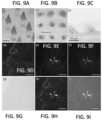
- FIG. 9A optical microscope images of the PVP/PVA-MNAs incorporating Allura Red dye before human skin application.
- FIG. 9 Bshows bright-field microscope image of Allura Red R40 dye microneedle traces on living human skin samples.
- FIG. 9 Cshows optical microscope images of the PVP/PVA-MNAs incorporating Allura Red dye after human skin application.
- FIGS. 9 D-Ishow intradermal co-delivery of Alexa488-labeled Poly(I:C) and Alexa555-labeled OVA from tip-loaded CMC/Treh MNAs. 20 ⁇ optical magnification. Fluorescence microscope composite images demonstrate delivery cavities penetrating the epidermis and upper dermis, and delivery of both the antigen and adjuvant to targeted skin microenvironments.
- FIG. 10 Ashows an optical microscope images of MNAs integrating both Alexa555-OVA and Alexa488-Poly(I:C), with the fluorescent image being a representative figure of the pyramid head incorporating both cargos.
- FIG. 10 Bshows representative optical microscope images of the Alexa555-OVA and Alexa488-Poly(I:C)-loaded MNAs after an in vivo application to the depicted mouse.
- FIGS. 10 C and 10 Dshow the effective co-delivery of Alexa488-Poly(I:C) and Alexa555-OVA to the skin microenvironments using novel MNAs.
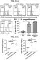
- FIG. 11 Aillustrates representative histograms from flow cytometry analysis showing remaining CFSE-labeled cells in spleens of immunized and unimmunized mice.
- FIG. 11 Bshows quantification of specific cell lysis, with 100% lysis corresponding to complete elimination of target cells (mean ⁇ SD, 3 mice per group).
- FIG. 11 Cshows serum concentrations of OVA-specific IgG1 and IgG2c antibodies (bars represent mean values, 3 mice per group).
- MNAsmicroneedle arrays
- biologicalrefers to pharmaceutically active agents, such as analgesic agents, anesthetic agents, anti-asthmatic agents, antibiotics, anti-depressant agents, anti-diabetic agents, anti-fungal agents, anti-hypertensive agents, anti-inflammatory agents, anti-neoplastic agents, anxiolytic agents, enzymatically active agents, nucleic acid constructs, immunostimulating agents, immunosuppressive agents, vaccines, and the like.
- pharmaceutically active agentssuch as analgesic agents, anesthetic agents, anti-asthmatic agents, antibiotics, anti-depressant agents, anti-diabetic agents, anti-fungal agents, anti-hypertensive agents, anti-inflammatory agents, anti-neoplastic agents, anxiolytic agents, enzymatically active agents, nucleic acid constructs, immunostimulating agents, immunosuppressive agents, vaccines, and the like.
- the bioactive materialcan comprise dissoluble materials, insoluble but dispersible materials, natural or formulated macro, micro and nano particulates, and/or mixtures of two or more of dissoluble, dispersible insoluble materials and natural and/or formulated macro, micro and nano particulates.
- any other suitable bioactive materialsuch as those discussed above, can be used in the novel MNA designs.
- pre-formedmeans that a structure or element is made, constructed, and/or formed into a particular shape or configuration prior to use. Accordingly, the shape or configuration of a pre-formed microneedle array is the shape or configuration of that microneedle array prior to insertion of one or more of the microneedles of the microneedle array into the patient.
- undercutor “undercut feature” refers to a recessed surface that is deemed to be inaccessible using standard molding methods and, in particular, to a feature (e.g., an indentation, protrusion, or other geometric shape) that restricts or prevents the withdraw of a molded part having this feature from a conventional one-piece mold.
- a featuree.g., an indentation, protrusion, or other geometric shape
- the singular forms “a,” “an,” and “the”include the plural forms unless the context clearly dictates otherwise. Additionally, the term “includes” means “comprises.” Furthermore, as used herein, the term “and/or” means any one item or combination of items in the phrase. In addition, the term “exemplary” means serving as a non-limiting example, instance, or illustration. As used herein, the terms “e.g.,” and “for example,” introduce a list of one or more non-limiting embodiments, examples, instances, and/or illustrations.
- microneedle arrayshave many advantages over conventional needle-based injection techniques. For this reason, cutaneous vaccination or drug delivery using microneedle arrays offers a viable and attractive approach to effective immunization or cancer immunotherapy due to the aforementioned theoretical advantages of the skin.
- MNAsphysically penetrate the stratum corneum, thereby eliminating formulation complexities and resulting in localized deposition of vaccines or drugs in the skin microenvironments.
- the microneedlesare benign to pain receptors, enabling minimally-invasive pain-free immunization.
- MNAscan provide highly effective vaccination due to their higher antigen loading capacity, tunable release kinetics, simple manufacturing, and long-term stability.
- Such MNAscan be created from water-soluble polymers that dissolve when inserted into the skin.
- the microneedles of MNAsare preferably strong enough in their dry-state to penetrate the stratum corneum, and then rapidly dissolve in the fluid environment of the skin, thereby releasing vaccines.
- Precise delivery of bioactive materials, such as vaccines, to the cutaneous microenvironmentscan result in improved efficiencies, thereby requiring relatively lower doses compared to traditional needle injections.
- microneedles with undercut geometries that include dissolvable and/or non-dissolvable materialscan be formed by the novel methods and systems described herein.
- the novel MNAscan be formed via a single-step micromolding process that utilizes flexible production mold materials.
- the use of such flexible production mold materialsallows for the production of a range of geometric designs not otherwise possible.
- the flexible production mold materialcan comprise, for example, any elastomer or other flexible material that allows for the removable of the microneedles of a desired design (e.g., a desired amount of undercut and/or other geometric shape).
- the ability to mold a variety of geometric shapes that are otherwise difficult to manufacture by moldingenables novel and innovative functionally-graded MNAs, such as the ones shown in FIG. 1 , for targeted delivery of a variety of bioactive materials to skin and other tissues (e.g., cardiac and ocular tissue).
- FIG. 1illustrates examples of different structures of microneedles that can be formed using the methods and systems described herein.
- four different microneedle structures 10 , 12 , 14 , 16are shown in a production mold 18 that is formed from a flexible material, such as a flexible elastomer.
- Microneedle 10is formed with a dissolvable backing layer 20 , a dissolvable stem 22 , and a microneedle tip 24 that is loaded with a bioactive material.
- microneedle 10is fabricated from a dissolving (or biodegradable) material throughout.
- the bioactive materialcan be mixed into the dissolving material, but is preferably located at the tip of the needles as shown in FIG. 1 to improve delivery efficiency.
- microneedles 10 , 12 , 14 , 16can have a pyramid head-shape with a sharp tip and an undercut stem that connects to a backing layer through filleted bases.
- Fillets 32can provide improved mechanical performance during tissue insertion.
- the MNAs described hereincan be fabricated from any moldable dissolving, biodegradable, and/or biocompatible, non-dissolvable materials including carboxymethylcellulose, trehalose, polyvinylpyrrolidone, poly(vinyl alcohol), maltodextrin, silk, glucose, hyaluronic acid, poly(methyl methacrylate), polycarbonate, poly(lactic-co-glycolic acid), poly(lactic acid), light curable resins, and their combinations to incorporate any bioactive materials, including cosmetics, dermal-fillers, statins, growth factors, pain killers, anti-histamines, vitamins, anesthetics, anti-aging agents, small molecule drugs, haptens, allergens, anti-inflammatory agents, proteins, peptides, micro vesicles, exosomes, polyplexes (siRNA, shRNA, DNA vector complexes), recombinant viral vectors (i.e., Adenovirus, Lentivirus, Vaccinia
- Microneedle 12is formed with a non-dissolvable backing layer 26 , a non-dissolvable stem 28 , and a microneedle tip 24 that is loaded with a bioactive material.
- This functionally-graded, undercut MNA designcan be fabricated with more than one material.
- the pyramid portioncan be created using a dissolving or biodegradable material, while the stem portion and the backing layer are manufactured from a non-dissolvable material, such as a non-dissolvable biocompatible rigid polymer (e.g., poly(methyl methacrylate), polycarbonate, VeroWhite and other UV-curable and heat curable resins).
- a non-dissolvable biocompatible rigid polymere.g., poly(methyl methacrylate), polycarbonate, VeroWhite and other UV-curable and heat curable resins.
- Microneedle 12can, therefore, provide a sharp needle tip along with enhanced mechanical performance through a filleted base enables successful tissue penetration and a pyramid head that serves as the bioactive material dosage form where the bioactive material(s) are incorporated into a dissolving or degrading biomaterial matrix.
- the undercut stem portionimproves the mechanical performance during penetration while ensuring tissue retention during implantation and the non-dissolvable undercut stem portion prevents back diffusion of the embedded bioactive material(s) during both fabrication and implantation processes.
- the non-dissolvable backing layercan also help prevent absorption of humidity during storage, which may result in excessive curvature of the backing layers and render MNA applications suboptimal.
- Microneedle 14is similar in shape to microneedles 12 , 14 , but further includes another dissolving layer 30 .
- the pyramid portionis created using a dissolving or biodegradable material and a more quickly dissolving layer is provided adjacent to the stem/needle tip connection.
- the dissolving layercan be formed for example, from a small molecular weight quickly dissolving polymer such as glucose, sucrose, trehalose, maltodextrin, or polyvinylpyrrolidone.
- the rest of the stem portion and the backing layercan be formed from a non-dissolvable material, such as a non-dissolvable biocompatible rigid polymer such as acrylated polyesters, epoxies, UV-curable monomers, resins, silicones.
- a non-dissolvable biocompatible rigid polymersuch as acrylated polyesters, epoxies, UV-curable monomers, resins, silicones.
- Microneedle 14therefore provides a sharp needle tip along with enhanced mechanical performance through a filleted base enables successful tissue penetration, a pyramid head that serves as the bioactive material dosage form where the bioactive material(s) are incorporated into a dissolving or degrading biomaterial matrix, an undercut stem portion improves the mechanical performance during penetration while ensuring tissue retention during implantation, and a quickly dissolving layer along with the mechanical mismatch between dissolving and non-dissolving layers that facilitates quick separation of pyramid tips.
- Microneedle 16is similar to microneedle 14 but further comprises a conforming backing layer 34 , which may be non-dissolvable.
- microneedle 16can be formed with a pyramid portion that is created using a dissolving or biodegradable material, the quickly dissolving layer can be formed as described above, the rest of the stem portion can be manufactured from a non-dissolvable material, and the backing layer can be manufactured from a conformable material, such as a non-dissolvable conformable polymer (silicones, UV-curable polymers, elastomers).
- Microneedle 16can therefore provide a sharp needle tip along with enhanced mechanical performance through a filleted base enables successful tissue penetration, a pyramid head that serves as the bioactive material dosage form where the bioactive material(s) are incorporated into dissolving or degrading biomaterial matrix, and an undercut stem portion that improves the mechanical performance during penetration while ensuring tissue retention during implantation,
- the quickly dissolving layer along with the mechanical mismatch between dissolving and non-dissolving layerscan facilitate quick separation of pyramid tips and the non-dissolvable undercut stem portion prevents back diffusion of the embedded bioactive material during both fabrication and implantation processes.
- the backing layercan conform to non-uniform skin topography better and, if non-dissolvable, it can help prevent absorption of humidity which may result in excessive curvature of the backing layers and render MNA applications suboptimal.
- FIG. 2 A- 2 Cillustrate the ability of a microneedle array 50 that has microneedles 52 and a conformable backing layer 34 to bend, and after bending return to an original state, or at least a state that is closer in shape to the original state than the bent state.
- the conformable backing layer of microneedle 16 and the quickly dissolving layer 30 of microneedles 14 , 16can be used in combination with any of the other structures disclosed herein (e.g., microneedles 10 , 12 ).
- the processes disclosed hereincan be used to create a microneedle that is formed entirely from non-dissolving materials for use as a coated MNAs, tissue adhesives (patches), and/or microbarbs.
- Non-dissolvable undercut microbarb or microneedle geometrieshave been created previously using additive manufacturing or mechanical micromachining processes. However, high-throughput manufacturing of those geometries is hindered due to lack of effective micromolding processes.
- the MNAscan be formed using additive manufacturing (AM), including micro-additive manufacturing ( ⁇ AM) techniques.
- AMadditive manufacturing
- ⁇ AMmicro-additive manufacturing
- the methods and systems described hereincan allow medical researchers with little microfabrication expertise to directly produce their MNA designs from a computer-aided design (CAD) drawing without the complex requirements of the subtractive fabrication processes.
- CADcomputer-aided design
- the pAM techniques described hereinprovide an effective means for fabrication of novel MNAs designed specifically for effective cutaneous and non-cutaneous drug delivery.
- the MNA designsincluded uniquely-shaped micron-scale needles that comprising sharp pyramid heads and undercut stems with filleted bases to ensure successful skin penetration and retention.
- the MNAs disclosed hereinare fabricated by a three-dimensional (3D) ⁇ AM approach with 3D direct laser writing, which offers transformative potential for the MNA field with its unparalleled level of simplicity and design capabilities.
- replicas of the master MNAscan be obtained from a mechanically-strong, moldable resin by a two-step micromolding approach with high fidelity. These replicas can then be used to prepare productions molds, such as polydimethylsiloxane (PDMS) production molds, which enabled fabrication of novel, tip-loaded dissolvable MNAs with undercut microneedles.
- productions moldssuch as polydimethylsiloxane (PDMS) production molds, which enabled fabrication of novel, tip-loaded dissolvable MNAs with undercut microneedles.
- the resulting MNAsare, in some embodiments, fully-dissolving MNAs with true undercut features for effective cutaneous drug delivery.
- a plurality of master MNAscan be printed, replicas of the master MNAs quickly obtained, and then the plurality of replicas can be assembled together to create larger MNAs.
- the MNAscan incorporate a model antigen Ovalbumin (OVA) ⁇ a model adjuvant Poly(I:C) from a biodissolvable material composition (70% CMC/30% Treh) using a spin-casting method.
- OvalbuminOvalbumin
- a model adjuvant Poly(I:C)from a biodissolvable material composition (70% CMC/30% Treh) using a spin-casting method.
- FIGS. 3 A and 3 Billustrate an exemplary microneedle array (MNA) ( 100 ) that comprises microneedles with a sharp-tipped pyramid head ( 102 ) and an undercut stem portion ( 104 ).
- MNAmicroneedle array
- the microneedlesinclude a filleted base ( 106 ).
- the methods and systems disclosed hereinenable reproducible fabrication of high-quality, tip-loaded dissolving MNAs with undercut features from different and widely-used dissolving microneedle materials, including CMC, PVP, Silk, HA, CMC/Trehalose, CMC/Glucose, CMC/Sucrose, PVP/PVA, and any other moldable biodissolvable compositions.
- the microneedlehas a height ( 110 ) that is between 50-1500 ⁇ m, such as 750 ⁇ m in height, and an apex angle (A) of the pyramid head that is between 10°-60, such as 30°.
- the stem portion of the microneedlecan have a width ( 112 ) that is between 50-500 ⁇ m, such as 150 ⁇ m.
- the stem portion ( 104 )can extend from the bottom region of a three-dimensional (3D) pyramid head to a backing layer ( 108 ) of the microneedle with a radius filleted connection that ranged from 15-75 ⁇ m, such as 35 ⁇ m.
- a width ( 114 ) of the bottom region of the pyramid headcan be, for example, between 100-400 ⁇ m, such as 250 ⁇ m ⁇ 250 ⁇ m base area.
- the tip-to-tip distance ( 116 ) between the microneedles in the arraycan be between 100-800 ⁇ m, such as 650 ⁇ m.
- the MNAcan include between 1-1000 microneedles, such as 25 microneedles in a 5 ⁇ 5 array configuration on a backing layer with an area of 4.75 mm ⁇ 4.75 mm.
- the fillet at the microneedle base ( 106 )can help reduce the associated mechanical stress concentrations at sharp corners, and in turn, increases the microneedle performance during manufacturing processes and skin insertion.
- the apex angle, the width, and the height of the microneedleswere selected to provide improved skin insertion mechanics and to reduce failures in penetration.
- the undercut, or anchor featurecan improve skin retention during application and, using the novel methods and systems disclosed herein, can be achieved without also interfering with the processing steps, thereby allowing direct removal of the MNAs from the molds.
- 3D- ⁇ AMthree-dimensional micro-additive manufacturing
- Exemplary manufacturing systems and processesare presented in a flow chart in FIG. 4 A and graphically illustrated in FIG. 4 B .
- the processcan comprise the following steps to create novel fast-dissolving MNAs while simultaneously achieving high-throughput fabrication.
- a 3D-CAD drawing ( 132 ) of the MNA designcan be prepared (process step 120 ).
- a direct production of the master MNA ( 134 ) from the CAD drawingcan be produced by 3D direct laser writing using a non-dissolvable resin (e.g., IP-S photoresist) (process step 122 ).
- the 3D- ⁇ AM manufacturing systemcan be, for example, a Nanoscribe 3D printing system ( 136 ).
- a quick and high-fidelity replication of the master MNAcan be formed with a UV-curable resin (e.g., VeroWhitePlus, Tangoblack, Digital Materials) using a two-step micromolding approach (process step 124 ).
- This approachcan include a negative elastomer mold ( 138 ) to form the replica ( 140 ) of the master MNA.
- the MNA master moldscan be created with multiple master MNA replicas (e.g., six replicas) onto 3D-printed MNA holders (process step 126 ).
- the 3D printed MNA holder ( 142 )can be formed of, for example, a resin material.
- the combination of a plurality of master MNA replicas together onto, for example, 3D-printed MNA holderscan allow for the creation of larger MNAs and improve productivity.
- the MNA production molds ( 144 )can be manufactured from the elastomer PDMS using micromolding (process step 128 ).
- Sixth, tip-loaded, dissolving MNAs ( 150 )can be fabricated (process step 130 ).
- the MNAscan have undercut microneedles incorporating one or more bioactive material, such as a vaccine or any other bioactive material from a water-soluble biocompatible material (e.g., a composition of CMC and Treh) through a multiple-step spin-casting method using a centrifuge.
- a bioactive componente.g., vaccine
- the dissolvable hydrogele.g., CMC/Trehalose
- the master MNA, the MNA master molds including replicas of the master MNA, and the elastomer production moldscan be reused for a large number of processing cycles, thereby greatly reducing the fabrication costs for MNAs with unique designs and improving productivity.
- FIGS. 5 A- 5 Eshow final products corresponding to different manufacturing or processing steps.
- FIG. 5 Ashows an optical microscope image of the master MNA ( 134 ) created using 3D direct laser writing
- FIG. 5 Dshows an optical microscope image of an individual undercut needle on the 3D printed master MNA ( 134 ).
- the master MNAwas fabricated from IP-S photoresist by 3D direct laser writing.
- IP-Sis a specific material designed for 3D laser lithography and provides high resolution and mechanical integrity for micro- and nano-structures.
- 3D laser lithography based on two-photon polymerizationprovided an effective means for fabrication of undercut MNA designs with smooth edges and sharp tips, and without any unwanted residues (e.g., machining chips).
- FIG. 5 Bshows an optical microscope image of the replica ( 140 ) of the master MNA created through a two-stage micromolding strategy: elastomer molding combined with UV-curable micromolding
- FIG. 5 Eshows an optical microscope image of an individual undercut needle on the replica ( 140 ) of the master MNA.
- the master MNAwas produced using a two-step micromolding process.
- the IP-S master MNAwas used to fabricate PDMS flexible mold through elastomer molding and then the PDMS mold was used to manufacture several VeroWhite MNA replicas through UV-curable micromolding.
- VeroWhite resinis a wear-resistant, acrylic-based photo-polymer that can be used with 3D Polyjet printers.
- MNA replicaswere then used to create MNA master molds that include a number of MNAs (e.g., six MNA replicas).
- the final MNA master moldswere post-processed in a vacuum oven to facilitate successful curing of PDMS on the mold surface and then the elastomer MNA production molds, which consisted of microneedle-shaped wells, were fabricated from PDMS.
- FIG. 5 Cshows an optical microscope image of the microneedle-shaped wells in MNA production molds ( 144 ).
- dissolving MNAs that integrate the vaccine in the tip portion of the needleswere fabricated using the conventional three-stage manufacturing strategy through master mold to production mold to final dissolvable MNAs.
- dissolving MNAs that incorporated the vaccine (10 ⁇ g OVA ⁇ 25 ⁇ g Poly(I:C)) in the tip portion of the undercut needleswere fabricated through the spin-casting process from two different material compositions (i.e., CMC/Trehalose and PVP/PVA). Furthermore, tip-loaded CMC/Trehalose MNAs with undercut microneedles integrating a colored model drug (e.g., Doxorubicin) were fabricated to facilitate imaging and demonstrate compatibility with chemotherapeutic agents.
- the systems and methods described hereinenabled effective and rapid fabrication of tip-loaded dissolving MNAs with undercut microneedles from different dissolvable material compositions.
- FIG. 6 Ashows an optical microscope image of final dissolving (CMC/Treh) MNAs incorporating biocargos (e.g., OVA+Poly(I:C)).
- FIG. 4 Bshows an optical microscope image of dissolving, tip-loaded PVP/PVA MNAs with undercut microneedles integrating OVA
- FIG. 4 Cshows an optical microscope image of an individual tip-loaded CMC/Treh undercut microneedle incorporating Doxorubicin as a colored drug to facilitate imaging.
- a unique MNA designwas directly created from the 3D-CAD drawing shown in FIG. 1 using 3D laser printing (Nanoscribe Photonic Professional, GT) with the photopolymetric resist IP-S.
- the Nanoscribe printing systemwas equipped with a laser generator, an optical cabinet, a Zeiss optical microscope attached to a lens to focus the laser beam, a Galvo mirror system to direct the laser-beam scanning, a piezoelectric stage for precise motion control, and an operation software (Nanowrite) to execute 3D printing. The whole system was placed on an optical table to eliminate vibrations during the printing process.
- the MNA designwas converted into ‘STL’ (StereoLithography) format.
- the STL filewas loaded into the specialized software (DeScribe, Germany) of the Nanoscribe system to select the processing conditions (i.e., the distance of slicing, hatching, and splitting).
- the STL filewas converted into ‘GWL’ (General Writing Lithography) format to be exported in the Nanowrite software for printing the master MNA.
- the master MNAwas fabricated using Galvo-scan mode in XY plane and piezo-scan mode in Z direction.
- the master MNAwas split into blocks of 220 ⁇ m ⁇ 220 ⁇ m ⁇ 200 ⁇ m within the working range and then stitched together.
- the laser power and writing speedwas set to be 100 mW and 6 cm/s, respectively.
- the master MNAwas then printed through two-photon polymerization of the IP-S photoresist by a femtosecond pulsed laser at a wavelength of 750 nm using a unique deep-in-liquid mode with the objective of 25 ⁇ and NA0.8 in Shell and Scaffold mode.
- the master MNAwas developed in the photoresist solvent propylene glycol monomethyl ether acetate (PGMEA) for 30 min, followed by 5 min isopropyl alcohol (IPA) rinse. After the master MNA was dried in the air, it was placed under a UV (365 nm) light with 16 mW/cm2 intensity for 30 min to further crosslink the body to strengthen the MNA structure.
- PGMEAphotoresist solvent propylene glycol monomethyl ether acetate
- IPAisopropyl alcohol
- a two-stage micromolding methodwas performed.
- an elastomer mold which is negative of the master MNAwas manufactured from polydimethylsiloxane (PDMS) using a micromolding approach. Elastomer molding using PDMS provides an accurate and reproducible replication of high-fidelity micron-scale structures.
- PDMSpolydimethylsiloxane
- the master MNAwas mounted in a petri-dish with a diameter of 5 cm and PDMS was obtained using the two-component curable silicone elastomer, SYLGARD® 184 (Dow Corning), by mixing the base material with a curing agent in 10:1 SYLGARD®-to-curing agent ratio. Subsequently, the mixture was poured over the master MNA mounted into the petri-dish and degassed for 15 min Next, the master MNA with the degassed mixture was placed in an oven and cured at 70° C. for 1 h. The cured PDMS was cooled down to room temperature for 5 min and then separated from the master MNA to obtain the negative PDMS mold.
- SYLGARD® 184Low Corning
- the second processing stepused the negative PDMS mold for fabrication of positive MNA replicas from the UV-curable resin (Stratasys®, VeroWhiteplus-RGD835).
- the UV-curable resin(Stratasys®, VeroWhiteplus-RGD835).
- 20 ⁇ l of liquid (uncured) resinwas poured onto the molds and then the molds were placed in a centrifuge to fill the microneedle-shaped wells with the resin at 4500 RPM and at 20° C. for 1 min.
- the resinwas then cured under UV light (365 nm) with 21.7 mW/cm2 intensity for 5 min from each of the top and bottom sides to cure both the base and the microneedle tips.
- MNA master moldsTo facilitate scalable manufacturing of dissolving MNAs, the MNA master molds were created through assembling six replicas of the master MNA onto the MNA holders fabricated by Stratasys® from a non-dissolvable photo-polymer (VeroWhite) using a high-resolution (16 ⁇ m) Polyjet 3D printing system (Objet Connex 500 multi-material).
- the 3D model of the MNA holderscreated using SolidWorks 2018 CAD software and then converted into the ‘STL’ (StereoLithography) file format.
- the specialized software(Objet Studio) sliced this 3D model into 2D cross-sectional layers, creating a computer file that was sent to the 3D printer system.
- the channels in the 3D printed MNA holderwere designed and fabricated to serve as raised pockets in the MNA production molds to assist as reservoirs for both the bioactive cargo (e.g., vaccine) and the structural hydrogel material of dissolving MNAs during the spin-casting process.
- the created MNA master moldswere baked at 80° C. overnight in a vacuum oven to facilitate effective fabrication of elastomer MNA production molds.
- the MNA production molds that included microneedle-shaped wellswere fabricated from a commonly-used elastomer polydimethylsiloxane (PDMS) as described for the replication of the MNA master.
- the base materialwas mixed with a curing agent in 10:1 SYLGARD®-to-curing agent ratio. Subsequently, the mixture was poured over the MNA master mold placed in a 10 cm diameter petri-dish and degassed for 15 min. Next, the master mold with the degassed mixture was placed in an oven to cure PDMS at 70° C. for 1 h. After cooling down the cured PDMS to room temperature, it was separated from the MNA master mold to fabricate the PDMS MNA production molds.
- PDMSelastomer polydimethylsiloxane
- the prepared hydrogelwas refrigerated at 4° C. for 24 h for the mixture to equilibrate.
- Tip-loaded CMC/Treh-MNAs with the unique designswere then manufactured through a multiple-step spin-casting technique using a centrifuge (Thermo Fisher Scientific Sorvall Legend XTR with Swinging Bucket Rotor TX-750).
- OVA solution25 mg/mL OVA in endotoxin-free water
- the production moldswere centrifuged for 1 min at 20° C. and at 4500 rpm to fill the microneedle-shaped cavities.
- the excess OVA solution within the reservoirwas recovered.
- the production moldswere again centrifuged for 30 min at 20° C. and at 4500 rpm to ensure that dry OVA cargo was located at the tip portion of the needle-shaped cavities in the production molds.
- hydrogel(30% w/w 70:30 CMC:Treh) was loaded over each of the MNAs on the PDMS production molds to fill the microneedle-shaped geometries in the production molds and to form the backing layers of the MNAs.
- the hydrogel-loaded production moldswere then centrifuged for 5 h at 20° C. and at 4500 rpm to obtain dissolving MNAs loaded with 10 ⁇ g OVA ⁇ 25 ⁇ g Poly(I:C). Due to the specific geometry of the MNA production molds, each replication produced six arrays of OVA ⁇ Poly(I:C)-loaded dissolving MNAs simultaneously.
- a biodissolvable polymer composition of Polyvinylpyrrolidone (PVP) and Polyvinyl alcohol (PVA) (40% w/w 60:40 PVP:PVA)was also used to fabricate MNAs with different biocargos.
- PVPPolyvinylpyrrolidone
- PVAPolyvinyl alcohol
- the fabricated master MNAs, replicas of MNAs, elastomer MNA production molds, and OVA ⁇ Poly(I:C)-loaded dissolving MNAswere imaged using bright-field optical microscopy to assess geometric integrity of the novel microneedles with undercut features.
- Human skin explantswere prepared from deidentified healthy donors undergoing plastic surgery acquired through the Pitt Biospecimen Core and used according to University of Pittsburgh Medical Center guidelines. Tissue was rinsed in 70% ethanol and then in phosphate-buffered saline (PBS). Human skin explants (approximately 1 mm thick) were harvested using a Silver's miniature skin graft knife (Padgett, Integra Miltex, Plainsboro, NJ), and then cut into 20 mm ⁇ 20 mm square pieces. The resulting human skin samples were comprised of unaltered epidermis and a thin layer of underlying dermis, and maintained as explants in normal physiological state by culture at an air-fluid interface.
- PBSphosphate-buffered saline
- Imaging analysisTo evaluate MNA-directed intradermal bioactive materials (e.g., vaccine) delivery to living human skin explants, a number of imaging analyses was performed. Tip-loaded CMC/Treh-MNAs integrating a colored cargo (i.e., Allura Red R40 dye) were fabricated using the manufacturing strategy described above. Prior to application of MNAs to the human skin explants, MNAs were imaged using bright-field optical microscopy.
- a colored cargoi.e., Allura Red R40 dye
- MNAswere applied to human skin explants and removed after 10 min.
- the targeted human skin regionswere then examined under a bright-field microscope to image the patterns of the colored biocargo deposited from CMC/Treh-MNAs into the human skin. Remaining MNA materials were also imaged using optical microscopy after human skin applications.
- CMC/Treh MNAs that incorporate both Alexa555-labeled OVA and Alexa488-labeled Poly(I:C)were fabricated, applied to human skin for 10 min, and removed. The targeted human skin explants were then histologically analyzed.
- the MNA-treated human skin sampleswere fixed in 2% paraformaldehyde followed by immersion in sucrose solution, with 3 changes of this solution over 24 h.
- Tissue sectionswere then flash frozen in optimum cutting temperature (OCT) histology compound, and cryo-sectioned into approximately 10 ⁇ m thick sections.
- OCToptimum cutting temperature
- the sectioned human skin sampleswere counter-stained using nuclear DAPI fluorescent dye.
- the stained sectionswere then imaged using a Nikon transmission fluorescent microscope to detect Alexa555-OVA and Alexa488-Poly(I:C) into the cross-sections of human skin, as well as using bright field microscope to better demonstrate the stratum corneum breaching.
- mice and animal husbandryFemale C57BL/6J mice were purchased from The Jackson Laboratory (Bar Harbor, ME) and used at 8-10 weeks of age. Mice were maintained under specific pathogen-free conditions at the University of Pittsburgh, and all experiments were conducted in accordance with the institutional animal care and use committee (IACUC) guidelines.
- IACUCinstitutional animal care and use committee
- OVA+Poly(I:C) loaded MNAswere imaged before and after in vivo application Imaging of the MNA-treated mouse was performed on the IVIS 200 in vivo imaging system (PerkinElmer) using the filters to assay for fluorescent-labeled Poly(I:C) and OVA the MNA application site. The image was post-processed using Living Image software (PerkinElmer).
- OVA-specific antibody responseblood was collected from anesthetized mice at the time of sacrifice by cardiac puncture, and serum was isolated using BD Microtainer serum separator tubes (BD Biosciences, San Jose, CA). OVA-specific IgG1 and IgG2c antibodies in serum were measured by indirect ELISAs. Costar EIA/RIA plates (Corning Inc., Corning, NY) were coated with OVA (100 ⁇ g/mL in 0.5 M carbonate-bicarbonate buffer, pH 9.6; Sigma) by overnight incubation at 4° C. Plates were washed (3 ⁇ ) with 0.05% Tween20 in PBS, and blocked with 1% goat serum in PBS for 1 hour at 37° C.
- Serum samples and standards(anti-OVA IgG1 from Cayman Chemical, Ann Arbor, MI; anti-OVA IgG2c from Chondrex, Redmond, WA) were diluted with 1% goat serum, added to plates, and incubated 2 hours at 37° C. After washing (3 ⁇ ), plates were incubated for 1 hour at 37° C. with biotinylated secondary antibodies (goat anti-mouse IgG1 or IgG2c, 1:20,000 in 1% goat serum; Jackson ImmunoResearch, West Grove, PA). Plates were then washed (3 ⁇ ) and incubated for 30 min with streptavidin-HRP (1:1000 in 1% goat serum; BD Biosciences).
- TMB4,4′,5,5′-tetramethylbenzidine
- OD450absorbance at 450 nm
- splenocytes from na ⁇ ve micewere pulsed with 2 ⁇ g/ml OVA257-264 (SIINFEKL) peptide, or left unpulsed for 1 h.
- Antigen pulsed splenocyteswere washed and stained with high concentration carboxyfluorescein succinimidyl ester (CFSE, 10 ⁇ M), while unpulsed splenocytes were labeled with low concentration CFSE (1 ⁇ M) for 15 min at 37° C.
- CFSEcarboxyfluorescein succinimidyl ester
- a 1:1 mixture of pulsed target cells and unpulsed control cellswas intravenously (IV) injected into immunized and na ⁇ ve mice.
- % Lysis⁇ 1 ⁇ [(mean CFSElow/CFSEhigh ratio from na ⁇ ve mice)/(CFSElow/CFSEhigh ratio from vaccinated mouse)] ⁇ 100.
- the MNAs described hereincan be used for a broad range of intradermal and non-cutaneous (e.g., ocular and cardiac tissues) drug delivery applications.
- the biocargo of interestcan be located at the very tip of microneedles in the spin-casting process (i.e., a portion of a pyramid) or the entire pyramid region can be loaded with the biocargo depending on the dose requirements.
- FIG. 7 Ashows PVP/PVA MNAs incorporating Texas Red labeled Dextran (approximately 40 kDa molecular weight) at the tips of microneedles
- FIG. 7 Bshows tip-loaded CMC/Treh MNAs integrating Allura Red R40 dye (approximately 500 Da molecular weight) at the pyramid region of microneedles.
- FIGS. 7 C and 7 Dshow tip-loaded PVP/PVA MNAs incorporating multiple cargos such as Texas Red labeled Dextran and Allura Red R40 dye
- FIG. 7 Dshows tip-loaded PVP/PVA MNAs incorporating multiple cargos such as Texas Red-labeled Dextran and Alexa488-labeled PLGA microparticles (10 ⁇ m mean diameter).
- FIGS. 7 A and 7 Cshow the MNAs obtained after the first and twelfth cycles using t, respectively).
- FIGS. 7 A and 7 Cshow the MNAs obtained after the first and twelfth cycles using t, respectively.
- Additive manufacturing or 3D printingas used in the systems and methods described herein provides for accurate and reproducible manufacturing of 3D complex geometries without design limitations and offers a high degree of design flexibility and control. Using the systems and methods here, rapid design-to-fabrication turnaround for optimal application-driven drug delivery systems is possible.
- microneedle designswere fabricated with diverse geometries.
- FIGS. 8 A- 8 Bshow that this technology enables fabrication of a wide range of microneedle geometries with high-fidelity towards application-driven optimization. Furthermore, as shown in FIGS. 8 A- 8 B , it allows a wide range of design changes, including height, width, apex angle, and geometry of the microneedles without requiring complex and custom processing steps. As such, this technology paves the way for design and fabrication of application-driven unique MNA designs.
- MNAs tip-loaded with Allura Red R40 dyewere manufactured using the presented fabrication strategy. Human skin explants were prepared as described above. Allura Red R40 dye loaded MNAs were applied to living human skin explants and removed after 10 min Images of these MNAs before ( FIG. 9 A ) and after ( FIG. 9 B ) application demonstrated high-quality MNAs and the complete dissolution of the microneedles, respectively. The corresponding deposits of MNA-embedded cargo (e.g., Allura Red R40 dye) in the targeted skin was shown in FIG. 9 C .
- MNA-embedded cargoe.g., Allura Red R40 dye
- FIGS. 9 D-IDAPI nuclear stain, Alexa488-labeled Poly(I:C), Alexa555-labeled OVA, Brightfield, overlay of 3 different fluorescent colors, merged image of all the fluorescent images with the brightfield image, respectively).
- the scale barscorrespond to 100 ⁇ m.
- CMC/Treh MNAs with high-fidelity undercut microneedles incorporating both Alexa555-labeled OVA and Alexa488-labeled Poly(I:C)were fabricated as described above.
- Alexa555-OVA+Alexa488-Poly(I:C) MNAswere imaged using bright-field optical microscopy and epi-fluorescent microscopy ( FIG. 10 A ). MNAs were then applied to mice and removed after 10 min. The remaining MNA material after applications was also imaged using bright-field optical microscopy ( FIG. 10 B ).
- MNA-treated micewere imaged using the IVIS 200 live animal imaging system with filters for both Alexa488-Poly(I:C) and Alexa555-OVA.
- MNA-directed co-delivery of Poly(I:C) and OVAwere shown in FIGS. 10 C and 10 D , respectively. Together, these images demonstrated successful application of novel MNAs in vivo in mice, and in turn, MNA-directed effective cutaneous drug and vaccine delivery with new MNA designs.
- cytotoxic-T-cellCTL
- antibody responseswere quantified using the standard in vivo lytic assay and ELISA, respectively.
- Cutaneous vaccination with MNAselicited robust antigen-specific cellular immune responses ( FIGS. 11 A-B ).
- equivalent numbers of antigen-pulsed (CFSEhigh) target cells and unpulsed (CFSElow) target cellswere recovered from na ⁇ ve or unimmunized mice ( FIG. 11 A ), thereby indicating the absence of antigen-specific cytolytic activity.
- specific lysis of antigen-pulsed target cellswas dramatically enhanced in immunized mice, as shown by reduced survival of OVA-pulsed targets than unpulsed targets.
- mice immunized with OVA-MNAdemonstrated greater OVA-specific lysis in vivo, with significantly (as compared to IM-OVA) lower survival and recovery of OVA pulsed targets than unplused targets ( FIG. 11 A ).
- Immunization with OVA+Poly(I:C)further improved the performance of vaccination ( FIG. 11 A ).
- Quantification of antigen specific lysis by standard techniquesconfirmed that MNA immunization elicited potent CTL immunity ( FIG. 11 B ), which was further improved by the inclusion of the adjuvant Poly(I:C).
- mice with both MNAs and IMresulted in IgG responses
- those vaccinated with dissolving MNAshad significantly higher IgG titers, thereby indicating the importance of MNA technology in immunization efforts.
- the presented novel MNAsprovide a unique opportunity for the specific and precise delivery of embedded antigen ⁇ adjuvant to defined microenvironments within the skin. For example, we and others have shown that the skin is rich in dendritic cells and other antigen presenting cells (APCs) essential for vaccine induced immune induction.
- APCsantigen presenting cells
- targeting skin APCs using MNAscan be an effective strategy for vaccination in general, and in particular for the induction of cell mediated immune responses, including CTL responses essential to prevent or treat many infectious diseases and cancer.
- the presented MNA technologycould enable capability relevant to a broad range of vaccination strategies.
- novel dissolving MNAs with undercut microneedlesprovide for effective intradermal vaccination.
- the unique MNA designsinclude pyramid heads and undercut stem regions with filleted bases.
- the manufacturing approach to create the undercut MNAsstrategically involved 3D laser printing and a number of micromolding processes.
- Successful and reproducible fabrication of dissolvable MNAs with undercut needles that incorporate a myriad of biocargoswas achieved using different biocompatible and water-soluble polymers.
- the unprecedented level of geometric capability with 3D laser printingwas demonstrated towards application-driven MNA designs for several cutaneous and non-cutaneous drug delivery applications.
- the introduced novel MNAs with undercut featuresfulfilled strength requirements for failure-free skin penetration in humans and mice, and successfully delivered their biocargos to targeted cutaneous microenvironments.
- cutaneous vaccination using the model antigen-loaded MNAselicited significantly more potent antigen-specific cellular and humoral immune responses than those obtained by traditional intramuscular injection (IM-OVA) based immunization.
- IM-OVAintramuscular injection
- MNA-directed co-delivery of the antigen (OVA) with the adjuvant (Poly(I:C))improved the immunogenicity of the vaccine with respect to IM-based vaccination towards enhanced MNA-directed cutaneous vaccination.
- the presented approachprovides an effective means of fabricating novel dissolving MNAs for a broad range of cutaneous and non-cutaneous drug delivery applications.
Landscapes
- Engineering & Computer Science (AREA)
- Health & Medical Sciences (AREA)
- Mechanical Engineering (AREA)
- Manufacturing & Machinery (AREA)
- Life Sciences & Earth Sciences (AREA)
- Public Health (AREA)
- Heart & Thoracic Surgery (AREA)
- Veterinary Medicine (AREA)
- Animal Behavior & Ethology (AREA)
- General Health & Medical Sciences (AREA)
- Biomedical Technology (AREA)
- Medical Informatics (AREA)
- Hematology (AREA)
- Anesthesiology (AREA)
- Dermatology (AREA)
- Surgery (AREA)
- Vascular Medicine (AREA)
- Epidemiology (AREA)
- Medicinal Chemistry (AREA)
- Molecular Biology (AREA)
- Chemical & Material Sciences (AREA)
- Media Introduction/Drainage Providing Device (AREA)
- Medicines Containing Antibodies Or Antigens For Use As Internal Diagnostic Agents (AREA)
- Medicinal Preparation (AREA)
- Materials For Medical Uses (AREA)
Abstract
Description
Claims (20)
Priority Applications (1)
| Application Number | Priority Date | Filing Date | Title |
|---|---|---|---|
| US17/611,486US12214150B2 (en) | 2019-05-16 | 2020-05-15 | Microneedle arrays with undercut features for cutaneous and non-cutaneous drug delivery |
Applications Claiming Priority (3)
| Application Number | Priority Date | Filing Date | Title |
|---|---|---|---|
| US201962848939P | 2019-05-16 | 2019-05-16 | |
| PCT/US2020/033235WO2020232394A1 (en) | 2019-05-16 | 2020-05-15 | Microneedle arrays with undercut features for cutaneous and non-cutaneous drug delivery |
| US17/611,486US12214150B2 (en) | 2019-05-16 | 2020-05-15 | Microneedle arrays with undercut features for cutaneous and non-cutaneous drug delivery |
Related Parent Applications (1)
| Application Number | Title | Priority Date | Filing Date |
|---|---|---|---|
| PCT/US2020/033235A-371-Of-InternationalWO2020232394A1 (en) | 2019-05-16 | 2020-05-15 | Microneedle arrays with undercut features for cutaneous and non-cutaneous drug delivery |
Related Child Applications (1)
| Application Number | Title | Priority Date | Filing Date |
|---|---|---|---|
| US19/020,688ContinuationUS20250152929A1 (en) | 2019-05-16 | 2025-01-14 | Microneedle arrays with undercut features for cutaneous and non-cutaneous drug delivery |
Publications (2)
| Publication Number | Publication Date |
|---|---|
| US20220241570A1 US20220241570A1 (en) | 2022-08-04 |
| US12214150B2true US12214150B2 (en) | 2025-02-04 |
Family
ID=73289266
Family Applications (2)
| Application Number | Title | Priority Date | Filing Date |
|---|---|---|---|
| US17/611,486Active2041-01-17US12214150B2 (en) | 2019-05-16 | 2020-05-15 | Microneedle arrays with undercut features for cutaneous and non-cutaneous drug delivery |
| US19/020,688PendingUS20250152929A1 (en) | 2019-05-16 | 2025-01-14 | Microneedle arrays with undercut features for cutaneous and non-cutaneous drug delivery |
Family Applications After (1)
| Application Number | Title | Priority Date | Filing Date |
|---|---|---|---|
| US19/020,688PendingUS20250152929A1 (en) | 2019-05-16 | 2025-01-14 | Microneedle arrays with undercut features for cutaneous and non-cutaneous drug delivery |
Country Status (8)
| Country | Link |
|---|---|
| US (2) | US12214150B2 (en) |
| EP (1) | EP3969099A4 (en) |
| JP (1) | JP2022532215A (en) |
| CN (1) | CN113874065A (en) |
| AU (1) | AU2020275934B2 (en) |
| BR (1) | BR112021022680A2 (en) |
| CA (1) | CA3138521A1 (en) |
| WO (1) | WO2020232394A1 (en) |
Families Citing this family (9)
| Publication number | Priority date | Publication date | Assignee | Title |
|---|---|---|---|---|
| US11213482B1 (en) | 2020-03-05 | 2022-01-04 | University of Pittsburgh—Of the Commonwealth System of Higher Educat | SARS-CoV-2 subunit vaccine and microneedle array delivery system |
| AU2021254428A1 (en)* | 2020-04-09 | 2022-10-13 | University Of Pittsburgh - Of The Commonwealth System Of Higher Education | Microneedle array delivery of adenovirus vectored vaccines with and without adjuvants |
| US11844920B2 (en) | 2020-07-23 | 2023-12-19 | Microneedles Inc | Microneedle immunotherapeutic multi-component system and a method for vaccination |
| US20250082236A1 (en)* | 2021-07-19 | 2025-03-13 | Carnegie Mellon University | Scalable Manufacturing of Microneedle Arrays Using Automated High-Throughput Manufacturing Systems and High-Capacity Molding |
| KR20230014326A (en)* | 2021-07-21 | 2023-01-30 | 한국기계연구원 | Method of manufacturing a thin film attached to the skin coated with a functional mixture and thin film attached to the skin manufactured using the same |
| WO2023053078A1 (en)* | 2021-09-30 | 2023-04-06 | Scuola Superiore Sant'anna | Device with soluble hook-shaped micro-elements for the deployment of substances into the leaves of plants |
| CN114191700B (en)* | 2021-12-17 | 2022-08-05 | 北京理工大学 | A kind of soluble microneedle and medicine feeding device |
| KR102570928B1 (en)* | 2022-01-07 | 2023-08-28 | 주식회사 쿼드메디슨 | Particle attached microneedle and manufacturing method using the same |
| CN117815113B (en)* | 2024-03-04 | 2024-06-25 | 北京青颜博识健康管理有限公司 | A soluble microneedle that can be dried at high temperature and its preparation method and use |
Citations (187)
| Publication number | Priority date | Publication date | Assignee | Title |
|---|---|---|---|---|
| US5312456A (en) | 1991-01-31 | 1994-05-17 | Carnegie Mellon University | Micromechanical barb and method for making the same |
| US5658515A (en) | 1995-09-25 | 1997-08-19 | Lee; Abraham P. | Polymer micromold and fabrication process |
| WO1998000194A2 (en) | 1996-06-28 | 1998-01-08 | Sontra Medical, L.P. | Ultrasound enhancement of transdermal transport |
| WO1998029134A2 (en) | 1996-12-31 | 1998-07-09 | Altea Technologies, Inc. | Microporation of tissue for delivery of bioactive agents |
| US6331266B1 (en) | 1999-09-29 | 2001-12-18 | Becton Dickinson And Company | Process of making a molded device |
| US20020082543A1 (en) | 2000-12-14 | 2002-06-27 | Jung-Hwan Park | Microneedle devices and production thereof |
| US6451240B1 (en) | 1999-06-09 | 2002-09-17 | The Procter & Gamble Company | Method of manufacturing an intracutaneous microneedle array |
| US20020193729A1 (en) | 2001-04-20 | 2002-12-19 | Cormier Michel J.N. | Microprojection array immunization patch and method |
| US20020198509A1 (en) | 1999-10-14 | 2002-12-26 | Mikszta John A. | Intradermal delivery of vaccines and gene therapeutic agents via microcannula |
| US6511463B1 (en) | 1999-11-18 | 2003-01-28 | Jds Uniphase Corporation | Methods of fabricating microneedle arrays using sacrificial molds |
| US6565871B2 (en) | 1994-12-02 | 2003-05-20 | Elan Drug Delivery Ltd. | Solid dose delivery vehicle and methods of making same |
| US6611707B1 (en) | 1999-06-04 | 2003-08-26 | Georgia Tech Research Corporation | Microneedle drug delivery device |
| US6623707B1 (en) | 2000-06-19 | 2003-09-23 | Corning Incorporated | Monolithic catalyst dehydrogenation reactor |
| US6652478B1 (en) | 1999-06-09 | 2003-11-25 | The Procter & Gamble Company | Intracutaneous edged microneedle apparatus |
| US6656147B1 (en) | 2000-07-17 | 2003-12-02 | Becton, Dickinson And Company | Method and delivery device for the transdermal administration of a substance |
| US6663820B2 (en) | 2001-03-14 | 2003-12-16 | The Procter & Gamble Company | Method of manufacturing microneedle structures using soft lithography and photolithography |
| CN1475900A (en) | 2002-06-19 | 2004-02-18 | 三星电子株式会社 | Method and device for controlling a display device |
| US20040058882A1 (en) | 1995-05-19 | 2004-03-25 | Elof Eriksson | Microseeding device for gene delivery by microneedle injection |
| US6743211B1 (en) | 1999-11-23 | 2004-06-01 | Georgia Tech Research Corporation | Devices and methods for enhanced microneedle penetration of biological barriers |
| US6767211B2 (en) | 2001-03-13 | 2004-07-27 | Carolyn W. Hall | Method and apparatus for behaviorally reinforced training with guided practice |
| US20050008683A1 (en) | 2000-06-29 | 2005-01-13 | Becton Dickinson And Company | Method for delivering interferons to the intradermal compartment |
| US20050013221A1 (en) | 2003-07-15 | 2005-01-20 | Kabushiki Kaisha Toshiba | Optical disk apparatus and optical disk processing method |
| US20050019918A1 (en) | 2003-06-03 | 2005-01-27 | Hidetoshi Sumimoto | Treatment of cancer by inhibiting BRAF expression |
| JP2005035945A (en) | 2003-07-16 | 2005-02-10 | Cardio Corp | Combined therapy for tissue regeneration |
| WO2005025413A2 (en) | 2003-09-11 | 2005-03-24 | Theranos, Inc. | Medical device for analyte monitoring and drug delivery |
| US20050065463A1 (en) | 2003-09-18 | 2005-03-24 | Nano Device And System Research Inc. | Applicator for applying functional substances into human skin |
| US6881203B2 (en) | 2001-09-05 | 2005-04-19 | 3M Innovative Properties Company | Microneedle arrays and methods of manufacturing the same |
| US20050089553A1 (en) | 2003-10-28 | 2005-04-28 | Cormier Michel J. | Method and apparatus for reducing the incidence of tobacco use |
| US20050095298A1 (en) | 2002-04-22 | 2005-05-05 | Hans Gronlund | Microparticles comprising carbohydrate beads covalently linked with allergen |
| US6899838B2 (en) | 2002-07-12 | 2005-05-31 | Becton, Dickinson And Company | Method of forming a mold and molding a micro-device |
| CN1621102A (en) | 2004-12-13 | 2005-06-01 | 纳生微电子(苏州)有限公司 | Preparing method for epidermis needle and its application |
| US6908453B2 (en) | 2002-01-15 | 2005-06-21 | 3M Innovative Properties Company | Microneedle devices and methods of manufacture |
| US6924087B2 (en) | 2002-10-13 | 2005-08-02 | Nano Pass Technologies Ltd. | Polymer microneedles |
| US6931277B1 (en) | 1999-06-09 | 2005-08-16 | The Procter & Gamble Company | Intracutaneous microneedle array apparatus |
| US7132054B1 (en) | 2004-09-08 | 2006-11-07 | Sandia Corporation | Method to fabricate hollow microneedle arrays |
| US7211062B2 (en) | 2002-06-25 | 2007-05-01 | Theraject, Inc. | Solid solution perforator for drug delivery and other applications |
| US20070161964A1 (en) | 2006-01-10 | 2007-07-12 | Yuzhakov Vadim V | Microneedle array, patch, and applicator for transdermal drug delivery |
| WO2007080596A2 (en) | 2006-01-12 | 2007-07-19 | Nano Pass Technologies Ltd. | Device for superficial abrasive treatment of the skin |
| WO2007113648A2 (en) | 2006-04-05 | 2007-10-11 | Pfizer Products Inc. | Ctla4 antibody combination therapy |
| US7285113B2 (en) | 2000-03-09 | 2007-10-23 | Nanopass Technologies Ltd. | Systems and methods for the transport of fluids through a biological barrier and production techniques for such systems |
| US20070260201A1 (en) | 2006-05-02 | 2007-11-08 | Georgia Tech Research Corporation | Method for drug delivery to ocular tissue using microneedle |
| US20070299388A1 (en) | 2006-04-25 | 2007-12-27 | Alza Corporation | Microprojection array application with multilayered microprojection member for high drug loading |
| US7315758B2 (en) | 2004-06-03 | 2008-01-01 | Lynntech, Inc. | Transdermal delivery of therapeutic agent |
| US7316665B2 (en) | 2004-08-25 | 2008-01-08 | Becton, Dickinson And Company | Method and device for the delivery of a substance including a covering |
| US20080009763A1 (en) | 2006-06-09 | 2008-01-10 | Jin-Chern Chiou | Microprobe array structure and method for manufacturing the same |
| US7364568B2 (en) | 2001-10-26 | 2008-04-29 | Massachusetts Institute Of Technology | Microneedle transdermal transport device |
| WO2008091602A2 (en) | 2007-01-22 | 2008-07-31 | Corium International, Inc. | Applicators for microneedle arrays |
| US20080208134A1 (en) | 2006-08-18 | 2008-08-28 | Toppan Printing Co., Ltd. | Micro-needle and micro-needle patch |
| US20080213461A1 (en) | 2005-06-17 | 2008-09-04 | Georgia Tech Research Corporation | Coated Microstructures and Methods of Manufacture Thereof |
| US20080214987A1 (en) | 2006-12-22 | 2008-09-04 | Nanomed Devices, Inc. | Microdevice And Method For Transdermal Delivery And Sampling Of Active Substances |
| US20080221532A1 (en) | 2006-12-07 | 2008-09-11 | Fujifilm Corporation | Microneedle sheet and method for manufacturing the same |
| WO2008114218A2 (en) | 2007-03-19 | 2008-09-25 | Insuline Medical Ltd. | Method and device for drug delivery |
| US7429333B2 (en) | 2007-01-03 | 2008-09-30 | National Chiao Tung University | Method for fabricating microneedle array and method for fabricating embossing mold of microneedle array |
| US20080269658A1 (en) | 2004-04-02 | 2008-10-30 | Melvin Frederick Vinton | Hyperbaric Dressing |
| US20080269685A1 (en) | 2007-04-16 | 2008-10-30 | Parminder Singh | Solvent-cast microneedle arrays containing active |
| WO2009004995A1 (en) | 2007-06-29 | 2009-01-08 | Stelic Institute Of Regenerative Medicine, Stelic Institute & Co. | Method of fixing and expressing physiologically active substance |
| WO2009009004A1 (en) | 2007-07-09 | 2009-01-15 | Apogee Technology, Inc. | Coating formulations including polyphosphazene polyelectrolytes and biologically active agents and asperities coated with such formulations |
| US20090017210A1 (en) | 2007-07-09 | 2009-01-15 | Andrianov Alexander K | Methods and systems for coating a microneedle with a dosage of a biologically active compound |
| US20090054842A1 (en) | 2003-11-18 | 2009-02-26 | Nanopass Technologies Ltd. | Enhanced penetration system and method for sliding microneedles |
| US7497980B2 (en) | 2003-11-10 | 2009-03-03 | Agency For Science, Technology And Research | Microneedles and microneedle fabrication |
| WO2009040548A1 (en) | 2007-09-28 | 2009-04-02 | The Queen's University Of Belfast | Delivery device and method |
| WO2009081122A1 (en) | 2007-12-21 | 2009-07-02 | University College Cardiff Consultants Limited | Monitoring system for microneedle drug delivery |
| US7560036B2 (en) | 2004-08-05 | 2009-07-14 | Apogee Technology, Inc. | System and method for drug delivery and microfluidic applications using microneedles |
| WO2009094394A1 (en) | 2008-01-23 | 2009-07-30 | Georgia Tech Research Corporation | Microneedle devices and methods of drug delivery or fluid withdrawal |
| US7578954B2 (en) | 2003-02-24 | 2009-08-25 | Corium International, Inc. | Method for manufacturing microstructures having multiple microelements with through-holes |
| US7588552B2 (en) | 2002-03-04 | 2009-09-15 | Nano Pass Technologies Ltd. | Devices and methods for transporting fluid across a biological barrier |
| US20090232855A1 (en) | 2001-04-13 | 2009-09-17 | Sun Sang Kwon | Percutaneous controlled releasing material using nano-sized polymer particles and external application agent containing the same |
| US7591806B2 (en) | 2004-05-18 | 2009-09-22 | Bai Xu | High-aspect-ratio microdevices and methods for transdermal delivery and sampling of active substances |
| US7611481B2 (en) | 2004-03-24 | 2009-11-03 | Corium International, Inc. | Transdermal delivery device |
| US7648484B2 (en) | 2000-08-28 | 2010-01-19 | Nanopass Technologies Ltd. | Microneedle structure and production method therefor |
| US20100042137A1 (en) | 2008-02-19 | 2010-02-18 | Oronsky Bryan T | Acupuncture and acupressure therapies |
| WO2010022252A2 (en) | 2008-08-21 | 2010-02-25 | Third Rock Ventures | Device and method for drug evaluation and local treatment |
| JP2010069253A (en) | 2008-09-22 | 2010-04-02 | Fujifilm Corp | Transdermal absorption sheet and method for manufacturing the same |
| US7699819B2 (en) | 2006-02-21 | 2010-04-20 | The Hong Kong University Of Science And Technology | Molecular sieve and zeolite microneedles and preparation thereof |
| US7731968B2 (en) | 2000-05-22 | 2010-06-08 | Becton, Dickinson And Company | Topical delivery of vaccines |
| WO2010071918A1 (en) | 2008-12-22 | 2010-07-01 | The University Of Queensland | Patch production |
| USD619245S1 (en) | 2010-01-08 | 2010-07-06 | Ratio, Inc. | Drug delivery device |
| US7753888B2 (en) | 2003-11-21 | 2010-07-13 | The Regents Of The University Of California | Method and/or apparatus for puncturing a surface for extraction, in situ analysis, and/or substance delivery using microneedles |
| US7785301B2 (en) | 2006-11-28 | 2010-08-31 | Vadim V Yuzhakov | Tissue conforming microneedle array and patch for transdermal drug delivery or biological fluid collection |
| US20100228203A1 (en) | 2007-05-15 | 2010-09-09 | Quan Ying-Shu | Microneedle device and method for producing the same |
| US20100233093A1 (en) | 2009-03-16 | 2010-09-16 | Gachon University Of Medicine & Science Industry-Academic Corperation Foundation | Magnetic resonance imaging contrast agent with paramagnetic-inositol phosphates complexes |
| US7846488B2 (en) | 2004-11-18 | 2010-12-07 | 3M Innovative Properties Company | Masking method for coating a microneedle array |
| WO2010141377A2 (en) | 2009-06-01 | 2010-12-09 | Profusa, Inc. | Method and system for directing a localized biological response to an implant |
| US20110046575A1 (en) | 2005-01-31 | 2011-02-24 | Kanji Takada | Percutaneously absorbable preparation, percutaneously absorbable preparation holding sheet, and percutaneously absorbable preparation holding equipment |
| US20110098651A1 (en)* | 2009-10-23 | 2011-04-28 | University of Pittsburgh- Of the Commonwealth System of Higher Education, & Carnegie Mellon Univ | Dissolvable microneedle arrays for transdermal delivery to human skin |
| US7942827B2 (en) | 2005-07-11 | 2011-05-17 | Infotonics Technology Center, Inc. | Minimally invasive allergy testing system |
| USD638534S1 (en) | 2010-01-08 | 2011-05-24 | Ratio, Inc. | Drug delivery device |
| US20110172605A1 (en) | 2008-06-24 | 2011-07-14 | U-Needle Holding B.V. | Microneedle, microneedle array and production method therefor |
| WO2011135533A2 (en) | 2010-04-28 | 2011-11-03 | Kimberly-Clark Worldwide, Inc. | Nanopatterned medical device with enhanced cellular interaction |
| WO2011135531A2 (en) | 2010-04-28 | 2011-11-03 | Kimberly-Clark Worldwide, Inc. | MEDICAL DEVICES FOR DELIVERY OF siRNA |
| US8052633B2 (en) | 2004-01-30 | 2011-11-08 | Mark Anthony Fernance Kendall | Delivery device |
| JP2011224332A (en) | 2010-03-29 | 2011-11-10 | Fujifilm Corp | Skin absorption sheet and method for manufacturing the same |
| US8057842B2 (en) | 2004-11-18 | 2011-11-15 | 3M Innovative Properties Company | Method of contact coating a microneedle array |
| US8062573B2 (en) | 2002-09-16 | 2011-11-22 | Theraject, Inc. | Solid micro-perforators and methods of use |
| US8062835B2 (en) | 2006-08-18 | 2011-11-22 | Toppan Printing Co., Ltd. | Method of manufacturing master plate, method of manufacturing microneedle patch and apparatus exposure apparatus |
| US8088321B2 (en) | 2004-12-07 | 2012-01-03 | 3M Innovative Properties Company | Method of molding a microneedle |
| US8101114B2 (en) | 2006-05-01 | 2012-01-24 | Georgia Tech Research Corporation | Particle based molding |
| US20120064124A1 (en) | 2010-09-09 | 2012-03-15 | Micell Technologies, Inc. | Macrolide dosage forms |
| US8137736B2 (en) | 2008-09-09 | 2012-03-20 | Nanjing University | Fabrication method for hollow microneedles for drug delivery |
| US20120078189A1 (en) | 2010-09-29 | 2012-03-29 | Fujifilm Corporation | Needle array transdermal absorption sheet and method for manufacturing needle array transdermal absorption sheet |
| US8162901B2 (en) | 2002-10-07 | 2012-04-24 | Valeritas, Inc. | Microneedle array patch |
| WO2012054582A2 (en) | 2010-10-19 | 2012-04-26 | Trustees Of Tufts College | Silk fibroin-based microneedles and methods of making the same |
| US8172815B2 (en) | 2000-06-08 | 2012-05-08 | Becton, Dickinson And Company | Device for withdrawing or administrating a substance and method of a device |
| US8192787B2 (en) | 2004-08-16 | 2012-06-05 | Innoture Limited | Method of producing a microneedle or microimplant |
| US8216190B2 (en) | 2000-10-16 | 2012-07-10 | Corium International, Inc. | Microstructures for delivering a composition cutaneously to skin |
| US8236368B2 (en) | 2006-07-21 | 2012-08-07 | Industry-Academic Cooperation Foundation, Yonsei University | Method for preparing a hollow microneedle |
| US8250729B2 (en) | 2006-07-12 | 2012-08-28 | University Of Utah Research Foundation | 3D fabrication of needle tip geometry and knife blade |
| US8267889B2 (en) | 2004-11-18 | 2012-09-18 | 3M Innovative Properties Company | Low-profile microneedle array applicator |
| US20120265145A1 (en) | 2009-12-23 | 2012-10-18 | De-Biotech S.A. | Soluble microneedle |
| WO2012153266A2 (en) | 2011-05-09 | 2012-11-15 | University College Cork | Method |
| US8328757B2 (en) | 2010-01-08 | 2012-12-11 | Wisconsin Alumni Research Foundation | Bladder arrangement for microneedle-based drug delivery device |
| US8354033B2 (en) | 2006-06-23 | 2013-01-15 | Robert Bosch Gmbh | Method for producing porous microneedles and their use |
| US8361037B2 (en) | 2001-09-19 | 2013-01-29 | Valeritas, Inc. | Microneedles, microneedle arrays, and systems and methods relating to same |
| US8366677B2 (en) | 2007-08-06 | 2013-02-05 | Transderm, Inc. | Microneedle arrays formed from polymer films |
| US8376984B2 (en) | 2005-07-14 | 2013-02-19 | Terry L. James | Apparatus, system, and method to deliver optimal elements in order to enhance the aesthetic appearance of the skin |
| WO2013033400A2 (en) | 2011-08-31 | 2013-03-07 | Perosphere Inc. | Methods for effectively and rapidly desensitizing allergic patients |
| US20130072902A1 (en) | 2011-06-28 | 2013-03-21 | Kanji Takada | Microneedle assembly formulation for skin treatment |
| US8402629B2 (en) | 2009-07-07 | 2013-03-26 | Electronics And Telecommunications Research Institute | Method of manufacturing hollow microneedle structures |
| US8414959B2 (en) | 2004-11-18 | 2013-04-09 | 3M Innovative Properties Company | Method of contact coating a microneedle array |
| US8419708B2 (en) | 2006-02-10 | 2013-04-16 | Hisamitsu Pharmaceuticals Co., Inc. | Transdermal drug administration apparatus having microneedles |
| US20130096532A1 (en) | 2011-10-17 | 2013-04-18 | Rutgers, The State University Of New Jersey | Polymer-Based Micro-Needle Array Designs, Fabrication Processes, and Methods of Use Thereof for Drug Delivery |
| US8444622B2 (en) | 2009-11-13 | 2013-05-21 | The Invention Science Fund I, Llc | Device, system, and method for targeted delivery of anti-inflammatory medicaments to a mammalian subject |
| US8449807B2 (en) | 2007-06-20 | 2013-05-28 | 3M Innovative Properties Company | Ultrasonic injection molding on both sides of a web |
| US20130165772A1 (en) | 2011-12-27 | 2013-06-27 | The General Hospital Corporation | Microneedle devices and uses thereof |
| US8491534B2 (en) | 2007-11-21 | 2013-07-23 | Bioserentach Co., Ltd. | Preparation for body surface application and preparation for body surface application-holding sheet |
| US20130190794A1 (en) | 2010-07-14 | 2013-07-25 | The University Of Queensland | Patch applying apparatus |
| US8506530B2 (en) | 2006-08-30 | 2013-08-13 | Robert Bosch Gmbh | Microneedles to be placed in the skin for the transdermal application of pharmaceuticals |
| US8540672B2 (en) | 2010-12-22 | 2013-09-24 | Valeritas, Inc. | Microneedle patch applicator |
| US8545741B2 (en) | 2010-04-01 | 2013-10-01 | Nurim Wellness Co. Ltd. | Method of manufacturing microstructure |
| US8554317B2 (en) | 2005-11-30 | 2013-10-08 | 3M Innovative Properties Company | Microneedle arrays and methods of use thereof |
| US8551391B2 (en) | 2004-02-17 | 2013-10-08 | Avery Dennison Corporation | Method of making microneedles |
| US8560059B2 (en) | 2007-03-09 | 2013-10-15 | Covidien Lp | System and methods for optical sensing and drug delivery using microneedles |
| WO2013166162A1 (en) | 2012-05-01 | 2013-11-07 | University Of Pittsburgh - Of The Commonwealth System Of Higher Education | Tip-loaded microneedle arrays for transdermal insertion |
| US8579862B2 (en) | 2009-02-23 | 2013-11-12 | Medrx Co., Ltd. | Applicator for microneedle array |
| US8603384B2 (en) | 2008-03-11 | 2013-12-10 | Stichting Voor De Technische Wetenschappen | Integrated microneedle array and a method for manufacturing thereof |
| WO2014012147A1 (en) | 2012-07-20 | 2014-01-23 | Latrobe University | Method of diagnosis and treatment |
| US8636696B2 (en) | 2011-06-10 | 2014-01-28 | Kimberly-Clark Worldwide, Inc. | Transdermal device containing microneedles |
| US20140066843A1 (en) | 2011-03-07 | 2014-03-06 | 3M Innovative Properties Company | Microneedle devices and methods |
| US8671544B2 (en) | 2004-03-12 | 2014-03-18 | Agency For Science, Technology And Research | Methods and moulds for use in fabricating side-ported microneedles |
| US8696638B2 (en) | 2009-07-23 | 2014-04-15 | Hisamitsu Pharmaceutical Co., Inc. | Microneedle array |
| US8696637B2 (en) | 2011-02-28 | 2014-04-15 | Kimberly-Clark Worldwide | Transdermal patch containing microneedles |
| US8708966B2 (en) | 1998-06-10 | 2014-04-29 | Georgia Tech Research Corporation | Microneedle devices and methods of manufacture and use thereof |
| US20140142541A1 (en) | 2012-11-16 | 2014-05-22 | City University Of Hong Kong | Mechanically robust fast-dissolving microneedles for transdermal drug and vaccine delivery |
| US20140142492A1 (en) | 2011-07-26 | 2014-05-22 | Industry-Acadmic Cooperation Foundation, Yonsei Univ. | Electro-microneedle integrated body for in-situ cutaneous gene transfer and method of manufacturing same |
| US8747362B2 (en) | 2009-06-10 | 2014-06-10 | Hisamitsu Pharmaceutical Co., Inc | Microneedle device |
| US8771781B2 (en) | 2007-05-15 | 2014-07-08 | Hisamitsu Pharmaceutical Co., Inc. | Method of coating microneedle |
| US20140200511A1 (en) | 2009-10-30 | 2014-07-17 | Searete Llc | Systems, devices, and methods for making or administering frozen particles |
| US8784385B2 (en) | 2008-10-31 | 2014-07-22 | The Invention Science Fund I, Llc | Frozen piercing implements and methods for piercing a substrate |
| US8785400B2 (en) | 2006-11-22 | 2014-07-22 | Curedm Group Holdings, Llc | Methods and compositions relating to islet cell neogenesis |
| US8788212B2 (en) | 2008-10-31 | 2014-07-22 | The Invention Science Fund I, Llc | Compositions and methods for biological remodeling with frozen particle compositions |
| US8784383B2 (en) | 2010-11-30 | 2014-07-22 | Becton, Dickinson And Company | Insulin pump dermal infusion set having partially integrated mechanized cannula insertion with disposable activation portion |
| US8788037B2 (en) | 2009-12-22 | 2014-07-22 | The Invention Science Fund I, Llc | Device, method, and system for neural modulation as vaccine adjuvant in a vertebrate subject |
| US8788211B2 (en) | 2008-10-31 | 2014-07-22 | The Invention Science Fund I, Llc | Method and system for comparing tissue ablation or abrasion data to data related to administration of a frozen particle composition |
| US8784368B2 (en) | 2009-11-13 | 2014-07-22 | The Invention Science Fund I, Llc | Device, system, and method for targeted delivery of anti-inflammatory medicaments to a mammalian subject |
| US8784373B2 (en) | 2010-09-02 | 2014-07-22 | California Institute Of Technology | Drug delivery by carbon nanotube arrays |
| US8784860B2 (en) | 2005-10-27 | 2014-07-22 | Cordis Corporation | Local administration of a combination of rapamycin and cilostazol for the treatment of vascular disease |
| US8791062B2 (en) | 2010-11-09 | 2014-07-29 | The Regents Of The University Of California | Skin permeating and cell entering (SPACE) peptides and methods of use thereof |
| US8790658B2 (en) | 2007-07-26 | 2014-07-29 | Sanofi Pasteur Limited | Antigen-adjuvant compositions and methods |
| US8790259B2 (en) | 2009-10-22 | 2014-07-29 | Corventis, Inc. | Method and apparatus for remote detection and monitoring of functional chronotropic incompetence |
| US8791107B2 (en) | 2011-02-25 | 2014-07-29 | Takeda Pharmaceutical Company Limited | N-substituted oxazinopteridines and oxazinopteridinones |
| US8793075B2 (en) | 2008-10-31 | 2014-07-29 | The Invention Science Fund I, Llc | Compositions and methods for therapeutic delivery with frozen particles |
| US8790257B2 (en) | 2007-09-14 | 2014-07-29 | Corventis, Inc. | Multi-sensor patient monitor to detect impending cardiac decompensation |
| US8796436B2 (en) | 2003-04-17 | 2014-08-05 | Alnylam Pharmaceuticals, Inc. | Modified iRNA agents |
| US8795259B2 (en) | 2008-08-01 | 2014-08-05 | Wisconsin Alumni Research Foundation | Drug delivery platform incorporating hydrogel pumping mechanism with guided fluid flow |
| US8795201B2 (en) | 2005-09-30 | 2014-08-05 | Intuity Medical, Inc. | Catalysts for body fluid sample extraction |
| US8795230B2 (en) | 2010-11-30 | 2014-08-05 | Becton, Dickinson And Company | Adjustable height needle infusion device |
| US8798722B2 (en) | 2009-02-27 | 2014-08-05 | Virginia Tech Intellectual Properties, Inc. | Fiber array for optical imaging and therapeutics |
| US8795174B2 (en) | 2007-09-14 | 2014-08-05 | Corventis, Inc. | Adherent device with multiple physiological sensors |
| US8795234B2 (en) | 2010-11-30 | 2014-08-05 | Becton, Dickinson And Company | Integrated spring-activated ballistic insertion for drug infusion device |
| CN104117137A (en) | 2014-07-08 | 2014-10-29 | 清华大学 | Capsule type hollow medicine loading micro-needle array and producing method thereof |
| US20140336487A1 (en)* | 2011-09-02 | 2014-11-13 | Joseph Wang | Microneedle arrays for biosensing and drug delivery |
| US20150030642A1 (en) | 2012-03-06 | 2015-01-29 | Technical Institute of Physics and Chemistry of the Chinese Academy of Sciences | Polymer micro-needle array chip, preparation process and use thereof |
| WO2015016235A1 (en) | 2013-07-30 | 2015-02-05 | Asti株式会社 | Microneedle array and microneedle array manufacturing method |
| WO2015048777A1 (en) | 2013-09-30 | 2015-04-02 | Georgia Tech Research Corporation | Microneedle patches, systems, and methods |
| JP2016030072A (en) | 2014-07-29 | 2016-03-07 | 富士フイルム株式会社 | Microneedle array |
| US20160271380A1 (en)* | 2013-03-22 | 2016-09-22 | 3M Innovative Properties Company | Microneedle applicator comprising a counter assembly |
| US20170274196A1 (en) | 2014-09-03 | 2017-09-28 | Newsouth Innovations Pty Limited | Microfluidic devices and fabrication |
| JP2017213171A (en) | 2016-05-31 | 2017-12-07 | 富士フイルム株式会社 | Recessed pattern mold, method of manufacturing recessed pattern mold, and method of manufacturing pattern sheet |
| KR101832716B1 (en) | 2016-05-31 | 2018-02-27 | 이승욱 | Micro needle device and it's manufacturing method which can control drug quantity and dosing speed |
| US20180236215A1 (en)* | 2017-02-17 | 2018-08-23 | Allergan, Inc. | Microneedle array with active ingredient |
| US20180304062A1 (en) | 2015-10-16 | 2018-10-25 | University Of Pittsburgh - Of The Commonwealth System Of Higher Education | Multi-component bio-active drug delivery and controlled release to the skin by microneedle array devices |
| US20180333898A1 (en) | 2017-05-17 | 2018-11-22 | University Medical Pharmaceuticals Corp. | System and method for manufacturing microneedle devices |
| US20190000966A1 (en) | 2016-01-05 | 2019-01-03 | University Of Pittsburgh - Of The Commonwealth System Of Higher Education | Skin microenvironment targeted delivery for promoting immune and other responses |
| US20190015650A1 (en) | 2017-07-16 | 2019-01-17 | Massachusetts Institute Of Technology | Microneedle tattoo patches and use thereof |
| RU2679107C2 (en) | 2013-01-21 | 2019-02-05 | Пхэан Эстетикс Инк. | Microneedle, mould for producing same and method of producing microneedle |
| US20190255307A1 (en) | 2015-03-18 | 2019-08-22 | University Of Pittsburgh - Of The Commonwealth System Of Higher Education | Bioactive components conjugated to substrates of microneedle arrays |
| US20200345994A1 (en)* | 2018-01-03 | 2020-11-05 | The Trustees Of Columbia University In The City Of New York | Microneedle for local delivery of therapeutic agent |
- 2020
- 2020-05-15CNCN202080035822.7Apatent/CN113874065A/enactivePending
- 2020-05-15WOPCT/US2020/033235patent/WO2020232394A1/ennot_activeCeased
- 2020-05-15CACA3138521Apatent/CA3138521A1/enactivePending
- 2020-05-15AUAU2020275934Apatent/AU2020275934B2/enactiveActive
- 2020-05-15JPJP2021568014Apatent/JP2022532215A/enactivePending
- 2020-05-15BRBR112021022680Apatent/BR112021022680A2/enunknown
- 2020-05-15USUS17/611,486patent/US12214150B2/enactiveActive
- 2020-05-15EPEP20806768.6Apatent/EP3969099A4/enactivePending
- 2025
- 2025-01-14USUS19/020,688patent/US20250152929A1/enactivePending
Patent Citations (223)
| Publication number | Priority date | Publication date | Assignee | Title |
|---|---|---|---|---|
| US5312456A (en) | 1991-01-31 | 1994-05-17 | Carnegie Mellon University | Micromechanical barb and method for making the same |
| US6565871B2 (en) | 1994-12-02 | 2003-05-20 | Elan Drug Delivery Ltd. | Solid dose delivery vehicle and methods of making same |
| US20040058882A1 (en) | 1995-05-19 | 2004-03-25 | Elof Eriksson | Microseeding device for gene delivery by microneedle injection |
| US5658515A (en) | 1995-09-25 | 1997-08-19 | Lee; Abraham P. | Polymer micromold and fabrication process |
| WO1998000194A2 (en) | 1996-06-28 | 1998-01-08 | Sontra Medical, L.P. | Ultrasound enhancement of transdermal transport |
| WO1998029134A2 (en) | 1996-12-31 | 1998-07-09 | Altea Technologies, Inc. | Microporation of tissue for delivery of bioactive agents |
| US8708966B2 (en) | 1998-06-10 | 2014-04-29 | Georgia Tech Research Corporation | Microneedle devices and methods of manufacture and use thereof |
| US8257324B2 (en) | 1999-06-04 | 2012-09-04 | Georgia Tech Research Corporation | Microneedle drug delivery device |
| US7226439B2 (en) | 1999-06-04 | 2007-06-05 | Georgia Tech Research Corporation | Microneedle drug delivery device |
| US6611707B1 (en) | 1999-06-04 | 2003-08-26 | Georgia Tech Research Corporation | Microneedle drug delivery device |
| US6451240B1 (en) | 1999-06-09 | 2002-09-17 | The Procter & Gamble Company | Method of manufacturing an intracutaneous microneedle array |
| US7416541B2 (en) | 1999-06-09 | 2008-08-26 | Corium International, Inc. | Intracutaneous microneedle array apparatus |
| US6931277B1 (en) | 1999-06-09 | 2005-08-16 | The Procter & Gamble Company | Intracutaneous microneedle array apparatus |
| US6652478B1 (en) | 1999-06-09 | 2003-11-25 | The Procter & Gamble Company | Intracutaneous edged microneedle apparatus |
| US7052268B2 (en) | 1999-09-29 | 2006-05-30 | Becton, Dickinson And Company | Method and apparatus for manufacturing a device |
| US6331266B1 (en) | 1999-09-29 | 2001-12-18 | Becton Dickinson And Company | Process of making a molded device |
| US7473247B2 (en) | 1999-10-14 | 2009-01-06 | Becton, Dickinson And Company | Intradermal delivery of vaccines and gene therapeutic agents via microcannula |
| US20020198509A1 (en) | 1999-10-14 | 2002-12-26 | Mikszta John A. | Intradermal delivery of vaccines and gene therapeutic agents via microcannula |
| US7332197B2 (en) | 1999-11-18 | 2008-02-19 | Memscap S.A. | Methods of fabricating microneedle arrays using sacrificial molds, and microneedle arrays fabricated thereby |
| US6511463B1 (en) | 1999-11-18 | 2003-01-28 | Jds Uniphase Corporation | Methods of fabricating microneedle arrays using sacrificial molds |
| US6743211B1 (en) | 1999-11-23 | 2004-06-01 | Georgia Tech Research Corporation | Devices and methods for enhanced microneedle penetration of biological barriers |
| US7285113B2 (en) | 2000-03-09 | 2007-10-23 | Nanopass Technologies Ltd. | Systems and methods for the transport of fluids through a biological barrier and production techniques for such systems |
| US7731968B2 (en) | 2000-05-22 | 2010-06-08 | Becton, Dickinson And Company | Topical delivery of vaccines |
| US8172815B2 (en) | 2000-06-08 | 2012-05-08 | Becton, Dickinson And Company | Device for withdrawing or administrating a substance and method of a device |
| US6623707B1 (en) | 2000-06-19 | 2003-09-23 | Corning Incorporated | Monolithic catalyst dehydrogenation reactor |
| US20050008683A1 (en) | 2000-06-29 | 2005-01-13 | Becton Dickinson And Company | Method for delivering interferons to the intradermal compartment |
| US6656147B1 (en) | 2000-07-17 | 2003-12-02 | Becton, Dickinson And Company | Method and delivery device for the transdermal administration of a substance |
| US7648484B2 (en) | 2000-08-28 | 2010-01-19 | Nanopass Technologies Ltd. | Microneedle structure and production method therefor |
| US7850657B2 (en) | 2000-08-28 | 2010-12-14 | Nanopass Technologies Ltd. | Microneedle structure and production method therefor |
| US8454844B2 (en) | 2000-08-28 | 2013-06-04 | Nanopass Technologies Ltd. | Microneedle structure and production method therefor |
| US8216190B2 (en) | 2000-10-16 | 2012-07-10 | Corium International, Inc. | Microstructures for delivering a composition cutaneously to skin |
| US20020082543A1 (en) | 2000-12-14 | 2002-06-27 | Jung-Hwan Park | Microneedle devices and production thereof |
| US6767211B2 (en) | 2001-03-13 | 2004-07-27 | Carolyn W. Hall | Method and apparatus for behaviorally reinforced training with guided practice |
| US6663820B2 (en) | 2001-03-14 | 2003-12-16 | The Procter & Gamble Company | Method of manufacturing microneedle structures using soft lithography and photolithography |
| US7763203B2 (en) | 2001-03-14 | 2010-07-27 | Corium International, Inc. | Method of manufacturing microneedle structures using photolithography |
| US20090232855A1 (en) | 2001-04-13 | 2009-09-17 | Sun Sang Kwon | Percutaneous controlled releasing material using nano-sized polymer particles and external application agent containing the same |
| US20020193729A1 (en) | 2001-04-20 | 2002-12-19 | Cormier Michel J.N. | Microprojection array immunization patch and method |
| US6881203B2 (en) | 2001-09-05 | 2005-04-19 | 3M Innovative Properties Company | Microneedle arrays and methods of manufacturing the same |
| US8361037B2 (en) | 2001-09-19 | 2013-01-29 | Valeritas, Inc. | Microneedles, microneedle arrays, and systems and methods relating to same |
| US8246582B2 (en) | 2001-10-26 | 2012-08-21 | Massachusetts Institute Of Technology | Microneedle transdermal transport device |
| US7651475B2 (en) | 2001-10-26 | 2010-01-26 | Massachusetts Institute Of Technology | Microneedle transport device |
| US7364568B2 (en) | 2001-10-26 | 2008-04-29 | Massachusetts Institute Of Technology | Microneedle transdermal transport device |
| US6908453B2 (en) | 2002-01-15 | 2005-06-21 | 3M Innovative Properties Company | Microneedle devices and methods of manufacture |
| US7588552B2 (en) | 2002-03-04 | 2009-09-15 | Nano Pass Technologies Ltd. | Devices and methods for transporting fluid across a biological barrier |
| US20050095298A1 (en) | 2002-04-22 | 2005-05-05 | Hans Gronlund | Microparticles comprising carbohydrate beads covalently linked with allergen |
| CN1475900A (en) | 2002-06-19 | 2004-02-18 | 三星电子株式会社 | Method and device for controlling a display device |
| US7211062B2 (en) | 2002-06-25 | 2007-05-01 | Theraject, Inc. | Solid solution perforator for drug delivery and other applications |
| US6899838B2 (en) | 2002-07-12 | 2005-05-31 | Becton, Dickinson And Company | Method of forming a mold and molding a micro-device |
| US8062573B2 (en) | 2002-09-16 | 2011-11-22 | Theraject, Inc. | Solid micro-perforators and methods of use |
| US8162901B2 (en) | 2002-10-07 | 2012-04-24 | Valeritas, Inc. | Microneedle array patch |
| US6924087B2 (en) | 2002-10-13 | 2005-08-02 | Nano Pass Technologies Ltd. | Polymer microneedles |
| US7578954B2 (en) | 2003-02-24 | 2009-08-25 | Corium International, Inc. | Method for manufacturing microstructures having multiple microelements with through-holes |
| US8796436B2 (en) | 2003-04-17 | 2014-08-05 | Alnylam Pharmaceuticals, Inc. | Modified iRNA agents |
| US20050019918A1 (en) | 2003-06-03 | 2005-01-27 | Hidetoshi Sumimoto | Treatment of cancer by inhibiting BRAF expression |
| US20050013221A1 (en) | 2003-07-15 | 2005-01-20 | Kabushiki Kaisha Toshiba | Optical disk apparatus and optical disk processing method |
| JP2005035945A (en) | 2003-07-16 | 2005-02-10 | Cardio Corp | Combined therapy for tissue regeneration |
| WO2005025413A2 (en) | 2003-09-11 | 2005-03-24 | Theranos, Inc. | Medical device for analyte monitoring and drug delivery |
| US8353861B2 (en) | 2003-09-18 | 2013-01-15 | Texmac, Inc. | Applicator for applying functional substances into human skin |
| US20050065463A1 (en) | 2003-09-18 | 2005-03-24 | Nano Device And System Research Inc. | Applicator for applying functional substances into human skin |
| US20050089553A1 (en) | 2003-10-28 | 2005-04-28 | Cormier Michel J. | Method and apparatus for reducing the incidence of tobacco use |
| US7497980B2 (en) | 2003-11-10 | 2009-03-03 | Agency For Science, Technology And Research | Microneedles and microneedle fabrication |
| US20090054842A1 (en) | 2003-11-18 | 2009-02-26 | Nanopass Technologies Ltd. | Enhanced penetration system and method for sliding microneedles |
| US7753888B2 (en) | 2003-11-21 | 2010-07-13 | The Regents Of The University Of California | Method and/or apparatus for puncturing a surface for extraction, in situ analysis, and/or substance delivery using microneedles |
| US8052633B2 (en) | 2004-01-30 | 2011-11-08 | Mark Anthony Fernance Kendall | Delivery device |
| US8551391B2 (en) | 2004-02-17 | 2013-10-08 | Avery Dennison Corporation | Method of making microneedles |
| US8671544B2 (en) | 2004-03-12 | 2014-03-18 | Agency For Science, Technology And Research | Methods and moulds for use in fabricating side-ported microneedles |
| US7611481B2 (en) | 2004-03-24 | 2009-11-03 | Corium International, Inc. | Transdermal delivery device |
| US20080269658A1 (en) | 2004-04-02 | 2008-10-30 | Melvin Frederick Vinton | Hyperbaric Dressing |
| US7591806B2 (en) | 2004-05-18 | 2009-09-22 | Bai Xu | High-aspect-ratio microdevices and methods for transdermal delivery and sampling of active substances |
| US7315758B2 (en) | 2004-06-03 | 2008-01-01 | Lynntech, Inc. | Transdermal delivery of therapeutic agent |
| US7560036B2 (en) | 2004-08-05 | 2009-07-14 | Apogee Technology, Inc. | System and method for drug delivery and microfluidic applications using microneedles |
| US8192787B2 (en) | 2004-08-16 | 2012-06-05 | Innoture Limited | Method of producing a microneedle or microimplant |
| US7316665B2 (en) | 2004-08-25 | 2008-01-08 | Becton, Dickinson And Company | Method and device for the delivery of a substance including a covering |
| US7132054B1 (en) | 2004-09-08 | 2006-11-07 | Sandia Corporation | Method to fabricate hollow microneedle arrays |
| US7846488B2 (en) | 2004-11-18 | 2010-12-07 | 3M Innovative Properties Company | Masking method for coating a microneedle array |
| US8267889B2 (en) | 2004-11-18 | 2012-09-18 | 3M Innovative Properties Company | Low-profile microneedle array applicator |
| US8057842B2 (en) | 2004-11-18 | 2011-11-15 | 3M Innovative Properties Company | Method of contact coating a microneedle array |
| US8741377B2 (en) | 2004-11-18 | 2014-06-03 | 3M Innovative Properties Company | Method of contact coating a microneedle array |
| US8758298B2 (en) | 2004-11-18 | 2014-06-24 | 3M Innovative Properties Company | Low-profile microneedle array applicator |
| US8414959B2 (en) | 2004-11-18 | 2013-04-09 | 3M Innovative Properties Company | Method of contact coating a microneedle array |
| US8246893B2 (en) | 2004-12-07 | 2012-08-21 | 3M Innovative Properties Company | Method of molding a microneedle |
| US8821779B2 (en) | 2004-12-07 | 2014-09-02 | 3M Innovative Properties Company | Method of molding a microneedle |
| US8088321B2 (en) | 2004-12-07 | 2012-01-03 | 3M Innovative Properties Company | Method of molding a microneedle |
| CN1621102A (en) | 2004-12-13 | 2005-06-01 | 纳生微电子(苏州)有限公司 | Preparing method for epidermis needle and its application |
| US20110046575A1 (en) | 2005-01-31 | 2011-02-24 | Kanji Takada | Percutaneously absorbable preparation, percutaneously absorbable preparation holding sheet, and percutaneously absorbable preparation holding equipment |
| US8506980B2 (en) | 2005-01-31 | 2013-08-13 | Bioserentach Co., Ltd. | Percutaneously absorbable preparation, percutaneously absorbable preparation holding sheet, and percutaneously absorbable preparation holding equipment |
| US20080213461A1 (en) | 2005-06-17 | 2008-09-04 | Georgia Tech Research Corporation | Coated Microstructures and Methods of Manufacture Thereof |
| US7942827B2 (en) | 2005-07-11 | 2011-05-17 | Infotonics Technology Center, Inc. | Minimally invasive allergy testing system |
| US8376984B2 (en) | 2005-07-14 | 2013-02-19 | Terry L. James | Apparatus, system, and method to deliver optimal elements in order to enhance the aesthetic appearance of the skin |
| US8795201B2 (en) | 2005-09-30 | 2014-08-05 | Intuity Medical, Inc. | Catalysts for body fluid sample extraction |
| US8784860B2 (en) | 2005-10-27 | 2014-07-22 | Cordis Corporation | Local administration of a combination of rapamycin and cilostazol for the treatment of vascular disease |
| US8554317B2 (en) | 2005-11-30 | 2013-10-08 | 3M Innovative Properties Company | Microneedle arrays and methods of use thereof |
| US20070161964A1 (en) | 2006-01-10 | 2007-07-12 | Yuzhakov Vadim V | Microneedle array, patch, and applicator for transdermal drug delivery |
| US7658728B2 (en) | 2006-01-10 | 2010-02-09 | Yuzhakov Vadim V | Microneedle array, patch, and applicator for transdermal drug delivery |
| US8414548B2 (en) | 2006-01-10 | 2013-04-09 | Vadim V. Yuzhakov | Method of making microneedle array and device for applying microneedle array to skin |
| WO2007080596A2 (en) | 2006-01-12 | 2007-07-19 | Nano Pass Technologies Ltd. | Device for superficial abrasive treatment of the skin |
| US8419708B2 (en) | 2006-02-10 | 2013-04-16 | Hisamitsu Pharmaceuticals Co., Inc. | Transdermal drug administration apparatus having microneedles |
| US7699819B2 (en) | 2006-02-21 | 2010-04-20 | The Hong Kong University Of Science And Technology | Molecular sieve and zeolite microneedles and preparation thereof |
| WO2007113648A2 (en) | 2006-04-05 | 2007-10-11 | Pfizer Products Inc. | Ctla4 antibody combination therapy |
| US20070299388A1 (en) | 2006-04-25 | 2007-12-27 | Alza Corporation | Microprojection array application with multilayered microprojection member for high drug loading |
| US8101114B2 (en) | 2006-05-01 | 2012-01-24 | Georgia Tech Research Corporation | Particle based molding |
| US20070260201A1 (en) | 2006-05-02 | 2007-11-08 | Georgia Tech Research Corporation | Method for drug delivery to ocular tissue using microneedle |
| US20080009763A1 (en) | 2006-06-09 | 2008-01-10 | Jin-Chern Chiou | Microprobe array structure and method for manufacturing the same |
| US8354033B2 (en) | 2006-06-23 | 2013-01-15 | Robert Bosch Gmbh | Method for producing porous microneedles and their use |
| US8250729B2 (en) | 2006-07-12 | 2012-08-28 | University Of Utah Research Foundation | 3D fabrication of needle tip geometry and knife blade |
| US8236368B2 (en) | 2006-07-21 | 2012-08-07 | Industry-Academic Cooperation Foundation, Yonsei University | Method for preparing a hollow microneedle |
| US20080208134A1 (en) | 2006-08-18 | 2008-08-28 | Toppan Printing Co., Ltd. | Micro-needle and micro-needle patch |
| US8062835B2 (en) | 2006-08-18 | 2011-11-22 | Toppan Printing Co., Ltd. | Method of manufacturing master plate, method of manufacturing microneedle patch and apparatus exposure apparatus |
| US8506530B2 (en) | 2006-08-30 | 2013-08-13 | Robert Bosch Gmbh | Microneedles to be placed in the skin for the transdermal application of pharmaceuticals |
| US8785400B2 (en) | 2006-11-22 | 2014-07-22 | Curedm Group Holdings, Llc | Methods and compositions relating to islet cell neogenesis |
| US7785301B2 (en) | 2006-11-28 | 2010-08-31 | Vadim V Yuzhakov | Tissue conforming microneedle array and patch for transdermal drug delivery or biological fluid collection |
| US20080221532A1 (en) | 2006-12-07 | 2008-09-11 | Fujifilm Corporation | Microneedle sheet and method for manufacturing the same |
| US20080214987A1 (en) | 2006-12-22 | 2008-09-04 | Nanomed Devices, Inc. | Microdevice And Method For Transdermal Delivery And Sampling Of Active Substances |
| US7429333B2 (en) | 2007-01-03 | 2008-09-30 | National Chiao Tung University | Method for fabricating microneedle array and method for fabricating embossing mold of microneedle array |
| WO2008091602A2 (en) | 2007-01-22 | 2008-07-31 | Corium International, Inc. | Applicators for microneedle arrays |
| US8821446B2 (en) | 2007-01-22 | 2014-09-02 | Corium International, Inc. | Applicators for microneedles |
| US8560059B2 (en) | 2007-03-09 | 2013-10-15 | Covidien Lp | System and methods for optical sensing and drug delivery using microneedles |
| WO2008114218A2 (en) | 2007-03-19 | 2008-09-25 | Insuline Medical Ltd. | Method and device for drug delivery |
| US20080269685A1 (en) | 2007-04-16 | 2008-10-30 | Parminder Singh | Solvent-cast microneedle arrays containing active |
| US20100228203A1 (en) | 2007-05-15 | 2010-09-09 | Quan Ying-Shu | Microneedle device and method for producing the same |
| US8167852B2 (en) | 2007-05-15 | 2012-05-01 | Cosmed Pharmaceutical Co., Ltd. | Microneedle device and method for producing the same |
| US8771781B2 (en) | 2007-05-15 | 2014-07-08 | Hisamitsu Pharmaceutical Co., Inc. | Method of coating microneedle |
| US8449807B2 (en) | 2007-06-20 | 2013-05-28 | 3M Innovative Properties Company | Ultrasonic injection molding on both sides of a web |
| US8637136B2 (en) | 2007-06-20 | 2014-01-28 | 3M Innovative Properties Company | Articles injection molded on a web |
| WO2009004995A1 (en) | 2007-06-29 | 2009-01-08 | Stelic Institute Of Regenerative Medicine, Stelic Institute & Co. | Method of fixing and expressing physiologically active substance |
| WO2009009004A1 (en) | 2007-07-09 | 2009-01-15 | Apogee Technology, Inc. | Coating formulations including polyphosphazene polyelectrolytes and biologically active agents and asperities coated with such formulations |
| US20090017210A1 (en) | 2007-07-09 | 2009-01-15 | Andrianov Alexander K | Methods and systems for coating a microneedle with a dosage of a biologically active compound |
| US8790658B2 (en) | 2007-07-26 | 2014-07-29 | Sanofi Pasteur Limited | Antigen-adjuvant compositions and methods |
| US8366677B2 (en) | 2007-08-06 | 2013-02-05 | Transderm, Inc. | Microneedle arrays formed from polymer films |
| US8790257B2 (en) | 2007-09-14 | 2014-07-29 | Corventis, Inc. | Multi-sensor patient monitor to detect impending cardiac decompensation |
| US8795174B2 (en) | 2007-09-14 | 2014-08-05 | Corventis, Inc. | Adherent device with multiple physiological sensors |
| WO2009040548A1 (en) | 2007-09-28 | 2009-04-02 | The Queen's University Of Belfast | Delivery device and method |
| US8491534B2 (en) | 2007-11-21 | 2013-07-23 | Bioserentach Co., Ltd. | Preparation for body surface application and preparation for body surface application-holding sheet |
| US20120123341A1 (en) | 2007-12-21 | 2012-05-17 | James Caradoc Birchall | Monitoring system for microneedle drug delivery |
| WO2009081122A1 (en) | 2007-12-21 | 2009-07-02 | University College Cardiff Consultants Limited | Monitoring system for microneedle drug delivery |
| WO2009094394A1 (en) | 2008-01-23 | 2009-07-30 | Georgia Tech Research Corporation | Microneedle devices and methods of drug delivery or fluid withdrawal |
| US20100042137A1 (en) | 2008-02-19 | 2010-02-18 | Oronsky Bryan T | Acupuncture and acupressure therapies |
| US8603384B2 (en) | 2008-03-11 | 2013-12-10 | Stichting Voor De Technische Wetenschappen | Integrated microneedle array and a method for manufacturing thereof |
| US20110172605A1 (en) | 2008-06-24 | 2011-07-14 | U-Needle Holding B.V. | Microneedle, microneedle array and production method therefor |
| US8795259B2 (en) | 2008-08-01 | 2014-08-05 | Wisconsin Alumni Research Foundation | Drug delivery platform incorporating hydrogel pumping mechanism with guided fluid flow |
| US20110230736A1 (en) | 2008-08-21 | 2011-09-22 | Ninepoint Medical, Inc. | Device and method for drug evaluation and local treatment |
| WO2010022252A2 (en) | 2008-08-21 | 2010-02-25 | Third Rock Ventures | Device and method for drug evaluation and local treatment |
| US8137736B2 (en) | 2008-09-09 | 2012-03-20 | Nanjing University | Fabrication method for hollow microneedles for drug delivery |
| JP2010069253A (en) | 2008-09-22 | 2010-04-02 | Fujifilm Corp | Transdermal absorption sheet and method for manufacturing the same |
| US8798933B2 (en) | 2008-10-31 | 2014-08-05 | The Invention Science Fund I, Llc | Frozen compositions and methods for piercing a substrate |
| US8784385B2 (en) | 2008-10-31 | 2014-07-22 | The Invention Science Fund I, Llc | Frozen piercing implements and methods for piercing a substrate |
| US8793075B2 (en) | 2008-10-31 | 2014-07-29 | The Invention Science Fund I, Llc | Compositions and methods for therapeutic delivery with frozen particles |
| US8788211B2 (en) | 2008-10-31 | 2014-07-22 | The Invention Science Fund I, Llc | Method and system for comparing tissue ablation or abrasion data to data related to administration of a frozen particle composition |
| US8784384B2 (en) | 2008-10-31 | 2014-07-22 | The Invention Science Fund I, Llc | Frozen compositions and array devices thereof |
| US8788212B2 (en) | 2008-10-31 | 2014-07-22 | The Invention Science Fund I, Llc | Compositions and methods for biological remodeling with frozen particle compositions |
| US8798932B2 (en) | 2008-10-31 | 2014-08-05 | The Invention Science Fund I, Llc | Frozen compositions and methods for piercing a substrate |
| WO2010071918A1 (en) | 2008-12-22 | 2010-07-01 | The University Of Queensland | Patch production |
| US8734697B2 (en) | 2008-12-22 | 2014-05-27 | The University Of Queensland | Patch production |
| US8579862B2 (en) | 2009-02-23 | 2013-11-12 | Medrx Co., Ltd. | Applicator for microneedle array |
| US8798722B2 (en) | 2009-02-27 | 2014-08-05 | Virginia Tech Intellectual Properties, Inc. | Fiber array for optical imaging and therapeutics |
| US20100233093A1 (en) | 2009-03-16 | 2010-09-16 | Gachon University Of Medicine & Science Industry-Academic Corperation Foundation | Magnetic resonance imaging contrast agent with paramagnetic-inositol phosphates complexes |
| WO2010141377A2 (en) | 2009-06-01 | 2010-12-09 | Profusa, Inc. | Method and system for directing a localized biological response to an implant |
| US8747362B2 (en) | 2009-06-10 | 2014-06-10 | Hisamitsu Pharmaceutical Co., Inc | Microneedle device |
| US8402629B2 (en) | 2009-07-07 | 2013-03-26 | Electronics And Telecommunications Research Institute | Method of manufacturing hollow microneedle structures |
| US8696638B2 (en) | 2009-07-23 | 2014-04-15 | Hisamitsu Pharmaceutical Co., Inc. | Microneedle array |
| US8790259B2 (en) | 2009-10-22 | 2014-07-29 | Corventis, Inc. | Method and apparatus for remote detection and monitoring of functional chronotropic incompetence |
| US8834423B2 (en) | 2009-10-23 | 2014-09-16 | University of Pittsburgh—of the Commonwealth System of Higher Education | Dissolvable microneedle arrays for transdermal delivery to human skin |
| US20110098651A1 (en)* | 2009-10-23 | 2011-04-28 | University of Pittsburgh- Of the Commonwealth System of Higher Education, & Carnegie Mellon Univ | Dissolvable microneedle arrays for transdermal delivery to human skin |
| US20140200511A1 (en) | 2009-10-30 | 2014-07-17 | Searete Llc | Systems, devices, and methods for making or administering frozen particles |
| US8444622B2 (en) | 2009-11-13 | 2013-05-21 | The Invention Science Fund I, Llc | Device, system, and method for targeted delivery of anti-inflammatory medicaments to a mammalian subject |
| US8784368B2 (en) | 2009-11-13 | 2014-07-22 | The Invention Science Fund I, Llc | Device, system, and method for targeted delivery of anti-inflammatory medicaments to a mammalian subject |
| US8788037B2 (en) | 2009-12-22 | 2014-07-22 | The Invention Science Fund I, Llc | Device, method, and system for neural modulation as vaccine adjuvant in a vertebrate subject |
| US20120265145A1 (en) | 2009-12-23 | 2012-10-18 | De-Biotech S.A. | Soluble microneedle |
| USD638534S1 (en) | 2010-01-08 | 2011-05-24 | Ratio, Inc. | Drug delivery device |
| USD619245S1 (en) | 2010-01-08 | 2010-07-06 | Ratio, Inc. | Drug delivery device |
| US8328757B2 (en) | 2010-01-08 | 2012-12-11 | Wisconsin Alumni Research Foundation | Bladder arrangement for microneedle-based drug delivery device |
| JP2011224332A (en) | 2010-03-29 | 2011-11-10 | Fujifilm Corp | Skin absorption sheet and method for manufacturing the same |
| US8545741B2 (en) | 2010-04-01 | 2013-10-01 | Nurim Wellness Co. Ltd. | Method of manufacturing microstructure |
| WO2012020332A2 (en) | 2010-04-28 | 2012-02-16 | Kimberly-Clark Worldwide, Inc. | Injection molded microneedle array and method for forming the microneedle array |
| WO2011135533A2 (en) | 2010-04-28 | 2011-11-03 | Kimberly-Clark Worldwide, Inc. | Nanopatterned medical device with enhanced cellular interaction |
| WO2011135532A2 (en) | 2010-04-28 | 2011-11-03 | Kimberly-Clark Worldwide, Inc. | Composite microneedle array including nanostructures thereon |
| WO2011135531A2 (en) | 2010-04-28 | 2011-11-03 | Kimberly-Clark Worldwide, Inc. | MEDICAL DEVICES FOR DELIVERY OF siRNA |
| US20130190794A1 (en) | 2010-07-14 | 2013-07-25 | The University Of Queensland | Patch applying apparatus |
| US8784373B2 (en) | 2010-09-02 | 2014-07-22 | California Institute Of Technology | Drug delivery by carbon nanotube arrays |
| US20120064124A1 (en) | 2010-09-09 | 2012-03-15 | Micell Technologies, Inc. | Macrolide dosage forms |
| US20120078189A1 (en) | 2010-09-29 | 2012-03-29 | Fujifilm Corporation | Needle array transdermal absorption sheet and method for manufacturing needle array transdermal absorption sheet |
| WO2012054582A2 (en) | 2010-10-19 | 2012-04-26 | Trustees Of Tufts College | Silk fibroin-based microneedles and methods of making the same |
| US20130338632A1 (en) | 2010-10-19 | 2013-12-19 | Trustees Of Tufts College | Silk fibroin-based microneedles and methods of making the same |
| US8791062B2 (en) | 2010-11-09 | 2014-07-29 | The Regents Of The University Of California | Skin permeating and cell entering (SPACE) peptides and methods of use thereof |
| US8784383B2 (en) | 2010-11-30 | 2014-07-22 | Becton, Dickinson And Company | Insulin pump dermal infusion set having partially integrated mechanized cannula insertion with disposable activation portion |
| US8795234B2 (en) | 2010-11-30 | 2014-08-05 | Becton, Dickinson And Company | Integrated spring-activated ballistic insertion for drug infusion device |
| US8795230B2 (en) | 2010-11-30 | 2014-08-05 | Becton, Dickinson And Company | Adjustable height needle infusion device |
| US8540672B2 (en) | 2010-12-22 | 2013-09-24 | Valeritas, Inc. | Microneedle patch applicator |
| US8791107B2 (en) | 2011-02-25 | 2014-07-29 | Takeda Pharmaceutical Company Limited | N-substituted oxazinopteridines and oxazinopteridinones |
| US8696637B2 (en) | 2011-02-28 | 2014-04-15 | Kimberly-Clark Worldwide | Transdermal patch containing microneedles |
| US20140066843A1 (en) | 2011-03-07 | 2014-03-06 | 3M Innovative Properties Company | Microneedle devices and methods |
| WO2012153266A2 (en) | 2011-05-09 | 2012-11-15 | University College Cork | Method |
| US8636696B2 (en) | 2011-06-10 | 2014-01-28 | Kimberly-Clark Worldwide, Inc. | Transdermal device containing microneedles |
| US20130072902A1 (en) | 2011-06-28 | 2013-03-21 | Kanji Takada | Microneedle assembly formulation for skin treatment |
| US20140142492A1 (en) | 2011-07-26 | 2014-05-22 | Industry-Acadmic Cooperation Foundation, Yonsei Univ. | Electro-microneedle integrated body for in-situ cutaneous gene transfer and method of manufacturing same |
| WO2013033400A2 (en) | 2011-08-31 | 2013-03-07 | Perosphere Inc. | Methods for effectively and rapidly desensitizing allergic patients |
| US20140336487A1 (en)* | 2011-09-02 | 2014-11-13 | Joseph Wang | Microneedle arrays for biosensing and drug delivery |
| US20130096532A1 (en) | 2011-10-17 | 2013-04-18 | Rutgers, The State University Of New Jersey | Polymer-Based Micro-Needle Array Designs, Fabrication Processes, and Methods of Use Thereof for Drug Delivery |
| US20130165772A1 (en) | 2011-12-27 | 2013-06-27 | The General Hospital Corporation | Microneedle devices and uses thereof |
| US20150030642A1 (en) | 2012-03-06 | 2015-01-29 | Technical Institute of Physics and Chemistry of the Chinese Academy of Sciences | Polymer micro-needle array chip, preparation process and use thereof |
| WO2013166162A1 (en) | 2012-05-01 | 2013-11-07 | University Of Pittsburgh - Of The Commonwealth System Of Higher Education | Tip-loaded microneedle arrays for transdermal insertion |
| US20150126923A1 (en) | 2012-05-01 | 2015-05-07 | University of Pittsburgh of the commonwealth System of Higher Education and Carnegie Mellon Uni | Tip-loaded microneedle arrays for transdermal insertion |
| US20180272621A1 (en) | 2012-05-01 | 2018-09-27 | University Of Pittsburgh - Of The Commonwealth System Of Higher Education | Tip-loaded microneedle arrays for transdermal insertion |
| WO2014012147A1 (en) | 2012-07-20 | 2014-01-23 | Latrobe University | Method of diagnosis and treatment |
| US20140142541A1 (en) | 2012-11-16 | 2014-05-22 | City University Of Hong Kong | Mechanically robust fast-dissolving microneedles for transdermal drug and vaccine delivery |
| RU2679107C2 (en) | 2013-01-21 | 2019-02-05 | Пхэан Эстетикс Инк. | Microneedle, mould for producing same and method of producing microneedle |
| US20160271380A1 (en)* | 2013-03-22 | 2016-09-22 | 3M Innovative Properties Company | Microneedle applicator comprising a counter assembly |
| WO2015016235A1 (en) | 2013-07-30 | 2015-02-05 | Asti株式会社 | Microneedle array and microneedle array manufacturing method |
| US20160158512A1 (en) | 2013-07-30 | 2016-06-09 | Asti Corporation | Microneedle array and microneedle array manufacturing method |
| WO2015048777A1 (en) | 2013-09-30 | 2015-04-02 | Georgia Tech Research Corporation | Microneedle patches, systems, and methods |
| CN104117137A (en) | 2014-07-08 | 2014-10-29 | 清华大学 | Capsule type hollow medicine loading micro-needle array and producing method thereof |
| JP2016030072A (en) | 2014-07-29 | 2016-03-07 | 富士フイルム株式会社 | Microneedle array |
| US20170274196A1 (en) | 2014-09-03 | 2017-09-28 | Newsouth Innovations Pty Limited | Microfluidic devices and fabrication |
| US10441768B2 (en) | 2015-03-18 | 2019-10-15 | University of Pittsburgh—of the Commonwealth System of Higher Education | Bioactive components conjugated to substrates of microneedle arrays |
| US20190255307A1 (en) | 2015-03-18 | 2019-08-22 | University Of Pittsburgh - Of The Commonwealth System Of Higher Education | Bioactive components conjugated to substrates of microneedle arrays |
| US20180304062A1 (en) | 2015-10-16 | 2018-10-25 | University Of Pittsburgh - Of The Commonwealth System Of Higher Education | Multi-component bio-active drug delivery and controlled release to the skin by microneedle array devices |
| US20190000966A1 (en) | 2016-01-05 | 2019-01-03 | University Of Pittsburgh - Of The Commonwealth System Of Higher Education | Skin microenvironment targeted delivery for promoting immune and other responses |
| JP2017213171A (en) | 2016-05-31 | 2017-12-07 | 富士フイルム株式会社 | Recessed pattern mold, method of manufacturing recessed pattern mold, and method of manufacturing pattern sheet |
| KR101832716B1 (en) | 2016-05-31 | 2018-02-27 | 이승욱 | Micro needle device and it's manufacturing method which can control drug quantity and dosing speed |
| US20180236215A1 (en)* | 2017-02-17 | 2018-08-23 | Allergan, Inc. | Microneedle array with active ingredient |
| US20180333898A1 (en) | 2017-05-17 | 2018-11-22 | University Medical Pharmaceuticals Corp. | System and method for manufacturing microneedle devices |
| US20190015650A1 (en) | 2017-07-16 | 2019-01-17 | Massachusetts Institute Of Technology | Microneedle tattoo patches and use thereof |
| US20200345994A1 (en)* | 2018-01-03 | 2020-11-05 | The Trustees Of Columbia University In The City Of New York | Microneedle for local delivery of therapeutic agent |
Non-Patent Citations (14)
Also Published As
| Publication number | Publication date |
|---|---|
| EP3969099A1 (en) | 2022-03-23 |
| US20250152929A1 (en) | 2025-05-15 |
| CN113874065A (en) | 2021-12-31 |
| BR112021022680A2 (en) | 2022-03-29 |
| JP2022532215A (en) | 2022-07-13 |
| AU2020275934B2 (en) | 2025-09-25 |
| CA3138521A1 (en) | 2020-11-19 |
| EP3969099A4 (en) | 2023-06-21 |
| AU2020275934A1 (en) | 2021-12-02 |
| WO2020232394A1 (en) | 2020-11-19 |
| US20220241570A1 (en) | 2022-08-04 |
Similar Documents
| Publication | Publication Date | Title |
|---|---|---|
| US12214150B2 (en) | Microneedle arrays with undercut features for cutaneous and non-cutaneous drug delivery | |
| Balmert et al. | Dissolving undercut microneedle arrays for multicomponent cutaneous vaccination | |
| US20250288732A1 (en) | Dissolvable microneedle arrays for transdermal delivery to human skin | |
| US20230311426A1 (en) | Tip-loaded microneedle arrays for transdermal insertion | |
| Bediz et al. | Dissolvable microneedle arrays for intradermal delivery of biologics: fabrication and application | |
| US20230285731A1 (en) | Multi-component bio-active drug delivery and controlled release to the skin by microneedle array devices | |
| Li et al. | A fast-dissolving microneedle array loaded with chitosan nanoparticles to evoke systemic immune responses in mice | |
| US10792857B2 (en) | Polymeric microneedles and rapid additive manufacturing of the same | |
| Moga et al. | Rapidly–dissolvable microneedle patches via a highly scalable and reproducible soft lithography approach | |
| US20220193386A1 (en) | Microneedle arrays for cancer therapy applications | |
| WO2014142135A1 (en) | Micro-needle patch | |
| US20240148862A1 (en) | Skin microenvironment targeted delivery for promoting immune and other responses | |
| WO2019094349A1 (en) | Microneedle array device, methods of manufacture and use thereof | |
| CN102325563A (en) | patch preparation | |
| Gamazo et al. | Understanding the basis of transcutaneous vaccine delivery | |
| Sultana et al. | Exploring new frontiers in drug delivery with minimally invasive microneedles: fabrication techniques, biomedical applications, and regulatory aspects | |
| Naik et al. | Drug delivery through microneedles for improved permeability and efficacy: fabrication, methodology and applications | |
| Park et al. | Research progress on detachable microneedles for advanced applications | |
| CN115087429A (en) | Microneedle patch for immunostimulatory drug delivery | |
| US20230145599A1 (en) | Microneedle Array Delivery of Adenovirus Vectored Vaccines With and Without Adjuvants | |
| RU2816629C2 (en) | Microneedle matrices with undercut elements for dermal and non-dermal drug delivery | |
| CN117547500A (en) | Microneedle patch for treating rheumatoid arthritis and preparation method thereof | |
| Anjani et al. | Transdermal drug delivery | |
| Kumari | Vaccine Adduct Microneedle Fabrication through 3D Printing Technology: A State-of-Art Review | |
| Ahmed | 3D printed microneedle for vaccine delivery: prospect and application |
Legal Events
| Date | Code | Title | Description |
|---|---|---|---|
| FEPP | Fee payment procedure | Free format text:ENTITY STATUS SET TO UNDISCOUNTED (ORIGINAL EVENT CODE: BIG.); ENTITY STATUS OF PATENT OWNER: SMALL ENTITY | |
| FEPP | Fee payment procedure | Free format text:ENTITY STATUS SET TO SMALL (ORIGINAL EVENT CODE: SMAL); ENTITY STATUS OF PATENT OWNER: SMALL ENTITY | |
| AS | Assignment | Owner name:UNIVERSITY OF PITTSBURGH - OF THE COMMONWEALTH SYSTEM OF HIGHER EDUCATION, PENNSYLVANIA Free format text:ASSIGNMENT OF ASSIGNORS INTEREST;ASSIGNORS:FALO, LOUIS D., JR.;KORKMAZ, EMRULLAH;SIGNING DATES FROM 20190528 TO 20190530;REEL/FRAME:058849/0513 | |
| STPP | Information on status: patent application and granting procedure in general | Free format text:DOCKETED NEW CASE - READY FOR EXAMINATION | |
| STPP | Information on status: patent application and granting procedure in general | Free format text:NON FINAL ACTION MAILED | |
| STPP | Information on status: patent application and granting procedure in general | Free format text:RESPONSE TO NON-FINAL OFFICE ACTION ENTERED AND FORWARDED TO EXAMINER | |
| STPP | Information on status: patent application and granting procedure in general | Free format text:FINAL REJECTION MAILED | |
| STPP | Information on status: patent application and granting procedure in general | Free format text:DOCKETED NEW CASE - READY FOR EXAMINATION | |
| STPP | Information on status: patent application and granting procedure in general | Free format text:NON FINAL ACTION MAILED | |
| STPP | Information on status: patent application and granting procedure in general | Free format text:NOTICE OF ALLOWANCE MAILED -- APPLICATION RECEIVED IN OFFICE OF PUBLICATIONS | |
| ZAAB | Notice of allowance mailed | Free format text:ORIGINAL CODE: MN/=. | |
| STPP | Information on status: patent application and granting procedure in general | Free format text:PUBLICATIONS -- ISSUE FEE PAYMENT VERIFIED | |
| STCF | Information on status: patent grant | Free format text:PATENTED CASE |