US12168103B2 - Neurosurgical apparatus and method - Google Patents
Neurosurgical apparatus and methodDownload PDFInfo
- Publication number
- US12168103B2 US12168103B2US17/502,691US202117502691AUS12168103B2US 12168103 B2US12168103 B2US 12168103B2US 202117502691 AUS202117502691 AUS 202117502691AUS 12168103 B2US12168103 B2US 12168103B2
- Authority
- US
- United States
- Prior art keywords
- catheter
- elongate structure
- fluid
- brain
- posterior
- Prior art date
- Legal status (The legal status is an assumption and is not a legal conclusion. Google has not performed a legal analysis and makes no representation as to the accuracy of the status listed.)
- Active, expires
Links
- 238000000034methodMethods0.000titleclaimsabstractdescription54
- 210000004556brainAnatomy0.000claimsabstractdescription46
- 239000012530fluidSubstances0.000claimsabstractdescription42
- 238000003780insertionMethods0.000claimsabstractdescription20
- 230000037431insertionEffects0.000claimsabstractdescription20
- 238000013459approachMethods0.000claimsabstractdescription19
- 208000015122neurodegenerative diseaseDiseases0.000claimsabstractdescription7
- 210000002637putamenAnatomy0.000claimsdescription44
- 239000003814drugSubstances0.000claimsdescription21
- 229940079593drugDrugs0.000claimsdescription14
- 210000001159caudate nucleusAnatomy0.000claimsdescription8
- 229940124597therapeutic agentDrugs0.000claimsdescription7
- 238000010992refluxMethods0.000claimsdescription6
- 208000023105Huntington diseaseDiseases0.000claimsdescription4
- 108700019146TransgenesProteins0.000claimsdescription4
- 201000002212progressive supranuclear palsyDiseases0.000claimsdescription4
- 239000013603viral vectorSubstances0.000claimsdescription4
- 108020000948Antisense OligonucleotidesProteins0.000claimsdescription3
- 102000004190EnzymesHuman genes0.000claimsdescription3
- 108090000790EnzymesProteins0.000claimsdescription3
- 208000001089Multiple system atrophyDiseases0.000claimsdescription3
- 108010025020Nerve Growth FactorProteins0.000claimsdescription3
- 102000007072Nerve Growth FactorsHuman genes0.000claimsdescription3
- 208000018737Parkinson diseaseDiseases0.000claimsdescription3
- 239000000074antisense oligonucleotideSubstances0.000claimsdescription3
- 238000012230antisense oligonucleotidesMethods0.000claimsdescription3
- 229940044683chemotherapy drugDrugs0.000claimsdescription3
- 239000003102growth factorSubstances0.000claimsdescription3
- 229940051026immunotoxinDrugs0.000claimsdescription3
- 230000002637immunotoxinEffects0.000claimsdescription3
- 239000002596immunotoxinSubstances0.000claimsdescription3
- 231100000608immunotoxinToxicity0.000claimsdescription3
- 230000002401inhibitory effectEffects0.000claimsdescription3
- 239000002105nanoparticleSubstances0.000claimsdescription3
- 208000014094Dystonic diseaseDiseases0.000claimsdescription2
- 208000000323Tourette SyndromeDiseases0.000claimsdescription2
- 208000016620Tourette diseaseDiseases0.000claimsdescription2
- 206010044565TremorDiseases0.000claimsdescription2
- 208000010118dystoniaDiseases0.000claimsdescription2
- 239000003193general anesthetic agentSubstances0.000claims2
- 208000015439Lysosomal storage diseaseDiseases0.000claims1
- 238000002716delivery methodMethods0.000abstract1
- 238000001802infusionMethods0.000description14
- 239000004429CalibreSubstances0.000description7
- 210000004727amygdalaAnatomy0.000description6
- 230000008901benefitEffects0.000description6
- 210000003722extracellular fluidAnatomy0.000description6
- 210000001905globus pallidusAnatomy0.000description6
- 210000001320hippocampusAnatomy0.000description6
- 210000001009nucleus accumbenAnatomy0.000description6
- 210000003523substantia nigraAnatomy0.000description6
- 210000004281subthalamic nucleusAnatomy0.000description6
- 230000000694effectsEffects0.000description4
- 102000034615Glial cell line-derived neurotrophic factorHuman genes0.000description3
- 108091010837Glial cell line-derived neurotrophic factorProteins0.000description3
- WQZGKKKJIJFFOK-GASJEMHNSA-NGlucoseNatural productsOC[C@H]1OC(O)[C@H](O)[C@@H](O)[C@@H]1OWQZGKKKJIJFFOK-GASJEMHNSA-N0.000description3
- 208000032843HemorrhageDiseases0.000description3
- 239000008103glucoseSubstances0.000description3
- 210000003128headAnatomy0.000description3
- 238000002595magnetic resonance imagingMethods0.000description3
- 210000003625skullAnatomy0.000description3
- 208000024827Alzheimer diseaseDiseases0.000description2
- 102000003729NeprilysinHuman genes0.000description2
- 108090000028NeprilysinProteins0.000description2
- 210000004227basal gangliaAnatomy0.000description2
- 210000001175cerebrospinal fluidAnatomy0.000description2
- 238000006073displacement reactionMethods0.000description2
- 210000003140lateral ventricleAnatomy0.000description2
- 239000000203mixtureSubstances0.000description2
- 230000004770neurodegenerationEffects0.000description2
- 230000004431optic radiationsEffects0.000description2
- 108090000623proteins and genesProteins0.000description2
- 102000004169proteins and genesHuman genes0.000description2
- 230000008685targetingEffects0.000description2
- 230000003442weekly effectEffects0.000description2
- 241001295644BradynotesSpecies0.000description1
- 241000282693CercopithecidaeSpecies0.000description1
- 208000011990Corticobasal DegenerationDiseases0.000description1
- 229910052688GadoliniumInorganic materials0.000description1
- 235000010627Phaseolus vulgarisNutrition0.000description1
- 244000046052Phaseolus vulgarisSpecies0.000description1
- 230000009471actionEffects0.000description1
- 230000003444anaesthetic effectEffects0.000description1
- 210000001367arteryAnatomy0.000description1
- 230000001580bacterial effectEffects0.000description1
- 210000004027cellAnatomy0.000description1
- 210000003169central nervous systemAnatomy0.000description1
- 238000004891communicationMethods0.000description1
- 239000000470constituentSubstances0.000description1
- 239000002537cosmeticSubstances0.000description1
- 210000001723extracellular spaceAnatomy0.000description1
- 210000001214frontal sinusAnatomy0.000description1
- UIWYJDYFSGRHKR-UHFFFAOYSA-Ngadolinium atomChemical compound[Gd]UIWYJDYFSGRHKR-UHFFFAOYSA-N0.000description1
- 238000001415gene therapyMethods0.000description1
- 238000011065in-situ storageMethods0.000description1
- 208000015181infectious diseaseDiseases0.000description1
- 210000002425internal capsuleAnatomy0.000description1
- 239000002502liposomeSubstances0.000description1
- 230000007774longtermEffects0.000description1
- 210000001259mesencephalonAnatomy0.000description1
- 238000012986modificationMethods0.000description1
- 230000004048modificationEffects0.000description1
- 210000004877mucosaAnatomy0.000description1
- 210000002569neuronAnatomy0.000description1
- 210000004129prosencephalonAnatomy0.000description1
- 230000010349pulsationEffects0.000description1
- 238000005086pumpingMethods0.000description1
- 238000011002quantificationMethods0.000description1
- 239000000700radioactive tracerSubstances0.000description1
- 238000001356surgical procedureMethods0.000description1
- 238000002560therapeutic procedureMethods0.000description1
- 238000001890transfectionMethods0.000description1
- 210000003462veinAnatomy0.000description1
- 210000001030ventral striatumAnatomy0.000description1
- 230000000007visual effectEffects0.000description1
Images


Classifications
- A—HUMAN NECESSITIES
- A61—MEDICAL OR VETERINARY SCIENCE; HYGIENE
- A61M—DEVICES FOR INTRODUCING MEDIA INTO, OR ONTO, THE BODY; DEVICES FOR TRANSDUCING BODY MEDIA OR FOR TAKING MEDIA FROM THE BODY; DEVICES FOR PRODUCING OR ENDING SLEEP OR STUPOR
- A61M25/00—Catheters; Hollow probes
- A61M25/0067—Catheters; Hollow probes characterised by the distal end, e.g. tips
- A—HUMAN NECESSITIES
- A61—MEDICAL OR VETERINARY SCIENCE; HYGIENE
- A61M—DEVICES FOR INTRODUCING MEDIA INTO, OR ONTO, THE BODY; DEVICES FOR TRANSDUCING BODY MEDIA OR FOR TAKING MEDIA FROM THE BODY; DEVICES FOR PRODUCING OR ENDING SLEEP OR STUPOR
- A61M2210/00—Anatomical parts of the body
- A61M2210/06—Head
- A61M2210/0693—Brain, cerebrum
Definitions
- This applicationis directed to an improved neurosurgical apparatus and method.
- the calibre of the vesselsreduces from ventral to dorsal and so do the perivascular spaces.
- the perivascular spacesare in direct communication with the extra cellular fluid in the putamen and arterial pulsations in the lenticulo striate vessels act in cooperation with the peri-vascular spaces to create fluid pumps that drive extra cellular fluid dorsoventrally in the opposite direction to the arterial supply.
- the extra cellular fluidis cleared into the perivascular spaces with increasing efficiency as one moves from the dorsal to the ventral putamen as a consequence of the increasing calibre of the lenticulo striate vessels.
- Catheters or cannulaehave previously been passed into regions of the brain, such as the putamen, for the delivery of drugs (or transplanted cells) through a frontal burr hole at an angle of approximately 45° to the anterior commissure—posterior commissure plane.
- the disadvantage of this trajectoryis that, in order to place an adequate length of the catheter or cannula within the putamen the frontal entry point in the skull needs to be very close to the mid sagittal plane because of the orientation of the putamen which angles laterally from its anterior to posterior aspect.
- the putamenalso angles from medial to lateral in the vertical or coronal plane from its dorsal to ventral aspect.
- a frontal entry point close to the mid sagittal planeincreases the risk of causing haemorrhage because the calibre and density of the veins draining the cortex into the sagittal sinus increase as they approach the midline.
- the putamenis bean shaped with a concavity on its medial aspect and is tapered at its posterior end. A trajectory entering its most dorsal portion and passing at 45° into it will result in a relatively short catheter length contained within the putamen if the distal end of the catheter is to remain within the structure.
- the short length of catheter that is within the structurelimits the volume of drug that can be infused into the putamen as there is a tendency for the drug to reflux along the length of the catheter, generally giving an elongate infusion volume.
- a method for delivering a fluid to a brain of a subjectmay include delivering the fluid to the brain by inserting at least one intraparenchymal catheter into a target area of the brain using a posterior to anterior approach.
- a method for treating a neurodegenerative disordermay include delivering a fluid to a brain of a subject by inserting at least one intraparenchymal catheter into a target area of the brain using a posterior to anterior approach.
- a method for delivering a fluid to an elongate structure of a brain of a subjectmay include delivering the fluid to the elongate structure by inserting at least one intraparenchymal catheter into the brain along an insertion axis that is substantially aligned with a long axis of the elongate structure.
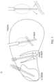
- FIGS. 1 to 3illustrate prior techniques that involve inserting catheters through a frontal burr hole at an angle of approximately 45° to the anterior commissure—posterior commissure plane.
- FIGS. 4 to 6shows catheter placement in accordance with an embodiment.
- the cathetercan thus be seen to extend substantially along the long axis of the putamen. Fluid thus refluxes back from the catheter tip to the step in the outer surface profile of the catheter, thereby providing a distribution that covers a large part of the putamen.
- FIG. 7shows how a percutaneous port based system may be used to provide a fluid connection to catheters placed in accordance an embodiment.
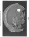
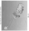
- FIGS. 8 and 9show an anterior to posterior trajectory for catheter delivery to the putamen and FIGS. 10 to 13 illustrate delivery achieved using an anterior to posterior trajectory.
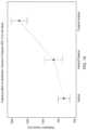
- FIG. 14shows the effect of trajectory on distribution volumes in the putamen as observed following 400 ⁇ l infusion volumes in human putamen (95% CI for the Mean).
- FIG. 15shows the effect of trajectory on percentage coverage of Putamen (per catheter) and illustrates the percentage coverage observed directly relative to the size of each putamen into which the catheter was implanted. Data have not been normalised to account for differences in the volumes of each Putamen (95% CI for the Mean).
- FIG. 16shows the effect of trajectory on percentage coverage of putamen (per pair of catheters)—as in FIG. 15 but coverage following pairs of catheters are displayed (95% CI for the Mean).
- FIG. 17shows a schematic view of the human brain.
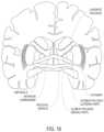
- FIG. 18shows a schematic coronal view of the human brain.
- FIG. 19shows a schematic perspective view of the basal ganglia of the human brain.
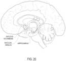
- FIG. 20shows another schematic sagittal view of the human brain.
- the fluidmay be delivered to structures within the brain, including one or more of the hippocampus, the putamen, the globus pallidus, the amygdala, the nucleus basalis, the nucleus accumbens, the substantia nigra, the caudate nucleus and the subthalamic nucleus, as shown in FIGS. 18 , 19 and 20 .
- the target structurespreferably have a long axis orientated in an anterior to posterior direction. This particular orientation allows the structures to be targeted along their length, allowing a greater length of catheter to be inserted into the structure, which can reduce catheter reflux. Accordingly, more fluid can be delivered into the structure.
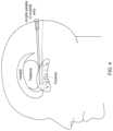
- the catheteris inserted along a trajectory through the occipitoparietal region, approximately parallel to the anterior commissure—posterior commissure plane, as shown in FIG. 17 .
- the posterior to anterior approachallows the disclosed method to be carried out on a subject such as a patient under general anaesthetic and in a prone position. Movement of the brain during neurosurgery can be a particular problem as even a few millimetres of displacement can significantly affect the accuracy of e.g. a catheter inserted into the brain. Not only will the brain move if and when the patient is moved, but the action of inserting a catheter can itself displace the brain in the direction of the insertion. Indeed, the present inventors have identified that during a conventional superior/vertical approach into the brain the brain can displace in potentially two different directions, making it difficult for surgeons to finely control catheter placement.
- the prone position of the patient in disclosed methodscan have advantages, as when the patient is turned from their back onto their front the brain displaces anteriorly in the skull. The catheter insertion is then from the posterior toward the anterior, however, as the brain has already displaced anteriorly any further displacement due to catheter insertion is likely to be minimal.
- the disclosed methodscan therefore provide highly accurate targeting of structures in the brain.
- the patientmay be a human.
- the insertion trajectorymay be substantially aligned with the long axis of a target structure, such as the hippocampus, the putamen, the globus pallidus, the amygdala, the nucleus basalis, the nucleus accumbens, the substantia nigra, the caudate nucleus and the subthalamic nucleus.
- a target structuresuch as the hippocampus, the putamen, the globus pallidus, the amygdala, the nucleus basalis, the nucleus accumbens, the substantia nigra, the caudate nucleus and the subthalamic nucleus.
- the methodcomprises using a catheter insertion trajectory that passes from posterior to anterior through the occipital, occipitoparietal, or occipitotemporal region approximately parallel to the anterior commissure—posterior commissure plane.
- this trajectorypreferably passes through the optic radiation, lateral to the posterior horn of the lateral ventricle and thence into the posterior part of the structure and along its long axis to its anterior portion.
- the fluidmay be delivered into structures of the brain that are at least 2 cm or at least 4 cm or at least 6 cm from the skull of the patient.
- the structuresmay be in areas of the forebrain and/or in areas of the midbrain.
- the methodmay include delivering a fluid (e.g. a fluid containing a therapeutic agent) to the brain.
- a fluide.g. a fluid containing a therapeutic agent
- the fluid deliveryis conveniently by convection enhanced delivery (CED).
- CEDconvection enhanced delivery
- the methodpreferably comprises the step of inserting a catheter into the brain along a trajectory through the occipitoparietal region approximately parallel (e.g. parallel to within 10°, 20°, 30°, or 40°) to the anterior commissure—posterior commissure plane.
- the trajectorypreferably passes from posterior to anterior.
- the methodmay include delivering a fluid to a target structure such as the hippocampus, the putamen, the globus pallidus, the amygdala, the nucleus basalis, the nucleus accumbens, the substantia nigra, the caudate nucleus and the subthalamic nucleus that comprises the step of inserting a catheter into the brain along an axis that is substantially aligned (e.g. to within 10°, 20°, 30° or 40°) with the long axis of the structure.
- the insertion trajectorypreferably passes from posterior to anterior.
- An advantage of disclosed embodimentsis that the inserted catheter will have a greater portion confined to the target structure, typically about 10 to about 40 min or about 15 to about 30 mm, whereas a more vertical trajectory through the frontal region could achieve a catheter length of between 10 and 15 min most typically. Indeed, the upper limit for the length of the catheter confined to the target structure using methods of disclosed embodiments is limited only by the size of the target structure in the patient.
- the catheter placed along the long axis of the putamenwould avoid the larger calibre and greater density of lenticulo striate vessels, reducing the likelihood of causing haemorrhage and reducing the impact of perivascular pumping of fluid from the extracellular space which is greater as one moves from the dorsal to the ventral striatum.
- Thisalso has the advantage of potentially increasing the volume of distribution in the target structure and as drug would be drawn ventrally by the flow of extracellular fluid which occurs predominantly in a dorsoventral direction, this would achieve greater total coverage of the structure than would be achieved by the more conventional trajectory. Passing the catheter tip along this trajectory into the head of the caudate nucleus would be advantageous for treating Huntingdon's, multiple system atrophy and cortico-basal degeneration.
- Optimising the volume of distribution of therapy within regions of the brainis particularly important when delivering viral vectors for gene therapy. This is because uncontrolled transfection of neurons at distant sites within the CNS could cause long-term and debilitating side effects.
- the methodmay be used to deliver any therapeutic agent.
- the methodmay be used to treat conditions such as Parkinson's disease, Huntington's disease, Alzheimer's disease, Multiple-System Atrophy, Progressive Supranuclear Palsy (PSP), dystonia, tremor, Tourette's syndrome or other neurodegenerative diseases.
- PPSProgressive Supranuclear Palsy
- the therapeutic agentmay be selected from, for example, one or more of a chemotherapy drug, a neurotrophin, an enzyme, a growth factor, an antibody, an immunotoxin, small inhibitory RNA (siRNA), antisense oligonucleotides, viral vectors, drug releasing nanoparticles (including liposomes and micels), transgenes and combinations or mixtures thereof.
- the therapeutic agentmay be glial cell-derived neurotrophic factor (GDNF) or neprilysin, which may be administered alone or in combination or consecutively. The combination and/or consecutive administration of GDNF and neprilysin may be used for the treatment of Alzheimer's disease.
- the therapeutic agentmay be administered in combination with artificial cerebrospinal fluid (aCSF).
- ACSFas disclosed herein may comprise glucose, proteins and ionic constituents.
- the aCSFmay omit glucose, so as to reduce the likelihood of bacterial growth in any catheter used to administer the composition to a subject.
- the aCSFdoes not comprise glucose or proteins.
- the methodmay include treating a neurodegenerative disorder, the method comprising delivering fluid to the brain of a patient using an intraparenchymal catheter, wherein the catheter is inserted into the brain using a posterior to anterior approach as described above.
- the methodmay include delivering fluid to an elongate structure of the brain using an intraparenchymal catheter, the method comprising the step of inserting a catheter into the brain along an insertion axis that is substantially aligned with a long axis of the elongate structure.
- Suitable elongate structuresinclude the hippocampus, the putamen, the globus pallidus, the amygdala, the nucleus basalis, the nucleus accumbens, the substantia nigra, the caudate nucleus or the subthalamic nucleus.
- Disclosed embodimentsmay use a catheter of the type described in WO03/077785 (incorporated herein by reference) that is inserted via a guide tube.
- a recessed step catheter as described in WO2014/016591 (incorporated herein by reference)may also be used.
- a catheter and/or cannula having a stepped outer profilemay be employed. Any step or steps in the outer profile are preferably located within the structure of the putamen.
- embodimentsextend to a catheter adapted for insertion in accordance with the method described above. The method may involve implanting a percutaneous port based catheter system, for example as described in WO2008/062173 or WO2011/098769 (both incorporated herein by reference).
- the fluidmay be administered via at least one convection enhanced delivery (CED) catheter, especially an intraparenchymal catheter.
- CEDconvection enhanced delivery
- the fluidmay be administered via at least two, at least three or four or more such catheters.
- two cathetersmay be used to administer the fluid bilaterally.
- the fluidmay be delivered via at least one or at least two chronically implanted CED catheters or via three or more of such catheters.
- Chronically implanted CED cathetersrefer to catheters that will be left in situ in the brain of a subject for at least 30 days, optionally for at least six months. Chronically implanted catheters may remain in place for up to one year or even for the lifetime of a subject.
- the fluidmay be administered on at least two, preferably three, optionally four consecutive days. Alternatively, the fluid may be administered on two out of three, four or five days, or three out of four, five, six or seven days.
- the fluidis for administration for a number of consecutive days or for regular administration over a number of days, it may independently or additionally be for administration weekly, fortnightly, monthly, every six, eight, twelve or fifteen or more weeks. For example, a cycle of two or three days of infusions may be repeated every fortnight. Alternatively, it may be for administration in a series of cycles of infusions, with 6, 7, 8, 9, 10, 11 or 12 days between the end of a first cycle of infusions and the next cycle of infusions.
- the fluidmay be for administration by infusion for between 6 and 10, especially between 7 and 9 hours, each day for three consecutive days.
- This pattern of administrationmay then be repeated weekly, or fortnightly, or for example with 6, 7, 8, 9, 10, 11 or 12 days between the end of a first cycle of three days of infusions and the next three days of infusions.
- Both the anterior to posterior and posterior to anterior trajectoriespassed approximately parallel to the anterior commissure—posterior commissure plane to target the long axis of the putamen.
- the entry point for the anterior to posterior trajectorywas located between the orbits and the coronal suture, while the posterior to anterior trajectory had an entry point in the occipitoparietal region and passed through the optic radiation, lateral to the posterior horn of the lateral ventricle.
- the distribution volumewas identified manually by assigning voxels of MRI scans to each catheter via a visual assessment and the results are shown in FIG. 14 .
- Percentages of coveragewere calculated by dividing the volume of distribution by the volume of the putamen into which the infusion was targeted and are shown in FIG. 15 .
- both the anterior to posterior and posterior to anterior approachesimproved the volume of distribution over that achieved using the conventional vertical approach.
- both the anterior to posterior and posterior to anterior approachestargeted the long axis of the putamen, the volume of distribution was significantly improved in the posterior to anterior approach.
- the inventorbelieves that the angle of insertion needed to avoid the eye of a patient in an anterior to posterior approach leads to the reduced volume of distribution.
- a posterior to anterior approachallows the angle of insertion to be optimised, which improves the volume of distribution.
- This approachalso has cosmetic benefits for patients as the catheter ports can usually be hidden under the patient's hair on the back of their head.
Landscapes
- Health & Medical Sciences (AREA)
- Life Sciences & Earth Sciences (AREA)
- Biophysics (AREA)
- Pulmonology (AREA)
- Engineering & Computer Science (AREA)
- Anesthesiology (AREA)
- Biomedical Technology (AREA)
- Heart & Thoracic Surgery (AREA)
- Hematology (AREA)
- Animal Behavior & Ethology (AREA)
- General Health & Medical Sciences (AREA)
- Public Health (AREA)
- Veterinary Medicine (AREA)
- Infusion, Injection, And Reservoir Apparatuses (AREA)
- Media Introduction/Drainage Providing Device (AREA)
Abstract
Description
- Barua N U, Lowis S P, Woolley M, O Sullivan S, Harrison R, Gill S S. Acta Neurochir (Wein). 2013 155:1459-65
- Brady M L, Raghavan R, Alexander A, Kubota K, Sillay K and Emborg M E. Stereotact Funct Neurosurg 2013 91:69-78
- Slevin J T, Herhardt G A, Smith C D, Gash D M, Kryscio R and Young B. J. Neurosurg. 2005 102:216-222
Claims (15)
Priority Applications (1)
| Application Number | Priority Date | Filing Date | Title |
|---|---|---|---|
| US17/502,691US12168103B2 (en) | 2014-01-30 | 2021-10-15 | Neurosurgical apparatus and method |
Applications Claiming Priority (9)
| Application Number | Priority Date | Filing Date | Title |
|---|---|---|---|
| GB201401552AGB201401552D0 (en) | 2014-01-30 | 2014-01-30 | Neurosurgical apparatus and method |
| GB1401552.3 | 2014-01-30 | ||
| GB1401552 | 2014-01-30 | ||
| GB1412941 | 2014-07-22 | ||
| GB201412941AGB201412941D0 (en) | 2014-07-22 | 2014-07-22 | Neurosurgical apparatus and method |
| GB1412941.5 | 2014-07-22 | ||
| US14/610,185US10369329B2 (en) | 2014-01-30 | 2015-01-30 | Neurosurgical apparatus and method |
| US16/454,637US11160954B2 (en) | 2014-01-30 | 2019-06-27 | Neurosurgical apparatus and method |
| US17/502,691US12168103B2 (en) | 2014-01-30 | 2021-10-15 | Neurosurgical apparatus and method |
Related Parent Applications (1)
| Application Number | Title | Priority Date | Filing Date |
|---|---|---|---|
| US16/454,637ContinuationUS11160954B2 (en) | 2014-01-30 | 2019-06-27 | Neurosurgical apparatus and method |
Publications (2)
| Publication Number | Publication Date |
|---|---|
| US20220032005A1 US20220032005A1 (en) | 2022-02-03 |
| US12168103B2true US12168103B2 (en) | 2024-12-17 |
Family
ID=53678066
Family Applications (3)
| Application Number | Title | Priority Date | Filing Date |
|---|---|---|---|
| US14/610,185Active2037-06-07US10369329B2 (en) | 2014-01-30 | 2015-01-30 | Neurosurgical apparatus and method |
| US16/454,637Active2035-07-31US11160954B2 (en) | 2014-01-30 | 2019-06-27 | Neurosurgical apparatus and method |
| US17/502,691Active2036-02-01US12168103B2 (en) | 2014-01-30 | 2021-10-15 | Neurosurgical apparatus and method |
Family Applications Before (2)
| Application Number | Title | Priority Date | Filing Date |
|---|---|---|---|
| US14/610,185Active2037-06-07US10369329B2 (en) | 2014-01-30 | 2015-01-30 | Neurosurgical apparatus and method |
| US16/454,637Active2035-07-31US11160954B2 (en) | 2014-01-30 | 2019-06-27 | Neurosurgical apparatus and method |
Country Status (1)
| Country | Link |
|---|---|
| US (3) | US10369329B2 (en) |
Families Citing this family (1)
| Publication number | Priority date | Publication date | Assignee | Title |
|---|---|---|---|---|
| KR102651292B1 (en) | 2015-10-31 | 2024-03-25 | 아이오 테라퓨틱스, 인크. | Treatment of nervous system disorders using combinations of rxr agonists and thyroid hormones |
Citations (16)
| Publication number | Priority date | Publication date | Assignee | Title |
|---|---|---|---|---|
| US5935795A (en) | 1991-09-20 | 1999-08-10 | Amgen Inc. | Glial cell line-derived neurotrophic factor antibody |
| US6042579A (en) | 1997-04-30 | 2000-03-28 | Medtronic, Inc. | Techniques for treating neurodegenerative disorders by infusion of nerve growth factors into the brain |
| US6348050B1 (en) | 1999-04-30 | 2002-02-19 | Medtronic, Inc. | Infusion systems for creating microenvironments in a living body |
| US6609020B2 (en) | 1999-12-01 | 2003-08-19 | Steven Gill | Neurosurgical guide device |
| WO2003077785A1 (en) | 2002-03-12 | 2003-09-25 | Steven Streatfield Gill | Catheter and guide tube for intracerebral application |
| US20040215162A1 (en)* | 2003-04-25 | 2004-10-28 | Ad-Tech Medical Instrument Corp. | Intracranial catheter assembly for precise treatment of brain tissue |
| US20070161919A1 (en)* | 1998-08-05 | 2007-07-12 | Bioneuronics Corporation | Methods and systems for continuous EEG monitoring |
| WO2008062173A1 (en) | 2006-11-23 | 2008-05-29 | Renishaw Plc | Neurological apparatus comprising a percutaneous access device |
| WO2008098769A1 (en) | 2007-02-17 | 2008-08-21 | Esm Ennepetaler Schneid- Und Mähtechnik Gmbh & Co. Kg | Cutting tool for cutting stalks |
| US20090011980A1 (en) | 2004-06-11 | 2009-01-08 | Steven Streatfield Gill | Method of Treating Parkinson's Disease in Humans by Direct Infusion of Glial Cell-Line Derived Neurotrophic Factor Into the Zona Incerta |
| US7974696B1 (en) | 1998-08-05 | 2011-07-05 | Dilorenzo Biomedical, Llc | Closed-loop autonomic neuromodulation for optimal control of neurological and metabolic disease |
| US7981863B2 (en) | 2001-09-19 | 2011-07-19 | Neuronova Ab | Treatment of Parkinson's disease with PDGF |
| WO2014016591A1 (en) | 2012-07-24 | 2014-01-30 | Renishaw Plc | Neurosurgical apparatus and methods |
| US9320914B2 (en) | 2008-03-03 | 2016-04-26 | DePuy Synthes Products, Inc. | Endoscopic delivery of red/NIR light to the subventricular zone |
| US20170143966A1 (en) | 2014-03-31 | 2017-05-25 | Functional Neuromodulation, Inc. | Systems and methods for determining a trajectory for a brain stimulation lead |
| US10130814B2 (en) | 2015-04-09 | 2018-11-20 | Renishaw Plc | Movement disorder |
- 2015
- 2015-01-30USUS14/610,185patent/US10369329B2/enactiveActive
- 2019
- 2019-06-27USUS16/454,637patent/US11160954B2/enactiveActive
- 2021
- 2021-10-15USUS17/502,691patent/US12168103B2/enactiveActive
Patent Citations (17)
| Publication number | Priority date | Publication date | Assignee | Title |
|---|---|---|---|---|
| US5935795A (en) | 1991-09-20 | 1999-08-10 | Amgen Inc. | Glial cell line-derived neurotrophic factor antibody |
| US6042579A (en) | 1997-04-30 | 2000-03-28 | Medtronic, Inc. | Techniques for treating neurodegenerative disorders by infusion of nerve growth factors into the brain |
| US9113801B2 (en) | 1998-08-05 | 2015-08-25 | Cyberonics, Inc. | Methods and systems for continuous EEG monitoring |
| US20070161919A1 (en)* | 1998-08-05 | 2007-07-12 | Bioneuronics Corporation | Methods and systems for continuous EEG monitoring |
| US7974696B1 (en) | 1998-08-05 | 2011-07-05 | Dilorenzo Biomedical, Llc | Closed-loop autonomic neuromodulation for optimal control of neurological and metabolic disease |
| US6348050B1 (en) | 1999-04-30 | 2002-02-19 | Medtronic, Inc. | Infusion systems for creating microenvironments in a living body |
| US6609020B2 (en) | 1999-12-01 | 2003-08-19 | Steven Gill | Neurosurgical guide device |
| US7981863B2 (en) | 2001-09-19 | 2011-07-19 | Neuronova Ab | Treatment of Parkinson's disease with PDGF |
| WO2003077785A1 (en) | 2002-03-12 | 2003-09-25 | Steven Streatfield Gill | Catheter and guide tube for intracerebral application |
| US20040215162A1 (en)* | 2003-04-25 | 2004-10-28 | Ad-Tech Medical Instrument Corp. | Intracranial catheter assembly for precise treatment of brain tissue |
| US20090011980A1 (en) | 2004-06-11 | 2009-01-08 | Steven Streatfield Gill | Method of Treating Parkinson's Disease in Humans by Direct Infusion of Glial Cell-Line Derived Neurotrophic Factor Into the Zona Incerta |
| WO2008062173A1 (en) | 2006-11-23 | 2008-05-29 | Renishaw Plc | Neurological apparatus comprising a percutaneous access device |
| WO2008098769A1 (en) | 2007-02-17 | 2008-08-21 | Esm Ennepetaler Schneid- Und Mähtechnik Gmbh & Co. Kg | Cutting tool for cutting stalks |
| US9320914B2 (en) | 2008-03-03 | 2016-04-26 | DePuy Synthes Products, Inc. | Endoscopic delivery of red/NIR light to the subventricular zone |
| WO2014016591A1 (en) | 2012-07-24 | 2014-01-30 | Renishaw Plc | Neurosurgical apparatus and methods |
| US20170143966A1 (en) | 2014-03-31 | 2017-05-25 | Functional Neuromodulation, Inc. | Systems and methods for determining a trajectory for a brain stimulation lead |
| US10130814B2 (en) | 2015-04-09 | 2018-11-20 | Renishaw Plc | Movement disorder |
Non-Patent Citations (12)
| Title |
|---|
| Anderson, R. C. E.; Kennedy, B.; Yanes, C. L.; Garvin, J.; Needle, M.; Canoll, P.; Feldstein, N. A.; Bruce, J. N. "Convection-enhanced delivery of topotecan into diffuse intrinsic brainstem tumors in children" (2013) J Neurosurg Pediatrics 1, pp. 289-295. |
| Barua, N. U.; Lowis, S. P.; Woolley, M.; O'Sullivan, S.; Harrison, R.; Gill, S. S. "Robot-guided convection-enhanced delivery of carboplatin for advanced brainstem glioma." (2013) Acta Neurochirurgica, 155, 1459-1465. |
| Bejjani, B. P.; Dormont, D.; Pidoux, B.; Yelnik, J.; Damier, P.; Arnulf, I.; Bonnet, A. M.; Marsault, C.; Agid, Y.; Philippon, J.; Cornu, P. Bilateral subthalamic stimulation for Parkinson's disease by using three-dimensional stereotactic magnetic resonance imaging and electrophysiological guidance. (2000) J Neurosurg. 92(4), 615-625. |
| Brady, M. L.; Raghavan, R.; Alexander, A.; Kubota, K.; Sillay, K.; Emborg, M. E. "Pathways of infusate loss during convection enhanced delivery into the Putamen nucleus." (2013) Stereotact Funct Neurosurg. 2013 ; 91(2): 69-78. |
| Chen, M. Y.; Lonser, R. R.; Morrison, P. F.; Governale, L. S.; Oldfield, E. H. "Variables affecting convection-enhanced delivery to the striatum: a systematic examination of rate of infusion, cannula size, infusate concentration, and tissue-cannula sealing time." (1999) Journ. Neurosurg., 90(2), 315-320. |
| Freed, C. R.; Greene, P. E.; Breeze, R. E.; Tsai, W-Y.; Dumouchel, W.; Kao, R.; Dillon, S.; Winfiled, H.; Culver, S.; Trojanowski, J. Q.; Eidelberg, D.; Fahn, S. "Transplantation of embryonic dopamine neurons for severe Parkinson's disease." (2001) The New Engl J Med, 344(10), 710-719. |
| Gill, S. S.; Patel, N. K.; Hotton, G. R.; O'Sullivan, K.; McCarter, R.; Bunnage, M.; Brooks, D. J.; Svendsen, C. N.; Heywood, P. "Direct brain infusion of glial cell line-derived neurotrophic factor in Parkinson disease." (2003) Nature Medicine, 9, 589-595. |
| Murata, J-I.; Kitagaw, M. Uesugi, H.; Saito, H.; Iwasaki, Y.; Kikuchi, S.; Tashiro, K.; Sawamura, Y. "Electrical stimulation of the posterior subthalamic area for the treatment of intractable proximal tremor." (2003) JNS Journal of Neurosurgery, 99(4), 708-715. |
| Richardson, M. R.; Kells, A. P.; Rosenbluth, K. H.; Salegio, E. A.; Fiandaca, M. S.; Larson, P. S.; Starr, P. A.; Martin, A. J.; Lonser, R. R.; Federoff, H. J.; Forsayeth, J. R.; Bankierwicz, K. S. "Interventional MRI-guided Putaminal delivery of AAV2-GDNF for a planned clinical trial in Parkinson's disease." Mol Ther 19(6), 1048-1057. |
| Rosenblad, C.; Kirk, D.; Bjorklund, A. "Sequential administration of GDNF into the substantia nigra and striatum promotes dopamine neuron survival and axonal sprouting but not striatal reinnervation or functional recovery in the partial 6-OHDA lesion model." (2000) Exp Neurol. 161(2), 503-516. |
| Slevin, J. T.; Gerhardt, G. A.; Smith, C. D.; Gash, D. M.; Kryscio, R.; Young, B. "Improvement of bilateral motor functions in patients with Parkinson disease through the unilateral intraputaminal infusion of glial cell line-derived neurotrophic factor." (2005) 102(2), 216-222. |
| Yin, D.; Valles, F. E.; Fiandaca, M. S.; Bringas, J.; Gimenez, F.; Berger, M. S.; Forsayeth, J.; Bankiewicz, K. S. "Optimal region of the putamen for image-guided convection-enhanced delivery of therapeutics in human and non-human primates." (2011) NeuroImage 54(1), S196-S203. |
Also Published As
| Publication number | Publication date |
|---|---|
| US20220032005A1 (en) | 2022-02-03 |
| US10369329B2 (en) | 2019-08-06 |
| US11160954B2 (en) | 2021-11-02 |
| US20190314605A1 (en) | 2019-10-17 |
| US20150209549A1 (en) | 2015-07-30 |
Similar Documents
| Publication | Publication Date | Title |
|---|---|---|
| AU2018206687B2 (en) | Systems and methods for reducing or preventing backflow in a delivery system | |
| Barua et al. | Robot-guided convection-enhanced delivery of carboplatin for advanced brainstem glioma | |
| Bankiewicz et al. | AAV viral vector delivery to the brain by shape-conforming MR-guided infusions | |
| Lewis et al. | Chronic, intermittent convection-enhanced delivery devices | |
| Souweidane et al. | Gene therapy for late infantile neuronal ceroid lipofuscinosis: neurosurgical considerations | |
| Lonser et al. | Convection-enhanced selective excitotoxic ablation of the neurons of the globus pallidus internus for treatment of parkinsonism in nonhuman primates | |
| US20110282319A1 (en) | Catheter and guide tube for intracerebral application | |
| Bienemann et al. | The development of an implantable catheter system for chronic or intermittent convection-enhanced delivery | |
| WO2014184576A2 (en) | Delivery | |
| Gill et al. | In vitro and in vivo testing of a novel recessed-step catheter for reflux-free convection-enhanced drug delivery to the brain | |
| US20080086061A1 (en) | Needle assembly for use in delivering precise dosages of proteinaceous pharmaceutical compositions and methods for use of same | |
| Lam et al. | Neurosurgical convection-enhanced delivery of treatments for Parkinson’s disease | |
| US20150080708A1 (en) | Intrabody fluid transfer devices, systems and methods | |
| Grondin et al. | Intracranial delivery of proteins and peptides as a therapy for neurodegenerative diseases | |
| Han et al. | Interventional MRI-guided catheter placement and real time drug delivery to the central nervous system | |
| US12168103B2 (en) | Neurosurgical apparatus and method | |
| Beffinger et al. | Delivery of antibodies into the murine brain via convection-enhanced delivery | |
| Aquilina et al. | Convection-enhanced delivery in children: Techniques and applications | |
| Song et al. | Convection-enhanced delivery for the treatment of pediatric neurologic disorders | |
| US11344714B2 (en) | Intrathecal catheter with features to reduce drug dispersion | |
| Leung et al. | Assessment of hippocampal adeno-associated viral vector gene delivery via frameless stereotaxis in a nonhuman primate | |
| Naidoo et al. | Convection-Enhanced Drug Delivery in the Central Nervous System | |
| EP3833394B1 (en) | A biologically inert fluid for use in the treatment of a cns disorder | |
| US20230114337A1 (en) | Alternative cannula configurations to control fluid distribution in tissue | |
| Lee et al. | IntraBrain Injector (IBI): A Stereotactic-Guided Device for Repeated Delivery of Therapeutic Agents Into the Brain Parenchyma |
Legal Events
| Date | Code | Title | Description |
|---|---|---|---|
| FEPP | Fee payment procedure | Free format text:ENTITY STATUS SET TO UNDISCOUNTED (ORIGINAL EVENT CODE: BIG.); ENTITY STATUS OF PATENT OWNER: LARGE ENTITY | |
| STPP | Information on status: patent application and granting procedure in general | Free format text:DOCKETED NEW CASE - READY FOR EXAMINATION | |
| AS | Assignment | Owner name:XCED LLP, UNITED KINGDOM Free format text:ASSIGNMENT OF ASSIGNORS INTEREST;ASSIGNOR:GILL, STEVEN STREATFIELD;REEL/FRAME:059674/0480 Effective date:20220420 Owner name:RENISHAW NEURO SOLUTIONS LTD, UNITED KINGDOM Free format text:ASSIGNMENT OF ASSIGNORS INTEREST;ASSIGNOR:GILL, STEVEN STREATFIELD;REEL/FRAME:059674/0480 Effective date:20220420 | |
| AS | Assignment | Owner name:NEUROCHASE INNOVATIONS LTD, WALES Free format text:ASSIGNMENT OF ASSIGNORS INTEREST;ASSIGNOR:XCED LLP;REEL/FRAME:062698/0048 Effective date:20230214 | |
| STPP | Information on status: patent application and granting procedure in general | Free format text:NON FINAL ACTION MAILED | |
| STPP | Information on status: patent application and granting procedure in general | Free format text:RESPONSE TO NON-FINAL OFFICE ACTION ENTERED AND FORWARDED TO EXAMINER | |
| STPP | Information on status: patent application and granting procedure in general | Free format text:FINAL REJECTION MAILED | |
| STPP | Information on status: patent application and granting procedure in general | Free format text:ADVISORY ACTION MAILED | |
| STPP | Information on status: patent application and granting procedure in general | Free format text:DOCKETED NEW CASE - READY FOR EXAMINATION | |
| STPP | Information on status: patent application and granting procedure in general | Free format text:NOTICE OF ALLOWANCE MAILED -- APPLICATION RECEIVED IN OFFICE OF PUBLICATIONS | |
| STPP | Information on status: patent application and granting procedure in general | Free format text:PUBLICATIONS -- ISSUE FEE PAYMENT VERIFIED | |
| STCF | Information on status: patent grant | Free format text:PATENTED CASE |