US12137987B2 - Ultrasound systems and methods for sustained spatial attention - Google Patents
Ultrasound systems and methods for sustained spatial attentionDownload PDFInfo
- Publication number
- US12137987B2 US12137987B2US17/491,308US202117491308AUS12137987B2US 12137987 B2US12137987 B2US 12137987B2US 202117491308 AUS202117491308 AUS 202117491308AUS 12137987 B2US12137987 B2US 12137987B2
- Authority
- US
- United States
- Prior art keywords
- needle
- probe
- ultrasound
- probe head
- plane
- Prior art date
- Legal status (The legal status is an assumption and is not a legal conclusion. Google has not performed a legal analysis and makes no representation as to the accuracy of the status listed.)
- Active, expires
Links
- 238000002604ultrasonographyMethods0.000titleclaimsabstractdescription291
- 238000000034methodMethods0.000titleabstractdescription26
- 230000002459sustained effectEffects0.000titleabstractdescription4
- 239000000523sampleSubstances0.000claimsabstractdescription384
- 238000003780insertionMethods0.000claimsdescription89
- 230000037431insertionEffects0.000claimsdescription49
- 230000000737periodic effectEffects0.000claimsdescription32
- 210000004204blood vesselAnatomy0.000description10
- 238000012544monitoring processMethods0.000description6
- 238000012285ultrasound imagingMethods0.000description6
- 210000003484anatomyAnatomy0.000description5
- 230000006870functionEffects0.000description4
- 238000012545processingMethods0.000description3
- 238000009877renderingMethods0.000description3
- 230000006978adaptationEffects0.000description2
- 238000004891communicationMethods0.000description2
- 238000010586diagramMethods0.000description2
- 238000012986modificationMethods0.000description2
- 230000004048modificationEffects0.000description2
- 238000013459approachMethods0.000description1
- 230000001934delayEffects0.000description1
- 230000002792vascularEffects0.000description1
Images


Classifications
- A—HUMAN NECESSITIES
- A61—MEDICAL OR VETERINARY SCIENCE; HYGIENE
- A61B—DIAGNOSIS; SURGERY; IDENTIFICATION
- A61B34/00—Computer-aided surgery; Manipulators or robots specially adapted for use in surgery
- A61B34/20—Surgical navigation systems; Devices for tracking or guiding surgical instruments, e.g. for frameless stereotaxis
- A—HUMAN NECESSITIES
- A61—MEDICAL OR VETERINARY SCIENCE; HYGIENE
- A61B—DIAGNOSIS; SURGERY; IDENTIFICATION
- A61B17/00—Surgical instruments, devices or methods
- A61B17/34—Trocars; Puncturing needles
- A61B17/3403—Needle locating or guiding means
- A—HUMAN NECESSITIES
- A61—MEDICAL OR VETERINARY SCIENCE; HYGIENE
- A61B—DIAGNOSIS; SURGERY; IDENTIFICATION
- A61B8/00—Diagnosis using ultrasonic, sonic or infrasonic waves
- A61B8/08—Clinical applications
- A61B8/0833—Clinical applications involving detecting or locating foreign bodies or organic structures
- A61B8/0841—Clinical applications involving detecting or locating foreign bodies or organic structures for locating instruments
- A—HUMAN NECESSITIES
- A61—MEDICAL OR VETERINARY SCIENCE; HYGIENE
- A61B—DIAGNOSIS; SURGERY; IDENTIFICATION
- A61B8/00—Diagnosis using ultrasonic, sonic or infrasonic waves
- A61B8/08—Clinical applications
- A61B8/0833—Clinical applications involving detecting or locating foreign bodies or organic structures
- A61B8/085—Clinical applications involving detecting or locating foreign bodies or organic structures for locating body or organic structures, e.g. tumours, calculi, blood vessels, nodules
- A—HUMAN NECESSITIES
- A61—MEDICAL OR VETERINARY SCIENCE; HYGIENE
- A61B—DIAGNOSIS; SURGERY; IDENTIFICATION
- A61B8/00—Diagnosis using ultrasonic, sonic or infrasonic waves
- A61B8/42—Details of probe positioning or probe attachment to the patient
- A61B8/4245—Details of probe positioning or probe attachment to the patient involving determining the position of the probe, e.g. with respect to an external reference frame or to the patient
- A61B8/4254—Details of probe positioning or probe attachment to the patient involving determining the position of the probe, e.g. with respect to an external reference frame or to the patient using sensors mounted on the probe
- A—HUMAN NECESSITIES
- A61—MEDICAL OR VETERINARY SCIENCE; HYGIENE
- A61B—DIAGNOSIS; SURGERY; IDENTIFICATION
- A61B8/00—Diagnosis using ultrasonic, sonic or infrasonic waves
- A61B8/44—Constructional features of the ultrasonic, sonic or infrasonic diagnostic device
- A61B8/4444—Constructional features of the ultrasonic, sonic or infrasonic diagnostic device related to the probe
- A—HUMAN NECESSITIES
- A61—MEDICAL OR VETERINARY SCIENCE; HYGIENE
- A61B—DIAGNOSIS; SURGERY; IDENTIFICATION
- A61B8/00—Diagnosis using ultrasonic, sonic or infrasonic waves
- A61B8/44—Constructional features of the ultrasonic, sonic or infrasonic diagnostic device
- A61B8/4483—Constructional features of the ultrasonic, sonic or infrasonic diagnostic device characterised by features of the ultrasound transducer
- A61B8/4488—Constructional features of the ultrasonic, sonic or infrasonic diagnostic device characterised by features of the ultrasound transducer the transducer being a phased array
- A—HUMAN NECESSITIES
- A61—MEDICAL OR VETERINARY SCIENCE; HYGIENE
- A61B—DIAGNOSIS; SURGERY; IDENTIFICATION
- A61B8/00—Diagnosis using ultrasonic, sonic or infrasonic waves
- A61B8/46—Ultrasonic, sonic or infrasonic diagnostic devices with special arrangements for interfacing with the operator or the patient
- A61B8/461—Displaying means of special interest
- A61B8/463—Displaying means of special interest characterised by displaying multiple images or images and diagnostic data on one display
- A—HUMAN NECESSITIES
- A61—MEDICAL OR VETERINARY SCIENCE; HYGIENE
- A61B—DIAGNOSIS; SURGERY; IDENTIFICATION
- A61B90/00—Instruments, implements or accessories specially adapted for surgery or diagnosis and not covered by any of the groups A61B1/00 - A61B50/00, e.g. for luxation treatment or for protecting wound edges
- A61B90/36—Image-producing devices or illumination devices not otherwise provided for
- A61B90/361—Image-producing devices, e.g. surgical cameras
- A—HUMAN NECESSITIES
- A61—MEDICAL OR VETERINARY SCIENCE; HYGIENE
- A61B—DIAGNOSIS; SURGERY; IDENTIFICATION
- A61B90/00—Instruments, implements or accessories specially adapted for surgery or diagnosis and not covered by any of the groups A61B1/00 - A61B50/00, e.g. for luxation treatment or for protecting wound edges
- A61B90/39—Markers, e.g. radio-opaque or breast lesions markers
- A—HUMAN NECESSITIES
- A61—MEDICAL OR VETERINARY SCIENCE; HYGIENE
- A61B—DIAGNOSIS; SURGERY; IDENTIFICATION
- A61B17/00—Surgical instruments, devices or methods
- A61B17/34—Trocars; Puncturing needles
- A61B17/3403—Needle locating or guiding means
- A61B2017/3405—Needle locating or guiding means using mechanical guide means
- A—HUMAN NECESSITIES
- A61—MEDICAL OR VETERINARY SCIENCE; HYGIENE
- A61B—DIAGNOSIS; SURGERY; IDENTIFICATION
- A61B17/00—Surgical instruments, devices or methods
- A61B17/34—Trocars; Puncturing needles
- A61B17/3403—Needle locating or guiding means
- A61B2017/3413—Needle locating or guiding means guided by ultrasound
- A—HUMAN NECESSITIES
- A61—MEDICAL OR VETERINARY SCIENCE; HYGIENE
- A61B—DIAGNOSIS; SURGERY; IDENTIFICATION
- A61B34/00—Computer-aided surgery; Manipulators or robots specially adapted for use in surgery
- A61B34/20—Surgical navigation systems; Devices for tracking or guiding surgical instruments, e.g. for frameless stereotaxis
- A61B2034/2046—Tracking techniques
- A61B2034/2055—Optical tracking systems
- A61B2034/2057—Details of tracking cameras
- A—HUMAN NECESSITIES
- A61—MEDICAL OR VETERINARY SCIENCE; HYGIENE
- A61B—DIAGNOSIS; SURGERY; IDENTIFICATION
- A61B34/00—Computer-aided surgery; Manipulators or robots specially adapted for use in surgery
- A61B34/20—Surgical navigation systems; Devices for tracking or guiding surgical instruments, e.g. for frameless stereotaxis
- A61B2034/2046—Tracking techniques
- A61B2034/2063—Acoustic tracking systems, e.g. using ultrasound
- A—HUMAN NECESSITIES
- A61—MEDICAL OR VETERINARY SCIENCE; HYGIENE
- A61B—DIAGNOSIS; SURGERY; IDENTIFICATION
- A61B34/00—Computer-aided surgery; Manipulators or robots specially adapted for use in surgery
- A61B34/20—Surgical navigation systems; Devices for tracking or guiding surgical instruments, e.g. for frameless stereotaxis
- A61B2034/2046—Tracking techniques
- A61B2034/2065—Tracking using image or pattern recognition
- A—HUMAN NECESSITIES
- A61—MEDICAL OR VETERINARY SCIENCE; HYGIENE
- A61B—DIAGNOSIS; SURGERY; IDENTIFICATION
- A61B34/00—Computer-aided surgery; Manipulators or robots specially adapted for use in surgery
- A61B34/20—Surgical navigation systems; Devices for tracking or guiding surgical instruments, e.g. for frameless stereotaxis
- A61B2034/2068—Surgical navigation systems; Devices for tracking or guiding surgical instruments, e.g. for frameless stereotaxis using pointers, e.g. pointers having reference marks for determining coordinates of body points
- A—HUMAN NECESSITIES
- A61—MEDICAL OR VETERINARY SCIENCE; HYGIENE
- A61B—DIAGNOSIS; SURGERY; IDENTIFICATION
- A61B90/00—Instruments, implements or accessories specially adapted for surgery or diagnosis and not covered by any of the groups A61B1/00 - A61B50/00, e.g. for luxation treatment or for protecting wound edges
- A61B90/36—Image-producing devices or illumination devices not otherwise provided for
- A61B90/37—Surgical systems with images on a monitor during operation
- A61B2090/378—Surgical systems with images on a monitor during operation using ultrasound
- A—HUMAN NECESSITIES
- A61—MEDICAL OR VETERINARY SCIENCE; HYGIENE
- A61B—DIAGNOSIS; SURGERY; IDENTIFICATION
- A61B90/00—Instruments, implements or accessories specially adapted for surgery or diagnosis and not covered by any of the groups A61B1/00 - A61B50/00, e.g. for luxation treatment or for protecting wound edges
- A61B90/39—Markers, e.g. radio-opaque or breast lesions markers
- A61B2090/3937—Visible markers
- A—HUMAN NECESSITIES
- A61—MEDICAL OR VETERINARY SCIENCE; HYGIENE
- A61B—DIAGNOSIS; SURGERY; IDENTIFICATION
- A61B90/00—Instruments, implements or accessories specially adapted for surgery or diagnosis and not covered by any of the groups A61B1/00 - A61B50/00, e.g. for luxation treatment or for protecting wound edges
- A61B90/39—Markers, e.g. radio-opaque or breast lesions markers
- A61B2090/3983—Reference marker arrangements for use with image guided surgery
- A—HUMAN NECESSITIES
- A61—MEDICAL OR VETERINARY SCIENCE; HYGIENE
- A61B—DIAGNOSIS; SURGERY; IDENTIFICATION
- A61B8/00—Diagnosis using ultrasonic, sonic or infrasonic waves
- A61B8/08—Clinical applications
- A61B8/0891—Clinical applications for diagnosis of blood vessels
- A—HUMAN NECESSITIES
- A61—MEDICAL OR VETERINARY SCIENCE; HYGIENE
- A61B—DIAGNOSIS; SURGERY; IDENTIFICATION
- A61B8/00—Diagnosis using ultrasonic, sonic or infrasonic waves
- A61B8/44—Constructional features of the ultrasonic, sonic or infrasonic diagnostic device
- A61B8/4427—Device being portable or laptop-like
- A—HUMAN NECESSITIES
- A61—MEDICAL OR VETERINARY SCIENCE; HYGIENE
- A61B—DIAGNOSIS; SURGERY; IDENTIFICATION
- A61B8/00—Diagnosis using ultrasonic, sonic or infrasonic waves
- A61B8/44—Constructional features of the ultrasonic, sonic or infrasonic diagnostic device
- A61B8/4444—Constructional features of the ultrasonic, sonic or infrasonic diagnostic device related to the probe
- A61B8/4455—Features of the external shape of the probe, e.g. ergonomic aspects
Definitions
- VADvascular access device
- Disclosed hereinare ultrasound systems and methods for sustained spatial attention in one or more spatial regions.
- an ultrasound probeincluding, in some embodiments, a probe body, a probe head extending from a distal end of the probe body, and a camera integrated into a side of the ultrasound probe.
- the probe headincludes a plurality of ultrasonic transducers arranged in an array.
- the camerais configured for recording one or more still or moving images of a procedural field with a depth of field including a plane of a distal end of the probe head and a field of view including a spatial region about the probe head.
- the ultrasound probefurther includes a light-pattern projector integrated into the side of the ultrasound probe including the camera.
- the light-pattern projectoris configured to project a light pattern in the spatial region about the probe head focused in the plane of the distal end of the probe head.
- the light patternis configured for guided insertion of a needle into an anatomical target under the probe head in the procedural field.
- the light patternincludes periodic hash marks along one or more rays radiating from a central axis of the ultrasound probe in the plane of the probe head.
- Each hash mark of the hash markscorresponds to a depth under the probe head accessible by the needle along an associated ray at a needle-insertion angle with respect to the plane of the probe head.
- the light patternincludes periodic concentric circular arcs bound between two or more rays radiating from a central axis of the ultrasound probe in the plane of the probe head.
- Each circular arc of the circular arcscorresponds to a depth under the probe head accessible by the needle along an associated ray at a needle-insertion angle with respect to the plane of the probe head.
- the ultrasound probefurther includes a needle-guide holder extending from a side of the probe head in common with the side of the ultrasound probe including the camera.
- the ultrasound probefurther includes a single-use needle guide coupled to the needle-guide holder.
- the needle-guide holder, the needle guide, or a combination of the needle-guide holder and the needle guideincludes at least one degree of freedom enabling the needle guide to swivel between sides of the ultrasound probe.
- an ultrasound systemincluding, in some embodiments, a console and an ultrasound probe.
- the consoleincludes a display configured to render on a display screen thereof ultrasound images and one or more still or moving images of a procedural field.
- the ultrasound probeincludes a probe body, a probe head extending from a distal end of the probe body, and a camera integrated into a side of the ultrasound probe.
- the probe headincludes a plurality of ultrasonic transducers arranged in an array.
- the camerais configured for recording the one-or-more still or moving images of the procedural field with a depth of field including a plane of a distal end of the probe head and a field of view including a spatial region about the probe head.
- the ultrasound probefurther includes a needle-guide holder extending from a side of the probe head in common with the side of the ultrasound probe including the camera.
- the ultrasound probefurther includes a single-use needle guide coupled to the needle-guide holder.
- the needle-guide holder, the needle guide, or a combination of the needle-guide holder and the needle guideincludes at least one degree of freedom enabling the needle guide to swivel between sides of the ultrasound probe.
- the ultrasound probefurther includes a light-pattern projector integrated into the side of the ultrasound probe including the camera.
- the light-pattern projectoris configured to project a light pattern in the spatial region about the probe head focused in the plane of the distal end of the probe head.
- the light patternis configured for guided insertion of a needle into an anatomical target under the probe head in the procedural field.
- the light patternincludes periodic hash marks along one or more rays radiating from a central axis of the ultrasound probe in the plane of the probe head.
- Each hash mark of the hash markscorresponds to a depth under the probe head accessible by the needle along an associated ray at a needle-insertion angle with respect to the plane of the probe head.
- the light patternincludes periodic concentric circular arcs bound between two or more rays radiating from a central axis of the ultrasound probe in the plane of the probe head.
- Each circular arc of the circular arcscorresponds to a depth under the probe head accessible by the needle along an associated ray at a needle-insertion angle with respect to the plane of the probe head.
- the one-or-more still or moving imagesshow both the light pattern in the spatial region about the probe head and the needle in relation to the light pattern when both the light pattern and the needle are present in the spatial region about the probe head.
- the one-or-more still or moving imagesshow both the light pattern and the needle in relation to the light pattern for the guided insertion of the needle into the anatomical target under the probe head optionally on the display.
- the displayis further configured to render on the display screen one or more overlying needle trajectories lying over the ultrasound images in accordance with one or more depths accessible by the needle indicated by the light pattern.
- the one-or-more overlying needle trajectoriesare configured for the guided insertion of the needle into the anatomical target under the probe head on the display.
- the displayis further configured to render on the display screen an overlying pattern lying over the one-or-more still or moving images.
- the overlying patternis configured for guided insertion of a needle into an anatomical target under the probe head on the display.
- the overlying patternincludes periodic hash marks along one or more rays radiating from a central axis of the ultrasound probe in the plane of the probe head.
- Each hash mark of the hash markscorresponds to a depth under the probe head accessible by the needle along an associated ray at a needle-insertion angle with respect to the plane of the probe head.
- the overlying patternincludes periodic concentric circular arcs bound between two or more rays radiating from a central axis of the ultrasound probe in the plane of the probe head.
- Each circular arc of the circular arcscorresponds to a depth under the probe head accessible by the needle along an associated ray at a needle-insertion angle with respect to the plane of the probe head.
- the one-or-more still or moving imagesshow the needle in relation to the overlying pattern when the needle is present in the spatial region about the probe head.
- the one-or-more still or moving imagesshow the needle in relation to the overlying pattern for the guided insertion of the needle into the anatomical target under the probe head optionally on the display.
- the displayis further configured to render on the display screen one or more overlying needle trajectories lying over the ultrasound images in accordance with one or more depths accessible by the needle indicated by the overlying pattern.
- the one-or-more overlying needle trajectoriesare configured for the guided insertion of the needle into an anatomical target under the probe head on the display.
- an ultrasound probeincluding, in some embodiments, a probe body, a probe head extending from a distal end of the probe body, and a display integrated into a side of the ultrasound probe.
- the probe headincludes a plurality of ultrasonic transducers arranged in an array.
- the displayis configured to render on a display screen thereof ultrasound images and one or more overlying needle trajectories lying over the ultrasound images.
- the one-or-more overlying needle trajectoriesare configured for guided insertion of a needle into an anatomical target under the probe head on the display.
- the ultrasound probefurther includes a light-pattern projector integrated into the side of the ultrasound probe including the display.
- the light-pattern projectoris configured to project a light pattern in a spatial region about the probe head focused in a plane of a distal end of the probe head.
- the light patternis configured for the guided insertion of the needle into the anatomical target under the probe head in the procedural field.
- the light patternincludes periodic hash marks along one or more rays radiating from a central axis of the ultrasound probe in the plane of the probe head.
- Each hash mark of the hash markscorresponds to a depth under the probe head accessible by the needle along an associated ray at a needle-insertion angle with respect to the plane of the probe head.
- the light patternincludes periodic concentric circular arcs bound between two or more rays radiating from a central axis of the ultrasound probe in the plane of the probe head.
- Each circular arc of the circular arcscorresponds to a depth under the probe head accessible by the needle along an associated ray at a needle-insertion angle with respect to the plane of the probe head.
- the one-or-more overlying needle trajectories lying over the ultrasound imagesare in accordance with one or more depths accessible by the needle indicated by the light pattern.
- the ultrasound probefurther includes a needle-guide holder extending from the side of the ultrasound probe including the display.
- the ultrasound probefurther includes a single-use needle guide coupled to the needle-guide holder.
- the needle-guide holder, the needle guide, or a combination of the needle-guide holder and the needle guideincludes at least one degree of freedom enabling the needle guide to swivel between sides of the ultrasound probe.
- the ultrasound probe-obtaining stepincludes obtaining an ultrasound probe.
- the ultrasound probeincludes a probe body, a probe head extending from a distal end of the probe body, and a camera integrated into a side of the ultrasound probe.
- the ultrasound probe-moving stepincludes moving the ultrasound probe over a patient while the ultrasound probe emits generated ultrasound signals into the patient from ultrasonic transducers in the probe head and receives reflected ultrasound signals from the patient by the ultrasonic transducers.
- the recording stepincludes recording one or more still or moving images of a procedural field with a depth of field including a plane of a distal end of the probe head and a field of view including a spatial region about the probe head.
- the ultrasound image-monitoring stepincludes monitoring ultrasound images rendered on a display screen of a display associated with a console of the ultrasound system to identify an anatomical target of the patient under the probe head.
- the needle-inserting stepincludes inserting a needle into the anatomical target.
- the inserting of the needleis guided by the display with reference to the one-or-more still or moving images rendered on the display screen thereof.
- the methodfurther includes a needle guide-attaching step.
- the needle guide-attaching stepincludes attaching a needle guide to a needle-guide holder extending from the probe body.
- the needle guideincludes a needle through hole configured to direct the needle into the patient under the probe head at a needle-insertion angle with respect to the plane of the probe head.
- the methodfurther includes a needle guide-swiveling step.
- the needle guide-swiveling stepincludes swiveling the needle guide between sides of the ultrasound probe to find a suitable needle trajectory before the needle-inserting step.
- the needle-guide holder, the needle guide, or a combination of the needle-guide holder and the needle guideincludes at least one degree of freedom enabling the swiveling of the needle guide.
- the needleis guided in the procedural field during the needle-inserting step in accordance with a light pattern in the spatial region about the probe head.
- the light patternis projected from a light-pattern projector integrated into the side of the ultrasound probe including the camera and focused in the plane of the distal end of the probe head for guiding the needle in the procedural field.
- the light patternincludes periodic hash marks along one or more rays radiating from a central axis of the ultrasound probe in the plane of the probe head.
- Each hash mark of the hash markscorresponds to a depth under the probe head accessible by the needle along an associated ray at a needle-insertion angle with respect to the plane of the probe head.
- the light patternincludes periodic concentric circular arcs bound between two or more rays radiating from a central axis of the ultrasound probe in the plane of the probe head.
- Each circular arc of the circular arcscorresponds to a depth under the probe head accessible by the needle along an associated ray at a needle-insertion angle with respect to the plane of the probe head.
- the needleis further guided on the display during the needle-inserting step.
- the one-or-more still or moving imagesshow both the light pattern in the spatial region about the probe head and the needle in relation to the light pattern for guiding the needle on the display.
- the needleis further guided on the display during the needle-inserting step.
- the ultrasound imagesshow one or more overlying needle trajectories in accordance with one or more depths accessible by the needle indicated by the light pattern for guiding the needle on the display.
- the needleis guided on the display during the needle-inserting step in accordance with an overlying pattern rendered over the one-or-more still or moving images on the display screen for guiding the needle on the display.
- the overlying patternincludes periodic hash marks along one or more rays radiating from a central axis of the ultrasound probe in the plane of the probe head.
- Each hash mark of the hash markscorresponds to a depth under the probe head accessible by the needle along an associated ray at a needle-insertion angle with respect to the plane of the probe head.
- the overlying patternincludes periodic concentric circular arcs bound between two or more rays radiating from a central axis of the ultrasound probe in the plane of the probe head.
- Each circular arc of the circular arcscorresponds to a depth under the probe head accessible by the needle along an associated ray at a needle-insertion angle with respect to the plane of the probe head.
- the needleis further guided on the display during the needle-inserting step.
- the ultrasound imagesshow one or more overlying needle trajectories in accordance with one or more depths accessible by the needle indicated by the overlying pattern for guiding the needle on the display.
- FIG. 1illustrates an ultrasound system with a first ultrasound probe in accordance with some embodiments.
- FIG. 2illustrates a perspective view of the ultrasound probe of FIG. 1 in accordance with some embodiments.
- FIG. 3illustrates the ultrasound system with a second ultrasound probe in accordance with some embodiments.
- FIG. 4illustrates a front view of the ultrasound probe of FIG. 3 in accordance with some embodiments.
- FIG. 5illustrates a side view of the ultrasound probe of FIG. 3 in accordance with some embodiments.
- FIG. 6illustrates a schematic of guided insertion of a needle into an anatomical target with a light pattern projected in a procedural field in accordance with some embodiments.
- FIG. 7illustrates a schematic of a first light pattern or first overlying pattern in accordance with some embodiments.
- FIG. 8illustrates a schematic of a second light pattern or a second overlying pattern in accordance with some embodiments.
- FIG. 9illustrates a schematic of a third light pattern or a third overlying pattern in accordance with some embodiments.
- FIG. 10illustrates guided insertion of a needle into an anatomical target with the first light pattern or the first overlying pattern over one or more still or moving images on a display in accordance with some embodiments.
- FIG. 11illustrates guided insertion of a needle into an anatomical target with the third light pattern or the third overlying pattern over the one-or-more still or moving images on the display in accordance with some embodiments.
- FIG. 12illustrates a block diagram of the ultrasound system in accordance with some embodiments.
- Labelssuch as “left,” “right,” “top,” “bottom,” “front,” “back,” and the like are used for convenience and are not intended to imply, for example, any particular fixed location, orientation, or direction. Instead, such labels are used to reflect, for example, relative location, orientation, or directions. Singular forms of “a,” “an,” and “the” include plural references unless the context clearly dictates otherwise.
- proximal portion or proximal sectionof, for example, a catheter includes a portion or section of the catheter intended to be near a clinician when the catheter is used on a patient.
- proximal lengthof, for example, the catheter includes a length of the catheter intended to be near the clinician when the catheter is used on the patient.
- proximal endof, for example, the catheter includes an end of the catheter intended to be near the clinician when the catheter is used on the patient.
- the proximal portion, the proximal section, or the proximal length of the cathetercan include the proximal end of the catheter; however, the proximal portion, the proximal section, or the proximal length of the catheter need not include the proximal end of the catheter. That is, unless context suggests otherwise, the proximal portion, the proximal section, or the proximal length of the catheter is not a terminal portion or terminal length of the catheter.
- a “distal portion” or a “distal section” of, for example, a catheterincludes a portion or section of the catheter intended to be near or in a patient when the catheter is used on the patient.
- a “distal length” of, for example, the catheterincludes a length of the catheter intended to be near or in the patient when the catheter is used on the patient.
- a “distal end” of, for example, the catheterincludes an end of the catheter intended to be near or in the patient when the catheter is used on the patient.
- the distal portion, the distal section, or the distal length of the cathetercan include the distal end of the catheter; however, the distal portion, the distal section, or the distal length of the catheter need not include the distal end of the catheter. That is, unless context suggests otherwise, the distal portion, the distal section, or the distal length of the catheter is not a terminal portion or terminal length of the catheter.
- an ultrasound systemsuch as the foregoing requires a clinician to switch his or her spatial attention between different spatial regions, particularly between 1) a relatively close ultrasound probe being used for ultrasound imaging and 2) a relatively distant display rendering corresponding ultrasound images.
- Having to switch spatial attention between the ultrasound probe and the displaycan be difficult when ultrasound imaging and attempting to simultaneously establish an insertion site with a needle, place a VAD such as a catheter in a blood vessel of a patient at the insertion site, or the like.
- Such difficultiescan be pronounced for less experienced clinicians, older clinicians having reduced lens flexibility in their eyes, etc.
- Ultrasound systemsare needed that do not require clinicians to continuously switch their spatial attention between different spatial regions.
- an ultrasound systemcan include a console and an ultrasound probe.
- a display of the consolecan be configured to display ultrasound images and one or more still or moving images of a procedural field.
- the ultrasound probecan include a camera integrated into the ultrasound probe for recording the one-or-more still or moving images of the procedural field with a depth of field including a distal end of a probe head and a field of view including a spatial region about the probe head.
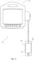
- FIGS. 1 and 3illustrate an ultrasound system 100 including a console 102 and either a first ultrasound probe 104 or a second ultrasound probe 204 in accordance with some embodiments.
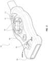
- FIG. 2illustrates a perspective view of the ultrasound probe 104 in accordance with some embodiments.
- the ultrasound probe 104includes a probe body 106 , a probe head 108 extending from a distal end of the probe body 106 , and a plurality of ultrasonic transducers 110 arranged in an array in the probe head 108 .
- the ultrasound probe 104can also include a camera 112 integrated into a side of the ultrasound probe 104 , a light-pattern projector 114 (e.g., a laser light-pattern projector) integrated into the side of the ultrasound probe 104 , or both the camera 112 and the light-pattern projector 114 integrated into the side of the ultrasound probe 104 .
- the side of the ultrasound probe 104 including the camera 112 or the light-pattern projector 114is shown in FIG. 2 as a major side of the ultrasound probe 104 , specifically a top side (or front face) of the ultrasound probe 104 , which is convenient for an out-of-plane view of a needle 116 (see FIG.
- the foregoing side of the ultrasound probe 104conveniently includes various buttons 118 of the ultrasound probe 104 useful for operating the ultrasound probe 104 or 204 or the ultrasound system 100 while establishing an insertion site with the needle 116 .
- the side of the ultrasound probe 104 including the camera 112 or the light-pattern projector 114can alternatively be a minor side of the ultrasound probe 104 , which is convenient for an in-plane view of the needle 116 when establishing an insertion site with the needle 116 as set forth in the method below.
- the camera 112is configured for recording one or more still or moving images 120 (see FIGS. 10 and 11 ) of a procedural field including a subject portion of a patient therein with a depth of field including a plane of a distal end of the probe head 108 and a field of view including a spatial region about the probe head 108 .
- the one-or-more still or moving images 120can be rendered on the display screen of the display 158 along with the ultrasound images 136 associated therewith, which allows a clinician to sustain spatial attention on the display 158 when establishing an insertion site with the needle 116 , thereby obviating the clinician from frequently switching his or her spatial attention between the display 158 and the procedural field as done with existing ultrasound systems.
- the light-pattern projector 114is configured to project a light pattern 122 in the spatial region about the probe head 108 focused in the plane of the distal end of the probe head 108 , thereby including the foregoing subject portion of the patient in the procedural field.
- the light pattern 122is configured for guided insertion of the needle 116 into an anatomical target under the probe head 108 in the procedural field.
- the light pattern 122 when projected in the spatial region about the probe head 108allows a clinician to sustain spatial attention in the procedural field when establishing an insertion site with the needle 116 as set forth in the method below, thereby obviating the clinician from frequently switching his or her spatial attention between the procedural field and the display 158 as done with existing ultrasound systems.
- FIG. 7illustrates a schematic of a first light pattern 122 a in accordance with some embodiments.
- FIG. 8illustrates a schematic of a second light pattern 122 b in accordance with some embodiments.
- the light pattern 122is referenced.
- the light pattern 122 a , 122 b , or the likeis referenced.
- the light pattern 122 a of 122 bincludes periodic hash marks 124 along one or more rays 126 radiating from a central axis of the ultrasound probe 104 in the plane of the probe head 108 .
- the light pattern 122 aincludes the hash marks 124 along one ray 126 radiating from the central axis of the ultrasound probe 104
- the light pattern 122 bincludes the hash marks 124 along three rays 126 radiating from the central axis of the ultrasound probe 104 .
- each hash mark of the hash marks 124corresponds to a depth under the probe head 108 accessible by the needle 116 along an associated ray 126 at a needle-insertion angle with respect to the plane of the probe head 108 .
- FIG. 9illustrates a schematic of a third light pattern 122 c in accordance with some embodiments.
- the light pattern 122 cincludes periodic concentric circular arcs 128 bound between two or more rays 126 radiating from the central axis of the ultrasound probe 104 in the plane of the probe head 108 .
- the light pattern 122 cincludes the circular arcs 128 bound between three rays 126 radiating from the central axis of the ultrasound probe 104 .
- each circular arc of the circular arcs 128corresponds to a depth under the probe head 108 accessible by the needle 116 along an associated ray 126 at a needle-insertion angle with respect to the plane of the probe head 108 .
- the associated ray 126can be an intervening ray between the two-or-more rays 126 of the light pattern 122 c radiating from the central axis of the ultrasound probe 104 .
- the intervening rayneed not be a visible ray of the light pattern 122 c ; the intervening ray can be envisioned between the two-or-more rays 126 of the light pattern 122 c and followed with the needle 116 when establishing an insertion site therewith as set forth in the method below.
- the ultrasound probe 104can also include a needle-guide holder 130 extending from the side of the probe head 108 in common with the side of the ultrasound probe 104 including the camera 112 , whether the foregoing side is the major or minor side of the ultrasound probe 104 including the camera 112 or the light-pattern projector 114 .
- the ultrasound probe 104can also include a single-use needle guide 132 configured to couple to the needle-guide holder 130 .
- the needle guide 132 , the needle-guide holder 130 , or a combination of the needle guide 132 and the needle-guide holder 130can include at least one degree of freedom enabling the needle guide 132 to swivel between sides of the ultrasound probe 104 .
- the needle guide 132can swivel between minor sides of the ultrasound probe 104 if the needle-guide holder 130 extends from a major side of the ultrasound probe 104 .
- the needle guide 132can alternatively swivel between major sides of the ultrasound probe 104 if the needle-guide holder 130 extends from a minor side of the ultrasound probe 104 .
- the needle guide 132 and the needle-guide holder 130can include a joint (e.g., ball joint) formed therebetween that provides the degree of freedom needed. If the needle guide 132 is used with the needle 116 to establish an insertion site, the needle guide 132 can be advantageously swiveled along each circular arc of the circular arcs 128 of the light pattern 122 c . The needle 116 can be subsequently inserted along any existing or envisioned ray of the light pattern 122 c to establish an insertion site.
- a jointe.g., ball joint
- FIGS. 4 and 5illustrate different views of the ultrasound probe 204 in accordance with some embodiments.
- the ultrasound probe 204includes a probe body 206 , a probe head 208 extending from a distal end of the probe body 206 , and the plurality of ultrasonic transducers 110 arranged in an array in the probe head 208 .
- the ultrasound probe 204can include the camera 112 integrated into a side of the ultrasound probe 204 , the light-pattern projector 114 integrated into the side of the ultrasound probe 204 , or both the camera 112 and the light-pattern projector 114 integrated into the side of the ultrasound probe 204 .
- the ultrasound probe 204is like the ultrasound probe 104 in certain ways. Therefore, the description set forth above for the ultrasound probe 104 likewise applies to the ultrasound probe 204 .
- the ultrasound probe 204also includes a display 134 integrated into the side of the ultrasound probe 204 , specifically the top side (or front face) of the ultrasound probe 204 , which differentiates the ultrasound probe 204 from the ultrasound probe 104 .
- the display 134is configured to render ultrasound images 136 on a display screen thereof, which allows a clinician to sustain spatial attention in the procedural field when establishing an insertion site with the needle 116 as set forth in the method below, thereby obviating the clinician from frequently switching his or her spatial attention between the procedural field, which includes the display 134 , and another display (e.g., the display 158 of the console 102 ) as done with existing ultrasound systems.
- the display 134is configured to render one or more overlying needle trajectories 138 over the ultrasound images 136 .
- the one-or-more needle trajectories 138are configured for guided insertion of the needle 116 into an anatomical target under the probe head 208 on the display 134 .
- the one-or-more needle trajectories 138are in accordance with one or more depths accessible by the needle 116 as indicated by the light pattern 122 .
- the ultrasound probe 104 or 204can include magnetic sensors to enhance guided insertion of the needle 116 into an anatomical target as set forth herein with magnetic-based needle guidance.
- magnetic-based needle guidanceis disclosed in U.S. Pat. Nos. 8,388,541; 8,781,555; 8,849,382; 9,456,766; 9,492,097; 9,521,961; 9,554,716; 9,636,031; 9,649,048; 10,449,330; 10,524,691; and 10,751,509, each of which is incorporated by reference in its entirety into this application.
- FIG. 12illustrates a block diagram of the ultrasound system 100 in accordance with some embodiments.
- the console 102includes a variety of components including a processor 140 and memory 142 such as random-access memory (“RAM”) or non-volatile memory (e.g., electrically erasable programmable read-only memory [“EEPROM”]) for controlling various functions of the ultrasound system 100 during operation thereof.
- a processor 140and memory 142 such as random-access memory (“RAM”) or non-volatile memory (e.g., electrically erasable programmable read-only memory [“EEPROM”]) for controlling various functions of the ultrasound system 100 during operation thereof.
- RAMrandom-access memory
- non-volatile memorye.g., electrically erasable programmable read-only memory [“EEPROM”]
- the console 102is configured to instantiate by way of executable instructions 144 stored in the memory 142 and executed by the processor 140 various processes for controlling the various functions of the ultrasound system 100 .
- the various processescan include beamforming by way of a beamformer configured to drive the ultrasonic transducers 110 , wherein driving the ultrasonic transducers 110 includes emitting generated ultrasound signals as well as receiving, amplifying, and digitizing reflected ultrasound signals; signal processing by way of a signal processor configured to detect an amplitude of each of the foregoing reflected ultrasound signals or the digitized signals corresponding thereto; and image processing by way of an image processor configured to manage storage of detected amplitudes and send the ultrasound images 136 corresponding to collections of the detected amplitudes to the display screen of the display 134 or 158 upon completion of the ultrasound images 136 .
- the various processescan include processing electrical signals corresponding to color and brightness data from an image sensor of the camera 112 of the ultrasound probe 104 or 204 into the one-or-more still or moving images 120 ; determining depths for various anatomical structures in the ultrasound images 136 by way of delays in time between emitting the generated ultrasound signals from the ultrasonic transducers 110 and receiving the reflected ultrasound signals by the ultrasonic transducers 110 ; adjusting a scale of the light pattern 122 projected from the light-pattern projector 114 in accordance with both the depths for the various anatomical structures in the ultrasound images 136 and a needle-insertion angle, wherein the needle-insertion angle is selected from a single ultrasound system-defined needle-insertion angle, a clinician-selected needle-insertion angle among various ultrasound system-defined needle-insertion angles, and a dynamic needle-insertion angle determined by way of magnetic-based needle guidance; adjusting a scale of the overlying pattern 160 lying over the one-or-more still or moving
- the console 102also includes a digital controller/analog interface 146 in communication with both the processor 140 and other system components to govern interfacing between the ultrasound probe 104 or 204 and the foregoing system components.
- Ports 148are also included in the console 102 for connection with additional system components including can be universal serial bus (“USB”) ports, though other types of ports can be used for these connections or any other connections shown or described herein.
- USBuniversal serial bus
- a power connection 150is included with the console 102 to enable an operable connection to an external power supply 152 .
- An internal power supply 154e.g., a battery
- Power management circuitry 156is included with the digital controller/analog interface 146 of the console 102 to regulate power use and distribution.
- a display 158 integrated into the console 102is configured to render on a display screen thereof a graphical user interface (“GUI”), the ultrasound images 136 attained by the ultrasound probe 104 or 204 , the one-or-more still or moving images 120 of the procedural field attained by the camera 112 of the ultrasound probe 104 or 204 , an overlying pattern 160 lying over the one-or-more still or moving images 120 , the one-or-more needle trajectories 138 lying over the ultrasound images 136 , etc. That said, the display 158 can alternatively be separate from the console 102 and communicatively coupled thereto. Regardless, control buttons (see FIGS.
- GUIgraphical user interface
- a console button interface 162 of the console 102can be used to immediately call up to the display screen a desired mode of the ultrasound system 100 for assisting with an ultrasound-based medical procedure such as that for establishing an insertion site with the needle 116 , placing a VAD such as a catheter in a blood vessel of a patient at the insertion site, or the like.
- a mode of the ultrasound system 100 for establishing an insertion site with the needle 116can include rendering the one-or-more still or moving images 120 of the procedural field, the overlying pattern 160 lying over the one-or-more still or moving images 120 , the one-or-more needle trajectories 138 lying over the ultrasound images 136 , or a combination thereof.
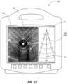
- FIGS. 10 and 11illustrate guided insertion of the needle 116 into an anatomical target of an ultrasound image with the light pattern 122 , specifically the light pattern 122 a and 122 c , respectively, as shown in the one-or-more still or moving images 120 adjacent the ultrasound image on the display 158 .
- the one-or-more still or moving images 120show at least the needle 116 when the needle 116 is present in the spatial region about the probe head 108 or 208 , which, even alone, allows a clinician to sustain spatial attention on the display 158 when establishing an insertion site with the needle 116 .
- the ultrasound probe 104 or 204includes the light-pattern projector 114 , however, the one-or-more still or moving images 120 can show both the light pattern 122 in the spatial region about the probe head 108 or 208 and the needle 116 in relation to the light pattern 122 for guided insertion of the needle 116 into an anatomical target under the probe head 108 or 208 on the display 158 .
- Having both the light pattern 122 and the needle 116 shown in the one-or-more still or moving images 120further allows a clinician to sustain spatial attention on the display 158 when establishing the insertion site with the needle 116 , thereby obviating the clinician from frequently switching his or her spatial attention between the display 158 and the procedural field as done with existing ultrasound systems.
- FIGS. 10 and 11also illustrate guided insertion of the needle 116 into an anatomical target of an ultrasound image respectively with the overlying pattern 160 , specifically the overlying pattern 160 a and 160 c , respectively, over the one-or-more still or moving images 120 adjacent the ultrasound image on the display 158 .
- the one-or-more still or moving images 120can show the overlying pattern 160 lying thereover.
- the one-or-more still or moving images 120can thusly show both the overlying pattern 160 and the needle 116 in relation to the overlying pattern 160 for guided insertion of the needle 116 into an anatomical target under the probe head 108 or 208 on the display 158 .
- Having both the overlying pattern 160 and the needle 116 shown in the one-or-more still or moving images 120further allows a clinician to sustain spatial attention on the display 158 when establishing the insertion site with the needle 116 , thereby obviating the clinician from frequently switching his or her spatial attention between the display 158 and the procedural field as done with existing ultrasound systems.
- the overlying pattern 160 a or 160 bincludes the periodic hash marks 124 along one or more rays 126 radiating from the central axis of the ultrasound probe 104 or 204 in the plane of the probe head 108 or 208 ; however, unlike the light pattern 122 a or 122 b , the hash marks 124 and the one-or-more rays 126 are virtual, existing only on the display screen.
- the overlying pattern 160 alikewise includes the hash marks 124 along one ray 126 radiating from the central axis of the ultrasound probe 104 or 204
- the overlying pattern 160 blikewise includes the hash marks 124 along three rays 126 radiating from the central axis of the ultrasound probe 104 or 204 .
- Each hash mark of the hash marks 124corresponds to a depth under the probe head 108 or 208 accessible by the needle 116 along an associated ray 126 at a needle-insertion angle with respect to the plane of the probe head 108 or 208 .
- the overlying pattern 160 cincludes periodic concentric circular arcs 128 bound between two or more rays 126 radiating from a central axis of the ultrasound probe 104 or 204 in the plane of the probe head 108 or 208 ; however, unlike the light pattern 122 c , the circular arcs 128 and the two-or-more rays 126 are virtual, existing only on the display screen.
- the overlying pattern 160 clikewise includes the circular arcs 128 bound between three rays 126 radiating from the central axis of the ultrasound probe 104 or 204 .
- Each circular arc of the circular arcs 128corresponds to a depth under the probe head 108 or 208 accessible by the needle 116 along an associated ray 126 at a needle-insertion angle with respect to the plane of the probe head 108 or 208 .
- the associated ray 126can be an intervening ray between the two-or-more rays 126 of the overlying pattern 160 c radiating from the central axis of the ultrasound probe 104 or 204 .
- the intervening rayneed not be a visible ray of the overlying pattern 160 c ; the intervening ray can be envisioned between the two-or-more rays 126 of the overlying pattern 160 c and followed with the needle 116 when establishing an insertion site therewith as set forth in the method below.
- the display 158is configured to render on the display screen thereof the one-or-more needle trajectories 138 lying over the ultrasound images 136 .
- the one-or-more needle trajectories 138are configured for guided insertion of the needle 116 into an anatomical target under the probe head 108 or 208 on the display 158 .
- the one-or-more needle trajectories 138are in accordance with one or more depths accessible by the needle 116 indicated by the light pattern 122 c or the overlying pattern 160 c.
- the needle trajectories 138 labeled ‘ 1 ’ in FIG. 11are straightforwardly understood as being in a plane perpendicular to that of an ultrasound beam for a so called out-of-plane view with respect to the needle 116 .
- Moving the needle 116 from circular arc 128 to circular arc 128 of the light pattern 122 c or overlying pattern 160 c of FIG. 11 toward the central axis of the ultrasound probe 104 or 204 while keeping the needle-insertion angle constantmoves the needle 116 from trajectory to trajectory of the one-or-more needle trajectories 138 in a same direction (e.g., up) on the display screen.
- inserting the needle 116 into a patient at the circular arc 128 nearest the central axis of the ultrasound probe 104 or 204results in overshooting an anatomical target, for example, a blood vessel under the probe head 108 or 208 .
- the needle 116could still access the blood vessel but distal of the probe head 108 or 208 .
- the needle trajectories 138 labeled ‘ 2 ’ and ‘ 3 ’ in of FIG. 11are in mirrored planes oblique to that of the ultrasound beam, and, as such, approach the blood vessel obliquely.
- moving the needle 116 from circular arc 128 to circular arc 128 of the light pattern 122 c or overlying pattern 160 c of FIG. 11 toward the central axis of the ultrasound probe 104 or 204 while keeping the needle-insertion angle constantmoves the needle 116 from trajectory to trajectory of the one-or-more needle trajectories 138 in a same direction (e.g., up) on the display screen.
- the ultrasound probe 104 or 204includes the buttons 118 for operating the ultrasound probe 104 or 204 or the ultrasound system 100 of which the ultrasound probe 104 or 204 is part.
- the buttons 118can be configured for selecting a desired mode of the ultrasound system 100 as set forth above.
- the ultrasound probe 104 or 204includes a button-and-memory controller 164 configured for operable communication with a probe interface 166 of the console 102 , which probe interface 166 includes an input/output (“I/O”) component 168 for interfacing with the ultrasonic transducers 110 and a button-and-memory I/O component 170 for interfacing with the button-and-memory controller 164 .
- I/Oinput/output
- Methodsinclude a method of using the ultrasound system 100 to establish an insertion site for access to an anatomical structure (e.g., blood vessel) of a patient.
- the methodincludes one or more steps selected from an ultrasound probe-obtaining step, an ultrasound probe-moving step, a recording step, an ultrasound image-monitoring step, a needle guide-attaching step, a needle guide-swiveling step, and a needle-inserting step.
- the ultrasound probe-obtaining stepincludes obtaining the ultrasound probe 104 .
- the ultrasound probe 104includes the probe body 106 , the probe head 108 extending from the distal end of the probe body 106 , and the camera 112 integrated into the side of the ultrasound probe 104 .
- the needle guide-attaching stepincludes attaching the needle guide 132 to the needle-guide holder 130 extending from the probe body 106 .
- the needle guide 132includes a needle through hole configured to direct the needle 116 into the patient under the probe head 108 at the needle-insertion angle defined by the needle guide 132 .
- the ultrasound probe-moving stepincludes moving the ultrasound probe 104 over the patient while the ultrasound probe 104 emits generated ultrasound signals into the patient from the ultrasonic transducers 110 in the probe head 108 and receives reflected ultrasound signals from the patient by the ultrasonic transducers 110 .
- the recording stepincludes recording the one-or-more still or moving images 120 of the procedural field including a subject portion of the patient therein.
- the one-or-more still or moving images 120are recorded with a depth of field including the plane of the distal end of the probe head 108 and the field of view including the spatial region about the probe head 108 .
- the ultrasound image-monitoring stepincludes monitoring ultrasound images 136 rendered on the display screen of the display 158 associated with the console 102 of the ultrasound system 100 to identify an anatomical target of the patient under the probe head 108 .
- the needle guide-swiveling stepincludes swiveling the needle guide 132 between sides of the ultrasound probe 104 to find a suitable needle trajectory before the needle-inserting step.
- the needle-guide holder 130 , the needle guide 132 , or a combination of the needle-guide holder 130 and the needle guide 132 such as the joint formed therebetweenincludes at least one degree of freedom enabling the swiveling of the needle guide 132 .
- the needle-inserting stepincludes inserting the needle 116 into the anatomical target.
- the inserting of the needle 116 into the anatomical target during the needle-inserting stepis guided in the procedural field with reference to the light pattern 122 in the spatial region about the probe head 108 , on the display 158 with reference to the one-or-more still or moving images 120 or the one-or-more needle trajectories 138 rendered on the display screen thereof, or a combination thereof.
- the light pattern 122is projected into the spatial region about the probe head 108 from the light-pattern projector 114 and focused in the plane of the distal end of the probe head 108 for guiding the needle 116 in the procedural field.
- the light pattern 122 a or 122 bincludes the periodic hash marks 124 along the one-or-more rays 126 radiating from the central axis of the ultrasound probe 104 in the plane of the probe head 108 .
- Each hash mark of the hash marks 124corresponds to a depth under the probe head 108 accessible by the needle 116 along an associated ray 126 at a needle-insertion angle with respect to the plane of the probe head 108 .
- the light pattern 122 cincludes the periodic concentric circular arcs 128 bound between the two-or-more rays 126 radiating from the central axis of the ultrasound probe 104 in the plane of the probe head 108 .
- Each circular arc of the circular arcs 128corresponds to a depth under the probe head 108 accessible by the needle 116 along an associated ray 126 at a needle-insertion angle with respect to the plane of the probe head 108 .
- the one-or-more still or moving images 120can show both the light pattern 122 in the spatial region about the probe head 108 and the needle 116 in relation to the light pattern 122 for guiding the needle 116 on the display 158 .
- the ultrasound probe 104does not include the light-pattern projector 114 , or if a clinician prefers not to use the light-pattern projector 114 of the ultrasound probe 104 , the one-or-more still or moving images 120 can show the overlying pattern 160 lying thereover for guiding the needle 116 on the display 158 .
- the overlying pattern 160 a or 160 bincludes the periodic hash marks 124 along the one-or-more rays 126 radiating from the central axis of the ultrasound probe 1014 in the plane of the probe head 108 .
- Each hash mark of the hash marks 124corresponds to a depth under the probe head 108 accessible by the needle 116 along an associated ray 126 at a needle-insertion angle with respect to the plane of the probe head 108 .
- the overlying pattern 160 cincludes the periodic concentric circular arcs 128 bound between the two-or-more rays 126 radiating from the central axis of the ultrasound probe 104 in the plane of the probe head 108 .
- Each circular arc of the circular arcs 128corresponds to a depth under the probe head 108 accessible by the needle 116 along an associated ray 126 at a needle-insertion angle with respect to the plane of the probe head 108 .
- the ultrasound images 136can show the one-or-more needle trajectories 138 in accordance with one or more depths accessible by the needle 116 indicated by the light pattern 122 or overlying pattern 160 in the one-or-more still or moving images 120 for guiding the needle 116 on the display 158 .
- the foregoing methodinvolves the ultrasound probe 104 ; however, the method can be modified for the ultrasound probe 204 .
- the ultrasound images 136are displayed on the display 134 of the ultrasound probe 204 , optionally, in combination with the ultrasound images 136 and the one-or-more still or moving images 120 on the display 158 of the console 102 .
- displaying the images on the display 134 of the ultrasound probe 204allows a clinician to sustain spatial attention in the procedural field when establishing the insertion site with the needle 116 in the needle-inserting step, thereby obviating the clinician from frequently switching his or her spatial attention between the procedural field, which includes the display 134 , and another display (e.g., the display 158 of the console 102 ) as done with existing ultrasound systems.
Landscapes
- Health & Medical Sciences (AREA)
- Life Sciences & Earth Sciences (AREA)
- Surgery (AREA)
- Engineering & Computer Science (AREA)
- Animal Behavior & Ethology (AREA)
- Veterinary Medicine (AREA)
- Biomedical Technology (AREA)
- Heart & Thoracic Surgery (AREA)
- Medical Informatics (AREA)
- Molecular Biology (AREA)
- Nuclear Medicine, Radiotherapy & Molecular Imaging (AREA)
- General Health & Medical Sciences (AREA)
- Public Health (AREA)
- Pathology (AREA)
- Physics & Mathematics (AREA)
- Biophysics (AREA)
- Radiology & Medical Imaging (AREA)
- Oral & Maxillofacial Surgery (AREA)
- Gynecology & Obstetrics (AREA)
- Robotics (AREA)
- Vascular Medicine (AREA)
- Ultra Sonic Daignosis Equipment (AREA)
Abstract
Description
Claims (17)
Priority Applications (2)
| Application Number | Priority Date | Filing Date | Title |
|---|---|---|---|
| US17/491,308US12137987B2 (en) | 2020-10-02 | 2021-09-30 | Ultrasound systems and methods for sustained spatial attention |
| US18/936,364US20250057604A1 (en) | 2020-10-02 | 2024-11-04 | Ultrasound Systems and Methods for Sustained Spatial Attention |
Applications Claiming Priority (2)
| Application Number | Priority Date | Filing Date | Title |
|---|---|---|---|
| US202063086971P | 2020-10-02 | 2020-10-02 | |
| US17/491,308US12137987B2 (en) | 2020-10-02 | 2021-09-30 | Ultrasound systems and methods for sustained spatial attention |
Related Child Applications (1)
| Application Number | Title | Priority Date | Filing Date |
|---|---|---|---|
| US18/936,364ContinuationUS20250057604A1 (en) | 2020-10-02 | 2024-11-04 | Ultrasound Systems and Methods for Sustained Spatial Attention |
Publications (2)
| Publication Number | Publication Date |
|---|---|
| US20220104886A1 US20220104886A1 (en) | 2022-04-07 |
| US12137987B2true US12137987B2 (en) | 2024-11-12 |
Family
ID=78650039
Family Applications (2)
| Application Number | Title | Priority Date | Filing Date |
|---|---|---|---|
| US17/491,308Active2041-10-17US12137987B2 (en) | 2020-10-02 | 2021-09-30 | Ultrasound systems and methods for sustained spatial attention |
| US18/936,364PendingUS20250057604A1 (en) | 2020-10-02 | 2024-11-04 | Ultrasound Systems and Methods for Sustained Spatial Attention |
Family Applications After (1)
| Application Number | Title | Priority Date | Filing Date |
|---|---|---|---|
| US18/936,364PendingUS20250057604A1 (en) | 2020-10-02 | 2024-11-04 | Ultrasound Systems and Methods for Sustained Spatial Attention |
Country Status (4)
| Country | Link |
|---|---|
| US (2) | US12137987B2 (en) |
| EP (1) | EP4216825B1 (en) |
| CN (2) | CN216221488U (en) |
| WO (1) | WO2022072727A2 (en) |
Families Citing this family (24)
| Publication number | Priority date | Publication date | Assignee | Title |
|---|---|---|---|---|
| JP6252480B2 (en) | 2011-10-21 | 2017-12-27 | 日産化学工業株式会社 | Improved synthesis of conjugated polymers by oxidative polymerization and related compositions |
| CN111712732A (en)* | 2018-02-12 | 2020-09-25 | 皇家飞利浦有限公司 | Workflow assistance for medical Doppler ultrasound evaluation |
| US11759166B2 (en) | 2019-09-20 | 2023-09-19 | Bard Access Systems, Inc. | Automatic vessel detection tools and methods |
| US11877810B2 (en) | 2020-07-21 | 2024-01-23 | Bard Access Systems, Inc. | System, method and apparatus for magnetic tracking of ultrasound probe and generation of 3D visualization thereof |
| EP4185209A1 (en) | 2020-08-04 | 2023-05-31 | Bard Access Systems, Inc. | System and method for optimized medical component insertion monitoring and imaging enhancement |
| US11890139B2 (en) | 2020-09-03 | 2024-02-06 | Bard Access Systems, Inc. | Portable ultrasound systems |
| US11992363B2 (en) | 2020-09-08 | 2024-05-28 | Bard Access Systems, Inc. | Dynamically adjusting ultrasound-imaging systems and methods thereof |
| CN216257185U (en) | 2020-09-10 | 2022-04-12 | 巴德阿克塞斯系统股份有限公司 | Ultrasound Probes and Ultrasound Systems |
| WO2022067101A1 (en) | 2020-09-25 | 2022-03-31 | Bard Access Systems, Inc. | Minimum catheter length tool |
| WO2022072727A2 (en) | 2020-10-02 | 2022-04-07 | Bard Access Systems, Inc. | Ultrasound systems and methods for sustained spatial attention |
| EP4228516A1 (en) | 2020-10-15 | 2023-08-23 | Bard Access Systems, Inc. | Ultrasound imaging system for generation of a three-dimensional ultrasound image |
| CN216933458U (en) | 2020-11-24 | 2022-07-12 | 巴德阿克塞斯系统股份有限公司 | Object recognition and needle guidance system |
| CN114569155A (en) | 2020-12-01 | 2022-06-03 | 巴德阿克塞斯系统股份有限公司 | Ultrasound imaging system and method for obtaining ultrasound image by the same |
| CN114569156A (en) | 2020-12-01 | 2022-06-03 | 巴德阿克塞斯系统股份有限公司 | Ultrasound imaging system and method for identifying one or more of a plurality of blood vessels |
| EP4258997A1 (en) | 2020-12-14 | 2023-10-18 | Bard Access Systems, Inc. | Securement of hands-free ultrasound probe |
| US12349983B2 (en) | 2021-03-05 | 2025-07-08 | Bard Access Systems, Inc. | Systems and methods for ultrasound-and-bioimpedance-based guidance of medical devices |
| CN217960146U (en) | 2021-04-15 | 2022-12-06 | 巴德阿克塞斯系统股份有限公司 | Ultrasound imaging system |
| CN116058873A (en) | 2021-11-03 | 2023-05-05 | 巴德阿克塞斯系统股份有限公司 | Interoperation optimization function through Doppler and image-based vessel discrimination |
| US12433567B2 (en) | 2022-03-16 | 2025-10-07 | Bard Access Systems, Inc. | Ultrasound imaging system |
| US12207967B2 (en) | 2022-04-20 | 2025-01-28 | Bard Access Systems, Inc. | Ultrasound imaging system |
| US12102481B2 (en) | 2022-06-03 | 2024-10-01 | Bard Access Systems, Inc. | Ultrasound probe with smart accessory |
| US12137989B2 (en) | 2022-07-08 | 2024-11-12 | Bard Access Systems, Inc. | Systems and methods for intelligent ultrasound probe guidance |
| WO2024196746A2 (en)* | 2023-03-17 | 2024-09-26 | The United States Government As Represented By The Department Of Veterans Affairs | Imaging system control wand with indicator light, and systems and methods of using same |
| WO2025024821A1 (en)* | 2023-07-27 | 2025-01-30 | Bard Access Systems, Inc. | Optical needle guide |
Citations (327)
| Publication number | Priority date | Publication date | Assignee | Title |
|---|---|---|---|---|
| US5148809A (en) | 1990-02-28 | 1992-09-22 | Asgard Medical Systems, Inc. | Method and apparatus for detecting blood vessels and displaying an enhanced video image from an ultrasound scan |
| US5181513A (en) | 1990-05-29 | 1993-01-26 | Pierre-Jean Touboul | Method of acquiring ultrasound images |
| US5325293A (en) | 1992-02-18 | 1994-06-28 | Dorne Howard L | System and method for correlating medical procedures and medical billing codes |
| US5441052A (en) | 1992-12-28 | 1995-08-15 | Kabushiki Kaisha Toshiba | Color doppler-type ultrasonic diagnostic apparatus |
| US5549554A (en) | 1994-04-01 | 1996-08-27 | Advanced Cardiovascular Systems, Inc. | Catheters having separable reusable components |
| US5573529A (en) | 1994-10-31 | 1996-11-12 | Haak; Benjamin A. | Color coded medical instruments |
| US5775322A (en) | 1996-06-27 | 1998-07-07 | Lucent Medical Systems, Inc. | Tracheal tube and methods related thereto |
| US5879297A (en) | 1997-05-08 | 1999-03-09 | Lucent Medical Systems, Inc. | System and method to determine the location and orientation of an indwelling medical device |
| US5908387A (en) | 1996-06-21 | 1999-06-01 | Quinton Instrument Company | Device and method for improved quantitative coronary artery analysis |
| EP0933063A1 (en) | 1997-12-15 | 1999-08-04 | Medison Co., Ltd. | Ultrasonic color doppler imaging system |
| US5967984A (en) | 1995-06-30 | 1999-10-19 | Boston Scientific Corporation | Ultrasound imaging catheter with a cutting element |
| US5970119A (en) | 1997-11-18 | 1999-10-19 | Douglas Holtz (Part Interest) | Radiological scaling and alignment device |
| US6004270A (en) | 1998-06-24 | 1999-12-21 | Ecton, Inc. | Ultrasound system for contrast agent imaging and quantification in echocardiography using template image for image alignment |
| US6019724A (en) | 1995-02-22 | 2000-02-01 | Gronningsaeter; Aage | Method for ultrasound guidance during clinical procedures |
| US6068599A (en) | 1997-07-14 | 2000-05-30 | Matsushita Electric Industrial Co., Ltd. | Blood vessel puncturing device using ultrasound |
| US6074367A (en) | 1997-10-01 | 2000-06-13 | Scimed Life Systems, Inc. | Preinsertion measurement of catheters |
| JP2000271136A (en) | 1999-03-25 | 2000-10-03 | Toshiba Corp | Ultrasound therapy device and ultrasound therapy device control method |
| US6129668A (en) | 1997-05-08 | 2000-10-10 | Lucent Medical Systems, Inc. | System and method to determine the location and orientation of an indwelling medical device |
| US6132379A (en) | 1998-11-04 | 2000-10-17 | Patacsil; Estelito G. | Method and apparatus for ultrasound guided intravenous cannulation |
| US6233476B1 (en) | 1999-05-18 | 2001-05-15 | Mediguide Ltd. | Medical positioning system |
| US6263230B1 (en) | 1997-05-08 | 2001-07-17 | Lucent Medical Systems, Inc. | System and method to determine the location and orientation of an indwelling medical device |
| US20020038088A1 (en) | 1999-08-20 | 2002-03-28 | Novasonics Inc. | Miniaturized ultrasound apparatus and method |
| US6375615B1 (en) | 1995-10-13 | 2002-04-23 | Transvascular, Inc. | Tissue penetrating catheters having integral imaging transducers and their methods of use |
| US6436043B2 (en) | 1999-12-21 | 2002-08-20 | Koninklijke Phillips Electronics N.V. | Ultrasonic image processing method and examination system for displaying an ultrasonic composite image sequence of an artery |
| US20020148277A1 (en) | 2001-04-11 | 2002-10-17 | Manabu Umeda | Method of making ultrasonic probe and ultrasonic probe |
| US6498942B1 (en) | 1999-08-06 | 2002-12-24 | The University Of Texas System | Optoacoustic monitoring of blood oxygenation |
| US6503205B2 (en) | 1998-11-18 | 2003-01-07 | Cardiosonix Ltd. | Dual ultrasonic transducer probe for blood flow measurement, and blood vessel diameter determination method |
| US6508769B2 (en) | 1999-12-28 | 2003-01-21 | Koninklijke Philips Electronics N.V. | Ultrasonic image processing method and examination system for displaying an ultrasonic color-coded image sequence of an object having moving parts |
| US6511458B2 (en) | 1998-01-13 | 2003-01-28 | Lumend, Inc. | Vascular re-entry catheter |
| US6524249B2 (en) | 1998-11-11 | 2003-02-25 | Spentech, Inc. | Doppler ultrasound method and apparatus for monitoring blood flow and detecting emboli |
| US20030047126A1 (en) | 2001-09-12 | 2003-03-13 | Tomaschko Daniel K. | System for identifying medical devices |
| US20030060714A1 (en) | 2001-09-24 | 2003-03-27 | Henderson Richard W. | Medical ultrasound transducer with interchangeable handle |
| US6543642B1 (en) | 2001-09-21 | 2003-04-08 | Daydots International, Inc. | Disposable glove dispenser system |
| US20030073900A1 (en) | 2001-10-12 | 2003-04-17 | Pranitha Senarith | System and method for monitoring the movement of an interventional device within an anatomical site |
| US6554771B1 (en) | 2001-12-18 | 2003-04-29 | Koninklijke Philips Electronics N.V. | Position sensor in ultrasound transducer probe |
| US20030093001A1 (en) | 2001-11-09 | 2003-05-15 | Antti Martikainen | Method and assembly for identifying a measuring cuff |
| US20030106825A1 (en) | 2001-12-07 | 2003-06-12 | The Procter & Gamble Company | Package containing a window and performance characteristic indicator |
| US20030120154A1 (en) | 2001-11-28 | 2003-06-26 | Frank Sauer | Method and apparatus for ultrasound guidance of needle biopsies |
| US6592520B1 (en) | 2001-07-31 | 2003-07-15 | Koninklijke Philips Electronics N.V. | Intravascular ultrasound imaging apparatus and method |
| US6592565B2 (en) | 2001-04-26 | 2003-07-15 | Zbylut J. Twardowski | Patient-tailored, central-vein catheters |
| US6613002B1 (en) | 1999-06-05 | 2003-09-02 | Wilson-Cook Medical Incorporated | System of indicia for a medical device |
| US6612992B1 (en) | 2000-03-02 | 2003-09-02 | Acuson Corp | Medical diagnostic ultrasound catheter and method for position determination |
| US6623431B1 (en) | 2002-02-25 | 2003-09-23 | Ichiro Sakuma | Examination method of vascular endothelium function |
| US6641538B2 (en) | 2001-11-22 | 2003-11-04 | Kabushiki Kaisha Toshiba | Ultrasonic diagnostic apparatus and method of controlling a ultrasonic diagnostic apparatus |
| US6647135B2 (en) | 1999-12-07 | 2003-11-11 | Koninklijke Philips Electronics N.V. | Ultrasonic image processing method and system for displaying a composite image sequence of an artery segment |
| US6687386B1 (en) | 1999-06-15 | 2004-02-03 | Hitachi Denshi Kabushiki Kaisha | Object tracking method and object tracking apparatus |
| US20040055925A1 (en) | 2000-06-13 | 2004-03-25 | Judith Franks-Farah | Male clean intermittent catheter system |
| US6749569B1 (en) | 2003-01-07 | 2004-06-15 | Esaote S.P.A. | Method and apparatus for ultrasound imaging |
| US6754608B2 (en) | 2001-05-23 | 2004-06-22 | Radi Medical Systems Ab | Interactive measurement system |
| US6755789B2 (en) | 2002-02-05 | 2004-06-29 | Inceptio Medical Technologies, Llc | Ultrasonic vascular imaging system and method of blood vessel cannulation |
| US20050000975A1 (en) | 2003-05-28 | 2005-01-06 | Carco Darlene Marie | Sterile surgical glove dispenser |
| EP1504713A1 (en) | 2003-07-14 | 2005-02-09 | Surgical Navigation Technologies, Inc. | Navigation system for cardiac therapies |
| US6857196B2 (en) | 2003-04-30 | 2005-02-22 | Robert Dalrymple | Method and apparatus for measuring a intracorporal passage image |
| US20050049504A1 (en) | 2003-08-27 | 2005-03-03 | Meng-Tsung Lo | Ultrasonic vein detector and relating method |
| US20050165299A1 (en) | 2004-01-23 | 2005-07-28 | Traxyz Medical, Inc. | Methods and apparatus for performing procedures on target locations in the body |
| US20050251030A1 (en) | 2004-04-21 | 2005-11-10 | Azar Fred S | Method for augmented reality instrument placement using an image based navigation system |
| US20050267365A1 (en) | 2004-06-01 | 2005-12-01 | Alexander Sokulin | Method and apparatus for measuring anatomic structures |
| US6979294B1 (en) | 2002-12-13 | 2005-12-27 | California Institute Of Technology | Split-screen display system and standardized methods for ultrasound image acquisition and processing for improved measurements of vascular structures |
| US20060015039A1 (en) | 2004-07-19 | 2006-01-19 | Cassidy Kenneth T | Guidewire bearing markings simplifying catheter selection |
| US20060013523A1 (en) | 2004-07-16 | 2006-01-19 | Luna Innovations Incorporated | Fiber optic position and shape sensing device and method relating thereto |
| US20060020204A1 (en) | 2004-07-01 | 2006-01-26 | Bracco Imaging, S.P.A. | System and method for three-dimensional space management and visualization of ultrasound data ("SonoDEX") |
| US20060079781A1 (en) | 2002-12-18 | 2006-04-13 | Koninklijke Philips Electronics N.V. | Ultrasonic apparatus for estimating artery parameters |
| US7074187B2 (en) | 2002-12-13 | 2006-07-11 | Selzer Robert H | System and method for improving ultrasound image acquisition and replication for repeatable measurements of vascular structures |
| US20060184029A1 (en) | 2005-01-13 | 2006-08-17 | Ronen Haim | Ultrasound guiding system and method for vascular access and operation mode |
| US20060210130A1 (en) | 2002-12-18 | 2006-09-21 | Laurence Germond-Rouet | Ultrasonic doppler system for determining movement of artery walls |
| AU2006201646A1 (en) | 2005-04-26 | 2006-11-09 | Biosense Webster, Inc. | Display of catheter tip with beam direction for ultrasound system |
| US20070043341A1 (en) | 2001-05-30 | 2007-02-22 | Anderson R R | Apparatus and method for laser treatment with spectroscopic feedback |
| US20070049822A1 (en) | 2005-08-31 | 2007-03-01 | Sonosite, Inc. | Medical device guide locator |
| US20070073155A1 (en) | 2005-09-02 | 2007-03-29 | Ultrasound Ventures, Llc | Ultrasound guidance system |
| US7244234B2 (en) | 2003-11-11 | 2007-07-17 | Soma Development Llc | Ultrasound guided probe device and method of using same |
| US20070199848A1 (en) | 2006-02-28 | 2007-08-30 | Ellswood Mark R | Packaging with color-coded identification |
| US20070239120A1 (en) | 1998-02-24 | 2007-10-11 | Brock David L | Flexible instrument |
| US20070249911A1 (en) | 2006-04-21 | 2007-10-25 | Simon David A | Method and apparatus for optimizing a therapy |
| US20080021322A1 (en) | 2006-05-24 | 2008-01-24 | Michael Benjamin Stone | Ultrasonic imaging apparatus and method |
| US20080033293A1 (en) | 2006-05-08 | 2008-02-07 | C. R. Bard, Inc. | User interface and methods for sonographic display device |
| US20080033759A1 (en) | 2006-08-02 | 2008-02-07 | Vastrac, Inc. | Information manager for a procedure-based medical practice |
| US20080051657A1 (en) | 2005-02-28 | 2008-02-28 | Rold Michael D | Systems And Methods For Estimating The Size And Position Of A Medical Device To Be Applied Within A Patient |
| US7359554B2 (en) | 2002-08-26 | 2008-04-15 | Cleveland Clinic Foundation | System and method for identifying a vascular border |
| EP1591074B1 (en) | 2004-04-26 | 2008-05-21 | BrainLAB AG | Visualization of procedural guidelines for medical procedures |
| US20080146915A1 (en) | 2006-10-19 | 2008-06-19 | Mcmorrow Gerald | Systems and methods for visualizing a cannula trajectory |
| US20080177186A1 (en) | 2007-01-18 | 2008-07-24 | Slater Charles R | Methods and Apparatus for Determining a Treatment Volume of a Fluid Treatment Agent for Treating The Interior of a Blood Vessel |
| US20080221425A1 (en) | 2007-03-09 | 2008-09-11 | Olson Eric S | System and method for local deformable registration of a catheter navigation system to image data or a model |
| US20080294037A1 (en) | 2007-05-23 | 2008-11-27 | Jacob Richter | Apparatus and Method for Guided Chronic Total Occlusion Penetration |
| US20080300491A1 (en) | 2007-06-04 | 2008-12-04 | Medtronic, Inc. | Percutaneous needle guide and methods of use |
| US20090012399A1 (en) | 2005-02-07 | 2009-01-08 | Kazuhiro Sunagawa | Ultrasonic diagnostic apparatus |
| US7534209B2 (en) | 2000-05-26 | 2009-05-19 | Physiosonics, Inc. | Device and method for mapping and tracking blood flow and determining parameters of blood flow |
| US20090143684A1 (en) | 2007-12-04 | 2009-06-04 | Civco Medical Instruments Co., Inc. | Needle guide system for use with ultrasound transducers to effect shallow path needle entry and method of use |
| US20090143672A1 (en) | 2007-12-04 | 2009-06-04 | Harms Steven E | Method for mapping image reference points to facilitate biopsy using magnetic resonance imaging |
| US20090156926A1 (en) | 2007-11-26 | 2009-06-18 | C.R. Bard, Inc. | Integrated System for Intravascular Placement of a Catheter |
| US7599730B2 (en) | 2002-11-19 | 2009-10-06 | Medtronic Navigation, Inc. | Navigation system for cardiac therapies |
| US20090306509A1 (en) | 2005-03-30 | 2009-12-10 | Worcester Polytechnic Institute | Free-hand three-dimensional ultrasound diagnostic imaging with position and angle determination sensors |
| US20100020926A1 (en) | 2008-07-25 | 2010-01-28 | Jan Boese | Method for representing interventional instruments in a 3d data set of an anatomy to be examined as well as a reproduction system for performing the method |
| US7681579B2 (en) | 2005-08-02 | 2010-03-23 | Biosense Webster, Inc. | Guided procedures for treating atrial fibrillation |
| US7691061B2 (en) | 2004-06-24 | 2010-04-06 | Terumo Kabushiki Kaisha | Ultrasonic diagnostic apparatus and method of processing an ultrasound signal |
| US7699779B2 (en) | 2003-05-19 | 2010-04-20 | Hitachi, Ltd. | Ultrasonic treatment equipment |
| US20100106015A1 (en)* | 2008-10-23 | 2010-04-29 | Norris Perry R | Medical device alignment |
| US7720520B2 (en) | 2004-12-01 | 2010-05-18 | Boston Scientific Scimed, Inc. | Method and system for registering an image with a navigation reference catheter |
| US7727153B2 (en) | 2003-04-07 | 2010-06-01 | Sonosite, Inc. | Ultrasonic blood vessel measurement apparatus and method |
| US7734326B2 (en) | 2002-06-20 | 2010-06-08 | Brainlab Ag | Method and device for preparing a drainage |
| US20100179428A1 (en) | 2008-03-17 | 2010-07-15 | Worcester Polytechnic Institute | Virtual interactive system for ultrasound training |
| US20100211026A2 (en) | 2005-03-04 | 2010-08-19 | C. R. Bard, Inc. | Access port identification systems and methods |
| US20100277305A1 (en) | 2007-06-01 | 2010-11-04 | Koninklijke Philips Electronics N.V. | Wireless Ultrasound Probe Asset Tracking |
| US7831449B2 (en) | 2001-02-02 | 2010-11-09 | Thompson Reuters (Healthcare) Inc. | Method and system for extracting medical information for presentation to medical providers on mobile terminals |
| US20100286515A1 (en) | 2007-09-28 | 2010-11-11 | Dietrich Gravenstein | Novel Methods and Devices for Noninvasive Measurement of Energy Absorbers in Blood |
| US20100312121A1 (en) | 2009-06-09 | 2010-12-09 | Zhonghui Guan | Apparatus for a needle director for an ultrasound transducer probe |
| US20110002518A1 (en) | 2009-07-01 | 2011-01-06 | General Electric Company | Method and system for processing ultrasound data |
| US7905837B2 (en) | 2006-09-04 | 2011-03-15 | Ge Medical Systems Global Technology Company, Llc | Ultrasound diagnostic apparatus |
| US20110071404A1 (en) | 2009-09-23 | 2011-03-24 | Lightlab Imaging, Inc. | Lumen Morphology and Vascular Resistance Measurements Data Collection Systems, Apparatus and Methods |
| US7925327B2 (en) | 2002-12-04 | 2011-04-12 | Koninklijke Philips Electronics N.V. | Apparatus and method for assisting the navigation of a catheter in a vessel |
| US7927278B2 (en) | 2002-12-13 | 2011-04-19 | California Institute Of Technology | Split-screen display system and standardized methods for ultrasound image acquisition and multi-frame data processing |
| US8014848B2 (en) | 2004-04-26 | 2011-09-06 | Brainlab Ag | Visualization of procedural guidelines for a medical procedure |
| US8050523B2 (en) | 2007-04-20 | 2011-11-01 | Koninklijke Philips Electronics N.V. | Optical fiber shape sensing systems |
| US8060181B2 (en) | 2006-04-07 | 2011-11-15 | Brainlab Ag | Risk assessment for planned trajectories |
| US20110295108A1 (en) | 2007-11-26 | 2011-12-01 | C.R. Bard, Inc. | Apparatus for use with needle insertion guidance system |
| US8075488B2 (en) | 2005-05-12 | 2011-12-13 | Compumedics Medical Innovation Pty. Ltd. | Ultrasound diagnosis and treatment apparatus |
| US20110313293A1 (en) | 2009-10-08 | 2011-12-22 | C. R. Bard, Inc. | Support and cover structures for an ultrasound probe head |
| US8090427B2 (en) | 2003-09-04 | 2012-01-03 | Koninklijke Philips Electronics N.V. | Methods for ultrasound visualization of a vessel with location and cycle information |
| US8105239B2 (en) | 2006-02-06 | 2012-01-31 | Maui Imaging, Inc. | Method and apparatus to visualize the coronary arteries using ultrasound |
| US8172754B2 (en) | 2006-04-18 | 2012-05-08 | Panasonic Corporation | Ultrasonograph |
| US8175368B2 (en) | 2005-04-05 | 2012-05-08 | Scimed Life Systems, Inc. | Systems and methods for image segmentation with a multi-state classifier |
| US8200313B1 (en) | 2008-10-01 | 2012-06-12 | Bioquantetics, Inc. | Application of image-based dynamic ultrasound spectrography in assisting three dimensional intra-body navigation of diagnostic and therapeutic devices |
| US8211023B2 (en) | 2006-08-11 | 2012-07-03 | Koninklijke Philips Electronics N.V. | Ultrasound system for cerebral blood flow monitoring |
| US20120179038A1 (en) | 2011-01-07 | 2012-07-12 | General Electric Company | Ultrasound based freehand invasive device positioning system and method |
| US20120197132A1 (en) | 2011-01-31 | 2012-08-02 | Analogic Corporation | Ultrasound imaging apparatus |
| US20120209121A1 (en) | 2011-02-15 | 2012-08-16 | General Electric Company | Ultrasound probe including a securing member |
| US20120220865A1 (en) | 2010-12-31 | 2012-08-30 | Volcano Corporation | Pulmonary Embolism Diagnostic Devices and Associated Methods and Systems |
| US20120238875A1 (en) | 2004-11-30 | 2012-09-20 | Eric Savitsky | Embedded Motion Sensing Technology for Integration within Commercial Ultrasound Probes |
| US8298147B2 (en) | 2005-06-24 | 2012-10-30 | Volcano Corporation | Three dimensional co-registration for intravascular diagnosis and therapy |
| US20120277576A1 (en) | 2011-04-26 | 2012-11-01 | Chun Kee Lui | Echogenic infusion port catheter |
| US8303505B2 (en) | 2005-12-02 | 2012-11-06 | Abbott Cardiovascular Systems Inc. | Methods and apparatuses for image guided medical procedures |
| US8323202B2 (en) | 2007-11-16 | 2012-12-04 | Pneumrx, Inc. | Method and system for measuring pulmonary artery circulation information |
| US8328727B2 (en) | 2000-03-23 | 2012-12-11 | Tensys Medical, Inc. | Method and apparatus for assessing hemodynamic parameters within the circulatory system of a living subject |
| US20130041250A1 (en) | 2011-08-09 | 2013-02-14 | Ultrasonix Medical Corporation | Methods and apparatus for locating arteries and veins using ultrasound |
| US8409103B2 (en) | 2005-05-06 | 2013-04-02 | Vasonova, Inc. | Ultrasound methods of positioning guided vascular access devices in the venous system |
| WO2013059714A1 (en) | 2011-10-21 | 2013-04-25 | C.R.Bard, Inc. | Systems and methods for ultrasound-based medical device assessment |
| US20130131499A1 (en) | 2010-02-09 | 2013-05-23 | Koninklijke Philips Electronics N.V. | Apparatus, system and method for imaging and treatment using optical position sensing |
| US20130131502A1 (en) | 2011-11-18 | 2013-05-23 | Michael Blaivas | Blood vessel access system and device |
| US8449465B2 (en) | 2005-10-14 | 2013-05-28 | Cleveland Clinic Foundation | System and method for characterizing vascular tissue |
| US20130150724A1 (en) | 2010-01-07 | 2013-06-13 | Verathon Inc. | Blood vessel access devices, systems, and methods |
| US20130188832A1 (en) | 2009-04-14 | 2013-07-25 | Qinglin Ma | Systems and methods for adaptive volume imaging |
| US20130218024A1 (en)* | 2011-10-09 | 2013-08-22 | Clear Guide Medical, Llc | Interventional In-Situ Image-Guidance by Fusing Ultrasound and Video |
| US8553954B2 (en) | 2010-08-24 | 2013-10-08 | Siemens Medical Solutions Usa, Inc. | Automated system for anatomical vessel characteristic determination |
| US8556815B2 (en) | 2009-05-20 | 2013-10-15 | Laurent Pelissier | Freehand ultrasound imaging systems and methods for guiding fine elongate instruments |
| US8585600B2 (en) | 2010-12-09 | 2013-11-19 | Ge Medical Systems Global Technology Company, Llc | Ultrasound volume probe navigation and control method and device |
| US20130324840A1 (en) | 2010-10-08 | 2013-12-05 | Jian Zhongping | Detection of blood-vessel wall artifacts |
| US20140005530A1 (en) | 2012-06-29 | 2014-01-02 | General Electric Company | Ultrasound imaging method and ultrasound imaging apparatus |
| US8622913B2 (en) | 2010-09-28 | 2014-01-07 | General Electric Company | Method and system for non-invasive monitoring of patient parameters |
| US20140031690A1 (en) | 2012-01-10 | 2014-01-30 | Panasonic Corporation | Ultrasound diagnostic apparatus and method for identifying blood vessel |
| US20140036091A1 (en) | 2011-11-02 | 2014-02-06 | Seno Medical Instruments, Inc. | Interframe energy normalization in an optoacoustic imaging system |
| US20140073976A1 (en) | 2012-09-12 | 2014-03-13 | Heartflow, Inc. | Systems and methods for estimating ischemia and blood flow characteristics from vessel geometry and physiology |
| US20140100440A1 (en) | 2012-10-05 | 2014-04-10 | Volcano Corporation | System and method for instant and automatic border detection |
| US8706457B2 (en) | 2012-05-14 | 2014-04-22 | Heartflow, Inc. | Method and system for providing information from a patient-specific model of blood flow |
| US8734357B2 (en) | 2010-08-12 | 2014-05-27 | Heartflow, Inc. | Method and system for patient-specific modeling of blood flow |
| US8744211B2 (en) | 2011-08-31 | 2014-06-03 | Analogic Corporation | Multi-modality image acquisition |
| US20140155737A1 (en) | 2011-08-16 | 2014-06-05 | Koninklijke Philips N.V. | Curved multi-planar reconstruction using fiber optic shape data |
| US8754865B2 (en) | 2011-11-16 | 2014-06-17 | Volcano Corporation | Medical measuring system and method |
| US8764663B2 (en) | 2012-03-14 | 2014-07-01 | Jeffrey Smok | Method and apparatus for locating and distinguishing blood vessel |
| US20140188133A1 (en) | 2007-11-26 | 2014-07-03 | C. R. Bard, Inc. | Iconic Representations for Guidance of an Indwelling Medical Device |
| US20140188440A1 (en) | 2012-12-31 | 2014-07-03 | Intuitive Surgical Operations, Inc. | Systems And Methods For Interventional Procedure Planning |
| US8781555B2 (en) | 2007-11-26 | 2014-07-15 | C. R. Bard, Inc. | System for placement of a catheter including a signal-generating stylet |
| US8781194B2 (en) | 2009-04-17 | 2014-07-15 | Tufts Medical Center, Inc. | Aneurysm detection |
| US8790263B2 (en) | 2007-02-05 | 2014-07-29 | Siemens Medical Solutions Usa, Inc. | Automated movement detection with audio and visual information |
| WO2014115150A1 (en) | 2013-01-24 | 2014-07-31 | Tylerton International Holdings Inc. | Body structure imaging |
| JP2014150928A (en) | 2013-02-07 | 2014-08-25 | Hitachi Aloka Medical Ltd | Ultrasonic diagnostic device |
| US20140257104A1 (en) | 2013-03-05 | 2014-09-11 | Ezono Ag | Method and system for ultrasound imaging |
| US20140276081A1 (en) | 2013-03-12 | 2014-09-18 | St. Jude Medical Puerto Rico Llc | Ultrasound assisted needle puncture mechanism |
| US20140276690A1 (en) | 2013-03-14 | 2014-09-18 | The Spectranetics Corporation | Controller to select optical channel parameters in a catheter |
| US20140276059A1 (en) | 2013-03-12 | 2014-09-18 | Volcano Corporation | Externally imaging a body structure within a patient |
| US20140276085A1 (en) | 2013-03-13 | 2014-09-18 | Volcano Corporation | Coregistered intravascular and angiographic images |
| US8849382B2 (en) | 2007-11-26 | 2014-09-30 | C. R. Bard, Inc. | Apparatus and display methods relating to intravascular placement of a catheter |
| WO2014174305A2 (en) | 2013-04-26 | 2014-10-30 | Ucl Business Plc | A method and apparatus for determining the location of a medical instrument with respect to ultrasound imaging, and a medical instrument to facilitate such determination |
| US20140343431A1 (en) | 2011-12-16 | 2014-11-20 | Koninklijke Philips N.V. | Automatic blood vessel identification by name |
| US20150005738A1 (en) | 2013-06-26 | 2015-01-01 | Corindus, Inc. | System and method for monitoring of guide catheter seating |
| US20150011887A1 (en) | 2013-07-04 | 2015-01-08 | Samsung Medison Co., Ltd. | Ultrasound system and method for providing object information |
| US8939908B2 (en) | 2009-07-16 | 2015-01-27 | Unex Corporation | Ultrasonic blood vessel inspecting apparatus |
| WO2015017270A1 (en) | 2013-07-29 | 2015-02-05 | Intuitive Surgical Operations, Inc. | Shape sensor systems with redundant sensing |
| US8961420B2 (en) | 2010-04-01 | 2015-02-24 | Siemens Medical Solutions Usa, Inc. | System for cardiac condition detection and characterization |
| US20150065916A1 (en) | 2013-08-29 | 2015-03-05 | Vasculogic, Llc | Fully automated vascular imaging and access system |
| US20150073279A1 (en) | 2013-09-11 | 2015-03-12 | Boston Scientific Scimed, Inc. | Systems and methods for selection and displaying of images using an intravascular ultrasound imaging system |
| US20150112200A1 (en) | 2008-12-18 | 2015-04-23 | C. R. Bard, Inc. | Needle Guide Including Enhanced Visibility Entrance |
| US9022940B2 (en) | 2008-07-18 | 2015-05-05 | Joseph H. Meier | Handheld imaging devices and related methods |
| US20150209526A1 (en) | 2014-01-14 | 2015-07-30 | Volcano Corporation | Devices and methods for forming vascular access |
| US20150209113A1 (en) | 2014-01-29 | 2015-07-30 | Becton, Dickinson And Company | Wearable Electronic Device for Enhancing Visualization During Insertion of an Invasive Device |
| US9138290B2 (en) | 2007-07-27 | 2015-09-22 | Meridian Cardiovascular Systems, Inc. | Method of ablating arterial plaque |
| US9155517B2 (en) | 2007-07-13 | 2015-10-13 | Ezono Ag | Opto-electrical ultrasound sensor and system |
| US20150294497A1 (en) | 2012-05-31 | 2015-10-15 | Koninklijke Philips N.V. | Ultrasound imaging system and method for image guidance procedure |
| US20150297097A1 (en) | 2014-01-14 | 2015-10-22 | Volcano Corporation | Vascular access evaluation and treatment |
| US20150327841A1 (en) | 2014-05-13 | 2015-11-19 | Kabushiki Kaisha Toshiba | Tracking in ultrasound for imaging and user interface |
| US9204858B2 (en) | 2010-02-05 | 2015-12-08 | Ultrasonix Medical Corporation | Ultrasound pulse-wave doppler measurement of blood flow velocity and/or turbulence |
| US20150359991A1 (en) | 2013-03-05 | 2015-12-17 | Ezono Ag | System for image guided procedure |
| US9220477B2 (en) | 2009-12-18 | 2015-12-29 | Konica Minolta, Inc. | Ultrasonic diagnostic device, and region-to-be-detected image display method and measurement method using same |
| US20160029995A1 (en) | 2013-03-15 | 2016-02-04 | Nilus Medical Llc | Hemodynamic monitoring device and methods of using same |
| US20160029998A1 (en) | 2013-12-04 | 2016-02-04 | Obalon Therapeutics, Inc. | Systems and methods for locating and/or characterizing intragastric devices |
| US9257220B2 (en) | 2013-03-05 | 2016-02-09 | Ezono Ag | Magnetization device and method |
| US20160058420A1 (en) | 2013-05-06 | 2016-03-03 | Nkij Engineering Ab | Magnetomotive Probe System And Method Of Use Thereof |
| US9295447B2 (en) | 2011-08-17 | 2016-03-29 | Volcano Corporation | Systems and methods for identifying vascular borders |
| US20160100970A1 (en) | 2014-10-09 | 2016-04-14 | Obalon Therapeutics, Inc. | Ultrasonic systems and methods for locating and/or characterizing intragastric devices |
| US20160101263A1 (en) | 2014-10-10 | 2016-04-14 | Intuitive Surgical Operations, Inc. | Systems and methods for reducing measurement error using optical fiber shape sensors |
| US9320493B2 (en) | 2014-07-08 | 2016-04-26 | Nadarasa Visveshwara | System and method for measuring fluidics in arteries |
| US20160113699A1 (en) | 2013-05-23 | 2016-04-28 | CardioSonic Ltd. | Devices and methods for renal denervation and assessment thereof |
| US20160120607A1 (en) | 2014-11-03 | 2016-05-05 | Michael Sorotzkin | Ultrasonic imaging device for examining superficial skin structures during surgical and dermatological procedures |
| US20160143622A1 (en) | 2013-06-26 | 2016-05-26 | Koninklijke Philips N.V. | System and method for mapping ultrasound shear wave elastography measurements |
| US9364171B2 (en) | 2010-12-22 | 2016-06-14 | Veebot Systems, Inc. | Systems and methods for autonomous intravenous needle insertion |
| US20160166232A1 (en) | 2014-12-10 | 2016-06-16 | Volcano Corporation | Devices, systems, and methods for in-stent restenosis prediction |
| US20160202053A1 (en) | 2013-03-13 | 2016-07-14 | Hansen Medical, Inc. | Reducing incremental measurement sensor error |
| US20160213398A1 (en)* | 2015-01-26 | 2016-07-28 | Ming-Wei Liu | Ultrasound needle guide apparatus |
| US9427207B2 (en) | 2011-04-05 | 2016-08-30 | Houston Medical Robotics, Inc. | Motorized systems and methods for accessing the lumen of a vessel |
| US9445780B2 (en) | 2009-12-04 | 2016-09-20 | University Of Virginia Patent Foundation | Tracked ultrasound vessel imaging |
| US20160278743A1 (en) | 2014-06-11 | 2016-09-29 | Olympus Corporation | Medical diagnostic apparatus, method for operating medical diagnostic apparatus, and computer-readable recording medium |
| US20160278869A1 (en) | 2015-01-19 | 2016-09-29 | Bard Access Systems, Inc. | Device and Method for Vascular Access |
| US9456804B2 (en) | 2013-06-04 | 2016-10-04 | Seiko Epson Corporation | Ultrasound measurement apparatus and ultrasound measurement method |
| US9459087B2 (en) | 2013-03-05 | 2016-10-04 | Ezono Ag | Magnetic position detection system |
| US20160296208A1 (en) | 2007-02-09 | 2016-10-13 | Board Of Regents, The University Of Texas System | Intravascular Photoacoustic and Ultrasound Echo Imaging |
| US9468413B2 (en) | 2008-09-05 | 2016-10-18 | General Electric Company | Method and apparatus for catheter guidance using a combination of ultrasound and X-ray imaging |
| US9492097B2 (en) | 2007-11-26 | 2016-11-15 | C. R. Bard, Inc. | Needle length determination and calibration for insertion guidance system |
| US9521961B2 (en) | 2007-11-26 | 2016-12-20 | C. R. Bard, Inc. | Systems and methods for guiding a medical instrument |
| US20160374644A1 (en) | 2015-06-25 | 2016-12-29 | Rivanna Medical Llc | Ultrasonic Guidance of a Probe with Respect to Anatomical Features |
| US9554716B2 (en) | 2007-11-26 | 2017-01-31 | C. R. Bard, Inc. | Insertion guidance system for needles and medical components |
| US9597008B2 (en) | 2011-09-06 | 2017-03-21 | Ezono Ag | Imaging probe and method of obtaining position and/or orientation information |
| US20170086785A1 (en) | 2015-09-30 | 2017-03-30 | General Electric Company | System and method for providing tactile feedback via a probe of a medical imaging system |
| US9610061B2 (en) | 2011-04-14 | 2017-04-04 | Regents Of The University Of Minnesota | Vascular characterization using ultrasound imaging |
| US20170100092A1 (en) | 2015-10-08 | 2017-04-13 | Decision Sciences Medical Company, LLC | Acoustic orthopedic tracking system and methods |
| US9636031B2 (en) | 2007-11-26 | 2017-05-02 | C.R. Bard, Inc. | Stylets for use with apparatus for intravascular placement of a catheter |
| US9649037B2 (en) | 2009-12-03 | 2017-05-16 | Deltex Medical Limited | Method and apparatus for hemodynamic monitoring using combined blood flow and blood pressure measurement |
| US9649048B2 (en) | 2007-11-26 | 2017-05-16 | C. R. Bard, Inc. | Systems and methods for breaching a sterile field for intravascular placement of a catheter |
| US20170164923A1 (en) | 2015-12-14 | 2017-06-15 | Konica Minolta, Inc. | Image Processor, Ultrasound Diagnostic Device Including Same, And Image Processing Method |
| WO2017096487A1 (en) | 2015-12-10 | 2017-06-15 | 1929803 Ontario Corp. D/B/A Ke2 Technologies | Systems and methods for automated fluid response measurement |
| EP3181083A1 (en) | 2015-12-18 | 2017-06-21 | Biosense Webster (Israel), Ltd. | Using force sensor to give angle of ultrasound beam |
| US20170172424A1 (en) | 2014-12-22 | 2017-06-22 | Eggers & Associates, Inc. | Wearable Apparatus, System and Method for Detection of Cardiac Arrest and Alerting Emergency Response |
| US20170188839A1 (en) | 2014-09-25 | 2017-07-06 | Fujifilm Corporation | Photoacoustic image generation apparatus |
| US9702969B2 (en) | 2009-05-13 | 2017-07-11 | Koninklijke Philips Electronics N.V. | Ultrasonic blood flow doppler audio with pitch shifting |
| US20170196535A1 (en) | 2014-06-04 | 2017-07-13 | Hitachi, Ltd. | Medical treatment system |
| US9717415B2 (en) | 2007-03-08 | 2017-08-01 | Sync-Rx, Ltd. | Automatic quantitative vessel analysis at the location of an automatically-detected tool |
| US20170215842A1 (en) | 2014-08-28 | 2017-08-03 | Samsung Electronics Co., Ltd. | Ultrasound diagnosis apparatus for self-diagnosis and remote-diagnosis, and method of operating the ultrasound diagnosis apparatus |
| US9731066B2 (en) | 2011-09-30 | 2017-08-15 | General Electric Company | Device, system and method of automatic vessel access based on real time volumetric ultrasound |
| US20170259013A1 (en)* | 2012-10-30 | 2017-09-14 | Elwha Llc | Systems and Methods for Generating an Injection Guide |
| US20170265840A1 (en) | 2014-12-01 | 2017-09-21 | Koninklijke Philips N.V. | Registration of optical shape sensing tool |
| US20170303894A1 (en) | 2015-01-08 | 2017-10-26 | The Charlotte Mecklenburg Hospital Authority D/B/A Carolinas Healthcare System | Ultrasound probe couplers and related methods |
| US9814531B2 (en) | 2011-08-26 | 2017-11-14 | EBM Corporation | System for diagnosing bloodflow characteristics, method thereof, and computer software program |
| US9814433B2 (en) | 2012-10-24 | 2017-11-14 | Cathworks Ltd. | Creating a vascular tree model |
| WO2017214428A1 (en) | 2016-06-08 | 2017-12-14 | The United States Of America, As Represented By The Secretary, Department Of Health And Human Services | Tissue characterization with acoustic wave tomosynthesis |
| US20170367678A1 (en) | 2016-06-22 | 2017-12-28 | Cesare Sirtori | Ultrasound automated method for measuring the thickness of the walls of the left anterior descending, right and circumflex coronary arteries |
| US9861337B2 (en) | 2013-02-04 | 2018-01-09 | General Electric Company | Apparatus and method for detecting catheter in three-dimensional ultrasound images |
| US20180015256A1 (en) | 2016-07-14 | 2018-01-18 | C. R. Bard, Inc. | Automated Catheter-To-Vessel Size Comparison Tool And Related Methods |
| WO2018026878A1 (en) | 2016-08-02 | 2018-02-08 | Avent, Inc. | Motor-assisted needle guide assembly for ultrasound needle placement |
| US9895138B2 (en) | 2011-06-06 | 2018-02-20 | Toshiba Medical Systems Corporation | Ultrasonic diagnostic apparatus |
| US20180116723A1 (en) | 2016-10-28 | 2018-05-03 | Medtronic Ardian Luxembourg S.A.R.L. | Methods and Systems for Optimizing Perivascular Neuromodulation Therapy Using Computational Fluid Dynamics |
| US20180125450A1 (en) | 2015-04-24 | 2018-05-10 | U.S. Government, As Represented By The Secretary Of The Army | Vascular Targeting System |
| US20180161502A1 (en) | 2015-06-15 | 2018-06-14 | The University Of Sydney | Insertion system and method |
| KR20180070878A (en) | 2016-12-19 | 2018-06-27 | 지멘스 메디컬 솔루션즈 유에스에이, 인크. | Method of providing annotation information of ultrasound probe and ultrasound system |
| US20180199914A1 (en) | 2015-07-22 | 2018-07-19 | Koninklijke Philips N.V. | Fiber-optic realshape sensor for enhanced dopper measurement display |
| WO2018134726A1 (en) | 2017-01-20 | 2018-07-26 | Politecnico Di Torino | Method and apparatus to characterise non-invasively images containing venous blood vessels |
| US20180214119A1 (en) | 2017-01-27 | 2018-08-02 | Wayne State University | Ultrasound and photoacoustic systems and methods for fetal brain assessment during delivery |
| US10043272B2 (en) | 2014-09-16 | 2018-08-07 | Esaote S.P.A. | Method and apparatus for acquiring and fusing ultrasound images with pre-acquired images |
| US20180225993A1 (en) | 2017-01-24 | 2018-08-09 | Tietronix Software, Inc. | System and method for three-dimensional augmented reality guidance for use of medical equipment |
| US20180235576A1 (en) | 2017-02-22 | 2018-08-23 | Covidien Lp | Ultrasound doppler and elastography for ablation prediction and monitoring |
| US20180250078A1 (en) | 2015-09-10 | 2018-09-06 | Xact Robotics Ltd. | Systems and methods for guiding the insertion of a medical tool |
| US20180272108A1 (en) | 2017-03-27 | 2018-09-27 | Biosense Webster (Israel) Ltd | Catheter with improved loop contraction and greater contraction displacement |
| US20180286287A1 (en) | 2017-03-28 | 2018-10-04 | Covidien Lp | System and methods for training physicians to perform ablation procedures |
| US20180279996A1 (en) | 2014-11-18 | 2018-10-04 | C.R. Bard, Inc. | Ultrasound imaging system having automatic image presentation |
| US20180310955A1 (en) | 2017-04-27 | 2018-11-01 | Bard Access Systems, Inc. | Magnetizing System For Needle Assemblies |
| US20180317881A1 (en) | 2017-05-05 | 2018-11-08 | International Business Machines Corporation | Automating ultrasound examination of a vascular system |
| WO2018206473A1 (en) | 2017-05-11 | 2018-11-15 | Koninklijke Philips N.V. | Workflow, system and method for motion compensation in ultrasound procedures |
| JP2018175547A (en) | 2017-04-17 | 2018-11-15 | ニプロ株式会社 | Puncture guide and ultrasound diagnostic apparatus with puncture guide |
| US20180366035A1 (en) | 2017-06-20 | 2018-12-20 | Ezono Ag | System and method for image-guided procedure analysis and training |
| KR20190013133A (en)* | 2017-07-31 | 2019-02-11 | (재)예수병원유지재단 | Apparatus for guiding syringe arrangement based on ultrasonic probe |
| US20190060014A1 (en) | 2017-08-30 | 2019-02-28 | Intuitive Surgical Operations, Inc. | System and method for providing on-demand functionality during a medical procedure |
| US20190069923A1 (en)* | 2015-11-08 | 2019-03-07 | Qin Wang | Paracentesis needle frame |
| US20190076121A1 (en) | 2017-09-13 | 2019-03-14 | Bard Access Systems, Inc. | Ultrasound Finger Probe |
| US20190088019A1 (en) | 2016-03-16 | 2019-03-21 | Koninklijke Philips N.V. | Calculation device for superimposing a laparoscopic image and an ultrasound image |
| US20190105017A1 (en)* | 2017-10-11 | 2019-04-11 | Geoffrey Steven Hastings | Laser assisted ultrasound guidance |
| US20190117190A1 (en) | 2016-04-19 | 2019-04-25 | Koninklijke Philips N.V. | Ultrasound imaging probe positioning |
| US20190223757A1 (en) | 2017-12-04 | 2019-07-25 | Bard Access Systems, Inc. | Systems And Methods For Visualizing Anatomy, Locating Medical Devices, Or Placing Medical Devices |
| US20190239850A1 (en) | 2018-02-06 | 2019-08-08 | Steven Philip Dalvin | Augmented/mixed reality system and method for the guidance of a medical exam |
| US10380920B2 (en) | 2013-09-23 | 2019-08-13 | SonoSim, Inc. | System and method for augmented ultrasound simulation using flexible touch sensitive surfaces |
| US10380919B2 (en) | 2013-11-21 | 2019-08-13 | SonoSim, Inc. | System and method for extended spectrum ultrasound training using animate and inanimate training objects |
| EP3530221A1 (en) | 2018-02-26 | 2019-08-28 | Covidien LP | System and method for performing a percutaneous navigation procedure |
| US20190282324A1 (en) | 2018-03-15 | 2019-09-19 | Zoll Medical Corporation | Augmented Reality Device for Providing Feedback to an Acute Care Provider |
| US10424225B2 (en) | 2013-09-23 | 2019-09-24 | SonoSim, Inc. | Method for ultrasound training with a pressure sensing array |
| US20190298457A1 (en) | 2016-11-08 | 2019-10-03 | Koninklijke Philips N.V. | System and method for tracking an interventional instrument with feedback concerning tracking reliability |
| US20190307516A1 (en) | 2018-04-06 | 2019-10-10 | Medtronic, Inc. | Image-based navigation system and method of using same |
| US10449330B2 (en) | 2007-11-26 | 2019-10-22 | C. R. Bard, Inc. | Magnetic element-equipped needle assemblies |
| US20190339525A1 (en) | 2018-05-07 | 2019-11-07 | The Cleveland Clinic Foundation | Live 3d holographic guidance and navigation for performing interventional procedures |
| US20190355278A1 (en) | 2018-05-18 | 2019-11-21 | Marion Surgical Inc. | Virtual reality surgical system including a surgical tool assembly with haptic feedback |
| WO2019232451A1 (en) | 2018-05-31 | 2019-12-05 | Matt Mcgrath Design & Co, Llc | Method of medical imaging using multiple arrays |
| US20190365348A1 (en) | 2015-06-23 | 2019-12-05 | Hemonitor Medical Ltd. | Systems and methods for hand-free continuous ultrasonic monitoring |
| WO2020002620A1 (en) | 2018-06-29 | 2020-01-02 | Koninklijke Philips N.V. | Biopsy prediction and guidance with ultrasound imaging and associated devices, systems, and methods |
| US10524691B2 (en) | 2007-11-26 | 2020-01-07 | C. R. Bard, Inc. | Needle assembly including an aligned magnetic element |
| WO2020016018A1 (en) | 2018-07-18 | 2020-01-23 | Koninklijke Philips N.V. | Automatic image vetting on a handheld medical scanning device |
| US20200041261A1 (en) | 2017-10-06 | 2020-02-06 | Advanced Scanners, Inc. | Generation of one or more edges of luminosity to form three-dimensional models of objects |
| WO2020044769A1 (en) | 2018-08-27 | 2020-03-05 | 富士フイルム株式会社 | Ultrasound diagnosis device and ultrasound diagnosis device control method |
| US20200069285A1 (en) | 2018-08-31 | 2020-03-05 | General Electric Company | System and method for ultrasound navigation |
| US20200113540A1 (en) | 2017-06-07 | 2020-04-16 | Koninklijke Philips N.V. | Ultrasound system and method |
| US20200129136A1 (en) | 2018-10-31 | 2020-04-30 | Medtronic, Inc. | Real-time rendering and referencing for medical procedures |
| WO2020102665A1 (en) | 2018-11-16 | 2020-05-22 | Lang Philipp K | Augmented reality guidance for surgical procedures with adjustment of scale, convergence and focal plane or focal point of virtual data |
| US20200188028A1 (en) | 2017-08-21 | 2020-06-18 | The Trustees Of Columbia University In The City Of New York | Systems and methods for augmented reality guidance |
| US20200230391A1 (en) | 2019-01-18 | 2020-07-23 | Becton, Dickinson And Company | Intravenous therapy system for blood vessel detection and vascular access device placement |
| WO2020186198A1 (en) | 2019-03-13 | 2020-09-17 | University Of Florida Research Foundation | Guidance and tracking system for templated and targeted biopsy and treatment |
| US20210007710A1 (en) | 2019-07-12 | 2021-01-14 | Verathon Inc. | Representation of a target during aiming of an ultrasound probe |
| US10896628B2 (en) | 2017-01-26 | 2021-01-19 | SonoSim, Inc. | System and method for multisensory psychomotor skill training |
| US20210045716A1 (en) | 2019-08-13 | 2021-02-18 | GE Precision Healthcare LLC | Method and system for providing interaction with a visual artificial intelligence ultrasound image segmentation module |
| US11120709B2 (en) | 2012-12-18 | 2021-09-14 | SonoSim, Inc. | System and method for teaching basic ultrasound skills |
| US20210307838A1 (en) | 2017-12-29 | 2021-10-07 | Weipeng (Suzhou) Co., Ltd. | Surgical navigation method and system |
| US20210353255A1 (en) | 2014-03-31 | 2021-11-18 | Koninklijke Philips N.V. | Haptic feedback for ultrasound image acquisition |
| US20210402144A1 (en) | 2020-06-29 | 2021-12-30 | Bard Access Systems, Inc. | Automatic Dimensional Frame Reference for Fiber Optic |
| US20220022969A1 (en) | 2020-07-21 | 2022-01-27 | Bard Access Systems, Inc. | System, Method and Apparatus for Magnetic Tracking of Ultrasound Probe and Generation of 3D Visualization Thereof |
| US20220031965A1 (en) | 2020-08-03 | 2022-02-03 | Bard Access Systems, Inc. | Alternative-Reality Devices and Methods for Establishing and Visualizing Historical Sites of Vascular Access |
| US20220039685A1 (en) | 2020-08-04 | 2022-02-10 | Bard Access Systems, Inc. | Systemized and Method for Optimized Medical Component Insertion Monitoring and Imaging Enhancement |
| US20220039777A1 (en) | 2020-08-10 | 2022-02-10 | Bard Access Systems, Inc. | System and Method for Generating Vessel Representations in Mixed Reality/Virtual Reality |
| US20220096797A1 (en) | 2020-09-25 | 2022-03-31 | Bard Access Systems, Inc. | Minimum Catheter Length Tool |
| WO2022072727A2 (en) | 2020-10-02 | 2022-04-07 | Bard Access Systems, Inc. | Ultrasound systems and methods for sustained spatial attention |
| US20220117582A1 (en) | 2020-10-15 | 2022-04-21 | Bard Access Systems, Inc. | Ultrasound Imaging System for Generation of a Three-Dimensional Ultrasound Image |
| US11311269B2 (en) | 2008-04-22 | 2022-04-26 | Ezono Ag | Ultrasound imaging system and method for providing assistance in an ultrasound imaging system |
| US20220160434A1 (en) | 2020-11-24 | 2022-05-26 | Bard Access Systems, Inc. | Ultrasound System with Target and Medical Instrument Awareness |
| US20220168050A1 (en) | 2020-12-01 | 2022-06-02 | Bard Access Systems, Inc. | Ultrasound Probe with Target Tracking Capability |
| US20220172354A1 (en) | 2020-12-01 | 2022-06-02 | Bard Access Systems, Inc. | Ultrasound System with Pressure and Flow Determination Capability |
| US20220211442A1 (en) | 2021-01-06 | 2022-07-07 | Bard Access Systems, Inc. | Needle Guidance Using Fiber Optic Shape Sensing |
| CN114129137B (en) | 2021-12-02 | 2022-09-09 | 深圳先进技术研究院 | Intravascular imaging system, device and imaging method |
| WO2022203713A2 (en) | 2020-09-18 | 2022-09-29 | Bard Access Systems, Inc. | Ultrasound probe with pointer remote control capability |
| WO2022263763A1 (en) | 2021-06-16 | 2022-12-22 | Quantum Surgical | Medical robot for placement of medical instruments under ultrasound guidance |
| US11600201B1 (en) | 2015-06-30 | 2023-03-07 | The Regents Of The University Of California | System and method for converting handheld diagnostic ultrasound systems into ultrasound training systems |
| US20230113291A1 (en) | 2020-04-02 | 2023-04-13 | Koninklijke Philips N.V. | Ultrasound probe, user console, system and method |
| US20230240643A1 (en) | 2020-06-16 | 2023-08-03 | Innovacell Ag | Parallel path puncture device guide and method |
| WO2023235435A1 (en) | 2022-06-03 | 2023-12-07 | Bard Access Systems, Inc. | Ultrasound probe with smart accessory |
| US20240008929A1 (en) | 2022-07-08 | 2024-01-11 | Bard Access Systems, Inc. | Systems and Methods for Intelligent Ultrasound Probe Guidance |
| US20240050061A1 (en) | 2022-08-15 | 2024-02-15 | Bard Access Systems, Inc. | Spatially Aware Medical Device Configured for Performance of Insertion Pathway Approximation |
| US20240062678A1 (en) | 2022-08-17 | 2024-02-22 | Bard Access Systems, Inc. | Ultrasound Training System |
Family Cites Families (5)
| Publication number | Priority date | Publication date | Assignee | Title |
|---|---|---|---|---|
| ES2900584T3 (en)* | 2010-12-23 | 2022-03-17 | Bard Access Systems Inc | System for guiding a rigid instrument |
| US9247906B2 (en)* | 2011-06-28 | 2016-02-02 | Christie Digital Systems Usa, Inc. | Method and apparatus for detection of catheter location for intravenous access |
| JP2014161444A (en)* | 2013-02-22 | 2014-09-08 | Toshiba Corp | Ultrasound diagnostic device, medical image processor and control program |
| CN109512473A (en)* | 2017-09-20 | 2019-03-26 | 史军 | Medical Devices with visual puncturing device |
| FR3071396B1 (en)* | 2017-09-22 | 2022-01-21 | Koelis | MEDICAL INSTRUMENT GUIDE DEVICE AND CORRESPONDING METHOD |
- 2021
- 2021-09-30WOPCT/US2021/053018patent/WO2022072727A2/ennot_activeCeased
- 2021-09-30EPEP21810161.6Apatent/EP4216825B1/enactiveActive
- 2021-09-30CNCN202122406096.XUpatent/CN216221488U/enactiveActive
- 2021-09-30USUS17/491,308patent/US12137987B2/enactiveActive
- 2021-09-30CNCN202111160220.7Apatent/CN114376613A/enactivePending
- 2024
- 2024-11-04USUS18/936,364patent/US20250057604A1/enactivePending
Patent Citations (363)
| Publication number | Priority date | Publication date | Assignee | Title |
|---|---|---|---|---|
| US5148809A (en) | 1990-02-28 | 1992-09-22 | Asgard Medical Systems, Inc. | Method and apparatus for detecting blood vessels and displaying an enhanced video image from an ultrasound scan |
| US5181513A (en) | 1990-05-29 | 1993-01-26 | Pierre-Jean Touboul | Method of acquiring ultrasound images |
| US5325293A (en) | 1992-02-18 | 1994-06-28 | Dorne Howard L | System and method for correlating medical procedures and medical billing codes |
| US5441052A (en) | 1992-12-28 | 1995-08-15 | Kabushiki Kaisha Toshiba | Color doppler-type ultrasonic diagnostic apparatus |
| US5549554A (en) | 1994-04-01 | 1996-08-27 | Advanced Cardiovascular Systems, Inc. | Catheters having separable reusable components |
| US5573529A (en) | 1994-10-31 | 1996-11-12 | Haak; Benjamin A. | Color coded medical instruments |
| US6019724A (en) | 1995-02-22 | 2000-02-01 | Gronningsaeter; Aage | Method for ultrasound guidance during clinical procedures |
| US5967984A (en) | 1995-06-30 | 1999-10-19 | Boston Scientific Corporation | Ultrasound imaging catheter with a cutting element |
| US7637870B2 (en) | 1995-10-13 | 2009-12-29 | Medtronic Vascular, Inc. | Tissue penetrating catheters having integral imaging transducers and their methods of use |
| US8727988B2 (en) | 1995-10-13 | 2014-05-20 | Medtronic Vascular, Inc. | Tissue penetrating catheters having integral imaging transducers and their methods of use |
| US20140180098A1 (en) | 1995-10-13 | 2014-06-26 | Medtronic Vascular, Inc. | Tissue Penetrating Catheters Having Integral Imaging Transducers and Their Methods of Use |
| US6375615B1 (en) | 1995-10-13 | 2002-04-23 | Transvascular, Inc. | Tissue penetrating catheters having integral imaging transducers and their methods of use |
| US5908387A (en) | 1996-06-21 | 1999-06-01 | Quinton Instrument Company | Device and method for improved quantitative coronary artery analysis |
| US5775322A (en) | 1996-06-27 | 1998-07-07 | Lucent Medical Systems, Inc. | Tracheal tube and methods related thereto |
| US6129668A (en) | 1997-05-08 | 2000-10-10 | Lucent Medical Systems, Inc. | System and method to determine the location and orientation of an indwelling medical device |
| US6216028B1 (en) | 1997-05-08 | 2001-04-10 | Lucent Medical Systems, Inc. | Method to determine the location and orientation of an indwelling medical device |
| US6263230B1 (en) | 1997-05-08 | 2001-07-17 | Lucent Medical Systems, Inc. | System and method to determine the location and orientation of an indwelling medical device |
| US5879297A (en) | 1997-05-08 | 1999-03-09 | Lucent Medical Systems, Inc. | System and method to determine the location and orientation of an indwelling medical device |
| US6068599A (en) | 1997-07-14 | 2000-05-30 | Matsushita Electric Industrial Co., Ltd. | Blood vessel puncturing device using ultrasound |
| US6074367A (en) | 1997-10-01 | 2000-06-13 | Scimed Life Systems, Inc. | Preinsertion measurement of catheters |
| US5970119A (en) | 1997-11-18 | 1999-10-19 | Douglas Holtz (Part Interest) | Radiological scaling and alignment device |
| US6245018B1 (en) | 1997-12-15 | 2001-06-12 | Medison Co., Ltd. | Ultrasonic color doppler imaging system capable of discriminating artery and vein |
| EP0933063A1 (en) | 1997-12-15 | 1999-08-04 | Medison Co., Ltd. | Ultrasonic color doppler imaging system |
| US6511458B2 (en) | 1998-01-13 | 2003-01-28 | Lumend, Inc. | Vascular re-entry catheter |
| US20070239120A1 (en) | 1998-02-24 | 2007-10-11 | Brock David L | Flexible instrument |
| US6004270A (en) | 1998-06-24 | 1999-12-21 | Ecton, Inc. | Ultrasound system for contrast agent imaging and quantification in echocardiography using template image for image alignment |
| US6132379A (en) | 1998-11-04 | 2000-10-17 | Patacsil; Estelito G. | Method and apparatus for ultrasound guided intravenous cannulation |
| US6524249B2 (en) | 1998-11-11 | 2003-02-25 | Spentech, Inc. | Doppler ultrasound method and apparatus for monitoring blood flow and detecting emboli |
| US6503205B2 (en) | 1998-11-18 | 2003-01-07 | Cardiosonix Ltd. | Dual ultrasonic transducer probe for blood flow measurement, and blood vessel diameter determination method |
| JP2000271136A (en) | 1999-03-25 | 2000-10-03 | Toshiba Corp | Ultrasound therapy device and ultrasound therapy device control method |
| US6233476B1 (en) | 1999-05-18 | 2001-05-15 | Mediguide Ltd. | Medical positioning system |
| US6613002B1 (en) | 1999-06-05 | 2003-09-02 | Wilson-Cook Medical Incorporated | System of indicia for a medical device |
| US6687386B1 (en) | 1999-06-15 | 2004-02-03 | Hitachi Denshi Kabushiki Kaisha | Object tracking method and object tracking apparatus |
| US6498942B1 (en) | 1999-08-06 | 2002-12-24 | The University Of Texas System | Optoacoustic monitoring of blood oxygenation |
| US20020038088A1 (en) | 1999-08-20 | 2002-03-28 | Novasonics Inc. | Miniaturized ultrasound apparatus and method |
| US6647135B2 (en) | 1999-12-07 | 2003-11-11 | Koninklijke Philips Electronics N.V. | Ultrasonic image processing method and system for displaying a composite image sequence of an artery segment |
| US6436043B2 (en) | 1999-12-21 | 2002-08-20 | Koninklijke Phillips Electronics N.V. | Ultrasonic image processing method and examination system for displaying an ultrasonic composite image sequence of an artery |
| US6508769B2 (en) | 1999-12-28 | 2003-01-21 | Koninklijke Philips Electronics N.V. | Ultrasonic image processing method and examination system for displaying an ultrasonic color-coded image sequence of an object having moving parts |
| US6612992B1 (en) | 2000-03-02 | 2003-09-02 | Acuson Corp | Medical diagnostic ultrasound catheter and method for position determination |
| US8328727B2 (en) | 2000-03-23 | 2012-12-11 | Tensys Medical, Inc. | Method and apparatus for assessing hemodynamic parameters within the circulatory system of a living subject |
| US7534209B2 (en) | 2000-05-26 | 2009-05-19 | Physiosonics, Inc. | Device and method for mapping and tracking blood flow and determining parameters of blood flow |
| US20040055925A1 (en) | 2000-06-13 | 2004-03-25 | Judith Franks-Farah | Male clean intermittent catheter system |
| US6840379B2 (en) | 2000-06-13 | 2005-01-11 | Judith Franks-Farah | Male clean intermittent catheter system |
| US7831449B2 (en) | 2001-02-02 | 2010-11-09 | Thompson Reuters (Healthcare) Inc. | Method and system for extracting medical information for presentation to medical providers on mobile terminals |
| US20020148277A1 (en) | 2001-04-11 | 2002-10-17 | Manabu Umeda | Method of making ultrasonic probe and ultrasonic probe |
| US6592565B2 (en) | 2001-04-26 | 2003-07-15 | Zbylut J. Twardowski | Patient-tailored, central-vein catheters |
| US6754608B2 (en) | 2001-05-23 | 2004-06-22 | Radi Medical Systems Ab | Interactive measurement system |
| US20070043341A1 (en) | 2001-05-30 | 2007-02-22 | Anderson R R | Apparatus and method for laser treatment with spectroscopic feedback |
| US6592520B1 (en) | 2001-07-31 | 2003-07-15 | Koninklijke Philips Electronics N.V. | Intravascular ultrasound imaging apparatus and method |
| US20030047126A1 (en) | 2001-09-12 | 2003-03-13 | Tomaschko Daniel K. | System for identifying medical devices |
| US6543642B1 (en) | 2001-09-21 | 2003-04-08 | Daydots International, Inc. | Disposable glove dispenser system |
| US20030060714A1 (en) | 2001-09-24 | 2003-03-27 | Henderson Richard W. | Medical ultrasound transducer with interchangeable handle |
| US20030073900A1 (en) | 2001-10-12 | 2003-04-17 | Pranitha Senarith | System and method for monitoring the movement of an interventional device within an anatomical site |
| US20030093001A1 (en) | 2001-11-09 | 2003-05-15 | Antti Martikainen | Method and assembly for identifying a measuring cuff |
| US6641538B2 (en) | 2001-11-22 | 2003-11-04 | Kabushiki Kaisha Toshiba | Ultrasonic diagnostic apparatus and method of controlling a ultrasonic diagnostic apparatus |
| US20030120154A1 (en) | 2001-11-28 | 2003-06-26 | Frank Sauer | Method and apparatus for ultrasound guidance of needle biopsies |
| US6601705B2 (en) | 2001-12-07 | 2003-08-05 | The Procter & Gamble Company | Package containing a window and performance characteristic indicator |
| US20030106825A1 (en) | 2001-12-07 | 2003-06-12 | The Procter & Gamble Company | Package containing a window and performance characteristic indicator |
| US6554771B1 (en) | 2001-12-18 | 2003-04-29 | Koninklijke Philips Electronics N.V. | Position sensor in ultrasound transducer probe |
| US6755789B2 (en) | 2002-02-05 | 2004-06-29 | Inceptio Medical Technologies, Llc | Ultrasonic vascular imaging system and method of blood vessel cannulation |
| US6623431B1 (en) | 2002-02-25 | 2003-09-23 | Ichiro Sakuma | Examination method of vascular endothelium function |
| US7734326B2 (en) | 2002-06-20 | 2010-06-08 | Brainlab Ag | Method and device for preparing a drainage |
| US7359554B2 (en) | 2002-08-26 | 2008-04-15 | Cleveland Clinic Foundation | System and method for identifying a vascular border |
| US7599730B2 (en) | 2002-11-19 | 2009-10-06 | Medtronic Navigation, Inc. | Navigation system for cardiac therapies |
| US7925327B2 (en) | 2002-12-04 | 2011-04-12 | Koninklijke Philips Electronics N.V. | Apparatus and method for assisting the navigation of a catheter in a vessel |
| US6979294B1 (en) | 2002-12-13 | 2005-12-27 | California Institute Of Technology | Split-screen display system and standardized methods for ultrasound image acquisition and processing for improved measurements of vascular structures |
| US7074187B2 (en) | 2002-12-13 | 2006-07-11 | Selzer Robert H | System and method for improving ultrasound image acquisition and replication for repeatable measurements of vascular structures |
| US7927278B2 (en) | 2002-12-13 | 2011-04-19 | California Institute Of Technology | Split-screen display system and standardized methods for ultrasound image acquisition and multi-frame data processing |
| US20060079781A1 (en) | 2002-12-18 | 2006-04-13 | Koninklijke Philips Electronics N.V. | Ultrasonic apparatus for estimating artery parameters |
| US20060210130A1 (en) | 2002-12-18 | 2006-09-21 | Laurence Germond-Rouet | Ultrasonic doppler system for determining movement of artery walls |
| US6749569B1 (en) | 2003-01-07 | 2004-06-15 | Esaote S.P.A. | Method and apparatus for ultrasound imaging |
| US7727153B2 (en) | 2003-04-07 | 2010-06-01 | Sonosite, Inc. | Ultrasonic blood vessel measurement apparatus and method |
| US6857196B2 (en) | 2003-04-30 | 2005-02-22 | Robert Dalrymple | Method and apparatus for measuring a intracorporal passage image |
| US7699779B2 (en) | 2003-05-19 | 2010-04-20 | Hitachi, Ltd. | Ultrasonic treatment equipment |
| US20050000975A1 (en) | 2003-05-28 | 2005-01-06 | Carco Darlene Marie | Sterile surgical glove dispenser |
| EP1504713A1 (en) | 2003-07-14 | 2005-02-09 | Surgical Navigation Technologies, Inc. | Navigation system for cardiac therapies |
| US20050049504A1 (en) | 2003-08-27 | 2005-03-03 | Meng-Tsung Lo | Ultrasonic vein detector and relating method |
| US8090427B2 (en) | 2003-09-04 | 2012-01-03 | Koninklijke Philips Electronics N.V. | Methods for ultrasound visualization of a vessel with location and cycle information |
| US7244234B2 (en) | 2003-11-11 | 2007-07-17 | Soma Development Llc | Ultrasound guided probe device and method of using same |
| US20050165299A1 (en) | 2004-01-23 | 2005-07-28 | Traxyz Medical, Inc. | Methods and apparatus for performing procedures on target locations in the body |
| US20050251030A1 (en) | 2004-04-21 | 2005-11-10 | Azar Fred S | Method for augmented reality instrument placement using an image based navigation system |
| EP1591074B1 (en) | 2004-04-26 | 2008-05-21 | BrainLAB AG | Visualization of procedural guidelines for medical procedures |
| US8014848B2 (en) | 2004-04-26 | 2011-09-06 | Brainlab Ag | Visualization of procedural guidelines for a medical procedure |
| US20050267365A1 (en) | 2004-06-01 | 2005-12-01 | Alexander Sokulin | Method and apparatus for measuring anatomic structures |
| US7691061B2 (en) | 2004-06-24 | 2010-04-06 | Terumo Kabushiki Kaisha | Ultrasonic diagnostic apparatus and method of processing an ultrasound signal |
| US20060020204A1 (en) | 2004-07-01 | 2006-01-26 | Bracco Imaging, S.P.A. | System and method for three-dimensional space management and visualization of ultrasound data ("SonoDEX") |
| US20060013523A1 (en) | 2004-07-16 | 2006-01-19 | Luna Innovations Incorporated | Fiber optic position and shape sensing device and method relating thereto |
| US20060015039A1 (en) | 2004-07-19 | 2006-01-19 | Cassidy Kenneth T | Guidewire bearing markings simplifying catheter selection |
| US20120238875A1 (en) | 2004-11-30 | 2012-09-20 | Eric Savitsky | Embedded Motion Sensing Technology for Integration within Commercial Ultrasound Probes |
| US11062624B2 (en) | 2004-11-30 | 2021-07-13 | The Regents Of University Of California | Embedded motion sensing technology for integration within commercial ultrasound probes |
| US7720520B2 (en) | 2004-12-01 | 2010-05-18 | Boston Scientific Scimed, Inc. | Method and system for registering an image with a navigation reference catheter |
| US20060184029A1 (en) | 2005-01-13 | 2006-08-17 | Ronen Haim | Ultrasound guiding system and method for vascular access and operation mode |
| US20090012399A1 (en) | 2005-02-07 | 2009-01-08 | Kazuhiro Sunagawa | Ultrasonic diagnostic apparatus |
| US20080051657A1 (en) | 2005-02-28 | 2008-02-28 | Rold Michael D | Systems And Methods For Estimating The Size And Position Of A Medical Device To Be Applied Within A Patient |
| US20100211026A2 (en) | 2005-03-04 | 2010-08-19 | C. R. Bard, Inc. | Access port identification systems and methods |
| US20090306509A1 (en) | 2005-03-30 | 2009-12-10 | Worcester Polytechnic Institute | Free-hand three-dimensional ultrasound diagnostic imaging with position and angle determination sensors |
| US8175368B2 (en) | 2005-04-05 | 2012-05-08 | Scimed Life Systems, Inc. | Systems and methods for image segmentation with a multi-state classifier |
| AU2006201646A1 (en) | 2005-04-26 | 2006-11-09 | Biosense Webster, Inc. | Display of catheter tip with beam direction for ultrasound system |
| US8409103B2 (en) | 2005-05-06 | 2013-04-02 | Vasonova, Inc. | Ultrasound methods of positioning guided vascular access devices in the venous system |
| US8075488B2 (en) | 2005-05-12 | 2011-12-13 | Compumedics Medical Innovation Pty. Ltd. | Ultrasound diagnosis and treatment apparatus |
| US8298147B2 (en) | 2005-06-24 | 2012-10-30 | Volcano Corporation | Three dimensional co-registration for intravascular diagnosis and therapy |
| US7681579B2 (en) | 2005-08-02 | 2010-03-23 | Biosense Webster, Inc. | Guided procedures for treating atrial fibrillation |
| US20070049822A1 (en) | 2005-08-31 | 2007-03-01 | Sonosite, Inc. | Medical device guide locator |
| US20070073155A1 (en) | 2005-09-02 | 2007-03-29 | Ultrasound Ventures, Llc | Ultrasound guidance system |
| US8449465B2 (en) | 2005-10-14 | 2013-05-28 | Cleveland Clinic Foundation | System and method for characterizing vascular tissue |
| US8303505B2 (en) | 2005-12-02 | 2012-11-06 | Abbott Cardiovascular Systems Inc. | Methods and apparatuses for image guided medical procedures |
| US8105239B2 (en) | 2006-02-06 | 2012-01-31 | Maui Imaging, Inc. | Method and apparatus to visualize the coronary arteries using ultrasound |
| US9582876B2 (en) | 2006-02-06 | 2017-02-28 | Maui Imaging, Inc. | Method and apparatus to visualize the coronary arteries using ultrasound |
| US20070199848A1 (en) | 2006-02-28 | 2007-08-30 | Ellswood Mark R | Packaging with color-coded identification |
| US8060181B2 (en) | 2006-04-07 | 2011-11-15 | Brainlab Ag | Risk assessment for planned trajectories |
| US8172754B2 (en) | 2006-04-18 | 2012-05-08 | Panasonic Corporation | Ultrasonograph |
| US20070249911A1 (en) | 2006-04-21 | 2007-10-25 | Simon David A | Method and apparatus for optimizing a therapy |
| US20080033293A1 (en) | 2006-05-08 | 2008-02-07 | C. R. Bard, Inc. | User interface and methods for sonographic display device |
| US8228347B2 (en) | 2006-05-08 | 2012-07-24 | C. R. Bard, Inc. | User interface and methods for sonographic display device |
| US20080021322A1 (en) | 2006-05-24 | 2008-01-24 | Michael Benjamin Stone | Ultrasonic imaging apparatus and method |
| US20080033759A1 (en) | 2006-08-02 | 2008-02-07 | Vastrac, Inc. | Information manager for a procedure-based medical practice |
| US8211023B2 (en) | 2006-08-11 | 2012-07-03 | Koninklijke Philips Electronics N.V. | Ultrasound system for cerebral blood flow monitoring |
| US7905837B2 (en) | 2006-09-04 | 2011-03-15 | Ge Medical Systems Global Technology Company, Llc | Ultrasound diagnostic apparatus |
| US20080146915A1 (en) | 2006-10-19 | 2008-06-19 | Mcmorrow Gerald | Systems and methods for visualizing a cannula trajectory |
| US20080177186A1 (en) | 2007-01-18 | 2008-07-24 | Slater Charles R | Methods and Apparatus for Determining a Treatment Volume of a Fluid Treatment Agent for Treating The Interior of a Blood Vessel |
| US8790263B2 (en) | 2007-02-05 | 2014-07-29 | Siemens Medical Solutions Usa, Inc. | Automated movement detection with audio and visual information |
| US20160296208A1 (en) | 2007-02-09 | 2016-10-13 | Board Of Regents, The University Of Texas System | Intravascular Photoacoustic and Ultrasound Echo Imaging |
| US9717415B2 (en) | 2007-03-08 | 2017-08-01 | Sync-Rx, Ltd. | Automatic quantitative vessel analysis at the location of an automatically-detected tool |
| US20080221425A1 (en) | 2007-03-09 | 2008-09-11 | Olson Eric S | System and method for local deformable registration of a catheter navigation system to image data or a model |
| US8050523B2 (en) | 2007-04-20 | 2011-11-01 | Koninklijke Philips Electronics N.V. | Optical fiber shape sensing systems |
| US20080294037A1 (en) | 2007-05-23 | 2008-11-27 | Jacob Richter | Apparatus and Method for Guided Chronic Total Occlusion Penetration |
| US20100277305A1 (en) | 2007-06-01 | 2010-11-04 | Koninklijke Philips Electronics N.V. | Wireless Ultrasound Probe Asset Tracking |
| US20080300491A1 (en) | 2007-06-04 | 2008-12-04 | Medtronic, Inc. | Percutaneous needle guide and methods of use |
| US9155517B2 (en) | 2007-07-13 | 2015-10-13 | Ezono Ag | Opto-electrical ultrasound sensor and system |
| US9138290B2 (en) | 2007-07-27 | 2015-09-22 | Meridian Cardiovascular Systems, Inc. | Method of ablating arterial plaque |
| US20100286515A1 (en) | 2007-09-28 | 2010-11-11 | Dietrich Gravenstein | Novel Methods and Devices for Noninvasive Measurement of Energy Absorbers in Blood |
| US8323202B2 (en) | 2007-11-16 | 2012-12-04 | Pneumrx, Inc. | Method and system for measuring pulmonary artery circulation information |
| US20110295108A1 (en) | 2007-11-26 | 2011-12-01 | C.R. Bard, Inc. | Apparatus for use with needle insertion guidance system |
| US9456766B2 (en) | 2007-11-26 | 2016-10-04 | C. R. Bard, Inc. | Apparatus for use with needle insertion guidance system |
| US8849382B2 (en) | 2007-11-26 | 2014-09-30 | C. R. Bard, Inc. | Apparatus and display methods relating to intravascular placement of a catheter |
| US9636031B2 (en) | 2007-11-26 | 2017-05-02 | C.R. Bard, Inc. | Stylets for use with apparatus for intravascular placement of a catheter |
| US9492097B2 (en) | 2007-11-26 | 2016-11-15 | C. R. Bard, Inc. | Needle length determination and calibration for insertion guidance system |
| US20090156926A1 (en) | 2007-11-26 | 2009-06-18 | C.R. Bard, Inc. | Integrated System for Intravascular Placement of a Catheter |
| US8781555B2 (en) | 2007-11-26 | 2014-07-15 | C. R. Bard, Inc. | System for placement of a catheter including a signal-generating stylet |
| US9649048B2 (en) | 2007-11-26 | 2017-05-16 | C. R. Bard, Inc. | Systems and methods for breaching a sterile field for intravascular placement of a catheter |
| US8388541B2 (en) | 2007-11-26 | 2013-03-05 | C. R. Bard, Inc. | Integrated system for intravascular placement of a catheter |
| US9521961B2 (en) | 2007-11-26 | 2016-12-20 | C. R. Bard, Inc. | Systems and methods for guiding a medical instrument |
| US20140188133A1 (en) | 2007-11-26 | 2014-07-03 | C. R. Bard, Inc. | Iconic Representations for Guidance of an Indwelling Medical Device |
| US10751509B2 (en) | 2007-11-26 | 2020-08-25 | C. R. Bard, Inc. | Iconic representations for guidance of an indwelling medical device |
| US20170079548A1 (en) | 2007-11-26 | 2017-03-23 | C. R. Bard, Inc. | Systems and Methods for Guiding a Medical Instrument |
| US10449330B2 (en) | 2007-11-26 | 2019-10-22 | C. R. Bard, Inc. | Magnetic element-equipped needle assemblies |
| US9554716B2 (en) | 2007-11-26 | 2017-01-31 | C. R. Bard, Inc. | Insertion guidance system for needles and medical components |
| US10524691B2 (en) | 2007-11-26 | 2020-01-07 | C. R. Bard, Inc. | Needle assembly including an aligned magnetic element |
| US20090143672A1 (en) | 2007-12-04 | 2009-06-04 | Harms Steven E | Method for mapping image reference points to facilitate biopsy using magnetic resonance imaging |
| US20090143684A1 (en) | 2007-12-04 | 2009-06-04 | Civco Medical Instruments Co., Inc. | Needle guide system for use with ultrasound transducers to effect shallow path needle entry and method of use |
| US20100179428A1 (en) | 2008-03-17 | 2010-07-15 | Worcester Polytechnic Institute | Virtual interactive system for ultrasound training |
| US11311269B2 (en) | 2008-04-22 | 2022-04-26 | Ezono Ag | Ultrasound imaging system and method for providing assistance in an ultrasound imaging system |
| US9022940B2 (en) | 2008-07-18 | 2015-05-05 | Joseph H. Meier | Handheld imaging devices and related methods |
| US20100020926A1 (en) | 2008-07-25 | 2010-01-28 | Jan Boese | Method for representing interventional instruments in a 3d data set of an anatomy to be examined as well as a reproduction system for performing the method |
| US8068581B2 (en) | 2008-07-25 | 2011-11-29 | Siemens Aktiengesellschaft | Method for representing interventional instruments in a 3D data set of an anatomy to be examined as well as a reproduction system for performing the method |
| US9468413B2 (en) | 2008-09-05 | 2016-10-18 | General Electric Company | Method and apparatus for catheter guidance using a combination of ultrasound and X-ray imaging |
| US8200313B1 (en) | 2008-10-01 | 2012-06-12 | Bioquantetics, Inc. | Application of image-based dynamic ultrasound spectrography in assisting three dimensional intra-body navigation of diagnostic and therapeutic devices |
| US20100106015A1 (en)* | 2008-10-23 | 2010-04-29 | Norris Perry R | Medical device alignment |
| US20150112200A1 (en) | 2008-12-18 | 2015-04-23 | C. R. Bard, Inc. | Needle Guide Including Enhanced Visibility Entrance |
| US20130188832A1 (en) | 2009-04-14 | 2013-07-25 | Qinglin Ma | Systems and methods for adaptive volume imaging |
| US8781194B2 (en) | 2009-04-17 | 2014-07-15 | Tufts Medical Center, Inc. | Aneurysm detection |
| US9702969B2 (en) | 2009-05-13 | 2017-07-11 | Koninklijke Philips Electronics N.V. | Ultrasonic blood flow doppler audio with pitch shifting |
| US8556815B2 (en) | 2009-05-20 | 2013-10-15 | Laurent Pelissier | Freehand ultrasound imaging systems and methods for guiding fine elongate instruments |
| US20100312121A1 (en) | 2009-06-09 | 2010-12-09 | Zhonghui Guan | Apparatus for a needle director for an ultrasound transducer probe |
| US20110002518A1 (en) | 2009-07-01 | 2011-01-06 | General Electric Company | Method and system for processing ultrasound data |
| US8939908B2 (en) | 2009-07-16 | 2015-01-27 | Unex Corporation | Ultrasonic blood vessel inspecting apparatus |
| US20110071404A1 (en) | 2009-09-23 | 2011-03-24 | Lightlab Imaging, Inc. | Lumen Morphology and Vascular Resistance Measurements Data Collection Systems, Apparatus and Methods |
| US20110313293A1 (en) | 2009-10-08 | 2011-12-22 | C. R. Bard, Inc. | Support and cover structures for an ultrasound probe head |
| US9649037B2 (en) | 2009-12-03 | 2017-05-16 | Deltex Medical Limited | Method and apparatus for hemodynamic monitoring using combined blood flow and blood pressure measurement |
| US9445780B2 (en) | 2009-12-04 | 2016-09-20 | University Of Virginia Patent Foundation | Tracked ultrasound vessel imaging |
| US9220477B2 (en) | 2009-12-18 | 2015-12-29 | Konica Minolta, Inc. | Ultrasonic diagnostic device, and region-to-be-detected image display method and measurement method using same |
| US20130150724A1 (en) | 2010-01-07 | 2013-06-13 | Verathon Inc. | Blood vessel access devices, systems, and methods |
| US9204858B2 (en) | 2010-02-05 | 2015-12-08 | Ultrasonix Medical Corporation | Ultrasound pulse-wave doppler measurement of blood flow velocity and/or turbulence |
| US20130131499A1 (en) | 2010-02-09 | 2013-05-23 | Koninklijke Philips Electronics N.V. | Apparatus, system and method for imaging and treatment using optical position sensing |
| US8961420B2 (en) | 2010-04-01 | 2015-02-24 | Siemens Medical Solutions Usa, Inc. | System for cardiac condition detection and characterization |
| US8734357B2 (en) | 2010-08-12 | 2014-05-27 | Heartflow, Inc. | Method and system for patient-specific modeling of blood flow |
| US8553954B2 (en) | 2010-08-24 | 2013-10-08 | Siemens Medical Solutions Usa, Inc. | Automated system for anatomical vessel characteristic determination |
| US8622913B2 (en) | 2010-09-28 | 2014-01-07 | General Electric Company | Method and system for non-invasive monitoring of patient parameters |
| US20130324840A1 (en) | 2010-10-08 | 2013-12-05 | Jian Zhongping | Detection of blood-vessel wall artifacts |
| US8585600B2 (en) | 2010-12-09 | 2013-11-19 | Ge Medical Systems Global Technology Company, Llc | Ultrasound volume probe navigation and control method and device |
| US9913605B2 (en) | 2010-12-22 | 2018-03-13 | Veebot Systems, Inc. | Systems and methods for autonomous intravenous needle insertion |
| US9364171B2 (en) | 2010-12-22 | 2016-06-14 | Veebot Systems, Inc. | Systems and methods for autonomous intravenous needle insertion |
| US20120220865A1 (en) | 2010-12-31 | 2012-08-30 | Volcano Corporation | Pulmonary Embolism Diagnostic Devices and Associated Methods and Systems |
| US20120179038A1 (en) | 2011-01-07 | 2012-07-12 | General Electric Company | Ultrasound based freehand invasive device positioning system and method |
| US20120197132A1 (en) | 2011-01-31 | 2012-08-02 | Analogic Corporation | Ultrasound imaging apparatus |
| US20120209121A1 (en) | 2011-02-15 | 2012-08-16 | General Electric Company | Ultrasound probe including a securing member |
| US9427207B2 (en) | 2011-04-05 | 2016-08-30 | Houston Medical Robotics, Inc. | Motorized systems and methods for accessing the lumen of a vessel |
| US9610061B2 (en) | 2011-04-14 | 2017-04-04 | Regents Of The University Of Minnesota | Vascular characterization using ultrasound imaging |
| US20120277576A1 (en) | 2011-04-26 | 2012-11-01 | Chun Kee Lui | Echogenic infusion port catheter |
| US9895138B2 (en) | 2011-06-06 | 2018-02-20 | Toshiba Medical Systems Corporation | Ultrasonic diagnostic apparatus |
| US20130041250A1 (en) | 2011-08-09 | 2013-02-14 | Ultrasonix Medical Corporation | Methods and apparatus for locating arteries and veins using ultrasound |
| US20140155737A1 (en) | 2011-08-16 | 2014-06-05 | Koninklijke Philips N.V. | Curved multi-planar reconstruction using fiber optic shape data |
| US9295447B2 (en) | 2011-08-17 | 2016-03-29 | Volcano Corporation | Systems and methods for identifying vascular borders |
| US9814531B2 (en) | 2011-08-26 | 2017-11-14 | EBM Corporation | System for diagnosing bloodflow characteristics, method thereof, and computer software program |
| US8744211B2 (en) | 2011-08-31 | 2014-06-03 | Analogic Corporation | Multi-modality image acquisition |
| US10674935B2 (en) | 2011-09-06 | 2020-06-09 | Ezono Ag | Imaging probe and method of obtaining position and/or orientation information |
| US10758155B2 (en) | 2011-09-06 | 2020-09-01 | Ezono Ag | Imaging probe and method of obtaining position and/or orientation information |
| US10765343B2 (en) | 2011-09-06 | 2020-09-08 | Ezono Ag | Imaging probe and method of obtaining position and/or orientation information |
| US9597008B2 (en) | 2011-09-06 | 2017-03-21 | Ezono Ag | Imaging probe and method of obtaining position and/or orientation information |
| US9731066B2 (en) | 2011-09-30 | 2017-08-15 | General Electric Company | Device, system and method of automatic vessel access based on real time volumetric ultrasound |
| US20130218024A1 (en)* | 2011-10-09 | 2013-08-22 | Clear Guide Medical, Llc | Interventional In-Situ Image-Guidance by Fusing Ultrasound and Video |
| WO2013059714A1 (en) | 2011-10-21 | 2013-04-25 | C.R.Bard, Inc. | Systems and methods for ultrasound-based medical device assessment |
| US20180228465A1 (en) | 2011-10-21 | 2018-08-16 | C. R. Bard, Inc. | Systems and Methods for Ultrasound-Based Medical Device Assessment |
| US9949720B2 (en) | 2011-10-21 | 2018-04-24 | C. R. Bard, Inc. | Systems and methods for ultrasound-based medical device assessment |
| US20130102889A1 (en) | 2011-10-21 | 2013-04-25 | C. R. Bard, Inc. | Systems and Methods for Ultrasound-Based Medical Device Assessment |
| US20140036091A1 (en) | 2011-11-02 | 2014-02-06 | Seno Medical Instruments, Inc. | Interframe energy normalization in an optoacoustic imaging system |
| US8754865B2 (en) | 2011-11-16 | 2014-06-17 | Volcano Corporation | Medical measuring system and method |
| US20130131502A1 (en) | 2011-11-18 | 2013-05-23 | Michael Blaivas | Blood vessel access system and device |
| US20140343431A1 (en) | 2011-12-16 | 2014-11-20 | Koninklijke Philips N.V. | Automatic blood vessel identification by name |
| US9357980B2 (en) | 2012-01-10 | 2016-06-07 | Konica Minolta, Inc. | Ultrasound diagnostic apparatus and method for identifying blood vessel |
| US20140031690A1 (en) | 2012-01-10 | 2014-01-30 | Panasonic Corporation | Ultrasound diagnostic apparatus and method for identifying blood vessel |
| US8764663B2 (en) | 2012-03-14 | 2014-07-01 | Jeffrey Smok | Method and apparatus for locating and distinguishing blood vessel |
| US8706457B2 (en) | 2012-05-14 | 2014-04-22 | Heartflow, Inc. | Method and system for providing information from a patient-specific model of blood flow |
| US20150294497A1 (en) | 2012-05-31 | 2015-10-15 | Koninklijke Philips N.V. | Ultrasound imaging system and method for image guidance procedure |
| US9715757B2 (en) | 2012-05-31 | 2017-07-25 | Koninklijke Philips N.V. | Ultrasound imaging system and method for image guidance procedure |
| US20140005530A1 (en) | 2012-06-29 | 2014-01-02 | General Electric Company | Ultrasound imaging method and ultrasound imaging apparatus |
| US20140073976A1 (en) | 2012-09-12 | 2014-03-13 | Heartflow, Inc. | Systems and methods for estimating ischemia and blood flow characteristics from vessel geometry and physiology |
| US20140100440A1 (en) | 2012-10-05 | 2014-04-10 | Volcano Corporation | System and method for instant and automatic border detection |
| US9814433B2 (en) | 2012-10-24 | 2017-11-14 | Cathworks Ltd. | Creating a vascular tree model |
| US20170259013A1 (en)* | 2012-10-30 | 2017-09-14 | Elwha Llc | Systems and Methods for Generating an Injection Guide |
| US11120709B2 (en) | 2012-12-18 | 2021-09-14 | SonoSim, Inc. | System and method for teaching basic ultrasound skills |
| US20140188440A1 (en) | 2012-12-31 | 2014-07-03 | Intuitive Surgical Operations, Inc. | Systems And Methods For Interventional Procedure Planning |
| WO2014115150A1 (en) | 2013-01-24 | 2014-07-31 | Tylerton International Holdings Inc. | Body structure imaging |
| US9861337B2 (en) | 2013-02-04 | 2018-01-09 | General Electric Company | Apparatus and method for detecting catheter in three-dimensional ultrasound images |
| JP2014150928A (en) | 2013-02-07 | 2014-08-25 | Hitachi Aloka Medical Ltd | Ultrasonic diagnostic device |
| US9459087B2 (en) | 2013-03-05 | 2016-10-04 | Ezono Ag | Magnetic position detection system |
| US10434278B2 (en) | 2013-03-05 | 2019-10-08 | Ezono Ag | System for image guided procedure |
| US20140257104A1 (en) | 2013-03-05 | 2014-09-11 | Ezono Ag | Method and system for ultrasound imaging |
| US9257220B2 (en) | 2013-03-05 | 2016-02-09 | Ezono Ag | Magnetization device and method |
| US20150359991A1 (en) | 2013-03-05 | 2015-12-17 | Ezono Ag | System for image guided procedure |
| US20140276081A1 (en) | 2013-03-12 | 2014-09-18 | St. Jude Medical Puerto Rico Llc | Ultrasound assisted needle puncture mechanism |
| US20140276059A1 (en) | 2013-03-12 | 2014-09-18 | Volcano Corporation | Externally imaging a body structure within a patient |
| US20140276085A1 (en) | 2013-03-13 | 2014-09-18 | Volcano Corporation | Coregistered intravascular and angiographic images |
| US20160202053A1 (en) | 2013-03-13 | 2016-07-14 | Hansen Medical, Inc. | Reducing incremental measurement sensor error |
| US20140276690A1 (en) | 2013-03-14 | 2014-09-18 | The Spectranetics Corporation | Controller to select optical channel parameters in a catheter |
| US20160029995A1 (en) | 2013-03-15 | 2016-02-04 | Nilus Medical Llc | Hemodynamic monitoring device and methods of using same |
| WO2014174305A2 (en) | 2013-04-26 | 2014-10-30 | Ucl Business Plc | A method and apparatus for determining the location of a medical instrument with respect to ultrasound imaging, and a medical instrument to facilitate such determination |
| US20160058420A1 (en) | 2013-05-06 | 2016-03-03 | Nkij Engineering Ab | Magnetomotive Probe System And Method Of Use Thereof |
| US20160113699A1 (en) | 2013-05-23 | 2016-04-28 | CardioSonic Ltd. | Devices and methods for renal denervation and assessment thereof |
| US9456804B2 (en) | 2013-06-04 | 2016-10-04 | Seiko Epson Corporation | Ultrasound measurement apparatus and ultrasound measurement method |
| US20160143622A1 (en) | 2013-06-26 | 2016-05-26 | Koninklijke Philips N.V. | System and method for mapping ultrasound shear wave elastography measurements |
| US20150005738A1 (en) | 2013-06-26 | 2015-01-01 | Corindus, Inc. | System and method for monitoring of guide catheter seating |
| US20150011887A1 (en) | 2013-07-04 | 2015-01-08 | Samsung Medison Co., Ltd. | Ultrasound system and method for providing object information |
| WO2015017270A1 (en) | 2013-07-29 | 2015-02-05 | Intuitive Surgical Operations, Inc. | Shape sensor systems with redundant sensing |
| US20150065916A1 (en) | 2013-08-29 | 2015-03-05 | Vasculogic, Llc | Fully automated vascular imaging and access system |
| US20150073279A1 (en) | 2013-09-11 | 2015-03-12 | Boston Scientific Scimed, Inc. | Systems and methods for selection and displaying of images using an intravascular ultrasound imaging system |
| US10424225B2 (en) | 2013-09-23 | 2019-09-24 | SonoSim, Inc. | Method for ultrasound training with a pressure sensing array |
| US10380920B2 (en) | 2013-09-23 | 2019-08-13 | SonoSim, Inc. | System and method for augmented ultrasound simulation using flexible touch sensitive surfaces |
| US10380919B2 (en) | 2013-11-21 | 2019-08-13 | SonoSim, Inc. | System and method for extended spectrum ultrasound training using animate and inanimate training objects |
| US11315439B2 (en) | 2013-11-21 | 2022-04-26 | SonoSim, Inc. | System and method for extended spectrum ultrasound training using animate and inanimate training objects |
| US20160029998A1 (en) | 2013-12-04 | 2016-02-04 | Obalon Therapeutics, Inc. | Systems and methods for locating and/or characterizing intragastric devices |
| US20150209526A1 (en) | 2014-01-14 | 2015-07-30 | Volcano Corporation | Devices and methods for forming vascular access |
| US20150297097A1 (en) | 2014-01-14 | 2015-10-22 | Volcano Corporation | Vascular access evaluation and treatment |
| US20150209113A1 (en) | 2014-01-29 | 2015-07-30 | Becton, Dickinson And Company | Wearable Electronic Device for Enhancing Visualization During Insertion of an Invasive Device |
| US20210353255A1 (en) | 2014-03-31 | 2021-11-18 | Koninklijke Philips N.V. | Haptic feedback for ultrasound image acquisition |
| US20150327841A1 (en) | 2014-05-13 | 2015-11-19 | Kabushiki Kaisha Toshiba | Tracking in ultrasound for imaging and user interface |
| US20170196535A1 (en) | 2014-06-04 | 2017-07-13 | Hitachi, Ltd. | Medical treatment system |
| US20160278743A1 (en) | 2014-06-11 | 2016-09-29 | Olympus Corporation | Medical diagnostic apparatus, method for operating medical diagnostic apparatus, and computer-readable recording medium |
| US9320493B2 (en) | 2014-07-08 | 2016-04-26 | Nadarasa Visveshwara | System and method for measuring fluidics in arteries |
| US20170215842A1 (en) | 2014-08-28 | 2017-08-03 | Samsung Electronics Co., Ltd. | Ultrasound diagnosis apparatus for self-diagnosis and remote-diagnosis, and method of operating the ultrasound diagnosis apparatus |
| US10043272B2 (en) | 2014-09-16 | 2018-08-07 | Esaote S.P.A. | Method and apparatus for acquiring and fusing ultrasound images with pre-acquired images |
| US20170188839A1 (en) | 2014-09-25 | 2017-07-06 | Fujifilm Corporation | Photoacoustic image generation apparatus |
| US20160100970A1 (en) | 2014-10-09 | 2016-04-14 | Obalon Therapeutics, Inc. | Ultrasonic systems and methods for locating and/or characterizing intragastric devices |
| US20160101263A1 (en) | 2014-10-10 | 2016-04-14 | Intuitive Surgical Operations, Inc. | Systems and methods for reducing measurement error using optical fiber shape sensors |
| US20160120607A1 (en) | 2014-11-03 | 2016-05-05 | Michael Sorotzkin | Ultrasonic imaging device for examining superficial skin structures during surgical and dermatological procedures |
| US20180279996A1 (en) | 2014-11-18 | 2018-10-04 | C.R. Bard, Inc. | Ultrasound imaging system having automatic image presentation |
| US20170265840A1 (en) | 2014-12-01 | 2017-09-21 | Koninklijke Philips N.V. | Registration of optical shape sensing tool |
| US20160166232A1 (en) | 2014-12-10 | 2016-06-16 | Volcano Corporation | Devices, systems, and methods for in-stent restenosis prediction |
| US20170172424A1 (en) | 2014-12-22 | 2017-06-22 | Eggers & Associates, Inc. | Wearable Apparatus, System and Method for Detection of Cardiac Arrest and Alerting Emergency Response |
| US20170303894A1 (en) | 2015-01-08 | 2017-10-26 | The Charlotte Mecklenburg Hospital Authority D/B/A Carolinas Healthcare System | Ultrasound probe couplers and related methods |
| US20160278869A1 (en) | 2015-01-19 | 2016-09-29 | Bard Access Systems, Inc. | Device and Method for Vascular Access |
| US20160213398A1 (en)* | 2015-01-26 | 2016-07-28 | Ming-Wei Liu | Ultrasound needle guide apparatus |
| US20180125450A1 (en) | 2015-04-24 | 2018-05-10 | U.S. Government, As Represented By The Secretary Of The Army | Vascular Targeting System |
| US20180161502A1 (en) | 2015-06-15 | 2018-06-14 | The University Of Sydney | Insertion system and method |
| US20190365348A1 (en) | 2015-06-23 | 2019-12-05 | Hemonitor Medical Ltd. | Systems and methods for hand-free continuous ultrasonic monitoring |
| US20160374644A1 (en) | 2015-06-25 | 2016-12-29 | Rivanna Medical Llc | Ultrasonic Guidance of a Probe with Respect to Anatomical Features |
| US11600201B1 (en) | 2015-06-30 | 2023-03-07 | The Regents Of The University Of California | System and method for converting handheld diagnostic ultrasound systems into ultrasound training systems |
| US20180199914A1 (en) | 2015-07-22 | 2018-07-19 | Koninklijke Philips N.V. | Fiber-optic realshape sensor for enhanced dopper measurement display |
| US20180250078A1 (en) | 2015-09-10 | 2018-09-06 | Xact Robotics Ltd. | Systems and methods for guiding the insertion of a medical tool |
| US20170086785A1 (en) | 2015-09-30 | 2017-03-30 | General Electric Company | System and method for providing tactile feedback via a probe of a medical imaging system |
| US20170100092A1 (en) | 2015-10-08 | 2017-04-13 | Decision Sciences Medical Company, LLC | Acoustic orthopedic tracking system and methods |
| US20190069923A1 (en)* | 2015-11-08 | 2019-03-07 | Qin Wang | Paracentesis needle frame |
| WO2017096487A1 (en) | 2015-12-10 | 2017-06-15 | 1929803 Ontario Corp. D/B/A Ke2 Technologies | Systems and methods for automated fluid response measurement |
| US20170164923A1 (en) | 2015-12-14 | 2017-06-15 | Konica Minolta, Inc. | Image Processor, Ultrasound Diagnostic Device Including Same, And Image Processing Method |
| EP3181083A1 (en) | 2015-12-18 | 2017-06-21 | Biosense Webster (Israel), Ltd. | Using force sensor to give angle of ultrasound beam |
| US20190088019A1 (en) | 2016-03-16 | 2019-03-21 | Koninklijke Philips N.V. | Calculation device for superimposing a laparoscopic image and an ultrasound image |
| US20190117190A1 (en) | 2016-04-19 | 2019-04-25 | Koninklijke Philips N.V. | Ultrasound imaging probe positioning |
| WO2017214428A1 (en) | 2016-06-08 | 2017-12-14 | The United States Of America, As Represented By The Secretary, Department Of Health And Human Services | Tissue characterization with acoustic wave tomosynthesis |
| US20170367678A1 (en) | 2016-06-22 | 2017-12-28 | Cesare Sirtori | Ultrasound automated method for measuring the thickness of the walls of the left anterior descending, right and circumflex coronary arteries |
| US20180015256A1 (en) | 2016-07-14 | 2018-01-18 | C. R. Bard, Inc. | Automated Catheter-To-Vessel Size Comparison Tool And Related Methods |
| WO2018026878A1 (en) | 2016-08-02 | 2018-02-08 | Avent, Inc. | Motor-assisted needle guide assembly for ultrasound needle placement |
| US20180116723A1 (en) | 2016-10-28 | 2018-05-03 | Medtronic Ardian Luxembourg S.A.R.L. | Methods and Systems for Optimizing Perivascular Neuromodulation Therapy Using Computational Fluid Dynamics |
| US20190298457A1 (en) | 2016-11-08 | 2019-10-03 | Koninklijke Philips N.V. | System and method for tracking an interventional instrument with feedback concerning tracking reliability |
| KR20180070878A (en) | 2016-12-19 | 2018-06-27 | 지멘스 메디컬 솔루션즈 유에스에이, 인크. | Method of providing annotation information of ultrasound probe and ultrasound system |
| WO2018134726A1 (en) | 2017-01-20 | 2018-07-26 | Politecnico Di Torino | Method and apparatus to characterise non-invasively images containing venous blood vessels |
| US20180225993A1 (en) | 2017-01-24 | 2018-08-09 | Tietronix Software, Inc. | System and method for three-dimensional augmented reality guidance for use of medical equipment |
| US20210166583A1 (en) | 2017-01-24 | 2021-06-03 | Tienovix, Llc | System and method for three-dimensional augmented reality guidance for use of equipment |
| US10896628B2 (en) | 2017-01-26 | 2021-01-19 | SonoSim, Inc. | System and method for multisensory psychomotor skill training |
| US20180214119A1 (en) | 2017-01-27 | 2018-08-02 | Wayne State University | Ultrasound and photoacoustic systems and methods for fetal brain assessment during delivery |
| US20180235576A1 (en) | 2017-02-22 | 2018-08-23 | Covidien Lp | Ultrasound doppler and elastography for ablation prediction and monitoring |
| US20180272108A1 (en) | 2017-03-27 | 2018-09-27 | Biosense Webster (Israel) Ltd | Catheter with improved loop contraction and greater contraction displacement |
| US20180286287A1 (en) | 2017-03-28 | 2018-10-04 | Covidien Lp | System and methods for training physicians to perform ablation procedures |
| JP2018175547A (en) | 2017-04-17 | 2018-11-15 | ニプロ株式会社 | Puncture guide and ultrasound diagnostic apparatus with puncture guide |
| US20180310955A1 (en) | 2017-04-27 | 2018-11-01 | Bard Access Systems, Inc. | Magnetizing System For Needle Assemblies |
| US20180317881A1 (en) | 2017-05-05 | 2018-11-08 | International Business Machines Corporation | Automating ultrasound examination of a vascular system |
| WO2018206473A1 (en) | 2017-05-11 | 2018-11-15 | Koninklijke Philips N.V. | Workflow, system and method for motion compensation in ultrasound procedures |
| US20200113540A1 (en) | 2017-06-07 | 2020-04-16 | Koninklijke Philips N.V. | Ultrasound system and method |
| US20180366035A1 (en) | 2017-06-20 | 2018-12-20 | Ezono Ag | System and method for image-guided procedure analysis and training |
| KR20190013133A (en)* | 2017-07-31 | 2019-02-11 | (재)예수병원유지재단 | Apparatus for guiding syringe arrangement based on ultrasonic probe |
| US20200188028A1 (en) | 2017-08-21 | 2020-06-18 | The Trustees Of Columbia University In The City Of New York | Systems and methods for augmented reality guidance |
| US20190060014A1 (en) | 2017-08-30 | 2019-02-28 | Intuitive Surgical Operations, Inc. | System and method for providing on-demand functionality during a medical procedure |
| US20190076121A1 (en) | 2017-09-13 | 2019-03-14 | Bard Access Systems, Inc. | Ultrasound Finger Probe |
| US20200041261A1 (en) | 2017-10-06 | 2020-02-06 | Advanced Scanners, Inc. | Generation of one or more edges of luminosity to form three-dimensional models of objects |
| US20190105017A1 (en)* | 2017-10-11 | 2019-04-11 | Geoffrey Steven Hastings | Laser assisted ultrasound guidance |
| US20190223757A1 (en) | 2017-12-04 | 2019-07-25 | Bard Access Systems, Inc. | Systems And Methods For Visualizing Anatomy, Locating Medical Devices, Or Placing Medical Devices |
| US20210307838A1 (en) | 2017-12-29 | 2021-10-07 | Weipeng (Suzhou) Co., Ltd. | Surgical navigation method and system |
| US20190239850A1 (en) | 2018-02-06 | 2019-08-08 | Steven Philip Dalvin | Augmented/mixed reality system and method for the guidance of a medical exam |
| EP3530221A1 (en) | 2018-02-26 | 2019-08-28 | Covidien LP | System and method for performing a percutaneous navigation procedure |
| US20190282324A1 (en) | 2018-03-15 | 2019-09-19 | Zoll Medical Corporation | Augmented Reality Device for Providing Feedback to an Acute Care Provider |
| US20190307516A1 (en) | 2018-04-06 | 2019-10-10 | Medtronic, Inc. | Image-based navigation system and method of using same |
| US20190339525A1 (en) | 2018-05-07 | 2019-11-07 | The Cleveland Clinic Foundation | Live 3d holographic guidance and navigation for performing interventional procedures |
| US20190355278A1 (en) | 2018-05-18 | 2019-11-21 | Marion Surgical Inc. | Virtual reality surgical system including a surgical tool assembly with haptic feedback |
| WO2019232451A1 (en) | 2018-05-31 | 2019-12-05 | Matt Mcgrath Design & Co, Llc | Method of medical imaging using multiple arrays |
| WO2019232454A9 (en) | 2018-05-31 | 2020-02-27 | Matt Mcgrath Design & Co, Llc | Anatomical attachment device and associated method of use |
| WO2020002620A1 (en) | 2018-06-29 | 2020-01-02 | Koninklijke Philips N.V. | Biopsy prediction and guidance with ultrasound imaging and associated devices, systems, and methods |
| WO2020016018A1 (en) | 2018-07-18 | 2020-01-23 | Koninklijke Philips N.V. | Automatic image vetting on a handheld medical scanning device |
| WO2020044769A1 (en) | 2018-08-27 | 2020-03-05 | 富士フイルム株式会社 | Ultrasound diagnosis device and ultrasound diagnosis device control method |
| US20200069285A1 (en) | 2018-08-31 | 2020-03-05 | General Electric Company | System and method for ultrasound navigation |
| US20200129136A1 (en) | 2018-10-31 | 2020-04-30 | Medtronic, Inc. | Real-time rendering and referencing for medical procedures |
| WO2020102665A1 (en) | 2018-11-16 | 2020-05-22 | Lang Philipp K | Augmented reality guidance for surgical procedures with adjustment of scale, convergence and focal plane or focal point of virtual data |
| US20200230391A1 (en) | 2019-01-18 | 2020-07-23 | Becton, Dickinson And Company | Intravenous therapy system for blood vessel detection and vascular access device placement |
| WO2020186198A1 (en) | 2019-03-13 | 2020-09-17 | University Of Florida Research Foundation | Guidance and tracking system for templated and targeted biopsy and treatment |
| US20210007710A1 (en) | 2019-07-12 | 2021-01-14 | Verathon Inc. | Representation of a target during aiming of an ultrasound probe |
| US20210045716A1 (en) | 2019-08-13 | 2021-02-18 | GE Precision Healthcare LLC | Method and system for providing interaction with a visual artificial intelligence ultrasound image segmentation module |
| US20230113291A1 (en) | 2020-04-02 | 2023-04-13 | Koninklijke Philips N.V. | Ultrasound probe, user console, system and method |
| US20230240643A1 (en) | 2020-06-16 | 2023-08-03 | Innovacell Ag | Parallel path puncture device guide and method |
| US20210402144A1 (en) | 2020-06-29 | 2021-12-30 | Bard Access Systems, Inc. | Automatic Dimensional Frame Reference for Fiber Optic |
| US20240058074A1 (en) | 2020-07-21 | 2024-02-22 | Bard Access Systems, Inc. | System, Method and Apparatus for Magnetic Tracking of Ultrasound Probe and Generation of 3D Visualization Thereof |
| US20220022969A1 (en) | 2020-07-21 | 2022-01-27 | Bard Access Systems, Inc. | System, Method and Apparatus for Magnetic Tracking of Ultrasound Probe and Generation of 3D Visualization Thereof |
| US20220031965A1 (en) | 2020-08-03 | 2022-02-03 | Bard Access Systems, Inc. | Alternative-Reality Devices and Methods for Establishing and Visualizing Historical Sites of Vascular Access |
| US20220039685A1 (en) | 2020-08-04 | 2022-02-10 | Bard Access Systems, Inc. | Systemized and Method for Optimized Medical Component Insertion Monitoring and Imaging Enhancement |
| WO2022031762A1 (en) | 2020-08-04 | 2022-02-10 | Bard Access Systems, Inc. | System and method for optimized medical component insertion monitoring and imaging enhancement |
| US20220039777A1 (en) | 2020-08-10 | 2022-02-10 | Bard Access Systems, Inc. | System and Method for Generating Vessel Representations in Mixed Reality/Virtual Reality |
| US20220381630A1 (en) | 2020-09-18 | 2022-12-01 | Bard Access Systems, Inc. | Ultrasound Probe with Pointer Remote Control Capability |
| WO2022203713A2 (en) | 2020-09-18 | 2022-09-29 | Bard Access Systems, Inc. | Ultrasound probe with pointer remote control capability |
| US20220096797A1 (en) | 2020-09-25 | 2022-03-31 | Bard Access Systems, Inc. | Minimum Catheter Length Tool |
| WO2022072727A2 (en) | 2020-10-02 | 2022-04-07 | Bard Access Systems, Inc. | Ultrasound systems and methods for sustained spatial attention |
| US20220117582A1 (en) | 2020-10-15 | 2022-04-21 | Bard Access Systems, Inc. | Ultrasound Imaging System for Generation of a Three-Dimensional Ultrasound Image |
| WO2022081904A1 (en) | 2020-10-15 | 2022-04-21 | Bard Access Systems, Inc. | Ultrasound imaging system for generation of a three-dimensional ultrasound image |
| US20220160434A1 (en) | 2020-11-24 | 2022-05-26 | Bard Access Systems, Inc. | Ultrasound System with Target and Medical Instrument Awareness |
| US20220168050A1 (en) | 2020-12-01 | 2022-06-02 | Bard Access Systems, Inc. | Ultrasound Probe with Target Tracking Capability |
| US20220172354A1 (en) | 2020-12-01 | 2022-06-02 | Bard Access Systems, Inc. | Ultrasound System with Pressure and Flow Determination Capability |
| US20220211442A1 (en) | 2021-01-06 | 2022-07-07 | Bard Access Systems, Inc. | Needle Guidance Using Fiber Optic Shape Sensing |
| WO2022263763A1 (en) | 2021-06-16 | 2022-12-22 | Quantum Surgical | Medical robot for placement of medical instruments under ultrasound guidance |
| CN114129137B (en) | 2021-12-02 | 2022-09-09 | 深圳先进技术研究院 | Intravascular imaging system, device and imaging method |
| WO2023235435A1 (en) | 2022-06-03 | 2023-12-07 | Bard Access Systems, Inc. | Ultrasound probe with smart accessory |
| US20230389893A1 (en) | 2022-06-03 | 2023-12-07 | Bard Access Systems, Inc. | Ultrasound Probe with Smart Accessory |
| US20240008929A1 (en) | 2022-07-08 | 2024-01-11 | Bard Access Systems, Inc. | Systems and Methods for Intelligent Ultrasound Probe Guidance |
| WO2024010940A1 (en) | 2022-07-08 | 2024-01-11 | Bard Access Systems, Inc. | Systems and methods for intelligent ultrasound probe guidance |
| US20240050061A1 (en) | 2022-08-15 | 2024-02-15 | Bard Access Systems, Inc. | Spatially Aware Medical Device Configured for Performance of Insertion Pathway Approximation |
| WO2024039608A1 (en) | 2022-08-15 | 2024-02-22 | Bard Access Systems, Inc. | Spatially aware medical device configured for performance of insertion pathway approximation |
| US20240062678A1 (en) | 2022-08-17 | 2024-02-22 | Bard Access Systems, Inc. | Ultrasound Training System |
| WO2024039719A1 (en) | 2022-08-17 | 2024-02-22 | Bard Access Systems, Inc. | Ultrasound training system |
Non-Patent Citations (75)
| Title |
|---|
| EZono, eZSimulator, https://www.ezono.com/en/ezsimulator/, last accessed Sep. 13, 2022. |
| Khsan Mohammad et al: "Assistive technology for ultrasound-guided central venous catheter placement", Journal of Medical Ultrasonics, Japan Society of Ultrasonics in Medicine, Tokyo, JP, vol. 45, No. 1, Apr. 19, 2017, pp. 41-57, XPO36387340, ISSN: 1346-4523, DOI: 10.1007/S10396-017-0789-2 [retrieved on Apr. 19, 2017]. |
| Lu Zhenyu et al "Recent advances in 5 robot-assisted echography combining perception control and cognition." Cognitive Computation and Systems the Institution of Engineering and Technology, Michael Faraday House, Six Hills Way, Stevenage Herts. SG1 2AY UK vol. 2 No. 3 Sep. 2, 2020 (Sep. 2, 2020). |
| Pagoulatos, N. et al. "New spatial localizer based on fiber optics with applications in 3D ultrasound imaging" Proceeding of Spie, vol. 3976 (Apr. 18, 2000; Apr. 18, 2000). |
| PCT/US12/61182 International Seach Report and Written Opinion dated Mar. 11, 2013. |
| PCT/US2021/042369 filed Jul. 20, 2021 International Search Report and Written Opinion dated Oct. 25, 2021. |
| PCT/US2021/044419 filed Aug. 3, 2021 International Search Report and Written Opinion dated Nov. 19, 2021. |
| PCT/US2021/045218 filed Aug. 9, 2021 International Search Report and Written Opinion dated Nov. 23, 2021. |
| PCT/US2021/049123 filed Sep. 3, 2021 International Search Report and Written Opinion dated Feb. 4, 2022. |
| PCT/US2021/049294 filed Sep. 7, 2021 International Search Report and Written Opinion dated Dec. 8, 2021. |
| PCT/US2021/049712 filed Sep. 9, 2021 International Search Report and Written Opinion dated Dec. 14, 2021. |
| PCT/US2021/050973 filed Sep. 17, 2021 International Search Report and Written Opinion dated Nov. 7, 2022. |
| PCT/US2021/052055 filed Sep. 24, 2021 International Search Report and Written Opinion dated Dec. 20, 2021. |
| PCT/US2021/053018 filed Sep. 30, 2021 International Search Report and Written Opinion dated May 3, 2022. |
| PCT/US2021/055076 filed Oct. 14, 2021 International Search Report and Written Opinion dated Mar. 25, 2022. |
| PCT/US2021/060622 filed Nov. 23, 2021 International Search Report and Written Opinion dated Mar. 3, 2022. |
| PCT/US2021/061267 filed Nov. 30, 2021 International Search Report and Written Opinion dated Mar. 9, 2022. |
| PCT/US2021/061276 filed Nov. 30, 2021 International Search Report and Written Opinion dated Mar. 9, 2022. |
| PCT/US2023/024067 filed May 31, 2023 International Search Report and Written Opinion dated Sep. 15, 2023. |
| PCT/US2023/027147 filed Jul. 7, 2023 International Search Report and Written Opinion dated Oct. 2, 2023. |
| PCT/US2023/030160 filed Aug. 14, 2023 International Search Report and Written Opinion dated Oct. 23, 2023. |
| PCT/US2023/030347 filed Aug. 16, 2023 International Search Report and Written Opinion dated Nov. 6, 2023. |
| Practical guide for safe central venous catheterization and management 2017 Journal of Anesthesia vol. 34 published online Nov. 30, 2019 pp. 167-186. |
| Sebastian Vogt: "Real-Time Augmented Reality for Image-Guided Interventions", Oct. 5, 2009, XPO55354720, Retrieved from the Internet: URL: https://opus4.kobv.de/opus4-fau/frontdoor/deliver/index/docld/1235/file/SebastianVogtDissertation.pdf. |
| Sonosim, https://sonosim.com/ultrasound-simulation/? last accessed Sep. 13, 2022. |
| State, A., et al. (Aug. 1996). Technologies for augmented reality systems: Realizing ultrasound-guided needle biopsies. In Proceedings of the 23rd annual conference on computer graphics and interactive techniques (pp. 439-446) (Year: 1996). |
| Stolka, P.J., et al., (2014). Needle Guidance Using Handheld Stereo Vision and Projection for Ultrasound-Based Interventions. In: Golland, P., Hata, N., Barillot, C., Hornegger, J., Howe, R. (eds) Medical Image Computing and Computer-Assisted Intervention—MICCAI 2014. MICCAI 2014. (Year: 2014).* |
| U.S. Appl. No. 13/656,563, filed Oct. 19, 2012 Decision on Appeal dated Nov. 1, 2017. |
| U.S. Appl. No. 13/656,563, filed Oct. 19, 2012 Examiner's Answer dated Nov. 16, 2015. |
| U.S. Appl. No. 13/656,563, filed Oct. 19, 2012 Final Office Action dated Dec. 5, 2014. |
| U.S. Appl. No. 13/656,563, filed Oct. 19, 2012 Non-Final Office Action dated Jul. 18, 2014. |
| U.S. Appl. No. 15/650,474, filed Jul. 14, 2017 Final Office Action dated Jun. 2, 2020. |
| U.S. Appl. No. 15/650,474, filed Jul. 14, 2017 Non-Final Office Action dated Dec. 16, 2019. |
| U.S. Appl. No. 15/650,474, filed Jul. 14, 2017 Notice of Allowance dated Dec. 11, 2020. |
| U.S. Appl. No. 15/650,474, filed Jul. 14, 2017 Notice of Allowance dated Mar. 1, 2021. |
| U.S. Appl. No. 15/951,903, filed Apr. 12, 2018 Advisory Action dated Dec. 22, 2020. |
| U.S. Appl. No. 15/951,903, filed Apr. 12, 2018 Board Decision dated Apr. 20, 2022. |
| U.S. Appl. No. 15/951,903, filed Apr. 12, 2018 Examiner's Answer dated Jun. 3, 2021. |
| U.S. Appl. No. 15/951,903, filed Apr. 12, 2018 Final Office Action dated Oct. 13, 2020. |
| U.S. Appl. No. 15/951,903, filed Apr. 12, 2018 Non-Final Office Action dated May 22, 2020. |
| U.S. Appl. No. 15/951,903, filed Apr. 12, 2018 Notice of Allowance dated May 2, 2022. |
| U.S. Appl. No. 17/020,476, filed Sep. 14, 2020 Non-Final Office Action dated Feb. 9, 2022. |
| U.S. Appl. No. 17/380,767, filed Jul. 20, 2021 Non-Final Office Action dated Mar. 6, 2023. |
| U.S. Appl. No. 17/380,767, filed Jul. 20, 2021 Notice of Allowance dated Aug. 31, 2023. |
| U.S. Appl. No. 17/380,767, filed Jul. 20, 2021 Restriction Requirement dated Dec. 15, 2022. |
| U.S. Appl. No. 17/393,283, filed Aug. 3, 2021 Advisory Action dated Jan. 19, 2024. |
| U.S. Appl. No. 17/393,283, filed Aug. 3, 2021 Final Office Action dated Oct. 16, 2023. |
| U.S. Appl. No. 17/393,283, filed Aug. 3, 2021 Non-Final Office Action dated Feb. 29, 2024. |
| U.S. Appl. No. 17/393,283, filed Aug. 3, 2021 Non-Final Office Action dated Mar. 31, 2023. |
| U.S. Appl. No. 17/393,283, filed Aug. 3, 2021 Restriction Requirement dated Jan. 12, 2023. |
| U.S. Appl. No. 17/397,486, filed Aug. 9, 2021 Advisory Action dated Oct. 5, 2023. |
| U.S. Appl. No. 17/397,486, filed Aug. 9, 2021 Final Office Action dated Aug. 4, 2023. |
| U.S. Appl. No. 17/397,486, filed Aug. 9, 2021 Non-Final Office Action dated Jan. 23, 2023. |
| U.S. Appl. No. 17/397,486, filed Aug. 9, 2021 Non-Final Office Action dated Mar. 1, 2024. |
| U.S. Appl. No. 17/397,486, filed Aug. 9, 2021 Notice of Allowance dated Jul. 10, 2024. |
| U.S. Appl. No. 17/397,486, filed Aug. 9, 2021 Restriction Requirement dated Aug. 12, 2022. |
| U.S. Appl. No. 17/478,754, filed Sep. 17, 2021 Non-Final Office Action dated Jul. 1, 2024. |
| U.S. Appl. No. 17/478,754, filed Sep. 17, 2021 Restriction Requirement dated Jan. 22, 2024. |
| U.S. Appl. No. 17/501,909, filed Oct. 14, 2021 Advisory Action dated Jan. 24, 2024. |
| U.S. Appl. No. 17/501,909, filed Oct. 14, 2021 Final Office Action dated Aug. 5, 2024. |
| U.S. Appl. No. 17/501,909, filed Oct. 14, 2021 Final Office Action dated Nov. 21, 2023. |
| U.S. Appl. No. 17/501,909, filed Oct. 14, 2021 Non-Final Office Action dated Jun. 6, 2023. |
| U.S. Appl. No. 17/501,909, filed Oct. 14, 2021 Non-Final Office Action dated Mar. 21, 2024. |
| U.S. Appl. No. 17/501,909, filed Oct. 14, 2021 Restriction Requirement dated Feb. 1, 2023. |
| U.S. Appl. No. 17/832,389, filed Jun. 3, 2022 Advisory Action dated Apr. 5, 2024. |
| U.S. Appl. No. 17/832,389, filed Jun. 3, 2022 Final Office Action dated Jan. 25, 2024. |
| U.S. Appl. No. 17/832,389, filed Jun. 3, 2022 Non-Final Office Action dated Oct. 6, 2023. |
| U.S. Appl. No. 17/832,389, filed Jun. 3, 2022 Notice of Allowance dated May 15, 2024. |
| U.S. Appl. No. 17/832,389, filed Jun. 3, 2022 Restriction Requirement dated Jul. 13, 2023. |
| U.S. Appl. No. 17/861,031, filed Jul. 8, 2022 Advisory Action dated Jun. 7, 2024. |
| U.S. Appl. No. 17/861,031, filed Jul. 8, 2022 Final Office Action dated Mar. 15, 2024. |
| U.S. Appl. No. 17/861,031, filed Jul. 8, 2022 Non-Final Office Action dated Sep. 14, 2023. |
| U.S. Appl. No. 17/861,031, filed Jul. 8, 2022 Notice of Allowance dated Jul. 3, 2024. |
| U.S. Appl. No. 18/385,101, filed Oct. 30, 2023 Notice of Allowance dated Aug. 20, 2024. |
| William F Garrett et al: "Real-time incremental visualization of dynamic ultrasound volumes using parallel BSP trees", Visualization '96. Proceedings, IEEE, NE, Oct. 27, 1996, pp. 235-ff, XPO58399771, ISBN: 978-0-89791-864-0 abstract, figures 1-7, pp. 236-240. |
Also Published As
| Publication number | Publication date |
|---|---|
| CN114376613A (en) | 2022-04-22 |
| EP4216825A2 (en) | 2023-08-02 |
| US20250057604A1 (en) | 2025-02-20 |
| WO2022072727A2 (en) | 2022-04-07 |
| EP4216825B1 (en) | 2025-08-20 |
| CN216221488U (en) | 2022-04-08 |
| EP4216825C0 (en) | 2025-08-20 |
| WO2022072727A3 (en) | 2022-06-02 |
| US20220104886A1 (en) | 2022-04-07 |
Similar Documents
| Publication | Publication Date | Title |
|---|---|---|
| US12137987B2 (en) | Ultrasound systems and methods for sustained spatial attention | |
| EP4251063B1 (en) | Ultrasound probe with target tracking capability | |
| US20240307024A1 (en) | Dynamically Adjusting Ultrasound-Imaging Systems and Methods Thereof | |
| EP2547262B1 (en) | Automatic positioning of imaging plane in ultrasonic imaging | |
| US11633170B2 (en) | Systems and methods for ultrasound probe needle tracking status indicators | |
| US20230107629A1 (en) | Non-Uniform Ultrasound Image Modification of Targeted Sub-Regions | |
| US20110245670A1 (en) | Ultrasonic diagnostic apparatus | |
| JP2000500031A (en) | Aiming for freehand needle guidance | |
| CN117582288A (en) | Configuring a space-aware medical device to perform insertion path approximation | |
| EP3582693B1 (en) | Focus tracking in ultrasound system for device tracking | |
| JP2007159653A (en) | Ultrasonic probe for puncture and ultrasonic diagnostic apparatus | |
| JP2015527109A (en) | 3D ultrasound guidance for multiple invasive devices | |
| US20180325602A1 (en) | System and workflow for grid-less transperineal prostate interventions | |
| JP2017099603A (en) | Ultrasonic diagnostic apparatus | |
| US20220241024A1 (en) | Ultrasound object point tracking | |
| US20230263575A1 (en) | Ultrasonic diagnostic apparatus, ultrasonic probe, and attachment for ultrasonic probe | |
| US20100030082A1 (en) | Ultrasound Apparatus | |
| US11484290B2 (en) | Wirelessly programmable transducer-based remote control with inertial sensor | |
| US20220096119A1 (en) | Hands-Free Ultrasound Probes, Assemblies, Systems, and Methods | |
| EP3804629A1 (en) | Ultrasound object point tracking | |
| WO2025207580A1 (en) | Systems and methods for medical device tracking | |
| JPH02189140A (en) | Ultrasonic endoscope device |
Legal Events
| Date | Code | Title | Description |
|---|---|---|---|
| FEPP | Fee payment procedure | Free format text:ENTITY STATUS SET TO UNDISCOUNTED (ORIGINAL EVENT CODE: BIG.); ENTITY STATUS OF PATENT OWNER: LARGE ENTITY | |
| STPP | Information on status: patent application and granting procedure in general | Free format text:DOCKETED NEW CASE - READY FOR EXAMINATION | |
| STPP | Information on status: patent application and granting procedure in general | Free format text:NON FINAL ACTION MAILED | |
| STPP | Information on status: patent application and granting procedure in general | Free format text:RESPONSE TO NON-FINAL OFFICE ACTION ENTERED AND FORWARDED TO EXAMINER | |
| STPP | Information on status: patent application and granting procedure in general | Free format text:FINAL REJECTION MAILED | |
| STCV | Information on status: appeal procedure | Free format text:NOTICE OF APPEAL FILED | |
| STCV | Information on status: appeal procedure | Free format text:APPEAL BRIEF (OR SUPPLEMENTAL BRIEF) ENTERED AND FORWARDED TO EXAMINER | |
| STPP | Information on status: patent application and granting procedure in general | Free format text:NON FINAL ACTION MAILED | |
| STPP | Information on status: patent application and granting procedure in general | Free format text:RESPONSE TO NON-FINAL OFFICE ACTION ENTERED AND FORWARDED TO EXAMINER | |
| STPP | Information on status: patent application and granting procedure in general | Free format text:NOTICE OF ALLOWANCE MAILED -- APPLICATION RECEIVED IN OFFICE OF PUBLICATIONS | |
| STPP | Information on status: patent application and granting procedure in general | Free format text:AWAITING TC RESP., ISSUE FEE NOT PAID | |
| STPP | Information on status: patent application and granting procedure in general | Free format text:NOTICE OF ALLOWANCE MAILED -- APPLICATION RECEIVED IN OFFICE OF PUBLICATIONS | |
| STPP | Information on status: patent application and granting procedure in general | Free format text:PUBLICATIONS -- ISSUE FEE PAYMENT VERIFIED | |
| STPP | Information on status: patent application and granting procedure in general | Free format text:PUBLICATIONS -- ISSUE FEE PAYMENT VERIFIED | |
| STCF | Information on status: patent grant | Free format text:PATENTED CASE |