US12137932B2 - Medical assemblies and methods for implantation of multiple medical leads through a single entry - Google Patents
Medical assemblies and methods for implantation of multiple medical leads through a single entryDownload PDFInfo
- Publication number
- US12137932B2 US12137932B2US17/008,183US202017008183AUS12137932B2US 12137932 B2US12137932 B2US 12137932B2US 202017008183 AUS202017008183 AUS 202017008183AUS 12137932 B2US12137932 B2US 12137932B2
- Authority
- US
- United States
- Prior art keywords
- lumen
- catheter
- medical
- trocar
- leads
- Prior art date
- Legal status (The legal status is an assumption and is not a legal conclusion. Google has not performed a legal analysis and makes no representation as to the accuracy of the status listed.)
- Active, expires
Links
- 238000000034methodMethods0.000titleabstractdescription45
- 238000002513implantationMethods0.000titleabstractdescription29
- 238000000429assemblyMethods0.000titleabstractdescription16
- 230000000712assemblyEffects0.000titleabstractdescription16
- 230000000638stimulationEffects0.000claimsdescription6
- 239000012781shape memory materialSubstances0.000claimsdescription5
- 238000003780insertionMethods0.000description43
- 230000037431insertionEffects0.000description43
- 210000004749ligamentum flavumAnatomy0.000description11
- 210000000278spinal cordAnatomy0.000description11
- 239000000463materialSubstances0.000description8
- 239000007943implantSubstances0.000description7
- 210000001519tissueAnatomy0.000description6
- 239000004677NylonSubstances0.000description4
- 229920001778nylonPolymers0.000description4
- 241001269524DuraSpecies0.000description3
- 239000002184metalSubstances0.000description3
- 239000007787solidSubstances0.000description3
- 210000000988bone and boneAnatomy0.000description2
- 230000037361pathwayEffects0.000description2
- 230000002787reinforcementEffects0.000description2
- 229910001220stainless steelInorganic materials0.000description2
- 239000010935stainless steelSubstances0.000description2
- 230000002411adverseEffects0.000description1
- 210000001175cerebrospinal fluidAnatomy0.000description1
- 229920001903high density polyethylenePolymers0.000description1
- 239000004700high-density polyethyleneSubstances0.000description1
- 208000015181infectious diseaseDiseases0.000description1
- 238000013160medical therapyMethods0.000description1
- 239000004033plasticSubstances0.000description1
- 229920003023plasticPolymers0.000description1
- 229920002635polyurethanePolymers0.000description1
- 239000004814polyurethaneSubstances0.000description1
- 229920001169thermoplasticPolymers0.000description1
- 229920002725thermoplastic elastomerPolymers0.000description1
- 239000004416thermosoftening plasticSubstances0.000description1
Images


Classifications
- A—HUMAN NECESSITIES
- A61—MEDICAL OR VETERINARY SCIENCE; HYGIENE
- A61N—ELECTROTHERAPY; MAGNETOTHERAPY; RADIATION THERAPY; ULTRASOUND THERAPY
- A61N1/00—Electrotherapy; Circuits therefor
- A61N1/02—Details
- A61N1/04—Electrodes
- A61N1/05—Electrodes for implantation or insertion into the body, e.g. heart electrode
- A61N1/0551—Spinal or peripheral nerve electrodes
- A61N1/0553—Paddle shaped electrodes, e.g. for laminotomy
- A—HUMAN NECESSITIES
- A61—MEDICAL OR VETERINARY SCIENCE; HYGIENE
- A61B—DIAGNOSIS; SURGERY; IDENTIFICATION
- A61B17/00—Surgical instruments, devices or methods
- A61B17/34—Trocars; Puncturing needles
- A61B17/3401—Puncturing needles for the peridural or subarachnoid space or the plexus, e.g. for anaesthesia
- A—HUMAN NECESSITIES
- A61—MEDICAL OR VETERINARY SCIENCE; HYGIENE
- A61B—DIAGNOSIS; SURGERY; IDENTIFICATION
- A61B17/00—Surgical instruments, devices or methods
- A61B17/34—Trocars; Puncturing needles
- A61B17/3468—Trocars; Puncturing needles for implanting or removing devices, e.g. prostheses, implants, seeds, wires
- A—HUMAN NECESSITIES
- A61—MEDICAL OR VETERINARY SCIENCE; HYGIENE
- A61N—ELECTROTHERAPY; MAGNETOTHERAPY; RADIATION THERAPY; ULTRASOUND THERAPY
- A61N1/00—Electrotherapy; Circuits therefor
- A61N1/02—Details
- A61N1/04—Electrodes
- A61N1/05—Electrodes for implantation or insertion into the body, e.g. heart electrode
- A61N1/0551—Spinal or peripheral nerve electrodes
- A—HUMAN NECESSITIES
- A61—MEDICAL OR VETERINARY SCIENCE; HYGIENE
- A61B—DIAGNOSIS; SURGERY; IDENTIFICATION
- A61B17/00—Surgical instruments, devices or methods
- A61B17/22—Implements for squeezing-off ulcers or the like on inner organs of the body; Implements for scraping-out cavities of body organs, e.g. bones; for invasive removal or destruction of calculus using mechanical vibrations; for removing obstructions in blood vessels, not otherwise provided for
- A61B17/22004—Implements for squeezing-off ulcers or the like on inner organs of the body; Implements for scraping-out cavities of body organs, e.g. bones; for invasive removal or destruction of calculus using mechanical vibrations; for removing obstructions in blood vessels, not otherwise provided for using mechanical vibrations, e.g. ultrasonic shock waves
- A61B17/22012—Implements for squeezing-off ulcers or the like on inner organs of the body; Implements for scraping-out cavities of body organs, e.g. bones; for invasive removal or destruction of calculus using mechanical vibrations; for removing obstructions in blood vessels, not otherwise provided for using mechanical vibrations, e.g. ultrasonic shock waves in direct contact with, or very close to, the obstruction or concrement
- A61B17/2202—Implements for squeezing-off ulcers or the like on inner organs of the body; Implements for scraping-out cavities of body organs, e.g. bones; for invasive removal or destruction of calculus using mechanical vibrations; for removing obstructions in blood vessels, not otherwise provided for using mechanical vibrations, e.g. ultrasonic shock waves in direct contact with, or very close to, the obstruction or concrement the ultrasound transducer being inside patient's body at the distal end of the catheter
- A61B2017/22021—Implements for squeezing-off ulcers or the like on inner organs of the body; Implements for scraping-out cavities of body organs, e.g. bones; for invasive removal or destruction of calculus using mechanical vibrations; for removing obstructions in blood vessels, not otherwise provided for using mechanical vibrations, e.g. ultrasonic shock waves in direct contact with, or very close to, the obstruction or concrement the ultrasound transducer being inside patient's body at the distal end of the catheter electric leads passing through the catheter
- A—HUMAN NECESSITIES
- A61—MEDICAL OR VETERINARY SCIENCE; HYGIENE
- A61M—DEVICES FOR INTRODUCING MEDIA INTO, OR ONTO, THE BODY; DEVICES FOR TRANSDUCING BODY MEDIA OR FOR TAKING MEDIA FROM THE BODY; DEVICES FOR PRODUCING OR ENDING SLEEP OR STUPOR
- A61M25/00—Catheters; Hollow probes
- A61M25/0021—Catheters; Hollow probes characterised by the form of the tubing
- A61M25/0041—Catheters; Hollow probes characterised by the form of the tubing pre-formed, e.g. specially adapted to fit with the anatomy of body channels
- A—HUMAN NECESSITIES
- A61—MEDICAL OR VETERINARY SCIENCE; HYGIENE
- A61M—DEVICES FOR INTRODUCING MEDIA INTO, OR ONTO, THE BODY; DEVICES FOR TRANSDUCING BODY MEDIA OR FOR TAKING MEDIA FROM THE BODY; DEVICES FOR PRODUCING OR ENDING SLEEP OR STUPOR
- A61M25/00—Catheters; Hollow probes
- A61M25/0067—Catheters; Hollow probes characterised by the distal end, e.g. tips
- A61M25/0068—Static characteristics of the catheter tip, e.g. shape, atraumatic tip, curved tip or tip structure
- A—HUMAN NECESSITIES
- A61—MEDICAL OR VETERINARY SCIENCE; HYGIENE
- A61M—DEVICES FOR INTRODUCING MEDIA INTO, OR ONTO, THE BODY; DEVICES FOR TRANSDUCING BODY MEDIA OR FOR TAKING MEDIA FROM THE BODY; DEVICES FOR PRODUCING OR ENDING SLEEP OR STUPOR
- A61M25/00—Catheters; Hollow probes
- A61M25/01—Introducing, guiding, advancing, emplacing or holding catheters
- A61M25/0102—Insertion or introduction using an inner stiffening member, e.g. stylet or push-rod
Definitions
- Embodimentsare related to medical assemblies for implantation of medical leads into a body. More particularly, embodiments are related to medical assemblies for implantation of multiple medical leads through a single entry to a defined space within the body.
- Medical leadsare implanted into a defined space within a body of a patient to provide medical therapy within the defined space.
- a distal end of one or more medical leadsmay be implanted within an epidural space of the patient in order to deliver electrical stimulation pulses from electrodes on the distal end of the lead(s).
- the electrical stimulationmay be for various reasons, for instance, to provide pain management.
- the conventional manner of implantationis to perform two separate implantation procedures, with each procedure creating a separate entry to the defined space.
- each leadIn the example of the epidural space, each lead must pass through the ligamentum flavum in order to enter the epidural space.
- the conventional implantation processinvolves puncturing the ligamentum flavum with a needle large enough to pass a first medical lead through a lumen of the needle. A needle is then used to again puncture the ligamentum flavum at a different site to allow a second medical lead to pass through the needle and into the epidural space.
- each puncturecreates a risk of also puncturing the dura and causing a cerebral spinal fluid leak.
- Embodimentsaddress issues such as these and others by providing assemblies that allow for implantation of multiple leads through a single entry. In this manner, the risks and inconveniences of implanting multiple leads may be reduced.
- the assembliesmay include features such as catheters with multiple lumens, catheters with multiple sheaths, and/or catheters with a lumen having an oblong lateral cross-section.
- the leadsmay be implanted by being inserted through the lumens and/or sheaths of the catheters, where the catheter and/or sheaths may be deflectable so to facilitate directing the lead within the defined space of the body.
- Embodimentsprovide a medical assembly that includes a catheter having multiple lumens and a deflectable distal end.
- a trocaris disposed within a first of the lumens, and a guide wire is disposed within a second of the lumens.
- Embodimentsprovide a medical assembly that includes a catheter having multiple lumens.
- a first sheathis disposed within a first of the lumens, the first sheath being deflectable.
- a second sheathis disposed within a second of the lumens, the second sheath being deflectable.
- Embodimentsprovide a medical assembly that includes a catheter having a lumen with an oblong lateral cross-section.
- a first sheathis disposed within the lumen, the first sheath being deflectable.
- a second sheathis disposed within the lumens, the second sheath being deflectable.
- Embodimentsprovide a medical assembly that includes a catheter having a lumen with an oblong lateral cross-section and having a distal end with a pre-formed bend.
- An introduceris present within the lumen of the catheter, and the introducer is positioned within the lumen at the distal end of the catheter such that the pre-formed bend is held straight.
- Embodimentsprovide a method of inserting a medical assembly.
- the methodinvolves inserting a needle into a defined space within a patient and feeding a guide wire through a lumen of the needle to position the guide wire into the defined space.
- the methodfurther involves removing the needle while maintaining the guide wire within the defined space and feeding a catheter with multiple lumens along the guide wire by passing the guide wire through one of the multiple lumens. Additionally, the method involves deflecting a distal end of the catheter when the catheter enters the defined space.
- Embodimentsprovide a method of inserting a medical assembly.
- the methodinvolves inserting a needle into a defined space within a body and feeding a guide wire through a lumen of the needle to position the guide wire into the defined space.
- the methodfurther involves removing the needle while maintaining the guide wire within the defined space and feeding a catheter with multiple lumens along the guide wire by passing the guide wire through one of the multiple lumens.
- the methodinvolves forcing a first sheath within one of the lumens of the catheter into the defined space where the first sheath then deflects.
- Embodimentsprovide a method of inserting a medical assembly.
- the methodinvolves providing a catheter with a lumen having an oblong lateral cross section, the catheter having an introducer present within the lumen, the introducer having a lumen, the catheter having a distal end with a pre-formed bend that is straightened by the presence of the introducer within the lumen of the catheter at the distal end.
- the methodfurther involves feeding the catheter within the introducer present within the lumen of the catheter along a guide wire by passing the guide wire through the lumen of the introducer until the distal end enters a predefined space within a body.
- the methodfurther involves removing the introducer to allow the distal end of the catheter to achieve the pre-formed bend within the defined space and inserting a first medical lead and a second medical lead through the lumen of the catheter with a distal end of the first medical lead and the second medical lead entering the defined space.
- Embodimentsprovide a medical assembly that includes a catheter having multiple lumens and a deflectable distal end and a trocar disposed within a first of the multiple lumens.
- the medical assemblyfurther includes a needle disposed within a second of the lumens with a distal tip of the needle being exposed from the second of the lumens.
- Embodimentsprovide a method of inserting a medical assembly that involves inserting a needle into a defined space within a patient, the needle being present within one lumen of a catheter having multiple lumens with a distal tip of the needle being exposed from the one lumen. The method further involves upon the needle and catheter entering the defined space, retracting the needle within the one lumen and deflecting a distal end of the catheter within the defined space.
- Embodimentsprovide a medical assembly that includes a catheter having multiple lumens and a deflectable distal end.
- the medical assemblyfurther includes a first sheath disposed within a first of the multiple lumens and a needle disposed within a second of the lumens with a distal tip of the needle being exposed from the second of the lumens.
- Embodimentsprovide a method of inserting a medical assembly that involves inserting a needle and a catheter into a defined space within a patient, the needle being present within one lumen of the catheter having multiple lumens with a distal tip of the needle being exposed from the one lumen. The method further involves upon the needle and catheter entering the defined space, removing the needle from the one lumen and forcing a first sheath within one of the lumens of the catheter into the defined space where the first sheath then deflects.
- Embodimentsprovide a medical assembly that includes a catheter having a lumen with an oblong lateral cross-section and an introducer present within the lumen of the catheter, the introducer having a lumen.
- a needleis present within the lumen of the introducer with a distal tip of the needle being exposed beyond a distal end of the introducer and the catheter.
- Embodimentsprovide a method of inserting a medical assembly that involves providing a catheter with a lumen having an oblong lateral cross section, the catheter having an introducer present within the lumen, the introducer having a lumen with a needle present within the lumen of the introducer with a distal tip of the needle exposed beyond a distal end of the introducer and catheter.
- the methodfurther involves inserting the needle into a body until the distal end of the needle and the catheter enters a predefined space within the body and removing the needle and the introducer.
- the methodfurther involves inserting a first medical lead and a second medical lead through the lumen of the catheter with a distal end of the first medical lead and the second medical lead entering the defined space.
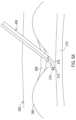
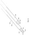
- FIG. 1 Ashows a first example of an assembly for implanting multiple medical leads through a single entry by way of a catheter with multiple lumens and a deflectable distal end.
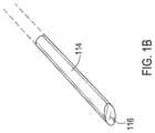
- FIG. 1 Bshows one example of a trocar that may be used with the assembly of FIG. 1 A .
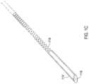
- FIG. 1 Cshows another example of a trocar that may be used with the assembly of FIG. 1 A .
- FIGS. 2 A- 2 Eshow the assembly of FIG. 1 A during an implantation procedure.
- FIG. 3shows a set of acts that utilize the assembly of FIG. 1 A to implant multiple medical leads.
- FIG. 4shows a second example of an assembly for implanting multiple medical leads through a single entry by way of a catheter with multiple lumens, each lumen containing a deflectable sheath.
- FIGS. 5 A- 5 Bshow the assembly of FIG. 4 during an implantation procedure.
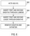
- FIG. 6shows a set of acts that utilize the assembly of FIG. 4 to implant multiple medical leads.
- FIG. 7 Ashows a third example of an assembly for implanting multiple medical leads through a single entry by way of a catheter having a lumen with an oblong lateral cross-section.
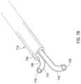
- FIG. 7 Bshows a fourth example of an assembly for implanting multiple medical leads through a single entry by way of a catheter having a lumen with an oblong lateral cross-section and having sheaths within the lumen.
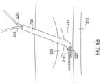
- FIGS. 8 A- 8 Bshow the assembly of FIG. 7 A during one implantation procedure.
- FIGS. 8 CA- 8 CBshow the assembly of FIG. 7 during another implantation procedure.
- FIGS. 8 DA- 8 DBshow a fifth assembly utilizing a lumen with an oblong lateral cross-section during an implantation procedure
- FIG. 9 Ashows a set of acts that utilize the assembly of FIG. 7 A to implant multiple medical leads.
- FIG. 9 Bshows a set of acts that utilize the assembly of FIG. 7 B to implant multiple medical leads.
- FIG. 9 Cshows a set of acts that utilize an assembly including multiple paddle medical leads.
- FIG. 10shows a sixth example of an assembly for implanting multiple medical leads through a single entry by way of a catheter having a central lumen housing a guidewire.
- FIG. 11shows a seventh example of an assembly for implanting multiple medical leads through a single entry by way of a catheter having a central lumen housing a guidewire and having sheaths in lead lumens.
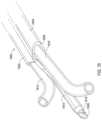
- FIG. 12shows an eighth example of an assembly for implanting multiple medical leads through a single entry by way of a catheter having a lumen housing an implantation needle.
- FIG. 13 Ashows a ninth example of an assembly for implanting multiple medical leads through a single entry by way of a catheter having a central extended lumen housing an implantation needle.
- FIG. 13 Bshows a tenth example of an assembly for implanting multiple medical leads through a single entry by way of a catheter having a central non-extended lumen housing an implantation needle.
- FIGS. 14 A- 14 Cshow the assembly of FIG. 12 during an implantation procedure.
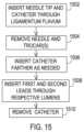
- FIG. 15shows a set of acts that utilize the assembly of FIG. 12 to implant multiple medical leads.
- FIG. 16shows an eleventh example of an assembly for implanting multiple medical leads through a single entry by way of a catheter having a central lumen housing an implantation needle while including sheaths in other lumens.
- FIGS. 17 A- 17 Bshow the assembly of FIG. 16 during an implantation procedure.
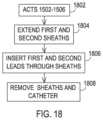
- FIG. 18shows a set of acts that utilize the assembly of FIG. 16 to implant multiple medical leads.
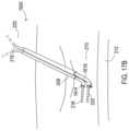
- FIG. 19shows an assembly like that of FIG. 7 A during an alternative implantation procedure.
- FIG. 20 Ashows a set of acts that utilize the assembly of FIG. 7 A during the alternative implantation procedure for implanting multiple medical leads.
- FIG. 20 Bshows a set of acts that utilize the assembly of FIG. 7 B during the alternative implantation procedure for implanting multiple medical leads.
- FIG. 20 Cshows a set of acts that utilize the assembly of FIG. 7 A during the alternative implantation procedure for implanting multiple medical leads where at least one is a paddle lead.
- Embodimentsprovide assemblies for implanting multiple medical leads through a single entry to a defined space within the body, such as the epidural space.
- the assembliesinclude a catheter that may have features such as multiple lumens, an extended lumen adjacent other lumens, multiple sheaths within one or more of the multiple lumens, and/or a lumen having an oblong lateral cross-section.
- the various embodimentsmay utilize one or more of implantation needles, guidewires, and the like to introduce the catheter into the defined space through the single entry.
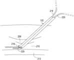
- FIG. 1 Ashows one example of a medical assembly for implanting multiple medical leads into a defined space within a body.
- the assemblyincludes a multi-lumen catheter 102 that may be implanted into the defined space to provide a passageway for the multiple leads to then be inserted.
- the catheter 102includes a first lumen body 104 and a second lumen body 106 that are integrally formed and may be a unitary structure, each defining a respective lumen 108 , 110 .
- the lumen body 104is used as a guide that follows along a guidewire 112 passing through the lumen 108 where the guidewire 112 has already been inserted into the defined space.
- the catheter 102has a deflectable distal end shown in FIG. 1 A .
- the deflectabilitymay be achieved by having the catheter be constructed of flexible materials.
- the cathetermay be constructed of nylon or similar materials, may have an internal liner also constructed of nylon, and may also include an internal reinforcement such as a braid constructed of a metal such as stainless steel.
- the deflectable endmay be constructed of shape memory materials such as Nylon, Polyurethane, or any other thermoplastics, thermoplastic elastomers, and the like. The desired bend of the distal end may be pre-formed through a heat forming process, and then held straight by a trocar so that once the trocar is removed, the distal end achieves the bent configuration.
- the distal endmay be relatively stiff in order to penetrate through tissue that is present in the pathway to the defined space.
- the defined spacemay be the epidural space where access is achieved by piercing through the ligamentum flavum that requires a relatively stiff catheter 102 even where a needle has previously been inserted, especially when the needle is of a smaller diameter than the multi-lumen catheter 102 .
- the epidural spaceis bordered by the spinal cord such that care must be exercised when attempting to insert objects such as catheters and leads into the epidural space.
- the catheter 102has the deflectable distal end such that upon entering the epidural space, the catheter tip deflects as discussed above to an orientation largely parallel to the spinal cord such as by following the guidewire 112 and/or by using shape memory.
- the catheter 102must have a rigid distal tip.
- a trocar 114made of a rigid material such as a hard plastic or metal is present within the lumen 110 which creates a rigid distal tip and which holds the catheter in a straight configuration during insertion.
- the trocar 114may be retractable, such as where the trocar 114 is solid as shown in FIG.
- the trocar 114as shown in FIG. 1 C may include a flexible region 118 such as a coiled area that can deflect and thereby allow the catheter 102 to deflect with the trocar 114 ′ in place.
- the lumen body 106 of the catheter 102may include a beveled distal end 107 .
- the trocar 114 , 114 ′may include a beveled distal end 116 which may be oriented with the bevel in the same plane as the bevel of the lumen body 106 as shown in FIG. 1 A .
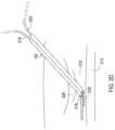
- FIGS. 2 A- 2 Eshow a series of phases of the multi-lumen catheter 102 being inserted and the implantable medical leads ultimately being inserted into the defined space. These phases are illustrated and discussed with reference to implantation into the epidural space of a body. However, it will be appreciated that the assemblies and techniques may also be applicable to other defined spaces within a body.
- FIG. 3shows a series of acts taken to progress through the phases of FIGS. 2 A- 2 E . Additionally, while a hub is not shown on a proximal end of the needle and catheter, it will be appreciated that a hub may be present for each device to allow insertion and removal of the various objects.
- FIG. 2 Ashows a needle 214 being inserted into a body 202 by passing through the skin 204 and through the ligamentum flavum 208 , adjacent vertebral bones 206 pursuant to the needle operation 302 of FIG. 3 .
- the needle 214enters the epidural space 210 but care is exercised to avoid contact with the dura or any other layers about the spinal cord 212 .
- FIG. 2 Bshows a guidewire 216 being inserted into the epidural space 210 by feeding the guidewire 216 through a lumen within the needle 214 pursuant to the guidewire operation 304 of FIG. 3 .
- the needle 214is then removed while maintaining the guidewire in position within the epidural space pursuant to a needle operation 306 .
- FIG. 2 Cshows the catheter 102 then being inserted into the epidural space 210 by feeding the catheter 102 along the guidewire 216 with the guidewire 216 passing through the lumen 108 of the lumen body 104 pursuant to a catheter operation 308 .
- the trocar 114is then removed from the catheter 102 in this example where the trocar 114 as shown in FIG. 1 B is solid pursuant to a removal operation 310 which allows the catheter 102 to be inserted farther such that the distal tip deflects as it follows the path of the guidewire 216 and/or deflects by achieving a pre-formed bend.
- the trocar 114 ′ as shown in FIG. 1 B_is present and includes a flexible portion 118 also shown in FIG.
- the trocar 114 ′ of FIG. 1 Bmay remain in place as the distal end begins to deflect and then is removed once the catheter 102 is inserted to an approximate final position within the epidural space. In either case, once the catheter 102 is inserted to the approximate final position, the guidewire 216 is then removed pursuant to the removal operation 310 .
- FIG. 2 Dthen shows first and second implantable medical leads 218 , 220 being inserted through each lumen of the catheter 102 so that the medical leads 218 , 220 are then directed into the epidural space and in a direction largely parallel to the spinal cord 212 and pursuant to an insertion operation 312 .
- the catheter 102is then removed pursuant to a removal operation 314 to leave the medical leads 218 , 220 in position within the epidural space 210 as shown in FIG. 2 E .
- FIG. 4shows another example of a medical assembly for implanting multiple medical leads into a defined space within a body.
- the assemblyincludes a multi-lumen catheter 402 that may be implanted into the defined space to provide a passageway for the multiple leads to then be inserted.
- the catheter 402includes a first lumen body 404 and a second lumen body 406 that are integrally formed and may be a unitary structure, each defining a respective lumen 408 , 410 .
- the lumen body 404may also be used as a guide that follows along a guidewire passing through the lumen 108 where the guidewire has already been inserted into the defined space.
- the catheter 402has first and second deflectable sheaths 412 , 416 that are present within the multiple lumens 408 , 410 , respectively. This allows the distal end of the sheaths to deflect upon the catheter 402 entering the defined space so as to avoid damaging the tissues surrounding the defined space and so as to properly direct the leads once they are inserted through lumens 418 , 420 within the sheaths 412 , 416 .
- the sheaths 412 , 416may be deflectable by constructing them from a flexible material.
- the sheathsmay be constructed of various layers and materials such as nylon, may have an internal liner also constructed of various materials such as high density polyethylene and the like, and may also include an internal reinforcement such as a braid constructed of a metal such as stainless steel.
- the sheaths 412 , 416may also utilize shape memory to establish pre-formed bends on the distal ends where the preformed bends are held straight by the catheter 402 and are achieved upon the distal ends of the sheaths 412 , 416 exiting the distal end of the catheter 402 .
- the distal end of the catheter 402may be deflectable or may be rigid, there may still be a need for the distal end to be stiffened by a trocar in order to penetrate through tissue that is present in the pathway to the defined space.
- the trocar 114 shown in FIG. 1 Bmay be retractable, such as where the trocar 114 is solid as shown in FIG. 1 B or may alternatively include a flexible region 118 as shown in FIG. 1 C , such as a coiled region, that can deflect and thereby allow the catheter 102 to deflect with the trocar 114 ′ in place as shown in FIG. 1 C .
- the lumen body 406 of the catheter 402includes a beveled distal end like the previous embodiment.
- the trocar 114(as shown in FIG. 1 B ), 114 ′ (as shown in FIG. 1 C ) may include a beveled distal end 116 which may be oriented with the bevel in the same plane as the bevel of the lumen body 406 as show in FIG. 4 .
- FIGS. 5 A- 5 Bshow a series of phases of the multi-lumen catheter 402 being inserted and the implantable medical leads ultimately being inserted into the defined space. These phases are also illustrated and discussed with reference to implantation into the epidural space of a body. However, it will be appreciated that the assemblies and techniques may also be applicable to other defined spaces within a body.
- FIG. 6shows a series of acts taken to progress through the phases of FIGS. 5 A- 5 B .
- FIGS. 2 A and 2 Bare performed along with the acts in the operations 302 - 310 discussed above and pursuant to an initial operation 602 .
- the sheaths 418 , 420may already be present within the catheter lumens 408 , 410 when inserting the catheter 402 where in that case the guidewire 216 passes through the sheath lumen 418 while a trocar 114 (as shown in FIG. 1 B ) may be present within the sheath lumen 420 .
- the guidewire 216 and trocar 114are then removed.
- the distal ends of the sheaths 412 , 416are extended distally from the catheter 402 pursuant to the sheath operation 604 , allowing the distal ends of the sheaths 412 , 416 to deflect as needed to obtain an orientation largely parallel to the spinal cord 212 .
- the implantable medical leads 218 , 220are inserted through the sheath lumens 418 , 420 to enter into the epidural space 210 and are guided by the sheaths 412 , 416 into the orientation that is largely parallel to the spinal cord 212 pursuant to the lead operation 606 .
- the sheaths 412 , 416 and catheter 402may then be removed, individually or as a whole, pursuant to the removal operation 608 , which then produces the phase shown in FIG. 2 E .
- FIG. 7 Ashows another example of a medical assembly for implanting multiple medical leads into a defined space within a body.
- the assemblyincludes a catheter 702 having a single lumen body 704 but with a lateral cross-section having an oblong shape such as an ellipse that is capable of receiving multiple medical leads.
- a removable introducer 708 having a conical distal end for ease of insertionis present within an oblong lumen 706 of the catheter 702 to provide stiffness.
- the introducer 708also includes a lumen 710 that may be used to receive a guidewire or a needle during insertion of the catheter 702 into the defined space within the body.
- the catheter 702may have a shape memory providing a pre-formed bend on the distal end which is held straight by the introducer 708 and is achieved upon removal of the introducer 708 .
- FIG. 7 Bshows an example of a medical assembly where a catheter 712 has the single lumen body 704 with a lateral cross-section having an oblong shape.
- sheaths 714 and 716 having sheath lumens 718 and 720are present within the oblong lumen 706 of the catheter 712 .
- these sheathsmay be used to ultimately guide the medical leads into place.
- the sheathsmay be deflectable so as to orient in a direction parallel to the spinal cord 212 such as by being constructed of a flexible material and/or by being constructed of a shape memory material to provide pre-formed bends.
- Such pre-formed bendsmay be held straight by the catheter 712 and then achieved upon the distal ends being extended from the catheter 712 .
- This catheter 712may utilize an introducer 708 which is then removed once in the defined space within the body to allow insertion of the sheaths 714 , 716 .
- FIG. 8 Ashows an assembly including catheter 702 being inserted into the epidural space 210 in accordance with guidewire operation 902 of FIG. 9 A by following a guidewire 216 that has already been inserted pursuant to the initial operation 900 .
- FIG. 8 Bshows the lumen body 704 of the catheter 702 in position with the medical leads 218 , 220 being inserted through the lumen 706 pursuant to insertion operation 906 once the guidewire 216 and introducer 708 have been removed pursuant to removal operation 904 .
- the catheter 702is then removed pursuant the removal operation 908 , with the medical leads 218 , 220 then being in position as shown in FIG. 2 E .
- FIG. 8 CAshows an assembly including catheter 712 after insertion into the epidural space 210 and removal of the introducer 708 and guidewire 216 in accordance with the initial operation 910 of FIG. 9 B .
- the first and second sheaths 714 , 716are also inserted through the lumen 706 pursuant to the sheath operation 912 and to extend the distal ends of the sheaths 714 , 716 beyond the distal end of the catheter 712 . This allows the distal ends of the sheaths 714 , 716 to deflect into an orientation generally parallel to the spinal cord 212 .
- FIG. 8 CAshows an assembly including catheter 712 after insertion into the epidural space 210 and removal of the introducer 708 and guidewire 216 in accordance with the initial operation 910 of FIG. 9 B .
- the first and second sheaths 714 , 716are also inserted through the lumen 706 pursuant to the sheath operation 912 and to extend the distal ends of the sheaths
- FIG. 8 CBshows the leads 218 , 220 being inserted through the sheaths 714 , 716 until they reach their approximate final position pursuant to the lead operation 914 .
- the catheter 712 and sheaths 714 , 716are then removed pursuant to the removal operation 916 with the medical leads 218 , 220 being in position as shown in FIG. 2 E .
- FIG. 8 DAshows an assembly including catheter lumen body 704 after insertion into the epidural space 210 and removal of the introducer 708 and guidewire 216 in accordance with the initial operation 918 of FIG. 9 C .
- first and second paddle leads 802 , 804are being inserted through the lumen 706 pursuant to the insertion operations 920 and 922 .
- the width of the paddle 806 , 808may be of a size that is too large for two paddles 806 , 808 to be laterally adjacent within the oblong lumen 706 .
- one paddle 806is inserted first, at insertion operation 920 , ahead of the other paddle 808 and moved distally down the catheter lumen body 704 .
- the other paddle 808is then inserted pursuant to the insertion operation 922 such that the latter paddle 808 is adjacent to only the lead body of the lead 802 where the lead body has a smaller width than the paddle 806 .
- Both leads 802 , 804are fully inserted through the catheter lumen body 704 as shown in FIG. 8 DB until reaching their approximate final positions.
- the catheter 702is then removed pursuant to the removal operation 924 with the paddle leads 802 , 804 being in an approximate final position similar to that shown for percutaneous leads in FIG. 2 E .
- FIG. 10shows another embodiment of a medical assembly including a catheter 1000 .
- This catheter 1000includes multiple lumen bodies 1002 , 1004 that are integrally formed and may be a unitary structure, each having a lumen 1008 .
- each of the lumen bodies 1002 , 1004has a beveled distal end 1006 .
- this embodimentincludes a dedicated guidewire lumen body 1010 that is integral with the other lumen bodies and that extends distally beyond the lumen bodies 1002 , 1004 and includes a guidewire lumen 1012 .
- the guidewire lumen body 1010may be made of a flexible material to allow the guidewire lumen body 1010 to deflect as it follows the path of the guidewire 112 upon entering the defined space within the body.
- a trocarmay be positioned in each lumen body 1002 , 1004 and may be retracted once the distal end of the lumen bodies 1002 , 1004 enter the defined space.
- FIG. 10The assembly of FIG. 10 is inserted into the epidural space in the same manner as discussed above for the embodiment of FIG. 1 A , utilizing the same approach as illustrated in FIGS. 2 A- 2 E and FIG. 3 except that the dedicated guidewire lumen 1012 receives the guidewire 216 as shown in FIGS. 2 B and 2 C rather than the guidewire 216 being present within a lumen of the lumen bodies that is used to introduce a medical lead.
- FIG. 11shows another embodiment of a medical assembly including a catheter 1100 .
- this catheter 1100includes multiple lumen bodies 1102 , 1104 that are integrally formed and may be a unitary structure and define lumens 1106 .
- This catheter 1100also includes a dedicated and deflectable guidewire lumen body 1112 that receives the guidewire 112 .
- this assemblyalso includes sheaths 1114 and 1116 present within the lumens 1106 of the lumen bodies 1102 , 1104 . The distal ends of these sheaths 1114 , 1116 may be deflectable and/or have shape memory as discussed above for the sheaths present in other embodiments.
- FIG. 11The assembly of FIG. 11 is inserted into the epidural space in the same manner as discussed above for the embodiment of FIG. 4 , utilizing the same approach as illustrated in FIGS. 2 A- 2 C, 5 A- 5 B and FIG. 6 except that the dedicated guidewire lumen 1112 receives the guidewire 216 as shown in FIGS. 2 B and 2 C rather than the guidewire 216 being present within a lumen of the lumen bodies that is used to introduce a medical lead.
- FIG. 12shows an embodiment of a medical assembly that eliminates the use of a guidewire during insertion.
- the assemblyincludes a catheter 1200 that has multiple lumen bodies 1202 , 1204 that are integrally formed and may be a unitary structure with each having a lumen 1210 , 1212 .
- Each of the lumens 1210 , 1212 defined by the lumen bodies 1202 , 1204may be substantially the same diameter.
- the distal end 1208 of the lumen body 1204includes a beveled surface 1206 like that of the embodiment of FIG. 1 A .
- an insertion needle 1214is present within the lumen 1212 of the lumen body 1202 .
- the insertion needle 1214includes a beveled distal end 1216 having a lumen 1218 that may include a removable trocar for added stiffness and to fill the lumen 1218 during insertion. Further, a trocar may also be present within the lumen 1210 to provide additional stiffness during insertion.
- the needle 1214 and any trocar within the needle 1214may be retracted as may any trocar present within the lumen 1210 .
- the distal end of the catheter 1200may be deflected, such as by further movement and/or shape memory providing a pre-formed bend.
- FIG. 13 Ashows another embodiment of a medical assembly that eliminates the use of a guidewire during insertion.
- the assemblyincludes a catheter 1300 that has multiple lumen bodies 1302 , 1304 that are integrally formed and may be a unitary structure with lumens 1310 and beveled surfaces 1306 on the distal ends 1308 of each.
- the catheter 1300also includes a dedicated and deflectable needle lumen body 1312 that receives the insertion needle 1314 .
- the insertion needle 1314 of this exampleincludes a beveled tip 1316 and may also include a lumen 1318 which may be filled by a trocar during insertion.
- the dedicated needle lumen body 1312defines a lumen having a smaller diameter than the lumen defined by the lumen bodies 1302 , 1304 that receive the medical leads.
- the needle lumen body 1312 of this exampleextends distally beyond the distal end of the lumen bodies 1302 , 1304 that receive the medical leads and may be flexible to deflect and/or may include shape memory so as to achieve a pre-formed bend upon removal of the insertion needle 1314 .
- the multiple lumen bodies 1302 , 1304may also be deflectable and/or include shape memory so as to achieve a pre-formed bend upon removal of trocars present within the lumens 1310 during insertion into the defined space.
- FIG. 13 Bshows another embodiment of a medical assembly like that of FIG. 10 or 13 A except that a dedicated lumen body 1332 of a catheter 1320 terminates at or near the distal end of multiple lumen bodies 1322 , 1324 rather than extending farther distally.
- the dedicated lumen body 1332may be for purposes of receiving a guidewire where the catheter 1320 is inserted in the same manner as the catheter 1000 of FIG. 10 or may be for purposes of receiving an insertion needle where the catheter 1320 is inserted in the same manner as the catheter 1300 of FIG. 13 A .
- the distal end of the cathetermay be flexible so as to be deflectable and/or may include shape memory to establish a pre-formed bend.
- the dedicated lumen body 1332defines a lumen 1334 that has a smaller diameter than the lumen of the other lumen bodies.
- FIGS. 14 A- 14 Cshow a series of phases of the multi-lumen catheter embodiments of FIGS. 12 - 13 B being inserted without the use of a guidewire and also show the implantable medical leads ultimately being inserted into the defined space. While the catheter 1200 of FIG. 12 is specifically illustrated, it will be appreciated that the same phases and operations are also applicable to the catheters 1300 and 1320 .
- FIG. 15shows a series of acts taken to progress through the phases of FIGS. 14 A- 14 C . Additionally, while a hub is not shown on a proximal end of the needle and catheter combination, it will be appreciated that a hub may be present to allow insertion and removal of the various objects.
- FIG. 14 Ashows the assembly including the catheter 1200 and the insertion needle 1214 being inserted into a body 202 by passing the assembly including the catheter 1200 and insertion needle 1214 through the skin 204 and through the ligamentum flavum 208 , adjacent vertebral bones 206 pursuant to the needle operation 1502 of FIG. 15 .
- the needle 1214enters the epidural space 210 but care is exercised to avoid contact with the dura or any other layers about the spinal cord 212 .
- the needle 1214 and any trocarsmay then removed from the needle 1214 and catheter 1200 pursuant to removal operation 1504 to allow the distal end of the catheter to deflect and/or if applicable achieve a pre-formed bend.
- the catheter 1200is inserted further as the deflection occurs pursuant to insertion operation 1506 to reach the position as shown in FIG. 14 B .
- FIG. 14 Cthen shows first and second implantable medical leads 218 , 220 being inserted through each lumen of the catheter 1200 so that the medical leads 218 , 220 are then directed into the epidural space and in a direction largely parallel to the spinal cord 212 and pursuant to an insertion operation 1508 .
- the catheter 1200is then removed pursuant to a removal operation 1510 to leave the medical leads 218 , 220 in position within the epidural space 210 as shown in FIG. 2 E .
- FIG. 16shows another embodiment of a medical assembly including a catheter 1600 where a guidewire is not used for insertion.
- This assemblyis similar to that of FIG. 13 A except that sheaths 1614 and 1616 are present within the lumens 1606 of lumen bodies 1602 and 1604 where these lumen bodies are integrally formed and may be a unitary structure. These sheaths 1614 , 1616 may deflectable and or have a shape memory providing a pre-formed bend as discussed above for the sheaths of other embodiments.
- the catheter 1600includes a dedicated needle lumen body 1610 and a needle tip 1608 extends from the needle lumen body 1610 . Where the needle tip 1608 includes a lumen, a trocar 1618 may be present to fill the lumen during insertion.
- the dedicated needle lumen body 1610defines a lumen that has a smaller diameter than the lumen of the other lumen bodies.
- the assembly of FIG. 12may include sheaths like those of FIG. 16 during the insertion process once the needle 1214 and any trocars are removed. Similarly, sheaths may be used within the assemblies of FIGS. 13 A and 13 B .
- FIGS. 17 A- 17 Bshow a series of phases of the multi-lumen catheter embodiment of FIG. 16 being inserted without the use of a guidewire and also show the implantable medical leads ultimately being inserted into the defined space. While the catheter 1600 of FIG. 16 is specifically illustrated, it will be appreciated that the same phases and operations are also applicable to the catheters 1200 , 1300 , and 1320 where those assemblies utilize sheaths during the lead implantation process.
- FIG. 17 Ashows a phase that occurs after the catheter 1600 has been inserted into the defined space and the insertion needle and any trocars have been removed from the catheter 1600 pursuant to initial operation 1802 of FIG. 18 . Additionally, the needle lumen 1610 has deflected, and the first and second sheaths 1614 , 1616 have been inserted and extended through the catheter 1600 until the distal ends of the sheaths 1614 , 1616 exit from the catheter and deflect pursuant to the sheath operation 1804 . As shown in FIG. 17 B , the first and second leads 218 , 220 are then inserted through the sheaths 1614 , 1616 until entering the epidural space and reaching an approximate final position pursuant to insertion operation 1806 . The catheter 1600 and sheaths 1614 , 1616 are then removed pursuant to the removal operation 1808 while the leads 218 , 220 remain in their approximate final position such as that shown in FIG. 2 E .
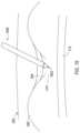
- FIG. 19shows a medical assembly including a catheter 1900 being inserted into the epidural space 210 .
- This assemblyis the same as that of FIG. 7 A except that an insertion needle 1902 is present within the lumen of the introducer that is present within the lumen of the catheter 1900 such that a guidewire is not used during insertion.
- the catheter 1900is inserted into the epidural space pursuant to the initial operation 2000 of FIG. 20 A which includes inserting the catheter 1900 , then removing the insertion needle. Additionally, the introducer is also removed at the removal operation 2002 which may occur in unison with removal of the needle.
- the catheter 1900is inserted further as needed as in insertion operation 2004 , such as shown in FIG. 8 B , until the distal end of the catheter 1900 has deflected or established a pre-formed bend within the epidural space.
- the first and second leads 218 , 220are then inserted through the catheter 1900 until reaching an approximate final position within the epidural space 210 pursuant to the insertion operation 2006 .
- the catheter 1900is then removed in a removal operation 2008 with the leads being in the approximate final position as shown in FIG. 2 E .
- first and second sheathsare inserted through the oblong lumen in a sheath operation 2012 until the distal end of the sheaths enter the epidural space and then deflect into an appropriate position as shown in FIG. 8 CA .
- First and second medical leadsare then inserted through the sheaths until reaching an approximate final position pursuant to insertion operation 2014 and as shown in FIG. 8 CB .
- the catheter 1900 and sheathsare then removed in a removal operation 2016 with the leads being in the approximate final position as shown in FIG. 2 E .
- the catheter 1900is used to implant at least one paddle lead
- the catheter 1900has been inserted with an insertion needle in the lumen of the introducer
- the needle and introducerare removed pursuant to the initial operation 2018 of FIG. 20 C .
- the oblong lumen of the cathetercan accommodate one paddle and one lead body and where two paddle leads are being implanted
- the first paddle leadis inserted into the oblong lumen and moved distally in a paddle operation 2020 and as shown in FIG. 8 DA .
- the second paddle leadis inserted trailing the first paddle in the oblong lumen so that the second paddle is laterally adjacent to the lead body of the first paddle lead in a paddle operation 2022 and as shown in FIG. 8 DB .
- the catheter 1900is removed in a removal operation 2024 with the paddle leads being in the approximate final position similar to that shown for percutaneous leads in FIG. 2 E .
Landscapes
- Health & Medical Sciences (AREA)
- Life Sciences & Earth Sciences (AREA)
- Surgery (AREA)
- Heart & Thoracic Surgery (AREA)
- General Health & Medical Sciences (AREA)
- Veterinary Medicine (AREA)
- Engineering & Computer Science (AREA)
- Biomedical Technology (AREA)
- Nuclear Medicine, Radiotherapy & Molecular Imaging (AREA)
- Public Health (AREA)
- Animal Behavior & Ethology (AREA)
- Pathology (AREA)
- Medical Informatics (AREA)
- Molecular Biology (AREA)
- Neurosurgery (AREA)
- Cardiology (AREA)
- Radiology & Medical Imaging (AREA)
- Orthopedic Medicine & Surgery (AREA)
- Neurology (AREA)
- Anesthesiology (AREA)
- Media Introduction/Drainage Providing Device (AREA)
Abstract
Description
Claims (13)
Priority Applications (1)
| Application Number | Priority Date | Filing Date | Title |
|---|---|---|---|
| US17/008,183US12137932B2 (en) | 2011-06-20 | 2020-08-31 | Medical assemblies and methods for implantation of multiple medical leads through a single entry |
Applications Claiming Priority (4)
| Application Number | Priority Date | Filing Date | Title |
|---|---|---|---|
| US201161498914P | 2011-06-20 | 2011-06-20 | |
| US201161498906P | 2011-06-20 | 2011-06-20 | |
| US13/525,560US10758262B2 (en) | 2011-06-20 | 2012-06-18 | Medical assemblies and methods for implantation of multiple medical leads through a single entry |
| US17/008,183US12137932B2 (en) | 2011-06-20 | 2020-08-31 | Medical assemblies and methods for implantation of multiple medical leads through a single entry |
Related Parent Applications (1)
| Application Number | Title | Priority Date | Filing Date |
|---|---|---|---|
| US13/525,560DivisionUS10758262B2 (en) | 2011-06-20 | 2012-06-18 | Medical assemblies and methods for implantation of multiple medical leads through a single entry |
Publications (2)
| Publication Number | Publication Date |
|---|---|
| US20210052295A1 US20210052295A1 (en) | 2021-02-25 |
| US12137932B2true US12137932B2 (en) | 2024-11-12 |
Family
ID=47354275
Family Applications (2)
| Application Number | Title | Priority Date | Filing Date |
|---|---|---|---|
| US13/525,560Active2033-12-17US10758262B2 (en) | 2011-06-20 | 2012-06-18 | Medical assemblies and methods for implantation of multiple medical leads through a single entry |
| US17/008,183Active2034-02-11US12137932B2 (en) | 2011-06-20 | 2020-08-31 | Medical assemblies and methods for implantation of multiple medical leads through a single entry |
Family Applications Before (1)
| Application Number | Title | Priority Date | Filing Date |
|---|---|---|---|
| US13/525,560Active2033-12-17US10758262B2 (en) | 2011-06-20 | 2012-06-18 | Medical assemblies and methods for implantation of multiple medical leads through a single entry |
Country Status (1)
| Country | Link |
|---|---|
| US (2) | US10758262B2 (en) |
Families Citing this family (18)
| Publication number | Priority date | Publication date | Assignee | Title |
|---|---|---|---|---|
| US20140039586A1 (en)* | 2012-08-03 | 2014-02-06 | Boston Scientific Neuromodulation Corporation | Systems and methods for making and using a multi-lead introducer for use with electrical stimulation systems |
| WO2014158723A1 (en) | 2013-03-13 | 2014-10-02 | Boston Scientific Neuromodulation Corporation | System and method for making and using a lead introducer for an implantable electrical stimulation system |
| US9629658B2 (en) | 2013-09-06 | 2017-04-25 | Boston Scientific Neuromodulation Corporation | Systems and methods for making and using a lead introducer for an implantable electrical stimulation system |
| CN105517625A (en) | 2013-09-06 | 2016-04-20 | 波士顿科学神经调制公司 | Lead introducer for implantable electrical stimulation system |
| US9604050B2 (en) | 2014-02-20 | 2017-03-28 | Boston Scientific Neuromodulation Corporation | Systems and methods for percutaneously implanting into a patient a paddle lead of an electrical stimulation system |
| WO2016130575A1 (en) | 2015-02-13 | 2016-08-18 | Boston Scientific Neuromodulation Corporation | Retractor and tools for implantation of electrical stimulation leads |
| US10881428B2 (en) | 2015-04-22 | 2021-01-05 | Medtronic, Inc. | Shaped lead introducer for epidural space |
| US9937322B2 (en) | 2015-04-23 | 2018-04-10 | Medtronic, Inc. | Assemblies and methods for deflectable shaft catheters |
| US9867964B2 (en) | 2015-04-23 | 2018-01-16 | Medtronic, Inc. | Interventional medical systems, assemblies, and construction methods |
| WO2016176211A1 (en) | 2015-04-28 | 2016-11-03 | Boston Scientific Neuromodulation Corporation | Systems and methods for making and using a lead introducer with a seal for an electrical stimulation system |
| US11166745B2 (en) | 2017-09-12 | 2021-11-09 | Jessica Jameson | Multi-port epidural needle |
| WO2019140286A2 (en)* | 2018-01-11 | 2019-07-18 | Wendel Mark | Method and device for inserting at least one medical component within the body |
| WO2020172071A2 (en) | 2019-02-19 | 2020-08-27 | Boston Scientific Neuromodulation Corporation | Lead introducers and systems and methods including the lead introducers |
| US11524139B2 (en) | 2019-07-15 | 2022-12-13 | Medtronic, Inc. | Catheter with active return curve |
| US11524143B2 (en) | 2019-07-15 | 2022-12-13 | Medtronic, Inc. | Catheter with distal and proximal fixation members |
| CN111991674B (en)* | 2020-09-26 | 2024-03-26 | 龙德勇 | Ablation device and method for fat-thickness obstructive cardiomyopathy |
| ES2953725T3 (en)* | 2020-12-15 | 2023-11-15 | Capri Medical Ltd | Implant delivery device |
| DE102021210108A1 (en) | 2021-09-14 | 2023-03-16 | B. Braun Melsungen Aktiengesellschaft | Introducer assembly for an invasive medical component |
Citations (97)
| Publication number | Priority date | Publication date | Assignee | Title |
|---|---|---|---|---|
| US701075A (en) | 1902-02-19 | 1902-05-27 | Richard P Mccully | Catheter or like instrument. |
| US3144868A (en) | 1960-10-21 | 1964-08-18 | Mario E Jascalevich | Drainage and feeding cannulae |
| US3495595A (en) | 1966-11-02 | 1970-02-17 | Thomas G Soper | Medicosurgical tube and method |
| US3804097A (en) | 1972-12-14 | 1974-04-16 | P Rudie | Method of irrigating and treating an abcess |
| US4072153A (en) | 1976-03-03 | 1978-02-07 | Swartz William H | Post hysterectomy fluid drainage tube |
| US4141365A (en)* | 1977-02-24 | 1979-02-27 | The Johns Hopkins University | Epidural lead electrode and insertion needle |
| US4203436A (en) | 1977-04-05 | 1980-05-20 | Lars Grimsrud | Assembly for dividing a hollow hypodermic needle into two separated flow conduits |
| US4405313A (en) | 1982-01-29 | 1983-09-20 | Sisley James R | Figure-eight, dual-lumen catheter and method of using |
| EP0232994A2 (en) | 1986-02-04 | 1987-08-19 | Sherwood Medical Company | Catheter introducer |
| US4808157A (en)* | 1987-07-13 | 1989-02-28 | Neuro Delivery Technology, Inc. | Multi-lumen epidural-spinal needle |
| US4958901A (en) | 1987-07-13 | 1990-09-25 | Neurodelivery Technology, Inc. | Method for making a multi-lumen epidural-spinal needle and tip and stock configuration for the same |
| US5084013A (en) | 1989-01-13 | 1992-01-28 | Haruo Takase | Suction tube for use in surgical operation |
| US5167220A (en) | 1990-08-09 | 1992-12-01 | Brown Cathy K | Systems and methods for maintaining a clear visual field during endoscopic procedures |
| US5197976A (en) | 1991-09-16 | 1993-03-30 | Atrium Medical Corporation | Manually separable multi-lumen vascular graft |
| US5255691A (en)* | 1991-11-13 | 1993-10-26 | Medtronic, Inc. | Percutaneous epidural lead introducing system and method |
| US5286253A (en) | 1992-10-09 | 1994-02-15 | Linvatec Corporation | Angled rotating surgical instrument |
| US5318530A (en) | 1991-12-06 | 1994-06-07 | Bissel Medical Products, Inc. | Gastrointestinal tube with inflatable bolus |
| US5318517A (en) | 1992-03-30 | 1994-06-07 | Reiman James A | Multiple lumen thoracostomy catheter and method for administering anesthesia |
| USD353454S (en) | 1992-10-15 | 1994-12-13 | Neurodelivery Technology, Inc. | Combined epidural introducer and spinal needle |
| USD367324S (en) | 1994-08-12 | 1996-02-20 | American Cyanamid Company | Asymmetric single portal cannula |
| USD371732S (en) | 1994-10-27 | 1996-07-16 | United Technologies Corporation | Attachment tool for a system for repairing damaged gas turbine engine airfoils |
| US5558634A (en) | 1993-04-30 | 1996-09-24 | Mitchell; Paul G. | Apparatus for the removal of adherent viscoelastic |
| US5599304A (en) | 1994-05-10 | 1997-02-04 | Mount Sinai School Of Medicine Of The City University Of New York | Sinonasal suction apparatus |
| US5666619A (en) | 1995-12-06 | 1997-09-09 | Xerox Corporation | Electrode wire support for scavengeless development |
| US5709224A (en)* | 1995-06-07 | 1998-01-20 | Radiotherapeutics Corporation | Method and device for permanent vessel occlusion |
| US5752939A (en) | 1992-12-24 | 1998-05-19 | Kabushiki Kaisha Hayashidera Medinooru | Catheter for continuous ambulatory peritoneal dialysis |
| US5782795A (en) | 1995-06-30 | 1998-07-21 | Xomed Surgical Products, Inc. | Surgical suction cutting instrument with internal irrigation |
| US5785686A (en) | 1996-05-02 | 1998-07-28 | Runge; Thomas M. | Cannula system for a biventricular cardiac support system or a cardiopulmonary bypass system |
| US5807311A (en) | 1996-11-29 | 1998-09-15 | Palestrant; Aubrey M. | Dialysis catheter having rigid and collapsible lumens and related method |
| US5846219A (en) | 1994-05-26 | 1998-12-08 | Vancaillie; Thierry G. | Variable backflow suction-hydraulic curet |
| US5858009A (en) | 1997-08-14 | 1999-01-12 | Medtronic, Inc. | Multi-lumen cannula |
| US5891111A (en) | 1997-04-14 | 1999-04-06 | Porges | Flexible surgical drain with a plurality of individual ducts |
| US6152909A (en) | 1996-05-20 | 2000-11-28 | Percusurge, Inc. | Aspiration system and method |
| US6179776B1 (en) | 1999-03-12 | 2001-01-30 | Scimed Life Systems, Inc. | Controllable endoscopic sheath apparatus and related method of use |
| US6214016B1 (en) | 1999-04-29 | 2001-04-10 | Medtronic, Inc. | Medical instrument positioning device internal to a catheter or lead and method of use |
| US6309401B1 (en) | 1999-04-30 | 2001-10-30 | Vladimir Redko | Apparatus and method for percutaneous implant of a paddle style lead |
| US20010044591A1 (en)* | 1991-07-16 | 2001-11-22 | Heartport, Inc. | System for cardiac procedures |
| US20020007144A1 (en)* | 1992-07-06 | 2002-01-17 | Phillip Jack Snoke | System for enhancing visibility in the epidural space |
| US20020062143A1 (en)* | 1999-04-30 | 2002-05-23 | Medtronic, Inc. | Techniques for selective activation of neurons in the brain, spinal cord parenchyma or peripheral nerve |
| US6512958B1 (en) | 2001-04-26 | 2003-01-28 | Medtronic, Inc. | Percutaneous medical probe and flexible guide wire |
| US20030028146A1 (en)* | 2001-08-02 | 2003-02-06 | Teodulo Aves | Epidural catheter needle |
| US6524302B2 (en) | 2001-04-26 | 2003-02-25 | Scimed Life Systems, Inc. | Multi-lumen catheter |
| US20030163082A1 (en) | 2002-02-26 | 2003-08-28 | Mertens Steven P. | Lumen weld |
| US20040092863A1 (en) | 2002-10-31 | 2004-05-13 | Medical Components, Inc. | Splittable multiple catheter assembly |
| US20040116848A1 (en)* | 2002-12-16 | 2004-06-17 | Gardeski Kenneth C. | Multi-Lumen steerable catheter |
| US6755812B2 (en) | 2001-12-11 | 2004-06-29 | Cardiac Pacemakers, Inc. | Deflectable telescoping guide catheter |
| US6758854B1 (en) | 1997-05-09 | 2004-07-06 | St. Jude Medical | Splittable occlusion balloon sheath and process of use |
| US6836687B2 (en)* | 2000-03-31 | 2004-12-28 | Medtronic, Inc. | Method and system for delivery of a medical electrical lead within a venous system |
| US20050004555A1 (en)* | 2003-06-25 | 2005-01-06 | Pursley Matt D. | Method and apparatus for curving a catheter |
| US20050059925A1 (en) | 1999-01-15 | 2005-03-17 | Maginot Thomas J. | Catheter systems and associated methods |
| WO2005023359A1 (en) | 2003-08-29 | 2005-03-17 | Medtronic, Inc. | Percutaneous flat lead introducer |
| US20050096585A1 (en) | 2002-10-31 | 2005-05-05 | Medical Components, Inc. | Splittable multiple catheter assembly |
| US20050228339A1 (en) | 2004-04-12 | 2005-10-13 | The Trustees Of The University Of Pennsylvania | Multi-lumen catheter |
| US20050283111A1 (en) | 2004-06-22 | 2005-12-22 | Dan Maurice | Catheter assembly with degradable material |
| US7014626B2 (en) | 2000-11-08 | 2006-03-21 | Endovascular Technologies, Inc. | Dual-lumen peel-away sheath introducer |
| US20060122458A1 (en) | 2004-10-15 | 2006-06-08 | Baxano, Inc. | Devices and methods for tissue access |
| US7089040B2 (en) | 2000-01-27 | 2006-08-08 | Kyocera Corporation | Portable radio communication apparatus |
| US20060247751A1 (en)* | 2005-04-28 | 2006-11-02 | Seifert Kevin R | Guide catheters for accessing cardiac sites |
| US20070006697A1 (en) | 2005-07-11 | 2007-01-11 | Ivel Leonard L | Self-aligning blade holder |
| US20070225661A1 (en) | 2006-03-24 | 2007-09-27 | Ash Access Technology, Inc. | Indwelling catheter with anti-clotting features |
| US20070244430A1 (en)* | 2006-03-28 | 2007-10-18 | Bedell Raymond L | Multiple lumen epidural introducer |
| US20070249896A1 (en) | 2004-04-21 | 2007-10-25 | Eric Goldfarb | Endoscopic methods and devices for transnasal procedures |
| US20080065049A1 (en)* | 2006-09-12 | 2008-03-13 | Boston Scientific Scimed, Inc. | Variable stiffness direct injection system |
| US20080082079A1 (en) | 2006-09-28 | 2008-04-03 | Tyco Healthcare Group Lp | Low profile catheter assembly |
| US7359755B2 (en) | 2003-08-08 | 2008-04-15 | Advanced Neuromodulation Systems, Inc. | Method and apparatus for implanting an electrical stimulation lead using a flexible introducer |
| US20080097347A1 (en)* | 2006-09-22 | 2008-04-24 | Babak Arvanaghi | Bendable needle assembly |
| US7381204B2 (en) | 2001-01-24 | 2008-06-03 | Arrow International, Inc. | Multi-lumen catheter with attachable hub |
| US7384422B2 (en) | 2002-05-06 | 2008-06-10 | Pressure Products Medical Supplies, Inc. | Telescopic, separable introducer and method of using the same |
| US7455666B2 (en) | 2001-07-13 | 2008-11-25 | Board Of Regents, The University Of Texas System | Methods and apparatuses for navigating the subarachnoid space |
| US7553307B2 (en) | 2004-10-15 | 2009-06-30 | Baxano, Inc. | Devices and methods for tissue modification |
| US20090192435A1 (en) | 2007-10-26 | 2009-07-30 | C. R. Bard, Inc. | Solid-body catheter including lateral distal openings |
| US20090204052A1 (en) | 2007-10-17 | 2009-08-13 | Spire Corporation | Manufacture of split tip catheters |
| US20090204079A1 (en) | 2007-10-17 | 2009-08-13 | Spire Corporation | Catheters with enlarged arterial lumens |
| US20090205189A1 (en) | 2008-02-15 | 2009-08-20 | Spire Corporation | Manufacture of fixed tip catheters |
| US20090209940A1 (en) | 2008-02-15 | 2009-08-20 | Spire Corporation | Fusion manufacture of multi-lumen catheters |
| US20090281379A1 (en) | 2008-05-12 | 2009-11-12 | Xlumena, Inc. | System and method for transluminal access |
| US7662128B2 (en) | 2002-12-23 | 2010-02-16 | Salcudean Septimiu E | Steerable needle |
| US7666204B2 (en) | 1999-04-09 | 2010-02-23 | Evalve, Inc. | Multi-catheter steerable guiding system and methods of use |
| US7717899B2 (en) | 2002-01-28 | 2010-05-18 | Cardiac Pacemakers, Inc. | Inner and outer telescoping catheter delivery system |
| US7738969B2 (en) | 2004-10-15 | 2010-06-15 | Baxano, Inc. | Devices and methods for selective surgical removal of tissue |
| US7794448B2 (en) | 2004-05-27 | 2010-09-14 | Abbott Laboratories | Multiple lumen catheter and method of making same |
| US7798999B2 (en) | 2007-06-05 | 2010-09-21 | Cook Incorporated | Adjustable length catheter |
| US20100249726A1 (en) | 2009-03-25 | 2010-09-30 | King Saud University | Trocarless intravenous cannula with a multifilament tip |
| US20110202067A1 (en)* | 2010-02-16 | 2011-08-18 | Medtronic, Inc. | Introducer for lead delivery |
| US8112159B2 (en) | 2003-10-03 | 2012-02-07 | Medtronic, Inc. | Kit for implantation of therapy elements |
| US8206370B2 (en) | 2006-04-21 | 2012-06-26 | Abbott Laboratories | Dual lumen guidewire support catheter |
| US8298210B2 (en) | 2005-10-26 | 2012-10-30 | Medtronic Vascular, Inc. | Catheter having oval aspiration lumen and method of making |
| US8313496B2 (en) | 2001-02-02 | 2012-11-20 | Lsi Solutions, Inc. | System for endoscopic suturing |
| US8348899B2 (en) | 2005-03-10 | 2013-01-08 | Medical Components, Inc. | Methods for inserting a catheter |
| US8585950B2 (en) | 2009-01-29 | 2013-11-19 | Angiodynamics, Inc. | Multilumen catheters and method of manufacturing |
| US20140018772A1 (en) | 2012-07-16 | 2014-01-16 | Merit Medical Systems, Inc. | Self-centering catheter with anti-occlusion features |
| US8974486B2 (en) | 2008-04-01 | 2015-03-10 | Robert Kotler | Device and method for maintaining unobstructed nasal passageways after nasal surgery |
| US9149602B2 (en) | 2005-04-22 | 2015-10-06 | Advanced Cardiovascular Systems, Inc. | Dual needle delivery system |
| US9265407B2 (en) | 2004-04-21 | 2016-02-23 | Acclarent, Inc. | Endoscopic methods and devices for transnasal procedures |
| US9468362B2 (en) | 2004-04-21 | 2016-10-18 | Acclarent, Inc. | Endoscopic methods and devices for transnasal procedures |
| US9604050B2 (en) | 2014-02-20 | 2017-03-28 | Boston Scientific Neuromodulation Corporation | Systems and methods for percutaneously implanting into a patient a paddle lead of an electrical stimulation system |
| US20170143890A1 (en) | 2015-11-20 | 2017-05-25 | Vascutech Medical Llc | Dual Lumen Retrograde Catheter and Hub Attachment |
Family Cites Families (3)
| Publication number | Priority date | Publication date | Assignee | Title |
|---|---|---|---|---|
| US5662619A (en)* | 1995-11-27 | 1997-09-02 | Zarate; Alfredo R. | Venous dialysis needle |
| US7087040B2 (en)* | 2001-02-28 | 2006-08-08 | Rex Medical, L.P. | Apparatus for delivering ablation fluid to treat lesions |
| WO2007021772A2 (en)* | 2005-08-09 | 2007-02-22 | Trans1, Inc. | Exchange system for axial spinal procedures |
- 2012
- 2012-06-18USUS13/525,560patent/US10758262B2/enactiveActive
- 2020
- 2020-08-31USUS17/008,183patent/US12137932B2/enactiveActive
Patent Citations (99)
| Publication number | Priority date | Publication date | Assignee | Title |
|---|---|---|---|---|
| US701075A (en) | 1902-02-19 | 1902-05-27 | Richard P Mccully | Catheter or like instrument. |
| US3144868A (en) | 1960-10-21 | 1964-08-18 | Mario E Jascalevich | Drainage and feeding cannulae |
| US3495595A (en) | 1966-11-02 | 1970-02-17 | Thomas G Soper | Medicosurgical tube and method |
| US3804097A (en) | 1972-12-14 | 1974-04-16 | P Rudie | Method of irrigating and treating an abcess |
| US4072153A (en) | 1976-03-03 | 1978-02-07 | Swartz William H | Post hysterectomy fluid drainage tube |
| US4141365A (en)* | 1977-02-24 | 1979-02-27 | The Johns Hopkins University | Epidural lead electrode and insertion needle |
| US4203436A (en) | 1977-04-05 | 1980-05-20 | Lars Grimsrud | Assembly for dividing a hollow hypodermic needle into two separated flow conduits |
| US4405313A (en) | 1982-01-29 | 1983-09-20 | Sisley James R | Figure-eight, dual-lumen catheter and method of using |
| EP0232994A2 (en) | 1986-02-04 | 1987-08-19 | Sherwood Medical Company | Catheter introducer |
| US4808157A (en)* | 1987-07-13 | 1989-02-28 | Neuro Delivery Technology, Inc. | Multi-lumen epidural-spinal needle |
| US4958901A (en) | 1987-07-13 | 1990-09-25 | Neurodelivery Technology, Inc. | Method for making a multi-lumen epidural-spinal needle and tip and stock configuration for the same |
| US5084013A (en) | 1989-01-13 | 1992-01-28 | Haruo Takase | Suction tube for use in surgical operation |
| US5167220A (en) | 1990-08-09 | 1992-12-01 | Brown Cathy K | Systems and methods for maintaining a clear visual field during endoscopic procedures |
| US20010044591A1 (en)* | 1991-07-16 | 2001-11-22 | Heartport, Inc. | System for cardiac procedures |
| US5197976A (en) | 1991-09-16 | 1993-03-30 | Atrium Medical Corporation | Manually separable multi-lumen vascular graft |
| US5255691A (en)* | 1991-11-13 | 1993-10-26 | Medtronic, Inc. | Percutaneous epidural lead introducing system and method |
| US5318530A (en) | 1991-12-06 | 1994-06-07 | Bissel Medical Products, Inc. | Gastrointestinal tube with inflatable bolus |
| US5318517A (en) | 1992-03-30 | 1994-06-07 | Reiman James A | Multiple lumen thoracostomy catheter and method for administering anesthesia |
| US20020007144A1 (en)* | 1992-07-06 | 2002-01-17 | Phillip Jack Snoke | System for enhancing visibility in the epidural space |
| US5286253A (en) | 1992-10-09 | 1994-02-15 | Linvatec Corporation | Angled rotating surgical instrument |
| USD353454S (en) | 1992-10-15 | 1994-12-13 | Neurodelivery Technology, Inc. | Combined epidural introducer and spinal needle |
| US5752939A (en) | 1992-12-24 | 1998-05-19 | Kabushiki Kaisha Hayashidera Medinooru | Catheter for continuous ambulatory peritoneal dialysis |
| US5558634A (en) | 1993-04-30 | 1996-09-24 | Mitchell; Paul G. | Apparatus for the removal of adherent viscoelastic |
| US5599304A (en) | 1994-05-10 | 1997-02-04 | Mount Sinai School Of Medicine Of The City University Of New York | Sinonasal suction apparatus |
| US5846219A (en) | 1994-05-26 | 1998-12-08 | Vancaillie; Thierry G. | Variable backflow suction-hydraulic curet |
| USD367324S (en) | 1994-08-12 | 1996-02-20 | American Cyanamid Company | Asymmetric single portal cannula |
| USD371732S (en) | 1994-10-27 | 1996-07-16 | United Technologies Corporation | Attachment tool for a system for repairing damaged gas turbine engine airfoils |
| US5709224A (en)* | 1995-06-07 | 1998-01-20 | Radiotherapeutics Corporation | Method and device for permanent vessel occlusion |
| US5782795A (en) | 1995-06-30 | 1998-07-21 | Xomed Surgical Products, Inc. | Surgical suction cutting instrument with internal irrigation |
| US5666619A (en) | 1995-12-06 | 1997-09-09 | Xerox Corporation | Electrode wire support for scavengeless development |
| US5785686A (en) | 1996-05-02 | 1998-07-28 | Runge; Thomas M. | Cannula system for a biventricular cardiac support system or a cardiopulmonary bypass system |
| US6152909A (en) | 1996-05-20 | 2000-11-28 | Percusurge, Inc. | Aspiration system and method |
| US5807311A (en) | 1996-11-29 | 1998-09-15 | Palestrant; Aubrey M. | Dialysis catheter having rigid and collapsible lumens and related method |
| US5891111A (en) | 1997-04-14 | 1999-04-06 | Porges | Flexible surgical drain with a plurality of individual ducts |
| US6758854B1 (en) | 1997-05-09 | 2004-07-06 | St. Jude Medical | Splittable occlusion balloon sheath and process of use |
| US5858009A (en) | 1997-08-14 | 1999-01-12 | Medtronic, Inc. | Multi-lumen cannula |
| US20050059925A1 (en) | 1999-01-15 | 2005-03-17 | Maginot Thomas J. | Catheter systems and associated methods |
| US6179776B1 (en) | 1999-03-12 | 2001-01-30 | Scimed Life Systems, Inc. | Controllable endoscopic sheath apparatus and related method of use |
| US7666204B2 (en) | 1999-04-09 | 2010-02-23 | Evalve, Inc. | Multi-catheter steerable guiding system and methods of use |
| US6214016B1 (en) | 1999-04-29 | 2001-04-10 | Medtronic, Inc. | Medical instrument positioning device internal to a catheter or lead and method of use |
| US20020062143A1 (en)* | 1999-04-30 | 2002-05-23 | Medtronic, Inc. | Techniques for selective activation of neurons in the brain, spinal cord parenchyma or peripheral nerve |
| US6309401B1 (en) | 1999-04-30 | 2001-10-30 | Vladimir Redko | Apparatus and method for percutaneous implant of a paddle style lead |
| US7089040B2 (en) | 2000-01-27 | 2006-08-08 | Kyocera Corporation | Portable radio communication apparatus |
| US6836687B2 (en)* | 2000-03-31 | 2004-12-28 | Medtronic, Inc. | Method and system for delivery of a medical electrical lead within a venous system |
| US7014626B2 (en) | 2000-11-08 | 2006-03-21 | Endovascular Technologies, Inc. | Dual-lumen peel-away sheath introducer |
| US7381204B2 (en) | 2001-01-24 | 2008-06-03 | Arrow International, Inc. | Multi-lumen catheter with attachable hub |
| US8313496B2 (en) | 2001-02-02 | 2012-11-20 | Lsi Solutions, Inc. | System for endoscopic suturing |
| US6512958B1 (en) | 2001-04-26 | 2003-01-28 | Medtronic, Inc. | Percutaneous medical probe and flexible guide wire |
| US6524302B2 (en) | 2001-04-26 | 2003-02-25 | Scimed Life Systems, Inc. | Multi-lumen catheter |
| US7455666B2 (en) | 2001-07-13 | 2008-11-25 | Board Of Regents, The University Of Texas System | Methods and apparatuses for navigating the subarachnoid space |
| US6902547B2 (en)* | 2001-08-02 | 2005-06-07 | Teodulo Aves | Medical needle |
| US20030028146A1 (en)* | 2001-08-02 | 2003-02-06 | Teodulo Aves | Epidural catheter needle |
| US6755812B2 (en) | 2001-12-11 | 2004-06-29 | Cardiac Pacemakers, Inc. | Deflectable telescoping guide catheter |
| US7717899B2 (en) | 2002-01-28 | 2010-05-18 | Cardiac Pacemakers, Inc. | Inner and outer telescoping catheter delivery system |
| US20030163082A1 (en) | 2002-02-26 | 2003-08-28 | Mertens Steven P. | Lumen weld |
| US7384422B2 (en) | 2002-05-06 | 2008-06-10 | Pressure Products Medical Supplies, Inc. | Telescopic, separable introducer and method of using the same |
| US20050096585A1 (en) | 2002-10-31 | 2005-05-05 | Medical Components, Inc. | Splittable multiple catheter assembly |
| US20040092863A1 (en) | 2002-10-31 | 2004-05-13 | Medical Components, Inc. | Splittable multiple catheter assembly |
| US20040116848A1 (en)* | 2002-12-16 | 2004-06-17 | Gardeski Kenneth C. | Multi-Lumen steerable catheter |
| US7662128B2 (en) | 2002-12-23 | 2010-02-16 | Salcudean Septimiu E | Steerable needle |
| US20050004555A1 (en)* | 2003-06-25 | 2005-01-06 | Pursley Matt D. | Method and apparatus for curving a catheter |
| US7359755B2 (en) | 2003-08-08 | 2008-04-15 | Advanced Neuromodulation Systems, Inc. | Method and apparatus for implanting an electrical stimulation lead using a flexible introducer |
| WO2005023359A1 (en) | 2003-08-29 | 2005-03-17 | Medtronic, Inc. | Percutaneous flat lead introducer |
| US8112159B2 (en) | 2003-10-03 | 2012-02-07 | Medtronic, Inc. | Kit for implantation of therapy elements |
| US20050228339A1 (en) | 2004-04-12 | 2005-10-13 | The Trustees Of The University Of Pennsylvania | Multi-lumen catheter |
| US9468362B2 (en) | 2004-04-21 | 2016-10-18 | Acclarent, Inc. | Endoscopic methods and devices for transnasal procedures |
| US20070249896A1 (en) | 2004-04-21 | 2007-10-25 | Eric Goldfarb | Endoscopic methods and devices for transnasal procedures |
| US9089258B2 (en) | 2004-04-21 | 2015-07-28 | Acclarent, Inc. | Endoscopic methods and devices for transnasal procedures |
| US9265407B2 (en) | 2004-04-21 | 2016-02-23 | Acclarent, Inc. | Endoscopic methods and devices for transnasal procedures |
| US7794448B2 (en) | 2004-05-27 | 2010-09-14 | Abbott Laboratories | Multiple lumen catheter and method of making same |
| US20050283111A1 (en) | 2004-06-22 | 2005-12-22 | Dan Maurice | Catheter assembly with degradable material |
| US7553307B2 (en) | 2004-10-15 | 2009-06-30 | Baxano, Inc. | Devices and methods for tissue modification |
| US20060122458A1 (en) | 2004-10-15 | 2006-06-08 | Baxano, Inc. | Devices and methods for tissue access |
| US7738969B2 (en) | 2004-10-15 | 2010-06-15 | Baxano, Inc. | Devices and methods for selective surgical removal of tissue |
| US8348899B2 (en) | 2005-03-10 | 2013-01-08 | Medical Components, Inc. | Methods for inserting a catheter |
| US9149602B2 (en) | 2005-04-22 | 2015-10-06 | Advanced Cardiovascular Systems, Inc. | Dual needle delivery system |
| US20060247751A1 (en)* | 2005-04-28 | 2006-11-02 | Seifert Kevin R | Guide catheters for accessing cardiac sites |
| US20070006697A1 (en) | 2005-07-11 | 2007-01-11 | Ivel Leonard L | Self-aligning blade holder |
| US8298210B2 (en) | 2005-10-26 | 2012-10-30 | Medtronic Vascular, Inc. | Catheter having oval aspiration lumen and method of making |
| US20070225661A1 (en) | 2006-03-24 | 2007-09-27 | Ash Access Technology, Inc. | Indwelling catheter with anti-clotting features |
| US20070244430A1 (en)* | 2006-03-28 | 2007-10-18 | Bedell Raymond L | Multiple lumen epidural introducer |
| US8206370B2 (en) | 2006-04-21 | 2012-06-26 | Abbott Laboratories | Dual lumen guidewire support catheter |
| US20080065049A1 (en)* | 2006-09-12 | 2008-03-13 | Boston Scientific Scimed, Inc. | Variable stiffness direct injection system |
| US20080097347A1 (en)* | 2006-09-22 | 2008-04-24 | Babak Arvanaghi | Bendable needle assembly |
| US20080082079A1 (en) | 2006-09-28 | 2008-04-03 | Tyco Healthcare Group Lp | Low profile catheter assembly |
| US7798999B2 (en) | 2007-06-05 | 2010-09-21 | Cook Incorporated | Adjustable length catheter |
| US20090204052A1 (en) | 2007-10-17 | 2009-08-13 | Spire Corporation | Manufacture of split tip catheters |
| US20090204079A1 (en) | 2007-10-17 | 2009-08-13 | Spire Corporation | Catheters with enlarged arterial lumens |
| US20090192435A1 (en) | 2007-10-26 | 2009-07-30 | C. R. Bard, Inc. | Solid-body catheter including lateral distal openings |
| US20090209940A1 (en) | 2008-02-15 | 2009-08-20 | Spire Corporation | Fusion manufacture of multi-lumen catheters |
| US20090205189A1 (en) | 2008-02-15 | 2009-08-20 | Spire Corporation | Manufacture of fixed tip catheters |
| US8974486B2 (en) | 2008-04-01 | 2015-03-10 | Robert Kotler | Device and method for maintaining unobstructed nasal passageways after nasal surgery |
| US20090281379A1 (en) | 2008-05-12 | 2009-11-12 | Xlumena, Inc. | System and method for transluminal access |
| US8585950B2 (en) | 2009-01-29 | 2013-11-19 | Angiodynamics, Inc. | Multilumen catheters and method of manufacturing |
| US20100249726A1 (en) | 2009-03-25 | 2010-09-30 | King Saud University | Trocarless intravenous cannula with a multifilament tip |
| US20110202067A1 (en)* | 2010-02-16 | 2011-08-18 | Medtronic, Inc. | Introducer for lead delivery |
| US20140018772A1 (en) | 2012-07-16 | 2014-01-16 | Merit Medical Systems, Inc. | Self-centering catheter with anti-occlusion features |
| US9604050B2 (en) | 2014-02-20 | 2017-03-28 | Boston Scientific Neuromodulation Corporation | Systems and methods for percutaneously implanting into a patient a paddle lead of an electrical stimulation system |
| US20170143890A1 (en) | 2015-11-20 | 2017-05-25 | Vascutech Medical Llc | Dual Lumen Retrograde Catheter and Hub Attachment |
Also Published As
| Publication number | Publication date |
|---|---|
| US20120323254A1 (en) | 2012-12-20 |
| US10758262B2 (en) | 2020-09-01 |
| US20210052295A1 (en) | 2021-02-25 |
Similar Documents
| Publication | Publication Date | Title |
|---|---|---|
| US12137932B2 (en) | Medical assemblies and methods for implantation of multiple medical leads through a single entry | |
| US10617402B2 (en) | Minimally invasive method to implant a subcutaneous electrode | |
| CN102387834B (en) | Stimulation leads, delivery systems, and methods of use thereof | |
| US8233994B2 (en) | Active fixation medical lead | |
| EP1667761B1 (en) | Percutaneous flat lead introducer | |
| CN110974368B (en) | Wire and system for penetrating tissue at a tissue site | |
| US9750931B2 (en) | Interventional medical systems, assemblies and methods | |
| US7022109B1 (en) | Pain abatement catheter system | |
| US20070225786A1 (en) | Left ventricular lead shapes | |
| US20140257240A1 (en) | Articulable introducer sheath | |
| US8827982B2 (en) | Pre-formed delivery catheters | |
| JP2013526346A (en) | Method, system and device for suppressing misalignment | |
| US11291471B2 (en) | Interventional medical systems for delivery of a medical electrical lead to an epicardial implant site | |
| US8486089B2 (en) | Introducer for lead delivery | |
| US20170312499A1 (en) | Stimulation leads, delivery systems and methods of use | |
| US20200289145A1 (en) | Method and tools for clearing the epidural space in preparation for medical lead implantation | |
| US20050021003A1 (en) | Apparatus for directional guidance of a vascular device and method of use | |
| US11801072B2 (en) | Multi-port epidural needle |
Legal Events
| Date | Code | Title | Description |
|---|---|---|---|
| FEPP | Fee payment procedure | Free format text:ENTITY STATUS SET TO UNDISCOUNTED (ORIGINAL EVENT CODE: BIG.); ENTITY STATUS OF PATENT OWNER: LARGE ENTITY | |
| AS | Assignment | Owner name:MEDTRONIC INC., MINNESOTA Free format text:ASSIGNMENT OF ASSIGNORS INTEREST;ASSIGNORS:BONDE, ERIC H.;FALKNER, PHILLIP C.;HORRIGAN, JOHN B.;AND OTHERS;SIGNING DATES FROM 20120820 TO 20120829;REEL/FRAME:053681/0439 | |
| STPP | Information on status: patent application and granting procedure in general | Free format text:APPLICATION DISPATCHED FROM PREEXAM, NOT YET DOCKETED | |
| STPP | Information on status: patent application and granting procedure in general | Free format text:DOCKETED NEW CASE - READY FOR EXAMINATION | |
| STPP | Information on status: patent application and granting procedure in general | Free format text:NON FINAL ACTION MAILED | |
| STPP | Information on status: patent application and granting procedure in general | Free format text:NON FINAL ACTION MAILED | |
| STPP | Information on status: patent application and granting procedure in general | Free format text:RESPONSE TO NON-FINAL OFFICE ACTION ENTERED AND FORWARDED TO EXAMINER | |
| STPP | Information on status: patent application and granting procedure in general | Free format text:FINAL REJECTION MAILED | |
| STPP | Information on status: patent application and granting procedure in general | Free format text:DOCKETED NEW CASE - READY FOR EXAMINATION | |
| STPP | Information on status: patent application and granting procedure in general | Free format text:NON FINAL ACTION MAILED | |
| STPP | Information on status: patent application and granting procedure in general | Free format text:RESPONSE TO NON-FINAL OFFICE ACTION ENTERED AND FORWARDED TO EXAMINER | |
| STPP | Information on status: patent application and granting procedure in general | Free format text:NOTICE OF ALLOWANCE MAILED -- APPLICATION RECEIVED IN OFFICE OF PUBLICATIONS | |
| ZAAB | Notice of allowance mailed | Free format text:ORIGINAL CODE: MN/=. | |
| STPP | Information on status: patent application and granting procedure in general | Free format text:PUBLICATIONS -- ISSUE FEE PAYMENT VERIFIED | |
| STCF | Information on status: patent grant | Free format text:PATENTED CASE |