US12102531B2 - Tissue cutting systems, devices and methods - Google Patents
Tissue cutting systems, devices and methodsDownload PDFInfo
- Publication number
- US12102531B2 US12102531B2US16/659,238US201916659238AUS12102531B2US 12102531 B2US12102531 B2US 12102531B2US 201916659238 AUS201916659238 AUS 201916659238AUS 12102531 B2US12102531 B2US 12102531B2
- Authority
- US
- United States
- Prior art keywords
- distal end
- edge
- cutting
- catheter
- lumen
- Prior art date
- Legal status (The legal status is an assumption and is not a legal conclusion. Google has not performed a legal analysis and makes no representation as to the accuracy of the status listed.)
- Active, expires
Links
- 238000000034methodMethods0.000titleclaimsabstractdescription66
- 238000005520cutting processMethods0.000titleclaimsdescription234
- 230000008439repair processEffects0.000claimsabstractdescription88
- 210000004115mitral valveAnatomy0.000claimsabstractdescription78
- 238000009434installationMethods0.000claimsabstractdescription7
- 210000005246left atriumAnatomy0.000claimsdescription19
- 239000011248coating agentSubstances0.000claimsdescription9
- 238000000576coating methodMethods0.000claimsdescription9
- 210000005166vasculatureAnatomy0.000claimsdescription8
- 230000002861ventricularEffects0.000claimsdescription7
- 230000002093peripheral effectEffects0.000claimsdescription5
- 238000013158balloon valvuloplastyMethods0.000claimsdescription4
- 210000005245right atriumAnatomy0.000claimsdescription3
- 238000005452bendingMethods0.000claimsdescription2
- 210000001519tissueAnatomy0.000description50
- 210000005240left ventricleAnatomy0.000description18
- 238000013459approachMethods0.000description10
- 239000000463materialSubstances0.000description10
- 206010067171RegurgitationDiseases0.000description9
- 208000005907mitral valve insufficiencyDiseases0.000description7
- 229920000642polymerPolymers0.000description7
- 210000003484anatomyAnatomy0.000description6
- 238000001356surgical procedureMethods0.000description6
- 230000001746atrial effectEffects0.000description4
- 239000008280bloodSubstances0.000description4
- 210000004369bloodAnatomy0.000description4
- 210000002837heart atriumAnatomy0.000description4
- 238000003384imaging methodMethods0.000description4
- 239000007943implantSubstances0.000description4
- 229910052751metalInorganic materials0.000description4
- 239000002184metalSubstances0.000description4
- 210000003540papillary muscleAnatomy0.000description4
- 238000011144upstream manufacturingMethods0.000description4
- 229910045601alloyInorganic materials0.000description3
- 239000000956alloySubstances0.000description3
- 230000008878couplingEffects0.000description3
- 238000010168coupling processMethods0.000description3
- 238000005859coupling reactionMethods0.000description3
- 238000005516engineering processMethods0.000description3
- 210000003709heart valveAnatomy0.000description3
- 230000007246mechanismEffects0.000description3
- 238000002360preparation methodMethods0.000description3
- 229910001220stainless steelInorganic materials0.000description3
- 239000010935stainless steelSubstances0.000description3
- 229910000684Cobalt-chromeInorganic materials0.000description2
- 241000628997FlosSpecies0.000description2
- WAIPAZQMEIHHTJ-UHFFFAOYSA-N[Cr].[Co]Chemical compound[Cr].[Co]WAIPAZQMEIHHTJ-UHFFFAOYSA-N0.000description2
- 230000008901benefitEffects0.000description2
- 230000017531blood circulationEffects0.000description2
- 230000002612cardiopulmonary effectEffects0.000description2
- 239000010952cobalt-chromeSubstances0.000description2
- 238000010276constructionMethods0.000description2
- 230000007547defectEffects0.000description2
- 238000012282endovascular techniqueMethods0.000description2
- 238000001125extrusionMethods0.000description2
- 238000002594fluoroscopyMethods0.000description2
- 238000004519manufacturing processMethods0.000description2
- 238000002324minimally invasive surgeryMethods0.000description2
- 229910001000nickel titaniumInorganic materials0.000description2
- 230000007704transitionEffects0.000description2
- 206010019280Heart failuresDiseases0.000description1
- 206010027727Mitral valve incompetenceDiseases0.000description1
- 208000012287ProlapseDiseases0.000description1
- 229910000831SteelInorganic materials0.000description1
- HZEWFHLRYVTOIW-UHFFFAOYSA-N[Ti].[Ni]Chemical compound[Ti].[Ni]HZEWFHLRYVTOIW-UHFFFAOYSA-N0.000description1
- 210000000709aortaAnatomy0.000description1
- 210000001765aortic valveAnatomy0.000description1
- 230000009286beneficial effectEffects0.000description1
- 230000008859changeEffects0.000description1
- 239000000788chromium alloySubstances0.000description1
- 238000004891communicationMethods0.000description1
- 230000000295complement effectEffects0.000description1
- 239000002131composite materialSubstances0.000description1
- 229920001940conductive polymerPolymers0.000description1
- 239000004020conductorSubstances0.000description1
- 230000001276controlling effectEffects0.000description1
- 230000002596correlated effectEffects0.000description1
- 230000000875corresponding effectEffects0.000description1
- 238000013461designMethods0.000description1
- 230000035487diastolic blood pressureEffects0.000description1
- 238000002592echocardiographyMethods0.000description1
- 230000000694effectsEffects0.000description1
- 229910000701elgiloys (Co-Cr-Ni Alloy)Inorganic materials0.000description1
- 230000002708enhancing effectEffects0.000description1
- 210000001105femoral arteryAnatomy0.000description1
- 230000010247heart contractionEffects0.000description1
- 230000006872improvementEffects0.000description1
- 208000014674injuryDiseases0.000description1
- 238000003780insertionMethods0.000description1
- 230000037431insertionEffects0.000description1
- 230000002452interceptive effectEffects0.000description1
- 238000007914intraventricular administrationMethods0.000description1
- 238000002595magnetic resonance imagingMethods0.000description1
- 238000000968medical method and processMethods0.000description1
- 229910044991metal oxideInorganic materials0.000description1
- 150000004706metal oxidesChemical class0.000description1
- 150000002739metalsChemical class0.000description1
- 208000031225myocardial ischemiaDiseases0.000description1
- HLXZNVUGXRDIFK-UHFFFAOYSA-Nnickel titaniumChemical compound[Ti].[Ti].[Ti].[Ti].[Ti].[Ti].[Ti].[Ti].[Ti].[Ti].[Ti].[Ni].[Ni].[Ni].[Ni].[Ni].[Ni].[Ni].[Ni].[Ni].[Ni].[Ni].[Ni].[Ni].[Ni]HLXZNVUGXRDIFK-UHFFFAOYSA-N0.000description1
- 230000000750progressive effectEffects0.000description1
- 238000005086pumpingMethods0.000description1
- 238000011084recoveryMethods0.000description1
- 238000007634remodelingMethods0.000description1
- 239000000523sampleSubstances0.000description1
- 239000004065semiconductorSubstances0.000description1
- 229910001285shape-memory alloyInorganic materials0.000description1
- 239000010959steelSubstances0.000description1
- 230000007847structural defectEffects0.000description1
- 210000002435tendonAnatomy0.000description1
- 230000001225therapeutic effectEffects0.000description1
- 230000008733traumaEffects0.000description1
- 230000000472traumatic effectEffects0.000description1
- 230000002792vascularEffects0.000description1
- 210000001631vena cava inferiorAnatomy0.000description1
Images


Classifications
- A—HUMAN NECESSITIES
- A61—MEDICAL OR VETERINARY SCIENCE; HYGIENE
- A61F—FILTERS IMPLANTABLE INTO BLOOD VESSELS; PROSTHESES; DEVICES PROVIDING PATENCY TO, OR PREVENTING COLLAPSING OF, TUBULAR STRUCTURES OF THE BODY, e.g. STENTS; ORTHOPAEDIC, NURSING OR CONTRACEPTIVE DEVICES; FOMENTATION; TREATMENT OR PROTECTION OF EYES OR EARS; BANDAGES, DRESSINGS OR ABSORBENT PADS; FIRST-AID KITS
- A61F2/00—Filters implantable into blood vessels; Prostheses, i.e. artificial substitutes or replacements for parts of the body; Appliances for connecting them with the body; Devices providing patency to, or preventing collapsing of, tubular structures of the body, e.g. stents
- A61F2/02—Prostheses implantable into the body
- A61F2/24—Heart valves ; Vascular valves, e.g. venous valves; Heart implants, e.g. passive devices for improving the function of the native valve or the heart muscle; Transmyocardial revascularisation [TMR] devices; Valves implantable in the body
- A61F2/2442—Annuloplasty rings or inserts for correcting the valve shape; Implants for improving the function of a native heart valve
- A61F2/2445—Annuloplasty rings in direct contact with the valve annulus
- A—HUMAN NECESSITIES
- A61—MEDICAL OR VETERINARY SCIENCE; HYGIENE
- A61B—DIAGNOSIS; SURGERY; IDENTIFICATION
- A61B17/00—Surgical instruments, devices or methods
- A61B17/12—Surgical instruments, devices or methods for ligaturing or otherwise compressing tubular parts of the body, e.g. blood vessels or umbilical cord
- A61B17/128—Surgical instruments, devices or methods for ligaturing or otherwise compressing tubular parts of the body, e.g. blood vessels or umbilical cord for applying or removing clamps or clips
- A61B17/1285—Surgical instruments, devices or methods for ligaturing or otherwise compressing tubular parts of the body, e.g. blood vessels or umbilical cord for applying or removing clamps or clips for minimally invasive surgery
- A—HUMAN NECESSITIES
- A61—MEDICAL OR VETERINARY SCIENCE; HYGIENE
- A61B—DIAGNOSIS; SURGERY; IDENTIFICATION
- A61B17/00—Surgical instruments, devices or methods
- A61B17/32—Surgical cutting instruments
- A—HUMAN NECESSITIES
- A61—MEDICAL OR VETERINARY SCIENCE; HYGIENE
- A61B—DIAGNOSIS; SURGERY; IDENTIFICATION
- A61B18/00—Surgical instruments, devices or methods for transferring non-mechanical forms of energy to or from the body
- A61B18/04—Surgical instruments, devices or methods for transferring non-mechanical forms of energy to or from the body by heating
- A61B18/12—Surgical instruments, devices or methods for transferring non-mechanical forms of energy to or from the body by heating by passing a current through the tissue to be heated, e.g. high-frequency current
- A61B18/14—Probes or electrodes therefor
- A61B18/1492—Probes or electrodes therefor having a flexible, catheter-like structure, e.g. for heart ablation
- A—HUMAN NECESSITIES
- A61—MEDICAL OR VETERINARY SCIENCE; HYGIENE
- A61F—FILTERS IMPLANTABLE INTO BLOOD VESSELS; PROSTHESES; DEVICES PROVIDING PATENCY TO, OR PREVENTING COLLAPSING OF, TUBULAR STRUCTURES OF THE BODY, e.g. STENTS; ORTHOPAEDIC, NURSING OR CONTRACEPTIVE DEVICES; FOMENTATION; TREATMENT OR PROTECTION OF EYES OR EARS; BANDAGES, DRESSINGS OR ABSORBENT PADS; FIRST-AID KITS
- A61F2/00—Filters implantable into blood vessels; Prostheses, i.e. artificial substitutes or replacements for parts of the body; Appliances for connecting them with the body; Devices providing patency to, or preventing collapsing of, tubular structures of the body, e.g. stents
- A61F2/02—Prostheses implantable into the body
- A61F2/24—Heart valves ; Vascular valves, e.g. venous valves; Heart implants, e.g. passive devices for improving the function of the native valve or the heart muscle; Transmyocardial revascularisation [TMR] devices; Valves implantable in the body
- A61F2/2442—Annuloplasty rings or inserts for correcting the valve shape; Implants for improving the function of a native heart valve
- A61F2/2466—Delivery devices therefor
- A—HUMAN NECESSITIES
- A61—MEDICAL OR VETERINARY SCIENCE; HYGIENE
- A61B—DIAGNOSIS; SURGERY; IDENTIFICATION
- A61B17/00—Surgical instruments, devices or methods
- A61B17/00234—Surgical instruments, devices or methods for minimally invasive surgery
- A61B2017/00238—Type of minimally invasive operation
- A61B2017/00243—Type of minimally invasive operation cardiac
- A—HUMAN NECESSITIES
- A61—MEDICAL OR VETERINARY SCIENCE; HYGIENE
- A61B—DIAGNOSIS; SURGERY; IDENTIFICATION
- A61B17/00—Surgical instruments, devices or methods
- A61B17/00234—Surgical instruments, devices or methods for minimally invasive surgery
- A61B2017/00292—Surgical instruments, devices or methods for minimally invasive surgery mounted on or guided by flexible, e.g. catheter-like, means
- A61B2017/003—Steerable
- A—HUMAN NECESSITIES
- A61—MEDICAL OR VETERINARY SCIENCE; HYGIENE
- A61B—DIAGNOSIS; SURGERY; IDENTIFICATION
- A61B17/00—Surgical instruments, devices or methods
- A61B17/00234—Surgical instruments, devices or methods for minimally invasive surgery
- A61B2017/00358—Snares for grasping
- A—HUMAN NECESSITIES
- A61—MEDICAL OR VETERINARY SCIENCE; HYGIENE
- A61B—DIAGNOSIS; SURGERY; IDENTIFICATION
- A61B17/00—Surgical instruments, devices or methods
- A61B2017/00743—Type of operation; Specification of treatment sites
- A61B2017/00778—Operations on blood vessels
- A61B2017/00783—Valvuloplasty
- A—HUMAN NECESSITIES
- A61—MEDICAL OR VETERINARY SCIENCE; HYGIENE
- A61B—DIAGNOSIS; SURGERY; IDENTIFICATION
- A61B17/00—Surgical instruments, devices or methods
- A61B2017/00831—Material properties
- A61B2017/00867—Material properties shape memory effect
- A—HUMAN NECESSITIES
- A61—MEDICAL OR VETERINARY SCIENCE; HYGIENE
- A61B—DIAGNOSIS; SURGERY; IDENTIFICATION
- A61B17/00—Surgical instruments, devices or methods
- A61B2017/00831—Material properties
- A61B2017/00876—Material properties magnetic
- A—HUMAN NECESSITIES
- A61—MEDICAL OR VETERINARY SCIENCE; HYGIENE
- A61B—DIAGNOSIS; SURGERY; IDENTIFICATION
- A61B18/00—Surgical instruments, devices or methods for transferring non-mechanical forms of energy to or from the body
- A61B2018/00315—Surgical instruments, devices or methods for transferring non-mechanical forms of energy to or from the body for treatment of particular body parts
- A61B2018/00345—Vascular system
- A61B2018/00351—Heart
- A61B2018/00369—Heart valves
- A—HUMAN NECESSITIES
- A61—MEDICAL OR VETERINARY SCIENCE; HYGIENE
- A61B—DIAGNOSIS; SURGERY; IDENTIFICATION
- A61B18/00—Surgical instruments, devices or methods for transferring non-mechanical forms of energy to or from the body
- A61B2018/00571—Surgical instruments, devices or methods for transferring non-mechanical forms of energy to or from the body for achieving a particular surgical effect
- A61B2018/00601—Cutting
- A—HUMAN NECESSITIES
- A61—MEDICAL OR VETERINARY SCIENCE; HYGIENE
- A61B—DIAGNOSIS; SURGERY; IDENTIFICATION
- A61B18/00—Surgical instruments, devices or methods for transferring non-mechanical forms of energy to or from the body
- A61B18/04—Surgical instruments, devices or methods for transferring non-mechanical forms of energy to or from the body by heating
- A61B18/12—Surgical instruments, devices or methods for transferring non-mechanical forms of energy to or from the body by heating by passing a current through the tissue to be heated, e.g. high-frequency current
- A61B18/14—Probes or electrodes therefor
- A61B2018/1405—Electrodes having a specific shape
- A61B2018/1407—Loop
- A—HUMAN NECESSITIES
- A61—MEDICAL OR VETERINARY SCIENCE; HYGIENE
- A61B—DIAGNOSIS; SURGERY; IDENTIFICATION
- A61B18/00—Surgical instruments, devices or methods for transferring non-mechanical forms of energy to or from the body
- A61B18/04—Surgical instruments, devices or methods for transferring non-mechanical forms of energy to or from the body by heating
- A61B18/12—Surgical instruments, devices or methods for transferring non-mechanical forms of energy to or from the body by heating by passing a current through the tissue to be heated, e.g. high-frequency current
- A61B18/14—Probes or electrodes therefor
- A61B2018/1405—Electrodes having a specific shape
- A61B2018/144—Wire
- A—HUMAN NECESSITIES
- A61—MEDICAL OR VETERINARY SCIENCE; HYGIENE
- A61B—DIAGNOSIS; SURGERY; IDENTIFICATION
- A61B18/00—Surgical instruments, devices or methods for transferring non-mechanical forms of energy to or from the body
- A61B18/04—Surgical instruments, devices or methods for transferring non-mechanical forms of energy to or from the body by heating
- A61B18/12—Surgical instruments, devices or methods for transferring non-mechanical forms of energy to or from the body by heating by passing a current through the tissue to be heated, e.g. high-frequency current
- A61B18/14—Probes or electrodes therefor
- A61B2018/1475—Electrodes retractable in or deployable from a housing
- A—HUMAN NECESSITIES
- A61—MEDICAL OR VETERINARY SCIENCE; HYGIENE
- A61B—DIAGNOSIS; SURGERY; IDENTIFICATION
- A61B90/00—Instruments, implements or accessories specially adapted for surgery or diagnosis and not covered by any of the groups A61B1/00 - A61B50/00, e.g. for luxation treatment or for protecting wound edges
- A61B90/39—Markers, e.g. radio-opaque or breast lesions markers
- A61B2090/3966—Radiopaque markers visible in an X-ray image
- A—HUMAN NECESSITIES
- A61—MEDICAL OR VETERINARY SCIENCE; HYGIENE
- A61F—FILTERS IMPLANTABLE INTO BLOOD VESSELS; PROSTHESES; DEVICES PROVIDING PATENCY TO, OR PREVENTING COLLAPSING OF, TUBULAR STRUCTURES OF THE BODY, e.g. STENTS; ORTHOPAEDIC, NURSING OR CONTRACEPTIVE DEVICES; FOMENTATION; TREATMENT OR PROTECTION OF EYES OR EARS; BANDAGES, DRESSINGS OR ABSORBENT PADS; FIRST-AID KITS
- A61F2/00—Filters implantable into blood vessels; Prostheses, i.e. artificial substitutes or replacements for parts of the body; Appliances for connecting them with the body; Devices providing patency to, or preventing collapsing of, tubular structures of the body, e.g. stents
- A61F2/02—Prostheses implantable into the body
- A61F2/24—Heart valves ; Vascular valves, e.g. venous valves; Heart implants, e.g. passive devices for improving the function of the native valve or the heart muscle; Transmyocardial revascularisation [TMR] devices; Valves implantable in the body
- A61F2/2442—Annuloplasty rings or inserts for correcting the valve shape; Implants for improving the function of a native heart valve
- A61F2/246—Devices for obstructing a leak through a native valve in a closed condition
- A—HUMAN NECESSITIES
- A61—MEDICAL OR VETERINARY SCIENCE; HYGIENE
- A61F—FILTERS IMPLANTABLE INTO BLOOD VESSELS; PROSTHESES; DEVICES PROVIDING PATENCY TO, OR PREVENTING COLLAPSING OF, TUBULAR STRUCTURES OF THE BODY, e.g. STENTS; ORTHOPAEDIC, NURSING OR CONTRACEPTIVE DEVICES; FOMENTATION; TREATMENT OR PROTECTION OF EYES OR EARS; BANDAGES, DRESSINGS OR ABSORBENT PADS; FIRST-AID KITS
- A61F2/00—Filters implantable into blood vessels; Prostheses, i.e. artificial substitutes or replacements for parts of the body; Appliances for connecting them with the body; Devices providing patency to, or preventing collapsing of, tubular structures of the body, e.g. stents
- A61F2/02—Prostheses implantable into the body
- A61F2/24—Heart valves ; Vascular valves, e.g. venous valves; Heart implants, e.g. passive devices for improving the function of the native valve or the heart muscle; Transmyocardial revascularisation [TMR] devices; Valves implantable in the body
- A61F2/2442—Annuloplasty rings or inserts for correcting the valve shape; Implants for improving the function of a native heart valve
- A61F2/2463—Implants forming part of the valve leaflets
Definitions
- the present inventionrelates generally to medical methods, devices, and systems.
- the present inventionrelates to methods, devices, and systems for the endovascular, percutaneous or minimally invasive surgical treatment of bodily tissues, such as tissue approximation or valve repair.
- the present inventionrelates to cutting of leaflet tissue in preparation of subsequent implanting of a medical implant through minimally invasive procedures.
- tissue approximationincludes coapting the leaflets of the valves in a therapeutic arrangement which may then be maintained by fastening or fixing the leaflets.
- Such coaptationcan be used to treat regurgitation which most commonly occurs in the mitral valve.
- Mitral valve regurgitationis characterized by retrograde flow from the left ventricle of a heart through an incompetent mitral valve into the left atrium.
- the mitral valveacts as a check valve to prevent flow of oxygenated blood back into the left atrium. In this way, the oxygenated blood is pumped into the aorta through the aortic valve.
- Regurgitation of the valvecan significantly decrease the pumping efficiency of the heart, placing the patient at risk of severe, progressive heart failure.
- Mitral valve regurgitationcan result from many different mechanical defects in the mitral valve or the left ventricular wall.
- the valve leaflets, the valve chordae which connect the leaflets to the papillary muscles, the papillary muscles themselves or the left ventricular wallmay be damaged or otherwise dysfunctional.
- the valve annulusmay be damaged, dilated, or weakened, limiting the ability of the mitral valve to close adequately against the high pressures of the left ventricle.
- valve replacement or repairincluding leaflet and annulus remodeling, the latter generally referred to as valve annuloplasty.
- One technique for mitral valve repairwhich relies on suturing adjacent segments of the opposed valve leaflets together is referred to as the “bow-tie” or “edge-to-edge” technique. While all these techniques can be effective, they usually rely on open heart surgery where the patient's chest is opened, typically via a sternotomy, and the patient placed on cardiopulmonary bypass. The need to both open the chest and place the patient on bypass is traumatic and has associated high mortality and morbidity.
- a fixation devicecan be installed into the heart using minimally invasive techniques.
- the fixation devicecan hold the adjacent segments of the opposed valve leaflets together and may reduce mitral valve regurgitation.
- One such device used to clip the anterior and posterior leaflets of the mitral valve togetheris the MitraClip® fixation device, sold by Abbott Vascular or Abbott Structural Heart, Santa Clara, Calif., USA.
- Such methods, devices, and systemsshould preferably not require open chest access and be capable of being performed either endovascularly, i.e., using devices which are advanced to the heart from a point in the patient's vasculature remote from the heart, or by another minimally invasive approach.
- the methods, devices, and systemsmay be useful for repair of tissues in the body other than heart valves. At least some of these objectives will be met by the inventions described hereinbelow.
- the present disclosuredescribes methods, devices and systems that may be employed to detach a previously sutured “bowtie” or implanted device that clips together the anterior and posterior leaflets of a valve from one or all of the leaflets.
- the technology described and claimed hereincould also be adapted to selectively target and cut tissue in other areas of the human anatomy via similar endovascular procedures.
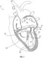
- FIG. 1illustrates the left ventricle and left atrium of the heart during systole.
- FIG. 2 Aillustrates free edges of leaflets of the mitral valve in normal coaptation
- FIG. 2 Billustrates the free edges in regurgitative coaptation.
- FIGS. 3 A- 3 Cillustrate grasping of the leaflets with a fixation device, inversion of the distal elements of the fixation device, and removal of the fixation device, respectively.
- FIG. 4illustrates the position of the fixation device in a desired orientation relative to the leaflets.
- FIG. 5illustrates a system for detaching an edge-to-edge repair device from one or more leaflets.
- FIGS. 6 A and 6 Billustrate a cutting member according to one embodiment of the system of FIG. 5 .
- FIG. 7illustrates a cutting member according to another embodiment of the system of FIG. 5 .
- FIG. 8illustrates a cutting member according to another embodiment of the system of FIG. 5 .
- FIG. 9illustrates a cutting member according to yet another embodiment of the system of FIG. 5 .
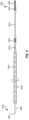
- FIG. 10illustrates a catheter according to one embodiment of the system of FIG. 5 .
- FIGS. 11 A- 11 Cillustrate alternate cross-sectional views of the catheter of FIG. 10 .
- FIGS. 12 A and 12 Billustrate a catheter according to another embodiment of the system of FIG. 5 .
- FIG. 13 A and 13 Beach illustrates a catheter according to other embodiments of the system of FIG. 5 .
- FIGS. 14 A and 14 Billustrate alternate cross-sectional views of the catheter of FIG. 13 .
- FIG. 15illustrates another alternate cross-sectional view of yet another embodiment of a catheter of the system of FIG. 5 .
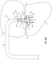
- FIG. 16illustrates a partial cut-away view of a human heart, showing a distal portion of the leaflet cutting device inserted into the right atrium and into the septal wall.
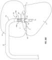
- FIG. 17illustrates a partial cut-away view of a human heart, showing a distal portion of the leaflet cutting device after having passed through the septal wall into the left atrium of the heart and having been steered toward and positioned adjacent the mitral valve.
- FIGS. 18 A and 18 Billustrate partial cut-away views of a human heart from the atrial side and from the ventricular side, respectively, showing distal portions of a cutting member and a capture member passing through orifices in the mitral valve from the left atrium into the left ventricle.
- FIG. 19illustrates a partial cut-away view of a human heart from the left ventricle, showing a shape-memory snare loop having been deployed from the capturing member in preparation for capturing the cutting member.
- FIG. 20illustrates a partial cut-away view of a human heart from the left ventricle, showing the cutting member having been advanced through the snare loop of the capturing member.
- FIG. 21illustrates a partial cut-away view of a human heart from the left ventricle, showing the snare loop being withdrawn back into the capturing member, thereby contacting and capturing the cutting wire.
- FIGS. 22 A- 22 Cillustrate partial cut-away views of a human heart from the left ventricle, showing the snare being progressively withdrawn back into the capturing member so as to position an exposed portion of the cutting member in position preparatory for cutting a portion of the leaflet of the mitral valve adjacent the edge-to-edge repair device.
- FIG. 23illustrates a partial cut-away view of a human heart from the left atrium, showing the exposed portion of the cutting member cutting through a portion of one of the leaflets of the mitral valve around the edge-to-edge repair device.
- FIG. 24is a schematic view of the system of FIG. 5 , showing one embodiment in which the capture member is withdrawn completely from the device, thereby drawing the cutting member distally through one lumen of the device, around the mitral valve and edge-to-edge repair device, and then back through another lumen of the device in a proximal direction, and thereby positioning an exposed cutting portion formed in the middle of the cutting member at the mitral valve.
- FIG. 25is a schematic representation of the system of FIG. 5 , showing another embodiment in which the capture member is partially withdrawn in a proximal direction, thereby drawing a distal end portion of the cutting member around the mitral valve and edge-to-edge repair device, and thereby positioning an exposed cutting portion formed proximately the distal end of the cutting member.
- FIGS. 26 A- 26 Dare schematic representations of yet another embodiment of the system of FIG. 5 , in which a puncture is made in one of the leaflets of the mitral valve, the capturing member is inserted through the puncture, two separate cutting members are passed through the orifices located on opposite sides of the edge-to-edge repair device, which are then captured by the snare loop of the capturing member and then drawn proximally back into the capturing member so as to place exposed cutting portions of each cutting wire in position to cut portions of the leaflet around the edge-to-edge repair device.
- an edge-to-edge repair devicesuch as a suture “bow-tie” or fixation device
- itneeds to be removed or at least detached from one or both of anterior and posterior leaflets.
- thishas been done during a high-risk invasive procedure such as open-heart surgery.
- the presently described system 100 , and associated devices and methodsallow a physician or clinician to address heart problems in a minimally invasive procedure.
- FIG. 1illustrates the left ventricle (LV) of a normal heart H in systole.
- the left ventricle (LV)is contracting and blood flows outwardly through the tricuspid (aortic) valve (AV) in the direction of the arrows.
- Back flow of blood or “regurgitation” through the mitral valve (MV)is prevented since the mitral valve is configured as a “check valve” which prevents back flow when pressure in the left ventricle is higher than that in the left atrium (LA).
- the mitral valve (MV)comprises a pair of leaflets having free edges (FE) which meet evenly to close, as illustrated in FIG. 1 .
- the opposite ends of the leaflets (LF)are attached to the surrounding heart structure along an annular region referred to as the annulus (AN).
- the free edges (FE) of the leaflets (LF)are secured to the lower portions of the left ventricle LV through chordae tendinae (CT) (referred to hereinafter as the chordae) which include a plurality of branching tendons secured over the lower surfaces of each of the valve leaflets (LF).
- CTchordae
- the chordae (CT)in turn, are attached to the papillary muscles (PM) which extend upwardly from the lower portions of the left ventricle and intraventricular septum IVS.
- FIG. 2 ASeveral structural defects in the heart can cause mitral valve regurgitation. Regurgitation occurs when the valve leaflets do not close properly allowing leakage from the ventricle into the atrium. As shown in FIG. 2 A , the free edges of the anterior and posterior leaflets normally meet along a line of coaptation (C). An example of a defect causing regurgitation is shown in FIG. 2 B .
- FIG. 2 BAn example of a defect causing regurgitation is shown in FIG. 2 B .
- an enlargement of the heartcauses the mitral annulus to become enlarged, making it impossible for the free edges (FE) to meet during systole. This results in a gap (G) which allows blood to leak through the valve during ventricular systole.
- Ruptured or elongated chordaecan also cause a valve leaflet to prolapse since inadequate tension is transmitted to the leaflet via the chordae. While the other leaflet maintains a normal profile, the two valve leaflets do not properly meet and leakage from the left ventricle into the left atrium will occur. Such regurgitation can also occur in patients who have suffered ischemic heart disease where the left ventricle does not contract sufficiently to effect proper closure.
- fixation devicesare used for grasping, approximating and fixating tissues, such as valve leaflets, to treat cardiac valve regurgitation, particularly mitral valve regurgitation.
- Certain fixation devicesare described in in U.S. Pat. No. 7,563,267, which is incorporated herein by this reference.
- the fixation devicesmay rely upon the use of an interventional tool that is positioned near a desired treatment site and used to grasp the target tissue.
- the interventional toolIn endovascular applications, the interventional tool is typically an interventional catheter.
- the interventional toolis typically an interventional instrument. Fixation of the grasped tissue is accomplished by maintaining grasping with a portion of the interventional tool which is left behind as an implant.
- the fixation devicesare well adapted for the repair of valves, especially cardiac valves such as the mitral valve.
- an interventional tool 10having a delivery device, such as a shaft 12 , and a fixation device 14 , is schematically illustrated having approached the mitral valve MV from the atrial side and grasped the leaflets LF.
- the mitral valvemay be accessed either surgically or by using endovascular techniques, and either by a retrograde approach through the ventricle or by an antegrade approach through the atrium, as described above. For illustration purposes, an antegrade approach is described.
- the fixation device 14(sometimes also referred to herein as a “suture ‘bow-tie’” or an “edge-to-edge repair device”) is releasably attached to the shaft 12 of the interventional tool 10 at its distal end.
- proximalshall mean the direction toward the end of the device to be manipulated by the user outside the patient's body
- distalshall mean the direction toward the working end of the device that is positioned at the treatment site and away from the user.
- proximalshall refer to the atrial or upstream side of the valve leaflets and distal shall refer to the ventricular or downstream side of the valve leaflets.
- the fixation device 14typically comprises proximal elements 16 (or gripping elements) and distal elements 18 (or fixation elements) which protrude radially outward and are positionable on opposite sides of the leaflets LF as shown to capture or retain the leaflets therebetween.
- the proximal elements 16are preferably comprised of cobalt chromium, nitinol or stainless steel
- the distal elements 18are preferably comprised of cobalt chromium or stainless steel, however any suitable materials may be used.
- the fixation device 14is coupleable to the shaft 12 by a coupling mechanism 17 .
- the coupling mechanism 17allows the fixation device 14 to detach and be left behind as an implant to hold the leaflets together in the coapted position.
- fixation device 14it may be desired to reposition or remove the fixation device 14 after the proximal elements 16 , distal elements 18 , or both have been deployed to capture the leaflets LF.
- Such repositioning or removalmay be desired for a variety of reasons, such as to reapproach the valve in an attempt to achieve better valve function, more optimal positioning of the device 14 on the leaflets, better purchase on the leaflets, to detangle the device 14 from surrounding tissue such as chordae, to exchange the device 14 with one having a different design, or to abort the fixation procedure, to name a few.
- the distal elements 18may be released and optionally inverted to a configuration suitable for withdrawal of the device 14 from the valve without tangling or interfering with or damaging the chordae, leaflets or other tissue. According to another embodiment, any of the endovascular methods described herein for disabling or removal of the fixation device may also be used.
- FIG. 3 Billustrates inversion wherein the distal elements 18 are moveable in the direction of arrows 40 to an inverted position.
- the proximal elements 16may be raised, if desired.
- the device 14may be repositioned to a desired orientation wherein the distal elements may then be reverted to a grasping position against the leaflets as in FIG. 3 A .
- the fixation device 14may be withdrawn (indicated by arrow 42 ) from the leaflets as shown in FIG. 3 C .
- Such inversionreduces trauma to the leaflets and minimizes any entanglement of the device with surrounding tissues.
- FIG. 4illustrates the position of the fixation device 14 in a desired orientation in relation to the leaflets LF.
- Thisis a short-axis view of the mitral valve MV from the atrial side, therefore, the proximal elements 16 are shown in solid line and the distal elements 18 are shown in dashed line.
- the proximal and distal elements 16 , 18are positioned to be substantially perpendicular to the line of coaptation C.
- the device 14may be moved roughly along the line of coaptation to the location of regurgitation.
- the leaflets LFare held in place so that during diastole, as shown in FIG.
- leaflets LFremain in position between the elements 16 , 18 surrounded by openings or orifices 0 which result from the diastolic pressure gradient.
- leaflets LFare coapted such that their proximal or upstream surfaces are facing each other in a vertical orientation, parallel to the direction of blood flow through mitral valve MV.
- the upstream surfacesmay be brought together to be in contact with one another or may be held slightly apart but will preferably be maintained in the vertical orientation in which the upstream surfaces face each other at the point of coaptation. This simulates the double orifice geometry of a standard surgical “bow-tie” repair where the edges of the leaflets are brought into apposition and sutured together to form the double orifice.
- Color Doppler echowill show if the regurgitation of the valve has been reduced. If the resulting mitral flow pattern is satisfactory, the leaflets may be fixed together in this orientation. If the resulting color Doppler image shows insufficient improvement in mitral regurgitation, the interventional tool 10 may be repositioned. This may be repeated until an optimal result is produced wherein the leaflets LF are held in place.
- fixation device 14is then detached from the shaft 12 and left behind as an implant to hold the leaflets together in the coapted position.
- Minimally invasive systems, methods, and devices for removing or at least detaching the suture “bow-tie” or fixation devices from one or both of anterior and posterior leafletsare disclosed herein. These minimally invasive systems, methods, and devices allow a practitioner to remove or at least detaching the suture “bow-tie” or fixation devices from one or both of anterior and posterior leaflets and, optionally, then proceed to do perform other medical procedures in the heart, without necessarily requiring open heart access or other more invasive procedures.
- Such systems, methods, and devicesare configured to be effective even if the suture “bow-tie” or fixation device has been installed for weeks, months, or years, such that tissue surrounding the device may have grown over, into, or around the suture “bow-tie” or fixation device.
- An embodiment of the present inventiondiscloses systems that can include guidewires, catheters and other components that can perform various specific functions, and also multifunctional catheters that can perform any combination of functions. Such functions may include holding or retaining an installed fixation device or the suture “bow-tie”; cutting a leaflet or leaflets; removing the suture “bow-tie” or a fixation device; and repairing the leaflet(s). Related methods for performing such functions are also disclosed.
- the mitral valvemay be accessed using the systems, methods, and devices disclosed and/or claimed herein either surgically or by using endovascular techniques, and either by a retrograde approach through the ventricle or by an antegrade approach through the atrium, as described above. For illustration purposes, an antegrade approach is described.
- the devices and associated methods and systems described hereinmay be used in combination with imaging modalities such as x-ray, fluoroscopy, echocardiography, charge coupled device cameras, complementary metal oxide semiconductor cameras, magnetic resonance imaging, and other imaging modalities.
- imaging modalitiessuch as x-ray, fluoroscopy, echocardiography, charge coupled device cameras, complementary metal oxide semiconductor cameras, magnetic resonance imaging, and other imaging modalities.
- imaging modalitiessuch as x-ray, fluoroscopy, echocardiography, charge coupled device cameras, complementary metal oxide semiconductor cameras, magnetic resonance imaging, and other imaging modalities.
- imaging modalitiessuch as x-ray, fluoroscopy, echocardiography, charge coupled device cameras, complementary metal oxide semiconductor cameras, magnetic resonance imaging, and other imaging modalities.
- the availability of such imaging modalities during such proceduresmay help practitioners visualize, for example, where the fixation devices are, how they are connected to the heart, and where to direct the various catheters and/or other devices.
- FIG. 5illustrates a system 100 that may be used to at least detach an edge-to-edge repair device, such as a suture “bow-tie” or a fixation device, from at least one of the anterior and posterior leaflets LF.
- the system 100can achieve this without open access to the heart, via a minimally invasive intravascular procedure, and through a single transseptal puncture. It will be understood, however, that the system 100 can also be used to detach a suture “bow-tie” or a fixation device from both or all leaflets to which the fixation device is attached.
- system 100can be used with the suture “bow-tie” or fixation devices identified herein or various other devices that are used to repair the leaflets, such as during an edge-to-edge repair.
- the suture “bow-tie” or fixation devicescan be considered as edge-to-edge repair devices.
- the system 100can take the form of an elongate member 102 , such as a multicomponent catheter, which is adapted to be advanced through a patient's vasculature.
- Elongate member or catheter 102can include a guiding catheter 110 through which a delivery catheter 120 can be advanced to a previously implanted edge-to-edge repair device (not shown).
- the guiding catheter 110can be, in one configuration, a steerable catheter or introducer, such as a Steerable Introducer sold under the trademark AGILIS.
- the guiding catheter 110can be steered through the tortuous anatomy to the heart and, using a transseptal approach, advanced through the septum between the right and left atria toward the mitral valve (MV). Once in place, the guiding catheter 110 , whether alone or in combination with the delivery catheter 120 , can position a distal end 122 of the delivery catheter 120 relative to the mitral valve (MV) for performing edge-to-edge repair device detachment as described herein.
- the steerability of guiding catheter 110can be achieved through any number of known structures and methods, including but not limited to those disclosed in U.S. Pat. No. 7,653,267.
- delivery catheter 120can also move axially relative to the guide catheter.
- a distal end portion of delivery catheter 120can also be steerable independent of, and in a different plane from, guiding catheter 110 .
- the delivery catheter 120accommodates a capturing member 140 and a cutting member 150 that extends from a proximal end 124 to the distal end 122 .
- the capturing member 140such as a snare, is used to capture and aid with positioning a cutting region of the cutting member 150 relative to the tissue to be cut to detach the edge-to-edge repair device from the leaflets LF.
- withdrawing the capturing member 140 into the delivery catheter 120while at the same time advancing the cutting member 150 , provides the desired looping and positioning of the cutting member 150 so that the cutting region of the cutting member 150 can be used to cut the leaflets.
- the capturing member 140can include a looped portion 144 at a distal end 142 .
- This looped portion 144can include a narrowed portion 146 that facilitates preferential collapsing of the capturing member 140 when the capturing member 140 draws the cutting member 150 into the distal end 122 of the delivery catheter 120 .
- two halves of the looped portion 144can collapse together with the narrowed portion 146 acting as a hinge or pivot that aids with the left and right sides moving together as the distal end 122 of the delivery catheter 120 contacts a proximal end 148 of the looped portion 144 .
- other structurescan be formed in the looped portion 144 to aid with the preferential collapsing.
- a wire forming the snarecan be ground or narrowed to preferentially bend.
- FIG. 5illustrates the capturing member 140 having the looped portion 144
- other types of capturing mechanismsmay include, but are not limited to, for example, sheaths, conduits, expandable baskets, vacuums, magnets, vices, and clamps.
- the capturing member 140 and the cutting member 150can include magnets or be formed of materials that magnetically attract so that the distal end 152 of the cutting member 150 might be attracted to the distal end 142 of the capturing member 140 or vice versa.
- the capturing member 140can be a wire, tubular member, or other elongate structure and can be made of metal, plastic, shape-memory alloys or polymers, or any suitable material, e.g., such as those described herein (e.g., cobalt-chromium alloys, stainless steel, nickel-titanium, Elgiloy®, etc.
- a distal end portion of capturing member 140can include a pre-formed, resilient, shape-memory loop configuration and/or orientation, such as looped portion 144 .
- the loopcan collapse and straighten when held within the constraints of the delivery catheter 120 . Once advanced distally beyond the distal end of the delivery catheter 120 , the loop can self-expand and return to its pre-formed, shape-memory configuration and orientation.
- Capturing member 140can also include one or more radiopaque markers positioned adjacent and/or around the looped portion 144 . As discussed in additional detail below, such radiopaque markers assist in locating and visualizing the location, position and orientation of such capturing member 140 and cutting member 150 , as well as their respective locations, positions and orientations relative to one another.
- the cutting member 150can electrically communicate with an electrosurgical device 130 (such as an electrosurgical generator), which can selectively provide radio-frequency energy to the cutting member 150 .
- the cutting member 150therefore, can function as a probe electrode that can cut, coagulate, desiccate, and fulgurate tissue, such as the leaflets LF. More particularly, the cutting member 150 can be used to cut at least a portion of at least one of the anterior and posterior leaflets LF or other tissue to which the edge-to-edge repair device is coupled.
- the cutting member 150can include a conductive core 158 with an electrically insulating coating 160 formed over at least a portion of the core 158 .
- the cutting member 150is a guide wire that includes a proximal end 154 that can be electrically connected or coupled to an electrosurgical device 130 , which can be used to selectively apply electrical energy to cutting member 150 .
- the electrical cooperationcan occur through an uncoated or exposed portion 159 at the proximal end 154 of core 158 , which can be connected to electrosurgical device 130 . Additional details regarding the cutting member 140 will be provided in connection with FIGS. 6 - 9 below.
- Portions of capturing member 140 and cutting member 150can also include good torque characteristics.
- rotational forceswhen rotational forces are applied at their respective proximal ends to cause rotational movement at the proximal ends, such rotational movement is translated relatively closely to the respective distal ends.
- the relative positioning and orientation of the distal end of capturing member 140 (including any pre-formed bends or curves and/or loop 144 ) and the distal end of cutting member (including any pre-formed bends or curves)can be closely correlated to the rotational position of the corresponding proximal ends thereof.
- System 100can also include a handle or fixture, schematically illustrated at 112 , located at or near the proximal end 124 of system 100 .
- Handle or fixture 112can incorporate any number of known structures and methods for manipulating and controlling the advancing, withdrawing, positioning and orienting of the various components of system 100 , including, but not limited to, guiding member 110 , delivery catheter 120 , capturing member 140 , cutting member 150 and other components and sub-components of system 100 , relative to a patient's anatomy and/or relative one another.
- FIGS. 6 - 9illustrate various possible configurations of the cutting member 150 .
- the cutting members illustrated in FIGS. 6 and 7enable a full floss technique, wherein the capture member 140 , such as a snare, pulls the distal end of cutting member 150 distally out the distal end of one lumen of the delivery catheter, around one of the leaflets of the mitral valve, and then pulls the distal end of the cutting member back through the other lumen of the delivery catheter in a proximal direction until the distal end of the cutting member exits the proximal end of the delivery catheter (as illustrated in FIG. 23 ).
- Each illustrated cutting member 150 a - 150 dhas a proximal end 154 a - 154 d and a distal end 152 a - 152 d.
- Formed at the distal end 152 a - 152 dis an atraumatic tip 156 a - 156 d that can be formed, in one configuration, from a coil.
- This atraumatic tip 156 a - 156 dcan be shaped by a physician or clinician using the cutting member 150 or can have a preformed shape to aid with positioning the cutting member 150 relative to the edge-to-edge repair device, leaflets, and the orifices O formed by the edge-to-edge repair device and the leaflets.
- the coilcan be formed of a metal or alloy, or other deformable or resiliently deformable material.
- Each cutting member 150 a - 150 dhas a conductive core 158 a - 158 d and a coating 160 a - 160 d disposed over the conductive core 158 a - 158 d.
- the coating 160 a - 160 dpartially covers the conductive core 158 a - 158 d, except for one or more bare regions 162 a - 162 d where the conductive core 158 a - 158 d is uncoated or exposed.
- each cutting member 150 a - 150 dincludes an uncoated, exposed contact region 159 a - 159 d disposed at respective proximal ends 154 a - 154 d that allow an electrical contact or coupling to be made with electrosurgical device 130 .
- These regions 159 a - 159 daccommodate electrical communication with the electrosurgical device 130 .
- an uncoated, exposed cutting region 162 ais disposed between the proximal end 154 a and the distal end 152 a of the cutting member 150 a for tissue contact.
- two uncoated, exposed cutting regions 162 bare included, one cutting region 162 b located near the middle of cutting member 150 b and a second cutting region 162 b near the distal end 152 b of cutting member 150 b, again for tissue contact.
- an uncoated, exposed cutting region 162 cis provided near the distal end 152 c for tissue contact, without a second region disposed between the proximal end 154 c and the distal end 152 c.
- each cutting member 150 a - 150 dincludes radiopaque markers 164 a - 164 d. These markers 164 a - 164 d define the peripheral bounds of the cutting regions 162 a - 162 d and aid in positioning those regions of the cutting member 150 a - 150 d. For instance, the markers 164 a - 164 d can be viewed via fluoroscopy, x-ray, or other imaging technique.
- markers 164 a - 164 dare illustrated defining the bounds of the cutting regions 162 a - 162 d, other markers can be included to define other locations of the cutting member 150 a - 150 d or any other part of the system 100 or relationship between two or more parts of the system 100 .
- cutting member 150 dcan include: an elongate electrically conductive core 158 d; an electrically insulative coating 160 d covering a large portion of the conductive core 158 d; one or more bare, exposed cutting regions 162 d; a bare, exposed contact region 159 d; one or more radiopaque markers 164 d positioned adjacent the cutting region 162 d; and an atraumatic tip 156 d.
- cutting member 150 dcan include one or more tapered transition regions, such as tapered transition region 165 d located between a main body portion of the cutting member 150 d and the cutting region 162 d.
- the electrically conductive core 158 dcan be made of surgical quality steel and can have a diameter of approximately 0.014′′ (or approximately 0.04 cm) at its proximal end and a diameter of approximately 0.005′′ (or approximately 0.01 cm) to approximately 0.008′′ (or approximately 0.02 cm) in the cutting region(s) 162 d.
- a proximal portion of the cutting membercould also be formed from a hypotube.
- Cutting member 150 dcan have an overall length of approximately 70′′ (or approximately 180 cm), with a cutting region 162 d of approximately 4′′ (or approximately 10 cm) in length, and the atraumatic tip can be approximately 0.4′′ (or approximately 1 cm) to approximately 1.2′′ (or approximately 3 cm) in length.
- the cutting member 150can be a wire, tubular member, or other elongate structure, with the conductive core being made of a metal, alloy, conductive polymer, or other conductive material.
- the coating for the cutting member 150can be a polymer, such as polyterafluoroethylene or other polymer, a hydrophilic coating, a composite material, or other material the can provide lubricity to the cutting member and/or insulative properties to limit unwanted electrical conductivity of the conductive core with the tissue or the other components of the system 100 .
- FIG. 10illustrated is one configuration of the delivery catheter 120 .
- the delivery catheter 120can be a microcatheter that is advanceable within a lumen of the guiding catheter 110 .
- the delivery catheter 120can have a multi-lumen structure, such as including a lumen 126 that can receive the capturing member 140 and a lumen 128 that can receive the cutting member 150 .
- the delivery cathetercan include additional lumens 126 and/or lumens 128 , such as illustrated in FIG. 15 and discussed hereinafter.
- the lumens 126 and 128can extend from luer connectors 170 and 172 , through extension tubular members 174 and 174 , which can join the luer connectors 170 and 172 to a reminder of the delivery catheter 120 , to the distal end 122 .
- These lumens 126 and 128can be formed during extrusion of the shaft 125 of the delivery catheter 120 when the shaft 125 is formed of a polymer or other material capable of being extruded.
- FIGS. 10 a - 10 cillustrates different cross-sections for the shaft 125 and different lumen constructions.
- the lumen 126 aincludes wings that generally surround or extend upwardly to overlap a portion of the lumen 128 a in a transverse direction relative to a longitudinal axis of the lumen 128 a.
- the lumen 126 bhas a generally planar upper surface with a curved bottom surface that approximates the curvature of the shaft 125 b.
- both the lumen 128 c and the lumen 126 cinclude curvatures that approximate the curvature of the shaft 125 c.
- the distal end 122 of the shaft 125can also have different configurations.
- the tubular portions of the shaft 125 forming the lumen 126 d and 128 dcan include a cut or slit 129 in the material so that the lumens 126 d and 128 d, and associated tubular portion, can be spaced apart. This provides for better manipulation of the delivery catheter 120 d and better tension during the cutting process.
- the cross-section of the distal end 122 d illustrated in FIG. 12 Balso aids with enhancing flexibility.
- each tubular memberis independently advanceable and also independently positionable relative to the distal end 122 of the delivery catheter 120 and the mitral valve MV.
- FIGS. 13 , 14 and 15Such a structure is illustrated in FIGS. 13 , 14 and 15 and is further described below.
- the delivery catheter 120 ecan be a microcatheter that is advanceable within a lumen of the guiding catheter 110 .
- the delivery catheter 120 ecan also include a first tubular member 180 and a second tubular member 182 .
- lumens 126 e and 128 e of delivery catheter 120 ecan be configured and sized to receive first tubular member 182 and second tubular member 180 , respectively.
- First tubular member 182can extend through lumen 126 e of delivery catheter 120 e and terminate at its proximal end in a luer connector 170 .
- first tubular member 182can be configured and sized to receive capturing member 140 therethrough.
- second tubular member 180can extend through lumen 128 e of delivery catheter 120 e and terminate at its proximal end in a luer connector 172 .
- the lumen formed within tubular member 180can be configured and sized to receive cutting member 150 therethrough.
- the tubular members 180 and 182can be moved forward and backward, or proximal and distal, relative to a shaft shell 186 .
- Tubular members 180 and 182can also move axially relative to, and independent of, one another. This allows for enhanced positioning of the distal ends of tubular members 180 and 182 (and, thus, enhanced positioning of capturing member 140 and cutting member 150 ) relative to the mitral valve MV during the procedure.
- the distal end of the tubular member 182can be closer to the mitral valve MV than the distal end of the tubular member 180 during the procedure, or vice versa, to change the angle at which the looped portion 144 receives the distal end 152 of the cutting member 150 .
- one or bothcan be pre-formed with a resilient, pre-formed shape-memory curve or bend to aid with steerability and positioning of the distal ends of the tubular members 180 and 182 at desired locations within the heart.
- the positioning of the delivery catheter 120 e and/or the distal ends of the various elements of capturing member 140 and cutting member 150can be achieved by the steerability of the guiding catheter 110 , pre-formed, shape-memory curves or bends in tubular members 180 and 182 , or any combination thereof.
- a distal portion of the delivery catheter 120 ecould also be made to be independently steerable once it is advanced beyond the distal end of guiding catheter 110 .
- FIG. 13 Billustrates yet another embodiment of the delivery catheter 120 f that very similar to the embodiment shown in FIG. 13 A , except that catheter 120 f includes a side port 129 f near the proximal end to receive cutting member 150 in a “monorail” or rapid-exchange fashion.
- This arrangementcan simplify the insertion of cutting member 150 into the delivery catheter 120 f and can also allow the overall length of cutting member to be shortened.
- delivery catheter 120 acan be take the form of a simple outer shell 186 that surrounds tubular members 180 and 182 (without providing separate lumens, such as lumens 126 e and 128 e ).
- the shell 186can have a fixed shape, such as illustrated in FIG. 14 A or can have a flexible form that approximates an outer surface profile of the tubular members 180 and 182 , as shown in FIG. 13 B .
- FIG. 14 Bwhile the shell 186 generally approximates the outer surface profile of the tubular members 180 and 182 , spaces can be formed between the shell 186 and the tubular member 180 and 182 , such as spaces 190 . Varying a durometer of the polymer or material forming the shell 186 allows for the different configurations.
- the shaft and other portions of the delivery catheters 120can be fabricated from polymers, metals, alloys, combinations thereof, or other materials that can accommodate the manufacture of the delivery catheter.
- FIG. 15Illustrated in FIG. 15 is another cross-sectional configuration of the delivery catheter described herein.
- the delivery catheters 120 described hereincan include multiple lumens, with those lumens accommodating or receiving either the capture member 140 and the cutting member(s) 150 directly or including tubular members, such as tubular members 180 a, 180 b and 182 , that accommodate or receive the capture member 140 and the cutting members 150 a and 150 b.
- the delivery catheter 120 g of FIG. 15can include 3 lumens: lumen 126 g and two lumens 128 g. Disposed within each of the lumens 128 g is a tubular member 180 a and 180 b ) (similar to the tubular member 180 described in reference to FIGS. 12 and 13 ), which can receive two separate cutting members 150 a and 150 b. Disposed within the lumen 126 g is a needle 192 , which can be used to puncture the septum and, optionally, leaflet tissue or tissue adjacent to the leaflets to allow passage of the capture member 140 through a leaflet or other tissue.
- the needle 192can be removed and the capture member 140 , whether alone or with the tubular member 182 , can be advanced through the lumen 126 g.
- This configurationallows the delivery device 120 g to accommodate two cutting members 150 a and 150 b that can be used to cut tissue either sequentially or simultaneously.
- the system 100can be used to position the delivery catheter 120 in various spaces within a patient's anatomy. For instance, and as will be described herein, the system 100 can be used to position the capturing member 140 and the cutting member 150 relative to leaflets in apposition to an edge-to-edge repair device, such as the MitraClip® or a suture “bow-tie.” However, the presently described system 100 and associated methods and devices can be used for other valvular structures or other tissues in an anatomy.
- the guiding catheter 110can access the left atrium LA from the femoral artery using the inferior vena cava as illustrated in FIG. 16 . Following a trans septal puncture using an appropriately placed needle advanced through a lumen of the guiding catheter, the guiding catheter 110 is introduced into the left atrium as illustrated in FIG. 17 . Once positioned in the left atrium, the steerability of guiding catheter 110 is used to direct the distal end of delivery catheter 120 toward and adjacent to the mitral valve as further shown in FIG. 17 .
- the guiding catheter 110can be steered toward the mitral valve (MV) so that the center of the guiding catheter 110 is substantially centered on the previously implanted edge-to-edge repair device. This can include generally aligning a longitudinal axis of the guiding catheter 110 toward the edge-to-edge repair device (not shown). Once in place, the delivery catheter 120 can be advanced along the lumen in preparation for deployment.
- MVmitral valve
- the distal ends of lumens 126 and 128can be positioned through orifices 0 on either side of edge-to-edge repair device.
- the shaft of the delivery catheter 120can be advanced so that the lumen 126 d and the lumen 128 d can each be advanced into a separate orifice located to either side of the edge-to-edge repair device, i.e., the lumen 126 d extends through one orifice O and the lumen 128 d extends through the other orifice O.
- tubular members 180 and 182are advanced into and through orifices O on either side of the edge-to-edge repair device so that the tubular member 180 is in one orifice O and the tubular member 182 is in the other orifice O as illustrated in FIGS. 18 A and 18 B .
- the capture member 140can be advanced from the lumen 128 (and from the tubular member 182 ) into the left ventricle.
- the looped portion 144can self-expand to form a ring or opening (as shown in FIG. 19 ) with which to receive the cutting member 150 .
- the cutting member 150is advanced from the lumen 128 (and from the tubular member 180 ) it is captured by the looped portion 144 as shown in FIG. 20 .
- the tubular members 180 and 182can also be independently moved to help position the distal ends of the lumens 126 and 128 .
- radiopaque markerscan be provided at different locations around the looped portion 144 , as well as at various locations on the distal end portion of the cutting member 150 , to assist in locating and visualizing the relative locations, positions and orientation of such components relative to one another.
- the capture member 140can be withdrawn towards the deliver catheter 120 , and the lumen 126 as illustrated in FIG. 22 .
- the cutting member 150can also be advanced in a distal direction at the same time to form a loop in the cutting member 150 and/or vary a length of available cutting member to contact the leaflet that will be cut.
- the pushing of the cutting member 150 and pulling of the capture member 140can position the cutting member 150 , and more particularly, the bare cutting regions 162 in the desired leaflet location as shown in FIGS. 22 A- 22 C .
- the leafletcan be cut by applying electrical energy to the cutting member 150 and drawing the exposed cutting regions 162 through the leaflet tissue, as illustrated in FIG. 23 , thereby detaching the edge-to-edge repair device from one of the leaflets while remaining attached to the other leaflet. Thereafter, the physician or clinician can then perform one or more of a mitral valve annuloplasty, balloon valvuloplasty, mitral valve repair, installation of a replacement valve, and combinations thereof.
- FIG. 24is a schematic view of the system of FIG. 13 A , showing one embodiment (sometimes referred to herein as a “full floss” embodiment) in which the capture member 140 can be completely withdrawn from the device.
- the capture member 140is withdrawn in a proximal direction, thereby drawing the distal end of cutting member 150 distally through one lumen of the device (such as lumen 128 ), around the mitral valve and edge-to-edge repair device, and then back through the other lumen of the device (such as lumen 126 ) in a proximal direction, thereby positioning an exposed cutting region 162 formed in the middle of the cutting member 150 at the mitral valve.
- both uncoated ends 159 and 162 of the cutting memberto be accessible at the proximal end of the device for connection to the electrosurgical device 130 and thereby form an electrical circuit for the passage of electrosurgical energy.
- the capturing member 140is completely withdrawn from the delivery catheter 120 in order to deploy and position the cutting member 150 .
- FIG. 25is a schematic representation of the system of FIG. 13 A , showing another embodiment in which the capture member 140 is only partially withdrawn in a proximal direction to position an exposed cutting portion 162 formed near a distal end portion of the cutting member 150 around the mitral valve and edge-to-edge repair device.
- the overall of length of the capture member 140can be shorter than that required with the embodiment of FIG. 24 .
- this embodimentmay require the use of a patient return electrode in order to complete the electrical circuit, since only one end of the cutting member is accessible at the proximal end of the device for connection to the electrosurgical device 130 .
- the cutting member 150can be advanced through the leaflet as a puncturing tool without capturing or positioning with the capture member 140 .
- a combination of puncturing and cutting the leaflet with the cutting member 150 and lassoing either with the capture member 140 or another lasso advanced through another lumen of the delivery catheter 120can used to (i) puncture the leaflet, (ii) capture a portion of the edge-to-edge repair device, (iii) cut one leaflet, (iv) capture another portion of the edge-to-edge repair device, and (v) cut the other leaflet before removing the edge-to-edge repair device. This would allow for removal or at least partial detachment of multiple edge-to-edge repair devices from one or more leaflets.
- FIGS. 26 A- 26 DThis alternate approach, using the delivery catheter 120 f, is schematically illustrated in FIGS. 26 A- 26 D .
- the needle 192can be advanced through the tissue of one of the leaflets, such as from a distal end of the tubular member 182 , to create a puncture P through the tissue of one of the mitral valve leaflets and positioned adjacent the edge-to-edge repair device 18 .
- the needle 192is removed and the capture member 140 (and/or the lumen 126 containing capture member 140 ) is advance through the puncture P in the leaflet tissue and into the ventricle.
- the delivery catheter 120 fcan be advanced over the needle 192 and through the puncture to position a distal end of the delivery catheter 120 f in a desired position to deploy the capture member 140 .
- the tubular member 182is sized to accommodate the needle 192 , and when the needle 192 is removed, receives the capture member 140 . Telescoping the tubular member 182 over the needle 192 , once it has punctured the tissue, can place the distal end of the lumen 126 f in the ventricle so the capture member 140 , once the needle 192 is removed and the capture member 140 is disposed within the lumen 126 f, can be deployed from the lumen 126 f into the desired location for capturing the cutting member 150 , as shown in FIG. 26 A .
- the two tubular members 180 a and 180 bare advanced to position the distal ends of tubular members 180 a and 180 b through orifices O on either side of the edge-to-edge repair device 18 , as shown in FIG. 26 B .
- the distal ends of the cutting members 150 a and 150 bare in position for deployment so they might be advance into the looped portion 144 .
- the tubular members 180 a, 180 b and 182can be moved together or independently to position the distal ends of the lumens 126 f and 128 f.
- any grouping of the tubular members 180 a, 180 b and 182can be moved together.
- the tubular members 180 a and 180 bcan be moved together, while independently from the tubular member 182 . Any combination of movement of the tubular members 180 a, 180 b and 182 is possible.
- the capture member 140can be withdrawn towards the deliver catheter 120 , and the lumen 126 , as shown in FIG. 26 C .
- distal end portions of cutting members 150 a and 150 bare drawn through their respective orifices O, around the tissue separating each orifice and the puncture P and then pass into the distal end of lumen 126 of tube 182 , as shown in FIG. 26 D .
- the cutting members 150 a and/or 150 bcan be advanced to vary a length of available cutting member to contact the leaflet that will be cut.
- the pushing of the cutting members 150 a and/or 150 b and the pulling of the capture member 140can position the cutting members 150 a and 150 b , and more particularly, the bare regions 162 a and 162 b in the desired leaflet location.
- the leafletcan then be cut by applying electrical energy to the cutting members 150 a and 150 b and drawing the exposed cutting regions 162 through the leaflet tissue located between each orifice O and puncture P.
- the tubular members 180 a and 180 bcan optionally be moved to selectively isolate or insulate the tissue from the cutting member(s) 150 .
- the tubular member 180 acan be advanced to isolate or insulate the tissue, such as leaflet tissue, from the cutting member 150 a, while the cutting member 150 b is used to cut the tissue (i.e., the cutting member 150 b is energized to cut the tissue).
- the tubular member 180 ais retracted to expose the cutting member 150 a and the tubular member 180 b is advanced to isolate or insulate the tissue from the cutting member 150 b, and then cutting member 150 a can be energized to cut the tissue.
- delivery catheter 120 fcan also be used to sequentially cut the edge-to-edge repair device 18 from both leaflets of the mitral valve.
- delivery device 120 fcan first be deployed in the manner discussed above to cut one leaflet and then be subsequently deployed in a similar manner to cut the other leaflet.
- the physician or cliniciancan then move on to perform one or more of a mitral valve annuloplasty, balloon valvuloplasty, mitral valve repair, installation of a replacement valve, and combinations thereof.
- the technology disclosed hereincan be directed to systems, devices and methods for at least partially detaching an edge-to-edge repair device that holds together anterior and posterior leaflets of a mitral valve of a human heart.
- the devicesare directed to at least partially detaching an edge-to-edge repair device that holds together anterior and posterior leaflets of a mitral valve of a human heart and can include one or more of the following elements: an elongate member having a proximal end, a proximal end portion, a distal end portion and a distal end, the elongate member; a guiding catheter with a distal end portion that can be selectively bent to reorient the distal end portion of the elongate member; a delivery catheter coaxially located within the guiding catheter; a delivery catheter shell having a first lumen and a second lumen, each extending from a proximal end to a distal end of the delivery catheter shell; a first tubular member having
- the devicescan also include one or more of the following additional features: wherein one or both of the first and second tubular members include a preferential, shape-memory bend in the distal end portion thereof; wherein the cutting member comprises a first cutting region and a second cutting region, wherein the first cutting region is disposed at a proximal end of the cutting member and the second cutting region is disposed between the proximal end and a distal end of the cutting member; wherein the first cutting region is disposed at and forms a distal end of the cutting member, proximal of an atraumatic coil tip; wherein the cutting member further comprises one or more radiopaque markers defining peripheral bounds of the cutting regions; wherein the capture member comprises a self-expanding, shape-memory loop portion for capturing the cutting member; wherein the loop portion comprises a narrowed portion configured to facilitate preferential collapsing of the capture member upon drawing the capture member into the second tubular member; and/or wherein the capture member further comprises one or more radiopa
- the methodscan be directed to at least partially detaching an edge-to-edge repair device that holds anterior and posterior leaflets of the mitral valve together, wherein a first orifice in the mitral valve is located to one side of the edge-to-edge repair device and a second orifice in the mitral valve is located to an other side of the edge-to-edge repair device, and can include, for example, any one or more of the following steps or acts: advancing a capture member from a first catheter portion that extends from a distal end of a delivery catheter and through the first orifice; advancing a cutting member from a second catheter portion that extends from the distal end of the delivery catheter and through the second orifice; capturing the cutting member with the capture member and positioning the cutting member around a portion of one of the leaflets of the mitral valve adjacent to the edge-to-edge repair device; and/or cutting at least a portion of one of the leaflets adjacent to the edge-to-edge repair device to detach the edge-to-edge repair device from the
- the methodscan include any one or more of the following steps or acts: advancing an elongate member through a least a portion of the vasculature of a patient and into a left atrium of a patient's heart, the elongate member having a proximal end portion, distal end portion and a distal end, the proximal end portion being located and accessible outside the patient's vasculature, the elongate member having a guiding catheter with a distal end portion that can be selectively bent to reorient the distal end portion of the elongate member, the elongate member also having a delivery catheter coaxially located within the steering catheter, the delivery catheter having a first lumen and a second lumen, each extending from a proximal end to a distal end of the delivery catheter, the delivery catheter also having a capture member extending through the first lumen and cutting member extending through the second lumen; puncturing, with the distal end of the elongate member, a
- the methodscan also include any one or more of the following additional steps or acts: advancing a distal end portion of the capture member beyond a distal end of the first catheter portion, the distal end portion of the capture member having a self-expanding, shape-memory loop, and allowing the self-expanding, shape-memory loop to deploy on a ventricular side of the mitral valve; advancing a distal end portion of the cutting member beyond a distal end of the second catheter portion and through the loop of the capturing member; withdrawing the capturing member in a proximal direction and causing the loop to snare the cutting member and draw it around the edge-to-edge repair device and into contact with a portion of the tissue of the leaflet between the first and second orifices and adjacent to the edge-to-edge repair device; withdrawing the capture member in a proximal direction until one of the electrically conductive cutting regions passes around and is positioned against the tissue of the leaflet adjacent the edge-to-edge repair device;
Landscapes
- Health & Medical Sciences (AREA)
- Life Sciences & Earth Sciences (AREA)
- Surgery (AREA)
- Cardiology (AREA)
- Engineering & Computer Science (AREA)
- Public Health (AREA)
- General Health & Medical Sciences (AREA)
- Veterinary Medicine (AREA)
- Animal Behavior & Ethology (AREA)
- Biomedical Technology (AREA)
- Heart & Thoracic Surgery (AREA)
- Medical Informatics (AREA)
- Molecular Biology (AREA)
- Nuclear Medicine, Radiotherapy & Molecular Imaging (AREA)
- Vascular Medicine (AREA)
- Oral & Maxillofacial Surgery (AREA)
- Transplantation (AREA)
- Plasma & Fusion (AREA)
- Otolaryngology (AREA)
- Physics & Mathematics (AREA)
- Reproductive Health (AREA)
- Surgical Instruments (AREA)
- Prostheses (AREA)
Abstract
Description
Claims (20)
Priority Applications (3)
| Application Number | Priority Date | Filing Date | Title |
|---|---|---|---|
| US16/659,238US12102531B2 (en) | 2018-10-22 | 2019-10-21 | Tissue cutting systems, devices and methods |
| EP19804912.4AEP3870088B1 (en) | 2018-10-22 | 2019-10-22 | Tissue cutting systems and devices |
| PCT/US2019/057482WO2020086614A1 (en) | 2018-10-22 | 2019-10-22 | Tissue cutting systems, devices and methods |
Applications Claiming Priority (2)
| Application Number | Priority Date | Filing Date | Title |
|---|---|---|---|
| US201862748947P | 2018-10-22 | 2018-10-22 | |
| US16/659,238US12102531B2 (en) | 2018-10-22 | 2019-10-21 | Tissue cutting systems, devices and methods |
Publications (2)
| Publication Number | Publication Date |
|---|---|
| US20200121460A1 US20200121460A1 (en) | 2020-04-23 |
| US12102531B2true US12102531B2 (en) | 2024-10-01 |
Family
ID=70279342
Family Applications (1)
| Application Number | Title | Priority Date | Filing Date |
|---|---|---|---|
| US16/659,238Active2042-01-21US12102531B2 (en) | 2018-10-22 | 2019-10-21 | Tissue cutting systems, devices and methods |
Country Status (3)
| Country | Link |
|---|---|
| US (1) | US12102531B2 (en) |
| EP (1) | EP3870088B1 (en) |
| WO (1) | WO2020086614A1 (en) |
Families Citing this family (32)
| Publication number | Priority date | Publication date | Assignee | Title |
|---|---|---|---|---|
| US9011468B2 (en) | 2011-09-13 | 2015-04-21 | Abbott Cardiovascular Systems Inc. | Independent gripper |
| US8945177B2 (en) | 2011-09-13 | 2015-02-03 | Abbott Cardiovascular Systems Inc. | Gripper pusher mechanism for tissue apposition systems |
| US9572666B2 (en) | 2014-03-17 | 2017-02-21 | Evalve, Inc. | Mitral valve fixation device removal devices and methods |
| EP3868306A1 (en) | 2016-06-20 | 2021-08-25 | Evalve, Inc. | Transapical removal device |
| US10736632B2 (en) | 2016-07-06 | 2020-08-11 | Evalve, Inc. | Methods and devices for valve clip excision |
| US11071564B2 (en) | 2016-10-05 | 2021-07-27 | Evalve, Inc. | Cardiac valve cutting device |
| US20210212756A1 (en)* | 2017-08-25 | 2021-07-15 | Nasser Rafiee | Tissue cutting systems and methods |
| US11534303B2 (en) | 2020-04-09 | 2022-12-27 | Evalve, Inc. | Devices and systems for accessing and repairing a heart valve |
| CA3147410A1 (en) | 2019-07-15 | 2021-01-21 | Evalve, Inc. | Proximal element actuator fixation and release mechanisms |
| JP7566004B2 (en) | 2019-07-15 | 2024-10-11 | エバルブ,インコーポレイティド | Wide clip with non-deforming wings |
| JP7543391B2 (en) | 2019-07-15 | 2024-09-02 | エバルブ,インコーポレイティド | Method of Actuating Individual Proximal Elements |
| EP4033970B1 (en) | 2019-09-26 | 2024-11-20 | Evalve, Inc. | Systems for intra-procedural cardiac pressure monitoring |
| US11464636B2 (en) | 2019-10-11 | 2022-10-11 | Evalve, Inc. | Repair clip for variable tissue thickness |
| WO2021092107A1 (en) | 2019-11-06 | 2021-05-14 | Evalve, Inc. | Stabilizer for a medical delivery system |
| EP4292546B1 (en) | 2019-11-08 | 2025-04-23 | Evalve, Inc. | Medical device delivery system with locking system |
| US11701229B2 (en) | 2019-11-14 | 2023-07-18 | Evalve, Inc. | Kit with coaptation aid and fixation system and methods for valve repair |
| US11801140B2 (en) | 2019-11-14 | 2023-10-31 | Evalve, Inc. | Catheter assembly with coaptation aid and methods for valve repair |
| US12109115B2 (en) | 2019-12-18 | 2024-10-08 | Evalve, Inc. | Wide clip with deformable width |
| US12048448B2 (en) | 2020-05-06 | 2024-07-30 | Evalve, Inc. | Leaflet grasping and cutting device |
| US12171486B2 (en) | 2020-05-06 | 2024-12-24 | Evalve, Inc. | Devices and methods for clip separation |
| US12178444B2 (en) | 2020-05-06 | 2024-12-31 | Evalve, Inc. | Clip removal systems and methods |
| US12414811B2 (en) | 2020-05-06 | 2025-09-16 | Evalve, Inc. | Devices and methods for leaflet cutting |
| US12171485B2 (en) | 2020-05-06 | 2024-12-24 | Evalve, Inc. | Systems and methods for leaflet cutting using a hook catheter |
| CN115666456A (en)* | 2020-05-21 | 2023-01-31 | 美敦力瓦斯科尔勒公司 | Steerable delivery device and system for stented prosthesis |
| CN111568607B (en)* | 2020-06-17 | 2025-06-13 | 上海纽脉医疗科技股份有限公司 | A pipe fitting for a leaflet catching ring and a leaflet catching ring |
| CA3192683A1 (en)* | 2020-09-22 | 2022-03-31 | David M. Costello | Method and device for removing heart valve therapy |
| WO2022081328A1 (en) | 2020-10-15 | 2022-04-21 | Evalve, Inc. | Biased distal assemblies with locking mechanism |
| CN114642480B (en)* | 2020-12-18 | 2024-06-18 | 应脉医疗科技(上海)有限公司 | Valve clip excision device |
| EP4082481B1 (en) | 2021-04-30 | 2024-04-17 | Evalve Inc. | Fixation device having a flexure portion |
| EP4088689B1 (en)* | 2021-05-14 | 2024-06-05 | Evalve, Inc. | Systems and kit for separating native heart valve leaflets attached together by a fixation device |
| CN114041872B (en)* | 2021-09-28 | 2023-07-18 | 四川大学华西医院 | A leaflet segmentation device |
| WO2025029754A1 (en)* | 2023-07-28 | 2025-02-06 | Kar Health, LLC | Catheter for laceration of valve leaflet and associated systems and methods |
Citations (530)
| Publication number | Priority date | Publication date | Assignee | Title |
|---|---|---|---|---|
| US1996261A (en) | 1933-06-17 | 1935-04-02 | Charles R Storz | Adenotome |
| US3296668A (en) | 1965-03-03 | 1967-01-10 | Winthrop J Aiken | Clip for sheets and the like |
| US3470875A (en) | 1966-10-06 | 1969-10-07 | Alfred A Johnson | Surgical clamping and suturing instrument |
| US3671979A (en) | 1969-09-23 | 1972-06-27 | Univ Utah | Catheter mounted artificial heart valve for implanting in close proximity to a defective natural heart valve |
| US3675639A (en) | 1970-05-11 | 1972-07-11 | Hugo S Cimber | Device for and method of temporary sterilizing a female |
| US3776237A (en) | 1972-05-11 | 1973-12-04 | Tecna Corp | Surgical tool and method of providing a surgical opening |
| US3874388A (en) | 1973-02-12 | 1975-04-01 | Ochsner Med Found Alton | Shunt defect closure system |
| US4007743A (en) | 1975-10-20 | 1977-02-15 | American Hospital Supply Corporation | Opening mechanism for umbrella-like intravascular shunt defect closure device |
| US4056854A (en) | 1976-09-28 | 1977-11-08 | The United States Of America As Represented By The Department Of Health, Education And Welfare | Aortic heart valve catheter |
| US4091815A (en) | 1976-07-06 | 1978-05-30 | Larsen Otis M | Flexible tube clamp |
| US4235238A (en) | 1978-05-11 | 1980-11-25 | Olympus Optical Co., Ltd. | Apparatus for suturing coeliac tissues |
| WO1981000668A1 (en) | 1979-09-07 | 1981-03-19 | A Jansen | Medical device for connecting two portions of the intestine,ancillary device for use therewith,and method for insertion of an intestinal button suture with the aid of this device |
| GB1598111A (en) | 1977-02-23 | 1981-09-16 | Davis R B | Heart valve prosthesis |
| US4312337A (en) | 1980-09-08 | 1982-01-26 | Donohue Brian T | Cannula and drill guide apparatus |
| US4484579A (en) | 1982-07-19 | 1984-11-27 | University Of Pittsburgh | Commissurotomy catheter apparatus and method |
| US4487205A (en) | 1982-04-26 | 1984-12-11 | Ethicon, Inc. | Non-metallic, bio-compatible hemostatic clips |
| US4498476A (en) | 1981-08-27 | 1985-02-12 | Ethicon, Inc. | Non-metallic, bio-compatible hemostatic clips with interlocking latch means |
| US4510934A (en) | 1983-05-13 | 1985-04-16 | Batra Subhash K | Suture |
| GB2151142A (en) | 1983-12-13 | 1985-07-17 | Wolf Gmbh Richard | Endoscope and auxiliary instrument with a deflectable leading end |
| US4531522A (en) | 1983-06-20 | 1985-07-30 | Ethicon, Inc. | Two-piece tissue fastener with locking top and method for applying same |
| US4578061A (en) | 1980-10-28 | 1986-03-25 | Lemelson Jerome H | Injection catheter and method |
| EP0179562A1 (en) | 1984-09-28 | 1986-04-30 | The University of Glasgow, University Court | A heart valve prosthesis |
| DE3504292C1 (en) | 1985-02-08 | 1986-07-24 | Richard Wolf Gmbh, 7134 Knittlingen | Instrument for endoscopic interventions, especially for percutaneous gallstone removal or gallbladder surgery |
| US4641366A (en) | 1984-10-04 | 1987-02-03 | Nec Corporation | Portable radio communication apparatus comprising an antenna member for a broad-band signal |
| US4777951A (en) | 1986-09-19 | 1988-10-18 | Mansfield Scientific, Inc. | Procedure and catheter instrument for treating patients for aortic stenosis |
| US4809695A (en) | 1981-10-21 | 1989-03-07 | Owen M. Gwathmey | Suturing assembly and method |
| US4872455A (en) | 1987-11-25 | 1989-10-10 | Corvita Corporation | Anastomosis trimming device and method of using the same |
| US4878495A (en) | 1987-05-15 | 1989-11-07 | Joseph Grayzel | Valvuloplasty device with satellite expansion means |
| US4917089A (en) | 1988-08-29 | 1990-04-17 | Sideris Eleftherios B | Buttoned device for the transvenous occlusion of intracardiac defects |
| US4944295A (en) | 1981-10-21 | 1990-07-31 | Owen Gwathmay | Suturing assembly |
| US4969890A (en) | 1987-07-10 | 1990-11-13 | Nippon Zeon Co., Ltd. | Catheter |
| US4994077A (en) | 1989-04-21 | 1991-02-19 | Dobben Richard L | Artificial heart valve for implantation in a blood vessel |
| WO1991001689A1 (en) | 1989-07-28 | 1991-02-21 | Jacob Segalowitz | Valvulotome catheter |
| DE9100873U1 (en) | 1991-01-25 | 1991-04-18 | Wisap Gesellschaft für wissenschaftlichen Apparatebau mbH, 8029 Sauerlach | Round tube morcellator |
| US5015249A (en) | 1989-12-26 | 1991-05-14 | Nakao Naomi L | Endoscopic stapling device and method |
| US5042707A (en) | 1990-10-16 | 1991-08-27 | Taheri Syde A | Intravascular stapler, and method of operating same |
| US5047041A (en) | 1989-08-22 | 1991-09-10 | Samuels Peter B | Surgical apparatus for the excision of vein valves in situ |
| US5049153A (en) | 1989-12-26 | 1991-09-17 | Nakao Naomi L | Endoscopic stapling device and method |
| US5053043A (en) | 1990-09-28 | 1991-10-01 | Vance Products Incorporated | Suture guide and method of placing sutures through a severed duct |
| US5069679A (en) | 1989-02-16 | 1991-12-03 | Taheri Syde A | Method and apparatus for removing venous valves |
| US5071428A (en) | 1989-09-08 | 1991-12-10 | Ventritex, Inc. | Method and apparatus for providing intrapericardial access and inserting intrapericardial electrodes |
| US5078722A (en) | 1990-08-14 | 1992-01-07 | Cordis Corporation | Method and apparatus for removing deposits from a vessel |
| US5078723A (en) | 1989-05-08 | 1992-01-07 | Medtronic, Inc. | Atherectomy device |
| US5171259A (en) | 1990-04-02 | 1992-12-15 | Kanji Inoue | Device for nonoperatively occluding a defect |
| US5190554A (en) | 1992-04-08 | 1993-03-02 | Eastern Virginia Medical School | Appendix extractor |
| US5217460A (en) | 1991-03-22 | 1993-06-08 | Knoepfler Dennis J | Multiple purpose forceps |
| US5226911A (en) | 1991-10-02 | 1993-07-13 | Target Therapeutics | Vasoocclusion coil with attached fibrous element(s) |
| EP0558031A2 (en) | 1992-02-27 | 1993-09-01 | United States Surgical Corporation | Apparatus and method for subcuticular stapling of body tissue |
| US5254130A (en) | 1992-04-13 | 1993-10-19 | Raychem Corporation | Surgical device |
| US5282845A (en) | 1990-10-01 | 1994-02-01 | Ventritex, Inc. | Multiple electrode deployable lead |
| US5318525A (en) | 1992-04-10 | 1994-06-07 | Medtronic Cardiorhythm | Steerable electrode catheter |
| US5320632A (en) | 1991-11-13 | 1994-06-14 | Harald Heidmueller | Surgical suturing apparatus |
| US5332402A (en) | 1992-05-12 | 1994-07-26 | Teitelbaum George P | Percutaneously-inserted cardiac valve |
| US5336227A (en) | 1991-11-05 | 1994-08-09 | Wilk & Nakao Medical Technology Incorporated | Surgical cauterization snare with polyp capturing web net |
| US5342393A (en) | 1992-08-27 | 1994-08-30 | Duke University | Method and device for vascular repair |
| WO1994018893A1 (en) | 1993-02-22 | 1994-09-01 | Valleylab, Inc. | A laparoscopic dissection tension retractor device and method |
| WO1994018881A1 (en) | 1993-02-22 | 1994-09-01 | Stanford Surgical Technologies, Inc. | Method for performing thoracoscopic cardiac bypass procedures |
| US5350399A (en) | 1991-09-23 | 1994-09-27 | Jay Erlebacher | Percutaneous arterial puncture seal device and insertion tool therefore |
| US5368601A (en) | 1992-04-30 | 1994-11-29 | Lasersurge, Inc. | Trocar wound closure device |
| FR2705556A1 (en) | 1993-05-28 | 1994-12-02 | Leclerc Yves | Surgical blade |
| US5373854A (en) | 1993-07-15 | 1994-12-20 | Kolozsi; William Z. | Biopsy apparatus for use in endoscopy |
| US5387219A (en) | 1992-09-23 | 1995-02-07 | Target Therapeutics | Medical retrieval snare with coil wrapped loop |
| US5391182A (en) | 1993-08-03 | 1995-02-21 | Origin Medsystems, Inc. | Apparatus and method for closing puncture wounds |
| US5395030A (en) | 1992-06-04 | 1995-03-07 | Olympus Optical Co., Ltd. | Surgical device for stapling and fastening body tissues |
| WO1995008292A1 (en) | 1993-09-20 | 1995-03-30 | Boston Scientific Corporation | Multi-motion multiple biopsy sampling device |
| US5403326A (en) | 1993-02-01 | 1995-04-04 | The Regents Of The University Of California | Method for performing a gastric wrap of the esophagus for use in the treatment of esophageal reflux |
| US5411552A (en) | 1990-05-18 | 1995-05-02 | Andersen; Henning R. | Valve prothesis for implantation in the body and a catheter for implanting such valve prothesis |
| WO1995011620A2 (en) | 1993-10-20 | 1995-05-04 | Applied Medical Resources Corporation | Laparoscopic surgical clamp |
| US5417699A (en) | 1992-12-10 | 1995-05-23 | Perclose Incorporated | Device and method for the percutaneous suturing of a vascular puncture site |
| US5417684A (en) | 1993-03-11 | 1995-05-23 | Wilson Greatbatch Ltd. | Laparoscopic surgical grasper with a loop with gripping formations |
| US5417700A (en) | 1992-03-30 | 1995-05-23 | Thomas D. Egan | Automatic suturing and ligating device |
| US5423830A (en) | 1993-07-07 | 1995-06-13 | Schneebaum; Cary W. | Polyp retrieval method and associated instrument assembly |
| US5423882A (en) | 1991-12-26 | 1995-06-13 | Cordis-Webster, Inc. | Catheter having electrode with annular recess and method of using same |
| US5423857A (en) | 1993-11-02 | 1995-06-13 | Ethicon, Inc. | Three piece surgical staple |
| WO1995015715A1 (en) | 1993-12-06 | 1995-06-15 | Heartport, Inc. | Devices and methods for intracardiac procedures |
| US5431666A (en) | 1994-02-24 | 1995-07-11 | Lasersurge, Inc. | Surgical suture instrument |
| US5450860A (en) | 1993-08-31 | 1995-09-19 | W. L. Gore & Associates, Inc. | Device for tissue repair and method for employing same |
| US5456684A (en) | 1994-09-08 | 1995-10-10 | Hutchinson Technology Incorporated | Multifunctional minimally invasive surgical instrument |
| US5456400A (en) | 1993-04-22 | 1995-10-10 | United States Surgical Corporation | Apparatus and clip for fastening body tissue |
| EP0684012A2 (en) | 1994-05-28 | 1995-11-29 | Forschungszentrum Karlsruhe GmbH | Thread for applying a surgical suture, transfer holder and surgical suturing instrument |
| US5472423A (en) | 1993-02-05 | 1995-12-05 | Gronauer; Volker | Flexible catheter |
| US5478353A (en) | 1987-05-14 | 1995-12-26 | Yoon; Inbae | Suture tie device system and method for suturing anatomical tissue proximate an opening |
| US5496332A (en) | 1994-10-20 | 1996-03-05 | Cordis Corporation | Wound closure apparatus and method for its use |
| US5507755A (en) | 1993-08-03 | 1996-04-16 | Origin Medsystems, Inc. | Apparatus and method for closing puncture wounds |
| US5527322A (en) | 1993-11-08 | 1996-06-18 | Perclose, Inc. | Device and method for suturing of internal puncture sites |
| WO1996020655A1 (en) | 1994-12-30 | 1996-07-11 | Vnus Medical Technologies, Inc. | Method and apparatus for minimally invasive treatment of chronic venous insufficiency |
| US5536251A (en) | 1993-02-22 | 1996-07-16 | Heartport, Inc. | Thoracoscopic devices and methods for arresting the heart |
| WO1996022735A1 (en) | 1995-01-25 | 1996-08-01 | Inbae Yoon | Methods and apparatus for suturing tissue |
| US5542949A (en) | 1987-05-14 | 1996-08-06 | Yoon; Inbae | Multifunctional clip applier instrument |
| EP0727239A2 (en) | 1995-02-14 | 1996-08-21 | Daig Corporation | Guiding introducers for use in the treatment of accessory pathways around the mitral valve using a retrograde approach |
| US5554185A (en) | 1994-07-18 | 1996-09-10 | Block; Peter C. | Inflatable prosthetic cardiovascular valve for percutaneous transluminal implantation of same |
| WO1996030072A1 (en) | 1995-03-30 | 1996-10-03 | Heartport, Inc. | System and methods for performing endovascular procedures |
| US5562678A (en)* | 1995-06-02 | 1996-10-08 | Cook Pacemaker Corporation | Needle's eye snare |
| US5569274A (en) | 1993-02-22 | 1996-10-29 | Heartport, Inc. | Endoscopic vascular clamping system and method |
| US5571085A (en) | 1995-03-24 | 1996-11-05 | Electro-Catheter Corporation | Steerable open lumen catheter |
| US5571137A (en) | 1992-07-02 | 1996-11-05 | Marlow Surgical Technologies, Inc. | Endoscopic instrument system and method |
| US5582611A (en) | 1992-05-19 | 1996-12-10 | Olympus Optical Co., Ltd. | Surgical device for stapling and/or fastening body tissues |
| US5584803A (en) | 1991-07-16 | 1996-12-17 | Heartport, Inc. | System for cardiac procedures |
| US5617854A (en)* | 1994-06-22 | 1997-04-08 | Munsif; Anand | Shaped catheter device and method |
| US5618306A (en) | 1994-02-14 | 1997-04-08 | Heartport, Inc. | Endoscopic microsurgical instruments and methods |
| US5620461A (en) | 1989-05-29 | 1997-04-15 | Muijs Van De Moer; Wouter M. | Sealing device |
| WO1997018746A2 (en) | 1995-11-17 | 1997-05-29 | Circon Corporation | Articulation mechanism for an endoscope |
| US5636634A (en) | 1993-03-16 | 1997-06-10 | Ep Technologies, Inc. | Systems using guide sheaths for introducing, deploying, and stabilizing cardiac mapping and ablation probes |
| EP0782836A1 (en) | 1995-12-22 | 1997-07-09 | Hewlett-Packard Company | Catheter with bending control |
| US5649937A (en) | 1992-06-04 | 1997-07-22 | Olympus Optical Co., Ltd. | Tissue-fixing surgical device, and method of fixing tissues |
| WO1997025927A1 (en) | 1996-01-16 | 1997-07-24 | Inbae Yoon | Knotting element for use in suturing anatomical tissue and methods therefor |
| WO1997026034A1 (en) | 1996-01-16 | 1997-07-24 | Heartport, Inc. | Method and apparatus for minimizing the risk of air embolism in the thoracic cavity |
| US5662681A (en) | 1996-04-23 | 1997-09-02 | Kensey Nash Corporation | Self locking closure for sealing percutaneous punctures |
| US5669917A (en) | 1994-02-24 | 1997-09-23 | Lasersurge, Inc. | Surgical crimping device and method of use |
| US5669919A (en) | 1996-08-16 | 1997-09-23 | Medtronic, Inc. | Annuloplasty system |
| WO1997038748A2 (en) | 1996-04-16 | 1997-10-23 | Cardeon Corporation | Catheter system for surgical access and circulatory support of the heart |
| WO1997039688A2 (en) | 1996-04-22 | 1997-10-30 | Vnus Medical Technologies, Inc. | Method and apparatus for delivery of an appliance in a vessel |
| US5690671A (en) | 1994-12-13 | 1997-11-25 | Micro Interventional Systems, Inc. | Embolic elements and methods and apparatus for their delivery |
| US5695504A (en) | 1995-02-24 | 1997-12-09 | Heartport, Inc. | Devices and methods for performing a vascular anastomosis |
| US5695505A (en) | 1995-03-09 | 1997-12-09 | Yoon; Inbae | Multifunctional spring clips and cartridges and applicators therefor |
| WO1997048436A2 (en) | 1996-06-17 | 1997-12-24 | Cardeon Corporation | Externally valved catheter for controlled antegrade and retrograde fluid flow |
| US5706824A (en) | 1996-05-20 | 1998-01-13 | Symbiosis Corporation | Endoscopic biopsy forceps instrument having a constant force spring biasing the jaws closed |
| US5709707A (en) | 1995-10-30 | 1998-01-20 | Children's Medical Center Corporation | Self-centering umbrella-type septal closure device |
| US5716367A (en) | 1995-10-18 | 1998-02-10 | Nissho Corporation | Catheter assembly for intracardiac suture |
| US5718725A (en) | 1992-12-03 | 1998-02-17 | Heartport, Inc. | Devices and methods for intracardiac procedures |
| WO1998007375A1 (en) | 1996-08-22 | 1998-02-26 | The Trustees Of Columbia University | Endovascular flexible stapling device |
| US5722421A (en) | 1995-09-15 | 1998-03-03 | Symbiosis Corporation | Clevis having deflection limiting stops for use in an endoscopic biopsy forceps instrument |
| US5725556A (en) | 1995-12-15 | 1998-03-10 | M & R Medical, Inc. | Suture locking apparatus |
| US5741280A (en) | 1994-01-18 | 1998-04-21 | Coral Medical | Knot tying method and apparatus |
| US5741271A (en) | 1991-11-05 | 1998-04-21 | Nakao; Naomi L. | Surgical retrieval assembly and associated method |
| US5746747A (en) | 1994-05-13 | 1998-05-05 | Mckeating; John A. | Polypectomy instrument |
| WO1998024372A1 (en) | 1996-12-02 | 1998-06-11 | Angiotrax, Inc. | Apparatus and methods for percutaneously performing surgery |
| US5769812A (en) | 1991-07-16 | 1998-06-23 | Heartport, Inc. | System for cardiac procedures |
| US5769863A (en) | 1994-08-10 | 1998-06-23 | Heartport, Inc. | Surgical knot pusher and method of use |
| WO1998030153A1 (en) | 1997-01-09 | 1998-07-16 | Coalescent Surgical, Inc. | Pinned retainer surgical fasteners, instruments and methods for minimally invasive vascular and endoscopic surgery |
| US5782845A (en) | 1996-07-31 | 1998-07-21 | Shewchuk; Dwight | Trocar site suturing device |
| WO1998032382A1 (en) | 1997-01-24 | 1998-07-30 | Hearten Medical, Inc. | Device for the treatment of damaged heart valve leaflets and method of using the device |
| WO1998035638A1 (en) | 1997-02-18 | 1998-08-20 | Hearten Medical, Inc. | Method and device for soft tissue modification |
| US5797927A (en) | 1995-09-22 | 1998-08-25 | Yoon; Inbae | Combined tissue clamping and suturing instrument |
| US5797960A (en) | 1993-02-22 | 1998-08-25 | Stevens; John H. | Method and apparatus for thoracoscopic intracardiac procedures |
| US5810876A (en) | 1995-10-03 | 1998-09-22 | Akos Biomedical, Inc. | Flexible forceps device |
| US5810849A (en) | 1997-06-09 | 1998-09-22 | Cardiologics, L.L.C. | Device and method for suturing blood vessels and the like |
| US5820630A (en) | 1996-10-22 | 1998-10-13 | Annex Medical, Inc. | Medical forceps jaw assembly |
| US5820591A (en) | 1990-02-02 | 1998-10-13 | E. P. Technologies, Inc. | Assemblies for creating compound curves in distal catheter regions |
| US5836955A (en) | 1995-07-14 | 1998-11-17 | C.R. Bard, Inc. | Wound closure apparatus and method |
| US5840081A (en) | 1990-05-18 | 1998-11-24 | Andersen; Henning Rud | System and method for implanting cardiac valves |
| US5843103A (en) | 1997-03-06 | 1998-12-01 | Scimed Life Systems, Inc. | Shaped wire rotational atherectomy device |
| US5853422A (en) | 1996-03-22 | 1998-12-29 | Scimed Life Systems, Inc. | Apparatus and method for closing a septal defect |
| US5855590A (en) | 1995-04-03 | 1999-01-05 | Heartport, Inc. | Clamp assembly and method of use |
| WO1999000059A1 (en) | 1997-06-27 | 1999-01-07 | The Trustees Of Columbia University In The City Of New York | Method and apparatus for circulatory valve repair |
| WO1999001377A1 (en) | 1997-07-01 | 1999-01-14 | Kempro Italiana S.R.L. | Process to obtain a high-concentration colloidal silica suspension and product thus obtained |
| US5861003A (en) | 1996-10-23 | 1999-01-19 | The Cleveland Clinic Foundation | Apparatus and method for occluding a defect or aperture within body surface |
| US5860990A (en) | 1995-08-24 | 1999-01-19 | Nr Medical, Inc. | Method and apparatus for suturing |
| WO1999007295A1 (en) | 1997-08-05 | 1999-02-18 | Femrx, Inc. | Disposable laparoscopic morcellator |
| FR2768324A1 (en) | 1997-09-12 | 1999-03-19 | Jacques Seguin | Surgical instrument for joining soft tissues through percutaneous access |
| US5891160A (en) | 1996-02-23 | 1999-04-06 | Cardiovascular Technologies, Llc | Fastener delivery and deployment mechanism and method for placing the fastener in minimally invasive surgery |
| US5895417A (en) | 1996-03-06 | 1999-04-20 | Cardiac Pathways Corporation | Deflectable loop design for a linear lesion ablation apparatus |
| US5895404A (en) | 1997-09-29 | 1999-04-20 | Ruiz; Carlos E. | Apparatus and methods for percutaneously forming a passageway between adjacent vessels or portions of a vessel |
| US5906620A (en) | 1995-06-29 | 1999-05-25 | Nakao; Naomi L. | Surgical cauterization snare with ligating suture |
| US5908420A (en) | 1997-10-03 | 1999-06-01 | Everest Medical Corporation | Surgical scissors with bipolar distal electrodes |
| US5947363A (en) | 1994-06-17 | 1999-09-07 | Heartport, Inc. | Surgical stapling instrument and method thereof |
| WO1999044524A2 (en) | 1998-03-05 | 1999-09-10 | Scimed Life Systems, Inc. | Expandable pmr device and method |
| US5954732A (en) | 1997-09-10 | 1999-09-21 | Hart; Charles C. | Suturing apparatus and method |
| US5957973A (en) | 1988-03-09 | 1999-09-28 | Endovascular Technologies, Inc. | Multicapsule intraluminal grafting system and method |
| US5957949A (en) | 1997-05-01 | 1999-09-28 | World Medical Manufacturing Corp. | Percutaneous placement valve stent |
| US5972020A (en) | 1997-02-14 | 1999-10-26 | Cardiothoracic Systems, Inc. | Surgical instrument for cardiac valve repair on the beating heart |
| US5972030A (en) | 1993-02-22 | 1999-10-26 | Heartport, Inc. | Less-invasive devices and methods for treatment of cardiac valves |
| US5980455A (en) | 1993-02-22 | 1999-11-09 | Heartport, Inc. | Method for manipulating a tissue structure within a thoracic cavity |
| US5997547A (en) | 1991-11-05 | 1999-12-07 | Nakao; Naomi L. | Surgical retrieval assembly and associated method |
| US6007546A (en) | 1998-10-26 | 1999-12-28 | Boston Scientific Ltd. | Injection snare |
| WO2000003651A1 (en) | 1998-07-15 | 2000-01-27 | Corazon Technologies, Inc. | Methods and devices for reducing the mineral content of vascular calcified lesions |
| WO2000003759A2 (en) | 1998-07-15 | 2000-01-27 | St. Jude Medical, Inc. | Mitral and tricuspid valve repair |
| US6022360A (en) | 1997-08-06 | 2000-02-08 | Ethicon, Inc. | Suture retrograder |
| US6033419A (en) | 1998-05-15 | 2000-03-07 | Sulzer Carbomedics Inc. | Apparatus and method for cutting a heart valve annulus |
| WO2000012168A1 (en) | 1998-09-01 | 2000-03-09 | Cardeon Corporation | System and methods for catheter procedures with circulatory support in high risk patients |
| US6053933A (en) | 1996-08-10 | 2000-04-25 | Deutsches Zentrum Fur Luft- Und Raumfahrt E.V. | Gripping unit for application in minimally invasive surgery |
| US6056769A (en) | 1997-02-11 | 2000-05-02 | Biointerventional Corporation | Expansile device for use in blood vessels and tracts in the body and tension application device for use therewith and method |
| US6063106A (en) | 1997-09-19 | 2000-05-16 | Gibson; William Frits Stewart | Surgical spacer |
| US6066146A (en) | 1998-06-24 | 2000-05-23 | Carroll; Brendan J. | Laparascopic incision closure device |
| US6090118A (en) | 1998-07-23 | 2000-07-18 | Mcguckin, Jr.; James F. | Rotational thrombectomy apparatus and method with standing wave |
| WO2000044313A1 (en) | 1999-01-27 | 2000-08-03 | Viacor Incorporated | Cardiac valve procedure methods and devices |
| US6099553A (en) | 1998-05-21 | 2000-08-08 | Applied Medical Resources Corporation | Suture clinch |
| US6117144A (en) | 1995-08-24 | 2000-09-12 | Sutura, Inc. | Suturing device and method for sealing an opening in a blood vessel or other biological structure |
| US6123665A (en) | 1997-07-30 | 2000-09-26 | Olympus Optical Co., Ltd. | Endoscope apparatus and surgical instrument therefor |
| WO2000060995A2 (en) | 1999-04-09 | 2000-10-19 | Evalve, Inc. | Methods and apparatus for cardiac valve repair |
| US6136010A (en) | 1999-03-04 | 2000-10-24 | Perclose, Inc. | Articulating suturing device and method |
| US6139508A (en) | 1998-08-04 | 2000-10-31 | Endonetics, Inc. | Articulated medical device |
| US6143024A (en) | 1998-06-04 | 2000-11-07 | Sulzer Carbomedics Inc. | Annuloplasty ring having flexible anterior portion |
| US6162233A (en) | 1996-02-23 | 2000-12-19 | Cardiovascular Technologies, Llc | Wire fasteners for use in minimally invasive surgery and means and methods for handling those fasteners |
| US6165204A (en) | 1999-06-11 | 2000-12-26 | Scion International, Inc. | Shaped suture clip, appliance and method therefor |
| WO2001000114A1 (en) | 1999-06-25 | 2001-01-04 | Vahid Saadat | Apparatus and methods for treating tissue |
| US6171320B1 (en) | 1996-12-25 | 2001-01-09 | Niti Alloys Technologies Ltd. | Surgical clip |
| US6174322B1 (en) | 1997-08-08 | 2001-01-16 | Cardia, Inc. | Occlusion device for the closure of a physical anomaly such as a vascular aperture or an aperture in a septum |
| US6180059B1 (en) | 1995-06-05 | 2001-01-30 | Therox, Inc. | Method for the preparation and delivery of gas-enriched fluids |
| US6197043B1 (en) | 1999-08-18 | 2001-03-06 | James A. Davidson | Isoelastic suture material and device |
| US6203553B1 (en) | 1999-09-08 | 2001-03-20 | United States Surgical | Stapling apparatus and method for heart valve replacement |
| US6206907B1 (en) | 1999-05-07 | 2001-03-27 | Cardia, Inc. | Occlusion device with stranded wire support arms |
| WO2001026557A1 (en) | 1999-10-13 | 2001-04-19 | The Hospital Corporation, Doing Business As Massachusetts General Hospital General | Devices and methods for percutaneous mitral valve repair |
| WO2001028455A1 (en) | 1999-10-21 | 2001-04-26 | Myocor, Inc. | Methods and devices for improving cardiac function in hearts |
| WO2001028432A1 (en) | 1999-10-21 | 2001-04-26 | Edwards Lifesciences Corporation | Minimally invasive mitral valve repair method and apparatus |
| US20010002445A1 (en) | 1997-12-29 | 2001-05-31 | The Cleveland Clinic Foundation | Bioprosthetic cardiovascular valve system |
| WO2001047438A1 (en) | 1999-12-23 | 2001-07-05 | Edwards Lifesciences Corporation | Enhanced visualization of medical implants |
| WO2001049213A2 (en) | 1999-12-31 | 2001-07-12 | Advanced Bio Prosthetic Surfaces, Ltd. | Endoluminal cardiac and venous valve prostheses and methods of manufacture and delivery thereof |
| WO2001050985A1 (en) | 2000-01-14 | 2001-07-19 | Viacor Incorporated | Tissue annuloplasty band and apparatus and method for fashioning, sizing and implanting the same |
| US6264617B1 (en) | 1977-09-12 | 2001-07-24 | Symbiosis Corporation | Radial jaw biopsy forceps |
| US20010010005A1 (en) | 2000-01-24 | 2001-07-26 | Kammerer Gene W. | Meniscal repair device |
| US6267781B1 (en) | 1998-08-31 | 2001-07-31 | Quantum Therapeutics Corp. | Medical device and methods for treating valvular annulus |
| WO2001054618A1 (en) | 2000-01-31 | 2001-08-02 | Mitralife | Percutaneous mitral annuloplasty and cardiac reinforcement |
| US6283127B1 (en) | 1992-12-03 | 2001-09-04 | Wesley D. Sterman | Devices and methods for intracardiac procedures |
| WO2001066001A2 (en) | 2000-03-03 | 2001-09-13 | C.R. Bard, Inc. | Suture clips, delivery devices and methods |
| WO2001070320A1 (en) | 2000-03-20 | 2001-09-27 | Corazon Technologies, Inc. | Methods and systems for enhancing fluid flow through an obstructed vascular site |
| US6299637B1 (en) | 1999-08-20 | 2001-10-09 | Samuel M. Shaolian | Transluminally implantable venous valve |
| JP2001517529A (en) | 1997-09-30 | 2001-10-09 | ボストン サイエンティフィック コーポレイション | Equipment for electrosurgical tissue removal |
| US20010037084A1 (en) | 2000-04-28 | 2001-11-01 | Mahase Nardeo | Steerable medical catheter with bendable encapsulated metal spring tip fused to polymeric shaft |
| US6319250B1 (en) | 1998-11-23 | 2001-11-20 | C.R. Bard, Inc | Tricuspid annular grasp catheter |
| US20010044635A1 (en) | 1998-11-20 | 2001-11-22 | William L. Androlia | Tissue excision and cutting apparatus and its forceps |
| DE10116168A1 (en) | 2001-03-31 | 2001-11-29 | Joachim Heinzl | Clip has gripper arms operated by articulated lever mechanism and joined by spacer piece, tie rod, pulley cable, locking mechanism with cogging and pawl |
| WO2001089440A2 (en) | 2000-05-25 | 2001-11-29 | St. Jude Medical, Inc. | Devices with a bendable tip for medical procedures |
| US6332893B1 (en) | 1997-12-17 | 2001-12-25 | Myocor, Inc. | Valve to myocardium tension members device and method |
| US6334860B1 (en) | 1998-12-18 | 2002-01-01 | Karl Storz Gmbh & Co. Kg | Bipolar medical instrument |
| WO2002000099A2 (en) | 2000-06-23 | 2002-01-03 | Viacor Incorporated | Automated annular plication for mitral valve repair |
| WO2002001999A2 (en) | 2000-06-30 | 2002-01-10 | Viacor, Incorporated | Method and apparatus for performing a procedure on a cardiac valve |
| WO2002003892A1 (en) | 2000-07-06 | 2002-01-17 | Medtentia Ab | Annuloplasty devices |
| US20020013547A1 (en) | 1991-07-15 | 2002-01-31 | Paskar Larry D. | Shapeable catheter |
| US20020013571A1 (en) | 1999-04-09 | 2002-01-31 | Evalve, Inc. | Methods and devices for capturing and fixing leaflets in valve repair |
| US20020026233A1 (en) | 2000-08-29 | 2002-02-28 | Alexander Shaknovich | Method and devices for decreasing elevated pulmonary venous pressure |
| US6355030B1 (en) | 1998-09-25 | 2002-03-12 | Cardiothoracic Systems, Inc. | Instruments and methods employing thermal energy for the repair and replacement of cardiac valves |
| US6358277B1 (en) | 2000-06-21 | 2002-03-19 | The International Heart Institute Of Montana Foundation | Atrio-ventricular valvular device |
| US20020035381A1 (en) | 2000-09-18 | 2002-03-21 | Cameron Health, Inc. | Subcutaneous electrode with improved contact shape for transthoracic conduction |
| US20020035361A1 (en) | 1999-06-25 | 2002-03-21 | Houser Russell A. | Apparatus and methods for treating tissue |
| US6368326B1 (en) | 1998-09-28 | 2002-04-09 | Daos Limited | Internal cord fixation device |
| WO2002034167A2 (en) | 2000-10-23 | 2002-05-02 | St. Jude Medical, Inc. | Mitral and tricuspid valve repair |
| US20020055767A1 (en) | 2000-10-18 | 2002-05-09 | Forde Sean T. | Over-the-wire interlock attachment/detachment mechanism |
| US20020055774A1 (en) | 2000-09-07 | 2002-05-09 | Liddicoat John R. | Fixation band for affixing a prosthetic heart valve to tissue |
| US20020055775A1 (en) | 1999-01-26 | 2002-05-09 | Alain F. Carpentier | Flexible heart valve |
| US6387104B1 (en) | 1999-11-12 | 2002-05-14 | Scimed Life Systems, Inc. | Method and apparatus for endoscopic repair of the lower esophageal sphincter |
| US20020058995A1 (en) | 1991-07-16 | 2002-05-16 | Stevens John H. | Endovascular aortic valve replacement |
| US20020058910A1 (en) | 1994-10-24 | 2002-05-16 | Hermann George D. | Large-diameter introducer sheath having hemostasis valve and removable steering mechanism |
| US6402780B2 (en) | 1996-02-23 | 2002-06-11 | Cardiovascular Technologies, L.L.C. | Means and method of replacing a heart valve in a minimally invasive manner |
| US6406420B1 (en) | 1997-01-02 | 2002-06-18 | Myocor, Inc. | Methods and devices for improving cardiac function in hearts |
| US20020077687A1 (en) | 2000-12-14 | 2002-06-20 | Ahn Samuel S. | Catheter assembly for treating ischemic tissue |
| US20020087169A1 (en) | 1998-02-24 | 2002-07-04 | Brock David L. | Flexible instrument |
| US6419669B1 (en) | 1999-09-20 | 2002-07-16 | Appriva Medical, Inc. | Method and apparatus for patching a tissue opening |
| US6419640B1 (en) | 2000-10-03 | 2002-07-16 | Thomas V. Taylor | Multiple-specimen, endoscopic biopsy forceps |
| US20020103532A1 (en) | 2001-01-30 | 2002-08-01 | Langberg Jonathan J. | Transluminal mitral annuloplasty |
| WO2002060352A1 (en) | 2001-01-30 | 2002-08-08 | Ev3 Santa Rosa, Inc. | Medical system and method for remodeling an extravascular tissue structure |
| WO2002062263A2 (en) | 2001-02-05 | 2002-08-15 | Viacor, Inc. | Apparatus and method for reducing mitral regurgitation |
| US20020147456A1 (en) | 2001-02-26 | 2002-10-10 | Diduch David R. | Superelastic suture passing devices and methods |
| US6464707B1 (en) | 1999-04-01 | 2002-10-15 | David B. Bjerken | Vacuum-assisted remote suture placement system |
| US20020156526A1 (en) | 2001-04-24 | 2002-10-24 | Hlavka Edwin J. | Method and apparatus for catheter-based annuloplasty |
| US20020161378A1 (en) | 2000-06-20 | 2002-10-31 | Downing Stephen W. | Apparatuses and methods for performing minimally invasive diagnostic and surgical procedures inside of a beating heart |
| US20020169360A1 (en) | 1998-07-16 | 2002-11-14 | Cardiothoracic Systems, Inc., A California Corporation | Surgical procedures and devices for increasing cardiac output of the heart |
| US20020173811A1 (en) | 2001-05-21 | 2002-11-21 | Hosheng Tu | Apparatus and methods for valve removal |
| US6485489B2 (en) | 1999-10-02 | 2002-11-26 | Quantum Cor, Inc. | Catheter system for repairing a mitral valve annulus |
| US20020183787A1 (en) | 2001-06-01 | 2002-12-05 | Velocimed, L.L.C. | Closure devices, related delivery methods and tools, and related methods of use |
| US20020183835A1 (en) | 2001-02-05 | 2002-12-05 | Taylor Daniel C. | Method and apparatus for improving mitral valve function |
| US20030005797A1 (en) | 1999-05-03 | 2003-01-09 | Hopper Ryan K. | Clamp device |
| US6508828B1 (en) | 2000-11-03 | 2003-01-21 | Radi Medical Systems Ab | Sealing device and wound closure device |
| US6517550B1 (en) | 2000-02-02 | 2003-02-11 | Board Of Regents, The University Of Texas System | Foreign body retrieval device |
| US20030045778A1 (en) | 2000-04-03 | 2003-03-06 | Ohline Robert M. | Tendon-driven endoscope and methods of insertion |
| WO2003020179A1 (en) | 2001-08-31 | 2003-03-13 | Mitral Interventions | Apparatus for valve repair |
| US20030050693A1 (en) | 2001-09-10 | 2003-03-13 | Quijano Rodolfo C. | Minimally invasive delivery system for annuloplasty rings |
| US6533796B1 (en) | 2000-10-11 | 2003-03-18 | Lsi Solutions, Inc. | Loader for surgical suturing instrument |
| US6540755B2 (en) | 1995-02-14 | 2003-04-01 | Daig Corporation | Guiding introducers for use in the treatment of accessory pathways around the mitral valve using a retrograde approach |
| WO2003028558A2 (en) | 2001-10-01 | 2003-04-10 | Ample Medical Corporation | Methods and devices for heart valve treatments |
| US20030069570A1 (en) | 1999-10-02 | 2003-04-10 | Witzel Thomas H. | Methods for repairing mitral valve annulus percutaneously |
| US20030074012A1 (en) | 2000-10-10 | 2003-04-17 | Coalescent Surgical, Inc. | Minimally invasive annuloplasty procedure and apparatus |
| US20030083742A1 (en) | 2000-02-02 | 2003-05-01 | Paul A. Spence | Heart valve repair apparatus and methods |
| WO2003037171A2 (en) | 2001-11-01 | 2003-05-08 | Cardiac Dimensions, Inc. | Focused compression mitral valve device and method |
| US6562037B2 (en) | 1998-02-12 | 2003-05-13 | Boris E. Paton | Bonding of soft biological tissues by passing high frequency electric current therethrough |
| US6562052B2 (en) | 1995-08-24 | 2003-05-13 | Sutura, Inc. | Suturing device and method |
| US20030105520A1 (en) | 2001-12-05 | 2003-06-05 | Cardiac Dimensions, Inc. | Anchor and pull mitral valve device and method |
| US20030105519A1 (en) | 1997-09-04 | 2003-06-05 | Roland Fasol | Artificial chordae replacement |
| US6575971B2 (en) | 2001-11-15 | 2003-06-10 | Quantum Cor, Inc. | Cardiac valve leaflet stapler device and methods thereof |
| WO2003047467A1 (en) | 2001-12-04 | 2003-06-12 | Edwards Lifesciences Corporation | Minimally-invasive annuloplasty repair segment delivery template system |
| WO2003049619A2 (en) | 2001-12-08 | 2003-06-19 | Lattouf Omar M | Treatments for a patient with congestive heart failure |
| US20030120340A1 (en) | 2001-12-26 | 2003-06-26 | Jan Liska | Mitral and tricuspid valve repair |
| US20030120341A1 (en) | 2001-12-21 | 2003-06-26 | Hani Shennib | Devices and methods of repairing cardiac valves |
| US20030130730A1 (en) | 2001-10-26 | 2003-07-10 | Cohn William E. | Method and apparatus for reducing mitral regurgitation |
| US20030130669A1 (en) | 2002-01-10 | 2003-07-10 | Damarati John Jairo | Method and device for endoscopic suturing |
| US6599311B1 (en) | 1998-06-05 | 2003-07-29 | Broncus Technologies, Inc. | Method and assembly for lung volume reduction |
| US20030144697A1 (en) | 2002-01-30 | 2003-07-31 | Cardiac Dimensions, Inc. | Fixed length anchor and pull mitral valve device and method |
| US20030167071A1 (en) | 2002-03-01 | 2003-09-04 | Evalve, Inc. | Suture fasteners and methods of use |
| US6616684B1 (en) | 2000-10-06 | 2003-09-09 | Myocor, Inc. | Endovascular splinting devices and methods |
| US20030171776A1 (en) | 2002-03-06 | 2003-09-11 | Cardiac Dimensions, Inc. | Transvenous staples, assembly and method for mitral valve repair |
| US6626921B2 (en) | 1999-04-16 | 2003-09-30 | Integrated Vascular Interventional Technologies, L.C. | Externally positioned anvil apparatus for cutting anastomosis |
| US20030187467A1 (en) | 2002-03-26 | 2003-10-02 | Stefan Schreck | Sequential heart valve leaflet repair device and method of use |
| US20030195562A1 (en) | 2002-04-12 | 2003-10-16 | Ethicon, Inc. | Suture anchoring and tensioning device and method for using same |
| US6641592B1 (en) | 1999-11-19 | 2003-11-04 | Lsi Solutions, Inc. | System for wound closure |
| US20030208231A1 (en) | 1999-05-11 | 2003-11-06 | Williamson Warren P. | Surgical clamp devices and methods especially useful in cardiac surgery |
| US20030229395A1 (en) | 1998-06-16 | 2003-12-11 | Cardiac Concepts, Inc. | Mitral valve annuloplasty ring |
| US20030233038A1 (en) | 1994-11-03 | 2003-12-18 | Hassett James A. | Guiding introducer system for use in medical procedures in the left ventricle |
| WO2003105667A2 (en) | 2002-06-12 | 2003-12-24 | Mitral Interventions, Inc. | Method and apparatus for tissue connection |
| CN1469724A (en) | 2000-10-18 | 2004-01-21 | �������ɭ | Device for deploying endovascular implants |
| US20040015232A1 (en) | 2002-07-16 | 2004-01-22 | Medtronic, Inc. | Suturing rings for implantable heart valve prosthesis |
| US6685648B2 (en) | 1996-10-11 | 2004-02-03 | Transvascular, Inc. | Systems and methods for delivering drugs to selected locations within the body |
| US6689164B1 (en) | 1999-10-12 | 2004-02-10 | Jacques Seguin | Annuloplasty device for use in minimally invasive procedure |
| WO2004012583A2 (en) | 2002-08-01 | 2004-02-12 | The General Hospital Corporation | Cardiac devices and methods for minimally invasive repair of ischemic mitral regurgitation |
| WO2004012789A2 (en) | 2002-08-02 | 2004-02-12 | Cedars-Sinai Medical Center | Methods and apparatus for atrioventricular valve repair |
| US20040030319A1 (en)* | 2001-06-29 | 2004-02-12 | A.B. Korkor Medical, Inc. | Catheter introducer having an expandable tip |
| WO2004014282A2 (en) | 2002-08-13 | 2004-02-19 | The General Hospital Corporation | Cardiac devices and methods for percutaneous repair of atrioventricular valves |
| US20040034380A1 (en) | 2001-06-29 | 2004-02-19 | Woolfson Steven B. | Method and apparatus for resecting and replacing an aortic valve |
| US20040044350A1 (en)* | 1999-04-09 | 2004-03-04 | Evalve, Inc. | Steerable access sheath and methods of use |
| US20040044365A1 (en) | 2002-09-03 | 2004-03-04 | Bachman Alan B. | Single catheter mitral valve repair device and method for use |
| US6701929B2 (en) | 1999-03-03 | 2004-03-09 | Hany Hussein | Device and method for treatment of congestive heart failure |
| US20040049207A1 (en) | 1999-04-09 | 2004-03-11 | Evalve, Inc., A Delaware Corporation | Fixation device and methods for engaging tissue |
| WO2004019811A2 (en) | 2002-08-28 | 2004-03-11 | Heart Leaflet Technologies | Method and device for treating diseased valve |
| US6709382B1 (en) | 1999-05-04 | 2004-03-23 | Simon Marcus Horner | Cardiac assist method and apparatus |
| US20040059345A1 (en) | 2001-01-12 | 2004-03-25 | Nakao Naomi L. | Medical cauterization snare assembly and associated methodology |
| US6719767B1 (en) | 2000-08-11 | 2004-04-13 | Edwards Lifesciences Corporation | Device and a method for treatment of atrioventricular regurgitation |
| WO2004030570A2 (en) | 2002-10-01 | 2004-04-15 | Ample Medical, Inc. | Devices for retaining native heart valve leaflet |
| US20040078053A1 (en) | 1998-01-30 | 2004-04-22 | St. Jude Medical Atg, Inc. | Medical graft connector or plug structures, and methods of making and installing same |
| WO2004037317A2 (en) | 2002-10-21 | 2004-05-06 | Mitralign Incorporated | Method and apparatus for performing catheter-based annuloplasty using local plications |
| US20040097979A1 (en) | 2002-11-14 | 2004-05-20 | Oleg Svanidze | Aortic valve implantation device |
| US6740107B2 (en) | 2001-12-19 | 2004-05-25 | Trimedyne, Inc. | Device for treatment of atrioventricular valve regurgitation |
| WO2004045378A2 (en) | 2002-11-15 | 2004-06-03 | The Government Of The United States Of America As Represented By The Secretary Of Health And Human Services | Method and device for catheter-based repair of cardiac valves |
| US20040106989A1 (en) | 2002-07-03 | 2004-06-03 | Wilson Robert F. | Leaflet reinforcement for regurgitant valves |
| WO2004045463A2 (en) | 2002-11-15 | 2004-06-03 | Advanced Cardiovascular Systems, Inc. | Apparatuses and methods for hart valve repair |
| US6746471B2 (en) | 1998-07-29 | 2004-06-08 | Myocor, Inc. | Transventricular implant tools and devices |
| US20040111099A1 (en) | 2000-10-10 | 2004-06-10 | Coalescent Surgical, Inc. | Minimally invasive valve repair procedure and apparatus |
| US20040116951A1 (en) | 2002-11-13 | 2004-06-17 | Rosengart Todd K. | Apparatus and method for cutting a heart valve |
| US20040116848A1 (en) | 2002-12-16 | 2004-06-17 | Gardeski Kenneth C. | Multi-Lumen steerable catheter |
| US6755777B2 (en) | 1997-01-02 | 2004-06-29 | Myocor, Inc. | Heart wall tension reduction apparatus and method |
| US20040127982A1 (en) | 2002-10-01 | 2004-07-01 | Ample Medical, Inc. | Devices, systems, and methods for reshaping a heart valve annulus |
| US20040127849A1 (en) | 2002-08-23 | 2004-07-01 | Kantor John D. | Catheter having a low-friction guidewire lumen and method of manufacture |
| US20040133062A1 (en) | 2002-10-11 | 2004-07-08 | Suresh Pai | Minimally invasive cardiac force transfer structures |
| US20040133240A1 (en) | 2003-01-07 | 2004-07-08 | Cardiac Dimensions, Inc. | Electrotherapy system, device, and method for treatment of cardiac valve dysfunction |
| US20040133232A1 (en) | 1998-05-01 | 2004-07-08 | Microvention, Inc. | Embolectomy catheters and methods for treating stroke and other small vessel thromboembolic disorders |
| US20040133220A1 (en) | 2000-01-31 | 2004-07-08 | Randall Lashinski | Adjustable transluminal annuloplasty system |
| US6764510B2 (en) | 2002-01-09 | 2004-07-20 | Myocor, Inc. | Devices and methods for heart valve treatment |
| US6767349B2 (en) | 2001-03-01 | 2004-07-27 | Pentax Corporation | Bipolar forceps for endoscopes |
| US20040148021A1 (en) | 2002-08-29 | 2004-07-29 | Cartledge Richard G. | Implantable devices for controlling the internal circumference of an anatomic orifice or lumen |
| US20040147826A1 (en) | 2003-01-27 | 2004-07-29 | Cardiac Pacemakers, Inc. | System and method for accessing the coronary sinus to facilitate insertion of pacing leads |
| US20040186566A1 (en) | 2003-03-18 | 2004-09-23 | Hindrichs Paul J. | Body tissue remodeling methods and apparatus |
| WO2004082523A2 (en) | 2003-03-14 | 2004-09-30 | Edwards Lifesciences Corporation | Mitral valve repair system |
| US20040193191A1 (en) | 2003-02-06 | 2004-09-30 | Guided Delivery Systems, Inc. | Devices and methods for heart valve repair |
| US20040215339A1 (en) | 2002-10-24 | 2004-10-28 | Drasler William J. | Venous valve apparatus and method |
| US20040220593A1 (en) | 2003-05-01 | 2004-11-04 | Secant Medical, Llc | Restraining clip for mitral valve repair |
| US20040220657A1 (en) | 2003-05-02 | 2004-11-04 | Cardiac Dimensions, Inc., A Washington Corporation | Tissue shaping device with conformable anchors |
| US20040225233A1 (en) | 2003-05-09 | 2004-11-11 | Frankowski Brian J. | Magnetic guidewires |
| US20040225305A1 (en) | 1999-06-25 | 2004-11-11 | Usgi Medical | Apparatus and methods for forming and securing gastrointestinal tissue folds |
| US20040225353A1 (en) | 2003-05-05 | 2004-11-11 | Rex Medical | Percutaneous aortic valve |
| US20040242960A1 (en) | 2003-03-17 | 2004-12-02 | Orban Joseph P. | Endoscopic tissue removal apparatus and method |
| US20040260393A1 (en) | 2000-09-20 | 2004-12-23 | Ample Medical, Inc. | Devices, systems, and methods for reshaping a heart valve annulus |
| WO2004112585A2 (en) | 2003-06-20 | 2004-12-29 | Medtronic Vascular, Inc. | Valve annulus reduction system |
| US20050004668A1 (en) | 2003-07-02 | 2005-01-06 | Flexcor, Inc. | Annuloplasty rings and methods for repairing cardiac valves |
| US20050004665A1 (en) | 2003-07-02 | 2005-01-06 | Lishan Aklog | Annuloplasty rings and methods for repairing cardiac valves |
| US20050038508A1 (en) | 2003-08-13 | 2005-02-17 | Shlomo Gabbay | Implantable cardiac prosthesis for mitigating prolapse of a heart valve |
| US20050038383A1 (en) | 2003-08-14 | 2005-02-17 | Scimed Life Systems, Inc. | Catheter having a cutting balloon including multiple cavities or multiple channels |
| US20050049698A1 (en) | 2001-11-13 | 2005-03-03 | Bolling Steven F. | Methods of implanting a mitral valve annuloplasty ring to correct mitral regurgitation |
| WO2005018507A2 (en) | 2003-07-18 | 2005-03-03 | Ev3 Santa Rosa, Inc. | Remotely activated mitral annuloplasty system and methods |
| US20050055089A1 (en) | 2000-09-20 | 2005-03-10 | Ample Medical, Inc. | Devices, systems, and methods for reshaping a heart valve annulus |
| US20050059351A1 (en) | 2002-08-30 | 2005-03-17 | Patrick Cauwels | User-specified outputs in mobile wireless communication devices and methods therefor |
| US20050065453A1 (en) | 2003-02-24 | 2005-03-24 | Senorx, Inc. | Biopsy device with selectable tissue receiving aperture orientation and site illumination |
| WO2005032421A2 (en) | 2003-09-15 | 2005-04-14 | Medtronic Vascular, Inc. | Apparatus and method for elongation of a papillary muscle |
| US20050085903A1 (en) | 2003-10-17 | 2005-04-21 | Jan Lau | Heart valve leaflet locator |
| US20050119735A1 (en) | 2002-10-21 | 2005-06-02 | Spence Paul A. | Tissue fastening systems and methods utilizing magnetic guidance |
| US20050131438A1 (en) | 2001-06-29 | 2005-06-16 | Cohn William E. | Method and apparatus for resecting and replacing an aortic valve |
| US20050143809A1 (en) | 2003-12-23 | 2005-06-30 | Sadra Medical A Delaware Corporation | Methods and apparatus for endovascularly replacing a heart valve |
| WO2005062931A2 (en) | 2003-12-23 | 2005-07-14 | Mitralign, Inc. | Tissue fastening systems and methods utilizing magnetic guidance |
| US20050159810A1 (en) | 2004-01-15 | 2005-07-21 | Farzan Filsoufi | Devices and methods for repairing cardiac valves |
| US20050159763A1 (en) | 1999-04-05 | 2005-07-21 | Starion Instruments, Corp. | Suture welding device |
| US6932810B2 (en) | 1997-09-09 | 2005-08-23 | Sherwood Services Ag | Apparatus and method for sealing and cutting tissue |
| US20050192633A1 (en) | 2004-01-23 | 2005-09-01 | Montpetit Karen P. | Tissue fastening and cutting tool, and methods |
| US20050197694A1 (en) | 2001-10-16 | 2005-09-08 | Extensia Medical, Inc. | Systems for heart treatment |
| US20050197695A1 (en) | 2004-03-03 | 2005-09-08 | Sorin Biomedica Cardio S.R.L. | Minimally-invasive cardiac-valve prosthesis |
| US6945978B1 (en) | 2002-11-15 | 2005-09-20 | Advanced Cardiovascular Systems, Inc. | Heart valve catheter |
| US20050228495A1 (en) | 2004-01-15 | 2005-10-13 | Macoviak John A | Suspended heart valve devices, systems, and methods for supplementing, repairing, or replacing a native heart valve |
| US20050228422A1 (en) | 2002-11-26 | 2005-10-13 | Ample Medical, Inc. | Devices, systems, and methods for reshaping a heart valve annulus, including the use of magnetic tools |
| US20050256452A1 (en) | 2002-11-15 | 2005-11-17 | Demarchi Thomas | Steerable vascular sheath |
| US6966914B2 (en) | 2001-05-17 | 2005-11-22 | The Regents Of The University Of California | Retrieval catheter |
| US20050267493A1 (en) | 2001-02-06 | 2005-12-01 | Schreck Stefan G | Method and system for tissue repair using dual catheters |
| WO2005112792A2 (en) | 2004-05-14 | 2005-12-01 | Evalve, Inc. | Locking mechanisms for fixation devices and methods of engaging tissue |
| US20050273160A1 (en) | 2004-04-23 | 2005-12-08 | Lashinski Randall T | Pulmonary vein valve implant |
| US20050277876A1 (en) | 2004-06-10 | 2005-12-15 | Scott Hayden | Medical device guiding system |
| US20060004247A1 (en) | 2004-06-30 | 2006-01-05 | Ethicon Incorporated | Systems and methods for assisting cardiac valve coaptation |
| US20060009759A1 (en) | 2004-06-02 | 2006-01-12 | Chrisitian Steven C | Loop ablation apparatus and method |
| US6986775B2 (en) | 2002-06-13 | 2006-01-17 | Guided Delivery Systems, Inc. | Devices and methods for heart valve repair |
| US20060015003A1 (en) | 2004-07-15 | 2006-01-19 | Micardia Corporation | Magnetic devices and methods for reshaping heart anatomy |
| US20060015179A1 (en) | 2004-07-19 | 2006-01-19 | Neil Bulman-Fleming | Aortic annuloplasty ring |
| US20060020334A1 (en) | 2004-05-05 | 2006-01-26 | Lashinski Randall T | Methods of cardiac valve replacement using nonstented prosthetic valve |
| US20060030885A1 (en) | 2002-10-15 | 2006-02-09 | Hyde Gregory M | Apparatuses and methods for heart valve repair |
| US20060030867A1 (en) | 2004-07-09 | 2006-02-09 | Reza Zadno | Device and method for repairing tissue |
| US7004970B2 (en) | 1999-10-20 | 2006-02-28 | Anulex Technologies, Inc. | Methods and devices for spinal disc annulus reconstruction and repair |
| US20060058871A1 (en) | 2004-09-14 | 2006-03-16 | Edwards Lifesciences, Ag | Device and method for treatment of heart valve regurgitation |
| WO2006037073A2 (en) | 2004-09-27 | 2006-04-06 | Evalve, Inc. | Methods and devices for tissue grasping and assessment |
| US20060074484A1 (en) | 2004-10-02 | 2006-04-06 | Huber Christoph H | Methods and devices for repair or replacement of heart valves or adjacent tissue without the need for full cardiopulmonary support |
| US7033390B2 (en) | 2002-01-02 | 2006-04-25 | Medtronic, Inc. | Prosthetic heart valve system |
| US20060089671A1 (en) | 1999-04-09 | 2006-04-27 | Evalve, Inc. | Fixation devices for variation in engagement of tissue |
| US20060089711A1 (en) | 2004-10-27 | 2006-04-27 | Medtronic Vascular, Inc. | Multifilament anchor for reducing a compass of a lumen or structure in mammalian body |
| US7056294B2 (en) | 2000-04-13 | 2006-06-06 | Ev3 Sunnyvale, Inc | Method and apparatus for accessing the left atrial appendage |
| US20060135961A1 (en) | 2004-12-17 | 2006-06-22 | Biocardia, Inc. | Steerable guide catheters and methods for their use |
| EP1674040A2 (en) | 1999-10-21 | 2006-06-28 | Edwards Lifesciences Corporation | Minimally invasive mitral valve repair |
| US20060184198A1 (en) | 2005-01-31 | 2006-08-17 | Kms Biopsy, Llc | End effector for surgical instrument, surgical instrument, and method for forming the end effector |
| WO2006105008A1 (en) | 2005-03-25 | 2006-10-05 | Ample Medical, Inc. | Device, systems, and methods for reshaping a heart valve annulus |
| US20060229708A1 (en)* | 2005-02-07 | 2006-10-12 | Powell Ferolyn T | Methods, systems and devices for cardiac valve repair |
| WO2006113906A1 (en) | 2005-04-20 | 2006-10-26 | The Cleveland Clinic Foundation | Apparatus and method for replacing a cardiac valve |
| WO2006115876A2 (en) | 2005-04-22 | 2006-11-02 | Wilk Patent, Llc | Medical closure device and associated method |
| US20060276890A1 (en)* | 2005-06-03 | 2006-12-07 | Solem Jan O | Devices and methods for percutaneous repair of the mitral valve via the coronary sinus |
| US20070016225A1 (en) | 2005-07-15 | 2007-01-18 | Nakao Naomi L | Endoscope retrieval instrument assembly |
| US20070038293A1 (en) | 1999-04-09 | 2007-02-15 | St Goar Frederick G | Device and methods for endoscopic annuloplasty |
| US20070060997A1 (en) | 2005-09-15 | 2007-03-15 | Jan De Boer | Multi-lumen steerable catheter |
| US20070073185A1 (en) | 2005-03-03 | 2007-03-29 | Nakao Naomi L | Needle biopsy forceps with integral sample ejector |
| US20070173757A1 (en) | 2003-10-01 | 2007-07-26 | Marc-Alan Levine | Long nose manipulatable catheter |
| US7258694B1 (en) | 2002-06-17 | 2007-08-21 | Origin Medsystems, Inc. | Medical punch and surgical procedure |
| US20070198082A1 (en) | 2005-12-15 | 2007-08-23 | The Cleveland Clinic Foundation | Apparatus and method for treating a regurgitant valve |
| US20070260225A1 (en) | 2002-11-15 | 2007-11-08 | Applied Medical Resources Corporation | Steerable sheath actuator |
| WO2007136829A1 (en) | 2006-05-19 | 2007-11-29 | Conmed Endoscopic Technologies, Inc. | Steerable medical instrument |
| US20070287884A1 (en) | 2006-06-13 | 2007-12-13 | Intuitive Surgical, Inc. | Extendable suction surface for bracing medial devices during robotically assisted medical procedures |
| US20080009858A1 (en) | 2006-07-06 | 2008-01-10 | Centum Research Llc | Laparoscopic instrument tip and method of specimen collection |
| FR2903292A1 (en) | 2006-11-28 | 2008-01-11 | Perouse Soc Par Actions Simpli | Surgical cutting tool for cardiac surgery field, has cutting head with blade and counter blade mobile with respect to each other for defining cutting line, and unit controlling relative displacement of blade and counter blade |
| US20080039935A1 (en) | 2006-08-14 | 2008-02-14 | Wally Buch | Methods and apparatus for mitral valve repair |
| US20080045936A1 (en) | 1999-07-19 | 2008-02-21 | Matthias Vaska | Methods and devices for ablation |
| US7338467B2 (en) | 2002-04-19 | 2008-03-04 | Universitatsklinikum Freiburg | Device for minimally invasive intravascular aortic valve extraction |
| US20080097467A1 (en) | 2006-10-18 | 2008-04-24 | Gruber William H | System and methods for preventing intravasation during intrauterine procedures |
| US20080188850A1 (en) | 2007-02-06 | 2008-08-07 | Microcube, Llc | Delivery system for delivering a medical device to a location within a patient's body |
| US20080195126A1 (en) | 2007-02-14 | 2008-08-14 | Jan Otto Solem | Suture and method for repairing a heart |
| WO2008103722A2 (en) | 2007-02-23 | 2008-08-28 | Endovalve, Inc. | Mitral valve system |
| US20080243249A1 (en) | 2007-03-30 | 2008-10-02 | Kohm Andrew C | Devices for multipoint emplacement in a body part and methods of use of such devices |
| EP1980288A1 (en) | 2007-04-12 | 2008-10-15 | DeFonzo, Stephan A. | Dialysis catheter |
| US20080294175A1 (en) | 2007-05-21 | 2008-11-27 | Epitek, Inc. | Left atrial appendage closure |
| US20080312496A1 (en) | 2007-06-12 | 2008-12-18 | Zwolinski Andrew M | Specimen removal pouch |
| EP2005912A2 (en) | 2006-03-09 | 2008-12-24 | Olympus Medical Systems Corp. | Treatment instrument for endoscope |
| US20090012538A1 (en) | 2007-07-03 | 2009-01-08 | Justin Saliman | Methods and devices for continuous suture passing |
| US20090036768A1 (en) | 2006-11-17 | 2009-02-05 | Boston Scientific Scimed, Inc. | Medical devices |
| US7497822B1 (en) | 2003-04-10 | 2009-03-03 | Torax Medical, Inc. | Stomach reduction methods and apparatus |
| US7533790B1 (en) | 2007-03-08 | 2009-05-19 | Cardica, Inc. | Surgical stapler |
| US20090163934A1 (en) | 1999-04-09 | 2009-06-25 | Evalve, Inc. | Detachment mechanism for implantable fixation devices |
| US20090192510A1 (en) | 2008-01-29 | 2009-07-30 | Tyco Healthcare Group Lp | Polyp Encapsulation System and Method |
| US20090204005A1 (en) | 2008-02-07 | 2009-08-13 | Broncus Technologies, Inc. | Puncture resistant catheter for sensing vessels and for creating passages in tissue |
| US20090209991A1 (en) | 2008-02-15 | 2009-08-20 | Hinchliffe Peter W J | Surgical instrument for grasping and cutting tissue |
| US20090209955A1 (en) | 2006-06-20 | 2009-08-20 | Forster David C | Prosthetic valve implant site preparation techniques |
| US20090276039A1 (en) | 2005-12-23 | 2009-11-05 | Clinasys Llc | Implantable Prosthetic Valve |
| US20090281619A1 (en) | 2008-05-09 | 2009-11-12 | Edwards Lifesciences Corporation | Low Profile Delivery System for Transcatheter Heart Valve |
| US20100022823A1 (en) | 2005-09-27 | 2010-01-28 | Evalve, Inc. | Methods and devices for tissue grasping and assessment |
| US20100044410A1 (en) | 2008-08-22 | 2010-02-25 | Medtronic, Inc. | Endovascular Stapling Apparatus and Methods of Use |
| WO2010024801A1 (en) | 2008-08-25 | 2010-03-04 | Cardiokinetix, Inc. | Retrievable cardiac devices |
| US20100121437A1 (en) | 2008-04-16 | 2010-05-13 | Cardiovascular Technologies, Llc | Transvalvular intraannular band and chordae cutting for ischemic and dilated cardiomyopathy |
| US20100152612A1 (en) | 2008-12-12 | 2010-06-17 | Boston Scientific Scimed, Inc. | Endoscopes having multiple lumens for tissue acquisition and removal and related methods of use |
| US20100217261A1 (en) | 2009-02-20 | 2010-08-26 | Boston Scientific Scimed, Inc. | Asymmetric dual directional steerable catheter sheath |
| US20100262231A1 (en) | 2006-09-19 | 2010-10-14 | Yossi Tuval | Sinus-Engaging Valve Fixation Member |
| US20100268226A1 (en) | 2009-04-06 | 2010-10-21 | Myra Epp | System and method for resecting a valve |
| WO2010121076A2 (en) | 2009-04-15 | 2010-10-21 | Cardiaq Valve Technologies, Inc. | Vascular implant and delivery system |
| US20100298929A1 (en) | 2005-02-07 | 2010-11-25 | Thornton Troy L | Methods, systems and devices for cardiac valve repair |
| US20110009864A1 (en) | 2009-07-08 | 2011-01-13 | Tyco Healthcare Group Lp | Electrosurgical Jaws with Offset Knife |
| US20110184405A1 (en) | 2010-01-22 | 2011-07-28 | Tyco Healthcare Group Lp | Compact Jaw Including Split Pivot Pin |
| US20110224710A1 (en) | 2004-10-15 | 2011-09-15 | Bleich Jeffery L | Methods, systems and devices for carpal tunnel release |
| US20110238052A1 (en) | 2010-03-25 | 2011-09-29 | Tyco Healthcare Group Lp | Shaped Circuit Boards Suitable for Use in Electrosurgical Devices and Rotatable Assemblies Including Same |
| US20120022640A1 (en) | 2010-07-21 | 2012-01-26 | Yossi Gross | Techniques for percutaneous mitral valve replacement and sealing |
| US20120022527A1 (en) | 2010-07-23 | 2012-01-26 | Ethicon Endo-Surgery, Inc. | Electrosurgical cutting and sealing instrument |
| WO2012020521A1 (en) | 2010-08-10 | 2012-02-16 | 日本ライフライン株式会社 | Catheter |
| US20120065464A1 (en) | 2010-09-14 | 2012-03-15 | Evalve, Inc. | Flexible actuator mandrel for tissue apposition systems |
| US20120150194A1 (en) | 2009-05-08 | 2012-06-14 | Itv Denkendorf Produktservice Gmbh | Thread having coated anchoring structures for anchoring in biological tissues and process for production thereof |
| US20120157765A1 (en) | 2008-06-17 | 2012-06-21 | Vladimir Mitelberg | Endoscopic Suturing System With Retained End Cap |
| US20120172915A1 (en) | 2010-12-30 | 2012-07-05 | Claret Medical, Inc. | Embolic protection device for protecting the cerebral vasculature |
| US8216234B2 (en) | 2004-11-10 | 2012-07-10 | Ethicon Endo-Surgery, Inc. | Tissue resection device |
| US20120179184A1 (en) | 2009-09-15 | 2012-07-12 | Boris Orlov | Heart valve remodeling |
| US8257356B2 (en) | 2004-10-15 | 2012-09-04 | Baxano, Inc. | Guidewire exchange systems to treat spinal stenosis |
| US20120265222A1 (en) | 2009-10-01 | 2012-10-18 | Virtual Ports Ltd. | System and method for forming a knot |
| CN102770080A (en) | 2009-12-30 | 2012-11-07 | Ams研究公司 | Systems, implants, tools and methods for treating pelvic disease |
| US20120310330A1 (en) | 2011-06-01 | 2012-12-06 | Micardia Corporation | Percutaneous transcatheter repair of heart valves via trans-apical access |
| US20120316639A1 (en) | 2011-06-13 | 2012-12-13 | Holly Kleinschrodt | Systems and delivery handles for delivering prosthetic heart valves disposed on valve holders |
| EP2537487A1 (en) | 2003-12-23 | 2012-12-26 | Sadra Medical, Inc. | Repositionable heart valve |
| US20120330348A1 (en) | 2011-06-03 | 2012-12-27 | Reverse Medical Corporation | Embolic Implant and Method of Use |
| US20130041314A1 (en) | 2011-08-11 | 2013-02-14 | Cook Medical Technologies Llc | Steerable Catheters |
| US20130066341A1 (en) | 2011-09-13 | 2013-03-14 | Abbott Cardiovascular Systems, Inc. | Independent gripper |
| US20130066342A1 (en) | 2011-09-13 | 2013-03-14 | Abbott Cardiovascular Systems, Inc. | Gripper pusher mechanism for tissue apposition systems |
| US8398708B2 (en) | 2010-03-05 | 2013-03-19 | Edwards Lifesciences Corporation | Retaining mechanisms for prosthetic valves |
| WO2013049734A1 (en) | 2011-09-28 | 2013-04-04 | Mitracore Technologies Inc. | Apparatuses and methods for cutting a tissue bridge and/or removing a heart valve clip or suture |
| US20130172828A1 (en) | 2011-12-28 | 2013-07-04 | Gary S. Kappel | Endoscopic guide wire track |
| WO2013103934A1 (en) | 2012-01-06 | 2013-07-11 | University Of Louisville Research Foundation, Inc. | Endoscopic snare device |
| US8500768B2 (en) | 2010-05-11 | 2013-08-06 | Cardiac Inventions Unlimited Inc. | Apparatus for safe performance of transseptal technique and placement and positioning of an ablation catheter |
| US8523881B2 (en) | 2010-07-26 | 2013-09-03 | Valtech Cardio, Ltd. | Multiple anchor delivery tool |
| EP2641570A1 (en) | 2012-03-22 | 2013-09-25 | Brecker, Stephen | Replacement heart valve |
| US20130317515A1 (en) | 2011-08-01 | 2013-11-28 | Olympus Medical Systems Corp. | Treatment instrument |
| US8623077B2 (en) | 2001-06-29 | 2014-01-07 | Medtronic, Inc. | Apparatus for replacing a cardiac valve |
| US20140039511A1 (en) | 2012-08-02 | 2014-02-06 | St. Jude Medical, Inc. | Apparatus and method for heart valve repair |
| EP2702965A1 (en) | 2012-08-30 | 2014-03-05 | Biotronik AG | Release device for releasing a medical implant from a catheter and catheter comprising a release device |
| US8690858B2 (en) | 2000-09-20 | 2014-04-08 | Mvrx, Inc. | Devices, systems, and methods for reshaping a heart valve annulus, including the use of magnetic tools |
| WO2014064694A2 (en) | 2012-10-23 | 2014-05-01 | Valtech Cardio, Ltd. | Controlled steering functionality for implant-delivery tool |
| US20140135799A1 (en) | 2011-02-18 | 2014-05-15 | Guided Delivery Systems Inc. | Implant retrieval device |
| WO2014121280A2 (en) | 2013-02-04 | 2014-08-07 | Twelve, Inc. | Hydraulic delivery systems for prosthetic heart valve devices and associated methods |
| US8821518B2 (en) | 2007-11-05 | 2014-09-02 | Ceterix Orthopaedics, Inc. | Suture passing instrument and method |
| US20140276913A1 (en) | 2013-03-12 | 2014-09-18 | Boston Scientific Scimed, Inc. | Retrieval device and related methods of use |
| US20140309670A1 (en) | 2013-04-16 | 2014-10-16 | Ethicon Endo-Surgery, Inc. | Method and apparatus for joining hollow organ sections in anastomosis |
| US20140324164A1 (en) | 2011-08-05 | 2014-10-30 | Mitraltech Ltd. | Techniques for percutaneous mitral valve replacement and sealing |
| US20140350662A1 (en) | 2011-11-21 | 2014-11-27 | Mor Research Applications Ltd. | Device for placement in the tricuspid annulus |
| US20140358224A1 (en) | 2013-05-30 | 2014-12-04 | Tendyne Holdlings, Inc. | Six cell inner stent device for prosthetic mitral valves |
| CN104244841A (en) | 2012-03-06 | 2014-12-24 | 海莱夫简易股份公司 | Treatment catheter member with encircling function |
| US20140379074A1 (en) | 2012-01-31 | 2014-12-25 | Mitral Valve Technologies Sa | Mitral valve docking devices, systems and methods |
| US20150005801A1 (en) | 2013-06-27 | 2015-01-01 | Covidien Lp | Microcatheter system |
| US20150005704A1 (en) | 2013-05-07 | 2015-01-01 | St. Jude Medical, Atrial Fibrillation Division, Inc. | Steerable Medical Device Having Multiple Curve Profiles |
| US8926588B2 (en) | 2011-07-05 | 2015-01-06 | Medtronic Vascular, Inc. | Steerable delivery catheter |
| US20150051698A1 (en) | 2013-08-14 | 2015-02-19 | Sorin Group Italia S.r.I. | Apparatus and method for chordal replacement |
| US20150094800A1 (en) | 2013-09-27 | 2015-04-02 | Surendra K. Chawla | Valve repair device |
| US20150112430A1 (en) | 2013-10-21 | 2015-04-23 | Medtronic Vascular Galway | Systems, Devices and Methods for Transcatheter Valve Delivery |
| US20150211946A1 (en) | 2011-12-21 | 2015-07-30 | Institut De Radioprotection Et De Sûreté Nucléaire | Self-calibrated flow meter |
| US20150230947A1 (en) | 2014-02-14 | 2015-08-20 | Cook Medical Technologies Llc | Tricuspid Regurgitation Model and Methods of Evaluating Intraluminal Medical Devices |
| US9126032B2 (en) | 2010-12-13 | 2015-09-08 | Pacesetter, Inc. | Pacemaker retrieval systems and methods |
| US20150257877A1 (en) | 2014-03-17 | 2015-09-17 | Evalve, Inc. | Double orifice device for transcatheter mitral valve replacement |
| US20150306806A1 (en) | 2014-04-25 | 2015-10-29 | Medtronic Ablation Frontiers Llc | Methods of dimensionally stabilizing a lumen of a multi-lumen device during manufacture |
| US20150313581A1 (en) | 2014-05-03 | 2015-11-05 | Endochoice, Inc. | Large Capacity Biopsy Forceps |
| US9211119B2 (en) | 2007-07-03 | 2015-12-15 | Ceterix Orthopaedics, Inc. | Suture passers and methods of passing suture |
| US20160015410A1 (en) | 2013-03-11 | 2016-01-21 | Mayo Foundation For Medical Education And Research | Pericardial modification systems and methods for heart failure treatment |
| WO2016022797A1 (en) | 2014-08-06 | 2016-02-11 | Edwards Lifesciences Corporation | Multi-lumen cannulae |
| US20160074165A1 (en) | 2014-09-12 | 2016-03-17 | Mitral Valve Technologies Sarl | Mitral repair and replacement devices and methods |
| EP3009103A1 (en) | 2014-10-03 | 2016-04-20 | St. Jude Medical, Cardiology Division, Inc. | Flexible catheters and methods of forming same |
| US9370341B2 (en) | 2008-10-23 | 2016-06-21 | Covidien Lp | Surgical retrieval apparatus |
| US20160174979A1 (en) | 2014-12-19 | 2016-06-23 | Abbott Cardiovascular Systems, Inc. | Grasping for tissue repair |
| WO2016144708A1 (en) | 2015-03-06 | 2016-09-15 | Boston Scientific Scimed, Inc. | Low profile valve locking mechanism and commissure assembly |
| WO2016150806A1 (en) | 2015-03-20 | 2016-09-29 | Jenavalve Technology, Inc. | Heart valve prosthesis delivery system and method for delivery of heart valve prosthesis with introducer sheath |
| US20160317174A1 (en) | 2009-11-17 | 2016-11-03 | W. L. Gore & Associates, Inc. | Aortic dissection septal cutting tool |
| US9498331B2 (en) | 2000-09-20 | 2016-11-22 | Mvrx, Inc. | Devices, systems, and methods for reshaping a heart valve annulus |
| US20170042678A1 (en) | 2015-08-14 | 2017-02-16 | Caisson Interventional Llc | Systems and methods for heart valve therapy |
| US9572666B2 (en) | 2014-03-17 | 2017-02-21 | Evalve, Inc. | Mitral valve fixation device removal devices and methods |
| US20170100183A1 (en) | 2015-10-09 | 2017-04-13 | Medtronic, Inc. | Method for closure and ablation of atrial appendage |
| US20170202559A1 (en) | 2014-07-07 | 2017-07-20 | Truminim Llc | Percutaneous system and methods for enhanced epidural access for spine surgery |
| WO2017223073A1 (en) | 2016-06-20 | 2017-12-28 | University Of Miami | Transapical removal device |
| US20180008268A1 (en)* | 2016-07-06 | 2018-01-11 | Revive Medical, LLC | Methods and devices for valve clip excision |
| US20180028215A1 (en) | 2011-09-28 | 2018-02-01 | Mitracore Technologies Inc. | Apparatuses and methods for cutting a tissue bridge and/or removing a heart valve clip or suture |
| US20180092661A1 (en) | 2016-10-05 | 2018-04-05 | Evalve, Inc. | Cardiac valve cutting device |
| US9949833B2 (en) | 2011-04-06 | 2018-04-24 | DePuy Synthes Products, Inc. | Finishing RASP and orthopaedic surgical procedure for using the same to implant a revision hip prosthesis |
| US20180133010A1 (en) | 2016-11-11 | 2018-05-17 | Evalve, Inc. | Opposing disk device for grasping cardiac valve tissue |
| US20180161159A1 (en) | 2016-12-08 | 2018-06-14 | Evalve, Inc. | Adjustable arm device for grasping tissues |
| WO2018236766A1 (en) | 2017-06-19 | 2018-12-27 | Heartstitch, Inc. | SUTURE SYSTEMS AND METHODS FOR SUITURING BODY TISSUE |
| US20190029790A1 (en) | 2017-07-31 | 2019-01-31 | Edwards Lifesciences Corporation | Bicuspid valve dissection device |
| WO2019040943A1 (en) | 2017-08-25 | 2019-02-28 | Lederman Robert J | Catheters and manipulators with articulable ends |
| US10238493B1 (en) | 2018-01-09 | 2019-03-26 | Edwards Lifesciences Corporation | Native valve repair devices and procedures |
| US20190298517A1 (en) | 2018-03-27 | 2019-10-03 | Medtronic, Inc. | Devices and methods for aortic valve preparation prior to transcatheter prosthetic valve procedures |
| WO2019195336A1 (en) | 2018-04-02 | 2019-10-10 | The Regents Of The University Of California | System and method for percutaneous removal of transcatheter heart valves |
| US20190307458A1 (en)* | 2017-12-22 | 2019-10-10 | Free Flow Medical, Inc. | Devices, treatments and methods to restore tissue elastic recoil |
| US20210113232A1 (en) | 2019-10-18 | 2021-04-22 | Boston Scientific Scimed, Inc. | Filament cutting devices, systems, and methods |
| US20210145574A1 (en) | 2019-07-15 | 2021-05-20 | Evalve, Inc. | Proximal element actuator fixation and release mechanisms |
| US11013554B2 (en)* | 2014-11-14 | 2021-05-25 | Medtronic Ardian Lexembourg S.A.R.L. | Catheter apparatuses for modulation of nerves in communication with pulmonary system and associated systems and methods |
| US11406250B2 (en)* | 2005-02-02 | 2022-08-09 | Intuitive Surgical Operations, Inc. | Methods and apparatus for treatment of atrial fibrillation |
Family Cites Families (1)
| Publication number | Priority date | Publication date | Assignee | Title |
|---|---|---|---|---|
| US7653267B2 (en) | 2006-05-15 | 2010-01-26 | Newport Corporation | Electro-optic modulator with adjustable cavity size |
- 2019
- 2019-10-21USUS16/659,238patent/US12102531B2/enactiveActive
- 2019-10-22WOPCT/US2019/057482patent/WO2020086614A1/ennot_activeCeased
- 2019-10-22EPEP19804912.4Apatent/EP3870088B1/enactiveActive
Patent Citations (687)
| Publication number | Priority date | Publication date | Assignee | Title |
|---|---|---|---|---|
| US1996261A (en) | 1933-06-17 | 1935-04-02 | Charles R Storz | Adenotome |
| US3296668A (en) | 1965-03-03 | 1967-01-10 | Winthrop J Aiken | Clip for sheets and the like |
| US3470875A (en) | 1966-10-06 | 1969-10-07 | Alfred A Johnson | Surgical clamping and suturing instrument |
| US3671979A (en) | 1969-09-23 | 1972-06-27 | Univ Utah | Catheter mounted artificial heart valve for implanting in close proximity to a defective natural heart valve |
| US3675639A (en) | 1970-05-11 | 1972-07-11 | Hugo S Cimber | Device for and method of temporary sterilizing a female |
| US3776237A (en) | 1972-05-11 | 1973-12-04 | Tecna Corp | Surgical tool and method of providing a surgical opening |
| US3874388A (en) | 1973-02-12 | 1975-04-01 | Ochsner Med Found Alton | Shunt defect closure system |
| US4007743A (en) | 1975-10-20 | 1977-02-15 | American Hospital Supply Corporation | Opening mechanism for umbrella-like intravascular shunt defect closure device |
| US4091815A (en) | 1976-07-06 | 1978-05-30 | Larsen Otis M | Flexible tube clamp |
| US4056854A (en) | 1976-09-28 | 1977-11-08 | The United States Of America As Represented By The Department Of Health, Education And Welfare | Aortic heart valve catheter |
| GB1598111A (en) | 1977-02-23 | 1981-09-16 | Davis R B | Heart valve prosthesis |
| US6264617B1 (en) | 1977-09-12 | 2001-07-24 | Symbiosis Corporation | Radial jaw biopsy forceps |
| US4235238A (en) | 1978-05-11 | 1980-11-25 | Olympus Optical Co., Ltd. | Apparatus for suturing coeliac tissues |
| WO1981000668A1 (en) | 1979-09-07 | 1981-03-19 | A Jansen | Medical device for connecting two portions of the intestine,ancillary device for use therewith,and method for insertion of an intestinal button suture with the aid of this device |
| US4312337A (en) | 1980-09-08 | 1982-01-26 | Donohue Brian T | Cannula and drill guide apparatus |
| US4578061A (en) | 1980-10-28 | 1986-03-25 | Lemelson Jerome H | Injection catheter and method |
| US4498476A (en) | 1981-08-27 | 1985-02-12 | Ethicon, Inc. | Non-metallic, bio-compatible hemostatic clips with interlocking latch means |
| US4809695A (en) | 1981-10-21 | 1989-03-07 | Owen M. Gwathmey | Suturing assembly and method |
| US4944295A (en) | 1981-10-21 | 1990-07-31 | Owen Gwathmay | Suturing assembly |
| US4487205A (en) | 1982-04-26 | 1984-12-11 | Ethicon, Inc. | Non-metallic, bio-compatible hemostatic clips |
| US4484579A (en) | 1982-07-19 | 1984-11-27 | University Of Pittsburgh | Commissurotomy catheter apparatus and method |
| US4510934A (en) | 1983-05-13 | 1985-04-16 | Batra Subhash K | Suture |
| US4531522A (en) | 1983-06-20 | 1985-07-30 | Ethicon, Inc. | Two-piece tissue fastener with locking top and method for applying same |
| GB2151142A (en) | 1983-12-13 | 1985-07-17 | Wolf Gmbh Richard | Endoscope and auxiliary instrument with a deflectable leading end |
| EP0179562A1 (en) | 1984-09-28 | 1986-04-30 | The University of Glasgow, University Court | A heart valve prosthesis |
| US4641366A (en) | 1984-10-04 | 1987-02-03 | Nec Corporation | Portable radio communication apparatus comprising an antenna member for a broad-band signal |
| DE3504292C1 (en) | 1985-02-08 | 1986-07-24 | Richard Wolf Gmbh, 7134 Knittlingen | Instrument for endoscopic interventions, especially for percutaneous gallstone removal or gallbladder surgery |
| US4686965A (en) | 1985-02-08 | 1987-08-18 | Richard Wolf Gmbh | Instrument for endoscopic operations |
| US4777951A (en) | 1986-09-19 | 1988-10-18 | Mansfield Scientific, Inc. | Procedure and catheter instrument for treating patients for aortic stenosis |
| US5478353A (en) | 1987-05-14 | 1995-12-26 | Yoon; Inbae | Suture tie device system and method for suturing anatomical tissue proximate an opening |
| US5542949A (en) | 1987-05-14 | 1996-08-06 | Yoon; Inbae | Multifunctional clip applier instrument |
| US4878495A (en) | 1987-05-15 | 1989-11-07 | Joseph Grayzel | Valvuloplasty device with satellite expansion means |
| US4969890A (en) | 1987-07-10 | 1990-11-13 | Nippon Zeon Co., Ltd. | Catheter |
| US4872455A (en) | 1987-11-25 | 1989-10-10 | Corvita Corporation | Anastomosis trimming device and method of using the same |
| US5957973A (en) | 1988-03-09 | 1999-09-28 | Endovascular Technologies, Inc. | Multicapsule intraluminal grafting system and method |
| US4917089A (en) | 1988-08-29 | 1990-04-17 | Sideris Eleftherios B | Buttoned device for the transvenous occlusion of intracardiac defects |
| US5069679A (en) | 1989-02-16 | 1991-12-03 | Taheri Syde A | Method and apparatus for removing venous valves |
| US4994077A (en) | 1989-04-21 | 1991-02-19 | Dobben Richard L | Artificial heart valve for implantation in a blood vessel |
| US5078723A (en) | 1989-05-08 | 1992-01-07 | Medtronic, Inc. | Atherectomy device |
| US5620461A (en) | 1989-05-29 | 1997-04-15 | Muijs Van De Moer; Wouter M. | Sealing device |
| WO1991001689A1 (en) | 1989-07-28 | 1991-02-21 | Jacob Segalowitz | Valvulotome catheter |
| US5047041A (en) | 1989-08-22 | 1991-09-10 | Samuels Peter B | Surgical apparatus for the excision of vein valves in situ |
| US5071428A (en) | 1989-09-08 | 1991-12-10 | Ventritex, Inc. | Method and apparatus for providing intrapericardial access and inserting intrapericardial electrodes |
| US5015249A (en) | 1989-12-26 | 1991-05-14 | Nakao Naomi L | Endoscopic stapling device and method |
| US5049153A (en) | 1989-12-26 | 1991-09-17 | Nakao Naomi L | Endoscopic stapling device and method |
| US5820591A (en) | 1990-02-02 | 1998-10-13 | E. P. Technologies, Inc. | Assemblies for creating compound curves in distal catheter regions |
| US5171259A (en) | 1990-04-02 | 1992-12-15 | Kanji Inoue | Device for nonoperatively occluding a defect |
| US5411552A (en) | 1990-05-18 | 1995-05-02 | Andersen; Henning R. | Valve prothesis for implantation in the body and a catheter for implanting such valve prothesis |
| US5840081A (en) | 1990-05-18 | 1998-11-24 | Andersen; Henning Rud | System and method for implanting cardiac valves |
| US6168614B1 (en) | 1990-05-18 | 2001-01-02 | Heartport, Inc. | Valve prosthesis for implantation in the body |
| US5078722A (en) | 1990-08-14 | 1992-01-07 | Cordis Corporation | Method and apparatus for removing deposits from a vessel |
| US5053043A (en) | 1990-09-28 | 1991-10-01 | Vance Products Incorporated | Suture guide and method of placing sutures through a severed duct |
| US5282845A (en) | 1990-10-01 | 1994-02-01 | Ventritex, Inc. | Multiple electrode deployable lead |
| US5042707A (en) | 1990-10-16 | 1991-08-27 | Taheri Syde A | Intravascular stapler, and method of operating same |
| DE9100873U1 (en) | 1991-01-25 | 1991-04-18 | Wisap Gesellschaft für wissenschaftlichen Apparatebau mbH, 8029 Sauerlach | Round tube morcellator |
| US5217460A (en) | 1991-03-22 | 1993-06-08 | Knoepfler Dennis J | Multiple purpose forceps |
| US20020013547A1 (en) | 1991-07-15 | 2002-01-31 | Paskar Larry D. | Shapeable catheter |
| US5584803A (en) | 1991-07-16 | 1996-12-17 | Heartport, Inc. | System for cardiac procedures |
| US5769812A (en) | 1991-07-16 | 1998-06-23 | Heartport, Inc. | System for cardiac procedures |
| US20020058995A1 (en) | 1991-07-16 | 2002-05-16 | Stevens John H. | Endovascular aortic valve replacement |
| US5350399A (en) | 1991-09-23 | 1994-09-27 | Jay Erlebacher | Percutaneous arterial puncture seal device and insertion tool therefore |
| US5226911A (en) | 1991-10-02 | 1993-07-13 | Target Therapeutics | Vasoocclusion coil with attached fibrous element(s) |
| US5741271A (en) | 1991-11-05 | 1998-04-21 | Nakao; Naomi L. | Surgical retrieval assembly and associated method |
| US5336227A (en) | 1991-11-05 | 1994-08-09 | Wilk & Nakao Medical Technology Incorporated | Surgical cauterization snare with polyp capturing web net |
| US5997547A (en) | 1991-11-05 | 1999-12-07 | Nakao; Naomi L. | Surgical retrieval assembly and associated method |
| US5320632A (en) | 1991-11-13 | 1994-06-14 | Harald Heidmueller | Surgical suturing apparatus |
| US5522873A (en) | 1991-12-26 | 1996-06-04 | Webster Laboratories, Inc. | Catheter having electrode with annular recess and method of using same |
| US5423882A (en) | 1991-12-26 | 1995-06-13 | Cordis-Webster, Inc. | Catheter having electrode with annular recess and method of using same |
| EP0558031A2 (en) | 1992-02-27 | 1993-09-01 | United States Surgical Corporation | Apparatus and method for subcuticular stapling of body tissue |
| US5417700A (en) | 1992-03-30 | 1995-05-23 | Thomas D. Egan | Automatic suturing and ligating device |
| US5190554A (en) | 1992-04-08 | 1993-03-02 | Eastern Virginia Medical School | Appendix extractor |
| US5318525A (en) | 1992-04-10 | 1994-06-07 | Medtronic Cardiorhythm | Steerable electrode catheter |
| US5254130A (en) | 1992-04-13 | 1993-10-19 | Raychem Corporation | Surgical device |
| US5368601A (en) | 1992-04-30 | 1994-11-29 | Lasersurge, Inc. | Trocar wound closure device |
| US5507757A (en) | 1992-04-30 | 1996-04-16 | Lasersurge, Inc. | Method of closing puncture wounds |
| US5626588A (en) | 1992-04-30 | 1997-05-06 | Lasersurge, Inc. | Trocar wound closure device |
| US5332402A (en) | 1992-05-12 | 1994-07-26 | Teitelbaum George P | Percutaneously-inserted cardiac valve |
| US5582611A (en) | 1992-05-19 | 1996-12-10 | Olympus Optical Co., Ltd. | Surgical device for stapling and/or fastening body tissues |
| US5649937A (en) | 1992-06-04 | 1997-07-22 | Olympus Optical Co., Ltd. | Tissue-fixing surgical device, and method of fixing tissues |
| US5395030A (en) | 1992-06-04 | 1995-03-07 | Olympus Optical Co., Ltd. | Surgical device for stapling and fastening body tissues |
| US5571137A (en) | 1992-07-02 | 1996-11-05 | Marlow Surgical Technologies, Inc. | Endoscopic instrument system and method |
| US5342393A (en) | 1992-08-27 | 1994-08-30 | Duke University | Method and device for vascular repair |
| US5387219A (en) | 1992-09-23 | 1995-02-07 | Target Therapeutics | Medical retrieval snare with coil wrapped loop |
| US5718725A (en) | 1992-12-03 | 1998-02-17 | Heartport, Inc. | Devices and methods for intracardiac procedures |
| US6283127B1 (en) | 1992-12-03 | 2001-09-04 | Wesley D. Sterman | Devices and methods for intracardiac procedures |
| US5417699A (en) | 1992-12-10 | 1995-05-23 | Perclose Incorporated | Device and method for the percutaneous suturing of a vascular puncture site |
| US5403326A (en) | 1993-02-01 | 1995-04-04 | The Regents Of The University Of California | Method for performing a gastric wrap of the esophagus for use in the treatment of esophageal reflux |
| US5472423A (en) | 1993-02-05 | 1995-12-05 | Gronauer; Volker | Flexible catheter |
| US5536251A (en) | 1993-02-22 | 1996-07-16 | Heartport, Inc. | Thoracoscopic devices and methods for arresting the heart |
| US5855614A (en) | 1993-02-22 | 1999-01-05 | Heartport, Inc. | Method and apparatus for thoracoscopic intracardiac procedures |
| US5797960A (en) | 1993-02-22 | 1998-08-25 | Stevens; John H. | Method and apparatus for thoracoscopic intracardiac procedures |
| US5823956A (en) | 1993-02-22 | 1998-10-20 | Heartport, Inc. | Method and apparatus for thoracoscopic intracardiac procedures |
| US5972030A (en) | 1993-02-22 | 1999-10-26 | Heartport, Inc. | Less-invasive devices and methods for treatment of cardiac valves |
| US5829447A (en) | 1993-02-22 | 1998-11-03 | Heartport, Inc. | Method and apparatus for thoracoscopic intracardiac procedures |
| US5980455A (en) | 1993-02-22 | 1999-11-09 | Heartport, Inc. | Method for manipulating a tissue structure within a thoracic cavity |
| WO1994018893A1 (en) | 1993-02-22 | 1994-09-01 | Valleylab, Inc. | A laparoscopic dissection tension retractor device and method |
| US5569274A (en) | 1993-02-22 | 1996-10-29 | Heartport, Inc. | Endoscopic vascular clamping system and method |
| WO1994018881A1 (en) | 1993-02-22 | 1994-09-01 | Stanford Surgical Technologies, Inc. | Method for performing thoracoscopic cardiac bypass procedures |
| US5571215A (en) | 1993-02-22 | 1996-11-05 | Heartport, Inc. | Devices and methods for intracardiac procedures |
| US5417684A (en) | 1993-03-11 | 1995-05-23 | Wilson Greatbatch Ltd. | Laparoscopic surgical grasper with a loop with gripping formations |
| US5636634A (en) | 1993-03-16 | 1997-06-10 | Ep Technologies, Inc. | Systems using guide sheaths for introducing, deploying, and stabilizing cardiac mapping and ablation probes |
| US5456400A (en) | 1993-04-22 | 1995-10-10 | United States Surgical Corporation | Apparatus and clip for fastening body tissue |
| FR2705556A1 (en) | 1993-05-28 | 1994-12-02 | Leclerc Yves | Surgical blade |
| US5423830A (en) | 1993-07-07 | 1995-06-13 | Schneebaum; Cary W. | Polyp retrieval method and associated instrument assembly |
| US5373854A (en) | 1993-07-15 | 1994-12-20 | Kolozsi; William Z. | Biopsy apparatus for use in endoscopy |
| US5391182A (en) | 1993-08-03 | 1995-02-21 | Origin Medsystems, Inc. | Apparatus and method for closing puncture wounds |
| US5507755A (en) | 1993-08-03 | 1996-04-16 | Origin Medsystems, Inc. | Apparatus and method for closing puncture wounds |
| US5450860A (en) | 1993-08-31 | 1995-09-19 | W. L. Gore & Associates, Inc. | Device for tissue repair and method for employing same |
| WO1995008292A1 (en) | 1993-09-20 | 1995-03-30 | Boston Scientific Corporation | Multi-motion multiple biopsy sampling device |
| WO1995011620A2 (en) | 1993-10-20 | 1995-05-04 | Applied Medical Resources Corporation | Laparoscopic surgical clamp |
| US5423857A (en) | 1993-11-02 | 1995-06-13 | Ethicon, Inc. | Three piece surgical staple |
| US5527322A (en) | 1993-11-08 | 1996-06-18 | Perclose, Inc. | Device and method for suturing of internal puncture sites |
| US6206893B1 (en) | 1993-11-08 | 2001-03-27 | Perclose, Inc. | Device and method for suturing of internal puncture sites |
| WO1995015715A1 (en) | 1993-12-06 | 1995-06-15 | Heartport, Inc. | Devices and methods for intracardiac procedures |
| US5741280A (en) | 1994-01-18 | 1998-04-21 | Coral Medical | Knot tying method and apparatus |
| US5618306A (en) | 1994-02-14 | 1997-04-08 | Heartport, Inc. | Endoscopic microsurgical instruments and methods |
| US5669917A (en) | 1994-02-24 | 1997-09-23 | Lasersurge, Inc. | Surgical crimping device and method of use |
| US5431666A (en) | 1994-02-24 | 1995-07-11 | Lasersurge, Inc. | Surgical suture instrument |
| US5746747A (en) | 1994-05-13 | 1998-05-05 | Mckeating; John A. | Polypectomy instrument |
| EP0684012A2 (en) | 1994-05-28 | 1995-11-29 | Forschungszentrum Karlsruhe GmbH | Thread for applying a surgical suture, transfer holder and surgical suturing instrument |
| US5947363A (en) | 1994-06-17 | 1999-09-07 | Heartport, Inc. | Surgical stapling instrument and method thereof |
| US5617854A (en)* | 1994-06-22 | 1997-04-08 | Munsif; Anand | Shaped catheter device and method |
| US5554185A (en) | 1994-07-18 | 1996-09-10 | Block; Peter C. | Inflatable prosthetic cardiovascular valve for percutaneous transluminal implantation of same |
| US5769863A (en) | 1994-08-10 | 1998-06-23 | Heartport, Inc. | Surgical knot pusher and method of use |
| US5456684A (en) | 1994-09-08 | 1995-10-10 | Hutchinson Technology Incorporated | Multifunctional minimally invasive surgical instrument |
| US5496332A (en) | 1994-10-20 | 1996-03-05 | Cordis Corporation | Wound closure apparatus and method for its use |
| US20020058910A1 (en) | 1994-10-24 | 2002-05-16 | Hermann George D. | Large-diameter introducer sheath having hemostasis valve and removable steering mechanism |
| US20030233038A1 (en) | 1994-11-03 | 2003-12-18 | Hassett James A. | Guiding introducer system for use in medical procedures in the left ventricle |
| US20050251001A1 (en) | 1994-11-03 | 2005-11-10 | Hassett James A | Guiding introducer system for use in medical procedures in the left ventricle |
| US5690671A (en) | 1994-12-13 | 1997-11-25 | Micro Interventional Systems, Inc. | Embolic elements and methods and apparatus for their delivery |
| US5609598A (en) | 1994-12-30 | 1997-03-11 | Vnus Medical Technologies, Inc. | Method and apparatus for minimally invasive treatment of chronic venous insufficiency |
| WO1996020655A1 (en) | 1994-12-30 | 1996-07-11 | Vnus Medical Technologies, Inc. | Method and apparatus for minimally invasive treatment of chronic venous insufficiency |
| WO1996022735A1 (en) | 1995-01-25 | 1996-08-01 | Inbae Yoon | Methods and apparatus for suturing tissue |
| EP0727239A2 (en) | 1995-02-14 | 1996-08-21 | Daig Corporation | Guiding introducers for use in the treatment of accessory pathways around the mitral valve using a retrograde approach |
| US6540755B2 (en) | 1995-02-14 | 2003-04-01 | Daig Corporation | Guiding introducers for use in the treatment of accessory pathways around the mitral valve using a retrograde approach |
| US5695504A (en) | 1995-02-24 | 1997-12-09 | Heartport, Inc. | Devices and methods for performing a vascular anastomosis |
| US5725542A (en) | 1995-03-09 | 1998-03-10 | Yoon; Inbae | Multifunctional spring clips and cartridges and applicators therefor |
| US5695505A (en) | 1995-03-09 | 1997-12-09 | Yoon; Inbae | Multifunctional spring clips and cartridges and applicators therefor |
| US5571085A (en) | 1995-03-24 | 1996-11-05 | Electro-Catheter Corporation | Steerable open lumen catheter |
| WO1996030072A1 (en) | 1995-03-30 | 1996-10-03 | Heartport, Inc. | System and methods for performing endovascular procedures |
| US5855590A (en) | 1995-04-03 | 1999-01-05 | Heartport, Inc. | Clamp assembly and method of use |
| US5562678A (en)* | 1995-06-02 | 1996-10-08 | Cook Pacemaker Corporation | Needle's eye snare |
| US6180059B1 (en) | 1995-06-05 | 2001-01-30 | Therox, Inc. | Method for the preparation and delivery of gas-enriched fluids |
| US20020022848A1 (en) | 1995-06-07 | 2002-02-21 | Heartport, Inc. | Method and apparatus for minimizing the risk of air embolism when performing a procedure in a patient's thoracic cavity |
| US5906620A (en) | 1995-06-29 | 1999-05-25 | Nakao; Naomi L. | Surgical cauterization snare with ligating suture |
| US5836955A (en) | 1995-07-14 | 1998-11-17 | C.R. Bard, Inc. | Wound closure apparatus and method |
| US6117144A (en) | 1995-08-24 | 2000-09-12 | Sutura, Inc. | Suturing device and method for sealing an opening in a blood vessel or other biological structure |
| US6562052B2 (en) | 1995-08-24 | 2003-05-13 | Sutura, Inc. | Suturing device and method |
| US6245079B1 (en) | 1995-08-24 | 2001-06-12 | Sutura, Inc. | Suturing device and method for sealing an opening in a blood vessel or other biological structure |
| US6551331B2 (en) | 1995-08-24 | 2003-04-22 | Sutura, Inc. | Suturing device and method for sealing an opening in a blood vessel or other biological structure |
| US5860990A (en) | 1995-08-24 | 1999-01-19 | Nr Medical, Inc. | Method and apparatus for suturing |
| US5722421A (en) | 1995-09-15 | 1998-03-03 | Symbiosis Corporation | Clevis having deflection limiting stops for use in an endoscopic biopsy forceps instrument |
| US5797927A (en) | 1995-09-22 | 1998-08-25 | Yoon; Inbae | Combined tissue clamping and suturing instrument |
| US5810876A (en) | 1995-10-03 | 1998-09-22 | Akos Biomedical, Inc. | Flexible forceps device |
| US5716367A (en) | 1995-10-18 | 1998-02-10 | Nissho Corporation | Catheter assembly for intracardiac suture |
| US5709707A (en) | 1995-10-30 | 1998-01-20 | Children's Medical Center Corporation | Self-centering umbrella-type septal closure device |
| WO1997018746A2 (en) | 1995-11-17 | 1997-05-29 | Circon Corporation | Articulation mechanism for an endoscope |
| US5725556A (en) | 1995-12-15 | 1998-03-10 | M & R Medical, Inc. | Suture locking apparatus |
| EP0782836A1 (en) | 1995-12-22 | 1997-07-09 | Hewlett-Packard Company | Catheter with bending control |
| US5749828A (en) | 1995-12-22 | 1998-05-12 | Hewlett-Packard Company | Bending neck for use with invasive medical devices |
| WO1997025927A1 (en) | 1996-01-16 | 1997-07-24 | Inbae Yoon | Knotting element for use in suturing anatomical tissue and methods therefor |
| WO1997026034A1 (en) | 1996-01-16 | 1997-07-24 | Heartport, Inc. | Method and apparatus for minimizing the risk of air embolism in the thoracic cavity |
| US6162233A (en) | 1996-02-23 | 2000-12-19 | Cardiovascular Technologies, Llc | Wire fasteners for use in minimally invasive surgery and means and methods for handling those fasteners |
| US5891160A (en) | 1996-02-23 | 1999-04-06 | Cardiovascular Technologies, Llc | Fastener delivery and deployment mechanism and method for placing the fastener in minimally invasive surgery |
| US6402780B2 (en) | 1996-02-23 | 2002-06-11 | Cardiovascular Technologies, L.L.C. | Means and method of replacing a heart valve in a minimally invasive manner |
| US5895417A (en) | 1996-03-06 | 1999-04-20 | Cardiac Pathways Corporation | Deflectable loop design for a linear lesion ablation apparatus |
| US5853422A (en) | 1996-03-22 | 1998-12-29 | Scimed Life Systems, Inc. | Apparatus and method for closing a septal defect |
| US6117159A (en) | 1996-03-22 | 2000-09-12 | Scimed Life Systems, Inc. | Apparatus and method for closing a septal defect |
| WO1997038748A2 (en) | 1996-04-16 | 1997-10-23 | Cardeon Corporation | Catheter system for surgical access and circulatory support of the heart |
| WO1997039688A2 (en) | 1996-04-22 | 1997-10-30 | Vnus Medical Technologies, Inc. | Method and apparatus for delivery of an appliance in a vessel |
| US5662681A (en) | 1996-04-23 | 1997-09-02 | Kensey Nash Corporation | Self locking closure for sealing percutaneous punctures |
| US5706824A (en) | 1996-05-20 | 1998-01-13 | Symbiosis Corporation | Endoscopic biopsy forceps instrument having a constant force spring biasing the jaws closed |
| WO1997048436A2 (en) | 1996-06-17 | 1997-12-24 | Cardeon Corporation | Externally valved catheter for controlled antegrade and retrograde fluid flow |
| US5782845A (en) | 1996-07-31 | 1998-07-21 | Shewchuk; Dwight | Trocar site suturing device |
| US6053933A (en) | 1996-08-10 | 2000-04-25 | Deutsches Zentrum Fur Luft- Und Raumfahrt E.V. | Gripping unit for application in minimally invasive surgery |
| US5669919A (en) | 1996-08-16 | 1997-09-23 | Medtronic, Inc. | Annuloplasty system |
| WO1998007375A1 (en) | 1996-08-22 | 1998-02-26 | The Trustees Of Columbia University | Endovascular flexible stapling device |
| US6482224B1 (en) | 1996-08-22 | 2002-11-19 | The Trustees Of Columbia University In The City Of New York | Endovascular flexible stapling device |
| US6685648B2 (en) | 1996-10-11 | 2004-02-03 | Transvascular, Inc. | Systems and methods for delivering drugs to selected locations within the body |
| US5820630A (en) | 1996-10-22 | 1998-10-13 | Annex Medical, Inc. | Medical forceps jaw assembly |
| US5861003A (en) | 1996-10-23 | 1999-01-19 | The Cleveland Clinic Foundation | Apparatus and method for occluding a defect or aperture within body surface |
| WO1998024372A1 (en) | 1996-12-02 | 1998-06-11 | Angiotrax, Inc. | Apparatus and methods for percutaneously performing surgery |
| US6171320B1 (en) | 1996-12-25 | 2001-01-09 | Niti Alloys Technologies Ltd. | Surgical clip |
| US6406420B1 (en) | 1997-01-02 | 2002-06-18 | Myocor, Inc. | Methods and devices for improving cardiac function in hearts |
| US20040133063A1 (en) | 1997-01-02 | 2004-07-08 | Myocor | Methods and devices for improving cardiac function in hearts |
| US6755777B2 (en) | 1997-01-02 | 2004-06-29 | Myocor, Inc. | Heart wall tension reduction apparatus and method |
| WO1998030153A1 (en) | 1997-01-09 | 1998-07-16 | Coalescent Surgical, Inc. | Pinned retainer surgical fasteners, instruments and methods for minimally invasive vascular and endoscopic surgery |
| WO1998032382A1 (en) | 1997-01-24 | 1998-07-30 | Hearten Medical, Inc. | Device for the treatment of damaged heart valve leaflets and method of using the device |
| US6056769A (en) | 1997-02-11 | 2000-05-02 | Biointerventional Corporation | Expansile device for use in blood vessels and tracts in the body and tension application device for use therewith and method |
| US5972020A (en) | 1997-02-14 | 1999-10-26 | Cardiothoracic Systems, Inc. | Surgical instrument for cardiac valve repair on the beating heart |
| WO1998035638A1 (en) | 1997-02-18 | 1998-08-20 | Hearten Medical, Inc. | Method and device for soft tissue modification |
| US5989284A (en) | 1997-02-18 | 1999-11-23 | Hearten Medical, Inc. | Method and device for soft tissue modification |
| US5843103A (en) | 1997-03-06 | 1998-12-01 | Scimed Life Systems, Inc. | Shaped wire rotational atherectomy device |
| US5957949A (en) | 1997-05-01 | 1999-09-28 | World Medical Manufacturing Corp. | Percutaneous placement valve stent |
| US5810849A (en) | 1997-06-09 | 1998-09-22 | Cardiologics, L.L.C. | Device and method for suturing blood vessels and the like |
| US6269819B1 (en) | 1997-06-27 | 2001-08-07 | The Trustees Of Columbia University In The City Of New York | Method and apparatus for circulatory valve repair |
| US20040002719A1 (en) | 1997-06-27 | 2004-01-01 | Oz Mehmet C. | Method and apparatus for circulatory valve repair |
| WO1999000059A1 (en) | 1997-06-27 | 1999-01-07 | The Trustees Of Columbia University In The City Of New York | Method and apparatus for circulatory valve repair |
| US7464712B2 (en) | 1997-06-27 | 2008-12-16 | The Trustees Of Columbia University In The City Of New York | Method and apparatus for circulatory valve repair |
| US20010005787A1 (en) | 1997-06-27 | 2001-06-28 | Oz Mehmet C. | Method and apparatus for circulatory valve repair |
| US20050004583A1 (en) | 1997-06-27 | 2005-01-06 | Oz Mehmet C. | Method and apparatus for circulatory valve repair |
| WO1999001377A1 (en) | 1997-07-01 | 1999-01-14 | Kempro Italiana S.R.L. | Process to obtain a high-concentration colloidal silica suspension and product thus obtained |
| US6123665A (en) | 1997-07-30 | 2000-09-26 | Olympus Optical Co., Ltd. | Endoscope apparatus and surgical instrument therefor |
| WO1999007295A1 (en) | 1997-08-05 | 1999-02-18 | Femrx, Inc. | Disposable laparoscopic morcellator |
| US6022360A (en) | 1997-08-06 | 2000-02-08 | Ethicon, Inc. | Suture retrograder |
| US6174322B1 (en) | 1997-08-08 | 2001-01-16 | Cardia, Inc. | Occlusion device for the closure of a physical anomaly such as a vascular aperture or an aperture in a septum |
| US20030105519A1 (en) | 1997-09-04 | 2003-06-05 | Roland Fasol | Artificial chordae replacement |
| US6932810B2 (en) | 1997-09-09 | 2005-08-23 | Sherwood Services Ag | Apparatus and method for sealing and cutting tissue |
| US5954732A (en) | 1997-09-10 | 1999-09-21 | Hart; Charles C. | Suturing apparatus and method |
| US6461366B1 (en) | 1997-09-12 | 2002-10-08 | Evalve, Inc. | Surgical device for connecting soft tissue |
| US20020183766A1 (en) | 1997-09-12 | 2002-12-05 | Evalve, Inc. | Surgical device for connecting soft tissue |
| US20060135993A1 (en) | 1997-09-12 | 2006-06-22 | Evalve, Inc | Surgical device for connecting soft tissue |
| FR2768324A1 (en) | 1997-09-12 | 1999-03-19 | Jacques Seguin | Surgical instrument for joining soft tissues through percutaneous access |
| WO1999013777A1 (en) | 1997-09-12 | 1999-03-25 | Jacques Seguin | Surgical instrument for fixing soft tissue |
| US6770083B2 (en) | 1997-09-12 | 2004-08-03 | Evalve, Inc. | Surgical device for connecting soft tissue |
| US20040236354A1 (en) | 1997-09-12 | 2004-11-25 | Evalve, Inc. | Surgical device for connecting soft tissue |
| US7288097B2 (en) | 1997-09-12 | 2007-10-30 | Evalve, Inc. | Surgical device for connecting soft tissue |
| US6063106A (en) | 1997-09-19 | 2000-05-16 | Gibson; William Frits Stewart | Surgical spacer |
| US5895404A (en) | 1997-09-29 | 1999-04-20 | Ruiz; Carlos E. | Apparatus and methods for percutaneously forming a passageway between adjacent vessels or portions of a vessel |
| US6494881B1 (en) | 1997-09-30 | 2002-12-17 | Scimed Life Systems, Inc. | Apparatus and method for electrode-surgical tissue removal having a selectively insulated electrode |
| JP2001517529A (en) | 1997-09-30 | 2001-10-09 | ボストン サイエンティフィック コーポレイション | Equipment for electrosurgical tissue removal |
| US5908420A (en) | 1997-10-03 | 1999-06-01 | Everest Medical Corporation | Surgical scissors with bipolar distal electrodes |
| US6332893B1 (en) | 1997-12-17 | 2001-12-25 | Myocor, Inc. | Valve to myocardium tension members device and method |
| US20040127983A1 (en) | 1997-12-17 | 2004-07-01 | Myocor, Inc. | Valve to myocardium tension members device and method |
| US20060195012A1 (en) | 1997-12-17 | 2006-08-31 | Myocor, Inc. | Valve to myocardium tension members device and method |
| US20010002445A1 (en) | 1997-12-29 | 2001-05-31 | The Cleveland Clinic Foundation | Bioprosthetic cardiovascular valve system |
| US20040078053A1 (en) | 1998-01-30 | 2004-04-22 | St. Jude Medical Atg, Inc. | Medical graft connector or plug structures, and methods of making and installing same |
| US6562037B2 (en) | 1998-02-12 | 2003-05-13 | Boris E. Paton | Bonding of soft biological tissues by passing high frequency electric current therethrough |
| US20020087148A1 (en) | 1998-02-24 | 2002-07-04 | Brock David L. | Flexible instrument |
| US20020087169A1 (en) | 1998-02-24 | 2002-07-04 | Brock David L. | Flexible instrument |
| WO1999044524A2 (en) | 1998-03-05 | 1999-09-10 | Scimed Life Systems, Inc. | Expandable pmr device and method |
| US20040133232A1 (en) | 1998-05-01 | 2004-07-08 | Microvention, Inc. | Embolectomy catheters and methods for treating stroke and other small vessel thromboembolic disorders |
| US6033419A (en) | 1998-05-15 | 2000-03-07 | Sulzer Carbomedics Inc. | Apparatus and method for cutting a heart valve annulus |
| US6099553A (en) | 1998-05-21 | 2000-08-08 | Applied Medical Resources Corporation | Suture clinch |
| US6143024A (en) | 1998-06-04 | 2000-11-07 | Sulzer Carbomedics Inc. | Annuloplasty ring having flexible anterior portion |
| US6599311B1 (en) | 1998-06-05 | 2003-07-29 | Broncus Technologies, Inc. | Method and assembly for lung volume reduction |
| US20030229395A1 (en) | 1998-06-16 | 2003-12-11 | Cardiac Concepts, Inc. | Mitral valve annuloplasty ring |
| US6066146A (en) | 1998-06-24 | 2000-05-23 | Carroll; Brendan J. | Laparascopic incision closure device |
| WO2000003759A2 (en) | 1998-07-15 | 2000-01-27 | St. Jude Medical, Inc. | Mitral and tricuspid valve repair |
| US20040167539A1 (en) | 1998-07-15 | 2004-08-26 | St. Jude Medical, Inc. | Mitral and tricuspid valve repair |
| US6695866B1 (en) | 1998-07-15 | 2004-02-24 | St. Jude Medical, Inc. | Mitral and tricuspid valve repair |
| US6165183A (en) | 1998-07-15 | 2000-12-26 | St. Jude Medical, Inc. | Mitral and tricuspid valve repair |
| WO2000003651A1 (en) | 1998-07-15 | 2000-01-27 | Corazon Technologies, Inc. | Methods and devices for reducing the mineral content of vascular calcified lesions |
| US20020169360A1 (en) | 1998-07-16 | 2002-11-14 | Cardiothoracic Systems, Inc., A California Corporation | Surgical procedures and devices for increasing cardiac output of the heart |
| US6090118A (en) | 1998-07-23 | 2000-07-18 | Mcguckin, Jr.; James F. | Rotational thrombectomy apparatus and method with standing wave |
| US6746471B2 (en) | 1998-07-29 | 2004-06-08 | Myocor, Inc. | Transventricular implant tools and devices |
| US6139508A (en) | 1998-08-04 | 2000-10-31 | Endonetics, Inc. | Articulated medical device |
| US6267781B1 (en) | 1998-08-31 | 2001-07-31 | Quantum Therapeutics Corp. | Medical device and methods for treating valvular annulus |
| WO2000012168A1 (en) | 1998-09-01 | 2000-03-09 | Cardeon Corporation | System and methods for catheter procedures with circulatory support in high risk patients |
| US6355030B1 (en) | 1998-09-25 | 2002-03-12 | Cardiothoracic Systems, Inc. | Instruments and methods employing thermal energy for the repair and replacement of cardiac valves |
| US6368326B1 (en) | 1998-09-28 | 2002-04-09 | Daos Limited | Internal cord fixation device |
| US6007546A (en) | 1998-10-26 | 1999-12-28 | Boston Scientific Ltd. | Injection snare |
| US20010044635A1 (en) | 1998-11-20 | 2001-11-22 | William L. Androlia | Tissue excision and cutting apparatus and its forceps |
| US6319250B1 (en) | 1998-11-23 | 2001-11-20 | C.R. Bard, Inc | Tricuspid annular grasp catheter |
| US6334860B1 (en) | 1998-12-18 | 2002-01-01 | Karl Storz Gmbh & Co. Kg | Bipolar medical instrument |
| US20020055775A1 (en) | 1999-01-26 | 2002-05-09 | Alain F. Carpentier | Flexible heart valve |
| WO2000044313A1 (en) | 1999-01-27 | 2000-08-03 | Viacor Incorporated | Cardiac valve procedure methods and devices |
| US6701929B2 (en) | 1999-03-03 | 2004-03-09 | Hany Hussein | Device and method for treatment of congestive heart failure |
| US6136010A (en) | 1999-03-04 | 2000-10-24 | Perclose, Inc. | Articulating suturing device and method |
| US6464707B1 (en) | 1999-04-01 | 2002-10-15 | David B. Bjerken | Vacuum-assisted remote suture placement system |
| US20050159763A1 (en) | 1999-04-05 | 2005-07-21 | Starion Instruments, Corp. | Suture welding device |
| US20060089671A1 (en) | 1999-04-09 | 2006-04-27 | Evalve, Inc. | Fixation devices for variation in engagement of tissue |
| US7608091B2 (en) | 1999-04-09 | 2009-10-27 | Evalve, Inc. | Methods and apparatus for cardiac valve repair |
| US20080051807A1 (en) | 1999-04-09 | 2008-02-28 | Evalve, Inc. | Methods and apparatus for cardiac valve repair |
| US20080097489A1 (en) | 1999-04-09 | 2008-04-24 | Evalve, Inc. | Fixation devices, systems and methods for engaging tissue |
| US20080167714A1 (en) | 1999-04-09 | 2008-07-10 | Evalve, Inc. | Methods and apparatus for cardiac valve repair |
| US20070129737A1 (en) | 1999-04-09 | 2007-06-07 | Evalve, Inc. | Fixation devices, systems and methods for engaging tissue |
| US20080183194A1 (en) | 1999-04-09 | 2008-07-31 | Evalve, Inc. | Methods and devices for capturing and fixing leaflets in valve repair |
| US20040003819A1 (en) | 1999-04-09 | 2004-01-08 | Evalve, Inc. | Methods and apparatus for cardiac valve repair |
| US6752813B2 (en) | 1999-04-09 | 2004-06-22 | Evalve, Inc. | Methods and devices for capturing and fixing leaflets in valve repair |
| US7226467B2 (en) | 1999-04-09 | 2007-06-05 | Evalve, Inc. | Fixation device delivery catheter, systems and methods of use |
| US20020013571A1 (en) | 1999-04-09 | 2002-01-31 | Evalve, Inc. | Methods and devices for capturing and fixing leaflets in valve repair |
| US20070118155A1 (en) | 1999-04-09 | 2007-05-24 | Evalve, Inc. | Fixation devices, systems and methods for engaging tissue |
| US20090156995A1 (en) | 1999-04-09 | 2009-06-18 | Evalve, Inc. | Steerable access sheath and methods of use |
| US20070100356A1 (en) | 1999-04-09 | 2007-05-03 | Evalve, Inc. | Delivery device, systems and methods of use |
| US20090163934A1 (en) | 1999-04-09 | 2009-06-25 | Evalve, Inc. | Detachment mechanism for implantable fixation devices |
| US7563273B2 (en) | 1999-04-09 | 2009-07-21 | Evalve, Inc. | Methods and devices for capturing and fixing leaflets in valve repair |
| US20070038293A1 (en) | 1999-04-09 | 2007-02-15 | St Goar Frederick G | Device and methods for endoscopic annuloplasty |
| JP2006528911A (en) | 1999-04-09 | 2006-12-28 | エヴァルヴ インコーポレイテッド | Fastening device, device and method for engaging tissue |
| US20040030382A1 (en) | 1999-04-09 | 2004-02-12 | Evalve, Inc. | Methods and apparatus for cardiac valve repair |
| US7563267B2 (en) | 1999-04-09 | 2009-07-21 | Evalve, Inc. | Fixation device and methods for engaging tissue |
| US20090198322A1 (en) | 1999-04-09 | 2009-08-06 | Deem Mark E | Methods And Apparatus For Cardiac Valve Repair |
| US7604646B2 (en) | 1999-04-09 | 2009-10-20 | Evalve, Inc. | Locking mechanisms for fixation devices and methods of engaging tissue |
| US20040092962A1 (en) | 1999-04-09 | 2004-05-13 | Evalve, Inc., A Delaware Corporation | Multi-catheter steerable guiding system and methods of use |
| WO2000060995A2 (en) | 1999-04-09 | 2000-10-19 | Evalve, Inc. | Methods and apparatus for cardiac valve repair |
| US20040087975A1 (en) | 1999-04-09 | 2004-05-06 | Evalve, Inc. | Fixation device delivery catheter, systems and methods of use |
| US6629534B1 (en) | 1999-04-09 | 2003-10-07 | Evalve, Inc. | Methods and apparatus for cardiac valve repair |
| US7666204B2 (en) | 1999-04-09 | 2010-02-23 | Evalve, Inc. | Multi-catheter steerable guiding system and methods of use |
| US20080051703A1 (en) | 1999-04-09 | 2008-02-28 | Evalve, Inc. | Multi-catheter steerable guiding system and methods of use |
| US20090326567A1 (en) | 1999-04-09 | 2009-12-31 | Evalve, Inc. | Locking mechanisms for fixation devices and methods of engaging tissue |
| US20060020275A1 (en) | 1999-04-09 | 2006-01-26 | Evalve, Inc. | Locking mechanisms for fixation devices and methods of engaging tissue |
| US20040225300A1 (en) | 1999-04-09 | 2004-11-11 | Evalve, Inc. | Methods and devices for capturing and fixing leaflets in valve repair |
| US20100016958A1 (en) | 1999-04-09 | 2010-01-21 | Evalve, Inc. | Methods and apparatus for cardiac valve repair |
| US20040049207A1 (en) | 1999-04-09 | 2004-03-11 | Evalve, Inc., A Delaware Corporation | Fixation device and methods for engaging tissue |
| WO2004103162A2 (en) | 1999-04-09 | 2004-12-02 | Evalve, Inc. | Fixation devices, systems and methods for engaging tissue |
| US7655015B2 (en) | 1999-04-09 | 2010-02-02 | Evalve, Inc. | Fixation devices, systems and methods for engaging tissue |
| US20050021056A1 (en) | 1999-04-09 | 2005-01-27 | Evalve, Inc. | Leaflet structuring |
| US20040044350A1 (en)* | 1999-04-09 | 2004-03-04 | Evalve, Inc. | Steerable access sheath and methods of use |
| US20040039442A1 (en) | 1999-04-09 | 2004-02-26 | Evalve, Inc. | Methods and apparatus for cardiac valve repair |
| US20050021057A1 (en) | 1999-04-09 | 2005-01-27 | Evalve, Inc. | Leaflet structuring |
| US20050033446A1 (en) | 1999-04-09 | 2005-02-10 | Evalve, Inc. A California Corporation | Methods and apparatus for cardiac valve repair |
| US6626921B2 (en) | 1999-04-16 | 2003-09-30 | Integrated Vascular Interventional Technologies, L.C. | Externally positioned anvil apparatus for cutting anastomosis |
| US6860179B2 (en) | 1999-05-03 | 2005-03-01 | Irwin Industrial Tool Company | Clamp device |
| US20030005797A1 (en) | 1999-05-03 | 2003-01-09 | Hopper Ryan K. | Clamp device |
| US6709382B1 (en) | 1999-05-04 | 2004-03-23 | Simon Marcus Horner | Cardiac assist method and apparatus |
| US6206907B1 (en) | 1999-05-07 | 2001-03-27 | Cardia, Inc. | Occlusion device with stranded wire support arms |
| US20030208231A1 (en) | 1999-05-11 | 2003-11-06 | Williamson Warren P. | Surgical clamp devices and methods especially useful in cardiac surgery |
| US6165204A (en) | 1999-06-11 | 2000-12-26 | Scion International, Inc. | Shaped suture clip, appliance and method therefor |
| US20040225305A1 (en) | 1999-06-25 | 2004-11-11 | Usgi Medical | Apparatus and methods for forming and securing gastrointestinal tissue folds |
| US6669687B1 (en) | 1999-06-25 | 2003-12-30 | Vahid Saadat | Apparatus and methods for treating tissue |
| WO2001000114A1 (en) | 1999-06-25 | 2001-01-04 | Vahid Saadat | Apparatus and methods for treating tissue |
| US7955340B2 (en) | 1999-06-25 | 2011-06-07 | Usgi Medical, Inc. | Apparatus and methods for forming and securing gastrointestinal tissue folds |
| US20020035361A1 (en) | 1999-06-25 | 2002-03-21 | Houser Russell A. | Apparatus and methods for treating tissue |
| US6626899B2 (en) | 1999-06-25 | 2003-09-30 | Nidus Medical, Llc | Apparatus and methods for treating tissue |
| US20040133192A1 (en) | 1999-06-25 | 2004-07-08 | Houser Russell A. | Apparatus and methods for treating tissue |
| US20080045936A1 (en) | 1999-07-19 | 2008-02-21 | Matthias Vaska | Methods and devices for ablation |
| US6197043B1 (en) | 1999-08-18 | 2001-03-06 | James A. Davidson | Isoelastic suture material and device |
| US6299637B1 (en) | 1999-08-20 | 2001-10-09 | Samuel M. Shaolian | Transluminally implantable venous valve |
| US6203553B1 (en) | 1999-09-08 | 2001-03-20 | United States Surgical | Stapling apparatus and method for heart valve replacement |
| US6702825B2 (en) | 1999-09-20 | 2004-03-09 | Ev3 Sunnyvale, Inc. | Anastomosis catheter |
| US20040186486A1 (en) | 1999-09-20 | 2004-09-23 | Ev3 Sunnyvale, Inc. | Method of closing an opening in a wall of the heart |
| US6419669B1 (en) | 1999-09-20 | 2002-07-16 | Appriva Medical, Inc. | Method and apparatus for patching a tissue opening |
| US6485489B2 (en) | 1999-10-02 | 2002-11-26 | Quantum Cor, Inc. | Catheter system for repairing a mitral valve annulus |
| US20030069570A1 (en) | 1999-10-02 | 2003-04-10 | Witzel Thomas H. | Methods for repairing mitral valve annulus percutaneously |
| US6689164B1 (en) | 1999-10-12 | 2004-02-10 | Jacques Seguin | Annuloplasty device for use in minimally invasive procedure |
| US20040153144A1 (en) | 1999-10-12 | 2004-08-05 | Jacques Seguin | Annuloplasty device for use in minimally invasive procedure |
| WO2001026557A1 (en) | 1999-10-13 | 2001-04-19 | The Hospital Corporation, Doing Business As Massachusetts General Hospital General | Devices and methods for percutaneous mitral valve repair |
| US6875224B2 (en) | 1999-10-13 | 2005-04-05 | Massachusetts General Hospital | Devices and methods for percutaneous mitral valve repair |
| US6312447B1 (en) | 1999-10-13 | 2001-11-06 | The General Hospital Corporation | Devices and methods for percutaneous mitral valve repair |
| US7004970B2 (en) | 1999-10-20 | 2006-02-28 | Anulex Technologies, Inc. | Methods and devices for spinal disc annulus reconstruction and repair |
| WO2001028432A1 (en) | 1999-10-21 | 2001-04-26 | Edwards Lifesciences Corporation | Minimally invasive mitral valve repair method and apparatus |
| US20060064116A1 (en) | 1999-10-21 | 2006-03-23 | Allen William J | Minimally invasive mitral valve repair method and apparatus |
| US20060064115A1 (en) | 1999-10-21 | 2006-03-23 | Allen William J | Minimally invasive mitral valve repair method and apparatus |
| US7112207B2 (en) | 1999-10-21 | 2006-09-26 | Edwards Lifesciences Corporation | Minimally invasive mitral valve repair method and apparatus |
| EP1674040A2 (en) | 1999-10-21 | 2006-06-28 | Edwards Lifesciences Corporation | Minimally invasive mitral valve repair |
| WO2001028455A1 (en) | 1999-10-21 | 2001-04-26 | Myocor, Inc. | Methods and devices for improving cardiac function in hearts |
| US6626930B1 (en) | 1999-10-21 | 2003-09-30 | Edwards Lifesciences Corporation | Minimally invasive mitral valve repair method and apparatus |
| US6387104B1 (en) | 1999-11-12 | 2002-05-14 | Scimed Life Systems, Inc. | Method and apparatus for endoscopic repair of the lower esophageal sphincter |
| US6641592B1 (en) | 1999-11-19 | 2003-11-04 | Lsi Solutions, Inc. | System for wound closure |
| WO2001047438A1 (en) | 1999-12-23 | 2001-07-05 | Edwards Lifesciences Corporation | Enhanced visualization of medical implants |
| WO2001049213A2 (en) | 1999-12-31 | 2001-07-12 | Advanced Bio Prosthetic Surfaces, Ltd. | Endoluminal cardiac and venous valve prostheses and methods of manufacture and delivery thereof |
| WO2001050985A1 (en) | 2000-01-14 | 2001-07-19 | Viacor Incorporated | Tissue annuloplasty band and apparatus and method for fashioning, sizing and implanting the same |
| US20010010005A1 (en) | 2000-01-24 | 2001-07-26 | Kammerer Gene W. | Meniscal repair device |
| US20010044568A1 (en) | 2000-01-31 | 2001-11-22 | Langberg Jonathan J. | Endoluminal ventricular retention |
| WO2001054618A1 (en) | 2000-01-31 | 2001-08-02 | Mitralife | Percutaneous mitral annuloplasty and cardiac reinforcement |
| US20040133220A1 (en) | 2000-01-31 | 2004-07-08 | Randall Lashinski | Adjustable transluminal annuloplasty system |
| US6402781B1 (en) | 2000-01-31 | 2002-06-11 | Mitralife | Percutaneous mitral annuloplasty and cardiac reinforcement |
| US20040138744A1 (en) | 2000-01-31 | 2004-07-15 | Randall Lashinski | Transluminal mitral annuloplasty with active anchoring |
| US6537314B2 (en) | 2000-01-31 | 2003-03-25 | Ev3 Santa Rosa, Inc. | Percutaneous mitral annuloplasty and cardiac reinforcement |
| US6709456B2 (en) | 2000-01-31 | 2004-03-23 | Ev3 Santa Rosa, Inc. | Percutaneous mitral annuloplasty with hemodynamic monitoring |
| US6797002B2 (en) | 2000-02-02 | 2004-09-28 | Paul A. Spence | Heart valve repair apparatus and methods |
| US6517550B1 (en) | 2000-02-02 | 2003-02-11 | Board Of Regents, The University Of Texas System | Foreign body retrieval device |
| US20030083742A1 (en) | 2000-02-02 | 2003-05-01 | Paul A. Spence | Heart valve repair apparatus and methods |
| US20040088047A1 (en) | 2000-02-02 | 2004-05-06 | Paul A. Spence | Heart valve repair apparatus and methods |
| WO2001066001A2 (en) | 2000-03-03 | 2001-09-13 | C.R. Bard, Inc. | Suture clips, delivery devices and methods |
| WO2001070320A1 (en) | 2000-03-20 | 2001-09-27 | Corazon Technologies, Inc. | Methods and systems for enhancing fluid flow through an obstructed vascular site |
| US20030045778A1 (en) | 2000-04-03 | 2003-03-06 | Ohline Robert M. | Tendon-driven endoscope and methods of insertion |
| US7056294B2 (en) | 2000-04-13 | 2006-06-06 | Ev3 Sunnyvale, Inc | Method and apparatus for accessing the left atrial appendage |
| US20010037084A1 (en) | 2000-04-28 | 2001-11-01 | Mahase Nardeo | Steerable medical catheter with bendable encapsulated metal spring tip fused to polymeric shaft |
| WO2001089440A2 (en) | 2000-05-25 | 2001-11-29 | St. Jude Medical, Inc. | Devices with a bendable tip for medical procedures |
| US20040024414A1 (en) | 2000-06-20 | 2004-02-05 | Downing Stephen W. | Apparatuses and methods for performing minimally invasive diagnostic and surgical procedures inside of a beating heart |
| US20020161378A1 (en) | 2000-06-20 | 2002-10-31 | Downing Stephen W. | Apparatuses and methods for performing minimally invasive diagnostic and surgical procedures inside of a beating heart |
| US6358277B1 (en) | 2000-06-21 | 2002-03-19 | The International Heart Institute Of Montana Foundation | Atrio-ventricular valvular device |
| WO2002000099A2 (en) | 2000-06-23 | 2002-01-03 | Viacor Incorporated | Automated annular plication for mitral valve repair |
| US6702826B2 (en) | 2000-06-23 | 2004-03-09 | Viacor, Inc. | Automated annular plication for mitral valve repair |
| WO2002001999A2 (en) | 2000-06-30 | 2002-01-10 | Viacor, Incorporated | Method and apparatus for performing a procedure on a cardiac valve |
| US20020042651A1 (en) | 2000-06-30 | 2002-04-11 | Liddicoat John R. | Method and apparatus for performing a procedure on a cardiac valve |
| WO2002003892A1 (en) | 2000-07-06 | 2002-01-17 | Medtentia Ab | Annuloplasty devices |
| US20020173841A1 (en) | 2000-07-06 | 2002-11-21 | Paul A. Spence | Annuloplasty devices and related heart valve repair methods |
| US6719767B1 (en) | 2000-08-11 | 2004-04-13 | Edwards Lifesciences Corporation | Device and a method for treatment of atrioventricular regurgitation |
| US20060064118A1 (en) | 2000-08-11 | 2006-03-23 | Kimblad Per O | Device and a method for treatment of atrioventricular regurgitation |
| US7011669B2 (en) | 2000-08-11 | 2006-03-14 | Edwards Lifesciences Corporation | Device and method for treatment of atrioventricular regurgitation |
| US20020026233A1 (en) | 2000-08-29 | 2002-02-28 | Alexander Shaknovich | Method and devices for decreasing elevated pulmonary venous pressure |
| US20020055774A1 (en) | 2000-09-07 | 2002-05-09 | Liddicoat John R. | Fixation band for affixing a prosthetic heart valve to tissue |
| US20020035381A1 (en) | 2000-09-18 | 2002-03-21 | Cameron Health, Inc. | Subcutaneous electrode with improved contact shape for transthoracic conduction |
| US9498331B2 (en) | 2000-09-20 | 2016-11-22 | Mvrx, Inc. | Devices, systems, and methods for reshaping a heart valve annulus |
| US20050055089A1 (en) | 2000-09-20 | 2005-03-10 | Ample Medical, Inc. | Devices, systems, and methods for reshaping a heart valve annulus |
| US8690858B2 (en) | 2000-09-20 | 2014-04-08 | Mvrx, Inc. | Devices, systems, and methods for reshaping a heart valve annulus, including the use of magnetic tools |
| US20060252984A1 (en) | 2000-09-20 | 2006-11-09 | Ample Medical, Inc. | Devices, systems, and methods for reshaping a heart valve annulus |
| US20040127981A1 (en) | 2000-09-20 | 2004-07-01 | Ample Medical, Inc. | Devices, systems, and methods for retaining a native heart valve leaflet |
| US20040260393A1 (en) | 2000-09-20 | 2004-12-23 | Ample Medical, Inc. | Devices, systems, and methods for reshaping a heart valve annulus |
| US6419640B1 (en) | 2000-10-03 | 2002-07-16 | Thomas V. Taylor | Multiple-specimen, endoscopic biopsy forceps |
| US6616684B1 (en) | 2000-10-06 | 2003-09-09 | Myocor, Inc. | Endovascular splinting devices and methods |
| US20040111099A1 (en) | 2000-10-10 | 2004-06-10 | Coalescent Surgical, Inc. | Minimally invasive valve repair procedure and apparatus |
| US20030074012A1 (en) | 2000-10-10 | 2003-04-17 | Coalescent Surgical, Inc. | Minimally invasive annuloplasty procedure and apparatus |
| US6533796B1 (en) | 2000-10-11 | 2003-03-18 | Lsi Solutions, Inc. | Loader for surgical suturing instrument |
| US20020055767A1 (en) | 2000-10-18 | 2002-05-09 | Forde Sean T. | Over-the-wire interlock attachment/detachment mechanism |
| CN1469724A (en) | 2000-10-18 | 2004-01-21 | �������ɭ | Device for deploying endovascular implants |
| WO2002034167A2 (en) | 2000-10-23 | 2002-05-02 | St. Jude Medical, Inc. | Mitral and tricuspid valve repair |
| US6508828B1 (en) | 2000-11-03 | 2003-01-21 | Radi Medical Systems Ab | Sealing device and wound closure device |
| US20020077687A1 (en) | 2000-12-14 | 2002-06-20 | Ahn Samuel S. | Catheter assembly for treating ischemic tissue |
| US20040059345A1 (en) | 2001-01-12 | 2004-03-25 | Nakao Naomi L. | Medical cauterization snare assembly and associated methodology |
| WO2002060352A1 (en) | 2001-01-30 | 2002-08-08 | Ev3 Santa Rosa, Inc. | Medical system and method for remodeling an extravascular tissue structure |
| US20020103532A1 (en) | 2001-01-30 | 2002-08-01 | Langberg Jonathan J. | Transluminal mitral annuloplasty |
| US20020183835A1 (en) | 2001-02-05 | 2002-12-05 | Taylor Daniel C. | Method and apparatus for improving mitral valve function |
| US6656221B2 (en) | 2001-02-05 | 2003-12-02 | Viacor, Inc. | Method and apparatus for improving mitral valve function |
| WO2002062263A2 (en) | 2001-02-05 | 2002-08-15 | Viacor, Inc. | Apparatus and method for reducing mitral regurgitation |
| US20050267493A1 (en) | 2001-02-06 | 2005-12-01 | Schreck Stefan G | Method and system for tissue repair using dual catheters |
| US20020147456A1 (en) | 2001-02-26 | 2002-10-10 | Diduch David R. | Superelastic suture passing devices and methods |
| US6767349B2 (en) | 2001-03-01 | 2004-07-27 | Pentax Corporation | Bipolar forceps for endoscopes |
| DE10116168A1 (en) | 2001-03-31 | 2001-11-29 | Joachim Heinzl | Clip has gripper arms operated by articulated lever mechanism and joined by spacer piece, tie rod, pulley cable, locking mechanism with cogging and pawl |
| US6619291B2 (en) | 2001-04-24 | 2003-09-16 | Edwin J. Hlavka | Method and apparatus for catheter-based annuloplasty |
| US6718985B2 (en) | 2001-04-24 | 2004-04-13 | Edwin J. Hlavka | Method and apparatus for catheter-based annuloplasty using local plications |
| US20040019378A1 (en) | 2001-04-24 | 2004-01-29 | Hlavka Edwin J. | Method and apparatus for performing catheter-based annuloplasty |
| US20020156526A1 (en) | 2001-04-24 | 2002-10-24 | Hlavka Edwin J. | Method and apparatus for catheter-based annuloplasty |
| US6966914B2 (en) | 2001-05-17 | 2005-11-22 | The Regents Of The University Of California | Retrieval catheter |
| US20020173811A1 (en) | 2001-05-21 | 2002-11-21 | Hosheng Tu | Apparatus and methods for valve removal |
| US20020183787A1 (en) | 2001-06-01 | 2002-12-05 | Velocimed, L.L.C. | Closure devices, related delivery methods and tools, and related methods of use |
| WO2003001893A2 (en) | 2001-06-27 | 2003-01-09 | Evalve, Inc. | Capturing and fixing leaflets in valve repair |
| US8623077B2 (en) | 2001-06-29 | 2014-01-07 | Medtronic, Inc. | Apparatus for replacing a cardiac valve |
| US20040030319A1 (en)* | 2001-06-29 | 2004-02-12 | A.B. Korkor Medical, Inc. | Catheter introducer having an expandable tip |
| US20040034380A1 (en) | 2001-06-29 | 2004-02-19 | Woolfson Steven B. | Method and apparatus for resecting and replacing an aortic valve |
| US20050131438A1 (en) | 2001-06-29 | 2005-06-16 | Cohn William E. | Method and apparatus for resecting and replacing an aortic valve |
| WO2003003930A1 (en) | 2001-07-03 | 2003-01-16 | Nidus Medical Llc | Apparatus and methods for treating tissue |
| US20030069593A1 (en) | 2001-08-31 | 2003-04-10 | Tremulis William S. | Method and apparatus for valve repair |
| WO2003020179A1 (en) | 2001-08-31 | 2003-03-13 | Mitral Interventions | Apparatus for valve repair |
| US20030050693A1 (en) | 2001-09-10 | 2003-03-13 | Quijano Rodolfo C. | Minimally invasive delivery system for annuloplasty rings |
| US20040138745A1 (en) | 2001-10-01 | 2004-07-15 | Ample Medical, Inc. | Methods and devices for heart valve treatments |
| US7291168B2 (en) | 2001-10-01 | 2007-11-06 | Ample Medical, Inc. | Methods and devices for heart valve treatments |
| WO2003028558A2 (en) | 2001-10-01 | 2003-04-10 | Ample Medical Corporation | Methods and devices for heart valve treatments |
| US20050197694A1 (en) | 2001-10-16 | 2005-09-08 | Extensia Medical, Inc. | Systems for heart treatment |
| US20030130730A1 (en) | 2001-10-26 | 2003-07-10 | Cohn William E. | Method and apparatus for reducing mitral regurgitation |
| US20040249452A1 (en) | 2001-11-01 | 2004-12-09 | Adams John M. | Focused compression mitral valve device and method |
| WO2003037171A2 (en) | 2001-11-01 | 2003-05-08 | Cardiac Dimensions, Inc. | Focused compression mitral valve device and method |
| US6949122B2 (en) | 2001-11-01 | 2005-09-27 | Cardiac Dimensions, Inc. | Focused compression mitral valve device and method |
| US20050049698A1 (en) | 2001-11-13 | 2005-03-03 | Bolling Steven F. | Methods of implanting a mitral valve annuloplasty ring to correct mitral regurgitation |
| US6575971B2 (en) | 2001-11-15 | 2003-06-10 | Quantum Cor, Inc. | Cardiac valve leaflet stapler device and methods thereof |
| US20090270858A1 (en) | 2001-11-15 | 2009-10-29 | Evalve, Inc. | Cardiac valve leaflet attachment device and methods thereof |
| US6926715B1 (en) | 2001-11-15 | 2005-08-09 | Wallace Neil Hauck | Cardiac valve leaflet attachment device and methods thereof |
| US20050149014A1 (en) | 2001-11-15 | 2005-07-07 | Quantumcor, Inc. | Cardiac valve leaflet attachment device and methods thereof |
| WO2003047467A1 (en) | 2001-12-04 | 2003-06-12 | Edwards Lifesciences Corporation | Minimally-invasive annuloplasty repair segment delivery template system |
| US20030105520A1 (en) | 2001-12-05 | 2003-06-05 | Cardiac Dimensions, Inc. | Anchor and pull mitral valve device and method |
| WO2003049619A2 (en) | 2001-12-08 | 2003-06-19 | Lattouf Omar M | Treatments for a patient with congestive heart failure |
| US6740107B2 (en) | 2001-12-19 | 2004-05-25 | Trimedyne, Inc. | Device for treatment of atrioventricular valve regurgitation |
| US20030120341A1 (en) | 2001-12-21 | 2003-06-26 | Hani Shennib | Devices and methods of repairing cardiac valves |
| US20030120340A1 (en) | 2001-12-26 | 2003-06-26 | Jan Liska | Mitral and tricuspid valve repair |
| US20040162610A1 (en) | 2001-12-26 | 2004-08-19 | Tradinco Ab | Mitral and tricuspid vlave repair |
| US7033390B2 (en) | 2002-01-02 | 2006-04-25 | Medtronic, Inc. | Prosthetic heart valve system |
| US20040243229A1 (en) | 2002-01-09 | 2004-12-02 | Myocor, Inc. | Devices and methods for heart valve treatment |
| US6764510B2 (en) | 2002-01-09 | 2004-07-20 | Myocor, Inc. | Devices and methods for heart valve treatment |
| US20030130669A1 (en) | 2002-01-10 | 2003-07-10 | Damarati John Jairo | Method and device for endoscopic suturing |
| US20040019377A1 (en) | 2002-01-14 | 2004-01-29 | Taylor Daniel C. | Method and apparatus for reducing mitral regurgitation |
| US20030144697A1 (en) | 2002-01-30 | 2003-07-31 | Cardiac Dimensions, Inc. | Fixed length anchor and pull mitral valve device and method |
| US20060184203A1 (en) | 2002-03-01 | 2006-08-17 | Evalve, Inc. | Suture anchors and methods of use |
| US7048754B2 (en) | 2002-03-01 | 2006-05-23 | Evalve, Inc. | Suture fasteners and methods of use |
| US20030167071A1 (en) | 2002-03-01 | 2003-09-04 | Evalve, Inc. | Suture fasteners and methods of use |
| WO2003073913A2 (en) | 2002-03-01 | 2003-09-12 | Evalve, Inc. | Suture fasteners and methods of use |
| US20030171776A1 (en) | 2002-03-06 | 2003-09-11 | Cardiac Dimensions, Inc. | Transvenous staples, assembly and method for mitral valve repair |
| US20060030866A1 (en) | 2002-03-26 | 2006-02-09 | Stefan Schreck | Sequential heart valve leaflet repair device and method of use |
| US20030187467A1 (en) | 2002-03-26 | 2003-10-02 | Stefan Schreck | Sequential heart valve leaflet repair device and method of use |
| US20030195562A1 (en) | 2002-04-12 | 2003-10-16 | Ethicon, Inc. | Suture anchoring and tensioning device and method for using same |
| US7338467B2 (en) | 2002-04-19 | 2008-03-04 | Universitatsklinikum Freiburg | Device for minimally invasive intravascular aortic valve extraction |
| US20040049211A1 (en) | 2002-06-12 | 2004-03-11 | Mitral Interventions, Inc. | Method and apparatus for tissue connection |
| WO2003105667A2 (en) | 2002-06-12 | 2003-12-24 | Mitral Interventions, Inc. | Method and apparatus for tissue connection |
| US6986775B2 (en) | 2002-06-13 | 2006-01-17 | Guided Delivery Systems, Inc. | Devices and methods for heart valve repair |
| US7258694B1 (en) | 2002-06-17 | 2007-08-21 | Origin Medsystems, Inc. | Medical punch and surgical procedure |
| US20040106989A1 (en) | 2002-07-03 | 2004-06-03 | Wilson Robert F. | Leaflet reinforcement for regurgitant valves |
| US20040015232A1 (en) | 2002-07-16 | 2004-01-22 | Medtronic, Inc. | Suturing rings for implantable heart valve prosthesis |
| WO2004006810A1 (en) | 2002-07-16 | 2004-01-22 | Medtronic, Inc. | Suturing ring for implantable heart valve prostheses |
| WO2004012583A2 (en) | 2002-08-01 | 2004-02-12 | The General Hospital Corporation | Cardiac devices and methods for minimally invasive repair of ischemic mitral regurgitation |
| WO2004012789A2 (en) | 2002-08-02 | 2004-02-12 | Cedars-Sinai Medical Center | Methods and apparatus for atrioventricular valve repair |
| WO2004014282A2 (en) | 2002-08-13 | 2004-02-19 | The General Hospital Corporation | Cardiac devices and methods for percutaneous repair of atrioventricular valves |
| US20040122448A1 (en) | 2002-08-13 | 2004-06-24 | The General Hospital Corporation | Cardiac devices and methods for percutaneous repair of atrioventricular valves |
| US20040127849A1 (en) | 2002-08-23 | 2004-07-01 | Kantor John D. | Catheter having a low-friction guidewire lumen and method of manufacture |
| US20040092858A1 (en) | 2002-08-28 | 2004-05-13 | Heart Leaflet Technologies, Inc. | Leaflet valve |
| WO2004019811A2 (en) | 2002-08-28 | 2004-03-11 | Heart Leaflet Technologies | Method and device for treating diseased valve |
| US20040249453A1 (en) | 2002-08-29 | 2004-12-09 | Cartledge Richard G. | Methods for controlling the internal circumference of an anatomic orifice or lumen |
| US20040148021A1 (en) | 2002-08-29 | 2004-07-29 | Cartledge Richard G. | Implantable devices for controlling the internal circumference of an anatomic orifice or lumen |
| US20050059351A1 (en) | 2002-08-30 | 2005-03-17 | Patrick Cauwels | User-specified outputs in mobile wireless communication devices and methods therefor |
| US20040044365A1 (en) | 2002-09-03 | 2004-03-04 | Bachman Alan B. | Single catheter mitral valve repair device and method for use |
| WO2004030570A2 (en) | 2002-10-01 | 2004-04-15 | Ample Medical, Inc. | Devices for retaining native heart valve leaflet |
| US20040127982A1 (en) | 2002-10-01 | 2004-07-01 | Ample Medical, Inc. | Devices, systems, and methods for reshaping a heart valve annulus |
| US20040133062A1 (en) | 2002-10-11 | 2004-07-08 | Suresh Pai | Minimally invasive cardiac force transfer structures |
| US20060030885A1 (en) | 2002-10-15 | 2006-02-09 | Hyde Gregory M | Apparatuses and methods for heart valve repair |
| WO2004037317A2 (en) | 2002-10-21 | 2004-05-06 | Mitralign Incorporated | Method and apparatus for performing catheter-based annuloplasty using local plications |
| US20050119735A1 (en) | 2002-10-21 | 2005-06-02 | Spence Paul A. | Tissue fastening systems and methods utilizing magnetic guidance |
| US20040215339A1 (en) | 2002-10-24 | 2004-10-28 | Drasler William J. | Venous valve apparatus and method |
| US20040116951A1 (en) | 2002-11-13 | 2004-06-17 | Rosengart Todd K. | Apparatus and method for cutting a heart valve |
| US20040097979A1 (en) | 2002-11-14 | 2004-05-20 | Oleg Svanidze | Aortic valve implantation device |
| WO2004045370A2 (en) | 2002-11-14 | 2004-06-03 | 3F Therapeutics, Inc. | Aortic valve implantation device |
| US20070260225A1 (en) | 2002-11-15 | 2007-11-08 | Applied Medical Resources Corporation | Steerable sheath actuator |
| WO2004045463A2 (en) | 2002-11-15 | 2004-06-03 | Advanced Cardiovascular Systems, Inc. | Apparatuses and methods for hart valve repair |
| US20050216039A1 (en) | 2002-11-15 | 2005-09-29 | Lederman Robert J | Method and device for catheter based repair of cardiac valves |
| WO2004045378A2 (en) | 2002-11-15 | 2004-06-03 | The Government Of The United States Of America As Represented By The Secretary Of Health And Human Services | Method and device for catheter-based repair of cardiac valves |
| US6945978B1 (en) | 2002-11-15 | 2005-09-20 | Advanced Cardiovascular Systems, Inc. | Heart valve catheter |
| US20040133273A1 (en) | 2002-11-15 | 2004-07-08 | Cox Daniel L. | Apparatuses and methods for heart valve repair |
| US20050256452A1 (en) | 2002-11-15 | 2005-11-17 | Demarchi Thomas | Steerable vascular sheath |
| US20050228422A1 (en) | 2002-11-26 | 2005-10-13 | Ample Medical, Inc. | Devices, systems, and methods for reshaping a heart valve annulus, including the use of magnetic tools |
| US20040116848A1 (en) | 2002-12-16 | 2004-06-17 | Gardeski Kenneth C. | Multi-Lumen steerable catheter |
| US20040133240A1 (en) | 2003-01-07 | 2004-07-08 | Cardiac Dimensions, Inc. | Electrotherapy system, device, and method for treatment of cardiac valve dysfunction |
| WO2004062725A1 (en) | 2003-01-07 | 2004-07-29 | Cardiac Dimensions, Inc. | Electrotherapy system, device, and method for treatment of cardiac valve dysfunction |
| US20040147826A1 (en) | 2003-01-27 | 2004-07-29 | Cardiac Pacemakers, Inc. | System and method for accessing the coronary sinus to facilitate insertion of pacing leads |
| US20040193191A1 (en) | 2003-02-06 | 2004-09-30 | Guided Delivery Systems, Inc. | Devices and methods for heart valve repair |
| US20050065453A1 (en) | 2003-02-24 | 2005-03-24 | Senorx, Inc. | Biopsy device with selectable tissue receiving aperture orientation and site illumination |
| US7381210B2 (en) | 2003-03-14 | 2008-06-03 | Edwards Lifesciences Corporation | Mitral valve repair system and method for use |
| WO2004082523A2 (en) | 2003-03-14 | 2004-09-30 | Edwards Lifesciences Corporation | Mitral valve repair system |
| US20040242960A1 (en) | 2003-03-17 | 2004-12-02 | Orban Joseph P. | Endoscopic tissue removal apparatus and method |
| US20040186566A1 (en) | 2003-03-18 | 2004-09-23 | Hindrichs Paul J. | Body tissue remodeling methods and apparatus |
| WO2004082538A2 (en) | 2003-03-18 | 2004-09-30 | St. Jude Medical, Inc. | Body tissue remodeling apparatus |
| US7497822B1 (en) | 2003-04-10 | 2009-03-03 | Torax Medical, Inc. | Stomach reduction methods and apparatus |
| US20040220593A1 (en) | 2003-05-01 | 2004-11-04 | Secant Medical, Llc | Restraining clip for mitral valve repair |
| US20040220657A1 (en) | 2003-05-02 | 2004-11-04 | Cardiac Dimensions, Inc., A Washington Corporation | Tissue shaping device with conformable anchors |
| US20040225353A1 (en) | 2003-05-05 | 2004-11-11 | Rex Medical | Percutaneous aortic valve |
| US20040225233A1 (en) | 2003-05-09 | 2004-11-11 | Frankowski Brian J. | Magnetic guidewires |
| WO2004112585A2 (en) | 2003-06-20 | 2004-12-29 | Medtronic Vascular, Inc. | Valve annulus reduction system |
| US20050004665A1 (en) | 2003-07-02 | 2005-01-06 | Lishan Aklog | Annuloplasty rings and methods for repairing cardiac valves |
| US20050004668A1 (en) | 2003-07-02 | 2005-01-06 | Flexcor, Inc. | Annuloplasty rings and methods for repairing cardiac valves |
| WO2005018507A2 (en) | 2003-07-18 | 2005-03-03 | Ev3 Santa Rosa, Inc. | Remotely activated mitral annuloplasty system and methods |
| US20050038508A1 (en) | 2003-08-13 | 2005-02-17 | Shlomo Gabbay | Implantable cardiac prosthesis for mitigating prolapse of a heart valve |
| US20050038383A1 (en) | 2003-08-14 | 2005-02-17 | Scimed Life Systems, Inc. | Catheter having a cutting balloon including multiple cavities or multiple channels |
| WO2005032421A2 (en) | 2003-09-15 | 2005-04-14 | Medtronic Vascular, Inc. | Apparatus and method for elongation of a papillary muscle |
| US20070173757A1 (en) | 2003-10-01 | 2007-07-26 | Marc-Alan Levine | Long nose manipulatable catheter |
| US20050085903A1 (en) | 2003-10-17 | 2005-04-21 | Jan Lau | Heart valve leaflet locator |
| EP2537487A1 (en) | 2003-12-23 | 2012-12-26 | Sadra Medical, Inc. | Repositionable heart valve |
| US20050143809A1 (en) | 2003-12-23 | 2005-06-30 | Sadra Medical A Delaware Corporation | Methods and apparatus for endovascularly replacing a heart valve |
| WO2005062931A2 (en) | 2003-12-23 | 2005-07-14 | Mitralign, Inc. | Tissue fastening systems and methods utilizing magnetic guidance |
| US20050159810A1 (en) | 2004-01-15 | 2005-07-21 | Farzan Filsoufi | Devices and methods for repairing cardiac valves |
| US20050228495A1 (en) | 2004-01-15 | 2005-10-13 | Macoviak John A | Suspended heart valve devices, systems, and methods for supplementing, repairing, or replacing a native heart valve |
| US20050192633A1 (en) | 2004-01-23 | 2005-09-01 | Montpetit Karen P. | Tissue fastening and cutting tool, and methods |
| US20050197695A1 (en) | 2004-03-03 | 2005-09-08 | Sorin Biomedica Cardio S.R.L. | Minimally-invasive cardiac-valve prosthesis |
| US20050273160A1 (en) | 2004-04-23 | 2005-12-08 | Lashinski Randall T | Pulmonary vein valve implant |
| US20060020334A1 (en) | 2004-05-05 | 2006-01-26 | Lashinski Randall T | Methods of cardiac valve replacement using nonstented prosthetic valve |
| US20060020327A1 (en) | 2004-05-05 | 2006-01-26 | Lashinski Randall T | Nonstented heart valves with formed in situ support |
| US7435257B2 (en) | 2004-05-05 | 2008-10-14 | Direct Flow Medical, Inc. | Methods of cardiac valve replacement using nonstented prosthetic valve |
| WO2005112792A2 (en) | 2004-05-14 | 2005-12-01 | Evalve, Inc. | Locking mechanisms for fixation devices and methods of engaging tissue |
| US20060009759A1 (en) | 2004-06-02 | 2006-01-12 | Chrisitian Steven C | Loop ablation apparatus and method |
| US20050277876A1 (en) | 2004-06-10 | 2005-12-15 | Scott Hayden | Medical device guiding system |
| US20060004247A1 (en) | 2004-06-30 | 2006-01-05 | Ethicon Incorporated | Systems and methods for assisting cardiac valve coaptation |
| US20060030867A1 (en) | 2004-07-09 | 2006-02-09 | Reza Zadno | Device and method for repairing tissue |
| US20060015003A1 (en) | 2004-07-15 | 2006-01-19 | Micardia Corporation | Magnetic devices and methods for reshaping heart anatomy |
| US20060015179A1 (en) | 2004-07-19 | 2006-01-19 | Neil Bulman-Fleming | Aortic annuloplasty ring |
| US20060058871A1 (en) | 2004-09-14 | 2006-03-16 | Edwards Lifesciences, Ag | Device and method for treatment of heart valve regurgitation |
| US7635329B2 (en) | 2004-09-27 | 2009-12-22 | Evalve, Inc. | Methods and devices for tissue grasping and assessment |
| WO2006037073A2 (en) | 2004-09-27 | 2006-04-06 | Evalve, Inc. | Methods and devices for tissue grasping and assessment |
| US20070197858A1 (en) | 2004-09-27 | 2007-08-23 | Evalve, Inc. | Methods and devices for tissue grasping and assessment |
| US20060074484A1 (en) | 2004-10-02 | 2006-04-06 | Huber Christoph H | Methods and devices for repair or replacement of heart valves or adjacent tissue without the need for full cardiopulmonary support |
| US8257356B2 (en) | 2004-10-15 | 2012-09-04 | Baxano, Inc. | Guidewire exchange systems to treat spinal stenosis |
| US20110224710A1 (en) | 2004-10-15 | 2011-09-15 | Bleich Jeffery L | Methods, systems and devices for carpal tunnel release |
| US20060089711A1 (en) | 2004-10-27 | 2006-04-27 | Medtronic Vascular, Inc. | Multifilament anchor for reducing a compass of a lumen or structure in mammalian body |
| US8216234B2 (en) | 2004-11-10 | 2012-07-10 | Ethicon Endo-Surgery, Inc. | Tissue resection device |
| US20060135961A1 (en) | 2004-12-17 | 2006-06-22 | Biocardia, Inc. | Steerable guide catheters and methods for their use |
| US20060184198A1 (en) | 2005-01-31 | 2006-08-17 | Kms Biopsy, Llc | End effector for surgical instrument, surgical instrument, and method for forming the end effector |
| US11406250B2 (en)* | 2005-02-02 | 2022-08-09 | Intuitive Surgical Operations, Inc. | Methods and apparatus for treatment of atrial fibrillation |
| US20060229708A1 (en)* | 2005-02-07 | 2006-10-12 | Powell Ferolyn T | Methods, systems and devices for cardiac valve repair |
| US20100298929A1 (en) | 2005-02-07 | 2010-11-25 | Thornton Troy L | Methods, systems and devices for cardiac valve repair |
| US20090177266A1 (en) | 2005-02-07 | 2009-07-09 | Powell Ferolyn T | Methods, systems and devices for cardiac valve repair |
| US20070073185A1 (en) | 2005-03-03 | 2007-03-29 | Nakao Naomi L | Needle biopsy forceps with integral sample ejector |
| WO2006105008A1 (en) | 2005-03-25 | 2006-10-05 | Ample Medical, Inc. | Device, systems, and methods for reshaping a heart valve annulus |
| WO2006105009A1 (en) | 2005-03-25 | 2006-10-05 | Ample Medical, Inc. | Devices, systems, and methods for reshaping a heart valve annulus |
| WO2006113906A1 (en) | 2005-04-20 | 2006-10-26 | The Cleveland Clinic Foundation | Apparatus and method for replacing a cardiac valve |
| WO2006115876A2 (en) | 2005-04-22 | 2006-11-02 | Wilk Patent, Llc | Medical closure device and associated method |
| US20060276890A1 (en)* | 2005-06-03 | 2006-12-07 | Solem Jan O | Devices and methods for percutaneous repair of the mitral valve via the coronary sinus |
| US20070016225A1 (en) | 2005-07-15 | 2007-01-18 | Nakao Naomi L | Endoscope retrieval instrument assembly |
| US20070060997A1 (en) | 2005-09-15 | 2007-03-15 | Jan De Boer | Multi-lumen steerable catheter |
| US20100022823A1 (en) | 2005-09-27 | 2010-01-28 | Evalve, Inc. | Methods and devices for tissue grasping and assessment |
| US20070198082A1 (en) | 2005-12-15 | 2007-08-23 | The Cleveland Clinic Foundation | Apparatus and method for treating a regurgitant valve |
| US20090276039A1 (en) | 2005-12-23 | 2009-11-05 | Clinasys Llc | Implantable Prosthetic Valve |
| EP2005912A2 (en) | 2006-03-09 | 2008-12-24 | Olympus Medical Systems Corp. | Treatment instrument for endoscope |
| WO2007136829A1 (en) | 2006-05-19 | 2007-11-29 | Conmed Endoscopic Technologies, Inc. | Steerable medical instrument |
| US20070287884A1 (en) | 2006-06-13 | 2007-12-13 | Intuitive Surgical, Inc. | Extendable suction surface for bracing medial devices during robotically assisted medical procedures |
| US20090209955A1 (en) | 2006-06-20 | 2009-08-20 | Forster David C | Prosthetic valve implant site preparation techniques |
| US20080009858A1 (en) | 2006-07-06 | 2008-01-10 | Centum Research Llc | Laparoscopic instrument tip and method of specimen collection |
| US20080039935A1 (en) | 2006-08-14 | 2008-02-14 | Wally Buch | Methods and apparatus for mitral valve repair |
| US20100262231A1 (en) | 2006-09-19 | 2010-10-14 | Yossi Tuval | Sinus-Engaging Valve Fixation Member |
| US20080097467A1 (en) | 2006-10-18 | 2008-04-24 | Gruber William H | System and methods for preventing intravasation during intrauterine procedures |
| US20090036768A1 (en) | 2006-11-17 | 2009-02-05 | Boston Scientific Scimed, Inc. | Medical devices |
| FR2903292A1 (en) | 2006-11-28 | 2008-01-11 | Perouse Soc Par Actions Simpli | Surgical cutting tool for cardiac surgery field, has cutting head with blade and counter blade mobile with respect to each other for defining cutting line, and unit controlling relative displacement of blade and counter blade |
| US20080188850A1 (en) | 2007-02-06 | 2008-08-07 | Microcube, Llc | Delivery system for delivering a medical device to a location within a patient's body |
| US20080195126A1 (en) | 2007-02-14 | 2008-08-14 | Jan Otto Solem | Suture and method for repairing a heart |
| WO2008103722A2 (en) | 2007-02-23 | 2008-08-28 | Endovalve, Inc. | Mitral valve system |
| US7533790B1 (en) | 2007-03-08 | 2009-05-19 | Cardica, Inc. | Surgical stapler |
| US20080243249A1 (en) | 2007-03-30 | 2008-10-02 | Kohm Andrew C | Devices for multipoint emplacement in a body part and methods of use of such devices |
| EP1980288A1 (en) | 2007-04-12 | 2008-10-15 | DeFonzo, Stephan A. | Dialysis catheter |
| US20080294175A1 (en) | 2007-05-21 | 2008-11-27 | Epitek, Inc. | Left atrial appendage closure |
| US20080312496A1 (en) | 2007-06-12 | 2008-12-18 | Zwolinski Andrew M | Specimen removal pouch |
| US20090012538A1 (en) | 2007-07-03 | 2009-01-08 | Justin Saliman | Methods and devices for continuous suture passing |
| US9211119B2 (en) | 2007-07-03 | 2015-12-15 | Ceterix Orthopaedics, Inc. | Suture passers and methods of passing suture |
| US8821518B2 (en) | 2007-11-05 | 2014-09-02 | Ceterix Orthopaedics, Inc. | Suture passing instrument and method |
| US8435237B2 (en) | 2008-01-29 | 2013-05-07 | Covidien Lp | Polyp encapsulation system and method |
| US20090192510A1 (en) | 2008-01-29 | 2009-07-30 | Tyco Healthcare Group Lp | Polyp Encapsulation System and Method |
| US20090204005A1 (en) | 2008-02-07 | 2009-08-13 | Broncus Technologies, Inc. | Puncture resistant catheter for sensing vessels and for creating passages in tissue |
| US20090209991A1 (en) | 2008-02-15 | 2009-08-20 | Hinchliffe Peter W J | Surgical instrument for grasping and cutting tissue |
| US20100121437A1 (en) | 2008-04-16 | 2010-05-13 | Cardiovascular Technologies, Llc | Transvalvular intraannular band and chordae cutting for ischemic and dilated cardiomyopathy |
| US20090281619A1 (en) | 2008-05-09 | 2009-11-12 | Edwards Lifesciences Corporation | Low Profile Delivery System for Transcatheter Heart Valve |
| US20120157765A1 (en) | 2008-06-17 | 2012-06-21 | Vladimir Mitelberg | Endoscopic Suturing System With Retained End Cap |
| US20100044410A1 (en) | 2008-08-22 | 2010-02-25 | Medtronic, Inc. | Endovascular Stapling Apparatus and Methods of Use |
| WO2010024801A1 (en) | 2008-08-25 | 2010-03-04 | Cardiokinetix, Inc. | Retrievable cardiac devices |
| US9370341B2 (en) | 2008-10-23 | 2016-06-21 | Covidien Lp | Surgical retrieval apparatus |
| US20100152612A1 (en) | 2008-12-12 | 2010-06-17 | Boston Scientific Scimed, Inc. | Endoscopes having multiple lumens for tissue acquisition and removal and related methods of use |
| US20100217261A1 (en) | 2009-02-20 | 2010-08-26 | Boston Scientific Scimed, Inc. | Asymmetric dual directional steerable catheter sheath |
| US20100268226A1 (en) | 2009-04-06 | 2010-10-21 | Myra Epp | System and method for resecting a valve |
| US8496655B2 (en) | 2009-04-06 | 2013-07-30 | Michael J. O'Donnell | System and method for resecting a valve |
| WO2010121076A2 (en) | 2009-04-15 | 2010-10-21 | Cardiaq Valve Technologies, Inc. | Vascular implant and delivery system |
| US20120150194A1 (en) | 2009-05-08 | 2012-06-14 | Itv Denkendorf Produktservice Gmbh | Thread having coated anchoring structures for anchoring in biological tissues and process for production thereof |
| US20110009864A1 (en) | 2009-07-08 | 2011-01-13 | Tyco Healthcare Group Lp | Electrosurgical Jaws with Offset Knife |
| US20120179184A1 (en) | 2009-09-15 | 2012-07-12 | Boris Orlov | Heart valve remodeling |
| US20120265222A1 (en) | 2009-10-01 | 2012-10-18 | Virtual Ports Ltd. | System and method for forming a knot |
| US20160317174A1 (en) | 2009-11-17 | 2016-11-03 | W. L. Gore & Associates, Inc. | Aortic dissection septal cutting tool |
| CN102770080A (en) | 2009-12-30 | 2012-11-07 | Ams研究公司 | Systems, implants, tools and methods for treating pelvic disease |
| JP2013516244A (en) | 2009-12-30 | 2013-05-13 | エーエムエス リサーチ コーポレイション | Pelvic condition treatment system, implant, tool and method |
| US20130109910A1 (en) | 2009-12-30 | 2013-05-02 | James A. Alexander | Systems, implants, tools, and methods for treatments of pelvic conditions |
| US20110184405A1 (en) | 2010-01-22 | 2011-07-28 | Tyco Healthcare Group Lp | Compact Jaw Including Split Pivot Pin |
| US8398708B2 (en) | 2010-03-05 | 2013-03-19 | Edwards Lifesciences Corporation | Retaining mechanisms for prosthetic valves |
| US20110238052A1 (en) | 2010-03-25 | 2011-09-29 | Tyco Healthcare Group Lp | Shaped Circuit Boards Suitable for Use in Electrosurgical Devices and Rotatable Assemblies Including Same |
| US8500768B2 (en) | 2010-05-11 | 2013-08-06 | Cardiac Inventions Unlimited Inc. | Apparatus for safe performance of transseptal technique and placement and positioning of an ablation catheter |
| US20120022640A1 (en) | 2010-07-21 | 2012-01-26 | Yossi Gross | Techniques for percutaneous mitral valve replacement and sealing |
| US20120022527A1 (en) | 2010-07-23 | 2012-01-26 | Ethicon Endo-Surgery, Inc. | Electrosurgical cutting and sealing instrument |
| US8523881B2 (en) | 2010-07-26 | 2013-09-03 | Valtech Cardio, Ltd. | Multiple anchor delivery tool |
| WO2012020521A1 (en) | 2010-08-10 | 2012-02-16 | 日本ライフライン株式会社 | Catheter |
| US20120065464A1 (en) | 2010-09-14 | 2012-03-15 | Evalve, Inc. | Flexible actuator mandrel for tissue apposition systems |
| US20180360457A1 (en) | 2010-09-14 | 2018-12-20 | Evalve, Inc. | Flexible actuator mandrel for tissue apposition systems |
| US9126032B2 (en) | 2010-12-13 | 2015-09-08 | Pacesetter, Inc. | Pacemaker retrieval systems and methods |
| US20120172915A1 (en) | 2010-12-30 | 2012-07-05 | Claret Medical, Inc. | Embolic protection device for protecting the cerebral vasculature |
| US20140135799A1 (en) | 2011-02-18 | 2014-05-15 | Guided Delivery Systems Inc. | Implant retrieval device |
| US9949833B2 (en) | 2011-04-06 | 2018-04-24 | DePuy Synthes Products, Inc. | Finishing RASP and orthopaedic surgical procedure for using the same to implant a revision hip prosthesis |
| JP2018030008A (en) | 2011-05-08 | 2018-03-01 | アポロ エンドサージェリー,インコーポレイティド | Endoscopic suture system |
| US20120310330A1 (en) | 2011-06-01 | 2012-12-06 | Micardia Corporation | Percutaneous transcatheter repair of heart valves via trans-apical access |
| US20120330348A1 (en) | 2011-06-03 | 2012-12-27 | Reverse Medical Corporation | Embolic Implant and Method of Use |
| JP2014523274A (en) | 2011-06-03 | 2014-09-11 | リバース メディカル コーポレーション | Embolization implant and method of using the same |
| US20120316639A1 (en) | 2011-06-13 | 2012-12-13 | Holly Kleinschrodt | Systems and delivery handles for delivering prosthetic heart valves disposed on valve holders |
| US8926588B2 (en) | 2011-07-05 | 2015-01-06 | Medtronic Vascular, Inc. | Steerable delivery catheter |
| EP2740419A1 (en) | 2011-08-01 | 2014-06-11 | Olympus Medical Systems Corp. | Treatment instrument |
| US20130317515A1 (en) | 2011-08-01 | 2013-11-28 | Olympus Medical Systems Corp. | Treatment instrument |
| US20140324164A1 (en) | 2011-08-05 | 2014-10-30 | Mitraltech Ltd. | Techniques for percutaneous mitral valve replacement and sealing |
| US20130041314A1 (en) | 2011-08-11 | 2013-02-14 | Cook Medical Technologies Llc | Steerable Catheters |
| CN103841899A (en) | 2011-09-13 | 2014-06-04 | 艾博特心脏血管系统公司 | Grasper Pusher Mechanism for Tissue Juxtaposition Systems |
| US20130066341A1 (en) | 2011-09-13 | 2013-03-14 | Abbott Cardiovascular Systems, Inc. | Independent gripper |
| US20130066342A1 (en) | 2011-09-13 | 2013-03-14 | Abbott Cardiovascular Systems, Inc. | Gripper pusher mechanism for tissue apposition systems |
| US9770256B2 (en) | 2011-09-28 | 2017-09-26 | Mitracore Technologies Inc. | Apparatuses and methods for cutting a tissue bridge and/or removing a heart valve clip or suture |
| US20180028215A1 (en) | 2011-09-28 | 2018-02-01 | Mitracore Technologies Inc. | Apparatuses and methods for cutting a tissue bridge and/or removing a heart valve clip or suture |
| WO2013049734A1 (en) | 2011-09-28 | 2013-04-04 | Mitracore Technologies Inc. | Apparatuses and methods for cutting a tissue bridge and/or removing a heart valve clip or suture |
| US20140228871A1 (en) | 2011-09-28 | 2014-08-14 | Mitracore Technologies Inc. | Apparatuses and methods for cutting a tissue bridge and/or removing a heart valve clip or suture |
| US20140350662A1 (en) | 2011-11-21 | 2014-11-27 | Mor Research Applications Ltd. | Device for placement in the tricuspid annulus |
| US20150211946A1 (en) | 2011-12-21 | 2015-07-30 | Institut De Radioprotection Et De Sûreté Nucléaire | Self-calibrated flow meter |
| US20130172828A1 (en) | 2011-12-28 | 2013-07-04 | Gary S. Kappel | Endoscopic guide wire track |
| US20140364866A1 (en) | 2012-01-06 | 2014-12-11 | University Of Louisville Research Foundation, Inc. | Endoscopic snare device |
| WO2013103934A1 (en) | 2012-01-06 | 2013-07-11 | University Of Louisville Research Foundation, Inc. | Endoscopic snare device |
| US20140379074A1 (en) | 2012-01-31 | 2014-12-25 | Mitral Valve Technologies Sa | Mitral valve docking devices, systems and methods |
| CN104244841A (en) | 2012-03-06 | 2014-12-24 | 海莱夫简易股份公司 | Treatment catheter member with encircling function |
| EP2641570A1 (en) | 2012-03-22 | 2013-09-25 | Brecker, Stephen | Replacement heart valve |
| US20140039511A1 (en) | 2012-08-02 | 2014-02-06 | St. Jude Medical, Inc. | Apparatus and method for heart valve repair |
| EP2702965A1 (en) | 2012-08-30 | 2014-03-05 | Biotronik AG | Release device for releasing a medical implant from a catheter and catheter comprising a release device |
| WO2014064694A2 (en) | 2012-10-23 | 2014-05-01 | Valtech Cardio, Ltd. | Controlled steering functionality for implant-delivery tool |
| WO2014121280A2 (en) | 2013-02-04 | 2014-08-07 | Twelve, Inc. | Hydraulic delivery systems for prosthetic heart valve devices and associated methods |
| US20160015410A1 (en) | 2013-03-11 | 2016-01-21 | Mayo Foundation For Medical Education And Research | Pericardial modification systems and methods for heart failure treatment |
| US20140276913A1 (en) | 2013-03-12 | 2014-09-18 | Boston Scientific Scimed, Inc. | Retrieval device and related methods of use |
| US20140309670A1 (en) | 2013-04-16 | 2014-10-16 | Ethicon Endo-Surgery, Inc. | Method and apparatus for joining hollow organ sections in anastomosis |
| US20150005704A1 (en) | 2013-05-07 | 2015-01-01 | St. Jude Medical, Atrial Fibrillation Division, Inc. | Steerable Medical Device Having Multiple Curve Profiles |
| US20140358224A1 (en) | 2013-05-30 | 2014-12-04 | Tendyne Holdlings, Inc. | Six cell inner stent device for prosthetic mitral valves |
| US20150005801A1 (en) | 2013-06-27 | 2015-01-01 | Covidien Lp | Microcatheter system |
| US20150051698A1 (en) | 2013-08-14 | 2015-02-19 | Sorin Group Italia S.r.I. | Apparatus and method for chordal replacement |
| US20150094800A1 (en) | 2013-09-27 | 2015-04-02 | Surendra K. Chawla | Valve repair device |
| US20150112430A1 (en) | 2013-10-21 | 2015-04-23 | Medtronic Vascular Galway | Systems, Devices and Methods for Transcatheter Valve Delivery |
| US20150230947A1 (en) | 2014-02-14 | 2015-08-20 | Cook Medical Technologies Llc | Tricuspid Regurgitation Model and Methods of Evaluating Intraluminal Medical Devices |
| US20150257877A1 (en) | 2014-03-17 | 2015-09-17 | Evalve, Inc. | Double orifice device for transcatheter mitral valve replacement |
| US10667804B2 (en) | 2014-03-17 | 2020-06-02 | Evalve, Inc. | Mitral valve fixation device removal devices and methods |
| US9572666B2 (en) | 2014-03-17 | 2017-02-21 | Evalve, Inc. | Mitral valve fixation device removal devices and methods |
| US20150306806A1 (en) | 2014-04-25 | 2015-10-29 | Medtronic Ablation Frontiers Llc | Methods of dimensionally stabilizing a lumen of a multi-lumen device during manufacture |
| US20150313581A1 (en) | 2014-05-03 | 2015-11-05 | Endochoice, Inc. | Large Capacity Biopsy Forceps |
| US20170202559A1 (en) | 2014-07-07 | 2017-07-20 | Truminim Llc | Percutaneous system and methods for enhanced epidural access for spine surgery |
| WO2016022797A1 (en) | 2014-08-06 | 2016-02-11 | Edwards Lifesciences Corporation | Multi-lumen cannulae |
| US20170232238A1 (en) | 2014-08-06 | 2017-08-17 | Edwars Litesciences Corporation | Multi-lumen cannulae |
| US20160074165A1 (en) | 2014-09-12 | 2016-03-17 | Mitral Valve Technologies Sarl | Mitral repair and replacement devices and methods |
| EP3009103A1 (en) | 2014-10-03 | 2016-04-20 | St. Jude Medical, Cardiology Division, Inc. | Flexible catheters and methods of forming same |
| US11013554B2 (en)* | 2014-11-14 | 2021-05-25 | Medtronic Ardian Lexembourg S.A.R.L. | Catheter apparatuses for modulation of nerves in communication with pulmonary system and associated systems and methods |
| US20160174979A1 (en) | 2014-12-19 | 2016-06-23 | Abbott Cardiovascular Systems, Inc. | Grasping for tissue repair |
| WO2016144708A1 (en) | 2015-03-06 | 2016-09-15 | Boston Scientific Scimed, Inc. | Low profile valve locking mechanism and commissure assembly |
| WO2016150806A1 (en) | 2015-03-20 | 2016-09-29 | Jenavalve Technology, Inc. | Heart valve prosthesis delivery system and method for delivery of heart valve prosthesis with introducer sheath |
| US20170042678A1 (en) | 2015-08-14 | 2017-02-16 | Caisson Interventional Llc | Systems and methods for heart valve therapy |
| US20170100183A1 (en) | 2015-10-09 | 2017-04-13 | Medtronic, Inc. | Method for closure and ablation of atrial appendage |
| US20190183571A1 (en) | 2016-06-20 | 2019-06-20 | University Of Miami | Transapical removal device |
| WO2017223073A1 (en) | 2016-06-20 | 2017-12-28 | University Of Miami | Transapical removal device |
| WO2018009718A1 (en) | 2016-07-06 | 2018-01-11 | Revive Medical, LLC | Methods and devices for valve clip excision |
| US20180008268A1 (en)* | 2016-07-06 | 2018-01-11 | Revive Medical, LLC | Methods and devices for valve clip excision |
| US20180092661A1 (en) | 2016-10-05 | 2018-04-05 | Evalve, Inc. | Cardiac valve cutting device |
| US20180133010A1 (en) | 2016-11-11 | 2018-05-17 | Evalve, Inc. | Opposing disk device for grasping cardiac valve tissue |
| US20180161159A1 (en) | 2016-12-08 | 2018-06-14 | Evalve, Inc. | Adjustable arm device for grasping tissues |
| WO2018106482A1 (en) | 2016-12-08 | 2018-06-14 | Evalve, Inc. | Adjustable arm device for grasping tissues |
| WO2018236766A1 (en) | 2017-06-19 | 2018-12-27 | Heartstitch, Inc. | SUTURE SYSTEMS AND METHODS FOR SUITURING BODY TISSUE |
| US20190029790A1 (en) | 2017-07-31 | 2019-01-31 | Edwards Lifesciences Corporation | Bicuspid valve dissection device |
| WO2019040943A1 (en) | 2017-08-25 | 2019-02-28 | Lederman Robert J | Catheters and manipulators with articulable ends |
| US20190307458A1 (en)* | 2017-12-22 | 2019-10-10 | Free Flow Medical, Inc. | Devices, treatments and methods to restore tissue elastic recoil |
| US10238493B1 (en) | 2018-01-09 | 2019-03-26 | Edwards Lifesciences Corporation | Native valve repair devices and procedures |
| US20190298517A1 (en) | 2018-03-27 | 2019-10-03 | Medtronic, Inc. | Devices and methods for aortic valve preparation prior to transcatheter prosthetic valve procedures |
| WO2019195336A1 (en) | 2018-04-02 | 2019-10-10 | The Regents Of The University Of California | System and method for percutaneous removal of transcatheter heart valves |
| US20210145574A1 (en) | 2019-07-15 | 2021-05-20 | Evalve, Inc. | Proximal element actuator fixation and release mechanisms |
| US20210113232A1 (en) | 2019-10-18 | 2021-04-22 | Boston Scientific Scimed, Inc. | Filament cutting devices, systems, and methods |
Non-Patent Citations (81)
| Title |
|---|
| Abe et al, "De Vega's Annuloplasty for Acquired Tricuspid Disease: Early and Late Results in 110 Patients", Ann. Thorac. Surg., pp. 670-676, vol. 48 (Jan. 1989). |
| Abe et al., "Updated in 1996—De Vega's Annuloplasty for Acquired Tricuspid Disease: Early and Late Results in 110 Patients", Ann. Thorac. Surg., pp. 1876-1877, vol. 62 (1996). |
| Alfieri et al., "An Effective Technique to Correct Anterior Mitral Leaflet Prolapse," J. Card Surg., 14:468-470 (1999). |
| Alfieri et al., "Novel Suture Device for Beating Heart Mitral Leaflet Approximation," Annals of Thoracic Surgery, 74:1488-1493 (2002). |
| Alfieri et al., "The double orifice technique in mitral valve repair: a simple solution for complex problems," Journal of Thoracic and Cardiovascular Surgery, 122:674-681 (2001). |
| Alfieri et al., "The edge to edge technique," The European Association for Cardio-Thoracic Surgery 14th Annual Meeting, Oct. 7-11, 2000, Book of Proceedings. |
| Ali Khan et al, Blade Atrial Septostomy: Experience with the First 50 Procedures, Cathet. Cardiovasc. Diagn., Aug. 1991, pp. 257-262, vol. 23. |
| Alvarez et al, Repairing the Degenerative Mitral Valve: Ten to Fifteen-year Follow-up, Journal of Thoracic Cardiovascular Surgery, Aug. 1996, pp. 238-247, vol. 112, No. 2. |
| Bailey, "Mitral Regurgitation" in Surgery of the Heart, Chapter 20, pp. 686-737 (1955). |
| Bhudia et al., "Edge-to-Edge (Alfieri) Mitral Repair: Results in Diverse Clinical Settings," Ann Thorac Surg, 77:1598-1606 (2004). |
| Borghetti et al., "Preliminary observations on haemodynamics during physiological stress conditions following ‘double-orifice’ mitral valve repair," European Journal of Cardio-thoracic Surgery, 20:262-269 (2001). |
| Castedo, "Edge-to-Edge Tricuspid Repair for Redeveloped Valve Incompetence after DeVega's Annuloplasty," Ann Thora Surg., 75:605-606 (2003). |
| Chinese Office Action issued in Chinese Application No. 200980158707.2 dated Sep. 9, 2013. |
| Communication dated Apr. 16, 2018 from the European Patent Office in counterpart European application No. 04752603.3. |
| Communication dated Apr. 28, 2017 issued by the European Patent Office in counterpart application No. 16196023.2. |
| Communication dated Jan. 26, 2017, from the European Patent Office in counterpart European application No. 16196023.2. |
| Dang N C et al., "Surgical Revision After Percutaneous Mitral Valve Repair with a Clip: Initial Multicenter Experience",The Annals of Thracic Surgery,Elsevier, United States, vol. 80, No. 6, pp. 2338-2342, (Dec. 1, 2005), XP027732951, ISSN:0003-4975 [retrieved on Dec. 1, 2005]. |
| Dottori et al., "Echocardiographic imaging of the Alfieri type mitral valve repair," Ital. Heart J., 2(4):319-320 (2001). |
| Downing et al., "Beating heart mitral valve surgery: Preliminary model and methodology," Journal of Thoracic and Cardiovascular Surgery, 123(6):1141-1146 (2002). |
| Extended European Search Report, dated Oct. 17, 2014, issued in European Patent Application No. 06751584.1. |
| Falk et al., "Computer-Enhanced Mitral Valve Surgery: Toward a Total Endoscopic Procedure," Seminars in Thoracic and Cardiovascular Surgery, 11(3):244-249 (1999). |
| Feldman et al, Randomized Comparison of Percutaneous Repair and Surgery for Mitral Regurgitation: 5-Year Results of Everest II. J Am Coll Cardiol. Dec. 29, 2015; 66(25):2844-2854. |
| Fucci et al, Improved Results with Mitral Valve Repair Using New Surgical Techniques, Eur. J. Cardiothorac. Surg., Nov. 1995, pp. 621-627, vol. 9. |
| Garcia-Rinaldi et al., "Left Ventricular Volume Reduction and Reconstruction is Ischemic Cardiomyopathy," Journal of Cardiac Surgery, 14:199-210 (1999). |
| Izzat et al., "Early Experience with Partial Left Ventriculectomy in the Asia-Pacific Region," Annuals of Thoracic Surgery, 67:1703-1707 (1999). |
| Kallner et al., "Transaortic Approach for the Alfieri Stitch," Ann Thorac Surg, 71:378-380 (2001). |
| Kavarana et al., "Transaortic Repair of Mitral Regurgitation," The Heart Surgery Forum, #2000-2389, 3(1):24-28 (2000). |
| Khan et al., "Blade Atrial Septostomy; Experience with the First 50 Procedures", Catheterization and Cardiovascular Diagnosis, 23:257-262 (1991). |
| Kron et al., "Surgical Relocation of the Posterior Papillary Muscle in Chronic Ischemic Mitral Regurgitation," Annals. of Thoracic Surgery, 74:600-601 (2002). |
| Kruger et al., "P73—Edge to Edge Technique in Complex Mitral Valve Repair," Thorac Cardiovasc Surg., 48(Suppl. 1):106 (2000). |
| Maisano et al., "The Double Orifice Repair for Barlow Disease: a Simple Solution for a Complex Repair," Supplement | Circulation, 100(18):1-94 (Nov. 1999). |
| Maisano et al., "The Double Orifice Technique as a Standardized Approach to Treat Mitral Regurgitation due to Severe Myxomatous Disease: Surgical Technique," European Journal of Cardio-thoracic Surgery, 17:201-205 (2000). |
| Maisano et al., "The Edge-to-edge Technique: A Simplified Method to Correct Mitral Insufficiency", Eur. J. Cardiothorac. Surg., pp. 240-246, vol. 13 ( Jan. 14, 1998). |
| Maisano et al., "The Future of Transcatheter Mitral Valve Interventions: Competitive or Complementary Role of Repair vs. Replacement?", Eur Heart J.36(26):1651-1659 ( Jul. 7, 2015 ). |
| Maisano et al., "Valve Repair for Traumatic Tricuspid Regurgitation," Eur. J. Cardio-Thorac Surg, 10:867-873 (1996). |
| Maisano et al., The future of transcatheter mitral valve interventions: competitive or complementary role of repair vs. replacement? Eur Heart J. Jul. 7, 2015; 36(26):1651-1659. |
| Mantovani et al., "Edge-to-edge Repair of Congenital Familiar Tricuspid Regurgitation: Case Report," J. Heart Valve Dis., 9:641-643 (2000). |
| McCarthy et al, "Tricuspid Valve Repair With the Cosgrove-Edwards Annuloplasty System", Ann. Thorac. Surg., 64:267-8 ( Jan. 16, 1997). |
| McCarthy et al., "Partial Left Ventriculectomy and Mitral Valve Repair for End-Stage Congestive Heart Failure," European Journal of Cardio-thoracic Surgery, 13:337-343 (1998). |
| Moainie et al., "Correction of Traumatic Tricuspid Regurgitation Using the Double Orifice Technique," Annals of Thoracic Surgery, 73:963-965 (2002). |
| Morales et al., "Development of an Off Bypass Mitral Valve Repair," The Heart Surgery Forum #1999-4693, 2(2):115-120 (1999). |
| Nakanishi et al., "Early Outcome with the Alfieri Mitral Valve Repair," J. Cardiol., 37: 263-266 (2001) [Abstract in English; Article in Japanese]. |
| Nielsen et al., "Edge-to-Edge Mitral Repair: Tension of the Approximating Suture and Leaflet Deformation During Acute Ischemic Mitral Regurgitation in the Ovine Heart," Circulation, 104(Suppl. I):I-29-I-35 (2001). |
| Nishimura et al, 2014 AHA/ACC guideline for the management of patients with valvular heart disease: executive summary: a report of the American College of Cardiology/American Heart Association Task Force on Practice Guidelines. J Am Coll Cardiol. Jun. 10, 2014; 63(22):2438-2488. |
| Nishimura, et al. 2014 AHA/ACC guideline for the management of patients with valvular heart disease: executive summary: a report of the American College of Cardiology/American Heart Association Task Force on Practice Guidelines. J Am Coll Cardiol. Jun. 10, 2014; 63(22):2438-88. |
| Park et al., Clinical Use of Blade Atrial Septostomy, Circulation, pp. 600-608, vol. 58, No. 4 (1978 ). |
| Privitera et al., "Alfieri Mitral Valve Repair: Clinical Outcome and Pathology," Circulation, 106:e173-e174 (2002). |
| Redaelli et al., "A Computational Study of the Hemodynamics After ‘Edge-To-Edge’ Mitral Valve Repair," Journal of Biomechanical Engineering, 123:565-570 (2001). |
| Reul et al., "Mitral Valve Reconstruction for Mitral Insufficiency," Progress in Cardiovascular Diseases, XXXIX(6):567-599 (1997). |
| Rose et al., "Late MitraClip Failure: Removal Technique for Leaflet-Sparing Mitral Valve Repair", Journal of Cardiac Surgery, (Jul. 4, 2012), XP055047339, DOI: 10.1111/j. 1540-8191.2012.01483.x [retrieved on Dec. 11, 2012]. |
| Supplementary European Search Report issued in European Application No. 05753261.6 dated Jun. 9, 2011, 3 pages total. |
| Tager et al., Long-Term Follow-Up of Rheumatic Patients Undergoing Left-Sided Valve Replacement With Tricuspid Annuloplasty—Validity of Preoperative Echocardiographic Criteria in the Decision to Perform Tricuspid Annuloplasty, Am. J. Cardiol., Apr. 15, 1998, pp. 1013-1016, vol. 81. |
| Takizawa H et al: "Development of a microfine active bending catheter equipped with MIF tactile sensors", Micro Electro Mechanical Systems, 1999. MEMS '99. Twelfth IEEE Interna Tional Conference On Orlando, FL, USA Jan. 17-21, 1999, Piscataway, NJ, USA, IEEE, US, Jan. 17, 1999 (Jan. 17, 1999), pp. 412-417, XP010321677, ISBN: 978-0-7803-5194-3 figures 1-3. |
| Tamura et al., "Edge to Edge Repair for Mitral Regurgitation in a Patient with Chronic Hemodialysis: Report of a Case," Kyobu Geka. The Japanese Journal of Thoracic Surgery, 54(9):788-790 (2001). |
| Timek et al., "Edge-to-edge mitral repair: gradients and three-dimensional annular dynamics in vivo during inotropic stimulation," Eur J. of Cardiothoracic Surg., 19:431-437 (2001). |
| Totaro, "Mitral valve repair for isolated prolapse of the anterior leaflet: an 11-year follow-up," European Journal of Cardio-thoracic Surgery, 15:119-126 (1999). |
| U.S. Appl. No. 14/216,787, Apr. 8, 2016, Office Action. |
| U.S. Appl. No. 14/216,787, Nov. 7, 2016, Notice of Allowance. |
| U.S. Appl. No. 14/216,813, Apr. 6, 2018, Office Action. |
| U.S. Appl. No. 14/216,813, Dec. 15, 2017, Office Action. |
| U.S. Appl. No. 14/216,813, Mar. 9, 2017, Office Action. |
| U.S. Appl. No. 14/577,852, Apr. 25, 2018, Notice of Allowance. |
| U.S. Appl. No. 14/577,852, May 16, 2017, Office Action. |
| U.S. Appl. No. 14/577,852, Oct. 20, 2016, Office Action. |
| U.S. Appl. No. 14/577,852, Sep. 7, 2017, Office Action. |
| U.S. Appl. No. 15/423,060, Apr. 25, 2019, Office Action. |
| U.S. Appl. No. 15/423,060, Aug. 19, 2019, Office Action. |
| U.S. Appl. No. 15/423,060, Jan. 27, 2020, Notice of Allowance. |
| U.S. Appl. No. 15/423,060, Oct. 28, 2019, Office Action. |
| U.S. Appl. No. 15/642,245, Aug. 9, 2019, Office Action. |
| U.S. Appl. No. 15/642,245, Jan. 29, 2019, Notice of Allowance. |
| U.S. Appl. No. 15/642,245, Mar. 27, 2020, Notice of Allowance. |
| U.S. Appl. No. 15/642,245, Nov. 6, 2019, Notice of Allowance. |
| U.S. Appl. No. 15/724,545, Dec. 27, 2019, Office Action. |
| U.S. Appl. No. 15/724,545, May 1, 2020, Office Action. |
| U.S. Provisional Application filed Jul. 6, 2016, by Khairkhahan., U.S. Appl. No. 62/359,121. |
| U.S. Provisional Application filed Nov. 7, 2016, by Khairkhahan., U.S. Appl. No. 62/418,571. |
| U.S. Provisional Application filed Oct. 22, 2018, by Dale et al., U.S. Appl. No. 62/748,947. |
| Uchida et al., Percutaneous Cardiomyotomy and Valvulotomy with Angioscopic Guidance, Am. Heart J., pp. 1221-1224, vol. 121 (Apr. 1991). |
| Umana et al., ‘Bow-Tie’ Mitral Valve Repair: An Adjuvant Technique for Ischemic Mitral Regurgitation, Ann. Thorac. Surg., pp. 1640-1646, vol. 66 (May 12, 1998). |
| Votta et al., "3-D Computational Analysis of the Stress Distribution on the Leaflets after Edge-to-Edge Repair of Mitral Regurgitation," Journal of Heart Valve Disease, 11:810-822 (2002). |
Also Published As
| Publication number | Publication date |
|---|---|
| EP3870088A1 (en) | 2021-09-01 |
| EP3870088B1 (en) | 2023-09-06 |
| WO2020086614A1 (en) | 2020-04-30 |
| US20200121460A1 (en) | 2020-04-23 |
Similar Documents
| Publication | Publication Date | Title |
|---|---|---|
| US12102531B2 (en) | Tissue cutting systems, devices and methods | |
| US11690717B2 (en) | Heart valve clamp | |
| US12396720B2 (en) | Devices and methods for locating and implanting tissue anchors at mitral valve commissure | |
| US12150691B2 (en) | Tissue excision, cutting, and removal systems and methods | |
| US11730591B2 (en) | Method and apparatus for percutaneous delivery and deployment of a cardiovascular prosthesis | |
| EP3547962B1 (en) | Cinch and post for tricuspid valve repair | |
| EP3541295B1 (en) | System for heart valve leaf repair | |
| JP5844406B2 (en) | Heart valve downsizing apparatus and method | |
| US6575971B2 (en) | Cardiac valve leaflet stapler device and methods thereof | |
| US9011531B2 (en) | Method and apparatus for repairing a mitral valve | |
| JP4551600B2 (en) | Percutaneous mitral annuloplasty and cardiac reinforcement | |
| US20070093869A1 (en) | Device, system, and method for contracting tissue in a mammalian body | |
| US20090105815A1 (en) | Push-in retainer system for use in the direct plication annuloplasty treatment of mitral valve regurgitation | |
| EP2227177A2 (en) | Access to the left atrium and reduction of mitral valve leaflet mobility | |
| JP2014523282A (en) | Minimally invasive repair of heart valve leaflets | |
| US20220168014A1 (en) | Systems, apparatuses, and methods for removing a medical implant from cardiac tissue | |
| CN111374798B (en) | Interventional guiding device | |
| US20100094334A1 (en) | Plication device with formable linear fastener for use in the direct plication annuloplasty treatment of mitral valve regurgitation | |
| CN114081674B (en) | A leaflet segmentation device | |
| US20250241756A1 (en) | Clips Implanted in the Heart and Great Vessels That Allow Their Complete or Partial Removal, Kits and Methods to Perform Such Removal |
Legal Events
| Date | Code | Title | Description |
|---|---|---|---|
| AS | Assignment | Owner name:EVALVE, INC., CALIFORNIA Free format text:ASSIGNMENT OF ASSIGNORS INTEREST;ASSIGNORS:EIDENSCHINK, TRACEE ELIZABETH JOHNSON;DALE, THEODORE PAUL;REEL/FRAME:050781/0702 Effective date:20191021 | |
| FEPP | Fee payment procedure | Free format text:ENTITY STATUS SET TO UNDISCOUNTED (ORIGINAL EVENT CODE: BIG.); ENTITY STATUS OF PATENT OWNER: LARGE ENTITY | |
| STPP | Information on status: patent application and granting procedure in general | Free format text:DOCKETED NEW CASE - READY FOR EXAMINATION | |
| STPP | Information on status: patent application and granting procedure in general | Free format text:NON FINAL ACTION MAILED | |
| STPP | Information on status: patent application and granting procedure in general | Free format text:RESPONSE TO NON-FINAL OFFICE ACTION ENTERED AND FORWARDED TO EXAMINER | |
| STPP | Information on status: patent application and granting procedure in general | Free format text:NOTICE OF ALLOWANCE MAILED -- APPLICATION RECEIVED IN OFFICE OF PUBLICATIONS | |
| ZAAA | Notice of allowance and fees due | Free format text:ORIGINAL CODE: NOA | |
| ZAAB | Notice of allowance mailed | Free format text:ORIGINAL CODE: MN/=. | |
| STPP | Information on status: patent application and granting procedure in general | Free format text:DOCKETED NEW CASE - READY FOR EXAMINATION | |
| STPP | Information on status: patent application and granting procedure in general | Free format text:NOTICE OF ALLOWANCE MAILED -- APPLICATION RECEIVED IN OFFICE OF PUBLICATIONS | |
| ZAAA | Notice of allowance and fees due | Free format text:ORIGINAL CODE: NOA | |
| ZAAB | Notice of allowance mailed | Free format text:ORIGINAL CODE: MN/=. | |
| STPP | Information on status: patent application and granting procedure in general | Free format text:PUBLICATIONS -- ISSUE FEE PAYMENT VERIFIED | |
| STCF | Information on status: patent grant | Free format text:PATENTED CASE |