US12097365B2 - Electrical stimulator for the treatment of back pain and methods of use - Google Patents
Electrical stimulator for the treatment of back pain and methods of useDownload PDFInfo
- Publication number
- US12097365B2 US12097365B2US18/046,835US202218046835AUS12097365B2US 12097365 B2US12097365 B2US 12097365B2US 202218046835 AUS202218046835 AUS 202218046835AUS 12097365 B2US12097365 B2US 12097365B2
- Authority
- US
- United States
- Prior art keywords
- electrode
- stimulation
- muscle
- lead
- stimulator
- Prior art date
- Legal status (The legal status is an assumption and is not a legal conclusion. Google has not performed a legal analysis and makes no representation as to the accuracy of the status listed.)
- Active
Links
Images


Classifications
- A—HUMAN NECESSITIES
- A61—MEDICAL OR VETERINARY SCIENCE; HYGIENE
- A61B—DIAGNOSIS; SURGERY; IDENTIFICATION
- A61B18/00—Surgical instruments, devices or methods for transferring non-mechanical forms of energy to or from the body
- A61B18/04—Surgical instruments, devices or methods for transferring non-mechanical forms of energy to or from the body by heating
- A61B18/12—Surgical instruments, devices or methods for transferring non-mechanical forms of energy to or from the body by heating by passing a current through the tissue to be heated, e.g. high-frequency current
- A61B18/14—Probes or electrodes therefor
- A61B18/1492—Probes or electrodes therefor having a flexible, catheter-like structure, e.g. for heart ablation
- A—HUMAN NECESSITIES
- A61—MEDICAL OR VETERINARY SCIENCE; HYGIENE
- A61B—DIAGNOSIS; SURGERY; IDENTIFICATION
- A61B5/00—Measuring for diagnostic purposes; Identification of persons
- A61B5/01—Measuring temperature of body parts ; Diagnostic temperature sensing, e.g. for malignant or inflamed tissue
- A—HUMAN NECESSITIES
- A61—MEDICAL OR VETERINARY SCIENCE; HYGIENE
- A61B—DIAGNOSIS; SURGERY; IDENTIFICATION
- A61B5/00—Measuring for diagnostic purposes; Identification of persons
- A61B5/02—Detecting, measuring or recording for evaluating the cardiovascular system, e.g. pulse, heart rate, blood pressure or blood flow
- A61B5/0205—Simultaneously evaluating both cardiovascular conditions and different types of body conditions, e.g. heart and respiratory condition
- A61B5/02055—Simultaneously evaluating both cardiovascular condition and temperature
- A—HUMAN NECESSITIES
- A61—MEDICAL OR VETERINARY SCIENCE; HYGIENE
- A61B—DIAGNOSIS; SURGERY; IDENTIFICATION
- A61B5/00—Measuring for diagnostic purposes; Identification of persons
- A61B5/05—Detecting, measuring or recording for diagnosis by means of electric currents or magnetic fields; Measuring using microwaves or radio waves
- A61B5/053—Measuring electrical impedance or conductance of a portion of the body
- A61B5/0538—Measuring electrical impedance or conductance of a portion of the body invasively, e.g. using a catheter
- A—HUMAN NECESSITIES
- A61—MEDICAL OR VETERINARY SCIENCE; HYGIENE
- A61B—DIAGNOSIS; SURGERY; IDENTIFICATION
- A61B5/00—Measuring for diagnostic purposes; Identification of persons
- A61B5/103—Measuring devices for testing the shape, pattern, colour, size or movement of the body or parts thereof, for diagnostic purposes
- A61B5/11—Measuring movement of the entire body or parts thereof, e.g. head or hand tremor or mobility of a limb
- A61B5/1107—Measuring contraction of parts of the body, e.g. organ or muscle
- A—HUMAN NECESSITIES
- A61—MEDICAL OR VETERINARY SCIENCE; HYGIENE
- A61B—DIAGNOSIS; SURGERY; IDENTIFICATION
- A61B5/00—Measuring for diagnostic purposes; Identification of persons
- A61B5/24—Detecting, measuring or recording bioelectric or biomagnetic signals of the body or parts thereof
- A61B5/316—Modalities, i.e. specific diagnostic methods
- A61B5/389—Electromyography [EMG]
- A61B5/395—Details of stimulation, e.g. nerve stimulation to elicit EMG response
- A—HUMAN NECESSITIES
- A61—MEDICAL OR VETERINARY SCIENCE; HYGIENE
- A61N—ELECTROTHERAPY; MAGNETOTHERAPY; RADIATION THERAPY; ULTRASOUND THERAPY
- A61N1/00—Electrotherapy; Circuits therefor
- A61N1/02—Details
- A61N1/04—Electrodes
- A61N1/05—Electrodes for implantation or insertion into the body, e.g. heart electrode
- A61N1/0551—Spinal or peripheral nerve electrodes
- A—HUMAN NECESSITIES
- A61—MEDICAL OR VETERINARY SCIENCE; HYGIENE
- A61N—ELECTROTHERAPY; MAGNETOTHERAPY; RADIATION THERAPY; ULTRASOUND THERAPY
- A61N1/00—Electrotherapy; Circuits therefor
- A61N1/02—Details
- A61N1/04—Electrodes
- A61N1/06—Electrodes for high-frequency therapy
- A—HUMAN NECESSITIES
- A61—MEDICAL OR VETERINARY SCIENCE; HYGIENE
- A61N—ELECTROTHERAPY; MAGNETOTHERAPY; RADIATION THERAPY; ULTRASOUND THERAPY
- A61N1/00—Electrotherapy; Circuits therefor
- A61N1/18—Applying electric currents by contact electrodes
- A61N1/32—Applying electric currents by contact electrodes alternating or intermittent currents
- A61N1/36—Applying electric currents by contact electrodes alternating or intermittent currents for stimulation
- A61N1/3605—Implantable neurostimulators for stimulating central or peripheral nerve system
- A61N1/3606—Implantable neurostimulators for stimulating central or peripheral nerve system adapted for a particular treatment
- A61N1/36062—Spinal stimulation
- A—HUMAN NECESSITIES
- A61—MEDICAL OR VETERINARY SCIENCE; HYGIENE
- A61N—ELECTROTHERAPY; MAGNETOTHERAPY; RADIATION THERAPY; ULTRASOUND THERAPY
- A61N1/00—Electrotherapy; Circuits therefor
- A61N1/18—Applying electric currents by contact electrodes
- A61N1/32—Applying electric currents by contact electrodes alternating or intermittent currents
- A61N1/36—Applying electric currents by contact electrodes alternating or intermittent currents for stimulation
- A61N1/3605—Implantable neurostimulators for stimulating central or peripheral nerve system
- A61N1/3606—Implantable neurostimulators for stimulating central or peripheral nerve system adapted for a particular treatment
- A61N1/36071—Pain
- A—HUMAN NECESSITIES
- A61—MEDICAL OR VETERINARY SCIENCE; HYGIENE
- A61N—ELECTROTHERAPY; MAGNETOTHERAPY; RADIATION THERAPY; ULTRASOUND THERAPY
- A61N1/00—Electrotherapy; Circuits therefor
- A61N1/18—Applying electric currents by contact electrodes
- A61N1/32—Applying electric currents by contact electrodes alternating or intermittent currents
- A61N1/36—Applying electric currents by contact electrodes alternating or intermittent currents for stimulation
- A61N1/372—Arrangements in connection with the implantation of stimulators
- A61N1/37211—Means for communicating with stimulators
- A—HUMAN NECESSITIES
- A61—MEDICAL OR VETERINARY SCIENCE; HYGIENE
- A61B—DIAGNOSIS; SURGERY; IDENTIFICATION
- A61B18/00—Surgical instruments, devices or methods for transferring non-mechanical forms of energy to or from the body
- A61B18/04—Surgical instruments, devices or methods for transferring non-mechanical forms of energy to or from the body by heating
- A61B18/12—Surgical instruments, devices or methods for transferring non-mechanical forms of energy to or from the body by heating by passing a current through the tissue to be heated, e.g. high-frequency current
- A61B18/1206—Generators therefor
- A—HUMAN NECESSITIES
- A61—MEDICAL OR VETERINARY SCIENCE; HYGIENE
- A61B—DIAGNOSIS; SURGERY; IDENTIFICATION
- A61B18/00—Surgical instruments, devices or methods for transferring non-mechanical forms of energy to or from the body
- A61B2018/00315—Surgical instruments, devices or methods for transferring non-mechanical forms of energy to or from the body for treatment of particular body parts
- A61B2018/00434—Neural system
- A—HUMAN NECESSITIES
- A61—MEDICAL OR VETERINARY SCIENCE; HYGIENE
- A61B—DIAGNOSIS; SURGERY; IDENTIFICATION
- A61B18/00—Surgical instruments, devices or methods for transferring non-mechanical forms of energy to or from the body
- A61B2018/00571—Surgical instruments, devices or methods for transferring non-mechanical forms of energy to or from the body for achieving a particular surgical effect
- A61B2018/00577—Ablation
- A—HUMAN NECESSITIES
- A61—MEDICAL OR VETERINARY SCIENCE; HYGIENE
- A61B—DIAGNOSIS; SURGERY; IDENTIFICATION
- A61B18/00—Surgical instruments, devices or methods for transferring non-mechanical forms of energy to or from the body
- A61B2018/00636—Sensing and controlling the application of energy
- A61B2018/00642—Sensing and controlling the application of energy with feedback, i.e. closed loop control
- A—HUMAN NECESSITIES
- A61—MEDICAL OR VETERINARY SCIENCE; HYGIENE
- A61B—DIAGNOSIS; SURGERY; IDENTIFICATION
- A61B18/00—Surgical instruments, devices or methods for transferring non-mechanical forms of energy to or from the body
- A61B2018/00636—Sensing and controlling the application of energy
- A61B2018/00696—Controlled or regulated parameters
- A61B2018/00702—Power or energy
- A—HUMAN NECESSITIES
- A61—MEDICAL OR VETERINARY SCIENCE; HYGIENE
- A61B—DIAGNOSIS; SURGERY; IDENTIFICATION
- A61B18/00—Surgical instruments, devices or methods for transferring non-mechanical forms of energy to or from the body
- A61B2018/00636—Sensing and controlling the application of energy
- A61B2018/00773—Sensed parameters
- A61B2018/00791—Temperature
- A—HUMAN NECESSITIES
- A61—MEDICAL OR VETERINARY SCIENCE; HYGIENE
- A61B—DIAGNOSIS; SURGERY; IDENTIFICATION
- A61B34/00—Computer-aided surgery; Manipulators or robots specially adapted for use in surgery
- A61B34/20—Surgical navigation systems; Devices for tracking or guiding surgical instruments, e.g. for frameless stereotaxis
- A61B2034/2046—Tracking techniques
- A61B2034/2048—Tracking techniques using an accelerometer or inertia sensor
- A—HUMAN NECESSITIES
- A61—MEDICAL OR VETERINARY SCIENCE; HYGIENE
- A61B—DIAGNOSIS; SURGERY; IDENTIFICATION
- A61B5/00—Measuring for diagnostic purposes; Identification of persons
- A61B5/02—Detecting, measuring or recording for evaluating the cardiovascular system, e.g. pulse, heart rate, blood pressure or blood flow
- A61B5/024—Measuring pulse rate or heart rate
- A61B5/02438—Measuring pulse rate or heart rate with portable devices, e.g. worn by the patient
- A—HUMAN NECESSITIES
- A61—MEDICAL OR VETERINARY SCIENCE; HYGIENE
- A61B—DIAGNOSIS; SURGERY; IDENTIFICATION
- A61B5/00—Measuring for diagnostic purposes; Identification of persons
- A61B5/08—Measuring devices for evaluating the respiratory organs
- A61B5/0816—Measuring devices for examining respiratory frequency
- A—HUMAN NECESSITIES
- A61—MEDICAL OR VETERINARY SCIENCE; HYGIENE
- A61B—DIAGNOSIS; SURGERY; IDENTIFICATION
- A61B5/00—Measuring for diagnostic purposes; Identification of persons
- A61B5/103—Measuring devices for testing the shape, pattern, colour, size or movement of the body or parts thereof, for diagnostic purposes
- A61B5/11—Measuring movement of the entire body or parts thereof, e.g. head or hand tremor or mobility of a limb
- A61B5/1118—Determining activity level
- A—HUMAN NECESSITIES
- A61—MEDICAL OR VETERINARY SCIENCE; HYGIENE
- A61B—DIAGNOSIS; SURGERY; IDENTIFICATION
- A61B5/00—Measuring for diagnostic purposes; Identification of persons
- A61B5/24—Detecting, measuring or recording bioelectric or biomagnetic signals of the body or parts thereof
- A61B5/316—Modalities, i.e. specific diagnostic methods
- A61B5/318—Heart-related electrical modalities, e.g. electrocardiography [ECG]
- A61B5/367—Electrophysiological study [EPS], e.g. electrical activation mapping or electro-anatomical mapping
- A—HUMAN NECESSITIES
- A61—MEDICAL OR VETERINARY SCIENCE; HYGIENE
- A61B—DIAGNOSIS; SURGERY; IDENTIFICATION
- A61B5/00—Measuring for diagnostic purposes; Identification of persons
- A61B5/45—For evaluating or diagnosing the musculoskeletal system or teeth
- A61B5/4519—Muscles
- A—HUMAN NECESSITIES
- A61—MEDICAL OR VETERINARY SCIENCE; HYGIENE
- A61B—DIAGNOSIS; SURGERY; IDENTIFICATION
- A61B5/00—Measuring for diagnostic purposes; Identification of persons
- A61B5/48—Other medical applications
- A61B5/4806—Sleep evaluation
- A61B5/4809—Sleep detection, i.e. determining whether a subject is asleep or not
- A—HUMAN NECESSITIES
- A61—MEDICAL OR VETERINARY SCIENCE; HYGIENE
- A61B—DIAGNOSIS; SURGERY; IDENTIFICATION
- A61B5/00—Measuring for diagnostic purposes; Identification of persons
- A61B5/48—Other medical applications
- A61B5/4836—Diagnosis combined with treatment in closed-loop systems or methods
- A—HUMAN NECESSITIES
- A61—MEDICAL OR VETERINARY SCIENCE; HYGIENE
- A61B—DIAGNOSIS; SURGERY; IDENTIFICATION
- A61B5/00—Measuring for diagnostic purposes; Identification of persons
- A61B5/68—Arrangements of detecting, measuring or recording means, e.g. sensors, in relation to patient
- A61B5/6846—Arrangements of detecting, measuring or recording means, e.g. sensors, in relation to patient specially adapted to be brought in contact with an internal body part, i.e. invasive
- A61B5/6847—Arrangements of detecting, measuring or recording means, e.g. sensors, in relation to patient specially adapted to be brought in contact with an internal body part, i.e. invasive mounted on an invasive device
- A61B5/6852—Catheters
Definitions
- This applicationrelates to apparatus and methods for treating back pain by combining circuitry for providing neuro-muscular electrical stimulation (NMES) therapy with circuitry for providing analgesic stimulation, performance monitoring and feedback, and/or selective ablation.
- NMESneuro-muscular electrical stimulation
- the human backis a complicated structure including bones, muscles, ligaments, tendons, nerves and other structures.
- the spinal columnconsists of interleaved vertebral bodies and intervertebral discs. These joints are capable of motion in several planes including flexion-extension, lateral bending, axial rotation, longitudinal axial distraction-compression, anterior-posterior sagittal translation, and left-right horizontal translation.
- the spineprovides connection points for a complex collection of muscles that are subject to both voluntary and involuntary control.
- Musclesprovide mechanical stability to the spinal column.
- Cross sectional images of the spinedemonstrate that the total area of the cross sections of the muscles surrounding the spinal column is much larger than the spinal column itself. Additionally, the muscles have much larger lever arms than those of the intervertebral disc and ligaments.
- the motor control systemsends signals down nerves to activate the muscles of the back in concert to maintain spine
- the multifidusis the largest and most medial of the lumbar back muscles. It consists of a repeating series of fascicles which stem from the laminae and spinous processes of the vertebrae, and exhibit a substantially similar pattern of attachments caudally. These fascicles are arranged in five overlapping groups such that each of the five lumbar vertebrae gives rise to one of these groups. At each segmental level, a fascicle arises from the base and caudolateral edge of the spinous process, and several fascicles arise, by way of a common tendon, from the caudal tip of the spinous process.
- fascicles in each groupdiverge caudally to assume separate attachments to the mammillary processes, the iliac crest, and the sacrum. Some of the deep fibers of the fascicles which attach to the mammillary processes attach to the capsules of the facet joints next to the mammillary processes. All the fascicles arriving from the spinous process of a given vertebra are innervated by the medial branch of the dorsal ramus that issues from below that vertebra.
- the neutral zoneis the range of intervertebral motion, measured from a neutral position, within which the spinal motion is produced with a minimal internal resistance.
- dysfunction of the spinal stabilization systemcan lead to instability and abnormal movement of the spine, resulting in overloading of structures when the spine moves beyond its neutral zone.
- High loadscan lead to inflammation, disc degeneration, ligament damage, facet joint degeneration, and muscle fatigue, all of which can result in pain.
- cliniciansFor patients believed to have back pain due to instability, clinicians first offer a group of therapies that attempts to minimize the abnormal range of motion that leads to the pain. If this group of therapies does not work, then the next group of therapies aims to block the pain produced by the abnormal range of motion.
- Spinal fusionis the standard surgical treatment for chronic back pain.
- One or more vertebraeare surgically fused together to prevent relative motion. Following fusion, motion is reduced across the vertebral motion segment.
- Dynamic stabilization implantsare intended to reduce abnormal motion and load transmission of a spinal motion segment, without fusion.
- Total disc replacement and artificial nucleus prosthesesalso aim to improve spine stability and load transmission while preserving motion.
- RF rhizotomyin which radio frequency (“RF”) energy is used to ablate the medial branch of the dorsal ramus that contains the afferent fibers responsible for transmitting pain signals from the facet joint.
- RFradio frequency
- the electrical parameters for RF ablation of nervesdiffer amongst various suppliers.
- TENSTranscutaneous Electrical Nerve Stimulation
- a modification to this approachis to use percutaneous wires connected to electrodes placed nearer to the nerves (PENS or Percutaneous Electrical Nerve Stimulation).
- PENS or TENS stimulation parametershave been published, including high-frequency (HF; >10 Hz), low-frequency (LF; ⁇ 10 Hz), variable-frequency (VF) and acupuncture-like (AL), which employs very low-frequency, high-amplitude stimulation.
- the intensity of the TENS or PENS stimulation(voltage or current) is generally adjusted to a level which achieves analgesia without causing irritation or pain from the stimulation itself.
- One such PENS deviceis described in U.S. Pat. No. 6,671,557.
- U.S. Pat. No. 7,324,852 B2describes an implantable electrical stimulation device with a plurality of electrodes that are implanted subcutaneously and are stimulated in a pre-determined pattern to provide pain relief.
- a Spinal Cord Stimulatoris an implanted electrical stimulation device with one or more electrodes that are placed adjacent or near to the spinal cord, with the goal of blocking the pain signals from being transmitted via the spinal cord to the brain.
- SCSwas originally designed and approved for radicular pain (sciatica), the technique is increasingly being used for lower back pain.
- Spinal cord stimulatorsmay be self-powered (i.e., contain a primary battery or cell) or may include a rechargeable battery (i.e., a secondary battery or cell), as described for example, in U.S. Pat. No. 6,516,227.
- the stimulator system described in the Sachs applicationseeks to rehabilitate the multifidus and restore neural drive, it does not provide relief of the pain during the application of the therapy. Thus, it is possible that for some patients the effectiveness of the therapy may be hindered by the continuation of pain, which may interfere with restoration of neural drive to the muscle or impede the patient's ability to tolerate the therapy. In addition, it is possible that as the tone of the multifidus muscle improves during use of the stimulator system described in the Sachs application, it may be desirable to reduce the stimulus amplitude, frequency or duration, or stimulation intervals.
- an implantable neuromuscular electrical stimulation systemdesigned to rehabilitate spinal stability and restore neural drive, while providing additional therapeutic modalities, such as the ability to alleviate pain during and between muscle stimulation intervals.
- an implantable neuromuscular electrical stimulation systemincludes one or more of a number of additional therapeutic modalities: a module that provided analgesic stimulation; a module that monitors muscle performance and adjusts the muscle stimulation regime; and/or a module that provides longer term pain relief by selectively and if necessary repeatedly ablating afferent nerve fibers.
- one embodiment of the stimulator system of the present inventioncombines circuitry to stimulate and rehabilitate the multifidus muscle with circuitry to stimulate afferent nerves to alleviate back pain during and between muscle stimulation intervals.
- the analgesic pulse regimemay be applied to afferent nerve fibers simultaneously with muscle stimulation pulses, or at other times.
- circuitry to stimulate and rehabilitate the multifidus musclemay be combined with circuitry that achieves pain blocking by selectively and repeatedly ablating afferent nerve fibers.
- circuitry to stimulate and rehabilitate the multifidus musclemay be combined with circuitry to monitor muscle performance during the stimulation therapy, and to adjust the applied stimulation pulses to account for changes in the muscle tone and neural drive.
- performance feedback circuitrymay detect the duration, frequency and strength of muscle contractions to further reduce the patient's perception of pain resulting from the muscle stimulation therapy, for example, to avoid spasm.
- the RF ablation modulemay be implemented as a standalone implantable system for selectively ablating unresectable tumors located in the liver, brain, thyroid, pancreas, kidney, lung, breast, or other body structures, thereby avoiding the need for repeated reoperations.
- the RF ablation modulemay be combined with the analgesic stimulation module, such that the analgesic module provides continual pain relief while the RF ablation module provides intermittent ablation of selected afferent nerve fibers or tissue.
- the analgesic stimulator modulemay be combined with the performance feedback module, to provide an implantable stimulator that monitors muscle exertion and may adjust the stimulatory regime applied to the afferent nerves to maintain patient comfort.
- the implantable electrical stimulation system of the present inventionincludes an implantable housing connected to at least one or more electrodes placed in appropriate anatomical locations and connected by leads to the housing. Feedthroughs (preferably hermetically sealed) connect the leads to the internal electronic circuitry. Stimulation electrodes may be logically connected in pairs to a stimulation channel designed to supply the stimulation regime needed for the therapeutic modality chosen for that electrode pair.
- the stimulator systemmay be arranged so that a different therapeutic modality may be applied to selected electrode pairs simultaneously. For example, the stimulator may apply neuromuscular electrical stimulation to the medial branch of the dorsal ramus to effect contraction and rehabilitation of the multifidus muscle, while simultaneously applying electrical stimulation to a different arrangement of electrodes placed adjacent to the spinal cord to effect spinal cord stimulation to relieve pain.
- the stimulator systemincludes an implantable housing including a controller, a memory, a power source (e.g., battery or cell), a telemetry system (e.g., transceiver), one or more modules containing therapeutic circuitries (e.g., muscle stimulation, analgesic stimulation, performance feedback or RF ablation) coupled to the electrodes via an electrode switching circuit, and one or more sensors.
- the controllerpreferably comprises a processor, nonvolatile memory for storing firmware, implant identification information, and system and environmental data, and volatile memory that serves as a buffer for computations and instructions during execution and firmware updating.
- the controllerpreferably is coupled to battery, transceiver, electrode switching circuit, therapeutic module circuitries and sensors to monitor system status and to activate the various therapeutic module circuitries in accordance with the programming stored in the memory.
- the battery(or cell) can be a primary or secondary (rechargeable) configuration that preferably uses long-lasting lithium chemistry (e.g., lithium-ion or lithium polymer). If rechargeable, the battery is coupled to an inductive charging circuit, thereby enabling the battery to be periodically coupled to an external control system for charging.
- a radio frequency transceiverpreferably is employed in the device for transmitting system information to, and receiving information from, the external control system, including system performance data, logged physiological data, commands, and firmware upgrades.
- the stimulator systemfurther comprises an external control system that may be coupled to the stimulator housing to supply power to the power source, to program/reprogram the controller, and to download system parameters and data stored within the memory.
- the external control systemmay be configured to transfer energy to the power source via inductive coupling.
- the external control systemcomprises a housing containing a controller, radio transceiver, inductive charging circuit and power source.
- the controlleris coupled to the inductive charging circuit, power source, radio transceiver, and memory for storing information to be transmitted between the external control system and the implantable housing.
- the external control systemmay include a data port, such as a USB port or Bluetooth wireless connection, that permits the external control system to be coupled to a conventional computer, such as a personal computer or laptop computer, to configure the stimulation programs input to the stimulator and to review and analyze data received from the stimulator.
- a data portsuch as a USB port or Bluetooth wireless connection
- the stimulator systemfurther may comprise monitoring and control software, configured to run on a conventional personal computer, laptop computer, “smart phone” or other computational device that enables the patient's physician to configure and monitor operation of the external control system and stimulator.
- the softwaremay include routines for controlling any of a number of parameters associated with operation of the various therapeutic module circuitries incorporated in the stimulator.
- the softwarefurther may be configured, for example, to send immediate commands to the stimulator to start or stop muscle or analgesic stimulation, to perform RF ablation, or to take a current reading of muscle activity and adjust the stimulation regime(s), or to change the electrodes used to apply stimulation.
- the softwaremay be configured to download data collected from the stimulator and stored on the external control system, such as during a patient visit to the physician's office.
- the implantable portion of the stimulatormay be placed subcutaneously using interventional radiologic techniques including radiographic imaging or ultrasound, while the electrode leads may be placed using surgical, percutaneous, or minimally invasive techniques.
- the stimulatorpreferably is programmed using radio frequency coupling of the transceivers in the stimulator and the external control system, while power is supplied to the battery of the stimulator by coupling the inductive charging circuits of the stimulator and external control system. Additional details of methods of implanting and operating a stimulator system in accordance with the present invention are described below.
- FIG. 1is a schematic view of an exemplary embodiment of a stimulator system constructed in accordance with the principles of the present invention.
- FIG. 2is a side view of the implantable portion of the stimulator system of FIG. 1 .
- FIG. 3is a generalized block diagram of the stimulator of FIG. 2 .
- FIG. 4is a schematic diagram of a first embodiment of the stimulator of FIG. 3 , wherein the stimulator is configured to deliver both neuromuscular stimulation and analgesic stimulation to afferent nerve fibers.
- FIG. 5is a schematic diagram of a second embodiment of the stimulator of FIG. 3 wherein the stimulator is configured to deliver neuromuscular stimulation, monitor the effects of the applied stimulation, and adapt the stimulation regime to improve muscle toning and reduce patient discomfort.
- FIG. 6is a schematic diagram of an alternative embodiment of the apparatus of the present invention that provides a selective ablation capability.
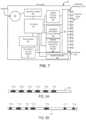
- FIG. 7is a schematic diagram of a further alternative embodiment of the stimulator of the present invention that includes neuromuscular stimulation, pain reduction, performance feedback and selective nerve ablation capabilities.
- FIGS. 8 A and 8 Bare, respectively, a plan view and detailed view of an exemplary electrode constructed in accordance with the principles of the present invention.
- FIG. 9is a schematic view of the lumbar portion of the human spine.
- FIG. 10is a sectional view showing a percutaneous method of implantation of an electrode lead suitable for use with a neuromuscular electrical stimulation system.
- FIG. 11is a sectional view showing a minimally invasive method of implantation of an electrode lead suitable for use with a neuromuscular electrical stimulation system.
- FIGS. 12 A to 12 Eare, respectively, side and perspective views of tools and implantable electrode leads suitable for use in a minimally invasive implantation method in accordance with the principles of the present invention.
- FIG. 13is a perspective view depicting deployment of an electrode lead in the medial branch of the dorsal root of a human spine.
- FIG. 14is a sectional view showing an electrode lead fixed adjacent to the medial branch of the dorsal root and having its proximal end located subcutaneously.
- FIGS. 15 A to 15 Eare, respectively, partial side views of various embodiments of lead fixation arrangements and a perspective view of the lead distal end when located in situ.
- FIGS. 16 A and 16 Bare, respectively, a partial side view of a further alternative embodiment of electrode lead and a perspective view of the electrode lead located in situ.
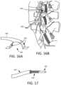
- FIG. 17is a partial sectional view of a further alternative of an electrode lead configured to facilitate removal upon the completion of NMES therapy.
- FIGS. 18 A to 18 Care respectively, perspective views depicting an electrode lead disposed in a crossing orientation, an adjacent orientation, and a trans-spinous orientation relative to a target nerve.
- Stimulator system 10comprises implantable stimulator 20 and external control system 30 .
- softwaremay be installed and run on a conventional laptop computer, and used by the patient's physician to program external control system 30 and/or to provide programming that is communicated by external control system 30 to stimulator 20 .
- external system 30may be coupled, either wirelessly or using a cable, to the physician's computer to download for review data stored on stimulator 20 , or to adjust the operational parameters of the stimulator.
- implantable stimulator 20is connected to a plurality of electrode leads.
- electrode lead 21is connected to electrode pair 22 , which is situated close to or around a peripheral nerve N where the nerve enters skeletal muscle SM, which may be a multifidus muscle.
- Electrode pair 22may deliver neuromuscular electrical stimulation (“NMES”) pulses to nerve N that induce contraction of muscle SM to effect contraction of the muscle, and restoration of neural control and rehabilitation of the muscle, as described in the aforementioned U.S. Patent Application Publication No. US2008/0228241 to Sachs.
- Electrode lead 23is illustratively disposed with electrode pair 24 adjacent or near to peripheral nerve P, such that electrical stimulation may be applied to achieve pain control in the region served by the peripheral nerves.
- Electrode lead 25illustratively includes quadripolar electrode array 26 , which is placed near spinal cord S in a manner well known to one skilled in the art to deliver Spinal Cord Stimulation therapy that reduces or blocks the transmission of pain signals to the patient's brain B.
- Implantable stimulator 20is controlled by, and optionally powered by, external control system 30 , which communicates with stimulator 20 via antenna 31 , which may comprise an inductive coil configured to transmit power and communicate information in a bidirectional manner across skin SK.
- antenna 31may comprise an inductive coil configured to transmit power and communicate information in a bidirectional manner across skin SK.
- the technology for antenna 31is well known to one skilled in the art and may include a magnet, a coil of wire, a longer range telemetry system (such as using MICS), or technology similar to a pacemaker programmer.
- coil 30may be used to transmit power only, and separate radio frequency transmitters may be provided in external control system 30 and stimulator 20 for establishing directional data communication.
- Electrode lead 27contains a plurality of electrodes 27 a - 27 d that may be used for multiple purposes, as described in detail below.
- the construction of electrode lead, the electrode design and manufacture, and connector block 29are all well known to those skilled in the art.
- an electrode leadmay contain more or fewer than four electrodes, as described in detail below with respect to FIGS. 8 A and 8 B .
- Stimulator 20includes controller 40 , telemetry system 41 coupled to antenna 42 (which may be inside or external to the hermetic housing), power supply 43 , electrode switching array 44 , system sensors 45 , and therapeutic circuitry modules 46 and 47 .
- Electrode switching array 44is selectably coupled to terminal array 48 , which is housed in connector block 29 and enables stimulator 20 to be coupled to one or more electrode leads, as shown in FIG. 1 .
- Controller 40may comprise a commercially available microcontroller unit including a programmable microprocessor, volatile memory, nonvolatile memory such as EEPROM for storing programming, and nonvolatile storage, e.g., Flash memory, for storing a log of system operational parameters and patient data. Controller 40 is coupled to telemetry system 41 that permits transmission of energy and data between implantable stimulator 20 and external control system 30 . Controller 40 also is coupled to therapeutic circuitry modules 46 and 47 that provide any of a number of complimentary therapeutic stimulation, analgesic, feedback or ablation treatment modalities as described in detail below. Controller 40 further may be coupled to electrode switching array 44 so that any set of electrodes of the electrode leads may be selectable coupled to therapeutic circuitry modules 46 and 47 . In this way, an appropriate electrode set may be chosen from the entire selection of electrodes implanted in the patient's body to achieve a desired therapeutic effect. Electrode switching array 44 preferably operates at high speed, thereby allowing successive stimulation pulses to be applied to different electrode combinations.
- Power supply 43powers the electrical components of implantable stimulator 20 , and may comprise a primary cell or battery, a secondary (rechargeable) cell or battery or a combination of both.
- power supply 43may not include a cell or battery, but instead comprise a capacitor that stores energy transmitted through the skin via a Transcutaneous Energy Transmission System (TETs), e.g., by inductive coupling.
- TETsTranscutaneous Energy Transmission System
- Stimulator 20may be programmed and/or controlled by, and may upload stored system and operational data to external control system 30 via telemetry system 41 .
- power supply 43comprises a lithium ion battery.
- System sensors 45may comprise one or more sensors that monitor operation of the systems of implantable stimulator 20 , and log data relating to system operation as well as system faults, which may be stored in a log for later readout using the external control system.
- Sensors 45may include, for example, a humidity sensor to measure moisture within housing 28 , which may provide information relating to the state of the electronic components, or a temperature sensor, e.g., for measuring battery temperature during charging to ensure safe operation of the battery.
- System sensors 45also may include a 3-axis accelerometer for determining whether the patient is active or asleep and to sense overall activity of the patient, which may be a surrogate measure for clinical parameters (e.g., more activity implies less pain), and/or a heart rate or breathing rate (minute ventilation) monitor, e.g., which may be obtained using one or more of the electrodes disposed on the electrode leads. Data from the system sensors may be logged by controller 40 and stored in nonvolatile memory for later transmission to external controller 30 via telemetry system 41 .
- system sensor 45may be used to determine the orientation of stimulator 20 , and by inference the orientation of the patient, at any time. For example, after implantation, external control system 30 may be used to take a reading from the implant, e.g., when the patient is lying prone, to calibrate the orientation of the accelerometer. If the patient is instructed to lie prone during therapy delivery, then the accelerometer may be programmed to record the orientation of the patient during stimulation, thus providing information on patient compliance.
- Implantable stimulator 20illustratively includes two therapeutic circuitry modules 46 and 47 , although more or fewer circuitry modules may be employed in a particular embodiment depending upon its intended application.
- therapeutic circuitry modules 46 and 47may be configured to provide different types of stimulation, either to induce muscle contractions or to block pain signals in afferent nerve fibers, to monitor muscle contractions induced by stimulation and vary the applied stimulation regime as needed to obtain a desired result, or to selectively and intermittently ablate nerve fibers to control pain and thereby facilitate muscle rehabilitation
- the therapeutic circuitry modulesare coupled to and controlled by controller 40 .
- implantable stimulator 50includes controller 51 , telemetry system 52 coupled to antenna 53 , power supply 54 , electrode switching array 55 , system sensors 56 , and NMES circuitry module 57 and analgesic stimulation circuitry module 58 .
- Electrode switching array 55is selectably coupled to terminal array 59 , which is coupled to the connector block 29 (see FIG. 2 ) and enables stimulator 50 to be coupled to one or more electrode leads.
- controller 51preferably includes a programmable microprocessor, volatile memory, nonvolatile memory, and nonvolatile storage, and is coupled to and controls operation of telemetry system 52 , NMES circuitry module 57 , analgesic stimulation circuitry module 58 , and electrode switching array 55 .
- Power supply 54powers the electrical components of implantable stimulator 50 , and may comprise a primary cell or battery, a secondary cell or battery, a combination of both or neither. In the latter case, power supply 54 may comprise a capacitor that stores energy transmitted through the skin via TETS.
- Stimulator 50may be programmed and/or controlled by, and may upload stored system and operational data to external control system 30 via telemetry system 52 .
- System sensors 56may comprise one or more sensors that monitor operation of stimulator 50 , as well as patient parameters, such as movement, heart rate, etc., and may log data relating to these parameters for later readout using the external control system.
- NMES circuitry moduleis configured to provide stimulatory pulses to the nerves innervating, or directly to the muscle fiber of, the multifidus or other selected muscle group to cause a predetermined series of muscle contractions in during a predetermined number of sessions to enhance muscle tone and improve neural drive in the muscle, as described in the above published application to Sachs, U.S. Patent Application Publication No. US 2008/0228241, the entirety of which is incorporated herein by reference.
- stimulator 50further includes analgesic stimulation circuitry module 58 to block or reduce pain associated with the previous injury or muscle contractions induced by the NMES therapy.
- analgesic stimulation circuitry module 58to block or reduce pain associated with the previous injury or muscle contractions induced by the NMES therapy.
- electrode pair 22is situated on the medial branch of the dorsal ramus to deliver NMES pulses that cause muscle contraction to effect restoration of neural drive to and rehabilitation of the multifidus muscle.
- Analgesic stimulation circuitry module 58may simultaneously be coupled to electrode pair 34 , via electrode lead 23 , and quad electrode 26 , via electrode lead 25 , to block or reduce pain signals generated in spinal cord S or peripheral nerve P.
- electrode pair 22also may be used, e.g., by controller 51 switching electrode switching array 55 to couple electrode pair 22 to analgesic stimulation circuitry module 58 , to deliver higher frequency stimulation to block afferent pain signals. In this manner, it is expected that NMES therapy may be provided while reducing patient discomfort and pain associated with any pre-existing injury.
- Stimulator 50 and the electrodesalso may be configured such that one set of electrodes is used to simulate the tissues on one side of the body, and another set of electrodes is used to simulate tissues on the other side of the body.
- the stimulator and electrode systemcan be configured to deliver unilateral or bilateral stimulation, or a combination of electrodes stimulating tissues in no particular geometric arrangement.
- a plurality of electrodesmay be implanted on or adjacent to the medial branch of the dorsal ramus, such that one pair delivers NMES via circuitry module 57 to effect contraction of the multifidus muscle, and another pair simultaneously or successively delivers higher frequency stimulation via circuitry module 58 to block the pain signals in the afferent fibers.
- the pairs of electrodesmay include one or more common electrodes. The timing of the different electrical stimulation delivered offers several options.
- the pain blocking stimulationmay occur simultaneously with the NMES stimulation, may be multiplexed with the NMES stimulation (i.e., time wise interleaved so that stimulation pulses are not delivered simultaneously on both electrode pairs), in an alternating manner (i.e., NMES then pain blocking and so on), or episodically, such as NMES for a period without pain blocking stimulation, and then pain blocking stimulation when the NMES is not being delivered.
- NMES stimulationis applied to the multifidus in sessions, typically one hour per day over a period of a few weeks.
- Such a regimeis similar to conventional strength training by physical exercise which typically follows a similar time course.
- stimulator 50may be used to apply SCS therapy to block or dampen the pain signals which may arise from the NMES exercise regime.
- SCS therapymay be used to block or dampen the pain signals which may arise from the NMES exercise regime.
- the desired therapeutic effect of restoration of neural drive and rehabilitation of the multifidusmay occur without substantial pain or discomfort.
- stimulator 50offers the capability to apply SCS therapy at the same time as NMES rehabilitation therapy for the multifidus.
- the patientmay have access to external control system 30 , and can thus activate implantable stimulator 50 in accordance with a rehabilitation plan developed jointly with his or her physician.
- controller 51may be programmed to provide a delay of specified duration between activation of the stimulator and initiation of the stimulation pulses. This delay allows the patient to assume a comfortable position before the stimulation is applied, e.g., by lying prone.
- the external control systemalso may include a multi-functional user interface, including a range of patient operated inputs (e.g., buttons, knobs, touch screen, etc.) that allows activation or suspension of different types of stimulation.
- implantable stimulator 50may be programmed to ramp up and ramp down the strength and duration of the stimulation pulses. This can be done in at least one of two manners. In the first manner, the stimulation pulse intensity is increased gradually (e.g., over 0.5 to 1 second) to a programmed maximum value to elicit the desired muscle contraction and then ramped down slowly. In this way, the muscle contraction has a smooth on and off sensation for the patient. In the second manner, the therapeutic dose (i.e., the number of contractions of a therapy period) are programmed to increase gradually until the desired level is achieved and then decrease gradually to zero, in much the same way that a good muscle strength training regime provides a stretching or warm-up phase and cool-down phase.
- the therapeutic dosei.e., the number of contractions of a therapy period
- stimulator 50via either input to the external control system or at a pre-determined time, and following the stimulation delay (if any), ramps us the stimulation amplitude from a low level (e.g., beginning at zero) to a pre-determined maximum level over a pre-determined period of time. Likewise, upon conclusion of the stimulation therapy period, stimulator 50 ramps the amplitude down from the pre-determined maximum level to a low level. It is expected that this embodiment, which provides a gradual increase and decrease of stimulation intensity, will provide a more comfortable experience for some patients.
- implantable stimulator 50preferably contains nonvolatile memory for storage, and is programmed to log data during the therapy session, along with internal parameters of the device. Such data logging may also record data from system sensors 56 , which may be downloaded from stimulator 50 using the external control system, to provide an indication of the effectiveness of the therapy. For example, if the sensors include a three axis accelerometer, then a patient's overall activity level on an hourly, daily, or weekly basis may be logged, for example, by recording an integral of all accelerometer measurements.
- the sensorsalso may include circuitry for determining heart rate, and such circuitry may be used to record the patient's maximum heart rate as a measure of overall activity.
- the stimulator 50is implanted subcutaneously, and system sensors 55 may be used to record and log baseline (i.e., pre-therapy) patient parameters such as total activity and maximum heart rate.
- baselinei.e., pre-therapy
- the therapythen is enabled, and the data logging may be used to assess progress of the therapy and the patient's change in status. For example, if the accelerometer shows increased overall activity, this would indicate that the pain, which was previously inhibiting activity, had been ameliorated.
- Such datamay be used by the physician to adjust the therapy by adjusting the programming of stimulator 50 using external control system 30 , and/or such information may be provided to the patient as encouraging feedback.
- the implantable stimulatorprovides a NMES stimulator therapy and further has the capability to monitor the progress of the therapy and to revise the therapy regime to reflect changes in the muscle characteristics resulting from the therapy.
- revisionmay be made by way of a physician periodically reprogramming the NMES parameters using external control system 30 , or alternatively the NMES stimulation parameters may be adjusted dynamically and automatically modified to keep the muscle contraction at a certain predetermined efficacious and tolerable level.
- stimulator 60may provide a closed loop feedback system, in which the system instantaneously responds to physiological changes affecting the stimulation characteristics of the muscle.
- inventive technologyis to improve stability of the spine, it also may be advantageously applied in other areas of muscle rehabilitation, e.g.:
- the implantable NMES stimulator described in the above-incorporated Sachs applicationdiscusses that the parameters for electrical stimulation may be programmed into the stimulator following testing by the physician of stimulation thresholds. Therapy parameters such as duration, frequency and strength of contraction also may be programmed into the stimulator according to the patient's needs, and the stage of therapy delivery. In some cases it is expected that the programmed parameters may need to be changed, for example during the course of the therapy program as the muscle becomes rehabilitated.
- Stimulator 60 of FIG. 5is designed to improve the NMES performance and reduce the need for frequent reprogramming by monitoring muscle performance during therapy, and adjusting the stimulation parameters accordingly. More specifically, implantable stimulator 60 includes controller 61 , telemetry system 62 coupled to antenna 63 , power supply 64 , electrode switching array 65 , system sensors 66 , and NMES circuitry module 67 and muscle performance monitoring circuitry module 68 . Electrode switching array 65 is selectably coupled to terminal array 69 , which is coupled to the connector block 29 (see FIG. 2 ) and enables stimulator 60 to be coupled to one or more electrode leads. Electrode switching array 65 also may include connection 69 a to the housing of stimulator 60 , so that the housing functions as an electrode.
- Controller 61preferably includes a programmable microprocessor, volatile memory, nonvolatile memory, and nonvolatile storage, and is coupled to and controls operation of telemetry system 62 , NMES circuitry module 67 , muscle performance monitoring circuitry module 68 , and electrode switching array 65 .
- Power supply 64powers the electrical components of implantable stimulator 60 , and may comprise a primary cell or battery, a secondary cell or battery, a combination of both or neither. In the latter case, power supply 64 may comprise or include a capacitor that stores energy transmitted through the skin via a Transcutaneous Energy Transmission System (“TETS”).
- TETSTranscutaneous Energy Transmission System
- Stimulator 60may be programmed and/or controlled by, and may upload stored system and operational data to external control system 30 via telemetry system 62 .
- System sensors 66may comprise one or more sensors that monitor operation of stimulator 60 , as well as patient parameters, such as movement, heart rate, etc., and may log data relating to these parameters for later readout using the external control system.
- stimulator 60further comprises muscle performance monitoring circuitry module 68 coupled to controller, and designed to monitor one or more parameters of muscle performance.
- the measured parametersmay be used to automatically modify the therapy delivered by NMES circuitry module 67 , and/or to provide stored and telemetered information via telemetry system 62 and external control system 30 that enable the physician to modify the parameters.
- muscle performance monitoring circuitry module 68may be coupled through electrode switching array 65 to selected electrodes coupled to terminal array 69 to measure electrical parameters of the tissue, such as impedance, or evoked potential from the stimulation.
- Circuitry module 68may in addition be coupled to system sensor 66 , for example, to obtain data from an accelerometer or other movement transducer, and/or temperature or pressure.
- Circuitry module 68also may be configured to receive inputs from other types of body sensors such as are known in the art, including those monitoring chemical properties (e.g., pH sensor, etc.).
- Circuitry module 68preferably includes at least one listening amplifier configured for electromyography (EMG).
- EMGis an electrical signal produced by muscle when it contracts, and the strength (power) of the EMG is an indicator of strength of muscle contraction.
- Configuration of an amplifier for measurement of EMGe.g., gain, frequency response, impedance, etc., is well known to those skilled in the art. As described in Stokes, Ian A F, Sharon M Henry, and Richard M Single, “Surface EMG electrodes do not accurately record from lumbar multifidus muscles,” Clinical Biomechanics (Bristol, Avon) 18, no.
- the implantable electrode leads used with stimulator 60advantageously are expected to provide a useful EMG signal.
- circuitry modules 67 and 68may be configured to perform impedance measurements, in a manner similar to that described in U.S. Pat. No. 6,406,421 B1 to Grandjean et al.
- an electrical impedance measurementmay be performed by injecting a current through one pair of electrodes, and measuring voltage through a different pair of electrodes disposed approximately along the same geometric path. See, e.g., Rutkove, S. B., “Electrical impedance myography: Background, current state, and future directions”, Muscle & Nerve 40 , No. 6 (December 2009): 936-46.
- a first pair of electrodesconsisting of the stimulator housing (via connection 69 a ) and one or more of electrodes disposed on an electrode lead may be used to inject current into the tissue (e.g., from NMES circuitry module 67 ), while voltage is measured by circuitry module between the stimulator housing and a different set of one or more of electrodes on the electrode leads.
- the same set of electrodesmay be used for both injecting current and measuring the resulting voltage.
- the foregoing impedance measurementsmay be of direct current (DC) or alternating current (AC). With AC impedance measurement, additional useful information may be obtained such as phase, frequency spectrum, and changes in parameters.
- the electrical impedance so measuredis an indication of the tissue volume and tissue organization (anisotropy) between the measurement electrodes, as reported in Garmirian et al., “Discriminating neurogenic from myopathic disease via measurement of muscle anisotropy”, Muscle Nerve, 2009 January; 39 (1): 16-24. Scc also, Miyatani, M., et al., “Validity of estimating limb muscle volume by bioelectrical impedance”, J. Applied Physio. (Bethesda, Md. 1985) 91, no.
- circuitry module 68may include or be coupled to a transducer that senses mechanical motion, such as vibration, acceleration or deflection, and may include piezoelectric polymers (e.g., PVDF) placed on a lead. The signal from such a transducer provides a surrogate measure of muscle contraction.
- circuitry module 68may include or be coupled to a transducer that senses pressure, such as a MEMS pressure sensors disposed on a lead, and which thus provides a surrogate measure of muscle contraction.
- stimulator 60is configured to sense EMG from more than one muscle, using multiple electrode leads or multiple electrodes on a single lead that passes through more than one muscle.
- the listening amplifier of circuitry module 68is multiplexed to listen for EMGs from more than one muscle.
- circuitry module 68may include multiple listening amplifiers that are arranged to simultaneously listen to EMGs from more than one muscle. It is well-known, for example from Jaap van Dicen et al., “Trunk Muscle Recruitment Patterns,” Spine Vol. 28, Number 8 pg. 834-841, that the relative timing and amplitude of EMGs in trunk muscles during the performance of specific tasks is different between healthy individuals and patients experiencing low back pain due to spinal instability.
- EMGsIn patients with spinal instability, recruitment patterns of the trunk muscles may be altered to compensate for the lack of spinal stability.
- the amplitude and timing of EMGs measured from multiple trunk musclestherefore may be used to diagnose the presence and degree of spinal instability, as well as the change of spinal instability during a course of therapy.
- the EMG datamay be used to automatically modify treatment parameters, or such data may be stored for later review by the physician to assist in diagnosis and revision of the therapy parameters.
- muscle performance monitoring circuitry module 68is configured to measure muscle contraction induced by NMES circuitry module 67 , and to modify the therapeutic parameters as muscle performance changes.
- the initial therapeutic parameterssuch as dose and duration of therapy session, are established and programmed into stimulator 60 using external control system 30 .
- muscle performancemay be monitored continuously or periodically using circuitry module 68 .
- circuitry module 68may instruct controller 61 to modify the parameters for subsequent NMES therapy sessions. For example, if the monitoring parameters reveal that the muscle mass has increased, indicative of muscle rehabilitation, or contractility has decreased, then the therapy dose may be automatically reduced some pre-determined amount as previously programmed by the physician.
- muscle performancemay be used to inhibit muscle contraction.
- painis caused by spasm of certain muscles in the back.
- Such spasmis accompanied by continuous increase in EMG activity.
- NMES stimulationmay be used to inhibit muscle contraction by configuring the listening amplifier of circuitry module 68 to continuously or periodically measure EMG. If the EMG satisfies conditions indicating that muscle spasm has occurred, then NMES circuitry module is directed by controller 61 to apply stimulation to the nerve innervating the muscle in spasm to block conduction of signals from the nervous system which cause the muscle spasm, thereby preventing spasm.
- the stimulation provided by NMES circuitry modulemay be inhibited from time to time to allow circuitry module 68 to assess from the EMG signal if the muscle is still in spasm; if spasm has ceased, then application stimulation by NMES circuitry module 67 is terminated.
- muscle performance monitoring circuitry module 68may be configured to measure a combination of EMG and tissue impedance to confirm that a muscle is in spasm, thereby improving the safety and reliability of the measurement. Muscle performance monitoring circuitry module 68 also may be used to track changes in activity and health of the muscle in response to neural activity. In other words, the amount of muscle contraction as determined by impedance measurement of tissue volume may be correlated to the amount of electrical activity in the muscle as determined by EMG. Further still, the electrodes and muscle performance monitoring circuitry module 68 may be configured to record electrical signals from the nerves as well as the muscle, such that a measurement of the EMG (and/or tissue volume) in response to neural activity may be used as an indication of the health of the muscle.
- Muscle performance monitoring circuitry module 68also may employ measurement of the change in muscle mass in response to NMES of the nerve to adjust the electrical stimulation parameters.
- an empirically derived transfer functionmay be determined that relates electrical stimulation parameters, such as current, pulse width, frequency and duration, to the strength of contraction of the muscle. Over time, this transfer function may change, for example, as a result of electrode changes from movement or tissue ingrowth.
- the strength of muscle contractionmay be used to automatically adjust the electrical parameters of the NMES stimulation provided by circuitry module 67 to achieve a desired muscle contraction.
- an implantable RF ablation deviceis described.
- a primary application of the inventive technologyis pain reduction in connection with improving stability of the spine, the inventive technology may be advantageously applied in other areas, for example:
- RF ablationThe field of RF ablation is well developed, and parameters suitable for ablating nerve fibers and other tissues, such as RF energy, and attendant issues is well known to those of ordinary skill in the art. See, e.g., Gazelle et al “Tumor ablation with radio-frequency energy”, Radiology, December 2000: 217(3): 633-46 and Haemerrich et al, “Thermal tumour ablation: devices, clinical applications and future directions”, Int. J. Hyperthermia, 2005 December; 21(8):755-60. To the inventors' knowledge, however, no one has suggested an RF ablation device that is configured to be chronically implanted and capable of performing repeated RF ablation.
- implantable device 70is described, which is intended for chronic implantation to perform serial RF ablations in scenarios where it is necessary to repeat RF ablation of tissue in a particular region of the body after certain periods of time.
- the components of device 70correspond closely to those described above with respect to the embodiment of FIG. 3 , and includes controller 71 , telemetry system 72 coupled to antenna 73 , power supply 74 , electrode switching array 75 , system sensors 76 , and terminal array 77 .
- electrode switching array 75is selectably coupled to terminal array 77 , which is coupled to the connector block 29 (see FIG. 2 ) that accepts one or more implantable electrode leads.
- Electrode switching array 75also may include connection 77 a to the housing of device 70 , so that the housing functions as an electrode.
- device 70further comprises RF ablation circuitry module 78 , as further described below.
- Controller 71preferably includes a programmable microprocessor, volatile memory, nonvolatile memory, and nonvolatile storage, and is coupled to and controls operation of telemetry system 72 , electrode switching array 75 and RF ablation circuitry module 78 .
- Power supply 74powers the electrical components of device 70 , and may comprise a primary cell or battery, a secondary cell or battery, a combination of both, or neither. In the latter case, power supply 74 may comprise or include a capacitor (such as a super capacitor of technology known to those skilled in the art) that stores energy transmitted through the skin via TETS.
- Device 70may be programmed and/or controlled by, and may upload stored system and operational data to external control system 30 via telemetry system 72 .
- System sensors 76may comprise one or more sensors that monitor operation of device 70 , as well as patient parameters, such as tissue impedance, and may log data relating to these parameters for later readout using the external control system.
- device 70further comprises RF ablation circuitry module 78 coupled to controller, and designed to periodically ablate tissue or nerve fibers using RF energy.
- controller 71may be configured to control operation of the telemetry system 72 to receive energy wirelessly from external control system 30 and store that energy in power supply 74 , and may be configured to communicate the amplitude of received power back to the external control system via telemetry system 72 or via modulation of the impedance of the antenna 73 .
- device 70may not include battery or capacitor, but instead may be arranged so that it is energized only when in communication with the external control system.
- Expected energy requirements for the RF ablation circuitry moduleare in a range of about 1-40 watts, depending upon the intended application.
- TETS systems with this power capacityare well known to those skilled in the art and have been used, for example, with artificial hearts or Left Ventricular Assist Devices (LVADs).
- LVADsLeft Ventricular Assist Devices
- the physical volume and other requirements of a high power TETS systemmay preclude its use in applications where the available surgical locations are limited.
- the TETS systemmay be of lower power capacity than the requirements of the RF generator, and device 70 may include an energy storage element, such as a super capacitor or low impedance secondary (rechargeable) cell, for powering RF ablation circuitry module 78 .
- the TETSmay operate continuously, such that a signal is generated when there is adequate energy stored in the implantable device to deliver the RF ablation energy at the desired power and for the desired time.
- a TETS system capable of transferring 1 Wmay be used to supply RF energy delivery of 5 W with 20% duty cycle.
- Telemetry system 72enables communications between the external control system and device 70 , allowing the implantable device to receive device and RF ablation operating parameters, as well as communicate logged information such as impedance between electrodes, temperature data and battery status to the external control system.
- Telemetry system 71also may provide programming to controller 71 to reconfigure the operative electrodes through which ablation energy is supplied using electrode switching array 75 , thereby allowing any electrode of a plurality of electrodes to be configured as a cathode, an anode or unconnected.
- the housing of device 70also may be configured as an electrode via connection 77 a of terminal array 77 .
- System sensors 76advantageously may be used to monitor the temperature of the tissue near the electrodes thru which energy for ablation is delivered. Typical tissue temperatures for RF ablation range from 50C to 130C, depending on the type of tissue being ablated and the time allocated to the ablation. System sensors 76 may comprise, e.g., temperature sensors disposed within the device housing, or alternatively may measure the temperature of the connection to the electrode leads, and use that data to infer or predict the tissue temperature. Temperature sensors may also be incorporated into the leads and placed closer to the tissue targeted for ablation.
- System sensors 76may be used in a passive (measuring) mode, or alternatively may comprise part of a feedback control system that continually or intermittently adjusts power delivered by the RF ablation circuitry module so that the temperature of the ablated tissue is maintained between desired limits for safety and efficacy.
- Implantable stimulator 80corresponds to stimulator 20 of FIG. 1 , and is programmed and controlled and/or powered by external control system 30 .
- Stimulator 80is intended for use, for example, in a stimulator that provides NMES stimulation, analgesic stimulation to block or reduce afferent pain signals in a nerve, and permits periodic nerve ablation (such as rhizotomy).
- stimulator 80includes muscle performance monitoring circuitry that supports testing of nerve fibers prior to rhizotomy, which to guide proper selection of the ablation electrodes.
- Stimulator 80 of FIG. 7includes controller 81 , telemetry system 82 coupled to antenna 83 , power supply 84 , electrode switching array 85 , system sensors 86 , terminal array 87 , NMES circuitry module 88 , analgesic stimulation circuitry module 89 , muscle performance monitoring circuitry module 90 , and RF ablation circuitry module 91 .
- electrode switching array 85is selectably coupled to terminal array 87 under the control of controller 81 , and enables any one or more of the therapeutic circuitry modules of stimulator 80 to be selectably coupled to selected electrodes of one or more electrode leads.
- Electrode switching array 85also may include connection 87 a to the housing of stimulator 80 , so that the housing also may serve as an electrode.
- Controller 81preferably includes a programmable microprocessor, volatile memory, nonvolatile memory, and nonvolatile storage, and is coupled to and controls operation of telemetry system 82 , electrode switching array 85 , NMES circuitry module 88 , analgesic stimulation circuitry module 89 , muscle performance monitoring circuitry module 90 , and RF ablation circuitry module 91 .
- Power supply 84powers the electrical components of implantable stimulator 80 , and may comprise a primary cell or battery, a secondary cell or battery, a combination of both, or neither, as discussed above.
- Stimulator 80may be programmed and/or controlled by, and may upload stored system and operational data to external control system 30 via telemetry system 82 .
- System sensors 86may comprise one or more sensors that monitor operation of stimulator 80 , as well as various patient parameters as discussed above.
- stimulator 80further comprises NMES circuitry module 88 and analgesic stimulation circuitry module 89 , as described above with respect to the embodiment of FIG. 4 , muscle performance monitoring circuitry module 90 as described above with respect to the embodiment of FIG. 5 , and RF ablation circuitry module 91 as described above with respect to the embodiment of FIG. 6 .
- NMES circuitry module 88 and analgesic stimulation circuitry module 89as described above with respect to the embodiment of FIG. 4
- muscle performance monitoring circuitry module 90as described above with respect to the embodiment of FIG. 5
- RF ablation circuitry module 91as described above with respect to the embodiment of FIG. 6 .
- the muscle performance monitoring circuitry module 90may measure the progress of the therapy and adjust the NMES stimulation parameters or circumvent spasm. In addition, depending upon the patient's reported condition and measurement data provided by the muscle performance monitoring circuitry module 90 , the physician may periodically activate RF ablation circuitry module 91 to denervate selected nerve fibers.
- electrode leadsconfigured to provide NMES stimulation to a nerve to cause muscle contraction; to stimulate a nerve to inhibit pain signals from propagating to the brain; to stimulate a nerve to inhibit motor nerve signals thereby reducing or stopping contraction of a muscle (e.g., in spasm); to record electrical signals such as electromyography or tissue impedance; or for performing in situ RF ablation are now described.
- electrode lead 100 carrying electrodes 101 a to 101 fis described.
- the number of electrodesmay be as few as 1 and as many as may be realistically placed within the target anatomical space.
- Electrode configurations commonly used in the artinclude 1 (for unipolar stimulation), 2, 4 (peripheral nerve stimulation), 8, 16 (spinal cord stimulators) or up to 22 electrodes (cochlear implants).
- distal-most electrode 101 awill be referred to as electrode #1
- electrode 101 bwill be electrode #2 and so on moving proximally along lead 100 up to the total number of electrodes.
- electrode lead 100When employed with an implantable stimulator as described herein that provides multiple independent current outputs, electrode lead 100 is capable of delivering multiple therapies simultaneously, in an overlaid fashion or staggered. Electrodes 101 a to 101 f may be sized and positioned relative to each other to allow for generation of a voltage field tailored to the specific type of stimulation, sensing or ablation desired for the given therapies.
- electrode lead 100is placed parallel to a target nerve in a caudal to cranial orientation (with the cranial direction being the direction tending towards afferent neural activity). Then so positioned, electrodes 1 and 2, which are most cranial, may be sized and spaced to allow for optimal blocking of afferent pain signals being transmitted along the nerve (for example the pain signals being carried from the facet joint along the medial branch). More caudally, electrodes 3 and 4 may be sized and spaced to allow for optimal recruitment of large fiber motor neurons. Because the action potentials required for activation of a muscle travel efferently, these potentials are not blocked by the more cranial blocking action of electrodes 1 and 2.
- electrodes 5 and 6 placed most caudallymay be sized and positioned for sensing and recording of muscle recruitment through capturing the EMG signal of the muscle, which may be processed, for example, by the muscle performance monitoring circuitry module as described above with respect to the embodiment of FIG. 4 .
- Such an arrangementtherefore allows for simultaneous blocking of pain arising from the facet joint, stimulation of the motor fibers of the nerve eliciting muscle contraction, and sensing of the elicited response (which would enable a closed loop system, improving device longevity and recruitment efficiency) without any of the stimulation pulses negatively impacting the performance of the others.
- Electrode lead 110 carrying electrodes 111 a to 111 gis described.
- Distal-most electrode 111 a againwill be referred to as electrode #1
- electrode 111 bwill be electrode #2 and so on moving proximally along lead 111 .
- a blocking action of electrodes 1 and 2may be used to mute the sensory perception of stimulation.
- NMES stimulation therapy of the motor fibers in patientsmay be achieved where the patients would otherwise not tolerate the stimulation because of the resulting bi-directional action potential generated by neural stimulation.
- electrodes 5 and 6which will likely be placed intramuscularly, to record the volume EMG signal in the muscle.
- Changes in this signal over timemay provide an indication of the degree to which motor control has been compromised due to injury.
- the datamay be used as a positive indicator that additional therapy may be required to maintain spinal stability.
- the L3, L4 and L5 portions of the lumbar spineare described.
- the dorsal root D, dorsal ramus medial branch MB, intermediate branch I, and a representation of multifidus fascicular innervations A, B, and Care identified.
- Also shown in FIG. 9are the superior articular process SAP, the spinous process SP, pedicle P, inferior anterior process IAP, and the transverse process TP.
- needle 115is delivered transcutaneously and transmuscularly, to the vertebra. More specifically, lead 120 then is attached to the pedicle of the vertebra or within the fascia near the medial branch of the dorsal ramus nerve. Alternatively lead 120 may be deployed into the multifidus muscle M, in the vicinity of the medial branch MB nerve, and proximal to the fascicle branches A, B and C. When deployed in this location, the NMES stimulation is expected to provide recruitment of the nerve and the multifidus fascicles.
- a minimally invasive approach for implanting an electrode lead for NMES stimulationis described.
- a separationis formed along natural tissue planes, for example, at the plane between multifidus muscle M and longissimus muscle LT, thereby providing access to the medial branch MB.
- the tissue separationis accomplished using cannula 130 having a blunt dissection tool and an endoscope, as described below.
- cannula 130comprises of one or more tubes 140 and 140 ′, transparent distal blunt dissection cone 160 , through which tissue can be visualized, and side port 180 for delivery of fluids and application of vacuum.
- Tube 140houses an endoscope for visualization and is fitted with tip 150 configured for blunt dissection.
- Tube 140 ′remains in position when tube 140 is removed for deployment of lead 160 , depicted in FIG. 12 B .
- the cannula for blunt dissectionmay have a predetermined shape suitable separating the natural tissue plane between the multifidus and longissimus muscles or be malleable such that the clinician can customize the curvature of the cannula as needed for specific patient anatomy.
- electrode lead 160comprises of distal end 170 having two or more electrodes 180 for stimulation, and proximal end 190 for connection to an implantable stimulator or subcutaneous receiver.
- Distal end 170may include two or more electrodes made of stainless steel, platinum-iridium or other suitably biocompatible material for delivery of electrical stimulation pulses.
- the electrodesmay be cylindrical, planar or other suitable geometry, have at least 5 sq mm of surface area and may vary in length, width and diameter depending on lead body.
- Electrode lead 160further may include an internal lumen that extends from proximal end 190 to distal tip 170 , through which stylet 200 may be inserted.
- Stylet 200may have a flat-blade tip suitable for engaging the proximal section of fixation screw 210 , such that stylet 200 may be used to advance or withdraw the fixation screw by rotating the stylet. As depicted in FIG. 12 C , a portion of the surface of electrode 180 may be masked with non-conductive insulating material 220 , and thus used to orient the lead and direct stimulation current toward a selected nerve, e.g., the medial branch of the dorsal ramus nerve.
- a selected nervee.g., the medial branch of the dorsal ramus nerve.
- Electrode lead 230has a ribbon-like cross section to facilitate passage between muscle planes, improve flex fatigue in one axis, provide better stability in vivo, and provide better guidance of stimulation current to targeted nerve(s).
- Proximal end 240has an equal number of terminations as there are electrodes 250 on distal end 260 , with one independent conductor for each electrode. Each termination may be connected to an output of the NMES stimulator, and each conductor coupling the terminations on the proximal end to electrodes 250 may comprise a cable or thin film structure. Each individual conductor may be comprised of a cable or coiled structure.
- Electrode lead 230also may include fixation screw 270 that is driven using removable stylet 280 .
- Cannula 300is configured having a first lumen that accepts endoscope 320 and a second lumen that accepts minimally invasive surgical instrument 340 , with which lead 160 or 230 may be deployed under endoscopic visualization.
- Cannula 300is passed through a small incision made anterior to the natural tissue plane between the multifidus muscle M and the longissimus muscle LT. Under endoscopic visualization, a tract then is created using blunt dissection, during which tissue planes, blood vessels, ligaments, and fascia may be directly visualized using endoscope 320 .
- the electrode leadUpon reaching the vertebra and visualizing superior articular process SAP, and more particularly pedicle P and medial branch MB of dorsal root D, as depicted in FIG. 13 , the electrode lead is deployed.
- the leadmay be deployed under endoscopic visualization and fixed in bone, ligament, or fascia using a fixation feature, such as corkscrew 210 above, or a suitable hook, barb or tine, as described below.
- FIG. 14shows one possible lead configuration after implantation, in which the proximal end of an electrode lead 350 is located subcutaneously.
- Proximal end 360 of electrode lead 350may include a receiver for wirelessly receiving energy using a transcutaneous energy transmission system (TETS), which are known in the art, or may be coupled to an implantable NMES stimulator, as described in the above-incorporated patent publication to Sachs.
- TETStranscutaneous energy transmission system
- Distal tip 370 of electrode lead 350may be fixed in the pedicle adjacent medial branch MB, such that lead 350 lies between multifidus muscle M and longissimus muscle LT.
- Stimulation energy from an external pulse generatormay be transmitted to the subcutaneous receiver; alternatively electrode lead 350 could be tunneled subcutaneously to an implantable pulse generator.
- an electrode lead suitable for NMES therapypreferably includes a fixation feature that permits the distal end of the lead to be attached to the mamillary process, the anterior process, the pedicle, or locations in between.
- FIG. 15 Ashows electrode lead 400 having cork-screw fixation feature 410 , similar to that described with respect to the embodiment of FIG. 12 B , suitable for installation in a crossing orientation. In this manner of attachment, as shown in FIG. 15 E , the lead crosses over the nerve.
- Lead 400also may include bands 420 of polyester or other material that promotes tissue ingrowth to fasten the lead in position.
- FIG. 15 Bshows electrode lead 430 , including cork-screw fixation element 440 that exits through the side wall of the electrode lead.
- Lead 430is particularly well-suited for installation in an adjacent orientation, in which the lead lies adjacent to the target nerve or muscle.
- FIG. 15 Cdepicts electrode lead 450 that includes barbs 460 that may be embedded into the tissue or bone adjacent to the target nerve or tissue to retain the distal end of the electrode lead in position.
- FIG. 15 Cdepicts electrode lead 470 having barb 480 that may be extended from within a lumen of the electrode lead, e.g., using a stylet, to drive the barb into the tissue or bone adjacent to the target nerve or tissue to retain the lead in position.
- Electrode lead 490includes a distal region that forms bracket 500 having electrodes 510 , such that bracket 500 may be disposed around a target nerve or muscle to prevent movement of the distal end of the lead, as illustrated in FIG. 16 B .
- tissue fixationinclude both resorbable and non-resorbable tines, and tissue hooks. Many of the same fixation features are suitable for engaging tissues like muscle, fascia or ligaments. The foregoing fixation features are intended to orient stimulation electrodes in adjacent or crossing relationships relative to the medial branch nerve. Still other means of stabilization include polymer and tissue matrix materials intended for the promotion of tissue ingrowth. Such features may take the form of polymer and metal surface treatments to create pores of appropriate diameter, polymers fabricated with pores of appropriate aperture size, and harvested or cultured cellular and extra cellular matrices that may be applied to the lead where fixation is desired.
- electrode leads 520may include materials and/or material treatments that resist tissue ingrowth, e.g., along regions of the electrode lead intended to lie between or within tissue planes that move. Materials and treatments that have pores suitable for reducing the amount and ability of tissue to become attached to the lead will make removal of the leads simpler once therapy has completed.
- electrode lead 520comprises flexible metal jacket 530 and outer polymeric cover 540 .
- Flexible metal jacket 530protects the electrode lead against trauma such as needle sticks post-implant, while polymeric cover 540 prevents tissue ingrowth, significantly improving lead removability upon completion of therapy.
- FIGS. 18 A to 18 Cvarious arrangements for disposing the electrodes of an electrode lead in relation to medial branch MB are illustrated.
- lead 550is placed so that electrodes 560 are disposed in a crossing orientation with respect to medial branch MB.
- lead 550is placed so that electrodes 560 are disposed in an adjacent orientation with respect to medial branch MB, such that lead 550 is disposed substantially parallel to a length of the nerve.
- lead 550is implanted as a trans-spinous process implant. In this case, a pathway is bored through spinous process SP to the area of lamina LA, exiting near medial branch MB.
- FIGS. 18 A to 18 Cdepict unilateral deployment of stimulation leads to the right or left of the dorsal ramus medial branch. It should of course be understood that it is within the scope of this invention to provide bilateral stimulation training of the multifidus muscle. It further should be understood that multiple levels, for example the medial branch of the dorsal ramus L3, L4 and L5, may be implanted with leads to train the multifidus muscle to its fullest extent.
- stimulation of one or more other anatomical structuressuch as ligaments, tendons, or nerves of other than spine stabilization muscles (e.g., transverse abdominus, psoas, interspinales, longissimus, ileocostalis, intertransversus, quadratus) may comprise adequate therapy.
- other anatomical structuressuch as ligaments, tendons, or nerves of other than spine stabilization muscles (e.g., transverse abdominus, psoas, interspinales, longissimus, ileocostalis, intertransversus, quadratus) may comprise adequate therapy.
Landscapes
- Health & Medical Sciences (AREA)
- Life Sciences & Earth Sciences (AREA)
- Engineering & Computer Science (AREA)
- Veterinary Medicine (AREA)
- Animal Behavior & Ethology (AREA)
- General Health & Medical Sciences (AREA)
- Public Health (AREA)
- Biomedical Technology (AREA)
- Nuclear Medicine, Radiotherapy & Molecular Imaging (AREA)
- Radiology & Medical Imaging (AREA)
- Heart & Thoracic Surgery (AREA)
- Surgery (AREA)
- Cardiology (AREA)
- Physics & Mathematics (AREA)
- Molecular Biology (AREA)
- Medical Informatics (AREA)
- Neurology (AREA)
- Neurosurgery (AREA)
- Biophysics (AREA)
- Pathology (AREA)
- Orthopedic Medicine & Surgery (AREA)
- Otolaryngology (AREA)
- Pain & Pain Management (AREA)
- Plasma & Fusion (AREA)
- Physiology (AREA)
- Pulmonology (AREA)
- Dentistry (AREA)
- Oral & Maxillofacial Surgery (AREA)
- Electrotherapy Devices (AREA)
Abstract
Description
- For neuromuscular electrical stimulation (NMES):
- Bipolar electrode pairs
- Biphasic rectangular charge balanced
- 0.5-500 ms pulse width (adjustable to control intensity)
- 10-30 Hz (to achieve tetanic contraction)
- Constant current, <50 mA (<50V)
- For PENS type stimulation:
- Multiple bipolar electrode system
- Biphasic pulses
- 20-40 Hz (including possibility of variable frequency over time of application of therapy)
- Constant current (typically 5-20 mA)
- For Spinal Cord Stimulation:
- Multiple electrode configurations
- Biphasic rectangular charge balanced
- Typically 500 μsec pulse width
- Current control (preferred) or voltage control, typically up to 10 mA into a 1KΩ load
- For Radio Frequency Ablation:
- 450-500 KHz
- RF heating energy
- For neuromuscular electrical stimulation (NMES):
- Restoration of function of leg muscles to allow standing and walking in paraplegic patients (referred to as Functional Electrical Stimulation (FES));
- Rehabilitation of injured or weakened muscles following surgery or correction of osteoarthritis, such as rehabilitation of the quadriceps after knee surgery;
- Restoration of neural drive and rehabilitation of muscles that are part of the stabilizing system in the back, including the lumbar multifidus;
- Providing stimulation to effect breathing (diaphragm and/or intercostal muscles); and
- Providing mechanical muscle power to perform a bodily function, for example, as in cardiomyoplasty.
- RF rhizotomy, in which a sensory nerve is ablated to prevent sensory signals (e.g., pain) from reaching the brain, such as rhizotomy of the medial branch of the dorsal ramus in patients with facet joint pain;
- RF ablation of unresectable tumors located in the liver, brain, musculoskeletal system, thyroid and parathyroid glands, pancreas, kidney, lung, and breast, in which it is difficult to achieve complete tumor necrosis, leading to recurrence of the tumors and necessitating repeated RF ablation; and
- Treatment of tumors in which the root of the tumor is located in tissue that is considered too risky for surgical intervention, such as tumors with roots in the digestive tract, uterine wall or certain esophageal tumors, and for which regular repeat surgery is required to remove new growths.
Claims (20)
Priority Applications (3)
| Application Number | Priority Date | Filing Date | Title |
|---|---|---|---|
| US18/046,835US12097365B2 (en) | 2010-03-11 | 2022-10-14 | Electrical stimulator for the treatment of back pain and methods of use |
| US18/614,512US20240341808A1 (en) | 2012-06-13 | 2024-03-22 | Systems and methods for enhanced implantation of electrode leads between tissue layers |
| US18/884,063US20250001164A1 (en) | 2010-03-11 | 2024-09-12 | Electrical stimulator for the treatment of back pain and methods of use |
Applications Claiming Priority (18)
| Application Number | Priority Date | Filing Date | Title |
|---|---|---|---|
| US33994310P | 2010-03-11 | 2010-03-11 | |
| US33995710P | 2010-03-11 | 2010-03-11 | |
| US13/045,421US9248278B2 (en) | 2010-03-11 | 2011-03-10 | Modular stimulator for treatment of back pain, implantable RF ablation system and methods of use |
| US13/045,435US10925637B2 (en) | 2010-03-11 | 2011-03-10 | Methods of implanting electrode leads for use with implantable neuromuscular electrical stimulator |
| US201261659334P | 2012-06-13 | 2012-06-13 | |
| US13/797,100US9999763B2 (en) | 2012-06-13 | 2013-03-12 | Apparatus and methods for anchoring electrode leads adjacent to nervous tissue |
| US14/061,614US9072897B2 (en) | 2007-03-09 | 2013-10-23 | Systems and methods for restoring muscle function to the lumbar spine |
| US14/792,430US9474906B2 (en) | 2007-03-09 | 2015-07-06 | Systems and methods for restoring muscle function to the lumbar spine |
| US14/849,478US9861811B2 (en) | 2010-03-11 | 2015-09-09 | Electrical stimulator for treatment of back pain and methods of use |
| US15/202,435US9950159B2 (en) | 2013-10-23 | 2016-07-05 | Systems and methods for restoring muscle function to the lumbar spine and kits for implanting the same |
| US15/853,543US10661078B2 (en) | 2010-03-11 | 2017-12-22 | Modular stimulator for treatment of back pain, implantable RF ablation system and methods of use |
| US15/948,945US10449355B2 (en) | 2012-06-13 | 2018-04-09 | Systems and methods for restoring muscle function to the lumbar spine and kits for implanting the same |
| US16/656,500US11376427B2 (en) | 2012-06-13 | 2019-10-17 | Systems and methods for restoring muscle function to the lumbar spine and kits for implanting the same |
| US16/817,574US10926083B2 (en) | 2010-03-11 | 2020-03-12 | Stimulator for treatment of back pain utilizing feedback |
| US17/173,121US12048844B2 (en) | 2010-03-11 | 2021-02-10 | Modular stimulator for treatment of back pain, implantable RF ablation system and methods of use |
| US17/810,586US11786725B2 (en) | 2012-06-13 | 2022-07-01 | Systems and methods for restoring muscle function to the lumbar spine and kits for implanting the same |
| US17/812,981US11684774B2 (en) | 2010-03-11 | 2022-07-15 | Electrical stimulator for treatment of back pain and methods of use |
| US18/046,835US12097365B2 (en) | 2010-03-11 | 2022-10-14 | Electrical stimulator for the treatment of back pain and methods of use |
Related Parent Applications (4)
| Application Number | Title | Priority Date | Filing Date |
|---|---|---|---|
| US17/810,586Continuation-In-PartUS11786725B2 (en) | 2010-03-11 | 2022-07-01 | Systems and methods for restoring muscle function to the lumbar spine and kits for implanting the same |
| US17/810,586ContinuationUS11786725B2 (en) | 2010-03-11 | 2022-07-01 | Systems and methods for restoring muscle function to the lumbar spine and kits for implanting the same |
| US17/812,981Continuation-In-PartUS11684774B2 (en) | 2010-03-11 | 2022-07-15 | Electrical stimulator for treatment of back pain and methods of use |
| US17/812,981ContinuationUS11684774B2 (en) | 2010-03-11 | 2022-07-15 | Electrical stimulator for treatment of back pain and methods of use |
Related Child Applications (2)
| Application Number | Title | Priority Date | Filing Date |
|---|---|---|---|
| US18/614,512Continuation-In-PartUS20240341808A1 (en) | 2012-06-13 | 2024-03-22 | Systems and methods for enhanced implantation of electrode leads between tissue layers |
| US18/884,063ContinuationUS20250001164A1 (en) | 2010-03-11 | 2024-09-12 | Electrical stimulator for the treatment of back pain and methods of use |
Publications (2)
| Publication Number | Publication Date |
|---|---|
| US20230067424A1 US20230067424A1 (en) | 2023-03-02 |
| US12097365B2true US12097365B2 (en) | 2024-09-24 |
Family
ID=85288635
Family Applications (1)
| Application Number | Title | Priority Date | Filing Date |
|---|---|---|---|
| US18/046,835ActiveUS12097365B2 (en) | 2010-03-11 | 2022-10-14 | Electrical stimulator for the treatment of back pain and methods of use |
Country Status (1)
| Country | Link |
|---|---|
| US (1) | US12097365B2 (en) |
Families Citing this family (3)
| Publication number | Priority date | Publication date | Assignee | Title |
|---|---|---|---|---|
| WO2016011512A1 (en) | 2014-07-25 | 2016-01-28 | Saluda Medical Pty Ltd | Neural stimulation dosing |
| US20250152235A1 (en)* | 2023-11-15 | 2025-05-15 | Helande Innovations, Inc. | Systems and methods for an implanted radiofrequency ablation system |
| CN120346450B (en)* | 2025-06-18 | 2025-09-02 | 浙江美柏健生物科技有限公司 | Physiotherapy equipment control system and method based on artificial intelligence |
Citations (307)
| Publication number | Priority date | Publication date | Assignee | Title |
|---|---|---|---|---|
| US1526595A (en) | 1922-08-21 | 1925-02-17 | Gillman George | Blood-extracting device |
| US3077884A (en) | 1957-06-13 | 1963-02-19 | Batrow Lab Inc | Electro-physiotherapy apparatus |
| US3416534A (en) | 1966-04-11 | 1968-12-17 | Gen Electric | Body organ electrode |
| US3710777A (en) | 1970-12-23 | 1973-01-16 | C Sparks | Method and apparatus for growing graft tubes in place |
| US3754555A (en) | 1971-10-05 | 1973-08-28 | G Schmitt | Controllable barbed intracardial electrode |
| US3875947A (en) | 1974-02-19 | 1975-04-08 | James L Jula | Device for screwing body tissue electrode into body tissue |
| US3893463A (en) | 1973-12-07 | 1975-07-08 | Medtronic Inc | Dual channel stimulator |
| US3902501A (en) | 1973-06-21 | 1975-09-02 | Medtronic Inc | Endocardial electrode |
| US3976082A (en) | 1974-02-25 | 1976-08-24 | German Schmitt | Intracardial stimulation electrode |
| US3999551A (en) | 1975-01-07 | 1976-12-28 | Bio-Medical Research, Ltd. | Subcutaneous guide assembly |
| US4010757A (en) | 1974-02-19 | 1977-03-08 | Jula James L | Auxiliary tool for removing electrode from holder |
| US4026301A (en) | 1975-04-21 | 1977-05-31 | Medtronic, Inc. | Apparatus and method for optimum electrode placement in the treatment of disease syndromes such as spinal curvature |
| US4031899A (en) | 1976-01-22 | 1977-06-28 | Vitatron Medical B.V. | Long life cardiac pacer with switching power delivery means and method of alternately delivering power to respective circuit portions of a stimulus delivery system |
| US4149528A (en) | 1977-10-03 | 1979-04-17 | Hewlett-Packard Company | Electrode assembly for sensing heart activity |
| US4235246A (en) | 1979-02-05 | 1980-11-25 | Arco Medical Products Company | Epicardial heart lead and assembly and method for optimal fixation of same for cardiac pacing |
| US4269198A (en) | 1979-12-26 | 1981-05-26 | Medtronic, Inc. | Body implantable lead |
| US4342317A (en) | 1980-04-01 | 1982-08-03 | The Professional Staff Association Of The Rancho Los Amigos Hospital, Inc. | Method of electrical muscle stimulation for treatment of scoliosis and other spinal deformities |
| US4408609A (en) | 1980-04-01 | 1983-10-11 | Professional Staff Association Of The Rancho Los Amigos Hospital, Inc. | Electrical muscle stimulation for treatment of scoliosis and other spinal deformities |
| US4418693A (en) | 1980-12-10 | 1983-12-06 | Leveen Eric G | Vein and tubing passer surgical instrument |
| US4471777A (en) | 1983-03-30 | 1984-09-18 | Mccorkle Jr Charles E | Endocardial lead extraction apparatus and method |
| US4528984A (en) | 1983-04-25 | 1985-07-16 | Empi, Inc. | Autoprogrammable functional electrical stimulation apparatus and method |
| US4549556A (en) | 1982-12-08 | 1985-10-29 | Cordis Corporation | Implantable lead |
| US4574806A (en) | 1984-10-01 | 1986-03-11 | Cordis Corporation | Tunnelling device for peripheral vascular reconstruction |
| US4608986A (en) | 1984-10-01 | 1986-09-02 | Cordis Corporation | Pacing lead with straight wire conductors |
| US4658835A (en) | 1985-07-25 | 1987-04-21 | Cordis Corporation | Neural stimulating lead with fixation canopy formation |
| US4832687A (en) | 1987-12-31 | 1989-05-23 | Smith Iii Ray C | Subcutaneous tunneling instrument and method |
| US4917093A (en) | 1987-06-12 | 1990-04-17 | Minnesota Mining And Manufacturing Company | Biological tissue stimulator with adjustable high voltage power supply dependent upon load impedance |
| US5069680A (en) | 1989-12-06 | 1991-12-03 | Medtronic, Inc. | Muscle stimulator with variable duty cycle |
| US5199430A (en) | 1991-03-11 | 1993-04-06 | Case Western Reserve University | Micturitional assist device |
| US5215088A (en) | 1989-11-07 | 1993-06-01 | The University Of Utah | Three-dimensional electrode device |
| US5273053A (en) | 1992-11-02 | 1993-12-28 | Medtronic, Inc. | Suture sleeve with lead locking device |
| EP0587269A2 (en) | 1992-09-09 | 1994-03-16 | Telectronics N.V. | A multiple channel cardiosynchronous myoplasty apparatus |
| US5300108A (en) | 1993-01-05 | 1994-04-05 | Telectronics Pacing Systems, Inc. | Active fixation lead with a dual-pitch, free spinning compound screw |
| US5330515A (en) | 1992-06-17 | 1994-07-19 | Cyberonics, Inc. | Treatment of pain by vagal afferent stimulation |
| US5376108A (en) | 1993-05-20 | 1994-12-27 | Telectronics Pacing Systems, Inc. | Electrode lead anchoring apparatus and method employing dual suture collars |
| US5496345A (en) | 1992-06-02 | 1996-03-05 | General Surgical Innovations, Inc. | Expansible tunneling apparatus for creating an anatomic working space |
| US5501452A (en) | 1994-06-24 | 1996-03-26 | Halvorson; Glen A. | Putting training device using muscle memory |
| US5507788A (en) | 1994-08-11 | 1996-04-16 | The Regents Of The University Of California | Method and apparatus for controlling skeletal muscle fatigue during electrical stimulation |
| US5522854A (en) | 1994-05-19 | 1996-06-04 | Duke University | Method and apparatus for the prevention of arrhythmia by nerve stimulation |
| US5569183A (en) | 1994-06-01 | 1996-10-29 | Archimedes Surgical, Inc. | Method for performing surgery around a viewing space in the interior of the body |
| US5575797A (en) | 1993-09-24 | 1996-11-19 | Siemens Elema Ab | Device for explanting a medical electrode device |
| US5638825A (en) | 1994-04-21 | 1997-06-17 | Nec Corporation | Evoked potential measuring apparatus |
| US5651781A (en) | 1995-04-20 | 1997-07-29 | Grace-Wells Technology Partners No. 1, L.P. | Surgical cutting instrument |
| US5733307A (en) | 1996-09-17 | 1998-03-31 | Amei Technologies, Inc. | Bone anchor having a suture trough |
| US5741321A (en) | 1996-01-11 | 1998-04-21 | Medtronic, Inc. | Active fixation medical electrical lead having improved turning tool |
| US5760341A (en) | 1996-09-10 | 1998-06-02 | Medtronic, Inc. | Conductor cable for biomedical lead |
| US5782841A (en) | 1993-08-10 | 1998-07-21 | Medtronic, Inc. | Tunneling tool for subcutaneous lead placement |
| US5807234A (en) | 1997-06-27 | 1998-09-15 | Pacesetter, Inc. | Myostimulator control using metabolic demand and muscle performance |
| US5873900A (en) | 1995-09-13 | 1999-02-23 | Empi, Inc. | Electronic pain feedback system and method |
| CN1211930A (en) | 1996-01-08 | 1999-03-24 | 新技术(Sa-Ysy)有限公司 | Electrical muscle controller |
| US5897584A (en) | 1996-10-22 | 1999-04-27 | Medtronic, Inc. | Torque transfer device for temporary transvenous endocardial lead |
| US5916172A (en) | 1995-03-29 | 1999-06-29 | The University Of Queensland | Diagnosis of neuromuscular dysfunction |
| US5957968A (en) | 1997-09-26 | 1999-09-28 | Medtronic, Inc. | Suture sleeve with lead locking device |
| US5980515A (en) | 1997-12-19 | 1999-11-09 | Irvine Biomedical, Inc. | Devices and methods for lead extraction |
| US6104957A (en) | 1998-08-21 | 2000-08-15 | Alo; Kenneth M. | Epidural nerve root stimulation with lead placement method |
| US6119516A (en) | 1997-05-23 | 2000-09-19 | Advantedge Systems, Inc. | Biofeedback system for monitoring the motion of body joint |
| EP1053762A2 (en) | 1999-05-18 | 2000-11-22 | CODMAN & SHURTLEFF, INC. | Tunnelling lead terminal having a disposable sheath |
| WO2001058520A1 (en) | 2000-02-09 | 2001-08-16 | Transneuronix, Inc. | Medical implant device for electrostimulation using discrete micro-electrodes |
| US6314325B1 (en) | 1998-04-07 | 2001-11-06 | William R. Fitz | Nerve hyperpolarization method and apparatus for pain relief |
| US6319241B1 (en) | 1998-04-30 | 2001-11-20 | Medtronic, Inc. | Techniques for positioning therapy delivery elements within a spinal cord or a brain |
| US6366819B1 (en) | 2000-10-03 | 2002-04-02 | Medtronic, Inc. | Biostable small French lead |
| US6381496B1 (en) | 1999-10-01 | 2002-04-30 | Advanced Bionics Corporation | Parameter context switching for an implanted device |
| US20020065543A1 (en) | 2000-11-29 | 2002-05-30 | Pacesetter, Inc. | Double threaded stylet for extraction of leads with a threaded electrode |
| US20020068960A1 (en) | 1999-03-24 | 2002-06-06 | Saberski Lloyd R. | Apparatus and methods for reducing pain and/or retraining muscles |
| US6406421B1 (en) | 1998-09-08 | 2002-06-18 | Medtronic, Inc. | System and method of determining skeletal muscle contraction by serial lead impedance measurements |
| US20020099419A1 (en) | 2001-01-25 | 2002-07-25 | Biocontrol Medical Bcm Ltd. | Method and apparatus for selective control of nerve fibers |
| US20020115945A1 (en) | 2001-01-11 | 2002-08-22 | Herman Richard M. | Method for restoring gait in individuals with chronic spinal cord injury |
| US6451036B1 (en) | 1998-04-10 | 2002-09-17 | Endicor Medical, Inc. | Rotational atherectomy system with stationary cutting elements |
| US20020147485A1 (en) | 2000-11-15 | 2002-10-10 | George Mamo | Minimally invasive apparatus for implanting a sacral stimulation lead |
| US20020156513A1 (en) | 2000-08-17 | 2002-10-24 | Borkan William N. | Spinal cord stimulation leads |
| US6473654B1 (en) | 2000-03-08 | 2002-10-29 | Advanced Bionics Corporation | Lead anchor |
| US20020161415A1 (en) | 2001-04-26 | 2002-10-31 | Ehud Cohen | Actuation and control of limbs through motor nerve stimulation |
| US20020183765A1 (en) | 1998-05-26 | 2002-12-05 | Scimed Life Systems, Inc. | Implantable tissue fastener and system for treating gastroesophageal reflux disease |
| US6516227B1 (en) | 1999-07-27 | 2003-02-04 | Advanced Bionics Corporation | Rechargeable spinal cord stimulator system |
| US6527787B1 (en) | 1995-05-19 | 2003-03-04 | General Surgical Innovations, Inc. | Methods and devices for blood vessel harvesting |
| US6565594B1 (en) | 1997-09-24 | 2003-05-20 | Atrium Medical Corporation | Tunneling device |
| US20030100933A1 (en) | 2001-04-26 | 2003-05-29 | Biocontrol Medical Ltd. | Nerve stimulation for treating spasticity, tremor, muscle weakness, and other motor disorders |
| US20030120328A1 (en) | 2001-12-21 | 2003-06-26 | Transneuronix, Inc. | Medical implant device for electrostimulation using discrete micro-electrodes |
| US20030135120A1 (en) | 2001-10-24 | 2003-07-17 | Parks Thomas R. | Visualization of values of a physical property detected in an organism over time |
| US6600956B2 (en) | 2001-08-21 | 2003-07-29 | Cyberonics, Inc. | Circumneural electrode assembly |
| US6605094B1 (en) | 1999-11-19 | 2003-08-12 | Advanced Bionics Corporation | Integrated subcutaneous tunneling and carrying tool |
| US20030199938A1 (en) | 2002-04-22 | 2003-10-23 | Karel Smits | Precise cardiac lead placement based on impedance measurements |
| US20030204228A1 (en) | 2002-04-25 | 2003-10-30 | Cross Thomas E. | Surgical lead paddle |
| US6671557B1 (en) | 2000-10-10 | 2003-12-30 | Meagan Medical, Inc. | System and method for providing percutaneous electrical therapy |
| US20040030360A1 (en) | 2000-08-31 | 2004-02-12 | Meir Eini | Intravaginal device for electrically stimulating and/or for sensing electrical activity of muscles and or nerves defining and surrounding the intravaginal cavity |
| US6735474B1 (en) | 1998-07-06 | 2004-05-11 | Advanced Bionics Corporation | Implantable stimulator system and method for treatment of incontinence and pain |
| US20040111118A1 (en) | 2000-09-26 | 2004-06-10 | Hill Michael R.S. | Method and system for spinal cord stimulation prior to and during a medical procedure |
| US20040122482A1 (en) | 2002-12-20 | 2004-06-24 | James Tung | Nerve proximity method and device |
| US20040147969A1 (en) | 2000-01-11 | 2004-07-29 | Brian Mann | System for detecting, diagnosing, and treating cardiovascular disease |
| WO2004066820A2 (en) | 2003-01-24 | 2004-08-12 | Northstar Neuroscience Inc. | Systems and methods for reducing the likelihood of inducing collateral neural activity during neural stimulation threshold test procedures |
| US20040214790A1 (en) | 2002-12-30 | 2004-10-28 | Purdue Research Foundation | Method of treatment for central nervous system injury |
| US20040230281A1 (en) | 2003-04-11 | 2004-11-18 | Ron Heil | Expandable fixation elements for subcutaneous electrodes |
| US20040236383A1 (en) | 2003-05-23 | 2004-11-25 | Yelizarov Nikolay I. | MONES for muscle strain, repetitive strain injuries and related health conditions |
| US6845271B2 (en) | 1998-06-03 | 2005-01-18 | Neurocontrol Corporation | Treatment of shoulder dysfunction using a percutaneous intramuscular stimulation system |
| US6862479B1 (en) | 2000-08-30 | 2005-03-01 | Advanced Bionics Corporation | Spinal cord stimulation as a therapy for sexual dysfunction |
| US20050070971A1 (en) | 2003-08-01 | 2005-03-31 | Brad Fowler | Apparatus and methods for applying neural stimulation to a patient |
| US20050075701A1 (en) | 2003-10-01 | 2005-04-07 | Medtronic, Inc. | Device and method for attenuating an immune response |
| US20050080472A1 (en) | 2003-10-10 | 2005-04-14 | Atkinson Robert Emmett | Lead stabilization devices and methods |
| US20050107861A1 (en) | 2003-10-03 | 2005-05-19 | Harris Charmaine K. | Kit for implantation of therapy elements |
| US20050119713A1 (en) | 2000-08-18 | 2005-06-02 | Whitehurst Todd K. | Methods for implanting a spinal cord stimulator |
| US20050137644A1 (en) | 1998-10-26 | 2005-06-23 | Boveja Birinder R. | Method and system for vagal blocking and/or vagal stimulation to provide therapy for obesity and other gastrointestinal disorders |
| US20050149154A1 (en) | 2003-12-29 | 2005-07-07 | Biocontrol Medical Bcm Ltd. | Nerve-branch-specific action-potential activation,inhibition, and monitoring |
| US20050154389A1 (en) | 2003-12-16 | 2005-07-14 | Depuy Spine, Inc. | Methods and devices for minimally invasive spinal fixation element placement |
| CN1211930C (en) | 2001-12-27 | 2005-07-20 | 卓联半导体股份有限公司 | Method of stabilizing phase-locked loop |
| US20050165456A1 (en) | 2003-12-19 | 2005-07-28 | Brian Mann | Digital electrode for cardiac rhythm management |
| US20050177211A1 (en) | 2002-03-05 | 2005-08-11 | Baylis Medical Company Inc. | Electrosurgical device for treatment of tissue |
| US20050240243A1 (en) | 2004-02-25 | 2005-10-27 | Giancarlo Barolat | System and method for neurological stimulation of peripheral nerves to treat low back pain |
| US20050246006A1 (en) | 2004-04-30 | 2005-11-03 | Algotec Limited | Electrical nerve stimulation device |
| US6971393B1 (en) | 2000-11-15 | 2005-12-06 | George Mamo | Minimally invasive method for implanting a sacral stimulation lead |
| US20050283204A1 (en) | 2004-01-30 | 2005-12-22 | Felix Buhlmann | Automated adaptive muscle stimulation method and apparatus |
| US20050288730A1 (en) | 2002-04-08 | 2005-12-29 | Mark Deem | Methods and apparatus for renal neuromodulation |
| US20060004429A1 (en) | 2004-02-12 | 2006-01-05 | Ndi Medical, Inc. | Lead and electrode structures sized and configured for implantation in adipose tissue and associated methods of implantation |
| US20060009827A1 (en) | 2002-11-15 | 2006-01-12 | Kurth Paul A | Method and apparatus for anchoring of pacing leads |
| US20060032657A1 (en) | 2004-08-11 | 2006-02-16 | Cardiac Pacemakers, Inc. | Lead assembly with flexible portions and method therefor |
| US20060052856A1 (en) | 2004-09-08 | 2006-03-09 | Kim Daniel H | Stimulation components |
| US7018384B2 (en) | 2002-08-29 | 2006-03-28 | Medtronic, Inc. | Medical passing device and method |
| US20060074456A1 (en) | 2004-09-27 | 2006-04-06 | Advanced Neuromodulation Systems, Inc. | Method of using spinal cord stimulation to treat gastrointestinal and/or eating disorders or conditions |
| US20060106416A1 (en) | 2004-10-29 | 2006-05-18 | Douglas Raymond | Expandable ports and methods for minimally invasive surgery |
| US20060111754A1 (en) | 2000-01-20 | 2006-05-25 | Ali Rezai | Methods of treating medical conditions by neuromodulation of the sympathetic nervous system |
| US20060111746A1 (en) | 2001-04-20 | 2006-05-25 | Foreman Robert D | Cardiac neuromodulation and methods of using same |
| US20060155341A1 (en) | 2003-10-15 | 2006-07-13 | Tehrani Amir J | Device and method for biasing lung volume |
| US20060184222A1 (en) | 2000-01-20 | 2006-08-17 | Medtronic, Inc. | Medical electrical lead with interchangeable fixation member |
| US7096070B1 (en) | 2000-02-09 | 2006-08-22 | Transneuronix, Inc. | Medical implant device for electrostimulation using discrete micro-electrodes |
| WO2006091611A1 (en) | 2005-02-23 | 2006-08-31 | Medtronic, Inc. | Implantable neurostimulator supporting trial and chronic modes |
| US20060206166A1 (en) | 1999-05-29 | 2006-09-14 | Medtronic, Inc. | Peripheral nerve stimulation |
| US20060235484A1 (en) | 2005-03-14 | 2006-10-19 | Jaax Kristen N | Stimulation of a stimulation site within the neck or head |
| US20060241716A1 (en) | 2002-12-13 | 2006-10-26 | Finch Philip M | System and method for electrical stimulation of the intervertebral disc |
| US20060259074A1 (en) | 2005-02-22 | 2006-11-16 | Brian Kelleher | Methods and devices for anchoring to soft tissue |
| WO2006133445A2 (en) | 2005-06-09 | 2006-12-14 | Medtronic, Inc. | Implantable medical lead |
| WO2006135791A2 (en) | 2005-06-09 | 2006-12-21 | Medtronic, Inc. | Peripheral nerve field stimulation and spinal cord stimulation |
| US20060293662A1 (en) | 2005-06-13 | 2006-12-28 | Boyer Michael L Ii | Spinous process spacer |
| US20070027501A1 (en) | 2005-07-29 | 2007-02-01 | Medtronic, Inc. | Multi-application trial stimulator |
| US20070049980A1 (en) | 2005-08-30 | 2007-03-01 | Zielinski Todd M | Trans-septal pressure sensor |
| US20070060967A1 (en) | 2004-06-10 | 2007-03-15 | Ndi Medical, Llc | Implantable pulse generator systems and methods for providing functional and /or therapeutic stimulation of muscles and/or nerves and/or central nervous system tissue |
| US7206641B2 (en) | 2003-07-23 | 2007-04-17 | University Hospitals Of Cleveland | Mapping probe system for neuromuscular electrical stimulation apparatus |
| WO2007047954A2 (en) | 2005-10-21 | 2007-04-26 | Purdue Research Foundation | Wireless electrical stimulation of neural injury |
| US20070100377A1 (en) | 2005-10-28 | 2007-05-03 | Cyberonics, Inc. | Providing multiple signal modes for a medical device |
| WO2007051146A1 (en) | 2005-10-27 | 2007-05-03 | Medtronic, Inc. | Implantable medical electrical stimulation lead fixation method and apparatus |
| US20070100391A1 (en) | 2005-10-27 | 2007-05-03 | Cyberonics, Inc. | Sequenced therapy protocols for an implantable medical device |
| US20070100408A1 (en) | 2005-10-28 | 2007-05-03 | Medtronic, Inc. | Electrical stimulation lead with proximal common electrode |
| US7218970B2 (en) | 2003-06-20 | 2007-05-15 | Cardiac Pacemakers, Inc. | System for medical lead tunneling |
| US20070129780A1 (en) | 2005-12-05 | 2007-06-07 | Advanced Bionics Corporation | Cuff electrode arrangement for nerve stimulation and methods of treating disorders |
| US20070135768A1 (en) | 2005-12-14 | 2007-06-14 | Carlsen Wayne D | Spinal and epidural regional anesthesia catheter |
| US7239918B2 (en) | 2004-06-10 | 2007-07-03 | Ndi Medical Inc. | Implantable pulse generator for providing functional and/or therapeutic stimulation of muscles and/or nerves and/or central nervous system tissue |
| US20070179557A1 (en) | 2006-01-27 | 2007-08-02 | Maschino Steven E | Controlling neuromodulation using stimulus modalities |
| US20070208392A1 (en) | 2006-02-17 | 2007-09-06 | Alfred E. Mann Foundation For Scientific Research | System for functional electrical stimulation |
| US20070239224A1 (en) | 2004-02-12 | 2007-10-11 | Ndi Medical, Inc. | Systems and methods for bilateral stimulation of left and right branches of the dorsal genital nerves to treat urologic dysfunctions |
| US7286879B2 (en) | 2004-07-16 | 2007-10-23 | Boston Scientific Scimed, Inc. | Method of stimulating fastigium nucleus to treat neurological disorders |
| US20070276453A1 (en) | 2001-10-26 | 2007-11-29 | Hill Michael R | Method and apparatus to minimize the effects of a cardiac insult |
| WO2007138598A2 (en) | 2006-06-01 | 2007-12-06 | Tylerton International Inc. | Brain stimulation and rehabilitation |
| US7317948B1 (en) | 2002-02-12 | 2008-01-08 | Boston Scientific Scimed, Inc. | Neural stimulation system providing auto adjustment of stimulus output as a function of sensed impedance |
| US20080026981A1 (en) | 2004-03-23 | 2008-01-31 | Gerhard Muhrer | Pharmaceutical Compositions |
| US20080027505A1 (en) | 2006-07-26 | 2008-01-31 | G&L Consulting, Llc | System and method for treatment of headaches |
| WO2008048471A2 (en) | 2006-10-13 | 2008-04-24 | Apnex Medical, Inc. | Obstructive sleep apnea treatment devices, systems and methods |
| US20080103573A1 (en) | 2006-10-31 | 2008-05-01 | Medtronic, Inc. | Implantable medical elongated member including wire-like fixation elements |
| US20080103579A1 (en) | 2006-10-31 | 2008-05-01 | Medtronic, Inc. | Implantable medical elongated member with adhesive elements |
| US20080103570A1 (en) | 2006-10-31 | 2008-05-01 | Medtronic, Inc. | Implantable medical elongated member including intermediate fixation |
| US20080103574A1 (en) | 2006-10-31 | 2008-05-01 | Medtronic, Inc. | Implantable medical lead including a directional electrode and fixation elements along an interior surface |
| US7369894B2 (en) | 2002-09-06 | 2008-05-06 | Medtronic, Inc. | Method, system and device for treating disorders of the pelvic floor by electrical stimulation of the sacral and/or pudendal nerves |
| US20080132969A1 (en) | 2004-02-12 | 2008-06-05 | Ndi Medical, Inc. | Systems and methods for bilateral stimulation of left and right branches of the dorsal genital nerves to treat urologic dysfunctions |
| US20080132961A1 (en) | 2006-11-30 | 2008-06-05 | Advanced Bionics Corporation | Implant tool for use with a microstimulator |
| WO2008070807A2 (en) | 2006-12-06 | 2008-06-12 | Spinal Modulation, Inc. | Delivery devices, systems and methods for stimulating nerve tissue on multiple spinal levels |
| US7389149B2 (en) | 2004-06-30 | 2008-06-17 | Cvrx, Inc. | Connection structures for extra-vascular electrode lead body |
| US20080147156A1 (en) | 2006-12-06 | 2008-06-19 | Spinal Modulation, Inc. | Grouped leads for spinal stimulation |
| US20080177351A1 (en) | 1998-06-03 | 2008-07-24 | Neurocontrol Corporation | Method of providing percutaneous intramuscular stimulation |
| US20080183257A1 (en) | 2007-01-29 | 2008-07-31 | Spinal Modulation, Inc. | Sutureless lead retention features |
| US20080183221A1 (en) | 2006-12-06 | 2008-07-31 | Spinal Modulation, Inc. | Hard tissue anchors and delivery devices |
| US20080200972A1 (en) | 2005-01-11 | 2008-08-21 | Rittman William J | Combination electrical stimulating and infusion medical device and method |
| WO2008112178A1 (en) | 2007-03-09 | 2008-09-18 | Dan Sachs | Muscle stimulator |
| US20080234791A1 (en) | 2007-01-17 | 2008-09-25 | Jeffrey Edward Arle | Spinal cord implant systems and methods |
| US7444181B2 (en) | 2005-12-14 | 2008-10-28 | Boston Scientific Neuromodulation Corporation | Techniques for sensing and adjusting a compliance voltage in an implantable stimulator device |
| US20080269812A1 (en) | 2007-04-30 | 2008-10-30 | Medtronic, Inc. | Therapy adjustment |
| US20080269716A1 (en) | 2007-04-25 | 2008-10-30 | Medtronic, Inc. | Medical device implantation |
| US20090005833A1 (en) | 2007-06-20 | 2009-01-01 | Cameron Tracy L | Method for selecting electrodes for deep brain or cortical stimulation and pulse generator for deep brain or cortical stimulation |
| US20090018576A1 (en) | 2007-07-13 | 2009-01-15 | Xlumena, Inc. | Methods and Systems for Treating Hiatal Hernias |
| US20090020764A1 (en) | 2007-07-16 | 2009-01-22 | Anderson Brent A | Graphene-based transistor |
| US7489561B2 (en) | 2005-10-24 | 2009-02-10 | Cyberonics, Inc. | Implantable medical device with reconfigurable non-volatile program |
| WO2009020764A1 (en) | 2007-08-07 | 2009-02-12 | Medtronic, Inc. | Tissue locating guide with radiopaque reference markers |
| US7493175B2 (en) | 2003-04-11 | 2009-02-17 | Cardiac Pacemakers, Inc. | Subcutaneous lead with tined fixation |
| US7515971B1 (en) | 2005-09-09 | 2009-04-07 | Pacesetter, Inc. | Left atrial pressure sensor lead |
| US20090105700A1 (en) | 2007-09-28 | 2009-04-23 | Anderson Neil L | Catheter assembly |
| US20090112263A1 (en) | 2007-10-30 | 2009-04-30 | Scott Pool | Skeletal manipulation system |
| US7553313B2 (en) | 1998-10-27 | 2009-06-30 | The G.W. Bagby II Family Limited Liability Corporation | Apparatus and method for preparing a spinal implant surgical site for receiving a spinal fusion implant |
| US20090192567A1 (en) | 2008-01-25 | 2009-07-30 | Armstrong Randolph K | Method, Apparatus and System for Bipolar Charge Utilization During Stimulation by an Implantable Medical Device |
| US20090248095A1 (en) | 2008-04-01 | 2009-10-01 | Boston Scientific Neuromodulation Corporation | Anchoring units for leads of implantable electric stimulation systems and methods of making and using |
| US20090254095A1 (en) | 2008-03-18 | 2009-10-08 | Blake Levine | Subcutaneous tunneling device |
| US20090259280A1 (en) | 2007-10-15 | 2009-10-15 | Kevin Wilkin | Electrical stimulation lead with bioerodible anchors and anchor straps |
| WO2009134475A1 (en) | 2008-04-29 | 2009-11-05 | Medtronic, Inc. | Therapy program modification based on a therapy field model |
| US20090299444A1 (en) | 2008-05-29 | 2009-12-03 | Nevro Corporation | Percutaneous Leads with Laterally Displaceable Portions, and Associated Systems and Methods |
| US20090299201A1 (en) | 2008-06-02 | 2009-12-03 | Medtronic, Inc. | Electrode lead integrity reports |
| US20090326613A1 (en) | 2004-12-01 | 2009-12-31 | Penny Knoblich | Method and apparatus for improving renal function |
| US20100030227A1 (en) | 2008-07-31 | 2010-02-04 | Medtronic, Inc. | Medical lead implantation |
| US20100036280A1 (en) | 2006-09-08 | 2010-02-11 | Ull Meter A/S | Method of utilising measurements of threshold of pain |
| US20100036454A1 (en)* | 1998-06-03 | 2010-02-11 | Ndi Medical, Llc. | Systems and methods to place one or more leads in muscle for providing electrical stimulation to treat pain |
| US7668598B2 (en) | 2006-05-18 | 2010-02-23 | Uroplasty, Inc. | Method and apparatus for stimulating a nerve of a patient |
| US20100082086A1 (en) | 2008-09-28 | 2010-04-01 | Jie Zhu | Expandable neuromodular stimulation lead |
| US20100114206A1 (en) | 2008-10-31 | 2010-05-06 | Kaemmerer William F | Patient Interface Device and Therapy Delivery System |
| WO2010062622A2 (en) | 2008-10-27 | 2010-06-03 | Spinal Modulation, Inc. | Selective stimulation systems and signal parameters for medical conditions |
| WO2010062600A1 (en) | 2008-10-27 | 2010-06-03 | Cavitation Technologies, Inc. | Flow-through cavitation-assisted rapid modification of beverage fluids |
| US20100152809A1 (en) | 2008-12-05 | 2010-06-17 | Ndi Medical, Llc | Systems and methods to place one or more leads in tissue for providing functional and/or therapeutic stimulation |
| US20100174240A1 (en) | 2009-01-06 | 2010-07-08 | Medtronic, Inc. | Anchor having fill port for use with an implantable therapy delivery element |
| US20100179562A1 (en) | 2009-01-14 | 2010-07-15 | Linker Fred I | Stimulation leads, delivery systems and methods of use |
| US7761166B2 (en) | 2006-04-28 | 2010-07-20 | Medtronic, Inc. | Electrical stimulation of iliohypogastric nerve to alleviate chronic pelvic pain |
| US20100185161A1 (en) | 2002-09-30 | 2010-07-22 | Relievant Medsystems, Inc. | Systems and methods for navigating an instrument through bone |
| US20100211149A1 (en) | 2009-02-13 | 2010-08-19 | Pacesetter, Inc. | Implantable medical lead having an anchor providing enhanced fixation |
| US7792591B2 (en) | 2005-06-09 | 2010-09-07 | Medtronic, Inc. | Introducer for therapy delivery elements |
| US20100249875A1 (en) | 2009-03-24 | 2010-09-30 | Eyad Kishawi | Pain management with stimulation subthreshold to paresthesia |
| US7813803B2 (en) | 2005-06-09 | 2010-10-12 | Medtronic, Inc. | Regional therapies for treatment of pain |
| US20100280576A1 (en) | 2006-04-28 | 2010-11-04 | Medtronic, Inc. | Tree-based electrical stimulator programming |
| US20100292769A1 (en) | 2009-05-15 | 2010-11-18 | Brounstein Daniel M | Methods, systems and devices for neuromodulating spinal anatomy |
| US20100331883A1 (en) | 2004-10-15 | 2010-12-30 | Schmitz Gregory P | Access and tissue modification systems and methods |
| US20110022123A1 (en) | 2009-07-23 | 2011-01-27 | Medtronic, Inc. | Implantable medical device with inductive antenna filter |
| US20110021943A1 (en) | 2008-01-16 | 2011-01-27 | Cambridge Enterprise Limited | Neural interface |
| US20110022114A1 (en) | 2009-07-23 | 2011-01-27 | Navarro Rosa M | System and method for treating pain with peripheral and spinal neuromodulation |
| US20110054565A1 (en) | 2009-08-12 | 2011-03-03 | Medtronic, Inc. | Dorsal column stimulation therapy |
| US7908015B2 (en) | 2008-07-31 | 2011-03-15 | Medtronic, Inc. | Subcutaneously implantable lead including distal fixation mechanism |
| US7917230B2 (en) | 2007-01-30 | 2011-03-29 | Cardiac Pacemakers, Inc. | Neurostimulating lead having a stent-like anchor |
| US7930039B2 (en) | 2005-01-07 | 2011-04-19 | Medtronic, Inc. | Implantable retention system and method |
| US20110106207A1 (en) | 2009-11-02 | 2011-05-05 | Lawrence J Cauller | Parasthesia using short-pulse neural stimulation systems, devices and methods |
| US20110160538A1 (en) | 2009-12-31 | 2011-06-30 | Sundaram Ravikumar | Surgical instruments for laparoscopic aspiration and retraction |
| WO2011079866A1 (en) | 2009-12-30 | 2011-07-07 | Fundacion Fatronik | Apparatus for external activation of paralyzed body parts by stimulation of peripheral nerves |
| US7981144B2 (en) | 2006-09-21 | 2011-07-19 | Integrity Intellect, Inc. | Implant equipped for nerve location and method of use |
| US20110190786A1 (en) | 2010-01-29 | 2011-08-04 | Medtronic, Inc. | Introduction of medical lead into patient |
| US20110202112A1 (en) | 2009-08-19 | 2011-08-18 | Jonathan Ruais | System and method for detecting intermittent interruptions in electrical stimulation therapy of a patient |
| US8016846B2 (en) | 2005-10-27 | 2011-09-13 | Medtronic Xomed, Inc. | Micro-resecting and evoked potential monitoring system and method |
| US20110224682A1 (en) | 2010-03-11 | 2011-09-15 | Westlund Randy W | Methods of implanting electrode leads for use with implantable neuromuscular electrical stimulator |
| WO2011112773A2 (en) | 2010-03-11 | 2011-09-15 | Mainstay Medical, Inc. | Modular stimulator for treatment of back pain, implantable rf ablation system and methods of use |
| US20110251662A1 (en) | 2010-04-13 | 2011-10-13 | Medtronic Vascular, Inc. | Slidable Fixation Device for Securing a Medical Implant |
| US20110257660A1 (en) | 2003-08-08 | 2011-10-20 | Jones Timothy S | Apparatus for implanting an electrical stimulation lead |
| US20110270340A1 (en) | 2010-04-30 | 2011-11-03 | Medtronic Vascular,Inc. | Two-Stage Delivery Systems and Methods for Fixing a Leadless Implant to Tissue |
| US20120035953A1 (en) | 2007-01-22 | 2012-02-09 | Cyberonics, Inc. | Method and Apparatus for Integrating Implantable Medical Device Data |
| US20120089153A1 (en) | 2009-03-31 | 2012-04-12 | Inspire Medical Systems, Inc. | Percutaneous access for systems and methods of treating sleep apnea |
| WO2012057916A1 (en) | 2010-10-29 | 2012-05-03 | Medtronic Ablation Frontiers Llc | Catheter with coronary sinus ostium anchor |
| US20120116477A1 (en) | 2009-07-10 | 2012-05-10 | Bio-Medical Research Ltd. | Method and apparatus for stimulating the lower back and abdominal muscles |
| WO2012091747A1 (en) | 2010-12-29 | 2012-07-05 | Medtronic, Inc. | Implantable medical device fixation testing |
| US8229656B2 (en) | 2008-07-23 | 2012-07-24 | Omron Corporation | Engine starting device |
| US20120192874A1 (en) | 2011-01-28 | 2012-08-02 | Apnex Medical, Inc. | Obstructive sleep apnea treatment devices, systems and methods |
| US20120209285A1 (en) | 2011-02-16 | 2012-08-16 | Boston Scientific Neuromodulation Corporation | Systems and methods for implanting paddle lead assemblies of electrical stimulation systems |
| US8249701B2 (en) | 2008-10-15 | 2012-08-21 | Spinal Modulation, Inc. | Methods, devices and systems for programming neurostimulation |
| US20120215218A1 (en) | 2011-02-23 | 2012-08-23 | Lipani John D | System and Method for Electrical Stimulation of the Lumbar Vertebral Column |
| US20120283800A1 (en) | 2011-01-28 | 2012-11-08 | Stimwave Technologies Incorporated | Neural Stimulator System |
| US20120290055A1 (en) | 2008-12-05 | 2012-11-15 | Boggs Ii Joseph W | Systems and methods to place one or more leads in tissue to electrically stimulate nerves to treat pain |
| US8321021B2 (en) | 2006-01-13 | 2012-11-27 | Universität Duisburg-Essen | Stimulation system, in particular a cardiac pacemaker |
| US20120310314A1 (en) | 2010-11-11 | 2012-12-06 | Spr Therapeutics, Llc | Systems and methods for the treatment of pain through neural fiber stimulation |
| US20120310302A1 (en) | 2010-11-11 | 2012-12-06 | Spr Therapeutics, Llc | Systems and methods for the treatment of pain through neural fiber stimulation |
| US20120310140A1 (en) | 2010-12-01 | 2012-12-06 | Spinal Modulation, Inc. | Directed delivery of agents to neural anatomy |
| US20120310301A1 (en) | 2010-11-11 | 2012-12-06 | Spr Therapeutics, Llc | Systems and methods for the treatment of pain through neural fiber stimulation |
| US20120323253A1 (en) | 2010-09-28 | 2012-12-20 | Ellis Garai | Device and method for positioning an electrode in tissue |
| US20130023974A1 (en) | 2011-07-22 | 2013-01-24 | Jacob Amrani | Implantable lead with tethers |
| WO2013019853A1 (en) | 2011-08-02 | 2013-02-07 | Mainstay Medical, Inc. | Apparatus for anchoring electrode leads for use with implantable neuromuscular electrical stimulator |
| US8386045B2 (en) | 2008-04-29 | 2013-02-26 | Cardiac Pacemakers, Inc. | Systems and methods for selectively stimulating nerve roots |
| US20130053926A1 (en) | 2011-08-23 | 2013-02-28 | Juan Gabriel Hincapie Ordonez | Systems to detect vagus capture |
| US8391966B2 (en) | 2009-03-16 | 2013-03-05 | Neurosky, Inc. | Sensory-evoked potential (SEP) classification/detection in the time domain |
| WO2013036630A1 (en) | 2011-09-06 | 2013-03-14 | Spr Therapeutics, Llc | Systems and methods to place one or more leads in tissue to electrically stimulate nerves of passage to treat pain |
| US8409233B1 (en) | 1999-11-19 | 2013-04-02 | Boston Scientific Neuromodulation | Combined subcutaneous tunneler and carrier |
| US20130096641A1 (en) | 2008-08-01 | 2013-04-18 | Ndi Medical, Llc | Systems and Methods for Providing Percutaneous Electrical Stimulation |
| US20130106347A1 (en) | 2011-10-28 | 2013-05-02 | Medtronic, Inc. | Heat management for recharge coils for implantable medical devices |
| US8463383B2 (en) | 2008-08-01 | 2013-06-11 | Ndi Medical, Inc. | Portable assemblies, systems, and methods for providing functional or therapeutic neurostimulation |
| US20130155117A1 (en) | 2011-12-16 | 2013-06-20 | Samsung Electronics Co., Ltd. | Display apparatus and method and computer-readable storage medium |
| WO2013096260A1 (en) | 2011-12-19 | 2013-06-27 | Mainstay Medical Limited | Apparatus for rehabilitating a muscle and assessing progress of rehabilitation |
| US8498697B2 (en) | 2009-10-30 | 2013-07-30 | The University Of Hong Kong | Classification of somatosensory evoked potential waveforms |
| US20130197607A1 (en) | 2011-06-28 | 2013-08-01 | Greatbatch Ltd. | Dual patient controllers |
| US20130197615A1 (en) | 2010-04-27 | 2013-08-01 | Ndi Medical Llc | Systems and methods for percutaneous electrical stimulation |
| US20130211487A1 (en) | 2007-11-05 | 2013-08-15 | Nevro Corporation | Multi-frequency neural treatments and associated systems and methods |
| US20130238066A1 (en) | 2008-12-05 | 2013-09-12 | Spr Therapeutics, Llc | Systems and methods to place one or more leads in tissue to electrically stimulate nerves of passage to treat pain |
| WO2013138786A1 (en) | 2012-03-15 | 2013-09-19 | Spr Therapeutics, Llc | Systems and methods related to the treatment of back pain |
| US20130245715A1 (en) | 2012-03-16 | 2013-09-19 | Boston Scientific Neuromodulation Corporation | Field augmented current steering using voltage sources |
| US20130253605A1 (en) | 2012-03-08 | 2013-09-26 | Spr Therapeutics, Llc | System and method for treatment of pain related to limb joint replacement surgery |
| US20130261696A1 (en) | 2011-09-08 | 2013-10-03 | Nevro Corporation | Selective high frequency spinal cord modulation for inhibiting pain, including cephalic and/or total body pain with reduced side effects, and associated systems and methods |
| WO2013155117A1 (en) | 2012-04-09 | 2013-10-17 | Spinal Modulation, Inc. | Devices, systems and methods for modulation of the nervous system |
| US20130310901A1 (en) | 2012-05-21 | 2013-11-21 | Neural Diabetes, Llc | Methods and devices for modulating excitable tissue of the exiting spinal nerves |
| US20130338730A1 (en) | 2012-06-13 | 2013-12-19 | Mainstay Medical Limited | Apparatus and methods for anchoring electrode leads adjacent to nervous tissue |
| US20140031837A1 (en) | 2011-04-04 | 2014-01-30 | Stimwave Technologies, Inc. | Implantable Lead |
| US20140029695A1 (en) | 2011-03-30 | 2014-01-30 | Huawei Technologies Co., Ltd. | Method and Apparatus for Open Loop Transmission in a Multiple Antenna Wireless Communication System |
| US20140039574A1 (en) | 2012-08-04 | 2014-02-06 | Boston Scientific Neuromodulation Corporation | System and method for muscle reconditioning and neural rehabilitation |
| US20140046398A1 (en) | 2007-03-09 | 2014-02-13 | Mainstay Medical Limited | Systems and methods for restoring muscle function to the lumbar spine |
| WO2014099423A1 (en) | 2012-12-06 | 2014-06-26 | Spr Therapeutics, Llc | Systems and methods to place one or more leads in tissue to electrically stimulate nerves of passage to treat pain |
| US8798005B1 (en) | 2009-12-21 | 2014-08-05 | Sprint Spectrum L.P. | Comparative-throughput triggered handoff |
| US20140288616A1 (en) | 2012-06-13 | 2014-09-25 | Mainstay Medical Limited | Systems and methods for implanting electrode leads for use with implantable neuromuscular electrical stimulator |
| US8886337B2 (en) | 2010-04-27 | 2014-11-11 | Spr Therapeutics, Llc | System and method for treating shoulder pain related to subacromial impingement syndrome |
| US20140350653A1 (en) | 2012-06-13 | 2014-11-27 | Mainstay Medical Limited | Electrode leads for use with implantable neuromuscular electrical stimulator |
| US20150101188A1 (en) | 2010-03-31 | 2015-04-16 | Medtronic, Inc. | Medical leads and related systems that include coiled filars with longitudinally straight ends |
| US20150105840A1 (en) | 2008-12-05 | 2015-04-16 | Spr Therapeutics, Llc | Systems and methods to place one or more leads in tissue for providing functional and/or therapeutic stimulation |
| WO2015059570A1 (en) | 2013-10-23 | 2015-04-30 | Mainstay Medical Limited | Systems and methods for restoring muscle function to the lumbar spine |
| WO2015187426A1 (en) | 2014-06-03 | 2015-12-10 | Mainstay Medical Limited | Systems and methods for implanting electrode leads for use with implantable neuromuscular electrical stimulator |
| US20160045746A1 (en) | 2014-08-15 | 2016-02-18 | Axonics Modulation Technologies, Inc. | Integrated Electromyographic Clinician Programmer for Use with an Implantable Neurostimulator |
| US20160045747A1 (en) | 2014-08-15 | 2016-02-18 | Axonics Modulation Technologies, Inc. | Electromyographic Lead Positioning and Stimulation Titration in a Nerve Stimulation System for Treatment of Overactive Bladder |
| US20160106994A1 (en) | 2014-10-16 | 2016-04-21 | Mainstay Medical Limited | Systems and methods for monitoring muscle rehabilitation |
| US9320847B2 (en) | 2005-06-09 | 2016-04-26 | Medtronic, Inc. | Combination therapy including peripheral nerve field stimulation |
| US9339269B2 (en) | 2011-11-03 | 2016-05-17 | Vascomed Gmbh | Device for explanting electrode leads |
| US20160213927A1 (en) | 2012-03-15 | 2016-07-28 | Spr Therapeutics, Llc | Systems and methods related to the treatment of back pain |
| US20160310732A1 (en) | 2013-10-23 | 2016-10-27 | Mainstay Medical Limited | Systems and methods for restoring muscle function to the lumbar spine and kits for implanting the same |
| US9561364B2 (en) | 2012-04-19 | 2017-02-07 | Medtronic, Inc. | Strain relief loop holders for medical leads and systems |
| US9586041B2 (en) | 2013-08-26 | 2017-03-07 | Cook Medical Technologies Llc | Enhanced outer sheath for extraction device |
| WO2017044904A1 (en) | 2015-09-11 | 2017-03-16 | Nalu Medical, Inc. | Apparatus for peripheral or spinal stimulation |
| WO2017062508A1 (en) | 2015-10-05 | 2017-04-13 | Mc10, Inc. | Method and System for Neuromodulation and Stimulation |
| US20170100408A1 (en) | 2014-03-27 | 2017-04-13 | Laboratorio Chimico Internazionale S.P.A. | Ivabradine adsorbates |
| US9649490B2 (en) | 2011-06-16 | 2017-05-16 | Cook Medical Technologies Llc | Tip for lead extraction device |
| US20180008311A1 (en) | 2016-07-05 | 2018-01-11 | Mainstay Medical Limited | Systems and methods for enhanced implantation of electrode leads between tissue layers |
| US9889294B2 (en) | 2013-08-25 | 2018-02-13 | Talpanetics bv | Extractor for removing a lead from a patient |
| US10448999B2 (en) | 2013-03-15 | 2019-10-22 | The Spectranetics Corporation | Surgical instrument for removing an implanted object |
| US10653440B2 (en) | 2005-04-15 | 2020-05-19 | Cook Medical Technologies Llc | Tip for lead extraction device |
| US20200203858A1 (en) | 2017-06-08 | 2020-06-25 | Pct International, Inc. | Connecting device for connecting and grounding coaxial cable connectors |
| US10729415B2 (en) | 2016-05-20 | 2020-08-04 | Cook Medical Technologies Llc | Vibrating medical device assembly and method of retrieving embedded implantable device |
| US11331488B2 (en) | 2007-03-09 | 2022-05-17 | Mainstay Medical Limited | Systems and methods for enhancing function of spine stabilization muscles associated with a spine surgery intervention |
| US11679262B2 (en) | 2007-03-09 | 2023-06-20 | Mainstay Medical Limited | Systems and methods for restoring muscle function to the lumbar spine |
| US11679261B2 (en) | 2007-03-09 | 2023-06-20 | Mainstay Medical Limited | Systems and methods for enhancing function of spine stabilization muscles associated with a spine surgery intervention |
- 2022
- 2022-10-14USUS18/046,835patent/US12097365B2/enactiveActive
Patent Citations (393)
| Publication number | Priority date | Publication date | Assignee | Title |
|---|---|---|---|---|
| US1526595A (en) | 1922-08-21 | 1925-02-17 | Gillman George | Blood-extracting device |
| US3077884A (en) | 1957-06-13 | 1963-02-19 | Batrow Lab Inc | Electro-physiotherapy apparatus |
| US3416534A (en) | 1966-04-11 | 1968-12-17 | Gen Electric | Body organ electrode |
| US3710777A (en) | 1970-12-23 | 1973-01-16 | C Sparks | Method and apparatus for growing graft tubes in place |
| US3754555A (en) | 1971-10-05 | 1973-08-28 | G Schmitt | Controllable barbed intracardial electrode |
| US3902501A (en) | 1973-06-21 | 1975-09-02 | Medtronic Inc | Endocardial electrode |
| US3893463A (en) | 1973-12-07 | 1975-07-08 | Medtronic Inc | Dual channel stimulator |
| US3875947A (en) | 1974-02-19 | 1975-04-08 | James L Jula | Device for screwing body tissue electrode into body tissue |
| US4010757A (en) | 1974-02-19 | 1977-03-08 | Jula James L | Auxiliary tool for removing electrode from holder |
| US3976082A (en) | 1974-02-25 | 1976-08-24 | German Schmitt | Intracardial stimulation electrode |
| US3999551A (en) | 1975-01-07 | 1976-12-28 | Bio-Medical Research, Ltd. | Subcutaneous guide assembly |
| US4026301A (en) | 1975-04-21 | 1977-05-31 | Medtronic, Inc. | Apparatus and method for optimum electrode placement in the treatment of disease syndromes such as spinal curvature |
| US4031899A (en) | 1976-01-22 | 1977-06-28 | Vitatron Medical B.V. | Long life cardiac pacer with switching power delivery means and method of alternately delivering power to respective circuit portions of a stimulus delivery system |
| US4149528A (en) | 1977-10-03 | 1979-04-17 | Hewlett-Packard Company | Electrode assembly for sensing heart activity |
| US4235246A (en) | 1979-02-05 | 1980-11-25 | Arco Medical Products Company | Epicardial heart lead and assembly and method for optimal fixation of same for cardiac pacing |
| US4269198A (en) | 1979-12-26 | 1981-05-26 | Medtronic, Inc. | Body implantable lead |
| US4342317A (en) | 1980-04-01 | 1982-08-03 | The Professional Staff Association Of The Rancho Los Amigos Hospital, Inc. | Method of electrical muscle stimulation for treatment of scoliosis and other spinal deformities |
| US4408609A (en) | 1980-04-01 | 1983-10-11 | Professional Staff Association Of The Rancho Los Amigos Hospital, Inc. | Electrical muscle stimulation for treatment of scoliosis and other spinal deformities |
| US4418693A (en) | 1980-12-10 | 1983-12-06 | Leveen Eric G | Vein and tubing passer surgical instrument |
| US4549556A (en) | 1982-12-08 | 1985-10-29 | Cordis Corporation | Implantable lead |
| US4471777A (en) | 1983-03-30 | 1984-09-18 | Mccorkle Jr Charles E | Endocardial lead extraction apparatus and method |
| US4528984A (en) | 1983-04-25 | 1985-07-16 | Empi, Inc. | Autoprogrammable functional electrical stimulation apparatus and method |
| US4608986A (en) | 1984-10-01 | 1986-09-02 | Cordis Corporation | Pacing lead with straight wire conductors |
| US4574806A (en) | 1984-10-01 | 1986-03-11 | Cordis Corporation | Tunnelling device for peripheral vascular reconstruction |
| US4658835A (en) | 1985-07-25 | 1987-04-21 | Cordis Corporation | Neural stimulating lead with fixation canopy formation |
| US4917093A (en) | 1987-06-12 | 1990-04-17 | Minnesota Mining And Manufacturing Company | Biological tissue stimulator with adjustable high voltage power supply dependent upon load impedance |
| US4832687A (en) | 1987-12-31 | 1989-05-23 | Smith Iii Ray C | Subcutaneous tunneling instrument and method |
| US5215088A (en) | 1989-11-07 | 1993-06-01 | The University Of Utah | Three-dimensional electrode device |
| US5069680A (en) | 1989-12-06 | 1991-12-03 | Medtronic, Inc. | Muscle stimulator with variable duty cycle |
| US5199430A (en) | 1991-03-11 | 1993-04-06 | Case Western Reserve University | Micturitional assist device |
| US5496345A (en) | 1992-06-02 | 1996-03-05 | General Surgical Innovations, Inc. | Expansible tunneling apparatus for creating an anatomic working space |
| US5330515A (en) | 1992-06-17 | 1994-07-19 | Cyberonics, Inc. | Treatment of pain by vagal afferent stimulation |
| EP0587269A2 (en) | 1992-09-09 | 1994-03-16 | Telectronics N.V. | A multiple channel cardiosynchronous myoplasty apparatus |
| EP0587269B1 (en) | 1992-09-09 | 1998-12-16 | Telectronics N.V. | A multiple channel cardiosynchronous myoplasty apparatus |
| US5273053A (en) | 1992-11-02 | 1993-12-28 | Medtronic, Inc. | Suture sleeve with lead locking device |
| US5300108A (en) | 1993-01-05 | 1994-04-05 | Telectronics Pacing Systems, Inc. | Active fixation lead with a dual-pitch, free spinning compound screw |
| US5376108A (en) | 1993-05-20 | 1994-12-27 | Telectronics Pacing Systems, Inc. | Electrode lead anchoring apparatus and method employing dual suture collars |
| US5782841A (en) | 1993-08-10 | 1998-07-21 | Medtronic, Inc. | Tunneling tool for subcutaneous lead placement |
| US5575797A (en) | 1993-09-24 | 1996-11-19 | Siemens Elema Ab | Device for explanting a medical electrode device |
| US5638825A (en) | 1994-04-21 | 1997-06-17 | Nec Corporation | Evoked potential measuring apparatus |
| US5522854A (en) | 1994-05-19 | 1996-06-04 | Duke University | Method and apparatus for the prevention of arrhythmia by nerve stimulation |
| US5569183A (en) | 1994-06-01 | 1996-10-29 | Archimedes Surgical, Inc. | Method for performing surgery around a viewing space in the interior of the body |
| US5501452A (en) | 1994-06-24 | 1996-03-26 | Halvorson; Glen A. | Putting training device using muscle memory |
| US5507788A (en) | 1994-08-11 | 1996-04-16 | The Regents Of The University Of California | Method and apparatus for controlling skeletal muscle fatigue during electrical stimulation |
| US5916172A (en) | 1995-03-29 | 1999-06-29 | The University Of Queensland | Diagnosis of neuromuscular dysfunction |
| US5651781A (en) | 1995-04-20 | 1997-07-29 | Grace-Wells Technology Partners No. 1, L.P. | Surgical cutting instrument |
| US6527787B1 (en) | 1995-05-19 | 2003-03-04 | General Surgical Innovations, Inc. | Methods and devices for blood vessel harvesting |
| US5873900A (en) | 1995-09-13 | 1999-02-23 | Empi, Inc. | Electronic pain feedback system and method |
| CN1211930A (en) | 1996-01-08 | 1999-03-24 | 新技术(Sa-Ysy)有限公司 | Electrical muscle controller |
| US5741321A (en) | 1996-01-11 | 1998-04-21 | Medtronic, Inc. | Active fixation medical electrical lead having improved turning tool |
| US5760341A (en) | 1996-09-10 | 1998-06-02 | Medtronic, Inc. | Conductor cable for biomedical lead |
| US5733307A (en) | 1996-09-17 | 1998-03-31 | Amei Technologies, Inc. | Bone anchor having a suture trough |
| US5897584A (en) | 1996-10-22 | 1999-04-27 | Medtronic, Inc. | Torque transfer device for temporary transvenous endocardial lead |
| US6119516A (en) | 1997-05-23 | 2000-09-19 | Advantedge Systems, Inc. | Biofeedback system for monitoring the motion of body joint |
| US5807234A (en) | 1997-06-27 | 1998-09-15 | Pacesetter, Inc. | Myostimulator control using metabolic demand and muscle performance |
| US6565594B1 (en) | 1997-09-24 | 2003-05-20 | Atrium Medical Corporation | Tunneling device |
| US5957968A (en) | 1997-09-26 | 1999-09-28 | Medtronic, Inc. | Suture sleeve with lead locking device |
| US5980515A (en) | 1997-12-19 | 1999-11-09 | Irvine Biomedical, Inc. | Devices and methods for lead extraction |
| US6314325B1 (en) | 1998-04-07 | 2001-11-06 | William R. Fitz | Nerve hyperpolarization method and apparatus for pain relief |
| US6451036B1 (en) | 1998-04-10 | 2002-09-17 | Endicor Medical, Inc. | Rotational atherectomy system with stationary cutting elements |
| US6319241B1 (en) | 1998-04-30 | 2001-11-20 | Medtronic, Inc. | Techniques for positioning therapy delivery elements within a spinal cord or a brain |
| US20010053885A1 (en) | 1998-04-30 | 2001-12-20 | Frans Gielen | Techniques for positioning therapy delivery elements within a spinal cord or a brain |
| US20070123954A1 (en) | 1998-04-30 | 2007-05-31 | Medtronic, Inc. | Techniques for positioning therapy delivery elements within a spinal cord or brain |
| US20040097986A1 (en) | 1998-05-26 | 2004-05-20 | Scimed Life Systems Inc. | Implantable tissue fastener and system for treating gastroesophageal reflux disease |
| US20020183765A1 (en) | 1998-05-26 | 2002-12-05 | Scimed Life Systems, Inc. | Implantable tissue fastener and system for treating gastroesophageal reflux disease |
| US20100036454A1 (en)* | 1998-06-03 | 2010-02-11 | Ndi Medical, Llc. | Systems and methods to place one or more leads in muscle for providing electrical stimulation to treat pain |
| US6845271B2 (en) | 1998-06-03 | 2005-01-18 | Neurocontrol Corporation | Treatment of shoulder dysfunction using a percutaneous intramuscular stimulation system |
| US20080177351A1 (en) | 1998-06-03 | 2008-07-24 | Neurocontrol Corporation | Method of providing percutaneous intramuscular stimulation |
| US8249713B2 (en) | 1998-06-03 | 2012-08-21 | Spr Therapeutics, Llc | Treatment of shoulder dysfunction using a percutaneous intramuscular stimulation system |
| US6735474B1 (en) | 1998-07-06 | 2004-05-11 | Advanced Bionics Corporation | Implantable stimulator system and method for treatment of incontinence and pain |
| US6104957A (en) | 1998-08-21 | 2000-08-15 | Alo; Kenneth M. | Epidural nerve root stimulation with lead placement method |
| US6406421B1 (en) | 1998-09-08 | 2002-06-18 | Medtronic, Inc. | System and method of determining skeletal muscle contraction by serial lead impedance measurements |
| US20050137644A1 (en) | 1998-10-26 | 2005-06-23 | Boveja Birinder R. | Method and system for vagal blocking and/or vagal stimulation to provide therapy for obesity and other gastrointestinal disorders |
| US7553313B2 (en) | 1998-10-27 | 2009-06-30 | The G.W. Bagby II Family Limited Liability Corporation | Apparatus and method for preparing a spinal implant surgical site for receiving a spinal fusion implant |
| US20020068960A1 (en) | 1999-03-24 | 2002-06-06 | Saberski Lloyd R. | Apparatus and methods for reducing pain and/or retraining muscles |
| EP1053762B1 (en) | 1999-05-18 | 2005-08-31 | CODMAN & SHURTLEFF, INC. | Tunnelling lead terminal having a disposable sheath |
| US6324414B1 (en) | 1999-05-18 | 2001-11-27 | Depuy Orthopaedics, Inc. | Tunneling lead terminal having a disposal sheath |
| EP1053762A2 (en) | 1999-05-18 | 2000-11-22 | CODMAN & SHURTLEFF, INC. | Tunnelling lead terminal having a disposable sheath |
| US20060206166A1 (en) | 1999-05-29 | 2006-09-14 | Medtronic, Inc. | Peripheral nerve stimulation |
| US6516227B1 (en) | 1999-07-27 | 2003-02-04 | Advanced Bionics Corporation | Rechargeable spinal cord stimulator system |
| US20030120323A1 (en) | 1999-07-27 | 2003-06-26 | Meadows Paul M. | Rechargeable spinal cord stimulator system |
| US6381496B1 (en) | 1999-10-01 | 2002-04-30 | Advanced Bionics Corporation | Parameter context switching for an implanted device |
| US8409233B1 (en) | 1999-11-19 | 2013-04-02 | Boston Scientific Neuromodulation | Combined subcutaneous tunneler and carrier |
| US6605094B1 (en) | 1999-11-19 | 2003-08-12 | Advanced Bionics Corporation | Integrated subcutaneous tunneling and carrying tool |
| US20040147969A1 (en) | 2000-01-11 | 2004-07-29 | Brian Mann | System for detecting, diagnosing, and treating cardiovascular disease |
| US20070232936A1 (en) | 2000-01-11 | 2007-10-04 | Brian Mann | System for detecting, diagnosing, and treating cardiovascular disease |
| US20040167580A1 (en) | 2000-01-11 | 2004-08-26 | Brian Mann | Method for detecting, diagnosing, and treating cardiovascular disease |
| US20060009810A1 (en) | 2000-01-11 | 2006-01-12 | Brian Mann | Method for detecting, diagnosing, and treating cardiovascular disease |
| US20060184222A1 (en) | 2000-01-20 | 2006-08-17 | Medtronic, Inc. | Medical electrical lead with interchangeable fixation member |
| US20060111754A1 (en) | 2000-01-20 | 2006-05-25 | Ali Rezai | Methods of treating medical conditions by neuromodulation of the sympathetic nervous system |
| WO2001058520A1 (en) | 2000-02-09 | 2001-08-16 | Transneuronix, Inc. | Medical implant device for electrostimulation using discrete micro-electrodes |
| US7096070B1 (en) | 2000-02-09 | 2006-08-22 | Transneuronix, Inc. | Medical implant device for electrostimulation using discrete micro-electrodes |
| EP1255583A1 (en) | 2000-02-09 | 2002-11-13 | Transneuronix, Inc. | Medical implant device for electrostimulation using discrete micro-electrodes |
| EP1255583B1 (en) | 2000-02-09 | 2007-12-19 | Medtronic Transneuronix, Inc. | Medical implant device for electrostimulation using discrete micro-electrodes |
| US6473654B1 (en) | 2000-03-08 | 2002-10-29 | Advanced Bionics Corporation | Lead anchor |
| US20020156513A1 (en) | 2000-08-17 | 2002-10-24 | Borkan William N. | Spinal cord stimulation leads |
| US20050119713A1 (en) | 2000-08-18 | 2005-06-02 | Whitehurst Todd K. | Methods for implanting a spinal cord stimulator |
| US6862479B1 (en) | 2000-08-30 | 2005-03-01 | Advanced Bionics Corporation | Spinal cord stimulation as a therapy for sexual dysfunction |
| US20040030360A1 (en) | 2000-08-31 | 2004-02-12 | Meir Eini | Intravaginal device for electrically stimulating and/or for sensing electrical activity of muscles and or nerves defining and surrounding the intravaginal cavity |
| US20040111118A1 (en) | 2000-09-26 | 2004-06-10 | Hill Michael R.S. | Method and system for spinal cord stimulation prior to and during a medical procedure |
| US6366819B1 (en) | 2000-10-03 | 2002-04-02 | Medtronic, Inc. | Biostable small French lead |
| US6671557B1 (en) | 2000-10-10 | 2003-12-30 | Meagan Medical, Inc. | System and method for providing percutaneous electrical therapy |
| US6971393B1 (en) | 2000-11-15 | 2005-12-06 | George Mamo | Minimally invasive method for implanting a sacral stimulation lead |
| US20020147485A1 (en) | 2000-11-15 | 2002-10-10 | George Mamo | Minimally invasive apparatus for implanting a sacral stimulation lead |
| US20020065543A1 (en) | 2000-11-29 | 2002-05-30 | Pacesetter, Inc. | Double threaded stylet for extraction of leads with a threaded electrode |
| US20020115945A1 (en) | 2001-01-11 | 2002-08-22 | Herman Richard M. | Method for restoring gait in individuals with chronic spinal cord injury |
| US6600954B2 (en) | 2001-01-25 | 2003-07-29 | Biocontrol Medical Bcm Ltd. | Method and apparatus for selective control of nerve fibers |
| US20020099419A1 (en) | 2001-01-25 | 2002-07-25 | Biocontrol Medical Bcm Ltd. | Method and apparatus for selective control of nerve fibers |
| US20060111746A1 (en) | 2001-04-20 | 2006-05-25 | Foreman Robert D | Cardiac neuromodulation and methods of using same |
| US6839594B2 (en) | 2001-04-26 | 2005-01-04 | Biocontrol Medical Ltd | Actuation and control of limbs through motor nerve stimulation |
| US7324853B2 (en) | 2001-04-26 | 2008-01-29 | Biocontrol Medical Ltd. | Nerve stimulation for treating spasticity, tremor, muscle weakness, and other motor disorders |
| US20030100933A1 (en) | 2001-04-26 | 2003-05-29 | Biocontrol Medical Ltd. | Nerve stimulation for treating spasticity, tremor, muscle weakness, and other motor disorders |
| US20020161415A1 (en) | 2001-04-26 | 2002-10-31 | Ehud Cohen | Actuation and control of limbs through motor nerve stimulation |
| US6600956B2 (en) | 2001-08-21 | 2003-07-29 | Cyberonics, Inc. | Circumneural electrode assembly |
| US20030135120A1 (en) | 2001-10-24 | 2003-07-17 | Parks Thomas R. | Visualization of values of a physical property detected in an organism over time |
| US20070276453A1 (en) | 2001-10-26 | 2007-11-29 | Hill Michael R | Method and apparatus to minimize the effects of a cardiac insult |
| US20030120328A1 (en) | 2001-12-21 | 2003-06-26 | Transneuronix, Inc. | Medical implant device for electrostimulation using discrete micro-electrodes |
| CN1211930C (en) | 2001-12-27 | 2005-07-20 | 卓联半导体股份有限公司 | Method of stabilizing phase-locked loop |
| US7317948B1 (en) | 2002-02-12 | 2008-01-08 | Boston Scientific Scimed, Inc. | Neural stimulation system providing auto adjustment of stimulus output as a function of sensed impedance |
| US20050177211A1 (en) | 2002-03-05 | 2005-08-11 | Baylis Medical Company Inc. | Electrosurgical device for treatment of tissue |
| US20050288730A1 (en) | 2002-04-08 | 2005-12-29 | Mark Deem | Methods and apparatus for renal neuromodulation |
| US20030199938A1 (en) | 2002-04-22 | 2003-10-23 | Karel Smits | Precise cardiac lead placement based on impedance measurements |
| US20030204228A1 (en) | 2002-04-25 | 2003-10-30 | Cross Thomas E. | Surgical lead paddle |
| US7018384B2 (en) | 2002-08-29 | 2006-03-28 | Medtronic, Inc. | Medical passing device and method |
| US7369894B2 (en) | 2002-09-06 | 2008-05-06 | Medtronic, Inc. | Method, system and device for treating disorders of the pelvic floor by electrical stimulation of the sacral and/or pudendal nerves |
| US20100185161A1 (en) | 2002-09-30 | 2010-07-22 | Relievant Medsystems, Inc. | Systems and methods for navigating an instrument through bone |
| US20060009827A1 (en) | 2002-11-15 | 2006-01-12 | Kurth Paul A | Method and apparatus for anchoring of pacing leads |
| US20060241716A1 (en) | 2002-12-13 | 2006-10-26 | Finch Philip M | System and method for electrical stimulation of the intervertebral disc |
| US20040122482A1 (en) | 2002-12-20 | 2004-06-24 | James Tung | Nerve proximity method and device |
| US20040214790A1 (en) | 2002-12-30 | 2004-10-28 | Purdue Research Foundation | Method of treatment for central nervous system injury |
| WO2004066820A2 (en) | 2003-01-24 | 2004-08-12 | Northstar Neuroscience Inc. | Systems and methods for reducing the likelihood of inducing collateral neural activity during neural stimulation threshold test procedures |
| US7493175B2 (en) | 2003-04-11 | 2009-02-17 | Cardiac Pacemakers, Inc. | Subcutaneous lead with tined fixation |
| US20040230281A1 (en) | 2003-04-11 | 2004-11-18 | Ron Heil | Expandable fixation elements for subcutaneous electrodes |
| US20040236383A1 (en) | 2003-05-23 | 2004-11-25 | Yelizarov Nikolay I. | MONES for muscle strain, repetitive strain injuries and related health conditions |
| US8065020B2 (en) | 2003-06-20 | 2011-11-22 | Cardiac Pacemakers, Inc. | System for medical lead tunneling |
| US7218970B2 (en) | 2003-06-20 | 2007-05-15 | Cardiac Pacemakers, Inc. | System for medical lead tunneling |
| US7206641B2 (en) | 2003-07-23 | 2007-04-17 | University Hospitals Of Cleveland | Mapping probe system for neuromuscular electrical stimulation apparatus |
| US7684866B2 (en) | 2003-08-01 | 2010-03-23 | Advanced Neuromodulation Systems, Inc. | Apparatus and methods for applying neural stimulation to a patient |
| US20050070971A1 (en) | 2003-08-01 | 2005-03-31 | Brad Fowler | Apparatus and methods for applying neural stimulation to a patient |
| US20110257660A1 (en) | 2003-08-08 | 2011-10-20 | Jones Timothy S | Apparatus for implanting an electrical stimulation lead |
| US20050075701A1 (en) | 2003-10-01 | 2005-04-07 | Medtronic, Inc. | Device and method for attenuating an immune response |
| US20050107861A1 (en) | 2003-10-03 | 2005-05-19 | Harris Charmaine K. | Kit for implantation of therapy elements |
| US7797053B2 (en) | 2003-10-10 | 2010-09-14 | Aetherworks, Inc. | Lead stabilization devices and methods |
| US20050080472A1 (en) | 2003-10-10 | 2005-04-14 | Atkinson Robert Emmett | Lead stabilization devices and methods |
| US20060155341A1 (en) | 2003-10-15 | 2006-07-13 | Tehrani Amir J | Device and method for biasing lung volume |
| US20050154389A1 (en) | 2003-12-16 | 2005-07-14 | Depuy Spine, Inc. | Methods and devices for minimally invasive spinal fixation element placement |
| US20100174326A1 (en) | 2003-12-16 | 2010-07-08 | Depuy Spine, Inc. | Methods and devices for minimally invasive spinal fixation element placement |
| US7708763B2 (en) | 2003-12-16 | 2010-05-04 | Depuy Spine, Inc. | Methods and devices for minimally invasive spinal fixation element placement |
| US20050165456A1 (en) | 2003-12-19 | 2005-07-28 | Brian Mann | Digital electrode for cardiac rhythm management |
| US20050149154A1 (en) | 2003-12-29 | 2005-07-07 | Biocontrol Medical Bcm Ltd. | Nerve-branch-specific action-potential activation,inhibition, and monitoring |
| US20050283204A1 (en) | 2004-01-30 | 2005-12-22 | Felix Buhlmann | Automated adaptive muscle stimulation method and apparatus |
| US7499746B2 (en) | 2004-01-30 | 2009-03-03 | Encore Medical Asset Corporation | Automated adaptive muscle stimulation method and apparatus |
| US20080132969A1 (en) | 2004-02-12 | 2008-06-05 | Ndi Medical, Inc. | Systems and methods for bilateral stimulation of left and right branches of the dorsal genital nerves to treat urologic dysfunctions |
| US20060004429A1 (en) | 2004-02-12 | 2006-01-05 | Ndi Medical, Inc. | Lead and electrode structures sized and configured for implantation in adipose tissue and associated methods of implantation |
| US20070239224A1 (en) | 2004-02-12 | 2007-10-11 | Ndi Medical, Inc. | Systems and methods for bilateral stimulation of left and right branches of the dorsal genital nerves to treat urologic dysfunctions |
| US7324852B2 (en) | 2004-02-25 | 2008-01-29 | Giancarlo Barolat | System and method for neurological stimulation of peripheral nerves to treat low back pain |
| US20050240243A1 (en) | 2004-02-25 | 2005-10-27 | Giancarlo Barolat | System and method for neurological stimulation of peripheral nerves to treat low back pain |
| US20080026981A1 (en) | 2004-03-23 | 2008-01-31 | Gerhard Muhrer | Pharmaceutical Compositions |
| US20050246006A1 (en) | 2004-04-30 | 2005-11-03 | Algotec Limited | Electrical nerve stimulation device |
| US7239918B2 (en) | 2004-06-10 | 2007-07-03 | Ndi Medical Inc. | Implantable pulse generator for providing functional and/or therapeutic stimulation of muscles and/or nerves and/or central nervous system tissue |
| US20110004269A1 (en) | 2004-06-10 | 2011-01-06 | Medtronic Urinary Solutions, Inc. | Implantable pulse generator for providing functional and/or therapeutic stimulation of muscles and/or nerves and/or central nervous system tissue |
| US20070060967A1 (en) | 2004-06-10 | 2007-03-15 | Ndi Medical, Llc | Implantable pulse generator systems and methods for providing functional and /or therapeutic stimulation of muscles and/or nerves and/or central nervous system tissue |
| US7389149B2 (en) | 2004-06-30 | 2008-06-17 | Cvrx, Inc. | Connection structures for extra-vascular electrode lead body |
| US7286879B2 (en) | 2004-07-16 | 2007-10-23 | Boston Scientific Scimed, Inc. | Method of stimulating fastigium nucleus to treat neurological disorders |
| US20060032657A1 (en) | 2004-08-11 | 2006-02-16 | Cardiac Pacemakers, Inc. | Lead assembly with flexible portions and method therefor |
| US7450993B2 (en) | 2004-09-08 | 2008-11-11 | Spinal Modulation, Inc. | Methods for selective stimulation of a ganglion |
| US7337005B2 (en) | 2004-09-08 | 2008-02-26 | Spinal Modulations, Inc. | Methods for stimulating a nerve root ganglion |
| US7447546B2 (en) | 2004-09-08 | 2008-11-04 | Spinal Modulation, Inc. | Methods of neurostimulating targeted neural tissue |
| US20090210041A1 (en) | 2004-09-08 | 2009-08-20 | Kim Daniel H | Methods for stimulating a dorsal root ganglion |
| US7502651B2 (en) | 2004-09-08 | 2009-03-10 | Spinal Modulation, Inc. | Methods for stimulating a dorsal root ganglion |
| US8229565B2 (en) | 2004-09-08 | 2012-07-24 | Spinal Modulation, Inc. | Methods for stimulating a dorsal root ganglion |
| US7337006B2 (en) | 2004-09-08 | 2008-02-26 | Spinal Modulation, Inc. | Methods and systems for modulating neural tissue |
| US7580753B2 (en) | 2004-09-08 | 2009-08-25 | Spinal Modulation, Inc. | Method and system for stimulating a dorsal root ganglion |
| US20080167698A1 (en) | 2004-09-08 | 2008-07-10 | Spinal Modulation, Inc. | Neurostimulation system |
| US20060052856A1 (en) | 2004-09-08 | 2006-03-09 | Kim Daniel H | Stimulation components |
| US8082039B2 (en) | 2004-09-08 | 2011-12-20 | Spinal Modulation, Inc. | Stimulation systems |
| US20060074456A1 (en) | 2004-09-27 | 2006-04-06 | Advanced Neuromodulation Systems, Inc. | Method of using spinal cord stimulation to treat gastrointestinal and/or eating disorders or conditions |
| US20100331883A1 (en) | 2004-10-15 | 2010-12-30 | Schmitz Gregory P | Access and tissue modification systems and methods |
| US20060106416A1 (en) | 2004-10-29 | 2006-05-18 | Douglas Raymond | Expandable ports and methods for minimally invasive surgery |
| US20090326613A1 (en) | 2004-12-01 | 2009-12-31 | Penny Knoblich | Method and apparatus for improving renal function |
| US7930039B2 (en) | 2005-01-07 | 2011-04-19 | Medtronic, Inc. | Implantable retention system and method |
| US20080200972A1 (en) | 2005-01-11 | 2008-08-21 | Rittman William J | Combination electrical stimulating and infusion medical device and method |
| US20060259074A1 (en) | 2005-02-22 | 2006-11-16 | Brian Kelleher | Methods and devices for anchoring to soft tissue |
| WO2006091611A1 (en) | 2005-02-23 | 2006-08-31 | Medtronic, Inc. | Implantable neurostimulator supporting trial and chronic modes |
| US20060235484A1 (en) | 2005-03-14 | 2006-10-19 | Jaax Kristen N | Stimulation of a stimulation site within the neck or head |
| US10653440B2 (en) | 2005-04-15 | 2020-05-19 | Cook Medical Technologies Llc | Tip for lead extraction device |
| WO2006133445A2 (en) | 2005-06-09 | 2006-12-14 | Medtronic, Inc. | Implantable medical lead |
| US20070073357A1 (en) | 2005-06-09 | 2007-03-29 | Medtronic, Inc. | Peripheral nerve field stimulation and spinal cord stimulation |
| US7813803B2 (en) | 2005-06-09 | 2010-10-12 | Medtronic, Inc. | Regional therapies for treatment of pain |
| US9320847B2 (en) | 2005-06-09 | 2016-04-26 | Medtronic, Inc. | Combination therapy including peripheral nerve field stimulation |
| US7792591B2 (en) | 2005-06-09 | 2010-09-07 | Medtronic, Inc. | Introducer for therapy delivery elements |
| WO2006135791A2 (en) | 2005-06-09 | 2006-12-21 | Medtronic, Inc. | Peripheral nerve field stimulation and spinal cord stimulation |
| US20060293662A1 (en) | 2005-06-13 | 2006-12-28 | Boyer Michael L Ii | Spinous process spacer |
| US20070027501A1 (en) | 2005-07-29 | 2007-02-01 | Medtronic, Inc. | Multi-application trial stimulator |
| US20070049980A1 (en) | 2005-08-30 | 2007-03-01 | Zielinski Todd M | Trans-septal pressure sensor |
| US7515971B1 (en) | 2005-09-09 | 2009-04-07 | Pacesetter, Inc. | Left atrial pressure sensor lead |
| WO2007047954A2 (en) | 2005-10-21 | 2007-04-26 | Purdue Research Foundation | Wireless electrical stimulation of neural injury |
| US7489561B2 (en) | 2005-10-24 | 2009-02-10 | Cyberonics, Inc. | Implantable medical device with reconfigurable non-volatile program |
| US8016846B2 (en) | 2005-10-27 | 2011-09-13 | Medtronic Xomed, Inc. | Micro-resecting and evoked potential monitoring system and method |
| US20070100411A1 (en) | 2005-10-27 | 2007-05-03 | Medtronic, Inc. | Implantable medical electrical stimulation lead fixation method and apparatus |
| WO2007051146A1 (en) | 2005-10-27 | 2007-05-03 | Medtronic, Inc. | Implantable medical electrical stimulation lead fixation method and apparatus |
| US20070100391A1 (en) | 2005-10-27 | 2007-05-03 | Cyberonics, Inc. | Sequenced therapy protocols for an implantable medical device |
| US20070100408A1 (en) | 2005-10-28 | 2007-05-03 | Medtronic, Inc. | Electrical stimulation lead with proximal common electrode |
| US20070100377A1 (en) | 2005-10-28 | 2007-05-03 | Cyberonics, Inc. | Providing multiple signal modes for a medical device |
| US20070129780A1 (en) | 2005-12-05 | 2007-06-07 | Advanced Bionics Corporation | Cuff electrode arrangement for nerve stimulation and methods of treating disorders |
| US7444181B2 (en) | 2005-12-14 | 2008-10-28 | Boston Scientific Neuromodulation Corporation | Techniques for sensing and adjusting a compliance voltage in an implantable stimulator device |
| US20070135768A1 (en) | 2005-12-14 | 2007-06-14 | Carlsen Wayne D | Spinal and epidural regional anesthesia catheter |
| US8321021B2 (en) | 2006-01-13 | 2012-11-27 | Universität Duisburg-Essen | Stimulation system, in particular a cardiac pacemaker |
| US20070179557A1 (en) | 2006-01-27 | 2007-08-02 | Maschino Steven E | Controlling neuromodulation using stimulus modalities |
| US20070208392A1 (en) | 2006-02-17 | 2007-09-06 | Alfred E. Mann Foundation For Scientific Research | System for functional electrical stimulation |
| US20100280576A1 (en) | 2006-04-28 | 2010-11-04 | Medtronic, Inc. | Tree-based electrical stimulator programming |
| US7761166B2 (en) | 2006-04-28 | 2010-07-20 | Medtronic, Inc. | Electrical stimulation of iliohypogastric nerve to alleviate chronic pelvic pain |
| US7668598B2 (en) | 2006-05-18 | 2010-02-23 | Uroplasty, Inc. | Method and apparatus for stimulating a nerve of a patient |
| WO2007138598A2 (en) | 2006-06-01 | 2007-12-06 | Tylerton International Inc. | Brain stimulation and rehabilitation |
| US20080027505A1 (en) | 2006-07-26 | 2008-01-31 | G&L Consulting, Llc | System and method for treatment of headaches |
| US20100036280A1 (en) | 2006-09-08 | 2010-02-11 | Ull Meter A/S | Method of utilising measurements of threshold of pain |
| US7981144B2 (en) | 2006-09-21 | 2011-07-19 | Integrity Intellect, Inc. | Implant equipped for nerve location and method of use |
| WO2008048471A2 (en) | 2006-10-13 | 2008-04-24 | Apnex Medical, Inc. | Obstructive sleep apnea treatment devices, systems and methods |
| US20080103574A1 (en) | 2006-10-31 | 2008-05-01 | Medtronic, Inc. | Implantable medical lead including a directional electrode and fixation elements along an interior surface |
| US20080103570A1 (en) | 2006-10-31 | 2008-05-01 | Medtronic, Inc. | Implantable medical elongated member including intermediate fixation |
| US20080103579A1 (en) | 2006-10-31 | 2008-05-01 | Medtronic, Inc. | Implantable medical elongated member with adhesive elements |
| US20080103573A1 (en) | 2006-10-31 | 2008-05-01 | Medtronic, Inc. | Implantable medical elongated member including wire-like fixation elements |
| US20080132961A1 (en) | 2006-11-30 | 2008-06-05 | Advanced Bionics Corporation | Implant tool for use with a microstimulator |
| WO2008070807A2 (en) | 2006-12-06 | 2008-06-12 | Spinal Modulation, Inc. | Delivery devices, systems and methods for stimulating nerve tissue on multiple spinal levels |
| US20080147156A1 (en) | 2006-12-06 | 2008-06-19 | Spinal Modulation, Inc. | Grouped leads for spinal stimulation |
| US20080183221A1 (en) | 2006-12-06 | 2008-07-31 | Spinal Modulation, Inc. | Hard tissue anchors and delivery devices |
| US20080234791A1 (en) | 2007-01-17 | 2008-09-25 | Jeffrey Edward Arle | Spinal cord implant systems and methods |
| US20120035953A1 (en) | 2007-01-22 | 2012-02-09 | Cyberonics, Inc. | Method and Apparatus for Integrating Implantable Medical Device Data |
| US20080183257A1 (en) | 2007-01-29 | 2008-07-31 | Spinal Modulation, Inc. | Sutureless lead retention features |
| CN101678203A (en) | 2007-01-29 | 2010-03-24 | 脊髓调制公司 | Sutureless lead retention features |
| WO2008094952A2 (en) | 2007-01-29 | 2008-08-07 | Spinal Modulation, Inc. | Sutureless lead retention features |
| US7917230B2 (en) | 2007-01-30 | 2011-03-29 | Cardiac Pacemakers, Inc. | Neurostimulating lead having a stent-like anchor |
| WO2008112178A1 (en) | 2007-03-09 | 2008-09-18 | Dan Sachs | Muscle stimulator |
| US11331488B2 (en) | 2007-03-09 | 2022-05-17 | Mainstay Medical Limited | Systems and methods for enhancing function of spine stabilization muscles associated with a spine surgery intervention |
| US20190167995A1 (en) | 2007-03-09 | 2019-06-06 | Mainstay Medical Limited | Systems and methods for enhancing function of spine stabilization muscles associated with a spine surgery intervention |
| US10828490B2 (en) | 2007-03-09 | 2020-11-10 | Mainstay Medical Limited | Systems and methods for restoring muscle function to the lumbar spine |
| US20180353757A1 (en) | 2007-03-09 | 2018-12-13 | Mainstay Medical Limited | Systems and methods for restoring muscle function to the lumbar spine |
| US10016603B2 (en) | 2007-03-09 | 2018-07-10 | Mainstay Medical Limited | Systems and methods for restoring muscle function to the lumbar spine |
| US11103706B2 (en) | 2007-03-09 | 2021-08-31 | Mainstay Medical Limited | Systems and methods for enhancing function of spine stabilization muscles associated with a spine surgery intervention |
| EP2125100A1 (en) | 2007-03-09 | 2009-12-02 | Dan Sachs | Muscle stimulator |
| US9474906B2 (en) | 2007-03-09 | 2016-10-25 | Mainstay Medical Limited | Systems and methods for restoring muscle function to the lumbar spine |
| US20140046398A1 (en) | 2007-03-09 | 2014-02-13 | Mainstay Medical Limited | Systems and methods for restoring muscle function to the lumbar spine |
| US8428728B2 (en) | 2007-03-09 | 2013-04-23 | Mainstay Medical Limited | Muscle stimulator |
| US11679262B2 (en) | 2007-03-09 | 2023-06-20 | Mainstay Medical Limited | Systems and methods for restoring muscle function to the lumbar spine |
| US20080228241A1 (en)* | 2007-03-09 | 2008-09-18 | Dan Sachs | Muscle stimulator |
| US11951310B2 (en) | 2007-03-09 | 2024-04-09 | Mainstay Medical Limited | Systems and methods for restoring muscle function to the lumbar spine |
| US20150306405A1 (en) | 2007-03-09 | 2015-10-29 | Mainstay Medical Limited | Systems and methods for restoring muscle function to the lumbar spine |
| US11679261B2 (en) | 2007-03-09 | 2023-06-20 | Mainstay Medical Limited | Systems and methods for enhancing function of spine stabilization muscles associated with a spine surgery intervention |
| US20130218247A1 (en) | 2007-03-09 | 2013-08-22 | Mainstay Medical Limited | Muscle stimulator |
| US8606358B2 (en) | 2007-03-09 | 2013-12-10 | Mainstay Medical Limited | Muscle stimulator |
| US9072897B2 (en) | 2007-03-09 | 2015-07-07 | Mainstay Medical Limited | Systems and methods for restoring muscle function to the lumbar spine |
| US20080269716A1 (en) | 2007-04-25 | 2008-10-30 | Medtronic, Inc. | Medical device implantation |
| US20080269812A1 (en) | 2007-04-30 | 2008-10-30 | Medtronic, Inc. | Therapy adjustment |
| US20090005833A1 (en) | 2007-06-20 | 2009-01-01 | Cameron Tracy L | Method for selecting electrodes for deep brain or cortical stimulation and pulse generator for deep brain or cortical stimulation |
| US20090018576A1 (en) | 2007-07-13 | 2009-01-15 | Xlumena, Inc. | Methods and Systems for Treating Hiatal Hernias |
| US20090020764A1 (en) | 2007-07-16 | 2009-01-22 | Anderson Brent A | Graphene-based transistor |
| WO2009020764A1 (en) | 2007-08-07 | 2009-02-12 | Medtronic, Inc. | Tissue locating guide with radiopaque reference markers |
| US20090105700A1 (en) | 2007-09-28 | 2009-04-23 | Anderson Neil L | Catheter assembly |
| US20090259280A1 (en) | 2007-10-15 | 2009-10-15 | Kevin Wilkin | Electrical stimulation lead with bioerodible anchors and anchor straps |
| US20090112263A1 (en) | 2007-10-30 | 2009-04-30 | Scott Pool | Skeletal manipulation system |
| US20130211487A1 (en) | 2007-11-05 | 2013-08-15 | Nevro Corporation | Multi-frequency neural treatments and associated systems and methods |
| US20110021943A1 (en) | 2008-01-16 | 2011-01-27 | Cambridge Enterprise Limited | Neural interface |
| US20090192567A1 (en) | 2008-01-25 | 2009-07-30 | Armstrong Randolph K | Method, Apparatus and System for Bipolar Charge Utilization During Stimulation by an Implantable Medical Device |
| EP2273931A2 (en) | 2008-03-18 | 2011-01-19 | Lake Region Manufacturing, Inc. | Subcutaneous tunneling device |
| US20090254095A1 (en) | 2008-03-18 | 2009-10-08 | Blake Levine | Subcutaneous tunneling device |
| US20090248095A1 (en) | 2008-04-01 | 2009-10-01 | Boston Scientific Neuromodulation Corporation | Anchoring units for leads of implantable electric stimulation systems and methods of making and using |
| US8386045B2 (en) | 2008-04-29 | 2013-02-26 | Cardiac Pacemakers, Inc. | Systems and methods for selectively stimulating nerve roots |
| WO2009134475A1 (en) | 2008-04-29 | 2009-11-05 | Medtronic, Inc. | Therapy program modification based on a therapy field model |
| US20090299444A1 (en) | 2008-05-29 | 2009-12-03 | Nevro Corporation | Percutaneous Leads with Laterally Displaceable Portions, and Associated Systems and Methods |
| US20090299201A1 (en) | 2008-06-02 | 2009-12-03 | Medtronic, Inc. | Electrode lead integrity reports |
| US8229656B2 (en) | 2008-07-23 | 2012-07-24 | Omron Corporation | Engine starting device |
| US7908015B2 (en) | 2008-07-31 | 2011-03-15 | Medtronic, Inc. | Subcutaneously implantable lead including distal fixation mechanism |
| US20100030227A1 (en) | 2008-07-31 | 2010-02-04 | Medtronic, Inc. | Medical lead implantation |
| US20130096641A1 (en) | 2008-08-01 | 2013-04-18 | Ndi Medical, Llc | Systems and Methods for Providing Percutaneous Electrical Stimulation |
| US8463383B2 (en) | 2008-08-01 | 2013-06-11 | Ndi Medical, Inc. | Portable assemblies, systems, and methods for providing functional or therapeutic neurostimulation |
| US20100082086A1 (en) | 2008-09-28 | 2010-04-01 | Jie Zhu | Expandable neuromodular stimulation lead |
| US8249701B2 (en) | 2008-10-15 | 2012-08-21 | Spinal Modulation, Inc. | Methods, devices and systems for programming neurostimulation |
| WO2010062600A1 (en) | 2008-10-27 | 2010-06-03 | Cavitation Technologies, Inc. | Flow-through cavitation-assisted rapid modification of beverage fluids |
| WO2010062622A2 (en) | 2008-10-27 | 2010-06-03 | Spinal Modulation, Inc. | Selective stimulation systems and signal parameters for medical conditions |
| US20100137938A1 (en) | 2008-10-27 | 2010-06-03 | Eyad Kishawi | Selective stimulation systems and signal parameters for medical conditions |
| US20100114206A1 (en) | 2008-10-31 | 2010-05-06 | Kaemmerer William F | Patient Interface Device and Therapy Delivery System |
| US20150105840A1 (en) | 2008-12-05 | 2015-04-16 | Spr Therapeutics, Llc | Systems and methods to place one or more leads in tissue for providing functional and/or therapeutic stimulation |
| US20120290055A1 (en) | 2008-12-05 | 2012-11-15 | Boggs Ii Joseph W | Systems and methods to place one or more leads in tissue to electrically stimulate nerves to treat pain |
| US20100152809A1 (en) | 2008-12-05 | 2010-06-17 | Ndi Medical, Llc | Systems and methods to place one or more leads in tissue for providing functional and/or therapeutic stimulation |
| US20100152808A1 (en) | 2008-12-05 | 2010-06-17 | Ndi Medical, Llc | Systems and methods to place one or more leads in tissue to electrically stimulate nerves of passage to treat pain |
| US20130238066A1 (en) | 2008-12-05 | 2013-09-12 | Spr Therapeutics, Llc | Systems and methods to place one or more leads in tissue to electrically stimulate nerves of passage to treat pain |
| US20100174240A1 (en) | 2009-01-06 | 2010-07-08 | Medtronic, Inc. | Anchor having fill port for use with an implantable therapy delivery element |
| US20100179562A1 (en) | 2009-01-14 | 2010-07-15 | Linker Fred I | Stimulation leads, delivery systems and methods of use |
| US8170690B2 (en) | 2009-02-13 | 2012-05-01 | Pacesetter, Inc. | Implantable medical lead having an anchor providing enhanced fixation |
| US20100211149A1 (en) | 2009-02-13 | 2010-08-19 | Pacesetter, Inc. | Implantable medical lead having an anchor providing enhanced fixation |
| US8391966B2 (en) | 2009-03-16 | 2013-03-05 | Neurosky, Inc. | Sensory-evoked potential (SEP) classification/detection in the time domain |
| US20100249875A1 (en) | 2009-03-24 | 2010-09-30 | Eyad Kishawi | Pain management with stimulation subthreshold to paresthesia |
| US8380318B2 (en) | 2009-03-24 | 2013-02-19 | Spinal Modulation, Inc. | Pain management with stimulation subthreshold to paresthesia |
| US20120089153A1 (en) | 2009-03-31 | 2012-04-12 | Inspire Medical Systems, Inc. | Percutaneous access for systems and methods of treating sleep apnea |
| US20100292769A1 (en) | 2009-05-15 | 2010-11-18 | Brounstein Daniel M | Methods, systems and devices for neuromodulating spinal anatomy |
| US20120116477A1 (en) | 2009-07-10 | 2012-05-10 | Bio-Medical Research Ltd. | Method and apparatus for stimulating the lower back and abdominal muscles |
| US20110022114A1 (en) | 2009-07-23 | 2011-01-27 | Navarro Rosa M | System and method for treating pain with peripheral and spinal neuromodulation |
| US20110022123A1 (en) | 2009-07-23 | 2011-01-27 | Medtronic, Inc. | Implantable medical device with inductive antenna filter |
| US20110054565A1 (en) | 2009-08-12 | 2011-03-03 | Medtronic, Inc. | Dorsal column stimulation therapy |
| US20110202112A1 (en) | 2009-08-19 | 2011-08-18 | Jonathan Ruais | System and method for detecting intermittent interruptions in electrical stimulation therapy of a patient |
| US8498697B2 (en) | 2009-10-30 | 2013-07-30 | The University Of Hong Kong | Classification of somatosensory evoked potential waveforms |
| US20110106207A1 (en) | 2009-11-02 | 2011-05-05 | Lawrence J Cauller | Parasthesia using short-pulse neural stimulation systems, devices and methods |
| US8798005B1 (en) | 2009-12-21 | 2014-08-05 | Sprint Spectrum L.P. | Comparative-throughput triggered handoff |
| WO2011079866A1 (en) | 2009-12-30 | 2011-07-07 | Fundacion Fatronik | Apparatus for external activation of paralyzed body parts by stimulation of peripheral nerves |
| US20110160538A1 (en) | 2009-12-31 | 2011-06-30 | Sundaram Ravikumar | Surgical instruments for laparoscopic aspiration and retraction |
| US20110190786A1 (en) | 2010-01-29 | 2011-08-04 | Medtronic, Inc. | Introduction of medical lead into patient |
| US20110224682A1 (en) | 2010-03-11 | 2011-09-15 | Westlund Randy W | Methods of implanting electrode leads for use with implantable neuromuscular electrical stimulator |
| US20180133461A1 (en) | 2010-03-11 | 2018-05-17 | Mainstay Medical Limited | Modular stimulator for treatment of back pain, implantable rf ablation system and methods of use |
| US10661078B2 (en) | 2010-03-11 | 2020-05-26 | Mainstay Medical Limited | Modular stimulator for treatment of back pain, implantable RF ablation system and methods of use |
| WO2011112773A2 (en) | 2010-03-11 | 2011-09-15 | Mainstay Medical, Inc. | Modular stimulator for treatment of back pain, implantable rf ablation system and methods of use |
| US20150374992A1 (en) | 2010-03-11 | 2015-12-31 | Mainstay Medical Limited | Modular stimulator for treatment of back pain, implantable rf ablation system and methods of use |
| US9861811B2 (en) | 2010-03-11 | 2018-01-09 | Mainstay Medical Limited | Electrical stimulator for treatment of back pain and methods of use |
| US20110224665A1 (en) | 2010-03-11 | 2011-09-15 | Peter Andrew Crosby | Modular stimulator for treatment of back pain, implantable rf ablation system and methods of use |
| US9248278B2 (en) | 2010-03-11 | 2016-02-02 | Mainstay Medical Limited | Modular stimulator for treatment of back pain, implantable RF ablation system and methods of use |
| US20150101188A1 (en) | 2010-03-31 | 2015-04-16 | Medtronic, Inc. | Medical leads and related systems that include coiled filars with longitudinally straight ends |
| US20110251662A1 (en) | 2010-04-13 | 2011-10-13 | Medtronic Vascular, Inc. | Slidable Fixation Device for Securing a Medical Implant |
| US8886337B2 (en) | 2010-04-27 | 2014-11-11 | Spr Therapeutics, Llc | System and method for treating shoulder pain related to subacromial impingement syndrome |
| US20130197615A1 (en) | 2010-04-27 | 2013-08-01 | Ndi Medical Llc | Systems and methods for percutaneous electrical stimulation |
| US20110270340A1 (en) | 2010-04-30 | 2011-11-03 | Medtronic Vascular,Inc. | Two-Stage Delivery Systems and Methods for Fixing a Leadless Implant to Tissue |
| US20120323253A1 (en) | 2010-09-28 | 2012-12-20 | Ellis Garai | Device and method for positioning an electrode in tissue |
| WO2012057916A1 (en) | 2010-10-29 | 2012-05-03 | Medtronic Ablation Frontiers Llc | Catheter with coronary sinus ostium anchor |
| US20120310314A1 (en) | 2010-11-11 | 2012-12-06 | Spr Therapeutics, Llc | Systems and methods for the treatment of pain through neural fiber stimulation |
| US20120310302A1 (en) | 2010-11-11 | 2012-12-06 | Spr Therapeutics, Llc | Systems and methods for the treatment of pain through neural fiber stimulation |
| US20120310301A1 (en) | 2010-11-11 | 2012-12-06 | Spr Therapeutics, Llc | Systems and methods for the treatment of pain through neural fiber stimulation |
| US20120310140A1 (en) | 2010-12-01 | 2012-12-06 | Spinal Modulation, Inc. | Directed delivery of agents to neural anatomy |
| WO2012091747A1 (en) | 2010-12-29 | 2012-07-05 | Medtronic, Inc. | Implantable medical device fixation testing |
| US20120192874A1 (en) | 2011-01-28 | 2012-08-02 | Apnex Medical, Inc. | Obstructive sleep apnea treatment devices, systems and methods |
| US20120283800A1 (en) | 2011-01-28 | 2012-11-08 | Stimwave Technologies Incorporated | Neural Stimulator System |
| US20120209285A1 (en) | 2011-02-16 | 2012-08-16 | Boston Scientific Neuromodulation Corporation | Systems and methods for implanting paddle lead assemblies of electrical stimulation systems |
| US20120215218A1 (en) | 2011-02-23 | 2012-08-23 | Lipani John D | System and Method for Electrical Stimulation of the Lumbar Vertebral Column |
| US20140029695A1 (en) | 2011-03-30 | 2014-01-30 | Huawei Technologies Co., Ltd. | Method and Apparatus for Open Loop Transmission in a Multiple Antenna Wireless Communication System |
| US20140031837A1 (en) | 2011-04-04 | 2014-01-30 | Stimwave Technologies, Inc. | Implantable Lead |
| US9649490B2 (en) | 2011-06-16 | 2017-05-16 | Cook Medical Technologies Llc | Tip for lead extraction device |
| US20130197607A1 (en) | 2011-06-28 | 2013-08-01 | Greatbatch Ltd. | Dual patient controllers |
| WO2013016268A1 (en) | 2011-07-22 | 2013-01-31 | Amrani Jacob | Implantable lead with tethers |
| US20130023974A1 (en) | 2011-07-22 | 2013-01-24 | Jacob Amrani | Implantable lead with tethers |
| WO2013019853A1 (en) | 2011-08-02 | 2013-02-07 | Mainstay Medical, Inc. | Apparatus for anchoring electrode leads for use with implantable neuromuscular electrical stimulator |
| US9079019B2 (en) | 2011-08-02 | 2015-07-14 | Mainstay Medical Limited | Apparatus and methods for anchoring electrode leads for use with implantable neuromuscular electrical stimulator |
| US20130131766A1 (en) | 2011-08-02 | 2013-05-23 | Peter Andrew Crosby | Apparatus and methods for anchoring electrode leads for use with implantable neuromuscular electrical stimulator |
| US20130053926A1 (en) | 2011-08-23 | 2013-02-28 | Juan Gabriel Hincapie Ordonez | Systems to detect vagus capture |
| WO2013036630A1 (en) | 2011-09-06 | 2013-03-14 | Spr Therapeutics, Llc | Systems and methods to place one or more leads in tissue to electrically stimulate nerves of passage to treat pain |
| US20130261696A1 (en) | 2011-09-08 | 2013-10-03 | Nevro Corporation | Selective high frequency spinal cord modulation for inhibiting pain, including cephalic and/or total body pain with reduced side effects, and associated systems and methods |
| US20130106347A1 (en) | 2011-10-28 | 2013-05-02 | Medtronic, Inc. | Heat management for recharge coils for implantable medical devices |
| US9339269B2 (en) | 2011-11-03 | 2016-05-17 | Vascomed Gmbh | Device for explanting electrode leads |
| US20130155117A1 (en) | 2011-12-16 | 2013-06-20 | Samsung Electronics Co., Ltd. | Display apparatus and method and computer-readable storage medium |
| US20140058476A1 (en) | 2011-12-19 | 2014-02-27 | Mainstay Medical Limited | Apparatus and methods for rehabilitating a muscle and assessing progress of rehabilitation |
| WO2013096260A1 (en) | 2011-12-19 | 2013-06-27 | Mainstay Medical Limited | Apparatus for rehabilitating a muscle and assessing progress of rehabilitation |
| US9108053B2 (en) | 2011-12-19 | 2015-08-18 | Mainstay Medical Limited | Apparatus and methods for rehabilitating a muscle and assessing progress of rehabilitation |
| US20130253605A1 (en) | 2012-03-08 | 2013-09-26 | Spr Therapeutics, Llc | System and method for treatment of pain related to limb joint replacement surgery |
| US8965516B2 (en) | 2012-03-08 | 2015-02-24 | Spr Therapeutics, Llc | System and method for treatment of pain related to limb joint replacement surgery |
| US20130296966A1 (en) | 2012-03-15 | 2013-11-07 | Spr Therapeutics, Inc. | Systems and methods related to the treatment of back pain |
| WO2013138786A1 (en) | 2012-03-15 | 2013-09-19 | Spr Therapeutics, Llc | Systems and methods related to the treatment of back pain |
| US20160213927A1 (en) | 2012-03-15 | 2016-07-28 | Spr Therapeutics, Llc | Systems and methods related to the treatment of back pain |
| US20130245715A1 (en) | 2012-03-16 | 2013-09-19 | Boston Scientific Neuromodulation Corporation | Field augmented current steering using voltage sources |
| WO2013155117A1 (en) | 2012-04-09 | 2013-10-17 | Spinal Modulation, Inc. | Devices, systems and methods for modulation of the nervous system |
| US20140114385A1 (en) | 2012-04-09 | 2014-04-24 | Spinal Modulation, Inc. | Devices, systems and methods for modulation of the nervous system |
| US9561364B2 (en) | 2012-04-19 | 2017-02-07 | Medtronic, Inc. | Strain relief loop holders for medical leads and systems |
| US20130310901A1 (en) | 2012-05-21 | 2013-11-21 | Neural Diabetes, Llc | Methods and devices for modulating excitable tissue of the exiting spinal nerves |
| US9186501B2 (en) | 2012-06-13 | 2015-11-17 | Mainstay Medical Limited | Systems and methods for implanting electrode leads for use with implantable neuromuscular electrical stimulator |
| US20140288616A1 (en) | 2012-06-13 | 2014-09-25 | Mainstay Medical Limited | Systems and methods for implanting electrode leads for use with implantable neuromuscular electrical stimulator |
| US20160067476A1 (en) | 2012-06-13 | 2016-03-10 | Mainstay Medical Limited | Systems and methods for implanting electrode leads for use with implantable neuromuscular electrical stimulator |
| US10449355B2 (en) | 2012-06-13 | 2019-10-22 | Mainstay Medical Limited | Systems and methods for restoring muscle function to the lumbar spine and kits for implanting the same |
| US20140350653A1 (en) | 2012-06-13 | 2014-11-27 | Mainstay Medical Limited | Electrode leads for use with implantable neuromuscular electrical stimulator |
| US11376427B2 (en) | 2012-06-13 | 2022-07-05 | Mainstay Medical Limited | Systems and methods for restoring muscle function to the lumbar spine and kits for implanting the same |
| US20130338730A1 (en) | 2012-06-13 | 2013-12-19 | Mainstay Medical Limited | Apparatus and methods for anchoring electrode leads adjacent to nervous tissue |
| US10195419B2 (en) | 2012-06-13 | 2019-02-05 | Mainstay Medical Limited | Electrode leads for use with implantable neuromuscular electrical stimulator |
| US9981122B2 (en) | 2012-06-13 | 2018-05-29 | Mainstay Medical Limited | Systems and methods for implanting electrode leads for use with implantable neuromuscular electrical stimulator |
| US9999763B2 (en) | 2012-06-13 | 2018-06-19 | Mainstay Medical Limited | Apparatus and methods for anchoring electrode leads adjacent to nervous tissue |
| US20140039574A1 (en) | 2012-08-04 | 2014-02-06 | Boston Scientific Neuromodulation Corporation | System and method for muscle reconditioning and neural rehabilitation |
| WO2014099423A1 (en) | 2012-12-06 | 2014-06-26 | Spr Therapeutics, Llc | Systems and methods to place one or more leads in tissue to electrically stimulate nerves of passage to treat pain |
| US10448999B2 (en) | 2013-03-15 | 2019-10-22 | The Spectranetics Corporation | Surgical instrument for removing an implanted object |
| US9889294B2 (en) | 2013-08-25 | 2018-02-13 | Talpanetics bv | Extractor for removing a lead from a patient |
| US9586041B2 (en) | 2013-08-26 | 2017-03-07 | Cook Medical Technologies Llc | Enhanced outer sheath for extraction device |
| US9950159B2 (en) | 2013-10-23 | 2018-04-24 | Mainstay Medical Limited | Systems and methods for restoring muscle function to the lumbar spine and kits for implanting the same |
| US20160310732A1 (en) | 2013-10-23 | 2016-10-27 | Mainstay Medical Limited | Systems and methods for restoring muscle function to the lumbar spine and kits for implanting the same |
| WO2015059570A1 (en) | 2013-10-23 | 2015-04-30 | Mainstay Medical Limited | Systems and methods for restoring muscle function to the lumbar spine |
| US20170100408A1 (en) | 2014-03-27 | 2017-04-13 | Laboratorio Chimico Internazionale S.P.A. | Ivabradine adsorbates |
| WO2015187426A1 (en) | 2014-06-03 | 2015-12-10 | Mainstay Medical Limited | Systems and methods for implanting electrode leads for use with implantable neuromuscular electrical stimulator |
| US20160045747A1 (en) | 2014-08-15 | 2016-02-18 | Axonics Modulation Technologies, Inc. | Electromyographic Lead Positioning and Stimulation Titration in a Nerve Stimulation System for Treatment of Overactive Bladder |
| US20160045746A1 (en) | 2014-08-15 | 2016-02-18 | Axonics Modulation Technologies, Inc. | Integrated Electromyographic Clinician Programmer for Use with an Implantable Neurostimulator |
| US10471268B2 (en) | 2014-10-16 | 2019-11-12 | Mainstay Medical Limited | Systems and methods for monitoring muscle rehabilitation |
| US20160106994A1 (en) | 2014-10-16 | 2016-04-21 | Mainstay Medical Limited | Systems and methods for monitoring muscle rehabilitation |
| US20180256906A1 (en) | 2015-09-11 | 2018-09-13 | Nalu Medical, Inc. | Apparatus for peripheral or spinal stimulation |
| WO2017044904A1 (en) | 2015-09-11 | 2017-03-16 | Nalu Medical, Inc. | Apparatus for peripheral or spinal stimulation |
| WO2017062508A1 (en) | 2015-10-05 | 2017-04-13 | Mc10, Inc. | Method and System for Neuromodulation and Stimulation |
| US10729415B2 (en) | 2016-05-20 | 2020-08-04 | Cook Medical Technologies Llc | Vibrating medical device assembly and method of retrieving embedded implantable device |
| US20190328423A1 (en) | 2016-07-05 | 2019-10-31 | Mainstay Medical Limited | Systems and methods for enhanced implantation of electrode leads between tissue layers |
| US10327810B2 (en) | 2016-07-05 | 2019-06-25 | Mainstay Medical Limited | Systems and methods for enhanced implantation of electrode leads between tissue layers |
| WO2018007914A1 (en) | 2016-07-05 | 2018-01-11 | Mainstay Medical Limited | Systems and methods for enhanced implantation of electrode leads between tissue layers |
| US20180008311A1 (en) | 2016-07-05 | 2018-01-11 | Mainstay Medical Limited | Systems and methods for enhanced implantation of electrode leads between tissue layers |
| US11406421B2 (en) | 2016-07-05 | 2022-08-09 | Mainstay Medical Limited | Systems and methods for enhanced implantation of electrode leads between tissue layers |
| US11937847B2 (en) | 2016-07-05 | 2024-03-26 | Mainstay Medical Limited | Systems and methods for enhanced implantation of electrode leads between tissue layers |
| US20200203858A1 (en) | 2017-06-08 | 2020-06-25 | Pct International, Inc. | Connecting device for connecting and grounding coaxial cable connectors |
Non-Patent Citations (159)
| Title |
|---|
| "Ineffective treatments for low back pain might actually make the pain (and treatment worse," The Mighty, Military News, https://www.wearethemight.com/sponsored-content/ineffective-treatments-for-low-back-pain-might-actually-make-the-pain-and-treatment-worse/, accessed Oct. 10, 2023. |
| Airaksinen, et al., Chapter 4. European guidelines for the management of chronic nonspecific low back pain, European spine journal [I: official publication of the European Spine Society, the European Spinal Deformity Society, and the European Section of the Cervical Spine Research Society 15 Suppl 2:S192-300 (2006), http://www.ncbi.nlm.nih.gov/pubmed/16550448. |
| Baker, et al., Clinical Uses of Neuromuscular Electrical Stimulation, NeuroMuscular Electrical Stimulation—A Practical Guide, 4th ed., Rancho Los Amigos Research and Education Institute Inc (pp. 47-66) (2000). |
| Bhadra, et al., Peripheral nerve stimulation for restoration of motor function, Journal of Clinical Neurophysiology: Official Publication of the American Electroencephalographic Society, 14(5):378-33 (Sep. 1997). |
| Bogie, et al., Effects of Regular Use of Neuromuscular Electrical Stimulation on Tissue Health, Journal of Rehabilitation Research and Development, 40(6):469-475 (2003) available at: http://www.ncbi.nlm.nih.gov/pubmed/15077659 (Accessed Jan. 18, 2011). |
| Bowman, et al., Effects of Waveform Parameters on Comfort during Transcutaneous Neuromuscular Electrical Stimulation, Annals of Biomedical Engineering, 13:59-74 (1985). |
| Bradford, et al., Surface Electrical Stimulation in the Treatment of Idiopathic Scoliosis: Preliminary Results in 30 Patients, Spine, 8(7):757-764 (1983). |
| Brazier, et al., A Comparison of the EQ-5D and SF-6D Across Seven Patient Groups, Health Economics, 13:873-884 (2004). |
| Chou et al., "Interventional Therapies, Surgery, and Interdisciplinary Rehabilitation for Low Back Pain: An Evidence-Based Clinical Practice Guideline From the American Pain Society." Spine, 34(10):1066-1077 (2009). |
| Coghlan, et al., Electrical Muscle Stimulation for Deep Stabilizing Muscles in Abdominal Wall, Conference proceedings: Annual International Conference of the IEEE Engineering in Medicine and Biology Society, IEEE Engineering in Medicine and Biology Society, Conference, 2008 (pp. 2756-2759)available at: http://www.ncbi.nlm.nih.gov/pubmed/19163276. |
| Coghlan, et al., Neuromuscular Electrical Stimulation Training Results in Enhanced Activation of Spinal Stabilizing Muscles During Spinal Loading and Improvements in Pain Ratings, Conference proceedings: Annual International Conference of the IEEE Engineering in Medicine and Biology Society, IEEE Engineering in Medicine and Biology Society, Conference, 2011 (pp. 7622-7625) available at: http://www.ncbi.n1m.nih.gov/pubmed/22256103. |
| Costa et al., Motor Control Exercise for Chronic Low Back Pain: A Randomized Placebo-Controlled Trial, Physical Therapy, 89(12):1275-1286 (2009). |
| Crago, et al., The Choice of Pulse Duration for Chronic Electrical Stimulation Via Surface, Nerve, and Intramuscular Electrodes, Annals of Biomedical Engineering, 2(3):252-264 (1974). |
| Criterion Inc., NMES Treatment Protocols, 3 pages (accessed Jun. 7, 2012) available at http://www.criterionmed.com/PDF/NMES%20Treatment%20Protocols.pdf. |
| Deckers, et al., Chronic Low Back Pain: Restoration of Dynamic Stability, Neuromodulation, 18:478-486 (2015). |
| Durham, et al., Surface Electrical Stimulation Versus Brace in Treatment of Idiopathic Scoliosis, Spine, 15(9):888-891 (1990). |
| Dworkin et al., Interpreting the Clinical Importance of Treatment Outcomes in Chronic Pain Clinical Trials: IMMPACT Recommendations, The Journal of Pain, 9(2):105-121 (2008). |
| Eldabe et al., "Complications of Spinal Cord Stimulation and Peripheral Nerve Stimulation Techniques: A Review of the Literature." Pain Medicine, 17:325-336 (2016). |
| EMPI, Low Back Syndrome/Chronic Low Back Pain, NMES Guidelines for Treatment, 2 pages (2003). |
| Extended European Search Report dated Feb. 24, 2020 in EP Patent Appl. Serial No. 08726632.6. |
| Extended European Search Report dated Jan. 7, 2013 in EP Patent Appl. Serial No. 12176863. |
| Extended European Search Report dated Mar. 5, 2015 in EP Patent Appl. Serial No. 14189412.1. |
| Extended European Search Report dated Sep. 30, 2019 in EP Patent Appl. Serial No. 19173003.5. |
| Farrar et al., "Use of the Cumulative Proportion of Responders Analysis Graph to Present Pain Data Over Range of Cut-Off Points: Making Clinical Trial Data More Understandable." J Pain Symptom Manage, 31(4):369-377 (2006). |
| Federov et al., "Consequences of dichotomization." Pharmaceut. Statist., 8:50-61 (2009). |
| Ferreira, et al., Comparison of general exercise, motor control exercise and spinal manipulative therapy for chronic low back pain: A randomized trial, Pain, 131(1-2):31-37 (2007) available at: http://www.ncbi.nlm.nih.gov/pubmed/17250965. |
| Follett, et al., Prevention and Management of Intrathecal Drug Delivery and Spinal Cord Stimulation System Infections, Anesthesiology, 100:1582-94 (2004). |
| Freeman, et al., The Role of the Lumbar Multifidus in Chronic Low Back Pain: A Review, American Academy of Physical Medicine and Rehabilitation, 2:142-146 (2010). |
| Friedman, et al., Electrical stimulation for scoliosis, American Family Physician, 25(4):155-160 (1982) available at: http://www.ncbi.n1m.nih.gov/pubmed/6978055 (Accessed Oct. 19, 2011). |
| Garmirian ,et al., Discriminating Neurogenic from Myopathic Disease via Measurement of Muscle Anisotrophy, Muscle Nerve, 39(1):16-24 (2009) (abstract). |
| Gazelle, et al., Tumor Ablation with Radio-frequency Energy, Radiology, 217(3):633-646 (2000). |
| Ghamkhar, et al., Application of rehabilitative ultrasound in the assessment of low back pain: a literature review, Journal of Bodywork & Movement Therapies, 15(4):465-477 (2011). |
| Gilmore, et al., A Review of Peripheral Nerve Stimulation Techniques Targeting the Medial Branches of the Lumbar Dorsal Rami in the Treatment of Chronic Low Back Pain, Pain Medicine, 21(S1):S41-S46 (2020). |
| Glaser, et al., Electrical Muscle Stimulation as an Adjunct to Exercise Therapy in the Treatment of Nonacute Low Back Pain: A Randomized Trial, The Journal of Pain, 2(5), pp. 295-300 (2001). |
| Gondin, et al., "Electromyostimulation Training Effects on Neural Drive and Muscle Architecture." Med. Sci. Sports Exerc., 37(8):1291-1299, (2005). |
| Gondin, et al., Electromyostimulation Training Effects on Neural Drive and Muscle Architecture, Medicine & Science in Sports & Exercise, 37(8):1291-1299 (Aug. 2005). |
| Gorman, et al., The Effect of Stimulus Parameters on the Recruitment Characteristics of Direct Nerve Stimulation, IEEE Transactions on Bio-medical Engineering, 30 (7): 407-414 (1983). |
| Haemmerich, et al, Thermal Tumour Ablation: Devices, Clinical Applications and Future Directions, Int. J. Hyperthermia, 21(8):755-760 (2005) (abstract). |
| Hagg, et al., The Clinical Importance of Changes in Outcome Scores After Treatment for Chronic Low Back Pain, Eur. Spine. J., 12:12-20 (2003). |
| Hauggaard et al., "Specific spinal stabilisation exercises in patients with low back pain—a systematic review." Physical Therapy Reviews, 12(3):233-248 (2007). |
| Hayek et al., "Treatment-Limiting Complications of Percutaneous Spinal Cord Stimulator Implants: A Review of Eight Years of Experience from an Academic Center Database." Neuromodulation, 18:603-609 (2015). |
| Hebert et al., The Relationship of Transversus Abdominis and Lumbar Multifidus Activation and Prognostic Factors for Clinical Success With a Stabilization Exercise Program: A Cross-Sectional Study, Arch. Phys. Med. Rehabil., 91:78-85 (2010). |
| Herbert, et al., Scoliosis Treatment in Children Using a Programmable, Totally Implantable Muscle Stimulator (ESI), IEEE Transactions on Biomedical Engineering, 36(7): 801-802(Jul. 1989). |
| Hides et al., Long-Term Effects of Specific Stabilizing Exercises for First-Episode Low Back Pain, Spine, 26(11):E243-248 (2001). |
| Hodges, et al., Intervertebral Stiffness of the Spine is Increased by Evoked Contraction of Transversus Abdominis and the Diaphragm: in Vivo Porcine Studies, Spine 28(23):2594-2601 (Dec. 1, 2003) (abstract). |
| Hodges, et al., Response of the Deep Paraspinal Muscles to Cortical but not Transmastoid Stimulation is Increased at a Single Lumbar Level Following Interverebral Disc Lesion, Progress in Motor Control Vi—Brazil., 36:2-3 (2007). |
| Hodges., Is there a Role for Transversus Abdominis in Lumbo-Pelvis Stability?, Manual Therapy, 4(2):74-86, (1999). |
| Holm, et al, Sensorimotor Control of the Spine, J. Electromyogr. Kinesiol., 12(3):219-234 (2002), (Abstract). |
| Hortobagyi, et al., Neural adaptations to Electrical Stimulation Strength Training, European Journal of Applied Physiology, 2011 (pp. 2439-2449) available at: http://www.ncbi.nlm.nih.gov/pubmed/21643920 (Accessed Jul. 19, 2011). |
| Informal Response to Written Opinion dated Jan. 17, 2012 in Int'l PCT Patent Appl. Serial No. PCT/US2011/027834. |
| International Search Report & Written Opinion dated Apr. 5, 2013 in Int'l PCT Patent Application Serial No. PCT/US2012/070259. |
| International Search Report & Written Opinion dated Jan. 19, 2016 in Int'l PCT Patent Appl. Serial No. PCT/IB2015/055926. |
| International Search Report & Written Opinion dated Jun. 25, 2008 in Int'l PCT Patent Appl. No. PCT/US08/03126. |
| International Search Report & Written Opinion dated Mar. 19, 2015 in Int'l PCT Patent Appl. Serial No. PCT/IB2014/002920. |
| International Search Report & Written Opinion dated Oct. 17, 2012 in Int'l PCT Patent Appl. No. PCT/US12/49148. |
| International Search Report & Written Opinion dated Oct. 19, 2011 in Int'l PCT Patent Appl. No. PCT/US11/27834, 12 pages. |
| International Search Report & Written Opinion dated Oct. 20, 2017 in Int'l PCT Patent Appl. Serial No. PCT/IB2017/053946. |
| International Search Report & Written Opinion dated Sep. 28, 2017 in Int'l PCT Patent Appl. Serial No. PCT/IB2017/053945. |
| International Search Report & Written Opinion dated Sep. 3, 2013 in Int'l PCT Application No. PCT/US2013/045223. |
| International Search Report and Written Opinion dated Jan. 26, 2016 in Int'l PCT Patent Appl. Serial No. PCT/IB2015/057838. |
| International Search Report and Written Opinion dated Oct. 16, 2015 in Int'l PCT Patent Appl. Serial No. PCT/US2015/032732. |
| International Search Report dated Oct. 19, 2011 in Int'l PCT Patent Appl No. PCT/US2011/027934, 5 pages. |
| Jensen, et al., Mechanisms of spinal cord stimulation for the treatment of pain: Still in the dark after 50 years, Eur. J. Pain, 23:652-659 (2019). |
| Jinkins, Randy, The Anatomic and Physiologic Basis of Local, Referred and Radiating Lumbosacral Pain Syndromes Related to Disease of the Spine, J. Neuroradiol., 31:163-80 (2004). |
| Keller, et al., Muscular Contributions to Dynamic Dorsoventral Lumber Spine Stiffness, Eur. Spine J. 16(2): 245-254 (Apr. 29, 2006). |
| Kiesel, et al., Measurement of Lumbar Multifidus Muscle Contraction with Rehabilitative Ultrasound Imaging, Manual Therapy, 12(2):161-166 (2007) available at: http://www.ncbi.nlm.nih.gov/pubmed/16973400. |
| Lauridsen, et al., Responsiveness and Minimal Clinically Important Difference for Pain and Disability Instruments in Low Back Pain Patients, BMC Musculoskeletal Disorders, 7(82):16 pages (2006). |
| Lieber, Richard L., Skeletal Muscle Adaptability. II: Muscle Properties Following Spinal-Cord Injury, Developmental Medicine and Child Neurology, 28(4):533-542 (Aug. 1986). |
| Lieber, Richard L., Skeletal Muscle Adaptability. III: Muscle Properties Following Chronic Electrical Stimulation, Developmental medicine and child neurology, 28(5):662-670 (Oct. 1986). |
| Lieber, Richard., Comparison between Animal and Human Studies of Skeletal Muscle Adaptation to Chronic Stimulation, Clinical Orthopaedics and Related Research, 233:19-24 (1988). |
| McIntosh, et al., Low back pain (chronic), Clin. Evid., 10:1-28(2008). |
| Medtronic Extension Passer 3555 Accessory Kit—Technical Instructions, 2 pages (2001). |
| Medtronic Interstim Therapy 3093 & 3889—Implant Manual, 38 pages (2010). |
| Medtronic Model 3464 Receiver/Extension Internalization Manual, SE-4 for Spinal Cord Stimulation (SCS), 7 pages (1986). |
| Medtronic Tunneling Rod Accessory Kit 8590-41—Technical Manual, 9 pages (No date available). |
| Medtronic, Kinetra, Soletra, and Itrel II, 8870, Neurostimulators for Deep Brain Stimulation (DBS), Software Application Card, Programming Guide for Software A, Dec. 1, 2003, Published 2005, Retrieved from the Internet: URL:http:://www.boala-parkinson.ro/Carti%20tehnice/dbs-prog8870-gd.pdf [retrieved Aug. 23, 2018]. |
| MicroProbes for Life Science, Nerve Cuff electrodes,2018, available at https://microprobes.com/products/peripheral-electrodes/nerve-cuff, accessed Mar. 5, 2018. |
| Miyatani, et al., Validity of Estimating Limb Muscle Volume by Bioelectrical Impedance, J. Appl. Physiol., 91:386-394, (2001). |
| Mortimer, et al., Intramuscular electrical stimulation: tissue damage, Annals of Biomedical Engineering, 8(3):235-244 (1980). |
| Mortimer, et al., Peripheral Nerve and Muscle Stimulation. In: Horch KW, Dhillon G, eds, Neuroprosthetics: Theory and Practice (Series on Bioengineering & Biomedical Engineering—vol. (2), 2005, World Scientific Publishing Company, (pp. 1-48). |
| Nachemson, et al., Effectiveness of Treatment with a Brace in Girls Who Have Adolescent Idiopathic Scoliosis, The Journal of Bone and Joint Surgery, 77-A(6):815-822 (Jun. 1995). |
| OAAO Bock, ActiGait Implantable Drop Foot Stimulator, Surgeon Manual, 2006 (28 pages). |
| O'Donnell, et al., Electrical Stimulation in the Treatment of Idiopathic Scoliosis, Clinical Orthopaedics and Related Research, No. 229:107-112 (Apr. 1988). |
| Ostelo et al., Interpreting Change Scores for Pain and Functional Status in Low Back Pain: Towards International Consensus Regarding Minimal Important Change, Spine, 33(1):90-94 (2008). |
| Paicius, et al., Peripheral Nerve Field Stimulation for the Treatment of Chronic Low Back Pain: Preliminary Results of Long-Term Follow-up: A Case Series, Neuromodulation, 10(3):279-290 (2007) available at:http://www.blackwell-synergy.com/doi/abs/10.llll/j.1525-1403.2007.00116.x-. |
| Panjabi, Manohar., A hypothesis of Chronic Back Pain: Ligament Sub-Failure Injuries Lead to Muscle Control Dysfunction, European spine journal: official publication of the European Spine Society, the European Spinal Deformity Society, and the European Section of the Cervical Spine Research Society 15(5): 668-676, (May 2006), http://www.ncbi.nlm.nih.gov/pubmed/16047209. |
| Panjabi, Manohar., The Stabilizing System of the Spine, Part 1, Function, Dysfunction, Adaptation, and Enhancement, Journal of Spinal Disorders, 5(4)383-389 (Dec. 1992), Discussion 397., http://www.ncbi.nlm.nih.gov/pubmed/1490034. |
| Panjabi, Manohar., The stabilizing system of the spine, Part II, Neutral zone and instability hypothesis, Journal of Spinal Disorders, 5(4):390-396 (Dec. 1992), Discussion 397. http://www.ncbi.nlm.nih.gov/pubmed/1490035. |
| Partial International Search Report dated Aug. 4, 2015 in Int'l PCT Patent Application Serial No. PCT/US2015/032732. |
| PCT Written Opinion dated Aug. 23, 2013 in Int'l PCT Patent Appl. Serial No. PCT/US2010/049148. |
| Peckham, et al., Functional Electrical Stimulation for Neuromuscular Applications, Annual review of Biomedical Engineering, 7:327-360 (2005) available at: http://www.ncbi.nlm.nih.gov/pubmed/16004574. |
| Peterson, et al., Long-term Intramuscular Electrical Activation of the Phrenic Nerve: Safety and Reliability, IEEE Transactions on Bio-medical Engineering, 41(12):1115-1126 (1994). |
| Poitras, et al., Evidence-informed Management of Chronic Low Back Pain with Transcutaneous Electrical Nerve Stimulation, Interferential Current, Electrical Muscle Stimulation, Ultrasound, And Thermotherapy, The Spine Journal, 8:226-233 (2008). |
| Reed B., The Physiology of Neuromuscular Electrical Stimulation, Pediatric Physical Therapy, 9(3):96-102 (1997) available at: http://journals.lww.com/pedpt/pages/articleviewer.aspx?year=1997&issue=00-930&article=00002&type=abstract. |
| Rosatelli, et al., Three-Dimensional Study of the Musculotendinous Architecture Of Lumber Multifidus and its Functional Implications, Clinical Anatomy, 21(6):539-544 (Sep. 2008). |
| RS Medical, RS-4M Muscle Stimulator, available at http://www.rsmedical.com/documents/fact_sheet_RS4m.pdf (last visited Jul. 19, 2012). |
| Russo, et al., Muscle Control and Non-specific Chronic Low Back Pain, Neuromodulation: Technology at the Neural Interface, 21:1-9 (2017). |
| Rutkove., Electrical Impedance Myography: Background, Current State, and Future Directions, Muscle Nerve, 40(6):936-946 (2009). |
| Schwartz, et al., Therapeutic Electrical Stimulation of the Hypoglossal Nerve in Obstructive Sleep Apnea, Arch Otolaryngal Head Neck Surg., 127:1216-1223 (2001). |
| Senn et al., "Measurement in clinical trials: A neglected issue for statisticians?" Statist. Med., 28:3189-3209 (2009). |
| Sheffler et al., Neuromuscular Electrical Stimulation in Neurorehabilitation, Muscle Nerve, 35:562-590 (2007). |
| Sippl, Charles J., Computer Dictionary: Third Edition, pp. 2257 and 2340 (1984). |
| Sluijter, Radiofrequency Ablation in the Management of Spinal Pain, C212, IV(1):10-15, (2006). |
| Soer et al., Clinimetric properties of the EuroQol-50 in patients with chronic low backpain, The Spine Journal, 12:1035-1039 (2012). |
| Solomonow, et al., The Ligamento-Muscular Stabilizing System of the Spine, Spine, 23(23):2552-2562, (1998). |
| Spinal Fusion Guidelines, MD Guidelines, 2009. www.mdguidelines.com/spinal-fusion. |
| Stokes, et al., Surface EMG Electrodes Do Not Accurately Record from Lumbar Multifidus Muscles, Clin. Biomech, 18(1):9-13, (2003), (Abstract Only). |
| U.S. Appl. No. 12/075,174 / U.S. Pat. No. 8,428,728, filed Mar. 10, 2008 / Apr. 23, 2013. |
| U.S. Appl. No. 13/045,421 / U.S. Pat. No. 9,248,278, filed Mar. 10, 2011 / Feb. 2, 2016. |
| U.S. Appl. No. 13/045,435 / U.S. Pat. No. 10,925,637, filed Mar. 10, 2011 / Feb. 3, 2021. |
| U.S. Appl. No. 13/564,584 / U.S. Pat. No. 9,079,019, filed Aug. 1, 2012 / Jul. 14, 2015. |
| U.S. Appl. No. 13/718,806 / U.S. Pat. No. 9,108,053, filed Dec. 18, 2012 / Aug. 18, 2015. |
| U.S. Appl. No. 13/797,100 / U.S. Pat. No. 9,999,763, filed Mar. 12, 2013 / Jun. 19, 2018. |
| U.S. Appl. No. 13/858,809 / U.S. Pat. No. 8,606,358, filed Apr. 8, 2013 / Dec. 10, 2013. |
| U.S. Appl. No. 14/061,614 / U.S. Pat. No. 9,072,897, filed Oct. 23, 2013 / Jul. 7, 2015. |
| U.S. Appl. No. 14/295,153 / U.S. Pat. No. 9,186,501, filed Jun. 3, 2014 / Nov. 17, 2015. |
| U.S. Appl. No. 14/453,423 / U.S. Pat. No. 10,195,419, filed Aug. 6, 2014 / Feb. 5, 2019. |
| U.S. Appl. No. 14/792,430 / U.S. Pat. No. 9,474,906, filed Jul. 6, 2015 / Oct. 25, 2016. |
| U.S. Appl. No. 14/849,478 / U.S. Pat. No. 9,861,811, filed Sep. 9, 2015 / Jan. 9, 2018. |
| U.S. Appl. No. 14/882,087 / U.S. Pat. No. 10,471,268, filed Oct. 13, 2015 / Nov. 12, 2019. |
| U.S. Appl. No. 14/939,955 / U.S. Pat. No. 9,981,122, filed Nov. 12, 2015 / May 29, 2018. |
| U.S. Appl. No. 15/202,435 / U.S. Pat. No. 9,950,159, filed Jul. 5, 2016 / Apr. 24, 2018. |
| U.S. Appl. No. 15/202,485 / U.S. Pat. No. 10,327,810, filed Jul. 5, 2016 / Jun. 25, 2019. |
| U.S. Appl. No. 15/299,399 / U.S. Pat. No. 10,016,603, filed Oct. 20, 2016 / Jul. 10, 2018. |
| U.S. Appl. No. 15/853,543 / U.S. Pat. No. 10,661,078, filed Dec. 22, 2017 / May 26, 2020. |
| U.S. Appl. No. 15/944,730 / U.S. Pat. No. 10,828,490, filed Apr. 3, 2018 / Nov. 10, 2020. |
| U.S. Appl. No. 15/948,945 / U.S. Pat. No. 10,449,355, filed Apr. 9, 2018 / Oct. 22, 2019. |
| U.S. Appl. No. 16/264,632 / U.S. Pat. No. 11,103,706, filed Jan. 31, 2019 / Aug. 31, 2021. |
| U.S. Appl. No. 16/443,819 / U.S. Pat. No. 11,406,421, filed Jun. 17, 2019 / Aug. 9, 2022. |
| U.S. Appl. No. 16/656,500 / U.S. Pat. No. 11,376,427, filed Oct. 17, 2019 / Jul. 5, 2022. |
| U.S. Appl. No. 16/817,574 / U.S. Pat. No. 10,926,083, filed Mar. 12, 2020 / Feb. 23, 2021. |
| U.S. Appl. No. 17/092,073, filed Nov. 6, 2020. |
| U.S. Appl. No. 17/173,121, filed Feb. 10, 2021. |
| U.S. Appl. No. 17/411,713 / U.S. Pat. No. 11,331,488, filed Aug. 25, 2021 / May 17, 2022. |
| U.S. Appl. No. 17/647,341 / U.S. Pat. No. 11,471,670, filed Jan. 6, 2022 / Oct. 18, 2022. |
| U.S. Appl. No. 17/660,375, filed Apr. 22, 2022. |
| U.S. Appl. No. 17/810,586, filed Jul. 1, 2022. |
| U.S. Appl. No. 17/812,981, filed Jul. 15, 2022. |
| U.S. Appl. No. 17/812,989, filed Jul. 15, 2022. |
| U.S. Appl. No. 17/816,519, filed Aug. 1, 2022. |
| Unit III—The Spine, "Motions of the Spine," available at https://courses.vcu.edu/DANC291-003/unit_3.htm, accessed Mar. 5, 2018. |
| Van Buyten et al., Neuromuscular Reactivation—A New Therapy for Patients with Chronic Low Back Pain (CLBP): Results of a European Multicenter Feasibility Study, Neuromodulation, 16:e176 (2013). |
| Van, et al., The Use of Real-Time Ultrasound Imaging for Biofeedback of Lumbar Multifidus Muscle Contraction in Healthy Subjects, The Journal of Orthopaedic and Sports Physical Therapy, 36(12): 920-925 (2006) available at: http://www.ncbi.n1m.nih.gov/pubmed/17193869. |
| Van, et al., Trunk Muscle Recruitment Patterns in Patients with Low Back Pain Enhance the Stability of the Lumbar Spine, Spine, (2003), 28(8):834-841 (Abstract Only). |
| Verrills, et al., Peripheral Nerve Stimulation: A Treatment for Chronic Low Back Pain and Failed Back Surgery Syndrome?, Neuromodulation: Technology at the Neural Interface, 12(1):68-75, (2009). |
| Vrbova et al., Application of Muscle/Nerve Stimulation in Health and Disease, Springer Verlag (2008) available at: http://books.google.com/books?h1=en&1r=&id=jb8fDGxkbqEC&oi=fn-d&pg=PA1&dq=Application+of+Muscle/Nerve+Stimulation+in+Health+and+-Disease&ots=CMV5rXiDQD&sig=Wg8ulYOC4PgvVDzcjdwBub5U2To (Accessed Jun. 2, 2011). |
| Wallwork, et al., The Effect of Chronic Low Back Pain on Size and Contraction of the Lumbar Multifidus Muscle, Manual Therapy, 14(5):496-500 (2009) available at: http://www.ncbi.nlm.nih.gov/pubmed/19027343. |
| Ward, et al., Architectural Analysis and Intraoperative Measurements Demonstrate the Unique Design of the Multifidus for Lumbar Spine Stability, J. Bone Joint Surg. [Am.] 91:176-185, PMC2663324 (2009). |
| Wikipedia, "Interference Fit," http://en.wikipedia.org/wiki/Interference.sub.--fit, accessed Dec. 4, 2014. |
| Wikipedia, Cavernous Nerves, Updated Feb. 26, 2018, available at https://en.wikipedia.org/wiki/Cavernous_nerves. |
| Wikipedia, Dorsal Ramus of Spinal Nerve, Updated Feb. 12, 2018, available at https://en.wikipedia.org/wiki/Dorsal_ramus_of_spinal_nerve. |
| Wikipedia, Time-division Multiplexing, https://en.wikipedia.org/wiki/Time-division.sub.--multiplexing (accessed Nov. 12, 2015). |
| Wikipedia, Ventral Ramus of Spinal Nerve, Updated Feb. 12, 2018, available at https://en.wikipedia.org/wiki/Ventral_ramus_of spinal_nerve. |
| Wikipedia., Anterior superior iliac spine, Updated Feb. 12, 2018, available at https://en.wikipedia.org/wiki/Anterior_superior_iliac_spine. |
| Wikipedia., Blunt Dissection, Updated Feb. 14, 2018, available at https://en.wikipedia.org/wiki/Blunt_dissection. |
| Wright et al., Morphologic and Histochemical Characteristics of Skeletal Muscle after Long-Term Intramuscular Electrical Stimulation, Spine, 17(7):767-770 (1992) available at: http://www.ncbi.nlm.nih.gov/pubmed/1502640 (Accessed Aug. 2, 2011). |
| Written Opinion dated Nov. 16, 2011 in Int'l PCT Patent Appl. No. PCT/US2011/027934, 7 pages. |
| Written Opinion of the International Preliminary Examining Authority dated Feb. 3, 2014 in Int'l PCT Patent Appl. Serial No. PCT/US2012/070259. |
| Zundert, et al., Radiofrequency Treatment for Chronic Pain Syndromes, CPD Anaesthesia, 6(1):13-17 (2004). |
Also Published As
| Publication number | Publication date |
|---|---|
| US20230067424A1 (en) | 2023-03-02 |
Similar Documents
| Publication | Publication Date | Title |
|---|---|---|
| US11471670B2 (en) | Electrical stimulator for treatment of back pain and methods of use | |
| US8359101B2 (en) | System and method for electrical stimulation of the intervertebral disc | |
| US12097365B2 (en) | Electrical stimulator for the treatment of back pain and methods of use | |
| JP6035285B2 (en) | Neural stimulation method and system | |
| US6246912B1 (en) | Modulated high frequency tissue modification | |
| US11684774B2 (en) | Electrical stimulator for treatment of back pain and methods of use | |
| CN106456974A (en) | Systems and methods for implanting electrode leads for use with implantable neuromuscular electrical stimulators | |
| US20120232564A1 (en) | Epidural needle for spinal cord stimulation | |
| CN120641176A (en) | Signal delivery device for treating sleep apnea and associated methods and systems | |
| US20250001164A1 (en) | Electrical stimulator for the treatment of back pain and methods of use | |
| WO2024025686A1 (en) | Needle for implantation of lead for obstructive sleep apnea |
Legal Events
| Date | Code | Title | Description |
|---|---|---|---|
| AS | Assignment | Owner name:MAINSTAY MEDICAL LIMITED, IRELAND Free format text:ASSIGNMENT OF ASSIGNORS INTEREST;ASSIGNOR:MAINSTAY MEDICAL, INC.;REEL/FRAME:061431/0367 Effective date:20120921 Owner name:MAINSTAY MEDICAL, INC., MINNESOTA Free format text:ASSIGNMENT OF ASSIGNORS INTEREST;ASSIGNORS:CROSBY, PETER ANDREW;SACHS, DAN;RAWAT, PRASHANT BRIJMOHANSINGH;AND OTHERS;SIGNING DATES FROM 20110406 TO 20110410;REEL/FRAME:061431/0329 | |
| FEPP | Fee payment procedure | Free format text:ENTITY STATUS SET TO UNDISCOUNTED (ORIGINAL EVENT CODE: BIG.); ENTITY STATUS OF PATENT OWNER: SMALL ENTITY | |
| FEPP | Fee payment procedure | Free format text:ENTITY STATUS SET TO SMALL (ORIGINAL EVENT CODE: SMAL); ENTITY STATUS OF PATENT OWNER: SMALL ENTITY | |
| STPP | Information on status: patent application and granting procedure in general | Free format text:FINAL REJECTION MAILED | |
| STPP | Information on status: patent application and granting procedure in general | Free format text:DOCKETED NEW CASE - READY FOR EXAMINATION | |
| STPP | Information on status: patent application and granting procedure in general | Free format text:NON FINAL ACTION MAILED | |
| STPP | Information on status: patent application and granting procedure in general | Free format text:RESPONSE TO NON-FINAL OFFICE ACTION ENTERED AND FORWARDED TO EXAMINER | |
| STPP | Information on status: patent application and granting procedure in general | Free format text:FINAL REJECTION MAILED | |
| STPP | Information on status: patent application and granting procedure in general | Free format text:RESPONSE AFTER FINAL ACTION FORWARDED TO EXAMINER | |
| STPP | Information on status: patent application and granting procedure in general | Free format text:NOTICE OF ALLOWANCE MAILED -- APPLICATION RECEIVED IN OFFICE OF PUBLICATIONS | |
| STPP | Information on status: patent application and granting procedure in general | Free format text:PUBLICATIONS -- ISSUE FEE PAYMENT VERIFIED | |
| STCF | Information on status: patent grant | Free format text:PATENTED CASE |