US12082881B2 - Visualizing multiple parameters overlaid on an anatomical map - Google Patents
Visualizing multiple parameters overlaid on an anatomical mapDownload PDFInfo
- Publication number
- US12082881B2 US12082881B2US17/208,762US202117208762AUS12082881B2US 12082881 B2US12082881 B2US 12082881B2US 202117208762 AUS202117208762 AUS 202117208762AUS 12082881 B2US12082881 B2US 12082881B2
- Authority
- US
- United States
- Prior art keywords
- map
- measurements
- property
- organ
- texture
- Prior art date
- Legal status (The legal status is an assumption and is not a legal conclusion. Google has not performed a legal analysis and makes no representation as to the accuracy of the status listed.)
- Active, expires
Links
Images


Classifications
- A—HUMAN NECESSITIES
- A61—MEDICAL OR VETERINARY SCIENCE; HYGIENE
- A61B—DIAGNOSIS; SURGERY; IDENTIFICATION
- A61B34/00—Computer-aided surgery; Manipulators or robots specially adapted for use in surgery
- A61B34/10—Computer-aided planning, simulation or modelling of surgical operations
- G—PHYSICS
- G06—COMPUTING OR CALCULATING; COUNTING
- G06F—ELECTRIC DIGITAL DATA PROCESSING
- G06F16/00—Information retrieval; Database structures therefor; File system structures therefor
- G06F16/90—Details of database functions independent of the retrieved data types
- G06F16/904—Browsing; Visualisation therefor
- A—HUMAN NECESSITIES
- A61—MEDICAL OR VETERINARY SCIENCE; HYGIENE
- A61B—DIAGNOSIS; SURGERY; IDENTIFICATION
- A61B5/00—Measuring for diagnostic purposes; Identification of persons
- A61B5/24—Detecting, measuring or recording bioelectric or biomagnetic signals of the body or parts thereof
- A61B5/316—Modalities, i.e. specific diagnostic methods
- A61B5/318—Heart-related electrical modalities, e.g. electrocardiography [ECG]
- A61B5/339—Displays specially adapted therefor
- A—HUMAN NECESSITIES
- A61—MEDICAL OR VETERINARY SCIENCE; HYGIENE
- A61B—DIAGNOSIS; SURGERY; IDENTIFICATION
- A61B18/00—Surgical instruments, devices or methods for transferring non-mechanical forms of energy to or from the body
- A—HUMAN NECESSITIES
- A61—MEDICAL OR VETERINARY SCIENCE; HYGIENE
- A61B—DIAGNOSIS; SURGERY; IDENTIFICATION
- A61B5/00—Measuring for diagnostic purposes; Identification of persons
- A61B5/0033—Features or image-related aspects of imaging apparatus, e.g. for MRI, optical tomography or impedance tomography apparatus; Arrangements of imaging apparatus in a room
- A61B5/0036—Features or image-related aspects of imaging apparatus, e.g. for MRI, optical tomography or impedance tomography apparatus; Arrangements of imaging apparatus in a room including treatment, e.g., using an implantable medical device, ablating, ventilating
- A—HUMAN NECESSITIES
- A61—MEDICAL OR VETERINARY SCIENCE; HYGIENE
- A61B—DIAGNOSIS; SURGERY; IDENTIFICATION
- A61B5/00—Measuring for diagnostic purposes; Identification of persons
- A61B5/0033—Features or image-related aspects of imaging apparatus, e.g. for MRI, optical tomography or impedance tomography apparatus; Arrangements of imaging apparatus in a room
- A61B5/004—Features or image-related aspects of imaging apparatus, e.g. for MRI, optical tomography or impedance tomography apparatus; Arrangements of imaging apparatus in a room adapted for image acquisition of a particular organ or body part
- A61B5/0044—Features or image-related aspects of imaging apparatus, e.g. for MRI, optical tomography or impedance tomography apparatus; Arrangements of imaging apparatus in a room adapted for image acquisition of a particular organ or body part for the heart
- A—HUMAN NECESSITIES
- A61—MEDICAL OR VETERINARY SCIENCE; HYGIENE
- A61B—DIAGNOSIS; SURGERY; IDENTIFICATION
- A61B5/00—Measuring for diagnostic purposes; Identification of persons
- A61B5/24—Detecting, measuring or recording bioelectric or biomagnetic signals of the body or parts thereof
- A61B5/316—Modalities, i.e. specific diagnostic methods
- A61B5/318—Heart-related electrical modalities, e.g. electrocardiography [ECG]
- A61B5/346—Analysis of electrocardiograms
- A61B5/349—Detecting specific parameters of the electrocardiograph cycle
- A—HUMAN NECESSITIES
- A61—MEDICAL OR VETERINARY SCIENCE; HYGIENE
- A61B—DIAGNOSIS; SURGERY; IDENTIFICATION
- A61B5/00—Measuring for diagnostic purposes; Identification of persons
- A61B5/24—Detecting, measuring or recording bioelectric or biomagnetic signals of the body or parts thereof
- A61B5/316—Modalities, i.e. specific diagnostic methods
- A61B5/318—Heart-related electrical modalities, e.g. electrocardiography [ECG]
- A61B5/367—Electrophysiological study [EPS], e.g. electrical activation mapping or electro-anatomical mapping
- A—HUMAN NECESSITIES
- A61—MEDICAL OR VETERINARY SCIENCE; HYGIENE
- A61B—DIAGNOSIS; SURGERY; IDENTIFICATION
- A61B5/00—Measuring for diagnostic purposes; Identification of persons
- A61B5/74—Details of notification to user or communication with user or patient; User input means
- A61B5/742—Details of notification to user or communication with user or patient; User input means using visual displays
- A61B5/7425—Displaying combinations of multiple images regardless of image source, e.g. displaying a reference anatomical image with a live image
- A—HUMAN NECESSITIES
- A61—MEDICAL OR VETERINARY SCIENCE; HYGIENE
- A61B—DIAGNOSIS; SURGERY; IDENTIFICATION
- A61B5/00—Measuring for diagnostic purposes; Identification of persons
- A61B5/74—Details of notification to user or communication with user or patient; User input means
- A61B5/742—Details of notification to user or communication with user or patient; User input means using visual displays
- A61B5/743—Displaying an image simultaneously with additional graphical information, e.g. symbols, charts, function plots
- A—HUMAN NECESSITIES
- A61—MEDICAL OR VETERINARY SCIENCE; HYGIENE
- A61B—DIAGNOSIS; SURGERY; IDENTIFICATION
- A61B18/00—Surgical instruments, devices or methods for transferring non-mechanical forms of energy to or from the body
- A61B2018/00315—Surgical instruments, devices or methods for transferring non-mechanical forms of energy to or from the body for treatment of particular body parts
- A61B2018/00345—Vascular system
- A61B2018/00351—Heart
- A—HUMAN NECESSITIES
- A61—MEDICAL OR VETERINARY SCIENCE; HYGIENE
- A61B—DIAGNOSIS; SURGERY; IDENTIFICATION
- A61B18/00—Surgical instruments, devices or methods for transferring non-mechanical forms of energy to or from the body
- A61B2018/00571—Surgical instruments, devices or methods for transferring non-mechanical forms of energy to or from the body for achieving a particular surgical effect
- A61B2018/00577—Ablation
- A—HUMAN NECESSITIES
- A61—MEDICAL OR VETERINARY SCIENCE; HYGIENE
- A61B—DIAGNOSIS; SURGERY; IDENTIFICATION
- A61B18/00—Surgical instruments, devices or methods for transferring non-mechanical forms of energy to or from the body
- A61B2018/00636—Sensing and controlling the application of energy
- A61B2018/00773—Sensed parameters
- A61B2018/00839—Bioelectrical parameters, e.g. ECG, EEG
- A—HUMAN NECESSITIES
- A61—MEDICAL OR VETERINARY SCIENCE; HYGIENE
- A61B—DIAGNOSIS; SURGERY; IDENTIFICATION
- A61B34/00—Computer-aided surgery; Manipulators or robots specially adapted for use in surgery
- A61B34/10—Computer-aided planning, simulation or modelling of surgical operations
- A61B2034/101—Computer-aided simulation of surgical operations
- A61B2034/105—Modelling of the patient, e.g. for ligaments or bones
- A—HUMAN NECESSITIES
- A61—MEDICAL OR VETERINARY SCIENCE; HYGIENE
- A61B—DIAGNOSIS; SURGERY; IDENTIFICATION
- A61B2576/00—Medical imaging apparatus involving image processing or analysis
- A61B2576/02—Medical imaging apparatus involving image processing or analysis specially adapted for a particular organ or body part
- A61B2576/023—Medical imaging apparatus involving image processing or analysis specially adapted for a particular organ or body part for the heart
Definitions
- the present inventionrelates generally to medical procedures, and particularly to methods and systems for improving visualization of multiple parameters overlaid on an anatomical map.
- U.S. Patent Application Publication No. 2019/0099098describes a system that includes a display device configured to present a cardiac map, and a processing unit.
- the processing unitis configured to: receive electrical signals, generate the cardiac map, and facilitate display of the cardiac map, where each electrical signal corresponds to a map location.
- the processing unitis also configured to receive a user selection of a selected portion of the cardiac map, the selected portion including a set of map locations, each of the set of map locations corresponding to an electrical signal of a set of signals that is a subset of the received electrical signals.
- the set of map locationshas a first spatial arrangement and the processing unit is configured to facilitate display of a set of electrical signal representations, each representation corresponding to one of the set of electrical signals, the set of electrical signal representations having a second spatial arrangement, which corresponds to the first spatial arrangement.
- U.S. Pat. No. 10,470,682describes a system for determining electrophysiological data comprising an electronic control unit.
- the electronic control unitis configured to acquire electrophysiology signals from a plurality of electrodes of one or more catheters, select at least one clique of electrodes from the plurality of electrodes to determine a plurality of local electrical field data points, determine the location and orientation of the plurality of electrodes, process the electrophysiology signals from the at least one clique from a full set of bipole subcliques to derive the local electrical field data points associated with the at least one clique of electrodes, derive at least one orientation independent signal from the at least one clique of electrodes from the information content corresponding to weighted parts of electrogram signals, and display or output catheter orientation independent electrophysiologic information to a user or process.
- U.S. Patent Application Publication No. 2013/0151275describes systems and methods that allow a trial planner to visualize clinical capacity available in different geographic locations in the form of icons arranged according to geography.
- the inventionprovides a display including icons depicting research capacity in a plurality of geographical areas. Each icon has an aspect that depicts an aggregate capacity of a plurality of research centers that are located in the geographical region. The capacity can relate to available patient population, clinical capabilities, or local environment.
- An embodiment of the present inventionthat is described herein provides a system including a processor and a display.
- the processoris configured to: (i) receive a first dataset corresponding to a first property of an organ of a patient, and a second dataset corresponding to a second property of the organ, (ii) assign a first visual attribute to the first property and a second visual attribute to the second property, and (iii) produce a map of the organ including an overlay of the first and second visual attributes.
- the displayis configured to display the map.
- the first datasetincludes first measurements of the first property and the second dataset includes second measurements of the second property
- the first visual attributeincludes a color group including multiple colors
- the second visual attributeincludes a texture group including multiple texture types
- the processoris configured to map the first measurements into respective colors of the color group, and the second measurements into respective textures of the texture group.
- the mapincludes multiple sections on a surface of the organ, each of the multiple sections includes at least one of the first and second measurements, and the processor is configured, at each of the multiple sections, to: (a) check for the first and second measurements, and (b) assign to the map at least one of: (i) a color of the respective colors, which are corresponding to the first measurements, and (ii) a texture of the textures, which are corresponding to the second measurements.
- the organincludes a heart, and the first and second properties are selected from a list of properties consisting of: a voltage, a local activation time (LAT), a cycle length (CL) of an atrial fibrillation, and a standard deviation (STD) of the CL.
- a methodincluding receiving a first dataset corresponding to a first property of an organ of a patient, and a second dataset corresponding to a second property of the organ.
- a first visual attributeis assigned to the first property
- a second visual attributeis assigned to the second property.
- a map of the organ including an overlay of the first and second visual attributes,is produced and displayed.
- FIG. 1is a schematic, pictorial illustration of a catheter-based tracking and ablation system, in accordance with an exemplary embodiment of the present invention
- FIG. 2is a schematic, pictorial illustration of multiple virtual attributes, which are indicative of respective parameters, and are overlaid on a map of patient heart, in accordance with an exemplary embodiment of the present invention.
- FIG. 3is a flow chart that schematically illustrates a method for presenting multiple parameters overlaid on a heart map, in accordance with an exemplary embodiment of the present invention.
- a physicianmay use an anatomical map of the patient's heart with a displayed parameter that may help the physician to perform the ablation.
- Some proceduresrequire displaying of more than one parameter over the anatomical map.
- a number of anatomical mapscan be displayed and a different parameter can be displayed over each of the maps.
- This methodrequires the physician to switch between the maps during the procedure, and as a result, errors can occur in performing the procedure. For example, an error can occur when the physician assumes a value of a particular parameters (among a group of parameters) in an inaccurate position of the heart.
- Exemplary embodiments of the present inventionthat are described hereinbelow provide improved techniques for presenting multiple parameters overlaid on one anatomical map. Such a presentation provides the physician with an indication of the numerical values of all the required parameters in different areas over the area of the anatomical map.
- a cardiac ablation systemcomprises a processor, which is configured to receive multiple datasets corresponding to multiple respective properties of the heart.
- Each datasetcomprises multiple data points, such as measurements acquired at multiple sections of the heart, which are associated with respective sections of the heart.
- Each sectioncorresponds to a different area on the surface of the anatomical map of the heart.
- the processoris configured to produce a map comprising visual attributes indicative of the measurements and/or other parameters, overlaid on the anatomical map.
- the processoris further configured to display, on a display of the system, the visual attributes overlaid on the anatomical map.
- the datasetsmay comprise measurements and/or calculations of various cardiac parameters acquired at various sections of the heart.
- the cardiac parametersmay comprise local activation time (LAT), peak-to-peak voltage, cycle length (CL) in atrial fibrillation, and standard deviation (STD) of the cycle length.
- LATlocal activation time
- CLcycle length
- STDstandard deviation
- the datasetsmay comprise output of one or more algorithms that enable detection of complex fractionated atrial electrograms (CFAEs) in an intra-cardiac electrocardiogram (IC-ECG), based on parameters defined by a user of the system (e.g., the physician). For example, (i) a shortest complex interval (SCI) for calculating a value of the shortest interval between two consecutive CFAEs in the IC-ECG signal, and (ii) if the IC-ECG signal comprises two or more adjacent CFAE complexes, an interval confidence level (ICL) provides the number of CFAE intervals.
- the datasetsmay comprise: (i) the number of detected peaks, and (ii) the percentage of signals having voltage higher than a predefined threshold.
- the processoris configured to produce a map comprising the anatomical map and two or more visual attributes of the respective parameters, which are overlaid on the anatomical map.
- the processoris further configured to display the map on the display of the cardiac ablation system. For example, the physician may select a pair of the parameters described above (e.g., LAT and peak-to-peak voltage, or the CL and the STD of the CL, or the SCI and ICL), the processor assigns a visual attribute to each of the selected parameters, and displays the visual attributes over the anatomical map of the heart.
- the parameters described abovee.g., LAT and peak-to-peak voltage, or the CL and the STD of the CL, or the SCI and ICL
- the processoris configured to replace or remove at least one of the parameters from the display, and to select any suitable combination of parameters to be displayed over an anatomical map of the heart, or on any other type of map of any other organ in question.
- the disclosed techniquesprovide the physician with the capability to present any selected set of parameters overlaid on the anatomical map, and therefore, help to (i) improve the quality and (ii) shorten the cycle time, of the cardiac ablation procedures or any other medical procedure that requires displaying multiple parameters over a map of the organ in question.
- FIG. 1is a schematic, pictorial illustration of a catheter-based tracking and ablation system 20 , in accordance with an exemplary embodiment of the present invention.
- system 20comprises a catheter 22 , in the present example a cardiac catheter, and a control console 24 .
- catheter 22may be used for any suitable therapeutic and/or diagnostic purposes, such as ablation of tissue in a heart 26 .
- console 24comprises a processor 41 , typically a general-purpose computer, with suitable front end and interface circuits 38 for receiving signals via catheter 22 and for controlling the other components of system 20 described herein.
- Console 24further comprises a user display 35 , which is configured to receive from processor 41 a map 27 of heart 26 , and to display map 27 .
- map 27may comprise any suitable type of anatomical map produced using any suitable technique.
- the anatomical mapmay be produced using an anatomical image produced by using a suitable medical imaging system, or using a fast anatomical mapping (FAM) techniques using the CARTOTM system, produced by Biosense Webster Inc. (Irvine, Calif.), or using any other suitable technique, or using any suitable combination of the above.
- FAMfast anatomical mapping
- processor 41is configured to receive multiple datasets corresponding to multiple respective properties of heart 26 .
- Each datasetcomprises multiple data points, such as measurements acquired at multiple sections of heart 26 or other sorts of parameters, which are associated with respective sections of heart 26 .
- Each sectioncorresponds to a different area on the surface of map 27 .
- processor 41is configured to produce map 27 comprising visual attributes (VAs) indicative of the measurements and/or other parameters, overlaid on map 27 .
- VAsvisual attributes
- Processor 41is further configured to display, e.g., on display 35 , the VAs overlaid on map 27 , as will be shown and depicted in detail in FIG. 2 below.
- a physician 30may use map 27 having the VAs overlaid thereon, for planning the procedure in advance and/or during the ablation procedure.
- physician 30when performing the ablation procedure, physician 30 inserts catheter 22 through the vasculature system of a patient 28 lying on a table 29 .
- Catheter 22comprises one or more ablation electrodes 40 fitted at its distal end. Electrodes 40 are configured to ablate tissue at a target location of heart 26 .
- Physician 30navigates the distal end in close proximity to the target location in heart 26 by using a manipulator 32 for manipulating catheter 22 .
- catheter 22may comprise sensing electrodes (not shown) configured to produce, in response to sensing electrophysiological (EP) signals in tissue of heart 26 , signals indicative of the sensed EP signals.
- sensing electrodesnot shown
- the proximal end of catheter 22is connected, inter alia, to interface circuits 38 , so as to transfer these signals to processor 41 .
- the position of the distal end in the heart cavityis measured using a position sensor (not shown) of a magnetic position tracking system.
- console 24comprises a driver circuit 34 , which is configured to drive magnetic field generators 36 placed at known positions external to patient 28 lying on table 29 , e.g., below the patient's torso.
- the position sensoris coupled to the distal end, and is configured to generate position signals in response to sensed external magnetic fields from field generators 36 .
- the position signalsare indicative of the position the distal end of catheter 22 in the coordinate system of the position tracking system.
- the coordinate system of the position tracking systemare registered with the coordinate systems of system 20 and map 27 , so that processor 41 is configured to display on map 27 , the position of the distal end of catheter 22 .
- processor 41typically comprises a general-purpose computer, which is programmed in software to carry out the functions described herein.
- the softwaremay be downloaded to the computer in electronic form, over a network, for example, or it may, alternatively or additionally, be provided and/or stored on non-transitory tangible media, such as magnetic, optical, or electronic memory.
- system 20is shown by way of example, in order to illustrate certain problems that are addressed by exemplary embodiments of the present invention and to demonstrate the application of these embodiments in enhancing the performance of such a system.
- Exemplary embodiments of the present inventionare by no means limited to this specific sort of example system, and the principles described herein may similarly be applied to other sorts of medical systems.
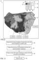
- FIG. 2is a schematic, pictorial illustration of multiple virtual attributes, which are indicative of respective parameters, and are overlaid on map 27 of heart 26 , in accordance with an exemplary embodiment of the present invention.
- processor 41receives two datasets corresponding to two parameters measured at different sections of heart 26 .
- the first parametercorresponds to local activation times (LATs)
- the second parametercorresponds to voltages.
- the term voltagemay refer to peak-to-peak voltage measured in tissue of heart 26 , or to any other voltage measured in any tissue of heart 26 or in tissue of any other organ of patient 28 .
- processor 41is configured to assign: (i) a first visual attribute (VA) 55 to the LAT measurements, and (ii) a second VA 66 to the voltage measurements.
- VA 55comprises a color group arranged in a color scale having multiple colors and color gradients, each color indicative of a corresponding range of LATs relative to a predefined reference LAT.
- a color 52e.g., red gradient
- a color 54e.g., yellow gradient
- a color 56e.g., green gradient
- a color 58e.g., blue gradient
- the negative values of LATare measured relative to the aforementioned predefined reference LAT.
- VA 66comprises a texture scale having multiple types of textures, each texture is indicative of a corresponding range of voltages.
- VA 66comprises: (i) a texture 60 having a wavy shape and indicative of low voltages (e.g., between about 0.05 mV and 0.5 mV), (ii) a texture 62 having an array of “X” shapes and indicative of medium voltages (e.g., between about 0.5 mV and 1.5 mV), and (iii) a texture 64 , which is blank (i.e., without any pattern) and is indicative of high voltages (e.g., about 1.5 mm mV).
- Grey scale, shading and/or shadingare utilized to represent color in this application.
- processor 41is configured to produce map 27 comprising an overlay of VAs 55 and 66 for each section of map 27 , and to display the map on display 35 .
- a section “A”has a color 54 and a texture 64
- a section “B”has a color 52 and a texture 62
- section “C”has a color 52 and a texture 64
- section “D”positioned in close proximity to an ostium 50 of heart 26 , has a color 56 and a texture 60 .
- map 27 with the overlaid LAT and voltageprovides physician 30 with a presentation indicative of two or more parameters at each section of heart 26 .
- sections “B” and “C”are in close proximity and have similar LAT but different voltage
- section “D” that has low voltageis surrounded with sections having medium and mainly high voltage.
- processor 41is configured to produce map 27 with more than two parameters, for example, using different sets of patterns and/or numbers and/or any other suitable notifications for each type of parameter.
- processor 41is configured to produce map 27 having other visual attributes indicative of other parameters, such as but not limited to cycle length in atrial fibrillation and standard deviation of the aforementioned cycle length.
- the datasetsmay comprise output of one or more algorithms that enable detection of complex fractionated atrial electrograms (CFAEs) in an intra-cardiac electrocardiogram (IC-ECG), based on parameters defined by physician 30 .
- CFAEscomplex fractionated atrial electrograms
- IC-ECGintra-cardiac electrocardiogram
- the datasetsmay comprise: (i) the number of detected peaks, and (ii) the percentage of signals having voltage higher than a predefined threshold.
- FIG. 3is a flow chart that schematically illustrates a method for presenting multiple parameters overlaid on map 27 , in accordance with an exemplary embodiment of the present invention.
- the methodbegins at a datasets receiving step 100 , with processor 41 (or any other suitable device) configured to receive first and second datasets corresponding to first and second properties of heart 26 or any other organ of patient 28 , in the present example the parameters may comprise measurements acquired in heart 26 , as described in detail in FIGS. 1 and 2 above.
- processor 41assigns first and second visual attributes, such as VAs 55 and 66 described in detail in FIG. 2 above, to the first and second properties (e.g., LAT and voltage), respectively.
- map 27comprises an anatomical map of heart 26 (obtained using any suitable technique) with VAs 55 and 66 overlaid on the anatomical map.
- processor 41displays map 27 on any suitable output device, such as but not limited to, display 35 as described in detail in FIG. 2 above.
- the methods and systems described hereincan also be used in other parameters of AF, such as in displaying regular cycle length (CL) during AF with standard deviation (STD) of the CL, wherein the section with minimal value of CL and STD of CL is intended to be ablated.
- the methods and systems described hereincan also be used in other applications, such as in presenting over an anatomical map multiple parameters of ventricle fibrillation, or any other two or more parameters presented over any anatomical map of any organ in question.
Landscapes
- Health & Medical Sciences (AREA)
- Life Sciences & Earth Sciences (AREA)
- Engineering & Computer Science (AREA)
- Surgery (AREA)
- Animal Behavior & Ethology (AREA)
- General Health & Medical Sciences (AREA)
- Public Health (AREA)
- Veterinary Medicine (AREA)
- Biomedical Technology (AREA)
- Heart & Thoracic Surgery (AREA)
- Medical Informatics (AREA)
- Molecular Biology (AREA)
- Physics & Mathematics (AREA)
- Pathology (AREA)
- Biophysics (AREA)
- Nuclear Medicine, Radiotherapy & Molecular Imaging (AREA)
- Cardiology (AREA)
- Radiology & Medical Imaging (AREA)
- Theoretical Computer Science (AREA)
- Databases & Information Systems (AREA)
- Robotics (AREA)
- General Engineering & Computer Science (AREA)
- Data Mining & Analysis (AREA)
- General Physics & Mathematics (AREA)
- Otolaryngology (AREA)
- Physiology (AREA)
- Measurement And Recording Of Electrical Phenomena And Electrical Characteristics Of The Living Body (AREA)
- Magnetic Resonance Imaging Apparatus (AREA)
- Apparatus For Radiation Diagnosis (AREA)
- User Interface Of Digital Computer (AREA)
- Surgical Instruments (AREA)
Abstract
Description
Claims (6)
Priority Applications (5)
| Application Number | Priority Date | Filing Date | Title |
|---|---|---|---|
| US17/208,762US12082881B2 (en) | 2021-03-22 | 2021-03-22 | Visualizing multiple parameters overlaid on an anatomical map |
| IL291275AIL291275A (en) | 2021-03-22 | 2022-03-10 | Visualizing multiple parameters overlaid on an anatomical map |
| JP2022043546AJP2022146920A (en) | 2021-03-22 | 2022-03-18 | Visualization of multiple parameters overlaid on anatomical maps |
| EP22163204.5AEP4062833A1 (en) | 2021-03-22 | 2022-03-21 | Visualizing multiple parameters overlaid on an anatomical map |
| CN202210281632.4ACN115105087A (en) | 2021-03-22 | 2022-03-22 | Visualizing multiple parameters superimposed on an anatomical map |
Applications Claiming Priority (1)
| Application Number | Priority Date | Filing Date | Title |
|---|---|---|---|
| US17/208,762US12082881B2 (en) | 2021-03-22 | 2021-03-22 | Visualizing multiple parameters overlaid on an anatomical map |
Publications (2)
| Publication Number | Publication Date |
|---|---|
| US20220296301A1 US20220296301A1 (en) | 2022-09-22 |
| US12082881B2true US12082881B2 (en) | 2024-09-10 |
Family
ID=80928938
Family Applications (1)
| Application Number | Title | Priority Date | Filing Date |
|---|---|---|---|
| US17/208,762Active2042-10-12US12082881B2 (en) | 2021-03-22 | 2021-03-22 | Visualizing multiple parameters overlaid on an anatomical map |
Country Status (5)
| Country | Link |
|---|---|
| US (1) | US12082881B2 (en) |
| EP (1) | EP4062833A1 (en) |
| JP (1) | JP2022146920A (en) |
| CN (1) | CN115105087A (en) |
| IL (1) | IL291275A (en) |
Families Citing this family (3)
| Publication number | Priority date | Publication date | Assignee | Title |
|---|---|---|---|---|
| EP4626317A1 (en)* | 2022-12-01 | 2025-10-08 | Biosense Webster (Israel) Ltd. | Point of interest (poi) map for cardiac arrhythmia diagnosis |
| KR102717123B1 (en)* | 2022-12-19 | 2024-10-15 | 연세대학교 산학협력단 | Method for visualizing extracted area with high probability of atrial fibrillation and apparatus for same |
| WO2024141350A1 (en)* | 2022-12-26 | 2024-07-04 | Koninklijke Philips N.V. | Producing combined error values |
Citations (30)
| Publication number | Priority date | Publication date | Assignee | Title |
|---|---|---|---|---|
| US5391199A (en) | 1993-07-20 | 1995-02-21 | Biosense, Inc. | Apparatus and method for treating cardiac arrhythmias |
| WO1996005768A1 (en) | 1994-08-19 | 1996-02-29 | Biosense, Inc. | Medical diagnosis, treatment and imaging systems |
| US6239724B1 (en) | 1997-12-30 | 2001-05-29 | Remon Medical Technologies, Ltd. | System and method for telemetrically providing intrabody spatial position |
| US6332089B1 (en) | 1996-02-15 | 2001-12-18 | Biosense, Inc. | Medical procedures and apparatus using intrabody probes |
| US20020065455A1 (en) | 1995-01-24 | 2002-05-30 | Shlomo Ben-Haim | Medical diagnosis, treatment and imaging systems |
| US6484118B1 (en) | 2000-07-20 | 2002-11-19 | Biosense, Inc. | Electromagnetic position single axis system |
| US20030120150A1 (en) | 2001-12-21 | 2003-06-26 | Assaf Govari | Wireless position sensor |
| US6618612B1 (en) | 1996-02-15 | 2003-09-09 | Biosense, Inc. | Independently positionable transducers for location system |
| US20040068178A1 (en) | 2002-09-17 | 2004-04-08 | Assaf Govari | High-gradient recursive locating system |
| CA2713305A1 (en)* | 2010-08-23 | 2012-02-23 | Justin D. Pearlman | Method of and system for signal separation during multivariate physiological monitoring |
| US20120184863A1 (en) | 2011-01-13 | 2012-07-19 | Rhythmia Medical, Inc. | Electroanatomical mapping |
| US20130151275A1 (en) | 2011-12-09 | 2013-06-13 | Fabio Alburquerque Thiers | Dynamic geographical display of research capacity |
| US20160022375A1 (en)* | 2014-07-24 | 2016-01-28 | Robert Blake | System and method for cardiac ablation |
| US20160242667A1 (en)* | 2015-02-20 | 2016-08-25 | Boston Scientific Scimed Inc. | Tissue contact sensing using a medical device |
| US20170202469A1 (en) | 2014-03-25 | 2017-07-20 | Acutus Medical ,Inc. | Cardiac analysis user interface system and method |
| US20170360319A1 (en)* | 2015-01-07 | 2017-12-21 | St. Jude Medical, Cardiology Division, Inc. | System, method, and apparatus for visualizing cardiac timing information using animations |
| US20180144828A1 (en)* | 2016-10-27 | 2018-05-24 | Progenics Pharmaceuticals, Inc. | Network for medical image analysis, decision support system, and related graphical user interface (gui) applications |
| US20190099098A1 (en) | 2017-10-02 | 2019-04-04 | Boston Scientific Scimed Inc. | Display of multiple electrograms anatomical map |
| US10470682B2 (en) | 2014-02-25 | 2019-11-12 | St. Jude Medical, Cardiology Division, Inc. | System and method for local electrophysiological characterization of cardiac substrate using multi-electrode catheters |
| US20200060567A1 (en)* | 2018-08-22 | 2020-02-27 | Biosense Webster (Israel) Ltd. | Atrial fibrillation mapping using atrial fibrillation cycle length (afcl) gradients |
| US20200074664A1 (en)* | 2017-03-13 | 2020-03-05 | Koninklijke Philips N.V. | Anatomical measurements from ultrasound data |
| US20200237452A1 (en)* | 2018-08-13 | 2020-07-30 | Theator inc. | Timeline overlay on surgical video |
| US20200273552A1 (en)* | 2019-02-21 | 2020-08-27 | Theator inc. | System for Detecting an Omitted Event During a Surgical Procedure |
| US20200273581A1 (en)* | 2019-02-21 | 2020-08-27 | Theator inc. | Post discharge risk prediction |
| US20200367751A1 (en)* | 2017-11-29 | 2020-11-26 | Universiteit Gent | Detection of rotational activity in cardiac electrophysiology |
| EP3750478A1 (en) | 2019-06-11 | 2020-12-16 | Biosense Webster (Israel) Ltd | Visually differentiating primary and secondary activations on electrophysiological maps |
| US20220079424A1 (en)* | 2020-09-15 | 2022-03-17 | Raytrx, Llc | Wireless swivel camera laparoscopic instrument with a virtual mapping and guidance system |
| US20220362162A1 (en)* | 2018-11-26 | 2022-11-17 | Arytha Biosciences, Llc | Nanoparticles containing cellular membrane and uses thereof |
| US20230148421A1 (en)* | 2014-08-22 | 2023-05-11 | University Of Delaware | Medical treatment simulation devices |
| US20230147888A1 (en)* | 2020-04-24 | 2023-05-11 | Lifelens Technologies, Llc | Visualizing physiologic data obtained from subjects |
- 2021
- 2021-03-22USUS17/208,762patent/US12082881B2/enactiveActive
- 2022
- 2022-03-10ILIL291275Apatent/IL291275A/enunknown
- 2022-03-18JPJP2022043546Apatent/JP2022146920A/enactivePending
- 2022-03-21EPEP22163204.5Apatent/EP4062833A1/ennot_activeWithdrawn
- 2022-03-22CNCN202210281632.4Apatent/CN115105087A/enactivePending
Patent Citations (33)
| Publication number | Priority date | Publication date | Assignee | Title |
|---|---|---|---|---|
| US5391199A (en) | 1993-07-20 | 1995-02-21 | Biosense, Inc. | Apparatus and method for treating cardiac arrhythmias |
| WO1996005768A1 (en) | 1994-08-19 | 1996-02-29 | Biosense, Inc. | Medical diagnosis, treatment and imaging systems |
| US6690963B2 (en) | 1995-01-24 | 2004-02-10 | Biosense, Inc. | System for determining the location and orientation of an invasive medical instrument |
| US20020065455A1 (en) | 1995-01-24 | 2002-05-30 | Shlomo Ben-Haim | Medical diagnosis, treatment and imaging systems |
| US6332089B1 (en) | 1996-02-15 | 2001-12-18 | Biosense, Inc. | Medical procedures and apparatus using intrabody probes |
| US6618612B1 (en) | 1996-02-15 | 2003-09-09 | Biosense, Inc. | Independently positionable transducers for location system |
| US6239724B1 (en) | 1997-12-30 | 2001-05-29 | Remon Medical Technologies, Ltd. | System and method for telemetrically providing intrabody spatial position |
| US6484118B1 (en) | 2000-07-20 | 2002-11-19 | Biosense, Inc. | Electromagnetic position single axis system |
| US20030120150A1 (en) | 2001-12-21 | 2003-06-26 | Assaf Govari | Wireless position sensor |
| US20040068178A1 (en) | 2002-09-17 | 2004-04-08 | Assaf Govari | High-gradient recursive locating system |
| CA2713305A1 (en)* | 2010-08-23 | 2012-02-23 | Justin D. Pearlman | Method of and system for signal separation during multivariate physiological monitoring |
| US20120184863A1 (en) | 2011-01-13 | 2012-07-19 | Rhythmia Medical, Inc. | Electroanatomical mapping |
| US20130151275A1 (en) | 2011-12-09 | 2013-06-13 | Fabio Alburquerque Thiers | Dynamic geographical display of research capacity |
| US10470682B2 (en) | 2014-02-25 | 2019-11-12 | St. Jude Medical, Cardiology Division, Inc. | System and method for local electrophysiological characterization of cardiac substrate using multi-electrode catheters |
| US20170202469A1 (en) | 2014-03-25 | 2017-07-20 | Acutus Medical ,Inc. | Cardiac analysis user interface system and method |
| US20160022375A1 (en)* | 2014-07-24 | 2016-01-28 | Robert Blake | System and method for cardiac ablation |
| US20230148421A1 (en)* | 2014-08-22 | 2023-05-11 | University Of Delaware | Medical treatment simulation devices |
| US20170360319A1 (en)* | 2015-01-07 | 2017-12-21 | St. Jude Medical, Cardiology Division, Inc. | System, method, and apparatus for visualizing cardiac timing information using animations |
| US20160242667A1 (en)* | 2015-02-20 | 2016-08-25 | Boston Scientific Scimed Inc. | Tissue contact sensing using a medical device |
| US20180144828A1 (en)* | 2016-10-27 | 2018-05-24 | Progenics Pharmaceuticals, Inc. | Network for medical image analysis, decision support system, and related graphical user interface (gui) applications |
| US20200027559A1 (en)* | 2016-10-27 | 2020-01-23 | Progenics Pharmaceuticals, Inc. | Network for medical image analysis, decision support system, and related graphical user interface (gui) applications |
| US20200126666A1 (en)* | 2016-10-27 | 2020-04-23 | Progenics Pharmaceuticals, Inc. | Network for medical image analysis, decision support system, and related graphical user interface (gui) applications |
| US20200074664A1 (en)* | 2017-03-13 | 2020-03-05 | Koninklijke Philips N.V. | Anatomical measurements from ultrasound data |
| US20190099098A1 (en) | 2017-10-02 | 2019-04-04 | Boston Scientific Scimed Inc. | Display of multiple electrograms anatomical map |
| US20200367751A1 (en)* | 2017-11-29 | 2020-11-26 | Universiteit Gent | Detection of rotational activity in cardiac electrophysiology |
| US20200237452A1 (en)* | 2018-08-13 | 2020-07-30 | Theator inc. | Timeline overlay on surgical video |
| US20200060567A1 (en)* | 2018-08-22 | 2020-02-27 | Biosense Webster (Israel) Ltd. | Atrial fibrillation mapping using atrial fibrillation cycle length (afcl) gradients |
| US20220362162A1 (en)* | 2018-11-26 | 2022-11-17 | Arytha Biosciences, Llc | Nanoparticles containing cellular membrane and uses thereof |
| US20200273581A1 (en)* | 2019-02-21 | 2020-08-27 | Theator inc. | Post discharge risk prediction |
| US20200273552A1 (en)* | 2019-02-21 | 2020-08-27 | Theator inc. | System for Detecting an Omitted Event During a Surgical Procedure |
| EP3750478A1 (en) | 2019-06-11 | 2020-12-16 | Biosense Webster (Israel) Ltd | Visually differentiating primary and secondary activations on electrophysiological maps |
| US20230147888A1 (en)* | 2020-04-24 | 2023-05-11 | Lifelens Technologies, Llc | Visualizing physiologic data obtained from subjects |
| US20220079424A1 (en)* | 2020-09-15 | 2022-03-17 | Raytrx, Llc | Wireless swivel camera laparoscopic instrument with a virtual mapping and guidance system |
Non-Patent Citations (1)
| Title |
|---|
| European Search Report for corresponding EPA No. 22163204.5 dated Jul. 28, 2022. |
Also Published As
| Publication number | Publication date |
|---|---|
| IL291275A (en) | 2022-10-01 |
| US20220296301A1 (en) | 2022-09-22 |
| JP2022146920A (en) | 2022-10-05 |
| EP4062833A1 (en) | 2022-09-28 |
| CN115105087A (en) | 2022-09-27 |
Similar Documents
| Publication | Publication Date | Title |
|---|---|---|
| EP4062833A1 (en) | Visualizing multiple parameters overlaid on an anatomical map | |
| EP4181075A1 (en) | Differential mapping of a body organ | |
| EP3973876B1 (en) | Real time removal of ep parameter outliers from visual map | |
| IL289283A (en) | Integration of confidence level into an electrophysiological map (ep) | |
| EP4079217B1 (en) | Improved electrophysiological (ep) map coloration by considering outliers | |
| US11819331B2 (en) | Visualization of epicardial and endocardial electroanatomical maps | |
| EP3614338B1 (en) | Post-mapping automatic identification of pulmonary veins | |
| EP4104764B1 (en) | Improving visualization of electrical signals propagating over the surface of patient organ | |
| US12133689B2 (en) | Displaying annotations on design line formed on anatomical map | |
| EP4079215B1 (en) | Multi-layered visualization of data points over heart map | |
| US12070277B2 (en) | Regional resolution in fast anatomical mapping | |
| US20230263452A1 (en) | Automatic storage and display of ecg signals indicative of atrial fibrillation |
Legal Events
| Date | Code | Title | Description |
|---|---|---|---|
| FEPP | Fee payment procedure | Free format text:ENTITY STATUS SET TO UNDISCOUNTED (ORIGINAL EVENT CODE: BIG.); ENTITY STATUS OF PATENT OWNER: LARGE ENTITY | |
| STPP | Information on status: patent application and granting procedure in general | Free format text:DOCKETED NEW CASE - READY FOR EXAMINATION | |
| AS | Assignment | Owner name:BIOSENSE WEBSTER (ISRAEL) LTD., ISRAEL Free format text:ASSIGNMENT OF ASSIGNORS INTEREST;ASSIGNORS:COHEN, BENJAMIN;TURGEMAN, AHARON;KATZ, NATAN SHARON;AND OTHERS;SIGNING DATES FROM 20210324 TO 20210925;REEL/FRAME:058889/0415 Owner name:BIOSENSE WEBSTER (ISRAEL) LTD., ISRAEL Free format text:ASSIGNMENT OF ASSIGNORS INTEREST;ASSIGNORS:COHEN, BENJAMIN;TURGEMAN, AHARON;KATZ, NATAN SHARON;AND OTHERS;SIGNING DATES FROM 20210324 TO 20210925;REEL/FRAME:058887/0339 | |
| STPP | Information on status: patent application and granting procedure in general | Free format text:NON FINAL ACTION MAILED | |
| STPP | Information on status: patent application and granting procedure in general | Free format text:RESPONSE TO NON-FINAL OFFICE ACTION ENTERED AND FORWARDED TO EXAMINER | |
| STPP | Information on status: patent application and granting procedure in general | Free format text:NOTICE OF ALLOWANCE MAILED -- APPLICATION RECEIVED IN OFFICE OF PUBLICATIONS | |
| STPP | Information on status: patent application and granting procedure in general | Free format text:DOCKETED NEW CASE - READY FOR EXAMINATION | |
| STPP | Information on status: patent application and granting procedure in general | Free format text:NOTICE OF ALLOWANCE MAILED -- APPLICATION RECEIVED IN OFFICE OF PUBLICATIONS | |
| STPP | Information on status: patent application and granting procedure in general | Free format text:PUBLICATIONS -- ISSUE FEE PAYMENT RECEIVED | |
| STPP | Information on status: patent application and granting procedure in general | Free format text:PUBLICATIONS -- ISSUE FEE PAYMENT VERIFIED | |
| STCF | Information on status: patent grant | Free format text:PATENTED CASE |