US12076566B2 - Neurostimulation titration utilizing T-wave alternans - Google Patents
Neurostimulation titration utilizing T-wave alternansDownload PDFInfo
- Publication number
- US12076566B2 US12076566B2US17/129,467US202017129467AUS12076566B2US 12076566 B2US12076566 B2US 12076566B2US 202017129467 AUS202017129467 AUS 202017129467AUS 12076566 B2US12076566 B2US 12076566B2
- Authority
- US
- United States
- Prior art keywords
- twa
- stimulation
- patient
- magnitude value
- target
- Prior art date
- Legal status (The legal status is an assumption and is not a legal conclusion. Google has not performed a legal analysis and makes no representation as to the accuracy of the status listed.)
- Active, expires
Links
Images


Classifications
- A—HUMAN NECESSITIES
- A61—MEDICAL OR VETERINARY SCIENCE; HYGIENE
- A61N—ELECTROTHERAPY; MAGNETOTHERAPY; RADIATION THERAPY; ULTRASOUND THERAPY
- A61N1/00—Electrotherapy; Circuits therefor
- A61N1/18—Applying electric currents by contact electrodes
- A61N1/32—Applying electric currents by contact electrodes alternating or intermittent currents
- A61N1/36—Applying electric currents by contact electrodes alternating or intermittent currents for stimulation
- A61N1/3605—Implantable neurostimulators for stimulating central or peripheral nerve system
- A61N1/36128—Control systems
- A61N1/36135—Control systems using physiological parameters
- A61N1/36139—Control systems using physiological parameters with automatic adjustment
- A—HUMAN NECESSITIES
- A61—MEDICAL OR VETERINARY SCIENCE; HYGIENE
- A61B—DIAGNOSIS; SURGERY; IDENTIFICATION
- A61B5/00—Measuring for diagnostic purposes; Identification of persons
- A61B5/24—Detecting, measuring or recording bioelectric or biomagnetic signals of the body or parts thereof
- A61B5/316—Modalities, i.e. specific diagnostic methods
- A61B5/318—Heart-related electrical modalities, e.g. electrocardiography [ECG]
- A61B5/346—Analysis of electrocardiograms
- A61B5/349—Detecting specific parameters of the electrocardiograph cycle
- A61B5/355—Detecting T-waves
- A—HUMAN NECESSITIES
- A61—MEDICAL OR VETERINARY SCIENCE; HYGIENE
- A61N—ELECTROTHERAPY; MAGNETOTHERAPY; RADIATION THERAPY; ULTRASOUND THERAPY
- A61N1/00—Electrotherapy; Circuits therefor
- A61N1/02—Details
- A61N1/04—Electrodes
- A61N1/05—Electrodes for implantation or insertion into the body, e.g. heart electrode
- A61N1/0551—Spinal or peripheral nerve electrodes
- A—HUMAN NECESSITIES
- A61—MEDICAL OR VETERINARY SCIENCE; HYGIENE
- A61N—ELECTROTHERAPY; MAGNETOTHERAPY; RADIATION THERAPY; ULTRASOUND THERAPY
- A61N1/00—Electrotherapy; Circuits therefor
- A61N1/18—Applying electric currents by contact electrodes
- A61N1/32—Applying electric currents by contact electrodes alternating or intermittent currents
- A61N1/36—Applying electric currents by contact electrodes alternating or intermittent currents for stimulation
- A61N1/3605—Implantable neurostimulators for stimulating central or peripheral nerve system
- A61N1/3606—Implantable neurostimulators for stimulating central or peripheral nerve system adapted for a particular treatment
- A61N1/36114—Cardiac control, e.g. by vagal stimulation
- A—HUMAN NECESSITIES
- A61—MEDICAL OR VETERINARY SCIENCE; HYGIENE
- A61N—ELECTROTHERAPY; MAGNETOTHERAPY; RADIATION THERAPY; ULTRASOUND THERAPY
- A61N1/00—Electrotherapy; Circuits therefor
- A61N1/18—Applying electric currents by contact electrodes
- A61N1/32—Applying electric currents by contact electrodes alternating or intermittent currents
- A61N1/36—Applying electric currents by contact electrodes alternating or intermittent currents for stimulation
- A61N1/3605—Implantable neurostimulators for stimulating central or peripheral nerve system
- A61N1/36128—Control systems
- A61N1/36132—Control systems using patient feedback
- A—HUMAN NECESSITIES
- A61—MEDICAL OR VETERINARY SCIENCE; HYGIENE
- A61N—ELECTROTHERAPY; MAGNETOTHERAPY; RADIATION THERAPY; ULTRASOUND THERAPY
- A61N1/00—Electrotherapy; Circuits therefor
- A61N1/18—Applying electric currents by contact electrodes
- A61N1/32—Applying electric currents by contact electrodes alternating or intermittent currents
- A61N1/36—Applying electric currents by contact electrodes alternating or intermittent currents for stimulation
- A61N1/3605—Implantable neurostimulators for stimulating central or peripheral nerve system
- A61N1/36128—Control systems
- A61N1/36146—Control systems specified by the stimulation parameters
- A61N1/3615—Intensity
- A—HUMAN NECESSITIES
- A61—MEDICAL OR VETERINARY SCIENCE; HYGIENE
- A61N—ELECTROTHERAPY; MAGNETOTHERAPY; RADIATION THERAPY; ULTRASOUND THERAPY
- A61N1/00—Electrotherapy; Circuits therefor
- A61N1/18—Applying electric currents by contact electrodes
- A61N1/32—Applying electric currents by contact electrodes alternating or intermittent currents
- A61N1/36—Applying electric currents by contact electrodes alternating or intermittent currents for stimulation
- A61N1/3605—Implantable neurostimulators for stimulating central or peripheral nerve system
- A61N1/36128—Control systems
- A61N1/36146—Control systems specified by the stimulation parameters
- A61N1/36167—Timing, e.g. stimulation onset
- A—HUMAN NECESSITIES
- A61—MEDICAL OR VETERINARY SCIENCE; HYGIENE
- A61N—ELECTROTHERAPY; MAGNETOTHERAPY; RADIATION THERAPY; ULTRASOUND THERAPY
- A61N1/00—Electrotherapy; Circuits therefor
- A61N1/18—Applying electric currents by contact electrodes
- A61N1/32—Applying electric currents by contact electrodes alternating or intermittent currents
- A61N1/36—Applying electric currents by contact electrodes alternating or intermittent currents for stimulation
- A61N1/362—Heart stimulators
- A61N1/37—Monitoring; Protecting
- A61N1/3702—Physiological parameters
- A—HUMAN NECESSITIES
- A61—MEDICAL OR VETERINARY SCIENCE; HYGIENE
- A61N—ELECTROTHERAPY; MAGNETOTHERAPY; RADIATION THERAPY; ULTRASOUND THERAPY
- A61N1/00—Electrotherapy; Circuits therefor
- A61N1/18—Applying electric currents by contact electrodes
- A61N1/32—Applying electric currents by contact electrodes alternating or intermittent currents
- A61N1/36—Applying electric currents by contact electrodes alternating or intermittent currents for stimulation
- A61N1/372—Arrangements in connection with the implantation of stimulators
- A61N1/37211—Means for communicating with stimulators
- A61N1/37217—Means for communicating with stimulators characterised by the communication link, e.g. acoustic or tactile
- A61N1/37223—Circuits for electromagnetic coupling
- A—HUMAN NECESSITIES
- A61—MEDICAL OR VETERINARY SCIENCE; HYGIENE
- A61N—ELECTROTHERAPY; MAGNETOTHERAPY; RADIATION THERAPY; ULTRASOUND THERAPY
- A61N1/00—Electrotherapy; Circuits therefor
- A61N1/18—Applying electric currents by contact electrodes
- A61N1/32—Applying electric currents by contact electrodes alternating or intermittent currents
- A61N1/36—Applying electric currents by contact electrodes alternating or intermittent currents for stimulation
- A61N1/372—Arrangements in connection with the implantation of stimulators
- A61N1/37211—Means for communicating with stimulators
- A61N1/37235—Aspects of the external programmer
- G—PHYSICS
- G16—INFORMATION AND COMMUNICATION TECHNOLOGY [ICT] SPECIALLY ADAPTED FOR SPECIFIC APPLICATION FIELDS
- G16H—HEALTHCARE INFORMATICS, i.e. INFORMATION AND COMMUNICATION TECHNOLOGY [ICT] SPECIALLY ADAPTED FOR THE HANDLING OR PROCESSING OF MEDICAL OR HEALTHCARE DATA
- G16H20/00—ICT specially adapted for therapies or health-improving plans, e.g. for handling prescriptions, for steering therapy or for monitoring patient compliance
- G16H20/30—ICT specially adapted for therapies or health-improving plans, e.g. for handling prescriptions, for steering therapy or for monitoring patient compliance relating to physical therapies or activities, e.g. physiotherapy, acupressure or exercising
- G—PHYSICS
- G16—INFORMATION AND COMMUNICATION TECHNOLOGY [ICT] SPECIALLY ADAPTED FOR SPECIFIC APPLICATION FIELDS
- G16H—HEALTHCARE INFORMATICS, i.e. INFORMATION AND COMMUNICATION TECHNOLOGY [ICT] SPECIALLY ADAPTED FOR THE HANDLING OR PROCESSING OF MEDICAL OR HEALTHCARE DATA
- G16H20/00—ICT specially adapted for therapies or health-improving plans, e.g. for handling prescriptions, for steering therapy or for monitoring patient compliance
- G16H20/40—ICT specially adapted for therapies or health-improving plans, e.g. for handling prescriptions, for steering therapy or for monitoring patient compliance relating to mechanical, radiation or invasive therapies, e.g. surgery, laser therapy, dialysis or acupuncture
- G—PHYSICS
- G16—INFORMATION AND COMMUNICATION TECHNOLOGY [ICT] SPECIALLY ADAPTED FOR SPECIFIC APPLICATION FIELDS
- G16H—HEALTHCARE INFORMATICS, i.e. INFORMATION AND COMMUNICATION TECHNOLOGY [ICT] SPECIALLY ADAPTED FOR THE HANDLING OR PROCESSING OF MEDICAL OR HEALTHCARE DATA
- G16H40/00—ICT specially adapted for the management or administration of healthcare resources or facilities; ICT specially adapted for the management or operation of medical equipment or devices
- G16H40/20—ICT specially adapted for the management or administration of healthcare resources or facilities; ICT specially adapted for the management or operation of medical equipment or devices for the management or administration of healthcare resources or facilities, e.g. managing hospital staff or surgery rooms
- G—PHYSICS
- G16—INFORMATION AND COMMUNICATION TECHNOLOGY [ICT] SPECIALLY ADAPTED FOR SPECIFIC APPLICATION FIELDS
- G16H—HEALTHCARE INFORMATICS, i.e. INFORMATION AND COMMUNICATION TECHNOLOGY [ICT] SPECIALLY ADAPTED FOR THE HANDLING OR PROCESSING OF MEDICAL OR HEALTHCARE DATA
- G16H40/00—ICT specially adapted for the management or administration of healthcare resources or facilities; ICT specially adapted for the management or operation of medical equipment or devices
- G16H40/60—ICT specially adapted for the management or administration of healthcare resources or facilities; ICT specially adapted for the management or operation of medical equipment or devices for the operation of medical equipment or devices
- G16H40/63—ICT specially adapted for the management or administration of healthcare resources or facilities; ICT specially adapted for the management or operation of medical equipment or devices for the operation of medical equipment or devices for local operation
- G—PHYSICS
- G16—INFORMATION AND COMMUNICATION TECHNOLOGY [ICT] SPECIALLY ADAPTED FOR SPECIFIC APPLICATION FIELDS
- G16Z—INFORMATION AND COMMUNICATION TECHNOLOGY [ICT] SPECIALLY ADAPTED FOR SPECIFIC APPLICATION FIELDS, NOT OTHERWISE PROVIDED FOR
- G16Z99/00—Subject matter not provided for in other main groups of this subclass
Definitions
- This applicationrelates to neuromodulation and, more specifically, to improved systems and methods for titrating stimulation therapies.
- CHFchronic heart failure
- CCDchronic cardiac dysfunction
- CHFtriggers compensatory activations of the sympathoadrenal (sympathetic) nervous system and the renin-angiotensin-aldosterone hormonal system, which initially helps to compensate for deteriorating heart-pumping function, yet, over time, can promote progressive left ventricular dysfunction and deleterious cardiac remodeling.
- Patients suffering from CHFare at increased risk of tachyarrhythmias, such as atrial fibrillation (AF), ventricular tachyarrhythmias (ventricular tachycardia (VT) and ventricular fibrillation (VF)), and atrial flutter, particularly when the underlying morbidity is a form of coronary artery disease, cardiomyopathy, mitral valve prolapse, or other valvular heart disease.
- AFatrial fibrillation
- VTventricular tachycardia
- VFventricular fibrillation
- Sympathoadrenal activationalso significantly increases the risk and severity of tachyarrhythmias due to neuronal action of the sympathetic nerve fibers in, on, or around the heart and through the release of epinephrine (adrenaline), which can exacerbate an already-elevated heart rate.
- epinephrineadrenaline
- VNSVagus nerve stimulation
- VNShas been demonstrated in canine studies as efficacious in simulated treatment of AF and heart failure, such as described in Zhang et al., “Chronic Vagus Nerve Stimulation Improves Autonomic Control and Attenuates Systemic Inflammation and Heart Failure Progression in a Canine High-Rate Pacing Model,” Circ Heart Fail 2009, 2, pp. 692-699 (Sep. 22, 2009), the disclosure of which is incorporated by reference.
- VNS therapycommonly requires implantation of a neurostimulator, a surgical procedure requiring several weeks of recovery before the neurostimulator can be activated and a patient can start receiving VNS therapy. Even after the recovery and activation of the neurostimulator, a full therapeutic dose of VNS is not immediately delivered to the patient to avoid causing significant patient discomfort and other undesirable side effects. Instead, to allow the patient to adjust to the VNS therapy, a titration process is utilized in which the intensity is gradually increased over a period of time under a control of a physician, with the patient given time between successive increases in VNS therapy intensity to adapt to the new intensity.
- the patient's tolerance thresholdAs stimulation is chronically applied at each new intensity level, the patient's tolerance threshold, or tolerance zone boundary, gradually increases, allowing for an increase in intensity during subsequent titration sessions.
- the titration processcan take significantly longer in practice because the increase in intensity is generally performed by a physician or other healthcare provider, and thus, for every step in the titration process to take place, the patient has to visit the provider's office to have the titration performed. Scheduling conflicts in the provider's office may increase the time between titration sessions, thereby extending the overall titration process, during which the patient in need of VNS does not receive the VNS at the full therapeutic intensity.
- a titration processthat continues over an extended period of time, such as six to twelve months, may be somewhat acceptable because the patient's health condition typically would not worsen in that period of time.
- the patient's conditionmay degrade rapidly if left untreated. As a result, there is a much greater urgency to completing the VNS titration process when treating a patient with a time-sensitive condition, such as CHF.
- a titration processis used to gradually increase the stimulation intensity to a desired therapeutic level until a target T-wave alternans change from a baseline T-wave alternans is achieved.
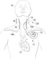
- FIG. 1is a front anatomical diagram showing, by way of example, placement of an implantable vagus stimulation device in a male patient, in accordance with one embodiment.
- FIGS. 2 A and 2 Bare diagrams respectively showing the implantable neurostimulator and the simulation therapy lead of FIG. 1 .
- FIG. 3is a diagram showing an external programmer for use with the implantable neurostimulator of FIG. 1 .
- FIG. 4is a diagram showing electrodes provided as on the stimulation therapy lead of FIG. 2 in place on a vagus nerve in situ.
- FIG. 5is a graph showing, by way of example, the relationship between the targeted therapeutic efficacy and the extent of potential side effects resulting from use of the implantable neurostimulator of FIG. 1 .
- FIG. 6is a graph showing, by way of example, the optimal duty cycle range based on the intersection depicted in FIG. 5 .
- FIG. 7is a timing diagram showing, by way of example, a stimulation cycle and an inhibition cycle of VNS as provided by implantable neurostimulator of FIG. 1 .
- FIGS. 8 A- 8 Care illustrative charts reflecting a heart rate response to gradually increased stimulation intensity at different frequencies.
- FIG. 9illustrates a method for delivering vagus nerve stimulation therapy.
- FIG. 10illustrates a titration process in accordance with embodiments of the present invention.
- FIGS. 11 A- 11 Bare block diagrams of neurostimulation systems in accordance with embodiments of the present invention.
- FIG. 12illustrates a titration process with variable titration parameters in accordance with embodiments of the present invention.
- FIG. 13is a scatter plot illustrating stimulation output current and corresponding T-wave alternans changes from baseline.
- FIGS. 14 A- 14 Bare illustrations of a method of assessing T-wave alternans in accordance with embodiments of the present invention.
- FIGS. 15 A- 15 Bare plots illustrating heart rate turbulence response for patients receiving VNS therapy in accordance with embodiments of the present invention.
- FIG. 16is a bar chart illustrating T-wave alternan magnitude levels for patients receiving different stimulation levels in accordance with embodiments of the present invention.
- FIG. 17is a bar chart illustrating heart rate turbulence slope levels for patients receiving different stimulation levels in accordance with embodiments of the present invention.
- CHF and other cardiovascular diseasescause derangement of autonomic control of the cardiovascular system, favoring increased sympathetic and decreased parasympathetic central outflow. These changes are accompanied by elevation of basal heart rate arising from chronic sympathetic hyperactivation along the neurocardiac axis.
- the vagus nerveis a diverse nerve trunk that contains both sympathetic and parasympathetic fibers, and both afferent and efferent fibers. These fibers have different diameters and myelination, and subsequently have different activation thresholds. This results in a graded response as intensity is increased. Low intensity stimulation results in a progressively greater tachycardia, which then diminishes and is replaced with a progressively greater bradycardia response as intensity is further increased.
- Peripheral neurostimulation therapiesthat target the fluctuations of the autonomic nervous system have been shown to improve clinical outcomes in some patients. Specifically, autonomic regulation therapy results in simultaneous creation and propagation of efferent and afferent action potentials within nerve fibers comprising the cervical vagus nerve.
- the therapydirectly improves autonomic balance by engaging both medullary and cardiovascular reflex control components of the autonomic nervous system.
- action potentialsUpon stimulation of the cervical vagus nerve, action potentials propagate away from the stimulation site in two directions, efferently toward the heart and afferently toward the brain. Efferent action potentials influence the intrinsic cardiac nervous system and the heart and other organ systems, while afferent action potentials influence central elements of the nervous system.
- FIG. 1is a front anatomical diagram showing, by way of example, placement of an implantable medical device (e.g., a vagus nerve stimulation (VNS) system 11 , as shown in FIG. 1 ) in a male patient 10 , in accordance with embodiments of the present invention.
- VNSvagus nerve stimulation
- the VNS provided through the stimulation system 11operates under several mechanisms of action. These mechanisms include increasing parasympathetic outflow and inhibiting sympathetic effects by inhibiting norepinephrine release and adrenergic receptor activation.
- VNStriggers the release of the endogenous neurotransmitter acetylcholine and other peptidergic substances into the synaptic cleft, which has several beneficial anti-arrhythmic, anti-apoptotic, and anti-inflammatory effects as well as beneficial effects at the level of the central nervous system.
- the implantable vagus stimulation system 11comprises an implantable neurostimulator or pulse generator 12 and a stimulating nerve electrode assembly 125 .
- the stimulating nerve electrode assembly 125preferably comprising at least an electrode pair, is conductively connected to the distal end of an insulated, electrically conductive lead assembly 13 and electrodes 14 .
- the electrodes 14may be provided in a variety of forms, such as, e.g., helical electrodes, probe electrodes, cuff electrodes, as well as other types of electrodes.
- the implantable vagus stimulation system 11can be remotely accessed following implant through an external programmer, such as the programmer 40 shown in FIG. 3 and described in further detail below.
- the programmer 40can be used by healthcare professionals to check and program the neurostimulator 12 after implantation in the patient 10 and to adjust stimulation parameters during the initial stimulation titration process.
- an external magnetmay provide basic controls, such as described in commonly assigned U.S. Pat. No. 8,600,505, entitled “Implantable Device For Facilitating Control Of Electrical Stimulation Of Cervical Vagus Nerves For Treatment Of Chronic Cardiac Dysfunction,” the disclosure of which is incorporated by reference.
- an electromagnetic controllermay enable the patient 10 or healthcare professional to interact with the implanted neurostimulator 12 to exercise increased control over therapy delivery and suspension, such as described in commonly-assigned U.S. Pat. No. 8,571,654, entitled “Vagus Nerve Neurostimulator With Multiple Patient-Selectable Modes For Treating Chronic Cardiac Dysfunction,” the disclosure of which is incorporated by reference.
- an external programmermay communicate with the neurostimulation system 11 via other wired or wireless communication methods, such as, e.g., wireless RF transmission. Together, the implantable vagus stimulation system 11 and one or more of the external components form a VNS therapeutic delivery system.
- the neurostimulator 12is typically implanted in the patient's right or left pectoral region generally on the same side (ipsilateral) as the vagus nerve 15 , 16 to be stimulated, although other neurostimulator-vagus nerve configurations, including contra-lateral and bi-lateral are possible.
- a vagus nervetypically comprises two branches that extend from the brain stem respectively down the left side and right side of the patient, as seen in FIG. 1 .
- the electrodes 14are generally implanted on the vagus nerve 15 , 16 about halfway between the clavicle 19 a - b and the mastoid process. The electrodes may be implanted on either the left or right side.
- the lead assembly 13 and electrodes 14are implanted by first exposing the carotid sheath and chosen branch of the vagus nerve 15 , 16 through a latero-cervical incision (perpendicular to the long axis of the spine) on the ipsilateral side of the patient's neck 18 .
- the helical electrodes 14are then placed onto the exposed nerve sheath and tethered.
- a subcutaneous tunnelis formed between the respective implantation sites of the neurostimulator 12 and helical electrodes 14 , through which the lead assembly 13 is guided to the neurostimulator 12 and securely connected.
- the neural stimulationis provided as a low level maintenance dose independent of cardiac cycle.
- the stimulation system 11bi-directionally stimulates either the left vagus nerve 15 or the right vagus nerve 16 .
- multiple electrodes 14 and multiple leads 13could be utilized to stimulate simultaneously, alternatively or in other various combinations.
- Stimulationmay be through multimodal application of continuously-cycling, intermittent and periodic electrical stimuli, which are parametrically defined through stored stimulation parameters and timing cycles. Both sympathetic and parasympathetic nerve fibers in the vagosympathetic complex are stimulated.
- cervical vagus nerve stimulationresults in propagation of action potentials from the site of stimulation in a bi-directional manner.
- the application of bi-directional propagation in both afferent and efferent directions of action potentials within neuronal fibers comprising the cervical vagus nerveimproves cardiac autonomic balance.
- Afferent action potentialspropagate toward the parasympathetic nervous system's origin in the medulla in the nucleus ambiguus, nucleus tractus solitarius, and the dorsal motor nucleus, as well as towards the sympathetic nervous system's origin in the intermediolateral cell column of the spinal cord.
- Efferent action potentialspropagate toward the heart 17 to activate the components of the heart's intrinsic nervous system.
- Either the left or right vagus nerve 15 , 16can be stimulated by the stimulation system 11 .
- the right vagus nerve 16has a moderately lower (approximately 30%) stimulation threshold than the left vagus nerve 15 for heart rate effects at the same stimulation frequency and pulse width.
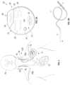
- FIGS. 2 A and 2 Bare diagrams respectively showing the implantable neurostimulator 12 and the stimulation lead assembly 13 of FIG. 1 .
- the neurostimulator 12can be adapted from a VNS THERAPY DEMIPULSE Model 103 or ASPIRESR Model 106 pulse generator, manufactured and sold by LivaNova PLC, Houston, Texas, although other manufactures and types of implantable VNS neurostimulators could also be used.
- the stimulation lead assembly 13 and electrodes 14are generally fabricated as a combined assembly and can be adapted from a Model 302 lead, PERENNIADURA Model 303 lead, or PERENNIAFLEX Model 304 lead, also manufactured and sold by LivaNova PLC, in three sizes based, for example, on a helical electrode inner diameter, although other manufactures and types of single-pin receptacle-compatible therapy leads and electrodes could also be used.
- the system 20may be configured to provide multimodal vagus nerve stimulation.
- the neurostimulator 12is parametrically programmed to deliver continuously-cycling, intermittent and periodic ON-OFF cycles of VNS. Such delivery produces action potentials in the underlying nerves that propagate bi-directionally, both afferently and efferently.
- the neurostimulator 12includes an electrical pulse generator that is tuned to improve autonomic regulatory function by triggering action potentials that propagate both afferently and efferently within the vagus nerve 15 , 16 .
- the neurostimulator 12is enclosed in a hermetically sealed housing 21 constructed of a biocompatible material, such as titanium.
- the housing 21contains electronic circuitry 22 powered by a battery 23 , such as a lithium carbon monofluoride primary battery or a rechargeable secondary cell battery.
- the electronic circuitry 22may be implemented using complementary metal oxide semiconductor integrated circuits that include a microprocessor controller that executes a control program according to stored stimulation parameters and timing cycles; a voltage regulator that regulates system power; logic and control circuitry, including a recordable memory 29 within which the stimulation parameters are stored, that controls overall pulse generator function, receives and implements programming commands from the external programmer, or other external source, collects and stores telemetry information, processes sensory input, and controls scheduled and sensory-based therapy outputs; a transceiver that remotely communicates with the external programmer using radio frequency signals; an antenna, which receives programming instructions and transmits the telemetry information to the external programmer; and a reed switch 30 that provides remote access to the operation of the neurostimulator 12 using an external programmer, a simple patient magnet, or an electromagnetic controller.
- the recordable memory 29can include both volatile (dynamic) and non-volatile/persistent (static) forms of memory, within which the stimulation parameters and timing cycles can be stored. Other electronic circuitry and components are possible.
- the neurostimulator 12includes a header 24 to securely receive and connect to the lead assembly 13 .
- the header 24encloses a receptacle 25 into which a single pin for the lead assembly 13 can be received, although two or more receptacles could also be provided, along with the corresponding electronic circuitry 22 .
- the header 24internally includes a lead connector block (not shown), a setscrew, and a spring contact (not shown) that electrically connects to the lead ring, thus completing the electrical circuit 26 .
- the housing 21may also contain a heart rate sensor 31 that is electrically interfaced with the logic and control circuitry, which receives the patient's sensed heart rate as sensory inputs.
- the heart rate sensor 31monitors heart rate using an electrocardiogram (ECG)-type electrode. Through the electrode, the patient's heart beat can be sensed by detecting ventricular depolarization.
- ECGelectrocardiogram
- a plurality of electrodescan be used to sense voltage differentials between electrode pairs, which can undergo signal processing for cardiac physiological measures, for instance, detection of the P-wave, QRS complex, and T-wave.
- the heart rate sensor 31provides the sensed heart rate to the control and logic circuitry as sensory inputs that can be used to determine the onset or presence of arrhythmias, particularly VT, and/or to monitor and record changes in the patient's heart rate over time or in response to applied stimulation signals.
- the lead assembly 13delivers an electrical signal from the neurostimulator 12 to the vagus nerve 15 , 16 via the electrodes 14 .
- the lead assembly 13On a proximal end, the lead assembly 13 has a lead connector 27 that transitions an insulated electrical lead body to a metal connector pin 28 and metal connector ring.
- the connector pin 28is guided through the receptacle 25 into the header 24 and securely fastened in place using a setscrew (not shown) that engages the connector pin 28 to electrically couple one electrode of the lead assembly 13 to the neurostimulator 12 while the spring contact makes electrical contact to the ring connected to the other electrode.
- the lead assembly 13terminates with the electrode 14 , which bifurcates into a pair of anodic and cathodic electrodes 62 (as further described infra with reference to FIG. 4 ).
- the lead connector 27is manufactured using silicone and the connector pin 28 and ring are made of stainless steel, although other suitable materials could be used, as well.
- the insulated lead body 13utilizes a silicone-insulated alloy conductor material.
- the electrodes 14are helical and placed around the cervical vagus nerve 15 , 16 at the location below where the superior and inferior cardiac branches separate from the cervical vagus nerve. In alternative embodiments, the helical electrodes may be placed at a location above where one or both of the superior and inferior cardiac branches separate from the cervical vagus nerve. In one embodiment, the helical electrodes 14 are positioned around the patient's vagus nerve oriented with the end of the helical electrodes 14 facing the patient's head. In an alternate embodiment, the helical electrodes 14 are positioned around the patient's vagus nerve 15 , 16 oriented with the end of the helical electrodes 14 facing the patient's heart 17 .
- the insulated electrical lead body 13is bifurcated into a pair of lead bodies that are connected to a pair of electrodes.
- the polarity of the electrodescould be configured into a proximal anode and a distal cathode, or a proximal cathode and a distal anode.
- the neurostimulator 12may be interrogated prior to implantation and throughout the therapeutic period with a healthcare provider-operable control system comprising an external programmer and programming wand (shown in FIG. 3 ) for checking proper operation, downloading recorded data, diagnosing problems, and programming operational parameters, such as described in commonly-assigned U.S. Pat. Nos. 8,600,505 and 8,571,654, cited supra.
- FIG. 3is a diagram showing an external programmer 40 for use with the implantable neurostimulator 12 of FIG. 1 .
- the external programmer 40includes a healthcare provider operable programming computer 41 and a programming wand 42 .
- use of the external programmeris restricted to healthcare providers, while more limited manual control is provided to the patient through “magnet mode.”
- the external programmer 40executes application software 45 specifically designed to interrogate the neurostimulator 12 .
- the programming computer 41interfaces to the programming wand 42 through a wired or wireless data connection.
- the programming wand 42can be adapted from a Model 201 Programming Wand, manufactured and sold by LivaNova PLC, and the application software 45 can be adapted from the Model 250 Programming Software suite, licensed by LivaNova PLC.
- Other configurations and combinations of external programmer 40 , programming wand 42 and application software 45are possible.
- the programming computer 41can be implemented using a general purpose programmable computer and can be a personal computer, laptop computer, ultrabook computer, netbook computer, handheld computer, tablet computer, smart phone, or other form of computational device.
- the programming computeris a tablet computer that may operate under the iOS operating system from Apple Inc., such as the iPad from Apple Inc., or may operate under the Android operating system from Google Inc., such as the Galaxy Tab from Samsung Electronics Co., Ltd.
- the programming computeris a personal digital assistant handheld computer operating under the Pocket-PC, Windows Mobile, Windows Phone, Windows RT, or Windows operating systems, licensed by Microsoft Corporation, Redmond, Washington, such as the Surface from Microsoft Corporation, the Dell Axim X5 and X50 personal data assistants, sold by Dell, Inc., Round Top, Tex., the HP Jornada personal data assistant, sold by Hewlett-Packard Company, Palo Alto, Tex.
- the programming computer 41functions through those components conventionally found in such devices, including, for instance, a central processing unit, volatile and persistent memory, touch-sensitive display, control buttons, peripheral input and output ports, and network interface.
- the computer 41operates under the control of the application software 45 , which is executed as program code as a series of process or method modules or steps by the programmed computer hardware. Other assemblages or configurations of computer hardware, firmware, and software are possible.
- the programming computer 41when connected to a neurostimulator 12 through wireless telemetry using the programming wand 42 , can be used by a healthcare provider to remotely interrogate the neurostimulator 12 and modify stored stimulation parameters.
- the programming wand 42provides data conversion between the digital data accepted by and output from the programming computer and the radio frequency signal format that is required for communication with the neurostimulator 12 .
- the programming computermay communicate with the implanted neurostimulator 12 using other wireless communication methods, such as wireless RF transmission.
- the programming computer 41may further be configured to receive inputs, such as physiological signals received from patient sensors (e.g., implanted or external).
- These sensorsmay be configured to monitor one or more physiological signals, e.g., vital signs, such as body temperature, pulse rate, respiration rate, blood pressure, etc. These sensors may be coupled directly to the programming computer 41 or may be coupled to another instrument or computing device which receives the sensor input and transmits the input to the programming computer 41 .
- the programming computer 41may monitor, record, and/or respond to the physiological signals in order to effectuate stimulation delivery in accordance with embodiments of the present invention.
- the healthcare provideroperates the programming computer 41 through a user interface that includes a set of input controls 43 and a visual display 44 , which could be touch-sensitive, upon which to monitor progress, view downloaded telemetry and recorded physiology, and review and modify programmable stimulation parameters.
- the telemetrycan include reports on device history that provide patient identifier, implant date, model number, serial number, magnet activations, total ON time, total operating time, manufacturing date, and device settings and stimulation statistics and on device diagnostics that include patient identifier, model identifier, serial number, firmware build number, implant date, communication status, output current status, measured current delivered, lead impedance, and battery status. Other kinds of telemetry or telemetry reports are possible.
- the programming wand 42is held by its handle 46 and the bottom surface 47 of the programming wand 42 is placed on the patient's chest over the location of the implanted neurostimulator 12 .
- a set of indicator lights 49can assist with proper positioning of the wand and a set of input controls 48 enable the programming wand 42 to be operated directly, rather than requiring the healthcare provider to awkwardly coordinate physical wand manipulation with control inputs via the programming computer 41 .
- the sending of programming instructions and receipt of telemetry informationoccur wirelessly through radio frequency signal interfacing. Other programming computer and programming wand operations are possible.
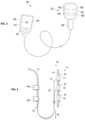
- FIG. 4is a diagram showing the helical electrodes 14 provided on the stimulation lead assembly 13 of FIG. 2 in place on a vagus nerve 15 , 16 in situ 50 .
- the specific surgical approach and implantation site selection particularsmay vary, depending upon physician discretion and patient physical structure.
- helical electrodes 14may be positioned on the patient's vagus nerve 61 oriented with the end of the helical electrodes 14 facing the patient's head.
- the insulated electrical lead body 13is bifurcated into a pair of lead bodies 57 , 58 that are connected to a pair of electrodes 51 , 52 .
- the polarity of the electrodes 51 , 52could be configured into a proximal anode and a distal cathode, or a proximal cathode and a distal anode.
- an anchor tether 53is fastened over the lead bodies 57 , 58 that maintains the helical electrodes' position on the vagus nerve 61 following implant.
- the conductors of the electrodes 51 , 52are manufactured using a platinum and iridium alloy, while the helical materials of the electrodes 51 , 52 and the anchor tether 53 are a silicone elastomer.
- the electrodes 51 , 52 and the anchor tether 53are coiled around the vagus nerve 61 proximal to the patient's head, each with the assistance of a pair of sutures 54 , 55 , 56 , made of polyester or other suitable material, which help the surgeon to spread apart the respective helices.
- the lead bodies 57 , 58 of the electrodes 51 , 52are oriented distal to the patient's head and aligned parallel to each other and to the vagus nerve 61 .
- a strain relief bend 60can be formed on the distal end with the insulated electrical lead body 13 aligned, for example, parallel to the helical electrodes 14 and attached to the adjacent fascia by a plurality of tie-downs 59 a - b.
- the neurostimulator 12delivers VNS under control of the electronic circuitry 22 .
- the stored stimulation parametersare programmable. Each stimulation parameter can be independently programmed to define the characteristics of the cycles of therapeutic stimulation and inhibition to ensure optimal stimulation for a patient 10 .
- the programmable stimulation parametersinclude output current, signal frequency, pulse width, signal ON time, signal OFF time, magnet activation (for VNS specifically triggered by “magnet mode”), and reset parameters. Other programmable parameters are possible.
- sets or “profiles” of preselected stimulation parameterscan be provided to physicians with the external programmer and fine-tuned to a patient's physiological requirements prior to being programmed into the neurostimulator 12 , such as described in commonly-assigned U.S. Pat. No. 8,630,709, entitled “Computer-Implemented System and Method for Selecting Therapy Profiles of Electrical Stimulation of Cervical Vagus Nerves for Treatment of Chronic Cardiac Dysfunction,” the disclosure of which is incorporated by reference.
- the VNSmay be delivered as a multimodal set of therapeutic doses, which are system output behaviors that are pre-specified within the neurostimulator 12 through the stored stimulation parameters and timing cycles implemented in firmware and executed by the microprocessor controller.
- the therapeutic dosesinclude a maintenance dose that includes continuously-cycling, intermittent and periodic cycles of electrical stimulation during periods in which the pulse amplitude is greater than 0 mA (“therapy ON”) and during periods in which the pulse amplitude is 0 mA (“therapy OFF”).
- the neurostimulator 12can operate either with or without an integrated heart rate sensor, such as respectively described in commonly-assigned U.S. Pat. No. 8,577,458, entitled “Implantable Device for Providing Electrical Stimulation of Cervical Vagus Nerves for Treatment of Chronic Cardiac Dysfunction with Leadless Heart Rate Monitoring,” and U.S. Patent application, entitled “Implantable Device for Providing Electrical Stimulation of Cervical Vagus Nerves for Treatment of Chronic Cardiac Dysfunction,” Ser. No. 13/314,119, filed on Dec. 7, 2011, pending, the disclosures of which are hereby incorporated by reference herein in their entirety.
- the neurostimulator 12can provide autonomic cardiovascular drive evaluation and self-controlled titration, such as respectively described in commonly-assigned U.S. Pat. No. 8,918,190, entitled “Implantable Device for Evaluating Autonomic Cardiovascular Drive in a Patient Suffering from Chronic Cardiac Dysfunction,” filed on Dec. 7, 2011, and U.S. Pat. No. 8,918,191, entitled “Implantable Device for Providing Electrical Stimulation of Cervical Vagus Nerves for Treatment of Chronic Cardiac Dysfunction with Bounded Titration,” filed on Dec. 7, 2011, the disclosures of which are incorporated by reference.
- the VNS stimulation signalmay be delivered as a therapy in a maintenance dose having an intensity that is insufficient to elicit undesirable side effects, such as cardiac arrhythmias.
- the VNScan be delivered with a periodic duty cycle in the range of 2% to 89% with a preferred range of around 4% to 36% that is delivered as a low intensity maintenance dose.
- the low intensity maintenance dosemay comprise a narrow range approximately at 17.5%, such as around 15% to 25%.
- the selection of duty cycleis a tradeoff among competing medical considerations.
- the duty cycleis determined by dividing the stimulation ON time by the sum of the ON and OFF times of the neurostimulator 12 during a single ON-OFF cycle. However, the stimulation time may also need to include ramp-up time and ramp-down time, where the stimulation frequency exceeds a minimum threshold (as further described infra with reference to FIG. 7 ).
- FIG. 5is a graph 70 showing, by way of example, the relationship between the targeted therapeutic efficacy 73 and the extent of potential side effects 74 resulting from use of the implantable neurostimulator 12 of FIG. 1 , after the patient has completed the titration process.
- the graph in FIG. 5provides an illustration of the failure of increased stimulation intensity to provide additional therapeutic benefit, once the stimulation parameters have reached the neural fulcrum zone, as will be described in greater detail below with respect to FIG. 8 .
- the x-axisrepresents the duty cycle 71 .
- the duty cycleis determined by dividing the stimulation ON time by the sum of the ON and OFF times of the neurostimulator 12 during a single ON-OFF cycle.
- the stimulation timemay also include ramp-up time and ramp-down time, where the stimulation frequency exceeds a minimum threshold (as further described infra with reference to FIG. 7 ).
- the total duty cyclemay be calculated as the ON time plus the ramp-up and ramp-down times divided by the OFF time, ON time, and ramp-up and ramp-down times, and may be, e.g., between 15% and 30%, and more specifically approximately 23%.
- the y-axisrepresents physiological response 72 to VNS therapy.
- the physiological response 72can be expressed quantitatively for a given duty cycle 71 as a function of the targeted therapeutic efficacy 73 and the extent of potential side effects 74 , as described infra.
- the maximum level of physiological response 72 (“max”)signifies the highest point of targeted therapeutic efficacy 73 or potential side effects 74 .
- Targeted therapeutic efficacy 73 and the extent of potential side effects 74can be expressed as functions of duty cycle 71 and physiological response 72 .
- the targeted therapeutic efficacy 73represents the intended effectiveness of VNS in provoking a beneficial physiological response for a given duty cycle and can be quantified by assigning values to the various acute and chronic factors that contribute to the physiological response 72 of the patient 10 due to the delivery of therapeutic VNS.
- Acute factors that contribute to the targeted therapeutic efficacy 73include beneficial changes in heart rate variability and increased coronary flow, reduction in cardiac workload through vasodilation, and improvement in left ventricular relaxation.
- Chronic factors that contribute to the targeted therapeutic efficacy 73include improved cardiovascular regulatory function, as well as decreased negative cytokine production, increased baroreflex sensitivity, increased respiratory gas exchange efficiency, favorable gene expression, renin-angiotensin-aldosterone system down-regulation, anti-arrhythmic, anti-apoptotic, and ectopy-reducing anti-inflammatory effects. These contributing factors can be combined in any manner to express the relative level of targeted therapeutic efficacy 73 , including weighting particular effects more heavily than others or applying statistical or numeric functions based directly on or derived from observed physiological changes.
- targeted therapeutic efficacy 73steeply increases beginning at around a 5% duty cycle, and levels off in a plateau near the maximum level of physiological response at around a 30% duty cycle. Thereafter, targeted therapeutic efficacy 73 begins decreasing at around a 50% duty cycle and continues in a plateau near a 25% physiological response through the maximum 100% duty cycle.
- the intersection 75 of the targeted therapeutic efficacy 73 and the extent of potential side effects 74represents one optimal duty cycle range for VNS.
- FIG. 6is a graph 80 showing, by way of example, the optimal duty cycle range 83 based on the intersection 75 depicted in FIG. 5 .
- the x-axisrepresents the duty cycle 81 as a percentage of stimulation time over stimulation time plus inhibition time.
- the y-axisrepresents therapeutic points 82 reached in operating the neurostimulator 12 at a given duty cycle 81 .
- the optimal duty cycle range 83is a function 84 of the intersection 75 of the targeted therapeutic efficacy 73 and the extent of potential side effects 74 .
- the therapeutic operating points 82can be expressed quantitatively for a given duty cycle 81 as a function of the values of the targeted therapeutic efficacy 73 and the extent of potential side effects 74 at the given duty cycle shown in the graph 70 of FIG. 5 .
- the optimal therapeutic operating point 85 (“max”)signifies a tradeoff that occurs at the point of highest targeted therapeutic efficacy 73 in light of lowest potential side effects 74 and that point will typically be found within the range of a 5% to 30% duty cycle 81 .
- Other expressions of duty cycles and related factorsare possible.
- VNSis delivered in a low level maintenance dose that uses alternating cycles of stimuli application (ON) and stimuli inhibition (OFF) that are tuned to activate both afferent and efferent pathways. Stimulation results in parasympathetic activation and sympathetic inhibition, both through centrally-mediated pathways and through efferent activation of preganglionic neurons and local circuit neurons.
- FIG. 7is a timing diagram showing, by way of example, a stimulation cycle and an inhibition cycle of VNS 90 , as provided by implantable neurostimulator 12 of FIG. 1 .
- the stimulation parametersenable the electrical stimulation pulse output by the neurostimulator 12 to be varied by both amplitude (output current 96 ) and duration (pulse width 94 ).
- the number of output pulses delivered per seconddetermines the signal frequency 93 .
- a pulse width in the range of 100 to 250 ⁇ secdelivers between 0.02 mA and 50 mA of output current at a signal frequency of about 10 Hz, although other therapeutic values could be used as appropriate.
- the stimulation signal delivered to the patientmay be defined by a stimulation parameter set comprising at least an amplitude, a frequency, a pulse width, and a duty cycle.
- the stimulation timeis considered the time period during which the neurostimulator 12 is ON and delivering pulses of stimulation
- the OFF timeis considered the time period occurring in-between stimulation times during which the neurostimulator 12 is OFF and inhibited from delivering stimulation or configured to deliver a negligible or ineffective stimulation.
- the neurostimulator 12implements a stimulation time 91 comprising an ON time 92 , a ramp-up time 97 and a ramp-down time 98 that respectively precede and follow the ON time 92 .
- the ON time 92is considered to be a time during which the neurostimulator 12 is ON and delivering pulses of stimulation at the full output current 96 .
- the OFF time 95is considered to comprise the ramp-up time 97 and ramp-down time 98 , which are used when the stimulation frequency is at least 10 Hz, although other minimum thresholds could be used, and both ramp-up and ramp-down times 97 , 98 last two seconds, although other time periods could also be used.
- the ramp-up time 97 and ramp-down time 98allow the strength of the output current of each output pulse to be gradually increased and decreased, thereby avoiding deleterious reflex behavior due to sudden delivery or inhibition of stimulation at a programmed intensity corresponding to the full output current 96 .
- VNSvagus neural stimulation has been shown to provide cardioprotective effects. Although delivered in a maintenance dose having an intensity that is insufficient to elicit undesirable side effects, such as cardiac arrhythmias, ataxia, coughing, hoarseness, throat irritation, voice alteration, or dyspnea, therapeutic VNS can nevertheless potentially ameliorate pathological tachyarrhythmias in some patients. Although VNS has been shown to decrease defibrillation threshold, VNS has not been shown to terminate VF in the absence of defibrillation. VNS prolongs ventricular action potential duration, so may be effective in terminating VT. In addition, the effect of VNS on the AV node may be beneficial in patients with AF by slowing conduction to the ventricles and controlling ventricular rate.
- autonomic regulation therapyresults in simultaneous creation of action potentials that simultaneously propagate away from the stimulation site in afferent and efferent directions within axons comprising the cervical vagus nerve complex.
- action potentialsUpon stimulation of the cervical vagus nerve, action potentials propagate away from the stimulation site in two directions, efferently toward the heart and afferently toward the brain.
- Different parameter settings for the neurostimulator 12may be adjusted to deliver varying stimulation intensities to the patient.
- the various stimulation parameter settings for current VNS devicesinclude output current amplitude, signal frequency, pulse width, signal ON time, and signal OFF time.
- a neural fulcrum zoneis identified, and neurostimulation therapy is delivered within the neural fulcrum zone.
- This neural fulcrum zonecorresponds to a combination of stimulation parameters at which autonomic engagement is achieved but for which a functional response determined by heart rate change is nullified due to the competing effects of afferently and efferently-transmitted action potentials.
- the tachycardia-inducing stimulation effectsare offset by the bradycardia-inducing effects, thereby minimizing side effects such as significant heart rate changes while providing a therapeutic level of stimulation.
- One method of identifying the neural fulcrum zoneis by delivering a plurality of stimulation signals at a fixed frequency but with one or more other parameter settings changed so as to gradually increase the intensity of the stimulation.
- FIGS. 8 A- 8 Cprovide illustrative charts reflecting the location of the neural fulcrum zone.
- FIG. 8 Ais a chart 800 illustrating a heart rate response in response to such a gradually increased intensity at a first frequency, in accordance with embodiments of the present invention.
- the x-axisrepresents the intensity level of the stimulation signal
- the y-axisrepresents the observed heart rate change from the patient's baseline basal heart rate observed when no stimulation is delivered.
- the stimulation intensityis increased by increasing the output current amplitude.
- a first set 810 of stimulation signalsis delivered at a first frequency (e.g., 10 Hz).
- a first frequencye.g. 10 Hz.
- the intensitye.g., output current amplitude
- a tachycardia zone 851 - 1is observed, during which period, the patient experiences a mild tachycardia.
- the patient's heart rate responsebegins to decrease and eventually enters a bradycardia zone 853 - 1 , in which a bradycardia response is observed in response to the stimulation signals.
- the neural fulcrum zoneis a range of stimulation parameters at which the functional effects from afferent activation are balanced with or nullified by the functional effects from efferent activation to avoid extreme heart rate changes while providing therapeutic levels of stimulation.
- the neural fulcrum zone 852 - 1can be located by identifying the zone in which the patient's response to stimulation produces either no heart rate change or a mildly decreased heart rate change (e.g., ⁇ 5% decrease, or a target number of beats per minute). As the intensity of stimulation is further increased at the fixed first frequency, the patient enters an undesirable bradycardia zone 853 - 1 .
- the patient's heart rate responseis used as an indicator of autonomic engagement.
- other physiological responsesmay be used to indicate the zone of autonomic engagement at which the propagation of efferent and afferent action potentials are balanced to identify the neural fulcrum zone.
- FIG. 8 Bis a chart 860 illustrating a heart rate response in response to such a gradually increased intensity at two additional frequencies, in accordance with embodiments of the present invention.
- the x-axis and y-axisrepresent the intensity level of the stimulation signal and the observed heart rate change, respectively, as in FIG. 8 A , and the first set 810 of stimulation signals from FIG. 8 A is also shown.
- a second set 820 of stimulation signalsis delivered at a second frequency lower than the first frequency (e.g., 5 Hz).
- the intensitye.g., output current amplitude
- a tachycardia zone 851 - 2is observed, during which period, the patient experiences a mild tachycardia.
- the patient's heart rate responsebegins to decrease and eventually enters a mild heart rate reduction zone 854 , in which a mild decrease in heart rate is observed in response to the stimulation signals.
- the low frequency of the stimulation signal in the second set 820 of stimulation signalslimits the functional effects of nerve fiber recruitment and, as a result, the heart response remains relatively limited. Although this low frequency stimulation results in minimal heart rate reduction, and, therefore, minimal side effects, the stimulation intensity is too low to result in effective recruitment of nerve fibers and engagement of the autonomic nervous system. As a result, a therapeutic level of stimulation is not delivered.
- a third set of 830 of stimulation signalsis delivered at a third frequency higher than the first and second frequencies (e.g., 20 Hz).
- the patientfirst experiences a tachycardia zone 851 - 3 .
- the level of increased heart rateis undesirable.
- the heart ratedecreases, similar to the decrease at the first and second frequencies but at a much higher rate.
- the patientfirst enters the neural fulcrum zone 852 - 3 and then the undesirable bradycardia zone 853 - 3 .
- the region in which the patient's heart rate response is between 0% and ⁇ 5%is much narrower than the neural fulcrum zone 852 - 1 for the first set 810 . Accordingly, when testing different operational parameter settings for a patient by increasing the output current amplitude by incremental steps, it can be more difficult to locate a programmable output current amplitude that falls within the neural fulcrum zone 852 - 3 .
- the resulting heart ratemay overshoot the neural fulcrum zone and create a situation in which the functional response transitions from the tachycardia zone 851 - 3 to the undesirable bradycardia zone 853 - 3 in a single step.
- the clinicianwould need to reduce the amplitude by a smaller increment or reduce the stimulation frequency in order to produce the desired heart rate response for the neural fulcrum zone 852 - 3 .
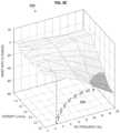
- FIG. 8 Cis a chart 880 illustrating mean heart rate response surfaces in conscious, normal dogs during 14 second periods of right cervical vagus VNS stimulation ON-time.
- the heart rate responses shown in z-axisrepresent the percentage heart rate change from the baseline heart rate at various sets of VNS parameters with the pulse width set at 250 ⁇ sec, the pulse amplitude ranging from 0 mA to 3.5 mA (provided by the x-axis), and the pulse frequency ranging from 2 Hz to 20 Hz (provided by the y-axis).
- Curve 890roughly represents the range of stimulation amplitude and frequency parameters at which a null response (i.e., 0% heart rate change from baseline) is produced.
- This null response curve 890is characterized by the opposition of functional responses (e.g., tachycardia and bradycardia) arising from afferent and efferent activation.
- Implantable medical devicesprovide therapy using electrical current as a stimulation vehicle.
- electrical currentas a stimulation vehicle.
- therapeutic levels of electrical stimulationare usually not well tolerated by patients without undergoing a process known as titration. Titration is a systematic method of slowly increasing, over time, stimulation parameters employed by an implanted device to deliver stimulation current until therapeutic levels become tolerated by the patient.
- FIG. 9is a flow diagram showing a method for delivering vagus nerve stimulation therapy 900 , in accordance with embodiments of the present invention.
- a titration processis used to gradually increase the stimulation intensity to a desired therapeutic level. If the stimulation intensity is increased too quickly before the patient is fully accommodated to the stimulation signal, the patient may experience undesirable side effects, such as coughing, hoarseness, throat irritation, or expiratory reflex.
- the titration processgradually increases stimulation intensity within a tolerable level, and maintains that intensity for a period of time to permit the patient to adjust to each increase in intensity, thereby gradually increasing the patient's side effect tolerance zone boundary to so as to accommodate subsequent increases in intensity. The titration process continues until adequate adaptation is achieved.
- the titration processis automated and is executed by the implanted device without manual adjustment of the stimulation intensity by the subject or health care provider.
- adequate adaptationis a composite threshold comprising one or more of the following: an acceptable side effect level, a target intensity level, and a target physiological response.
- adequate adaptionincludes all three objectives: an acceptable side effect level, a target intensity level, and a target physiological response.
- a patient's side effect profileis more sensitive to the stimulation output current than to the other stimulation parameters, such as frequency, pulse width, and duty cycle.
- accommodation to the stimulation output currentis a primary factor in completing the titration process.
- the other stimulation parametersare maintained at a level below the target levels, the output current can be increased to higher levels without eliciting undesirable side effects that would be result when the other parameters are at the target level.
- increasing the target output current while maintaining the other stimulation parameters (pulse width in particular) at reduced levelscan result in a faster accommodation and shorter overall titration time than would be achieved by attempting to increase the output current while stimulating at the target pulse width.
- a stimulation system 11including a neurostimulator 12 , a nerve stimulation lead assembly 13 , and a pair of electrodes 14 , is implanted in the patient.
- the patientundergoes an optional post-surgery recovery period, during which time the surgical incisions are allowed to heal and no VNS therapy occurs. This period may last, e.g., two weeks post surgery.
- the stimulation therapy processis initiated. During this process, VNS therapy is titrated by adjusting one or more of the stimulation parameters, including output current, pulse width, signal frequency, and duty cycle, as will be described in greater detail below. Completion of the titration process determines the stimulation intensity to be used for subsequent maintenance doses delivered in step 904 . These maintenance doses may be selected to provide the minimum stimulation intensity necessary to provide the desired therapeutic result.
- FIG. 10is a flow diagram illustrating a titration process 1000 in accordance with embodiments of the present invention.
- the neurostimulator 11When first initiating the titration process, the neurostimulator 11 is configured to generate a stimulation signal having an initial stimulation parameter set.
- the initial parameter setmay comprise an initial output current, an initial frequency, an initial pulse width, and an initial duty cycle.
- the various initial parameter settingsmay vary, but may be selected so that one or more of the parameters are set at levels below a predefined target parameter set level, such that the titration process is used to gradually increase the intensity parameters to achieve adequate adaptation.
- the initial frequencyis set at the target frequency level, while the initial output current, initial pulse width, and initial duty cycle are set below their respective target levels.
- the target parameter setcomprises a 10 Hz frequency, 250 ⁇ sec pulse width, a duty cycle of 14 sec ON and 1.1 minutes OFF, and an output current of between 1.5 mA-3.0 mA (e.g., 2.5 mA for right side stimulation and 3.0 mA for left side stimulation), and the initial parameter set comprises 10 Hz frequency, 130 ⁇ sec pulse width, a duty cycle of 14 sec ON and 1.1 minutes OFF, and an output current of between 0.25 mA-0.5 mA.
- the target parameter setincludes a 5 Hz frequency is used instead of a 10 Hz frequency.
- step 1001the stimulation system delivers stimulation to the patient. If this is the first titration session, then the stimulation would be delivered with the initial stimulation parameter set described above. If this is a subsequent titration session, then the stimulation intensity would remain at the same level at the conclusion of the previous titration session.
- the output currentis gradually increased until the stimulation results in an intolerable side effect level, the target output current (e.g., 2.5 mA) is reached, or adequate adaptation is achieved.
- adequate adaptationis a composite threshold comprising one or more of the following: an acceptable side effect level, a target intensity level, and a target physiological response.
- the target physiological responsecomprises a target heart rate change during stimulation.
- the patient's heart ratemay be monitored using an implanted or external heart rate monitor, and the patient's heart rate during stimulation is compared to the patient's baseline heart rate to determine the extent of heart rate change.
- the target heart rate changeis a heart rate change of between 4% and 5%. If at any point during the titration process 1000 adequate adaptation is achieved, the titration process ends and the stimulation intensity which resulted in the adequate adaptation is used for ongoing maintenance dose therapy delivery.
- the output currentmay be increased in any desired increment, but small increments, e.g., 0.1 mA or 0.25 mA, may be desirable so as to enable more precise adjustments. In some cases, the output current increments may be determined by the neurostimulator's maximum control capability. During the initial titration sessions, it is likely that the patient's side effect tolerance zone boundary will be reached well before the output current reaches the target level or adequate adaptation is achieved. At decision step 1003 , if the target output current has not been achieved but the maximum tolerable side effects have been exceeded, the process proceeds to step 1004 .
- step 1004the output current is reduced one increment to bring the side effects within acceptable levels.
- the frequencyis reduced. In embodiments in which the initial frequency was 10 Hz, in step 1004 , the frequency may be reduced, e.g., to 5 Hz or 2 Hz.
- step 1005the output current is gradually increased again at the reduced frequency level until the stimulation results in an intolerable side effect level or the target output current (e.g., 2.5 mA) is reached.
- the target output currente.g. 2.5 mA
- step 1007the titration session is concluded.
- the stimulation systemmay be programmed to continue delivering the stimulation signal at the last parameter settings achieved prior to conclusion of the titration session.
- another titration sessionmay be initiated and the process returns to step 1001 .
- Thiscan be any period of time sufficient to permit the patient to adjust to the increased stimulation levels. This can be, for example, as little as approximately two or three days, approximately one to two weeks, approximately four to eight weeks, or any other desired period of time.
- the titration sessionsare automatically initiated by the stimulation system or initiated by the patient without requiring any intervention by the health care provider. This can eliminate the need for the patient to schedule a subsequent visit to the health care provider, thereby potentially reducing the total amount of time needed for the titration process to complete.
- the stimulation systemincludes a physiological monitor, e.g., an implanted heart rate sensor, that communicates with the stimulation system's control system to enable the control system to detect when the target physiological response has been achieved and conclude the titration process.
- the stimulation systemcould in addition or alternatively include a patient control input to permit the patient to communicate to the control system that the acceptable side effect level has been exceeded.
- This control inputmay comprise an external control magnet that the patient can swipe over the implanted neurostimulator, or other internal or external communication device that the patient can use to provide an input to the control system.
- the stimulation systemmay be configured to wait a period of time after completing one session before initiating the next session. This period of time may be predetermined, e.g., two or three days.
- step 1008the output current is reduced one increment to restore an acceptable side effect condition, and the frequency is gradually increased until the stimulation results in an intolerable side effect level or the target frequency (e.g., 10 Hz) is reached.
- the target frequencye.g. 10 Hz
- the frequencyis reduced to restore an acceptable side effect level and the process proceeds to step 1007 .
- the current titration sessionis concluded and the stimulation system may be programmed to continue delivering the stimulation signal at the last parameter settings achieved prior to conclusion of the titration session.
- step 1010if the target frequency has been reached before the maximum tolerable side effects have been exceeded, the process proceeds to step 1010 and the duty cycle is gradually increased until the stimulation results in an intolerable side effect level or the target duty cycle (e.g., 14 sec ON and 1.1 min OFF) is reached, at which point the process proceeds to step 1007 and the titration session is concluded and ongoing stimulation delivered at the last intensity eliciting acceptable side effect levels.
- the target duty cyclee.g. 14 sec ON and 1.1 min OFF
- step 1011the pulse width is gradually increased until the stimulation results in an intolerable side effect level or the target pulse width (e.g., 250 ⁇ sec) is reached.
- the output currentis reduced (e.g., by up to 50%), and the pulse width may be increased in step 1011 at that reduced output current.
- the output currentmay be restored to the target output current.
- the output currentmay be reduced (or may be retained at the reduced level established prior to step 1011 , as described above), and the frequency and duty cycle are gradually increased in step 1013 (described below) at that reduced output current.
- This reduction in output current after achieving the target output currentmay enable the patient to maintain tolerability with increasing pulse width, frequency, and duty cycle in subsequent titration steps.
- step 1012if the target pulse width has not been achieved before the maximum tolerable side effects have been exceeded, the pulse width is reduced to restore an acceptable side effect level and the process proceeds to step 1007 . Again, in step 1007 , the current titration session is concluded.
- step 1012the process proceeds to step 1013 .
- step 1013the frequency and duty cycle are increased until the stimulation results in an intolerable side effect level or the target frequency and target duty cycle are reached.
- the frequency and duty cyclecan be increased in step 1013 simultaneously, sequentially, or on an alternating basis.
- step 1014if the target frequency and target duty cycle have not been achieved before the maximum tolerable side effects have been exceeded, the pulse width and/or frequency are reduced to restore an acceptable side effect level and the process continues to step 1007 and the titration session is concluded.
- the stimulation therapymay proceed with the maintenance dose at the target stimulation levels.
- the pulse widthmay be reduced in step 1004 .
- the pulse widthmay be reduced, e.g., to 150 ⁇ sec or less.
- the methodproceeds to step 1005 , in which the output current is gradually increased again at the reduced pulse width level until the stimulation results in an intolerable side effect level or the target output current (e.g., 2.5 mA) is reached.
- Therapycan also be autonomously titrated by the neurostimulator 12 in which titration progressively occurs in a self-paced, self-monitored fashion.
- the progression of titration sessionsmay occur on an autonomous schedule or may be initiated upon receipt of an input from the patient.
- the patient 10is expected to visit his healthcare provider to have the stimulation parameters stored by the neurostimulator 12 in the recordable memory 29 reprogrammed using an external programmer.
- the neurostimulator 12can be programmed to automatically titrate therapy by up titrating the VNS through periodic incremental increases as described above. The titration process 1000 will continue until the ultimate therapeutic goal is reached.
- VNSas parametrically defined by the maintenance dose operating mode, is delivered to at least one of the vagus nerves.
- the stimulation system 11delivers electrical therapeutic stimulation to the cervical vagus nerve of a patient 10 in a manner that results in creation and propagation (in both afferent and efferent directions) of action potentials within neuronal fibers of either the left or right vagus nerve independent of cardiac cycle.
- the sensed heart rate datacan be used to analyze therapeutic efficacy and patient condition. For instance, statistics could be determined from the sensed heart rate, either onboard by the neurostimulator 12 or by an external device, such as a programming computer following telemetric data retrieval.
- the sensed heart rate data statisticscan include determining a minimum heart rate over a stated time period, a maximum heart rate over a stated time period, an average heart rate over a stated time period, and a variability of heart rate over a stated period, where the stated period could be a minute, hour, day, week, month, or other selected time interval. Still other uses of the heart rate sensor 31 and the sensed heart rate data are possible.
- FIG. 11 Ais a simplified block diagram of an implanted neurostimulation system 1100 in accordance with embodiments of the present invention.
- the implanted neurostimulation system 1100comprises a control system 1102 comprising a processor programmed to operate the system 1100 , a memory 1103 , an optional physiological sensor 1104 , and a stimulation subsystem 1106 .
- the physiological sensor 1104may be configured to monitor any of a variety of patient physiological signals and the stimulation subsystem 1106 may be configured to deliver a stimulation signal to the patient.
- the physiological sensor 1104comprises an ECG sensor for monitoring heart rate and the stimulation subsystem 1106 comprises a neurostimulator 12 programmed to deliver ON-OFF cycles of stimulation to the patient's vagus nerve.
- the control system 1102is programmed to activate the neurostimulator 12 to deliver varying stimulation intensities to the patient and to monitor the physiological signals in response to those stimulation signals.
- the external programmer 1107 shown in FIG. 11 Amay be utilized by a clinician or by the patient for communicating with the implanted system 1100 to adjust parameters, activate therapy, retrieve data collected by the system 1100 or provide other input to the system 1100 .
- the external programmer 1107may be configured to program the implanted system 1100 with a prescribed time or window of time during which titration sessions may be initiated. This can be used to prevent a titration session from occurring at night when the patient's sleep is likely to be disturbed by the increase in stimulation intensity and resulting side effects.
- the implanted system 1100may include a patient input sensor 1105 .
- a patient magnet 1130may be used to provide external input to the system 1100 .
- the patient input sensor 1105will detect the presence of the magnetic field generated by the patient magnet 1130 and provide a control input to the control system 1102 .
- the system 1100may be programmed to receive patient inputs to set the time of day during which titration sessions are to be initiated.
- the patient input sensor 1105may comprise a motion sensor, such as an accelerometer, which is configured to detect tapping on the surface of the patient's chest.
- the patientmay use finger taps in one or more predetermined patterns to provide control inputs to the implanted system 1100 .
- the motion sensordetects three rapid taps to the patient's chest, that may trigger an operation on the implanted system 1100 (e.g., to initiate a titration session).
- the implanted system 1100will interpret those taps as a patient input indicating that the patient's tolerance zone boundary has been exceeded.
- the patient input sensor 1105may comprise an acoustic transducer or other sensor configured to detect acoustic signals.
- the system 1100may be programmed to interpret the detection of certain sounds as patient inputs.
- the patientmay utilize an electronic device, such as a smartphone or other portable audio device, to generate one or more predetermined sequences of tones.
- the system 1100may be programmed to interpret each of these sequences of tones as a different patient input.
- the patient input sensor 1105may be configured to detect when a patient is coughing, which can be interpreted by the system 1100 as an indication that the increased stimulation intensity exceeds the patient's tolerance zone boundary.
- the coughingcould be detected by an accelerometer to detect movement of the patient's chest, an acoustic transducer to detect the sound of the patient's coughing, or both.
- the titration of the stimulation signal delivery and the monitoring of the patient's physiological responsemay be advantageously implemented using control system in communication with both the stimulation subsystem 1106 and the physiological sensor 1104 , such as by incorporating all of these components into a single implantable device.
- the control systemmay be implemented in a separate implanted device or in an external programmer 1120 or other external device, as shown in FIG. 11 B .
- the external programmer 1120 in FIG. 11 Bmay include a control system 1112 and be utilized by a clinician or by the patient for adjusting stimulation parameters.
- the external programmer 1120is in wireless communication with the implanted medical device 1110 , which includes the stimulation subsystem 1116 .
- the physiological sensor 1114is incorporated into the implanted medical device 1110 , but in other embodiments, the sensor 1114 may be incorporated into a separate implanted device, may be provided externally and in communication with the external programmer 1120 , or may be provided as part of the external programmer 1120 .
- the implanted medical device 1110may include a memory 1113 that may store information in preparation for a transmission to the external programmer 1120 .
- Titrationis a method of varying over time stimulation parameters employed by an implanted device to deliver stimulation current, until therapeutic levels become tolerated by the patient.
- Embodiments provided abovedescribe automated titration processes used to gradually increase the stimulation intensity to a desired therapeutic level.
- the stimulation intensityis increased until the maximum tolerable side effects are exceeded, at which point the stimulation intensity is reduced to a tolerable level and the patient is provided with a period of time to adapt to the new intensity levels before the next titration session is initiated.
- the titration sessionsmay occur on a regular schedule (e.g., every two weeks), with an acclimation interval in between each titration session during which time stimulation at a tolerable intensity level is delivered.
- parameters other than or in addition to the acclimation intervalmay be adjusted based on the actual adaption experienced by the patient.
- the parameters that might be adjustedinclude, for example: current amplitude, pulse width, frequency, and OFF time.
- an automated titration processwhich utilizes an acclimation interval between titration sessions that may be adjusted based on the patient's response to the stimulation.
- FIG. 12illustrates a titration process 1200 with a variable acclimation interval. Steps 1201 - 1203 are similar to steps 901 - 903 illustrated in FIG. 9 and described above. However, in step 1204 , an outcome measure for the titration sessions is analyzed. In step 1205 , the acclimation interval between subsequent titration sessions is adjusted based on the analyzed outcome measure.
- the acclimation intervalmay be increased to provide the patient with additional time to recover and adapt to each set of increased stimulation intensities. Conversely, if the outcome measure indicates that the patient is adapting to the stimulation at a faster than expected rate, then the acclimation interval may be decreased to accelerate the adaption process and reduce the overall time required to complete the titration process and achieve a tolerable therapeutic maintenance dose level.
- the outcome measureis the patient's tolerance of a targeted increase in one or more of the stimulation parameters. For example, if the patient is unable to tolerate any increase in stimulation output current (or stimulation parameter) over the course of two or more titration sessions separated by a default acclimation interval (e.g., two weeks), it may be concluded that the patient is adapting to the stimulation at a slower than expected rate. In response, the acclimation interval between subsequent titration sessions may be increased (to, e.g., three or more weeks). If the patient continues to be incapable of tolerating any increase in stimulation output current in subsequent titration sessions, then the acclimation interval may be increased again (to, e.g., four or more weeks).
- a default acclimation intervale.g., two weeks
- the patientmay initially adapt to the increased stimulation intensity at a slower than expected rate, but after the acclimation interval is increased and subsequent titration sessions are successful at achieving the desired outcome measure, the patient's adaptation may accelerate, thereby permitting reduction of the acclimation interval back to the initial interval length. Accordingly, if the patient begins to adapt to the titration sessions after an increase in the acclimation interval, the system 1100 may be programmed to gradually reduce the acclimation interval in subsequent titration sessions.
- the systemmay be programmed to continue delivering stimulation at the last parameter settings achieved prior to conclusion of the titration session at an intensity just below the patient's tolerance zone boundary. This stimulation is delivered at this constant intensity until the next titration session is initiated.
- patientsare capable of enduring stimulation intensities just past the tolerance zone boundary for limited periods of time. The intensity levels just past the tolerance zone boundary may be considered by the patient as “moderately tolerable.” Patients may be willing to endure stimulation at the moderately tolerable levels for limited periods of time if it results in acceleration of the adaption process.
- an elevated stimulation sessionmay be initiated, during which time stimulation at moderately tolerable levels exceeding the tolerance zone boundary is delivered.
- This elevated stimulation sessionmay continue for any desired period of time, such as, e.g., several minutes or several hours, after which point the stimulation intensity will be reduced to a sustained stimulation intensity level below the tolerance zone boundary.
- the elevated stimulation sessionmay continue for less than one day, while the sustained stimulation is delivered for a period greater than one day, or the elevated stimulation session may continue for less than six hours, while the sustained stimulation is delivered for a period greater than one week. Any desired periods of time may be used.
- Various methodsare described herein for titrating stimulation by gradually increasing stimulation intensity until the patient's tolerance zone boundary is reached or exceeded.
- systems and methodsare provided for performing interactive training sessions in clinic for patients about to undergo titration on an ambulatory basis.
- the methodspermit clinicians to create a series of stimulation intensities (ranging from un-noticeable to noticeable but tolerable to intolerable), the patient's response to each stimulation, and the implanted device's response to patient inputs.
- the implanted medical device 1100may be used in conjunction with an external clinician programmer 1107 and patient input device (e.g., patient magnet 1130 or wireless-communications-enabled patient control device), to perform the titration processes on an ambulatory basis as described above, but is also programmed to execute in a training mode.
- This training modemay be initiated by the clinician using the clinician programmer 1107 while the patient is physically in the clinic for treatment and training.
- the training modemay be similar to the titration sessions described above, except that the increasing stimulation is initiated by the clinician using the programmer 1107 or automatically on an accelerated schedule.
- the patientcan use any of the herein described methods for providing a patient input to the device 1100 to indicate that the tolerance zone boundary has been reached.
- the device 1100may also transmit to the clinician programmer 1107 information regarding the stimulation being delivered.
- the programmer 1107may include a display which permits the clinician to observe the increasing intensity and receive a report of the intensity level that elicited the patient input indicating that the tolerance zone boundary was reached.
- the display on the programmer 1107may also be used to display feedback or instructions to the patient.
- the clinicianmay run the training mode multiple times so that the patient may become proficient at recognizing stimulation levels that are noticeable but tolerable, and distinguishing those tolerable levels from the truly intolerable stimulation levels. This can also provide training for the patient in the proper use of the patient input device.
- the programmer 1107may be used to select the stimulation parameter to be increased (e.g., output current, frequency, pulse width, or duty cycle), so that the patient and clinician can observe the different responses that may be elicited depending on the parameter being adjusted.
- the programmer 1107may be configured to pause the titration algorithm to hold the stimulation at a single level. This may be useful for facilitating a tolerance zone assessment by providing the patient additional time to experience the stimulation.
- the programmer 1107may also be used to terminate the training mode and return the device 1100 to its normal ambulatory mode, during which the desired ambulatory titration process may be performed.
- the training modemay also comprise an algorithm that sequences stimulation changes based on the training mode parameters programmed by the clinician.
- Stimulationmay be altered on a highly accelerated time scale in order to move the patient from tolerable to noticeable-but-tolerable to intolerable stimulation levels within the normal office follow-up period.
- This accelerated time scalemay be, for example, five, ten or fifteen minutes for all training. This is in contrast to the ambulatory mode titration process that seeks to advance therapy levels without the patient exceeding the tolerance-zone boundary. Having the patient experience all three tolerance phases in a single clinic visit can provide valuable patient training, resulting in accelerated adaptation speed.
- the system 1100may be programmed with an autonomous monitor to ensure that the training mode terminates automatically after a certain period has elapsed, even in the absence of a termination input from the clinician programmer. For example, the system 1100 may be programmed to automatically time-out and terminate the training mode 24 hours after initiation. After this automatic time-out, the system 1100 may automatically initiate the ambulatory mode.
- the systemmay enable patients to experience stimulation levels (usually following a stimulation increase) that may be unacceptable. Patients may also learn how to effectively deal with the intolerance through the use of the external patient input device. Clinicians can learn how individual patients react to various stimulation levels and the patients' cognitive ability to deal with unacceptable stimulation autonomously. Clinicians may also gain a sense of stimulation increases that an individual patient can tolerate and adjust the ambulatory titration algorithm accordingly.
- TWAT-wave alternans
- VFventricular fibrillation
- SCDsudden cardiac death
- TWAmay be used in place of or in addition to the heart rate monitoring described above in connection with the titration of stimulation and determination of optimal stimulation parameters for autonomic regulation therapy (ART).
- ARTautonomic regulation therapy
- a patient's heart rate response or a target stimulation parameter such as output currentmay be utilized when titrating stimulation and determining optimal stimulation parameters.
- a patient's TWAmay be used instead of or in addition to the heart rate response and target output current during stimulation titration.
- An implantable ART systemwhich includes a sensor for monitoring cardiac rhythm may be used. The implanted device can calculate TWA at various points during the titration period. This TWA calculation may be performed using methods similar to published algorithms, as will be described in greater detail below.
- the systemcould first establish a TWA baseline value monitored during the post-implant period (e.g. 1-2 weeks after implantation) before the initiation of stimulation.
- the titration processmay then be automatically performed by the implanted device, but instead of titrating to a target output current, as described above, the device would titrate stimulation intensity until a target TWA change from the established baseline TWA or target TWA level is observed. Once that target TWA change is achieved, the titration process is completed and the device may continue delivering the autonomic regulation therapy at the stimulation parameter settings that resulted in the target TWA change.
- the devicemay periodically monitor TWA levels and adjust stimulation as needed to maintain the target TWA change or target TWA level.
- Various methodsmay be used for determining the patient's TWA.
- One approachis to utilize a modified moving average analysis, as described in Nearing et al., “Modified Moving Average Analysis of T-Wave Alternans to Predict Ventricular Fibrillation with High Accuracy,” J. Appl. Physiol. 92: 541-549 (2002), the disclosure of which is incorporated by reference herein in its entirety.
- This approachinvolves constructing modified moving average computed beats by averaging alternate ECG beats. A weighted moving average is applied to limit the contribution of any one beat. The alternans estimate for any ECG segment is then determined as the maximum difference between A and B modified moving average computed beats within the ST segment and T-wave region.
- Another approachis to utilize a spectral analysis method of analyzing TWA in the frequency domain, as described in Smith et al., “Electrical alternans and cardiac electrical instability,” Circulation 77, No. 1, 110-121 (1988), the disclosure of which is incorporated by reference herein in its entirety.
- This approachutilizes a spectral analysis procedure to quantify the degree and statistical significance of waveform alternation present in the magnitude of the three orthogonal-lead ECG.
- the ECG signalmay be obtained using a sensor provided in a variety of locations.
- the ECG sensoris incorporated into the housing of the implanted neurostimulator, or the ECG may be detected using implanted ECG leads coupled to the implanted neurostimulator.
- ECGmay be monitored using an external device which is in communication with the implanted neurostimulator. The calculation of TWA and determination of change from the baseline TWA may be performed by either the external or implanted devices.
- the target TWA change from the baseline TWA or target TWA valuemay be any value that corresponds with the desired therapeutic effect.
- the target TWA change from the baseline TWAmay be in the range of approximately ⁇ 20 ⁇ V to approximately ⁇ 30 ⁇ V, e.g., a target TWA decrease of 25 ⁇ V.
- the target TWA change from the baseline TWAmay be a decrease of at least 10 ⁇ V.
- VNS therapy systemDEMIPULSE Model 103 pulse generator and PERENNIAFLEX Model 304 lead, both from LivaNova PLC of Houston, Texas
- the pulse generatorwas activated at 15 ⁇ 3 days after implantation. All patients were initially stimulated at a pulse width of 130 ⁇ s and a pulse frequency of 10 Hz, continuous cyclic 14-sec active (ON) and 66-sec inactive (OFF) to produce a duty cycle of 18%; 1,080 cycles/day.
- VNS activation and inactivation periodswere unrelated to the cardiac cycle (i.e., open loop), so no intracardiac sensing lead was used.
- VNS intensitywas gradually increased in 0.25 mA steps with the use of a radiofrequency programmer (Model 250 programming system from LivaNova PLC) to levels that produced acute VNS-related side effects (referred to as the “VNS tolerance zone boundary”), such as activation of the expiratory reflex (mild cough) or moderate heart rate reduction during the VNS-active phase.
- the output currentwas reduced by ⁇ 1 output current step (0.25 mA) to ensure that the therapy was well tolerated.
- the VNS stimulation intensitywas progressively increased to an average output current of 2.0 ⁇ 0.1 mA (for left side implantation patients: 2.2 ⁇ 0.2 mA; for right side implantation patients: 1.8 ⁇ 0.2 mA).
- Ambulatory ECG 24-hour recordingswere made using an externally-worn Holter monitor at baseline and after six and twelve months of chronic therapy. Measurements of TWA, HRT, HRV, ventricular tachycardia (VT) incidence, and heart rate were performed by an investigator.
- Peak TWAwas quantified from standard precordial leads V 1 and V 5 and a VF in 24-hour recordings with the Modified Moving Average (MMA) method, as described in the Nearing publication cited above. Limb leads were not recorded, as they are prone to motion artifact associated with daily activities.
- the MMA methodemploys the noise-rejection principle of recursive averaging.
- the maximum TWA level throughout the recordingis reported as the TWA value for that patient.
- a TWA threshold level of greater than or equal to 47 ⁇ Vwas defined in this study as an abnormal TWA test, and greater than or equal to 60 ⁇ V was defined as markedly abnormal.
- FIG. 13is a scatter plot 1300 illustrating stimulation output current and corresponding T-wave alternans changes from baseline.
- the stimulationwas delivered at a frequency of 10 Hz, a pulse width of 250- ⁇ sec, and a duty cycle of 18%.
- all but one of the data points in plot 1300are indicative of an undesirable increase in TWA from the baseline TWA.
- the devicemay be programmed to increase the output current (or other stimulation intensity parameter) until the target TWA change is achieved.
- stimulation output currents of 2.0 mA and aboveall but three of the data points in plot 1300 are indicative of a decrease in TWA from the baseline TWA.
- the devicemay be programmed to terminate the titration process and begin delivering ongoing therapy at the stimulation intensity that produced the desired decrease in TWA from baseline.
- FIGS. 14 A- 14 Bare assessments of TWA using the MMA method in a single representative patient with heart failure.
- the patient's ECG recordingswere separated into alternating A beats and B beats, and then an average was taken of each to produce an average A Beat signal 1401 and an average B Beat signal 1402 . These signals were superimposed to determine the TWA.
- FIG. 14 Aillustrates the QRS-aligned superimposed A Beat 1401 a and B Beat 1402 a taken at baseline, resulting in a TWA of 97 ⁇ V, calculated by performing a time-dependent difference between the curves and calculating the maximum value in the T-wave period. This TWA level of 97 ⁇ V is greater than the 60 ⁇ V threshold for strongly abnormal levels.
- FIG. 14 Aillustrates the QRS-aligned superimposed A Beat 1401 a and B Beat 1402 a taken at baseline, resulting in a TWA of 97 ⁇ V, calculated by performing a time-dependent difference between the curves and calculating the maximum value in
- FIGS. 14 A- 14 Billustrates the QRS-aligned superimposed A Beat 1401 b and B Beat 1402 b taken after twelve months of therapy, resulting in a TWA of 42 ⁇ V, which is less than the 47 ⁇ V threshold for abnormal levels.
- the patient's TWA levelsexperienced a significant decrease after twelve months of VNS therapy.
- HRTHeart rate turbulence
- TOHRT parameter turbulence onset
- TSHRT parameter turbulence slope
- FIGS. 15 A- 15 Bshow HRT response to the VNS stimulation therapy over the twelve month period in a representative patient.
- FIG. 15 Ashows the R-R interval in msec (y-axis) as a function of time (x-axis, showing the beat number), taken at baseline, prior to implantation of the pulse generator.
- FIG. 15 Bshows the R-R interval taken after twelve months of VNS therapy.
- the TO taken at baselinewas not statistically different than the TO taken at either six or twelve months.
- the baseline HRT turbulence slope (TS) 1501 awas 5.02 msec
- the HRT TS 1501 bhad nearly doubled to 9.71 msec. This increase is indicative of significantly improved baroreceptor sensitivity.
- FIG. 16is a bar chart 1600 illustrating the TWA magnitude levels (in ⁇ V) taken at baseline, at six months, and at twelve months for subjects receiving low intensity stimulation (pulse amplitude less than 2.0 mA), subjects receiving high intensity stimulation (pulse amplitude greater than or equal to 2.0 mA), and overall for all subjects.
- TWAwas reduced from 75.6 ⁇ 5.7 to 51.8 ⁇ 2.5 ⁇ V at twelve months ( ⁇ p ⁇ 0.001).
- TWAwas reduced from 64.0 ⁇ 0.5 to 48.5 ⁇ 2.5 ⁇ V at twelve months (*p ⁇ 0.05).
- TWAwas reduced from 71.0 ⁇ 4.6 to 50.5 ⁇ 1.8 ⁇ V at twelve months ( ⁇ p ⁇ 0.0001).
- the change in TWA levelswas significant for all patients.
- FIG. 17is a bar chart 1700 illustrating the heart rate turbulence slope (TS) levels (in msec) taken at baseline, at six months, and at twelve months for subjects receiving low intensity stimulation (pulse amplitude less than 2.0 mA), subjects receiving high intensity stimulation (pulse amplitude greater than or equal to 2.0 mA), and overall for all subjects.
- TSheart rate turbulence slope
- VNS stimulation for autonomic regulation therapy in patients with symptomatic heart failurecan decrease cardiac electrical instability, as reflected in reduced TWA levels and suppression of VT, and can improve baroreflex sensitivity, as reflected in increased HRT slope, both indicators of cardiovascular mortality risk.
- This method of chronic VNSmay provide a safe and effective means not only to improve cardiac mechanical function in patients with depressed left ventricular ejection fraction (LVEF) but also to protect against life-threatening ventricular arrhythmias and to improve cardioprotective autonomic reflexes.
- LVEFleft ventricular ejection fraction
- TWA and/or TSmay be utilized when titrating stimulation and determining optimal stimulation parameters.
- the stimulation intensitymay be increased until a target TWA magnitude or target TWA change from baseline is detected.
- the targetsmay vary, but some possible target TWA magnitudes include, for example, 45 ⁇ V, 47 ⁇ V, or 50 ⁇ V, and some possible target TWA changes from baseline include, for example, 5 ⁇ V, 7.5 ⁇ V, 10 ⁇ V, 12.5 ⁇ V, or 15 ⁇ V.
- the stimulation intensitymay be increased until a target TS magnitudes or target TS change from baseline is detected.
- the targetsmay vary, but some possible target TS magnitudes include, for example, 1.0 msec, 2.5 msec, or 5.0 msec, and some possible target TS changes from baseline include, for example, increases of 25%, 50%, 75%, 100%, 125%, or 150%.
- the heart rate measurements used to determine TWA and/or TS levelsmay be made by the implanted device using an implanted ECG heart rate sensor.
- the heart rate measurementsmay be made using an external heart rate sensor. This sensor may be in communication with the implanted device or an external device, which can calculate the TWA and/or TS levels based on the ECG signals obtained by the external heart rate sensor.
- the frequency with which the subject's heart rate is measured and the TWA and/or TS levels are determinedmay vary.
- the heart rateis measured and TWA and/or TS levels calculated on a substantially continuous basis during the titration process and optionally after titration is completed in order to determine whether subsequent intensity changes may be desired.
- the heart rate measurement and TWA and/or TS level calculationsmay be performed on a periodic basis, such as e.g., daily, weekly, monthly, semi-annually, or annually. This may be performed only during the initial titration process or also after titration has been completed. In some embodiments, multiple measurements can be made daily or weekly and used to generate a more robust average daily or weekly value.
- the heart rate measurements and TWA and/or TS level calculationsmay be made on demand by the subject's health care provider.
- the health care providermay transmit a control input to the implanted device to cause the implanted device to begin a period of heart rate monitoring and/or TWA/TS calculations.
- the TWA baseline valueis established either prior to implantation or in the post-implant period before initiation of stimulation.
- the stimulationmay be suspended for some period of time and a new TWA baseline value determined. It may be desirable to reset the TWA baseline on a periodic basis, such as, e.g., after the patient has received one month, several months, one year, or several years of autonomic regulation therapy.
- the targetmay be an absolute value of TWA magnitude, rather than a relative change from baseline. For example, if the magnitude of the observed TWA is above a threshold level (e.g., 65 ⁇ V), then the device would increase stimulation intensity until the TWA decreases to an acceptable level.
- a threshold levele.g., 65 ⁇ V
- the neurostimulation systemprovides an implantable device providing closed-loop therapy, wherein adjustments to the stimulation intensity are made based on TWA, TS, and/or TO calculations from cardiac signals monitored by the implanted device.
- the cardiac signalsmay be made by a different implanted device or an external device in communication with the implanted stimulator.
- the neurostimulation systemcould be open loop without making automatic changes to the stimulation intensity.
- the cardiac signalsmay be made by the implanted device or by an external device, and the TWA, TS, and/or TO calculations based on those cardiac signals may be made by the implanted device or by an external device.
- Information about the TWA, TS, and/or TO calculationscould be communicated to the patient and/or health care provider using, for example, an external device.
- the patient and/or health care providercan use this information to make adjustments to the stimulation therapy, to make adjustments to other therapies, such as medication adjustments, or merely to monitor the patient's progress.
- Example 1A method of operating an implantable medical device (IMD) comprising a neurostimulator coupled to an electrode assembly, said neurostimulator adapted to deliver a stimulation signal to a patient, said method comprising: monitoring an electrocardiogram (ECG) of the patient; activating the IMD to deliver to the patient a plurality of stimulation signals at increasing stimulation intensities until a target threshold is detected, wherein the target threshold comprises one or more of a target T-wave alternans (TWA) threshold, a target heart rate turbulence slope (TS) threshold, and a target heart rate turbulence onset (TO) threshold; and activating the IMD to deliver a therapeutic level of stimulation corresponding to the stimulation intensity at which the target threshold was detected.
- TWAtarget T-wave alternans
- TStarget heart rate turbulence slope
- TOtarget heart rate turbulence onset
- Example 2The method according to Example 1, wherein: the target threshold comprises the target TWA threshold.
- Example 3The method according to Example 2, wherein: the target TWA threshold comprises a TWA less than a target TWA magnitude.
- Example 4The method according to Example 3, wherein: the target TWA magnitude comprises approximately 47 ⁇ V.
- Example 5The method according to Example 2, further comprising: computationally determining a baseline TWA for the patient; wherein the target TWA threshold comprises a change in TWA from the baseline TWA equal to or greater than a target TWA change.
- Example 6The method according to Example 5, wherein: the target TWA change comprises approximately 10 ⁇ V.
- Example 7The method according to Example 5, further comprising: performing a baseline update process, the baseline update process comprising: temporarily ceasing the delivery of stimulation to the patient; monitoring the ECG of the patient to determine an updated baseline TWA for the patient; activating the IMD to deliver to the patient a plurality of stimulation signals at increasing stimulation intensities until a target change in TWA from the updated baseline TWA is observed; and activating the IMD to deliver a therapeutic level of stimulation corresponding to the stimulation intensity during which the target change in TWA from the updated baseline TWA was observed.
- a baseline update processcomprising: temporarily ceasing the delivery of stimulation to the patient; monitoring the ECG of the patient to determine an updated baseline TWA for the patient; activating the IMD to deliver to the patient a plurality of stimulation signals at increasing stimulation intensities until a target change in TWA from the updated baseline TWA is observed; and activating the IMD to deliver a therapeutic level of stimulation corresponding to the stimulation intensity during which the target change in TWA from the updated baseline TWA was observed.
- Example 8A method of operating an implantable medical device (IMD) comprising a neurostimulator coupled to an electrode assembly, said neurostimulator adapted to deliver a stimulation signal to a patient, said method comprising: monitoring an electrocardiogram (ECG) of the patient to determine a baseline for the patient, wherein the baseline comprises one or more of a baseline T-wave alternans (TWA), a baseline heart rate turbulence slope (TS), and a baseline heart rate turbulence onset (TO); activating the IMD to deliver to the patient a plurality of stimulation signals at increasing stimulation intensities until a target change from the baseline is observed; and identifying a neural fulcrum zone corresponding to the stimulation intensity during which the target change from the baseline was observed.
- ECGelectrocardiogram
- TWAT-wave alternans
- TSbaseline heart rate turbulence slope
- TObaseline heart rate turbulence onset
- Example 9The method according to Example 8, wherein: the baseline comprises the baseline TWA; and the target change from the baseline comprises a target change in TWA from the baseline TWA.
- Example 10The method according to Example 8, further comprising: activating the IMD to chronically deliver stimulation signals in the identified neural fulcrum zone.
- Example 11The method according to Example 10, wherein said activating the IMD to chronically deliver stimulation signals in the identified neural fulcrum zone comprises activating the IMD to treat chronic cardiac dysfunction.
- Example 12The method according to Example 8, wherein: said activating the IMD to deliver to the patient the plurality of stimulation signals at increasing stimulation intensities comprises activating the IMD to deliver to the patient the plurality of stimulation signals at increasing stimulation output currents until a target change from the baseline is observed.
- Example 13The method according to Example 8, wherein: said IMD is adapted to deliver the stimulation signal to a vagus nerve of the patient.
- Example 14The method according to Example 8, further comprising: performing a baseline update process, the baseline update process comprising: temporarily ceasing the delivery of stimulation to the patient; monitoring the ECG of the patient to determine an updated baseline for the patient; activating the IMD to deliver to the patient a plurality of stimulation signals at increasing stimulation intensities until a target change from the updated baseline is observed; and identifying an updated neural fulcrum zone corresponding to the stimulation intensity during which the target change from the updated baseline was observed.
- a baseline update processcomprising: temporarily ceasing the delivery of stimulation to the patient; monitoring the ECG of the patient to determine an updated baseline for the patient; activating the IMD to deliver to the patient a plurality of stimulation signals at increasing stimulation intensities until a target change from the updated baseline is observed; and identifying an updated neural fulcrum zone corresponding to the stimulation intensity during which the target change from the updated baseline was observed.
- Example 15A method of operating an implantable medical device (IMD) comprising a neurostimulator coupled to an electrode assembly, said neurostimulator adapted to deliver a stimulation signal to a patient, said method comprising: computationally determining a cardiac magnitude value for the patient, wherein the cardiac magnitude value comprises one or more of a T-wave alternans (TWA) magnitude value, a heart rate turbulence slope (TS) magnitude value, and a heart rate turbulence onset (TO) magnitude value; activating the IMD to deliver to the patient a plurality of stimulation signals at increasing stimulation intensities until the determined cardiac magnitude value reaches to a target cardiac magnitude; and activating the IMD to deliver a therapeutic level of stimulation corresponding to the stimulation intensity during which the determined cardiac magnitude value reached the target cardiac magnitude.
- TWAT-wave alternans
- TSheart rate turbulence slope
- TOheart rate turbulence onset
- Example 16The method according to Example 15, wherein: the computationally determining the cardiac magnitude value for the patient comprises computationally determining the TWA magnitude value; the activating the IMD to deliver to the patient the plurality of stimulation signals at increasing stimulation intensities comprises activating the IMD to deliver to the patient the plurality of stimulation signals at increasing stimulation intensities until the determined TWA magnitude value reaches to a target TWA magnitude; and the activating the IMD to deliver the therapeutic level of stimulation comprises activating the IMD to deliver the therapeutic level of stimulation corresponding to the stimulation intensity during which the determined TWA magnitude value reached the target TWA magnitude.
- Example 17The method according to Example 15, wherein: said computationally determining the cardiac magnitude value for the patient is performed by the IMD.
- a neurostimulation systemcomprising: an implantable medical device (IMD) comprising: an electrode assembly; a neurostimulator coupled to the electrode assembly, wherein the neurostimulator is adapted to deliver a stimulation signal to a patient; and an electrocardiogram (ECG) sensor; and a control system.
- IMDimplantable medical device
- ECGelectrocardiogram
- the control systembeing programmed to: monitor the ECG of the patient using the ECG sensor; activate the IMD to deliver to the patient a plurality of stimulation signals at increasing stimulation intensities until a target threshold is detected, wherein the target threshold comprises one or more of a target T-wave alternans (TWA) threshold, a target heart rate turbulence slope (TS) threshold, and a target heart rate turbulence onset (TO) threshold; and activate the IMD to deliver a therapeutic level of stimulation corresponding to the stimulation intensity at which the target threshold was detected.
- TWAtarget T-wave alternans
- TStarget heart rate turbulence slope
- TOtarget heart rate turbulence onset
- Example 19The system according to Example 18, wherein the target threshold comprises the target TWA threshold.
- Example 20The system according to Example 19, wherein the control system is programmed to: activate the IMD to deliver to the patient the plurality of stimulation signals at increasing stimulation intensities until a TWA less than a target TWA magnitude is detected.
- Example 21The system according to Example 20, wherein: the target TWA magnitude comprises approximately 47 ⁇ V.
- Example 22The system according to Example 18, wherein the control system is programmed to: computationally determine a baseline for the patient, wherein the baseline comprises one or more of a baseline TWA, a baseline TS, and a baseline TO; and activate the IMD to deliver to the patient the plurality of stimulation signals at increasing stimulation intensities until a change from the baseline equal to or greater than a target change is detected.
- the control systemis programmed to: computationally determine a baseline for the patient, wherein the baseline comprises one or more of a baseline TWA, a baseline TS, and a baseline TO; and activate the IMD to deliver to the patient the plurality of stimulation signals at increasing stimulation intensities until a change from the baseline equal to or greater than a target change is detected.
- Example 23The system according to Example 22, wherein: the baseline comprises the baseline TWA; the target change comprises a target TWA change of approximately 10 ⁇ V.
- Example 24The system according to Example 22, wherein the control system is programmed to: perform a baseline update process, the baseline update process comprising: temporarily ceasing the delivery of stimulation to the patient; monitoring the ECG of the patient to determine an updated baseline for the patient; activating the IMD to deliver to the patient a plurality of stimulation signals at increasing stimulation intensities until a target change from the updated baseline is observed; and activating the IMD to deliver a therapeutic level of stimulation corresponding to the stimulation intensity during which the target change from the updated baseline was observed.
- the baseline update processcomprising: temporarily ceasing the delivery of stimulation to the patient; monitoring the ECG of the patient to determine an updated baseline for the patient; activating the IMD to deliver to the patient a plurality of stimulation signals at increasing stimulation intensities until a target change from the updated baseline is observed; and activating the IMD to deliver a therapeutic level of stimulation corresponding to the stimulation intensity during which the target change from the updated baseline was observed.
- Example 25A neurostimulation system, comprising: an implantable medical device (IMD) comprising: an electrode assembly; a neurostimulator coupled to the electrode assembly, wherein the neurostimulator is adapted to deliver a stimulation signal to a patient; and an electrocardiogram (ECG) sensor; and a control system, wherein the control system is programmed to perform any of the methods described above in Examples 1-17.
- IMDimplantable medical device
- ECGelectrocardiogram
- Example 26A neurostimulation system, comprising: an implantable medical device (IMD) comprising: an electrode assembly; a neurostimulator coupled to the electrode assembly, wherein the neurostimulator is adapted to deliver a stimulation signal to a patient; an electrocardiogram (ECG) sensor; and a control system.
- the control systembeing programmed to: activate the IMD to deliver to the patient a plurality of stimulation signals; monitor the ECG of the patient using the ECG sensor; and determine a cardiac value based on the monitored ECG, wherein the cardiac value comprises one or more of a T-wave alternans (TWA) value, a heart rate turbulence slope (TS) value, and a heart rate turbulence onset (TO) level.
- TWAT-wave alternans
- TSheart rate turbulence slope
- TOheart rate turbulence onset
- the stimulationis applied to the vagus nerve.
- spinal cord stimulationmay be used in place of or in addition to vagus nerve stimulation for the above-described therapies.
- SCSmay utilize stimulating electrodes implanted in the epidural space, an electrical pulse generator implanted in the lower abdominal area or gluteal region, and conducting wires coupling the stimulating electrodes to the generator.
Landscapes
- Health & Medical Sciences (AREA)
- Life Sciences & Earth Sciences (AREA)
- Engineering & Computer Science (AREA)
- Public Health (AREA)
- General Health & Medical Sciences (AREA)
- Biomedical Technology (AREA)
- Veterinary Medicine (AREA)
- Animal Behavior & Ethology (AREA)
- Nuclear Medicine, Radiotherapy & Molecular Imaging (AREA)
- Radiology & Medical Imaging (AREA)
- Cardiology (AREA)
- Neurosurgery (AREA)
- Neurology (AREA)
- Heart & Thoracic Surgery (AREA)
- Medical Informatics (AREA)
- Biophysics (AREA)
- Physics & Mathematics (AREA)
- Primary Health Care (AREA)
- Epidemiology (AREA)
- Physiology (AREA)
- General Business, Economics & Management (AREA)
- Business, Economics & Management (AREA)
- Surgery (AREA)
- Pathology (AREA)
- Molecular Biology (AREA)
- Acoustics & Sound (AREA)
- Electromagnetism (AREA)
- Orthopedic Medicine & Surgery (AREA)
- Physical Education & Sports Medicine (AREA)
- Urology & Nephrology (AREA)
- Electrotherapy Devices (AREA)
Abstract
Description
Claims (14)
Priority Applications (2)
| Application Number | Priority Date | Filing Date | Title |
|---|---|---|---|
| US17/129,467US12076566B2 (en) | 2014-11-12 | 2020-12-21 | Neurostimulation titration utilizing T-wave alternans |
| US18/821,703US20240416130A1 (en) | 2014-11-12 | 2024-08-30 | Neurostimulation titration utilizing t-wave alternans |
Applications Claiming Priority (4)
| Application Number | Priority Date | Filing Date | Title |
|---|---|---|---|
| US201462078600P | 2014-11-12 | 2014-11-12 | |
| US14/938,641US10099055B2 (en) | 2014-11-12 | 2015-11-11 | Neurostimulation titration utilizing T-wave alternans |
| US16/159,415US10870005B2 (en) | 2014-11-12 | 2018-10-12 | Neurostimulation titration utilizing T-wave alternans |
| US17/129,467US12076566B2 (en) | 2014-11-12 | 2020-12-21 | Neurostimulation titration utilizing T-wave alternans |
Related Parent Applications (1)
| Application Number | Title | Priority Date | Filing Date |
|---|---|---|---|
| US16/159,415ContinuationUS10870005B2 (en) | 2014-11-12 | 2018-10-12 | Neurostimulation titration utilizing T-wave alternans |
Related Child Applications (1)
| Application Number | Title | Priority Date | Filing Date |
|---|---|---|---|
| US18/821,703ContinuationUS20240416130A1 (en) | 2014-11-12 | 2024-08-30 | Neurostimulation titration utilizing t-wave alternans |
Publications (2)
| Publication Number | Publication Date |
|---|---|
| US20210178163A1 US20210178163A1 (en) | 2021-06-17 |
| US12076566B2true US12076566B2 (en) | 2024-09-03 |
Family
ID=54704104
Family Applications (8)
| Application Number | Title | Priority Date | Filing Date |
|---|---|---|---|
| US14/563,181ActiveUS9504832B2 (en) | 2014-11-12 | 2014-12-08 | Neurostimulation titration process via adaptive parametric modification |
| US14/938,641Active2036-07-27US10099055B2 (en) | 2014-11-12 | 2015-11-11 | Neurostimulation titration utilizing T-wave alternans |
| US15/360,188Active2035-06-23US10500398B2 (en) | 2014-11-12 | 2016-11-23 | Neurostimulation titration process via adaptive parametric modification |
| US16/159,415Active2036-01-19US10870005B2 (en) | 2014-11-12 | 2018-10-12 | Neurostimulation titration utilizing T-wave alternans |
| US16/705,043Active2036-12-30US11865344B2 (en) | 2014-11-12 | 2019-12-05 | Neurostimulation titration process via adaptive parametric modification |
| US17/129,467Active2037-03-10US12076566B2 (en) | 2014-11-12 | 2020-12-21 | Neurostimulation titration utilizing T-wave alternans |
| US18/541,938ActiveUS12285615B2 (en) | 2014-11-12 | 2023-12-15 | Neurostimulation titration process via adaptive parametric modification |
| US18/821,703PendingUS20240416130A1 (en) | 2014-11-12 | 2024-08-30 | Neurostimulation titration utilizing t-wave alternans |
Family Applications Before (5)
| Application Number | Title | Priority Date | Filing Date |
|---|---|---|---|
| US14/563,181ActiveUS9504832B2 (en) | 2014-11-12 | 2014-12-08 | Neurostimulation titration process via adaptive parametric modification |
| US14/938,641Active2036-07-27US10099055B2 (en) | 2014-11-12 | 2015-11-11 | Neurostimulation titration utilizing T-wave alternans |
| US15/360,188Active2035-06-23US10500398B2 (en) | 2014-11-12 | 2016-11-23 | Neurostimulation titration process via adaptive parametric modification |
| US16/159,415Active2036-01-19US10870005B2 (en) | 2014-11-12 | 2018-10-12 | Neurostimulation titration utilizing T-wave alternans |
| US16/705,043Active2036-12-30US11865344B2 (en) | 2014-11-12 | 2019-12-05 | Neurostimulation titration process via adaptive parametric modification |
Family Applications After (2)
| Application Number | Title | Priority Date | Filing Date |
|---|---|---|---|
| US18/541,938ActiveUS12285615B2 (en) | 2014-11-12 | 2023-12-15 | Neurostimulation titration process via adaptive parametric modification |
| US18/821,703PendingUS20240416130A1 (en) | 2014-11-12 | 2024-08-30 | Neurostimulation titration utilizing t-wave alternans |
Country Status (4)
| Country | Link |
|---|---|
| US (8) | US9504832B2 (en) |
| EP (1) | EP3218048B1 (en) |
| ES (1) | ES2738382T3 (en) |
| WO (1) | WO2016077481A1 (en) |
Families Citing this family (37)
| Publication number | Priority date | Publication date | Assignee | Title |
|---|---|---|---|---|
| US10912712B2 (en) | 2004-03-25 | 2021-02-09 | The Feinstein Institutes For Medical Research | Treatment of bleeding by non-invasive stimulation |
| US9662490B2 (en) | 2008-03-31 | 2017-05-30 | The Feinstein Institute For Medical Research | Methods and systems for reducing inflammation by neuromodulation and administration of an anti-inflammatory drug |
| US20150241447A1 (en)* | 2009-11-17 | 2015-08-27 | Ralph J. ZITNIK | Vagus nerve stimulation screening test |
| US12172017B2 (en) | 2011-05-09 | 2024-12-24 | Setpoint Medical Corporation | Vagus nerve stimulation to treat neurodegenerative disorders |
| WO2013111137A2 (en) | 2012-01-26 | 2013-08-01 | Rainbow Medical Ltd. | Wireless neurqstimulatqrs |
| WO2014087337A1 (en) | 2012-12-06 | 2014-06-12 | Bluewind Medical Ltd. | Delivery of implantable neurostimulators |
| US20140364702A1 (en)* | 2013-06-06 | 2014-12-11 | Valeriy Nasedkin | Apparatus and method for functional state and/or performance assessment and training program adjustment |
| US9533153B2 (en) | 2014-08-12 | 2017-01-03 | Cyberonics, Inc. | Neurostimulation titration process |
| US11311725B2 (en) | 2014-10-24 | 2022-04-26 | Setpoint Medical Corporation | Systems and methods for stimulating and/or monitoring loci in the brain to treat inflammation and to enhance vagus nerve stimulation |
| US11406833B2 (en) | 2015-02-03 | 2022-08-09 | Setpoint Medical Corporation | Apparatus and method for reminding, prompting, or alerting a patient with an implanted stimulator |
| EP3682798B1 (en)* | 2015-02-10 | 2024-02-21 | LivaNova USA, Inc. | Titration of heart rate to improve stimulation therapy for heart failure patients |
| EP3259020B1 (en) | 2015-02-16 | 2020-11-11 | Newronika S.p.A. | Apparatus for treating neurological disorders |
| US10105540B2 (en) | 2015-11-09 | 2018-10-23 | Bluewind Medical Ltd. | Optimization of application of current |
| US10596367B2 (en) | 2016-01-13 | 2020-03-24 | Setpoint Medical Corporation | Systems and methods for establishing a nerve block |
| EP3405255A4 (en) | 2016-01-20 | 2019-10-16 | Setpoint Medical Corporation | IMPLANTABLE MICROSTIMULATORS AND INDUCTION RECHARGE SYSTEMS |
| US11471681B2 (en) | 2016-01-20 | 2022-10-18 | Setpoint Medical Corporation | Batteryless implantable microstimulators |
| CN108882885A (en) | 2016-01-20 | 2018-11-23 | 赛博恩特医疗器械公司 | Control of vagus nerve stimulation |
| US10583304B2 (en) | 2016-01-25 | 2020-03-10 | Setpoint Medical Corporation | Implantable neurostimulator having power control and thermal regulation and methods of use |
| US10124170B2 (en)* | 2016-06-13 | 2018-11-13 | Cyberonics, Inc. | Neurostimulator with titration assist |
| CN109963612B (en)* | 2016-11-09 | 2023-06-16 | 心脏起搏器股份公司 | Cardiac device adaptation curve |
| US10124178B2 (en) | 2016-11-23 | 2018-11-13 | Bluewind Medical Ltd. | Implant and delivery tool therefor |
| US20180353764A1 (en) | 2017-06-13 | 2018-12-13 | Bluewind Medical Ltd. | Antenna configuration |
| US12029591B2 (en) | 2017-06-20 | 2024-07-09 | Livanova Usa, Inc. | Titration assist management dashboard |
| US11173307B2 (en) | 2017-08-14 | 2021-11-16 | Setpoint Medical Corporation | Vagus nerve stimulation pre-screening test |
| US11167142B2 (en) | 2017-09-14 | 2021-11-09 | Livanova Usa, Inc. | Customizable titration for an implantable neurostimulator |
| IT201800002962A1 (en) | 2018-02-22 | 2019-08-22 | Newronika Srl | APPARATUS FOR THE TREATMENT OF NEUROLOGICAL DISORDERS USING ELECTROSTIMULATION AND METHOD OF PROCESSING THE NEUROLOGICAL SIGNAL COLLECTED BY THIS APP |
| US12415077B2 (en)* | 2018-04-03 | 2025-09-16 | Livanova Usa, Inc. | User interface for titration assist |
| US11260229B2 (en) | 2018-09-25 | 2022-03-01 | The Feinstein Institutes For Medical Research | Methods and apparatuses for reducing bleeding via coordinated trigeminal and vagal nerve stimulation |
| US11318309B2 (en) | 2018-12-13 | 2022-05-03 | Newronika S.P.A. | Method and apparatus for treating Tourette Syndrome by brain stimulation |
| US11173312B2 (en)* | 2019-03-28 | 2021-11-16 | Medtronic, Inc. | Increasing dynamic range of stimulation control |
| WO2020210786A1 (en) | 2019-04-12 | 2020-10-15 | Setpoint Medical Corporation | Vagus nerve stimulation to treat neurodegenerative disorders |
| EP4566539A1 (en) | 2020-05-21 | 2025-06-11 | The Feinstein Institutes for Medical Research | Systems and methods for vagus nerve stimulation |
| AU2021310547A1 (en) | 2020-07-17 | 2023-02-09 | Newronika S.P.A. | Systems and methods for adaptive deep brain stimulation |
| US11400299B1 (en) | 2021-09-14 | 2022-08-02 | Rainbow Medical Ltd. | Flexible antenna for stimulator |
| US20250082939A1 (en)* | 2021-12-23 | 2025-03-13 | Saluda Medical Pty Ltd | Assisted programming system for neural stimulation therapy |
| US11964153B1 (en) | 2023-04-03 | 2024-04-23 | Sayed Emal Wahezi | Adaptive pain syndrome management |
| US12191034B2 (en) | 2023-04-03 | 2025-01-07 | Sayed Emal Wahezi | Neuromodulation waveform watermarking and prescribing |
Citations (200)
| Publication number | Priority date | Publication date | Assignee | Title |
|---|---|---|---|---|
| US5330507A (en) | 1992-04-24 | 1994-07-19 | Medtronic, Inc. | Implantable electrical vagal stimulation for prevention or interruption of life threatening arrhythmias |
| US5522854A (en) | 1994-05-19 | 1996-06-04 | Duke University | Method and apparatus for the prevention of arrhythmia by nerve stimulation |
| US5707400A (en) | 1995-09-19 | 1998-01-13 | Cyberonics, Inc. | Treating refractory hypertension by nerve stimulation |
| US5928272A (en) | 1998-05-02 | 1999-07-27 | Cyberonics, Inc. | Automatic activation of a neurostimulator device using a detection algorithm based on cardiac activity |
| US5978709A (en) | 1998-06-16 | 1999-11-02 | Vitatron Medical, B.V. | Pacemaker system with improved techniques for preventing and suppressing atrial arrhythmias |
| US6006134A (en) | 1998-04-30 | 1999-12-21 | Medtronic, Inc. | Method and device for electronically controlling the beating of a heart using venous electrical stimulation of nerve fibers |
| US6354991B1 (en) | 1998-10-06 | 2002-03-12 | Bio Control Medical Ltd | Incontinence treatment device |
| US20020107553A1 (en) | 2000-10-26 | 2002-08-08 | Medtronic, Inc. | Method and apparatus for electrically stimulating the nervous system to improve ventricular dysfunction, heart failure, and other cardiac conditions |
| US6449507B1 (en) | 1996-04-30 | 2002-09-10 | Medtronic, Inc. | Method and system for nerve stimulation prior to and during a medical procedure |
| US6473644B1 (en) | 1999-10-13 | 2002-10-29 | Cyberonics, Inc. | Method to enhance cardiac capillary growth in heart failure patients |
| US6508771B1 (en) | 1999-03-05 | 2003-01-21 | Medtronic, Inc. | Method and apparatus for monitoring heart rate |
| US6522926B1 (en) | 2000-09-27 | 2003-02-18 | Cvrx, Inc. | Devices and methods for cardiovascular reflex control |
| US20030040774A1 (en) | 2001-08-21 | 2003-02-27 | Terry Reese S. | Treatment of congestive heart failure and autonomic cardiovascular drive disorders |
| US6600954B2 (en) | 2001-01-25 | 2003-07-29 | Biocontrol Medical Bcm Ltd. | Method and apparatus for selective control of nerve fibers |
| US20030153954A1 (en) | 2002-02-14 | 2003-08-14 | Euljoon Park | Sleep apnea therapy device using dynamic overdrive pacing |
| US6610713B2 (en) | 2000-05-23 | 2003-08-26 | North Shore - Long Island Jewish Research Institute | Inhibition of inflammatory cytokine production by cholinergic agonists and vagus nerve stimulation |
| US6616624B1 (en) | 2000-10-30 | 2003-09-09 | Cvrx, Inc. | Systems and method for controlling renovascular perfusion |
| US20030171781A1 (en) | 2002-03-06 | 2003-09-11 | Florio Joseph J. | Method and apparatus for using a rest mode indicator to automatically adjust control parameters of an implantable cardiac stimulation device |
| US6684105B2 (en) | 2001-08-31 | 2004-01-27 | Biocontrol Medical, Ltd. | Treatment of disorders by unidirectional nerve stimulation |
| US6690971B2 (en) | 1999-11-30 | 2004-02-10 | Biotronik Mess - Und Therapiegeraete Gmbh & Co. Ingenieurbuero Berlin | Device for regulating heart rate and heart pumping force |
| US6712772B2 (en) | 2001-11-29 | 2004-03-30 | Biocontrol Medical Ltd. | Low power consumption implantable pressure sensor |
| US20040199210A1 (en) | 2002-06-12 | 2004-10-07 | Shelchuk Anne M. | Vagal stimulation for improving cardiac function in heart failure or CHF patients |
| US20040210261A1 (en) | 2003-04-21 | 2004-10-21 | King Gary W. | Neurostimulation to treat effects of sleep apnea |
| US20040215265A1 (en) | 2003-04-23 | 2004-10-28 | Keizer Diederick M. | Sensing techniques for implantable medical devices |
| US6839594B2 (en) | 2001-04-26 | 2005-01-04 | Biocontrol Medical Ltd | Actuation and control of limbs through motor nerve stimulation |
| US6850801B2 (en) | 2001-09-26 | 2005-02-01 | Cvrx, Inc. | Mapping methods for cardiovascular reflex control devices |
| US6862480B2 (en) | 2001-11-29 | 2005-03-01 | Biocontrol Medical Ltd. | Pelvic disorder treatment device |
| US20050049655A1 (en) | 2003-08-27 | 2005-03-03 | Boveja Birinder R. | System and method for providing electrical pulses to the vagus nerve(s) to provide therapy for obesity, eating disorders, neurological and neuropsychiatric disorders with a stimulator, comprising bi-directional communication and network capabilities |
| US20050065553A1 (en) | 2003-06-13 | 2005-03-24 | Omry Ben Ezra | Applications of vagal stimulation |
| US6892098B2 (en) | 2001-04-26 | 2005-05-10 | Biocontrol Medical Ltd. | Nerve stimulation for treating spasticity, tremor, muscle weakness, and other motor disorders |
| US6896651B2 (en) | 1998-10-06 | 2005-05-24 | Biocontrol Medical Ltd. | Mechanical and electrical sensing for incontinence treatment |
| US6904318B2 (en) | 2000-09-26 | 2005-06-07 | Medtronic, Inc. | Method and system for monitoring and controlling systemic and pulmonary circulation during a medical procedure |
| US20050125044A1 (en) | 2000-05-23 | 2005-06-09 | North Shore-Long Island Jewish Research Institute | Inhibition of inflammatory cytokine production by cholinergic agonists and vagus nerve stimulation |
| US6907295B2 (en) | 2001-08-31 | 2005-06-14 | Biocontrol Medical Ltd. | Electrode assembly for nerve control |
| US20050131467A1 (en) | 2003-11-02 | 2005-06-16 | Boveja Birinder R. | Method and apparatus for electrical stimulation therapy for at least one of atrial fibrillation, congestive heart failure, inappropriate sinus tachycardia, and refractory hypertension |
| US20050165465A1 (en) | 2002-01-29 | 2005-07-28 | Pianca Anne M. | Lead assembly for implantable microstimulator |
| US6963779B1 (en) | 2002-05-28 | 2005-11-08 | Pacesetter, Inc. | System for the selective activation of functions in an implantable device by a magnetic field |
| US6963773B2 (en) | 1997-03-05 | 2005-11-08 | Medtronic Physio-Control Manufacturing Corp. | H-bridge circuit for generating a high-energy biphasic waveform in an external defibrillator using single SCR and IGBT switches in an integrated package |
| US20050267542A1 (en) | 2001-08-31 | 2005-12-01 | Biocontrol Medical Ltd. | Techniques for applying, configuring, and coordinating nerve fiber stimulation |
| US6985774B2 (en) | 2000-09-27 | 2006-01-10 | Cvrx, Inc. | Stimulus regimens for cardiovascular reflex control |
| US20060015153A1 (en) | 2004-07-15 | 2006-01-19 | Gliner Bradford E | Systems and methods for enhancing or affecting neural stimulation efficiency and/or efficacy |
| US7010345B2 (en) | 2000-10-26 | 2006-03-07 | Medtronic, Inc. | Method and apparatus to minimize effects of a cardiac insult |
| US20060100668A1 (en) | 2001-08-31 | 2006-05-11 | Biocontrol Medical Ltd. | Selective nerve fiber stimulation |
| US20060116592A1 (en)* | 2004-12-01 | 2006-06-01 | Medtronic, Inc. | Method and apparatus for detection and monitoring of T-wave alternans |
| US7072720B2 (en) | 1999-06-25 | 2006-07-04 | Emory University | Devices and methods for vagus nerve stimulation |
| US20060190053A1 (en) | 2002-03-22 | 2006-08-24 | Dobak John D Iii | Neural stimulation for treatment of metabolic syndrome and type 2 diabetes |
| US20060206155A1 (en) | 2004-06-10 | 2006-09-14 | Tamir Ben-David | Parasympathetic pacing therapy during and following a medical procedure, clinical trauma or pathology |
| US7123961B1 (en) | 2002-06-13 | 2006-10-17 | Pacesetter, Inc. | Stimulation of autonomic nerves |
| US20060253161A1 (en) | 2005-05-09 | 2006-11-09 | Cardiac Pacemakers, Inc. | Method and apparatus for controlling autonomic balance using neural stimulation |
| US7136705B1 (en) | 2002-05-31 | 2006-11-14 | Pacesetter, Inc. | Method and apparatus for monitoring sensor performance during rate-responsive cardiac stimulation |
| US7139607B1 (en) | 2002-06-12 | 2006-11-21 | Pacesetter, Inc. | Arrhythmia discrimination |
| US7158832B2 (en) | 2000-09-27 | 2007-01-02 | Cvrx, Inc. | Electrode designs and methods of use for cardiovascular reflex control devices |
| US7184828B2 (en) | 2000-09-26 | 2007-02-27 | Medtronic, Inc. | Method and system for spinal cord stimulation prior to and during a medical procedure |
| US7189204B2 (en) | 2002-12-04 | 2007-03-13 | Cardiac Pacemakers, Inc. | Sleep detection using an adjustable threshold |
| US20070067004A1 (en) | 2002-05-09 | 2007-03-22 | Boveja Birinder R | Methods and systems for modulating the vagus nerve (10th cranial nerve) to provide therapy for neurological, and neuropsychiatric disorders |
| US20070093870A1 (en) | 2005-10-25 | 2007-04-26 | Cyberonics, Inc. | Cranial nerve stimulation to treat eating disorders |
| US7218964B2 (en) | 2000-10-26 | 2007-05-15 | Medtronic, Inc. | Closed-loop neuromodulation for prevention and treatment of cardiac conditions |
| US7221979B2 (en) | 2003-04-30 | 2007-05-22 | Medtronic, Inc. | Methods and apparatus for the regulation of hormone release |
| US7225017B1 (en) | 2002-06-12 | 2007-05-29 | Pacesetter, Inc. | Parasympathetic nerve stimulation for ICD and/or ATP patients |
| US7225019B2 (en) | 1996-04-30 | 2007-05-29 | Medtronic, Inc. | Method and system for nerve stimulation and cardiac sensing prior to and during a medical procedure |
| US7237320B2 (en) | 2003-12-08 | 2007-07-03 | Hitachi Global Storage Technologies Netherlands B.V. | Methods of orienting an easy axis of a high-aspect ratio write head for improved writing efficiency |
| US7245967B1 (en) | 2002-06-12 | 2007-07-17 | Pacesetter, Inc. | Parasympathetic nerve stimulation for termination of supraventricular arrhythmias |
| US7260431B2 (en) | 2004-05-20 | 2007-08-21 | Cardiac Pacemakers, Inc. | Combined remodeling control therapy and anti-remodeling therapy by implantable cardiac device |
| US7269457B2 (en) | 1996-04-30 | 2007-09-11 | Medtronic, Inc. | Method and system for vagal nerve stimulation with multi-site cardiac pacing |
| US20070213773A1 (en) | 2001-10-26 | 2007-09-13 | Hill Michael R | Closed-Loop Neuromodulation for Prevention and Treatment of Cardiac Conditions |
| US20070233194A1 (en) | 2006-03-29 | 2007-10-04 | Catholic Healthcare West (D/B/A St. Joseph's Hospital And Medical Center) | Synchronization of vagus nerve stimulation with the cardiac cycle of a patient |
| US20070239210A1 (en) | 2006-04-10 | 2007-10-11 | Imad Libbus | System and method for closed-loop neural stimulation |
| US20070255320A1 (en) | 2006-04-28 | 2007-11-01 | Cyberonics, Inc. | Method and apparatus for forming insulated implantable electrodes |
| US7295881B2 (en) | 2003-12-29 | 2007-11-13 | Biocontrol Medical Ltd. | Nerve-branch-specific action-potential activation, inhibition, and monitoring |
| US20070276453A1 (en) | 2001-10-26 | 2007-11-29 | Hill Michael R | Method and apparatus to minimize the effects of a cardiac insult |
| US7305265B2 (en) | 2002-11-25 | 2007-12-04 | Terumo Kabushiki Kaisha | Heart treatment equipment for treating heart failure |
| US20080015659A1 (en) | 2003-12-24 | 2008-01-17 | Yi Zhang | Neurostimulation systems and methods for cardiac conditions |
| US7321793B2 (en) | 2003-06-13 | 2008-01-22 | Biocontrol Medical Ltd. | Vagal stimulation for atrial fibrillation therapy |
| US20080021503A1 (en) | 2001-08-03 | 2008-01-24 | Cardiac Pacemakers, Inc. | Neurostimulation and coronary artery disease treatment |
| US20080033511A1 (en) | 2002-03-22 | 2008-02-07 | Leptos Biomedical, Inc. | Dynamic nerve stimulation employing frequency modulation |
| US7336997B2 (en) | 2003-03-24 | 2008-02-26 | Terumo Kabushiki Kaisha | Heart treatment equipment and heart treatment method |
| US20080051839A1 (en) | 2006-08-25 | 2008-02-28 | Imad Libbus | System for abating neural stimulation side effects |
| US20080058874A1 (en) | 2006-09-01 | 2008-03-06 | Randy Westlund | Method and apparatus for optimizing vagal nerve stimulation using laryngeal activity |
| US20080061240A1 (en) | 2004-11-09 | 2008-03-13 | Heuft Systemtechnik Gmbh | Testing the Intergrity of Products in Containers |
| US20080082133A1 (en) | 2006-09-29 | 2008-04-03 | Xiaohong Zhou | Method and apparatus for induced t-wave alternans assessment |
| US20080091240A1 (en) | 2002-07-24 | 2008-04-17 | Biocontrol Medical, Ltd. | Parasympathetic stimulation for treating ventricular arrhythmia |
| US20080132983A1 (en) | 2004-06-10 | 2008-06-05 | Biocontrol Medical, Ltd. | Electrode assembly for nerve control |
| US20080135864A1 (en) | 2006-11-15 | 2008-06-12 | The Regents Of The University Of California | High light extraction efficiency light emitting diode (led) with emitters within structured materials |
| US7389149B2 (en) | 2004-06-30 | 2008-06-17 | Cvrx, Inc. | Connection structures for extra-vascular electrode lead body |
| US20080147140A1 (en) | 2006-12-13 | 2008-06-19 | David Ternes | Neural stimulation systems, devices and methods |
| US7395119B2 (en) | 2005-05-19 | 2008-07-01 | Cvrx, Inc. | Implantable electrode assembly having reverse electrode configuration |
| US7403819B1 (en) | 2002-06-12 | 2008-07-22 | Pacesetter, Inc. | Parasympathetic nerve stimulation for control of AV conduction |
| US20080183258A1 (en) | 2007-01-26 | 2008-07-31 | Inman D Michael | Electrode assembly with fibers for a medical device |
| US7418292B2 (en) | 2003-10-01 | 2008-08-26 | Medtronic, Inc. | Device and method for attenuating an immune response |
| US20080243196A1 (en) | 2007-04-02 | 2008-10-02 | Imad Libbus | Unidirectional neural stimulation systems, devices and methods |
| US7452800B2 (en) | 2005-11-09 | 2008-11-18 | The Regents Of The University Of California | Bonding a non-metal body to a metal surface using inductive heating |
| US20080319513A1 (en) | 2007-06-25 | 2008-12-25 | Cardiac Pacemakers, Inc. | Neural stimulation with respiratory rhythm management |
| US7480532B2 (en) | 2003-10-22 | 2009-01-20 | Cvrx, Inc. | Baroreflex activation for pain control, sedation and sleep |
| US20090024186A1 (en) | 2007-07-18 | 2009-01-22 | Real Cardiac Pacemakers, Inc | Systems and methods for providing neural stimulation transitions |
| US7481759B2 (en) | 2001-08-03 | 2009-01-27 | Cardiac Pacemakers, Inc. | Systems and methods for treatment of coronary artery disease |
| US20090030493A1 (en) | 2007-07-27 | 2009-01-29 | Colborn John C | Ribbon Electrode |
| US7493167B2 (en) | 2005-03-22 | 2009-02-17 | Greatbatch-Sierra, Inc. | Magnetically shielded AIMD housing with window for magnetically actuated switch |
| US7499747B2 (en) | 2004-03-02 | 2009-03-03 | Cvrx, Inc. | External baroreflex activation |
| US7499742B2 (en) | 2001-09-26 | 2009-03-03 | Cvrx, Inc. | Electrode structures and methods for their use in cardiovascular reflex control |
| US7499748B2 (en) | 2005-04-11 | 2009-03-03 | Cardiac Pacemakers, Inc. | Transvascular neural stimulation device |
| US7502650B2 (en) | 2003-09-22 | 2009-03-10 | Cvrx, Inc. | Baroreceptor activation for epilepsy control |
| US20090118777A1 (en) | 2007-08-09 | 2009-05-07 | Kobi Iki | Efferent and afferent splanchnic nerve stimulation |
| US20090124848A1 (en) | 2007-06-05 | 2009-05-14 | Northstar Neuroscience, Inc. | Receptacles for Implanted Device Control Magnets, and Associated Systems and Methods |
| US7542800B2 (en) | 2005-04-05 | 2009-06-02 | Cardiac Pacemakers, Inc. | Method and apparatus for synchronizing neural stimulation to cardiac cycles |
| US7548780B2 (en) | 2005-02-22 | 2009-06-16 | Cardiac Pacemakers, Inc. | Cell therapy and neural stimulation for cardiac repair |
| US7551958B2 (en) | 2005-05-24 | 2009-06-23 | Cardiac Pacemakers, Inc. | Safety control system for implantable neural stimulator |
| US7561922B2 (en) | 2004-12-22 | 2009-07-14 | Biocontrol Medical Ltd. | Construction of electrode assembly for nerve control |
| US7570999B2 (en) | 2005-12-20 | 2009-08-04 | Cardiac Pacemakers, Inc. | Implantable device for treating epilepsy and cardiac rhythm disorders |
| US7584004B2 (en) | 2005-06-13 | 2009-09-01 | Cardiac Pacemakers, Inc. | Vascularly stabilized peripheral nerve cuff assembly |
| US7587238B2 (en) | 2005-03-11 | 2009-09-08 | Cardiac Pacemakers, Inc. | Combined neural stimulation and cardiac resynchronization therapy |
| US7606622B2 (en) | 2006-01-24 | 2009-10-20 | Cardiac Pacemakers, Inc. | Method and device for detecting and treating depression |
| US20090270953A1 (en) | 2008-04-23 | 2009-10-29 | Ecker Robert M | Optical Device |
| US7613511B2 (en) | 2005-03-09 | 2009-11-03 | Cardiac Pacemakers, Inc. | Implantable vagal stimulator for treating cardiac ischemia |
| US7613516B2 (en) | 2001-11-29 | 2009-11-03 | Ams Research Corporation | Pelvic disorder treatment device |
| US7616990B2 (en) | 2005-10-24 | 2009-11-10 | Cardiac Pacemakers, Inc. | Implantable and rechargeable neural stimulator |
| US7617003B2 (en) | 2005-05-16 | 2009-11-10 | Cardiac Pacemakers, Inc. | System for selective activation of a nerve trunk using a transvascular reshaping lead |
| US7623926B2 (en) | 2000-09-27 | 2009-11-24 | Cvrx, Inc. | Stimulus regimens for cardiovascular reflex control |
| US7627384B2 (en) | 2004-11-15 | 2009-12-01 | Bio Control Medical (B.C.M.) Ltd. | Techniques for nerve stimulation |
| US7630760B2 (en) | 2005-11-21 | 2009-12-08 | Cardiac Pacemakers, Inc. | Neural stimulation therapy system for atherosclerotic plaques |
| US7634315B2 (en) | 2007-05-31 | 2009-12-15 | Pacesetter, Inc. | Techniques to monitor and trend nerve damage and recovery |
| US7634317B2 (en) | 2001-08-31 | 2009-12-15 | Bio Control Medical (B.C.M.) Ltd. | Techniques for applying, calibrating, and controlling nerve fiber stimulation |
| US7640057B2 (en) | 2005-04-25 | 2009-12-29 | Cardiac Pacemakers, Inc. | Methods of providing neural markers for sensed autonomic nervous system activity |
| US20090326595A1 (en) | 2008-06-30 | 2009-12-31 | Transoma Medical, Inc. | Prediction and Prevention of Cardiovascular Insult |
| US7647101B2 (en) | 2006-06-09 | 2010-01-12 | Cardiac Pacemakers, Inc. | Physical conditioning system, device and method |
| US7647114B2 (en) | 2003-12-24 | 2010-01-12 | Cardiac Pacemakers, Inc. | Baroreflex modulation based on monitored cardiovascular parameter |
| US20100010603A1 (en) | 2008-07-09 | 2010-01-14 | Tamir Ben-David | Electrode cuffs |
| US20100010556A1 (en) | 2008-07-08 | 2010-01-14 | Weiying Zhao | Systems and methods for delivering vagal nerve stimulation |
| US7657312B2 (en) | 2003-11-03 | 2010-02-02 | Cardiac Pacemakers, Inc. | Multi-site ventricular pacing therapy with parasympathetic stimulation |
| US7660628B2 (en) | 2005-03-23 | 2010-02-09 | Cardiac Pacemakers, Inc. | System to provide myocardial and neural stimulation |
| US7664548B2 (en) | 2006-10-06 | 2010-02-16 | Cardiac Pacemakers, Inc. | Distributed neuromodulation system for treatment of cardiovascular disease |
| US20100042173A1 (en) | 2006-01-11 | 2010-02-18 | Taraneh Ghaffari Farazi | System and method for evaluating and optimizing the contribution of particular heart chambers to the overall efficacy of cardiac pacing therapy |
| US7672733B2 (en) | 2004-10-29 | 2010-03-02 | Medtronic, Inc. | Methods and apparatus for sensing cardiac activity via neurological stimulation therapy system or medical electrical lead |
| US7676275B1 (en) | 2005-05-02 | 2010-03-09 | Pacesetter, Inc. | Endovascular lead for chronic nerve stimulation |
| US7684866B2 (en) | 2003-08-01 | 2010-03-23 | Advanced Neuromodulation Systems, Inc. | Apparatus and methods for applying neural stimulation to a patient |
| US7689286B2 (en) | 2006-05-02 | 2010-03-30 | Cardiac Pacemakers, Inc. | Myocardium conditioning using myocardial and parasympathetic stimulation |
| US7711415B1 (en) | 2006-11-08 | 2010-05-04 | Pacesetter, Inc. | Implantable devices, and methods for use therewith, for monitoring sympathetic and parasympathetic influences on the heart |
| US20100114227A1 (en) | 2008-10-30 | 2010-05-06 | Pacesetter, Inc. | Systems and Methds for Use by an Implantable Medical Device for Controlling Vagus Nerve Stimulation Based on Heart Rate Reduction Curves and Thresholds to Mitigate Heart Failure |
| US20100113954A1 (en) | 2008-10-31 | 2010-05-06 | Xiaohong Zhou | Device and Method to Detect The Severity of Ischemia and Heart Attack Risk |
| US20100114197A1 (en) | 2008-10-31 | 2010-05-06 | Medtronic, Inc. | Implantable medical device crosstalk evaluation and mitigation |
| US7734355B2 (en) | 2001-08-31 | 2010-06-08 | Bio Control Medical (B.C.M.) Ltd. | Treatment of disorders by unidirectional nerve stimulation |
| US7738956B1 (en) | 2006-01-27 | 2010-06-15 | Pacesetter, Inc. | Pacing schemes for revealing T-wave alternans (TWA) at low to moderate heart rates |
| US7751884B2 (en) | 2005-04-28 | 2010-07-06 | Cardiac Pacemakers, Inc. | Flexible neural stimulation engine |
| US20100174341A1 (en) | 2008-12-31 | 2010-07-08 | Bolea Stephen L | Obstructive Sleep Apnea Treatment Devices, Systems and Methods |
| US7769442B2 (en) | 2003-10-01 | 2010-08-03 | Medtronic, Inc. | Device and method for inhibiting release of pro-inflammatory mediator |
| US7769446B2 (en) | 2005-03-11 | 2010-08-03 | Cardiac Pacemakers, Inc. | Neural stimulation system for cardiac fat pads |
| US7778703B2 (en) | 2001-08-31 | 2010-08-17 | Bio Control Medical (B.C.M.) Ltd. | Selective nerve fiber stimulation for treating heart conditions |
| US7783353B2 (en) | 2003-12-24 | 2010-08-24 | Cardiac Pacemakers, Inc. | Automatic neural stimulation modulation based on activity and circadian rhythm |
| US7797041B2 (en) | 2006-10-11 | 2010-09-14 | Cardiac Pacemakers, Inc. | Transcutaneous neurostimulator for modulating cardiovascular function |
| US7801604B2 (en) | 2006-08-29 | 2010-09-21 | Cardiac Pacemakers, Inc. | Controlled titration of neurostimulation therapy |
| US7835797B2 (en) | 2007-12-04 | 2010-11-16 | Cvrx, Inc. | Method and system for implantable pressure transducer for regulating blood pressure |
| US7840266B2 (en) | 2005-03-11 | 2010-11-23 | Cardiac Pacemakers, Inc. | Integrated lead for applying cardiac resynchronization therapy and neural stimulation therapy |
| US7840271B2 (en) | 2000-09-27 | 2010-11-23 | Cvrx, Inc. | Stimulus regimens for cardiovascular reflex control |
| US7844346B2 (en) | 2002-05-23 | 2010-11-30 | Biocontrol Medical Ltd. | Electrode assembly for nerve control |
| US7848816B1 (en) | 2007-12-27 | 2010-12-07 | Pacesetter, Inc. | Acquiring nerve activity from carotid body and/or sinus |
| US7848812B2 (en) | 2007-07-20 | 2010-12-07 | Cvrx, Inc. | Elective service indicator based on pulse count for implantable device |
| US7869869B1 (en) | 2006-01-11 | 2011-01-11 | Pacesetter, Inc. | Subcardiac threshold vagal nerve stimulation |
| US7885711B2 (en) | 2003-06-13 | 2011-02-08 | Bio Control Medical (B.C.M.) Ltd. | Vagal stimulation for anti-embolic therapy |
| US7894907B2 (en) | 2006-06-20 | 2011-02-22 | Ebr Systems, Inc. | Systems and methods for implantable leadless nerve stimulation |
| US7904176B2 (en) | 2006-09-07 | 2011-03-08 | Bio Control Medical (B.C.M.) Ltd. | Techniques for reducing pain associated with nerve stimulation |
| US7904175B2 (en) | 2007-04-26 | 2011-03-08 | Cyberonics, Inc. | Trans-esophageal vagus nerve stimulation |
| US7916013B2 (en) | 2005-03-21 | 2011-03-29 | Greatbatch Ltd. | RFID detection and identification system for implantable medical devices |
| US7925342B2 (en) | 2006-10-06 | 2011-04-12 | Cardiac Pacemakers, Inc. | Implantable device for responsive neural stimulation therapy |
| US7925352B2 (en) | 2008-03-27 | 2011-04-12 | Synecor Llc | System and method for transvascularly stimulating contents of the carotid sheath |
| US20110098796A1 (en) | 2003-05-23 | 2011-04-28 | Tamir Ben-David | Electrode cuffs |
| US7991456B2 (en) | 2007-08-16 | 2011-08-02 | Medtronic, Inc. | Systems and methods for managing heart rate dependent conditions |
| US8005545B2 (en) | 2007-01-24 | 2011-08-23 | Bio Control Medical (B.C.M.) Ltd. | Parasympathetic stimulation for prevention and treatment of atrial fibrillation |
| US20110224749A1 (en) | 2001-08-31 | 2011-09-15 | Bio Control Medical (B.C.M.) Ltd. | Nerve stimulation techniques |
| US20110257708A1 (en) | 2010-04-15 | 2011-10-20 | Kramer Andrew P | Autonomic modulation using transient response with intermittent neural stimulation |
| US20110270332A1 (en) | 2010-04-29 | 2011-11-03 | Medtronic, Inc. | Therapy using perturbation and effect of physiological systems |
| US8060197B2 (en) | 2003-05-23 | 2011-11-15 | Bio Control Medical (B.C.M.) Ltd. | Parasympathetic stimulation for termination of non-sinus atrial tachycardia |
| US20110301479A1 (en) | 2010-06-03 | 2011-12-08 | Medtronic, Inc. | System and Method for Assessing a Likelihood of a Patient to Experience a Future Cardiac Arrhythmia Using Dynamic Changes in a Biological Parameter |
| US20110313488A1 (en) | 2010-06-16 | 2011-12-22 | Juan Gabriel Hincapie Ordonez | Automatic neural stimulation titration sweep |
| US8160705B2 (en) | 2005-02-23 | 2012-04-17 | Greatbatch Ltd | Shielded RF distance telemetry pin wiring for active implantable medical devices |
| US20120143286A1 (en) | 2010-12-07 | 2012-06-07 | Hahn Stephen J | Systems and methods for increasing stimulation dose |
| US20120172742A1 (en) | 2010-12-29 | 2012-07-05 | Shantha Arcot-Krishnamurthy | Systems and methods for using electrical impedance for neuro cardiac therapy |
| US20120185010A1 (en) | 2011-01-19 | 2012-07-19 | Xiaohong Zhou | Vagal stimulation |
| US20120185007A1 (en) | 2011-01-19 | 2012-07-19 | Ziegler Paul D | Vagal stimulation |
| US20120192874A1 (en) | 2011-01-28 | 2012-08-02 | Apnex Medical, Inc. | Obstructive sleep apnea treatment devices, systems and methods |
| US8249711B2 (en) | 2007-09-13 | 2012-08-21 | Cardiac Pacemakers, Inc. | Systems and methods for avoiding neural stimulation habituation |
| US20120271374A1 (en) | 2011-04-20 | 2012-10-25 | Medtronic, Inc. | Electrical therapy for facilitating inter-area brain synchronization |
| US20120303080A1 (en) | 2003-06-13 | 2012-11-29 | Bio Control Medical (B.C.M.) Ltd. | Parasympathetic nerve stimulation |
| US8369943B2 (en) | 2006-06-06 | 2013-02-05 | Cardiac Pacemakers, Inc. | Method and apparatus for neural stimulation via the lymphatic system |
| US8386038B2 (en) | 2009-07-01 | 2013-02-26 | Stefano Bianchi | Vagal stimulation during atrial tachyarrhythmia to facilitate cardiac resynchronization therapy |
| US8417354B2 (en) | 2005-05-10 | 2013-04-09 | Cardiac Pacemakers, Inc. | Methods for using a pulmonary artery electrode |
| US20130158618A1 (en) | 2011-12-07 | 2013-06-20 | Imad Libbus | Computer-Implemented System And Method For Selecting Therapy Profiles Of Electrical Stimulation Of Cervical Vagus Nerves For Treatment Of Chronic Cardiac Dysfunction |
| US20130158616A1 (en) | 2011-12-07 | 2013-06-20 | Imad Libbus | Implantable Device For Evaluating Autonomic Cardiovascular Drive In A Patient Suffering From Chronic Cardiac Dysfunction |
| US20130158617A1 (en) | 2011-12-07 | 2013-06-20 | Imad Libbus | Implantable Device For Providing Electrical Stimulation Of Cervical Vagus Nerves For Treatment Of Chronic Cardiac Dysfunction With Bounded Titration |
| US20130238047A1 (en) | 2011-12-07 | 2013-09-12 | Cyberonics, Inc. | Implantable device for providing electrical stimulation of cervical vagus nerves for treatment of chronic cardiac dysfunction with leadless heart rate monitoring |
| US8571654B2 (en) | 2012-01-17 | 2013-10-29 | Cyberonics, Inc. | Vagus nerve neurostimulator with multiple patient-selectable modes for treating chronic cardiac dysfunction |
| US20130289646A1 (en) | 2012-01-17 | 2013-10-31 | Cyberonics, Inc. | Implantable neurostimulator for providing electrical stimulation of cervical vagus nerves for treatment of chronic cardiac dysfunction with bounded titration |
| US8600505B2 (en) | 2011-12-07 | 2013-12-03 | Cyberonics, Inc. | Implantable device for facilitating control of electrical stimulation of cervical vagus nerves for treatment of chronic cardiac dysfunction |
| US20140025132A1 (en) | 2012-07-20 | 2014-01-23 | Cyberonics, Inc. | Implantable neurostimulator-implemented method for managing bradycardia through vagus nerve stimulation |
| US20140135863A1 (en) | 2012-11-09 | 2014-05-15 | Cyberonics, Inc. | Implantable neurostimulator-implemented method for enhancing post-exercise recovery through vagus nerve stimulation |
| US20140135862A1 (en) | 2012-11-09 | 2014-05-15 | Cyberonics, Inc. | Implantable neurostimulator-implemented method for managing tachyarrhythmia through vagus nerve stimulation |
| US20140135864A1 (en) | 2012-11-09 | 2014-05-15 | Cyberonics, Inc. | Implantable neurostimulator-implemented method for enhancing heart failure patient awakening through vagus nerve stimulation |
| US20140228905A1 (en) | 2006-10-13 | 2014-08-14 | Cyberonics, Inc. | Obstructive sleep apnea treatment devices, systems and methods |
| US20140277232A1 (en) | 2013-03-14 | 2014-09-18 | Cyberonics, Inc | Implantable neurostimulator-implemented method for managing tachyarrhythmic risk during sleep through vagus nerve stimulation |
| US20150094962A1 (en) | 2013-10-02 | 2015-04-02 | Cyberonics, Inc. | Obstructive sleep apnea treatment screening methods |
| US20150119956A1 (en) | 2013-10-30 | 2015-04-30 | Cyberonics, Inc. | Implantable neurostimulator-implemented method utilizing multi-modal stimulation parameters |
| US20150196762A1 (en) | 2014-01-14 | 2015-07-16 | Cyberonics, Inc. | Implantable neurostimulator-implemented method for managing hypertension through renal denervation and vagus nerve stimulation |
Family Cites Families (2)
| Publication number | Priority date | Publication date | Assignee | Title |
|---|---|---|---|---|
| EP2192947A1 (en) | 2008-04-30 | 2010-06-09 | Medtronic, Inc. | Techniques for placing medical leads for electrical stimulation of nerve tissue |
| US8688210B2 (en) | 2008-10-31 | 2014-04-01 | Medtronic, Inc. | Implantable medical device crosstalk evaluation and mitigation |
- 2014
- 2014-12-08USUS14/563,181patent/US9504832B2/enactiveActive
- 2015
- 2015-11-11EPEP15801044.7Apatent/EP3218048B1/enactiveActive
- 2015-11-11ESES15801044Tpatent/ES2738382T3/enactiveActive
- 2015-11-11WOPCT/US2015/060200patent/WO2016077481A1/enactiveApplication Filing
- 2015-11-11USUS14/938,641patent/US10099055B2/enactiveActive
- 2016
- 2016-11-23USUS15/360,188patent/US10500398B2/enactiveActive
- 2018
- 2018-10-12USUS16/159,415patent/US10870005B2/enactiveActive
- 2019
- 2019-12-05USUS16/705,043patent/US11865344B2/enactiveActive
- 2020
- 2020-12-21USUS17/129,467patent/US12076566B2/enactiveActive
- 2023
- 2023-12-15USUS18/541,938patent/US12285615B2/enactiveActive
- 2024
- 2024-08-30USUS18/821,703patent/US20240416130A1/enactivePending
Patent Citations (254)
| Publication number | Priority date | Publication date | Assignee | Title |
|---|---|---|---|---|
| US5330507A (en) | 1992-04-24 | 1994-07-19 | Medtronic, Inc. | Implantable electrical vagal stimulation for prevention or interruption of life threatening arrhythmias |
| US5522854A (en) | 1994-05-19 | 1996-06-04 | Duke University | Method and apparatus for the prevention of arrhythmia by nerve stimulation |
| US5707400A (en) | 1995-09-19 | 1998-01-13 | Cyberonics, Inc. | Treating refractory hypertension by nerve stimulation |
| US7184829B2 (en) | 1996-04-30 | 2007-02-27 | Medtronic, Inc. | Method and system for nerve stimulation prior to and during a medical procedure |
| US7711421B2 (en) | 1996-04-30 | 2010-05-04 | Medtronic, Inc. | Method and system for vagal nerve stimulation with multi-site cardiac pacing |
| US7269457B2 (en) | 1996-04-30 | 2007-09-11 | Medtronic, Inc. | Method and system for vagal nerve stimulation with multi-site cardiac pacing |
| US6718208B2 (en) | 1996-04-30 | 2004-04-06 | Medtronic, Inc. | Method and system for nerve stimulation prior to and during a medical procedure |
| US7225019B2 (en) | 1996-04-30 | 2007-05-29 | Medtronic, Inc. | Method and system for nerve stimulation and cardiac sensing prior to and during a medical procedure |
| US6449507B1 (en) | 1996-04-30 | 2002-09-10 | Medtronic, Inc. | Method and system for nerve stimulation prior to and during a medical procedure |
| US6963773B2 (en) | 1997-03-05 | 2005-11-08 | Medtronic Physio-Control Manufacturing Corp. | H-bridge circuit for generating a high-energy biphasic waveform in an external defibrillator using single SCR and IGBT switches in an integrated package |
| US6266564B1 (en) | 1998-04-30 | 2001-07-24 | Medtronic, Inc. | Method and device for electronically controlling the beating of a heart |
| US6006134A (en) | 1998-04-30 | 1999-12-21 | Medtronic, Inc. | Method and device for electronically controlling the beating of a heart using venous electrical stimulation of nerve fibers |
| US5928272A (en) | 1998-05-02 | 1999-07-27 | Cyberonics, Inc. | Automatic activation of a neurostimulator device using a detection algorithm based on cardiac activity |
| US5978709A (en) | 1998-06-16 | 1999-11-02 | Vitatron Medical, B.V. | Pacemaker system with improved techniques for preventing and suppressing atrial arrhythmias |
| US6652449B1 (en) | 1998-10-06 | 2003-11-25 | Bio Control Medical, Ltd. | Control of urge incontinence |
| US6354991B1 (en) | 1998-10-06 | 2002-03-12 | Bio Control Medical Ltd | Incontinence treatment device |
| US7387603B2 (en) | 1998-10-06 | 2008-06-17 | Ams Research Corporation | Incontinence treatment device |
| US6896651B2 (en) | 1998-10-06 | 2005-05-24 | Biocontrol Medical Ltd. | Mechanical and electrical sensing for incontinence treatment |
| US7582053B2 (en) | 1998-10-06 | 2009-09-01 | Ams Research Corporation | Control of urge incontinence |
| US8083663B2 (en) | 1998-10-06 | 2011-12-27 | Ams Research Corporation | Pelvic disorder treatment |
| US6508771B1 (en) | 1999-03-05 | 2003-01-21 | Medtronic, Inc. | Method and apparatus for monitoring heart rate |
| US7072720B2 (en) | 1999-06-25 | 2006-07-04 | Emory University | Devices and methods for vagus nerve stimulation |
| US6473644B1 (en) | 1999-10-13 | 2002-10-29 | Cyberonics, Inc. | Method to enhance cardiac capillary growth in heart failure patients |
| US6690971B2 (en) | 1999-11-30 | 2004-02-10 | Biotronik Mess - Und Therapiegeraete Gmbh & Co. Ingenieurbuero Berlin | Device for regulating heart rate and heart pumping force |
| US20090248097A1 (en) | 2000-05-23 | 2009-10-01 | Feinstein Institute For Medical Research, The | Inhibition of inflammatory cytokine production by cholinergic agonists and vagus nerve stimulation |
| US20050125044A1 (en) | 2000-05-23 | 2005-06-09 | North Shore-Long Island Jewish Research Institute | Inhibition of inflammatory cytokine production by cholinergic agonists and vagus nerve stimulation |
| US6610713B2 (en) | 2000-05-23 | 2003-08-26 | North Shore - Long Island Jewish Research Institute | Inhibition of inflammatory cytokine production by cholinergic agonists and vagus nerve stimulation |
| US6838471B2 (en) | 2000-05-23 | 2005-01-04 | North Shore-Long Island Jewish Research Institute | Inhibition of inflammatory cytokine production by cholinergic agonists and vagus nerve stimulation |
| US7184828B2 (en) | 2000-09-26 | 2007-02-27 | Medtronic, Inc. | Method and system for spinal cord stimulation prior to and during a medical procedure |
| US6904318B2 (en) | 2000-09-26 | 2005-06-07 | Medtronic, Inc. | Method and system for monitoring and controlling systemic and pulmonary circulation during a medical procedure |
| US6985774B2 (en) | 2000-09-27 | 2006-01-10 | Cvrx, Inc. | Stimulus regimens for cardiovascular reflex control |
| US7840271B2 (en) | 2000-09-27 | 2010-11-23 | Cvrx, Inc. | Stimulus regimens for cardiovascular reflex control |
| US7623926B2 (en) | 2000-09-27 | 2009-11-24 | Cvrx, Inc. | Stimulus regimens for cardiovascular reflex control |
| US7801614B2 (en) | 2000-09-27 | 2010-09-21 | Cvrx, Inc. | Stimulus regimens for cardiovascular reflex control |
| US6522926B1 (en) | 2000-09-27 | 2003-02-18 | Cvrx, Inc. | Devices and methods for cardiovascular reflex control |
| US7813812B2 (en) | 2000-09-27 | 2010-10-12 | Cvrx, Inc. | Baroreflex stimulator with integrated pressure sensor |
| US7158832B2 (en) | 2000-09-27 | 2007-01-02 | Cvrx, Inc. | Electrode designs and methods of use for cardiovascular reflex control devices |
| US20100016919A1 (en) | 2000-10-26 | 2010-01-21 | Medtronic, Inc. | Method and apparatus for electrically stimulating the nervous system to improve ventricular dysfunction, heart failure, and other cardiac conditions |
| US7010345B2 (en) | 2000-10-26 | 2006-03-07 | Medtronic, Inc. | Method and apparatus to minimize effects of a cardiac insult |
| US20020107553A1 (en) | 2000-10-26 | 2002-08-08 | Medtronic, Inc. | Method and apparatus for electrically stimulating the nervous system to improve ventricular dysfunction, heart failure, and other cardiac conditions |
| US7218964B2 (en) | 2000-10-26 | 2007-05-15 | Medtronic, Inc. | Closed-loop neuromodulation for prevention and treatment of cardiac conditions |
| US6616624B1 (en) | 2000-10-30 | 2003-09-09 | Cvrx, Inc. | Systems and method for controlling renovascular perfusion |
| US7485104B2 (en) | 2000-10-30 | 2009-02-03 | Cvrx, Inc. | Systems and methods for controlling renovascular perfusion |
| US6600954B2 (en) | 2001-01-25 | 2003-07-29 | Biocontrol Medical Bcm Ltd. | Method and apparatus for selective control of nerve fibers |
| US7628750B2 (en) | 2001-04-26 | 2009-12-08 | Biocontrol Medical Ltd. | Actuation and control of limbs through motor nerve stimulation |
| US6892098B2 (en) | 2001-04-26 | 2005-05-10 | Biocontrol Medical Ltd. | Nerve stimulation for treating spasticity, tremor, muscle weakness, and other motor disorders |
| US6839594B2 (en) | 2001-04-26 | 2005-01-04 | Biocontrol Medical Ltd | Actuation and control of limbs through motor nerve stimulation |
| US7324853B2 (en) | 2001-04-26 | 2008-01-29 | Biocontrol Medical Ltd. | Nerve stimulation for treating spasticity, tremor, muscle weakness, and other motor disorders |
| US7481759B2 (en) | 2001-08-03 | 2009-01-27 | Cardiac Pacemakers, Inc. | Systems and methods for treatment of coronary artery disease |
| US20080021503A1 (en) | 2001-08-03 | 2008-01-24 | Cardiac Pacemakers, Inc. | Neurostimulation and coronary artery disease treatment |
| US20030040774A1 (en) | 2001-08-21 | 2003-02-27 | Terry Reese S. | Treatment of congestive heart failure and autonomic cardiovascular drive disorders |
| US6622041B2 (en) | 2001-08-21 | 2003-09-16 | Cyberonics, Inc. | Treatment of congestive heart failure and autonomic cardiovascular drive disorders |
| US20050267542A1 (en) | 2001-08-31 | 2005-12-01 | Biocontrol Medical Ltd. | Techniques for applying, configuring, and coordinating nerve fiber stimulation |
| US7734355B2 (en) | 2001-08-31 | 2010-06-08 | Bio Control Medical (B.C.M.) Ltd. | Treatment of disorders by unidirectional nerve stimulation |
| US20110224749A1 (en) | 2001-08-31 | 2011-09-15 | Bio Control Medical (B.C.M.) Ltd. | Nerve stimulation techniques |
| US7346398B2 (en) | 2001-08-31 | 2008-03-18 | Bio Control Medical (B.C.M.) Ltd. | Electrode assembly for nerve control |
| US6684105B2 (en) | 2001-08-31 | 2004-01-27 | Biocontrol Medical, Ltd. | Treatment of disorders by unidirectional nerve stimulation |
| US7974693B2 (en) | 2001-08-31 | 2011-07-05 | Bio Control Medical (B.C.M.) Ltd. | Techniques for applying, configuring, and coordinating nerve fiber stimulation |
| US7885709B2 (en) | 2001-08-31 | 2011-02-08 | Bio Control Medical (B.C.M.) Ltd. | Nerve stimulation for treating disorders |
| US20060100668A1 (en) | 2001-08-31 | 2006-05-11 | Biocontrol Medical Ltd. | Selective nerve fiber stimulation |
| US6907295B2 (en) | 2001-08-31 | 2005-06-14 | Biocontrol Medical Ltd. | Electrode assembly for nerve control |
| US7805203B2 (en) | 2001-08-31 | 2010-09-28 | Bio Control Medical (B.C.M.) Ltd. | Method for surgically implanting an electrode device |
| US7890185B2 (en) | 2001-08-31 | 2011-02-15 | Bio Control Medical (B.C.M.) Ltd. | Treatment of disorders by unidirectional nerve stimulation |
| US7634317B2 (en) | 2001-08-31 | 2009-12-15 | Bio Control Medical (B.C.M.) Ltd. | Techniques for applying, calibrating, and controlling nerve fiber stimulation |
| US7778703B2 (en) | 2001-08-31 | 2010-08-17 | Bio Control Medical (B.C.M.) Ltd. | Selective nerve fiber stimulation for treating heart conditions |
| US6850801B2 (en) | 2001-09-26 | 2005-02-01 | Cvrx, Inc. | Mapping methods for cardiovascular reflex control devices |
| US7499742B2 (en) | 2001-09-26 | 2009-03-03 | Cvrx, Inc. | Electrode structures and methods for their use in cardiovascular reflex control |
| US20070276453A1 (en) | 2001-10-26 | 2007-11-29 | Hill Michael R | Method and apparatus to minimize the effects of a cardiac insult |
| US20070213773A1 (en) | 2001-10-26 | 2007-09-13 | Hill Michael R | Closed-Loop Neuromodulation for Prevention and Treatment of Cardiac Conditions |
| US6862480B2 (en) | 2001-11-29 | 2005-03-01 | Biocontrol Medical Ltd. | Pelvic disorder treatment device |
| US7613516B2 (en) | 2001-11-29 | 2009-11-03 | Ams Research Corporation | Pelvic disorder treatment device |
| US6712772B2 (en) | 2001-11-29 | 2004-03-30 | Biocontrol Medical Ltd. | Low power consumption implantable pressure sensor |
| US20050165465A1 (en) | 2002-01-29 | 2005-07-28 | Pianca Anne M. | Lead assembly for implantable microstimulator |
| US20030153954A1 (en) | 2002-02-14 | 2003-08-14 | Euljoon Park | Sleep apnea therapy device using dynamic overdrive pacing |
| US20030171781A1 (en) | 2002-03-06 | 2003-09-11 | Florio Joseph J. | Method and apparatus for using a rest mode indicator to automatically adjust control parameters of an implantable cardiac stimulation device |
| US20080033511A1 (en) | 2002-03-22 | 2008-02-07 | Leptos Biomedical, Inc. | Dynamic nerve stimulation employing frequency modulation |
| US20060190053A1 (en) | 2002-03-22 | 2006-08-24 | Dobak John D Iii | Neural stimulation for treatment of metabolic syndrome and type 2 diabetes |
| US20070067004A1 (en) | 2002-05-09 | 2007-03-22 | Boveja Birinder R | Methods and systems for modulating the vagus nerve (10th cranial nerve) to provide therapy for neurological, and neuropsychiatric disorders |
| US8065021B2 (en) | 2002-05-23 | 2011-11-22 | Bio Control Medical (B.C.M.) Ltd. | Electrode assembly for nerve control |
| US7844346B2 (en) | 2002-05-23 | 2010-11-30 | Biocontrol Medical Ltd. | Electrode assembly for nerve control |
| US6963779B1 (en) | 2002-05-28 | 2005-11-08 | Pacesetter, Inc. | System for the selective activation of functions in an implantable device by a magnetic field |
| US7136705B1 (en) | 2002-05-31 | 2006-11-14 | Pacesetter, Inc. | Method and apparatus for monitoring sensor performance during rate-responsive cardiac stimulation |
| US7277761B2 (en) | 2002-06-12 | 2007-10-02 | Pacesetter, Inc. | Vagal stimulation for improving cardiac function in heart failure or CHF patients |
| US20040199210A1 (en) | 2002-06-12 | 2004-10-07 | Shelchuk Anne M. | Vagal stimulation for improving cardiac function in heart failure or CHF patients |
| US7245967B1 (en) | 2002-06-12 | 2007-07-17 | Pacesetter, Inc. | Parasympathetic nerve stimulation for termination of supraventricular arrhythmias |
| US7225017B1 (en) | 2002-06-12 | 2007-05-29 | Pacesetter, Inc. | Parasympathetic nerve stimulation for ICD and/or ATP patients |
| US7139607B1 (en) | 2002-06-12 | 2006-11-21 | Pacesetter, Inc. | Arrhythmia discrimination |
| US7403819B1 (en) | 2002-06-12 | 2008-07-22 | Pacesetter, Inc. | Parasympathetic nerve stimulation for control of AV conduction |
| US7123961B1 (en) | 2002-06-13 | 2006-10-17 | Pacesetter, Inc. | Stimulation of autonomic nerves |
| US7904151B2 (en) | 2002-07-24 | 2011-03-08 | Bio Control Medical (B.C.M.) Ltd. | Parasympathetic stimulation for treating ventricular arrhythmia |
| US20080091240A1 (en) | 2002-07-24 | 2008-04-17 | Biocontrol Medical, Ltd. | Parasympathetic stimulation for treating ventricular arrhythmia |
| US7305265B2 (en) | 2002-11-25 | 2007-12-04 | Terumo Kabushiki Kaisha | Heart treatment equipment for treating heart failure |
| US7189204B2 (en) | 2002-12-04 | 2007-03-13 | Cardiac Pacemakers, Inc. | Sleep detection using an adjustable threshold |
| US7336997B2 (en) | 2003-03-24 | 2008-02-26 | Terumo Kabushiki Kaisha | Heart treatment equipment and heart treatment method |
| US20040210261A1 (en) | 2003-04-21 | 2004-10-21 | King Gary W. | Neurostimulation to treat effects of sleep apnea |
| US20040215265A1 (en) | 2003-04-23 | 2004-10-28 | Keizer Diederick M. | Sensing techniques for implantable medical devices |
| US7221979B2 (en) | 2003-04-30 | 2007-05-22 | Medtronic, Inc. | Methods and apparatus for the regulation of hormone release |
| US7650190B2 (en) | 2003-04-30 | 2010-01-19 | Medtronic, Inc. | Methods and apparatus for the regulation of hormone release |
| US20110098796A1 (en) | 2003-05-23 | 2011-04-28 | Tamir Ben-David | Electrode cuffs |
| US8060197B2 (en) | 2003-05-23 | 2011-11-15 | Bio Control Medical (B.C.M.) Ltd. | Parasympathetic stimulation for termination of non-sinus atrial tachycardia |
| US7321793B2 (en) | 2003-06-13 | 2008-01-22 | Biocontrol Medical Ltd. | Vagal stimulation for atrial fibrillation therapy |
| US7668602B2 (en) | 2003-06-13 | 2010-02-23 | Bio Control Medical (B.C.M.) Ltd. | Minimal-heart-rate reduction parasympathetic stimulation |
| US20050065553A1 (en) | 2003-06-13 | 2005-03-24 | Omry Ben Ezra | Applications of vagal stimulation |
| US20120303080A1 (en) | 2003-06-13 | 2012-11-29 | Bio Control Medical (B.C.M.) Ltd. | Parasympathetic nerve stimulation |
| US8005542B2 (en) | 2003-06-13 | 2011-08-23 | Bio Control Medical (B.C.M.) Ltd. | Therapeutic maintenance of atrial fibrillation by electrical stimulation |
| US7885711B2 (en) | 2003-06-13 | 2011-02-08 | Bio Control Medical (B.C.M.) Ltd. | Vagal stimulation for anti-embolic therapy |
| US7778702B2 (en) | 2003-06-13 | 2010-08-17 | Bio Control Medical (B.C.M) Ltd. | Combined parasympathetic stimulation and drug therapy |
| US7684866B2 (en) | 2003-08-01 | 2010-03-23 | Advanced Neuromodulation Systems, Inc. | Apparatus and methods for applying neural stimulation to a patient |
| US20050049655A1 (en) | 2003-08-27 | 2005-03-03 | Boveja Birinder R. | System and method for providing electrical pulses to the vagus nerve(s) to provide therapy for obesity, eating disorders, neurological and neuropsychiatric disorders with a stimulator, comprising bi-directional communication and network capabilities |
| US7502650B2 (en) | 2003-09-22 | 2009-03-10 | Cvrx, Inc. | Baroreceptor activation for epilepsy control |
| US7769442B2 (en) | 2003-10-01 | 2010-08-03 | Medtronic, Inc. | Device and method for inhibiting release of pro-inflammatory mediator |
| US7418292B2 (en) | 2003-10-01 | 2008-08-26 | Medtronic, Inc. | Device and method for attenuating an immune response |
| US7480532B2 (en) | 2003-10-22 | 2009-01-20 | Cvrx, Inc. | Baroreflex activation for pain control, sedation and sleep |
| US20050131467A1 (en) | 2003-11-02 | 2005-06-16 | Boveja Birinder R. | Method and apparatus for electrical stimulation therapy for at least one of atrial fibrillation, congestive heart failure, inappropriate sinus tachycardia, and refractory hypertension |
| US7657312B2 (en) | 2003-11-03 | 2010-02-02 | Cardiac Pacemakers, Inc. | Multi-site ventricular pacing therapy with parasympathetic stimulation |
| US7237320B2 (en) | 2003-12-08 | 2007-07-03 | Hitachi Global Storage Technologies Netherlands B.V. | Methods of orienting an easy axis of a high-aspect ratio write head for improved writing efficiency |
| US20110082514A1 (en) | 2003-12-23 | 2011-04-07 | Imad Libbus | Hypertension therapy based on activity and circadian rhythm |
| US20100286740A1 (en) | 2003-12-24 | 2010-11-11 | Imad Libbus | Automatic neural stimulation modulation based on motion and physiological activity |
| US7783353B2 (en) | 2003-12-24 | 2010-08-24 | Cardiac Pacemakers, Inc. | Automatic neural stimulation modulation based on activity and circadian rhythm |
| US7647114B2 (en) | 2003-12-24 | 2010-01-12 | Cardiac Pacemakers, Inc. | Baroreflex modulation based on monitored cardiovascular parameter |
| US20080015659A1 (en) | 2003-12-24 | 2008-01-17 | Yi Zhang | Neurostimulation systems and methods for cardiac conditions |
| US7295881B2 (en) | 2003-12-29 | 2007-11-13 | Biocontrol Medical Ltd. | Nerve-branch-specific action-potential activation, inhibition, and monitoring |
| US7499747B2 (en) | 2004-03-02 | 2009-03-03 | Cvrx, Inc. | External baroreflex activation |
| US20110015692A1 (en) | 2004-05-20 | 2011-01-20 | Imad Libbus | Combined remodeling control therapy and anti-remodeling therapy by implantable cardiac device |
| US7260431B2 (en) | 2004-05-20 | 2007-08-21 | Cardiac Pacemakers, Inc. | Combined remodeling control therapy and anti-remodeling therapy by implantable cardiac device |
| US7805193B2 (en) | 2004-05-20 | 2010-09-28 | Cardiac Pacemakers, Inc. | Combined remodeling control therapy and anti-remodeling therapy by implantable cardiac device |
| US20060206155A1 (en) | 2004-06-10 | 2006-09-14 | Tamir Ben-David | Parasympathetic pacing therapy during and following a medical procedure, clinical trauma or pathology |
| US8036745B2 (en) | 2004-06-10 | 2011-10-11 | Bio Control Medical (B.C.M.) Ltd. | Parasympathetic pacing therapy during and following a medical procedure, clinical trauma or pathology |
| US8116881B2 (en) | 2004-06-10 | 2012-02-14 | Bio Control Medical (B.C.M.) Ltd | Electrode assembly for nerve control |
| US20080132983A1 (en) | 2004-06-10 | 2008-06-05 | Biocontrol Medical, Ltd. | Electrode assembly for nerve control |
| US7389149B2 (en) | 2004-06-30 | 2008-06-17 | Cvrx, Inc. | Connection structures for extra-vascular electrode lead body |
| US20060015153A1 (en) | 2004-07-15 | 2006-01-19 | Gliner Bradford E | Systems and methods for enhancing or affecting neural stimulation efficiency and/or efficacy |
| US7672733B2 (en) | 2004-10-29 | 2010-03-02 | Medtronic, Inc. | Methods and apparatus for sensing cardiac activity via neurological stimulation therapy system or medical electrical lead |
| US20080061240A1 (en) | 2004-11-09 | 2008-03-13 | Heuft Systemtechnik Gmbh | Testing the Intergrity of Products in Containers |
| US7627384B2 (en) | 2004-11-15 | 2009-12-01 | Bio Control Medical (B.C.M.) Ltd. | Techniques for nerve stimulation |
| US20060116592A1 (en)* | 2004-12-01 | 2006-06-01 | Medtronic, Inc. | Method and apparatus for detection and monitoring of T-wave alternans |
| US7561922B2 (en) | 2004-12-22 | 2009-07-14 | Biocontrol Medical Ltd. | Construction of electrode assembly for nerve control |
| US7908008B2 (en) | 2005-02-22 | 2011-03-15 | Bio Control Medical (B.C.M.) Ltd. | Treatment for disorders by parasympathetic stimulation |
| US7548780B2 (en) | 2005-02-22 | 2009-06-16 | Cardiac Pacemakers, Inc. | Cell therapy and neural stimulation for cardiac repair |
| US8160705B2 (en) | 2005-02-23 | 2012-04-17 | Greatbatch Ltd | Shielded RF distance telemetry pin wiring for active implantable medical devices |
| US7613511B2 (en) | 2005-03-09 | 2009-11-03 | Cardiac Pacemakers, Inc. | Implantable vagal stimulator for treating cardiac ischemia |
| US7587238B2 (en) | 2005-03-11 | 2009-09-08 | Cardiac Pacemakers, Inc. | Combined neural stimulation and cardiac resynchronization therapy |
| US7769446B2 (en) | 2005-03-11 | 2010-08-03 | Cardiac Pacemakers, Inc. | Neural stimulation system for cardiac fat pads |
| US8131362B2 (en) | 2005-03-11 | 2012-03-06 | Cardiac Pacemakers, Inc. | Combined neural stimulation and cardiac resynchronization therapy |
| US7840266B2 (en) | 2005-03-11 | 2010-11-23 | Cardiac Pacemakers, Inc. | Integrated lead for applying cardiac resynchronization therapy and neural stimulation therapy |
| US7916013B2 (en) | 2005-03-21 | 2011-03-29 | Greatbatch Ltd. | RFID detection and identification system for implantable medical devices |
| US7493167B2 (en) | 2005-03-22 | 2009-02-17 | Greatbatch-Sierra, Inc. | Magnetically shielded AIMD housing with window for magnetically actuated switch |
| US7660628B2 (en) | 2005-03-23 | 2010-02-09 | Cardiac Pacemakers, Inc. | System to provide myocardial and neural stimulation |
| US7542800B2 (en) | 2005-04-05 | 2009-06-02 | Cardiac Pacemakers, Inc. | Method and apparatus for synchronizing neural stimulation to cardiac cycles |
| US7499748B2 (en) | 2005-04-11 | 2009-03-03 | Cardiac Pacemakers, Inc. | Transvascular neural stimulation device |
| US20090149900A1 (en) | 2005-04-11 | 2009-06-11 | Julia Moffitt | Transvascular neural stimulation device |
| US7640057B2 (en) | 2005-04-25 | 2009-12-29 | Cardiac Pacemakers, Inc. | Methods of providing neural markers for sensed autonomic nervous system activity |
| US7751884B2 (en) | 2005-04-28 | 2010-07-06 | Cardiac Pacemakers, Inc. | Flexible neural stimulation engine |
| US7676275B1 (en) | 2005-05-02 | 2010-03-09 | Pacesetter, Inc. | Endovascular lead for chronic nerve stimulation |
| US20060253161A1 (en) | 2005-05-09 | 2006-11-09 | Cardiac Pacemakers, Inc. | Method and apparatus for controlling autonomic balance using neural stimulation |
| US7561923B2 (en) | 2005-05-09 | 2009-07-14 | Cardiac Pacemakers, Inc. | Method and apparatus for controlling autonomic balance using neural stimulation |
| US8417354B2 (en) | 2005-05-10 | 2013-04-09 | Cardiac Pacemakers, Inc. | Methods for using a pulmonary artery electrode |
| US7617003B2 (en) | 2005-05-16 | 2009-11-10 | Cardiac Pacemakers, Inc. | System for selective activation of a nerve trunk using a transvascular reshaping lead |
| US7395119B2 (en) | 2005-05-19 | 2008-07-01 | Cvrx, Inc. | Implantable electrode assembly having reverse electrode configuration |
| US7551958B2 (en) | 2005-05-24 | 2009-06-23 | Cardiac Pacemakers, Inc. | Safety control system for implantable neural stimulator |
| US7584004B2 (en) | 2005-06-13 | 2009-09-01 | Cardiac Pacemakers, Inc. | Vascularly stabilized peripheral nerve cuff assembly |
| US8634921B2 (en) | 2005-10-24 | 2014-01-21 | Cardiac Pacemakers, Inc. | Implantable and rechargeable neural stimulator |
| US7616990B2 (en) | 2005-10-24 | 2009-11-10 | Cardiac Pacemakers, Inc. | Implantable and rechargeable neural stimulator |
| US20070093870A1 (en) | 2005-10-25 | 2007-04-26 | Cyberonics, Inc. | Cranial nerve stimulation to treat eating disorders |
| US7452800B2 (en) | 2005-11-09 | 2008-11-18 | The Regents Of The University Of California | Bonding a non-metal body to a metal surface using inductive heating |
| US7630760B2 (en) | 2005-11-21 | 2009-12-08 | Cardiac Pacemakers, Inc. | Neural stimulation therapy system for atherosclerotic plaques |
| US7570999B2 (en) | 2005-12-20 | 2009-08-04 | Cardiac Pacemakers, Inc. | Implantable device for treating epilepsy and cardiac rhythm disorders |
| US20100042173A1 (en) | 2006-01-11 | 2010-02-18 | Taraneh Ghaffari Farazi | System and method for evaluating and optimizing the contribution of particular heart chambers to the overall efficacy of cardiac pacing therapy |
| US7813805B1 (en) | 2006-01-11 | 2010-10-12 | Pacesetter, Inc. | Subcardiac threshold vagal nerve stimulation |
| US7869869B1 (en) | 2006-01-11 | 2011-01-11 | Pacesetter, Inc. | Subcardiac threshold vagal nerve stimulation |
| US20100331908A1 (en) | 2006-01-11 | 2010-12-30 | Taraneh Ghaffari Farazi | Subcardiac threshold vagal nerve stimulation |
| US7606622B2 (en) | 2006-01-24 | 2009-10-20 | Cardiac Pacemakers, Inc. | Method and device for detecting and treating depression |
| US7738956B1 (en) | 2006-01-27 | 2010-06-15 | Pacesetter, Inc. | Pacing schemes for revealing T-wave alternans (TWA) at low to moderate heart rates |
| US20070233194A1 (en) | 2006-03-29 | 2007-10-04 | Catholic Healthcare West (D/B/A St. Joseph's Hospital And Medical Center) | Synchronization of vagus nerve stimulation with the cardiac cycle of a patient |
| US20070239210A1 (en) | 2006-04-10 | 2007-10-11 | Imad Libbus | System and method for closed-loop neural stimulation |
| US20070255320A1 (en) | 2006-04-28 | 2007-11-01 | Cyberonics, Inc. | Method and apparatus for forming insulated implantable electrodes |
| US7689286B2 (en) | 2006-05-02 | 2010-03-30 | Cardiac Pacemakers, Inc. | Myocardium conditioning using myocardial and parasympathetic stimulation |
| US8369943B2 (en) | 2006-06-06 | 2013-02-05 | Cardiac Pacemakers, Inc. | Method and apparatus for neural stimulation via the lymphatic system |
| US7647101B2 (en) | 2006-06-09 | 2010-01-12 | Cardiac Pacemakers, Inc. | Physical conditioning system, device and method |
| US7894907B2 (en) | 2006-06-20 | 2011-02-22 | Ebr Systems, Inc. | Systems and methods for implantable leadless nerve stimulation |
| US20080051839A1 (en) | 2006-08-25 | 2008-02-28 | Imad Libbus | System for abating neural stimulation side effects |
| US8195290B2 (en) | 2006-08-29 | 2012-06-05 | Cardiac Pacemakers, Inc. | Controlled titration of neurostimulation therapy |
| US7801604B2 (en) | 2006-08-29 | 2010-09-21 | Cardiac Pacemakers, Inc. | Controlled titration of neurostimulation therapy |
| US20080058874A1 (en) | 2006-09-01 | 2008-03-06 | Randy Westlund | Method and apparatus for optimizing vagal nerve stimulation using laryngeal activity |
| US7801603B2 (en) | 2006-09-01 | 2010-09-21 | Cardiac Pacemakers, Inc. | Method and apparatus for optimizing vagal nerve stimulation using laryngeal activity |
| US7904176B2 (en) | 2006-09-07 | 2011-03-08 | Bio Control Medical (B.C.M.) Ltd. | Techniques for reducing pain associated with nerve stimulation |
| US8437837B2 (en) | 2006-09-29 | 2013-05-07 | Medtronic, Inc. | Method and apparatus for induced T-wave alternans assessment |
| US20080082133A1 (en) | 2006-09-29 | 2008-04-03 | Xiaohong Zhou | Method and apparatus for induced t-wave alternans assessment |
| US7664548B2 (en) | 2006-10-06 | 2010-02-16 | Cardiac Pacemakers, Inc. | Distributed neuromodulation system for treatment of cardiovascular disease |
| US7925342B2 (en) | 2006-10-06 | 2011-04-12 | Cardiac Pacemakers, Inc. | Implantable device for responsive neural stimulation therapy |
| US7797041B2 (en) | 2006-10-11 | 2010-09-14 | Cardiac Pacemakers, Inc. | Transcutaneous neurostimulator for modulating cardiovascular function |
| US20140228905A1 (en) | 2006-10-13 | 2014-08-14 | Cyberonics, Inc. | Obstructive sleep apnea treatment devices, systems and methods |
| US7711415B1 (en) | 2006-11-08 | 2010-05-04 | Pacesetter, Inc. | Implantable devices, and methods for use therewith, for monitoring sympathetic and parasympathetic influences on the heart |
| US20080135864A1 (en) | 2006-11-15 | 2008-06-12 | The Regents Of The University Of California | High light extraction efficiency light emitting diode (led) with emitters within structured materials |
| US20080147140A1 (en) | 2006-12-13 | 2008-06-19 | David Ternes | Neural stimulation systems, devices and methods |
| US8005545B2 (en) | 2007-01-24 | 2011-08-23 | Bio Control Medical (B.C.M.) Ltd. | Parasympathetic stimulation for prevention and treatment of atrial fibrillation |
| US20080183258A1 (en) | 2007-01-26 | 2008-07-31 | Inman D Michael | Electrode assembly with fibers for a medical device |
| US8224436B2 (en) | 2007-04-02 | 2012-07-17 | Cardiac Research, Inc. | Unidirectional neural stimulation systems, devices and methods |
| US20080243196A1 (en) | 2007-04-02 | 2008-10-02 | Imad Libbus | Unidirectional neural stimulation systems, devices and methods |
| US7904175B2 (en) | 2007-04-26 | 2011-03-08 | Cyberonics, Inc. | Trans-esophageal vagus nerve stimulation |
| US7634315B2 (en) | 2007-05-31 | 2009-12-15 | Pacesetter, Inc. | Techniques to monitor and trend nerve damage and recovery |
| US20090124848A1 (en) | 2007-06-05 | 2009-05-14 | Northstar Neuroscience, Inc. | Receptacles for Implanted Device Control Magnets, and Associated Systems and Methods |
| US20080319513A1 (en) | 2007-06-25 | 2008-12-25 | Cardiac Pacemakers, Inc. | Neural stimulation with respiratory rhythm management |
| US20090024186A1 (en) | 2007-07-18 | 2009-01-22 | Real Cardiac Pacemakers, Inc | Systems and methods for providing neural stimulation transitions |
| US7848812B2 (en) | 2007-07-20 | 2010-12-07 | Cvrx, Inc. | Elective service indicator based on pulse count for implantable device |
| US20090030493A1 (en) | 2007-07-27 | 2009-01-29 | Colborn John C | Ribbon Electrode |
| US20090118777A1 (en) | 2007-08-09 | 2009-05-07 | Kobi Iki | Efferent and afferent splanchnic nerve stimulation |
| US7991456B2 (en) | 2007-08-16 | 2011-08-02 | Medtronic, Inc. | Systems and methods for managing heart rate dependent conditions |
| US8249711B2 (en) | 2007-09-13 | 2012-08-21 | Cardiac Pacemakers, Inc. | Systems and methods for avoiding neural stimulation habituation |
| US7835797B2 (en) | 2007-12-04 | 2010-11-16 | Cvrx, Inc. | Method and system for implantable pressure transducer for regulating blood pressure |
| US7848816B1 (en) | 2007-12-27 | 2010-12-07 | Pacesetter, Inc. | Acquiring nerve activity from carotid body and/or sinus |
| US7925352B2 (en) | 2008-03-27 | 2011-04-12 | Synecor Llc | System and method for transvascularly stimulating contents of the carotid sheath |
| US20090270953A1 (en) | 2008-04-23 | 2009-10-29 | Ecker Robert M | Optical Device |
| US20090326595A1 (en) | 2008-06-30 | 2009-12-31 | Transoma Medical, Inc. | Prediction and Prevention of Cardiovascular Insult |
| US8401640B2 (en) | 2008-07-08 | 2013-03-19 | Cardiac Pacemakers, Inc. | Systems and methods for delivering vagal nerve stimulation |
| US20100010556A1 (en) | 2008-07-08 | 2010-01-14 | Weiying Zhao | Systems and methods for delivering vagal nerve stimulation |
| US8160701B2 (en) | 2008-07-08 | 2012-04-17 | Cardiac Pacemakers, Inc. | Systems and methods for delivering vagal nerve stimulation |
| US20100010603A1 (en) | 2008-07-09 | 2010-01-14 | Tamir Ben-David | Electrode cuffs |
| US20100114227A1 (en) | 2008-10-30 | 2010-05-06 | Pacesetter, Inc. | Systems and Methds for Use by an Implantable Medical Device for Controlling Vagus Nerve Stimulation Based on Heart Rate Reduction Curves and Thresholds to Mitigate Heart Failure |
| US20100114197A1 (en) | 2008-10-31 | 2010-05-06 | Medtronic, Inc. | Implantable medical device crosstalk evaluation and mitigation |
| US20100113954A1 (en) | 2008-10-31 | 2010-05-06 | Xiaohong Zhou | Device and Method to Detect The Severity of Ischemia and Heart Attack Risk |
| US20100174341A1 (en) | 2008-12-31 | 2010-07-08 | Bolea Stephen L | Obstructive Sleep Apnea Treatment Devices, Systems and Methods |
| US8386038B2 (en) | 2009-07-01 | 2013-02-26 | Stefano Bianchi | Vagal stimulation during atrial tachyarrhythmia to facilitate cardiac resynchronization therapy |
| US20110257708A1 (en) | 2010-04-15 | 2011-10-20 | Kramer Andrew P | Autonomic modulation using transient response with intermittent neural stimulation |
| US20110270332A1 (en) | 2010-04-29 | 2011-11-03 | Medtronic, Inc. | Therapy using perturbation and effect of physiological systems |
| US20110301479A1 (en) | 2010-06-03 | 2011-12-08 | Medtronic, Inc. | System and Method for Assessing a Likelihood of a Patient to Experience a Future Cardiac Arrhythmia Using Dynamic Changes in a Biological Parameter |
| US20110313488A1 (en) | 2010-06-16 | 2011-12-22 | Juan Gabriel Hincapie Ordonez | Automatic neural stimulation titration sweep |
| US20120143286A1 (en) | 2010-12-07 | 2012-06-07 | Hahn Stephen J | Systems and methods for increasing stimulation dose |
| US20120172742A1 (en) | 2010-12-29 | 2012-07-05 | Shantha Arcot-Krishnamurthy | Systems and methods for using electrical impedance for neuro cardiac therapy |
| US20120185010A1 (en) | 2011-01-19 | 2012-07-19 | Xiaohong Zhou | Vagal stimulation |
| US20120185007A1 (en) | 2011-01-19 | 2012-07-19 | Ziegler Paul D | Vagal stimulation |
| US20120192874A1 (en) | 2011-01-28 | 2012-08-02 | Apnex Medical, Inc. | Obstructive sleep apnea treatment devices, systems and methods |
| US20120271374A1 (en) | 2011-04-20 | 2012-10-25 | Medtronic, Inc. | Electrical therapy for facilitating inter-area brain synchronization |
| US20130158618A1 (en) | 2011-12-07 | 2013-06-20 | Imad Libbus | Computer-Implemented System And Method For Selecting Therapy Profiles Of Electrical Stimulation Of Cervical Vagus Nerves For Treatment Of Chronic Cardiac Dysfunction |
| US20130158616A1 (en) | 2011-12-07 | 2013-06-20 | Imad Libbus | Implantable Device For Evaluating Autonomic Cardiovascular Drive In A Patient Suffering From Chronic Cardiac Dysfunction |
| US20130158617A1 (en) | 2011-12-07 | 2013-06-20 | Imad Libbus | Implantable Device For Providing Electrical Stimulation Of Cervical Vagus Nerves For Treatment Of Chronic Cardiac Dysfunction With Bounded Titration |
| US20130238047A1 (en) | 2011-12-07 | 2013-09-12 | Cyberonics, Inc. | Implantable device for providing electrical stimulation of cervical vagus nerves for treatment of chronic cardiac dysfunction with leadless heart rate monitoring |
| US20150073512A1 (en) | 2011-12-07 | 2015-03-12 | Cyberonics, Inc. | Implantable device for evaluating autonomic cardiovascular drive in a patient suffering from chronic cardiac dysfunction |
| US8577458B1 (en) | 2011-12-07 | 2013-11-05 | Cyberonics, Inc. | Implantable device for providing electrical stimulation of cervical vagus nerves for treatment of chronic cardiac dysfunction with leadless heart rate monitoring |
| US8600505B2 (en) | 2011-12-07 | 2013-12-03 | Cyberonics, Inc. | Implantable device for facilitating control of electrical stimulation of cervical vagus nerves for treatment of chronic cardiac dysfunction |
| US8918190B2 (en) | 2011-12-07 | 2014-12-23 | Cyberonics, Inc. | Implantable device for evaluating autonomic cardiovascular drive in a patient suffering from chronic cardiac dysfunction |
| US8571654B2 (en) | 2012-01-17 | 2013-10-29 | Cyberonics, Inc. | Vagus nerve neurostimulator with multiple patient-selectable modes for treating chronic cardiac dysfunction |
| US20130289646A1 (en) | 2012-01-17 | 2013-10-31 | Cyberonics, Inc. | Implantable neurostimulator for providing electrical stimulation of cervical vagus nerves for treatment of chronic cardiac dysfunction with bounded titration |
| US20150119959A1 (en) | 2012-01-17 | 2015-04-30 | Cyberonics, Inc. | Implantable neurostimulator for providing electrical stimulation of cervical vagus nerves for treatment of chronic cardiac dysfunction with bounded titration |
| US8688212B2 (en) | 2012-07-20 | 2014-04-01 | Cyberonics, Inc. | Implantable neurostimulator-implemented method for managing bradycardia through vagus nerve stimulation |
| US20140025132A1 (en) | 2012-07-20 | 2014-01-23 | Cyberonics, Inc. | Implantable neurostimulator-implemented method for managing bradycardia through vagus nerve stimulation |
| US20140135863A1 (en) | 2012-11-09 | 2014-05-15 | Cyberonics, Inc. | Implantable neurostimulator-implemented method for enhancing post-exercise recovery through vagus nerve stimulation |
| US20140135862A1 (en) | 2012-11-09 | 2014-05-15 | Cyberonics, Inc. | Implantable neurostimulator-implemented method for managing tachyarrhythmia through vagus nerve stimulation |
| US20140135864A1 (en) | 2012-11-09 | 2014-05-15 | Cyberonics, Inc. | Implantable neurostimulator-implemented method for enhancing heart failure patient awakening through vagus nerve stimulation |
| US20150073511A1 (en) | 2012-11-09 | 2015-03-12 | Cyberonics, Inc. | Implantable neurostimulator-implemented method for enhancing heart failure patient awakening through vagus nerve stimulation |
| US20140277232A1 (en) | 2013-03-14 | 2014-09-18 | Cyberonics, Inc | Implantable neurostimulator-implemented method for managing tachyarrhythmic risk during sleep through vagus nerve stimulation |
| US20150094962A1 (en) | 2013-10-02 | 2015-04-02 | Cyberonics, Inc. | Obstructive sleep apnea treatment screening methods |
| US20150119956A1 (en) | 2013-10-30 | 2015-04-30 | Cyberonics, Inc. | Implantable neurostimulator-implemented method utilizing multi-modal stimulation parameters |
| US20150196762A1 (en) | 2014-01-14 | 2015-07-16 | Cyberonics, Inc. | Implantable neurostimulator-implemented method for managing hypertension through renal denervation and vagus nerve stimulation |
Non-Patent Citations (256)
Also Published As
| Publication number | Publication date |
|---|---|
| US9504832B2 (en) | 2016-11-29 |
| US20210178163A1 (en) | 2021-06-17 |
| EP3218048A1 (en) | 2017-09-20 |
| US11865344B2 (en) | 2024-01-09 |
| US20240416130A1 (en) | 2024-12-19 |
| US10870005B2 (en) | 2020-12-22 |
| US12285615B2 (en) | 2025-04-29 |
| US10099055B2 (en) | 2018-10-16 |
| US20160129256A1 (en) | 2016-05-12 |
| US20170106192A1 (en) | 2017-04-20 |
| ES2738382T3 (en) | 2020-01-22 |
| US10500398B2 (en) | 2019-12-10 |
| US20190046804A1 (en) | 2019-02-14 |
| US20200108256A1 (en) | 2020-04-09 |
| WO2016077481A1 (en) | 2016-05-19 |
| EP3218048B1 (en) | 2019-04-24 |
| US20240108901A1 (en) | 2024-04-04 |
| US20160129259A1 (en) | 2016-05-12 |
Similar Documents
| Publication | Publication Date | Title |
|---|---|---|
| US12076566B2 (en) | Neurostimulation titration utilizing T-wave alternans | |
| US11918813B2 (en) | Neurostimulation titration process | |
| US12157008B2 (en) | Responsive neurostimulation for the treatment of chronic cardiac dysfunction | |
| US12390642B2 (en) | Stimulation suspension in response to patient discomfort or state | |
| US9789316B2 (en) | Neurostimulation and recording of physiological response for the treatment of chronic cardiac dysfunction | |
| US10426961B2 (en) | Dynamic stimulation adjustment for identification of a neural fulcrum |
Legal Events
| Date | Code | Title | Description |
|---|---|---|---|
| FEPP | Fee payment procedure | Free format text:ENTITY STATUS SET TO UNDISCOUNTED (ORIGINAL EVENT CODE: BIG.); ENTITY STATUS OF PATENT OWNER: LARGE ENTITY | |
| STPP | Information on status: patent application and granting procedure in general | Free format text:APPLICATION DISPATCHED FROM PREEXAM, NOT YET DOCKETED | |
| AS | Assignment | Owner name:GOLDMAN SACHS BANK USA, AS COLLATERAL AGENT, NEW YORK Free format text:SECURITY INTEREST;ASSIGNOR:LIVANOVA USA, INC.;REEL/FRAME:057188/0001 Effective date:20210813 | |
| STPP | Information on status: patent application and granting procedure in general | Free format text:DOCKETED NEW CASE - READY FOR EXAMINATION | |
| STPP | Information on status: patent application and granting procedure in general | Free format text:RESPONSE TO NON-FINAL OFFICE ACTION ENTERED AND FORWARDED TO EXAMINER | |
| STPP | Information on status: patent application and granting procedure in general | Free format text:NON FINAL ACTION MAILED | |
| STPP | Information on status: patent application and granting procedure in general | Free format text:RESPONSE TO NON-FINAL OFFICE ACTION ENTERED AND FORWARDED TO EXAMINER | |
| STPP | Information on status: patent application and granting procedure in general | Free format text:FINAL REJECTION MAILED | |
| STPP | Information on status: patent application and granting procedure in general | Free format text:RESPONSE AFTER FINAL ACTION FORWARDED TO EXAMINER | |
| STPP | Information on status: patent application and granting procedure in general | Free format text:NOTICE OF ALLOWANCE MAILED -- APPLICATION RECEIVED IN OFFICE OF PUBLICATIONS | |
| AS | Assignment | Owner name:LIVANOVA USA, INC., TEXAS Free format text:CHANGE OF NAME;ASSIGNOR:CYBERONICS, INC.;REEL/FRAME:068338/0313 Effective date:20170630 Owner name:CYBERONICS, INC., TEXAS Free format text:ASSIGNMENT OF ASSIGNORS INTEREST;ASSIGNORS:LIBBUS, IMAD;KENKNIGHT, BRUCE;SIGNING DATES FROM 20180824 TO 20180912;REEL/FRAME:068194/0232 | |
| STPP | Information on status: patent application and granting procedure in general | Free format text:PUBLICATIONS -- ISSUE FEE PAYMENT VERIFIED | |
| STCF | Information on status: patent grant | Free format text:PATENTED CASE |