US12076198B2 - Therapeutic tooth bud ablation - Google Patents
Therapeutic tooth bud ablationDownload PDFInfo
- Publication number
- US12076198B2 US12076198B2US16/391,277US201916391277AUS12076198B2US 12076198 B2US12076198 B2US 12076198B2US 201916391277 AUS201916391277 AUS 201916391277AUS 12076198 B2US12076198 B2US 12076198B2
- Authority
- US
- United States
- Prior art keywords
- surgical
- ablation
- probe tip
- ablation probe
- stent
- Prior art date
- Legal status (The legal status is an assumption and is not a legal conclusion. Google has not performed a legal analysis and makes no representation as to the accuracy of the status listed.)
- Active, expires
Links
Images


Classifications
- A—HUMAN NECESSITIES
- A61—MEDICAL OR VETERINARY SCIENCE; HYGIENE
- A61C—DENTISTRY; APPARATUS OR METHODS FOR ORAL OR DENTAL HYGIENE
- A61C1/00—Dental machines for boring or cutting ; General features of dental machines or apparatus, e.g. hand-piece design
- A61C1/08—Machine parts specially adapted for dentistry
- A61C1/082—Positioning or guiding, e.g. of drills
- A61C1/084—Positioning or guiding, e.g. of drills of implanting tools
- A—HUMAN NECESSITIES
- A61—MEDICAL OR VETERINARY SCIENCE; HYGIENE
- A61C—DENTISTRY; APPARATUS OR METHODS FOR ORAL OR DENTAL HYGIENE
- A61C1/00—Dental machines for boring or cutting ; General features of dental machines or apparatus, e.g. hand-piece design
- A61C1/08—Machine parts specially adapted for dentistry
- A61C1/082—Positioning or guiding, e.g. of drills
- A—HUMAN NECESSITIES
- A61—MEDICAL OR VETERINARY SCIENCE; HYGIENE
- A61C—DENTISTRY; APPARATUS OR METHODS FOR ORAL OR DENTAL HYGIENE
- A61C3/00—Dental tools or instruments
- A—HUMAN NECESSITIES
- A61—MEDICAL OR VETERINARY SCIENCE; HYGIENE
- A61C—DENTISTRY; APPARATUS OR METHODS FOR ORAL OR DENTAL HYGIENE
- A61C5/00—Filling or capping teeth
- A61C5/40—Implements for surgical treatment of the roots or nerves of the teeth; Nerve needles; Methods or instruments for medication of the roots
- A61C5/42—Files for root canals; Handgrips or guiding means therefor
- A—HUMAN NECESSITIES
- A61—MEDICAL OR VETERINARY SCIENCE; HYGIENE
- A61C—DENTISTRY; APPARATUS OR METHODS FOR ORAL OR DENTAL HYGIENE
- A61C9/00—Impression cups, i.e. impression trays; Impression methods
- A61C9/004—Means or methods for taking digitized impressions
- A61C9/0046—Data acquisition means or methods
- A—HUMAN NECESSITIES
- A61—MEDICAL OR VETERINARY SCIENCE; HYGIENE
- A61C—DENTISTRY; APPARATUS OR METHODS FOR ORAL OR DENTAL HYGIENE
- A61C9/00—Impression cups, i.e. impression trays; Impression methods
- A61C9/004—Means or methods for taking digitized impressions
- Y—GENERAL TAGGING OF NEW TECHNOLOGICAL DEVELOPMENTS; GENERAL TAGGING OF CROSS-SECTIONAL TECHNOLOGIES SPANNING OVER SEVERAL SECTIONS OF THE IPC; TECHNICAL SUBJECTS COVERED BY FORMER USPC CROSS-REFERENCE ART COLLECTIONS [XRACs] AND DIGESTS
- Y10—TECHNICAL SUBJECTS COVERED BY FORMER USPC
- Y10T—TECHNICAL SUBJECTS COVERED BY FORMER US CLASSIFICATION
- Y10T29/00—Metal working
- Y10T29/49—Method of mechanical manufacture
- Y10T29/49826—Assembling or joining
Definitions
- U.S. patent application Ser. No. 12/863,183is a national stage filing under 35 U.S.C. 371 of International Application No. PCT/US10/34259, filed May 10, 2010.
- International Application No. PCT/US10/34259is an international application claiming the benefit under 35 U.S.C. Section 119(e) of U.S. Provisional Patent Application Ser. No. 61/177,143, filed May 11, 2009.
- the present applicationis also a continuation of U.S. patent application Ser. No. 15/694,794, filed Sep. 9, 2017.
- the present applicationis also a continuation of U.S. patent application Ser. No. 15/694,791, filed Sep. 9, 2017.
- the present applicationis also a continuation of U.S. patent application Ser. No. 13/093,844, filed Apr. 26, 2011.
- the present applicationis based on and claims priority from these applications, the disclosures of which are hereby expressly incorporated herein by reference in their entirety.
- Described hereinare a tooth bud ablation (TBA) procedure and a tooth bud ablation (TBA) system.
- third molarsi.e. “wisdom teeth” extractions
- Traditional surgical removal of third molarsis a highly invasive, painful, and complication-ridden procedure.
- third molar extractionrepresents the only procedure in the United States and Europe where it is considered “normal” to subject patients of any age group to such a highly invasive prophylactic surgery that carries significant lifelong risks for the excision of asymptotic or non-pathologic tissue.
- Dental practitionerse.g. general dentists, pediatric dentists, and oral surgeons
- FIGS. 1 and 2Routine panographic X-rays of adults taken during a random two-week period are shown in FIGS. 1 and 2 .
- These X-raysshow the examples of the range of problems that adult patients experience when they have third molars that are not extracted at an early age, including advanced decay and gum infections.
- FIG. 1shows a 48-year-old patient with both upper third molars present. There is a gum infection around both third molars that has caused 90% of the bone on the distal side of the second molars to be destroyed. In order to save the first molars, extraction of the second and third molars on the upper-arch will be necessary.
- FIG. 2shows another example in which a 36-year-old patient has all four third molars present.
- the upper third molarsare hyper-erupting because they have no opposing teeth to occlude against. They will eventually need to be extracted.
- the lower third molarsare horizontally impacted and show no signs of infection, but if they become infected, then the patient will almost certainly lose the adjacent second molars because of the bone damage that will occur.
- FIG. 3is an X-ray showing a 9-year-old patient with four third molar tooth buds present; three of them are in very early stages of enamel formation.
- the lower right third molar tooth buddoes not have enamel formed yet, but will shortly.
- This X-rayshows an example of the early stages in which the tiny third molar tooth buds begin to form, begin to develop enamel, and finally begin to develop roots.
- Early signs of problemsare almost always clearly evident by the time a patient is a teenager.
- the tooth budstarts to become encased in bone and appears to be “pushed down” into the mandible and maxilla as the child's jaw bone grows out and around the tooth bud with age. Future surgical access becomes far more invasive as the bone encases the forming third molar. Given the basic physiology involved, early intervention is the only approach that will eliminate the complications and high costs associated with extraction of fully formed third molars later in life.
- ablation proceduresusing different types of ablation means.
- Exemplary ablation proceduresinclude electrosurge tissue ablation (rats), cryoablation (dogs), laser ablation (dogs), and the use of a scalpel (humans). All but the first three ablation procedures (microwave ablation, radio frequency ablation, and irreversible electroporation) have significant limitations due to being highly invasive, high in cost, requiring cumbersome equipment, or due to the limited means of mechanical access in the oral cavity. Nor do these ablation procedures offer the potential for real-time feedback control to contain collateral tissue damage. To date, the only documented trial of any form of tooth bud ablation procedure utilizing ablation technology that is currently used in mainstream medicine is cryoablation (although preliminary animal trials have been completed using electrosurgical power and lasers).
- the electrosurgical probeplaced so that its stainless steel tip extended less than 1.0 mm beyond the plastic positioning device to ensure contact with the external surface of the oral mucosa of the maxillary tuberosity.
- the rat pupsreceived a single, unilateral, momentary pulse of monopolar electrosurgical energy to the external surface of the gum tissue of their maxillary tuberosity regions. It should be emphasized that this surface application of electrosurgical energy acted first to unnecessarily kill the overlying gum tissue, then bore a hole through the gum tissue, and otherwise damage not only the tooth buds, but other nearby tissue.
- the ratswere cared for, but after the experimental period, were euthanized to determine the effectiveness of the procedure.
- Described hereinis a method for creating a tooth bud ablation system including a custom surgical stent and an ablation probe tip for use in a tooth bud ablation procedure that results in the agenesis of a tooth in a patient.
- the methodpreferably includes the following steps: (a) measuring a three-dimensional location and volume of a tooth bud of the patient to obtain three-dimensional location measurements and volume measurements; (b) obtaining the ablation probe tip, the ablation probe tip having a shaft and a center of ablation, the center of ablation positioned on the shaft; (c) calculating a pre-defined angle to guide the ablation probe tip so that the center of ablation is within the tooth bud, the calculations of the pre-defined angle being based on the three-dimensional location measurements and volume measurements; (d) calculating a pre-defined depth to limit the depth of the ablation probe tip so that the center of ablation is within the tooth bud, the calculations of the pre-defined depth being based on the three-dimensional location measurements and volume measurements; and
- a tooth bud ablation procedurethat results in tooth agenesis, including the steps of: (a) physically seating a custom surgical stent having at least one surgical guide so the at least one surgical guide corresponds to at least one tooth bud surgical site; (b) using the at least one surgical guide, making a surgical access path at the at least one tooth bud surgical site; (c) using the at least one surgical guide, guiding placement of an ablation probe tip having a center of ablation so that the center of ablation is in the middle of a tooth bud at the at least one tooth bud surgical site; and (d) at least partially ablating at least one tooth bud.
- a tooth bud ablation systemfor use in a tooth bud ablation procedure that results in tooth agenesis, the system including: (a) a custom surgical stent with at least one surgical guide corresponding to at least one tooth bud surgical site; (b) an ablation probe tip having a center of ablation; and (c) the at least one surgical guide having structure for guiding placement of the ablation probe tip so that the center of ablation is in the middle of a tooth bud by inserting the ablation probe tip through the at least one surgical guide.
- an ablation procedureincluding the steps of: (a) physically seating a custom surgical stent having at least one surgical guide so the at least one surgical guide corresponds to at least one lesion or tumor surgical site; (b) using the at least one surgical guide, making a surgical access path at the at least one lesion or tumor surgical site; (c) using the at least one surgical guide, guiding placement of an ablation probe tip having a center of ablation so that the center of ablation is in the middle of a lesion or tumor at the at least one lesion or tumor surgical site; and (d) at least partially ablating at least one lesion or tumor.
- an ablation procedureincluding the steps of: (a) physically seating a custom surgical stent having at least one surgical guide so the at least one surgical guide corresponds to at least one lesion or tumor surgical site; (b) using the at least one surgical guide, guiding placement of an ablation probe tip having a center of ablation so that the center of ablation is in the middle of a lesion or tumor at the at least one lesion or tumor surgical site; and (c) at least partially ablating at least one lesion or tumor.
- Described hereinis a method for volume scanning both hard tissues and soft tissues of a patient, the method including the steps of: (a) using an impression of a material visible in a volume scan; (b) generating a volume scan in which hard tissue is visible and the impression is visible, and soft tissue being “visible” as the space between the visible hard tissue and the visible impression; and (c) providing results of the step of generating a volume scan for the purpose of manufacturing or fabricating a custom surgical stent having at least one surgical guide for guiding placement of an ablation probe tip.
- Described hereinis a method for simultaneous volume scanning of both hard tissues and soft tissues, the method including the steps of: (a) using a dental impression of a material visible in a volume scan; (b) physically seating the dental impression in a patient's mouth; (c) volume scanning the patient's mouth while the dental impression is seated therein; (d) the step of volume scanning generating a volume scan in which hard tissue is visible and the dental impression is visible, and soft tissue is “visible” as the space between the visible hard tissue and the visible dental impression; and (e) providing the results of the step of volume scanning for the purpose of manufacturing or fabricating a custom surgical stent having at least one surgical guide for guiding placement of an ablation probe tip.
- Described hereinis a method for manufacturing or fabricating a custom surgical stent, the method including the steps of: (a) using a volume scan image in which hard tissue is visible and a dental impression is visible, and soft tissue is “visible” as the space between the visible hard tissue and the visible dental impression; and (b) manufacturing or fabricating a custom surgical stent with at least one ablation probe tip guide for guiding at least one ablation probe tip to a pre-defined angle and depth of ablation based on information obtained from the volume scan image.
- a tooth bud ablation procedurethat results in tooth agenesis, including the steps of: (a) pre-operatively taking measurements to determine a three-dimensional location of the middle of a tooth bud; (b) placing an ablation probe tip having a center of ablation so that the center of ablation is in the three-dimensional location of the middle of a tooth bud; and (c) at least partially ablating at least one tooth bud.
- a custom surgical stentfor use in a tooth bud ablation procedure that results in tooth agenesis
- the custom surgical stentfor use with an ablation probe tip having a center of ablation
- the stentincluding: (a) a custom surgical stent with at least one surgical guide corresponding to at least one tooth bud surgical site; (b) the at least one surgical guide having guiding structure to guide placement of an ablation probe tip at a pre-defined angle so that a center of ablation of the ablation probe tip is in the middle of a tooth bud; and (c) the at least one surgical guide having mechanical stop structure to limit the depth of the ablation probe tip to a pre-defined depth.
- FIG. 1is an X-ray showing a 48-year-old patient with both upper third molars present, the X-ray being presented to show examples of the range of problems that adult patients experience when they have third molars that are not extracted at an early age.
- FIG. 2is an X-ray showing a 36-year-old patient with all four third molars present, the X-ray being presented to show examples of the range of problems that adult patients experience when they have third molars that are not extracted at an early age.
- FIG. 3is an X-ray showing a 9-year-old patient with four third molar tooth buds present; three of them are in very early stages of enamel formation, but the lower right third molar tooth bud does not yet have enamel formed.
- FIG. 4is a flow chart showing steps in preferred TBA procedures including: (1) routine screening and diagnosis; (2) pre-surgical impressions and scanning; (3) assembling a TBA surgical kit; (4) operator delivery of the TBA procedure; and (5) follow-up.
- FIG. 5is a simplified block diagram of a TBA probe system, a custom surgical stent, and a tooth bud.
- FIG. 6is a cross-sectional side view of an ablation probe tip in the process of being inserted through a surgical guide of a stent.
- FIG. 7is a cross-sectional side view of an ablation probe tip inserted through a surgical guide of a stent into the tooth bud.
- FIG. 8is a cross-sectional side view of an ablation probe tip having a linear array of temperature sensors inserted in the tooth bud.
- FIG. 9is a cross-sectional side view of an ablation probe tip ablating the tooth bud.
- FIG. 10is a cross-sectional side view of an ablation probe tip being removed from the ablated tooth bud.
- FIG. 11is a flow chart showing the steps of a TBA procedure that result in tooth agenesis.
- FIG. 12is a flowchart showing the steps that a software program for manufacturing or fabricating custom surgical stents and defining (and/or computing or calculating) the pre-determined parameter settings and/or treatment time settings.
- FIG. 13is a panographic X-ray showing a patient whose third molar tooth buds in the #17 and #32 positions are treatable by TBA.
- FIG. 14is a pre-operative cone beam computed tomography (“CBCT”) scan of a different patient.
- CBCTcone beam computed tomography
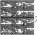
- FIG. 15is a series of X-rays showing successive 1.0 mm slices through both #17 and #32 in 1.0 mm increments.
- FIG. 16is a perspective view from a front corner showing a pre-operative upper-arch impression being taken of a simulated patient.
- FIG. 17is a cross-sectional view of an upper-arch impression being taken of a simulated patient.
- FIG. 18is a perspective view from above of the completed upper-arch impression.
- FIG. 19is a perspective view from above of the completed upper-arch impression, along with a stone model that will serve as a “positive” for manufacturing or fabricating of a custom surgical stent for that patient's upper-arch.
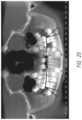
- FIG. 20is a CBCT scan with notations showing the measurement of the angle of entry into the tooth bud.
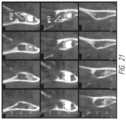
- FIG. 21is a series of X-rays with notations showing the measurement of the lateral angle of entry.
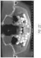
- FIG. 22is a CBCT scan with highlights showing the computed volume of each tooth bud.
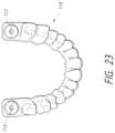
- FIG. 23is a perspective view from above of a surgical stent with two surgical guides, the stent having been manufactured or fabricated using the CBCT positioning information.
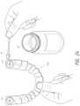
- FIG. 24is a perspective view showing topical anesthetic being applied to the base of the surgical guide.
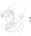
- FIG. 25is perspective view from a front corner of a surgical stent being seated on the upper-arch of the simulated patient.
- FIG. 26is a perspective view from a front corner of a local anesthetic being injected into a tooth bud site.
- FIG. 27is a perspective view from a front corner of a tissue trocar being used to punch to the base of a tooth bud.
- FIG. 28is a perspective view from a front corner of an ablation probe tip with a mechanical (physical) stop being positioned through the surgical guide into the tooth bud.
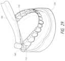
- FIG. 29is a perspective view from a front corner of the ablation probe tip being positioned in each tooth bud through the surgical guide so that the ablation probe tip's effective center of ablation is in the middle of each tooth bud.
- Third molar tooth bud agenesis(the lack of third molar formation) can only be conclusively determined by age 14. Third molar tooth buds are lying just 2.0 mm to 3.0 mm beneath the surface of the attached gingival (gum) tissue, making them accessible for rapid anesthesia and minimally invasive ablation with the correct selection of soft tissue ablation and supporting scanning and stent-manufacturing technologies.
- TSAminimally invasive tooth bud ablation
- the TBA procedure 70 ( FIG. 4 ) and TBA system 100 ( FIG. 5 ) for use in the TBA procedure 70seek to achieve: (1) a minimally invasive procedure consisting of a surgical access path at a surgical site (e.g. at each tooth bud surgical site), (2) that can predictably ablate all four third molar tooth buds 120 in thirty (30) minutes or less (including time to administer anesthesia) using either microwave (“MW”) or radio frequency (“RF”) ablation, (3) that can be administered by dental practitioners under normal office conditions, (4) with direct procedure costs reduced by 25% or more, and (5) with zero risks or complications when compared to traditional surgical extraction of fully developed third molars.
- a minimally invasive procedureconsisting of a surgical access path at a surgical site (e.g. at each tooth bud surgical site)
- MWmicrowave
- RFradio frequency
- TBA procedure 70is shown and described as a prophylactic third molar tooth bud ablation (TMTBA), but it is not limited thereto.
- TTBAprophylactic third molar tooth bud ablation
- there may be supernumerary teeth that should not be in a patient's mouthe.g. there may be two teeth #5), the removal of which would not be prophylactic in nature.
- One preferred advantage of the surgical phase 90 described hereinis that it is a minimally invasive surgical procedure.
- a minimally invasive surgical procedure designcoupled with electronic feedback controls using MW and RF ablation technology to limit soft tissue damage, performing this procedure on children aged 6-12 years old takes approximately thirty (30) (or fewer) minutes, including the time to administer local anesthetics.
- Another preferred advantage of the surgical phase 90 described hereinis that it will not accidentally disrupt adjacent second molar tooth development, even though the formation of second molars are well under way because these tooth buds 120 have started to form before birth.
- the use of relatively new scanning technologiese.g. computed tomography volume scanning such as cone beam computed tomography (CBCT) scanning and MRI volume scanning
- computed tomography volume scanningsuch as cone beam computed tomography (CBCT) scanning and MRI volume scanning
- CBCTcone beam computed tomography
- MRI volume scanningaccurate custom surgical stents 110 to guide ablation probe tip 108 placement
- the TBA procedure 70( FIG. 4 ) preferably includes a screening phase 72 , a pre-surgical phase 80 (also referred to as TBA pre-surgical phase 80 ) that includes pre-surgical scanning 82 and the assembling of a TBA surgical kit 88 (that includes pre-determined settings 105 as well as a surgical stent 110 ), a surgical phase 90 (also referred to as TBA surgical phase 90 ), and a follow-up phase 98 .
- a TBA system 100( FIG. 5 ) is preferably used during the surgical phase 90 (shown graphically in FIGS. 6 - 10 and as a flow chart in FIG. 11 ) of the TBA procedure 70 .
- the TBA system 100includes a TBA probe system 101 (including a generator 104 capable of emitting one or more types of ablation means 104 ′, a hand piece 106 , and an ablation probe tip 108 ) and at least one surgical stent 110 (which was manufactured or fabricated during the pre-surgical phase 80 ).
- Each stent 110has at least one surgical guide 112 to guide the placement of the ablation probe tip 108 so that its center of ablation 130 a is placed into the middle of the tooth bud 130 b .
- FIGS. 6 - 10show (and FIG. 11 describes) the procedure of inserting the ablation probe tip 108 through the surgical guide 112 of a stent 110 , ablating the tooth bud 120 , and removing the ablation probe tip 108 from the ablated tooth bud 120 ′.
- the TBA System 100The TBA System 100
- the TBA system 100 described hereinis the system that is used during the surgical phase 90 of the TBA procedure 70 .
- Some of the components (e.g. the custom surgical stent 110 and the pre-determined settings 105 ) used in the TBA system 100are part of the TBA surgical kit assembled during the pre-surgical phase 80 .
- the TBA system 100includes a TBA probe system 101 (including a generator 104 , a hand piece 106 , and an ablation probe tip 108 ) and at least one surgical stent 110 (each stent 110 has at least one surgical guide 112 to guide (direct) the placement of the ablation probe tip 108 to the middle of the tooth bud 130 b ).
- the generator 104 and the hand piece 106may be jointly referred to as the ablation probe unit 102 (or the programmable ablation probe unit 102 ).
- the generator 104 and hand piece 106may be integral or functionally connected together.
- the generator 104(and/or the ablation probe unit 102 ) may be programmed with pre-determined parameter settings 105 a and/or treatment time settings 105 b (referred to jointly as pre-determined settings 105 ).
- the generator 104(and/or the ablation probe unit 102 ) provides an ablation means 104 ′ for ablating the tooth bud 120 based on the pre-determined settings 105 .
- Central to the TBA system 100is the interaction between the ablation probe tip 108 and the surgical stents 110 (and specifically the surgical guides 112 ).
- the generator 104provides the ablation means 104 ′ suitable for ablating a tooth bud 120 during the surgical phase 90 of the TBA procedure 70 .
- MW energy and RF energyare discussed as exemplary preferred ablation means 104 ′.
- Another alternative preferred ablation means 104 ′is irreversible electroporation because it has subsecond activation times that can reduce collateral tissue damage.
- Yet another alternative preferred ablation means 104 ′include, but are not limited to, cryoablation, ultra-high intensity ultrasound, laser, chemical, thermal or hot tip (e.g. a tip having any source of heat including, but not limited to, a light bulb, a soldering iron, or steam heat), and/or mechanical means. These ablation means 104 ′ may also be combined either simultaneously or consecutively.
- ablation means 104 ′may also be used. It should be noted that although discussed primarily in terms of MW and RF, unless specifically set forth otherwise, the use of other ablation means 104 ′ is possible.
- the generator 104(alone or as part of an ablation probe unit 102 ) may be programmed by the operator and/or at the laboratory and/or factory and may be accomplished automatically or manually.
- the programming of the generator 104may include programming at least one pre-determined setting 105 .
- the hand piece 106is the functional intermediary between the generator 104 and the ablation probe tip 108 .
- the hand piece 106may be connected substantially at one end to the generator 104 . Substantially at the other end of the hand piece 106 , opposite the generator 104 , the end of the hand piece 106 (the surgical end) is adapted to accept the ablation probe tip 108 .
- the hand piece 106is preferably detachable from the generator 104 (if they are not an integral unit) and the ablation probe tip 108 is preferably detachable from the hand piece 106 .
- the ablation means 104 ′flows from the generator 104 through the ablation probe tip 108 and out to a center of ablation 130 a (the focal point of the ablation).
- the ablation probe tip 108is insertable through the surgical guide 112 , through the gingival tissue 122 , and into the middle of the tooth bud 130 b .
- the center of ablation 130 ais at the insertion end of the ablation probe tip 108 such that when the insertion end of the ablation probe tip 108 is positioned at the pre-defined angle ( ⁇ ) and pre-defined depth (x) during the surgical phase 90 , the center of ablation 130 a substantially coincides with or overlaps the middle of the tooth bud 130 b.
- the pre-defined angle ( ⁇ )is the angle at which the ablation probe tip's effective center of ablation 130 a is in the “middle” of the tooth bud 130 b as calculated (during the pre-surgical phase 80 ) as described herein or using an alternative method.
- the pre-defined depth (x)is the depth at which the ablation probe tip's effective center of ablation 130 a is in the “middle” of the tooth bud 130 b as calculated as described herein or using an alternative method.
- the phrase “middle of the tooth bud 130 b ”is meant to include the three-dimensional area within the tooth bud 120 and, in particular, the three-dimensional area within the tooth bud 120 that is more towards the absolute middle point than towards the outer periphery of the tooth.
- the pre-defined angle ( ⁇ ) and pre-defined depth (x)can also be referred to as the “calculated angle and depth,” the “prescribed angle and depth,” the “proper angle and depth,” the “correct angle and depth,” the “optimal angle and depth,” or the “ideal angle and depth.”
- the ablation probe tip 108includes a mechanical stop structure 140 (e.g. a band, protrusion, or shoulder) designed to physically limit the depth of the ablation probe tip 108 when used in conjunction with mechanical stop structure 142 (e.g. the upper surface, a protrusion on the upper surface, or a notch in the upper surface) of the surgical stent 110 and/or surgical guide 112 .
- mechanical stop structure 142 of the surgical guide 112 and the mechanical stop structure 140 of the ablation probe tip 108together limit how much of the ablation probe tip 108 can pass through the surgical guide 112 until there is a mechanical stop between the mechanical stop structure 142 of the surgical guide 112 and the mechanical stop structure 140 of the ablation probe tip 108 .
- Each ablation probe tip 108may be individually custom made (e.g. manufactured or fabricated) or may be selected from a family of ablation probe tips 108 (i.e. there may be a “family” of probe tips 108 that will cover all clinical possibilities for tooth bud diameters and depths).
- the characteristics of the ablation probe tip 108custom made or selected that may be taken into consideration include, for example, length, shape, angle, position of a mechanical stop structure 140 , diameter, and size, shape, and location of the center of ablation 130 a . For example, if a particular ablation probe tip 108 had mechanical stop structure 140 (shown as the bottom surface of an annular ring or shoulder in FIGS.
- the ablation probe tip 10810.0 mm from the absolute tip of the ablation probe tip 108 (and the center of ablation 130 a is substantially adjacent to the absolute tip), but the center of ablation 130 a was only 8.0 mm from the surface of the gingival tissue 122 (shown as (x) in FIG. 6 ), then the surgical guide 112 would have to be 2.0 mm thick (shown as (y) in FIG. 6 ).
- the ablation probe tip 108would either have to be made or selected so that the mechanical stop structure 140 is 8.5 mm from the center of ablation 130 a of the ablation probe tip 108 .
- the appropriate ablation probe tip 108preferably will result in the intra-operative placement of the effective center of ablation 130 a of the ablation probe tip 108 into the targeted middle of the tooth bud 130 b ⁇ 0.5 mm.
- the ablation probe tips 108may be sharp enough and/or may be strong enough so that the ablation probe tips 108 can be “self-introducing” in that the ablation probe tips 108 can be pushed through the gingival tissue 122 .
- tissue trocars 146described herein
- the ablation probe tips 108would not have to be as sharp and/or strong.
- the at least one custom surgical stent 110(also referred to as a “stent 110 ” or a “surgical stent 110 ”) has at least one surgical guide 112 (also referred to as “guides 112 ” or “ablation probe tip guides 112 ”). Two surgical stents 110 would be used, for example, if both upper and lower tooth buds 120 were to be ablated.
- the surgical stents 110are designed to seat in a patient's mouth and may be supported by at least one tooth (a tooth-supported surgical stent), soft tissue (a soft tissue-supported surgical stent), and/or bone (a bone-supported surgical stent). If the surgical stent 110 is supported by more than one of these, it could be considered a combination-supported surgical stent.
- Preferred surgical stents 110may “snap” into the mechanical undercuts inherent in the patient's erupted teeth.
- a surgical stent 110would have more than one surgical guide 112 if more than one tooth bud were to be ablated on either the upper or lower jaw.
- the surgical stents 110 and the guides 112 thereinare used to control both the pre-defined angle ( ⁇ ) and the pre-defined depth (x) of the ablation probe tip 108 in order to assure that the ablation probe tip's effective center of ablation 130 a is in the middle of the tooth bud 130 b ⁇ 0.5 mm.
- the pre-defined angle ( ⁇ )is primarily controlled by the angle of the surgical guides 112 (the passageways through the stent 110 ).
- the pre-defined depth (x)is primarily controlled by the interaction between the mechanical stop structure 142 of the surgical stent 110 (and/or surgical guide 112 ) and the mechanical stop structure 140 of the ablation probe tip 108 .
- the operatorinserts the ablation probe tip 108 at the entry angle ( ⁇ ) defined by the guide 112 and to the depth (x) limited by the mechanical stop structure 140 , 142 .
- the surgical guides 112are passageways through the surgical stent (the passageways being a type of guiding structure).
- the pre-defined angle ( ⁇ ) for each passageway (guide 112 )is determined by the position of the middle of the tooth bud 130 b . For example, if the middle of the tooth bud 130 b is “slightly forward” the angle ( ⁇ ) of the passageway (guides 112 ) would be “slightly forward” so that the ablation probe tip 108 is angled “slightly forward” so that the center of ablation 130 a is positioned substantially at the middle of the tooth bud 130 b .
- the angle ( ⁇ ) of the passagewayis determined (e.g.
- the guides 112may also be used to provide access for administering local anesthetic and to provide access to a tissue trocar 146 (if necessary).
- the mechanical stop structure 142is the upper surface of the surgical stent 110 and/or surgical guide 112 .
- the mechanical stop structure 142is substantially adjacent to or near the surgical guide 112 .
- the mechanical stop structure 142could be positioned at locations of the surgical stent 110 beyond the surgical guide 112 .
- Alternative preferred mechanical stop structure 142includes a protrusion on the upper surface or a notch in the upper surface.
- the size and shape of the mechanical stop structure 142is determined (calculated or designed) by a process that may be implemented as software or as a program and is based upon tooth bud volumes determined in pre-surgical volume scanning 82 as well as the length between the ablation probe tip mechanical stop structure 140 and its center of ablation 130 a .
- the process(that may be implemented by software or a program) would determine that the 2.6 mm ablation probe tip 108 is the appropriate ablation probe tip 108 (the 2.4 mm ablation probe tip 108 being too short), but that the surgical stent 110 and/or surgical guide 112 would have to be approximately 0.1 mm thick to make up the difference or the 2.6 mm ablation probe tip 108 would be able to be pushed in too far.
- FIG. 12is a flowchart showing the steps of a process (that may be implemented as one or more software program or subprograms if the shown steps are divided) that, in part, determines the pre-defined angle ( ⁇ ) and the pre-defined depth (x) (see steps 200 , 210 , 212 , 214 , 216 , and 218 ).
- a processthat may be implemented as one or more software program or subprograms if the shown steps are divided
- determines the pre-defined angle ( ⁇ ) and the pre-defined depth (x)see steps 200 , 210 , 212 , 214 , 216 , and 218 .
- the pre-determined settings 105include, for example, pre-determined parameter settings 105 a and/or treatment time settings 105 b that are needed to control (provide instructions to) the generator 104 (alone or as part of an ablation probe unit 102 ) to provide sufficient ablation means 104 ′ to ablate the tooth bud 120 , but not so much as to incur significant collateral soft tissue damage (e.g. to the gingival tissue 122 ).
- the pre-determined parameter settings 105 amight control the quantity and quality ablation means 104 ′ delivered to the tooth bud 120 .
- the actual pre-determined parameter settings 105 awill be highly dependent on the type of ablation means 104 ′ to be delivered.
- MW and RF ablation meansmight have parameters relating to wavelength and/or frequency
- hot tip ablation meansmight have parameters relating to temperature
- chemical ablation meansmight have parameters relating to the strength of the chemical and how fast the chemical is flowing into the tooth bud
- mechanical ablation meansmight have parameters relating to speed.
- the pre-determined settings 105are determined (which includes computing, calculating, looking up, processing, or otherwise determining) by a process (that may be implemented as software or a program) based upon tooth bud volumes determined in pre-surgical volume scanning 82 . It should be noted that the pre-determined settings 105 may take into consideration factors other than tooth bud volume including, but not limited to, image recognition programs to measure tooth bud location, age and size of the patient, and other relevant factors to successfully image the patient for the TBA procedure 70 . FIG.
- FIG. 12is a flowchart showing the steps of a process (that may be implemented as one or more software program or subprograms if the shown steps are divided) that, in part, determines the pre-determined parameter settings 105 a and/or treatment time settings 105 b (see steps 200 , 220 , 222 , and 224 ).
- the generator 104(and/or the ablation probe unit 102 ) may be programmed by the operator and/or technicians at the laboratory and/or factory.
- the programmingmay be automatic or manual.
- “Programming”includes having the pre-determined settings 105 pre-entered and/or entering (inputting) the pre-determined settings 105 manually or automatically into the generator 104 (and/or the ablation probe unit 102 ) via operator input mechanisms.
- the pre-determined settings 105may be preprogrammed into an ablation probe unit 102 , transmitted to the operator in the form of a programming signal (e.g. over the internet to be downloaded and installed in the ablation probe unit 102 or the generator 104 ), provided in the form of computer-readable media (e.g.
- the pre-determined settingsmight be displayed to the user for independent “verification” as the user could notice variations from normal pre-determined settings (e.g. the literature provided might provide a range and the operator would notice if the provided pre-determined settings 105 fell outside of the range).
- the pre-determined settingsmight be provided as a code that, when input, would only function if it corresponded with a logical setting (e.g. if the person's age was also input into the ablation probe unit 102 and the code was not a logical setting based on the age, the ablation probe unit 102 would not function).
- the pre-determined settings 105 for each TBA sitemay be included in the TBA surgical kit as a print out, on a disk or other computer readable storage media, or with instructions on how to obtain or download the information.
- the pre-determined ablation means parameter settings 105 acan also be referred to as “parameter settings 105 a ,” “preferred parameter settings 105 a ,” “optimal parameter settings 105 a ,” “ideal parameter settings 105 a ,” “pre-determined parameter settings 105 a ,” “recommended parameter settings 105 a ,” or “prescribed parameter settings 105 a.”
- At least one sharp instrumentsuch as a tissue trocar 146 (and sometimes a plurality of tissue trocars) may be used by the operator to introduce (initially create) the access opening through the thick attached gingival tissue 122 that overlays third molar tooth buds 120 .
- the tissue trocar tipsare preferably sharp enough to be pushed and/or punched through the gingival tissue 122 into the base of the tooth bud.
- the diameter of the tissue trocar 146rapidly increases up to 100% of the size of the ablation probe tip 108 .
- the TBA surgical kitis a package that includes the majority of the necessary components and information for the surgical phase 90 of the TBA procedure 70 .
- the TBA kitwill be assembled (or the assembly will be completed) based on the patient's impressions and volume scans.
- the TBA surgical kithas attractive packaging.
- the effective center of ablation 130 a of the ablation probe tip 108can be positioned at a pre-defined angle ( ⁇ ) and pre-defined depth (x) so that the ablation probe tip's effective center of ablation 130 a is positioned substantially in the “middle” of the tooth bud 130 b within approximately 50%, 25%, or even less than 10% of the average diameter of the tooth bud 120 . This is extremely accurate as compared to previous procedures.
- FIG. 4shows the steps and/or phases in an exemplary preferred TBA procedure 70 : (1) routine screening and diagnosis 72 ; (2) pre-surgical scanning 82 (including taking impressions 84 and using scanning technology 86 ); (3) assembling a TBA surgical kit 88 (including pre-determined settings 105 and a stent 110 ); (4) operator delivery of the surgical phase 90 of the TBA procedure 70 (shown in more detail in FIG. 11 ); and (5) post-surgical (follow-up) steps 98 .
- Steps (2) and (3)are also referred to jointly as the pre-surgical phase 80 during which steps are taken to create (including calculating, manufacturing, fabricating, selecting, and/or assembling) components of the TBA system 100 and/or the TBA surgical kit to be provided to the operator.
- Step (4)is also referred to as the surgical phase 90 of the TBA procedure 70 during which the steps shown in FIG. 11 are taken to ablate tooth buds 120 .
- Routine screening using panographic or intra-oral X-ray imaging techniquesis necessary to identify the presence of forming tooth buds 120 starting at age 6 through age 12 because of the wide range of ages involved with the formation of third molar tooth buds 120 .
- third molar tooth buds 120have been identified to be present using standard screening methods (screening phase 72 ), the next step is to pre-operatively measure the precise three-dimensional location and volume of each third molar tooth bud 120 .
- scanning technology 86e.g. computed tomography volume scanning such as CBCT.
- Scanning technology 86can be used to accurately generate the necessary three-dimensional volume scans (computed tomography volume scans) and measurements ⁇ 0.2 mm using, for example, the distal side of erupted first molars as durable physical landmarks (although it is possible to use soft tissue over bone as landmarks).
- the scanning technology 86produces tooth bud size and position data 86 ′ (also referred to as “volume scans” and/or “measurements”) that is provided for the step of producing the TBA surgical kit 88 .
- the tooth bud size and position data 86 ′may be provided as a scanning technology file that can be any data file generated by the scanning technology 86 with the data necessary to manufacture or fabricate a stent 110 .
- One exemplary type of scanning technology fileis a three-dimensional computer aided design (CAD) file.
- An impression 84 of the patient's teeth and gum tissueis made using standard impression materials such as PVS-type impression material (although other impression materials can be used).
- the impressions 84are then processed and/or scanned using scanning technology (e.g. CBCT imaging by dentists and/or CT imaging in the laboratory), and the resulting volume scan of the impression is emailed (or otherwise transmitted or delivered) to a laboratory and/or factory where the volume scan is used for manufacturing or fabricating. It is still possible to physically mail the PVS dental impressions 84 to the designated laboratory and/or factory for manufacturing or fabricating.
- scanning technologyis discussed primarily in terms of computed tomography volume scanning (e.g. CBCT technology), alternative scanning technologies including, but not limited to, ultrasound scanning technologies and future developed scanning technologies are included in the scope of the invention. Specialty software or programs may be used with the scanning technology 86 to accomplish the purpose described herein. It should be noted that alternative scanning technology 86 (including future developed scanning technology) may be used if it is able to accurately generate the necessary three-dimensional volume scans and measurements ⁇ 0.2 mm using the distal side of erupted first molars (or other landmarks) as durable physical landmarks.
- alternative scanning technologymay also be used as long as two- or three-dimensional scanning results in the positioning of the effective center of ablation 130 a within approximately 50%, 25%, or even less than 10% of the average diameter of the tooth bud 120 .
- the pre-surgical phase 80 of the TBA procedure 70includes assembling a TBA surgical kit 88 .
- This step of assembling a TBA surgical kit 88preferably includes computing pre-determined settings 105 and manufacturing or fabricating the stent 110 based on tooth bud size and position data 86 ′ obtained from the scanning technology 86 .
- the process of computing pre-determined settings 105may be controlled by a process (that may be implemented by software or a program).
- the process of manufacturing or fabricating the stent 110may also be controlled by a process (that may be implemented by software or a program).
- the process of manufacturing or fabricating the stent 110may be carried out using direct-digital manufacturing or fabricating techniques similar to the processes used for manufacturing or fabricating implant surgical stents directly from CBCT scans (e.g. the processes used for fabricating SurgiGuideTM and other implant surgical guides) and the process used for manufacturing or fabricating orthodontic aligners (e.g. orthodontic aligners made by Align Technology or ClearCorrect).
- direct-digital manufacturing or fabricating techniquessimilar to the processes used for manufacturing or fabricating implant surgical stents directly from CBCT scans (e.g. the processes used for fabricating SurgiGuideTM and other implant surgical guides) and the process used for manufacturing or fabricating orthodontic aligners (e.g. orthodontic aligners made by Align Technology or ClearCorrect).
- the direct-digital manufacturing or fabricating techniquesuse the tooth bud size and position data 86 ′ to position and angle the surgical guides 112 on the distal aspects of the surgical stents 110 and use the erupted first molars as the primary landmark for positioning.
- manufacturing or fabricatingwill usually be done remotely in a laboratory and/or factory, it is possible that larger clinics will have the ability to manufacture or fabricate surgical stents 110 in their own in-house laboratory and/or factory.
- Direct-digital manufacturing or fabricating techniquescan be defined as any manufacturing or fabricating process that creates physical parts directly from data (e.g. three-dimensional CAD files) using manufacturing or fabricating techniques including, but not limited to, surgical stent manufacturing or fabricating technologies, rapid turn-around fabrication technologies, computer aided manufacturing (CAM), technologies using CAD, computer numerical control (CNC) milling, “additive” manufacturing, direct-digital laser stereolithography fabrication, “3-D printing,” or any other manufacturing or fabricating means known or yet to be discovered that is capable of using the results generated by scanning to manufacture or fabricate the custom surgical stents.
- manufacturing or fabricating techniquesincluding, but not limited to, surgical stent manufacturing or fabricating technologies, rapid turn-around fabrication technologies, computer aided manufacturing (CAM), technologies using CAD, computer numerical control (CNC) milling, “additive” manufacturing, direct-digital laser stereolithography fabrication, “3-D printing,” or any other manufacturing or fabricating means known or yet to be discovered that is capable of using the results generated by scanning to
- direct-digital manufacturing or fabricating techniquesis one preferred manufacturing or fabricating technique, but more traditional manufacturing or fabricating techniques that require more labor intensive manual laboratory processing could also be used.
- At least one processthat may be implemented as software or as at least one program (e.g. custom software enhancements in the CBCT software) will preferably assist in the direct-digital manufacturing or fabricating of the surgical stents 110 and define (and/or compute or calculate) the pre-determined settings 105 .
- This processwould include defining (and/or computing or calculating) positioning and entry angle data required for placement of the ablation probe tip's effective center of ablation 130 a into the middle of the targeted tooth bud 120 .
- tooth bud volumesare preferably computed (possibly using the scanning technology) and then the tooth bud volumes are used to determine the pre-determined settings 105 necessary to effect therapeutic ablation.
- Tooth bud volumeswill generally range from 4.0 mm to 12.0 mm in diameter at ages 6-12.
- the ablation means 104 ′ and treatment timesare preferably considered in the calculations. Companies that make CBCT imaging equipment promote the development of procedure-specific software in order to gain end-user acceptance of their imaging systems in the market place. The process may use calculations and/or look-up charts (e.g. based on experimental data) for determining the necessary settings.
- FIG. 12is a flowchart showing the steps of a process (that may be implemented as one or more software programs or subprograms if the shown steps are divided) for manufacturing or fabricating custom surgical stents 110 and/or determining the pre-determined parameter settings 105 a and/or treatment time settings 105 b .
- the processbegins with receiving pre-operative measurements of the precise three-dimensional location and volume of each third molar tooth bud and information regarding the ablation probe unit including its ablation means capabilities 200 .
- the processwould preferably include the following steps: (a) determining an entry point for an ablation probe tip 210 ; computing the angle and depth of the path between the entry point and the middle of a tooth bud 212 ; (b) taking into consideration the depth of the path, creating or selecting an ablation probe tip having the proper distance between its mechanical stop and its center of ablation so that the ablation probe tip will be inserted so that its center of ablation will be in the middle of the tooth bud 214 ; (c) taking into consideration the angle and depth of the path and the thickness of the surgical stent, computing the surgical guide pathway through which the ablation probe tip will be inserted so that its center of ablation will be in the middle of the tooth bud 216 ; and (d) providing the surgical guide pathway as output for the creation of a surgical stent with surgical guides 218 .
- the processwould preferably include the following steps: (a) taking into consideration the information regarding the ablation probe unit including its ablation means capabilities, determining the proper power settings 220 ; (b) taking into consideration the information regarding the ablation probe unit including its ablation means capabilities, determining the proper time settings 222 ; and (c) providing the proper power and time settings as output for use in programming the ablation probe unit or generator 224 .
- the TBA surgical kitmay include at least one ablation probe tip 108 labeled for its respective surgical site, at least one tissue trocar 146 (if necessary), and patient and operator identification.
- the TBA surgical kitis provided to the operator.
- FIGS. 6 - 10show graphically, and FIG. 11 shows as a flow chart, the surgical phase 90 of the TBA procedure.
- the surgical phase 90may be performed by a dental operator (dental practitioner) in his office (e.g. a pediatric office and/or general dental office) under normal office conditions.
- the generator 104has been programmed with the pre-determined settings 105 and normal surgical procedures have been followed.
- the generator 104is preferably tuned so that the ablation means 104 ′ is set to ablate the small, substantially spherical ablation volumes of third molar tooth buds 120 in order to minimize (or possibly eliminate) collateral osseous and soft tissue damage, especially damage to adjacent second molars that are likely not yet erupted.
- the surgical phase 90uses single-use and disposable delivery systems that use components designed for intra-oral use.
- the first stepis physically seating a surgical stent 160 in a patient's mouth.
- the operatormakes an access path at the at least one tooth bud surgical site 162 .
- the operatoralso places the ablation probe tip so that the center of ablation is in the middle of a tooth bud at the at least one tooth bud surgical site (using the custom surgical stent to guide the placement) 164 .
- the ablation probe tipis “self-introducing,” the step of making an access path and the step of placing the ablation probe tip may occur simultaneously.
- the at least one tooth budis at least partially ablated 166 and the ablation probe tip is removed from the tooth bud 168 .
- the operatorpreferably starts the surgical phase 90 by placing the surgical stent 110 into place onto the patient's teeth prior to administering local anesthetic to the surgical site.
- the local anestheticwill then be administered through the surgical stent 110 and guides 112 that are in close approximation with the gingival tissue 122 , thus reducing the amount of anesthetic necessary because of the precise placement of anesthetic agent.
- Achieving local anesthesia in this procedurewill be easier than anesthetizing lower permanent molar teeth for routine fillings since only soft tissues, which will be 8.0 mm to 15.0 mm deep, are involved.
- the step of physically seating a surgical stent 110may also include physically seating the surgical stent in a patient's mouth, physically seating the surgical stent on a patient's erupted teeth, physically seating the surgical stent on at least one tooth in a patient's mouth, physically seating the surgical stent on a patient's soft tissue, physically seating the surgical stent on a patient's bone, or a combination of the above steps (e.g. physically seating the surgical stent on a patient's teeth, soft tissue, and bone).
- the operatorthen mechanically gains access to the tooth bud 120 through the stent surgical guides 112 by creating (introducing) a small surgical access path opening through the gingival tissue 122 approximately 0.1 mm to 2.0 mm (and more particularly 0.5 mm to 1.0 mm) in diameter using tissue trocars.
- tissue trocarstissue trocars
- the ablation probe tips 108are designed to be strong enough and sharp enough to act as “self-introducing” probe tips, they can be used to introduce the surgical access path.
- the ablation probe tip itselfis not self-introducing, the surgical access path may be introduced using a then there will be no need for separate tissue trocar 146 .
- the surgical access pathis preferably an incision, a puncture, or a hole through the gingival tissue 122 .
- the surgical access pathhas substantially the same diameter as the ablation probe tip 108 .
- the surgical access pathmay be a sutureless puncture (0.1 mm to 2.0 mm in diameter) or, more particularly, a sutureless puncture (0.5 mm to 1.0 mm in diameter).
- a trocar “punch”may be made through tough gingival tissue 122 .
- using a surgical access path to gain access or allow placement of the ablation probe tips 108 to the tooth bud 120does not kill, damage, or otherwise cause necrosis to the surrounding soft tissues (e.g. gingival tissues 122 ).
- Thiscan be compared to other processes such as coring, boring, cutting, electrosurge ablating, or other invasive procedures that kill, damage, and/or otherwise cause necrosis to the soft tissue to which the invasive procedure has been applied.
- the preferred procedures for introducing the surgical access pathmight kill individual cells, the soft tissue (the gingival tissue 122 ) does not become necrosed because the tissue is a collection of cells that can heal itself.
- the next step in the surgical phase 90is to insert the designated ablation probe tip 108 through the surgical stent 110 and into the tooth bud space until it is mechanically “stopped” in order to position the probe to the prescribed depth (which would be the pre-defined depth).
- the surgical stent 110 and its surgical guides 112are used to control the angle ( ⁇ ) and depth (x) of the ablation probe tip 108 so that the effective center of ablation 130 a of the ablation probe tip is in the middle of the tooth bud 130 b . It should be noted that the effective center of ablation 130 a for any given ablation technology does not necessarily correspond with the tip of the ablation probe.
- microwave ablation probeshave windows or slots that may be 0.5 mm to 2.0 mm from the tip depending on the frequency of the wavelength used.
- Cryoablation probeshave their center of ablation roughly in the middle of the probe, depending on the design and refrigerant used.
- a mechanical stop structure 140 on the ablation probe tip 108preferably seats firmly onto the mechanical stop structure 142 of the surgical stent guide 112 to prevent over extension of the ablation probe tip 108 .
- FIG. 8shows embedded temperature sensors 144 (or other types of feedback control mechanisms) that may be used during the ablation process.
- An independent feedback process using the temperature sensors 144is preferable for this clinical procedure.
- Use of temperature sensors 144 along with monitoring probe impedance characteristics and percentage of reflected energy in RF/MW circuitswill provide “go/no go” output for the clinician.
- Control algorithmsare preferably used to accelerate initial ablation means 104 ′ input followed by lower-level temperature maintenance for a defined period of time with independent confirmation that results in a fast process while simultaneously assuring complete tooth bud ablation.
- FIG. 9shows the actual ablation process.
- Activation of the ablation probe unit 102 to perform the ablation processis executed according to the pre-determined settings 105 .
- Activation of the ablation probe unit 102causes the generator 104 to provide the ablation means 104 ′ that passes through the hand piece 106 and the ablation probe tip 108 and into the tooth bud 120 .
- This step of at least partially ablating the tooth budis preferably accomplished without ablating any surrounding gingival tissue (although a minimal amount of surrounding gingival tissue may be ablated as an accidental byproduct of the step). This can also be thought of as the activation of the ablation probe unit 102 creating a zone of ablation that resides predominantly or completely within the tooth bud 120 .
- the feedback control mechanisms 144assure successful delivery of adequate ablation means 104 ′ to ablate the tooth bud 120 while minimizing damage to adjacent osseous and soft tissues by, for example, eliminating over-heating. Given the small tissue volumes involved for pediatric patients, activation using an RF ablation means 104 ′ would have an ablation time that is preferably less than three (3) minutes and activation using an MW ablation means 104 ′ would have an ablation time that is preferably less than thirty (30) seconds.
- FIG. 10shows the ablation probe tip 108 being removed from the now ablated tooth bud 120 ′. As shown in this figure, any access path created by the procedure rapidly closes.
- the patientmay have follow-up including, but not limited to, post-surgical instructions and, if necessary follow-up care and screening.
- Post-surgical instructions that may be given to parentsincludes the following: kids can go out and play immediately unless they were sedated, no post-surgical pain medication is necessary, bleeding (if any) will be gone in minutes, and post-surgical X-ray screening may be necessary at patient's next routine 6-month hygiene cleaning appointment to verify full ablation.
- FIGS. 13 - 29detail an exemplary simulated TBA procedure 70 including routine screening and diagnosis 72 , the pre-surgical phase 80 , and the surgical phase 90 .
- a patient's mouth 124(with gums 122 and teeth 126 ) is shown that looks like a stone model, but it should be understood that unless otherwise specified the shown mouth 124 would be a live patient's mouth.
- FIG. 13is a panographic X-ray showing a patient whose third molar tooth buds 120 in the #17 & #32 positions are treatable by a TBA procedure 70 .
- FIG. 14is a pre-operative CBCT scan (although other types of volume scanning could be used) of a patient. In a real procedure, the volume scan would be taken of the specific patient on whom the TBA procedure 70 is being performed. This CBCT “reconstructed” panographic scan has a 1.0 mm scale along its bottom edge.
- FIG. 15is a series of CBCT volume scan cross-sections showing successive 1.0 mm slices through both #17 and #32 in 1.0 mm increments. Each X-ray corresponds to 1.0 mm locations along the scale of FIG. 14 . The left-side scale is 1.0 mm vertically. The maximum tooth bud diameters are measured to be 8.0-9.0 mm.
- FIG. 16shows a pre-operative upper-arch impression 84 being taken of the simulated patient's mouth 124 (shown as a stone model for clarity, but an impression 84 would be taken of the patient himself) using an impression tray 128 . It is assumed that all four tooth buds of the wisdom teeth are present in the simulated patient.
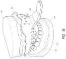
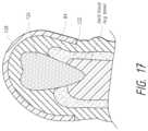
- FIG. 17is a cross-sectional view of the upper-arch impression 84 being taken of a simulated patient.
- FIG. 18shows the completed upper-arch impression 84 .
- a similar processwould be performed to manufacture or fabricate a pre-operative lower-arch impression 84 . At this time the practitioner may send impressions 84 and volume scan data to a laboratory and/or factory for processing.
- the laboratory and/or factoryuses the impressions 84 and volume scan data (scanning technology file) to create (including calculating, manufacturing, fabricating, selecting, and/or assembling) components of the TBA system 100 (including the surgical stents 110 and the pre-determined settings 105 ).
- the surgical stents 110 and the pre-determined settings 105 and other componentsare then assembled into the TBA surgical kit to be provided to the operator.
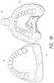
- FIG. 19shows the completed upper-arch impression 84 , along with a stone model 85 that will serve as a “positive” for manufacturing or fabricating a surgical stent 110 for that patient's upper-arch.
- the impressions 84can be computed tomography (“CT”) scanned to digitize as an alternative to making physical intermediates.
- CT volume scan filescanning technology file
- the practitionermay handle the processing in-house.
- FIG. 20is a CBCT scan with notations showing the measurement of the perpendicular angle of entry into the tooth bud 120 . The measurement is based on the distal aspect of the molar and the occlusal bite plane of the teeth.
- FIG. 21is a series of X-rays with notations showing the measurement of the lateral angle of entry. The measurement is determined relative to the vertical axis in order to avoid the jaw's boney interferences during surgical placement of the ablation probe unit 102 .
- FIG. 22is a CBCT scan with highlights showing the computed volume of each tooth bud 120 . CBCT volume data is used to determine and/or calculate the pre-determined settings 105 .
- FIG. 23shows the resulting surgical stent 110 that will be placed in a patient's mouth 124 .
- the shown stenthas two surgical guides 112 based upon the location of the patient's two tooth buds to be ablated.
- the surgical stent(s) 110 and the pre-determined setting(s) 105are provided to the operator along with the rest of the TBA surgical kit.
- the ablation probe unit 102 and/or the generator 104Prior to the surgical phase 90 of the TBA procedure 70 , the ablation probe unit 102 and/or the generator 104 should be set up so that at least one pre-determined setting 105 is correctly entered for at least one tooth bud 120 with safety interlocks carefully considered. (The pre-determined settings 105 may all be entered prior to the surgical phase 90 or they may be entered one at a time.) The surgical phase 90 of the TBA procedure 70 may then be performed.
- FIG. 24shows topical anesthetic 87 being applied to the base of the surgical guide 112 ( FIG. 24 ) prior to the surgical stents 110 being seated in a patient's mouth 124 .
- FIG. 25shows the surgical stent 110 being seated on the upper-arch of the simulated patient's mouth 124 (shown as a stone model for clarity). This process would be repeated on the lower-arch of the simulated patient.
- FIG. 26shows a local anesthetic being injected 89 into each site through a surgical guide 112 of the stent 110 .
- FIG. 27shows a tissue trocar 146 being used to create an access path through the gingival tissue 122 to the base of each tooth bud 120 .
- the tissue trocar 146is only necessary if self-introducing ablation probe tips 108 are not used.
- FIG. 28shows an ablation probe tip 108 with mechanical stop structure 140 ′ (shown as a shoulder) being inserted through the surgical guide 112 . This would be similar to the position of the ablation probe tip 108 in FIG. 6 .
- FIG. 29shows the ablation probe tip 108 positioned through the surgical guide 112 and into the tooth bud 120 through the surgical guide 112 so that the ablation probe tip's effective center of ablation 130 a is in the middle of each tooth bud 120 . This would be similar to the position of the ablation probe tip 108 in FIG. 7 .
- the ablation means 104 ′is delivered in this position ( FIG. 9 ).
- the ablation means 104 ′is delivered based on the pre-determined settings 105 (e.g. times, intensities, and other prescribed settings unique to each tooth bud).
- the ablation probe tip 108would then be removed and the process repeated at the site of each tooth bud 120 . Once the entire surgical phase 90 is complete, the surgical stents 110 are removed.
- the dental practitioner or an assistantprovides post-surgical instructions to the patient or a caregiver of the patient.
- An alternative to the pre-surgical phase 80 of the TBA procedure 70 described aboveincludes simultaneous three-dimensional scanning of both hard tissues (bone and teeth) and soft tissues (tooth bud 120 and gingival tissue 122 ). From the information obtained using this unique simultaneous three-dimensional scanning, a custom surgical stent 110 may be manufactured or fabricated. As discussed, the custom surgical stent 110 is used in the surgical phase 90 to help with the placement of the center of ablation 130 a into a tooth bud 120 that results in tooth agenesis.
- the simultaneous three-dimensional scanninguses a single scan to obtain both soft tissue and hard tissue information.
- Soft tissue informationgenerally does not show on a scan, although progress in volume scanning is improving and this may be possible in the near future.
- Known and future technologies able to provide a scan image of soft tissueare included in the scope of this invention.
- a typical X-ray scanwill only show the hard tissue.
- a dental impression 84is used that can be viewed on an X-ray.
- the dental impression 84is made of materials that are preferably “contrast optimized” for high resolution X-ray volume scanning.
- the ideal level of contrast agent in the range of 25% to 75% radiopacity(such as barium or iodine based compounds) is mixed into the dental impression materials so that the highest level of surface detail can be picked up on when volume scanning the dental impression 84 .
- the dental impression 84is placed in the patient's mouth 124 during the X-ray volume scan.
- the resulting X-ray volume scan imagewould show the tooth distinguished (is visible) and the dental impression 84 distinguished (is visible) and the void therebetween would be the soft tissue and would therefore be “visible.”
- the resulting X-ray volume scan with both hard and soft tissue informationmay then be used to formulate the custom stent 110 used in the surgical phase 90 described herein.
- an X-ray volume scan imageis generated in which hard tissue (e.g. a tooth) is visible hard tissue and the dental impression 84 is a visible dental impression and soft tissue (e.g. gingival tissue 122 ) is “visible” as the space between the visible hard tissue and the visible dental impression.
- hard tissuee.g. a tooth
- dental impression 84is a visible dental impression
- soft tissuee.g. gingival tissue 122
- One separate preferred pre-surgical phase 80 of the TBA procedure 70preferably includes using X-ray volume scans of dental impressions 84 to manufacture or fabricate surgical stents 110 .
- the X-ray volume scan of the dental impression 84is “super imposed” over the patient X-ray volume scan (e.g. CBCT scanning) using the dental hard tissues (the teeth) to “snap” the two volume scans together into an accurate overlay so that soft tissues of the mouth (which cannot be X-ray volume scanned directly) are accurately defined for the surgical stent manufacturing or fabricating (which must take into account the soft tissue and teeth) and probe positioning (which must take into account the tooth bud positioning from the patient's CBCT scan).
- One separate preferred pre-surgical phase 80 of the TBA procedure 70preferably includes using dental impression materials that are “contrast optimized” for high resolution X-ray volume scanning that is then used to manufacture or fabricate surgical stents 110 .
- the ideal level of contrast agent(such as barium or iodine based compounds) is mixed into the dental impression materials so that the highest level of surface detail can be picked up on when CT volume scanning the dental impression 84 .
- Separate preferred surgical procedurespreferably include the ablation of “non-tooth” bud lesions or tumors of the maxilla or mandible.
- a custom stentwould be manufactured or fabricated with guides to guide an ablation probe tip 108 to such a lesion or tumor located at least one lesion or tumor surgical site. The process could then be used to ablate such lesion or tumor.
- Separate TBA surgical procedurespreferably include the use of ultrasound scanning with combined ultra-high energy ultrasound ablation but without the use of a surgical stent for transgingival tooth bud ablation that results in tooth agenesis. This can be described as direct ultrasound scanning with ultra-high energy ultrasound built into the same scanning head.
- the Silvestri processrelies on molds taken from molds of the mouths of euthanized rat pups rather than using molds fabricated for the rat pup on which the procedure was to be performed.
- the present inventionuses the patient's mouth on which the procedure is to be performed.
- Another way in which the Silvestri process was inexactwas that the Silvestri process did not locate the forming tooth bud 120 . More specifically, the Silvestri process did not locate or determine the location of the forming tooth bud 120 pre-operatively relative to the landmarks that he used. Silvestri even states “ . . . when electrosurgical energy is applied near the invisible tooth strom in the tiny mouth of newborn rats, the effects of the electrosurgical energy cannot be nearly as local or precise.
- the TBA procedure 70 described hereincan be distinguished from the Silvestri procedure in several ways including for example, that (1) the TBA procedure 70 described herein is a minimally invasive procedure consisting of introducing a surgical access path at each tooth bud surgical site as opposed to the boring, killing, and damaging procedure described by Silvestri, (2) the TBA procedure 70 described herein is performed in such a manner that it can be described as exact (e.g.
- the patient's mouthas the mold for manufacturing or fabricating the surgical stent 110 , taking exact measurements of the patient's mouth (including the position of the tooth bud 120 ), and using calculated parameter and time settings 105 b ) as opposed to the Silvestri procedure that can be described as inexact, and (3) the TBA procedure 70 described herein can predictably ablate tooth buds 120 as opposed to the Silvestri procedure that was essentially unpredictable and could never, under any circumstances, be considered for treating human patients.
- FIGS. 4 , 11 , and 12are flow charts illustrating processes, methods, and/or systems. It will be understood that at least some of the blocks of these flow charts, components of all or some of the blocks of these flow charts, and/or combinations of blocks in these flow charts, may be implemented by software (e.g. coding, software, computer program instructions, software programs, subprograms, or other series of computer-executable or processor-executable instructions), by hardware (e.g. processors, memory), by firmware, and/or a combination of these forms.
- softwaree.g. coding, software, computer program instructions, software programs, subprograms, or other series of computer-executable or processor-executable instructions
- hardwaree.g. processors, memory
- firmwaree.g. firmware
- computer program instructionsmay be loaded onto a computer (or on a special purpose machine such as a volume scanner or scanning technology) to produce a machine, such that the instructions that execute on the computer create structures for implementing the functions specified in the flow chart block or blocks.
- These computer program instructionsmay also be stored in a memory that can direct a computer to function in a particular manner, such that the instructions stored in the memory produce an article of manufacture including instruction structures that implement the function specified in the flow chart block or blocks.
- the computer program instructionsmay also be loaded onto a computer (or on a special purpose machine such as a volume scanner or scanning technology) to cause a series of operational steps to be performed on or by the computer to produce a computer implemented process such that the instructions that execute on the computer provide steps for implementing the functions specified in the flow chart block or blocks.
- the term “loaded onto a computer”also includes being loaded into the memory of the computer or a memory associated with or accessible by the computer (or on a special purpose machine such as a volume scanner or scanning technology).
- memoryis defined to include any type of computer (or other technology)-readable media including, but not limited to, attached storage media (e.g. hard disk drives, network disk drives, servers), internal storage media (e.g.
- blocks of the flow chartssupport combinations of steps, structures, and/or modules for performing the specified functions. It will also be understood that each block of the flow charts, and combinations of blocks in the flow charts, may be divided and/or joined with other blocks of the flow charts without affecting the scope of the invention. This may result, for example, in computer-readable program code being stored in whole on a single memory, or various components of computer-readable program code being stored on more than one memory.
- the term “or”is used in its nonexclusive form (e.g. “A or B” includes A, B, A and B, or any combination thereof, but it would not have to include all of these possibilities). It should be noted that, unless otherwise specified, “and/or” is used similarly (e.g. “A and/or B” includes A, B, A and B, or any combination thereof, but it would not have to include all of these possibilities). It should be noted that, unless otherwise specified, the term “includes” means “comprises” (e.g. a device that includes or comprises A and B contains A and B but optionally may contain C or additional components other than A and B). It should be noted that, unless otherwise specified, the singular forms “a,” “an,” and “the” refer to one or more than one, unless the context clearly dictates otherwise.
Landscapes
- Health & Medical Sciences (AREA)
- Oral & Maxillofacial Surgery (AREA)
- Veterinary Medicine (AREA)
- Epidemiology (AREA)
- Life Sciences & Earth Sciences (AREA)
- Animal Behavior & Ethology (AREA)
- General Health & Medical Sciences (AREA)
- Public Health (AREA)
- Dentistry (AREA)
- Neurology (AREA)
- Biomedical Technology (AREA)
- Engineering & Computer Science (AREA)
- Neurosurgery (AREA)
- Nuclear Medicine, Radiotherapy & Molecular Imaging (AREA)
- Surgery (AREA)
- Dental Tools And Instruments Or Auxiliary Dental Instruments (AREA)
- Laser Surgery Devices (AREA)
- Prostheses (AREA)
- Surgical Instruments (AREA)
Abstract
Description
Preferred generators 104 may be multi-use devices designed as 110V counter-top units.Preferred generators 104 may be MW/RF generators with output levels determined initially through finite element analysis models or experimentally derived functions that exist for tumor ablation.- Preferred generators104 (and/or ablation probe units102) may have operator input mechanisms (e.g. knobs, dials, key pads, keyboards, I/O interfaces, connections to the internet, or other means for inputting or programming) in which the operator inputs (or allows input of) the
pre-determined settings 105. - Preferred generators104 (and/or ablation probe units102) may have output mechanisms103 (e.g. a display or audio) for providing setting feedback (e.g. calibration cycles and pre-determined settings105), warning feedback (e.g. to prevent operator mishandling), and intra-operative feedback on the progress of the procedure such as time remaining (e.g. a count down or a series of beeps to alert the operator to procedure completion) and/or temperature (e.g. to alert the operator to overheating).
- Preferred output displays may be digital readout displays (that may be color and/or in a large format) that permit the operator to easily see feedback intra-operatively from across a standard dental operatory (approximately 6-8 feet viewing distance).
Preferred hand pieces 106 preferably hold or secure anablation probe tip 108 by latching theablation probe tip 108 into the hand piece head. In somehand pieces 106, theablation probe tip 108 latches into the hand piece head at an angle (e.g. a 90 degree angle). It should be noted that the terms “latching” and “latch” are used to describe any type of secure fit including, but not limited to, clipping, snapping, or holding.Preferred hand pieces 106 preferably have a hand piece head (attached or integral) that is at an approximately 20 degree angle to the rest of the hand piece. This bend emulates a standard dental high-speed hand piece to facilitate easy access of both upper and lower surgical sites. In somepreferred hand pieces 106, the 20 degree bend can be adjusted intra-operatively to permit improved operator access to both upper- and lower-arches.Preferred hand pieces 106 preferably are rapidly detachable from thegenerator 104. Preferably the connectors are ultra-reliable for repeated removal/attachment from thegenerator 104.Preferred hand pieces 106 are preferably fully steam autoclavable. Alternativepreferred hand pieces 106 are disposable or have disposable covers.Preferred hand pieces 106 preferably have actuators to allow operator activation. The actuators may be separate from thehand pieces 106 or integral therewith. Exemplary actuators include, but are not limited to a wireless foot control or a hand-operated switch on thehand piece 106.- The
hand piece 106 may be integral with thegenerator 104 to form a hand-held integrated generator unit (hand-held integrated ablation probe unit).
- Preferred
ablation probe tips 108 are preferably disposable (e.g. single-use). - Preferred
ablation probe tips 108 may be specially designed to work with the specific ablation means104′ produced by thegenerator 104. Other preferredablation probe tips 108 may be designed to work with multiple types of ablation means104′ produced by thegenerator 104 orgenerators 104. - The design of the
ablation probe tip 108 may be dependent on the physics involved with transmitting ablation means104′ through the smallest possible diameter with an ideal maximum diameter. For example, an MW/RF ablation probe tip may be designed for transmitting MW/RF energy through the smallest possible diameter with an ideal maximum diameter of 0.5 mm to 1.0 mm targeted. - The “family” of
probe tips 108 may includeprobe tips 108 having a variety of characteristics. For example, the family might haveprobe tips 108 of different lengths ranging from 5.0 mm to 20.0 mm. This range would accommodate the various diameters of thetooth buds 120 and overlyinggingival tissue 122 thicknesses. - Intra-operative temperature sensing (shown as being performed by a linear array of
temperature sensors 144 inFIG.8 ) is preferably provided at or near the apex of the ablation probe tip108 (assuming placement in the ideal middle of thetooth bud 130b) and/or along theshaft 145 of theprobe tip 108.Temperature sensors 144 provide core temperatures for feedback control purposes (so that the operator can monitor the temperature and/or for software feedback control loops and emergency shutdown) and/or for safety controls to reduce or eliminate collateral tissue damage. Intra-operative tissue temperature is preferably measured, both to assure complete ablation and to prevent over-heating of tissues; this may require additional set up data or programming. Iftemperature sensors 144 are used, the appropriateablation probe tip 108 preferably will result in the intra-operative placement of the effective center ofablation 130aof theablation probe tip 108 into the targeted middle of thetooth bud 130b±1.0 mm.
- Preferred
- Preferred
surgical stents 110 are preferably disposable (e.g. single-use). - Manufacturing or fabricating of the custom
surgical stents 110 may be based upon Poly Vinyl Siloxane (PVS) full arch impressions of the patient's erupted teeth using either conventional lab fabrication techniques or direct-digital manufacturing or fabricating techniques. If an operator has a CBCT unit in his office, it may be possible to directly scan the PVS impressions and email the volume scan of the impression to eliminate the need to physically send them to the lab. The impression materials may include materials other than PVS and preferably will be contrast-optimized through the addition of X-ray contrast agents (such as barium or iodine) to provide optimized volume scans of the dental impression for resolving the fine surface detail of the teeth andgingival tissue 122. This unique material would be a radiographic contrast-optimized dental impression material for high resolution X-ray CT volume scanning. - Preferred
surgical stents 110 are preferably made of any appropriate material including, but not limited to, plastic, acrylic, or other nontoxic sturdy material suitable for use in a patient's mouth. One exemplarysurgical stent 110 composition may be, for example, clear acrylic (polymethyl methacrylate). It should be noted that materials suitable for additive-type manufacturing (or other direct-digital manufacturing or fabricating techniques) that resulted in nontoxic sturdy stents would be preferable. - Preferred
surgical stents 110 preferably have markings such as color codes or numbering clearly marking or identifying the tooth bud numbering sites. - Once the
surgical stent 110 is seated onto the patient's teeth, it preferably will remain firmly in place throughout thesurgical phase 90 of theTBA procedure 70. - The operator may administer local anesthetic through the
guides 112.
- Preferred
- An exemplary TBA surgical kit may consist of (a) a custom
surgical stent 110 for each arch as required, (b) at least oneablation probe tip 108 labeled its respective surgical site, (c) at least one tissue trocar146 (if necessary), and (d)pre-determined settings 105 for each TBA site along with patient and operator identification. - If feedback controls are a part of the ablation probe tip design, then the correct in situ tissue temperature settings are preferably computed and supplied with the
ablation probe tips 108 as part of the surgical kit. - The
generator 104 and/or thehand pieces 106 are standard equipment in a dental office and/or can be purchased separately. - The
ablation probe tips 108 may be pre-purchased (or extras may be kept in a practitioner's office) in which case the TBA surgical kit would provide a part number or other identifying information so that the practitioner would know whichablation probe tip 108 should be used with eachguide 112. - It should be noted some of the components may not be part of the physical TBA surgical kit. For example, the
pre-determined settings 105 may be provided electronically.
TheTBA Procedure 70
- An exemplary TBA surgical kit may consist of (a) a custom
- The terms “fabricating” and/or “manufacturing” include any suitable means of making a component (e.g. stent110). Although the terms are used together throughout most of the specification (e.g. “manufacturing or fabricating”), the absence of one term or another is irrelevant because they are used herein synonymously.
- The terms “proper,” “correct,” “optimal,” and “ideal,” are relative and may become more accurate as technology is developed. For example, when used in terms of the pre-defined angle (ϕ) and pre-defined depth (x) that are calculated and/or prescribed (e.g. the “proper angle and depth,” the “correct angle and depth,” the “optimal angle and depth,” or the “ideal angle and depth”), these phrases are meant to include the best possible angle and depth that is calculated using the best available information and technology.
- The terms “provide” and “providing” (and variations thereof) are meant to include standard means of provision including “transmit” and “transmitting,” but can also be used for non-traditional provisions as long as the data is “received” (which can also mean obtained). The terms “transmit” and “transmitting” (and variations thereof) are meant to include standard means of transmission, but can also be used for non-traditional transmissions as long as the data is “sent.” The terms “receive” and “receiving” (and variations thereof) are meant to include standard means of reception, but can also be used for non-traditional methods of obtaining as long as the data is “obtained.”
Claims (6)
Priority Applications (3)
| Application Number | Priority Date | Filing Date | Title |
|---|---|---|---|
| US16/391,277US12076198B2 (en) | 2009-05-11 | 2019-04-22 | Therapeutic tooth bud ablation |
| US18/243,634US20230414318A1 (en) | 2010-05-10 | 2023-09-07 | Therapeutic Tooth Bud Ablation |
| US18/680,245US20240315802A1 (en) | 2009-05-11 | 2024-05-31 | Therapeutic tooth bud ablation |
Applications Claiming Priority (9)
| Application Number | Priority Date | Filing Date | Title |
|---|---|---|---|
| US17714309P | 2009-05-11 | 2009-05-11 | |
| US12/863,183US9402693B2 (en) | 2009-05-11 | 2010-05-10 | Therapeutic tooth bud ablation |
| PCT/US2010/034259WO2010132368A1 (en) | 2009-05-11 | 2010-05-10 | Therapeutic tooth bud ablation |
| US13/093,844US10820963B2 (en) | 2009-05-11 | 2011-04-26 | Therapeutic tooth bud ablation |
| US13/093,842US9827068B2 (en) | 2009-05-11 | 2011-04-26 | Therapeutic tooth bud ablation |
| US15/694,791US10265140B2 (en) | 2009-05-11 | 2017-09-02 | Therapeutic tooth bud ablation |
| US15/694,794US10285778B2 (en) | 2009-05-11 | 2017-09-02 | Therapeutic tooth bud ablation |
| US15/886,729US10299885B2 (en) | 2009-05-11 | 2018-02-01 | Therapeutic tooth bud ablation |
| US16/391,277US12076198B2 (en) | 2009-05-11 | 2019-04-22 | Therapeutic tooth bud ablation |
Related Parent Applications (4)
| Application Number | Title | Priority Date | Filing Date |
|---|---|---|---|
| US13/093,844ContinuationUS10820963B2 (en) | 2009-05-11 | 2011-04-26 | Therapeutic tooth bud ablation |
| US15/694,794ContinuationUS10285778B2 (en) | 2009-05-11 | 2017-09-02 | Therapeutic tooth bud ablation |
| US15/694,791ContinuationUS10265140B2 (en) | 2009-05-11 | 2017-09-02 | Therapeutic tooth bud ablation |
| US15/886,729ContinuationUS10299885B2 (en) | 2009-05-11 | 2018-02-01 | Therapeutic tooth bud ablation |
Related Child Applications (2)
| Application Number | Title | Priority Date | Filing Date |
|---|---|---|---|
| US14/849,464ContinuationUS9855112B2 (en) | 2009-05-11 | 2015-09-09 | Therapeutic tooth bud ablation |
| US18/680,245ContinuationUS20240315802A1 (en) | 2009-05-11 | 2024-05-31 | Therapeutic tooth bud ablation |
Publications (2)
| Publication Number | Publication Date |
|---|---|
| US20190247144A1 US20190247144A1 (en) | 2019-08-15 |
| US12076198B2true US12076198B2 (en) | 2024-09-03 |
Family
ID=43085289
Family Applications (8)
| Application Number | Title | Priority Date | Filing Date |
|---|---|---|---|
| US12/863,183Active2032-03-27US9402693B2 (en) | 2009-05-11 | 2010-05-10 | Therapeutic tooth bud ablation |
| US13/093,844Active2030-09-18US10820963B2 (en) | 2009-05-11 | 2011-04-26 | Therapeutic tooth bud ablation |
| US13/093,842ActiveUS9827068B2 (en) | 2009-05-11 | 2011-04-26 | Therapeutic tooth bud ablation |
| US15/694,794ActiveUS10285778B2 (en) | 2009-05-11 | 2017-09-02 | Therapeutic tooth bud ablation |
| US15/694,791ActiveUS10265140B2 (en) | 2009-05-11 | 2017-09-02 | Therapeutic tooth bud ablation |
| US15/886,729ActiveUS10299885B2 (en) | 2009-05-11 | 2018-02-01 | Therapeutic tooth bud ablation |
| US16/391,277Active2034-03-11US12076198B2 (en) | 2009-05-11 | 2019-04-22 | Therapeutic tooth bud ablation |
| US18/680,245PendingUS20240315802A1 (en) | 2009-05-11 | 2024-05-31 | Therapeutic tooth bud ablation |
Family Applications Before (6)
| Application Number | Title | Priority Date | Filing Date |
|---|---|---|---|
| US12/863,183Active2032-03-27US9402693B2 (en) | 2009-05-11 | 2010-05-10 | Therapeutic tooth bud ablation |
| US13/093,844Active2030-09-18US10820963B2 (en) | 2009-05-11 | 2011-04-26 | Therapeutic tooth bud ablation |
| US13/093,842ActiveUS9827068B2 (en) | 2009-05-11 | 2011-04-26 | Therapeutic tooth bud ablation |
| US15/694,794ActiveUS10285778B2 (en) | 2009-05-11 | 2017-09-02 | Therapeutic tooth bud ablation |
| US15/694,791ActiveUS10265140B2 (en) | 2009-05-11 | 2017-09-02 | Therapeutic tooth bud ablation |
| US15/886,729ActiveUS10299885B2 (en) | 2009-05-11 | 2018-02-01 | Therapeutic tooth bud ablation |
Family Applications After (1)
| Application Number | Title | Priority Date | Filing Date |
|---|---|---|---|
| US18/680,245PendingUS20240315802A1 (en) | 2009-05-11 | 2024-05-31 | Therapeutic tooth bud ablation |
Country Status (6)
| Country | Link |
|---|---|
| US (8) | US9402693B2 (en) |
| EP (2) | EP4338701A3 (en) |
| AU (6) | AU2010247874B2 (en) |
| CA (3) | CA2761652C (en) |
| MX (1) | MX357449B (en) |
| WO (1) | WO2010132368A1 (en) |
Cited By (1)
| Publication number | Priority date | Publication date | Assignee | Title |
|---|---|---|---|---|
| US12336872B2 (en) | 2013-03-15 | 2025-06-24 | TriAgenics, Inc. | Therapeutic tooth bud ablation |
Families Citing this family (15)
| Publication number | Priority date | Publication date | Assignee | Title |
|---|---|---|---|---|
| CA2761652C (en) | 2009-05-11 | 2019-10-01 | Leigh E. Colby | Therapeutic tooth bud ablation |
| WO2014143014A1 (en)* | 2013-03-15 | 2014-09-18 | Triagenics, Llc | Therapeutic tooth bud ablation |
| NL2005040C2 (en)* | 2009-11-20 | 2011-05-24 | Dirk Peter Leenheer | METHOD FOR MAKING A NUMERIC THREE-DIMENSIONAL MODEL OF A STRUCTURE OF SOFT AND HARD PARTS, THREE-DIMENSIONAL MODEL AND BEARING. |
| US10064700B2 (en)* | 2013-02-14 | 2018-09-04 | Zvi Fudim | Surgical guide kit apparatus and method |
| US9438264B1 (en) | 2015-09-10 | 2016-09-06 | Realtek Semiconductor Corp. | High-speed capacitive digital-to-analog converter and method thereof |
| DE102013103209A1 (en)* | 2013-03-28 | 2014-10-02 | Sicat Gmbh & Co. Kg | Method for planning a root canal treatment of a patient |
| US20160228177A1 (en)* | 2013-09-09 | 2016-08-11 | Syneron Medical Ltd. | A system for periodontal treatment |
| CN117379107A (en) | 2017-06-23 | 2024-01-12 | 奥拉尔戴格诺斯蒂克斯有限公司 | Transoral ultrasound probe and method of use |
| WO2019018322A1 (en) | 2017-07-17 | 2019-01-24 | Board Of Regents, The University Of Texas System | Apparatus and methods for three-dimensional printed oral stents for head and neck radio therapy |
| US20190298176A1 (en)* | 2018-03-29 | 2019-10-03 | Luiz Alfredo Dos-Santos | Method of capturing digital occlusal bite data for edentulous patients |
| EP4413935A3 (en) | 2019-06-06 | 2024-09-18 | TriAgenics, Inc. | Ablation probe systems |
| WO2020247885A1 (en) | 2019-06-06 | 2020-12-10 | TriAgenics, Inc. | Ablation probe systems |
| AU2020475251C1 (en) | 2020-10-26 | 2024-09-19 | TriAgenics, Inc. | Ablation probe systems |
| US11033357B1 (en) | 2020-11-12 | 2021-06-15 | King Abdulaziz University | Methods for tooth extraction using a thermoelectric device |
| KR102695601B1 (en)* | 2022-09-01 | 2024-08-16 | 주식회사 디오 | surgical guide and manufacturing method thereof |
Citations (246)
| Publication number | Priority date | Publication date | Assignee | Title |
|---|---|---|---|---|
| US610355A (en) | 1898-09-06 | Compressible tube | ||
| US752378A (en) | 1904-02-16 | dailey | ||
| US1713971A (en) | 1925-06-17 | 1929-05-21 | Nelson H Lowry | Method of and apparatus for dental sterilization |
| US2317648A (en) | 1941-07-08 | 1943-04-27 | Ivar E Siqveland | Process and apparatus for producing intraosseous anesthesia |
| GB2057888A (en) | 1979-08-27 | 1981-04-08 | Weissman B | Dental retaining splint and method of reinforcing teeth using the same |
| US4666130A (en) | 1984-03-15 | 1987-05-19 | Energy Absorption Systems, Inc. | Expanded cell crash cushion |
| US4672963A (en) | 1985-06-07 | 1987-06-16 | Israel Barken | Apparatus and method for computer controlled laser surgery |
| US4700716A (en) | 1986-02-27 | 1987-10-20 | Kasevich Associates, Inc. | Collinear antenna array applicator |
| US4895517A (en) | 1987-04-14 | 1990-01-23 | Ultradent Products, Inc. | Methods for performing vital dental pulpotomy |
| US4925523A (en) | 1988-10-28 | 1990-05-15 | International Business Machines Corporation | Enhancement of ultraviolet laser ablation and etching organic solids |
| US5009595A (en) | 1989-07-28 | 1991-04-23 | Osborn Carl F | Dental mouth prop |
| US5015183A (en) | 1989-08-07 | 1991-05-14 | Fenick Thomas J | Locating device and method of placing a tooth implant |
| US5028463A (en) | 1989-07-25 | 1991-07-02 | Philips Recycling, Inc. | Molded thermoplastic parking block with slideably interengaged members |
| US5133660A (en) | 1989-08-07 | 1992-07-28 | Fenick Thomas J | Device for locating the optimum position for a tooth implant |
| US5261647A (en) | 1991-10-07 | 1993-11-16 | Ideal Steel And Builders' Supplies, Inc. | Guardrail assembly |
| US5443324A (en) | 1994-07-21 | 1995-08-22 | Waters Construction Co., Inc. | Pinning system for prefabricated barriers |
| US5480417A (en) | 1988-11-21 | 1996-01-02 | Technomed Medical Systems | Method and apparatus for the surgical treatment of tissues by thermal effect, and in particular the prostate, using a urethral microwave-emitting probe means |
| US5500012A (en) | 1992-07-15 | 1996-03-19 | Angeion Corporation | Ablation catheter system |
| US5556278A (en) | 1994-09-07 | 1996-09-17 | Meitner; Sean W. | Method for making and using a template for a dental implant osteotomy and components relating thereto |
| DE19510294A1 (en) | 1995-03-22 | 1996-10-02 | Ralf Bannuscher | Manufacturing operation patterns for implantation operations for installing implants in upper or lower jaw |
| US5613852A (en) | 1995-01-06 | 1997-03-25 | Board Of Regents Univ Of Ne At Lincoln | Dental implant drill guide system |
| US5623931A (en) | 1994-10-11 | 1997-04-29 | Siemens Medical Systems, Inc. | Needle guide for use with ultrasound imaging systems |
| US5642997A (en) | 1996-02-01 | 1997-07-01 | Gregg, Ii; Robert H. | Laser excisional new attachment procedure |
| US5688118A (en) | 1995-12-27 | 1997-11-18 | Denx Ltd. | Image sound and feeling simulation system for dentistry |
| US5725376A (en) | 1996-02-27 | 1998-03-10 | Poirier; Michel | Methods for manufacturing a dental implant drill guide and a dental implant superstructure |
| US5768134A (en) | 1994-04-19 | 1998-06-16 | Materialise, Naamloze Vennootschap | Method for making a perfected medical model on the basis of digital image information of a part of the body |
| US5792140A (en) | 1997-05-15 | 1998-08-11 | Irvine Biomedical, Inc. | Catheter having cooled multiple-needle electrode |
| US5800168A (en) | 1993-05-14 | 1998-09-01 | Cascione; Antonio | Adjustable guiding device for positioning dental implants, implantation system comprising it and method employing same |
| US5816814A (en) | 1995-01-12 | 1998-10-06 | Helsinki University Licensing, Ltd. | Third molar eruption predictor and method of use |
| US5842858A (en) | 1995-05-11 | 1998-12-01 | Artma Biomedical, Inc. | Method of imaging a person's jaw and a model therefor |
| US5843021A (en) | 1994-05-09 | 1998-12-01 | Somnus Medical Technologies, Inc. | Cell necrosis apparatus |
| US5851112A (en) | 1997-06-03 | 1998-12-22 | S.L.T. Japan Co., Ltd. | Dental handpiece |
| US5876020A (en) | 1996-05-30 | 1999-03-02 | Autostrada Del Brennero S.P.A. | High-performance deformable steel guardrail |
| US5906609A (en) | 1997-02-05 | 1999-05-25 | Sahar Technologies | Method for delivering energy within continuous outline |
| US5916213A (en) | 1997-02-04 | 1999-06-29 | Medtronic, Inc. | Systems and methods for tissue mapping and ablation |
| US5941845A (en) | 1997-08-05 | 1999-08-24 | Irvine Biomedical, Inc. | Catheter having multiple-needle electrode and methods thereof |
| US5941889A (en) | 1997-10-14 | 1999-08-24 | Civco Medical Instruments Inc. | Multiple angle disposable needle guide system |
| US5943719A (en) | 1996-11-01 | 1999-08-31 | Picker Medical Systems, Ltd. | Method and device for precise invasive procedures |
| US5967777A (en) | 1997-11-24 | 1999-10-19 | Klein; Michael | Surgical template assembly and method for drilling and installing dental implants |
| US6027497A (en) | 1996-03-29 | 2000-02-22 | Eclipse Surgical Technologies, Inc. | TMR energy delivery system |
| US6149134A (en) | 1998-10-01 | 2000-11-21 | Wisconsin Alumni Research Foundation | Composite material highway guardrail having high impact energy dissipation characteristics |
| US6162052A (en) | 1997-01-31 | 2000-12-19 | J. Morita Manufacturing Corporation | Medical laser handpiece |
| US6179830B1 (en) | 1996-07-24 | 2001-01-30 | J. Morita Manufacturing Corporation | Laser probe |
| US6203499B1 (en) | 1998-10-05 | 2001-03-20 | Atl Ultrasound Inc. | Multiple angle needle guide |
| US6241425B1 (en) | 1996-06-11 | 2001-06-05 | Jenan Kazim | Tethered marine stabilizing system |
| US6241725B1 (en) | 1993-12-15 | 2001-06-05 | Sherwood Services Ag | High frequency thermal ablation of cancerous tumors and functional targets with image data assistance |
| US6245062B1 (en) | 1998-10-23 | 2001-06-12 | Afx, Inc. | Directional reflector shield assembly for a microwave ablation instrument |
| US6270476B1 (en) | 1999-04-23 | 2001-08-07 | Cryocath Technologies, Inc. | Catheter |
| US6319006B1 (en) | 1999-11-03 | 2001-11-20 | Sirona Dental Systems Gmbh | Method for producing a drill assistance device for a tooth implant |
| US20020022864A1 (en) | 2000-06-07 | 2002-02-21 | Mahvi David M. | Multipolar electrode system for radiofrequency ablation |
| US6386869B1 (en) | 2000-02-18 | 2002-05-14 | Peter J. Zegarelli | Oral therapeutic delivery appliance |
| US20020058872A1 (en) | 2000-11-16 | 2002-05-16 | Kretztechnik Ag | Method for determination of the direction of introduction and for controlling the introduction path of biopsy needles |
| EP1210022A1 (en) | 1999-08-27 | 2002-06-05 | Link Technology Inc. | Non-stick electrosurgical forceps |
| US20020160337A1 (en) | 2001-04-30 | 2002-10-31 | Michael Klein | Method of using computer data to modify or alter an existing cast or model |
| US20030130575A1 (en) | 1991-10-18 | 2003-07-10 | Ashvin Desai | Method and apparatus for tissue treatment with laser and electromagnetic radiation |
| US6607524B1 (en) | 1997-08-07 | 2003-08-19 | Pharos Optics, Inc. | Surgical laser and method of ablating hard biological materials |
| US6621491B1 (en) | 2000-04-27 | 2003-09-16 | Align Technology, Inc. | Systems and methods for integrating 3D diagnostic data |
| US6622731B2 (en) | 2001-01-11 | 2003-09-23 | Rita Medical Systems, Inc. | Bone-treatment instrument and method |
| US20040097805A1 (en) | 2002-11-19 | 2004-05-20 | Laurent Verard | Navigation system for cardiac therapies |
| US20040102767A1 (en) | 2000-10-02 | 2004-05-27 | Andreas Stingl | Laser divice |
| US20040147915A1 (en) | 2001-10-05 | 2004-07-29 | Kazunari Hasebe | Heating balloon-tip catherter and its heating method |
| US20040157188A1 (en) | 2001-03-26 | 2004-08-12 | Tim Luth | Method and device system for removing material or for working material |
| US20040158194A1 (en) | 2003-02-06 | 2004-08-12 | Wolff Andy And Beiski Ben Z. | Oral devices and methods for controlled drug release |
| WO2004084748A1 (en) | 2003-03-26 | 2004-10-07 | University Of Technology, Sydney | A microwave antenna for medical ablation |
| US20040215186A1 (en) | 2003-03-03 | 2004-10-28 | Sinus Rhythm Technologies, Inc. | Electrical block positioning devices and methods of use therefor |
| US20040219482A1 (en) | 2003-02-04 | 2004-11-04 | Bina Shahin I. | Endodontic root-canal jig and fixture |
| US20040230187A1 (en) | 2000-06-07 | 2004-11-18 | Lee Fred T. | Multipolar electrode system for volumetric radiofrequency ablation |
| US20040249370A1 (en) | 2003-05-15 | 2004-12-09 | Norberto Berna | Laser tip incision template for bone surgery |
| US20050015081A1 (en) | 2003-07-18 | 2005-01-20 | Roman Turovskiy | Devices and methods for cooling microwave antennas |
| US20050090820A1 (en) | 2003-10-24 | 2005-04-28 | Sinus Rhythm Technologies, Inc. | Methods and devices for creating electrical block at specific sites in cardiac tissue with targeted tissue ablation |
| US6913463B2 (en) | 2002-02-20 | 2005-07-05 | Gordon D. Blacklock | Drilling guide for dental implantation |
| US20050170312A1 (en) | 2004-02-02 | 2005-08-04 | Pond Gary J. | Apparatus and methods for treating tooth root canals |
| US20050177074A1 (en) | 2004-02-11 | 2005-08-11 | Arie Becker | Assemblies and methods for measuring tooth apex location |
| US20050186533A1 (en) | 2004-02-02 | 2005-08-25 | Yechiel Cohen | Computer-controlled dental treatment system and method |
| US6936046B2 (en) | 2000-01-19 | 2005-08-30 | Medtronic, Inc. | Methods of using high intensity focused ultrasound to form an ablated tissue area containing a plurality of lesions |
| US20050245817A1 (en) | 2004-05-03 | 2005-11-03 | Clayton John B | Method and apparatus for implantation between two vertebral bodies |
| US6971877B2 (en)* | 2003-05-02 | 2005-12-06 | Leo J. Malin | Dental implant tool with attachment feature |
| US20050283148A1 (en) | 2004-06-17 | 2005-12-22 | Janssen William M | Ablation apparatus and system to limit nerve conduction |
| US20060009404A1 (en) | 2004-07-09 | 2006-01-12 | Williams Jason R | Tumor ablation in combination with pharmaceutical compositions |
| US7014461B2 (en) | 2003-01-23 | 2006-03-21 | Tactile Technologies Llc | Hard tissue surface geometry determination |
| WO2006031317A2 (en) | 2004-08-09 | 2006-03-23 | The Johns Hopkins University | Implantable mri compatible stimulation leads and antennas and related systems and methods |
| US20060074413A1 (en) | 2004-06-28 | 2006-04-06 | Kamran Behzadian | Method and apparatus for substantial and uniform ablation about a linear bipolar array of electrodes |
| US20060084958A1 (en) | 2003-01-24 | 2006-04-20 | Sialo-Lite Ltd. | System and method for salivary stones removal and joint arthroscopy |
| US7044735B2 (en) | 2003-05-02 | 2006-05-16 | Leo J. Malin | Method of installing a dental implant |
| US20060111705A1 (en) | 2004-11-19 | 2006-05-25 | Soring Gmbh | Device for destruction of tumor tissue |
| US20060115782A1 (en) | 2004-11-30 | 2006-06-01 | Chunhua Li | Systems and methods for coating a dental appliance |
| US20060125572A1 (en) | 2004-12-09 | 2006-06-15 | Van Der Weide Daniel W | Balanced nonlinear transmission line phase shifter |
| US20060127859A1 (en) | 2004-12-14 | 2006-06-15 | Huafeng Wen | Producing a physical toothmodel compatible with a physical dental arch model |
| US7097451B2 (en) | 2003-11-14 | 2006-08-29 | Brian Tang | Thermoplastic surgical template for performing dental implant osteotomies and method thereof |
| US20060195169A1 (en) | 2002-11-14 | 2006-08-31 | Yossi Gross | Surgical tools and techniques for stimulation |
| US20060265139A1 (en) | 2005-05-06 | 2006-11-23 | Van Der Weide Daniel W | Mechanical force detection of magnetic fields using heterodyne demodulation |
| US20060276781A1 (en) | 2004-04-29 | 2006-12-07 | Van Der Weide Daniel W | Cannula cooling and positioning device |
| US20060278825A1 (en) | 2005-06-10 | 2006-12-14 | Van Der Weide Daniel W | High aspect ratio micromechanical probe tips and methods of fabrication |
| US20070016183A1 (en) | 2005-07-01 | 2007-01-18 | Bruce Lee | Radio frequency ablation device for the destruction of tissue masses |
| US20070016181A1 (en) | 2004-04-29 | 2007-01-18 | Van Der Weide Daniel W | Microwave tissue resection tool |
| US7169155B2 (en) | 2001-12-14 | 2007-01-30 | Scimed Life Systems, Inc. | Methods and apparatus for guiding a needle |
| US20070049917A1 (en) | 2005-08-23 | 2007-03-01 | Deshan Yang | Floating sleeve microwave antenna for tumor ablation |
| US20070049918A1 (en) | 2005-08-24 | 2007-03-01 | Van Der Weide Daniel W | Microwave device for vascular ablation |
| WO2007057902A2 (en) | 2005-11-18 | 2007-05-24 | Apexum Ltd. | Ablating apparatus particularly useful for removal of dental periapical lesions |
| US7226288B2 (en) | 2003-03-13 | 2007-06-05 | Discus Dental Impressions, Inc. | Apparatus for evacuation of root canal |
| US20070135707A1 (en) | 2005-12-09 | 2007-06-14 | Thomas Redel | Computerized workflow method for stent planning and stenting procedure |
| US7249952B2 (en) | 2000-10-03 | 2007-07-31 | President And Fellows Of Harvard College | Methods and apparatus for simulating dental procedures and for training dental students |
| WO2007085719A1 (en) | 2006-01-24 | 2007-08-02 | Blouzard Francois | Device for guiding a drilling tool for setting at least one dental implant |
| US20070179485A1 (en) | 2005-12-19 | 2007-08-02 | Yeik Glenn D | Fiber optic laser energy delivery devices |
| US20070203551A1 (en) | 2005-07-01 | 2007-08-30 | Microsulis Limited | Radiation applicator and method of radiating tissue |
| US20070224570A1 (en) | 2004-12-21 | 2007-09-27 | Den-Mat Corporation | Dental illumination device with single or multiple total internal reflectors (tir) |
| US20070265628A1 (en) | 2006-04-19 | 2007-11-15 | Neue Magnetodyn Gmbh | Electrical intramedullary nail system |
| US20070288079A1 (en) | 2006-03-24 | 2007-12-13 | Micrablate | Energy delivery system and uses thereof |
| US20070294280A1 (en) | 2006-06-16 | 2007-12-20 | Searete Llc, A Limited Liability Corporation Of The State Of Delaware | Stent customization system and method |
| US20070293756A1 (en) | 2006-06-16 | 2007-12-20 | Searete Llc | Specialty stents with flow control features or the like |
| US20080009747A1 (en) | 2005-02-02 | 2008-01-10 | Voyage Medical, Inc. | Transmural subsurface interrogation and ablation |
| US20080033422A1 (en) | 2006-08-04 | 2008-02-07 | Turner Paul F | Microwave applicator with margin temperature sensing element |
| US20080033424A1 (en) | 2006-03-24 | 2008-02-07 | Micrablate | Transmission line with heat transfer ability |
| US20080045938A1 (en) | 2006-07-14 | 2008-02-21 | Micrablate | Energy delivery systems and uses thereof |
| US20080119921A1 (en) | 2004-04-29 | 2008-05-22 | Micrablate | Air-core microwave ablation antennas |
| WO2008067334A2 (en) | 2006-11-27 | 2008-06-05 | Rejuvedent Llc | A method and apparatus for hard tissue treatment and modification |
| US20080138761A1 (en) | 2006-12-06 | 2008-06-12 | Pond Gary J | Apparatus and methods for treating tooth root canals |
| US20080147056A1 (en) | 2006-07-14 | 2008-06-19 | Micrablate | Energy delivery systems and uses thereof |
| US20080176187A1 (en) | 2006-10-10 | 2008-07-24 | Stumpel Lambert J | Surgical guide for dental implant and methods therefor |
| US20080182218A1 (en) | 2004-09-24 | 2008-07-31 | Chen Jennifer C | Release agent receptacle |
| US20080199829A1 (en) | 2006-01-20 | 2008-08-21 | Paley Eric B | Real time display of acquired 3d dental data |
| WO2008128720A2 (en) | 2007-04-20 | 2008-10-30 | Medicim Nv | Method for deriving shape information |
| US7445402B1 (en) | 2007-12-18 | 2008-11-04 | Chih-Hung Chen | Barrier plate for highways |
| US20080275436A1 (en) | 2004-07-02 | 2008-11-06 | Nigel Cronin | Radiation Applicator and Method of Radiating Tissue |
| US7457443B2 (en) | 2001-05-31 | 2008-11-25 | Image Navigation Ltd. | Image guided implantology methods |
| US7467015B2 (en) | 2004-04-29 | 2008-12-16 | Neuwave Medical, Inc. | Segmented catheter for tissue ablation |
| US20090011382A1 (en) | 2007-05-25 | 2009-01-08 | Bavar Trevor | Surgical drill guide and index system |
| US20090041198A1 (en) | 2007-08-07 | 2009-02-12 | General Electric Company | Highly collimated and temporally variable x-ray beams |
| US20090062790A1 (en) | 2007-08-31 | 2009-03-05 | Voyage Medical, Inc. | Direct visualization bipolar ablation systems |
| US20090076498A1 (en) | 2007-08-31 | 2009-03-19 | Voyage Medical, Inc. | Visualization and ablation system variations |
| US20090088830A1 (en) | 2007-02-15 | 2009-04-02 | Siemens Corporate Research, Inc. | System and method for intraoperative guidance of stent placement during endovascular interventions |
| US7520877B2 (en) | 2000-06-07 | 2009-04-21 | Wisconsin Alumni Research Foundation | Radiofrequency ablation system using multiple prong probes |
| US20090118613A1 (en) | 2007-11-01 | 2009-05-07 | Tyco Healthcare Group Lp | Method for Volume Determination and Geometric Reconstruction |
| US20090136902A1 (en) | 2005-08-26 | 2009-05-28 | Gerhard Zundorf | Blank as a drilling template and for recording data sets |
| FR2925289A1 (en) | 2007-12-20 | 2009-06-26 | Anthogyr Soc Par Actions Simpl | DEVICE FOR CENTERING AND GUIDING A DENTAL HAND PIECE DRILL |
| US7553309B2 (en) | 2004-10-08 | 2009-06-30 | Covidien Ag | Electrosurgical system employing multiple electrodes and method thereof |
| US7565190B2 (en) | 2003-05-09 | 2009-07-21 | Ge Medical Systems Global Technology Company, Llc | Cardiac CT system and method for planning atrial fibrillation intervention |
| US20090253095A1 (en) | 2008-04-02 | 2009-10-08 | Neocis, Llc | Guided dental implantation system and associated device and method |
| US20090263764A1 (en) | 2008-04-16 | 2009-10-22 | Biomet 3I, Llc | Method for pre-operative visualization of instrumentation used with a surgical guide for dental implant placement |
| US20090306652A1 (en) | 2008-06-09 | 2009-12-10 | Buysse Steven P | Ablation Needle Guide |
| US20100021866A1 (en) | 2005-05-30 | 2010-01-28 | Tokyo University Of Science Educational Foundation Administrative Organization | Method of producing tooth, set of teeth, and method of producing tissue |
| US20100030061A1 (en) | 2008-07-31 | 2010-02-04 | Canfield Monte R | Navigation system for cardiac therapies using gating |
| US20100035201A1 (en) | 2008-08-10 | 2010-02-11 | Israel Beck | Universal Template Enabling Drilling and Placing a Dental Implant Into A Patient's Jaw |
| US20100082032A1 (en) | 2003-05-15 | 2010-04-01 | Norberto Berna | Treatment tip incision template |
| US20100100094A1 (en) | 2008-10-21 | 2010-04-22 | Hermes Innovations Llc | Tissue ablation methods |
| US20100114184A1 (en) | 2008-10-07 | 2010-05-06 | Brainsgate Ltd. | Flexible tools for preparing bony canals |
| US7725151B2 (en) | 2003-06-02 | 2010-05-25 | Van Der Weide Daniel Warren | Apparatus and method for near-field imaging of tissue |
| US20100129771A1 (en) | 2007-02-28 | 2010-05-27 | Organ Technologies Inc. | Method for production of tooth, and tooth produced by the method |
| US7736357B2 (en) | 2004-05-11 | 2010-06-15 | Wisconsin Alumni Research Foundation | Radiofrequency ablation with independently controllable ground pad conductors |
| US7736537B1 (en) | 2008-01-24 | 2010-06-15 | Mainstream Engineering Corp. | Replacement solvents having improved properties for refrigeration flushes |
| US20100203479A1 (en) | 2009-02-06 | 2010-08-12 | Bulloch Scott E | Dental implant system and methods |
| US7776035B2 (en) | 2004-10-08 | 2010-08-17 | Covidien Ag | Cool-tip combined electrode introducer |
| US7787132B2 (en) | 2006-02-08 | 2010-08-31 | Sirona Dental Systems Gmbh | Method and arrangement for a rapid and robust chromatic confocal 3D measurement technique |
| US7812815B2 (en) | 2005-01-25 | 2010-10-12 | The Broad of Trustees of the University of Illinois | Compact haptic and augmented virtual reality system |
| US20100268219A1 (en) | 2009-04-15 | 2010-10-21 | Medwaves, Inc. | Radio frequency based ablation system and method with dielectric transformer |
| US7819591B2 (en) | 2006-02-13 | 2010-10-26 | 3M Innovative Properties Company | Monocular three-dimensional imaging |
| US7822466B2 (en) | 2002-04-25 | 2010-10-26 | The Johns Hopkins University | Robot for computed tomography interventions |
| WO2010132368A1 (en) | 2009-05-11 | 2010-11-18 | Colby Leigh E | Therapeutic tooth bud ablation |
| US20100298705A1 (en) | 2009-05-20 | 2010-11-25 | Laurent Pelissier | Freehand ultrasound imaging systems and methods for guiding fine elongate instruments |
| US20100311028A1 (en) | 2009-06-04 | 2010-12-09 | Zimmer Dental, Inc. | Dental Implant Surgical Training Simulation System |
| US20100316974A1 (en) | 2009-06-11 | 2010-12-16 | Pou Yu Biotechnology Co., Ltd. | Method of making a surgical template used for a computer-guided dental implant surgery |
| WO2011017168A2 (en) | 2009-07-28 | 2011-02-10 | Neuwave Medical, Inc. | Energy delivery systems and uses thereof |
| EP2301469A1 (en) | 2009-09-28 | 2011-03-30 | Vivant Medical, Inc. | Microwave surface ablation using conical probe |
| US20110077635A1 (en) | 2009-09-29 | 2011-03-31 | Vivant Medical, Inc. | Fluid Cooled Choke Dielectric and Coaxial Cable Dielectric |
| US20110098697A1 (en) | 2009-10-28 | 2011-04-28 | Vivant Medical, Inc. | System and Method for Monitoring Ablation Size |
| US20110130689A1 (en) | 2009-06-27 | 2011-06-02 | Nathan Cohen | Oncological Ameliorization by Irradiation and/or Ensonification of Tumor Vascularization |
| US20110137156A1 (en) | 2009-02-17 | 2011-06-09 | Inneroptic Technology, Inc. | Systems, methods, apparatuses, and computer-readable media for image management in image-guided medical procedures |
| US7959441B2 (en) | 2006-08-24 | 2011-06-14 | Medical Dental Advanced Technologies Group, L.L.C. | Laser based enhanced generation of photoacoustic pressure waves in dental and medical treatments and procedures |
| US7976469B2 (en) | 2007-06-04 | 2011-07-12 | Medtronic, Inc. | Percutaneous needle guide |
| US7990548B2 (en) | 1998-08-05 | 2011-08-02 | Cadent Ltd. | Method and apparatus for imaging three-dimensional structure |
| US8013853B1 (en) | 2002-03-06 | 2011-09-06 | Regents Of The University Of Minnesota | Virtual dental patient |
| US20110238060A1 (en) | 2004-04-29 | 2011-09-29 | Neuwave Medical, Inc. | Microwave surgical device |
| US20110244417A1 (en) | 2010-04-05 | 2011-10-06 | Kenneth Hilsen | Device for selective targeting of a substance to a body part |
| US8048067B2 (en) | 2003-12-24 | 2011-11-01 | The Regents Of The University Of California | Tissue ablation with irreversible electroporation |
| US20120046536A1 (en) | 2010-08-20 | 2012-02-23 | Manhattan Technologies, Llc | Surgical Instrument Navigation Systems and Methods |
| US20120053577A1 (en) | 2010-08-25 | 2012-03-01 | Neuwave Medical, Inc. | Energy delivery systems and uses thereof |
| US20120100500A1 (en) | 2010-10-26 | 2012-04-26 | Fei Gao | Method and system of anatomy modeling for dental implant treatment planning |
| US20120143180A1 (en) | 2004-04-29 | 2012-06-07 | Neuwave Medical, Inc. | Triaxial antenna for microwave tissue ablation |
| US8211099B2 (en) | 2007-01-31 | 2012-07-03 | Tyco Healthcare Group Lp | Thermal feedback systems and methods of using the same |
| US8257341B1 (en) | 2008-05-27 | 2012-09-04 | Jack Maurice Fletcher | Inferior alveolar nerve block guide |
| US20130017507A1 (en) | 2010-01-22 | 2013-01-17 | Precision Through Imaging, Llc | Dental implantation system and method |
| US8363228B2 (en) | 2004-06-17 | 2013-01-29 | Cadent Ltd. | Method and apparatus for colour imaging a three-dimensional structure |
| US8417010B1 (en) | 2006-01-12 | 2013-04-09 | Diagnoscan, LLC | Digital x-ray diagnosis and evaluation of dental disease |
| US20130172731A1 (en) | 2011-12-30 | 2013-07-04 | Philip D. Gole | Image-overlay medical evaluation devices and techniques |
| US20130197355A1 (en) | 2012-02-01 | 2013-08-01 | Samsung Medison Co., Ltd. | Method of controlling needle guide apparatus, and ultrasound diagnostic apparatus using the same |
| US8626317B2 (en) | 2010-11-22 | 2014-01-07 | ProNerve, LLC | Bite block |
| US20140046316A1 (en) | 2012-08-07 | 2014-02-13 | Covidien Lp | Microwave ablation catheter and method of utilizing the same |
| US20140066917A1 (en) | 2011-06-06 | 2014-03-06 | Chenes Llc | Cool rf electrode |
| US8670816B2 (en) | 2012-01-30 | 2014-03-11 | Inneroptic Technology, Inc. | Multiple medical device guidance |
| US8672932B2 (en) | 2006-03-24 | 2014-03-18 | Neuwave Medical, Inc. | Center fed dipole for use with tissue ablation systems, devices and methods |
| US20140081260A1 (en) | 2011-06-06 | 2014-03-20 | Eric R. Cosman, Jr. | Cool RF Electrode |
| US20140093838A1 (en) | 2012-09-28 | 2014-04-03 | Robert P. Carmichael | Dental implant positioning system |
| US20140121658A1 (en) | 2011-06-06 | 2014-05-01 | Chenes Llc | Cool RF Electrode |
| US20140186796A1 (en) | 2012-12-27 | 2014-07-03 | Biomet 3I, Llc | Jigs for placing dental implant analogs in models and methods of doing the same |
| US20140193771A1 (en) | 2011-07-01 | 2014-07-10 | Maurizio Dolfi | Surgical guide system for dental implantology and method for making the same |
| US8807864B2 (en) | 2012-08-14 | 2014-08-19 | Sabic Innovative Plastics Ip B.V. | Road barrier energy absorbing systems and methods for making and using the same |
| US20140257272A1 (en) | 2009-08-07 | 2014-09-11 | Ulthera, Inc. | Handpiece and methods for performing subcutaneous surgery |
| US8834409B2 (en) | 2008-07-29 | 2014-09-16 | Covidien Lp | Method for ablation volume determination and geometric reconstruction |
| WO2014143014A1 (en) | 2013-03-15 | 2014-09-18 | Triagenics, Llc | Therapeutic tooth bud ablation |
| US20140276033A1 (en) | 2013-03-15 | 2014-09-18 | Covidien Lp | Microwave energy-device and system |
| WO2014152519A2 (en) | 2013-03-14 | 2014-09-25 | X-Nav Technologies, LLC | Image guided navigation system |
| US8892235B2 (en) | 2011-12-05 | 2014-11-18 | Anatomage Inc. | Method and system for generating a dental implant surgical drill guide |
| US20140378964A1 (en) | 2009-06-19 | 2014-12-25 | Angiodynamics, Inc. | Methods of Sterilization and Treating Infection Using Irreversible Electroporation |
| US20150018822A1 (en) | 2012-02-08 | 2015-01-15 | Custom Medical Applications, Inc. | Needle assemblies and systems for use in ablation procedures and related methods |
| US20150018801A1 (en) | 2013-07-10 | 2015-01-15 | Sargon Lazarof | Anesthesia applicators/injectors for dental and other applications and methods of use |
| US8945144B2 (en) | 2010-09-08 | 2015-02-03 | Covidien Lp | Microwave spacers and method of use |
| US20150038956A1 (en) | 2012-02-17 | 2015-02-05 | H.S. - Hospital Service S.P.A. | Microwave Device For Tissue Ablation |
| US8979871B2 (en) | 2009-08-13 | 2015-03-17 | Monteris Medical Corporation | Image-guided therapy of a tissue |
| US9028252B1 (en) | 2012-08-27 | 2015-05-12 | Navident Medical Device Ltd. | Device, system and method for in-situ drill guide sleeve orientation |
| US20150147714A1 (en) | 2011-10-28 | 2015-05-28 | Navigate Surgical Technologies, Inc. | Surgical location monitoring system and method |
| US20150150631A1 (en) | 2013-12-04 | 2015-06-04 | Choon Kee Lee | Positioning Guide Apparatus |
| US9066712B2 (en) | 2008-12-22 | 2015-06-30 | Myoscience, Inc. | Integrated cryosurgical system with refrigerant and electrical power source |
| US9113912B1 (en) | 2015-01-21 | 2015-08-25 | Serene Medical, Inc. | Systems and devices to identify and limit nerve conduction |
| US9119628B1 (en) | 2015-01-21 | 2015-09-01 | Serene Medical, Inc. | Systems and devices to identify and limit nerve conduction |
| US20150265371A1 (en) | 2014-03-18 | 2015-09-24 | Dio Corporation | Method of manufacturing guide stent of dental implant |
| US9192438B2 (en) | 2011-12-21 | 2015-11-24 | Neuwave Medical, Inc. | Energy delivery systems and uses thereof |
| US20150351831A1 (en) | 2006-07-28 | 2015-12-10 | Serene Medical, Inc. | Ablation apparatus and system to limit nerve conduction |
| US20150374456A1 (en) | 2013-03-15 | 2015-12-31 | Triagenics, Llc | Therapeutic Tooth Bud Ablation |
| US20160058508A1 (en) | 2009-05-27 | 2016-03-03 | Covidien Lp | Narrow gauge high strength choked wet tip microwave ablation antenna |
| US20160151113A1 (en) | 2010-03-26 | 2016-06-02 | Covidien Lp | Ablation devices with adjustable radiating section lengths, electrosurgical systems including same, and methods of adjusting ablation fields using same |
| US20160157967A1 (en) | 2014-12-05 | 2016-06-09 | Dio Corporation | Method for manufacturing surgical guide and crown, abutment in mouth for dental implant |
| US9402691B2 (en) | 2014-09-16 | 2016-08-02 | X-Nav Technologies, LLC | System for determining and tracking movement during a medical procedure |
| US9415194B2 (en) | 2011-06-14 | 2016-08-16 | Aerin Medical Inc. | Post nasal drip treatment |
| US9425234B2 (en) | 2010-07-15 | 2016-08-23 | Leigh E. Colby | Quantum dot digital radiographic detection system |
| US9422159B2 (en) | 2010-07-15 | 2016-08-23 | Leigh E. Colby | Quantum dot digital radiographic detection system |
| US20170039489A1 (en) | 2013-03-14 | 2017-02-09 | The Crawford Group, Inc. | Mobile Device-Enhanced User Selection of Specific Rental Vehicles for a Rental Vehicle Reservation |
| US9597160B1 (en) | 2015-11-12 | 2017-03-21 | Millennium Healthcare Technologies, Inc. | Laser-assisted periodontics |
| WO2017103209A1 (en) | 2015-12-17 | 2017-06-22 | Creo Medical Limited | Electrosurgical probe for delivering microwave energy |
| US9724236B2 (en) | 2010-03-31 | 2017-08-08 | Nidek Co., Ltd. | Ophthalmic laser treatment apparatus |
| US20170231718A1 (en) | 2016-02-17 | 2017-08-17 | Peter Wohrle | System and Method for Guiding Medical Instruments |
| US20170290554A1 (en) | 2016-04-06 | 2017-10-12 | X-Nav Technologies, LLC | System for providing probe trace fiducial-free tracking |
| US9861440B2 (en) | 2010-05-03 | 2018-01-09 | Neuwave Medical, Inc. | Energy delivery systems and uses thereof |
| CN107822710A (en) | 2017-11-17 | 2018-03-23 | 赛诺微医疗科技(浙江)有限公司 | Microwave signal restraining device, method, antenna module and microwave melt needle |
| US20180091169A1 (en) | 2013-03-15 | 2018-03-29 | TriAgenics, Inc. | Therapeutic Tooth Bud Ablation |
| US9943374B2 (en) | 2014-09-16 | 2018-04-17 | X-Nav Technologies, LLC | Image guidance system for detecting and tracking an image pose |
| US9943379B2 (en) | 2016-01-29 | 2018-04-17 | Millennium Healthcare Technologies, Inc. | Laser-assisted periodontics |
| US20180261922A1 (en) | 2017-03-09 | 2018-09-13 | Wisconsin Alumni Research Foundation | Microwave ablation antenna system with reflector and slot |
| US20190029751A1 (en) | 2016-04-04 | 2019-01-31 | Creo Medical Limited | Electrosurgical probe for delivering rf and microwave energy |
| US10330401B2 (en) | 2015-03-09 | 2019-06-25 | Leddynamics, Inc. | Magnetic coupling for heat flow management in thermoelectric modules and devices thereof |
| US10355248B2 (en) | 2017-05-27 | 2019-07-16 | Shenzhen China Star Optoelectronics Technology Co., Ltd. | Manufacturing method of organic light emitting diode display panel and organic light emitting diode display panel |
| US10350008B2 (en) | 2014-12-02 | 2019-07-16 | X-Nav Technologies, LLC | Visual guidance display for surgical procedure |
| US20200060761A1 (en) | 2018-08-23 | 2020-02-27 | Boston Scientific Scimed, Inc. | Microwave ablation probe with radiofrequency impedance sensing |
| US20200197089A1 (en) | 2018-12-19 | 2020-06-25 | Boston Scientific Scimed, Inc. | Irrigation cooling structure for microwave ablation tissue probe |
| WO2020247953A1 (en) | 2019-06-06 | 2020-12-10 | TriAgenics, Inc. | Ablation probe systems |
| WO2020247885A1 (en) | 2019-06-06 | 2020-12-10 | TriAgenics, Inc. | Ablation probe systems |
| WO2022093177A1 (en) | 2020-10-26 | 2022-05-05 | TriAgenics, Inc. | Ablation probe systems |
Family Cites Families (1)
| Publication number | Priority date | Publication date | Assignee | Title |
|---|---|---|---|---|
| US6219006B1 (en) | 1999-02-17 | 2001-04-17 | Ail Systems, Inc. | High efficiency broadband antenna |
- 2010
- 2010-05-10CACA2761652Apatent/CA2761652C/enactiveActive
- 2010-05-10EPEP24154865.0Apatent/EP4338701A3/enactivePending
- 2010-05-10EPEP10775341.0Apatent/EP2429444B1/enactiveActive
- 2010-05-10MXMX2011012049Apatent/MX357449B/enactiveIP Right Grant
- 2010-05-10CACA2939815Apatent/CA2939815C/enactiveActive
- 2010-05-10AUAU2010247874Apatent/AU2010247874B2/enactiveActive
- 2010-05-10USUS12/863,183patent/US9402693B2/enactiveActive
- 2010-05-10WOPCT/US2010/034259patent/WO2010132368A1/enactiveApplication Filing
- 2010-05-10CACA2939821Apatent/CA2939821C/enactiveActive
- 2011
- 2011-04-26USUS13/093,844patent/US10820963B2/enactiveActive
- 2011-04-26USUS13/093,842patent/US9827068B2/enactiveActive
- 2015
- 2015-08-13AUAU2015213338Apatent/AU2015213338B2/enactiveActive
- 2017
- 2017-05-01AUAU2017202885Apatent/AU2017202885B2/enactiveActive
- 2017-09-02USUS15/694,794patent/US10285778B2/enactiveActive
- 2017-09-02USUS15/694,791patent/US10265140B2/enactiveActive
- 2018
- 2018-02-01USUS15/886,729patent/US10299885B2/enactiveActive
- 2019
- 2019-04-22USUS16/391,277patent/US12076198B2/enactiveActive
- 2019-06-28AUAU2019204646Apatent/AU2019204646B2/enactiveActive
- 2020
- 2020-08-12AUAU2020217369Apatent/AU2020217369B2/enactiveActive
- 2021
- 2021-12-07AUAU2021282404Apatent/AU2021282404B2/enactiveActive
- 2024
- 2024-05-31USUS18/680,245patent/US20240315802A1/enactivePending
Patent Citations (331)
| Publication number | Priority date | Publication date | Assignee | Title |
|---|---|---|---|---|
| US752378A (en) | 1904-02-16 | dailey | ||
| US610355A (en) | 1898-09-06 | Compressible tube | ||
| US1713971A (en) | 1925-06-17 | 1929-05-21 | Nelson H Lowry | Method of and apparatus for dental sterilization |
| US2317648A (en) | 1941-07-08 | 1943-04-27 | Ivar E Siqveland | Process and apparatus for producing intraosseous anesthesia |
| GB2057888A (en) | 1979-08-27 | 1981-04-08 | Weissman B | Dental retaining splint and method of reinforcing teeth using the same |
| US4666130A (en) | 1984-03-15 | 1987-05-19 | Energy Absorption Systems, Inc. | Expanded cell crash cushion |
| US4672963A (en) | 1985-06-07 | 1987-06-16 | Israel Barken | Apparatus and method for computer controlled laser surgery |
| US4700716A (en) | 1986-02-27 | 1987-10-20 | Kasevich Associates, Inc. | Collinear antenna array applicator |
| US4895517A (en) | 1987-04-14 | 1990-01-23 | Ultradent Products, Inc. | Methods for performing vital dental pulpotomy |
| US4925523A (en) | 1988-10-28 | 1990-05-15 | International Business Machines Corporation | Enhancement of ultraviolet laser ablation and etching organic solids |
| US5480417A (en) | 1988-11-21 | 1996-01-02 | Technomed Medical Systems | Method and apparatus for the surgical treatment of tissues by thermal effect, and in particular the prostate, using a urethral microwave-emitting probe means |
| US5028463A (en) | 1989-07-25 | 1991-07-02 | Philips Recycling, Inc. | Molded thermoplastic parking block with slideably interengaged members |
| US5009595A (en) | 1989-07-28 | 1991-04-23 | Osborn Carl F | Dental mouth prop |
| US5133660A (en) | 1989-08-07 | 1992-07-28 | Fenick Thomas J | Device for locating the optimum position for a tooth implant |
| US5015183A (en) | 1989-08-07 | 1991-05-14 | Fenick Thomas J | Locating device and method of placing a tooth implant |
| US5261647A (en) | 1991-10-07 | 1993-11-16 | Ideal Steel And Builders' Supplies, Inc. | Guardrail assembly |
| US20030130575A1 (en) | 1991-10-18 | 2003-07-10 | Ashvin Desai | Method and apparatus for tissue treatment with laser and electromagnetic radiation |
| US5500012A (en) | 1992-07-15 | 1996-03-19 | Angeion Corporation | Ablation catheter system |
| US5800168A (en) | 1993-05-14 | 1998-09-01 | Cascione; Antonio | Adjustable guiding device for positioning dental implants, implantation system comprising it and method employing same |
| US6241725B1 (en) | 1993-12-15 | 2001-06-05 | Sherwood Services Ag | High frequency thermal ablation of cancerous tumors and functional targets with image data assistance |
| US5768134A (en) | 1994-04-19 | 1998-06-16 | Materialise, Naamloze Vennootschap | Method for making a perfected medical model on the basis of digital image information of a part of the body |
| US5843021A (en) | 1994-05-09 | 1998-12-01 | Somnus Medical Technologies, Inc. | Cell necrosis apparatus |
| US6179803B1 (en) | 1994-05-09 | 2001-01-30 | Somnus Medical Technologies, Inc. | Cell necrosis apparatus |
| US5443324A (en) | 1994-07-21 | 1995-08-22 | Waters Construction Co., Inc. | Pinning system for prefabricated barriers |
| US5556278A (en) | 1994-09-07 | 1996-09-17 | Meitner; Sean W. | Method for making and using a template for a dental implant osteotomy and components relating thereto |
| US5623931A (en) | 1994-10-11 | 1997-04-29 | Siemens Medical Systems, Inc. | Needle guide for use with ultrasound imaging systems |
| US5613852A (en) | 1995-01-06 | 1997-03-25 | Board Of Regents Univ Of Ne At Lincoln | Dental implant drill guide system |
| US5816814A (en) | 1995-01-12 | 1998-10-06 | Helsinki University Licensing, Ltd. | Third molar eruption predictor and method of use |
| DE19510294A1 (en) | 1995-03-22 | 1996-10-02 | Ralf Bannuscher | Manufacturing operation patterns for implantation operations for installing implants in upper or lower jaw |
| US5842858A (en) | 1995-05-11 | 1998-12-01 | Artma Biomedical, Inc. | Method of imaging a person's jaw and a model therefor |
| US5688118A (en) | 1995-12-27 | 1997-11-18 | Denx Ltd. | Image sound and feeling simulation system for dentistry |
| US5642997A (en) | 1996-02-01 | 1997-07-01 | Gregg, Ii; Robert H. | Laser excisional new attachment procedure |
| US6210355B1 (en) | 1996-02-23 | 2001-04-03 | Somnus Medical Technologies, Inc. | Cell necrosis apparatus |
| US5725376A (en) | 1996-02-27 | 1998-03-10 | Poirier; Michel | Methods for manufacturing a dental implant drill guide and a dental implant superstructure |
| US6027497A (en) | 1996-03-29 | 2000-02-22 | Eclipse Surgical Technologies, Inc. | TMR energy delivery system |
| US5876020A (en) | 1996-05-30 | 1999-03-02 | Autostrada Del Brennero S.P.A. | High-performance deformable steel guardrail |
| US6241425B1 (en) | 1996-06-11 | 2001-06-05 | Jenan Kazim | Tethered marine stabilizing system |
| US6179830B1 (en) | 1996-07-24 | 2001-01-30 | J. Morita Manufacturing Corporation | Laser probe |
| US5943719A (en) | 1996-11-01 | 1999-08-31 | Picker Medical Systems, Ltd. | Method and device for precise invasive procedures |
| US6162052A (en) | 1997-01-31 | 2000-12-19 | J. Morita Manufacturing Corporation | Medical laser handpiece |
| US5916213A (en) | 1997-02-04 | 1999-06-29 | Medtronic, Inc. | Systems and methods for tissue mapping and ablation |
| US5906609A (en) | 1997-02-05 | 1999-05-25 | Sahar Technologies | Method for delivering energy within continuous outline |
| US5792140A (en) | 1997-05-15 | 1998-08-11 | Irvine Biomedical, Inc. | Catheter having cooled multiple-needle electrode |
| US5851112A (en) | 1997-06-03 | 1998-12-22 | S.L.T. Japan Co., Ltd. | Dental handpiece |
| US5941845A (en) | 1997-08-05 | 1999-08-24 | Irvine Biomedical, Inc. | Catheter having multiple-needle electrode and methods thereof |
| US6607524B1 (en) | 1997-08-07 | 2003-08-19 | Pharos Optics, Inc. | Surgical laser and method of ablating hard biological materials |
| US5941889A (en) | 1997-10-14 | 1999-08-24 | Civco Medical Instruments Inc. | Multiple angle disposable needle guide system |
| US5967777A (en) | 1997-11-24 | 1999-10-19 | Klein; Michael | Surgical template assembly and method for drilling and installing dental implants |
| US8310683B2 (en) | 1998-08-05 | 2012-11-13 | Cadent Ltd. | Method and apparatus for imaging three-dimensional structure |
| US7990548B2 (en) | 1998-08-05 | 2011-08-02 | Cadent Ltd. | Method and apparatus for imaging three-dimensional structure |
| US6149134A (en) | 1998-10-01 | 2000-11-21 | Wisconsin Alumni Research Foundation | Composite material highway guardrail having high impact energy dissipation characteristics |
| US6203499B1 (en) | 1998-10-05 | 2001-03-20 | Atl Ultrasound Inc. | Multiple angle needle guide |
| US6245062B1 (en) | 1998-10-23 | 2001-06-12 | Afx, Inc. | Directional reflector shield assembly for a microwave ablation instrument |
| US20020128642A1 (en) | 1998-10-23 | 2002-09-12 | Afx, Inc. | Directional microwave ablation instrument with marking device |
| US6270476B1 (en) | 1999-04-23 | 2001-08-07 | Cryocath Technologies, Inc. | Catheter |
| EP1210022A1 (en) | 1999-08-27 | 2002-06-05 | Link Technology Inc. | Non-stick electrosurgical forceps |
| US6319006B1 (en) | 1999-11-03 | 2001-11-20 | Sirona Dental Systems Gmbh | Method for producing a drill assistance device for a tooth implant |
| US6936046B2 (en) | 2000-01-19 | 2005-08-30 | Medtronic, Inc. | Methods of using high intensity focused ultrasound to form an ablated tissue area containing a plurality of lesions |
| US6386869B1 (en) | 2000-02-18 | 2002-05-14 | Peter J. Zegarelli | Oral therapeutic delivery appliance |
| US6621491B1 (en) | 2000-04-27 | 2003-09-16 | Align Technology, Inc. | Systems and methods for integrating 3D diagnostic data |
| US7520877B2 (en) | 2000-06-07 | 2009-04-21 | Wisconsin Alumni Research Foundation | Radiofrequency ablation system using multiple prong probes |
| US20040230187A1 (en) | 2000-06-07 | 2004-11-18 | Lee Fred T. | Multipolar electrode system for volumetric radiofrequency ablation |
| US20020022864A1 (en) | 2000-06-07 | 2002-02-21 | Mahvi David M. | Multipolar electrode system for radiofrequency ablation |
| US20040102767A1 (en) | 2000-10-02 | 2004-05-27 | Andreas Stingl | Laser divice |
| US7249952B2 (en) | 2000-10-03 | 2007-07-31 | President And Fellows Of Harvard College | Methods and apparatus for simulating dental procedures and for training dental students |
| US20020058872A1 (en) | 2000-11-16 | 2002-05-16 | Kretztechnik Ag | Method for determination of the direction of introduction and for controlling the introduction path of biopsy needles |
| US6622731B2 (en) | 2001-01-11 | 2003-09-23 | Rita Medical Systems, Inc. | Bone-treatment instrument and method |
| US20040157188A1 (en) | 2001-03-26 | 2004-08-12 | Tim Luth | Method and device system for removing material or for working material |
| US7346417B2 (en) | 2001-03-26 | 2008-03-18 | Lb Medical Gmbh | Method and device system for removing material or for working material |
| US20020160337A1 (en) | 2001-04-30 | 2002-10-31 | Michael Klein | Method of using computer data to modify or alter an existing cast or model |
| US7457443B2 (en) | 2001-05-31 | 2008-11-25 | Image Navigation Ltd. | Image guided implantology methods |
| US20040147915A1 (en) | 2001-10-05 | 2004-07-29 | Kazunari Hasebe | Heating balloon-tip catherter and its heating method |
| US7169155B2 (en) | 2001-12-14 | 2007-01-30 | Scimed Life Systems, Inc. | Methods and apparatus for guiding a needle |
| US8057487B2 (en) | 2001-12-14 | 2011-11-15 | Boston Scientific Scimed, Inc. | Methods and apparatus for guiding a needle |
| US6913463B2 (en) | 2002-02-20 | 2005-07-05 | Gordon D. Blacklock | Drilling guide for dental implantation |
| US8013853B1 (en) | 2002-03-06 | 2011-09-06 | Regents Of The University Of Minnesota | Virtual dental patient |
| US7822466B2 (en) | 2002-04-25 | 2010-10-26 | The Johns Hopkins University | Robot for computed tomography interventions |
| US20060195169A1 (en) | 2002-11-14 | 2006-08-31 | Yossi Gross | Surgical tools and techniques for stimulation |
| US20040097805A1 (en) | 2002-11-19 | 2004-05-20 | Laurent Verard | Navigation system for cardiac therapies |
| US20060240378A1 (en) | 2003-01-22 | 2006-10-26 | Tactile Technologies Llc | Dental tool guides |
| US7014461B2 (en) | 2003-01-23 | 2006-03-21 | Tactile Technologies Llc | Hard tissue surface geometry determination |
| US20060084958A1 (en) | 2003-01-24 | 2006-04-20 | Sialo-Lite Ltd. | System and method for salivary stones removal and joint arthroscopy |
| US20040219482A1 (en) | 2003-02-04 | 2004-11-04 | Bina Shahin I. | Endodontic root-canal jig and fixture |
| US20040158194A1 (en) | 2003-02-06 | 2004-08-12 | Wolff Andy And Beiski Ben Z. | Oral devices and methods for controlled drug release |
| US20040215186A1 (en) | 2003-03-03 | 2004-10-28 | Sinus Rhythm Technologies, Inc. | Electrical block positioning devices and methods of use therefor |
| US7226288B2 (en) | 2003-03-13 | 2007-06-05 | Discus Dental Impressions, Inc. | Apparatus for evacuation of root canal |
| WO2004084748A1 (en) | 2003-03-26 | 2004-10-07 | University Of Technology, Sydney | A microwave antenna for medical ablation |
| US7044735B2 (en) | 2003-05-02 | 2006-05-16 | Leo J. Malin | Method of installing a dental implant |
| US6971877B2 (en)* | 2003-05-02 | 2005-12-06 | Leo J. Malin | Dental implant tool with attachment feature |
| US7565190B2 (en) | 2003-05-09 | 2009-07-21 | Ge Medical Systems Global Technology Company, Llc | Cardiac CT system and method for planning atrial fibrillation intervention |
| US8372061B2 (en) | 2003-05-15 | 2013-02-12 | Noberto Berna | Treatment tip incision template |
| US20040249370A1 (en) | 2003-05-15 | 2004-12-09 | Norberto Berna | Laser tip incision template for bone surgery |
| US7615047B2 (en) | 2003-05-15 | 2009-11-10 | Norberto Berna | Laser tip incision template for bone surgery |
| US20100082032A1 (en) | 2003-05-15 | 2010-04-01 | Norberto Berna | Treatment tip incision template |
| US20130150843A1 (en) | 2003-05-15 | 2013-06-13 | Norberto Berna | Treatment tip incision template |
| US7725151B2 (en) | 2003-06-02 | 2010-05-25 | Van Der Weide Daniel Warren | Apparatus and method for near-field imaging of tissue |
| US20050015081A1 (en) | 2003-07-18 | 2005-01-20 | Roman Turovskiy | Devices and methods for cooling microwave antennas |
| US20050090820A1 (en) | 2003-10-24 | 2005-04-28 | Sinus Rhythm Technologies, Inc. | Methods and devices for creating electrical block at specific sites in cardiac tissue with targeted tissue ablation |
| US7097451B2 (en) | 2003-11-14 | 2006-08-29 | Brian Tang | Thermoplastic surgical template for performing dental implant osteotomies and method thereof |
| US8048067B2 (en) | 2003-12-24 | 2011-11-01 | The Regents Of The University Of California | Tissue ablation with irreversible electroporation |
| US20050186533A1 (en) | 2004-02-02 | 2005-08-25 | Yechiel Cohen | Computer-controlled dental treatment system and method |
| US20050170312A1 (en) | 2004-02-02 | 2005-08-04 | Pond Gary J. | Apparatus and methods for treating tooth root canals |
| US20050177074A1 (en) | 2004-02-11 | 2005-08-11 | Arie Becker | Assemblies and methods for measuring tooth apex location |
| US20070016181A1 (en) | 2004-04-29 | 2007-01-18 | Van Der Weide Daniel W | Microwave tissue resection tool |
| US20080119921A1 (en) | 2004-04-29 | 2008-05-22 | Micrablate | Air-core microwave ablation antennas |
| US7467015B2 (en) | 2004-04-29 | 2008-12-16 | Neuwave Medical, Inc. | Segmented catheter for tissue ablation |
| US20120143180A1 (en) | 2004-04-29 | 2012-06-07 | Neuwave Medical, Inc. | Triaxial antenna for microwave tissue ablation |
| US20170014185A1 (en) | 2004-04-29 | 2017-01-19 | Neuwave Medical, Inc. | Triaxial antenna for microwave tissue ablation |
| US20060276781A1 (en) | 2004-04-29 | 2006-12-07 | Van Der Weide Daniel W | Cannula cooling and positioning device |
| US20070055224A1 (en) | 2004-04-29 | 2007-03-08 | Lee Fred T Jr | Intralumenal microwave device |
| US20110238060A1 (en) | 2004-04-29 | 2011-09-29 | Neuwave Medical, Inc. | Microwave surgical device |
| US20050245817A1 (en) | 2004-05-03 | 2005-11-03 | Clayton John B | Method and apparatus for implantation between two vertebral bodies |
| US7736357B2 (en) | 2004-05-11 | 2010-06-15 | Wisconsin Alumni Research Foundation | Radiofrequency ablation with independently controllable ground pad conductors |
| US20050283148A1 (en) | 2004-06-17 | 2005-12-22 | Janssen William M | Ablation apparatus and system to limit nerve conduction |
| US8363228B2 (en) | 2004-06-17 | 2013-01-29 | Cadent Ltd. | Method and apparatus for colour imaging a three-dimensional structure |
| US9168091B2 (en) | 2004-06-17 | 2015-10-27 | Serene Medical, Inc. | Ablation apparatus and system to limit nerve conduction |
| US20060074413A1 (en) | 2004-06-28 | 2006-04-06 | Kamran Behzadian | Method and apparatus for substantial and uniform ablation about a linear bipolar array of electrodes |
| US20080275436A1 (en) | 2004-07-02 | 2008-11-06 | Nigel Cronin | Radiation Applicator and Method of Radiating Tissue |
| US20060009404A1 (en) | 2004-07-09 | 2006-01-12 | Williams Jason R | Tumor ablation in combination with pharmaceutical compositions |
| US20080039709A1 (en) | 2004-08-09 | 2008-02-14 | Karmarkar Parag V | Implantable Mri compatible Stimulation Leads And Antennas And Related Systems And Methods |
| WO2006031317A2 (en) | 2004-08-09 | 2006-03-23 | The Johns Hopkins University | Implantable mri compatible stimulation leads and antennas and related systems and methods |
| US20080182218A1 (en) | 2004-09-24 | 2008-07-31 | Chen Jennifer C | Release agent receptacle |
| US7776035B2 (en) | 2004-10-08 | 2010-08-17 | Covidien Ag | Cool-tip combined electrode introducer |
| US8377057B2 (en) | 2004-10-08 | 2013-02-19 | Covidien Ag | Cool-tip combined electrode introducer |
| US7553309B2 (en) | 2004-10-08 | 2009-06-30 | Covidien Ag | Electrosurgical system employing multiple electrodes and method thereof |
| US20060111705A1 (en) | 2004-11-19 | 2006-05-25 | Soring Gmbh | Device for destruction of tumor tissue |
| US20060115782A1 (en) | 2004-11-30 | 2006-06-01 | Chunhua Li | Systems and methods for coating a dental appliance |
| US20060125572A1 (en) | 2004-12-09 | 2006-06-15 | Van Der Weide Daniel W | Balanced nonlinear transmission line phase shifter |
| US20060127859A1 (en) | 2004-12-14 | 2006-06-15 | Huafeng Wen | Producing a physical toothmodel compatible with a physical dental arch model |
| US20070224570A1 (en) | 2004-12-21 | 2007-09-27 | Den-Mat Corporation | Dental illumination device with single or multiple total internal reflectors (tir) |
| US7812815B2 (en) | 2005-01-25 | 2010-10-12 | The Broad of Trustees of the University of Illinois | Compact haptic and augmented virtual reality system |
| US20080009747A1 (en) | 2005-02-02 | 2008-01-10 | Voyage Medical, Inc. | Transmural subsurface interrogation and ablation |
| US20060265139A1 (en) | 2005-05-06 | 2006-11-23 | Van Der Weide Daniel W | Mechanical force detection of magnetic fields using heterodyne demodulation |
| US20100021866A1 (en) | 2005-05-30 | 2010-01-28 | Tokyo University Of Science Educational Foundation Administrative Organization | Method of producing tooth, set of teeth, and method of producing tissue |
| US20060278825A1 (en) | 2005-06-10 | 2006-12-14 | Van Der Weide Daniel W | High aspect ratio micromechanical probe tips and methods of fabrication |
| US20070016183A1 (en) | 2005-07-01 | 2007-01-18 | Bruce Lee | Radio frequency ablation device for the destruction of tissue masses |
| US20070203551A1 (en) | 2005-07-01 | 2007-08-30 | Microsulis Limited | Radiation applicator and method of radiating tissue |
| US7611508B2 (en) | 2005-08-23 | 2009-11-03 | Wisconsin Alumni Research Foundation | Floating sleeve microwave antenna for tumor ablation |
| US20070049917A1 (en) | 2005-08-23 | 2007-03-01 | Deshan Yang | Floating sleeve microwave antenna for tumor ablation |
| US20070049918A1 (en) | 2005-08-24 | 2007-03-01 | Van Der Weide Daniel W | Microwave device for vascular ablation |
| US20110238061A1 (en) | 2005-08-24 | 2011-09-29 | Neuwave Medical, Inc. | Microwave device for vascular ablation |
| US20090136902A1 (en) | 2005-08-26 | 2009-05-28 | Gerhard Zundorf | Blank as a drilling template and for recording data sets |
| WO2007057902A2 (en) | 2005-11-18 | 2007-05-24 | Apexum Ltd. | Ablating apparatus particularly useful for removal of dental periapical lesions |
| WO2007057902A3 (en) | 2005-11-18 | 2007-07-26 | Apexum Ltd | Ablating apparatus particularly useful for removal of dental periapical lesions |
| US20090258330A1 (en) | 2005-11-18 | 2009-10-15 | Apexum Ltd. | Method of Treating Dental Periapical Lesions |
| US20070135707A1 (en) | 2005-12-09 | 2007-06-14 | Thomas Redel | Computerized workflow method for stent planning and stenting procedure |
| US7492987B2 (en) | 2005-12-19 | 2009-02-17 | Trimedyne, Inc. | Fiber optic laser energy delivery devices |
| US20070179485A1 (en) | 2005-12-19 | 2007-08-02 | Yeik Glenn D | Fiber optic laser energy delivery devices |
| US9839402B2 (en) | 2006-01-12 | 2017-12-12 | Leigh E. Colby | Digital X-ray diagnosis and evaluation of dental disease |
| US9339245B2 (en) | 2006-01-12 | 2016-05-17 | Leigh E. Colby | Digital x-ray diagnosis and evaluation of dental disease |
| US8417010B1 (en) | 2006-01-12 | 2013-04-09 | Diagnoscan, LLC | Digital x-ray diagnosis and evaluation of dental disease |
| US20080199829A1 (en) | 2006-01-20 | 2008-08-21 | Paley Eric B | Real time display of acquired 3d dental data |
| WO2007085719A1 (en) | 2006-01-24 | 2007-08-02 | Blouzard Francois | Device for guiding a drilling tool for setting at least one dental implant |
| US7787132B2 (en) | 2006-02-08 | 2010-08-31 | Sirona Dental Systems Gmbh | Method and arrangement for a rapid and robust chromatic confocal 3D measurement technique |
| US7819591B2 (en) | 2006-02-13 | 2010-10-26 | 3M Innovative Properties Company | Monocular three-dimensional imaging |
| US20080033424A1 (en) | 2006-03-24 | 2008-02-07 | Micrablate | Transmission line with heat transfer ability |
| US20070288079A1 (en) | 2006-03-24 | 2007-12-13 | Micrablate | Energy delivery system and uses thereof |
| US8672932B2 (en) | 2006-03-24 | 2014-03-18 | Neuwave Medical, Inc. | Center fed dipole for use with tissue ablation systems, devices and methods |
| US20070265628A1 (en) | 2006-04-19 | 2007-11-15 | Neue Magnetodyn Gmbh | Electrical intramedullary nail system |
| US20070293756A1 (en) | 2006-06-16 | 2007-12-20 | Searete Llc | Specialty stents with flow control features or the like |
| US20070294280A1 (en) | 2006-06-16 | 2007-12-20 | Searete Llc, A Limited Liability Corporation Of The State Of Delaware | Stent customization system and method |
| US20080147056A1 (en) | 2006-07-14 | 2008-06-19 | Micrablate | Energy delivery systems and uses thereof |
| US20150133909A1 (en) | 2006-07-14 | 2015-05-14 | Neuwave Medical, Inc. | Energy delivery systems and uses thereof |
| US20080045938A1 (en) | 2006-07-14 | 2008-02-21 | Micrablate | Energy delivery systems and uses thereof |
| US20160367318A1 (en) | 2006-07-14 | 2016-12-22 | Neuwave Medical, Inc. | Energy delivery systems and uses thereof |
| US9072532B2 (en) | 2006-07-14 | 2015-07-07 | Neuwave Medical, Inc. | Energy delivery systems and uses thereof |
| US20150272671A1 (en) | 2006-07-14 | 2015-10-01 | Neuwave Medical, Inc. | Energy delivery systems and uses thereof |
| US20150351831A1 (en) | 2006-07-28 | 2015-12-10 | Serene Medical, Inc. | Ablation apparatus and system to limit nerve conduction |
| US20080033422A1 (en) | 2006-08-04 | 2008-02-07 | Turner Paul F | Microwave applicator with margin temperature sensing element |
| US7959441B2 (en) | 2006-08-24 | 2011-06-14 | Medical Dental Advanced Technologies Group, L.L.C. | Laser based enhanced generation of photoacoustic pressure waves in dental and medical treatments and procedures |
| US20080176187A1 (en) | 2006-10-10 | 2008-07-24 | Stumpel Lambert J | Surgical guide for dental implant and methods therefor |
| WO2008067334A2 (en) | 2006-11-27 | 2008-06-05 | Rejuvedent Llc | A method and apparatus for hard tissue treatment and modification |
| US20080138761A1 (en) | 2006-12-06 | 2008-06-12 | Pond Gary J | Apparatus and methods for treating tooth root canals |
| US8480666B2 (en) | 2007-01-31 | 2013-07-09 | Covidien Lp | Thermal feedback systems and methods of using the same |
| US8211099B2 (en) | 2007-01-31 | 2012-07-03 | Tyco Healthcare Group Lp | Thermal feedback systems and methods of using the same |
| US20090088830A1 (en) | 2007-02-15 | 2009-04-02 | Siemens Corporate Research, Inc. | System and method for intraoperative guidance of stent placement during endovascular interventions |
| US20100129771A1 (en) | 2007-02-28 | 2010-05-27 | Organ Technologies Inc. | Method for production of tooth, and tooth produced by the method |
| US9439608B2 (en) | 2007-04-20 | 2016-09-13 | Medicim Nv | Method for deriving shape information |
| WO2008128720A2 (en) | 2007-04-20 | 2008-10-30 | Medicim Nv | Method for deriving shape information |
| US20090011382A1 (en) | 2007-05-25 | 2009-01-08 | Bavar Trevor | Surgical drill guide and index system |
| US7976469B2 (en) | 2007-06-04 | 2011-07-12 | Medtronic, Inc. | Percutaneous needle guide |
| US20090041198A1 (en) | 2007-08-07 | 2009-02-12 | General Electric Company | Highly collimated and temporally variable x-ray beams |
| US20090062790A1 (en) | 2007-08-31 | 2009-03-05 | Voyage Medical, Inc. | Direct visualization bipolar ablation systems |
| US20090076498A1 (en) | 2007-08-31 | 2009-03-19 | Voyage Medical, Inc. | Visualization and ablation system variations |
| US20090118613A1 (en) | 2007-11-01 | 2009-05-07 | Tyco Healthcare Group Lp | Method for Volume Determination and Geometric Reconstruction |
| US9622813B2 (en) | 2007-11-01 | 2017-04-18 | Covidien Lp | Method for volume determination and geometric reconstruction |
| US7445402B1 (en) | 2007-12-18 | 2008-11-04 | Chih-Hung Chen | Barrier plate for highways |
| FR2925289A1 (en) | 2007-12-20 | 2009-06-26 | Anthogyr Soc Par Actions Simpl | DEVICE FOR CENTERING AND GUIDING A DENTAL HAND PIECE DRILL |
| US20100311006A1 (en) | 2007-12-20 | 2010-12-09 | Anthogyr | Device for centering and guiding a drill bit of a dental handpiece |
| US7736537B1 (en) | 2008-01-24 | 2010-06-15 | Mainstream Engineering Corp. | Replacement solvents having improved properties for refrigeration flushes |
| US20090253095A1 (en) | 2008-04-02 | 2009-10-08 | Neocis, Llc | Guided dental implantation system and associated device and method |
| US8221121B2 (en) | 2008-04-16 | 2012-07-17 | Biomet 3I, Llc | Method for pre-operative visualization of instrumentation used with a surgical guide for dental implant placement |
| US20090263764A1 (en) | 2008-04-16 | 2009-10-22 | Biomet 3I, Llc | Method for pre-operative visualization of instrumentation used with a surgical guide for dental implant placement |
| US8257341B1 (en) | 2008-05-27 | 2012-09-04 | Jack Maurice Fletcher | Inferior alveolar nerve block guide |
| US20090306652A1 (en) | 2008-06-09 | 2009-12-10 | Buysse Steven P | Ablation Needle Guide |
| US9271796B2 (en) | 2008-06-09 | 2016-03-01 | Covidien Lp | Ablation needle guide |
| US8834409B2 (en) | 2008-07-29 | 2014-09-16 | Covidien Lp | Method for ablation volume determination and geometric reconstruction |
| US20100030061A1 (en) | 2008-07-31 | 2010-02-04 | Canfield Monte R | Navigation system for cardiac therapies using gating |
| US20100035201A1 (en) | 2008-08-10 | 2010-02-11 | Israel Beck | Universal Template Enabling Drilling and Placing a Dental Implant Into A Patient's Jaw |
| US20100114184A1 (en) | 2008-10-07 | 2010-05-06 | Brainsgate Ltd. | Flexible tools for preparing bony canals |
| US20100100094A1 (en) | 2008-10-21 | 2010-04-22 | Hermes Innovations Llc | Tissue ablation methods |
| US9066712B2 (en) | 2008-12-22 | 2015-06-30 | Myoscience, Inc. | Integrated cryosurgical system with refrigerant and electrical power source |
| US20100203479A1 (en) | 2009-02-06 | 2010-08-12 | Bulloch Scott E | Dental implant system and methods |
| US20110137156A1 (en) | 2009-02-17 | 2011-06-09 | Inneroptic Technology, Inc. | Systems, methods, apparatuses, and computer-readable media for image management in image-guided medical procedures |
| US20100268219A1 (en) | 2009-04-15 | 2010-10-21 | Medwaves, Inc. | Radio frequency based ablation system and method with dielectric transformer |
| US10285778B2 (en) | 2009-05-11 | 2019-05-14 | TriAgenics, Inc. | Therapeutic tooth bud ablation |
| EP2429444B1 (en) | 2009-05-11 | 2024-02-28 | TriAgenics, Inc. | Therapeutic tooth bud ablation |
| US20110200960A1 (en) | 2009-05-11 | 2011-08-18 | Colby Leigh E | Therapeutic tooth bud ablation |
| CA2761652A1 (en) | 2009-05-11 | 2010-11-18 | Leigh E. Colby | Therapeutic tooth bud ablation |
| CA2939821A1 (en) | 2009-05-11 | 2010-11-18 | Triagenics, Llc | Method for volume scanning |
| CA2939815A1 (en) | 2009-05-11 | 2010-11-18 | Triagenics, Llc | Therapeutic tooth bud ablation |
| MX357449B (en) | 2009-05-11 | 2018-07-10 | Triagenics Llc | Therapeutic tooth bud ablation. |
| WO2010132368A1 (en) | 2009-05-11 | 2010-11-18 | Colby Leigh E | Therapeutic tooth bud ablation |
| US20180153640A1 (en) | 2009-05-11 | 2018-06-07 | TriAgenics, Inc. | Therapeutic Tooth Bud Ablation |
| US20180014910A1 (en) | 2009-05-11 | 2018-01-18 | Triagenics, Llc | Therapeutic Tooth Bud Ablation |
| US20170360528A1 (en) | 2009-05-11 | 2017-12-21 | Triagenics, Llc | Therapeutic Tooth Bud Ablation |
| US20110104632A1 (en) | 2009-05-11 | 2011-05-05 | Colby Leigh E | Therapeutic tooth ablation |
| US9827068B2 (en) | 2009-05-11 | 2017-11-28 | Triagenics, Llc | Therapeutic tooth bud ablation |
| AU2015213338A1 (en) | 2009-05-11 | 2015-09-03 | TriAgenics, Inc. | Therapeutic tooth bud ablation |
| AU2017202885A1 (en) | 2009-05-11 | 2017-07-20 | TriAgenics, Inc. | Therapeutic tooth bud ablation |
| US10265140B2 (en) | 2009-05-11 | 2019-04-23 | TriAgenics, Inc. | Therapeutic tooth bud ablation |
| US20110200961A1 (en) | 2009-05-11 | 2011-08-18 | Colby Leigh E | Therapeutic tooth bud ablation |
| EP2429444A1 (en) | 2009-05-11 | 2012-03-21 | Leigh E. Colby | Therapeutic tooth bud ablation |
| US20160324597A1 (en) | 2009-05-11 | 2016-11-10 | Triagenics, Llc | Therapeutic Tooth Bud Ablation |
| US10299885B2 (en) | 2009-05-11 | 2019-05-28 | TriAgenics, Inc. | Therapeutic tooth bud ablation |
| US9402693B2 (en) | 2009-05-11 | 2016-08-02 | Triagenics, Llc | Therapeutic tooth bud ablation |
| US10335248B2 (en) | 2009-05-11 | 2019-07-02 | TriAgenics, Inc. | Therapeutic tooth bud ablation |
| AU2010247874A1 (en) | 2009-05-11 | 2011-11-17 | TriAgenics, Inc. | Therapeutic tooth bud ablation |
| AU2019204646A1 (en) | 2009-05-11 | 2019-09-26 | TriAgenics, Inc. | Therapeutic tooth bud ablation |
| US10820963B2 (en) | 2009-05-11 | 2020-11-03 | TriAgenics, Inc. | Therapeutic tooth bud ablation |
| AU2020217369A1 (en) | 2009-05-11 | 2020-09-03 | TriAgenics, Inc. | Therapeutic tooth bud ablation |
| US20190247144A1 (en) | 2009-05-11 | 2019-08-15 | TriAgenics, Inc. | Therapeutic Tooth Bud Ablation |
| US20100298705A1 (en) | 2009-05-20 | 2010-11-25 | Laurent Pelissier | Freehand ultrasound imaging systems and methods for guiding fine elongate instruments |
| US20160058508A1 (en) | 2009-05-27 | 2016-03-03 | Covidien Lp | Narrow gauge high strength choked wet tip microwave ablation antenna |
| US20100311028A1 (en) | 2009-06-04 | 2010-12-09 | Zimmer Dental, Inc. | Dental Implant Surgical Training Simulation System |
| US20100316974A1 (en) | 2009-06-11 | 2010-12-16 | Pou Yu Biotechnology Co., Ltd. | Method of making a surgical template used for a computer-guided dental implant surgery |
| US20140378964A1 (en) | 2009-06-19 | 2014-12-25 | Angiodynamics, Inc. | Methods of Sterilization and Treating Infection Using Irreversible Electroporation |
| US20110130689A1 (en) | 2009-06-27 | 2011-06-02 | Nathan Cohen | Oncological Ameliorization by Irradiation and/or Ensonification of Tumor Vascularization |
| US9119649B2 (en) | 2009-07-28 | 2015-09-01 | Neuwave Medical, Inc. | Energy delivery systems and uses thereof |
| WO2011017168A2 (en) | 2009-07-28 | 2011-02-10 | Neuwave Medical, Inc. | Energy delivery systems and uses thereof |
| US20180132934A1 (en) | 2009-07-28 | 2018-05-17 | Neuwave Medical, Inc. | Energy delivery systems and uses thereof |
| EP3228272A1 (en) | 2009-07-28 | 2017-10-11 | Neuwave Medical, Inc. | Ablation system |
| US9877783B2 (en) | 2009-07-28 | 2018-01-30 | Neuwave Medical, Inc. | Energy delivery systems and uses thereof |
| US9566115B2 (en) | 2009-07-28 | 2017-02-14 | Neuwave Medical, Inc. | Energy delivery systems and uses thereof |
| US20140257272A1 (en) | 2009-08-07 | 2014-09-11 | Ulthera, Inc. | Handpiece and methods for performing subcutaneous surgery |
| US8979871B2 (en) | 2009-08-13 | 2015-03-17 | Monteris Medical Corporation | Image-guided therapy of a tissue |
| EP2301469A1 (en) | 2009-09-28 | 2011-03-30 | Vivant Medical, Inc. | Microwave surface ablation using conical probe |
| US20110077635A1 (en) | 2009-09-29 | 2011-03-31 | Vivant Medical, Inc. | Fluid Cooled Choke Dielectric and Coaxial Cable Dielectric |
| US20110098697A1 (en) | 2009-10-28 | 2011-04-28 | Vivant Medical, Inc. | System and Method for Monitoring Ablation Size |
| US20130017507A1 (en) | 2010-01-22 | 2013-01-17 | Precision Through Imaging, Llc | Dental implantation system and method |
| US20160151113A1 (en) | 2010-03-26 | 2016-06-02 | Covidien Lp | Ablation devices with adjustable radiating section lengths, electrosurgical systems including same, and methods of adjusting ablation fields using same |
| US9724236B2 (en) | 2010-03-31 | 2017-08-08 | Nidek Co., Ltd. | Ophthalmic laser treatment apparatus |
| US20110244417A1 (en) | 2010-04-05 | 2011-10-06 | Kenneth Hilsen | Device for selective targeting of a substance to a body part |
| US20180125579A1 (en) | 2010-05-03 | 2018-05-10 | Neuwave Medical, Inc. | Energy delivery systems and uses thereof |
| US9861440B2 (en) | 2010-05-03 | 2018-01-09 | Neuwave Medical, Inc. | Energy delivery systems and uses thereof |
| US9872729B2 (en) | 2010-05-03 | 2018-01-23 | Neuwave Medical, Inc. | Energy delivery systems and uses thereof |
| US20180078309A1 (en) | 2010-05-03 | 2018-03-22 | Neuwave Medical, Inc. | Energy delivery systems and uses thereof |
| US9425234B2 (en) | 2010-07-15 | 2016-08-23 | Leigh E. Colby | Quantum dot digital radiographic detection system |
| US9422159B2 (en) | 2010-07-15 | 2016-08-23 | Leigh E. Colby | Quantum dot digital radiographic detection system |
| US20120046536A1 (en) | 2010-08-20 | 2012-02-23 | Manhattan Technologies, Llc | Surgical Instrument Navigation Systems and Methods |
| US20120053577A1 (en) | 2010-08-25 | 2012-03-01 | Neuwave Medical, Inc. | Energy delivery systems and uses thereof |
| US8945144B2 (en) | 2010-09-08 | 2015-02-03 | Covidien Lp | Microwave spacers and method of use |
| US20120100500A1 (en) | 2010-10-26 | 2012-04-26 | Fei Gao | Method and system of anatomy modeling for dental implant treatment planning |
| US8626317B2 (en) | 2010-11-22 | 2014-01-07 | ProNerve, LLC | Bite block |
| US20140121658A1 (en) | 2011-06-06 | 2014-05-01 | Chenes Llc | Cool RF Electrode |
| US20140066917A1 (en) | 2011-06-06 | 2014-03-06 | Chenes Llc | Cool rf electrode |
| US20140081260A1 (en) | 2011-06-06 | 2014-03-20 | Eric R. Cosman, Jr. | Cool RF Electrode |
| US9415194B2 (en) | 2011-06-14 | 2016-08-16 | Aerin Medical Inc. | Post nasal drip treatment |
| US20140193771A1 (en) | 2011-07-01 | 2014-07-10 | Maurizio Dolfi | Surgical guide system for dental implantology and method for making the same |
| US20150147714A1 (en) | 2011-10-28 | 2015-05-28 | Navigate Surgical Technologies, Inc. | Surgical location monitoring system and method |
| US20160106518A1 (en) | 2011-12-05 | 2016-04-21 | Anatomage Inc. | Method and System for Generating a Dental Implant Surgical Drill Guide |
| US8892235B2 (en) | 2011-12-05 | 2014-11-18 | Anatomage Inc. | Method and system for generating a dental implant surgical drill guide |
| US9192438B2 (en) | 2011-12-21 | 2015-11-24 | Neuwave Medical, Inc. | Energy delivery systems and uses thereof |
| US20130172731A1 (en) | 2011-12-30 | 2013-07-04 | Philip D. Gole | Image-overlay medical evaluation devices and techniques |
| US8670816B2 (en) | 2012-01-30 | 2014-03-11 | Inneroptic Technology, Inc. | Multiple medical device guidance |
| US20130197355A1 (en) | 2012-02-01 | 2013-08-01 | Samsung Medison Co., Ltd. | Method of controlling needle guide apparatus, and ultrasound diagnostic apparatus using the same |
| US20150018822A1 (en) | 2012-02-08 | 2015-01-15 | Custom Medical Applications, Inc. | Needle assemblies and systems for use in ablation procedures and related methods |
| US20150038956A1 (en) | 2012-02-17 | 2015-02-05 | H.S. - Hospital Service S.P.A. | Microwave Device For Tissue Ablation |
| US20140046316A1 (en) | 2012-08-07 | 2014-02-13 | Covidien Lp | Microwave ablation catheter and method of utilizing the same |
| US8807864B2 (en) | 2012-08-14 | 2014-08-19 | Sabic Innovative Plastics Ip B.V. | Road barrier energy absorbing systems and methods for making and using the same |
| US9028252B1 (en) | 2012-08-27 | 2015-05-12 | Navident Medical Device Ltd. | Device, system and method for in-situ drill guide sleeve orientation |
| US20140093838A1 (en) | 2012-09-28 | 2014-04-03 | Robert P. Carmichael | Dental implant positioning system |
| US20140186796A1 (en) | 2012-12-27 | 2014-07-03 | Biomet 3I, Llc | Jigs for placing dental implant analogs in models and methods of doing the same |
| WO2014152519A2 (en) | 2013-03-14 | 2014-09-25 | X-Nav Technologies, LLC | Image guided navigation system |
| US9844324B2 (en) | 2013-03-14 | 2017-12-19 | X-Nav Technologies, LLC | Image guided navigation system |
| US20170039489A1 (en) | 2013-03-14 | 2017-02-09 | The Crawford Group, Inc. | Mobile Device-Enhanced User Selection of Specific Rental Vehicles for a Rental Vehicle Reservation |
| US20180318038A1 (en) | 2013-03-15 | 2018-11-08 | Triagenics, Llc | Therapeutic Tooth Bud Ablation |
| US10022202B2 (en) | 2013-03-15 | 2018-07-17 | Triagenics, Llc | Therapeutic tooth bud ablation |
| US20150374457A1 (en) | 2013-03-15 | 2015-12-31 | Triagenics, Llc | Therapeutic Tooth Bud Ablation |
| US11864961B2 (en) | 2013-03-15 | 2024-01-09 | TriAgenics, Inc. | Therapeutic tooth bud ablation |
| US20180091169A1 (en) | 2013-03-15 | 2018-03-29 | TriAgenics, Inc. | Therapeutic Tooth Bud Ablation |
| US20140276033A1 (en) | 2013-03-15 | 2014-09-18 | Covidien Lp | Microwave energy-device and system |
| US11399915B2 (en) | 2013-03-15 | 2022-08-02 | TriAgenics, Inc. | Therapeutic tooth bud ablation |
| US20150374456A1 (en) | 2013-03-15 | 2015-12-31 | Triagenics, Llc | Therapeutic Tooth Bud Ablation |
| WO2014143014A1 (en) | 2013-03-15 | 2014-09-18 | Triagenics, Llc | Therapeutic tooth bud ablation |
| US9855112B2 (en) | 2013-03-15 | 2018-01-02 | Triagenics, Llc | Therapeutic tooth bud ablation |
| US10765490B2 (en) | 2013-03-15 | 2020-09-08 | TriAgenics, Inc. | Therapeutic tooth bud ablation |
| US10298255B2 (en) | 2013-03-15 | 2019-05-21 | TriAgenics, Inc. | Therapeutic tooth bud ablation |
| US20220047356A1 (en) | 2013-03-15 | 2022-02-17 | TriAgenics, Inc. | Therapeutic tooth bud ablation |
| US11173012B2 (en) | 2013-03-15 | 2021-11-16 | TriAgenics, Inc. | Therapeutic tooth bud ablation |
| US20210007829A1 (en) | 2013-03-15 | 2021-01-14 | TriAgenics, Inc. | Therapeutic tooth bud ablation |
| US20150018801A1 (en) | 2013-07-10 | 2015-01-15 | Sargon Lazarof | Anesthesia applicators/injectors for dental and other applications and methods of use |
| US20150150631A1 (en) | 2013-12-04 | 2015-06-04 | Choon Kee Lee | Positioning Guide Apparatus |
| US20150265371A1 (en) | 2014-03-18 | 2015-09-24 | Dio Corporation | Method of manufacturing guide stent of dental implant |
| US9402691B2 (en) | 2014-09-16 | 2016-08-02 | X-Nav Technologies, LLC | System for determining and tracking movement during a medical procedure |
| US9943374B2 (en) | 2014-09-16 | 2018-04-17 | X-Nav Technologies, LLC | Image guidance system for detecting and tracking an image pose |
| US10350008B2 (en) | 2014-12-02 | 2019-07-16 | X-Nav Technologies, LLC | Visual guidance display for surgical procedure |
| US20160157967A1 (en) | 2014-12-05 | 2016-06-09 | Dio Corporation | Method for manufacturing surgical guide and crown, abutment in mouth for dental implant |
| US9119628B1 (en) | 2015-01-21 | 2015-09-01 | Serene Medical, Inc. | Systems and devices to identify and limit nerve conduction |
| US9113912B1 (en) | 2015-01-21 | 2015-08-25 | Serene Medical, Inc. | Systems and devices to identify and limit nerve conduction |
| US10330401B2 (en) | 2015-03-09 | 2019-06-25 | Leddynamics, Inc. | Magnetic coupling for heat flow management in thermoelectric modules and devices thereof |
| US9597160B1 (en) | 2015-11-12 | 2017-03-21 | Millennium Healthcare Technologies, Inc. | Laser-assisted periodontics |
| WO2017103209A1 (en) | 2015-12-17 | 2017-06-22 | Creo Medical Limited | Electrosurgical probe for delivering microwave energy |
| US9943379B2 (en) | 2016-01-29 | 2018-04-17 | Millennium Healthcare Technologies, Inc. | Laser-assisted periodontics |
| US20170231718A1 (en) | 2016-02-17 | 2017-08-17 | Peter Wohrle | System and Method for Guiding Medical Instruments |
| US20190029751A1 (en) | 2016-04-04 | 2019-01-31 | Creo Medical Limited | Electrosurgical probe for delivering rf and microwave energy |
| US20170290554A1 (en) | 2016-04-06 | 2017-10-12 | X-Nav Technologies, LLC | System for providing probe trace fiducial-free tracking |
| US20180261922A1 (en) | 2017-03-09 | 2018-09-13 | Wisconsin Alumni Research Foundation | Microwave ablation antenna system with reflector and slot |
| US10355248B2 (en) | 2017-05-27 | 2019-07-16 | Shenzhen China Star Optoelectronics Technology Co., Ltd. | Manufacturing method of organic light emitting diode display panel and organic light emitting diode display panel |
| CN107822710A (en) | 2017-11-17 | 2018-03-23 | 赛诺微医疗科技(浙江)有限公司 | Microwave signal restraining device, method, antenna module and microwave melt needle |
| US20200060761A1 (en) | 2018-08-23 | 2020-02-27 | Boston Scientific Scimed, Inc. | Microwave ablation probe with radiofrequency impedance sensing |
| US11540881B2 (en) | 2018-08-23 | 2023-01-03 | Boston Scientific Scimed, Inc. | Microwave ablation probe with radiofrequency impedance sensing |
| US20200197089A1 (en) | 2018-12-19 | 2020-06-25 | Boston Scientific Scimed, Inc. | Irrigation cooling structure for microwave ablation tissue probe |
| EP3979938A1 (en) | 2019-06-06 | 2022-04-13 | TriAgenics, Inc. | Ablation probe systems |
| US20220087744A1 (en) | 2019-06-06 | 2022-03-24 | TriAgenics, Inc. | Ablation Probe Systems |
| US11583337B2 (en) | 2019-06-06 | 2023-02-21 | TriAgenics, Inc. | Ablation probe systems |
| US20230116948A1 (en) | 2019-06-06 | 2023-04-20 | TriAgenics, Inc. | Ablation Probe Systems |
| WO2020247885A1 (en) | 2019-06-06 | 2020-12-10 | TriAgenics, Inc. | Ablation probe systems |
| AU2020287387B2 (en) | 2019-06-06 | 2024-02-01 | TriAgenics, Inc. | Ablation probe systems |
| WO2020247953A1 (en) | 2019-06-06 | 2020-12-10 | TriAgenics, Inc. | Ablation probe systems |
| WO2022093177A1 (en) | 2020-10-26 | 2022-05-05 | TriAgenics, Inc. | Ablation probe systems |
Non-Patent Citations (354)
| Title |
|---|
| AAOMS Task Force; "Summary of the Third Molar Clinical Trials: Report of the AAOMS Task Force for Third Molar Summary;" AAOMS; Journal of Oral and Maxillofacial Surgery; 2012; 70:2238-2248; USA; 11 pages. |
| AAOMS; "Advocacy white paper on evidence based third molar surgery;" American Association of Oral and Maxillofacial Surgeons; Nov. 10, 2011; USA. |
| AAOMS; "Management of Third Molar Teeth"; AAOMS White Paper; 2016; p. 1-2; AAOMS; USA; 2 pages. |
| AAOMS; "Parameters of Care: AAOMS Clinical Practice Guidelines for Oral and Maxillofacial Surgery (AAOMS ParCare) Sixth Edition"; Supplement to the Journal of Oral and Maxillofacial Surgery, Sixth Edition; Aug. 2017; cover and pp. e61-e64; vol. 75, No. 8, Suppl. 1; AAOMS; www.aaoms.org; USA; 5 pages. |
| AAOMS; "Supporting Information to the Management of Patients with Third Molar Teeth"; AAOMS; 2016; pp. 1-3; AAOMS; Rosement, IL, USA; 3 pages. |
| AAOMS; "The Management of Impacted Third Molar Teeth;" AAOMS Clinical Paper; 2017; p. 1-4; AAOMS; USA; 4 pages. |
| AAOMS; "Third Molar Clinical Studies—Summary of Data"; AAOMS—Third Molar Multidisciplinary Conference; 2012; 2 pages; AAOMS; USA; 2 pages. |
| AAOMS; "White paper on third molar data," Article, Mar. 2007, 25 pages. Published at www.aaoms.org. |
| AAOMS; "Wisdom Teeth Management"; Oral and maxillofacial surgeons: The experts in face, mouth and jaw surgery © 2005-2013; pp. 1-3; AAOMS E-book; USA; available online at: https://www.aaoms.org/images/uploads/pdfs/Ebook_Wisdom_Teeth_R.pdf; 3 pages. |
| AAPD; "Guideline on Pediatric Oral Surgery"; American Academy of Pediatric Dentistry (AAPD) Reference Manual Clinical Guidelines; Revised 2010; pp. 238-245; V. 32, No. 6; AAPD; Chicago, IL, USA; 8 pages. |
| AAPD; "Periodicity of Examination, Preventive Dental Services, Anticipatory Guidance/Counseling, and Oral Treatment for Infants, Children, and Adolescents"; The Reference Manual of Pediatric Dentistry; 2021; pp. 241-251; Chicago, Illinois, USA; 11 pages. |
| AAPD; "The Reference Manual of Pediatric Dentistry: Definitions, Oral health policies, Recommendations, Endorsements, Resources"; 2021-2022; cover and pp. 245-246; © America's Pediatric Dentists—the Big Authority on little teeth®; USA; 3 pages. |
| Abdullahi et al.; "Animal Models in Burn Research"; National Institutes of Health (NIH) Public Access; Author Manuscript; 2014; pp. 1-26; (available in PubMed Central on Sep. 1, 2015); USA; 26 pages. |
| Abrahams, et al.; "Pediatric Odontogenic Tumors"; Oral and Maxillofacial Surgery Clinics of North America; 2016; copyright notice and pp. 45-58 of journal; vol. 28; © Elsevier Inc.; 15 pages. |
| ADA & HPI; "Dental Fees, Results from the 2018 Survey of Dental Fees"; ADA, HPI, 2018 Survey of Dental Fees; © 2018 ADA; pp. 1-141; ADA; USA; 141 pages. |
| ADA & HPI; "U.S. Dental Expenditures, 2017 Update"; ADA & HPI 2017 Update; © 2017 ADA; pp. 1-10; ADA; USA; 10 pages. |
| ADA & HPI; "U.S. Dental Spending Down in 2020"; Health Policy Institute—infographic; accessed at least as early as Feb. 9, 2022; available online at: ada.org/hpi; USA; 1 page. |
| ADA & HPI; "U.S. Dental Spending Up in 2021"; Health Policy Institute—infographic; accessed at least as early as Jun. 27, 2023; available online at: https://www.ada.org/resources/research/health-policy-institute/us-dental-spending-in-2021; USA; 1 page. |
| Adeyemo; "Do pathologies associated with impacted lower third molars justify prophylactic removal? A critical review of the literature"; Oral Surgery, Oral Medicine, Oral Pathology, Oral Radiology, and Endodontology; 2006; pp. 448-452; © 2006 Mosby, Inc.; 5 pages. |
| Adeyomo, W. L.; "Indications for extraction of third molars: a review of 1763 cases"; Nigeria Postgrad Medical Journal; Mar. 15, 2008; abstract of article on pp. 42-46; vol. 1; Nigeria; 1 page. |
| Ahmad et al., "Caries Experience and Periodontal Pathology in Erupting Third Molars," Journal of Oral and Maxillofacial Surgery, 2008; 66(5):948-53 (ISSN: 1531-5053). Chapel Hill, NC 27599, USA. |
| Akinbami et al.; "Dry Socket: Incidence, Clinical Features, and Predisposing Factors"; International Journal of Dentistry; 2014; pp. 1-7; vol. 2014; Article ID 796102; Hindawi Publishing Corporation; 7 pages. |
| Al-Asfour; "Postoperative Infection after Surgical Removal of Impacted Mandibular Third Molars: An Analysis of 110 Consecutive Procedures"; Medical Principles and Practice; 2009; pp. 48-52; vol. 18; © 2008 Karger; Basel, Switzerland; available online at: www.karger.com/mpp; 5 pages. |
| Al-Khateeb et al., "Pathology Associated with Impacted Mandibular Third Molars in a Group of Jordanians," Journal of Oral and Maxillofacial Surgery, 2006; 64(11): 1598-602. Irbid, Jordan. |
| Al-Khateeb et al; "Pathology associated with impacted mandibular third molars in a group of Jordanians"; J Oral Maxillofacial Surgery; Nov. 2006; 1598-1602; vol. 64, No. 11; USA; 5 pages. |
| Allen, Bryana; "What to Know About Wisdom Teeth"; Delta Dental blog article; Mar. 8, 2018; Delta Dental of Washington; USA; 5 pages. |
| American Academy of Pediatric Dentistry (AAPD); "Guideline on Pediatric Oral Surgery"; American Academy of Pediatric Dentistry (AAPD) Reference Manual Clinical Guidelines; Adopted 2005; cover pages with copyright notices and pp. 218-224 of reference manual; vol. 31, No. 6; AAPD; Chicago, IL, USA; 9 pages. |
| American Association of Oral and Maxillofacial Surgeons (AAOMS); "Advocacy White Paper on Evidence Based Third Molar Surgery"; AAOMS; Nov. 10, 2011; AAOMS; USA; 6 pages. |
| American Association of Oral and Maxillofacial Surgeons, "White Paper on Third Molar Data," Article, Mar. 2007, 25 pages. Published at www.aaoms.org. |
| American Dental Association (ADA) & Health Policy Institute (HPI); "Dental Fees, Results from the 2016 Survey of Dental Fees"; ADA, HPI 2016 Survey of Dental Fees; © 2016 ADA; pp. 1-146; ADA; USA; 146 pages. |
| American Dental Association, Abstract of "Growth in Dental Spending Expected to Slow in 2009," American Dental Association, Feb. 26, 2009, 1 page. Published at www.ada.org. |
| American Public Health Association, "Opposition to Prophylactic Removal of Third Molars (Wisdom Teeth)," Policy Statement Database, Policy No. 20085, Oct. 28, 2008. Washington, DC 20001, USA. |
| Ash et al.; "A Study of Periodontal Hazards of Third Molars"; The Journal of Periodontology; Jul. 1962; pp. 209-219; vol. 33, Issue 3; USA; 11 pages. |
| Askboots, "Removing Wisdom Teeth," Web Article, 9 page, Sep. 21, 2007. BMJ Publishing Group Limited. Published at www.askboots.com. |
| Assael, Leon A.; "Indications for Elective Therapeutic Third Molar Removal: The Evidence Is In"; Journal of Oral and Maxillofacial Surgery editorial; 2005; pp. 1691-1692; vol. 63; AAOMS; USA; 2 pages. |
| Australian Examination Report No. 1 issued in App. No. AU2020475251; dated Jun. 29, 2023; 4 pages. |
| Australian Examination Report No. 1 issued in App. No. AU2021282404; dated Feb. 10, 2023; 2 pages. |
| Australian Examination Report No. 2 issued in App. No. AU2020475251; dated Sep. 14, 2023,;3 pages. |
| Australian Government; IP Australia; Patent Examination Report No. 1 (2010247874); Jan. 2, 2014; 3 pages. |
| Australian Notice of Acceptance issued in App. No. AU2021282404; dated Sep. 21, 2023; 3 pages. |
| Azab, et al.; Efficacy of secondary vs primary closure techniques for the prevention of postoperative complications after impacted mandibular third molar extractions; JADA; Oct. 2022; pp. 943-956.e48; vol. 153, No. 10; ADA; USA; 62 pages. |
| Baeensch et al.; "Third Molar Complications in the Elderly—A Matched-Pairs Analysis"; Journal of Oral and Maxillofacial Surgery; I; 2017;pp. 680-686; vol. 75; © 2016 American Association of Oral and Maxillofacial Surgeons; 7 pages. |
| Bagheri et al.; "Extraction Versus Nonextraction Management of Third Molars"; Oral and Maxillofacial Surgery Clinics of North America; 2007; pp. 15-21; vol. 19; Elsevier Inc.; 7 pages. |
| Bailey et al.; Surgical techniques for the removal of mandibular wisdom teeth (Review); Cochrane Database of Systematic Reviews; 2020; pp. 1-164; www.cochranelibrary.com; John Wiley & Sons, Ltd.; 164 pages. |
| Barka et al.; "Radiographic evaluation of third molar genesis in Greek orthodontic patients"; International Journal of General Medicine; 2013; pp. 747-755; vol. 6; Dove Press; 9 pages. |
| Bataineh, A.B., Abstract of "Sensory Nerve Impairment Following Mandibular Third Molar Surgery," Journal of Oral and Maxillofacial Surgery, 2001; 59(9): 1012-7 (ISSN: 0278-2391). Irbid, Jordan. |
| Behbehani et al.; "Prediction of mandibular third-molar impaction in adolescent orthodontic patients"; American Journal of Orthodontics and Dentofacial Orthopedics; 2006; pp. 47-55; vol. 130, No. 1; © 2006 by the American Association of Orthodontists; USA; 9 pages. |
| Bhuyan, et al.; "Insight into the molecular pathogenesis of odontogenic lesions"; Journal of Oral Biosciences; 2021; pp. 35-44; vol. 63; © Japanese Association for Oral Biology; Published by Elsevier RV; 10 pages. |
| Blakey et al., "Impact of Removal of Asymptomatic Third Molars on Periodontal Pathology," Journal of Oral and Maxillofacial Surgery, 2009; 67(2): 245-50. Chapel Hill, NC 27599, USA. |
| Blakey et al., "Periodontal Pathology Associated with Asymptomatic Third Molars" Journal of Oral and Maxillofacial Surgery, 2002; 60(11): 1227-33 (ISSN: 0278-2391). Chapel Hill, NC 27599, USA. |
| Blankenship et al., "Third Molar Development in the Estimation of Chronologic Age in American Blacks as Compared with Whites," Journal of Forensic Science, 2007, 52(2):428-33, Memphis, TN 38163, USA. |
| Blondeau et al., "Extraction of Impacted Mandibular Third Molars: Postoperative Complications and Their Risk Factors," Journal of the Canadian Dental Association, Jun. 2007, vol. 73, No. 4, (ISSN: 1488-2159). |
| Boffano et al.; "Lingual nerve deficit following mandibular third molar removal: Review of the literature and medicolegal considerations"; Journal of Oral and Maxillofacial Surgery; vol. 113; No. 3; Mar. 2012; e10-e18; 9 pages. |
| Bonetti et al.; "Orthodontic Extraction: Riskless Extraction of Impacted Lower Third Molars Close to the Mandibular Canal"; Journal of Oral and Maxillofacial Surgeons; 2007; pp. 2580-2586; vol. 65; AAOMS; USA; 7 pages. |
| Bouloux et al.; "What is the Risk of Future Extraction of Asymptomatic Third Molars? A Systematic Review"; Journal of Oral and Maxillofacial Surgery; 2015; pp. 806-811; vol. 73; AAOMS; USA; 6 pages. |
| Brace et al., "Pulmonary Thermal Ablation: Comparison of Radiofrequency and Microwave Devices by Using Gross Pathologic and CT findings in a Swine Model," Radiology, Jun. 2009, vol. 251, 705-11. Madison, WI 53792. |
| Brace, C. L; "Microwave Ablation With a Triaxial Antenna: Results in ex vivo Bovine Liver"; Author Manuscript of Study Results; National Institute of Health; IEEE Trans Microw Theory Tech. Jan. 2005; 53(1 ):215-220; University of Wisconsin—Madison, Madison, WI, 53706 USA; 21 pages. |
| Brace, C.L., "Microwave Ablation Technology Avoids Problems that Plague RFA, Offers Promise for New Applications," Diagnostic Imaging, Mar. 13, 2006, 2 pages. Madison, WI 53792. |
| Bruce et al.; "Age of patients and morbidity associated with mandibular third molar surgery"; JADA; Aug. 1980; pp. 240-245; vol. 101; USA; 6 pages. |
| Bruce et al; "An observational analysis of risk factors associated with symptomatic third molar teeth [version 2; peer review: 2 approved]"; Wellcome Open Research 2023; as published Jul. 17, 2023; p. 1-14; 14 pages. |
| Bui et al., "Types, Frequencies, and Risk Factors for Complications After Third Molar Extraction," Journal of Oral and Maxillofacial Surgery, 2003; 61:1379-1389, Chapel Hill, NC 27599, USA. |
| Cahill et al.; "Tooth eruption: evidence for the central role of the dental follicle"; Abstract; Journal of Oral Pathology; Jul. 1980; abstract of article that was published on pp. 189-200; vol. 9, No. 4; Medline; 1 page. |
| Campbell, John H.; "Pathology Associated with the Third Molar"; Oral and Maxillofacial Surgery Clinics of North America; 2013; vol. 25; pp. 1-10; Elsevier Inc.; 10 pages. |
| Carter et al.; "Predictors of Third Molar Impaction: A Systematic Review and Meta-analysis"; Journal of Dental Research; 2016; pp. 267-276; vol. 95, No. 3; © International & American Association for Dental Research 2015; Sage Publications Inc.; 10 pages. |
| CenExcel JBR; "Wisdom Teeth Removal by the Numbers"; CenExcel clinical trial data; Mar. 13, 2015; available online at: https://cenexelresearch.com/jbr/wisdom-teeth-removal-numbers/; Salt Lake City, UT, USA; 3 pages. |
| Centers for Medicare & Medicaid Services (CMS); "National Health Expenditures 2017 Highlights"; CMS.gov; 2018; pp. 1-3; Department of Health and Human Services (HHS); USA; 3 pages. |
| Centers for Medicare & Medicaid Services (CMS); "National Health Expenditures 2018 Highlights"; NHE; 2019; pp. 1-3; Department of Health and Human Services (HHS); USA; 3 pages. |
| Cervera-Espert et al.; "Coronectomy of impacted mandibular third molars: A meta-analysis and systematic review of the literature"; Medicina Oral Patologia Oral y Circugia Bucal; Jul. 1, 2016; pp. e505-e513; vol. 21, No. 4; Spain; 9 pages. |
| Chaffin et al.; "Review of Current U.S. Army Dental Emergency Rates"; Military Medicine; 2008; pp. 23-26; vol. 173, January Supplement 2008; Association of Military Surgeons of the U.S.; USA; 4 pages. |
| Chaudhry et al.; Efficacy of adjuvant ozone therapy in reducing postsurgical complications following impacted mandibular third-molar surgery'; Oct. 2021; pp. 842-854; vol. 152, No. 10; http://jada.ada.org; Canada; 13 pages. |
| Cheifetz et al.; "Preface-Proceedings of the Third Molar Multidisciplinary Conference"; Article Preface; Sep. 2012; p. S1; vol. 70, No. 9, Supp. 1; Journal of Oral and Maxillofacial Surgery; vol. 70, No. 9, Supplement 1; American Association of Oral and Maxillofacial Surgeons; USA; 1 page. |
| Chen, et al.; "Revisit incidence of complications after impacted mandibular third molar extraction: A nationwide population-based cohort study"; PLOS One article; Feb. 22, 2021; pp. 1-13; available at: https://doi.org/10.1371/journal.pone.0246625; 13 pages. |
| Chiapasco et al.; "Germectomy or Delayed Removal of Mandibular Impacted Third Molars: The Relationship Between Age and Incidence of Complications"; Journal of Oral and Maxillofacial Surgeons; 1995; pp. 418-422; vol. 53; American Association of Oral and Maxillofacial Surgeons; USA; 5 pages. |
| Cho et al.; "Postoperative interventions to reduce inflammatory complications after third molar surgery: review of the current evidence"; Australian Dental Journal; May 12, 2017; pp. 412-419; vol. 62, No. 4; https://doi.org/10.1111/adj.12526; Australia; 16 pages. |
| Chossegros et al.; "Is lingual nerve protection necessary for lower third molar germectomy? A prospective study of 300 procedures"; International Journal of Maxillofacial Surgeons; 2002; pp. 620-624; vol. 31; © International Journal of Maxillofacial Surgeons; published by Elsevier Science Ltd.; USA; 5 pages. |
| Christiaens et al., "Complications After Third Molar Extractions: Retrospective Analysis of 1,212 Teeth," Rev Stomatol Chir Maxillofac, 2002; 103(5):269-74 (ISSN: 0035-1768). Belgique, France. |
| Chuang et al.; "Risk Factors for Inflammatory Complications Following Third Molar Surgery in Adults"; Journal of Oral and Maxillofacial Surgery; 2008; pp. 2213-2218; vol. 66; American Association of Oral and Maxillofacial Surgeons; USA; 6 pages. |
| Cohen et al.; "Patients' Restrospective Preference for Extraction of Asymptomatic Third Molars"; Community Dentistry and Oral Epidemiology; 1990; pp. 260-263; vol. 18; DTIC File Copy of the Naval Dental Research Institute; USA; 8 pages. |
| Colby & Watson; "Fully Guided Tooth Bud Ablation in Pigs Results in Complete Tooth Bud Removal and Molar Agenesis"; Journal of Oral and Maxillofacial Surgery; 2022; pp. 456-466; vol. 81; American Association of Oral and Maxillofacial Surgeons; USA; 11 pages. |
| Colby, Leigh E.; "A New Paradigm for Third Molar Management. The Clinical Rationale Behind Fully Guided Third Molar Tooth Bud Ablation"; Dentistry Today; Sep. 11, 2023; p. 42.; 1 page. |
| Colby, Leigh E.; "Fully Guided Third Molar Tooth Bud Ablation in Pigs"; Journal of Oral and Maxillofacial Surgery; 2022; pp. 1522-1533; vol. 80; American Association of Oral and Maxillofacial Surgeons; USA; 12 pages. |
| Combes et al.; "Third molar-related morbidity in deployed Service personnel"; British Dental Journal; 2010; pp. 1-4; vol. 209, E6; British Dental Journal; Britain; 4 pages. |
| Cooper-Newland, Deborah L.; "Management of Impacted Third Molar Teeth"; The Journal of the Greater Houston Dental Society; Mar. 1996; pp. 10-12; vol. 67, No. 8; USA; 4 pages. |
| Cortigiano, Cameron; "Number of dentists by specialty type"; Becker's Dental + DSO Review; at least as early as Feb. 26, 2023; Becker's Dental + DSO website; 1 page. |
| Costich, Emmett R.; "Removal of third molars as a phase of preventive dentistry"; Dental Abstracts; Jan. 1968; pp. 9-21; vol. 13, No. 1; USA: 14 pages. |
| Cunha-Cruz et al.; "Recommendations for Third Molar Removal: A Practice-Based Cohort Study"; American Journal of Public Health; Apr. 2014; pp. 735-743; vol. 104, No. 4; American Journal of Public Health; USA; 9 pages. |
| Da Costa et al.; "Is there justification for prophylactic extraction of third molars? A systematic review"; Braz Oral Res.; Mar.-Apr. 2013; pp. 183-188; vol. 27, No. 2; Sao Paulo, Brazil; 6 pages. |
| Dai et al.; 'Mapping the amelogenin protein expression during porcine molar crown development; Annals of Anatomy; pp. 1-7; vol. 234; Elsevier GmbH; 7 pages. |
| D'Angeli, et al.; "The Evaluation of Further Complications after the Extraction of the Third Molar Germ: A Pilot Study in Paediatric Dentistry"; Healthcare; 2021; pp. 1-12; vol. 9; https://doi.org/10.3390/healthcare9020121; MDPI; 12 pages. |
| De Souza Povoa et al.; "Does the Coronectomy a Feasible and Safe Procedure to Avoid the Inferior Alveolar Nerve Injury during Third Molars Extractions? A Systematic Review"; Healthcare; 2021; pp. 1-13; vol. 9.; https://doi.org/10.3390/healthcare9060750; MDPI; Basel, Switzerland; 13 pages. |
| Deliverska et al.; "Complications After Extraction of Impacted Third Molars—Literature Review"; Journal of IMAB; 2016; pp. 1202-1211; vol. 22, Issue 3; Journal of IMAB; available online at: http://www.journal-imab-bg.org; 10 pages. |
| Dhongde et al.; "Assessment of Growth Status: Nolla's Dental Age v/s Chronological Age"; International Journal of Oral Health and Medical Research; Jan.-Feb. 2017; pp. 15-17; vol. 3, Issue 5; India; 3 pages. |
| Dodson et al.; "Impacted wisdom teeth"; Clinical Evidence; Aug. 29, 2014; pp. 1-15; vol. 2014, No. 1302; BMJ Publishing Group Ltd; 15 pages. |
| Dodson et al.; "Introduction;" © 2012 American Association of Oral and Maxillofacial Surgeons; © 2012 American Association of Oral and Maxillofacial Surgeons http://dx.doi.org/10.1016/j.joms.2012.04.022; Journal of Oral and Maxillofacial Surgery, vol. 70, No. 9, Supplement 1, Sep. 2012; pp. S2-S3; USA. |
| Dodson et al.; "Summary of the proceedings of the third molar multidisciplinary conference;" © 2012 American Association of Oral and Maxillofacial Surgeons ; Journal of Oral and Maxillofacial Surgery, http://dx.doi.org/10.1016/j.joms.2012.05.001; vol. 70, No. 9, Supplement 1, Sep. 2012; pp. S66-S69; USA. |
| Dodson Thomas B.; "Surveillance as a Management Strategy for Retained Third Molars: Is It Desirable?"; Journal of Oral and Maxillofacial Surgery; ; 2012; pp. 20-24; vol. 70, Suppl. 1; American Association of Oral and Maxillofacial Surgeons; USA; 5 pages. |
| Dodson, T. B., "Response to APHA C3: Opposition to Prophylactic Removal of Third Molars," Article, Oct. 26, 2008, 3 pages, Massachusetts General Hospital. Boston, MA 02114, USA. |
| Dodson, Thomas B.; "How Many Patients Have Third Molars and How Many Have One or More Asymptomatic, Disease-Free Third Molars?"; Journal of Oral and Maxillofacial Surgery; 2012; pp. S4-S7; vol. 70; American Association of Oral and Maxillofacial Surgeons; USA; 4 pages. |
| Dodson, Thomas B.; "The Management of the Asymptomatic, Disease-Free Wisdom Tooth: Removal Versus Retention"; Atlas of Oral and Maxillofacial Surgery Clinics North America; 2012; pp. 169-176; vol. 20; Elsevier Inc.; 8 pages. |
| Dodson, Thomas B.; "Writing a Scientific Paper Is Not Rocket Science!"; Journal of Oral and Maxillofacial Surgeons; 2015; pp. S160-S169; American Association of Oral and Maxillofacial Surgeons; USA; 10 pages. |
| Donnell, et al.; "Mandibular third molars: ‘naughty’ or Nice?"; British Dental Journal; Apr. 10, 2020; pp. 506-507; vol. 228, No. 7; British Dental Association; UK; 2 pages. |
| Dunn et al.; "Dental Emergency Rates at Two Expeditionary Medical Support Facilities Supporting Operations Enduring and Iraqi Freedom"; Military Medicine; Jul. 2004; pp. 510-514; vol. 169, No. 7; Association of Military Surgeons; USA; 5 pages. |
| Edwards et al.; "Impact of third molar removal on demands for postoperative care and job disruption: does anaesthetic choice make a difference"; Annals of the Royal College of Surgeons; 1999; pp. 119-123; England; 5 pages. |
| Eklund et al., "Third-Molar Removal Patterns in an Insured Population," Journal of the American Dental Association, vol. 132, No. 4, 469-475, 2001. |
| Eklund, Stephen A.; "Trends in dental treatment, 1992-2007"; Journal of American Dental Association Continuing Education; Apr. 2010; pp. 391-399; vol. 141; American Dental Association (ADA); USA; 9 pages. |
| Elter et al., "Third Molars Associated with Periodontal Pathology in Older Americans," Journal of Oral and Maxillofacial Surgery, Feb. 2005; 63(2): 179-84. Chapel Hill, NC 27599, USA. |
| Elter et al., "Third Molars Associated with Periodontal Pathology in the Third National Health and Nutrition Examination Survey," Journal of Oral and Maxillofacial Surgery, 2004; 62(4):440-5. Chapel Hill, NC 27599, USA. |
| Ethicon; "Neuwave™ Microwave Ablation System"; Product description; Ethicon; last updated Dec. 15, 2017; published online at: https://www.ethicon.com/na/products/microwave-ablation/microwave-ablation/neuwave-microwave-ablation-system; 5 pages. |
| Ethicon; "Neuwave™ Microwave Ablation System—Build your ablation program with the only system that has it all"; Brochure No. 068741-170308; Ethicon; 2017; 2 pages. |
| European Patent Office, European Search Report including the European Search Opinion and Supplementary European Search Report for Application No. EP 10775341.0, Jul. 11, 2014, 8 pages. |
| Extended European Search Report issued in App. No. EP20818571; dated May 31, 2023; 5 pages. |
| Extended European Search Report issued in App. No. EP20960099, dated Jan. 23, 2024, 7 pages. |
| Fernandes et al.; "Actuarial life-table analysis of lower impacted wisdom teeth in general dental practice"; Community Dentistry and Oral Epidemiol; 2009; pp. 58-67; vol. 38; John Wiley & Sons A/S; 10 pages. |
| Fernandes et al.; "Incidence of symptoms in previously symptom-free impacted lower third molars assessed in general dental practice"; BDJ; Sep. 4, 2009; pp. 1-21;vol. 207, E.10; Britain; available online at https://doi.org/10.1038/sj.bdj.2009.804; 21 pages. |
| Flick; "The Third Molar Controversy: Framing the Controversy as a Public Health Policy Issue"; Journal of Oral and Maxillofacial Surgeons; 1999; pp. 438-444; vol. 57; American Association of Oral and Maxillofacial Surgeons; USA; 7 pages. |
| Food & Drug Administration (FDA); "Review of Published Literature Between 2008 and 2018 of Relevance to Radiofrequency Radiation and Cancer;" FDA website; Feb. 2020; 113 pages; https://www.fda.gov/media/135043/download. |
| Fox, M.; "Shots kill budding wisdom teeth, study suggests;" © NBCNews.com; Apr. 2013; http://vitals.nbcnews.com/_news/2013/04/03/17584797-shots-kill-budding-wisdom-teeth-studysuggests% 0D%0A%0D%0ANBCNews.com; New York, NY. |
| Frenzel, Lou; "What's the Difference Between EM Near Field and Far Field?"; Article; Electronic Design; Jun. 6, 2012; published online at: http://electronicdesign. com/energy/whatsdifferencebetweenemnearfieldandfarfield; 6 pages. |
| Friedman J.W., "Opposition to Prophylactic Removal of Third Molars," APHA Oral Health Section, Feb. 18, 2008, pp. 1-10. |
| Friedman, Harold; "The Impacted Mandibular Third Molar Problem"; The Dental Students' Magazine; Dec. 1946; pp. 13-16, and 34; vol. 25 1946/47;; USA; 6 pages. |
| Friedman, J.W., "Containing the Cost of Third-Molar Extractions: A Dilemma for Health Insurance." Public Health Reports, vol. 98, No. 4, pp. 376-384, Jul.-Aug. 1983. Los Angeles, CA 90057, USA. |
| Friedman, J.W., "The Prophylactic Extraction of Third Molars: A Public Health Hazard," American Journal of Public Health, vol. 97, No. 9, Sep. 2007. Los Angeles, CA 90064, USA. |
| Friedman, Jay W.; "When Abstinence is Evidence-Based"; 2012 National Oral Health Conference presentation; May 2, 2012; pp. 1-49; USA; 49 pages. |
| Garaas et al.; "Prevalence of Visible Third Molars With Caries Experience or Periodontal Pathology in Middle-Aged and Older Americans"; Journal of Oral and Maxillofacial Surgeons ; 2011; pp. 463-470; vol. 69; © American Association of Oral and Maxillofacial Surgeons; USA; 8 pages. |
| Garvin, Jennifer; "Oral Surgeons are ‘consistently’ highest earning specialists"; ADA News; Mar. 6, 2017; p. 8; ADA News; USA; 1 page. |
| Ghaeminia H.; "Surgical removal versus retention for the management of asymptomatic disease-free impacted wisdom teeth (Review)"; Cochrane Database of Systematic Reviews; 2020; pp. 1-27; Iss. 5, Art. No. CD003879; DOI 10.102/14651858.CD003879.pub5; cochranelibrary.com; John Wiley & Sons, Ltd.; 27 pages. |
| Goldberg et al.; "Complications after mandibular third molar surgery: a statistical analysis of 500 consecutive procedures in private practice"; Journal of American Dental Association (JADA)—Brief Reports; 1985; pp. 277-279; vol. 111; USA; 3 pages. |
| Goldberg et al.; "Impacted Third Molar Referral Patterns"; JADA—Brief Reports; Sep. 1983; pp. 439-441; vol. 107; USA; 3 pages. |
| Gordon et al., "The effects of local hypothermia on odontogenesis," Journal of Oral Surgery, Apr. 1979, 37(4):235-44, PubMed—indexed for Medline, USA. |
| Gordon N.C. et al.; "The effects of local hypothermia on odontogenesis," Abstract; Journal of Oral Surgery, Apr. 1979; 37(4):235-44; PubMed—indexed for Medline; USA. |
| Guo et al.; "The influence of impaction to the third molar mineralization in northwestern Chinese population"; Int J Legal Med. article; Feb. 16, 2014; pp. 659-665; vol. 128; Springer; 7 pages. |
| Gupta et al.; "Recent Trends in the Market for Oral Surgeons, Endodontists, Orthodontists, Periodontists, and Pediatric Dentists"; ADA & HPI; Feb. 2017; pp. 1-14; ADA; USA; 14 pages. |
| Gupta et al.; "Trends in Fees and Reimbursement Rates for the Most Common Procedures in Endodontics, Periodontics, and Oral Surgery"; ADA & HPI; Apr. 2017 (Revised); pp. 1-8; ADA; USA; 8 pages. |
| Guralnick W.C. et al.; "Removal of third molars;" National Institutes of Health Consensus Development Conference Statement; Nov. 28-30, 1979; NIH Consens Statement Online Nov. 28-30, 1979; 2(11):65-68; USA. |
| Haddad et al.; "The Importance of Recognizing Pathology Associated with Retained Third Molars"; JCDA; Feb. 2006; pp. 41-45; vol. 72, No. 1; Canada; 5 pages. |
| Harbaugh et al.; "Persistent Opioid Use After Wisdom Tooth Extraction"; JAMA; Aug. 7, 2018; pp. 504-506; vol. 320, No. 5; American Medical Association; USA; 3 pages. |
| Harrison, L.; "Nerve blocks in children may destroy future molars;" © 2013 WebMD, LLC; Medscape Medical News; Apr. 8, 2013; www.medscape.com; pp. 1-2, USA. |
| Henry; "Enucleation of developing mandibular third molar by lateral trepanation;" Article; Proc., Royal Society Medicine 62:837-839; © 1969; Elsevier, United Kingdom; 3 pages; available online at: https://www.ncbi.nlm.nih.gov/pmc/articles/PMC1811173/pdf/procrsmed00306-0105.pdf. |
| Henry; "Prophylactic Enucleation of Wisdom Tooth Follicles;" Letter to the Editor; Lancet 227:921; © 1936; Elsevier, United Kingdom; 1 page. |
| Henry; "Prophylactic Odontectomy of the Developing Mandibular Third Molar: A New Operation;" Article; Americal Journal of Orthodontics and Oral Surgery; vol. 24, Iss. 1, Jan. 1938, pp. 72-84; first page submitted herewith; full 13 page article is available online as of Jun. 17, 2004, at: https://www.sciencedirect.com/science/article/abs/pii/S009663473890033X?via%3Dihub. |
| Hicks, E.P., "Third Molar Management: A Case Against Routine Removal in Adolescent and Young Adult Orthodontic Patients," Journal of Oral and Maxillofacial Surgery, 1999; 57(7):831-6. Lexington, KY, USA. |
| Hill et al.; "Conservative, non-surgical management of patients presenting with impacted lower third molars: A 5-year study"; British Journal of Oral and Maxillofacial Surgery; 2006; pp. 347-350;vol. 44; Elsevier Ltd.; 4 pages. |
| Hines-Peralta, et al.; "Microwave Ablation: Results with a 2.45-GHz Applicator in Ex Vivo Bovine and In Vivo Porcine Liver"; Journal of Radiology 239(1):94-102; Abstract; © 2006; 1 page, Oak Brook, Illinois, USA; available at: https://pubmed.ncbi.nlm.nih.gov/16484351/. |
| Hojjatollah, et al.; "Antenna Designs for Microwave Tissue Ablation;" Author manuscript; PubMed Central; Feb. 27, 2019; Department of Electrical and Computer Engineering, Kansas State University, Manhattan, Kansas, USA; 43 pages. |
| Hounsome et al.; "Prophylactic removal of impacted mandibular third molars: a systematic review and economic evaluation"; National Institute for Health Research—Health Technology Assessment; Jun. 2020; 146 pages; vol. 24, Iss. 30; NIHR Journals Library; Perth Scotland; 146 pages. |
| Huang et al., "Third-Molar Extraction as a Risk Factor for Temporomandibular Disorder," Journal of the American Dental Association, vol. 137, No. 11, pp. 1547-1554. American Dental Association, 2006. |
| Huffington Post; "Jenny Olenick Dies During Wisdom Teeth Surgery, Parents Sue ‘Negligent’ Dentists"; Huffington Post; posted Dec. 15, 2011, updated Dec. 20, 2011; USA; 2 pages. |
| International Commission on Non-Ionizing Radiation Protection (ICNIRP); "RF EMFS 100kHz-300GHz"; ICNIRP Presentation; 2022; 3 pages; available at: https://www.icnirp.org/en/frequencies/radiofrequency/index.htmlKIM; "Understanding the Nuances of Microwave Ablation for More Accurate Post-Treatment Assessment"; Future Oncology 14(17):1755-1764; © 2018; 10 pages; available at: https://www.futuremedicine.com/doi/full/10.2217/fon-2017-0736?rfr_dat=cr_pub++0pubmed&url_ver=Z39.88-2003&rfr_id=ori%3Arid%3Acrossref.org. |
| International Search Report and Written Opinion for Int'l Application No. PCT/US2018/053695, dated Nov. 30, 2018. 6 pages. |
| Itero, "OrthoCAD, A Digitally Perfect Orthodontic Impression", in-house text transcript of previous NPL submission of same title, Jul. 24, 2018, 3 pages. |
| Itero, "OrthoCAD," "a digitally perfect orthodontic impression," www.cadentic.com, © 2009 Cadent, Ltd., 7 pages. |
| Jasinevicious et al.; "Prophylactic third molar extractions: US dental school departments' recommendations from 1998/99-2004/05"; Quintessence International; Feb. 2008; pp. 165-176; vol. 39, No. 2; Quintessenz Verlags-GmbH; USA; 12 pages. |
| Jiang et al.; "Ten-Year Insurance Claims Data Study Reveals Increase In Utilization of Surgical Extraction Codes"; Dentail claim review; 2006; pp. 1-2; USA; 2 pages. |
| Jiang, P., "Ten-Year Insurance Claims Data Study Reveals Increase in Utilization of Surgical Extraction Codes," Online Periodical, Winter 2006/2007, 2 pages, Delta Dental of Minnesota, Eagan, MN 55122, USA. |
| Johnson et al.; "Frequency of Odontogenic Cysts and Tumors: a systematic review"; Journal of Investigative and Clinical Dentistry; 2014; pp. 5, 9-14;; © 2013 Wiley Publishing Asia Pty. Ltd.; 6 pages. |
| Jorgenson et al.; "Comparison of the efficacy of a standard inferior alveolar nerve block versus articaine infiltration for invasive dental treatment in permanent mandibular molars in children: a pilot study"; European Archives of Paediatric Dentistry; 2020; pp. 171-177; vol. 21; published online Dec. 12, 2019 at: https://dot.org/10.1007/s40368-019-00496-8; © European Academy of Paediatric Dentistry; Springer Nature; 7 pages. |
| Jung et al.; "Radiographic evaluation of third molar development in 6-24 year olds"; Imaging Science in Dentistry; 2014; vol. 44; pp. 185-191; Korean Academy of Oral and Maxillofacial Radiology; Korea; 7 pages. |
| Kajii et al., "Presence of Third Molar Germs in Orthodontic Patients in Japan," American Journal of Orthodontics & Dentofacial Orthopedics, vol. 119, Issue 3, pp. 245-250, Mar. 2001. |
| Kaminishi et al., "A 10-year Comparative Study of the Incidence of Third Molar Removal in the Aging Population," Journal of Oral Maxillofacial Surgery, 2006; 64(2): 173-4 (ISSN: 0278-2391). Los Angeles, CA, USA. |
| Kaminishi et al., "New Considerations in the Treatment of Compromised Third Molars," Journal of the California Dental Association, 2004; 32(10):823-5 (ISSN: 1043-2256). Los Angeles, CA. |
| Kan et al., "Residual Periodontal Defects Distal to the Mandibular Second Molar 6-36 Months after Impacted Third Molar Extraction," J. Clinical Periodontal, Nov. 2002; 29(11):1004-11. Hong Kong SAR, China. |
| Kandasamy et al; "The wisdom behind third molar extractions"; Australian Dental Journal—Review; 2009; pp. 284-292; vol. 54; Australian Dental Association; Australia; 9 pages. |
| Kim et al.; "Clinical and pathologic features related to the impacted third molars in patients of different ages: A retrospective study in the Korean population"; Journal of Dental Sciences; 2017; pp. 354-359; © Association for Dental Sciences of the Republic of China, China; publishing services by Elsevier B.V.; 6 pages. |
| Kim; "Understanding the Nuances of Microwave Ablation for More Accurate Post-Treatment Assessment"; Future Oncology 14(17):1755-1764; © 2018; 10 pages; available at: https://www.futuremedicine.com/doi/full/10.2217/fon-2017-0736?rfr_dat=cr_pub++0pubmed&url_ver=Z39.88-2003&rfr_id=ori%3Arid%3Acrossref.org. |
| Kinard et al.; "Most Patients with Asymptomatic, Disease-Free Third Molars Elect Extraction Over Retention as Their Preferred Treatment"; Journal of Oral and Maxillofacial Surgeons; 2010; pp. 2935-2942; vol. 68; American Association of Oral and Maxillofacial Surgeons; USA; 8 pages. |
| Kiukkonen, "Toxicity of dioxin to developing teeth and salivary glands," Helsinki University Biomedical Dissertations No. 77, Jun. 16, 2006, 94 pages. |
| Knutsson et al., "Dentists' Decision on Prophylactic Removal of Mandibular Third Molars: A 10-year Follow-Up Study," Community Dental and Oral Epidemiology, 2001; 29(4):308-14. Malmo University, Sweden. |
| Knutsson et al.; "Dentists' decisions on prophylactic removal of mandibular third molars: a 10-year follow-up study"; Community Dentistry and Oral Epidemiology; 2001; pp. 308-314; vol. 29; © Munksgaard; Denmark; 7 pages. |
| Kolokythas, Antonia; Tongue ‘Feels Different’ After an Injury: What Next?' J. Oral Maxillofac. Surg.; 2021; pp. 728-730; vol. 29; AAOMS; USA; 3 pages. |
| Koumaras, George M.; "What Costs are Associated With the Management of Third Molars?"; Journal of Oral and Maxillofacial Surgeons; Sep. 2012; pp. 8-10; vol. 70, Suppl. 1; AAOMS; USA; 3 pages. |
| Kunkel et al., "Guideline: Surgical Removal of Third Molars," Agency for Quality in Dentistry, Apr. 2006, pp. 1-16. Cologne, Germany 50931. |
| Kunkel et al., "Third Molar Complications Requiring Hospitalization," Oral Surg. Oral Med. Oral Pathol. Oral Radiol Endod., 2006; 102(3):300-6 (ISSN: 1528-395X). Mainz, Germany. |
| Kunkel et al., Severe Third Molar Complications Including Death-Lessons from 100 Cases Requiring Hospitalization,' Journal of Oral Maxillofacial Surgery, 2007; 65(9):1700-6. Mainz, Germany. |
| Kunkel et al.; "Severe third molar complications including death—lessons from 100 cases requiring hospitalization"; Journal of Oral and Maxillofacial Surgeons; 2007; abstract of article on pp. 1700-1706; vol. 65, No. 9; © AAOMS; © Elsevier; 1 page. |
| Kunkel et al.; "Third molar complications requiring hospitalization"; Oral Surgery, Oral Medicine, Oral Pathology and Oral Radiology Endodontics (OOOOE); Sep. 2006; pp. 300-306; vol. 102; ; Elsevier Inc.; 7 pages. |
| Kusek, K.; "Researchers use light to coax stem cells to repair teeth;" © 2013 President and Fellows of Harvard College; Harvard School of Engineering and Applied Sciences; Wyss Institute; May 28, 2014; http://www.seas.harvard.edu/news/2014/05/researchers-use-light-to-coax-stem-cells-to-repairteeth; pp. 1-6; Cambridge/Boston, MA, USA. |
| Lacerda-Santos, Jhonatan Thiago, et al. "Prevalence of second molar external root resorption caused by mandibular third molars: A CBCT study." Gen. Dent 71 (2023): 58-63. (1 page abstract). |
| Lakhani et al.; "Anterior arch crowding—a possible predictor for mandibular third molar impaction"; J. Pub. Med. Coll. Abbottabad; Jan.-Mar. 2011; abstract of article on pp. 63-65; vol. 23, No. 1; MedGen; USA; 1 page. |
| Langsten et al., "The Impact of Retained Third Molars on the Deployed Airman," Military Medicine, vol. 173, Supplement 1, Jan. 2008, pp. 27-28(2). Publisher: AMSUS, Eglin Air Force Base, FL 32542. |
| Larson, et al.; "The Effect of Removing the True Dental Follicle on Premolar Eruption in the Dog"; Archives of Oral Biology; 1994; pp. 271-275; vol. 39, No. 4; © 1994 Elsevier Science Ltd.; Great Britain; 5 pages. |
| Laskin, Daniel M.; "Indications and contraindications for removal of impacted third molar"; Dental Clinics of North America; Oct. 1969; pp. 919-928; vol. 13, No. 4; USA; 11 pages. |
| Lauesen et al.; "Association between third mandibular molar impaction and degree of root development in adolescents"; Angle Orthodontist; 2013; pp. 3-9; vol. 83, No. 1; The EH Angle Education and Research Foundation, Inc.; 7 pages. |
| Levin, D.; "Nipping wisdom teeth in the bud: childhood anesthesia may thwart the development of third molars;" © 2014 Tufts University; Tufts Dental Medicine, Fall 2013; http://now.tufts.edu/articles/nipping-wisdom-teeth-bud; 1 page; Boston, MA, USA. |
| Levin, David; "Nipping Wisdom Teeth in the Bud"; Magazine of the Tufts University Dental Alumni Associations; Fall 2013; pp. 1-4; Tufts School of Dental Medicine; USA; 4 pages. |
| Li et al.; "External root resorption in maxillary and mandibular second molars associated with impacted third molars: a cone-beam computed tomographic study"; Clinical Oral Investigations; 2019; pp. 4195-4203; vol. 23; Springer; 9 pages. |
| Libdy et al.; "The ability of orthodontists and maxillofacial surgeons in predicting spontaneous eruption of mandibular third molar using panoramic serial radiographs"; Dental Press Journal of Orthodontics; Jul.-Aug. 2020; pp. 68-74; vol. 25, No. 4; © 2020 Dental Press Journal of Orthodontics; 7 pages. |
| Ling et al.; "Which procedure is better—germectomy or surgical removal of mandibular third molars"; International Journal of Oral & Maxillofacial Surgery; abstract; Mar. 2017; 110-111; vol. 46, Suppl. 1; Elsevier Inc.; 2 pages. |
| Ling, et al.; "Which Procedure is Better: Germectomy or Surgical Removal of Mandibular Third Molar?"; Int. J. of Oral Maxillofacial Surgery 46:110; Abstract; 2017; 2 pages. |
| Liverpool Reviews and Implementation Group (LRiG); "Prophylactic removal of impacted third molars"; Final Protocol; Apr. 1, 2016; pp. 1-23; Liverpool Reviews and Implementation Group (LRiG) University of Liverpool; England; 23 pages. |
| Lloro et al.; "The Incidence of Dental Needs During Isolated Missions Compared to Non-isolated Missions: A Systematic Review and Implications for Future Prevention Strategies"; Military Medicine; Mar./Apr. 2019; pp. e148-e155; vol. 184; Association of Military Surgeons of the United States; 8 pages. |
| Lubner et al.; "Microwave Tumor Ablation: Mechanism of Action, Clinical Results, and Devices"; Journal of Vascular and Interventional Radiology 21(* Suppl.):S192-203; 2010; 28 pages; available at: https://www.ncbi.nlm.nih.gov/pmc/articles/PMC3065977/. |
| Luyen et al.; "Microwave Ablation at 10.0 GHz Achieves Comparable Ablation Zones to 1.9 GHz in Ex Vivo Bovine Liver"; IEEE Transactions on Biomedical Engineering; Jun. 2014; pp. 1702-1710; vol. 61, No. 6; IEEE; 9 pages. |
| Ma, Yuecui, Daogui Mu, and Xiangxin Li. "Risk factors for root resorption of second molars with impacted third molars: A meta-analysis of CBCT studies." Acta Odontologica Scandinavica 81.1 (2023): 18-28. (1 page abstract). |
| Magraw et al.; "Prevalence of Visible Third Molars in the United States Population: How Many Individuals Have Third Molars?"; J. Oral Maxillofac. Surg. 2016; pp. 13-17; vol. 74, No. 13-17; American Association of Oral and Maxilofacial Surgeons; USA; 5 pages. |
| Marchiori et al.; "Initial third molar development is delayed in jaws with short distal space: An early impaction sign?"; Archives of Oral Biology; 2019; pp. 1-8; vol. 106; Elsevier Ltd.; 8 pages. |
| Marchiori et al.; "Third-molar mineralization as a function of available retromolar space"; Acta Odontologica Scandinavica; 2016; pp. 509-517; vol. 74, No. 7; Taylor and Francis Group; 10 pages. |
| Marciani, Robert D.; "Third Molar Removal: An Overview of Indications, Imaging, Evaluation, and Assessment of Risk"; Oral and Maxillofacial Surgery Clinic of North America; 2007; pp. 1-13; vol. 19; Elsevier Saunders; USA; 13 pages. |
| Markiewicz et al.; "Corticosteroids Reduce Postoperative Morbidity After Third Molar Surgery: A Systemic Review and Meta-Analysis"; PlumX Metrics; Sep. 2008; abstract of article on pp. 1881-1894; vol. 66, No. 9; Elsevier; 2 pages. |
| Marks et al.; "Regional control by the dental follicle of alterations in alveolar bone metabolism during tooth eruption"; J. Oral. Pathol.; 1987; pp. 164-169; John Wiley & Sons Ltd.; USA; 6 pages. |
| Marks, SC Jr.; "The basic and applied biology of tooth eruption"; Connect Tissue Research; 1995; abstract of article on p. 149-157; vol. 32, No. 1-4; Medline; 1 page. |
| Masson, Gabrielle; "Number of US dentists per specialty: 2021 vs. 2022"; Becker's Dental + DSO Review data; 1 webpage; Feb. 8, 2022; article available via Becker's Dental + DSO webpage; 1 page. |
| Matzen et al.; "Radiographic signs of pathology determining removal of an impacted mandibular third molar assessed in a panoramic image or CBCT"; British Institutie of Radiology (BIR) Publications; Oct. 25, 2016; pp. 1-17; vol. 26, No. 1; The British Institute of Radiology; Britain; 17 pages. |
| Maung et al.; "Near-IR Imaging of Thermal Changes in Enamel during Laser Ablation"; Proc SPIE Int Soc. Opt. Eng.; Mar. 5, 2010; pp. 1-12; vol. 7546, No. 1.; www.ncbi.nlm.nih.gov/pmc/articles/PMC3175377; 12 pages. |
| Mazur et al.; "Clinical Indications to Germectomy in Pediatric Dentistry: A Systematic Review"; International Journal of Environmental Research and Public Health; 2022; pp. 1-13; www.mdpi.com/jo0urnal/ijerph; Basel, Switzerland; 13 pages. |
| McArdle et al., "Distal Cervical Caries in the Mandibular Second Molar: An Indication for the Prophylactic Removal of the Third Molar?" British Journal of Oral Maxillofacial Surgery, Feb. 2006; 44(1):42-5. London, UK. |
| Mcardle et al.; "Distal cervical caries in the mandibular second molar: an indication for the prophylactic removal of third molar teeth? Update"; British Journal of Oral and Maxillofacial Surger; 2014; pp. 185-189; vol. 52; Elsevier Ltd.; 5 pages. |
| Mcardle et al.; "The effects of Nice guidelines on the management of third molar teeth"; British Dental Journal; 2012; pp. 1-7; vol. 213, E8; Macmillan Publishers Limited; Britain; 7 pages. |
| Mehlisch et al.; "Effects of a sclerosing agent on odontogenesis"; Oral Surgery, Oral Medicine, Oral Pathology and Oral Radiology Journal; May 1971; pp. 723-730; vol. 31, No. 5; 11 pages. |
| Mercier et al.; "Risk and benefits of removal of impacted third molars"; J. Oral Maxillofac. Surg. 1992; pp. 17-27; vol. 21; Elsevier Inc.; 11 pages. |
| Mohan, et al.; "Prescription of Panoramic Radiographs in Children Using Age-based Prevalence of Dental Anomalies and Pathologies"; Research Article; accessed at least as early as Jun. 21, 2023; USA; 27 pages. |
| Monaco et al.; "Incidence of Delayed Infections After Lower Third Molar Extraction"; Journal of Environmental Research and Public Health; 2022; pp. 1-7; https://doi.org/10.3390/ijerph19074028; MDPI; Basel, Switzerland; 7 pages. |
| Moss et al., "Third Molar Periodontal Pathology and Caries in Senior Adults," Journal of Oral Maxillofacial Surgery, 2007; 65(1):103-8 (ISSN: 0278-2391). Chapel Hill, NC 27599, USA. |
| Nance et al., "Change in Third Molar Angulation and Position in Young Adults and Follow-Up Periodontal Pathology," Journal Oral Maxillofacial Surgery, Mar. 2006; 64(3):424-8. Chapel Hill, NC 27599, USA. |
| Nanci; "Ten Cate's Oral Histology 7th Edition: Development, Structure, and Function;" Elsevier Mosby; © 2008; St., Louis, MO; 3 pages submitted herewith (including cover, publication page, and table of contents). |
| National Health Service (NHS); "Overview-Wisdom tooth removal"; NHS webpage; May 17, 2021; NHS, UK; available online at: https://www.nhs.uk/conditions/wisdom-tooth-removal/. |
| National Institutes of Health, "Removal of Third Molars," NIH Consensus Statement Online, Nov. 28-30, 1979; 2(11):65-8. |
| Navy, Department of the; "Bumed Instruction"; Department of the Navy; 2010; pp. 1-22; Navy Medicine Website; USA; 22 pages. |
| Niedzielska et al.; "Panoramic radiographic predictors of mandibular third molar eruption"; Oral Surgery, Oral Medicine, Oral Pathology, Oral Radiology, and Endodontology (OOOOE); Aug. 2006; pp. 154-158; vol. 102, No. 2; Mosby, Inc.; 5 pages. |
| Nivedita et al.; "Prophylactic extraction of non-impacted third molars: is it necessary?"; Minerva Stomatologica; Dec. 2019; Abstract only of article in vol. 68, No. 6, pp. 297-302; Endizioni Minerva Medica; 1 page. |
| Norton et al.; "Concordance between Clinical Practice and Published Evidence Findings from The National Dental Practice-Based Research Network"; JADA; Jan. 2014; pp. 1-17; vol. 145, No. 1; Elsevier Inc.; USA; 17 pages. |
| Nunn et al.; "Retained Asymptomatic Third Molars and Risk for Second Molar Pathology"; Journal of Dental Research; 2013; pp. 1095-1099; vol. 92, No. 12; International & American Associations for Dental Research; USA; 5 pages. |
| Oenning et al.; "Mesial Inclination of Impacted Third Molars and Its Propensity to Stimulate External Root Resorption in Second Molars—A Cone-Beam Computed Tomographic Evaluation"; J. Oral Maxillofac. Surg.; 2015; pp. 379-386; vol. 73; American Association of Oral and Maxillofacial Surgeons; USA; 8 pages. |
| Offenbacher et al.; "What Are the Local and Systemic Implications of Third Molar Retention"; J. Oral Maxillofac. Surg.; 2012; pp. S58-S65; vol. 70, Suppl. 1; American Association of Oral and Maxillofacial Surgeons; USA; 8 pages. |
| Office Action (Non-Final Rejection) dated Jan. 8, 2024 for U.S. Appl. No. 18/250,534 (pp. 1-14). |
| Office Action (Notice of Allowance and Fees Due (PTOL-85)) dated Oct. 11, 2023 for U.S. Appl. No. 17/515,360 (pp. 1-9). |
| Okunev, et al.; "Trends in national opioid prescribing for dental procedures among patients enrolled in Medicaid"; JADA; Aug. 2021; pp. 622-631; vol. 152, No. 8; http://jada.ada.org; ADA; USA; 10 pages. |
| O'Neil, Raymund; "The impacted mandibular third molar, indications and assessment for surgery"; Apex; Autumn 1968;; pp. 8-11; vol. 2, No. 9; USA; 6 pages. |
| Orenuga et al.; "A radiographic study of third molar crown development in a group of Nigerian children"; Department of Child Dental Health; Pediatric Dental Journal; 2011; pp. 107-115; vol. 21, No. 2; Elsevier Ltd.; 9 pages. |
| Orhan et al.; "Radiographic evaluation of third molar development in relation to chronological age among Turkish children and youth"; Forensic Science International; 2007; pp. 46-51; vol. 165; Elsevier Ireland Ltd.; 6 pages. |
| Osensa; "Fiber Optic Temperature Sensors;" Product description; Osensa website; at least as early as May 30, 2020; published online at: www.osensa.com; Burnaby, BC, Canada; 3 pages. |
| Palmer, Craig; "CMS, ADA reports measure post-recession dental economy"; ADA News; Jan. 21, 2013; vol. 44 No. 2; pp. 1 and 9; USA; 2 pages. |
| Palmer, Craig; "Dental spending growth projected through 2021"; ADA News; Jun. 18, 2012; p. 8; ADA; USA; 1 page. |
| Palmer, Craig; "Dental spending growth resumes"; ADA News; Jan. 16, 2012; pp. 1 and 8; vol. 43, No. 2; Jan. 16, USA; 2 pages. |
| Palmer, Craig; "Growth in dental spending expected to slow in 2009"; ADA News; Feb. 26, 2009; www.ada.org/news/1103.aspx; USA; 1 page. |
| Park et al., Cortical Integrity of the Inferior Alveolar Canal as a Predictor of Paraesthesia After Third-Molar Extraction,' Journal of American Dental Association, 2010; vol. 141, No. 3, 271-8. |
| Passarelli, et al.; "Quality of Life of Patients with Mandibular Third Molars and Mild Pericoronitis. A Comparison between Two Different Treatments: Extraction or Periodontal Approach"; Antibiotics; Apr. 29, 2020; MDPI; vol. 9; pp. 1-5; doi: 10.3390/antibiotics9050222; MDPI; 5 pages. |
| Patent Cooperation Treaty, "International Preliminary Report on Patentability" for PCT/US2020/036508, May 18, 2021, 7 pages. |
| Patent Cooperation Treaty, "International Search Report" and "Written Opinion" for PCT/US2020/057383, Jul. 22, 2021, 9 pages. |
| Patent Cooperation Treaty, "International Search Report" for PCT/IL2006/001329, Mar. 7, 2007, 6 pages. |
| Patent Cooperation Treaty, "Notification of Transmittal of International Preliminary Report on Patentability," and "International Preliminary Report on Patentability," for PCT/US13/32357, at least as early as May 8, 2015, 14 pages. |
| Patent Cooperation Treaty, International Preliminary Report on Patentability, PCT/US10/34259, Aug. 31, 2011. |
| Patent Cooperation Treaty, International Search Report and Written Opinion for PCT/US2020/036508, Nov. 17, 2020, 18 pages. |
| Patent Cooperation Treaty, International Search Report and Written Opinion for PCT/US2020/036705, Nov. 17, 2020, 18 pages. |
| Patent Cooperation Treaty, Written Opinion International Search Report, PCT/US10/34259, Sep. 14, 2010. |
| Patent Cooperation Treaty; "International Preliminary Report on Patentability" for International application No. PCT/US2020/036508; May 17, 2021; 6 pages. |
| Patent Cooperation Treaty; "International Preliminary Report on Patentability" for International application No. PCT/US2020/036705; Dec. 7, 2021; 10 pages. |
| Patent Cooperation Treaty; "International Preliminary Report on Patentability" for International application No. PCT/US2020/057383; Apr. 3, 2023; 7 pages. |
| Patent Cooperation Treaty; International Searching Authority; International Search Report and Written Opinion of PCT/US13/32357; Jun. 5, 2013; 24 pages. |
| Pearson, Anthony A.; "The early innervation of the developing deciduous teeth"; Journal of Anatomy (J. Anat.); 1977; pp. 563-577; vol. 123, No. 3; J. Anat.; 15 pages. |
| Petsos, et al.; "Five-Years Periodontal Outcomes of Early Removal of Unerupted Third Molars Referred for Orthodontic Purposes"; Journal of Oral and Maxillofacial Surgery; 2021; pp. 520-531; vol. 79; © 2020 AAOMS; USA; 12 pages. |
| Philips et al.; "How Predictable Is the Position of Third Molars Over Time?"'; Journal of Oral and Maxillofacial Surgery; 2012; pp. S11-S14; vol. 70, Suppl. 1; American Association of Oral and Maxillofacial Surgeons; USA; 4 pages. |
| Phillips et al.; "Recovery after third-molar surgery: The effects of age and sex"; American Journal of Orthodontics and Dentofacial Orthopedics; Dec. 2010; pp. 700.e1-700.e8; vol. 138, No. 6; American Association of Orthodontists; USA; 8 pages. |
| Piecuch, Joseph F.; "What Strategies Are Helpful in the Operative Management of Third Molars?" Journal of Oral and Maxillofacial Surgery; 2012; pp. 25-32; vol. 70, No. 9, Suppl1. 1; American Association of Oral and Maxillofacial Surgeons; USA; 8 pages. |
| Pogrel, M. Anthony; "What Are the Risks of Operative Intervention?"; Journal of Oral and Maxillofacial Surgery; 2012; pp. S33-S36; vol. 70, Suppl. 1; American Association of Oral and Maxillofacial Surgeons; USA; 4 pages. |
| Pogrel, M.A et al.; "White paper on third molar data;" American Association of Oral and Maxillofacial Surgeons; Mar. 2007; http://www.aaoms.org/docs/third_molar_white_paper.pdf; pp. 1-25; USA. |
| Pogrel, M.A.; "What is the effect of timing of removal on the incidence and severity of complications?"; © 2012 American Association of Oral and Maxillofacial Surgeons; Journal of Oral and Maxillofacial Surgery; http://dx.doi.org/10.1016/j.joms.2012.04.028; vol. 70, No. 9, Supplement 1, Sep. 2012; pp. S37-S40; USA. |
| Portalatin Ariana; "Employment levels for 6 dental roles"; Becker's Dental + DSO Review; Staffing Issues; Jun. 29, 2023; Becker's Dental website, 1 page. |
| Poswillo, David; "Surgical options for third molars: a review"; Journal of the Royal Society of Medicine; Dec. 1981; pp. 911-913; The Royal Society of Medicine; England; 3 pages. |
| Prakash et al.; "An Optimal Sliding Choke Antenna for Hepatic Microwave Ablation"; IEEE Transactions on Biomedical Engineering; Oct. 2009; pp. 2470-2476; vol. 56, No. 10; IEEE; 7 pages. |
| Prakash et al.; "Introduction to microwave tumour ablation special issue"; International Journal of Hyperthermia; 2016; pp. 1-3; vol. 33, No. 1; Taylor & Francis Group; 3 pages. |
| Pratt et al., "Indications for Third Molar Surgery," J. R. Coll. Surg. Edinb., vol. 43, Apr. 1998, pp. 105-108. Portsmouth, UK. |
| Qi H., "CT-guided microwave ablation through the lungs for treating liver tumors near the diaphragm," Clinical Research Paper, Oncotarget Journal, 2017, vol. 8, (No. 45), pp. 79270-79278, 9 pages, www.impactjournals.com/oncotarget/. |
| Rafetto, Louis K.; "Managing Impacted Third Molars"; Oral Maxillofacial Surgery Clinics of North America; Aug. 2015; pp. 363-371; vol. 27, No. 3; Cross Mark (Elsevier); USA; 9 pages. |
| Rakprasitkul, S., "Pathologic Changes in the Pericoronal Tissues of Unerupted Third Molars," Quintessence International. 2001; 32(8):633-8 (ISSN: 0033-6572). Rajthevee, Bangkok, Thailand 10400, 1 page. |
| Rasmusson et al.; "Bisphosphonate Associated Osteonecrosis of the Jaw: An Update on Pathophysiology, Risk Factors, and Treatment"; International Journal of Dentistry; 2014; pp. 1-10; Hindawi Publishing Corporation; 10 pages. |
| Renton et al.; "What Has Been the United Kingdom's Experience With Retention of Third Molars?"; Journal of Oral and Maxillofacial Surgery; 2012; pp. S48-S57; vol. 70; American Association of Oral and Maxillofacial Surgeons; USA; 10 pages. |
| Retrouvey et al.; "Dental Development and Maturation, from the Dental Crypt to the Final Occlusion"; Pediatric Bone second edition; 2012; pp. 83-108; Elsevier Inc.; 26 pages. |
| Richardson, Margaret E.; "The Etiology and Prediction of Mandibular Third Molar Impaction"; The Angle Orthodontist; Jul. 1, 1977; pp. 165-172; vol. 47, No. 3; Allen Press; 8 pages. |
| Ricketts, R.M. et al.; "Third molar enucleation: diagnosis and technique;" Journal—California Dental Association; Apr. 1976; http://www.ncbi.nlm.nih.gov/pubmed/1074850; pp. 52-57; USA. |
| Ricketts, Robert Murray; "Studies leading to the practice of abortion of lower third molars"; Dental Clinics of North America; Jul. 1979; pp. 393-411; vol. 23, No. 3; ScienceDirect; USA; 20 pages. |
| Ruvo et al.; "The Impact of Delayed Clinical Healing After Third Molar Surgery on Health-Related Quality-of-Life Outcomes"; Journal of Oral and Maxillofacial Surgery; 2005; pp. 929-935; vol. 63; American Association of Oral and Maxillofacial Surgeons; USA; 7 pages. |
| Ryalat et al.; "Impaction of lower third molars and their association with age: radiological perspectives"; BMC Oral Health; 2018; pp. 1-5; vol. 18, No. 58; CrossMark; USA; 5 pages. |
| Sakhdari, Shirin, et al. "Frequency and Severity of Second Molar External Root Resorption Due to the Adjacent Third Molar and Related Factors: A Cone-Beam Computed Tomography Study." Frontiers in Dentistry 18 (2021), 7 pages. |
| Salagnac, J.M.; "Is the recommendation for the enucleation or the extraction of 3rd molars in subjects during or at the end of dento-facial orthopedic treatment always justified? The viewpoint of a practitioner after 40 years of orthodontic practice"; Journal Dentofacial Anom Ortho; 2014; pp. 1-14; vol. 17, No. 403; JDAO Journal; Great Britain; 14 pages. |
| Saltzman, Wendy; "Three Georgia boys die unexpectedly after wisdom tooth extractions"; CBS Atlanta News article; Apr. 26, 2012; CBS Atlanta News; USA; 3 pages. |
| Sanchez-Labrador et al; "Autogenous Dentin Graft in Bone Defects after Lower Third Molar Extraction: A Split-Mouth Clinical Trial"; Matierals; Jul. 10, 2020; pp. 1-17; doi: 10.3390/ma13143090; www.mdpi/journal/materials; MDPI; 17 pages. |
| Santosh, P.; "Impacted Mandibular Third Molars: Review of Literature and a Proposal of a Combined Clinical and Radiological Classification"; Annals of Medical and Health Sciences Research; Jul.-Aug. 2015; pp. 1-6; vol. 5, No. 4; Annals of Medical and Health Sciences Research; USA; 6 pages. |
| Sarica et al.; "A Retrospective Study: Do All Impacted Teeth Cause Pathology?"; Nigerian Journal of Clinical Practice; 2019; 527-533; vol. 22; Wolters Kluwer—Medknow; 7 pages. |
| Sarikov et al.; "Inferior Alveolar Nerve Injury after Mandibular Third Molar Extraction: A Literature Review"; Journal of Oral and Maxillofacial Research; 2014; pp. 1-15; vol. 5, No. 4; Journal of Oral and Maxillofacial Research; 15 pages. |
| Sarnat et al., "Developmental Stages of the Third Molar in Israeli Children," Pediatric Dentistry, 2003; 25(4):373-7 (ISSN: 0164-1263). Tel Aviv, Israel. |
| Schroeder et al.; "Association of Opioid Prescriptions From Dental Clinicians for US Adolescents and Young Adults with Subsequent Opioid Use and Abuse"; JAMA Internal Medicine; 2018; pp. E1-E8; American Medical Association; USA; 8 pages. |
| Schroeder et al.; "Estimated Cumulative Incidence of Wisdom Teeth Extractions in Privately Insured US Patients"; Frontiers in Dental Medicine—brief research report; Jul. 8, 2022; pp. 1-4; vol. 3, Article 937165; www.frontiersin.org; 4 pages. |
| Selinger et al.; "Inhibition of tooth development with a sclerosing agent, sodium tetradecyl sulfate"; Journal of Dental Research; 1966; pp. 236-242; vol. 45, No. 2; Sage Publications USA; 8 pages. |
| Shilling, Erik; "Patient gets $9.8 million in wisdom tooth suit"; USA Today; Oct. 12, 2012; USA; 1 page. |
| Shin et al.; "Prevalence of pathologies related to impacted mandibular third molars"; Springer Plus; published online Jun. 29, 2016; pp. 1-5; vol. 5, No. 915; Springer Open; 5 pages. |
| Shoshani-Dror, et al.; "Controversy regarding the need for prophylactic removal of impacted third molars: An overview;" Article; Quintessence Verlags-GmbH; © Sep. 2018; vol. 49, No. 8, 653-662; 10 pages; Online publication; document submitted is a personal PDF created for Leigh Colby, Account ID 2617823, created Sep. 2, 2022. |
| Shugars et al., "Incidence of Occlusal Dental Caries in Asymptomatic Third Molars" Journal Oral Maxillofacial Surgery, 2005; 63(3):341-6 (ISSN: 0278-2391). Chapel Hill, NC 27599, USA. |
| Shugars et al.; "Assessment of Oral Health-Related Quality of Life Before and After Third Molar Surgery"; Basic and Patient-Oriented Research; 2006; pp. 1721-1730; vol. 64; American Association of Oral and Maxillofacial Surgeons; USA; 10 pages. |
| Sifuentes-Cervantes et al.; "Third molar surgery: Past, present, and the future"; Oral Surgery Oral Medicine, Oral Pathology, and Oral Radiology; 2021; pp. 523-531; vol. 132; Elsevier Inc.; 9 pages. |
| Sigron et al.; "The most common complications after wisdom-tooth removal"; Research Science; Oct. 2014; pp. 1041-1046; vol. 124; Swiss Dental Journal; Sweden; 6 pages. |
| Silvestri et al., "Prevention of Third Molar Development in Dog with Long Pulse Diode Laser: A Preliminary Report," Lasers Surg. Med., 2007, 39(8):674-7 (ISSN: 0196-8092), Boston, MA 02111, USA. |
| Silvestri et al., "Prevention of Third Molar Tooth Development in Neonate Rat with a Long Pulse Diode Laser," Lasers Surg. Med., 2004, 35(5):385-91 (ISSN: 0196-8092), Boston, MA 02111, USA. |
| Silvestri et al., "Selectively Preventing Development of Third Molars in Rats Using Electrosurgical Energy," Journal of the American Dental Association, vol. 135, No. 10, 1397-1405. Boston, MA 02111, USA, Oct. 2004. |
| Silvestri et al., "The Unresolved Problem of the Third Molar: Would People be Better Off Without It?" Journal of the American Dental Association, Apr. 2003, vol. 134, No. 4, 450-5. Boston, MA 02111, USA. |
| Silvestri et al.; "Inferior alveolar nerve block and third-molar agenesis"; JADA Continuing Education; 2013; pp. 389-395; American Dental Association; USA; 7 pages. |
| Simo, et al.; "Microwave Ablation Using 915-MHz and 2.45-GHz Systems: What Are the Differences?"; HPB(Oxford); 15(12):991-996; 2013; 13 pages; available at: https://www.hpbonline.org/article/S1365-182X(15)31352-6/fulltext. |
| Singh et al.; "Management of asymptomatic impacted wisdom teeth: a multicenter comparison"; British Journal of Oral and Maxillofacial Surgery; 1996; pp. 389-393; vol. 34; The British Association of Oral and Maxillofacial Surgeons; Scotland, UK; 5 pages. |
| Singh et al.; "The predictivity of mandibular third molar position as a risk indicator for pericoronitis: A prospective study"; National Journal of Maxillofacial Surgery; 2018; pp. 215-221; vol. 9; Wolters Kluwer—Medknow; 7 pages. |
| Slade et al.; "The Impact of Third Molar Symptoms, Pain, and Swelling on Oral Health-Related Quality of Life"; Journal of Oral and Maxillofacial Surgery; 2004; pp. 1118-1124; vol. 62; American Association of Oral and Maxillofacial Surgeons; USA; 7 pages. |
| Smith et al.; "Microbial Contamination and the Sterilization/Disinfection of Surgical Guides Used in the Placement of Endosteal Implants"; The International Journal of Oral & Maxillofacial Implants; 2011; pages unnumbered copyright warning page and 274-281; vol. 26, No. 2; Quintessence Publishing Co, Inc.; USA; 9 pages. |
| Snyder, et al.; "Pain Medication as an Indicator of Interference with Lifestyle and Oral Function During Recovery After Third Molar Surgery"; Journal of Oral and Maxillofacial Surgery; Aug. 2005; pp. 1130-1137; vol. 63, No. 8; © 2005 AAOMS; USA; 8 pages. |
| Song et al., "The Effectiveness and Cost-Effectiveness of Prophylactic Removal of Wisdom Teeth," Executive Summary, Health Technology Assessment, 2000; vol. 4: No. 15. |
| Staderini et al.; "How to Manage Impacted Third Molars: Germectomy or Delated Removal? A Systemic Literature Review"; Medicina; 2019; pp. 1-14; vol. 55, No. 79; MDPI; Switzerland; 14 pages. |
| Steel et al.; "Current thinking in lower third molar surgery"; British Journal of Oral and Maxillofacial Surgery; 2021; available at https://www.sciencedirect.com/science/article/abs/pii/S0266435621002667 from Jul. 30, 2021; 9 pages. |
| Steel, et al.; "Current thinking in lower third molar surgery"; British Journal of Oral and Maxillofacial Surgery; 2022; pp. 257-265; vol. 60; © The British Association of Oral and Maxillofacial Surgeons; Elsevier Ltd.; 9 pages. |
| Sun et al.; "The characteristics of adjacent anatomy of mandibular third molar germs: a CBCT study to assess the risk of extraction"; Nature.com/scientificreports/; Oct. 26, 2017; © Sun et al.; Nature; 7 pages. |
| Svendsen, et al.; "Third molar impaction—a consequence of late M3 mineralization and early physical maturity"; European Journal of Orthodontics; Feb. 1, 1988; vol. 10, No. 1; European Orthodontic Society; Sweden; 2 pages. |
| Swee, J. et al.; "Inferior alveolar nerve block and third-molar agenesis: a retrospective clinical study;" © 2014 American Dental Association, all rights reserved; JADA 2013; 144(4); http://jada.ada.com; pp. 389-395; USA. |
| Syed et al.; "Prevalence of Distal Caries in Mandibular Second Molar Due to Impacted Third Molar"; Journal of Clinical and Diagnostic Research; Mar. 2017; pp. 28-30; vol. 11, No. 3; jcdr.net; 3 pages. |
| Synan et al.; "Management of Impacted Third Molars"; Oral and Maxillofacial Surgery Clinics of North America; 2020; pp. 519-559; vol. 32; © 2020 Elsevier Inc.; 41 pages. |
| Tambuwala et al.; "An Evaluation of Pathologic Changes in the Follicle of Impacted Mandibular Third Molars"; Journal of International Oral Health; 2015; pp. 58-62; vol. 7, No. 4; 5 pages. |
| Tassoker et al.; "Is There a Possible Association between Skeletal Face Types and Third Molar Impaction? A Retrospective Radiographic Study"; Medical Principles and Practice; 2019; pp. 70-74; vol. 28; Karger Open Access; Switzerland; 5 pages. |
| The Business Research Company; "Dental Services Market by Type (General Dentistry, Orthodontics; and Prosthodontics and Oral Surgery), by End Use Industry, by Country, By Competitor and Regional Analysis Global Forecast to 2022"; Dental Services Market Report; Aug. 2019; The Business Research Company; available online at: https://www.thebusinessresearchcompany.com/report/dental-services-market; 3 pages. |
| Tolman et al.; "Continued evaluation of the effects of a sclerosing agent on odontogenesis"; Oral Surgery Oral Medicine, Oral Pathology, and Oral Radiology Journal; Jul. 1972; pp. 172-174; 6 pages. |
| TriAgenics Brochure, "Zero3 TBA blocks third molar formation," 2023, 2 pages. |
| Triagenics; "Literature Survey on Third Molar Removal"; at least as early as Jul. 2, 2023; 15 pages. |
| Tuovinen, et al.; "Is the third molar erupting at a younger age than before?"; Acta Odontologica Scandinavica; 2022; pp. 20-3209; vol. 80, No. 3; https://doi.org/10.1080/00016357.2021.1985167; Taylor & Francis; 8 pages. |
| U. S. Department of Labor, "Dentists," Webpage, 3 pages, last modified Dec. 17, 2009. |
| University of York, the, "Prophylactic Removal of Impacted Third Molars: Is It Justified?" Effectiveness Matters, vol. 3, Issue 2, Heslington, York, UK, Oct. 1998, 4 pages. |
| University of York, the, "Prophylactic Removal of Impacted Third Molars: Is It Justified?" Effectiveness Matters, vol. 3, Issue 2, Oct. 1998. Heslington, York, UK. |
| Vandeplas, et al.; "Does Retaining Third Molars Result in the Development of Pathology Over Time? A Systematic Review"; 2020; Journal of Oral and Maxillofacial Surgery; vol. 78; pp. 1892-1908; AAOMS; 17 pages. |
| Venta et al., "Change in Clinical Status of Third Molars in Adults during 12 Years of Observation," Journal Oral Maxillofacial Surgery, 1999; 5794):386-9 (ISSN: 0278-2391). Helsinki, Finland. |
| Venta et al., "Malpractice Claims for Permanent Nerve Injuries Related to Third Molar Removals," Acta. Odontol. Scand., 1998; 56(4):193-6 (ISSN: 0001-6357). Helsinki, Finland. |
| Venta et al.; "A device to predict lower third molar eruption"; Oral Surgery, Oral Medicine, Oral Pathology; 1997; pp. 598-603; vol. 84, No. 6; Mosby—Year Book, Inc.; USA; 6 pages. |
| Venta et al.; "Clinical Outcome of Third Molars in Adults Followed During 18 Years"; Journal of Oral and Maxillofacial Surgery; 2004; pp. 182-185; vol. 62; American Association of Oral and Maxillofacial Surgeons; USA: 4 pages. |
| Venta, Irja; "How Often Do Asymptomatic, Disease-Free Third Molars Need to Be Removed";Journal of Oral and Maxillofacial Surgery; ; 2012; pp. S41-S47; Elsevier Inc.; 7 pages. |
| Versaci, Mary Beth; "Dental spending exceeds pre-pandemic levels in 2021"; ADA News; Jan. 9, 2023; USA; 2 pages. |
| Voegelin et al., "Complications during and after Surgical Removal of Mandibular Third Molars. Impact of Patient Related and Anatomical Factors," Schweiz Monatsschr Zahnmed, 2008; 118(3):192-8. Bern, Switzerland. |
| Vranckx et al.; "Artificial Intelligence (AI)-Driven Molar Angulation Measurements to Predict Third Molar Eruption on Panoramic Radiographs"; International Journal of Environmental Research and Public Health; May 25, 2020; pp. 1-13; vol. 17; www.mdpi.com/journal/ijerph; MDPI; Basel, Switzerland; 13 pages. |
| Vranckx et al.; "Radiographic prediction of mandibular third molar eruption and mandibular canal involvement based on angulation"; Orthodontics and Craniofacial Research; 2019; pp. 118-123; vol. 22; John Wiley & Sons Ltd.; available online at: wileyonlinelibrary.com/ocr; 6 pages. |
| Vranckx, Myrthel, et al. "Surgical experience and patient morbidity after third molar removal." Journal of Stomatology, Oral and Maxillofacial Surgery 123.3 (2022): 297-302. |
| Vranckx,et al; "Prophylactic vs. Symptomatic Third Molar Removal: Effects on Patient Postoperative Morbidity;" Feature Article; the Journal of Evidence-Based Dental Practice; © Sep. 2021; vol. 21, No. 3; 13 pages; Sage Publishing, Newbury Park, California; available online at: https://www.sciencedirect.com/science/article/pii/S1532338221000579?via%3Dihub. |
| Vujicic, et al.; "State of the Dental Market: Outlook 2018"; ADA and HPI; 2017; pp. 1-50; © ADA; USA; 50 pages. |
| Vujicic, et al.; "Value-based care in dentistry"; Journal of American Dental Association; Guest Editorial; Jun. 2023; pp. 449-452; vol. 154, No. 6; available at: http://jada.ada.org; ADA; USA; 3 pages. |
| Waite et al.; "Surgical Management of Impacted Third Molars"; Seminars in Orthodontics; 1998; pp. 113-123; vol. 4, No. 2; W.B. Saunders Company; USA; 11 pages. |
| Wealthy Dentist, the, "Dental Marketing: Dentists Refer Some Wisdom Tooth Extractions to Oral Surgeons," The Wealthy Dentist.com, at least as early as 2008, Tiburon, CA 94920, USA, 5 pages. |
| Wealthy Dentist, the, "Dental Marketing: Dentists Refer Some Wisdom Tooth Extractions to Oral Surgeons;" The Wealthy Dentist.com; at least as early as 2008; pp. 1-5. Tiburon, CA 94920, USA. |
| White et al.; "Recovery After Third Molar Surgery: Clinical and Health-Related Quality of Life Outcomes"; Journal of Oral and Maxillofacial Surgery; 2003; pp. 535-544; vol. 61; American Association of Oral and Maxillofacial Surgeons; USA; 10 pages. |
| White et al.; "Special Contribution: Third Molar Clinical Trials Annotated Bibliography"; Journal of Oral and Maxillofacial Surgery; 2016; pp. 4-12; vol. 74; American Association of Oral and Maxillofacial Surgeons; USA; 9 pages. |
| White, Raymond P.; "Recovery after third-molar surgery"; Advances in Orthodontics & Dentofacial Surgery; 2004; p. 289; American Association of Orthodontists; USA; 1 page. |
| Wikipedia, "Third Molar," Wikipedia, Webpage, 9 pages, last modified Apr. 19, 2011. |
| Wikipedia, "Tooth Development," Wikipedia, Webpage, 16 pages, last modified Apr. 18, 2011. |
| Wikipedia; "Near and far field"; Wikipedia entry; page last edited on Sep. 8, 2018; published online at https://en.wikipedia.org/wiki/Near_and_far_field; 11 pages. |
| Wise et al.; "Cellular, molecular, and genetic determinants of tooth eruption"; Critical Reviews in Oral Biology & Medicine; 2002; abstract of article on pp. 323-334; vol. 13, Issue 4; USA; 1 page. |
| Wu, R.-X.; Tian, B.-M.; Gao, R.; Chen, F.-M.; "Non-Impacted Third Molars: Angels or Devils?"; Journal of Clinical Medicine; 2023; 12;4455. https://doi.org/10.3390/jcm12134455; 3 pages. |
| Xiang et al.; "Impact of platelet-rich fibrin on mandibular third molar surgery recovery: a systemic review and meta-analysis"; BMC Oral Health; 2019; pp. 1-10; vol. 19, Issue 163; Springer Nature; available online at: https://doi.org/10.1186/s12903-019-0824-3; 10 pages. |
| X-Nav Technologies; "How It Works: X-Guide Dynamic 3D Navigation™ Workflow;" X-Nav Technologies; 2 pages; at least as early as 2020. http://www.x-navtech.com/x-guide/how-it-works. |
| Yang, Deshan; "Measurements, Antenna Design and Advanced Computer Modeling for Microwave Tissue Ablation"; Ph.D. Dissertation; University of Wisconsin—Madison; 2005; Madison, WI, USA; 272 pages. |
| Yildirim et al.; "Pathologic changes in soft tissues associated with asymptomatic impacted third molars"; OOOOE; Jul. 2008; pp. 14-18; vol. 106, No. 1; Mosby Inc.; USA; 5 pages. |
| Zandi et al.; "Evaluation of third molar development and its relation to chronological age: a panoramic radiographic study"; Journal of Oral and Maxillofacial Surgery; 2015 (published online Nov. 21, 2014); pp. 183-189; vol. 19; Springer-Verlag; Berlin/Heidelberg, Germany; 7 pages. |
| Zhang et al.; "Articaine Infiltration Versus Lidocaine Inferior Alveolar Nerve Block for Anesthetizing Primary Mandibular Molars: A Randomized, Controlled, Double-Blind Pilot Study"; Pediatric Dentistry; Sep.-Oct. 2021; pp. 344-348; vol. 43, No. 5; American Academy of Pediatric Dentistry; USA; 5 pages. |
| Zhang et al.; "Early Extraction: a silver bullet to avoid nerve injury in lower third molar removal"; International Journal of Oral & Maxillofacial Surgery; 2012; pp. 1280-1283; vol. 41; Elsevier Ltd.; available online at http://www.sciencedirect.com; 4 pages. |
| Zhou-Xi et al; "Pathologies associated with the mandibular third molar impaction"; Science Progress; 2021; pp. 1-10; vol. 104, No. 2; © Zhou-Xi, et al.; Sage; 10 pages. |
| ZZQ (Agency for Quality in Dentistry—a unit of the Institute of German Dentists); "Surgical removal of third molars"; Guideline; Apr. 2006; pp. 1-16; German Dental Association and National Association of Statutory Health Insurance Dentists; Germany; 16 pages. |
Cited By (1)
| Publication number | Priority date | Publication date | Assignee | Title |
|---|---|---|---|---|
| US12336872B2 (en) | 2013-03-15 | 2025-06-24 | TriAgenics, Inc. | Therapeutic tooth bud ablation |
Also Published As
Similar Documents
| Publication | Publication Date | Title |
|---|---|---|
| US20240315802A1 (en) | Therapeutic tooth bud ablation | |
| US11864961B2 (en) | Therapeutic tooth bud ablation | |
| US12336872B2 (en) | Therapeutic tooth bud ablation |
Legal Events
| Date | Code | Title | Description |
|---|---|---|---|
| FEPP | Fee payment procedure | Free format text:ENTITY STATUS SET TO UNDISCOUNTED (ORIGINAL EVENT CODE: BIG.); ENTITY STATUS OF PATENT OWNER: SMALL ENTITY | |
| FEPP | Fee payment procedure | Free format text:ENTITY STATUS SET TO SMALL (ORIGINAL EVENT CODE: SMAL); ENTITY STATUS OF PATENT OWNER: SMALL ENTITY | |
| AS | Assignment | Owner name:TRIAGENICS, INC., OREGON Free format text:ASSIGNMENT OF ASSIGNORS INTEREST;ASSIGNORS:TRIAGENICS, LLC;COLBY, LEIGH E.;REEL/FRAME:050320/0160 Effective date:20190906 Owner name:TRIAGENICS, LLC, OREGON Free format text:ASSIGNMENT OF ASSIGNORS INTEREST;ASSIGNOR:COLBY, LEIGH E;REEL/FRAME:050320/0117 Effective date:20170901 | |
| STPP | Information on status: patent application and granting procedure in general | Free format text:DOCKETED NEW CASE - READY FOR EXAMINATION | |
| AS | Assignment | Owner name:TRIAGENICS, INC., OREGON Free format text:ARTICLES OF ORGANIZATION;ASSIGNOR:TRIAGENICS, LLC;REEL/FRAME:054025/0542 Effective date:20170908 Owner name:TRIAGENICS, INC., OREGON Free format text:CERTIFICATE OF CONVERSION;ASSIGNOR:TRIAGENICS, INC.;REEL/FRAME:054268/0001 Effective date:20170921 | |
| STPP | Information on status: patent application and granting procedure in general | Free format text:NON FINAL ACTION MAILED | |
| AS | Assignment | Owner name:TRIAGENICS, INC., OREGON Free format text:CHANGE OF ADDRESS;ASSIGNOR:TRIAGENICS, INC.;REEL/FRAME:062387/0714 Effective date:20230112 | |
| STPP | Information on status: patent application and granting procedure in general | Free format text:RESPONSE TO NON-FINAL OFFICE ACTION ENTERED AND FORWARDED TO EXAMINER | |
| STPP | Information on status: patent application and granting procedure in general | Free format text:FINAL REJECTION MAILED | |
| STPP | Information on status: patent application and granting procedure in general | Free format text:DOCKETED NEW CASE - READY FOR EXAMINATION | |
| STPP | Information on status: patent application and granting procedure in general | Free format text:NOTICE OF ALLOWANCE MAILED -- APPLICATION RECEIVED IN OFFICE OF PUBLICATIONS | |
| ZAAB | Notice of allowance mailed | Free format text:ORIGINAL CODE: MN/=. | |
| STPP | Information on status: patent application and granting procedure in general | Free format text:AWAITING TC RESP., ISSUE FEE NOT PAID | |
| STPP | Information on status: patent application and granting procedure in general | Free format text:NOTICE OF ALLOWANCE MAILED -- APPLICATION RECEIVED IN OFFICE OF PUBLICATIONS | |
| STPP | Information on status: patent application and granting procedure in general | Free format text:PUBLICATIONS -- ISSUE FEE PAYMENT RECEIVED | |
| STPP | Information on status: patent application and granting procedure in general | Free format text:PUBLICATIONS -- ISSUE FEE PAYMENT VERIFIED | |
| STPP | Information on status: patent application and granting procedure in general | Free format text:AWAITING TC RESP, ISSUE FEE PAYMENT VERIFIED | |
| STPP | Information on status: patent application and granting procedure in general | Free format text:PUBLICATIONS -- ISSUE FEE PAYMENT VERIFIED | |
| STCF | Information on status: patent grant | Free format text:PATENTED CASE |